
Methods 32 (2004) 235–240
www.elsevier.com/locate/ymeth
Over-expression and production of plant allergensby molecular farming strategies
Gerhard Obermeyer,a,* Renate Gehwolf,a Wolfgang Sebesta,a Nichola Hamilton,a
Gabriele Gadermaier,b Fatima Ferreira,b Uli Commandeur,c Rainer Fischer,c
and Friedrich-Wilhelm Bentrupa
a Institute of Plant Physiology, University of Salzburg, Hellbrunnerstr. 34, 5020 Salzburg, Austriab Institute of General Biology and Genetics, University of Salzburg, Hellbrunnerstr. 34, 5020 Salzburg, Austria
c Institute of Molecular Biotechnology, RWTH Aachen, Worringer Weg 1, 52074 Aachen, Germany
Accepted 21 August 2003
Abstract
Recombinant allergens have become a valuable tool for diagnosis and may also be used for therapy in the near future. To supply
the required large amounts of functional recombinant proteins on a cost-effective basis, the production of allergens in plants by
molecular farming is an alternative to microbial expression systems. Especially as post-translational modifications of the allergens,
e.g., phosphorylation and glycosylation, may be important for recognition by the human immune system, the plant-based
production of recombinant allergens enables the correct folding, glycosylation, and other modifications of the recombinant allergen.
An introduction to the methods for plant transformation via the tumor-inducing bacterium, Agrobacterium tumefaciens, is given in
this paper.
� 2003 Elsevier Inc. All rights reserved.
Keywords: Agrobacterium tumefaciens; Allergen; Artemisia vulgaris; Art v1; Over-expression; Molecular farming; Plant expression; Plant
transformation
1. Introduction
The sequences of many allergenic proteins have been
identified by molecular biology methods and re-
combinant allergens became a valuable tool for diagnosisand therapy [1]. The recombinant pollen allergens are
produced in bacteria, yeast ormammalian cells; with each
expression system having its respective advantages and
disadvantages, e.g., proteins may not be folded correctly
in bacteria or expression in mammalian cells is very ex-
pensive. Additionally, the polypeptide backbone of an
allergenic plant protein may not be the only epitope rec-
ognized in the allergic response and post-translationalmodifications like glycosylation or phosphorylation may
also be involved in the formation of epitopes [2]. Glyco-
sylation of plant proteins in the dictyosomes is very
* Corresponding author. Fax: +43-662-8044-619.
E-mail address: [email protected] (G. Obermeyer).
1046-2023/$ - see front matter � 2003 Elsevier Inc. All rights reserved.
doi:10.1016/j.ymeth.2003.08.012
complex and N-linked glycosylation on asparagine resi-
dues as well as O-linked glycosylation on hydroxy proline
may occur. Additionally, plant-specific sugars like xylose
and fucose are incorporated into the glycan chains.
Therefore, glycan chains of recombinant allergens heter-ologously expressed in yeast or mammalian cells may be
very different from the natural plant allergen, and thus not
recognized by IgE antibodies of allergic patients.
To solve this problem, allergenic proteins can be ex-
pressed in a plant expression system. Usually, plants are
transformed by particle bombardment, infected with
recombinant viral vectors or by using a tumor-inducing
bacterium, Agrobacterium tumefaciens [3]. So far, to-bacco plants (Nicotiana benthamiana) have been infected
with a tobacco mosaic virus containing the sequence of
Bet v 1 [4]. However, this approach leads to a transient
expression only and the N. benthamiana plants were not
suitable for large-scale production of the recombinant
allergen due to their low yield per hectare.
236 G. Obermeyer et al. / Methods 32 (2004) 235–240
Therefore, a stable plant expression system for thelarge-scale production of allergens is preferable. The
Agrobacterium-mediated, nuclear transformation of
plants has been established for the production of various
pharmaceuticals and antibodies in plants, and is suitable
for lab-scale as well as large-scale production [5–12].
This paper gives a practical introduction into this tech-
nique to transform plants for the purpose of allergen
production. Special applications may require othertransformation methods, e.g., particle bombardment or
virus-mediated expression (see this issue), that might be
more suitable. But description of all transformation
strategies will be beyond the scope of this paper and the
reader is referred to publications addressing these and
related topics in more detail [3,13–15].
2. Description of methods
2.1. Agrobacterium-based transformation of tobacco
plants
The soil bacterium A. tumefaciens infects wounded
parts of dicot and some monocot plants and causes
undifferentiated, tumorous growth of plant tissue, theformation of so-called crown galls. Upon perception of
wounding signals from the plants, e.g., phenolic sub-
stances, virulence (vir) genes are expressed and a part of
a large (>200 kb), tumor-inducing (Ti) plasmid is
transferred to the plant cells. This transferred DNA
(T-DNA) is integrated into the plant genome and con-
tains genes for synthesis of phytohormones, e.g., cyto-
kines and auxins, promoting a tumor-like proliferationand growth of the infected cells. Additionally, unusual
amino acids (opines) serving as nutrients for the Agro-
bacteria are synthesized by the tumor cells (for review,
see [16–18]). Therefore, this microbe acts as a �natural�genetic engineer transforming a plant for its specific
needs. Usually, a Ti-plasmid contains the oncogenes and
approximately 35 vir genes as well as the T-DNA de-
limited by ca. 25 bp long repeats, the left (LB) and theright border (RB). It was observed that the vir genes
were able to mediate T-DNA transfer also from other,
smaller, so-called �disarmed� Ti plasmids. These vectors
are no longer oncogenic and allow easy manipulation of
the T-DNA, e.g., introduction of multiple cloning site,
marker genes or selectivity marker between the two
borders. The resulting �binary� Ti vectors are able to
replicate in E. coli and in Agrobacterium simplifying thecloning procedure for introduction of foreign genes into
the T-DNA region. All gene manipulation steps can be
performed on small Ti vectors in E. coli and only in a
last step the Ti vector is introduced into Agrobacteria for
subsequent plant transformation. A variety of binary Ti
vectors for Agrobacterium-based plant transformation
are now available [19].
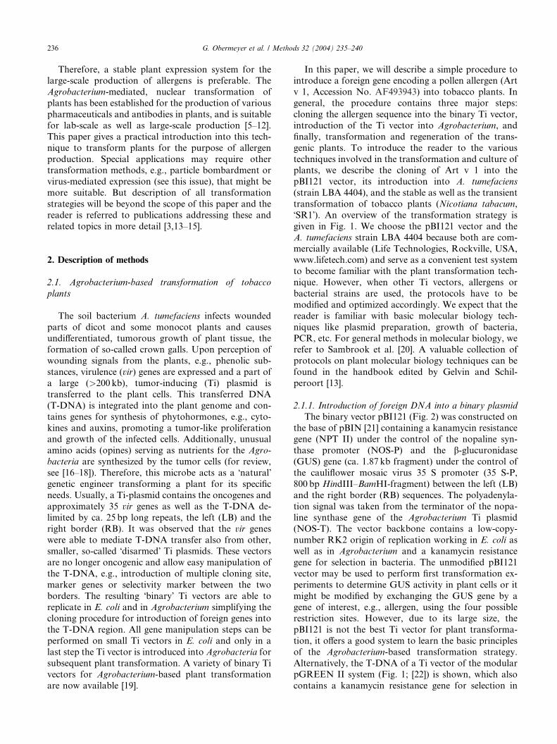
In this paper, we will describe a simple procedure tointroduce a foreign gene encoding a pollen allergen (Art
v 1, Accession No. AF493943) into tobacco plants. In
general, the procedure contains three major steps:
cloning the allergen sequence into the binary Ti vector,
introduction of the Ti vector into Agrobacterium, and
finally, transformation and regeneration of the trans-
genic plants. To introduce the reader to the various
techniques involved in the transformation and culture ofplants, we describe the cloning of Art v 1 into the
pBI121 vector, its introduction into A. tumefaciens
(strain LBA 4404), and the stable as well as the transient
transformation of tobacco plants (Nicotiana tabacum,
�SR1�). An overview of the transformation strategy is
given in Fig. 1. We choose the pBI121 vector and the
A. tumefaciens strain LBA 4404 because both are com-
mercially available (Life Technologies, Rockville, USA,www.lifetech.com) and serve as a convenient test system
to become familiar with the plant transformation tech-
nique. However, when other Ti vectors, allergens or
bacterial strains are used, the protocols have to be
modified and optimized accordingly. We expect that the
reader is familiar with basic molecular biology tech-
niques like plasmid preparation, growth of bacteria,
PCR, etc. For general methods in molecular biology, werefer to Sambrook et al. [20]. A valuable collection of
protocols on plant molecular biology techniques can be
found in the handbook edited by Gelvin and Schil-
peroort [13].
2.1.1. Introduction of foreign DNA into a binary plasmid
The binary vector pBI121 (Fig. 2) was constructed on
the base of pBIN [21] containing a kanamycin resistancegene (NPT II) under the control of the nopaline syn-
thase promoter (NOS-P) and the b-glucuronidase(GUS) gene (ca. 1.87 kb fragment) under the control of
the cauliflower mosaic virus 35 S promoter (35 S-P,
800 bp HindIII–BamHI-fragment) between the left (LB)
and the right border (RB) sequences. The polyadenyla-
tion signal was taken from the terminator of the nopa-
line synthase gene of the Agrobacterium Ti plasmid(NOS-T). The vector backbone contains a low-copy-
number RK2 origin of replication working in E. coli as
well as in Agrobacterium and a kanamycin resistance
gene for selection in bacteria. The unmodified pBI121
vector may be used to perform first transformation ex-
periments to determine GUS activity in plant cells or it
might be modified by exchanging the GUS gene by a
gene of interest, e.g., allergen, using the four possiblerestriction sites. However, due to its large size, the
pBI121 is not the best Ti vector for plant transforma-
tion, it offers a good system to learn the basic principles
of the Agrobacterium-based transformation strategy.
Alternatively, the T-DNA of a Ti vector of the modular
pGREEN II system (Fig. 1; [22]) is shown, which also
contains a kanamycin resistance gene for selection in
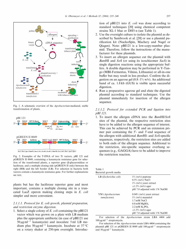
Fig. 2. Examples of the T-DNA of two Ti vectors, pBI 121 and
pGREEN II 0049, containing a kanamycin resistance gene for selec-
tion of the transformed plants, a reporter gene (b-glucuronidase or
luciferase, and a multiple cloning side (pGREEN II only) between the
right (RB) and the left border (LB). For selection in bacteria both
vectors contain a kanamycin resistance gene. For further explanations
see text.
Fig. 1. A schematic overview of the Agrobacterium-mediated, stable
transformation of plants.
Table 1
Bacterial growth media
LB (Escherichia coli) 1% (w/v) peptone
0.5% (w/v) NaCl
1% (w/v) yeast extract
�1.5% (w/v) agar
pH 7.0 adjusted with 1N NaOH
YM (Agrobacterium
tumefaciens)
0.04% (w/v) yeast extract
1% (w/v) mannitol
1.7mM NaCl
0.8mMMgSO4
2.2mM K2PO4
�1.5% (w/v) agar
pH 7.0 adjusted with 1N NaOH
For selection of the Agrobacterium strain LBA 4404 add
100lgml�1 streptomycin.
For selection of the Agrobacterium strain LBA 4404 containing the
plasmid pBI 121 or pGREEN II 0049 add 100lgml�1 streptomycin
and 50 lgml�1 kanamycin.
G. Obermeyer et al. / Methods 32 (2004) 235–240 237
plants but has the luciferase reporter gene and most
important, contains a multiple cloning site in a trun-
cated LacZ operon making cloning steps in E. coli
simpler and more convenient.
2.1.1.1. Protocol for E. coli growth, plasmid preparation,
and restriction enzyme digestion.
1. Select a single colony of E. coli containing the pBI121vector which was grown on a plate with LB medium
plus the appropriate antibiotic (in case of pBI121 use
50 lgml�1 kanamycin) and inoculate 5ml LB me-
dium plus 50 lgml�1 kanamycin. Incubate at 37 �Con a rotary shaker at 250 rpm overnight. Introduc-
tion of pBI121 into E. coli was done according tostandard techniques [20] using chemical competent
strains XL-1 blue or DH5-a (see Table 1).
2. Use the overnight culture to isolate the plasmid as de-
scribed by Sambrook et al. [20] or use a plasmid pu-
rification kit (NucleoSpin, Machery and Nagel or
Qiagen). Note: pBI121 is a low-copy-number plas-
mid. Therefore, follow the instructions of the manu-
facturer for these plasmids.3. To insert an allergen sequence cut the plasmid with
BamHI and SstI (or using its isoschizomer SacI) in
single digestion reactions using the appropriate buf-
fers. A double digestion may be performed in Y-Tan-
go (MBI-Fermentas, Vilnius, Lithuania) or all-in-one
buffer but may result in less product. Confirm the di-
gestion on an agarose gel (0.8–1% w/v). An additional
band of ca. 1.8 kb (GUS) is visible upon successfuldigestion.
4. Run a preparative agarose gel and elute the digested
plasmid according to standard techniques. Use the
plasmid immediately for insertion of the allergen
sequence.
2.1.1.2. Protocol for extended PCR and ligation into
pBI 121.1. To insert the allergen cDNA into the BamHI/SstI
sites of the plasmid, the respective restriction sites
have to be added to the allergen sequence of interest.
This can be achieved by PCR and an extension pri-
mer pair containing the 50- and 30-end sequence of
the allergen with additional BamHI- and SstI-specific
sequences, respectively, the restriction sites are added
to both ends of the allergen sequence. Additional tothe restriction, site-specific sequence overhang se-
quences (e.g., GAGGA) have to be added to improve
the restriction reaction.
238 G. Obermeyer et al. / Methods 32 (2004) 235–240
2. Confirm the PCR by separating the products on anagarose gel and elute the product of the appropriate
size from the gel.
3. Digest the PCR product with the respective enzymes
(see above) and purify the digested PCR product by
precipitation with ethanol.
4. The allergen is ligated into the plasmid using a 1:3 ra-
tio of plasmid to allergen DNA (other ratios may be
tested to improve the ligation reaction). Ligation iscarried out overnight at 16 �C using T4 DNA ligase
(3–5 units in a reaction volume of 110 ll).5. Incorporate the ligated plasmid into a chemically
competent E. coli strain by heat shock transformation
[20]. Select positive clones on kanamycin containing
agar plates and confirm the successful ligation and/
or plasmid incorporation by performing a PCR using
primer sets hybridizing to sequences of the insertedallergen and to the plasmid, respectively. Prepare
glycerol stocks from positive E. coli clones for long-
term storage at )80 �C.
2.1.1.3. Protocol for A. tumefaciens growth.
1. Inoculate a plate (solidifiedYMmedium+100 lgml�1
streptomycin) with A. tumefaciens, strain LBA 4404,
from a )80 �C frozen stock.2. Incubate at 28 �C for 2–3 days until single colonies
are visible.
3. Inoculate 10ml liquid YM medium with a single col-
ony and incubate at 28 �C for 3 days on a rotary sha-
ker (200 rpm).
4. Measure the absorption at 600 nm (OD600) to moni-
tor the growth of the bacterial culture. Usually, an
OD600 of 0.5–0.6 is obtained after 30 h under theseconditions.
2.1.2. Transformation of Agrobacterium
Several methods have been developed to mobilize
plasmids into Agrobacterium cells, e.g., tri-parental
mating, a freeze–thaw method [23], and electropora-
tion. As the strain LBA 4404 is already electrocom-
petent, we describe here the electro-transformation ofAgrobacterium.
2.1.2.1. Preparation of electrocompetent Agrobacterium
[29,24].
1. Pellet an overnight culture of Agrobacterium by cen-
trifugation (3000 rpm, 4 �C, 15min) and discard the
supernatant. Resuspend the bacteria pellet with 1/10
volume of sterile, ice-cold 10% (v/v) glycerol by gentlevortexing. Fill up to initial volume (10/10) with ice-
cold 10% (v/v) glycerol.
2. Repeat the centrifugation and washing steps with
10% (v/v) glycerol twice.
3. Finally resuspend in a 1/100 vol with 10% (v/v) ice-
cold glycerol, aliquot, and freeze in liquid N2. Store
at )80 �C for up to 3 months.
2.1.2.2. Electro-transformation of Agrobacterium.1. Thaw a fresh aliquot of electrocompetent Agrobacte-
ria on ice. Add plasmid samples in water or TE buffer
(<5 ll) in a pre-cooled electroporation cuvette
(0.1 cm distance between the electrode plates). Then,
add 20 ll of the electrocompetent bacteria and mix
gently.
2. Set the parameters of the electroporation device to a
field strength of 20 kV cm�1 and a time constant of5ms and apply one pulse. Many manufacturers pro-
vide good electroporation protocols for a variety of
bacteria in their instruction manual, e.g., BioRad Mi-
cropulser: select �Agr� (2.2 kV) or Gibco-BRL cell
porator: voltage 400V, 333 lF, low resistance, but
electroporation parameters should be optimized to
improve the transformation frequency.
3. Add electroporated Agrobacteria immediately to 1mlYM medium at room temperature and incubate for
3 h at 28 �C on a shaker at 250 rpm.
4. Plate on YM plates in the presence of 50 lgml�1
kanamycin at 28 �C for 2 days. Check for successful
transformation.
5. Grow transformed Agrobacterium clones in liquid
YM medium and prepare glycerol stocks.
2.1.3. Transformation of tobacco plants
For the production of large amounts of recombinant
allergens, a stable transformation of tobacco plants is
preferable to a transient expression. However, transient
expression of the foreign gene can be done quite fast and
it takes only about 1 week until the expression products
can be monitored instead of 6–8 months in a stable
transformation system. In some cases, it is important totest the constructed Ti vectors especially when the
physiological function of the introduced gene is not
known and its expression might be lethal for the plant.
An elegant and convenient transient expression method
for plants is the Agro-infiltration [25,26] in which plant
leaves are infiltrated with an Agrobacterium suspension
harbouring the constructed Ti vector. Alternatively, the
Ti vector itself can be introduced into plant cells usingparticle bombardment. But this method requires addi-
tional equipment (gene gun, etc.) and time-consuming
optimization of the bombardment procedure for the
respective plant tissue and vector (pressure, distance,
post-bombardment treatments, etc.).
Stable transformation of plants can be obtained by
the so-called �leaf-disc method� where parts of a leaf are
co-cultured with Agrobacteria that will infect some ofthe leaf cells. The infected, and subsequently trans-
formed, cells are able to grow on antibiotic-containing
media and form a callus from which shoots are
growing depending on the concentrations of the added
phytohormones. The shoots are then transferred to
another, hormone-free medium allowing the generation
of roots. The small plantlets may then be planted into
Table 2
Preparation of tobacco growth media Linsmaier–Skoog (LS) medium
(based on Murashige–Skoog (MS) salt solutions)
10�Macroelements per 1L
188mM KNO3 19.0 g
206mM NH4NO3 16.5 g
30mM CaCl2 4.4 g
15mM MgSO4 3.7 g
12.5mM KH2PO4 1.7 g
100�Fe-EDTA solution per 1L
10mM Na2-EDTA 3.73 g
10mM FeSO4 � 7H2O 2.73 g
Filter sterilize the solution!
B5 vitamin solution per 100ml
Nicotinic acid 100mg
Thiamine HCl 1000mg
Pyridoxine HCl 100mg
Myo-inositol 10,000mg
100�Microelements per 1L
10mM MnSO4 � 4H2O 2.25 g
3mM ZnSO4 0.85 g
10mM H3BO3 0.62 g
10mM KI 0.082 g
0.1mM Na2MoO4 � 2H2O 0.025 g
0.01mM CuSO4 � 5H2O 0.0025 g
0.01mM CoCl2 � 6H2O 0.0025 gL�1
Stock solutions (filter sterilized)
a-Naphthaleneacetic acid (a-NAA, 100mgml�1 in ethanol)
Benzyladenine (BA, 1mgml�1 in ethanol)
A ready-to-use mixture of MS salts is commercially available, e.g.,
Sigma, Life Technologies.
For 500ml LS-1 mix 50ml of macroelement sol. with 5ml of mi-
croelement sol. and add 15 g of sucrose. Add distilled water to a vol-
ume of 490ml and adjust pH with 1N NaOH between 5.7 and 5.8.
When solidified medium is needed add also 4 g of agar. Autoclave at
120 �C, 1 bar for 20min. Let cool to about 50 �C (hand warm) and add
5ml vitamin sol. and 5ml Fe-EDTA sol. before use.
For LS-2 prepare LS medium, autoclave and let cool to below
50 �C. Add the sterile-filtered hormones: 0.5 ll a-NAA and 500ll BA.
For LS-1-kan and LS-2-kan add kanamycin to a final concentra-
tion of 50lgml�1 to the appropriate medium.
G. Obermeyer et al. / Methods 32 (2004) 235–240 239
pots on normal soil or on a special substrate until theyare ready for analysis or purification of the expressed
gene product. It is important to harvest seeds from
plants showing the highest expression rates and to
analyse the F2 generation for antibiotic resistance and
expression levels. This transformation method requires
knowledge of aseptic techniques of cell and plant cul-
tures and it is of advantage to establish these tech-
niques before transformation experiments areperformed (see books on plant culture techniques
[14,27]). All steps of plant cultivation have to be per-
formed in a laminar flow hood.
2.1.3.1. ‘Leaf-disc’ transformation protocol.
1. Inoculate a YM (+ appropriate antibiotic) agar plate
with a frozen stock of A. tumefaciens containing the
appropriate Ti vector and incubate at 28 �C for 2–3days. Add one clone to 2ml of liquid YM medium
from this plate.
2. Grow to OD600 of 0.6–0.8.
3. Take a young leaf from tobacco plants (about 5–7
leaf stage), sterilize the surface of the leaf by dipping
into 70 (v/v)% ethanol, washing for 2min in 35 (v/
v)% commercial bleach, and finally, wash thor-
oughly with sterile water (2–5�). When sterile grownplants are used, surface sterilization is not necessary.
4. Prepare leaf sections by cutting small pieces
(5� 5mm) out of the leaf or by using a sharp cork
borer with a diameter of 5–10mm.
5. Transfer 3–5 leaf discs always with the upper side
down into small petri dishes containing 4–6ml LS
medium (no antibiotics and no hormones).
6. Add 50–100 ll of the overnight Agrobacterium cul-ture. Determine the optimal amount of bacteria to
be added experimentally by using dilutions of the
overnight cultures from OD¼ 0.6 to OD¼ 1.4.
7. Seal the petri dishes with Parafilm and incubate
them in a plant growth chamber at 24 �C for 2–3
days in the dark.
8. Wash the leaves in LS medium (Table 2) with car-
benicillin (500mgL�1) or another appropriate anti-biotic to kill the Agrobacteria. Take care not to
wet the lower leaf side. Place leaf discs on solidified
LS medium (LS-2-kan) containing kanamycin to se-
lect for kanamycin resistance and hormones (a-NAA and BA) to stimulate shoot growth.
9. Seal the plates with Parafilm and place in a lighted
growth chamber (16 h light/8 h dark) at 24 �C.10. Examine the leaf discs once or twice a week for con-
taminations. After 1–2 weeks, callus growth can be
noticed at the rims of the leaf discs and within 3–4
weeks tiny, green shoots can be observed whereas
the rest of the disc turns bleached.
11. Transfer 10–15mm long shoots to solidified LS me-
dium (LS-1-kan) in Magenta boxes to allow the
transformed shoots to regenerate roots.
12. At a height of at least 4 cm transfer plants to soil in
pots. Cover them with plastic bags with vent holes to
maintain high humidity conditions and gradually
adapt the plants to the standard greenhouse condi-
tions.
2.1.3.2. Agro-infiltration protocol.
1. Use a culture of Agrobacterium (LBA 4404 with pBI121) grown in YM medium supplemented with anti-
biotics to inoculate a YM medium. For vir gene in-
duction: YM+10mM Mes (pH 5.6) in supplement
with antibiotics and 20 lM acetosyringone. Grow
the culture overnight (OD600 0.6–0.8) at 28 �C and
pellet the bacteria for 20min at 15 �C, 4000g. Resus-
pend the bacterial pellet with infiltration medium
(INF-Med) to a final volume of 50ml and incubatefor 1–2 h at room temperature (see Table 3).
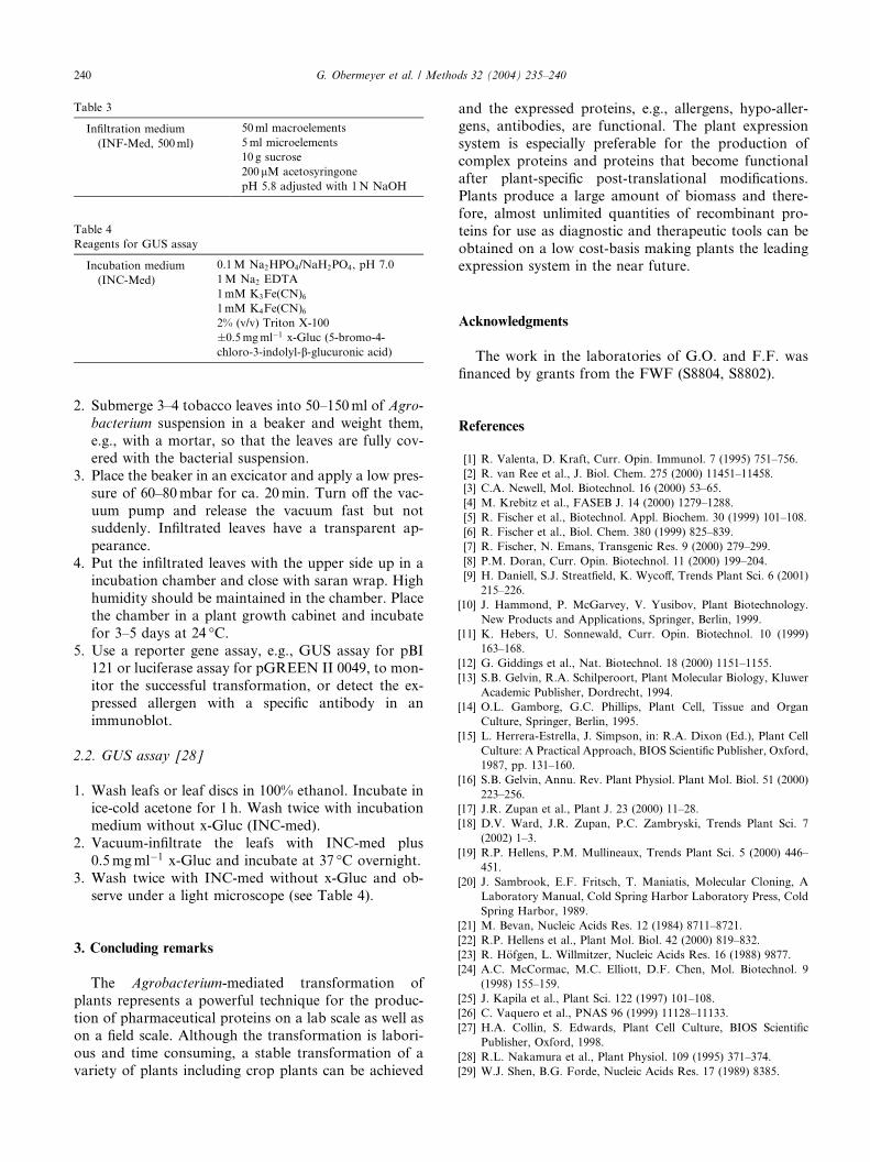
Table 3
Infiltration medium
(INF-Med, 500ml)
50ml macroelements
5ml microelements
10 g sucrose
200lM acetosyringone
pH 5.8 adjusted with 1N NaOH
Table 4
Reagents for GUS assay
Incubation medium
(INC-Med)
0.1M Na2HPO4/NaH2PO4, pH 7.0
1M Na2 EDTA
1mM K3Fe(CN)61mM K4Fe(CN)62% (v/v) Triton X-100
�0.5mgml�1 x-Gluc (5-bromo-4-
chloro-3-indolyl-b-glucuronic acid)
240 G. Obermeyer et al. / Methods 32 (2004) 235–240
2. Submerge 3–4 tobacco leaves into 50–150ml of Agro-
bacterium suspension in a beaker and weight them,
e.g., with a mortar, so that the leaves are fully cov-
ered with the bacterial suspension.
3. Place the beaker in an excicator and apply a low pres-
sure of 60–80mbar for ca. 20min. Turn off the vac-
uum pump and release the vacuum fast but notsuddenly. Infiltrated leaves have a transparent ap-
pearance.
4. Put the infiltrated leaves with the upper side up in a
incubation chamber and close with saran wrap. High
humidity should be maintained in the chamber. Place
the chamber in a plant growth cabinet and incubate
for 3–5 days at 24 �C.5. Use a reporter gene assay, e.g., GUS assay for pBI
121 or luciferase assay for pGREEN II 0049, to mon-
itor the successful transformation, or detect the ex-
pressed allergen with a specific antibody in an
immunoblot.
2.2. GUS assay [28]
1. Wash leafs or leaf discs in 100% ethanol. Incubate inice-cold acetone for 1 h. Wash twice with incubation
medium without x-Gluc (INC-med).
2. Vacuum-infiltrate the leafs with INC-med plus
0.5mgml�1 x-Gluc and incubate at 37 �C overnight.
3. Wash twice with INC-med without x-Gluc and ob-
serve under a light microscope (see Table 4).
3. Concluding remarks
The Agrobacterium-mediated transformation of
plants represents a powerful technique for the produc-
tion of pharmaceutical proteins on a lab scale as well as
on a field scale. Although the transformation is labori-
ous and time consuming, a stable transformation of a
variety of plants including crop plants can be achieved
and the expressed proteins, e.g., allergens, hypo-aller-gens, antibodies, are functional. The plant expression
system is especially preferable for the production of
complex proteins and proteins that become functional
after plant-specific post-translational modifications.
Plants produce a large amount of biomass and there-
fore, almost unlimited quantities of recombinant pro-
teins for use as diagnostic and therapeutic tools can be
obtained on a low cost-basis making plants the leadingexpression system in the near future.
Acknowledgments
The work in the laboratories of G.O. and F.F. was
financed by grants from the FWF (S8804, S8802).
References
[1] R. Valenta, D. Kraft, Curr. Opin. Immunol. 7 (1995) 751–756.
[2] R. van Ree et al., J. Biol. Chem. 275 (2000) 11451–11458.
[3] C.A. Newell, Mol. Biotechnol. 16 (2000) 53–65.
[4] M. Krebitz et al., FASEB J. 14 (2000) 1279–1288.
[5] R. Fischer et al., Biotechnol. Appl. Biochem. 30 (1999) 101–108.
[6] R. Fischer et al., Biol. Chem. 380 (1999) 825–839.
[7] R. Fischer, N. Emans, Transgenic Res. 9 (2000) 279–299.
[8] P.M. Doran, Curr. Opin. Biotechnol. 11 (2000) 199–204.
[9] H. Daniell, S.J. Streatfield, K. Wycoff, Trends Plant Sci. 6 (2001)
215–226.
[10] J. Hammond, P. McGarvey, V. Yusibov, Plant Biotechnology.
New Products and Applications, Springer, Berlin, 1999.
[11] K. Hebers, U. Sonnewald, Curr. Opin. Biotechnol. 10 (1999)
163–168.
[12] G. Giddings et al., Nat. Biotechnol. 18 (2000) 1151–1155.
[13] S.B. Gelvin, R.A. Schilperoort, Plant Molecular Biology, Kluwer
Academic Publisher, Dordrecht, 1994.
[14] O.L. Gamborg, G.C. Phillips, Plant Cell, Tissue and Organ
Culture, Springer, Berlin, 1995.
[15] L. Herrera-Estrella, J. Simpson, in: R.A. Dixon (Ed.), Plant Cell
Culture: A Practical Approach, BIOS Scientific Publisher, Oxford,
1987, pp. 131–160.
[16] S.B. Gelvin, Annu. Rev. Plant Physiol. Plant Mol. Biol. 51 (2000)
223–256.
[17] J.R. Zupan et al., Plant J. 23 (2000) 11–28.
[18] D.V. Ward, J.R. Zupan, P.C. Zambryski, Trends Plant Sci. 7
(2002) 1–3.
[19] R.P. Hellens, P.M. Mullineaux, Trends Plant Sci. 5 (2000) 446–
451.
[20] J. Sambrook, E.F. Fritsch, T. Maniatis, Molecular Cloning, A
Laboratory Manual, Cold Spring Harbor Laboratory Press, Cold
Spring Harbor, 1989.
[21] M. Bevan, Nucleic Acids Res. 12 (1984) 8711–8721.
[22] R.P. Hellens et al., Plant Mol. Biol. 42 (2000) 819–832.
[23] R. H€ofgen, L. Willmitzer, Nucleic Acids Res. 16 (1988) 9877.
[24] A.C. McCormac, M.C. Elliott, D.F. Chen, Mol. Biotechnol. 9
(1998) 155–159.
[25] J. Kapila et al., Plant Sci. 122 (1997) 101–108.
[26] C. Vaquero et al., PNAS 96 (1999) 11128–11133.
[27] H.A. Collin, S. Edwards, Plant Cell Culture, BIOS Scientific
Publisher, Oxford, 1998.
[28] R.L. Nakamura et al., Plant Physiol. 109 (1995) 371–374.
[29] W.J. Shen, B.G. Forde, Nucleic Acids Res. 17 (1989) 8385.


