niiioíipni- 'ari-' Y SURGICAL
Monitorízación con pulsioximetría durante la
extracción de terceros molares inferiores. Estudio comparativo
de tres anestésicos locales con epinefrina al 1:100.000
AUTORES/AVTHORS Regina Mestre Aspa (I), Imma Carrera Granó (1), Leonardo Berini Aytés (2), Cosme Gay Escoda (3)
(1) Odontóloga. Alumna del Master de Cirugía e Implantología Bucal. Facultad de Odontología de la Universidad de Barcelona. España.
(2) Profesor titular de Patología Quirúrgica Bucal y Maxilofacial. Profesor del Master de Cirugía e Implantología. Facultad de Odontología de la Universidad de Barcelona.
(3) Catedrático de Patología Quirúrgica Bucal y Maxilofacial. Director del Master de Cirugía e Implantología. Facultad de Odontología de la Universidad de Barcelona. Cirujano maxilofacial del Centro Médico Teknon. Barcelona.
Mestre R, Carrera I, Berini L, Gay C. Monitorización con pulsioximetría durante la extracción de terceros molares inferiores. Estudio comparativo de tres anestésicos locales con epinefrina al 1:100.000 Medicina Oral 2001; 6: 195-204. © Medicina Oral. B-96689336. ISSN 1137-2834.
RESUMEN
El objetivo de este estudio fue comparar la frecuencia cardíaca y la saturación de oxígeno bajo el efecto de tres anestésicos locales diferentes, con la misma concentración de epinefrina, durante la extracción de un tercer molar inferior en pacientes sanos. Los anestésicos locales utilizados fueron articaína al 4 %, lidocaína al 2 % y mepivacaína al 2 %, los tres con una concentración de epinefrina de 1:100.000. Se diseñó un estudio de doble ciego, paralelo y no aleatorizado. Los pacientes fueron divididos en tres grupos de quince, uno para cada anestésico. En cada paciente se monitorizó la frecuencia
Recibido: 23/7/99. Aceptado: 18/2/00.
Received: 23/7/99. Accepted: 18/2/00.
MEDICINA ORI VOL. 6 / N." 3
MAY,-JUL. 2001
cardíaca y la saturación de oxígeno antes de inyectar el anestésico, justo después, un minuto después de la utilización del motor para efectuar la ostectomía, al inicio de la fuerza para realizar la exodoncia, al finalizar la sutura y a la semana, al retirar los puntos de sutura, valor usado, este último, como ba-sal. También se registró la presión arterial y se calculó la presión arterial media, el producto presión-frecuencia y el cociente presión-frecuencia.
Sólo la variable frecuencia cardíaca presentó diferencias significativas a lo largo de las intervenciones, independientemente del tipo de anestésico local utilizado. La prueba estadística utilizada fue un análisis de la varianza multifactorial, y el grado de significación fue p < 0,05.
Como conclusiones de este estudio podemos decir que ninguno de los anestésicos utilizados afecta de forma importante las variables estudiadas en pacientes sanos, aunque éstas pueden verse alteradas significativamente por el estrés o la ansiedad que pueden comportar los diferentes momentos de la intervención.
Palabras clave: anestésicos locales, epinefrina, extracción quirúrgica del tercer molar inferior, constantes hemodinámi-cas.
INTRODUCCIÓN
Los anestésicos locales que se han utilizado en este estudio han sido mepivacaína, lidocaína y articaína, todos asociados a epinefrina a una concentración de 1:100.000. Estos tres anestésicos pertenecen al grupo de las amidas, pero la articaína está formada por un anillo tiofénico aromático, en lugar de por un anillo bencénico (1); esta diferencia de estructura molecular le confiere una mejor difusión intraósea y una menor toxicidad (menos efecto cardiodepresor) que los otros anestésicos locales de acción intermedia, la lidocaína, la mepivacaína y la prilocaína (2).
La epinefrina incorporada a la solución anestésica se utihza en cirugía bucal para aumentar la potencia y duración de la anestesia, para reducir los niveles plasmáticos de anestésico y para tener control local de la hemorragia. Su uso ha sido criticado por el peligro que supone una posible absorción sistémica masiva y los efectos cardiovasculares que puede provocar (3). Sin embargo, hay autores que prefieren obtener una mejor anestesia y, por tanto, un mejor control del dolor, añadiendo epinefrina al anestésico, antes que dejar de utilizarla y exponerse a reacciones cardiovasculares más graves como resultado del estrés que comporta el acto quirúrgico o un pobre control del dolor, lo que provocaría una liberación de catecolaminas endógenas considerablemente más importante y peligrosa (2, 4-7).
Hay diferentes factores que pueden influir en la respuesta sistémica de los vasoconstrictores, como pueden ser el lugar de la inyección, el tipo de vasoconstrictor, la concentración y el volumen de la solución inyectada, la edad y el estado físico del paciente. En pacientes jóvenes y sanos, pequeñas dosis de epinefrina pueden no originar ningún tipo de reacción sisté-
1 9 5
MESTRE R, y cois.
mica, incluso si existe un aumento de la concentración plasmática de esta catecolamina. No obstante, en pacientes mayores, con determinadas patologías, o empleando dosis más importantes de epinefrina, se han observado efectos sistémicos (8).
A nivel del miocardio, la epinefrina produce un aumento de la frecuencia cardíaca, un aumento de la fuerza de contracción y un aumento de la conducción (por una acción 6j); esto produce una elevación del volumen sistólico por minuto y de la presión arterial sistólica. Por efecto sobre los receptores a se produce vasoconstricción a nivel de piel, mucosas y área esplácnica, lo que provoca un aumento de las resistencias periféricas con elevación de la presión arterial sistólica en mayor grado que la diastólica, así como aumento de la presión diferencial y taquicardia (9).
También es importante remarcar que la epinefrina puede interactuar con las hormonas tiroideas, los antidepresivos tr i-cíclicos, los inhibidores de la monoanúnooxidasa, los B-blo-queadores, los bloqueadores del impulso nervioso y las feno-tiazinas (8, 10, 11).
La saturación de oxígeno es la medida del grado en que el oxígeno se une a la hemoglobina; se expresa como el porcentaje calculado al dividir la capacidad máxima de oxígeno por el contenido real del mismo, multiplicando el resultado por cien. Nos informa indirectamente del grado de hipoxerrúa que está sufriendo el paciente. La hipoxenüa se define como una deficiente oxigenación de la sangre. Sus signos clínicos son inquietud, confusión, ansiedad y aprensión, aunque estos síntomas no son específicos y pueden darse en otros cuadros clínicos. Aun así, en las hipoxemias severas podemos ver bradi-cardia y cianosis, que nos indican que estamos entrando en una situación crítica (12). Los grados de hipoxemia, segtín Poiset, Johnson y Nakamura (12), se muestran en la Tabla 1. Para Lowe y Brook (13) un 94-95 % de saturación de oxígeno se considera el límite superior de una amenaza de hipoxia severa. En los casos graves de hipoxemia nos encontramos con una disminución de la frecuencia respiratoria (que aumentaría con una hipoxemia moderada) y una disminución de la frecuencia cardíaca y de la presión arterial.
El pulsioxímetro mide la saturación de oxígeno en sangre arterial mediante diferencias de absorción del espectro entre los dos tipos de hemoglobina, la oxigenada y la no oxigenada.
TABLA 1
Grados de hipoxemia relacionados con la saturación de oxígeno según Poiset, Johnson y Nakamura (12)
Normal 97-100 % .SaO^
Oxigenación normal de los tejidos >95 9í .SaOj
Hipoxemia media 90-95 -3? 3 SaOj
Hipoxemia moderada 75-90 % •> SaOj
Hipoxemia severa <75 ^ ̂SaO^
36 MEDKl VOL,
lOML 6 / N.° 3
MAY.-JUL, 2001
El pulsioxímetro aplica la ley de Beer, que establece que la concentración no conocida de un soluto (hemoglobina) disuelta en un solvente conocido (sangre) puede ser determinada por la absorción de la luz del soluto. Para medir la absorción de oxihemoglobina y desoxihemoglobina se utilizan dos tipos de fotodiodos, uno que emite luz a 660 nm y otro que lo hace entre 900 y 940 nm. Además de la saturación de oxígeno, el pulsioxímetro también nos indica la frecuencia cardíaca.
Generalmente permite determinar saturaciones que oscilen entre el 70 y el 100 %. Lamentablemente, podemos encontrar diferentes factores que nos pueden impedir obtener unos registros correctos, como son: perfusión periférica disminuida, anomalías de la hemoglobina, existencia de pigmentos anómalos en sangre, etc. (14).
M A T E R I A L Y MÉTODO
Este estudio fue de doble ciego, de grupos paralelos, no aleatorizado y de casos consecutivos. Participaron en él cuarenta y cinco pacientes de edades comprendidas entre los dieciocho y los cuarenta y cinco años. Los pacientes incluidos en el estudio no presentaban patología sistémica ni alergias conocidas.
El tamaño de la muestra se determinó en base a estudios previos similares efectuados por Knoll-Kolher y cois. (15) y por Frabetti y cois. (16).
El elemento ciego se consiguió cubriendo los envases mediante etiquetas blancas con una franja de color diferente para cada anestésico; sólo los investigadores conocían el código de colores.
Monitorizamos la frecuencia cardíaca, la presión arterial sistólica, la presión arterial diastólica y el grado de saturación de oxígeno en sangre en diferentes momentos de la intervención y en la visita de control y de retirada de puntos (valor utilizado como basal). La frecuencia cardíaca y la saturación de oxígeno fueron valoradas antes de anestesiar, justo después de administrar el anestésico, un minuto después de la utilización del motor para efectuar la ostectomía, al iniciar la fuerza para la extracción del cordal y al finahzar la sutura. La presión arterial sólo fue valorada antes de administrar el anestésico y al finalizar la intervención. Para monito-rizar estas variables utilizamos un pulsioxímetro MiniOx Printer, un fonendoscopio Littman 3M y un esfigmomanó-metro Waitch; durante la toma de presión arterial el paciente se encontraba semiinclinado y con el esfigmomanómetro a nivel del corazón.
Los pacientes fueron divididos en tres grupos de quince individuos. A cada grupo se le administró un anestésico diferente: lidocaína al 2 %, mepivacaína al 2 % y articaína al 4 %, los tres con epinefrina a una concentración de 1:100.000, permitiéndose un máximo de tres ampollas (un total de 5,4 ce). En 17 de los 45 pacientes se utilizó una cantidad de solución anestésica igual o inferior a 3,6 ce. En los restantes 28 casos la cantidad utilizada fue superior a 3,6 ce (aunque siempre in-
196
Medicina Oral 2001; 6: 195-204 PULSIOXIMETRÍA DURANTE L A EXTRACCIÓN D E T E R C E R O S MOLARES INFERIORES/
PULSIOXYMETRY DURING LOWER THIRD MOLAR EXTRACTION
ferior a 5,4 ce) por provocarse dolor en el momento de la odontosección. La técnica anestésica utilizada fue la común en la extracción quirúrgica de un tercer molar inferior: bloqueo troncal de los nervios dentario inferior y lingual, e infil-trativa del nervio bucal y de la zona a intervenir.
Las extracciones quirúrgicas fueron realizadas por profesionales en formación en Cirugía e Implantología Bucal. Todos los casos incluidos en el estudio requerían realizar una ostectomía.
Cuando el paciente ya estaba anestesiado y el campo quirúrgico preparado, se realizaba una incisión en la zona del trígono retromolar, en el surco gíngivo-dentario del segundo molar y una descarga por mesial de éste, levantándose un colgajo mucoperióstico. A continuación se llevaba a cabo la ostectomía y la odontosección, si ésta era necesaria. Seguidamente se realizaba la extracción del tercer molar, se l impiaba la herida operatoria, se reposicionaba el colgajo y se suturaba. A la semana se retiraba la sutura (puntos de seda de 3/0).
En este estudio también se ha valorado el producto presión-frecuencia (RPP o rate pressure product), el cociente presión-frecuencia (PRQ o pressure rate quotient) y la presión arterial media.
El RPP resulta de multiplicar la presión arterial sistólica por la frecuencia cardíaca (17) y es un buen indicador del consumo de oxígeno por parte del núocardio en pacientes no anestesiados; puede ser útil clínicamente para detectar un excesivo trabajo cardíaco (3, 15, 17). El PRQ se calcula dividiendo la presión arterial media por la frecuencia cardíaca y, según los mismos autores (17), es más usado como indicador de isquemia cardíaca, ya que pone más énfasis en la frecuencia cardíaca que el RPP.
La presión arterial media (PAM) es la presión promedio durante la totalidad de cada ciclo cardíaco (18). Representa la relación entre la resistencia sistémica al flujo sanguíneo y la presión arterial, siendo la fuerza constante de la conducción del sistema vascular (19). Se calcula mediante la siguiente fórmula: PAM=PAD+1/3(PAS-PAD), siendo PAS la presión arterial sistólica y PAD la presión arterial diastólica.
Metodología estadística: una vez realizada la estadística descriptiva, se hizo un análisis de la varianza multifactorial utilizando el programa SPSS para Windows, versión 6.1, l i cencia n° 1250352.
• lidocaína
•mepivacaína
-articaína
1 2 3 4 5 6
Fase quirúrgica
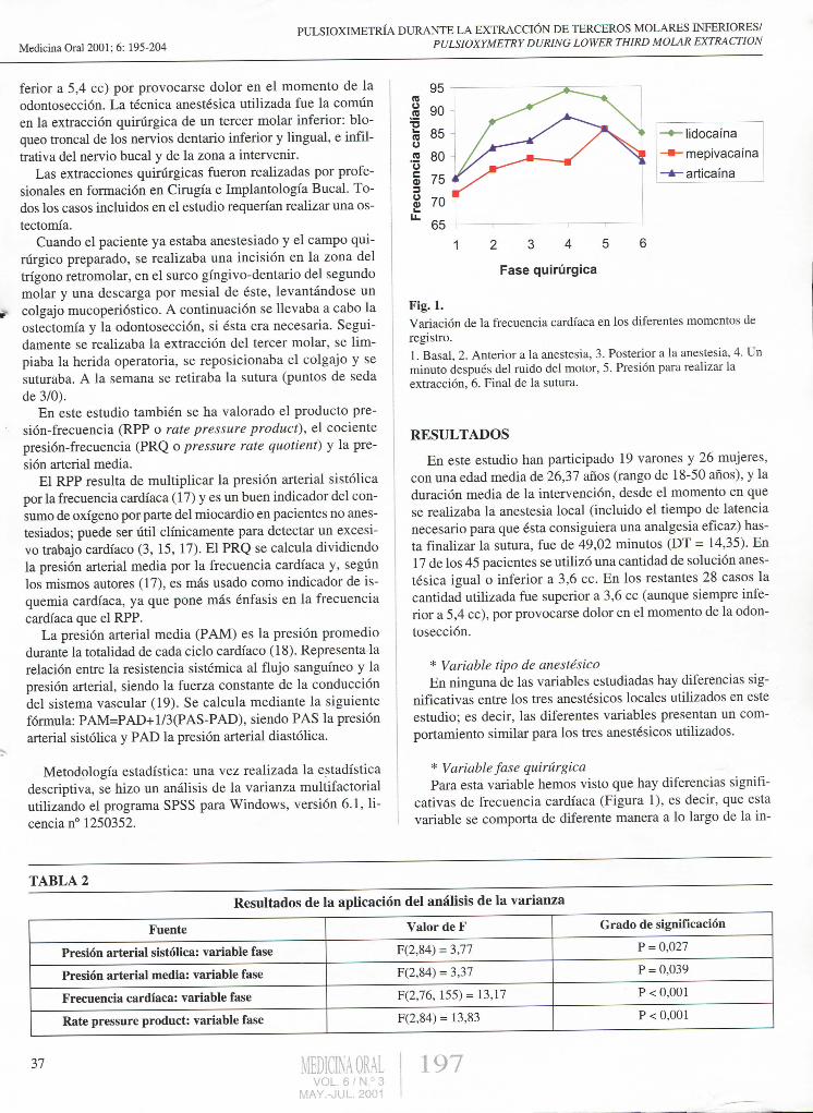
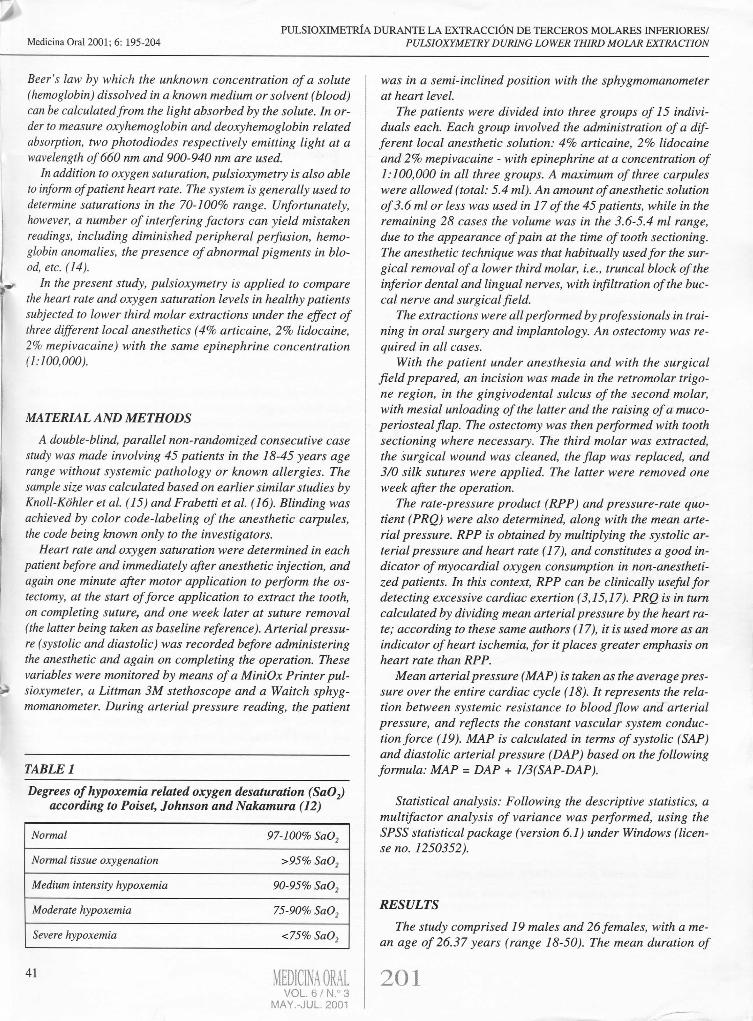
Fig. 1. Variación de la frecuencia cardíaca en los diferentes momentos de registro.
1. Basal, 2. Anterior a la anestesia, 3. Posterior a la anestesia, 4. Un minuto después del ruido del motor, 5. Presión para realizar la extracción, 6. Final de la sutura.
R E S U L T A D O S
En este estudio han participado 19 varones y 26 mujeres, con una edad media de 26,37 años (rango de 18-50 años), y la duración media de la intervención, desde el momento en que se realizaba la anestesia local (incluido el tiempo de latencia necesario para que ésta consiguiera una analgesia eficaz) hasta finalizar la sutura, fue de 49,02 minutos (DT = 14,35). En 17 de los 45 pacientes se utilizó una cantidad de solución anestésica igual o inferior a 3,6 ce. En los restantes 28 casos la cantidad utilizada fue superior a 3,6 ce (aunque siempre inferior a 5,4 ce), por provocarse dolor en el momento de la odontosección.
* Variable tipo de anestésico En ninguna de las variables estudiadas hay diferencias sig
nificativas entre los tres anestésicos locales utilizados en este estudio; es decir, las diferentes variables presentan un comportamiento similar para los tres anestésicos utiUzados.
* Variable fase quirúrgica Para esta variable hemos visto que hay diferencias signifi
cativas de frecuencia cardíaca (Figura 1), es decir, que esta variable se comporta de diferente manera a lo largo de la in-
TABLA 2
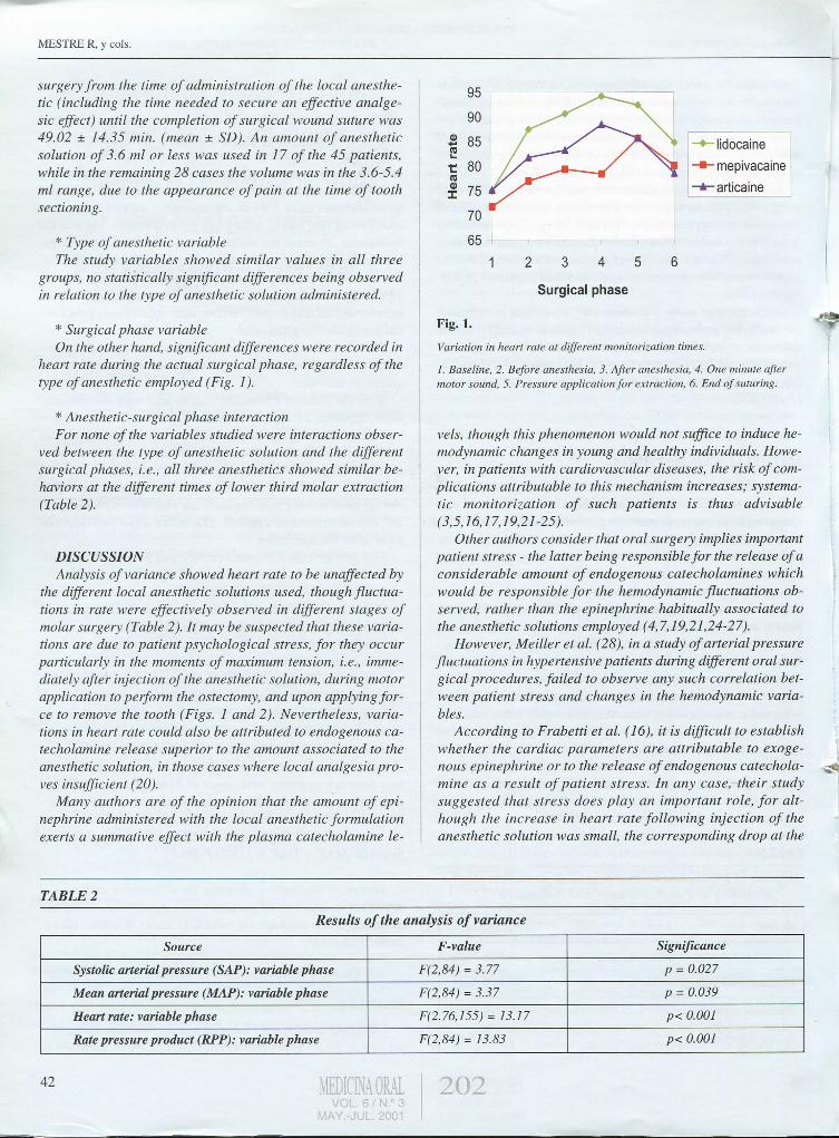
Resultados de la aplicación del análisis de la varianza
Fuente Valor de F Grado de significación
Presión arterial sistólica: variable fase F(2,84) = 3,77 P = 0,027
Presión arterial media: variable fase F(2,84) = 3,37 P = 0,039
Frecuencia cardíaca: variable fase F(2,76, 155)= 13,17 P < 0,001
Rate pressure product: variable fase F(2,84) = 13,83 P < 0,001
37 VOL. 6 / N.° 3
MAY.-JUL. 2001
197
MESTRE R, y cois.
1 2 3 4 5 6
Fase quirúrgica
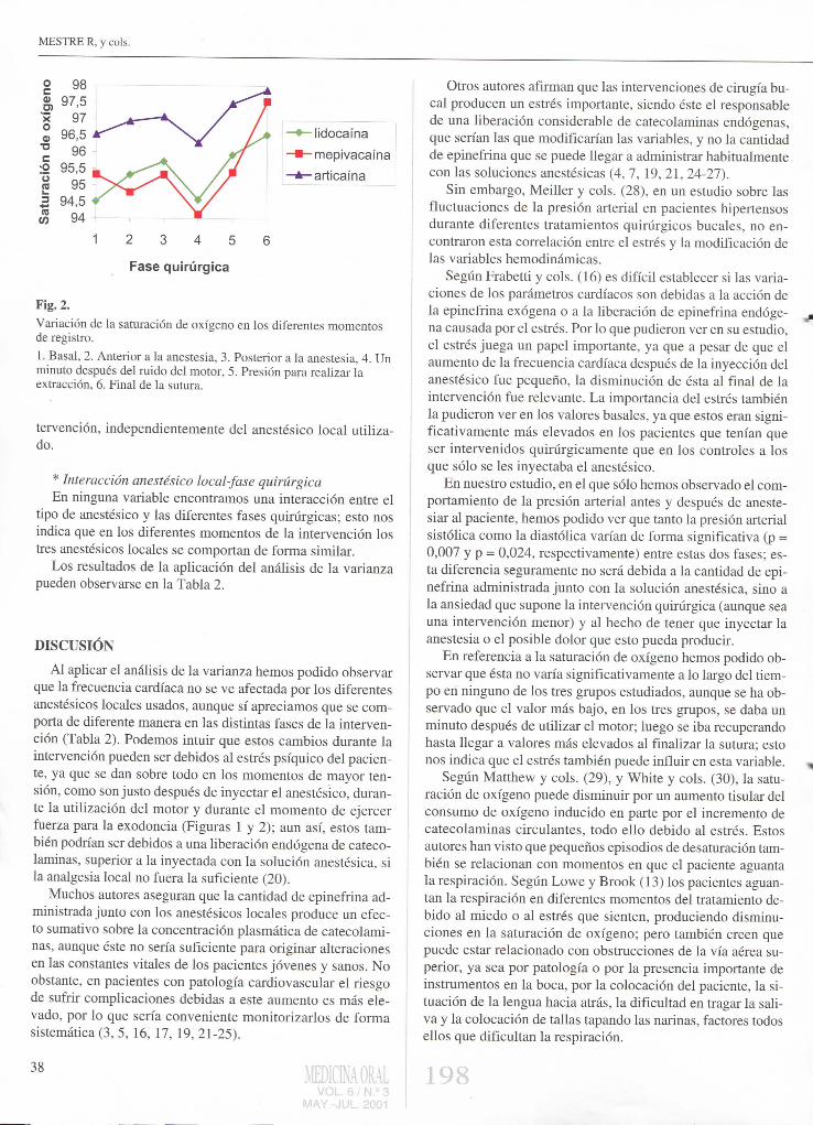
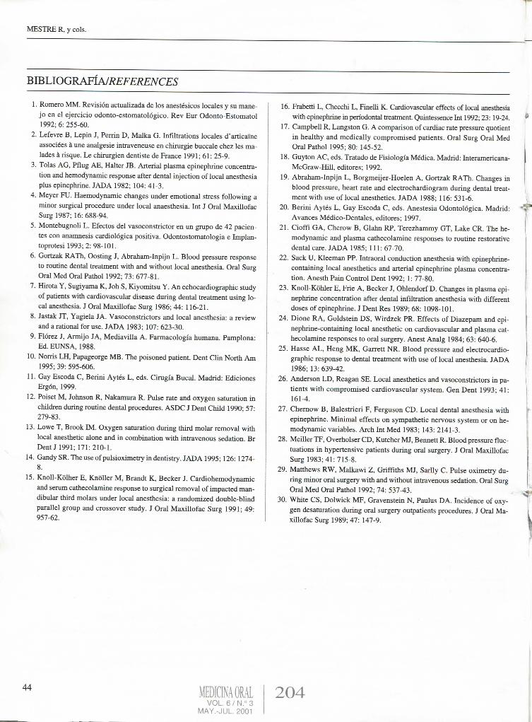
Fig. 2. Variación de la saturación de ox ígeno en los diferentes momentos de registro.
1. Basal, 2. Anterior a la anestesia, 3. Posterior a la anestesia, 4. Un minuto después del ruido del motor, 5. Presión para realizar la extracción, 6. Final de la sutura.
tervención, independientemente del anestésico local utilizado.
* Interacción anestésico local-fase quirúrgica En ninguna variable encontramos una interacción entre el
tipo de anestésico y las diferentes fases quirúrgicas; esto nos indica que en los diferentes momentos de la intervención los tres anestésicos locales se comportan de forma similar.
Los resultados de la aplicación del análisis de la varianza pueden observarse en la Tabla 2.
DISCUSIÓN
A l aplicar el análisis de la varianza hemos podido observar que la frecuencia cardíaca no se ve afectada por los diferentes anestésicos locales usados, aunque sí apreciamos que se comporta de diferente manera en las distintas fases de la intervención (Tabla 2). Podemos intuir que estos cambios durante la intervención pueden ser debidos al estrés psíquico del paciente, ya que se dan sobre todo en los momentos de mayor tensión, como son justo después de inyectar el anestésico, durante la utilización del motor y durante el momento de ejercer fuerza para la exodoncia (Figuras 1 y 2); aun así, estos también podrían ser debidos a una liberación endógena de catecolaminas, superior a la inyectada con la solución anestésica, si la analgesia local no fuera la suficiente (20).
Muchos autores aseguran que la cantidad de epinefrina administrada junto con los anestésicos locales produce un efecto sumativo sobre la concentración plasmática de catecolaminas, aunque éste no sería suficiente para originar alteraciones en las constantes vitales de los pacientes jóvenes y sanos. No obstante, en pacientes con patología cardiovascular el riesgo de sufrir complicaciones debidas a este aumento es más elevado, por lo que sería conveniente monitorizarlos de forma sistemática (3, 5, 16, 17, 19, 21-25).
Otros autores afirman que las intervenciones de cirugía bucal producen un estrés importante, siendo éste el responsable de una liberación considerable de catecolaminas endógenas, que serían las que modificarían las variables, y no la cantidad de epinefrina que se puede llegar a administrar habitualmente con las soluciones anestésicas (4, 7, 19, 21, 24-27).
Sin embargo, Meiller y cois. (28), en un estudio sobre las fluctuaciones de la presión arterial en pacientes hipertensos durante diferentes tratamientos quirúrgicos bucales, no encontraron esta correlación entre el estrés y la modificación de las variables hemodinámicas.
Según Frabetti y cois. (16) es difícil establecer si las variaciones de los parámetros cardíacos son debidas a la acción de la epinefrina exógena o a la liberación de epinefrina endógena causada por el estrés. Por lo que pudieron ver en su estudio, el estrés juega un papel importante, ya que a pesar de que el aumento de la frecuencia cardíaca después de la inyección del anestésico fue pequeño, la disminución de ésta al final de la intervención fue relevante. La importancia del estrés también la pudieron ver en los valores básales, ya que estos eran significativamente más elevados en los pacientes que tenían que ser intervenidos quirúrgicamente que en los controles a los que sólo se les inyectaba el anestésico.
En nuestro estudio, en el que sólo hemos observado el comportamiento de la presión arterial antes y después de anestesiar al paciente, hemos podido ver que tanto la presión arterial sistólica como la diastólica varían de forma significativa (p = 0,007 y p = 0,024, respectivamente) entre estas dos fases; esta diferencia seguramente no será debida a la cantidad de epinefrina administrada junto con la solución anestésica, sino a la ansiedad que supone la intervención quirúrgica (aunque sea una intervención menor) y al hecho de tener que inyectar la anestesia o el posible dolor que esto pueda producir.
En referencia a la saturación de oxígeno hemos podido observar que ésta no varía significativamente a lo largo del tiempo en ninguno de los tres grupos estudiados, aunque se ha observado que el valor más bajo, en los tres grupos, se daba un minuto después de utilizar el motor; luego se iba recuperando hasta llegar a valores más elevados al finalizar la sutura; esto nos indica que el estrés también puede influir en esta variable.
Según Matthew y cois. (29), y White y cois. (30), la saturación de oxígeno puede disminuir por un aumento tisular del consumo de oxígeno inducido en parte por el incremento de catecolaminas circulantes, todo ello debido al estrés. Estos autores han visto que pequeños episodios de desaturación también se relacionan con momentos en que el paciente aguanta la respiración. Según Lowe y Brook (13) los pacientes aguantan la respiración en diferentes momentos del tratamiento debido al miedo o al estrés que sienten, produciendo disminuciones en la saturación de oxígeno; pero también creen que puede estar relacionado con obstrucciones de la vía aérea superior, ya sea por patología o por la presencia importante de instrumentos en la boca, por la colocación del paciente, la situación de la lengua hacia atrás, la dificultad en tragar la saliva y la colocación de tallas tapando las narinas, factores todos ellos que dificultan la respiración.
M A Y -VJÜI

Medicina Oral 2001; 6: 195-204 PULSIOXIMETRÍA DURANTE L A EXTRACCIÓN D E T E R C E R O S MOLARES INFERIORES/
PULSIOXYMETRY DURING LOWER THIRD MOLAR EXTRACTION
En lo que se refiere al RPP, se cree que existe una relación entre este producto y la isquemia cardíaca (3,15,17). En nuestro estudio hemos podido ver que esta variable se comporta de diferente manera antes y después de anestesiar al paciente. En el caso de la articaína y la lidocaína, este valor estaba aumentado justo antes de inyectar la anestesia y disminuye justo después; en referencia a la mepivacaína, este valor aumenta después de haber inyectado el anestésico.
Según Tolas y cois. (3), Knoll-Kohler y cois. (15) y Campbell y cois. (17), el PRQ es más usado como indicador de isquemia cardíaca; un PRQ inferior a 1 se asociaría a isquemia subendocárdica. En nuestro estudio hemos podido ver que no existen diferencias significativas para el anestésico utilizado, ni tampoco según la variable fase quirúrgica en relación a este cociente hemodinámico.
Según Campbell y Langston (17), sólo los pacientes con valores anormales simultáneos de PRQ y RPP tienen un riesgo significativo de sufrir una isquemia cardíaca, que sería necesaria verificar con un electrocardiograma. Para ello sería conveniente monitorizar todos los pacientes con patología cardiovascular o hipertensión arterial tratada cuando tuviesen que ser sometidos a una intervención de cirugía bucal o a agresiones potencialmente dolorosas.
Finalmente podemos llegar a la conclusión de que la lidocaína al 2 %, la mepivacaína al 2 % y la articaína al 4 %, con epinefrina a una concentración de 1:100.000, no afectan de forma importante a la frecuencia cardíaca ni a la saturación de oxígeno en los pacientes sanos. El estrés o el miedo derivados de los diferentes momentos de la intervención son los que podrían alterar significativamente estas variables.
39 IDICINAORAL I 199 VOL. 6 / H° 3
MAY.-JUL. 2001

1 MESTRE R, y cois. Medicii
Pukbxjmetij monitori^flén during bwer third molar
extractíon. A comparéve study ofthreebcalanesthetics
with epinephrine 1:100,000
SUMMARY
A double-blind, parallel non-randomized study is made to compare the heart rate and oxygen saturation levéis in he-althy patients subjected to lower third molar extractions un-der the ejfect ofthree dijferent local anesthetics (4% articai-ne, 2% lidocaine, 2% mepivacaine) with the same epinephrine concentration (1:100,000). The patients were divided into three groups of 15 subjects each, according to the anesthetic employed. Heart rate and oxygen saturation were determi-ned in each patient before and immediately after anesthetic injection, and again one minute after motor application to perform the ostectomy, at the start of forcé application to ex-tract the tooth, on completing suture, and one week later at suture removal (the latter being taken as baseline reference). Blood pressure was also recorded, calculating the mean arterial pressure and the rate-pressureproduct (RPP) andpres-sure-rate quotient (PRQ). Only heart rate showedvariations during the intervention, regardless of the local anesthetic used (based on a multifactor analysis ofvariance and consi-dering statistical significance for p<0.05). It may be conclu-ded that none ofthe local anesthetics studied exert important effects upon the variables analyzed in healthy patients, though significant variations may arise due to the stress and/or an-xiety associated to certain moments of lower third molar extractíon.
Key words: local anesthetics, epinephrine, surgical lower third molar extraction, hemodynamic parameters.
INTRODUCTION
Mepivacaine, lidocaine and articaine are amide anesthetics, though articaine presents an aromatic thiophenic ring instead ofa benzene ring (1); this structural dijference provides articaine with an improved intraosseous diffusion performance and lesser toxicity (in the form of cardiac depressive action) than the other intermedíate action local anesthetics, i.e., lidocaine, mepivacaine andprilocaine (2).
Epinephrine added to the anesthetic solution is used in oral surgery to increase the potency and duration ofanesthesia, to reduce the plasma concentrations of anesthetic, and to ajford local control over bleeding. However, the use of epinephrine has been questioned due to the risk ofmassive systemic ab-sorption ofthe drug and the cardiovascular effects it may cause (3). Nevertheless, some authors prefer to secure improved anesthesia and thus a better control ofpain by adding epinephrine, than to avoid the latter and risk even worse cardiovascular reactions due to surgically induced patient stress or poor pain suppression - which would lead to even more important and dangerous consequences secondary to endoge-nous catecholamine reléase (2,4-7).
A number of factors may influence vasoconstrictor systemic response, including the injection site, the type of vasoconstrictor used, the volume and concentration ofthe injected solution, and the age and physical condition ofthe patient. In young and healthy individuáis, small epinephrine doses may induce no systemic reaction at all, even in the presence ofan increased systemic concentration of the catecholamine. In contrast, elderly patients with certain pathologies, or exposed to larger epinephrine doses, can suffer systemic effects (8).
Epinephrine action upon the myocardial J5j receptors produces an increase in heart rate, myocardial contractility (po-sitive inotropism) and conduction velocity. These net stimula-tory effects in tum cause an increase in systolic output and systolic arterial pressure. On the other hand, epinephrine action upon the vascular a receptors causes vasoconstriction in the skin, mucosas and splanchnic región, thus incrementing peripheral vascular resistance and causing a proportionately greater rise in systolic versus diastolic pressure (i.e., the dif-ferential pressure increases), with tachycardia (9).
It is also important to point out that epinephrine can inte-ract with the thyroid hormones, tricyclic antidepressants, mo-noamine oxidase inhibitors, beta-blockers, nerve conduction blockers and phenothiazines (8,10,11).
Oxygen saturation (Sa02) quantifies the degree of oxygen binding to hemoglobin, and is calculated by dividing the máximum oxygen capacity by the actual oxygen contents x 100 (i.e., expressed as a percentage). This parameter indirectly informs ofthe degree of patient hypoxemia (i.e., blood oxygen deficiency). The clinical signs of hypoxemia are restlessness, confusión, anxiety and apprehension - though these manifes-tations are not specific and can also be caused by other con-ditions. Nevertheless, severe hypoxemia is characterized by bradycardia and cyanosis, which indícate the onset ofa criti-cal patient condition (12). The degrees of hypoxemia according to Poiset, Johnson and Nakamura (12) are reflected in Table 1. According to Lowe and Brook (13), 94-95% oxygen saturation is the limit threatening severe hypoxemia. The latter involves a depressed breathing rate (versus an increased respiratory frequency in cases of modérate hypoxemia) and a decrease in heart rate and arterial pressure.
Pulsioxymeters measure blood oxygen saturation according to differences in the absorption spectra ofthe two types of hemoglobin (oxygenated and non-oxygenated), based on
40 IDICINAORÁL VOL. 6 / N . ° 3
MAY.-JUL. 2001
2 0 0 41
1
Medicina Oral 2001; 6: 195-204 PULSIOXIMETRÍA DURANTE L A EXTRACCIÓN D E T E R C E R O S MOLARES INFERIORES/
PULSIOXYMETRY DURING LOWER THIRD MOLAR EXTRACTION
Beer's law by which the unknown concentration ofa solute (hemoglobin) dissolved in a known médium orsolvent (blood) can be calculated from the light absorbed by the solute. In or-der to measure oxyhemoglobin and deoxyhemoglobin related absorption, two photodiodes respectively emitting light at a wavelength of660 nm and 900-940 nm are used.
In addition to oxygen saturation, pulsioxymetry is also able to inform of patient heart rate. The system is generally used to determine saturations in the 70-100% range. Unfortunately, however, a number of interfering factors can yield mistaken readings, including diminished peripheral perfusión, hemoglobin anomalies, the presence ofabnormal pigments in blood, etc. (14).
In the present study, pulsioxymetry is applied to compare the heart rate and oxygen saturation levéis in healthy patients subjected to lower third molar extractions under the effect of three different local anesthetics (4% articaine, 2% lidocaine, 2% mepivacaine) with the same epinephrine concentration (1:100,000).
MATERIAL AND METHODS
A double-blind, parallel non-randomized consecutive case study was made involving 45 patients in the 18-45 years age range without systemic pathology or known allergies. The sample size was calculated based on earlier similar studies by Knoll-Kohler et al. (15) and Frabetti et al. (16). Blinding was achieved by color code-labeling of the anesthetic carpules, the code being known only to the investigators.
Heart rate and oxygen saturation were determined in each patient before and immediately after anesthetic injection, and again one minute after motor application to perform the ostectomy, at the start of forcé application to extract the tooth, on completing suture, and one week later at suture removal (the latter being taken as baseline reference). Arterial pressure (systolic and diastolic) was recorded before administering the anesthetic and again on completing the operation. These variables were monitored by means ofa MiniOx Printer pul-sioxymeter, a Littman 3M stethoscope and a Waitch sphyg-momanometer. During arterial pressure reading, the patient
TABLE 1
Degrees of hypoxemia related oxygen desaturation (SaO^) according to Poiset, Johnson and Nakamura (12)
Normal 97-100% SaO^
Normal tissue oxygenation >95% SaO^
Médium intensity hypoxemia 90-95% SaO^
Modérate hypoxemia 75-90% SaO^
Severe hypoxemia <75%Sa02
was in a semi-inclined position with the sphygmomanometer at heart level.
The patients were divided into three groups of 15 individuáis each. Each group involved the administration ofa different local anesthetic solution: 4% articaine, 2% lidocaine and 2% mepivacaine - with epinephrine at a concentration of 1:100,000 in all three groups. A máximum ofthree carpules were allowed (total: 5.4 mi). An amount of anesthetic solution of3.6 mi or less was used in 17 ofthe 45 patients, while in the remaining 28 cases the volume was in the 3.6-5.4 mi range, due to the appearance ofpain at the time of tooth sectioning. The anesthetic technique was that habitually used for the surgical removal ofa lower third molar, i.e., truncal block ofthe inferior dental and lingual nerves, with infiltration ofthe buc-cal nerve and surgical field.
The extractions were allperformed by professionals in trai-ning in oral surgery and implantology. An ostectomy was re-quired in all cases.
With the patient under anesthesia and with the surgical field prepared, an incisión was made in the retromolar trigo-ne región, in the gingivodental sulcus of the second molar, with mesial unloading ofthe latter and the raising ofa muco-periosteal flap. The ostectomy was then performed with tooth sectioning where necessary. The third molar was extracted, the surgical wound was cleaned, the flap was replaced, and 3/0 silk sutures were applied. The latter were removed one week after the operation.
The rate-pressure product (RPP) and pressure-rate quotient (PRQ) were also determined, along with the mean arterial pressure. RPP is obtained by multiplying the systolic arterial pressure and heart rate (17), and constitutes a good in-dicator of myocardial oxygen consumption in non-anestheti-zed patients. In this context, RPP can be clinically usefulfor detecting excessive cardiac exertion (3,15,17). PRQ is in tum calculated by dividing mean arterial pressure by the heart rate; according to these same authors (17), it is used more as an indicator of heart ischemia, for it places greater emphasis on heart rate than RPP.
Mean arterial pressure (MAP) is taken as the average pressure over the entire cardiac cycle (18). It represents the rela-tion between systemic resistance to blood flow and arterial pressure, and reflects the constant vascular system conduction forcé (19). MAP is calculated in terms of systolic (SAP) and diastolic arterial pressure (DAP) based on thefollowing formula: MAP = DAP + I/3(SAP-DAP).
Statistical analysis: Following the descriptive statistics, a multifactor analysis of variance was performed, using the SPSS statistical package (versión 6.1) under Windows (licen-seno. 1250352).
RESULTS
The study comprised 19 males and 26females, with a mean age of 26.37 years (range 18-50). The mean duration of
41 MEDICMORÁL VOL. 6 / N.o 3
MAY.-JUL. 2001
201
MESTRE R, y cois. Medicii
surgery from the time of administration ofthe local anesthetic (including the time needed to secure an effective analge-sic effect) until the completion of surgical wound suture was 49.02 ± 14.35 min. (mean ± SD). An amount of anesthetic solution of 3.6 mi or less was used in 17 ofthe 45 patients, while in the remaining 28 cases the volume was in the 3.6-5.4 mi range, due to the appearance ofpain at the time of tooth sectioning.
* Type of anesthetic variable The study variables showed similar valúes in all three
groups, no statistically significant differences being observed in relation to the type of anesthetic solution administered.
* Surgical phase variable On the other hand, significant differences were recorded in
heart rate during the actual surgical phase, regardless ofthe type of anesthetic employed (Fig. 1).
* Anesthetic-surgical phase interaction For none ofthe variables studied were interactions obser
ved between the type of anesthetic solution and the different surgical phases, i.e., all three anesthetics showed similar be-haviors at the different times of lower third molar extraction (Table 2).
DISCUSSION Analysis ofvariance showed heart rate to be unaffected by
the different local anesthetic solutions used, though fluctua-tions in rate were ejfectively observed in different stages of molar surgery (Table 2). It may be suspected that these variations are due to patient psychological stress, for they occur particularly in the moments of máximum tensión, i.e., immediately after injection ofthe anesthetic solution, during motor application to perform the ostectomy, and upon applying forcé to remove the tooth (Figs. 1 and 2). Nevertheless, variations in heart rate could also be attributed to endogenous catecholamine reléase superior to the amount associated to the anesthetic solution, in those cases where local analgesia pro-ves insujficient (20).
Many authors are of the opinión that the amount of epinephrine administered with the local anesthetic formulation exerts a summative effect with the plasma catecholamine le-
- lidocaine -mepivacaine -articaine
1 2 3 4 5 6
Surgical phase
Fig. 1. Variation in heart rate at different monitorization times.
1. Baseline, 2. Before anesthesia, 3. After anesthesia, 4. One minute after motor sound, 5. Pressure application for extraction, 6. End of suturing.
veis, though this phenomenon would not suffice to induce hemodynamic changes in young and healthy individuáis. However, in patients with cardiovascular diseases, the risk ofcom-plications attributable to this mechanism increases; systema-tic monitorization of such patients is thus advisable (3,5,16,17,19,21-25).
Other authors consider that oral surgery implies important patient stress - the latter being responsible for the reléase ofa considerable amount of endogenous catecholamines which would be responsible for the hemodynamic fluctuations observed, rather than the epinephrine habitually associated to the anesthetic solutions employed (4,7,19,21,24-27).
However, Meiller et al. (28), in a study of arterial pressure fluctuations in hypertensive patients during different oral surgical procedures, failed to observe any such correlation between patient stress and changes in the hemodynamic variables.
According to Frabetti et al. (16), it is dijficult to establish whether the cardiac parameters are attributable to exoge-nous epinephrine or to the reléase of endogenous catecholamine as a result of patient stress. In any case, their study suggested that stress does play an important role, for alt-hough the increase in heart rate following injection of the anesthetic solution was small, the corresponding drop at the
^ Fig.;
TABLE 2
Results of the analysis ofvariance
Source F-value Significance
Systolic arterial pressure (SAP): variable phase F(2,84) = 3.77 p = 0.027
Mean arterial pressure (MAP): variable phase F(2,84) = 3.37 p = 0.039
Heart rate: variable phase F(2.76,I55) = 13.17 p< 0.001
Rate pressure product (RPP): variable phase F(2,84) = 13.83 p< 0.001
42
J

Medicina Oral 2001 ; 6: 195-204 P U L S I O X I M E T R Í A D U R A N T E L A E X T R A C C I Ó N D E T E R C E R O S M O L A R E S INFERIORES/
PULSIOXYMETRY DURING LOWER THIRD MOLAR EXTRACTION
-lidocaine •mepivacaine -articaine
Surgical phase
* ^ Fig.2. Variation in oxygen saturation (SaO-,) at dijferent monitorization times.
1. Baseline, 2. Befare anesthesia, 3. After anesthesia, 4. One minute after motor sound, 5. Pressure application for extraction, 6. End ofsuturing.
end of the operation was quite relevant. The importance of stress was also reflected by the baseline valúes, which were significantly higher among the patients programmedfor sur-gery than in the controls who were only subjected to anest-hetic injection.
In the present study, in which arterial pressure monitorization was limited to befare and after patient anesthesia, both the systolic and diastolic readings were seen to vary signifi-cantly (p = 0.007 andp = 0.024, respectively) between these two phases. This dijference was probably not attributable to the amount of epinephrine administered with the local anest-hetic solution, but rather to patient anxiety caused by the operation (even though lower third molar extraction constitutes only minor surgery) and the fact of requiring anesthetic injection, or the possible pain associated to it
Oxygen saturation did not vary significantly over time in any ofthe three patient groups studied, though the lowest valúes in all three corresponded to one minute after starting to use the motor for performing the ostectomy. The saturation valúes subsequently recovered to reach higher valúes at the end of suturing. This suggests that patient stress may likewise influence this variable.
According to Matthew et al. (29) and White et al. (30), oxygen saturation may drop due to an increase in tissue oxygen consumption partly induced by the rise in circulating cate-cholamines - all as a consequence of patient stress. These aut-horsfound that small desaturation episodes are also related to moments of patient breath-hold. According to Lowe and Brook (13), patients tend to hold their breath at different ti
mes during treatment because offear or stress - a phenome-non that induces minor decreases in oxygen saturation. Ho-wever, they are also ofthe opinión that such situations may be related to upper airways obstruction, associated either to pat-hology or the important presence of Instruments within the oral cavity, patient positioning, displacement ofthe tongue backwards, difficulties in swallowing saliva, etc. - all of which complicate breathing.
As regards the rate-pressure product (RPP), a relations-hip is though to exist between this parameter and cardiac ischemia (3,15,17). In our study, RPP behaved differently befare and after anesthetizing the patient. In the case of articaine and lidocaine, the valué was seen to be elevated im-mediately prior to injection ofthe anesthetic solution, follo-wed by a drop immediately after administration. In the case of mepivacaine, however, RPP increased after anesthetic injection.
According to Talas et al. (3), Knoll-Kóhler et al. (15) and Campbell and Langston (17), the pressure-rate quatient (PRQ) is the parameter mast widely used as an indicator of cardiac ischemia. In this context, PRQ < 1 is associated to su-bendocardial ischemia. In the present study, no significant differences in PRQ were observed in terms of either the anesthetic solution employed or the surgical phase involved.
According to Campbell and Langston (17), only patients with simultaneously abnormal RPP and PRQ valúes are at a significant risk of suffering cardiac ischemia, which would moreaver have ta be confirmed electracardiographically. Accordingly, it would be advisable to monitor all patients with knawn cardiovascular disease or arterial hypertension pragrammed far oral surgery or potentially painful proce-dures.
It may thus be concluded that 2% lidocaine, 2% mepivacaine and 4% articaine associated to epinephrine 1:100,000 exert no important effect upon heart rate or oxygen saturation in healthy patients. Any significant alterations in these para-meters may be attributed to patient stress and/or fear at different moments during surgery.
CORRESPONDENCIA/CORK£SPOiVÍ)£iVC£
Dr . Cosme Gay Escoda
C/Ganduxer, 140,4°
08022-Barcelona
Tfno. : 93 402 42 74
E-ma i l : [email protected]
http://www.gayescoda.com
43 203
MESTRE R, y cois.
BIBLIOGRAFIA/REFERENCES
1. Romero MM. Revisión actualizada de los anestésicos locales y su manejo en el ejercicio odonto-estomatológico. Rev Eur Odonto-Estomatol 1992; 6: 255-60.
2. Lefevre B, Lepin J, Perrin D, Malka G. Infiltrations locales d'articaíne associées á une analgesie intraveneuse en chirurgie buccale chez les ma-lades á risque. Le chirurgien dentiste de France 1991; 61: 25-9.
3. Tolas AG, Mug A E , Halter JB. Arterial plasma epinephrine concentration and hemodynamic response after dental injection of local anesthesia plus epinephrine. JADA 1982; 104: 41-3.
4. Meyer FU. Haemodynamic changes under emotional stress following a minor surgical procedure under local anaesthesia. Int J Oral Maxillofac Surg 1987; 16: 688-94.
5. Montebugnoli L . Efectos del vasoconstrictor en un grupo de 42 pacientes con anamnesis cardiológica positiva. Odontostomatologia e Implan-toprotesi 1993; 2: 98-101.
6. Gortzak RATh, Oosting J, Abraham-Inpijn L . Blood pressure response to routine dental treatment with and without local anesthesia. Oral Surg Oral Med Oral Pathol 1992; 73: 677-81.
7. Hirota Y, Sugiyama K, Joh S, Kiyomitsu Y . An echocardiographic study of patients with cardiovascular disease during dental treatment using local anesthesia. J Oral Maxillofac Surg 1986; 44: 116-21.
8. Jastak JT, Yagiela JA. Vasoconstrictors and local anesthesia: a review and a rational for use. JADA 1983; 107: 623-30.
9. Flórez J, Armijo JA, Mediavilla A. Farmacología humana. Pamplona: Ed. EUNSA, 1988.
10. Norris L H , Papageorge MB. The poisoned patient. Dent Clin North Am 1995; 39: 595-606.
11. Gay Escoda C, Berini Aytés L , eds. Cirugía Bucal. Madrid: Ediciones Ergón, 1999.
12. Poiset M, Johnson R, Nakamura R. Pulse rate and oxygen saturation in children during routine dental procedures. ASDC J Dent Child 1990; 57: 279-83.
13. Lowe T, Brook IM. Oxygen saturation during third molar removal with local anesthetic alone and in combination with intravenous sedation. Br Dent J 1991; 171: 210-1.
14. Gandy SR. The use ofpulsioximetryindentistry. JADA 1995; 126:1274-8.
15. KnoU-Kolher E , KnoUer M, Brandt K, Becker J. Cardiohemodynamic and serum cathecolamine response to surgical removal of impacted mandibular third molars under local anesthesia: a randomized double-blind parallel group and crossover study. J Oral Maxillofac Surg 1991; 49: 957-62.
16. Frabetti L , Checchi L , FineUi K. Cardiovascular effects of local anesthesia with epinephrine in períodontal treatment. Quintessence Int 1992; 23:19-24.
17. Campbell R, Langston G. A comparison of cardiac rate pressure quotient in healthy and medically compromised patients. Oral Surg Oral Med Oral Pathol 1995; 80: 145-52.
18. Guyton A C , eds. Tratado de Fisiología Médica. Madrid: Interamericana-McGraw-Hill, editores; 1992.
19. Abraham-Inpijn L , Borgmeijer-Hoelen A, Gortzak RATh. Changes in blood pressure, heart rate and electrochardiogram during dental treatment with use of local anesthetics. JADA 1988; 116: 531-6.
20. Berini Aytés L , Gay Escoda C, eds. Anestesia Odontológica. Madrid: Avances Médico-Dentales, editores; 1997.
21. Cioffi GA, Cherow B, Glahn RP, Terezhammy GT, Lake CR. The hemodynamic and plasma cathecolamine responses to routine restorative dental care. JADA 1985; 111: 67-70.
22. Sack U, Kleeman PP. Intraoral conduction anesthesia with epinephrine-containing local anesthetics and arterial epinephrine plasma concentration. Anesth Pain Control Dent 1992; 1: 77-80.
23. Knoll-Kohler E , Frie A, Becker J, Ohlendorf D. Changes in plasma epinephrine concentration after dental infiltration anesthesia with different doses of epinephrine. J Dent Res 1989; 68: 1098-101.
24. Dione RA, Goldstein DS, Wirdzek PR. Effects of Diazepam and epi-nephrine-containing local anesthetic on cardiovascular and plasma cathecolamine responses to oral surgery. Anest Analg 1984; 63: 640-6.
25. Hasse A L , Heng MK, Garrett NR. Blood pressure and electrocardio-graphic response to dental treatment with use of local anesthesia. JADA 1986; 13: 639-42.
26. Anderson L D , Reagan SE. Local anesthetics and vasoconstrictors in patients with compromised cardiovascular system. Gen Dent 1993; 41: 161-4.
27. Chemow B, Balestrieri F, Ferguson CD. Local dental anesthesia with epinephrine. Minimal effects on sympathetic nervous system or on hemodynamic variables. Arch Int Med 1983; 143: 2141-3.
28. Meiller T F , Overholser CD, Kutcher MJ, Bennett R. Blood pressure fluctuations in hypertensive patients during oral surgery. J Oral Maxillofac Surg 1983;41:715-8.
29. Matthews RW, Malkawi Z, Griffiths MJ, Sarlly C. Pulse oximetry during minor oral surgery with and without intravenous sedation. Oral Surg Oral Med Oral Pathol 1992; 74: 537-43.
30. White es, Dolwick MF, Gravenstein N, Paulus DA. Incidence of oxygen desaturation during oral surgery outpatients procedures. J Oral Maxillofac Surg 1989; 47: 147-9.
i
44 IDICINAORÁL VOL. 6 / N . ° 3
MAY.-JUL. 2001
2 0 4


