
Case Report J Korean Orthop Assoc 2014; 49: 244-249 • http://dx.doi.org/10.4055/jkoa.2014.49.3.244 www.jkoa.org
LimbSalvageSurgeryUsingWholeKneeJointAllograftReconstructioninOsteosarcoma
Hyun Guy Kang, M.D. , June Hyuk Kim, M.D., and Kwang Gi Kim, Ph.D.*Orthopaedic Oncology Clinic, *Biomedical Engineering Branch, National Cancer Center, Goyang, Korea
Limb salvage should be considered after complete remission in young and physically active patients with osteosarcoma. Herein we described a patient who was treated with whole knee allograft reconstruction for the clinical implications of biological reconstruction which can avoid the problems with several consecutive sessions of mega-prosthesis revision. The mid-term result of this whole knee joint allograft reconstruction showed that it provided optimal joint congruence with durable joint stability, well balanced mechanical axis without joint space narrowing, and satisfactory gait pattern.
Key words: allograft reconstruction, whole knee joint, limb salvage surgery, osteosarcoma
The distal femoral or proximal tibial hemi-side osteoarticular al-
lografts have been widely used for skeletal reconstruction in patients
with malignant bone tumors of adjacent knee joint. The overall rate
of distal femoral osteoarticular allograft survival was 78% at ten
years, and the rate of allograft survival without the need for resur-
facing with a knee prosthesis was 71% at ten years.1) Despite the fact
that hemi-osteoarticular allograft reconstruction shows a good long-
term outcome, this reconstruction has raised various problems such
as articular incongruence due to condylar size mismatching and joint
instability due to ligament connecting.
We report herein a successful case of limb salvage surgery with a
whole knee joint allograft containing the distal femur, proximal tibia
and fibula, collateral and cruciate ligaments, menisci, joint capsule
and patella tendon in a patient with an osteosarcoma of lower leg.
The patient was informed that data concerning the case would be
submitted for publication, and he consented.
CASE REPORT
1. Patient A 16-year-old boy presented with intermittent pain in the right
knee for 5 months. On physical examination, he had a normal range
of knee joint motion and no palpable mass and neurologic defi-
ciency; however, there was tenderness on the lateral condylar area
and tuberosity of the proximal tibia. The initial magnetic resonance
(MR) images exhibited a soft tissue extension toward fibular and lat-
eral joint capsular area (Fig. 1A, 1B). Plain radiographs of the knee
joint revealed bony destruction with osteolytic and sclerotic nature
at the lateral eccentric location of right proximal tibia as well as
tibial tuberosity (Fig. 1C, 1D). Positron emission tomography, bone
scan and chest computed tomography showed no metastatic lesion.
Histopathological examination of the tissue by incisional biopsy
confirmed osteoblastic osteosarcoma (Fig. 1E).
2. Whole knee joint allograft and operative pro
cedure
During the neoadjuvant chemotherapy, we released the patient’s in-
formation including age, sex, height and weight and lesion site to the
regional bone bank. The most optimal size and quality was selected
by clinical matching and plain radiographs. This current whole
knee joint allograft was obtained from a 34-year-old male donor.
The whole knee joint allograft was harvested by segmental cutting
pISSN : 1226-2102, eISSN : 2005-8918244
Copyright © 2014 by The Korean Orthopaedic Association
“This is an Open Access article distributed under the terms of the Creative Commons Attribution Non-Commercial License (http://creativecommons.org/licenses/by-nc/3.0/) which permits unrestricted non-commercial use, distribution, and reproduction in any medium, provided the original work is properly cited.”
The Journal of the Korean Orthopaedic Association Volume 49 Number 3 2014
Received November 21, 2013 Revised February 26, 2014 Accepted April 17, 2014Correspondence to: Hyun Guy Kang, M.D.Orthopaedic Oncology Clinic, National Cancer Center, 323 Ilsan-ro, Ilsandong-gu, Goyang 410-769, KoreaTEL: +82-31-920-1665 FAX: +82-31-920-2798 E-mail: [email protected]

245
Limb Salvage Surgery Using Whole Knee Joint Allograft Reconstruction in Osteosarcoma
of lower leg between middle of thigh and middle of lower leg (Fig.
2A). We could not inspect of allograft’s abnormality by MR imag-
ing since it was frozen, therefore we just have informed bone-size
matching by plain radiographs before surgery (Fig. 2B).
The procedure and possible complications were fully explained
before the surgery and we obtained operative permission for using
whole knee joint allograft.
Immediately after starting operation, the fresh frozen allograft was
thawed in warm saline water, and its soft tissue of outside the joint
capsule was removed with patella. The damage to the menisci, col-
lateral and cruciate ligaments, and articular cartilage was thoroughly
inspected and confirmed normality of all the joint structures (Fig.
2C). Surgery was performed through the lateral approach which
included the previous biopsy site. Wide excision was conducted by
osteotomy at a 6.5 cm of the proximal fibula, 13 cm of the proximal
tibia and 4.5 cm of the distal femur from the knee joint. Wide exci-
sion of soft tissue was performed including lower half of the patella
tendon, the anterior tibial artery and branches of the common pe-
roneal nerve and parts of the anterior and deep posterior compart-
ment muscles. The quadriceps tendon, patella and the upper half
of the patella tendon, and joint capsule of suprapatellar pouch were
remained. Since there were no specific abnormalities on the pre-
pared allograft, extracorporeal osteotomy of allograft was performed
as the same length of excised bones. Osteosynthesis was carried out
from the proximal tibia and fibula to the femur. Unreamed tibial
nail (Synthes, Solothurn, Switzerland) and anatomical locking plate
were used for tibia, the intramedullary ulna rod (Acumed, Hillsboro,
OR, USA) was inserted for fibula, and the anatomical locking plate
(Synthes) applied for femur osteosynthesis (Fig. 3). The patella ten-
don and joint capsule of allograft sutured to the each remnant host’s
one. We don’t need sutures for connecting of anterior and posterior
cruciate ligaments as well as medial and lateral collateral ligaments.
The medial gastrocnemius muscle transposition and split thickness
skin graft was done for exposed tibia.
Figure 1. (A) A coronal magnetic resonance (MR) image showed an eccentric laterally located tumor of the right proximal tibia touching upward with joint cartilage and capsule. (B) A transverse view MR image showed extraosseous tumor extension to the anterior compartment. (C) Anterior-posterior plain radiograph showed bony destruction with osteolytic and sclerotic nature. (D) Lateral plain radiograph showed tumor extension to the tibial tuberosity. (E) The osteoblastic osteosarcoma was diagnosed (H&E, ×200).
Figure 2. (A) Photograph showing the delivered whole knee joint fresh frozen allograft. (B) Plain radiograph showed a similar bony size compare with the patient’s knee. (C) Photograph illustrates the good intra-articular condition after removal of muscle and patellar bone.
246
Hyun Guy Kang, et al
3. Postoperative period
Although the patient was given access to a patient-controlled anal-
gesia pump which was programmed to deliver analgesics on demand
up to the third postoperative day, pain could be controlled with
intermittent medication thereafter. Long leg splint immobilization
was maintained by fourth week but quadriceps muscle strengthening
exercise permitted all the time after surgery. Passive and active joint
exercise was started from postoperative fifth week. Non-weight
bearing two-crutch ambulation was started from two months, and
partial weight bearing was permitted 3 months after surgery. In the
third postoperative month, the knee was able to actively move in
120 degrees of flexion without any block of extension. The weight
bearing ambulation without supporting tool was started from the
postoperative 6 months. Ankle-foot orthosis was applied for weak
dorsiflexion of ankle joint. Histopathological examination of the
surgical specimen revealed 80% tumor necrotic rate by neoadjuvant
chemotherapy. The wedge resections were performed for the meta-
static nodule on the lower lobe of left lung at one year, and lower
lobe of right lung at 20 months after limb salvage surgery. This pa-
tient has been followed-up until to postoperative 40 months with no
evidence of disease state after finish of chemotherapy schedule.
4. Postoperative assessment
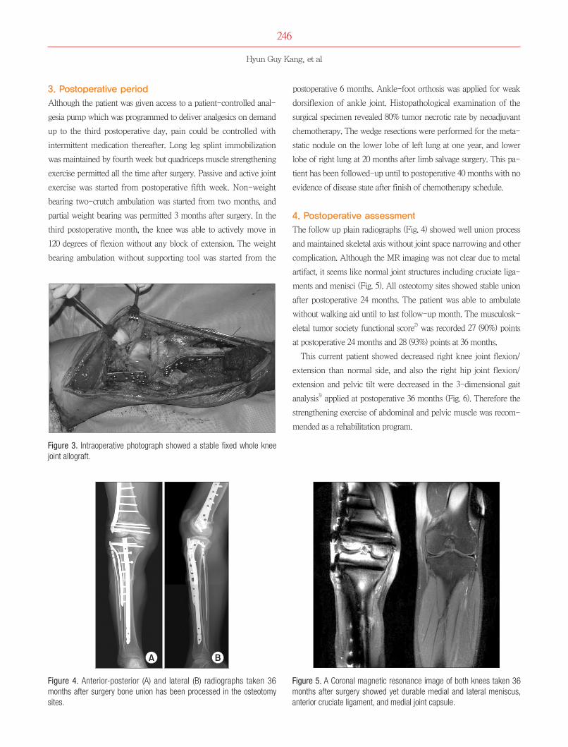
The follow up plain radiographs (Fig. 4) showed well union process
and maintained skeletal axis without joint space narrowing and other
complication. Although the MR imaging was not clear due to metal
artifact, it seems like normal joint structures including cruciate liga-
ments and menisci (Fig. 5). All osteotomy sites showed stable union
after postoperative 24 months. The patient was able to ambulate
without walking aid until to last follow-up month. The musculosk-
eletal tumor society functional score2) was recorded 27 (90%) points
at postoperative 24 months and 28 (93%) points at 36 months.
This current patient showed decreased right knee joint flexion/
extension than normal side, and also the right hip joint flexion/
extension and pelvic tilt were decreased in the 3-dimensional gait
analysis3) applied at postoperative 36 months (Fig. 6). Therefore the
strengthening exercise of abdominal and pelvic muscle was recom-
mended as a rehabilitation program.
Figure 3. Intraoperative photograph showed a stable fixed whole knee joint allograft.
Figure 4. Anterior-posterior (A) and lateral (B) radiographs taken 36 months after surgery bone union has been processed in the osteotomy sites.
Figure 5. A Coronal magnetic resonance image of both knees taken 36 months after surgery showed yet durable medial and lateral meniscus, anterior cruciate ligament, and medial joint capsule.

247
Limb Salvage Surgery Using Whole Knee Joint Allograft Reconstruction in Osteosarcoma
DISCUSSION
It has been hard to reconstruct a surgical defect using a distal femo-
ral or a proximal tibial osteoarticualr allograft due to size mismatch-
ing with host condyle. Radiologic matching by computed tomog-
raphy or plain radiography is currently the best option for detecting
a similar-sized allograft. Muscolo et al.1) selected distal femoral
osteoarticular allografts by measuring maximum total width, the an-
teroposterior dimension of the medial and lateral condyles and the
width of the intercondylar notch. Their proximal tibial osteoarticular
allograft were selected by comparing the age, gender, height, and
radiographs of the patient with corresponding data from the donor
to achieve the closest anatomic match.4) Whole knee joint allograft
reconstruction does not require matching process but it should be
obtained from a young healthy donor without a previous history of
knee joint diseases or injuries. It has been reported that fracture is a
common complication of allografts and that the incidences of infec-
tion and fracture are relatively high.5,6) In current case, we applied
dual internal fixation using intramedullary nail and locking plate of
titanium alloy implants for the tibia to prevent allograft fracture and
easy radiological follow-up.
Unlike hemiside osteoarticular allograft, the whole knee joint al-
lografts may result in less frequent meniscus injury or fatigue stress
of the cruciate ligaments due to best matched joint congruence as
well as less frequent articular destruction due to optimal joint excur-
sion. Beside, whole knee joint allograft is no need for host-allograft
sutures of the cruciate and collateral ligaments and joint capsule, the
joint laxity or postoperative ankylosis may be less occurs.
A previous experimental study in a rabbit model has demonstrat-
ed that there is no significant difference in subsequence reintegration
of cartilage, menisci and ligaments between whole knee joint auto-
grafts treated with heating at 60oC for 30 minutes and those that are
not.7,8) However, in the patient using pasteurized whole knee joint
autograft, the early collapse of medial condyle of tibia and exces-
sive ankylosis were occurred.9) Meniscal allograft transplantation has
been already performed for 20 years and has played an important
role in reducing knee joint pain and increasing function.10)
If a matched tibial hemiosteoarticular allograft is not available,
a whole knee joint allograft can be a good alternative option as
biological reconstruction. The host-allograft bone fixation without
ligaments and capsular suturing of the knee can provide wider sur-
gical margin, convenient surgical procedure with achieving best joint
congruence and minimize joint laxity.
This mega-biologic reconstruction using whole knee joint al-
lograft in the limb salvage surgery showed yet durable but need fur-
ther observation through long lasting period with case collection.
REFERENCES
1. Muscolo DL, Ayerza MA, Aponte-Tinao LA, Ranalletta M. Use of distal femoral osteoarticular allografts in limb salvage surgery. J Bone Joint Surg Am. 2005;87:2449-55.
2. Enneking WF, Dunham W, Gebhardt MC, Malawar M, Pritchard DJ. A system for the functional evaluation of recon-structive procedures after surgical treatment of tumors of the musculoskeletal system. Clin Orthop Relat Res. 1993;286:241-6.
3. Jang IG, Park HS, Nam KW, et al. 3D gait analysis of limb salvage patients with osteoarticular knee allograft reconstruc-tion. J Biomed Eng Res. 2010;31:74-80.
4. Muscolo DL, Ayerza MA, Farfalli G, Aponte-Tinao LA. Proxi-mal tibia osteoarticular allografts in tumor limb salvage sur-gery. Clin Orthop Relat Res. 2010;468:1396-404.
5. Hornicek FJ Jr, Mnaymneh W, Lackman RD, Exner GU, Mali-nin TI. Limb salvage with osteoarticular allografts after resec-tion of proximal tibia bone tumors. Clin Orthop Relat Res. 1998;352:179-86.
6. Hornicek FJ, Gebhardt MC, Tomford WW, et al. Factors af-fecting nonunion of the allograft-host junction. Clin Orthop Relat Res. 2001;382:87-98.
7. Judas F, Rosa S, Teixeira L, Lopes C, Ferreira Mendes A. Chondrocyte viability in fresh and frozen large human osteo-
Figure 6. The patient can walk with a satisfactory gait pattern.

248
Hyun Guy Kang, et al
chondral allografts: effect of cryoprotective agents. Transplant Proc. 2007;39:2531-4.
8. Ahmed AR, Watanabe H, Takagishi K. Reconstruction with autologous pasteurized whole knee joint I: experimental study in a rabbit model. J Orthop Sci. 2003;8:170-9.
9. Watanabe H, Ahmed AR, Shinozaki T, Yanagawa T, Terauchi M, Takagishi K. Reconstruction with autologous pasteurized
whole knee joint II: application for osteosarcoma of the proxi-mal tibia. J Orthop Sci. 2003;8:180-6.
10. von Lewinski G, Milachowski KA, Weismeier K, Kohn D, Wirth CJ. Twenty-year results of combined meniscal allograft transplantation, anterior cruciate ligament reconstruction and advancement of the medial collateral ligament. Knee Surg Sports Traumatol Arthrosc. 2007;15:1072-82.

249
Limb Salvage Surgery Using Whole Knee Joint Allograft Reconstruction in Osteosarcoma
전체슬관절동종골이식을이용한 골육종의사지구제수술
강현귀 • 김준혁 • 김광기*
국립암센터 골연부종양클리닉, *의공학과
청소년기의 사지구제수술 방법은 종양이 완치되었을 때를 고려해야 한다. 종양 대치물의 거대인공관절을 이용한 재건수술의 경우 향
후 반복적으로 교체 수술해야 하는 부담을 줄이고자 생물학적 재건수술의 일환으로 동종골 이식이 널리 사용되어 왔다. 본 증례에서
반월상 연골판과 인대가 포함된 전체 슬관절 동종골을 이용한 사지구제수술의 중간결과를 발표하고자 한다. 전체 슬관절 동종골은
대퇴골과 경골의 관절면이 일치하고 측부와 십자인대의 봉합이 필요 없어 관절의 안정성이 지속되었고 관절간격의 협소화 없이 기계
적 축을 잘 유지하여 만족스러운 보행을 보였다.
색인단어: 동종 이식 재건술, 전체 슬관절, 사지구제수술, 골육종
접수일 2013년 11월 21일 수정일 2014년 2월 26일 게재확정일 2014년 4월 17일책임저자 강현귀고양시 일산동구 일산로 323, 국립암센터 골연부종양클리닉TEL 031-920-1665, FAX 031-920-2798, E-mail [email protected]
Case Report J Korean Orthop Assoc 2014; 49: 244-249 • http://dx.doi.org/10.4055/jkoa.2014.49.3.244 www.jkoa.org
pISSN : 1226-2102, eISSN : 2005-8918249
Copyright © 2014 by The Korean Orthopaedic Association
“This is an Open Access article distributed under the terms of the Creative Commons Attribution Non-Commercial License (http://creativecommons.org/licenses/by-nc/3.0/) which permits unrestricted non-commercial use, distribution, and reproduction in any medium, provided the original work is properly cited.”
대한정형외과학회지:제 49권 제 3호 2014


