
4006
4004
SBLC
4023
HISTO LAB
GROSSLAB
GROSSLAB
ELEVATOR
STAIRSWCs
4th Floor
CARDSWIPE
4007
4080
Ber
Walker
Klinkha-
Building’s Front

If it fills the screen AND you are in the HSC library, try the Page Up & Page Down arrows on the keyboard
If you get no response, click out of the ‘Can’t do’ message, if it appears
Put the cursor on a blank region of the image & RIGHT-click the mouse
If they work, you’re off
Online HUMAN PARAMEDICAL ANATOMY
This image may be filling the screen, or occupying only the bottom right
From the menu that appears, select Full Screen
Navigation 1
Now try the Page Up & Page Down keys, or Up & Down arrows, on the keyboard
Very early warning Do not PRINT, unless you select Black on White. You’ll be using lots of toner, otherwise

‘Slide Show’ fills the screen, with apparently no escape. Once the image fills your screen, moving the mouse around should bring up a faint triangular icon in the bottom left
Clicking on this icon will bring up a menu with ‘End slide show’ (allowing you to leave the full screen),
Online HUMAN ANATOMY Navigation 2
EXITING When you have clicked for ‘End of Slide Show’, you’ll return to the browser screen, with a slide from this program visible, but smaller
CAUTION - Now you should click on the browser’s ‘Back’ icon to return to the Paramedical Anatomy page, with the choice of topics
If you click an X top-right, you will leave the Web site & the browser!

To have it fill the screen, go to the row of icons on the bottom left, & choose the wine-glass one next to the end (it should activate with ‘Slide Show’) DO NOT CLICK ON IT YET
‘Slide Show’ fills the screen, with apparently no escape. Once the image fills your screen, moving the mouse around should bring up a faint triangular icon in the bottom left
Clicking on this icon will bring up a menu with ‘End slide show’ (allowing you to leave the full screen),
Now click on ‘Slide Show’
If the image occupies the lower right of your screen - please read all of this before clicking anything
‘Next’ & ‘Previous’ could be used, but Page UP & Down, or the arrows, on your keyboard will move you faster though the slides
Online HUMAN ANATOMY Navigation 3

To progress through Powerpoints such as this, click on the single down arrow on the vertical bar to the right
To go back, hit ‘Page Up’, or click on the up arrow
On the vertical bar is a gray square: putting the cursor on this square & holding the left click on will allow you to mouse-drag the program long distances forward or back through the slides. Releasing the left click gives you a slide near where you are aiming for, if you do not know its number, (which is displayed as you scan)
or hit ‘Page Down’ on the keyboard
On these slides there is no icon on the screen image to click on to move forward or back, or to escape
Online HUMAN ANATOMY Navigation 4
Thereafter, your access and what you see on the screen are based more obviously on your own computer’s version of Powerpoint, when you retrieve from your own copy
It will save in later ppt versions, if this is more convenient for you
The Web version is in Powerpoint of 1995, so that as many people as possible can have access to it
Once you have the file visible on your own computer, you can save it with ‘Save as’ on the hard drive, on a floppy (for most files), or on Zip disc
Online HUMAN ANATOMY Navigation 5

Powerpoints such as this have a search function
Click on ‘Edit’ from the menu, above left
About two-thirds the way down the Edit menu click on ‘Find’
You’ll then get a window to type in the search term, and click on ‘Find next’
Having found or not found your term, you’ll have one or two windows to click out of
Online HUMAN ANATOMY Navigation 6

This is because the program is for anyone, however inexperienced
Some of the directions you may find elementary
Caution - The slides can be printed out, but you have to do this in a special way to get black lines on a white background, otherwise you’ll use up a ton of toner.
The font is usually the present one - Arial. It looks ugly on the screen, but is the choice because these slides are also used for projection.
Also related to slide projection is the choice of a black background. Projectors vary a lot in their intensity, and can ‘bleach out’ other backgrounds and have other strange effects on legibility.
Sometimes, for contrast, this Times Roman will appear
HUMAN PARAMEDICAL ANATOMY I

Because the background is black, in order to get black print on a white background some steps are needed
PRINTING these ppt slides
Format Custom Background Other color White of the white-to-black scale OK Apply to all
File Print Black & white (not ‘Pure black & white’) Frame slides 6 slide/page from ‘Print what?’ menu OK
File Close Not to save changes File Exit
6-to-a-page hand-out can be made more legible by copying at 120% Move page size off Auto onto standard Put original in corner, maybe over-riding framing metal

Along with the computer, there are one or two books to learn from: Marieb’s Essentials of Human Anatomy & Physiology 7th ed (Addison, Wesley & Longman), and Guy’s Learning Human Anatomy 2nd ed (Appleton & Lange)
The programs are set up so that you can click forward and back through the teaching diagrams and text. We will be interested in where you have problems with the flow of ideas.
Thanks for your interest in Anatomy. Here is an overview of how you may use this material.
The programs on the computer have various formats, so that we can get an idea of which are best suited to your learning anatomy. There may be questions later on your experiences that we would appreciate your answering.
See next an example of referencing
HUMAN PARAMEDICAL ANATOMY II

ANATOMY & SYSTEMS: Book Refs
getting their energy and distributing it
getting rid of waste products
moving around and manipulating things in their surroundings
detecting threats and change and adapting to them
keeping the body working at about the same level and temperature
keeping nasties outside the body and dealing with ones that have entered
and reproducing themselves.
Alimentary/digestive Vascular Respiratory
Urinary Respiratory Digestive
Musculo-skeletal
Nervous/Sensory
Nervous & Endocrine
Skin Blood & Lymphoid
Reproductive
The systems and their organs are shown well on pp. 5,6 of Marieb, and Figs 1-5 through 1-12 (pp. 8-10) of Guy

The computer slides have the advantage over textbook figures that we can present information slowly step-by-step and do not have to overload Figures. Also, we can return to figures for reorientation.
One approach is to try the computer material first to get a framework of essentials, and then read the book for another view and more detail. And we give specific book references.
Another trick is to have a pad on each side of the keyboard and move the mouse for left-handed use (can be done), leaving the right free to make notes and sketches (‘lefties’ please bear with us - we don’t know any better)
However, be flexible with your computer learning. Some programs can be clicked through with no keyboard use, so you might move the keyboard and have the textbook & page markers in its place for reference.
HUMAN PARAMEDICAL ANATOMY III
It is already time to give you an idea of the sort of images in the programs of the first Module
Some Figs use cartoon figures with anatomical detail added. Other are an attempt to show what the structure actually looks like.
The problem here is that most structures have special shapes, so it matters what angle or side one views from
The different views & the names that arise from them are a topic (Terms) of the first module after this Orientation
Some examples of the slides follow
HUMAN PARAMEDICAL ANATOMY IV

MEDIAL & LATERAL
(Outwards)Laterally
Thumb points
Little finger lies
Medially (Towards the midline)
MEDIAN (Midline)

TRANSVERSE
Transverse process of vertebra
BODY
Transverse colon
Transverse structures run cross-wise in the body
This could have been named the lateral process, but was not

Two LEFT chambers of the heart
ANTERIOR/FRONT VIEW OF THE HEART
The differences between left and right or laterality are interesting
Right VENTRICLE
Left ATRIUM
Left
VENTRICLE
SVCPT
Right ATRIUM
AOR

BODY WALL & CAVITIES: Casing & Contents
‘SOMATIC’ is applied to the limbs and body wall, ‘VISCERAL’ to the viscera
The body wall is musculo-skeletal and covered with skin
The cavities house soft organs: some tubular or bag-like with a space(s) inside (the lumen), others are more solid, e.g., the spleen, kidneys, and liver. These all are the VISCERA.

Every so often, we shall summarize some of what we have been looking at.
REVIEW SLIDES
An example follows

PRONE
ANATOMICAL TERMS SO FAR
SUPINE
RIGHT LEFT
PRONATION & SUPINATIONinferiorly, superiorly, medially,
laterally, ventrally, dorsally, etc
EXTERNAL
INTERNAL
SUPERFICIALDEEP
ROSTRAL
SUPERIOR
INFERIOR
CAUDAL
PROXIMAL
DISTAL
DORSAL/ POSTERIOR
VENTRAL/ ANTERIOR
TRANSVERSE

For learning Anatomy, labeled figures are a must, & Powerpoint lets us offer a lot of them.
What the textbook provides is explanatory & linking text, which we can offer only in small measure, otherwise the screen gets cluttered
So that one needs to find a comfortable way of getting the value from both sources: this you’ll have to experiment with to find what suits you
Ideas for brief linking pieces are welcome. What to me seems like a thread of an idea may in practice be too disjointed. However, if you’ve read cartoon strips, you’ve already coped with the problem
Any errors please call to my notice
HUMAN PARAMEDICAL ANATOMY V

At the beginning of each section, you will see a slide to show you which section you have entered. At the end, the Module-Topics slide will be repeated.
WHERE AM I?
Examples follow

WHERE AM I? Online Anatomy Module 1
APPENDICULAR SKELETON
CELL
INTRO & TERMS
EPITHELIUM
CONNECTIVE TISSUE
MUSCLE
NERVOUS SYSTEM
AXIAL SKELETON
MUSCLES
EMBRYOLOGY

WHERE AM I? Online Anatomy Module 1
APPENDICULAR SKELETON
CELL
INTRO & TERMS
EPITHELIUM
CONNECTIVE TISSUE
MUSCLE
NERVOUS SYSTEM
AXIAL SKELETON
MUSCLES
EMBRYOLOGY
You are at the End
Caution how you exit. BACK on your browser is neededUnfortunately there is no way that you can directly reach other topics listed here by clicking on them. You get there by going back to the Paramedical Anatomy menu

Those taking the course for formal credit will of course have the examinations to take , but the object is more to get a feel & a basis for the applications of medical anatomy.
A test of what you have learned is what you can explain and illustrate using just your new mind and pencil and paper.
This exercise will give you practice in explaining anatomical medical ideas to patients, and give practice in reformatting material learned in a rather constrained context. The anatomy has to be usable in a variety of clinical and theoretical contexts.
Every so often in the computer program, we shall refer to test material in the books (see next slide). At other times, the computer program will have its own self-testing sections.
One goal is to ‘complete’ sections of the course, but you are really building an understanding of the body & medicine that will never be complete, but always somehow applicable
HUMAN PARAMEDICAL ANATOMY VI

Marieb p. 13,
p. 20 questions 4, 5, , 7, 8, 9, 10,
QUESTIONS ON ANATOMICAL TERMS
Guy p. 4, Fig 1-18,
p. 11, Exercise 1
For questions that you cannot answer from this computer program, e.g., brachial region?, search in the book chapters

Once into Anatomy, one finds that the logic and the engineering principles of the structures, and how they work, let one appreciate the body more than if one just did not know.
SATISFACTIONS
Secondly, one often sees how the Anatomy - sizes, configurations, relations, etc - shapes what occurs in injury and disease. E.g., why it is the radius that breaks when one falls on the hand, rather than the other bone of the forearm; & why the break is at the wrist end, rather than mid-shaft.
Two examples follow

GROSS ANATOMY
This story of getting food to the mouth shows working anatomy as parts of the body, movements around joints, muscle actions, and some underlying actions of bones of the skeleton
Other stories of how the body works make sense only in terms of much smaller units seen with the microscope, down to the level of cells and parts of cells - microscopic anatomy or histology, for example in the kidney and the liver.
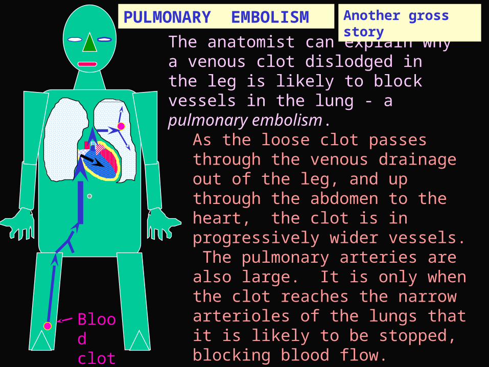
The anatomist can explain why a venous clot dislodged in the leg is likely to block vessels in the lung - a pulmonary embolism.
PULMONARY EMBOLISM
Blood clot
PTAs the loose clot passes through the venous drainage out of the leg, and up through the abdomen to the heart, the clot is in progressively wider vessels. The pulmonary arteries are also large. It is only when the clot reaches the narrow arterioles of the lungs that it is likely to be stopped, blocking blood flow.
Another gross story

SOURCE OF THE FIGURES
All the Figs are hand-drawn by me in Powerpoint
They are based on ideas derived from several textbooks, atlases, and personal experience in the lab
The only ones that are attempts at direct hand copies are in the Muscles & Skeleton sections, e.g.,
For those, my source is Friedrich Merkel’s atlas Die Anatomie des Menschen, published by JF Bergmann: Wiesbaden, in 1913
chosen for reliability, & because time & the status of ‘spoils of war’ have claimed the copyright
And a personal interest is that Merkel taught at Gottingen, where I was from 1961-1963
:
FOREARM X-section
Vol III Fig 57, p 48

Goals are usually presented at the beginning, as if one could not get started without being framed and organized by the offerer of the course
GOALS I
One usually comes to the course with some idea of what it entails: from friends, books & articles read, the lay press, the Web, the course listing, etc
My goals - hopes for an outcome, really - can be presented at any time. See if they harmonize at all with yours.
Later, reflect on whether what you have looked at and thought about here achieves your aims, and the general ones that I have in mind

To have had a lasting effect on your nerve cells’ dendrites, axons and synapses so that, on meeting anatomy in the future, your brain would judge it to be:
FAMILIAR You are comfortable in having seen material like this before
COMPREHENSIBLE You made sense of it then, and have confidence in being able to continue
APPLICABLE You have seen anatomical ideas usefully applied in the contextrs of normal functioning and disease & injury
EXTENSIBLE The anatomy was coherent enough that you could follow other kinds of anatomy applied to other situations
INTRIGUING The ideas of anatomy, function & dysfunction have an intellectual interest that could drive your curiosity to explore them further
GOALS II

WHERE AM I? Online Anatomy Module 1
APPENDICULAR SKELETON
CELL
ORIENTATION
EPITHELIUM
CONNECTIVE TISSUE
MUSCLE
NERVOUS SYSTEM
AXIAL SKELETON
MUSCLES
EMBRYOLOGY
You are at the End of the Orientation
Caution how you exit. BACK on your browser is neededUnfortunately there is no way that you can directly reach other topics listed here by clicking on them. You get there by going back to the Paramedical Anatomy menu


