EVALUVATION OF ORTHODONTIC TOOTH
MOVEMENT IN HUMANS WITH LOW LEVEL
LASER THERAPY
Dissertation submitted to
THE TAMILNADU DR. M.G.R.MEDICAL UNIVERSITY
In partial fulfillment for the degree of
MASTER OF DENTAL SURGERY
BRANCH V
ORTHODONTICS AND DENTOFACIAL ORTHOPAEDICS
APRIL – 2013
Acknowledgement
I would like to acknowledge and thank my beloved Professor and
Head, Dr. N.R. Krishnaswamy, M.D.S., M.Ortho (RCS, Edin), D.N.B. (Ortho),
Diplomate of Indian board of Orthodontics, Department of Orthodontics, Ragas
Dental College and Hospital, Chennai. I consider myself extremely fortunate
to have had the opportunity to train under him. His enthusiasm, integral
view on research, tireless pursuit for perfection and mission for providing
‘high quality work’, has made a deep impression on me. He has always been a
source of inspiration to strive for better not only in academics but also in life.
His patience and technical expertise that he has shared throughout the
duration of the course has encouraged me in many ways.
I am privileged to express my extreme gratefulness to my respected
guide, Professor Dr. Shahul Hameed Faizee, M.D.S for his undying
enthusiasm and guidance which helped me complete this study. His
everlasting inspiration, incessant encouragement, constructive criticism and
valuable suggestions conferred upon me have encouraged me. He has been an
integral part of my post graduate course during which I have come to know
his outlook towards life and wish to inculcate it someday.
I express my deep sense of gratitude and indebtedness to Professor,
Dr. Ashwin Mathew George, M.D.S D.N.B. (Ortho) for always being a pillar
of support and encouragement. His simplicity, innovative approaches and
impetus throughout the duration of my course has encouraged me in many
ways. He has helped me to tune myself to the changing environment in our
profession and his guidance will always be of paramount importance to me.
I am exceptionally gratified and sincerely express my thanks to my former
Professor Dr.S.Venketeshwaran ,M.D.S D.N.B. (Ortho), without whose counsel
this study would be incomplete.
My sincere thanks go out to Professor Mr. Kanakaraj Chairman &
Dr.S. Ramachandran, Principal, Ragas Dental College for providing me with
an opportunity to utilize the facilities available in this institution in order to
conduct this study.
I would also like to acknowledge Dr. Shakeel, (Reader),
Dr. Jayakumar (Reader), Dr. Anand (Reader), Dr. Rekha (Reader), Dr. Biju ,
Dr. Kavitha, Dr. Shobana, for their support, enthusiasm & professional
assistance throughout my post graduate course.
My deepest gratitude goes out to Dr. Premila Suganthan, Managing
Director and Laser Specialist, KP Tooth Care Dental Clinic, for her
vehement personal interest, wise counsel to render generous help to me in
carrying out this work from its inception to its consummation.
I am greatly beholden to Mr. Eshwar, Managing Director, 3D scan
solutions for his support, enthusiasm& professional assistance through my
post graduate course.
I would also like to thank Mr. Ravanan, Associate professor,
Presidency College, Chennai for his valuable suggestions during my statistical
work.
My heartfelt thanks to my wonderful batch mates, Dr. Deepak, Dr.
Manikandan, Dr. Nupur Aarthi, Dr. Ravanth, Dr. Siva, Dr. Vijayshri
Shakti who were cheerfully available at all times to help me. I wish them a
successful career ahead.
I also extend my gratitude to my juniors Dr.Femin, Dr.Gayatri,
Dr.Manali, Dr.Murali, Dr.Regina, Dr.Saptarishi, Dr.Vikram, Dr.Vishal,
Dr,Shrabani, Dr.Avdesh, Dr.Mercy, Dr.Divya, Dr.Anslem, Dr.Jayakrishna,
Dr.Piradhiba, Dr.Karthik for lending me their patients and their support.
My sincere thanks to my good friends Dr.John, Dr.Krishnakumar,
Dr.Soumya for their help and support rendered during my study.

I would like to thank Sister Lakshmi, Sister Rathi, Sister Kanaka, Ms
Haseena, Mr. Bhaskar, Ms Shalini, Mr. Mani, Ms Banu, Ms Divya and the
Scribbles team for their co-operation and help during my course of study.
And to My parents and my sister, I am forever indebted. They have
always been there to show me the right path and to correct me when I have
strayed. Life, as I see it is only because of the love, guidance and support
they have given me.
Above all, I thank the most merciful and compassionate Almighty
God, he guided and helped me throughout my life in every endeavor and for
that I am grateful.

CONTENTS
S.NO. TITLE PAGE NO.
1 INTRODUCTION 1
2 REVIEW OF LITERATURE 5
3 MATERIALS AND METHODS 48
4 RESULTS 55
5 DISCUSSION 56
6 SUMMARY AND CONCLUSION 72
7 BIBLIOGRAPHY 73
ABSTRACT
Introduction – The duration of orthodontic treatment is a major concern for the
patients. Many methods to reduce treatment time have been tested in the past each
having its own disadvantages. A non invasive method of accelerating tooth
movement is needed in order to enhance the rate of tooth movement. Hence the
aim of this study was to investigate the effects of low level lasers on orthodontic
tooth movement during canine retraction.
Materials and Methods – 13 adult patients were used in the study requiring
retraction of maxillary canines as a part of orthodontic treatment. Low level laser
therapy applied on the test side. The other side was considered as control.
Retraction was initiated on both sides using NiTi coil springs. The rate of tooth
movement was evaluated after 2 months.
Results – There was a significant difference in the rate of tooth movement
between the lased and non lased side. An average increase of 43% in the rate of
tooth movement was observed on the lased side.
Conclusions – Low level laser therapy is an efficient and non invasive method of
accelerating tooth movement.
Key words: Canine retraction, Low level laser therapy, White light scanning,
Biostimulation
Introduction
1
INTRODUCTION
Tooth movement through orthodontics occurs as a result of mechanical
forces acting on teeth resulting in a series of biologic responses. Over the
years not only orthodontists have had difficulties in treating and finishing a
case, but due to the long duration of treatment, patients neglect to go through
orthodontic treatment and tend to discontinue it73
.
In general, the period of time required for fixed appliance treatment for
orthodontic patients is approximately 2 - 3 years. During this period there is an
increased risk of root resorption, gingival inflammation and dental caries36
.
Reducing orthodontic treatment time requires increasing the rate of tooth
movement. Over the years, orthodontists have vested interests in reducing the
length of orthodontic treatment and hoped to accomplish treatment objectives
in timeliest manner possible. In an effort to meet these demands, orthodontists
have searched methods to increase the rate of orthodontic tooth movement.
In the past, many studies have been conducted in order to enhance
tooth movement. These include the following:
1. Low voltage currents were delivered using a special electric
device12
.
2. Intra-oral rare earth magnets11
.
Introduction
2
3. Chemical agents like Prostaglandins70
, Osteocalcin24
, Vitamin D8,
Calcitriol28
, Parathyroid hormone which are injected to enhance
tooth movement.
4. Corticotomy31
5. Distraction osteogenesis2
However the above mentioned procedures have distinct disadvantages. These
procedures are cumbersome, technique sensitive, cause pain and discomfort to
the patients and can be invasive in nature. Hence a non- invasive procedure
that was painless and could enhance tooth movement remained a question.
The development of Lasers has been a turning point in the history of
science and engineering43
. It has produced a completely new type of systems
with potentials for applications in a wide variety of fields. It was Albert
Einstein in 1917 who first explained the theory of stimulated emission which
became the basis of Laser. However, it was in late 1940s and fifties that
scientists and engineers did extensive work to realize a practical device based
on the principle of stimulated emission. Despite the pioneering work of
Townes and Prokhorov it was left to Theodore Maiman in 1960 to invent the
first Laser using ruby as a lasing medium that was stimulated using high
energy flashes of intense light43
.
Low level laser therapy (LLLT) is also known as “soft laser therapy”.
The use of LLLT in health care has been documented in the literature for more
Introduction
3
than three decades and is gaining an increasing acceptance in conventional
medical, physiotherapy, dental, and veterinary practice and refers to the
"reaction between laser and the irradiated biological tissue" (Baxter, 1994)63
.
The use of LLLT dates back to 1967 when Dr. Endre Mester, a
Hungarian physicist, accidentally discovered that a monochromatic laser could
help speed up the healing of soft tissue injuries. By the mid 1970’s laser
therapy gained popularity in Asia, Africa and the Soviet Union. Today LLLT
is used worldwide in the field of medicine, dentistry, physical therapy and
chiropractics43
.
LLLT has primarily been shown useful in the short-term treatment of
acute pain caused by rheumatoid arthritis, osteoarthritis, tendinopathy, and
possibly chronic joint disorders. LLLT has also been useful in the treatment of
both acute and chronic neck pain63
.
In dentistry it has been used for post extraction and bone healing
therapy. It also helps in curing Herpes labialis, periodontitis, aphthous ulcers,
mucositis, in case of paresthesia and trigeminal neuralgia63
. Moreover recently
it has been used for treating dentinal hypersensitivity68
.
In Orthodontics, LLLT has found to be proved as an effective tool. It is
widely used for the following procedures:
Esthetic contouring of the gingival scaffold.
Introduction
4
Pain reduction during orthodontic treatment15
Treatment of Temporomandibular disorders55
Rapid bone regeneration after Rapid Maxillary Expansion6
Minor surgical procedures like frenectomy, operculectomy
Although LLLT has been used in orthodontics, its use in accelerating
tooth movement is not well documented. Majority of the studies in literature
that examined the effect of low-intensity laser therapy on the rate of
orthodontic tooth movement were short-term animal studies29
. Its use for
plummeting the treatment duration still remains a debate. Several studies have
shown that LLLT promotes bone repair33
. Therefore, if LLLT can cause an
increase in bone remodeling, it may also have an effect on increasing the rate
of tooth movement.
Therefore the aim of our study is:
“EVALUVATION OF ORTHODONTIC TOOTH MOVEMENT IN
HUMANS WITH LOW LEVEL LASER THERAPY”
Review of Literature
5
REVIEW OF LITERATURE
Reitan et al27
(1964) stated that the initial force for canine retraction
should be light, because this produces biologic effects. These lighter forces
will produce less extensive hyalinized tissue that can be readily replaced by
cellular elements. He stated that an appropriate force of 150 to 200 gms for
maxillary canines should be used for translatory movement.
Burstone et al7 (1976) stated that Vertical loops or modified vertical
loops are basically frictionless springs which are used for canine and anterior
tooth retraction. The design and selection of a proper loop or retraction spring
should be based on a number of scientific criteria. Foremost among these
would be a sufficiently high moment-to-force ratio so that root apices are not
displaced mesially or anteriorly. A retraction spring with zero angulation of its
horizontal-occlusal arms delivers a moment when activated to produce a force.
The ratio of this moment and force is constant throughout the elastic range of
activation of the spring. The higher the moment-to-force ratio, the greater is
the clinician's control over the apices of the anterior teeth. An analysis of
design factors demonstrates that the higher the loop occluso-gingivally, the
shorter its horizontal length occlusally, and the greater the gingival horizontal
length as in a T loop; these are significant factors in increasing the moment-to-
force ratio. The placement of helices is a useful design consideration but the
main effect is in reducing the load-deflection rate. By keeping these design
factors in mind, the clinician can build into his retraction springs, without the
Review of Literature
6
placement of any gable bend, the largest possible moment-to-force ratio so as
to optimize his tooth movement.
Davidovitch et al12
(1980) determined the usefulness of exogenous
electric currents in accelerating orthodontic tooth movement and to study the
effect of electric-orthodontic treatment on periodontal cyclic nucleotides.
Maxillary canines were tipped in five cats by 80 g force. Enhanced bone
resorption was observed near the anode (PDL compression site), while bone
formation was pronounced near the cathode (PDL tension site). Staining for
cyclic nucleotides was increased when electric stimulation was added to the
mechanical force. He concluded that orthodontic tooth movement may be
accelerated by the use of locally applied electric currents.
Huffman et al14
(1983) conducted a clinical study to compare the
amount and rate of movement and the tipping of canines retracted on 0.016
inch and 0.020 inch round wire with a continuous force of 200 grams and
medium-width 0.022 inch by 0.028 inch nonangulated Siamese brackets. On
one side canines were retracted on 0.016 inch wire and on the other side of the
same arches, on 0.020 inch wire. Over 10 weeks, the mean amount of
movement for twenty-one canines on the 0.016 inch wire was 3.37 mm., and
for the twenty-one canines on the 0.020 inch wire it was 2.99 mm. The mean
rate of movement in twenty-five arches was 1.37 mm. per month on the 0.016
inch wire and 1.20 mm. per month on the 0.020 inch wire. Over a period of 10
weeks, the mean amount of tipping for seventeen canines on the 0.016 inch
Review of Literature
7
wire was 5.3 degrees, and for the seventeen canines on the 0.020 inch wire it
was 1.7 degrees. Since less tipping occurred on the 0.020 inch wire and the
rates of movement were similar, there appears to be an advantage in retracting
canines along 0.020 inch round wire rather than on 0.016 inch round wire. It
would seem, also, that a greater force is not required to slide a tooth bonded or
banded with an 0.022 by 0.028 inch bracket slot along an 0.020 inch round
wire than along an 0.016 inch round wire.
Burstone et al60
(1984) discussed the following points regarding the
mechanics of orthodontic tooth movement. He stated that Orthodontic forces
can be treated mathematically as vectors. When more than one force is applied
to a tooth, the forces can be combined to determine a single overall resultant.
Forces can also be divided into components in order to determine effects
parallel and perpendicular to the occlusal plane, Frankfort horizontal, or the
long axis of the tooth. Forces produce either translation (bodily movement),
rotation, or a combination of translation and rotation, depending upon the
relationship of the line of action of the force to the center of resistance of the
tooth. Since most forces are applied at the bracket, it is necessary to compute
equivalent force systems at the center of resistance in order to predict tooth
movement.
Yamasaki et al70
(1984) evaluated the effect of prostaglandin on
orthodontic tooth movement. In the first phase, lingual arch springs were
applied on both sides of the maxilla to upper first premolars which were
Review of Literature
8
scheduled for extraction. One side received submucosal injections of PGE, and
the other received vehicle injections. The rate of tooth movement in the buccal
direction approximately doubled on the side of several PGE, injections as
compared to the control side. In the second phase, the PGE, injections were
applied in canine-retraction cases for up to 3 weeks in first-premolar-
extraction cases. The rate of distal canine movement was almost double on the
side receiving PGE, injections as compared to the vehicle-injected side. In the
third phase, the PGE, injections were applied on routine canine retraction in
first-premolar-extraction cases. The rate of distal canine movement was almost
1.6 fold on the side of PGE, injections as compared to the vehicle-injected
side. Throughout this study, no side effects were observed macroscopically in
the gingiva and roentgenographically in the alveolar bone, except for a slight
pain reaction consistent with orthodontic tooth movement.
Mester41
(1985) reviewed the experimental and clinical use of lasers
over a 20-year period, during which laser effects on 15 biological systems
were studied. Low-energy laser radiation was found to have a stimulating
effect on cells, and high-energy radiation had an inhibiting effect. The
application of lasers to stimulate wound healing in cases of nonhealing ulcers
is recommended.
Collins et al8 (1988) determined if the rate and amount of orthodontic
tooth movement in a sample of cats could be enhanced by the injection of a
vitamin D metabolite, 25dihydroxycholecalciferol (1,25D) into the periodontal
Review of Literature
9
ligament. After 21 days of canine retraction with a light-wire retraction spring,
the teeth that had received weekly intraligamentous injections of a solution of
1,25D in dimethylsulfoxide (DMSO) had moved 60% further than matched
control teeth (P < 0.05). At the histologic level, increased numbers of
mononuclear osteoclasts were recruited and activated, resulting in greater
amounts of alveolar bone resorption on the pressure side of the periodontal
ligament. No obvious clinical, microscopic, or biochemical side effects were
noted.
Enwemeka17
(1988) in his study about lasers concluded that laser
biostimulation is potentially a useful tool in the treatment of wounds,
particularly those cutaneous and subcutaneous wounds that are either
complicated by infection or inherently require a prolonged period of time to
heal. The precise dosage and frequency of treatment required to promote
healing even in animal models remain elusive, as is experimental
determination of the depth of penetration of lasers.
Peter Ngan et al44
(1989) determined the perception of discomfort
over time by a group of 70 patients undergoing orthodontic treatment. Patients
who were selected for comprehensive orthodontic treatment completed
questionnaires before insertion of separators and initial arch wires and after
placement at 4 hours, 24 hours, and 7 days. The level of discomfort during
these time periods was assessed by a visual analogue scale. The results
showed a significant increase in the level of discomfort after insertion of either
Review of Literature
10
separators or arch wires at 4 hours and 24 hours, but not at 7 days. No
significant difference was found in the level of discomfort of patients more
than 16 years of age compared with those 16 years and under. No significant
difference in discomfort was found between the sexes. These results are useful
in relating expectations of discomfort to who undergo orthodontic treatment.
Sandy et al53
(1993) in his review highlighted the recent developments
in bone cell biology. He summarized that osteoblasts are recognized as the
cells that control both the resorptive and the formative phases of the
remodeling cycle, and receptor studies have shown them to be the target cells
for resorptive agents in bone. The osteoblast is perceived as a pivotal cell,
controlling many of the responses of bone to stimulation with hormones and
mechanical forces. It is apparent that not all the cellular responses induced by
mechanically deformed tissues can be explained by the current paradigm
emphasizing the importance of prostaglandin production and cAMP elevation;
the mobilization of membrane phospholipids giving rise to inositol phosphates
offers an alternative second messenger pathway. It is also argued from
circumstantial evidence that changes in cell shape produce a range of effects
mediated by membrane integral proteins (integrins) and the cytosk'eleton,
which may be important in transducing mechanical deformation into a
meaningful biologic response.
Darendeliler11
(1995) determined whether the application of either
samarium cobalt magnets or pulsed electromagnetic fields could increase the

Review of Literature
11
rate and amount of orthodontic tooth movement observed in guinea pigs.
Fifteen grams of laterally directed orthodontic force were applied to move the
maxillary central incisors of a sample of 18 young male Hartley guinea pigs
divided into three groups: group 1, an orthodontic coil spring was used to
move the incisors; group 2, a pair of samarium-cobalt magnets provided the
tooth moving force; and group 3, a coil spring was used in combination with a
pulsed electromagnetic field. The results showed that both the static magnetic
field produced by the samarium-cobalt magnets and the pulsed
electromagnetic field used in combination with the coil spring were successful
in increasing the rate of tooth movement over that produced by the coil springs
alone.
Lim et al35
(1995) tested the efficacy of LLLT in controlling
orthodontic postadjustment pain. Thirty-nine volunteers were selected for this
study that used a double-blind design with placebo control. Elastomeric
separators were placed at the proximal contacts of one premolar in each
quadrant of the dentition to induce orthodontic pain. The tip of a 30 mW
gallium-arsenide-aluminium (830 nm) diode laser probe was then placed at the
buccal gingiva and directed at the middle third of the root. Three different
treatment durations of 15, 30, and 60 seconds and one placebo treatment of 30
seconds were tested within each subject. The study was conducted over 5
days, and the visual analogue scale (VAS) was used to quantify the pain
experienced by the subjects before and after laser applications for each day.
Review of Literature
12
The results showed that showed that teeth exposed to laser treatment had
lower levels of pain as compared with those with the placebo treatment.
Lotzof et al38
(1996) compared the time required to retract canine teeth
by using two different preadjusted bracket systems (Tip-Edge, TP
Orthodontics, LaPorte, Ind., versus A-Company straight wire, Johnson and
Johnson, San Diego, Calif.) in a human sample. Anchorage loss as a result of
this movement was also evaluated. A sample of 12 patients was randomly
selected who required the removal of first premolars in one or both arches as a
part of their orthodontic treatment. The rate of retraction and anchorage loss
were evaluated. There was no statistically significant difference in the rates.
The mean anchorage loss was 1.71 mm for the Tip-Edge bracket, and 2.33 mm
for the straight wire bracket. The difference in the amount of anchorage loss
was inconclusive as the sample size was too small.
Hasler et al25
(1997) measured the rate of movement of the maxillary
canines into the healed or recent extraction alveolus of the first premolar in 22
patients aged 10-27 years. On one side of the dental arch, the first premolar
was extracted. After a median time of 86 days, the contralateral first premolar
was extracted and the distalization of both canines started using Gjessing
canine retraction springs. The experiment was ended when one of the two
canines had been sufficiently distalized. Recordings of the positions of the
canines at the beginning of the study, at the start of the distalization and at the
end were made from dental casts and standardized intraoral radiographs. The
Review of Literature
13
canine on the recent extraction side moved faster than that on the healed side,
but also tipped somewhat more.
Walsh et al68
(1997) The use of LLLT in the treatment of dentinal
hypersensitivity and periodontal ligament pain during orthodontic tooth
movement has been shown in clinical trials to be both safe and effective.
There is accumulating evidence which indicates the potential of lethal laser
photosensitization as a technique for the destruction of cariogenic and other
microorganisms within the mouth without causing undue thermal stress to the
tooth. Improvements in the design of LLLT equipment are necessary to enable
these various techniques to be accomplished within an adequate timeframe and
without breaching cross infection control requirements. Given the low -
technology, low-cost characteristics of LLLT, the future for hard tissue LLLT
applications is promising. As with soft tissue applications of LLLT, efforts
should be directed toward investigating the precise dosimetry required for
therapeutic laser effects, in order to achieve standardization of treatment
protocols.
Kobayashi et al32
(1998) evaluated the effects of local administration
of osteocalcin, a major noncollagenous bone matrix protein, on experimental
tooth movement in rats. An orthodontic elastic band was inserted between the
upper first and second molars, and the first molar was moved mesially.
Purified osteocalcin (0 to 10 micrograms) in 20 microliters of phosphate-
buffered saline was injected into the region of the root bifurcation of the first
Review of Literature
14
molar daily for 4 days. Tooth movement increased significantly following the
injections. Histological studies revealed that the injections markedly
stimulated the appearance of osteoclasts on the pressured side of the alveolar
bone surface. The results suggest that osteocalcin has an additive effect on the
rate of orthodontic tooth movement through the enhancement of
osteoclastogenesis on the pressured side.
Kawasaki et al29
(2000) evaluated the effect of tooth movement in
rats.A total of 10 g of orthodontic force was applied to rat molars to cause
experimental tooth movement. A Ga-Al-As diode laser was used to irradiate
the area around the moved tooth, and after 12 days, the amount of tooth
movement was measured. Calcein was injected subcutaneously to label the
newly formed alveolar bone for quantitative analysis. Immunohistochemical
staining of proliferating cell nuclear antigen was performed to evaluate
cellular proliferation. TRAPase staining was also performed to facilitate the
identification of osteoclasts. He concluded that these findings suggest that
low-energy laser irradiation can accelerate tooth movement accompanied with
alveolar bone remodeling.
Coombe et al9 (2001) investigated the effects of low level laser
irradiation on the human osteosarcoma cell line, SAOS-2. The cells were
irradiated as a single or daily dose for up to 10 days with a GaAlAs continuous
wave diode laser (830 nm, net output of 90 mW, energy levels of 0.3, 0.5, 1, 2,
and 4 Joules). Cell viability was not affected by laser irradiation, with the
Review of Literature
15
viability being greater than 90% for all experimental groups. Cellular
proliferation or activation was not found to be significantly affected by any of
the energy levels and varying exposure regimes investigated. No significant
early or late effects of laser irradiation on protein expression and alkaline
phosphatase activity were found. Investigation of intracellular calcium
concentration revealed a tendency of a transient positive change after
irradiation. He concluded that Low level laser irradiation was unable to
stimulate the osteosarcoma cells utilized for this research at a gross cell
population level.
Hashimoto et al24
(2001) evaluated the effect of local administration
of osteocalcin (OC) on experimental tooth movement in rats. The maxillary
first molar was first moved mesially with an initial tipping force of 30 g with a
closed-coil spring anchored to the incisor for 10 days (n = 48). Three
experimental groups (n = 8) were injected with purified rat OC at doses of 0.1,
1, and 10 micrograms, respectively. The injection into the palatal bifurcation
site of the first molar was repeated daily. The control groups (n = 8) were
injected with rat serum albumin (10 micrograms), phosphate buffered saline
(PBS), or were not injected. Tooth movement was evaluated daily by
measuring the inter-cuspal distance between the first and the second molars on
a precise plaster model. A significantly larger number of osteoclasts
accumulated on the mesial alveolar bone surface in the 1-microgram OC-
injected group on day 3 than that observed in control group. These results
Review of Literature
16
suggest that administration of OC accelerates orthodontic tooth movement due
to enhancement of osteoclastogenesis on the pressure side, primarily in the
early experimental period.
Koutna et al33
(2003) investigated the effect of low-power laser
irradiation on the proliferation activity of HeLa cells. The cells were irradiated
by a 830 nm semiconductor BTL-10 laser in a continuous or pulsed mode at
an energy density ranging from 2 to 99 J/cm2 (power output, 72 to 360 mW).
The irradiated cells were incubated and their proliferation activity was
assessed by the MTT (3-[4,5-dimethylthiazol-2-yl]-2,5-diphenyltetrazolium
bromide) assay at 24, 48, 72 and 96 h. In comparison with the control
populations, the irradiated cells showed a significant increase in proliferation,
regardless of the energy density used, at 72 and 96 h but not at 24 and 48 h. In
addition, the stimulation of proliferation was related to the mode of irradiation.
The cells irradiated in the pulsed mode (5 000 Hz) showed a higher
proliferation activity than the cells treated by continuous laser light. It is
concluded that low-power lasers stimulate HeLa cell proliferation.
Pugliese et al48
(2003) evaluated the influence of low-level laser
therapy on biomodulation of collagen and elastic fibers. Cutaneous wounds
were performed on the back of 72 Wistar rats and a Ga-Al-As low-level laser
was punctually applied with different energy densities. The animals were
killed after 24, 48, 72 hours and 5, 7 and 14 days. In this study, the authors
concluded that low-level laser therapy contributed to a larger expression of
Review of Literature
17
collagen and elastic fibers during the early phases of the wound healing
process.
Nicola et al45
(2003) studied the activity in bone cells after LLLT close
to the site of the bone injury. The femurs of 48 rats were perforated (24 in the
irradiated group and 24 in the control group) and the irradiated group was
treated with a GaAlAs laser of 660 nm, 10J/cm2 of radiant exposure on the
2nd, 4th, 6th and 8th days after surgery (DAS). Histomorphometric analysis of
the bone was carried out. They found that activity was higher in the irradiated
group than in the control group. They concluded that LLLT increases the
activity in bone cells (resorption and formation) around the site of the repair
without changing the bone structure.
Cruz et al10
(2004) investigated the orthodontic movement velocity in
human. Eleven patients were recruited for this 2-month study. One half of the
upper arcade was considered control group (CG) and received mechanical
activation of the canine teeth every 30 days. The opposite half received the
same mechanical activation and was also irradiated with a diode laser emitting
light at 780 nm, during 10 seconds at 20 mW, 5 J/cm2, on 4 days of each
month. All patients showed significant higher acceleration of the retraction of
canines on the side treated with LILT when compared to the control. His
findings suggested that LILT does accelerate human teeth movement and
could therefore considerably shorten the whole treatment duration.
Review of Literature
18
Sun et al63
(2004) stated that low-level laser therapy (LLLT) is a
newly developing technique in dentistry, although it has been used among
medical, dental, physiotherapy, and veterinary professions in some parts of the
world for decades. LLLT can offer tremendous therapeutic benefits to patients,
such as accelerated wound healing and pain relief. A thorough knowledge
about the mechanisms, recognition of the therapeutic window, and how to
properly use these cellular phenomena to reach the treatment goals is required.
Kawakami28
et al (2004) evaluated the effect of 1,25-
dihydroxyvitamin D3 (1,25(OH)2D3) on alveolar bone formation during tooth
movement in rats. Orthodontic elastics were inserted between the maxillary
first and second molars on bilateral sides in male rats. 1,25(OH)2D3 was
injected locally, at the concentration of 10(-10) M, once every 3 days in the
submucosal palatal area of the root bifurcation of the molar on the right side.
Histomorphometric analysis revealed that tooth movement without application
of 1,25(OH)2D3 decreased the mineral appositional rate (MAR) on the
compression area at 7 days. Repeated injections of 1,25(OH)2D3 in the
orthodontically treated animals distinctly stimulated alveolar bone formation
on the mesial side at 14 days. He concluded that local application of
1,25(OH)2D3 enhances the reestablishment of supporting tissue, especially
alveolar bone of teeth, after orthodontic treatment.
Stein et al61
(2005) investigated the effect of low-level laser irradiation
on proliferation and differentiation of a human osteoblast cell line. Cultured
Review of Literature
19
osteoblast cells were irradiated using He-Ne laser irradiation (632 nm; 10 mW
power output). On the second and third day after seeding the osteoblasts were
exposed to laser irradiation. The effect of irradiation on osteoblast
proliferation was quantified by cell count and colorimetric MTT
(dimethylthiazol tetrazolium bromide) assay 24 and 48 h after second
irradiation. The results showed a significant 31–58% increase in cell survival
(MTT assay) and higher cell count in the once-irradiated as compared to
nonirradiated cells was monitored. They concluded that LLLT promotes
proliferation and maturation of human osteoblasts in vitro. These results may
have clinical implications.
`Goulart22
et al (2006) evaluated the effect of gallinium-aluminium-
arsenic (GaAlAs) laser irradiation on the speed of orthodontic movement in
canine premolars. Eighteen dogs were divided into two groups, and their third
molars were extracted. An orthodontic device was placed between the first
molar and the second premolar for stabilization purpose. Group I was
irradiated with a dosage of 5.25 J/cm(2) on the right side, whereas the left side
was used as the control group. Group II was submitted to the same procedure,
but was irradiated with a dosage of 35.0 J/cm(2). Irradiations were done every
7 days, for a total of nine irradiations. The orthodontic space was measured
every 21 days. The results showed that the 5.25 J/cm(2) dosage accelerated
orthodontic movement during the first observation period, from 0 to 21 days
(p < 0.05), whereas the 35.0 J/cm(2) dosage retarded the orthodontic
Review of Literature
20
movement in the treated group when compared with the control group, during
both the first and second observation periods, from 0 to 42 days (p < 0.05). He
concluded that photoradiation may accelerate orthodontic movement at a
dosage of 5.25 J/cm(2), whereas a higher dosage, 35.0 J/cm(2), may retard it.
Turhani67
et al (2006) analyzed the effect of single low-level laser
therapy (LLLT) irradiation on pain perception in patients having fixed
appliance treatment. Seventy-six patients (46 women, 30 men; mean age, 23.1
years) enrolled in this single-blind study were assigned to 2 groups. The
patients in group 1 (G1; 38 patients, 13 men, 25 women; mean age, 25.1 years)
received a single course of LLLT (Mini Laser 2075, Helbo Photodynamic
Systems GmbH & Co KG, Linz, Austria; wavelength 670 nm, power output
75 mW) for 30 seconds per banded tooth. The patients in group 2 (G2; 38
patients, 17 men, 21 women; mean age, 21.0 years) received placebo laser
therapy without active laser irradiation. Pain perception was evaluated at 6, 30,
and 54 hours after LLLT by self-rating with a standardized questionnaire.
Major differences in pain perception were found between the 2 groups. He
concluded that LLLT immediately after multibanding reduced the prevalence
of pain perception at 6 and 30 hours. LLLT might have positive effects in
orthodontic patients not only immediately after multibanding, but also for
preventing pain during treatment.
Hamblin43
et al (2006) stated that despite many reports of positive
findings from experiments conducted in vitro, in animal models and in
Review of Literature
21
randomized controlled clinical trials, LLLT remains controversial. This likely
is due to two main reasons; firstly the biochemical mechanisms underlying the
positive effects are incompletely understood, and secondly the complexity of
rationally choosing amongst a large number of illumination parameters such as
wavelength, fluence, power density, pulse structure and treatment timing has
led to the publication of a number of negative studies as well as many positive
ones.
Lotzof et al38
(2006) compared the time required to retract canine teeth
by using two different preadjusted bracket systems (Tip-Edge, TP
Orthodontics, LaPorte, Ind., versus A-Company straight wire, Johnson and
Johnson, San Diego, Calif.) in a human sample. Anchorage loss as a result of
this movement was also evaluated. A sample of 12 patients was randomly
selected from the new patient pool at the postgraduate orthodontic clinic of
Montefiore Medical Center. All patients required the removal of first
premolars in one or both arches as a part of their orthodontic treatment. The
rate of retraction and anchorage loss were evaluated. The mean rates of
retraction were 1.88 mm per 3-week period and 1.63 mm per 3-week period
for the Tip-Edge and A-Company brackets, respectively. There was no
statistically significant difference in the rates (/9 > 0.05). The mean anchorage
loss was 1.71 mm for the Tip-Edge bracket, and 2.33 mm for the straight wire
bracket. The difference in the amount of anchorage loss was inconclusive as
the sample size was too small (power was 10%).
Review of Literature
22
Kanzaki et al23
(2006) tested the hypothesis that local RANKL gene
transfer into the periodontal tissue would accelerate tooth movement. The
upper first molars of 6-week-old male Wistar rats were moved palatally using
fixed orthodontic wires. The inactivated hemagglutinating-virus of Japan
(HVJ) envelope vector containing the mouse RANKL expression plasmid was
injected periodically into the palatal periodontal tissue of the upper first
molars during TM. Local RANKL gene transfer significantly enhanced
RANKL expression and osteoclastogenesis in periodontal tissue without any
systemic effects. Local RANKL gene transfer might be a useful tool not only
for shortening orthodontic treatment, but also for moving ankylosed teeth
where teeth, fuse to the surrounding bone.
Limpanichkul et al36
(2006) tested the hypothesis that mechanical
forces combined with low-level laser therapy stimulate the rate of orthodontic
tooth movement. It was a double blind, randomized placebo/control matched
pairs clinical trial to test the efficacy of GaAlAs low-level laser therapy
(LLLT) on 12 young adult patients who required retraction of maxillary
canines into first premolar extraction spaces using tension coil springs with
fixed edgewise appliance. LLLT was applied on the mucosa buccally, distally
and palatally to the canine on the test side and using a pseudo-application on
the placebo side. Dental impressions and casts were made at the
commencement of the trial and at the end of the first, second and third months
after starting the trial. Measurement of tooth movements was made on each
Review of Literature
23
stage model using a stereo microscope. He concluded that there was no
significant difference of means of the canine distal movement between the
LLLT side and the placebo side for any time periods. The energy density of
LLLT (GaAlAs) at the surface level in this study (25 J/cm(2)) was probably
too low to express either stimulatory effect or inhibitory effect on the rate of
orthodontic tooth movement.
Saito et al52
(2007) investigated the effects of low-power laser
irradiation on bone regeneration during expansion of a midpalatal suture in
rats. Gallium-aluminum-arsenide diode laser 100 mW irradiation was applied
to the midpalatal suture during expansion carried out over 7 days (3 or 10
minutes per day), 3 days (7 minutes per day for day 0-2 or 4-6), and 1 day (21
uninterrupted minutes on day 0). The bone regeneration in the midpalatal
suture estimated by histomorphometric method in the 7-day irradiation group
showed significant acceleration at 1.2- to 1.4-fold compared with that in the
nonirradiated rats, and this increased rate was irradiation dose-dependent.
Irradiation during the early period of expansion (days 0 to 2) was most
effective, whereas neither the later period (days 4 to 6) nor the one-time
irradiation had any effect on bone regeneration. He concluded that low-power
laser irradiation can accelerate bone regeneration in a midpalatal suture during
rapid palatal expansion and that this effect is dependent not only on the total
laser irradiation dosage but also on the timing and frequency of irradiation.
Review of Literature
24
Seifi et al54
(2007) investigated the quantitative effects of a pulsed 850
nm laser (Optodan) and a continuous 630 nm laser (KLO3) on the orthodontic
tooth movement in rabbits. 18 male albino rabbits divided into three equal
groups of control, Optodan and KLO3 were used in this study. In all the
groups, NiTi-closed coil springs were used on the first mandibular molars with
4-oz tension. The control group was not irradiated by laser, but the teeth in the
laser groups were irradiated 9 days according to the periodontal therapeutic
protocols. After 16 days, samples were sacrificed. The distance between the
distal surface of the first molar and the mesial surface of the second molar was
measured with 0.05-mm accuracy. The mean orthodontic tooth movements of
the first mandibular molars were 1.7 +/- 0.16 mm in control group, 0.69 +/-
0.16 mm in Optodan group and 0.86 +/- 0.13 mm in KLO3 group. It could not
be concluded that any low-level laser will reduce the speed of teeth movement
in orthodontic treatments, and further studies with less or more energies may
show different results.
Slattery59
(2008) highlighted the theories that had been postulated
with regards to the mechanism of low level laser therapy. The common
theories are Bioluminescence theory, Cellular oscillation theory Biological
field theory. All three theories share the basic premise that laser causes
activation in the cell, which in turn leads to an intensification of the
biochemical processes. It is within this context that the Arndt-Schutz law
becomes important with respect to low power laser application. This
Review of Literature
25
biological law states that "weak stimuli excite physiological activity,
moderately strong ones favor it, strong ones retard it and very strong ones
arrest it."
Kravitz et al34
(2008) stated that soft-tissue lasers have numerous
applications in orthodontics, including gingivectomy, frenectomy,
operculectomy, papilla flattening, uncovering temporary anchorage devices,
ablation of aphthous ulcerations, exposure of impacted teeth, and even tooth
whitening. As an adjunctive procedure, laser surgery has helped many
orthodontists to enhance the design of a patient's smile and improve treatment
efficacy. Before incorporating soft-tissue lasers into clinical practice, the
clinician must fully understand the basic science, safety protocol, and risks
associated with them.
Pinheiro et al47
(2008) reported the effect of LLLT on bone healing.
The amount of newly formed bone after 830nm laser irradiation of surgical
wounds created in the femur of rats was evaluated morphometricaly. Forty
Wistar rats were divided into four groups: group A (12 sessions, 4.8J/cm2 per
session, 28 days); group C (three sessions, 4.8J/cm2 per session, seven days).
Groups B and D acted as non-irradiated controls. Forty-eight hours after the
surgery, the defects of the laser groups were irradiated transcutaneously with a
CW 40mW 830nm diode laser, (f~1mm) with a total dose of 4.8J/cm2.
Irradiation was performed three times a week. Computerized morphometry
showed a significant difference between the areas of mineralized bone in
Review of Literature
26
groups C and D (p=0.017). There was no significant difference between
groups A and B (28 days) (p=0.383). In a second investigation, the effects of
LLLT on bone healing after the insertion of implants were determined. Better
bone healing after irradiation with the 830nm diode laser were shown from the
SEM study, suggesting that, under experimental conditions of the
investigation, LLLT at 830nm significantly improves bone healing at early
stages. and may increase bone repair at early stages of healing.
Youssef et al73
(2008) evaluated the effect of the low-level (GaAlAs)
diode laser (809 nm, 100 mW) on the canine retraction during an orthodontic
movement and to assess pain level during this treatment. A group of 15 adult
patients with age ranging from 14 to 23 years attended the orthodontic
department for whom the treatment plan included extraction of the upper and
lower first premolars because there was not enough space for a complete
alignment or presence of biprotrusion. For each patient, this diagnosis was
based on a standard orthodontic documentation with photographs, model casts,
cephalometric, panorama, and superior premolar periapical radiographies. The
orthodontic treatment was initiated 14 days after the premolar extraction with
a standard 18 slot edgewise brackets [Rocky Mountain Company (RMO)].
The canine retraction was accomplished by using prefabricated Ricketts
springs (RMO), in both upper and lower jaws. The right side of the upper and
lower jaw was chosen to be irradiated with the laser, whereas the left side was
considered the control without laser irradiation. The laser was applied with 0-,
Review of Literature
27
3-, 7- and 14-day intervals. The retraction spring was reactivated on day 21 for
all sides. The amount of canine retraction was measured at this stage with a
digital electronic caliper (Myoto, Japan) and compared each side of the
relative jaw (i.e., upper left canine with upper right canine and lower left
canine with lower right canine). The pain level was prompted by a patient
questionnaire. The velocity of canine movement was significantly greater in
the lased group than in the control group. The pain intensity was also at lower
level in the lased group than in the control group throughout the retraction
period. Our findings suggest that low-level laser therapy can highly accelerate
tooth movement during orthodontic treatment and can also effectively reduce
pain level.
Khaled et al30
(2008) developed a new method for three dimensional
3D imagining of the dental cast and evaluated it’s accuracy in analyzing the
different tooth movements. Each subject was clinically examined, and an
orthodontic diagnostic study cast was recorded. A 3D computer program was
specially designed for more accurate evaluation of the dental effects induced
by the three types of maxillary expanders, for the rotation and extrusion. The
reliability of generating 3D dental images using dental casts for 3D tooth
movement analysis has a great research potential in orthodontics because of its
ability to yield accurate and reproducible data.
Stein et al62
(2008) investigated the initial effect of low-level laser
therapy on growth and differentiation of human osteoblast-like cells. SaOS-2
Review of Literature
28
cells were irradiated with laser doses of 1 J/cm2 and 2 J/cm2 using a diode
laser with 670 nm wave length and an output power of 400 mW. Untreated
cells were used as controls. At 24 h, 48 h and 72 h post irradiation, cells were
collected and assayed for viability of attached cells and alkaline phosphatase
specific activity. In addition, mRNA expression levels of osteopontin and
collagen type I were assessed using semi-quantitative RT-PCR. These results
indicate that low-level laser therapy has a biostimulatory effect on human
osteoblast-like cells during the first 72 h after irradiation. Further studies are
needed to determine the potential of low-level laser therapy as new treatment
concept in bone regeneration.
Fujiyama et al19
(2008) tested the hypothesis that there is no
difference in the pain associated with orthodontic force application after the
application of local CO2 laser irradiation to the teeth involved. Separation
modules were placed at the distal contacts of the maxillary first molars in 90
patients in this single-blinded study. In 60 of these patients (42 females and 18
males; mean age _ 19.22 years) this was immediately followed by laser
therapy. The other 30 patients (18 females and 12 males; mean age _ 18.8
years) did not receive active laser irradiation. Patients were then instructed to
rate their levels of pain on a visual analog scale over time, and the amount of
tooth movement was analyzed. Significant pain reductions were observed with
laser treatment from immediately after insertion of separators through day 4,
but no differences from the nonirradiated control side were noted thereafter.
Review of Literature
29
No significant difference was noted in the amount of tooth movement between
the irradiated and nonirradiated group. He concluded that the hypothesis was
rejected. The results suggest that local CO2 laser irradiation will reduce pain
associated with orthodontic force application without interfering with the tooth
movement.
Barlow et al4 (2008) conducted ten prospective clinical trials to
compare the rates of closure under different variables and focusing only on
sliding mechanics. Of these ten trials on rate of closure, two compared arch
wire variables, seven compared material variables used to apply force, and one
examined bracket variables. Other articles which were not prospective clinical
trials on sliding mechanics, but containing relevant information were
examined and included as background information. He concluded that nickel-
titanium coil springs produce a more consistent force and a faster rate of
closure when compared with active ligatures as a method of force delivery to
close extraction space along a continuous arch wire; however, elastomeric
chain produces similar rates of closure when compared with nickel-titanium
springs.
Shpack et al58
(2008) compared tipping mechanics (TM) and bodily
mechanics (BM) with respect to duration, angulation, and anchorage loss
during canine retraction. TM and BM brackets were bonded to the upper right
and left canines, respectively, of 14 subjects requiring maxillary first premolar
extractions. The upper canines were retracted with variable nickel titanium
Review of Literature
30
closed coil springs (F = 0.50 or 0.75 N) attached posteriorly to a Nance
anchorage appliance through the first molars. Panoramic radiographs and
dental casts were taken at five time points. Canine angulation was assessed
with custom metallic jigs inserted into the vertical slots of the canine brackets
prior to radiographic exposure. Anchorage loss, as assessed by mesial molar
movement, was 1.2 +/- 0.3 mm and 1.4 +/- 0.5 mm for the TM and BM
groups, respectively. The results showed that bodily canine retraction occurred
faster (38 days) than tipping due to a shorter duration of root uprighting.
Anchorage loss (17%-20%) was similar for both retraction methods, ie,
maximum anchorage could not be provided by the Nance appliance. Both TM
and BM brackets had inadequate rotational control of the retracted canine.
Ross et al50
(2009) conducted a clinical review and with aq series of
case reports on the photobiomodulation effect of lasers. She stated that
Photobiomodulation (PBM), also commonly referred to as low-level laser
therapy (LLLT) or cold laser therapy uses light energy to elicit biological
responses from the cell and normalize cell function. She concluded that
Although low-level lasers are being used successfully in many dental clinics,
the wide range of applications is still largely unknown to many practitioners,
especially dental specialists. In these fields, there is the potential to see the
most definitive results of what laser therapy can do to improve clinical
outcomes and patient satisfaction.

Review of Literature
31
Kim et al31
(2009) et al investigate the combined effects of Corticision
and LLLT on the tooth movement rate and paradental remodeling in beagles.
The maxillary second premolars (n = 24) of 12 beagles were randomly
divided into four groups (n = 6 per group) based on the treatment modality:
group A, only orthodontic force (control); group B, orthodontic force plus
Corticision; group C, orthodontic force plus LLLT; group D, orthodontic force
plus Corticision and LLLT. Ratios of second premolar-to-canine movement
were greater by 2.23-fold in group B and 2.08-fold in group C, but 0.52-fold
lesser in group D than in group A. In group D, the labeling lines on lamina
dura were thin and discontinuous, but intratrabecular remodeling and
lamellation were found to be active. He concluded that periodic LLLT after
Corticision around a moving tooth decreased the tooth movement rate and
alveolar remodeling activity.
Tortamano65
et al (2009) evaluated the effect of low-level laser
therapy (LLLT) as a method of reducing pain reported by patients after
placement of their first orthodontic archwires. The sample comprised 60
orthodontic patients (ages, 12-18 years; mean, 15.9 years). All patients had
fixed orthodontic appliances placed in 1 dental arch (maxillary or mandibular),
received the first archwire, and were then randomly assigned to the
experimental (laser), placebo, or control group. This was a double-blind study.
LLLT was started in the experimental group immediately after placement of
the first archwire. Each tooth received a dose of 2.5 J per square centimeter on
Review of Literature
32
each side (buccal and lingual). The placebo group had the laser probe
positioned into the mouth at the same areas overlying the dental root and could
hear a sound every 10 seconds. The control group had no laser intervention.
There was no significant difference in pain symptomatology in the maxillary
or mandibular arches in an evaluated parameter. He concluded that LLLT
efficiently controls pain caused by the first archwire.
Seiryu et al57
(2010) hypothesized that CO2 laser irradiation may
reduce the early responses to nociceptive stimuli during tooth movement. The
distribution of Fos-immunoreactive (Fos-IR) neurons in the medullary dorsal
horn of rats was evaluated. Two hrs after tooth movement, Fos-IR neurons in
the ipsilateral part of the medullary dorsal horn increased significantly. CO(2)
laser irradiation to the gingiva just after tooth movement caused a significant
decrease of Fos-IR neurons. PGP 9.5- and CGRP-positive nerve fibers were
observed in the PDL of all study groups. The maximum temperature below the
mucosa during CO(2) laser irradiation was less than 40 degrees C. It was
suggested that CO(2) laser irradiation reduced the early responses to
nociceptive stimuli during tooth movement and might not have adverse effects
on periodontal tissue.
Ramia et al1 (2010) described the microscopic pulpal reactions
resulting from orthodontically induced tooth movement associated with low-
level laser therapy (LLLT) in rats. Forty-five young male Wistar rats were
randomly assigned to three groups. In group I (n _ 20), the maxillary right first
Review of Literature
33
molars were submitted to orthodontic movement with placement of a coil
spring. In group II (n _ 20), the teeth were submitted to orthodontic movement
plus LLLT at 4 seconds per point (buccal, palatal, and mesial) with a GaAlAs
diode laser source (830 nm, 100 mW, 18 J/cm2). Group III (n _ 5) served as a
control (no orthodontic movement or LLLT). Groups I and II were divided
into four subgroups according to the time elapsed between the start of tooth
movement and sacrifice (12 hours, 24 hours, 3 days, and 7 days). Up until the
3-day period, the specimens in group I presented a thicker odontoblastic layer,
no cell-free zone of Weil, pulp core with differentiated mesenchymal and
defense cells, and a high concentration of blood vessels. In group II, at the 12-
and 24-hour time points, the odontoblastic layer was disorganized and the cell-
free zone of Weil was absent, presenting undifferentiated cells, intensive
vascularization with congested capillaries, and scarce defense cells in the cell-
rich zone. In groups I and II, pulpal responses to the stimuli were more intense
in the area underneath the region of application of the force or force/laser. The
orthodontic-induced tooth movement and LLLT association showed reversible
hyperemia as a tissue response to the stimulus. LLLT leads to a faster repair of
the pulpal tissue due to orthodontic movement.
Gama et al51
(2010) investigated the influence of low-power laser on
tooth movement in rats. Tooth movement is closely related to the process of
bone remodeling. The biologic result, with the application of a force to the
tooth, is bone absorption on the pressure side and neoformation on the traction
Review of Literature
34
side of the alveolar bone. Thirty young-adult male Wistar rats weighing
between 250 and 300 g were divided into two groups, control and
experimental, containing 15 animals each. The animals received orthodontic
devices calibrated to release a force of 40 g/F, with the purpose of moving
the first upper molar mesially. Low-intensity laser, wavelength 790 nm, was
used in the experimental group; the dose was 4.5 J/cm2 per point, mesial and
distal, on the palatal side, 11 J/cm2 on the buccal side, and this procedure was
repeated every 48 h, totaling nine applications. The active movement was
clinically evaluated after 7, 13, and 19 days. He concluded that laser
phototherapy, with the parameters in the present study, did not significantly
increase the amount of tooth displacement during induced orthodontic
movement in rodents.
Yamaguchi et al69
(2010) designed a study to examine the effects of
low-energy laser irradiation on the expression of MMP-9, cathepsin K, and
alpha(v)beta3 integrin during experimental tooth movement. Fifty male, 6-
week-old Wistar strain rats were used in the experiment. A total force of 10g
was applied to the rat molars to induce tooth movement. A Ga-Al-As diode
laser was used to irradiate the area around the moving tooth and, after 7 days,
the amount of tooth movement was measured. To determine the amount of
tooth movement, plaster models of the maxillae were made using a silicone
impression material before (day 0) and after tooth movement (days 1, 2, 3, 4,
and 7). The models were scanned using a contact-type three-dimensional
Review of Literature
35
(3-D) measurement apparatus. He concluded that that low-energy laser
irradiation facilitates the velocity of tooth movement and MMP-9, cathepsin
K, and integrin subunits of alpha(v)beta3 expression in rats.
Marquezan et al40
(2010) determined the effect of two low-intensity
laser therapy (LILT) protocols on macroscopic and microscopic parameters of
experimental tooth movement. To induce experimental tooth movement in
rats, 40 cN of orthodontic force was applied to the left first molars. Next, a
gallium-aluminum-arsenide (Ga-Al-As) diode laser with a wavelength of 830
nm and power output of 100?mW was applied with fluence of 6000?J/cm(2)
on the area around the moved tooth. Two different application protocols were
used in the experimental groups: one with daily irradiation and another with
irradiation during early stages. The amount of tooth movement was measured
with a caliper, and tartrate-resistant acid phosphatase and picrosirius staining
were used to enable identification of osteoclasts and immature collagen,
respectively. He concluded that the tested LILT protocols were unable to
accelerate tooth movement. Even though the number of osteoclasts increased
when LILT was applied daily, the repair at the tension zone was inhibited.
Domniguez et al13
(2010) studied the effect of therapeutic laser on the
time required to complete a corrective non extraction orthodontic treatment in
patients with crowding. 60 consecutive patients with more than 5mm
crowding, age between 20 and 30 year old, were the initial sample. The first
group of 30 was the experimental group C-NE-LA (crowding-Non extraction-
Review of Literature
36
Laser) and the following 30 patients were the control group C-NE-NL
(Crowding -Non extraction-No Laser).The final sample was reduced to 23 in
the experimental group and 22 in the control group. The experimental group
was irradiated with Photon Lase III (AS-GA-Ir) at a wavelength of 830 nm,
energy 80 J for 22 seconds along the dental vestibular surface and 22 seconds
along the palatal surface of the teeth, 24 hours after the first control and then at
any appointment. The control group received identical treatment appliances
but was not laser irradiated. The outcome variable was: days to complete the
treatment. He concluded that low intensity laser applied during the orthodontic
treatment to correct dental crowding, under the protocol here described,
accelerated the dental movement, reducing in 30% the average time of
treatment.
Burrow5 (2010) compared the rates of retraction down an archwire of
maxillary canine teeth when bracketed with a self-ligating bracket was used on
one side and a conventional bracket on the other. In 43 patients requiring
maxillary premolar extraction, a self-ligating bracket (Damon3, SmartClip)
was used on the maxillary canine on one side and a conventional bracket
(Victory Series) on the other. The teeth were retracted down a 0.018-inch
stainless steel archwire, using a medium Sentalloy retraction spring (150 g).
The mean movement per 28 days for the conventional bracket was 1.17 mm.
For the Damon bracket it was 0.9 mm and for the SmartClip bracket it was
1.10 mm. The differences between the conventional and self-ligating brackets
Review of Literature
37
were statistically significant. They concluded that The retraction rate is faster
with the conventional bracket, probably because of the narrower bracket width
of the self-ligating brackets.
Oliveira et al46
(2010) reviewed the historical perspective of alveolar
corticotomies, presenting and illustrating with clinical cases its main
indications and finally discussing the biological reasons underlying its use.
Although corticotomies are primarily indicated to shorten orthodontic
treatment time, we believe that the more rational indications for ACS are for
cases where either skeletal anchorage devices cannot be used, or both (ACS
and anchorage devices) can be used in combination. The biological stimulus
generated by corticotomies is reflected in the structure of trabecular bone,
which provides an opportunity to enhance certain orthodontic movements.
Gorur et al21
(2010) evaluated the effect of low-level laser therapy on
traumatized permanent teeth with extrusive luxation in an orthodontic patient.
The treatment and follow-up evaluation of two orally luxated maxillary
permanent central incisors in a 19-year-old man is described. Detailed
anamnesis was taken, and extraoral, intraoral, radiographic examinations and
electrical and thermal pulpal tests were performed to determine the type of the
luxation and the further treatment protocol. Teeth were splinted with
composite resin, and antibiotic therapy was prescribed. Low-level laser
therapy was applied for 25 sessions. No root canal treatment was applied to
the teeth. Continuation of the orthodontic treatment was restarted after 6
Review of Literature
38
months. No sign of clinical or radiographic pathology was detected after 2
years from the end of the treatment. Teeth were identified healthy and sound
without any root canal intervention. Treatments with low-level laser
applications may be evaluated as noninvasive alternative treatment options in
comparison with endodontic treatment for teeth with extrusive luxation more
than 2 mm, especially for those who have orthodontic treatment needs.
Yordanova et al72
(2011) discussed an alternative surgical approach
for laser-assisted uncovering of ectopically impacted canines for orthodontic
reasons. She concluded that Er:YAG laser is a revolutionary technology
providing alternatives for orthodontists in solving different problems in their
everyday practice. It is effective and comfortable modality to reduce treatment
time and to promote excellent clinical results.
Sousa et al39
(2011) evaluated the effect of low-level laser irradiation
on the speed of orthodontic tooth movement of canines submitted to initial
retraction. Twenty-six canines were retracted by using NiTi spring (force of
150 g/side). Thirteen of those were irradiated with diode laser (780 nm,
20 mW, 10 sec, 5 J/cm2) for 3 days, and the other 13 were not irradiated
and thus were considered the control group. Patients were followed up for 4
months, and nine laser applications were performed (three each month). The
movement of the canines was evaluated through 3D casts. Periapical
radiographs of the studied teeth were submitted to Levander, Malmgreen, and
alveolar bone ridge analyses to evaluate tissue integrity. He concluded that
Review of Literature
39
the diode laser used within the protocol guidelines increased the speed of tooth
movement. This might reduce orthodontic treatment time.
Akhare et al2 (2011) studied the effects of dentoalveolar distraction on
the dentofacial structures. The study sample consisted of 20 maxillary canines
in 10 growing or adult subjects (mean age, 16.53 years; range, 13.08-25.67
years). First premolars were extracted, the dentoalveolar distraction surgical
procedure performed, and a custom-made intraoral, rigid, tooth-borne
distraction device was placed. The canines were moved rapidly into the
extraction sites in 8 to 14 days, at a rate of 0.8 mm per day. Full retraction of
the canines was achieved in a mean time of 10.05 (–2.01) days. The anchorage
teeth were able to withstand the retraction forces with minimal anchorage loss.
The mean change in canine inclination was 13.15° – 4.65°, anterior face height
and mandibular plane angle increased. No clinical and radiographic evidence
of complications, such as root fracture, root resorption, ankylosis, periodontal
problems, and soft tissue dehiscence, was observed. Patients had minimal to
moderate discomfort after the surgery. They conclude that the dentoalveolar
distraction technique is an innovative method that reduces overall orthodontic
treatment time by nearly 50%, with no unfavorable effects on surrounding
structures.
Mezomo42
(2011) measured the space closure during the retraction of
upper permanent canines with selfligating and conventional brackets. Fifteen
patients who required maxillary canine retraction into first premolar extraction
Review of Literature
40
sites as part of their orthodontic treatment completed this study. In a random
split-mouth design, the retraction of upper canines was performed using an
elastomeric chain with 150 g of force. The evaluations were performed in
dental casts (T0, initial; T1, 4 weeks; T2, 8 weeks; T3, 12 weeks). The amount
of movement and the rotation of the canines as well as anchorage loss of the
upper first molars were evaluated. The results showed that there was no
difference between self-ligating and conventional brackets regarding the distal
movement of upper canines and mesial movement of first molars. Rotation of
the upper canines was minimized with self-ligating brackets. He concluded
that the distal movement of the upper canines and anchorage loss of the first
molars were similar with both conventional and self-ligating brackets.
Rotation of the upper canines during sliding mechanics was minimized with
self-ligating brackets.
Seifi et al55
(2011) determined the efficacy of low level laser therapy
for clicking temporomandibular joint (TMJ) with a diode laser following
orthodontic treatment. LLLT with a diode laser was used for
temporomandibular clicking and postoperative findings were evaluated in a
case of an orthodontic patient following the termination of treatment. Patient
had a history of severe clicking before initiation of treatment protocol. Low
level diode laser (wave length 808 nm, power 0.7 watt, Time 60 seconds),
applied for the purpose of relieving the signs. During the process of
intervention and establishing the proper dental occlusion sign of
Review of Literature
41
temporomandibular joint dysfunction i.e. clicking reduced significantly but
remained at the lowest level from the perspective of frequency and severity
index. Patient had no sign and symptom at the end of treatment. He concluded
that Low level laser therapy serves as an adjuvant to orthodontic treatment
while establishing the proper occlusion of stomatognathic system has pivotal
role in function and stability of outcome.
Ibrahim et al64
(2011) evaluated the effect of low level laser therapy
on alveolar bone remodeling and rate of tooth movement secondary to
application of orthodontic forces. 42 male Guinea pigs were used in this study.
The animals were divided into two groups (each group contains 21 animals),
group (1) received soft laser therapy at the treatment site and group (2) as a
control group. The orthodontic device was cemented to the lower central
incisors to be activated once only. Daily measurements were taken directly
from the oral cavity to record the rate of tooth movement of the experimental
groups. Seven animals of each group were sacrificed at 3 days, 2 weeks and
one month. Radiographic assessment was carried out at these intervals using
Radio-Visio- Graphy (RVG), with its personal computer (PC) based version,
to monitor the changes in the bone density mesial to each lower central
incisor. The lower jaws were histologically treated to obtain mesiodistal
sections of the lower incisors with their supporting structures and stained by H
& E. He concluded that soft laser can enhance the rate of orthodontic tooth
movement due to stimulation of bone remodeling.
Review of Literature
42
Long et al37
(2012) evaluated the effectiveness of interventions on
accelerating orthodontic tooth movement. They searched the databases of
PubMed, Embase, Science Citation Index, CENTRAL, and SIGLE from
January 1990 to August 2011 for randomized or quasirandomized controlled
trials that assessed the effectiveness of interventions on accelerating
orthodontic tooth movement. They concluded that among the five
interventions, corticotomy is effective and safe to accelerate orthodontic tooth
movement, low-level laser therapy was unable to accelerate orthodontic tooth
movement, current evidence does not reveal whether electrical current and
pulsed electromagnetic fields are effective in accelerating orthodontic tooth
movement, and dentoalveolar or periodontal distraction is promising in
accelerating orthodontic tooth movement but lacks convincing evidence.
Cepera et al6 (2012) evaluated the effects of a low-level laser on bone
regeneration in rapid maxillary expansion. From the evaluation of bone
density, the results showed that the laser improved the opening of the
midpalatal suture and accelerated the bone regeneration process. He concluded
that the low-level laser, associated with rapid maxillary expansion, provided
efficient opening of the midpalatal suture and influenced the bone regeneration
process of the suture, accelerating healing.
Genc et al20
(2012) evaluated the effects of low-level laser therapy
(LLLT) on (1) the velocity of orthodontic tooth movement and (2) the nitric
oxide levels in gingival crevicular fluid (GCF) during orthodontic treatment.
Review of Literature
43
The sample consisted of 20 patients (14 girls, six boys) whose maxillary first
premolars were extracted and canines distalized. A gallium-aluminum-
arsenide (Ga-Al-As) diode laser was applied on the day 0, and the 3rd, 7th,
14th, 21st, and 28th days when the retraction of the maxillary lateral incisors
was initiated. The right maxillary lateral incisors composed the study group
(the laser group), whereas the left maxillary lateral incisors served as the
control. The teeth in the laser group received a total of ten doses of laser
application: five doses from the buccal and five doses from the palatal side
(two cervical, one middle, two apical) with an output power of 20 mW and a
dose of 0.71 J /cm(2). Gingival crevicular fluid samples were obtained on the
above-mentioned days, and the nitric oxide levels were analyzed. He
concluded that the application of low-level laser therapy accelerated
orthodontic tooth movement significantly; there were no statistically
significant changes in the nitric oxide levels of the gingival crevicular fluid
during orthodontic treatment.
Doshi Mehta et al15
(2012) evaluated of the efficacy of low-intensity
laser therapy in reducing orthodontic treatment duration and pain. Twenty
patients requiring extraction of first premolars were selected for this study. We
used a randomly assigned incomplete block split-mouth design. Individual
canine retraction by a nickel-titanium closed-coil spring was studied. The
experimental side received infrared radiation from a semiconductor
(aluminium gallium arsenide) diode laser with a wavelength of 810 nm. The
Review of Literature
44
laser regimen was applied on days 0, 3, 7, and 14 in the first month, and
thereafter on every 15th day until complete canine retraction was achieved on
the experimental side. Tooth movement was measured on progress models.
Each patient's pain response was ranked according to a visual analog scale. An
average increase of 30% in the rate of tooth movement was observed with the
low-intensity laser therapy. Pain scores on the experimental sides were
significantly lower compared with the control sides. Low-intensity laser
therapy is a good option to reduce treatment duration and pain.
Yi et al71
(2012) demonstrated that drinking coffee may accelerate
orthodontic tooth movement. Drinking coffee, as a daily habit of many people,
can be an effective accelerator of tooth movement with little side effect for
caffeine can break the calcium balance in bone tissue and directly inhibit the
development of osteoblasts, leading to temporary decreased bone mineral
density and consequently inducing faster orthodontic tooth movement. He
concluded that daily coffee consumption may be a promising approach to
enhance orthodontic tooth movement for its reversible effect on bone mineral
density and calcium balance.
Seifi et al56
(2012) conducted a study to enhance the orthodontic tooth
movement by reducing the cortical bone layer (resistant to bone re-sorption
relative to spongious bone) following Erbium, Chromium doped Yttrium
Scandium Gallium Garnet (Er-Cr: YSGG) laser irradiation, without reflection
of surgical soft tissue flap. 8 New Zealand Male rabbits were the samples for
Review of Literature
45
the research. The right first premolar of each rabbit (experiment group)
underwent treatment for mesial movement with 75 gram of orthodontic force
by using closed Ni-Ti coil spring (Dentaurum®). Coil spring was fixed in the
cervical region of first premolars by means of ligature wire and No-Mix
composite (Dentaurum®) and also activated to the cervical site of incisors.
The left first premolars of the subjects were considered as the control group.
Laser corticotomy was performed in anesthetized rabbits. Samples were
sacrificed for determination of tooth movement after initiating premolar
protraction on the 21th day. The amount of orthodontic tooth movement was
assessed by using a metal feeler gauge with the precision of 0.01 mm, between
mesial surface of the second premolars and distal surfaces of the first
premolars. The amount of orthodontic tooth movement in the experimental
group (mean=1.653±0.34 mm) was significantly (p<0.001) greater than that of
the control group (mean=0.936 ±0.28 mm). The innovated laser assisted
corticotomies enhanced the rate of orthodontic tooth movement on the
intervention side, significantly (p<0.001). He concluded that the innovated
method of laser assisted flapless corticotomy is a useful procedure for
reducing treatment time and damage to periodontium. It also eliminates the
necessity of more invasive intervention of flap surgery.
Fernando et al18
(2012) aimed to assess histologic changes after the
use of laser phototherapy (LPT) during induced tooth movement with 40 g/F
on young adult male rats. Thirty animals were divided into two groups (n=15),
Review of Literature
46
named according to the time of animal death (7, 13, and 19 days). Half of the
animals in each group were subjected to irradiation with infrared (IR) laser ,
the other half were used as nonirradiated controls. After animal death,
specimens were sectioned, processed, and stained with hematoxylin and eosin
(HE) and Sirius Red, and were used for semi-quantitative histologic analysis
by light microscopy. He found that LPT positively affected an important
aspect of dental movement; the hyalinization. He also found a significant
reduced expression of hyalinization after 19 days. On irradiated subjects,
hyalinization was increased at day 7 with significant reduction at day 13. He
concluded that it is possible to conclude that the use of laser light caused
histologic alterations during the orthodontic movement characterized by
increased formation of areas of hyalinization at early stages, and late reduction
when compared to nonirradiated animals.
Duan et al16
(2012) compared the speed of the orthodontic tooth
movement of rat molars under continuous wave (CW) and pulsed wave (PW)
low-level laser therapy (LLLT). Orthodontic movement was induced in 40 rats
with 10g coil springs. Rats were randomly assigned to five groups. In Group I,
the maxillary left first molars were irradiated with CW by a gallium aluminum
arsenide (GaAlAs) diode laser source (830nm, 180mW, 3.6J/cm(2), and
0.9W/cm(2) for 4sec at three locations for 3 consecutive days). In Groups II,
III, and IV, animals were irradiated with PW at 2, 4, and 8Hz, respectively
(50% duty cycle, average power of 90mW, 3.6J/cm(2), and 0.45W/cm(2) for
Review of Literature
47
8sec at three locations for 3 consecutive days). Group V served as the control
(no irradiation). The movement distance was measured on days 3, 7, and 14.
He concluded that the CW and PW treatments both led to faster orthodontic
tooth movement compared with the control group.
Altan et al3 (2012) evaluated the effects of 820-nm diode laser on
osteoclastic and osteoblastic cell proliferation-activity and RANKL/OPG
release during orthodontic tooth movement. Thirty-eight albino Wistar rats
were used for this experiment. Maxillary incisors of the subjects were moved
orthodontically by a helical spring with force of 20 g. An 820-nm Ga-Al-As
diode laser with an output power of 100 mW and a fiber probe with spot size
of 2 mm in diameter were used for laser treatment and irradiations were
performed on 5 points at the distal side of the tooth root on the first, second,
and 3rd days of the experiment. Total laser energy of 54 J (100 mW, 3.18
W/cm(2), 1717.2 J/cm(2)) was applied to group II and a total of 15 J (100
mW, 3.18 W/cm(2), 477 J/cm(2)) to group III. The experiment lasted for 8
days. The number of osteoclasts, osteoblasts, inflammatory cells and
capillaries, and new bone formation were evaluated histologically.
Immunohistochemical parameters were higher in group III than in group I,
while both were lower than group II. On the basis of these findings, he
concluded that low-level laser irradiation accelerates the bone remodeling
process by stimulating osteoblastic and osteoclastic cell proliferation and
function during orthodontic tooth movement.
Materials and Methods
48
MATERIALS AND METHODS
A group of 13 patients, in which 7 males and 6 females were selected
undergoing orthodontic treatment at Ragas Dental College and Hospital. The
mean age of the patients was 21 ± 4 years. The upper and lower first bicuspids
were extracted in these patients as a part of orthodontic treatment. For each
patient, the diagnosis and treatment plan was formulated based on a standard
orthodontic documentation with photographs, model casts, cephalometric, and
panoramic radiographs. Prior to the start of study, the procedure was explained
in detail and the possible side effects mentioned. A written consent was
obtained from these patients (Fig 1). The study protocol was approved by the
institutional research ethics committee.
The patients selected fulfilled the following criteria:
All permanent teeth erupted excluding the third molars in the maxillary
arch.
Root formation completed till the second molars.
The exclusion criteria included
Patients with previous orthodontic history
Presence of periodontal pathology and those having dilacerated tooth.
Those undergoing medical treatment and are on medication with drugs
like non-steroidal anti-inflammatory drugs (NSAID’S) and steroid
therapy were ruled out as it interferes with bone metabolism and could
hinder the results.
Materials and Methods
49
MATERIALS
20 gauge stainless steel wire. (Fig. 2)
Ligature wire of 0.009 inch thickness (Rocky Mountain Orthodontics,
USA) (Fig. 3)
NiTi coil springs( Rocky Mountain Orthodontics, USA) (Fig.4)
Dontrix Gauge (Ormco, USA) (Fig. 5)
Upper 0.18 stainless steel arch wire.(Rocky Mountain Orthodontics,
USA) (Fig.6)
Diode laser (Ezlase 940, BIOLASE, USA) (Fig. 7)
White Light Scanner (3D Scan solutions, India)(Fig. 8)
3D images of scanned models (Fig. 9)
Materials and Methods
50
STUDY DESIGN
All patients were treated with a standard 0.022-inch slot pre - adjusted
edgewise brackets. After leveling and aligning was complete, the anterior four
teeth were consolidated using a 0.009 inch ligature wire (Fig 3). The second
premolar and the first molar was also made into a single unit. A transpalatal
arch made of 20 gauge stainless steel wire was inserted extending between 1st
molars to reinforce anchorage (Fig 2). Retraction was performed on 0.18 inch
stainless steel arch wire. The canines were individually ligated using the same
ligature wire. NiTi coil springs (Fig 4) were used for individual retraction of
the canines.
Laser irradiation
A diode laser (Ezlase 940, BIOLASE, USA) was used in this study
(Fig 7). The major component of this system consists of the laser device itself,
a delivery system, and a controller. The output of this laser is controlled
electronically, where microprocessor controllers are used. This allows the
specialist to alter most aspects of the laser output. The 940 nm wavelength
exclusively for dentistry by Biolase is better absorbed by hemoglobin and
oxyhemoglobin than other wavelengths so that the ezlase works efficiently at
low power and with less heat. In addition to this it has an exclusive beam –
dispersing hand piece which makes it a state - of - the - art - equipment.
Materials and Methods
51
Before start of laser irradiation, protective goggles were worn, both by
the operator and the patient to eliminate the potential hazards of laser (Fig 7).
These glasses were provided by the manufacturer and had an optical density
(OD) of 4+. This OD was in accordance with the 940 nm wavelength used. It
allows penetration of the laser only 0.0001 mm which meant that the depth of
exposure is minimal. To prevent intraoperator variations, all irradiations were
done by the same operator.
The study was a split mouth design, with one quadrant of the maxillary
arch irradiated with the laser, whereas the other side taken as a control. The
diode laser was operated at 100 mW output with the energy set at 80 Joules
(Fig 7).
The patients were irradiated every 10 days for a period of two months.
A total of 5 areas were irradiated both on the labial and palatal PDL of the
canines on the root each for a period of 15 seconds (Fig 10).
These areas were:
1. Mesio–Cervical
2. Disto–Cervical
3. Middle
4. Mesio-Apical
5. Disto-Apical
Materials and Methods
52
The canine retraction was performed using NiTi coil springs [Rocky
Mountain Orthodontics (RMO)] (Fig. 11). The spring was activated to deliver
150 gms of force, which was measured using a Dontrix gauge (Ormco, Italy).
This force level was maintained throughout the entire study. (Fig. 12)
Analysis of the rate of orthodontic tooth movement
White light scanning
White light scanning is a 3d-scanning process using non-contact
optical scanning device which uses white light source to project fringes on the
part being scanned. The sensor of the scanner which is equipped with two
cameras take several images of the part during the measurement and sends the
images to a high end PC where advanced image software calculates point co-
ordinates throughout the visible area of the part under the scan. The data is of
high accuracy and the repeatability of the white light scanners is between 1-5
microns.
Using this scanner the casts obtained before (T1) and after 2 months of
retraction (T2) (Fig 13) were scanned and the resultant 3 dimensional data
were fed into the Geomagic Studio software.(Fig. 14)
Determining the amount of tooth movement
The Geomagic studio software was used in this study. This software
was used for making measurements which was done by a well trained
specialist in the field of reverse engineering. It is one of the most powerful,
Materials and Methods
53
precise and a user friendly reverse engineering and point cloud software. This
software was used to interpret the total tooth movement following two months
of study.
The amount of tooth movement in millimeters was analyzed by
measuring the distance between the following reference points on dental
models:
1. The tip of the mesial cusp of the first molar
2. The tip of the canine
The measurement recorder was blinded about the control and
experimental sides.
Analysis of Individual canine movement
The casts obtained before retraction (T1) and after 2 months of
retraction (T2) were scanned and the resultant data were fed into the Geomagic
studio software. Both the scanned imaged were then superimposed in the
software using the following reference points: ( Fig. 15)
1. Anteriorly the central and lateral incisors.
2. Posteriorly the 2nd
molars on both the quadrants.
Using these results the individual movement of canine was recorded.
Materials and Methods
54
Determination of the amount of anchorage loss
The same procedure followed for assessing the individual canine
movement was advocated to determine the mesial movement of 1st molars.
STATISTICAL ANALYSIS
The data obtained by making these measurements were statistically
analyzed using the SPSS v.17 (SPSS Inc., Chicago, Illinois, USA). The
descriptive statistics for the mean difference and standard deviations were
calculated for all variables.
Unpaired‘t’ test was used to compare the variables between the groups.
After the analysis, the data were sorted into various tables based on the
objectives of the study.
Results
55
RESULTS
To study the differences in tooth movement between the lased and
non-lased side an unpaired ‘t’ test was done. Table 1 shows the comparison of
tooth movement between the lased and non-lased side following 2 months of
study. The mean tooth movement for the 13 patients are shown in Table 2
The mean tooth movement measured from the canine cusp tip to
mesio-buccal cusp of 1st molar in the control group is 1.9145 ± 0.95043 with a
significant P value of 0.001. The mean tooth movement on the lased side was
3.4985 ± 1.09410 mm.
Table 3 shows the individual movement of the canine in a period of 2
months. When an unpaired ‘t’test was carried out in the 13 patients the control
group showed a mean canine tooth movement of 1.5302 ± 0.57490 and lased
group showed a mean canine tooth movement of 2.7673 ± 0.99867 with a
significant P value of 0.001. (Table 4)
The amount of anchorage loss during the 2 month study is given in
Table 5. An unpaired ‘t’test was used to determine the mean molar movement.
(Table 6) The control side showed a mean molar movement of 0.6440 ±
0.33056. The lased side had a mean value of 0.4610 ± 0.23679.
Figures
Fig.1 – CONSENT FORM
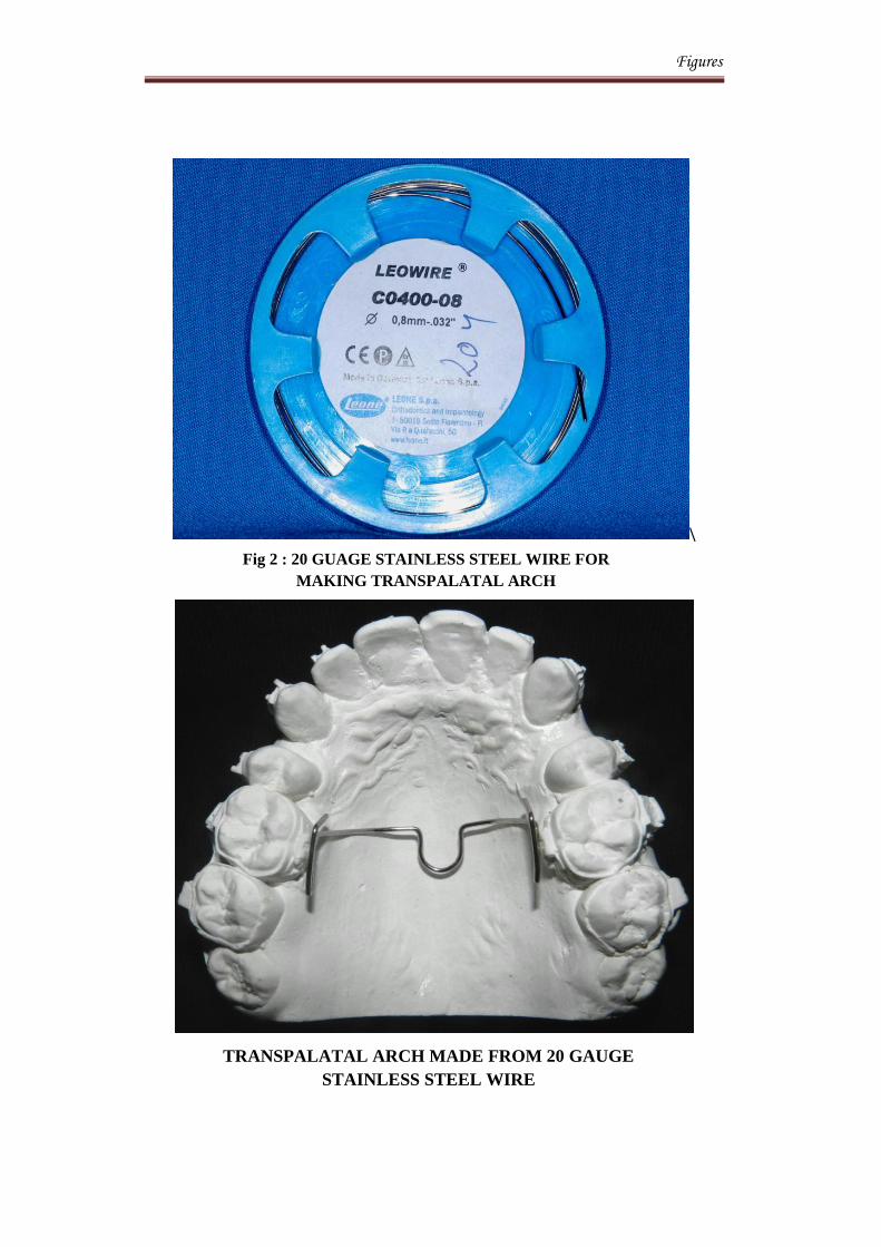
Figures
\
Fig 2 : 20 GUAGE STAINLESS STEEL WIRE FOR
MAKING TRANSPALATAL ARCH
TRANSPALATAL ARCH MADE FROM 20 GAUGE
STAINLESS STEEL WIRE
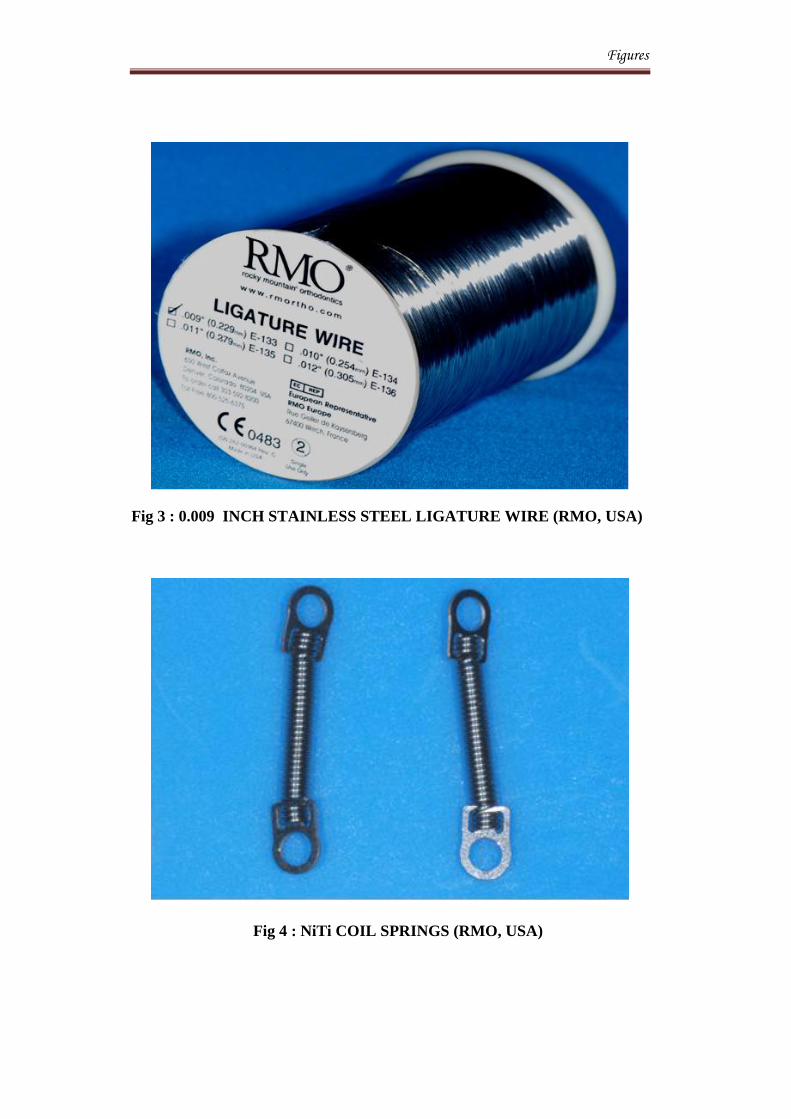
Figures
Fig 3 : 0.009 INCH STAINLESS STEEL LIGATURE WIRE (RMO, USA)
Fig 4 : NiTi COIL SPRINGS (RMO, USA)
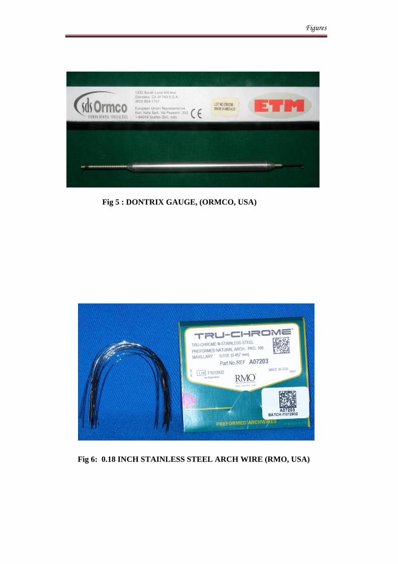
Figures
Fig 5 : DONTRIX GAUGE, (ORMCO, USA)
Fig 6: 0.18 INCH STAINLESS STEEL ARCH WIRE (RMO, USA)
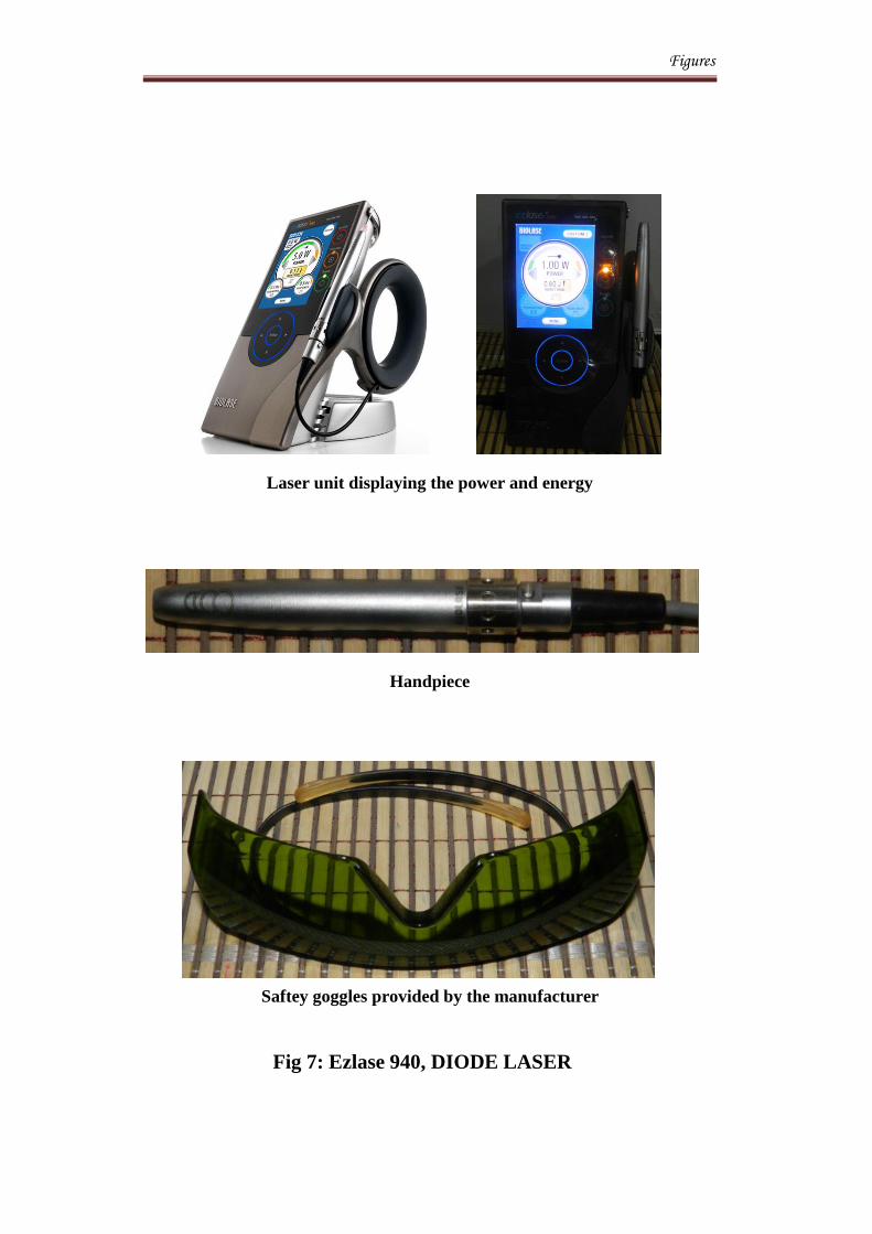
Figures
Laser unit displaying the power and energy
Handpiece
Fig 7: Ezlase 940, DIODE LASER
Saftey goggles provided by the manufacturer
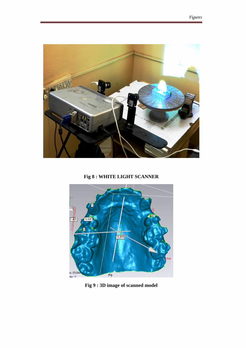
Figures
Fig 8 : WHITE LIGHT SCANNER
Fig 9 : 3D image of scanned model
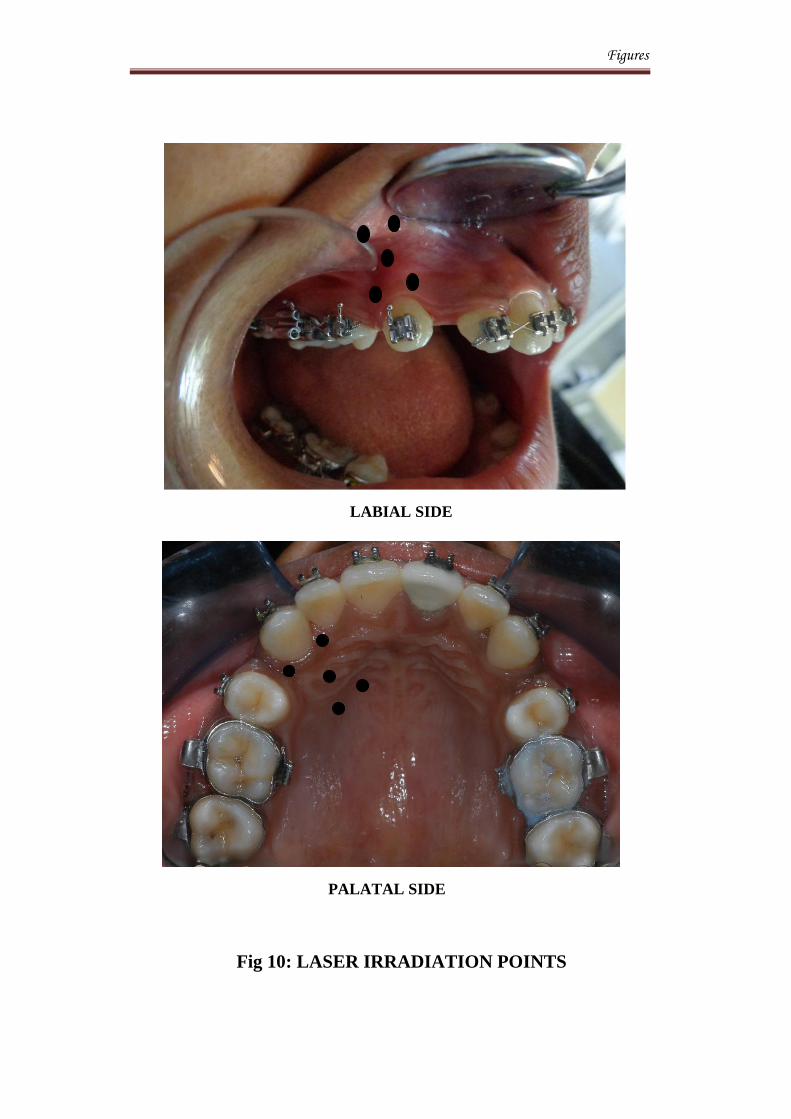
Figures
Fig 10: LASER IRRADIATION POINTS
LABIAL SIDE
PALATAL SIDE
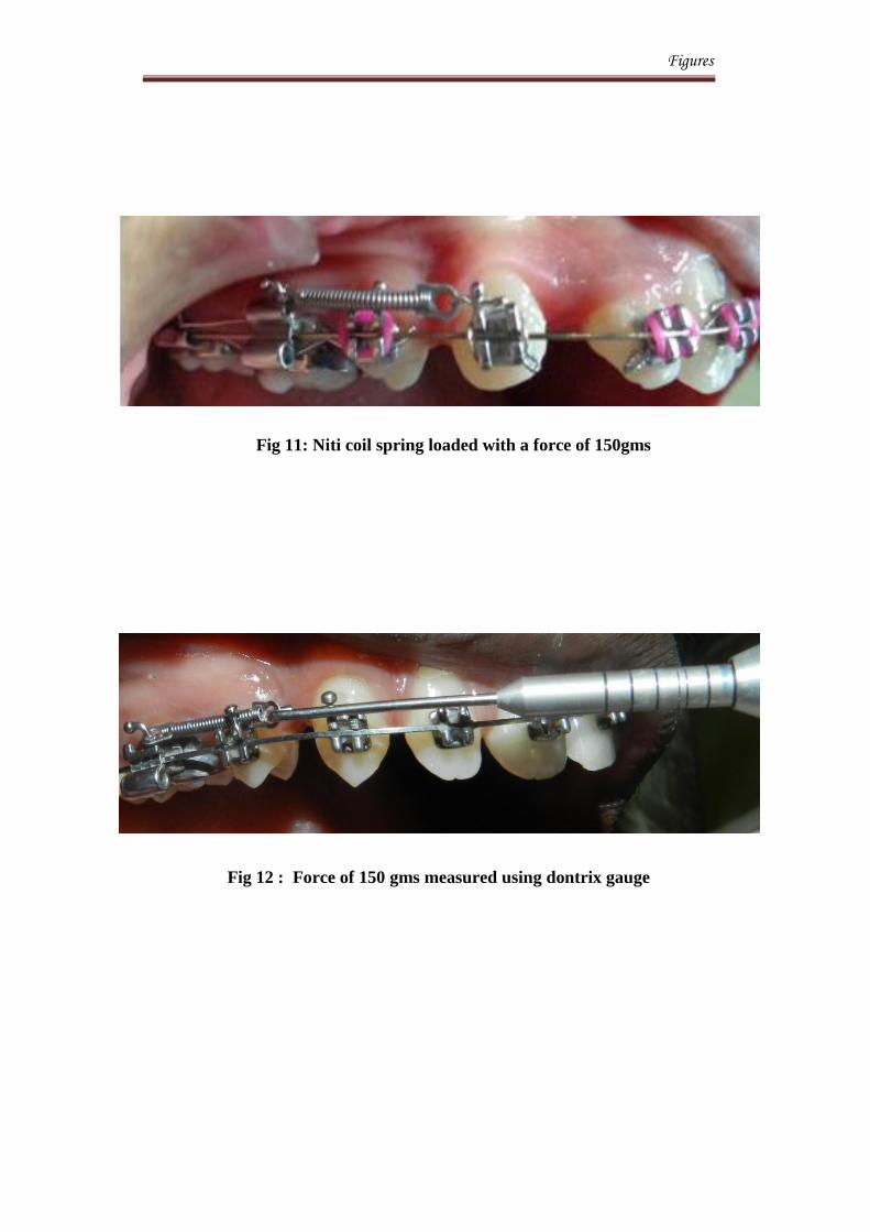
Figures
Fig 12 : Force of 150 gms measured using dontrix gauge
Fig 11: Niti coil spring loaded with a force of 150gms
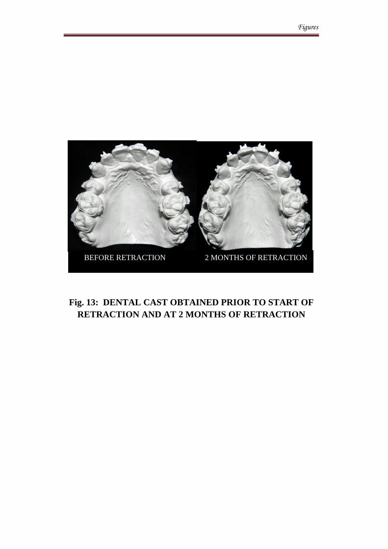
Figures
Fig. 13: DENTAL CAST OBTAINED PRIOR TO START OF
RETRACTION AND AT 2 MONTHS OF RETRACTION
BEFORE RETRACTION 2 MONTHS OF RETRACTION
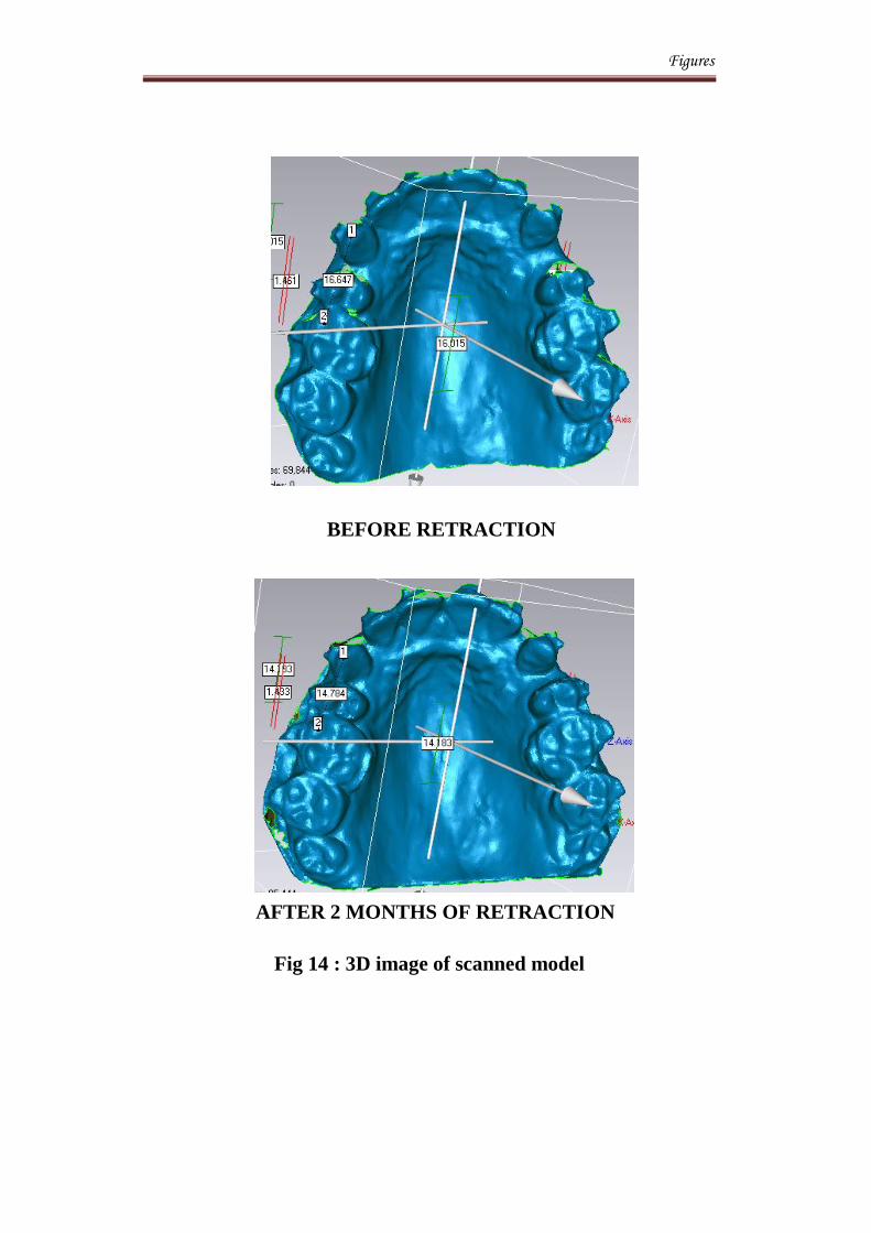
Figures
Fig 14 : 3D image of scanned model
BEFORE RETRACTION
AFTER 2 MONTHS OF RETRACTION
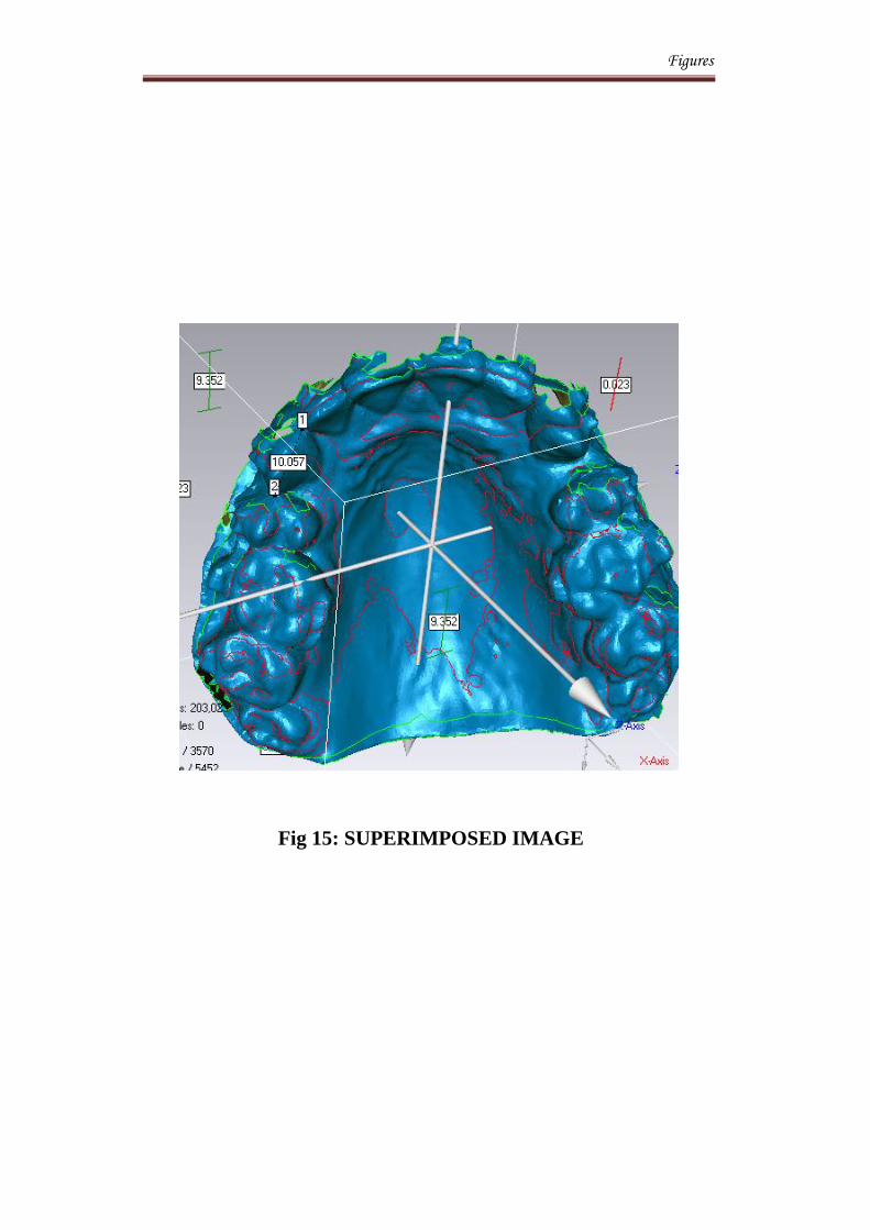
Figures
Fig 15: SUPERIMPOSED IMAGE
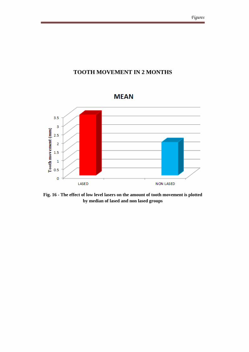
Figures
TOOTH MOVEMENT IN 2 MONTHS
Fig. 16 - The effect of low level lasers on the amount of tooth movement is plotted
by median of lased and non lased groups
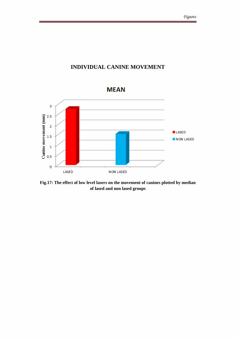
Figures
INDIVIDUAL CANINE MOVEMENT
Fig.17: The effect of low level lasers on the movement of canines plotted by median
of lased and non lased groups
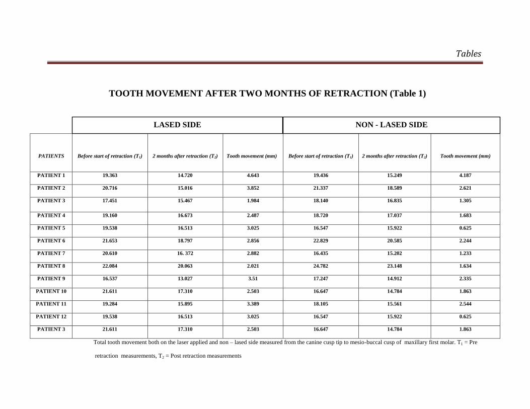
Tables
TOOTH MOVEMENT AFTER TWO MONTHS OF RETRACTION (Table 1)
PATIENTS
Before start of retraction (T1)
2 months after retraction (T2)
Tooth movement (mm)
Before start of retraction (T1)
2 months after retraction (T2)
Tooth movement (mm)
PATIENT 1 19.363 14.720 4.643 19.436 15.249 4.187
PATIENT 2 20.716 15.016 3.852 21.337 18.589 2.621
PATIENT 3 17.451 15.467 1.984 18.140 16.835 1.305
PATIENT 4 19.160 16.673 2.487 18.720 17.037 1.683
PATIENT 5 19.538 16.513 3.025 16.547 15.922 0.625
PATIENT 6 21.653 18.797 2.856 22.829 20.585 2.244
PATIENT 7 20.610 16. 372 2.882 16.435 15.202 1.233
PATIENT 8 22.084 20.063 2.021 24.782 23.148 1.634
PATIENT 9 16.537 13.027 3.51 17.247 14.912 2.335
PATIENT 10 21.611 17.310 2.503 16.647 14.784 1.863
PATIENT 11 19.284 15.895 3.389 18.105 15.561 2.544
PATIENT 12 19.538 16.513 3.025 16.547 15.922 0.625
PATIENT 3 21.611 17.310 2.503 16.647 14.784 1.863
Total tooth movement both on the laser applied and non – lased side measured from the canine cusp tip to mesio-buccal cusp of maxillary first molar. T1 = Pre
retraction measurements, T2 = Post retraction measurements
LASED SIDE NON - LASED SIDE
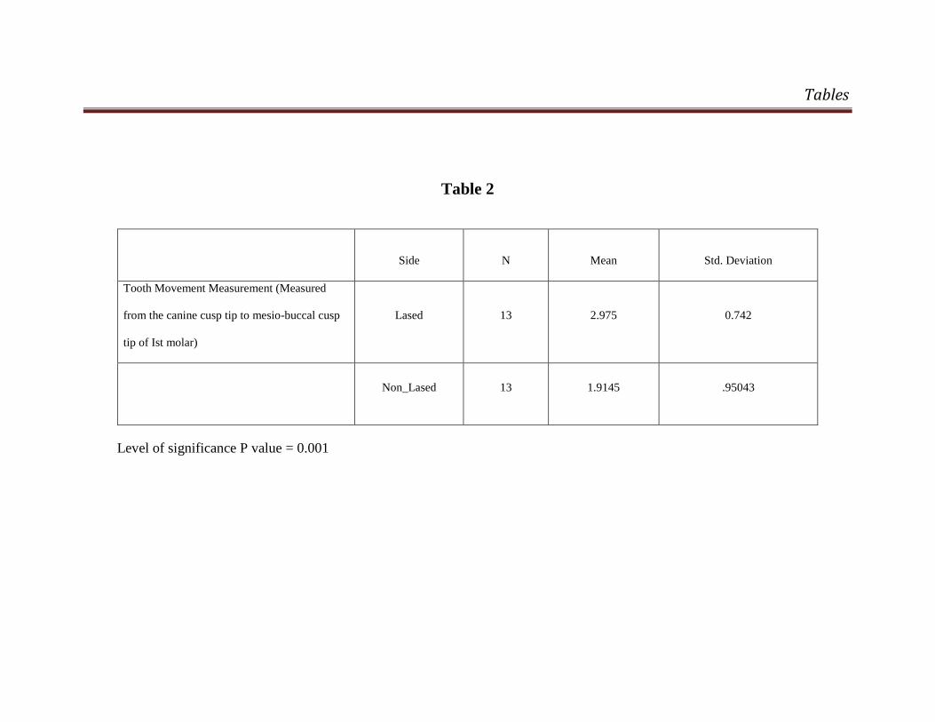
Tables
Table 2
Side N Mean Std. Deviation
Tooth Movement Measurement (Measured
from the canine cusp tip to mesio-buccal cusp
tip of Ist molar)
Lased 13 2.975 0.742
Non_Lased 13 1.9145 .95043
Level of significance P value = 0.001
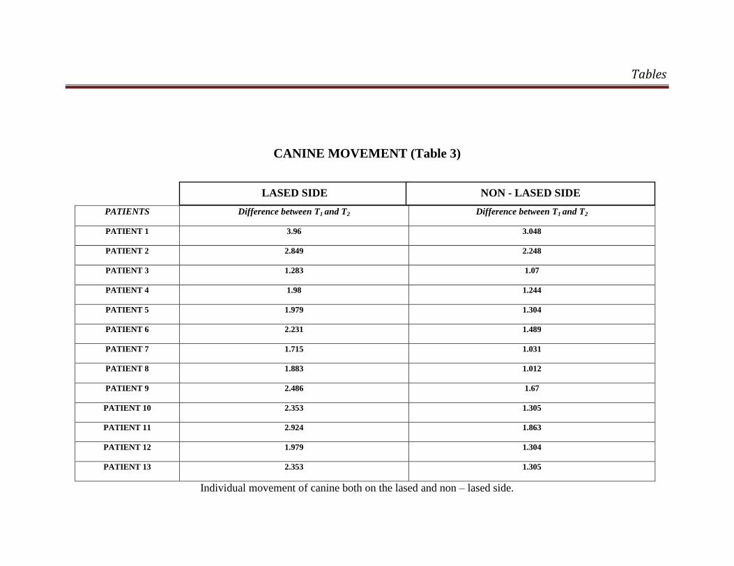
Tables
CANINE MOVEMENT (Table 3)
PATIENTS Difference between T1 and T2 Difference between T1 and T2
PATIENT 1 3.96 3.048
PATIENT 2 2.849 2.248
PATIENT 3 1.283 1.07
PATIENT 4 1.98 1.244
PATIENT 5 1.979 1.304
PATIENT 6 2.231 1.489
PATIENT 7 1.715 1.031
PATIENT 8 1.883 1.012
PATIENT 9 2.486 1.67
PATIENT 10 2.353 1.305
PATIENT 11 2.924 1.863
PATIENT 12 1.979 1.304
PATIENT 13 2.353 1.305
Individual movement of canine both on the lased and non – lased side.
LASED SIDE NON - LASED SIDE
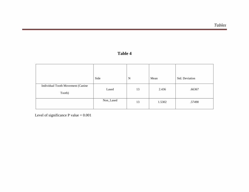
Tables
Table 4
Side N Mean Std. Deviation
Individual Tooth Movement (Canine
Tooth)
Lased 13 2.436 .66367
Non_Lased 13 1.5302 .57490
Level of significance P value = 0.001
Discussion
58
DISCUSSION
The term "accelerated orthodontic treatment" is a contentious
subject. The average time required to complete an orthodontic treatment is
2- to 3-years which is arduous for patient. Hence it is obligatory to accelerate
alveolar bone remodeling during treatment to abbreviate the time required36
.
The treatment time depends on distances the teeth need to be moved, treatment
goals, type of techniques employed and the cooperation of the patient. During
the past two decades many different approaches to fasten tooth movement
have been employed. Some attempts have been more creative than others. It
started in 1970’s with the evolution of powered brackets each having a tiny
working motor39
. Over the years low voltage currents13
, magnets11
, injection
of chemical agents70
and certain invasive procedures like corticotomy56
and
distraction osteogenesis2 have been employed to enhance tooth movement.
Owing to the demerits of the above procedures, other effective methods have
been searched for.
Davidovitch in 1980 examined the effects of electric currents on
orthodontic tooth movement. He concluded that electric currents, when
applied non - invasively to gingival tissues, are capable of activating
many PDL cells and neighbouring osteoblasts. This effect is localized to
well-defined zones near the electrodes and, therefore, may be used in
areas where bone remodelling is desired, such as in the case of
orthodontic treatment.12
Discussion
59
Accelerated Osteogenic Orthodontics or Wilckodontics is a surgical
intervention limited to the cortical portion of the alveolar bone. The biological
stimulus generated by corticotomies is reflected in the structure of trabecular
bone, which provides an opportunity to enhance certain orthodontic
movements56
.
Distraction osteogenesis was used as early as 1905 by Codivilla and
was later popularized by the clinical and research studies of Ilizarov.
Distraction osteogenesis was performed in the human mandible by Guerrero in
1990 and by McCarthy et al in 1992. Cortical holes are made in the alveolar
bone with a small, round, carbide bur. A thin, tapered, fissure bur was used to
connect the holes around the root. Akhare et al reviewed the effects of
distraction osteogenesis assisted canine retraction. He concluded that
dentoalveolar distraction technique is an innovative method that reduces
overall orthodontic treatment time by nearly 50%2.
Recently the OrthoAccel®’s (OA) Technology is predicated on the
application of pulsating, low magnitude forces (cyclic forces) to the teeth and
surrounding bone as a means of accelerating orthodontic tooth movement. The
OA appliance is a removable device similar to a retainer with a small motor,
into which the patient bites. Rather than using only constant pressure, the
device applies very light vibrations to the dentition. The patient places and
activates the device daily for twenty minutes of gentle pulsing. This
technology has revealed to accelerate tooth movement.
Discussion
60
In 1967, few years after the first working laser was invented, Endre
Mester in Hungary tested carcinogenic potential in mice. He shaved the dorsal
hair, divided them into two groups and gave a laser treatment with a low
powered ruby laser (694-nm) to one group. The irradiated group expressed no
carcinogenicity and instead to his surprise found that the hair on the treated
group grew back more quickly than the untreated group. This was the first
demonstration of "laser biostimulation". One important point that has been
demonstrated by multiple studies in cell culture, animal models and in clinical
studies is the concept of a biphasic dose response when the outcome is
compared with the total delivered light energy density41
.
The technology of Low Level Laser Therapy (LLLT) has proved to be
a boon in the field of orthodontics. The past decade has seen a veritable
explosion of research into the clinical applications of LLLT in orthodontics.
Majority of the applications of low level laser therapy (LLLT) in dentistry are
directed toward soft tissues and in recent years there has been increasing
interest in orthodontic applications of LLLT.
The documented literatures available on examining the effects of
LLLT on orthodontic tooth movement are scant. LLLT has shown to be an
effective tool in causing a biomodulatory effects on bone regeneration which
means that it may also have a considerable effect in accelerating orthodontic
tooth movement47
.
Discussion
61
Saito and Shimizu found that low intensity laser therapy can accelerate
bone regeneration in the midpalatal suture during rapid palatal expansion and
stimulate the synthesis of collagen, which is major matrix protein in bone52
.
In the following years, Kawasaki and Shimizu performed another experiment
on rats to find the effect of LLLT on orthodontic tooth movement. From his
study he inferred that orthodontically induced tooth movement associated with
LLLT produced an increase in the vascularization and this factor could
accelerate pulp tissue repair. He also showed that low level laser therapy had a
net effect of 30% increase in tooth movement29
. However its effect on tooth
movement in humans still remains uncertain.
Hence our study aimed at finding out the effects of Low Level Laser
Therapy on the rate of orthodontic tooth movement. We used a sample size of
13 patients in our study who had their upper and lower first bicuspids were
extracted for orthodontic treatment. We chose patients who had all permanent
teeth erupted with root formation completed excluding the third molars in the
maxillary arch. Patients with previous orthodontic history, with the presence
of periodontal pathology and those undergoing medical treatment and are on
medication with drugs like non-steroidal anti-inflammatory drugs (NSAID’S)
and steroid therapy were ruled out.
Stainless steel wire (Rocky Mountain Orthodontics, USA) of 0.18 inch
diameter was placed in the arch on which retraction was initiated. It is known
Tooth movement can be influenced by the type of material and diameter of
Discussion
62
orthodontic archwire especially during sliding mechanics. It is known that
stiffer wires can better resist the tendency of teeth tilting during sliding.
Moreover, friction increases as bracket slots are filled. For these reasons, a
round, 0.018-inch SS arch wire (RMO) was selected42
. The canine bracket was
secured tightly with stainless steel ligatures as elastomeric modules tend to
lose its elasticity and cause rotation during retraction.
The Ezlase 940 (Biolase) diode laser was used to irradiate the canines
prior to retraction According to the International Electrotecnical Commission
(IEC) 60825 – 1 standard this laser was classified as class 4 lasers. It had a
standardized wavelength of 940 nm, with an output power of 100 mW, and an
exposure time of 15 seconds per spot (mesio-cervical, disto-cervical, middle,
mesio-apical, disto-apical areas of the canine root). The 940 nm wavelength
works efficiently at low power. The low power was well absorbed by
hemoglobin and oxyhemoglobin. Hence only less amount of heat was
produced.
The wavelength at which the laser emits determines the effective depth
of penetration, within the tissue. The ability of a low level laser to penetrate a
given material depends on several factors. The most significant of these is the
absorption coefficient of the material through which the laser light travels70
.
Matter preferentially absorbs light at varying wavelengths. Laser photons that
travel through a given material with a high absorption coefficient for its
specific wavelength will lose energy through absorption more readily than a
Discussion
63
material with a lower coefficient for that material. Because these photons are
readily absorbed, this light travels much shorter distances than those light
wavelengths that are not absorbed. The absorption of photons from a laser’s
initial ray effectually degrades the power of light with distance travelled. In
most circumstances only a fraction of the photons from the initial beam may
reach these depths63
.
The laser irradiation was done once in 10 days for a period of 2
months. Impressions were made prior to irradiation (T1) and after two months
of retraction (T2). We chose to irradiate on 5 areas both on the lingual and
buccal periodontal ligament of canines for a period of 15 seconds per spot10
.
These areas were selected according to the work of Cruz et al. The reasons for
choosing these areas were in order to cover the periodontal fibres and alveolar
process around the canine tooth.
Before the start of laser irradiation, protective goggles were worn both
by the operator and the patient to avoid any optical hazards. These glasses
were provided by the manufacturer and had an optical density (OD) of 4+.
This OD was in accordance with the 940 nm wavelength used. It allows
penetration of the laser only 0.0001 mm which meant that the intensity of
exposure is extremely negligible.
Nickel titanium coil springs (Rocky Mountain Orthodontics, USA)
were used for retraction of the canines on both lased and non-lased sides.
These springs provide a force level that could be maintained for a longer
Discussion
64
period of time5. Barlow et al in 2008 evaluated the factors influencing the
efficacy of sliding mechanics to close an extraction space. He concluded that
nickel-titanium coil springs produce a more consistent force when compared
with elastomeric chains as a method of force delivery to close extraction space
along a continuous arch wire4.
The force selected for retraction of canines was 150 gms which was
measured using a Dontrix gauge (Ormco, Italy). Quinn and Yoshikawa
estimated that a force between 100 and 200 gms would be ideal for canine
retraction49
. Retain et al stated that the initial force application should be light,
because this produces biologic effects. These lighter forces will produce less
extensive hyalinized tissue that can be readily replaced by cellular elements.
He stated that an appropriate force of 150 to 200gms for maxillary canines
should be used for translatory movement27
.5 stated that
Models obtained before irradiation (T1) and after 2 months of canine
retraction (T2) were scanned using a white light scanner. White light scanning
is a 3d-scanning process using non-contact optical scanning device which uses
white light source to project fringes on the part being scanned. The sensor of
the scanner which is equipped with two cameras take several images of the
part during the measurement and the scanned images were imported o a high
end PC where advanced image software calculates point co-ordinates
throughout the visible area of the part under the scan.
Discussion
65
Khaled et al in 2008 developed a new method for three dimensional
imaging of the dental cast and evaluated it’s accuracy in analyzing the
different tooth movements. Each subject was clinically examined, and an
orthodontic diagnostic study cast was recorded. A 3D computer program was
specially designed for more accurate evaluation of the dental effects induced
by the three types of maxillary expanders, for the rotation and extrusion. He
concluded that the reliability of generating 3D dental images using dental casts
for 3D tooth movement analysis has a great research potential in orthodontics
because of its ability to yield accurate and reproducible data30
.
From the scanned data obtained by white light scanning, the amount of
tooth movement was analysed. This was accomplished by using the Geomagic
studio software. Using this software it was also possible to measure the
individual movement of the canine and the anchorage loss (mesial movement
of molars) which was done by superimposing both of the models obtained
before irradiation (T1) and after 2 months of canine retraction (T2). Geomagic
scanning and design software solutions are used to capture and model 3D
content from physical objects, organically sculpt complex shapes, and prepare
products for manufacturing. In addition, it produces powerful 3D metrology
and inspection software that verifies dimensional quality by comparing as-
built products to master designs. The key features of this software are:
Discussion
66
Produces the most accurate scan and probe data for creating high
quality 3 dimensional (3D), supporting all standard measurement
systems.
Integrates accurate 3 dimensional (3D) data directly into parametric
CAD systems for immediate use in design.
It processes the scan and probe data by collecting point data from all
major 3D scanners, digitizers and probes. Moreover it also optimizes scan data
i.e. any overlap of irrelevant data can be removed easily. In addition to this it
automatically or manually registers and merges multiple scan data sets.
Many theories have been postulated about the mechanism of action for
low level lasers. In literature, the most common discussed theories are:
Bioluminescence theory - DNA replication emits light at 630 nm.
Since this is very close to the wavelength of the He Ne-laser light, it is
postulated that laser may accelerate DNA replication via photonic
stimulation. Laser irradiation at this frequency is said to be non
mutagenic since it is not in the range to alter the genetic program by
affecting chromosomal ultra structure. The latter is more likely to
occur at ultra-violet light irradiation at 300 to 400 nm59
.
Cellular oscillation theory – The laser beam carries electromagnetic
oscillations of definite frequency. When it reaches the tissues the
electromagnetic oscillations gradually “swing and excite” single cells.
Discussion
67
This is thought to eventually intensify the biochemical processes that
ultimately regulate the performance of various vital organs59
.
Biological field theory – connections between tissues and organs in
the intact organism are not limited to humeral effects and nervous
control mechanisms alone. Rather there exists unique around every
cell, tissue and organ and higher structural levels exerting a
normalizing influence on lower levels. The resonance effect of the low
power laser is thought to restore the normal energetic status of the
organism, that is, restore its normal physiological state59
.
All three theories share the basic premise that laser causes activation in
the cell, which in turn leads to an intensification of the biochemical processes.
It is within this context that the Arndt-Schutz law becomes important with
respect to low power laser application. This biological law states that "weak
stimuli excite physiological activity, moderately strong ones favor it, strong
ones retard it and very strong ones arrest it." 59
In addition to the above theories, most recognized theory to explain the
effects of low level lasers is the PHOTOCHEMICAL THEORY.59
According to this theory; the light is absorbed by certain molecules, followed
by a cascade of biologic events. The common photoreceptors are the
endogenous porphyrins and cytochrome c-oxidase, leading to increased ATP
production. The principle of using LLLT is to supply direct biostimulative
light energy to the body cells. Cellular photoreceptors can absorb low level
Discussion
68
laser light and pass it on to mitochondria which promptly produces the cells
fuel, ATP59
.
In general Low Level Laser Therapy (LLLT) works on the principle of
inducing a biological response through energy transfer, in that the photonic
energy delivered into the tissue by the laser modulates the biological processes
within that tissue, and those within the biological system of which that tissue
is a part43
.
Our study evaluated the differences in tooth movement between the
lased and non lased sides. The lased side showed a mean tooth movement of
2.975 ± 0.742mm. On the non lased side the total movement was recorded to
be 1.91 ± 0.95 mm (Fig. 16). To ensure precise measurements of canine
retraction and anchorage loss, individual movement of both the canines and
molars were recorded using a white light scanner. The canine moved at a mean
distance of 2.436 ± 0.663 mm on lased side and 1.53 ± 0.57 mm (Fig. 17) on
control side. The anchorage loss during retraction was about 0.64 ± 0.33 mm
on lased side and 0.46 ± 0.23mm on non – lased side. Overall there was a
significant increase in the rate of tooth movement on the irradiated side. The
lased side showed about 35.66% increase in the rate of tooth movement. The
canine moved 32.72% more on the lased side. This increase was in accordance
with the work of Cruz et al who reported about 30%10
, Youssef et al who
showed a 20-40% increase73
and Doshi Mehta et al who showed 56 %
increase15
in the tooth movement on the lased side following low level laser
therapy.
Discussion
69
It was Cruz et al in 2004, who conducted the first human study on the
effect of low-intensity laser therapy on orthodontic tooth movement. They
showed that the irradiated canines were retracted at a rate 34% greater than the
control canines over 60 days. He concluded that LLLT significantly
accelerates orthodontic movement in humans with a healthy response from
periodontal tissues. Therefore it can be considered in order to shorten the
treatment duration10
.
The reason for the difference in tooth movement on the lased and non
lased side may be due to the fact that low level laser irradiation promotes
proliferation and differentiation of human osteoblasts61
. Its effects were
studied by Stein et al in 2005 who investigated the effect of low-level laser
irradiation on proliferation and differentiation of a human osteoblast cell line.
They used cultured osteoblast cells which was irradiated using He-Ne laser
having a wavelength of 632 nm with a power output of 10 mW. They noticed
a significant 31–58% increase in cell survival (MTT assay) and higher cell
count in the once-irradiated as compared to non- irradiated cells.
Differentiation and maturation of the cells was followed by osteogenic
markers: alkaline phosphatase (ALP), osteopontin (OP), and bone sialoprotein
(BSP). A two-fold enhancement of ALP activity and expression of OP and
BSP was much higher in the irradiated cells as compared to non-irradiated
osteoblasts. They concluded that LLLT given for a short duration can
significantly promote proliferation and differentiation of human osteoblasts in
Discussion
70
vitro as compared to non - irradiated cells. He also assumed that osteoblasts
that proliferate and differentiate at a higher rate at the site of injury may
increase the rate of calcium accumulation and promote bone repair. Thus, the
results of the present study may suggest the ability of LLLT to enhance bone
repair also in humans61
.
It is important to consider the following points when learning about the
mechanisms of low level laser therapy. The coherence of the electromagnetic
energy plays a crucial role in the efficacy of the treatment63
. This degree of
coherence is related to the spectral narrowness of the light source.
Furthermore, the coherent character of the laser light is not lost after
penetrating the tissue but is split into small coherent and polarized islands
called speckles. The speckle pattern is maintained through the irradiated
volume of tissue. Due to intensity differences within the speckle field,
temperature and electric field gradients occur. Such gradients create a force on
the cells and organelles. This explains how low level lasers have a stimulative
effect on the irradiated tissue63
.
On the contrary, our results were in contradiction to the work done by
Limpanichkul36
and colleagues who conducted their clinical study on human
subjects. Their results showed no difference between the experimental low-
intensity laser therapy subjects and the controls in a split-mouth study with
human subjects over 4 months. LLLT at the parameter settings in the study
had no effect on rate of tooth movement at any time periods. He claimed that
Discussion
71
the energy density of 25 J per square centimeter was probably too low to
express either stimulatory effect or inhibitory effect on the rate of orthodontic
tooth movement. This may be the reason for the negative result obtained in the
study.
To ensure precise measurements of the canine retraction and anchorage
loss, individual movement of both the canines and molars were also recorded
using a white light scanner. White light scanning provides accurate linear
measurements as the reproducibility of the white light scanners is between 1-5
microns.
Despite the fact that the positive results of this study was found to be
statistically significant, it lacked evidence regarding the efficiency of LLLT in
reducing the treatment time all together. Therefore future studies with larger
clinical trials in various malocclusions over a longer period of time are
warranted.
Summary and Conclusion
72
SUMMARY AND CONCLUSION
The effect of low level laser therapy (LLLT) on the rate of orthodontic
tooth movement was evaluated. 13 patients who required retraction of
maxillary canines as part of orthodontic treatment were selected for this study.
One side of the quadrant was lased with a diode laser while the other side
taken as control. NiTi coil springs were used for retraction of the canines on
both sides which was done on a 0.18 inch stainless steel wire. The casts
obtained before retraction and following 2 months of retraction was scanned
using a white light scanner and the measurements of these scanned data were
analyzed using the Geomagic software.
From the results obtained in our study, it is prudent to conclude that
LLLT significantly hastens orthodontic tooth movement in humans with a
healthy response from periodontal tissues. The irradiation parameters and
protocol used in this study were successful in accelerating the tooth
movement.
Future efforts should be directed toward investigating the precise
dosimetry required for therapeutic laser effects, in order to achieve
standardization of treatment protocols. Further studies and more clinical trials
are necessary to evaluate the effect of LLLT in decreasing the overall
orthodontic treatment duration.
Bibliography
73
BIBILOGRAPHY
1. Abi-Ramia LB, Stuani AS, Stuani AS, Stuani MB, Mendes Ade M.
Effects of low-level laser therapy and orthodontic tooth movement
on dental pulps in rats. Angle Orthod. 2010 Jan;80(1):116-22.
2. Akhare P, Akshay M. Daga, Shilpa Pharande Rapid Canine
Retraction and Orthodontic Treatment with Dentoalveolar
Distraction Osteogenesis, Journal of Clinical and Diagnostic
Research. 2011 November (Suppl-2), Vol-5(7): 1473-1477
3. Altan BA, Sokucu O, Ozkut MM, Inan S. Metrical and histological
investigation of the effects of low-level laser therapy on
orthodontic tooth movement. Lasers Med Sci. 2012 Jan;27(1):131-40
4. Barlow M, Kula K. Factors influencing efficiency of sliding
mechanics to close extraction space: a systematic review. Orthod
Craniofac Res. 2008 May;11(2):65-73.
5. Burrow SJ. Canine retraction rate with self-ligating brackets vs
conventional edgewise brackets Angle Orthod. 2010 Jul;80(4):
438-45.
6. Cepera F, Torres FC, Scanavini MA, Paranhos LR, Capelozza Filho L,
Cardoso MA, Siqueira DC, Siqueira DF. Effect of a low-level laser on
bone regeneration after rapid maxillary expansion. Am J Orthod
Dentofacial Orthop. 2012 Apr;141(4):444-50.
Bibliography
74
7. Charles J. Burstone, Herbert A. Koenig, Optimizing anterior and
canine retraction . Am J Orthod Volume70, Issue 1, July 1976,
Pages 1–19
8. Collins MK, Sinclair PM. The local use of vitamin D to increase the
rate of orthodontic tooth movement. Am J Orthod Dentofacial
Orthop. 1988 Oct;94(4):278-84.
9. Coombe AR, Ho CT, Darendeliler MA, Hunter N, Philips JR, Chapple
CC, Yum LW. The effects of low level laser irradiation on
osteoblastic cells. Clin Orthod Res. 2001 Feb;4(1):3-14.
10. Cruz DR, Kohara EK, Ribeiro MS, Wetter NU. Effects of low-
intensity laser therapy on the orthodontic movement velocity of
human teeth: a preliminary study. Lasers Surg Med.
2004;35(2):117-20.
11. Darendeliler MA, Sinclair PM, Kusy RP. The effects of samarium-
cobalt magnets and pulsed electromagnetic fields on tooth
movement. Am J Orthod Dentofacial Orthop. 1995 Jun;107(6):578-88.
12. Davidovitch Z, Finkelson MD, Steigman S, Shanfeld JL, Montgomery
PC, Korostoff E. Electric currents, bone remodeling, and
orthodontic tooth movement. II. Increase in rate of tooth
movement and periodontal cyclic nucleotide levels by combined
force and electric current. Am J Orthod. 1980 Jan;77(1):33-47.
Bibliography
75
13. Dominguez Angela, Velasquez Sergio, Acceleration Effect of
Orthodontic Movement by Application of Low-intensity Laser,
2010 , Journal of oral laser applications
14. Donald Joseph Huffman, David C. Way, A clinical evaluation of
tooth movement along arch wires of two different sizes. Am J
Orthod Volume 83, Issue 6, June 1983, Pages 453–459
15. Doshi-Mehta G, Bhad-Patil WA. Efficacy of low-intensity laser
therapy in reducing treatment time and orthodontic pain: a
clinical investigation. Am J Orthod Dentofacial Orthop. 2012
Mar;141(3):289-97.
16. Duan J, Na Y, Liu Y, Zhang Y, Effects of the pulse frequency of low-
level laser therapy on the tooth movement speed of rat molars.
Photomed Laser Surg. 2012 Nov;30(11):663-7
17. Enwemeka CS. Laser biostimulation of healing wounds: specific
effects and mechanisms of action. J Orthop Sports Phys Ther.
1988;9(10):333-8.
18. Fernando A. L. Habib, Sabrina K. C. Gama, Luciana M. P. Ramalho,
Maria Cristina T. Cangussú, Fernando P. dos Santos Neto, Juliana A.
Lacerda, Telma M. de Araújo, and Antônio L. B. Pinheiro. Effect of
Laser Phototherapy on the Hyalinization Following Orthodontic
Tooth Movement in Rats Photomedicine and Laser Surgery. March
2012, 30(3): 179-185.
Bibliography
76
19. Fujiyama K; Deguchi T; Murakami T; Fujii A; Kushima ;Yamamoto
T, Clinical effect of CO laser in reducing pain in orthodontics.
Angle Orthod 78:299-303
20. Genc G, Kocadereli I, Tasar F, Kilinc K, El S, Sarkarati B. Effect of
low-level laser therapy (LLLT) on orthodontic tooth movement.
Lasers Med Sci. 2012 Feb 18.
21. Görür I, Orhan K, Can-Karabulut DC, Orhan AI, Oztürk A. Low-level
laser therapy effects in traumatized permanent teeth with
extrusive luxation in an orthodontic patient. Angle Orthod. 2010
Sep;80(5):968-74.
22. Goulart CS, Nouer PR, Mouramartins L, Garbin IU, de Fátima
Zanirato Lizarelli R. Photoradiation and orthodontic movement:
experimental study with canines. Photomed Laser Surg. 2006
Apr;24(2):192-6
23. H Kanzaki, M Chiba, K Arai, I Takahashi, N Haruyama, M Nishimura,
H Mitani , Local RANKL gene transfer to the periodontal tissue
accelerates orthodontic tooth movement. Gene Therapy (impact
factor: 3.71). 05/2006; 13(8):678-85
24. Hashimoto F, Kobayashi Y, Mataki S, Kobayashi K, Kato Y, Sakai H.
Administration of osteocalcin accelerates orthodontic tooth
movement induced by a closed coil spring in rats. Eur J Orthod.
2001 Oct;23(5):535-45
Bibliography
77
25. Häsler R, Schmid G, Ingervall B, Gebauer U. A clinical comparison
of the rate of maxillary canine retraction into healed and recent
extraction sites--a pilot study. Eur J Orthod. 1997 Dec;19(6):711-9.
26. Hosseini MH1, Mahmoodzadeh Darbandi M2, Kamali A, Effect of
low level laser therapy on orthodontic movement in human, Journal
of Dental Medicine-Tehran University of Medical Sciences
2011;24(3):156-164
27. Kaare Reitan , Effects Of Force Magnitude And Direction Of Tooth
Movement On Different Alveolar Bone Types. The Angle
Orthodontist: October 1964, Vol. 34, No. 4, pp. 244-255.
28. Kawakami M, Takano-Yamamoto T. Local injection of 1,25-
dihydroxyvitamin D3 enhanced bone formation for tooth
stabilization after experimental tooth movement in rats. J Bone
Miner Metab. 2004;22(6):541-6.
29. Kawasaki K, Shimizu N. Effects of low-energy laser irradiation on
bone remodeling during experimental tooth movement in rats.
Lasers Surg Med. 2000;26(3):282-91.
30. Khaled M. Fawzy and Noha E. Sabet and Ibrahim M. Negm Software
for 3D Analysis of Tooth Movement 2008 Orthodontic Cyber Journal
Bibliography
78
31. Kim, S.-J., Moon, S.-U., Kang, S.-G. and Park, Y.-G. (2009), Effects
of low-level laser therapy after Corticision on tooth movement and
paradental remodeling. Lasers Surg. Med., 41: 524–533.
32. Kobayashi Y, Takagi H, Sakai H, Hashimoto F, Mataki S, Kobayashi
K, Kato Y. Effects of local administration of osteocalcin on
experimental tooth movement. Angle Orthod.1998 Jun;68(3):259-66.
33. Koutna M., Janisch R., Veselska R. Effects of low power laser
irradiation on cell proliferation SCRIPTA MEDICA (BRNO) – 76
(3): 163–172, June 2003
34. Kravitz ND, Kusnoto B. Soft-tissue lasers in orthodontics: an
overview. Am J Orthod Dentofacial Orthop. 2008 Apr;133(4
Suppl):S110-4.
35. Lim HM, Lew KK, Tay DK. A clinical investigation of the efficacy
of low level laser therapy in reducing orthodontic postadjustment
pain. Am J Orthod Dentofacial Orthop. 1995 Dec;108(6):614-22.
36. Limpanichkul W, Godfrey K, Srisuk N, Rattanayatikul C. Effects of
low-level laser therapy on the rate of orthodontic tooth movement.
Orthod Craniofac Res. 2006 Feb;9(1):
37. Long H, Pyakurel U, Wang Y, Liao L, Zhou Y, Lai W. Interventions
in accelerating orthodontic tooth movement Angle Orthod.
2012 Jun 21.
Bibliography
79
38. Lotzof LP, Fine HA,Cisneros GJ. Canine retraction: a comparison
of two preadjusted bracket systems. Am J Orthod Dentofacial
Orthop. 1996 Aug;110(2):191-6.
39. Marinês Vieira da Silva Sousa, Marco Antonio Scanavini, Eduardo
Kazuo Sannomiya, Leandro G. Velasco, and Fernanda Angelieri.
Photomedicine and Laser Surgery. March 2011, 29(3): 191-196.
40. Marquezan M, Bolognese AM, Araújo MT. Effects of two low-
intensity laser therapy protocols on experimental tooth movement.
Photomed Laser Surg. 2010 Oct;28 Suppl 2:S79-83
41. Mester E, Mester AF, Mester A. The biomedical effects of laser
application. Lasers Surg Med. 1985;5:31-9.
42. Mezomo M, de Lima ES, de Menezes LM, Weissheimer A, Allgayer
S. Maxillary canine retraction with self-ligating and conventional
brackets. Angle Orthod. 2011 Mar;81(2):292-7
43. Michael R. Hamblin, Ronald W. Waynant, Juanita Anders,
Mechanisms for Low-Light Therapy, Proc. of SPIE Vol. 6140,
614001, (2006).
44. Ngan P, Kess B, Wilson S. Perception of discomfort by patients
undergoing orthodontic treatment. Am J Orthod Dentofacial Orthop.
1989 Jul;96(1):47-53.
45. Nicola RA, Jorgetti V, Rigau J, Pacheco MT, dos Reis LM, Zângaro
RA. Effect of low-power GaAlAs laser (660 nm) on bone structure
Bibliography
80
and cell activity: an experimental animal study. Lasers Med Sci.
2003;18(2):89-94.
46. Oliveira; Rodrigo Villamarim Soares, Alveolar corticotomies in
orthodontics: Indications and effects on tooth movement Dental
Press J. Orthod. vol.15 no.4 Maringá July/Aug. 2010
47. Pinheiro, Oliveira, Martins, Ramalho, Marcos. Matos de Oliveira,
Jœnior , Nicolau , Laser Therapy Vol. 13, 2008, Journal of the World
Association for Laser Therapy
48. Pugliese LS, Medrado AP, Reis SR, Andrade Zde A. The influence of
low-level laser therapy on biomodulation of collagen and elastic
fibers. Pesqui Odontol Bras. 2003 Oct-Dec;17(4):307-13.
49. Quinn RS, Yoshikawa DK. A reassessment of force magnitude in
orthodontics. Am J Orthod. 1985 Sep;88(3):252-60.
50. Ross, G., and A. Ross.. Photobiomodulation: an invaluable tool for
all dental specialties. JLaser Dent 2009 17(3): 117–24.
51. Sabrina K. C. Gama, Fernando A. L. Habib, Juliana S. de Carvalho,
Monteiro, Gardênia M. Paraguassú, Telma Martins Araújo, Maria
Cristina T. Cangussú, and Antonio Luiz B. Pinheiro. Tooth
Movement After Infrared Laser Phototherapy: Clinical Study in
Rodents Photomedicine and Laser Surgery. October 2010, 28(S2): S-
79-S-83.
Bibliography
81
52. Saito S, Shimizu N. Stimulatory effects of low-power laser
irradiation on bone regeneration in midpalatal suture during
expansion in the rat. Am J Orthod Dentofacial Orthop. 1997
May;111(5):525-32.
53. Sandy JR, Farndale RW, Meikle MC. Recent advances in
understanding mechanically induced bone remodeling and their
relevance to orthodontic theory and practice. Am J Orthod
Dentofacial Orthop. 1993 Mar;103(3):212-22.
54. Seifi M, Shafeei HA, Daneshdoost S, Mir M. Effects of two types of
low-level laser wave lengths (850 and 630 nm) on the orthodontic
tooth movements in rabbits Lasers Med Sci. 2007 Nov;22(4):261-4
55. Seifi M, Vahid-Dastjerdi E.Immediate relief of TMJ clicking
following low level laser therapy after orthodontic treatment:A
Case Report. J Laser Med Sci. 2011; 2(1):43-5
56. Seifi M,Younessian F, Ameli N. The Innovated Laser Assisted
Flapless Corticotomy to Enhance Orthodontic Tooth Movement: J
Lasers Med Sci 2012; 3(1):20-5
57. Seiryu M, Deguchi T, Fujiyama K, Sakai Y, Daimaruya T, Takano-
Yamamoto T. Effects of CO2 laser irradiation of the gingiva during
tooth movement. J Dent Res. 2010 May;89(5):537-42.
58. Shpack N, Davidovitch M, Sarne O, Panayi N, Vardimon AD.
Duration and anchorage management of canine retraction with
Bibliography
82
bodily versus tipping mechanics. Angle Orthod. 2008 Jan;78(1):95-
100
59. Slattery, Erchonia Medical Laser 2008, The Mechanisms of Low
Level Laser Therapy
60. Smith RJ, Burstone CJ. Mechanics of tooth movement. Am J Orthod.
1984 Apr;85(4):294-307.
61. Stein A, Benayahu D, Maltz L, Oron U. Low-level laser irradiation
promotes proliferation and differentiation of human osteoblasts in
vitro. Photomed Laser Surg. 2005 Apr;23(2):161-6.
62. Stein E, Koehn J, Sutter W, Wendtlandt G, Wanschitz F, Thurnher
D, Baghestanian M, Turhani D. Initial effects of low-level laser
therapy on growth and differentiation of human osteoblast-like
cells. Wien Klin Wochenschr. 2008;120(3-4):112-7
63. Sun G, Tunér J. Low-level laser therapy in dentistry. Dent Clin
North Am. 2004 Oct;48(4):1061-76,
64. Tarek Momhamed Ibrahim, mostafa gheith ,Mona M. Abo-Elfotouh.
The Effect of Soft laser Application on Orthodontic Movement (In
vitro study). Journal of American Science 2011;7(6):125-135
65. Tortamano A, Lenzi DC, Haddad AC, Bottino MC, Dominguez GC,
Vigorito JW. Low-level laser therapy for pain caused by placement
of the first orthodontic archwire: a randomized clinical trial. Am J
Orthod Dentofacial Orthop. 2009 Nov;136(5):662-7.
Bibliography
83
66. Tunér J, Hode L. The Laser Therapy Handbook. 1st ed.
Grangesberg, Sweden: Prima Books AB; 2004:12-7.
67. Turhani D, Scheriau M, Kapral D, Benesch T, Jonke E, Bantleon HP.
Pain relief by single low-level laser irradiation in orthodontic
patients undergoing fixed appliance therapy. Am J Orthod
Dentofacial Orthop. 2006 Sep;130(3):371-7.
68. Walsh LJ. The current status of low level laser therapy in dentistry.
Part 2. Hard tissue applications. Aust Dent J. 1997 Oct;42(5):302-6.
69. Yamaguchi M, Hayashi M, Fujita S, Yoshida T, Utsunomiya T,
Yamamoto H, Kasai K. Low-energy laser irradiation facilitates the
velocity of tooth movement and the expressions of matrix
metalloproteinase-9, cathepsin K, and alpha(v) beta(3) integrin in
rats. Eur J Orthod. 2010 Apr;32(2):131-9
70. Yamasaki K, Shibata Y, Imai S, Tani Y, Shibasaki Y, Fukuhara T.
Clinical application of prostaglandin E1 (PGE1) upon orthodontic
tooth movement. Am J Orthod. 1984 Jun;85(6):508-18.
71. Yi J, Zhang L, Yan B, Yang L, Li Y, Zhao Z. Drinking coffee may
help accelerate orthodontic tooth movement. Dent Hypotheses
2012;3:72-5
72. Yordanova S, Yordanova M, Tomov G, LalabonovaH, ER:YAG
LASER APPLICATION IN ORTHODONTIC PRACTICE. A

Bibliography
84
CASE REPORT, Journal of IMAB - Annual Proceeding (Scientific
Papers) 2011, vol. 17, book 2
73. Youssef M, Ashkar S, Hamade E, Gutknecht N, Lampert F, Mir M.
The effect of low-level laser therapy during orthodontic
movement: a preliminary study. Lasers Med Sci. 2008 Jan;23(1):
27-33.


