comparative results of frenectomy by three surgical techniques
TRANSCRIPT
DentistryHungund et al., Dentistry 2013, 4:1
http://dx.doi.org/10.4172/2161-1122.1000183
Open AccessCase Report
Volume 4 • Issue 1 • 1000183DentistryISSN: 2161-1122 Dentistry, an open access journal
Comparative Results of Frenectomy by Three Surgical Techniques- Conventional, Unilateral Displaced Pedicle Flap and Bilateral Displaced Pedicle FlapHungund S1, Dodani K1, Kambalyal P1* and Kambalyal P2
1Department of Periodontology and Implantology, Darshan Dental College and Hospital, Udaipur, Rajasthan, India2Department of Orthodontics, Darshan Dental College and Hospital, Udaipur, Rajasthan, India
AbstractMaxillary labial frenum is capable of creating a diastema and recession, affecting aesthetics. Archer’s classical frenectomy technique
is an extensive procedure which causes scarring and loss of interdental papilla. This leads towards the conservative approaches like Edward’s frenectomy, frenum relocation by Z-plasty and free gingival graft. Since the procedure of frenectomy was first proposed, a number of modifications have been developed to solve the problem caused by an abnormal labial frenum. But in most of the techniques the zone of attached gingiva and aesthetics are not considered. Thus, the aim of this case report is to present case series of various frenectomy techniques for management of aberrant frenum. A series of cases of an aberrant frenum were approached by various surgical frenectomy techniques like conventional (classical) technique, Miller’s technique using unilateral pedicle flap and frenectomy technique using bilateral pedicle flap and results are reported. The frenectomy technique using pedicle flap gives good aesthetic results, colour match, gain in attached gingiva and no anaesthetic scar formation as healing takes place by primary intention.
*Corresponding author: Preeti Kambalyal, Department of Periodontology and Implantology, Darshan Dental College and Hospital, Loyara, Udaipur, Rajasthan, India, Tel: +919602717070; E-mail: [email protected]
Received November 20, 2013; Accepted December 19, 2013; Published December 21, 2013
Citation: Hungund S, Dodani K, Kambalyal P, Kambalyal P (2013) Comparative Results of Frenectomy by Three Surgical Techniques- Conventional, Unilateral Displaced Pedicle Flap and Bilateral Displaced Pedicle Flap. Dentistry 4: 183. doi:10.4172/2161-1122.1000183
Copyright: © 2013 Hungund S, et al. This is an open-access article distributed under the terms of the Creative Commons Attribution License, which permits unrestricted use, distribution, and reproduction in any medium, provided the original author and source are credited.
Keywords: Frenum; Frenectomy; Lateral pedicle flap
IntroductionA frenum is an anatomic structure formed by a fold of mucous
membrane and connective tissue and sometimes muscle fibres that attach the lip and cheeks to the alveolar mucosa and/or gingiva and the underlying periosteum [1].
Depending upon the extension of attachment of fibres, frenum has been classified as follows: [2]
1. Mucosal- when the fibres are attached up to mucogingival junction
2. Gingival- when fibres are inserted within attached gingiva
3. Papillary- when fibres are extended into interdental papilla; and
4. Papilla penetrating- when the fibres cross the alveolar process and extend up to the palatine papilla.
Clinically, papillary and papilla penetrating frenum are considered as pathological and have been found to be associated with loss of papilla, recession, diastema and plaque accumulation [3,4]. The abnormal frenum is detected visually by applying tension over the frenum to see the movement of the papillary tip or the blanch which is produced due to ischemia in the region [5]. In such cases it is necessary to perform a frenectomy for aesthetic and functional reasons. There are several surgical techniques for removal of labial frenum. Since the procedure of frenectomy was first proposed, a number of modifications have been
developed. In most of these procedures aesthetic outcome in terms of attached gingiva with colour matching was not considered and these procedures results in scar formation [6-8]. A better approach to make primary closure in the midline and to avoid anaesthetic scar by creating zone of attached gingiva, frenectomy is associated with lateral pedicle flap. This article is a compilation of series of clinical cases of an aberrant frenum which were approached by various surgical frenectomy techniques like conventional (classical) technique, Miller’s technique using unilaterally displaced pedicle flap, or frenectomy using bilaterally displaced pedicle flap and the results are presented.
Material and MethodsThese surgical techniques were undertaken at Darshan Dental
College and Hospital, Udaipur. The subjects underwent frenectomy for periodontal or orthodontic reasons. A frenum was considered abnormal when it was unusually broad or there was no apparent attached gingiva in the midline or the interdental papilla could be stretched by the frenum.
Conventional (classical) technique The classical technique was introduced by Archer. This surgical
approach was advocated in the midline diastema cases with an aberrant frenum to ensure the removal of the muscle fibres which were supposedly connecting the orbicularis oris with the palatine papilla. This technique is an excision type frenectomy which includes the interdental tissue along with the frenum [9].
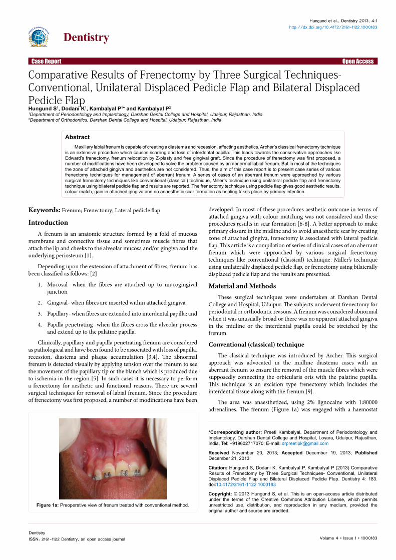
The area was anaesthetized, using 2% lignocaine with 1:80000 adrenalines. The frenum (Figure 1a) was engaged with a haemostat
Figure 1a: Preoperative view of frenum treated with conventional method.

Citation: Hungund S, Dodani K, Kambalyal P, Kambalyal P (2013) Comparative Results of Frenectomy by Three Surgical Techniques- Conventional, Unilateral Displaced Pedicle Flap and Bilateral Displaced Pedicle Flap. Dentistry 4: 183. doi:10.4172/2161-1122.1000183
Page 2 of 6
Volume 4 • Issue 1 • 1000183DentistryISSN: 2161-1122 Dentistry, an open access journal
which was inserted into the depth of the vestibule and incisions were placed on the upper and the under surface of the haemostat until the haemostat was free. The triangular resected portion of the frenum with the haemostat was removed. A blunt dissection was done to relieve the fibrous attachment (Figure 1b). The edges of the diamond shaped wound were sutured using 4-0 black silk with interrupted sutures (Figure 1c). The area was covered with a periodontal pack. The pack and the sutures were removed 1 week post-operatively.
Case 1: A 21 year old male patient was referred from Department of Orthodontics for frenectomy. On examination there was papillary frenum attachment (Figure 1a). The case was treated surgically by conventional method. One month post-operative view is shown in (Figure 1d).
Case 2: A 38 year old female patient had complaint of spacing in upper anterior teeth which was increasing with unaesthetic appearance. Examination revealed high maxillary frenum attachment which was papillary type with positive tension test (Figure 2a). The case was treated
surgically by conventional method. The one month post-operative view is shown in Figure 2b.
Frenectomy using unilateral single pedicle flap (miller’s technique)
This surgical technique was advocated by Miller PD in 1985. This technique was proposed for the post-orthodontic diastema cases.
After adequate local anaesthesia, a horizontal incision was taken to separate the frenum from the base of interdental papilla. This incision was extended apically up to the vestibular depth to completely separate the frenum from alveolar mucosa (Figure 3a). Any remnant of frenum tissue in the mid line and on the under surface of lip was excised (Figure 3b). A vertical parallel incision was taken on the mesial side of lateral incisor, 2-3 mm apical to marginal gingiva, up to vestibular depth. The gingiva and alveolar mucosa in between these two incisions were undermined by partial dissection to raise the flap. A horizontal
Figure 1b: Frenum excised.
Figure 1c: Sutures placed.
Figure 1e: 3 months post operative view showing scar at midline.
Figure 2a: Preoperative view of frenum treated with conventional method.
Figure 2b: 1 month post operative view.
Figure 1d: 1 month post operative view.
Citation: Hungund S, Dodani K, Kambalyal P, Kambalyal P (2013) Comparative Results of Frenectomy by Three Surgical Techniques- Conventional, Unilateral Displaced Pedicle Flap and Bilateral Displaced Pedicle Flap. Dentistry 4: 183. doi:10.4172/2161-1122.1000183
Page 3 of 6
Volume 4 • Issue 1 • 1000183DentistryISSN: 2161-1122 Dentistry, an open access journal
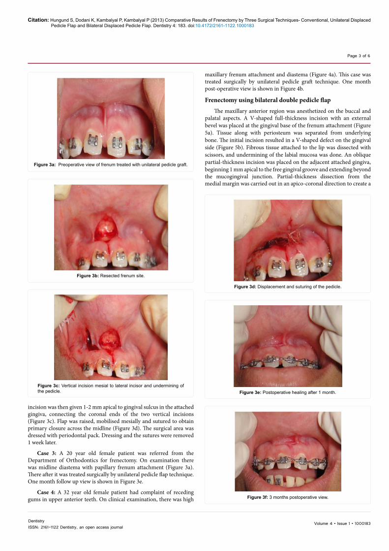
incision was then given 1-2 mm apical to gingival sulcus in the attached gingiva, connecting the coronal ends of the two vertical incisions (Figure 3c). Flap was raised, mobilised mesially and sutured to obtain primary closure across the midline (Figure 3d). The surgical area was dressed with periodontal pack. Dressing and the sutures were removed 1 week later.
Case 3: A 20 year old female patient was referred from the Department of Orthodontics for frenectomy. On examination there was midline diastema with papillary frenum attachment (Figure 3a). There after it was treated surgically by unilateral pedicle flap technique. One month follow up view is shown in Figure 3e.
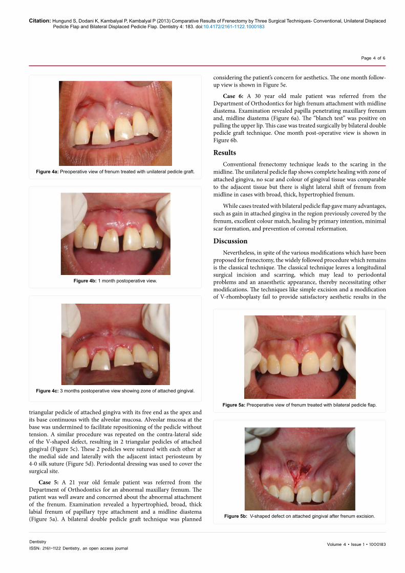
Case 4: A 32 year old female patient had complaint of receding gums in upper anterior teeth. On clinical examination, there was high
maxillary frenum attachment and diastema (Figure 4a). This case was treated surgically by unilateral pedicle graft technique. One month post-operative view is shown in Figure 4b.
Frenectomy using bilateral double pedicle flap
The maxillary anterior region was anesthetized on the buccal and palatal aspects. A V-shaped full-thickness incision with an external bevel was placed at the gingival base of the frenum attachment (Figure 5a). Tissue along with periosteum was separated from underlying bone. The initial incision resulted in a V-shaped defect on the gingival side (Figure 5b). Fibrous tissue attached to the lip was dissected with scissors, and undermining of the labial mucosa was done. An oblique partial-thickness incision was placed on the adjacent attached gingiva, beginning 1 mm apical to the free gingival groove and extending beyond the mucogingival junction. Partial-thickness dissection from the medial margin was carried out in an apico-coronal direction to create a
Figure 3a: Preoperative view of frenum treated with unilateral pedicle graft.
Figure 3b: Resected frenum site.
Figure 3c: Vertical incision mesial to lateral incisor and undermining of the pedicle.
Figure 3d: Displacement and suturing of the pedicle.
Figure 3e: Postoperative healing after 1 month.
Figure 3f: 3 months postoperative view.
Citation: Hungund S, Dodani K, Kambalyal P, Kambalyal P (2013) Comparative Results of Frenectomy by Three Surgical Techniques- Conventional, Unilateral Displaced Pedicle Flap and Bilateral Displaced Pedicle Flap. Dentistry 4: 183. doi:10.4172/2161-1122.1000183
Page 4 of 6
Volume 4 • Issue 1 • 1000183DentistryISSN: 2161-1122 Dentistry, an open access journal
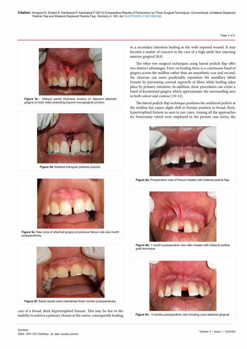
triangular pedicle of attached gingiva with its free end as the apex and its base continuous with the alveolar mucosa. Alveolar mucosa at the base was undermined to facilitate repositioning of the pedicle without tension. A similar procedure was repeated on the contra-lateral side of the V-shaped defect, resulting in 2 triangular pedicles of attached gingival (Figure 5c). These 2 pedicles were sutured with each other at the medial side and laterally with the adjacent intact periosteum by 4-0 silk suture (Figure 5d). Periodontal dressing was used to cover the surgical site.
Case 5: A 21 year old female patient was referred from the Department of Orthodontics for an abnormal maxillary frenum. The patient was well aware and concerned about the abnormal attachment of the frenum. Examination revealed a hypertrophied, broad, thick labial frenum of papillary type attachment and a midline diastema (Figure 5a). A bilateral double pedicle graft technique was planned
considering the patient’s concern for aesthetics. The one month follow-up view is shown in Figure 5e.
Case 6: A 30 year old male patient was referred from the Department of Orthodontics for high frenum attachment with midline diastema. Examination revealed papilla penetrating maxillary frenum and, midline diastema (Figure 6a). The “blanch test” was positive on pulling the upper lip. This case was treated surgically by bilateral double pedicle graft technique. One month post-operative view is shown in Figure 6b.
ResultsConventional frenectomy technique leads to the scaring in the
midline. The unilateral pedicle flap shows complete healing with zone of attached gingiva, no scar and colour of gingival tissue was comparable to the adjacent tissue but there is slight lateral shift of frenum from midline in cases with broad, thick, hypertrophied frenum.
While cases treated with bilateral pedicle flap gave many advantages, such as gain in attached gingiva in the region previously covered by the frenum, excellent colour match, healing by primary intention, minimal scar formation, and prevention of coronal reformation.
DiscussionNevertheless, in spite of the various modifications which have been
proposed for frenectomy, the widely followed procedure which remains is the classical technique. The classical technique leaves a longitudinal surgical incision and scarring, which may lead to periodontal problems and an anaesthetic appearance, thereby necessitating other modifications. The techniques like simple excision and a modification of V-rhomboplasty fail to provide satisfactory aesthetic results in the
Figure 4a: Preoperative view of frenum treated with unilateral pedicle graft.
Figure 4b: 1 month postoperative view.
Figure 4c: 3 months postoperative view showing zone of attached gingival.
Figure 5a: Preoperative view of frenum treated with bilateral pedicle flap.
Figure 5b: V-shaped defect on attached gingival after frenum excision.
Citation: Hungund S, Dodani K, Kambalyal P, Kambalyal P (2013) Comparative Results of Frenectomy by Three Surgical Techniques- Conventional, Unilateral Displaced Pedicle Flap and Bilateral Displaced Pedicle Flap. Dentistry 4: 183. doi:10.4172/2161-1122.1000183
Page 5 of 6
Volume 4 • Issue 1 • 1000183DentistryISSN: 2161-1122 Dentistry, an open access journal
case of a broad, thick hypertrophied frenum. This may be due to the inability to achieve a primary closure at the centre, consequently leading
Figure 5c: Oblique partial thickness incision on adjacent attached gingiva on both sides extending beyond mucogingival junction.
Figure 5d: Bilateral triangular pedicles sutured.
Figure 5e: New zone of attached gingiva at previous frenum site one month postoperatively.
to a secondary intention healing at the wide exposed wound. It may become a matter of concern in the case of a high smile line exposing anterior gingival [8,9].
The other two surgical techniques using lateral pedicle flap offer two distinct advantages. First, on healing there is a continuous band of gingiva across the midline rather than an anaesthetic scar and second, the clinician can more predictably reposition the maxillary labial frenum by preventing coronal regrowth of fibres while healing takes place by primary intention. In addition, these procedures can create a band of keratinized gingiva which approximates the surrounding area in both colour and contour [10-12].
The lateral pedicle flap technique positions the unilateral pedicle at the midline but causes slight shift in frenum position in broad, thick, hypertrophied frenum as seen in our cases. Among all the approaches for frenectomy which were employed in the present case series, the
Figure 5f: Same results were maintained three months postoperatively.
Figure 6a: Preoperative view of frenum treated with bilateral pedicle flap.
Figure 6b: 1 month postoperative view after treated with bilateral pedicle graft technique.
Figure 6c: 3 months postoperative view showing zone attached gingival.
Citation: Hungund S, Dodani K, Kambalyal P, Kambalyal P (2013) Comparative Results of Frenectomy by Three Surgical Techniques- Conventional, Unilateral Displaced Pedicle Flap and Bilateral Displaced Pedicle Flap. Dentistry 4: 183. doi:10.4172/2161-1122.1000183
Page 6 of 6
Volume 4 • Issue 1 • 1000183DentistryISSN: 2161-1122 Dentistry, an open access journal
bilateral double pedicle flap procedure offered many advantages such as gain in attached gingiva in the region previously covered by the frenum, excellent colour match, healing by primary intention, minimal scar formation, and prevention of coronal reformation.
In bilateral double pedicle graft technique, 2 triangular pedicles sutured together medially, that completely covers the V-shaped defect on the gingiva and act as a tissue dressing, and thus facilitating healing by primary intention and minimizing any chance of scar formation [13].
Frenectomy followed by gingival graft taken from the palate covers the wound area completely but may creates an aesthetic concern of unsatisfactory colour match by producing a “keloid,” “tattoo-like” or “tirepatch” appearance at the grafted area. This is because the donor site of the graft, in most cases, is the palate (keratinized gingiva) and at the time of transplantation, the receptor site receives genetic features of the palate, leaving the grafted area with whitish shade [8,14-16]. Also frenectomy by free gingival graft necessitates another surgical field to obtain the graft (donor site), which heals by second intention. Moreover, for a longer longevity of graft, it is necessary that it has proper dimensions; as a very thin graft has chances that it may undergo necrosis and causes exposure of receptor area. However, if graft is thicker, excess tissue will hinder an adequate nutrition and may also necessitates gingivoplasty after healing [17,18].
Furthermore, surgical procedures in the ventral aspect of the tongue like lingual frenectomy can lead to mucocele of the Blandin-Nuhn gland. Surgical trauma to these glands during lingual frenectomy probably during suturing causes extravasations of mucous to submucosal layer leads to the mucocele of Blandin-Nuhn glands [19]. Mark EP stated that periodontal surgery in maxillary labial area (including frenectomy) in patients taking angiotensin converting enzyme (ACE) inhibitor, aspirin, morphine, hydrazalazine, quinine, organic iodides and calcium channel blockers are prone to develop idiopathic angioedema of upper lip [20].
ConclusionIn conclusion, the conventional (classical) technique fails to provide
satisfactory aesthetic results in the case of a broad, thick hypertrophied frenum. This may be due to an inability to achieve primary closure at the centre, consequently leading to secondary intention healing at the wide exposed wound. It may become a matter of concern in the case of a high smile line exposing anterior gingiva. The unilateral pedicle flap technique shows complete healing with zone of attached gingiva, no scar formation and colour of gingival tissue was comparable to the adjacent tissue but there is slight lateral shift of frenum from midline in cases with broad, thick, hypertrophied frenum. The bilateral double pedicle flap technique has certain distinct advantages, as healing takes place by primary intention, zone of attached gingiva is formed in the midline, excellent colour match and with no anaesthetic scar formation. This technique may be suitable in situations where anterior aesthetics is of primary importance. The technique is reliable and easy to perform and provides excellent aesthetic results.
References
1. Henry SW, Levin MP, Tsaknis PJ (1976) Histological features of superior labial frenum. J Periodontol 47: 25-28.
2. Placek M, Miroslavs, Mrklas L (1974) Significance of the labial frenal attachment in the periodontal disease in man. Part1; Classification and epidemiology of the labial frenum attachment. J Periodontol 45: 891-894.
3. Dewel BF (1966) The labial frenum, midline diastema and palatine papilla: A clinical analysis. Dent Clin North Am 175-184.
4. Diaz-Pizan ME, Lagravere MO, Villena R (2006) Midline diastema and frenum morphology in the primary dentition. J Dent Child (Chic) 26: 11-14.
5. Gottsegen R (1954) Frenum position and vestibule depth in relation to gingival health. Oral Surg 7: 1069-1072.
6. Coleton SH (1977) Mucogingival surgical procedures employed in re-establshing the integrity of the gingival unit. The frenectomy and the free mucosal graft. Quintessance Int 8: 53-61.
7. Kahnberg KE (1977) Frenum surgery. I. A comparison of three surgical methods. Int J Oral Surg 6: 328-333.
8. Ito T, Johnson JD (1994) Color Atlas of Periodontal Surgery. Mosby, Wolfe, London.
9. Archer WH (1975) Oral surgery- a step by step atlas of operative techniques. (3rdedn), W B Saunders Company, Philadelphia, London, Toranto.
10. Miller PD (1985) Frenectomy, combined with a laterally positioned pedicle graft- functional and aesthetic considerations. J Periodontol 56: 102-106.
11. Bagga S, Bhatt M, Bhat GS, Thomas S (2006) Esthetic management of the upper labial frenum: a novel frenectomy technique. Quintessence Int 37: 819-823.
12. Miller PD (1991) Reconstructive periodontal plastic surgery (mucogingival surgery). J Tenn Dent Assoc 71: 14-18.
13. Hupp JR (2004) Contemporary Oral and Maxillofacial Surgery. St Louis, Mosby.
14. Breault LG, Fowler EB, Moore EA, Murray DJ (1999) The free gingival graft combined with the frenectomy: A clinical review. Gen Dent 47: 514-518.
15. Langer B, Langer L (1985) Subepithelial connective tissue graft technique for root coverage. J Periodontol 56: 397-402.
16. Lindhe J, Karring T, Lang NP (2005) Tratado de periodontia clinica e implantologia oral (4thedn). Rio de Janeiro, Guanabara Koogan.
17. Mormann W, Schaer F, Firestone AC (1981) The relationship between success of free gingival grafts and transplant thickness. J Periodontol 52: 74-80.
18. Ward VJ (1974) A clinical assessment of the use of the free gingival graft for correcting localized recession associated with frenal pull. J Periodontol 45: 78-83.
19. Santos TS, Martins- Filho, Paulo RS, Piva MR, Karam FK (2012) Mucocele of the glands of Blandin-Nuhn after lingual frenectomy. J Craniofacial Surgery 23: e657-658.
20. Mark EP, William EB, Scott LS, Robert FP, Robert BO, et al. (1991) Angioedema as a complication in periodontal surgery: Report of a case. J Periodontol 62: 643-645.
Citation: Hungund S, Dodani K, Kambalyal P, Kambalyal P (2013) Comparative Results of Frenectomy by Three Surgical Techniques- Conventional, Unilateral Displaced Pedicle Flap and Bilateral Displaced Pedicle Flap. Dentistry 4: 183. doi:10.4172/2161-1122.1000183
Submit your next manuscript and get advantages of OMICS Group submissionsUnique features:
User friendly/feasible website-translation of your paper to 50 world’s leading languagesAudio Version of published paperDigital articles to share and explore
Special features:
300 Open Access Journals25,000 editorial team21 days rapid review processQuality and quick editorial, review and publication processingIndexing at PubMed (partial), Scopus, EBSCO, Index Copernicus and Google Scholar etcSharing Option: Social Networking EnabledAuthors, Reviewers and Editors rewarded with online Scientific CreditsBetter discount for your subsequent articles
Submit your manuscript at: http://www.omicsonline.org/submission