• CNS Prion Disease• CNS Toxoplasmosis• CNS Vasculitis
Etiology and Pathology
Patient presentation and Demographics
Types and Stages of Disease
Imaging Findings and DDx
Treatment and Prognosis
• aka: Creutzfeldt-Jakob Disease (CJD), transmissible spongiform encephalopathy
• Rapidly progressing, fatal, potentially transmissible dementing disorder caused by a prion
• Prion = proteinaceous infectious particle devoid of DNA or RNA
Etiology
• PrPSc = conformationally abnormal isoform of normal host encoded PrPc. It’s insoluble and protease resistant
• Once introduced into cells, it starts a vicious self-perpetuating cycle turn normal PrPc into PrPSc
Pathology
• PcPSc is resistant to certain proteases on western blot
• variable accumulation of PrPSc in tissue
• spongiform encephalopathy with neuronal loss, and vacuolation with replacement gliosis
4 Types:
1. sCJD = spontaneous/somatic mutation of PrPcinto PrPSc
2. fCJD = mutation in PRNP gene which encodes for abnormal PrPSc
3. Iatrogenic CJD = primarilly CNS surgery or human derived hormones (HGH, gonadotropins)
4. vCJD = (aka: nvCJD), infected beef
Clinical Presentation:
• rapidly progressive dementia associated with myoclonic jerks and akinetic mutism
• variable constellation of other neuro symptoms• No gender preference• Age: Young in vCJD, >60 for sCJD, • sCJD all races/places, vCJD almost all in UK• sCJD (85%), fCJD (15%), Infectious/Iatrogenic
(<1%)
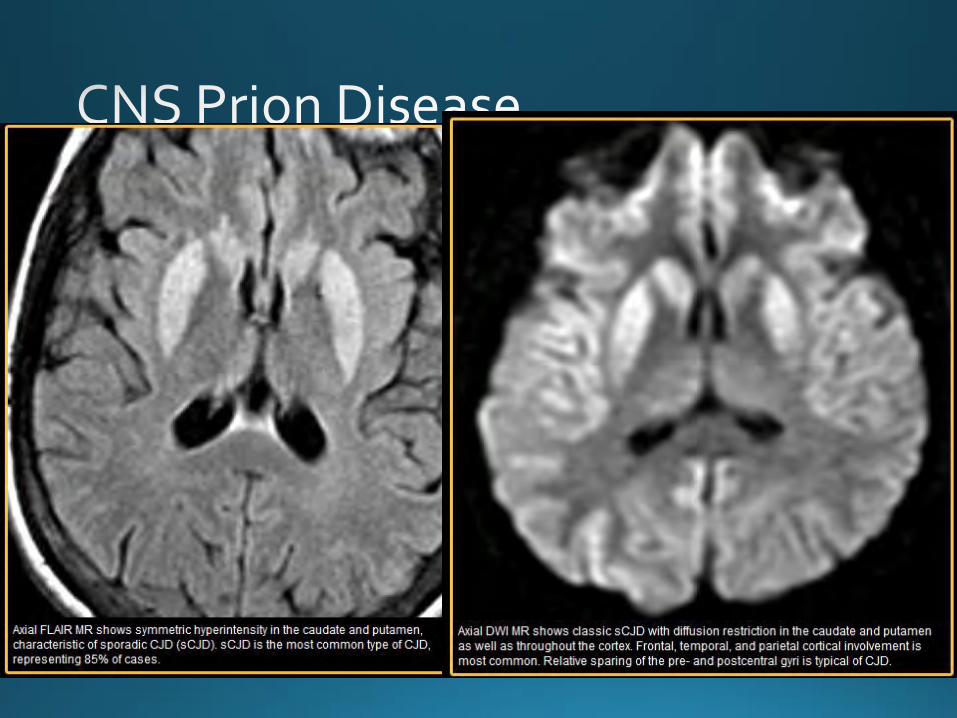
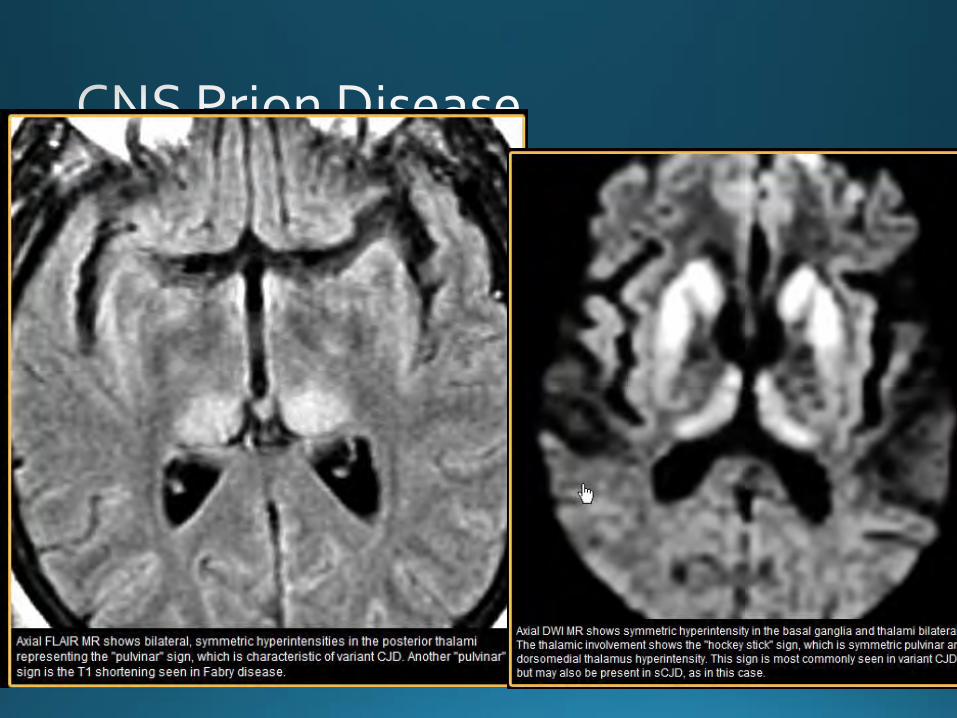
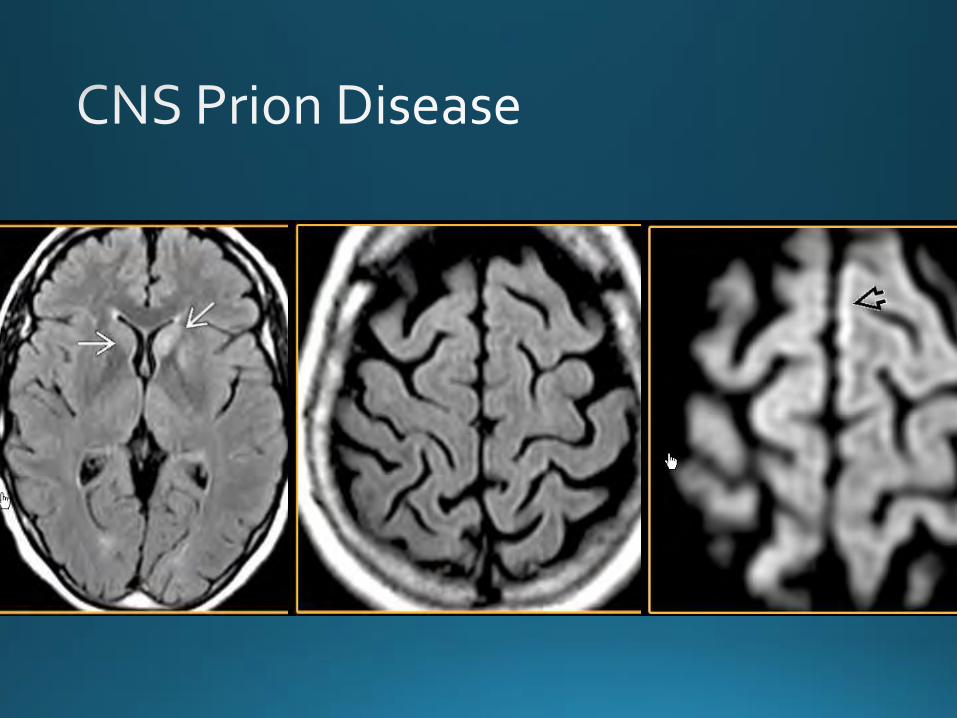
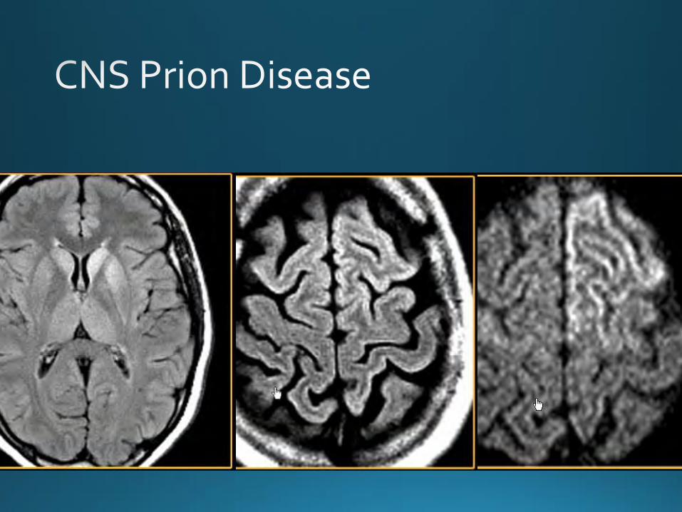
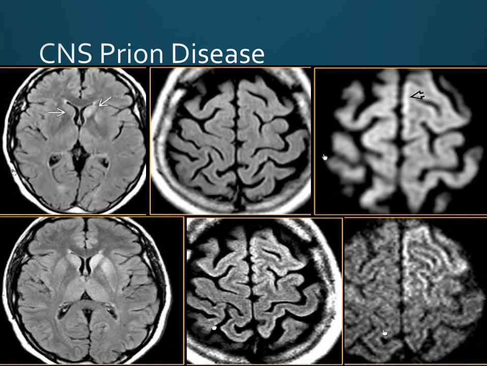
Imaging:
• Best tool = MR with FLAIR and DWI
• Best clue: progressive T2 hyperintensity in the BG, thalamus, and cerebral cortex
• CT has no real role, may show progressive ventriculomegally due to atrophy
Imaging:
• T2WI/FLAIR/DWI • “Pulvinar Sign” = symmetrical hyperintensitiy
in the posterior thalamus
• “Hockey Stick Sign” = symmetrical pulvinarand dorsomedial thalamic hyperintensity
• Cortical hyperintensity common in sCJD
Imaging Pearls:
• DWI signal may disappear late in the disease
• No contrast enhancement
• Lack of basal ganglia findings doesn’t exclude CJD if high clinical suspicion
What about Nuc Med?
• Regional hypometabolism on PET correlates with lesions
• SPECT shows decreased uptake of various tracers and decreased absolute values of rCBF• can be asymmetrical• sensitive for early CJD
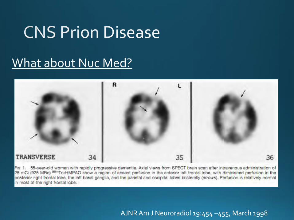
What about Nuc Med?
AJNR Am J Neuroradiol 19:454 –455, March 1998
Treatment and Prognosis
• long incubation, but once symptoms start they are relentless, death usually within months
• Mean survival of sCJD = 8mos, vCJD = 16mos, fCJD = 26mos
• No effective treatment

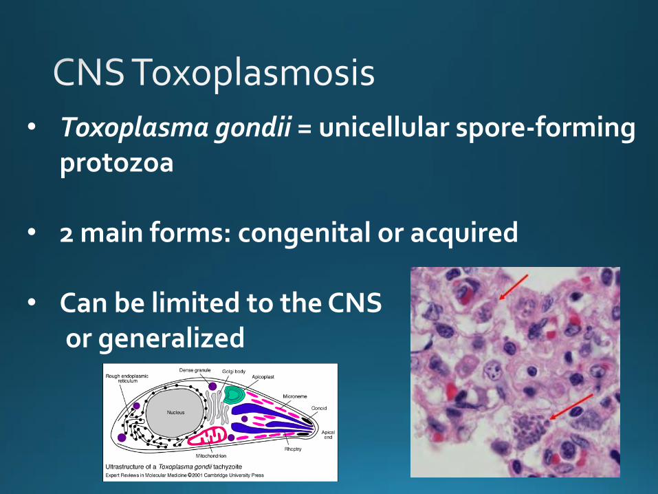
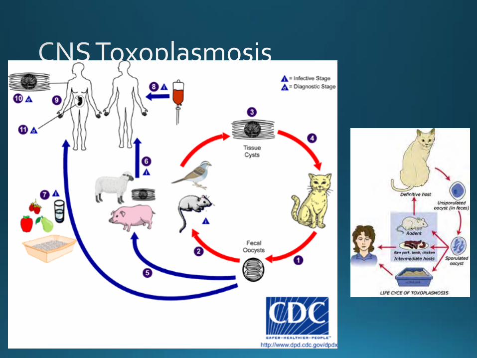
• Toxoplasma gondii = unicellular spore-forming protozoa
• 2 main forms: congenital or acquired
• Can be limited to the CNSor generalized
Congenital Toxoplasmosis• the “T” in TORCH
• transplacental infection only during “new” infections or if immunocompromised• most moms have circulating antibodies
which protect the fetus
• the earlier in the gestation, the worse prognosis
Congenital Toxoplasmosis: Presentation• commonly diagnosed during pregnancy with
labs and US
• newborn presents with seizures, chorioretinitis and hydrocephalus
• Hydrocephalus (and eventually encephalomalcia) due to ependymitis and obstruction of the aqueduct

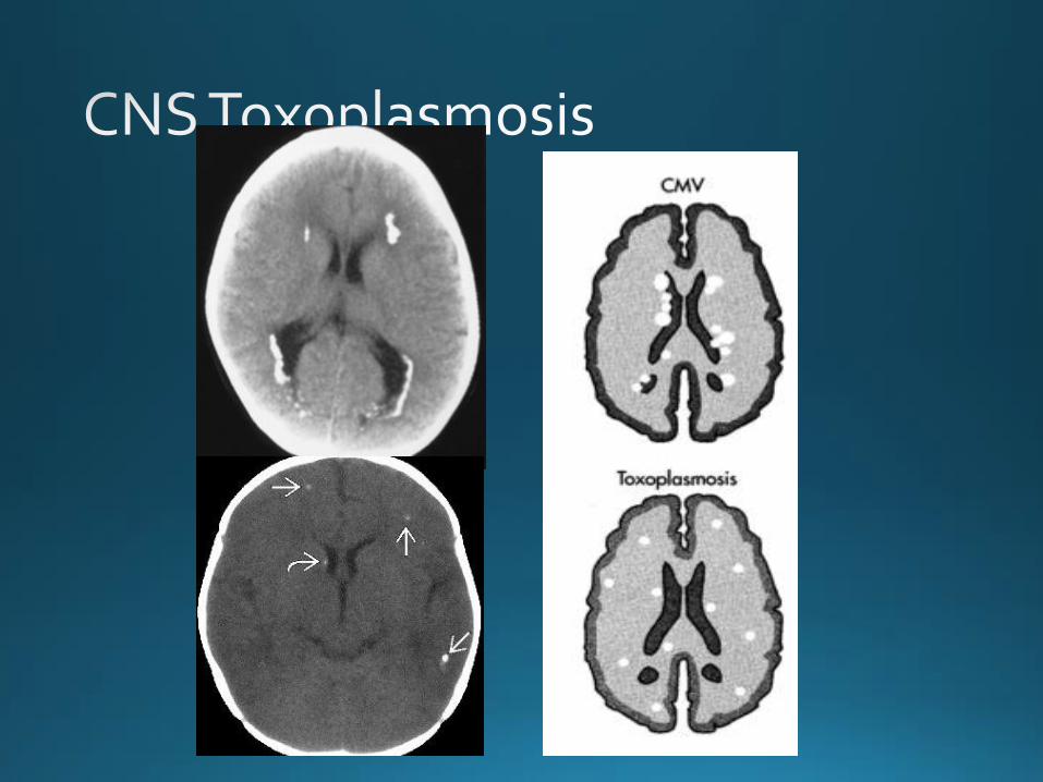
Congenital Toxoplasmosis: Imaging
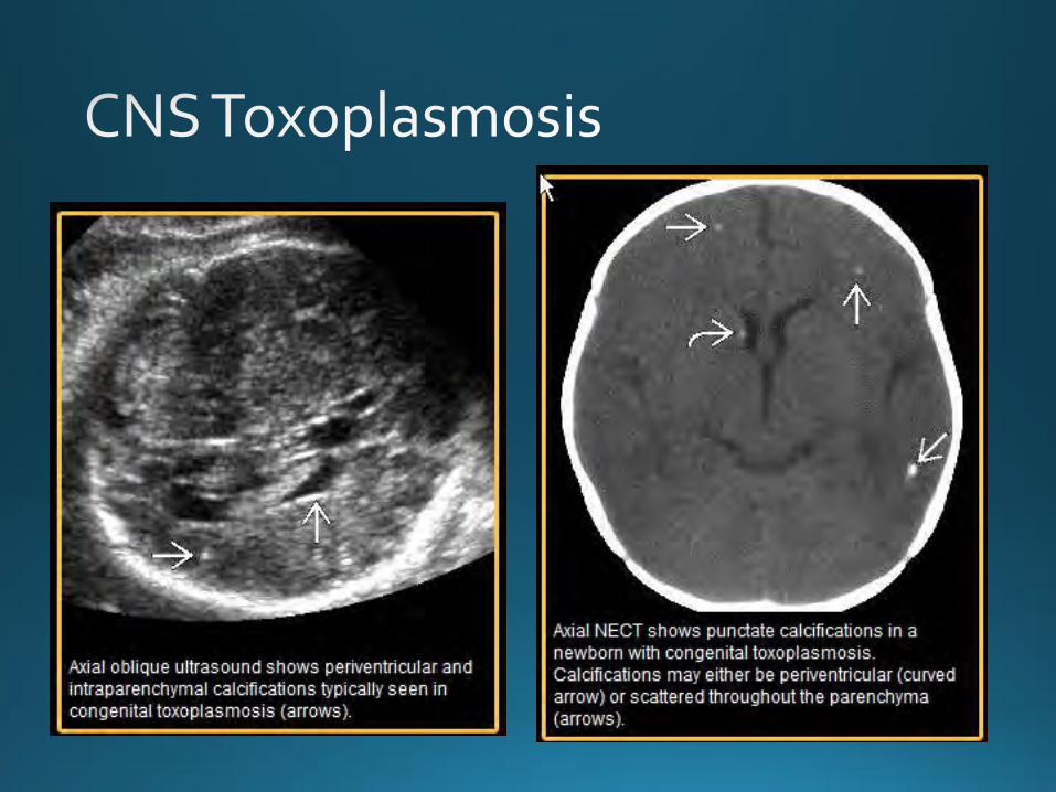
• multifocal, non-shadowing calcifications involving the basal ganglia, PVWM and cortex
• there can be large areas of parenchymal destruction
• Best first test is ultrasound
Congenital Toxoplasmosis: Imaging
• Protocol Recommendation:• monthly US to look for Ca and asses fetal
growth (IUGR is common)• Fetal MRI to evaluate the brain• confirm infection with reference lab and
amnio/cord blood viral PCR (to eval for other TORCH viruses)
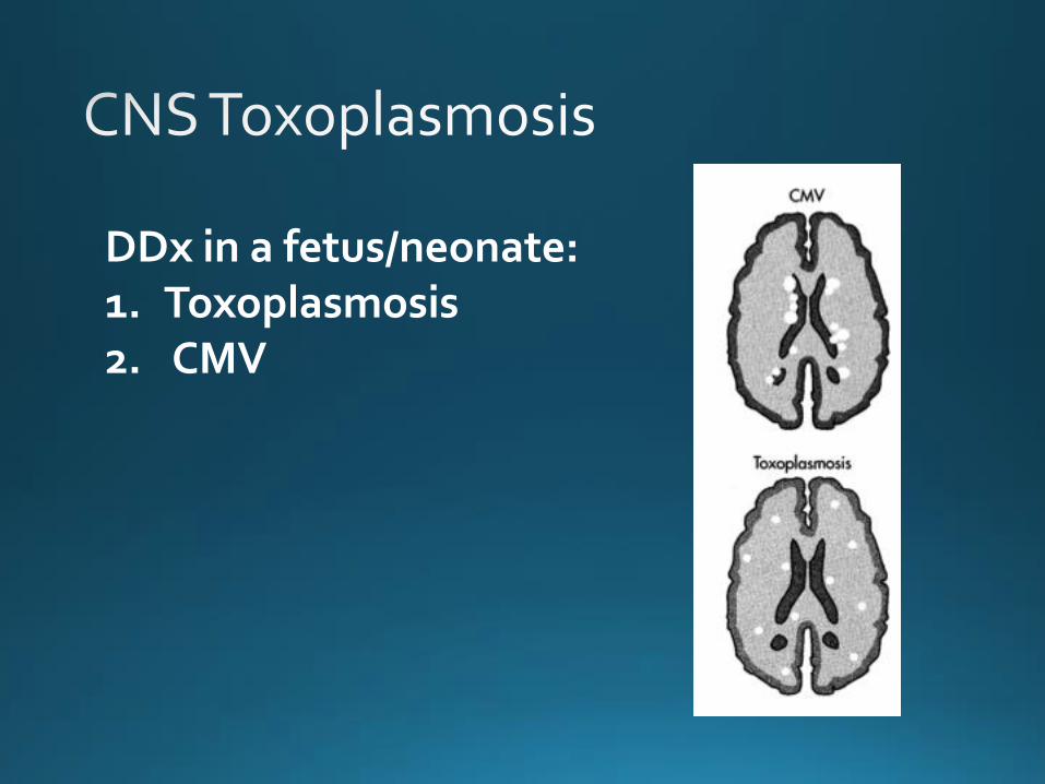
DDx in a fetus/neonate:1. Toxoplasmosis2. CMV
Congenital Toxoplasmosis:• 1st trimester infection is rare, but more severe• Infection at >20 weeks has higher likelihood of
affecting the fetus but less severe
• blindness, epilepsy, mental retardation, if no brain abnormalities = better prognosis
• Rx = termination or folate synthesis inhibitors (pyrimethamine/sulfadiazine or sulfadioxine), which can cause severe pancytopenia.
Acquired Toxoplasmosis:
• opportunistic infection
• most common CNS infection in AIDS pts
• usually a reactivation of a latent infection • 20-70% of USA population is seropositive
Acquired Toxoplasmosis:
• pts present with fever, malaise, headache• eventually develop personality changes or
seizures
• aka: Toxoplasmosis encephalitis (TE)
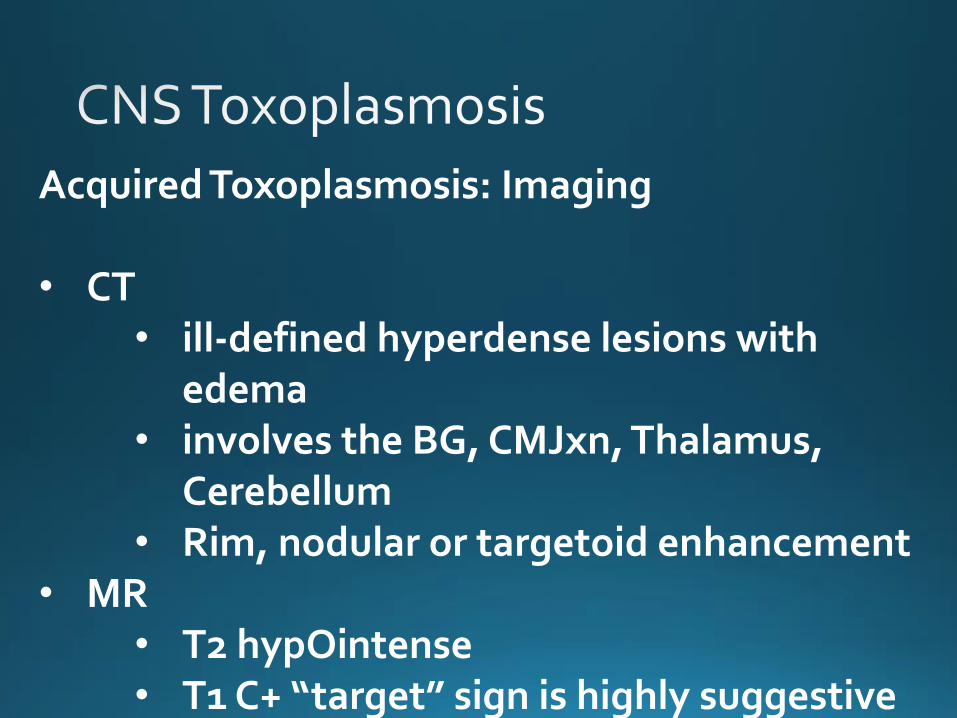
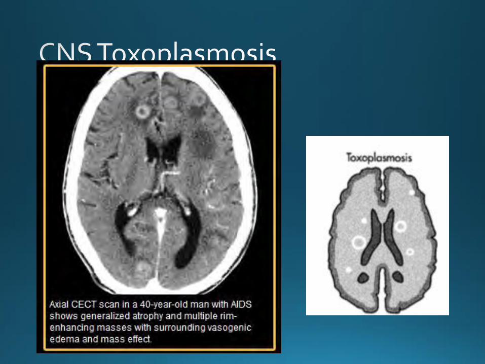
Acquired Toxoplasmosis: Imaging
• CT• ill-defined hyperdense lesions with
edema• involves the BG, CMJxn, Thalamus,
Cerebellum• Rim, nodular or targetoid enhancement
• MR• T2 hypOintense• T1 C+ “target” sign is highly suggestive
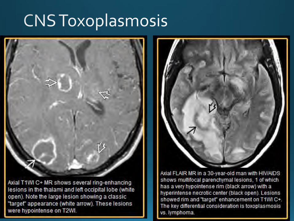
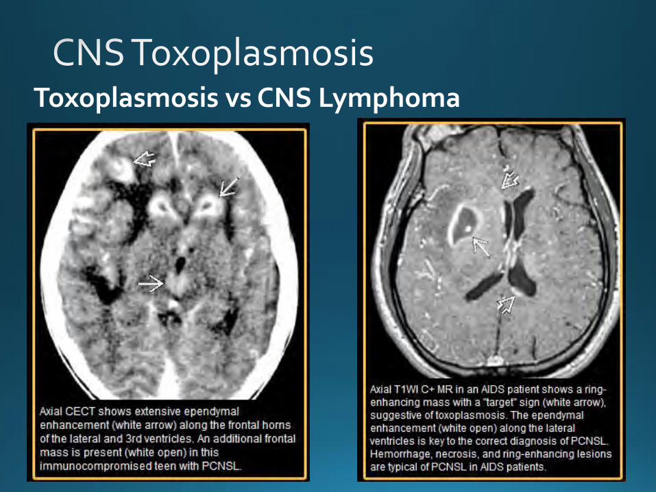
Toxoplasmosis vs CNS Lymphoma
• DDx = Primary CNS lymphoma
• TE lesions should resolve in 2-4 weeks
• Know whether treatment has been given, if poor response, suggest lymphoma
• PEARL:Multiple “target” lesions on T1WI C+
that are dark on T2WI
• Vasculitis = inflamatory changes in arterial walls
• Important to diagnose because they are potentially treatable
• There are dozens of different etiologies
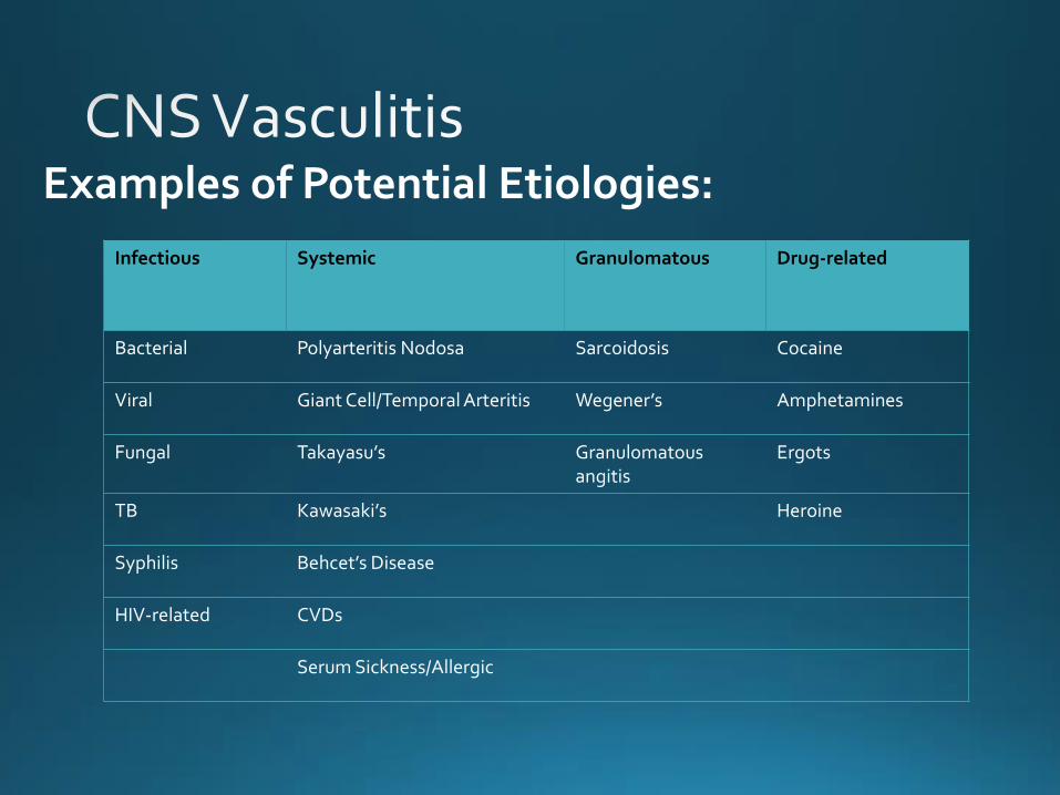
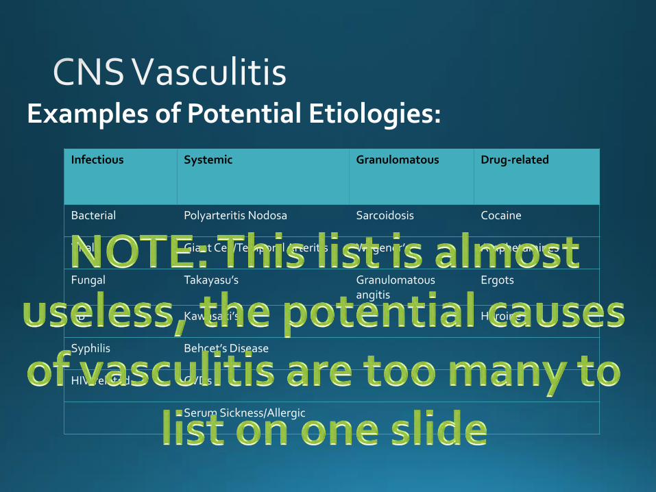
Examples of Potential Etiologies:
Infectious Systemic Granulomatous Drug-related
Bacterial Polyarteritis Nodosa Sarcoidosis Cocaine
Viral Giant Cell/Temporal Arteritis Wegener’s Amphetamines
Fungal Takayasu’s Granulomatous angitis
Ergots
TB Kawasaki’s Heroine
Syphilis Behcet’s Disease
HIV-related CVDs
Serum Sickness/Allergic
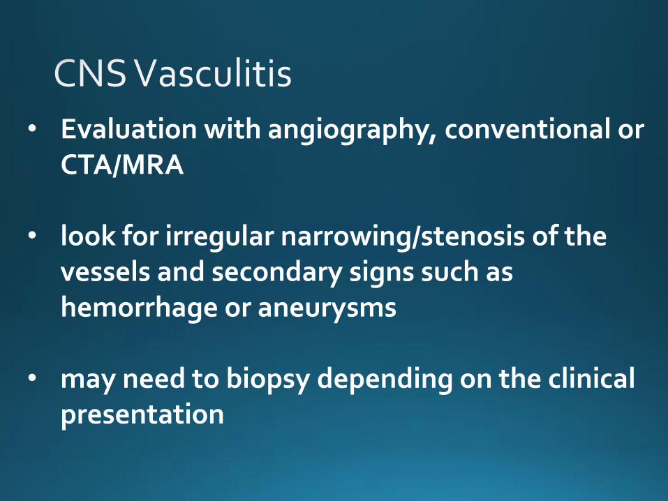
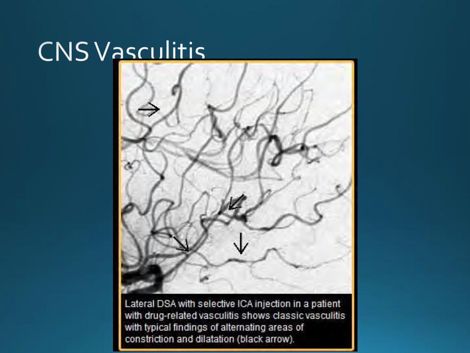
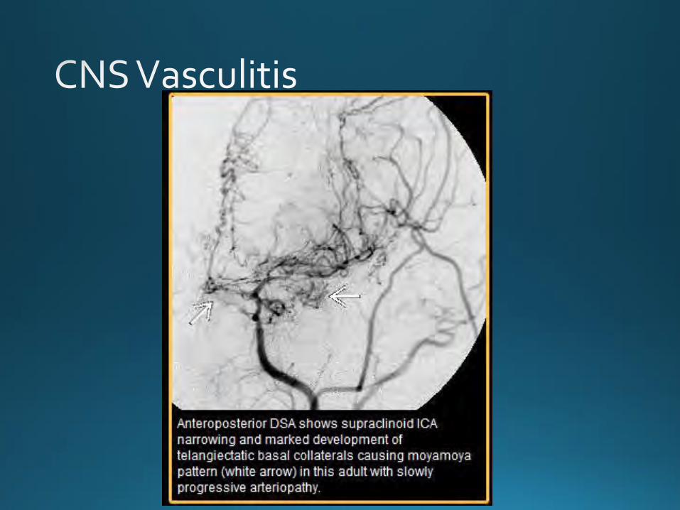
• Evaluation with angiography, conventional or CTA/MRA
• look for irregular narrowing/stenosis of the vessels and secondary signs such as hemorrhage or aneurysms
• may need to biopsy depending on the clinical presentation

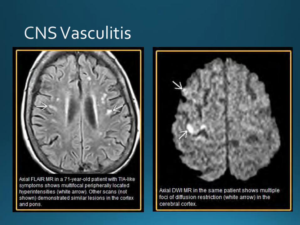
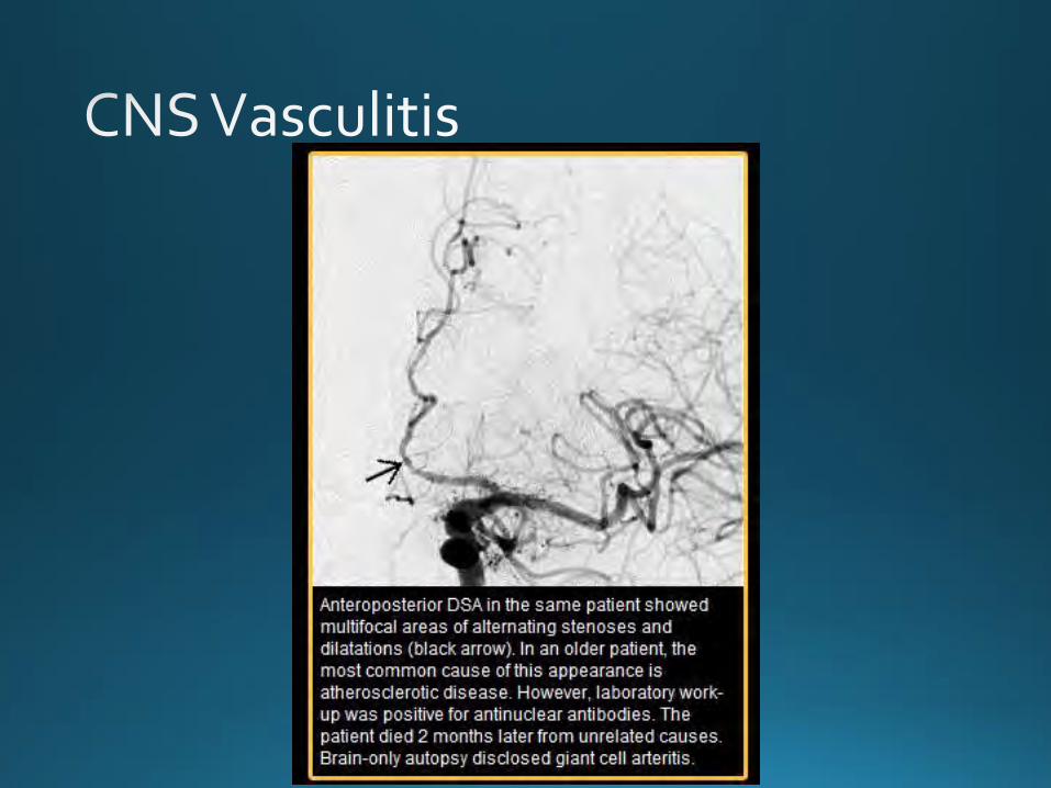
• PEARL:Multiple vascular territories producing an
atypical pattern in various stages.
• Most common “vasculitic” pattern on angio is atherosclerosis
• Most imaging findings are non-specific• If you are thinking vasculitis, get a tox screen,
LP, imaging and angiography. • ONLY biopsy allows for definite diagnosis
• CDC website
• Radiology Primer


