
Title
• Blood
Essential Question
• Describe the structure and
function of blood tissue.

Essentials of Human Anatomy & Physiology
Copyright © 2003 Pearson Education, Inc. publishing as Benjamin Cummings
Slides 10.1 – 10.31
Seventh Edition
Elaine N. Marieb
Chapter 10
Blood
Lecture Slides in PowerPoint by Jerry L. Cook

Blood
Characteristics
Slide 10.1a Copyright © 2003 Pearson Education, Inc. publishing as Benjamin Cummings
Fluid connective tissue composed of:
Living cells = formed elements
Non-living matrix = plasma

Blood
Slide 10.1b Copyright © 2003 Pearson Education, Inc. publishing as Benjamin Cummings
Figure 10.1

Physical Characteristics of Blood
Slide 10.2 Copyright © 2003 Pearson Education, Inc. publishing as Benjamin Cummings
Color range is scarlet red to dull red depending on O2
pH 7.35–7.45
Blood temp. is slightly higher than body temp.

Blood Plasma
Slide 10.3 Copyright © 2003 Pearson Education, Inc. publishing as Benjamin Cummings
90 percent water
Dissolved substances
Nutrients
Salts
Respiratory gases
Hormones
Proteins
Waste products

PLASMA

Plasma Proteins
Slide 10.4 Copyright © 2003 Pearson Education, Inc. publishing as Benjamin Cummings
Albumin – regulates osmotic pressure
Clotting proteins
Antibodies – immune cell

Formed Elements
Slide 10.5a Copyright © 2003 Pearson Education, Inc. publishing as Benjamin Cummings
Erythrocytes = red blood cells made up of hemoglobin

RED BLOOD CELLS


HEMOGLOBIN

Hemoglobin
Slide 10.7 Copyright © 2003 Pearson Education, Inc. publishing as Benjamin Cummings
Iron-containing protein
Binds strongly, but reversibly, to oxygen
has four oxygen binding sites
Each RBC has 250 million hemoglobin molecules
Formed Elements
Slide 10.5a Copyright © 2003 Pearson Education, Inc. publishing as Benjamin Cummings
Leukocytes = white blood cells

Leukocytes

Types of Leukocytes
Slide 10.10a Copyright © 2003 Pearson Education, Inc. publishing as Benjamin Cummings
Figure 10.4

Leukocyte Levels in the Blood
Slide 10.9 Copyright © 2003 Pearson Education, Inc. publishing as Benjamin Cummings
Normal levels
Between 4,000 and 11,000 cells/mm3

Leukocyte Levels in the Blood
Slide 10.9 Copyright © 2003 Pearson Education, Inc. publishing as Benjamin Cummings
Abnormal leukocyte levels
Leukocytosis
Above 11,000 leukocytes/mm3
Generally indicates an infection

Leukocyte Levels in the Blood
Slide 10.9 Copyright © 2003 Pearson Education, Inc. publishing as Benjamin Cummings
Abnormal leukocyte levels
Leukopenia
Abnormally low leukocyte level
Commonly caused by certain drugs

Eosinophils
Neutrophils
Basophils
Slide 10.12
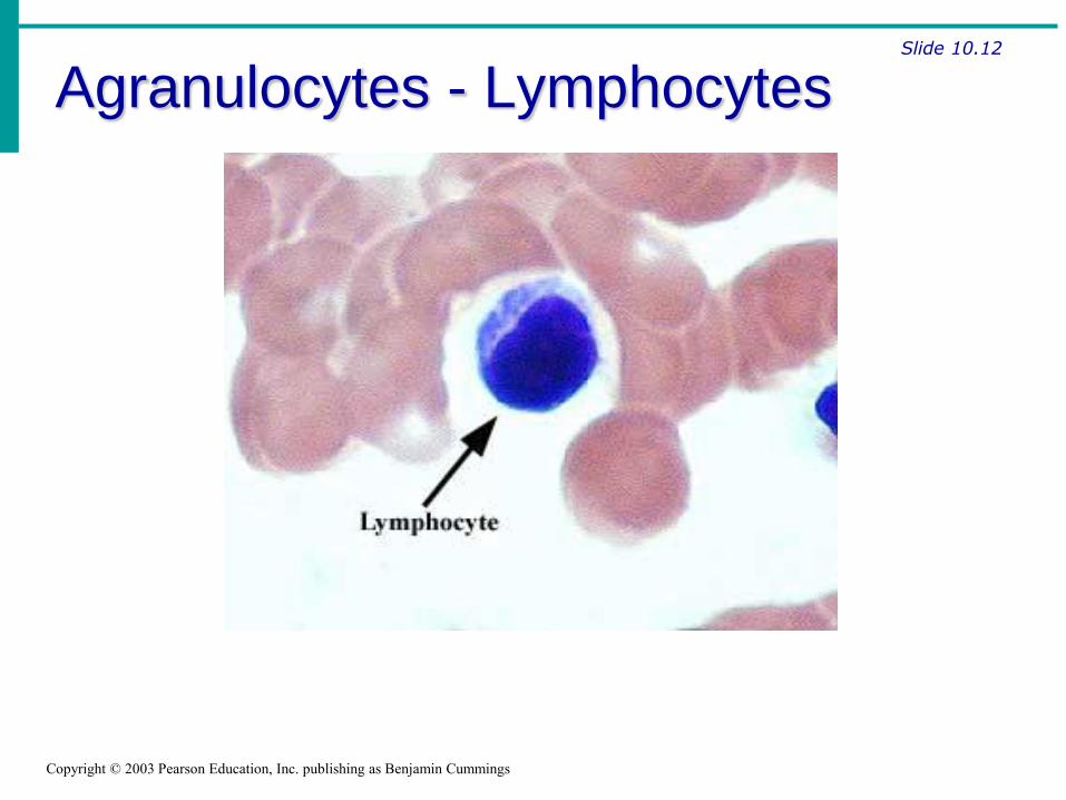
Agranulocytes - Lymphocytes
Copyright © 2003 Pearson Education, Inc. publishing as Benjamin Cummings

Slide 10.12
Agranulocytes - Monocytes
Copyright © 2003 Pearson Education, Inc. publishing as Benjamin Cummings

Neutrophils
Eosinophils
Basophils
Plasma Cell
Lymphocytes
Monocytes
Formed Elements
Slide 10.5a Copyright © 2003 Pearson Education, Inc. publishing as Benjamin Cummings
Platelets = cell fragments used for clotting

PLATELETS

Slide 10.5b Copyright © 2003 Pearson Education, Inc. publishing as Benjamin Cummings

Slide 10.5c Copyright © 2003 Pearson Education, Inc. publishing as Benjamin Cummings
Hematopoiesis
Slide 10.14 Copyright © 2003 Pearson Education, Inc. publishing as Benjamin Cummings
Making blood cells
Occurs in red bone marrow
derived from a common stem cell (hemocytoblast)

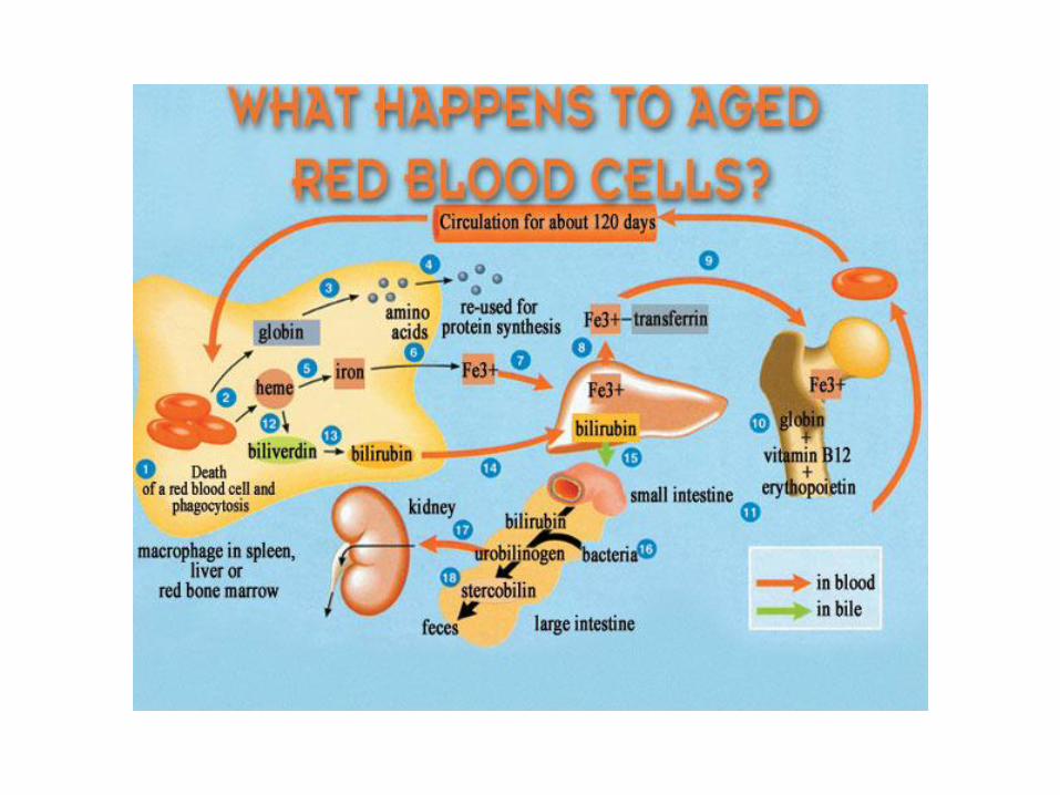

Fate of Erythrocytes
Slide 10.15 Copyright © 2003 Pearson Education, Inc. publishing as Benjamin Cummings
Unable to divide, grow, or make proteins
Wear out in 100 to 120 days
Eliminated by phagocytes in the spleen or liver
Lost cells are replaced by division of hemocytoblasts

Control of Erythrocyte Production
Slide 10.17 Copyright © 2003 Pearson Education, Inc. publishing as Benjamin Cummings
Figure 10.5

Hemostasis
Slide 10.18 Copyright © 2003 Pearson Education, Inc. publishing as Benjamin Cummings
Definition
Stoppage of blood flow
Result of a break in a blood vessel

1. Vascular Spasms
Slide 10.20 Copyright © 2003 Pearson Education, Inc. publishing as Benjamin Cummings
Anchored platelets release serotonin.
Smooth muscle contracts, causing vasoconstriction.
Blood vessel narrows, decreasing blood loss
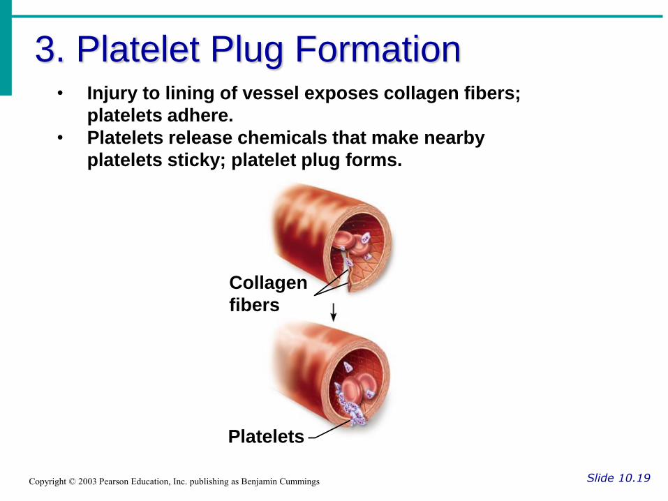
3. Platelet Plug Formation
Slide 10.19 Copyright © 2003 Pearson Education, Inc. publishing as Benjamin Cummings
• Injury to lining of vessel exposes collagen fibers;
platelets adhere.
• Platelets release chemicals that make nearby
platelets sticky; platelet plug forms.
Collagen
fibers
Platelets

3. Coagulation
Slide 10.21a Copyright © 2003 Pearson Education, Inc. publishing as Benjamin Cummings
Injured tissues release a series of chemicals to trigger a clotting cascade
Fibrin forms a meshwork (the basis for a clot)

4. Blood Clotting
Slide 10.22 Copyright © 2003 Pearson Education, Inc. publishing as Benjamin Cummings
Takes 3 to 6 minutes
The clot remains as endothelium regenerates
The clot is broken down after tissue repair

Figure 10.7

Undesirable Clotting
Slide 10.23 Copyright © 2003 Pearson Education, Inc. publishing as Benjamin Cummings
Thrombus
A clot in an unbroken blood vessel
Can be deadly in areas like the heart

Undesirable Clotting
Slide 10.23 Copyright © 2003 Pearson Education, Inc. publishing as Benjamin Cummings
Embolus
A thrombus that breaks away and floats freely in the bloodstream
Can later clog vessels in critical areas such as the brain

Blood Groups and Transfusions
Slide 10.25 Copyright © 2003 Pearson Education, Inc. publishing as Benjamin Cummings
Large losses of blood
Loss of 15 to 30 percent causes pallor and weakness
Loss of over 30 percent causes severe shock, which can be fatal

Blood Groups and Transfusions
Slide 10.25 Copyright © 2003 Pearson Education, Inc. publishing as Benjamin Cummings
Transfusions
The only way to replace blood quickly
Blood must be of the same blood group

Human Blood Groups
Slide 10.26a Copyright © 2003 Pearson Education, Inc. publishing as Benjamin Cummings
Blood contains genetically determined proteins
A foreign protein (antigen) may be attacked by the immune system and cause the blood to clump (agglutination)

ABO Blood Groups
Slide 10.27a Copyright © 2003 Pearson Education, Inc. publishing as Benjamin Cummings
Based on the presence or absence of two antigens
Type A
Type B
The lack of these antigens is called type O

ABO Blood Groups
Slide 10.27b Copyright © 2003 Pearson Education, Inc. publishing as Benjamin Cummings
The presence of both A and B is called type AB
The presence of either A or B is called types A and B, respectively

Rh Blood Groups
Slide 10.28 Copyright © 2003 Pearson Education, Inc. publishing as Benjamin Cummings
Named because of the presence or absence of one of eight Rh antigens (agglutinogen D)
Most Americans are Rh+
Problems can occur in mixing Rh+ blood into a body with Rh– blood

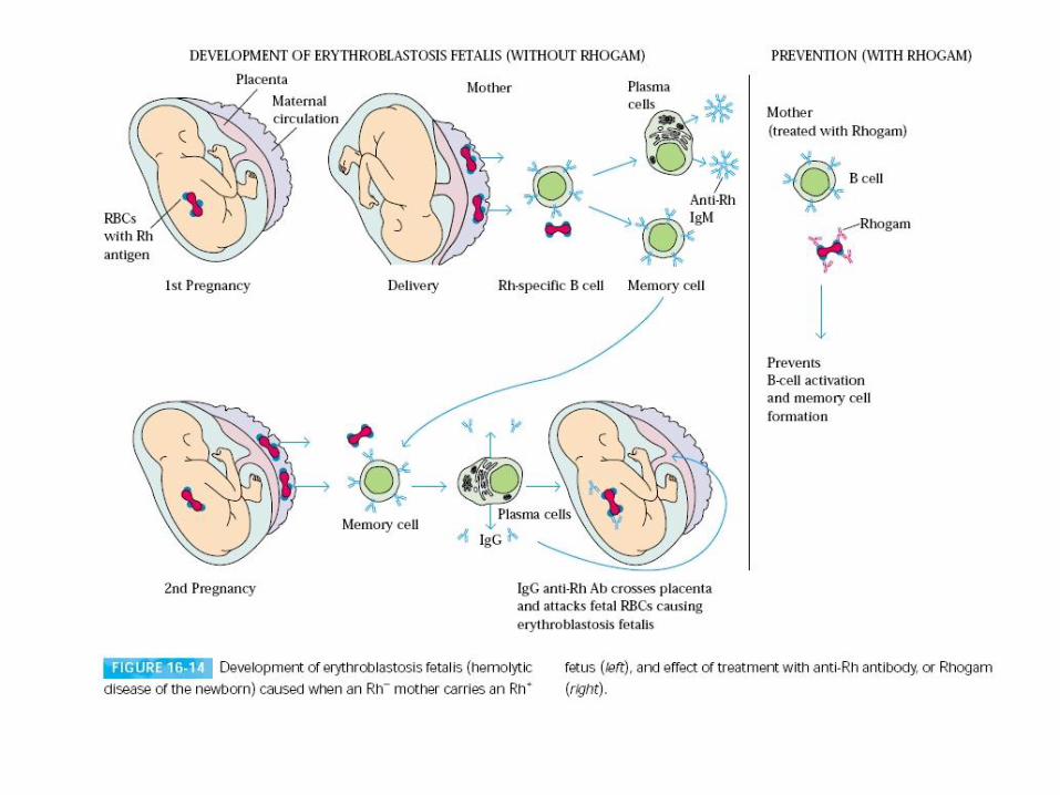
Rh Dangers During Pregnancy
Slide 10.29b Copyright © 2003 Pearson Education, Inc. publishing as Benjamin Cummings
The mismatch of an Rh– mother carrying an Rh+ baby can cause problems for the unborn child
The first pregnancy usually proceeds without problems, but the immune system is sensitized
In a second pregnancy, the mother’s immune system produces antibodies to attack the Rh+ blood (hemolytic disease of the newborn)
Mother is given RhoGam after first pregnancy


Title
• Anatomy of the heart and
blood vessels
Essential Question
• Describe the anatomy of the
heart and major blood
vessels of the body.

Essentials of Human Anatomy & Physiology
Copyright © 2003 Pearson Education, Inc. publishing as Benjamin Cummings
Slides 11.1 – 11.19
Seventh Edition
Elaine N. Marieb
Chapter 11
The Cardiovascular System
Lecture Slides in PowerPoint by Jerry L. Cook

The Cardiovascular System
Slide 11.1 Copyright © 2003 Pearson Education, Inc. publishing as Benjamin Cummings
Structure
A closed system of the heart and blood vessels
Function
Deliver O2 and nutrients and to remove CO2 and other waste products

The Heart
Slide 11.2b Copyright © 2003 Pearson Education, Inc. publishing as Benjamin Cummings
Figure 11.1

The Heart
Slide 11.2a Copyright © 2003 Pearson Education, Inc. publishing as Benjamin Cummings
Location
Thorax between the lungs
Pointed apex directed toward left hip and rests on the diaphragm
About the size of your fist

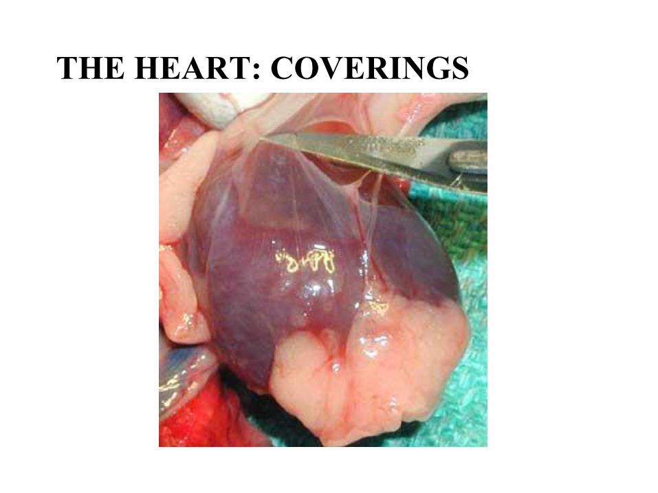
THE HEART: COVERINGS

The Heart: Coverings
Slide 11.3 Copyright © 2003 Pearson Education, Inc. publishing as Benjamin Cummings
Pericardium – a double serous membrane
Visceral Layer - next to heart
Parietal Layer - outside layer
Serous fluid fills the space between the layers of pericardium
THE HEART: COVERINGS

THE HEART: COVERINGS
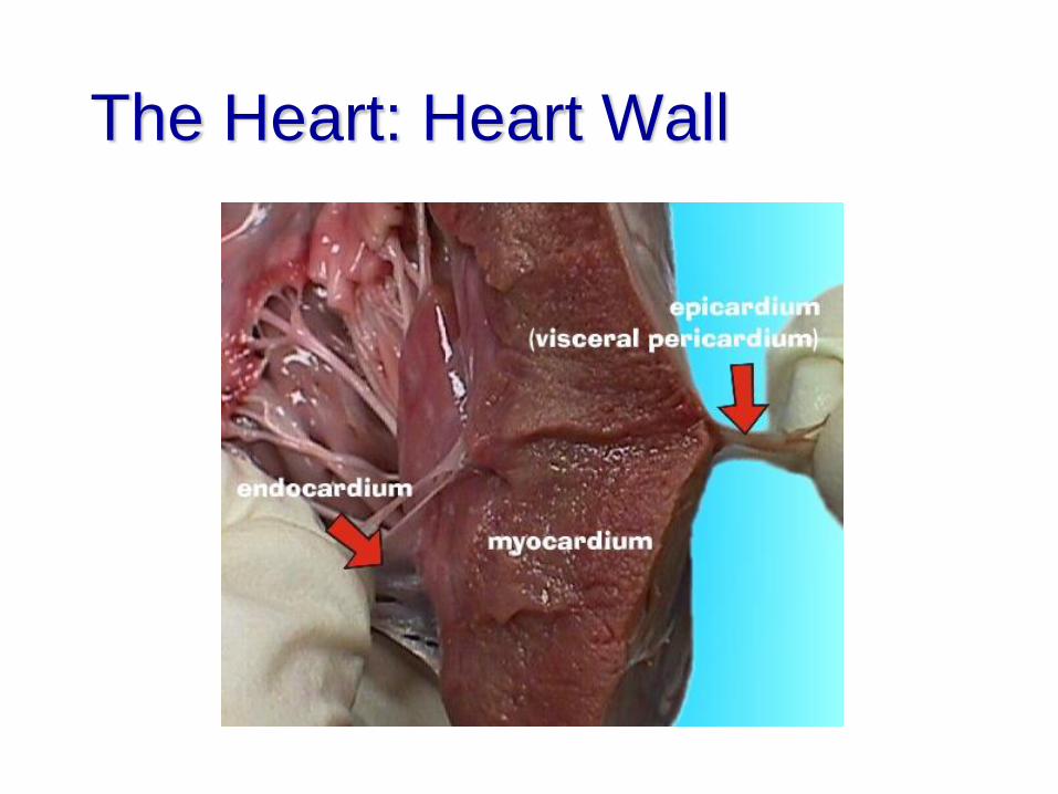
The Heart: Heart Wall

The Heart: Heart Wall
Slide 11.4 Copyright © 2003 Pearson Education, Inc. publishing as Benjamin Cummings
Three layers
Epicardium
Outside layer consists of connective tissue
layer (visceral pericardium)
The Heart: Heart Wall
Slide 11.4 Copyright © 2003 Pearson Education, Inc. publishing as Benjamin Cummings
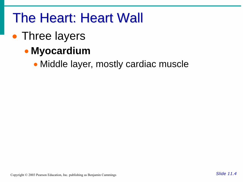
Three layers
Myocardium
Middle layer, mostly cardiac muscle

The Heart: Heart Wall
Slide 11.4 Copyright © 2003 Pearson Education, Inc. publishing as Benjamin Cummings
Three layers
Endocardium
Inner layer; endothelium that lines the heart


The Heart: Chambers
Slide 11.6 Copyright © 2003 Pearson Education, Inc. publishing as Benjamin Cummings
Right and left side act as separate pumps
Four chambers
Atria (right and left)
Receiving chambers
Ventricles (right and left)
Discharging chambers

External Heart Anatomy
Slide 11.5 Copyright © 2003 Pearson Education, Inc. publishing as Benjamin Cummings Figure 11.2a

Right ventricle
Muscular interventricular septum
Left
ventricle

Blood Circulation
Slide 11.7 Copyright © 2003 Pearson Education, Inc. publishing as Benjamin Cummings
Figure 11.3

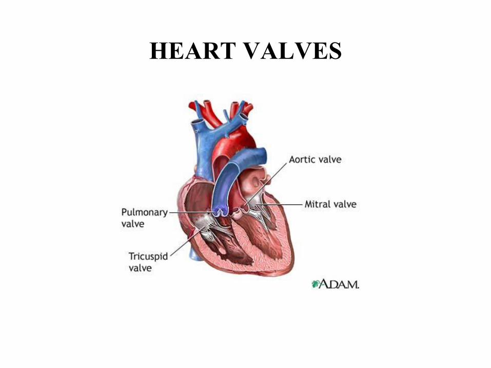
HEART VALVES

The Heart: Valves Characteristics
Slide 11.8 Copyright © 2003 Pearson Education, Inc. publishing as Benjamin Cummings
Allow blood to flow in only one direction
Open as blood is pumped through
Close to prevent backflow

The Heart: Valves
Slide 11.8 Copyright © 2003 Pearson Education, Inc. publishing as Benjamin Cummings
Four valves
Atrioventricular valves – between atria and ventricles
Bicuspid (mitral) valve – between the left atria and left ventricle
Tricuspid valve – between the right atria and right ventricle

The Heart: Valves
Slide 11.8 Copyright © 2003 Pearson Education, Inc. publishing as Benjamin Cummings
Four valves
Semilunar valves – between ventricle and major artery
Pulmonary semilunar valve – between RV and pulmonary artery
Aortic semilunar valve – between LV and aorta

HEART VALVES

Operation of Heart Valves
Slide 11.10 Copyright © 2003 Pearson Education, Inc. publishing as Benjamin Cummings
Figure 11.4
Podcast:
Minimally
Invasive Heart
Valve Surgery

The Heart: Associated Great Vessels
Arteries
Slide 11.11 Copyright © 2003 Pearson Education, Inc. publishing as Benjamin Cummings
Aorta receives blood from LV and travels to rest of body (oxygenated)
Pulmonary arteries receives blood from RV and travels to lungs (deoxygenated)

The Heart: Associated Great Vessels
Veins
Slide 11.11 Copyright © 2003 Pearson Education, Inc. publishing as Benjamin Cummings
Vena cava blood entering RA from body (deoxygenated)
Pulmonary veins (four) blood entering LA from lungs (oxygenated)

ASSOCIATED GREAT VESSELS

CORONARY CIRCULATION
http://video.google.com/vi
deoplay?docid=530428907
1571962318&q=heart+sur
gery+site%3Avideo.googl
e.com&total=305&start=1
0&num=10&so=0&type=s
earch&plindex=2
AWAKE
Open Heart
Surgery

Coronary Circulation
Slide 11.12 Copyright © 2003 Pearson Education, Inc. publishing as Benjamin Cummings
Blood in the heart chambers does not nourish the myocardium
Heart is nourished by coronary arteries and empties into the right atrium via the coronary sinus

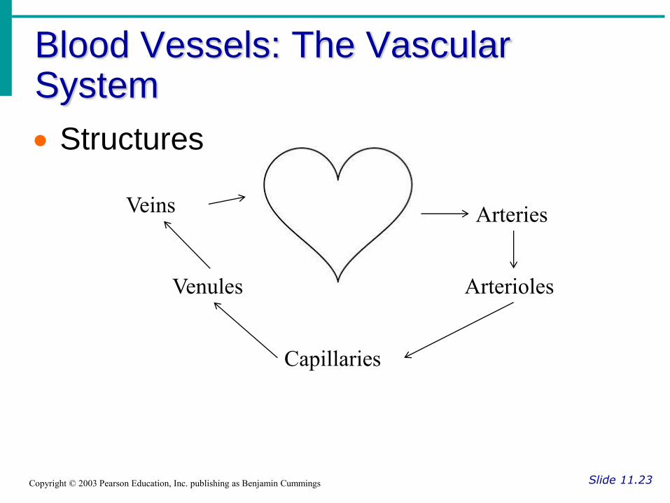
Blood Vessels: The Vascular System
Slide 11.23 Copyright © 2003 Pearson Education, Inc. publishing as Benjamin Cummings
Function Taking blood to the tissues and back
Blood Vessels: The Vascular System
Slide 11.23 Copyright © 2003 Pearson Education, Inc. publishing as Benjamin Cummings
Structures
Arteries
Arterioles
Capillaries
Venules
Veins

The Vascular System
Slide 11.24 Copyright © 2003 Pearson Education, Inc. publishing as Benjamin Cummings
Figure 11.8b


Differences Between Blood Vessel Types
Slide 11.26 Copyright © 2003 Pearson Education, Inc. publishing as Benjamin Cummings
Walls of arteries are the thickest
Lumens of veins are larger
Walls of capillaries are only one cell layer thick to allow for exchanges of gases and nutrients between blood and tissue

Diffusion at Capillary Beds
Slide 11.29 Copyright © 2003 Pearson Education, Inc. publishing as Benjamin Cummings
Figure 11.20

Movement of Blood Through Vessels
Slide 11.27 Copyright © 2003 Pearson Education, Inc. publishing as Benjamin Cummings
Most arterial blood is pumped by the heart
Veins use the milking action of muscles to help move blood back to the heart
Figure 11.9

Major Arteries of Systemic Circulation
Slide 11.30 Copyright © 2003 Pearson Education, Inc. publishing as Benjamin Cummings
Figure 11.11

Major Veins of Systemic Circulation
Slide 11.31 Copyright © 2003 Pearson Education, Inc. publishing as Benjamin Cummings
Figure 11.12

Arterial Supply of the Brain
Slide 11.32 Copyright © 2003 Pearson Education, Inc. publishing as Benjamin Cummings
Figure 11.13

Hepatic Portal Circulation
Slide 11.33 Copyright © 2003 Pearson Education, Inc. publishing as Benjamin Cummings
Figure 11.14

Circulation to the Fetus
Slide 11.34 Copyright © 2003 Pearson Education, Inc. publishing as Benjamin Cummings
Figure 11.15


Title
• Physiology of the Heart
Essential Question
• Name the elements of the
intrinsic conduction system
of the heart, and describe
the pathway of impulses
through this system.

The Heart: Conduction System
Slide 11.13a Copyright © 2003 Pearson Education, Inc. publishing as Benjamin Cummings
Intrinsic conduction system (nodal system)
muscle cells contract, w/o nerve impulses, in a regular, continuous way
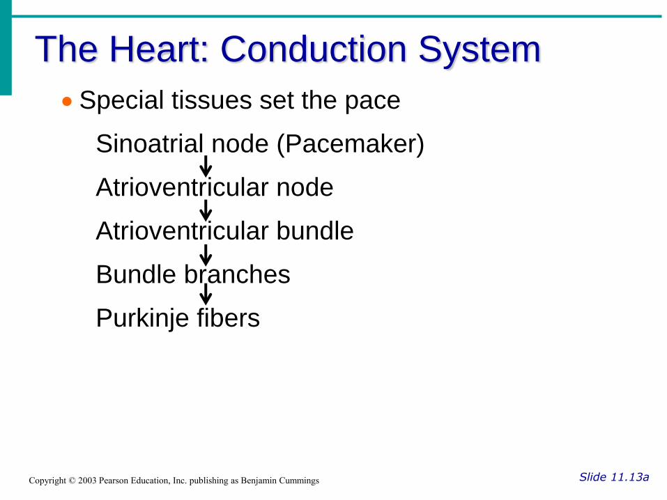
The Heart: Conduction System
Slide 11.13a Copyright © 2003 Pearson Education, Inc. publishing as Benjamin Cummings
Special tissues set the pace
Sinoatrial node (Pacemaker)
Atrioventricular node
Atrioventricular bundle
Bundle branches
Purkinje fibers

Heart Contractions
Slide 11.14b Copyright © 2003 Pearson Education, Inc. publishing as Benjamin Cummings
Figure 11.5

Filling of Heart Chambers – the Cardiac Cycle
Slide 11.15 Copyright © 2003 Pearson Education, Inc. publishing as Benjamin Cummings
Figure 11.6

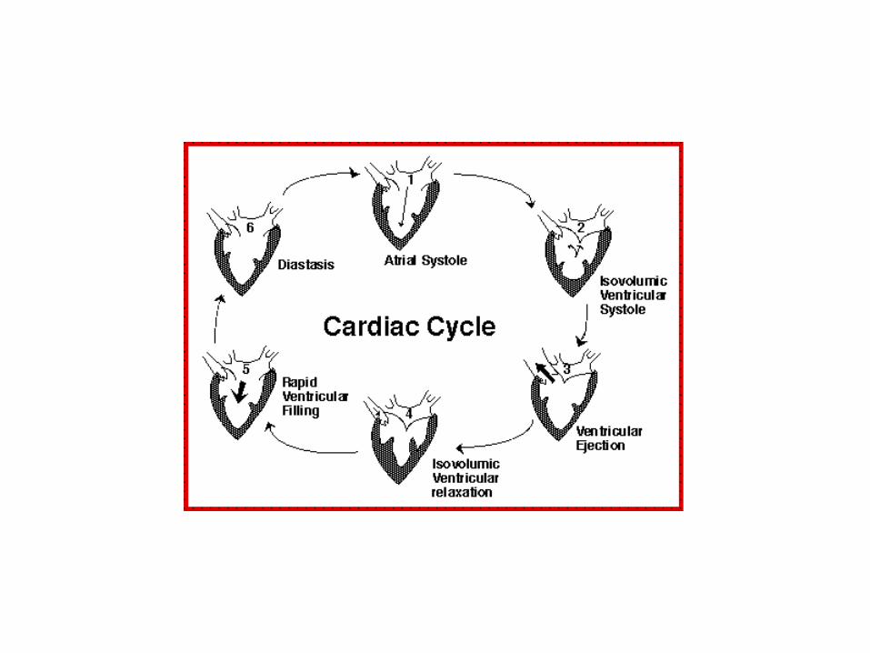
The Heart: Cardiac Cycle
Slide 11.16 Copyright © 2003 Pearson Education, Inc. publishing as Benjamin Cummings
Cardiac cycle – events of one complete heart beat
Overview
Atria contract simultaneously
Atria relax, then ventricles contract
Systole = contraction
Diastole = relaxation

The Heart: Cardiac Cycle
Slide 11.17 Copyright © 2003 Pearson Education, Inc. publishing as Benjamin Cummings
Mid-to-late diastole – blood flows into ventricles
Ventricular systole – blood pressure builds before ventricle contracts, pushing out blood
Early diastole – atria finish re-filling, ventricular pressure is low


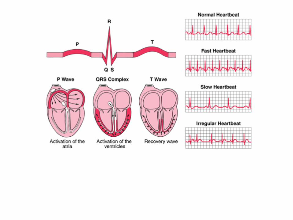
ECG WAVE
http://uploadImage:ECG
principle slow.gif -
Wikipedia, the free
encyclopedia.
Atrial
Reploarization


Title
• Physiology of Circulation
Essential Question
• Describe the regulation of
cardiac output and blood
pressure.

The Heart: Cardiac Output
Slide 11.18 Copyright © 2003 Pearson Education, Inc. publishing as Benjamin Cummings
Cardiac output (CO)
Amount of blood pumped by each side of the heart in one minute
CO = (heart rate [HR]) x (stroke volume [SV])
The Heart: Cardiac Output
Slide 11.18 Copyright © 2003 Pearson Education, Inc. publishing as Benjamin Cummings
Stroke volume
Volume of blood pumped by each ventricle in one contraction

Cardiac Output Regulation
Slide 11.19 Copyright © 2003 Pearson Education, Inc. publishing as Benjamin Cummings
Figure 11.7
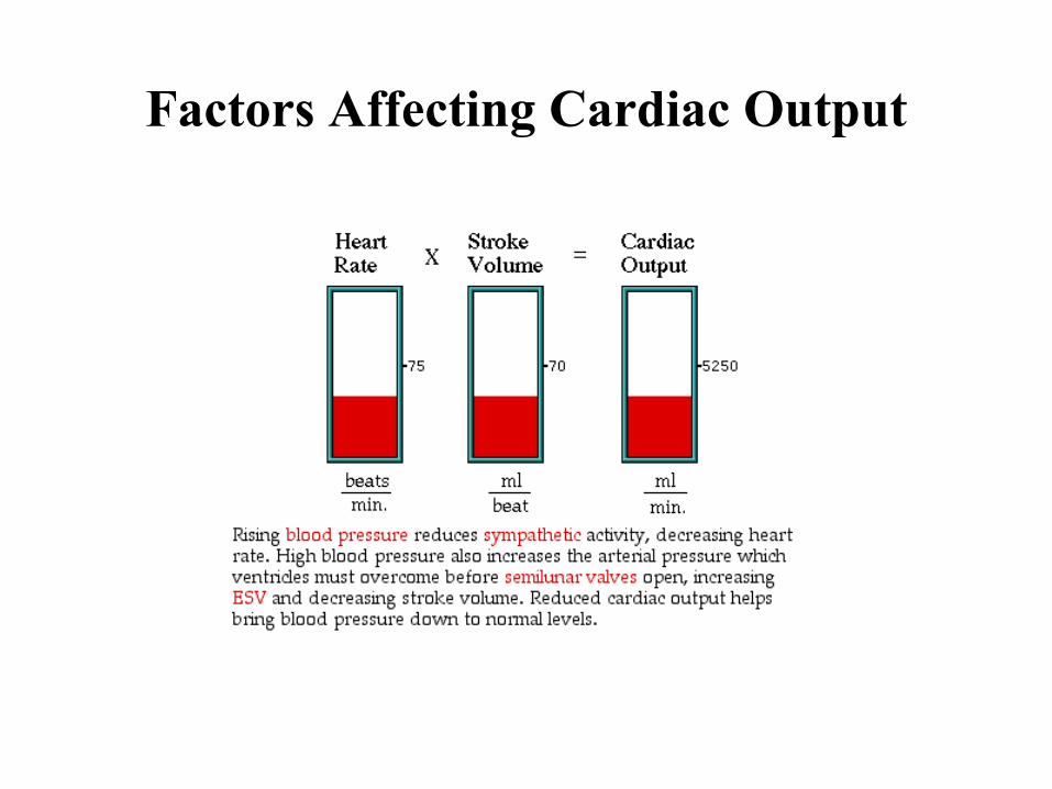
Factors Affecting Cardiac Output

The Heart: Regulation of Heart Rate
Slide 11.20 Copyright © 2003 Pearson Education, Inc. publishing as Benjamin Cummings
Starling’s law of the heart – the more that the cardiac muscle is stretched, the stronger the contraction

The Heart: Regulation of Heart Rate
Slide 11.21 Copyright © 2003 Pearson Education, Inc. publishing as Benjamin Cummings
Increased heart rate
Sympathetic nervous system
Crisis
Low blood pressure
Hormones
Epinephrine
Thyroxine
Exercise
Decreased blood volume

The Heart: Regulation of Heart Rate
Slide 11.22 Copyright © 2003 Pearson Education, Inc. publishing as Benjamin Cummings
Decreased heart rate
Parasympathetic nervous system
High blood pressure or blood volume
Decreased venous return

Pulse
Slide 11.35 Copyright © 2003 Pearson Education, Inc. publishing as Benjamin Cummings
Pulse – pressure wave of blood
Monitored at “pressure points” where pulse is easily palpated
Figure 11.16

Blood Pressure
Slide 11.36 Copyright © 2003 Pearson Education, Inc. publishing as Benjamin Cummings
Measurements made on large arteries
Systolic – pressure at the peak of ventricular contraction
Diastolic – pressure when ventricles relax


Measuring Arterial Blood Pressure
Slide 11.37 Copyright © 2003 Pearson Education, Inc. publishing as Benjamin Cummings
Figure 11.18
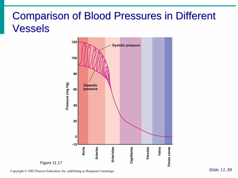
Comparison of Blood Pressures in Different
Vessels
Slide 11.38 Copyright © 2003 Pearson Education, Inc. publishing as Benjamin Cummings
Figure 11.17

Blood Pressure: Effects of Factors
Slide 11.39a Copyright © 2003 Pearson Education, Inc. publishing as Benjamin Cummings
Neural factors
Autonomic nervous system adjustments (sympathetic division)
Renal factors
Regulation by altering blood volume
Renin – hormonal control

Blood Pressure: Effects of Factors
Slide 11.39b Copyright © 2003 Pearson Education, Inc. publishing as Benjamin Cummings
Temperature
Heat has a vasodilation effect
Cold has a vasoconstricting effect
Chemicals
Various substances can cause increases or decreases
Diet

Factors Determining Blood Pressure
Slide 11.40 Copyright © 2003 Pearson Education, Inc. publishing as Benjamin Cummings
Figure 11.19

Variations in Blood Pressure
Slide 11.41 Copyright © 2003 Pearson Education, Inc. publishing as Benjamin Cummings
Normal
140–110 mm Hg systolic
80–75 mm Hg diastolic

Variations in Blood Pressure
Slide 11.41 Copyright © 2003 Pearson Education, Inc. publishing as Benjamin Cummings
Hypotension
Low systolic (below 110 mm HG)
Often associated with illness
Hypertension
High systolic (above 140 mm HG)
Can be dangerous if it is chronic


