Download - Ankle Instability and Pain
Marco Ucciferri, DPM, FACFAS
November 1, 2017
Ankle Instability and Chronic Pain
Introduction• 28,000 ankle sprains occur daily in the U.S. (Kaminski 2013)
• Ankle is the 2nd most commonly injured body site (Ferran 2006)
• Ankle sprains are the most common type of ankle injury
(Ferran 2006)
• A sprained ankle can happen to athletes and non-athletes,
children and adults.
• Inversion injury most common mechanism (Ferran 2006)
• Only risk factor is previous ankle sprain (Ferran 2006)
• Sex, generalized joint laxity or anatomical foot types are not risk
factors (Beynnon et al. 2002)
Introduction
• It is estimated that 80 to 85% of ankle sprains occur to the
lateral ligaments (Ryan et al., 1986)
• It is generally accepted that an eversion ankle sprain is more
severe, with greater instability. However, an inversion ankle
sprain is more common, with the lateral ligaments being
involved in 80% to 85% of all ankle sprains (Ryan et al., 1986)
• ATFL is most common injured ligament
• High rate of recurrence (20 - 40%) (Verhagen 2010)
• Chronic ankle instability (20 – 40%) (Verhagen 2000)
Classification
• Ankle sprain classified in two
categories:
Low ankle sprain
• Lateral ankle sprain “classic sprain” 80 – 85%
• Medial ankle sprain 5 – 10%
High ankle sprain
• Syndesmotic sprain 5 -10%
Low Ankle Sprain
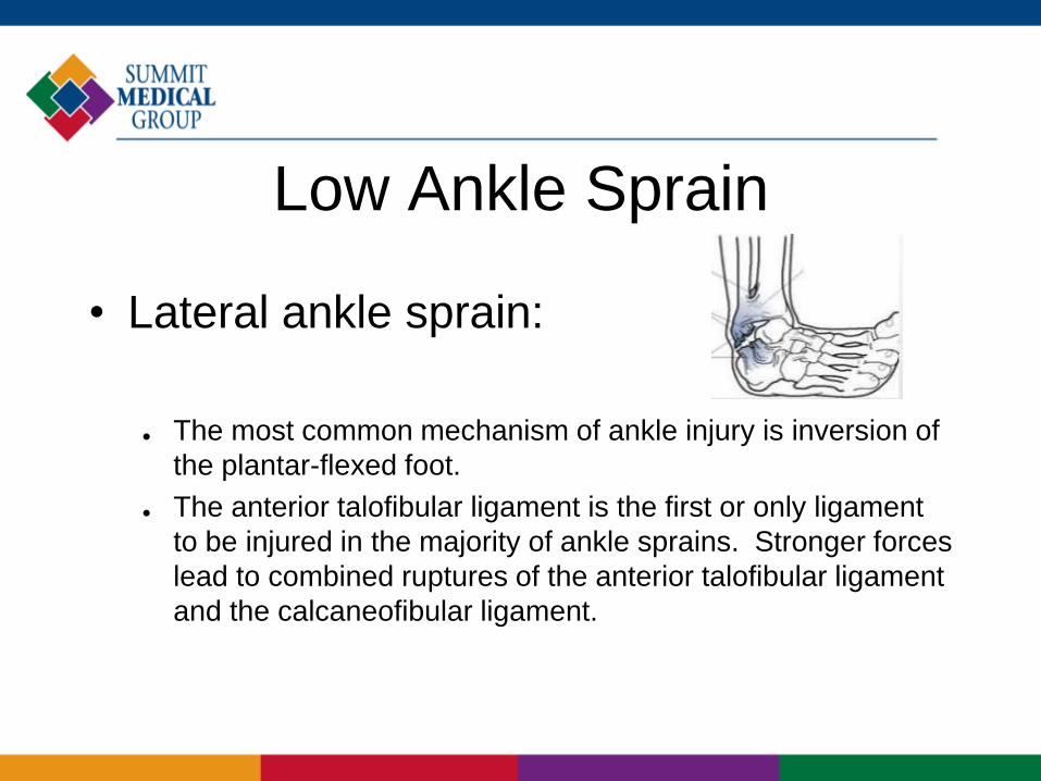
• Lateral ankle sprain:
The most common mechanism of ankle injury is inversion of
the plantar-flexed foot.
The anterior talofibular ligament is the first or only ligament
to be injured in the majority of ankle sprains. Stronger forces
lead to combined ruptures of the anterior talofibular ligament
and the calcaneofibular ligament.
Low Ankle Sprain
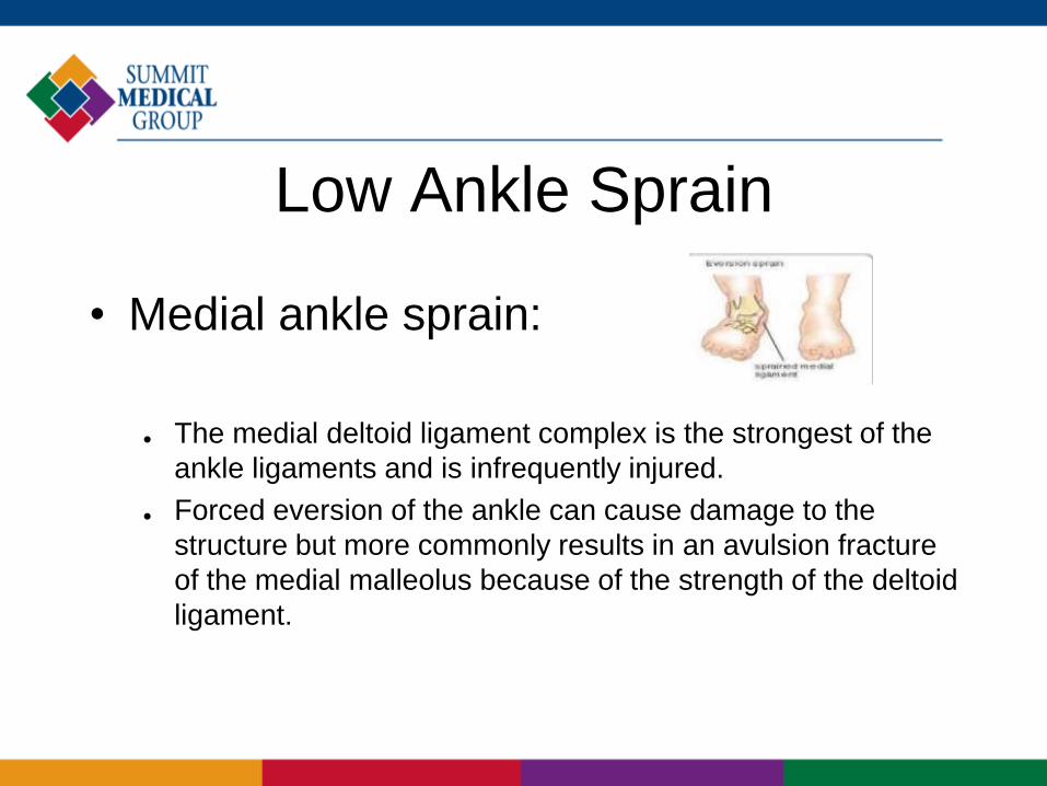
• Medial ankle sprain:
The medial deltoid ligament complex is the strongest of the
ankle ligaments and is infrequently injured.
Forced eversion of the ankle can cause damage to the
structure but more commonly results in an avulsion fracture
of the medial malleolus because of the strength of the deltoid
ligament.
High Ankle Sprain
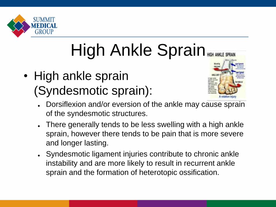
• High ankle sprain
(Syndesmotic sprain): Dorsiflexion and/or eversion of the ankle may cause sprain
of the syndesmotic structures.
There generally tends to be less swelling with a high ankle
sprain, however there tends to be pain that is more severe
and longer lasting.
Syndesmotic ligament injuries contribute to chronic ankle
instability and are more likely to result in recurrent ankle
sprain and the formation of heterotopic ossification.
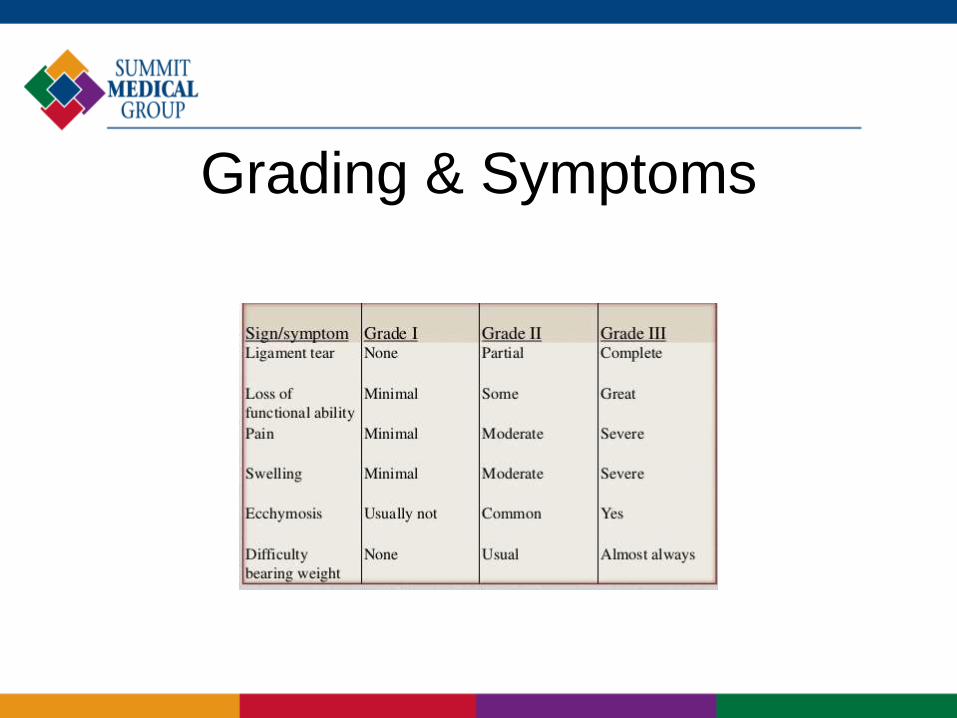
Grading & Symptoms
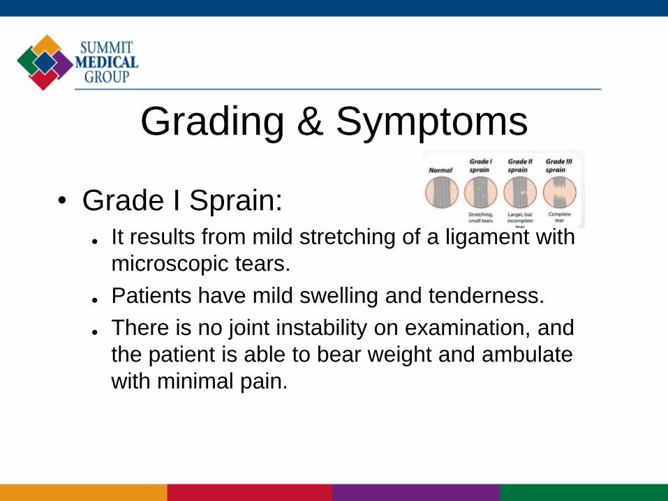
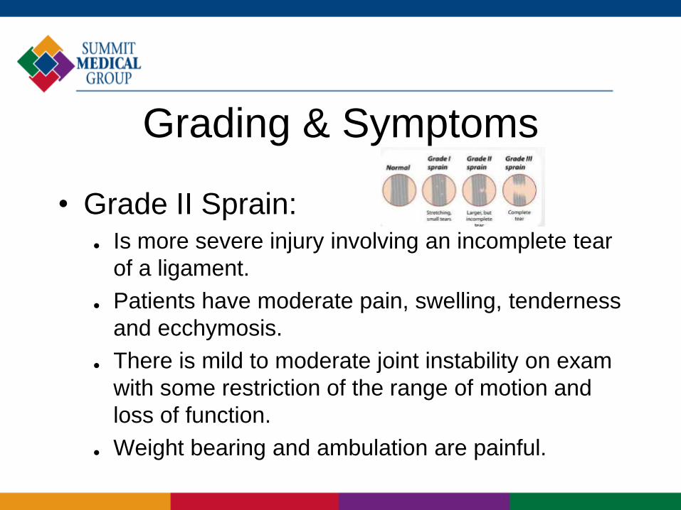
• Grade I Sprain: It results from mild stretching of a ligament with
microscopic tears.
Patients have mild swelling and tenderness.
There is no joint instability on examination, and
the patient is able to bear weight and ambulate
with minimal pain.
Grading & Symptoms
• Grade II Sprain: Is more severe injury involving an incomplete tear
of a ligament.
Patients have moderate pain, swelling, tenderness
and ecchymosis.
There is mild to moderate joint instability on exam
with some restriction of the range of motion and
loss of function.
Weight bearing and ambulation are painful.
Grading & Symptoms
Physical Examination
• There is swelling, ecchymosis and tenderness over affected
site.
• The degree of swelling or ecchymosis is proportional to the
likelihood of fracture.
• Palpation should include bony landmarks such as the lateral
malleolus, the medial malleolus, the fibula, the fifth metatarsal
and the physis in skeletally immature patients.
• Achilles tendon, peroneal tendons and posterior tibial tendon
should also be palpated.
• Tenderness over the anterior joint line or syndesmosis may
indicate a sprain of the interosseous membrane.
Physical Examination
• Recurrent sprains often have very little swelling.
• An individual with an ankle sprain can almost always walk on
the foot carefully with pain.
• Grade III ankle sprains often include an audible snap followed
by pain and swelling.
• A careful neurologic examination is essential to rule out loss of
sensation or motor weakness, as peroneal nerve and tibial
nerve injuries are sometimes seen with severe lateral ankle
sprains.
ManagementConservative Management
Initial Management
Initial management of ankle sprains requires the PRICER
regimen:
P = Protection … crutches, splint or brace
R = Rest …
I = Ice … 20 minutes every 2 hours
C = Compression …
E = Elevation …
R = Rehabilitation …
– This is probably the single most important factor in treatment,
particularly with Grade I and Grade II injuries.
– Pain and swelling can be reduced with the use of
electrotherapeutic modalities.
– Analgesics (NSAID) may be required.
ManagementConservative Management
Restoring of Full Range of Motion
• The patient may be non-weight-bearing on crutches for
the first 24 hours but should then commence partial
weight-bearing in normal heel-toe gait.
• It will be necessary from this stage to protect the
damaged joint with strapping or bracing.
• As soon as pain allows, active range of motion exercises
can be commenced.
ManagementConservative Management
Muscle Conditioning:
• Strengthening exercises should be commenced as soon
as pain allows.
• Active exercises should be performed initially with
gradually increasing resistance.
• Exercises should include plantarflexion and dorsiflexion,
inversion and eversion.
Functional Exercise:
• Functional exercises (e.g. jumping, hopping, twisting,
figure-of-eight running) should be commenced when the
athlete is pain-free, has full range of motion and
adequate muscle strength and proprioception.
ManagementConservative Management
Treatment of Grade III Injuries:
Treatment of Grade III ankle injuries requires initial
conservative management over a six-week period.
If the patient continues to make good progress and is able to
perform sporting activities with the aid of taping or bracing
and without persistent problems during or following activity,
surgery may not be required.
If, however, despite appropriate rehabilitation and protection,
the patient complains of recurrent episodes of instability or
persistent pain, then surgical reconstruction is indicated.
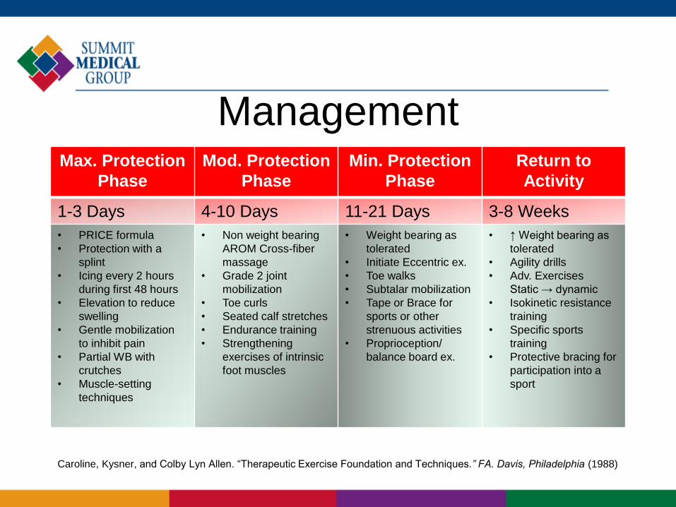
ManagementMax. Protection
Phase
Mod. Protection
Phase
Min. Protection
Phase
Return to
Activity
1-3 Days 4-10 Days 11-21 Days 3-8 Weeks
• PRICE formula
• Protection with a
splint
• Icing every 2 hours
during first 48 hours
• Elevation to reduce
swelling
• Gentle mobilization
to inhibit pain
• Partial WB with
crutches
• Muscle-setting
techniques
• Non weight bearing
AROM Cross-fiber
massage
• Grade 2 joint
mobilization
• Toe curls
• Seated calf stretches
• Endurance training
• Strengthening
exercises of intrinsic
foot muscles
• Weight bearing as
tolerated
• Initiate Eccentric ex.
• Toe walks
• Subtalar mobilization
• Tape or Brace for
sports or other
strenuous activities
• Proprioception/
balance board ex.
• ↑ Weight bearing as
tolerated
• Agility drills
• Adv. Exercises
Static → dynamic
• Isokinetic resistance
training
• Specific sports
training
• Protective bracing for
participation into a
sport
Caroline, Kysner, and Colby Lyn Allen. “Therapeutic Exercise Foundation and Techniques.” FA. Davis, Philadelphia (1988)
Objectives
• Discuss the evolution of lateral ankle
stabilization in the literature.
• Identify different techniques used to
perform lateral ankle stabilization and the
published outcomes.
• Discuss the preferred technique for lateral
ankle stabilization.
Surgical Treatment
• Conservative treatment supported for acute injuries.
Unger et al. JBJS. 1998 – Brostrom vs. Conservative.
• No difference in overall result, functional scores, objective or subjective
stability.
• Conservative group – return to activities 5.4 weeks sooner
• 10-30% develop residual instability
Karlsson et al., Lofvenberg et al. (20 year f/u), Colville et al…
• Trends in the Literature
Anatomic vs. Non-anatomic
Gold Standard – Brostrom-Gould
• Numerous modifications
Open vs. Arthroscopic Procedures
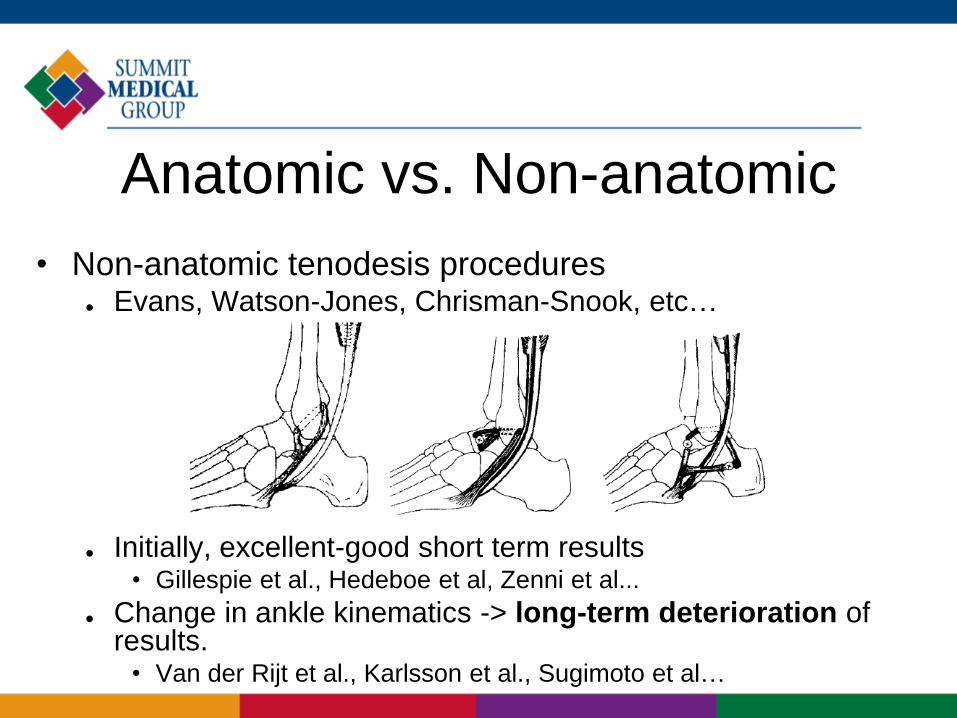
Anatomic vs. Non-anatomic
• Non-anatomic tenodesis procedures Evans, Watson-Jones, Chrisman-Snook, etc…
Initially, excellent-good short term results• Gillespie et al., Hedeboe et al, Zenni et al...
Change in ankle kinematics -> long-term deterioration of results.
• Van der Rijt et al., Karlsson et al., Sugimoto et al…
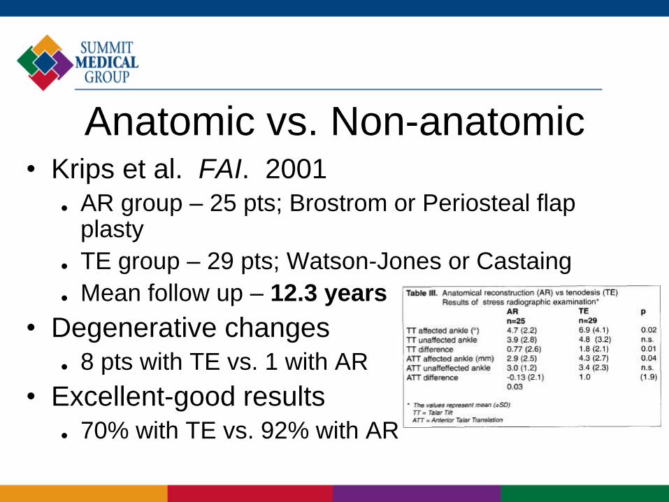
Anatomic vs. Non-anatomic• Krips et al. FAI. 2001
AR group – 25 pts; Brostrom or Periosteal flap plasty
TE group – 29 pts; Watson-Jones or Castaing
Mean follow up – 12.3 years
• Degenerative changes
8 pts with TE vs. 1 with AR
• Excellent-good results
70% with TE vs. 92% with AR
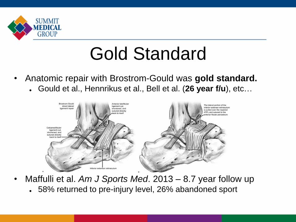
Gold Standard
• Anatomic repair with Brostrom-Gould was gold standard. Gould et al., Hennrikus et al., Bell et al. (26 year f/u), etc…
• Maffulli et al. Am J Sports Med. 2013 – 8.7 year follow up 58% returned to pre-injury level, 26% abandoned sport
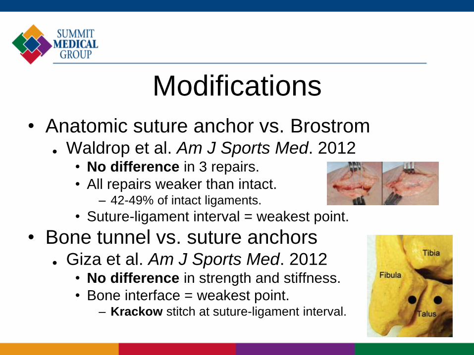
Modifications
• Anatomic suture anchor vs. Brostrom Waldrop et al. Am J Sports Med. 2012
• No difference in 3 repairs.
• All repairs weaker than intact.– 42-49% of intact ligaments.
• Suture-ligament interval = weakest point.
• Bone tunnel vs. suture anchors Giza et al. Am J Sports Med. 2012
• No difference in strength and stiffness.
• Bone interface = weakest point.– Krackow stitch at suture-ligament interval.
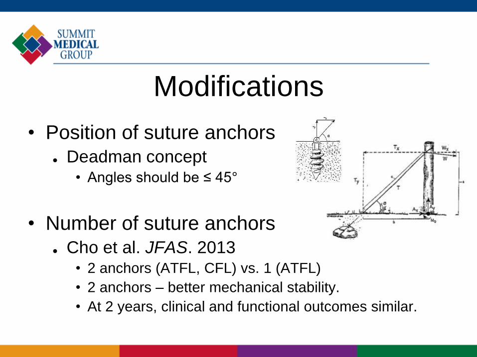
Modifications
• Position of suture anchors
Deadman concept• Angles should be ≤ 45°
• Number of suture anchors
Cho et al. JFAS. 2013• 2 anchors (ATFL, CFL) vs. 1 (ATFL)
• 2 anchors – better mechanical stability.
• At 2 years, clinical and functional outcomes similar.
Modifications
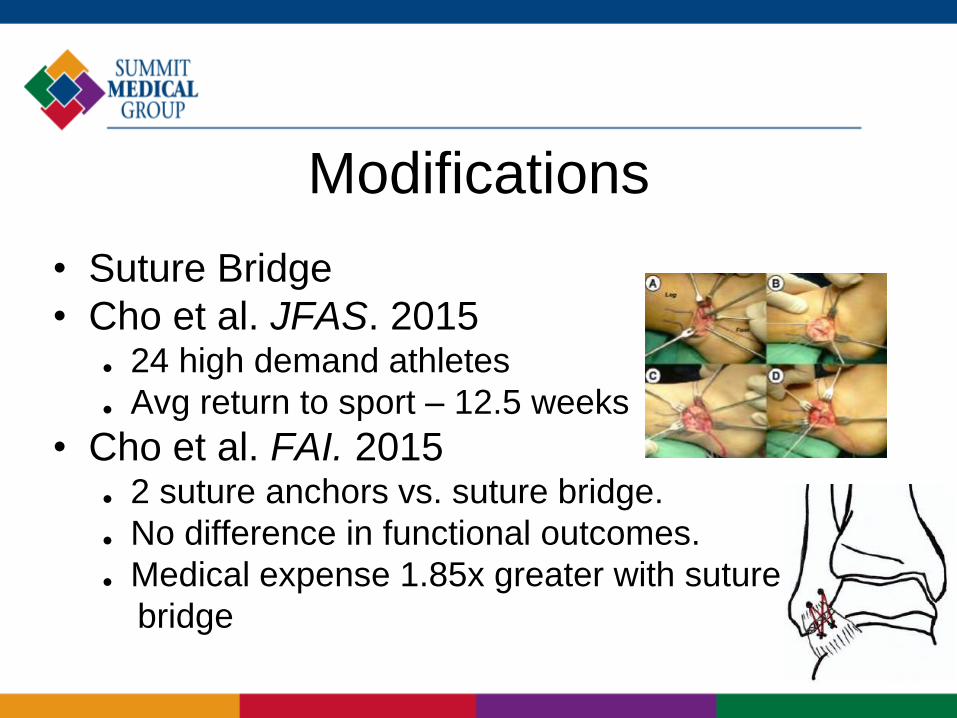
• Suture Bridge
• Cho et al. JFAS. 2015 24 high demand athletes
Avg return to sport – 12.5 weeks
• Cho et al. FAI. 2015 2 suture anchors vs. suture bridge.
No difference in functional outcomes.
Medical expense 1.85x greater with suture
bridge
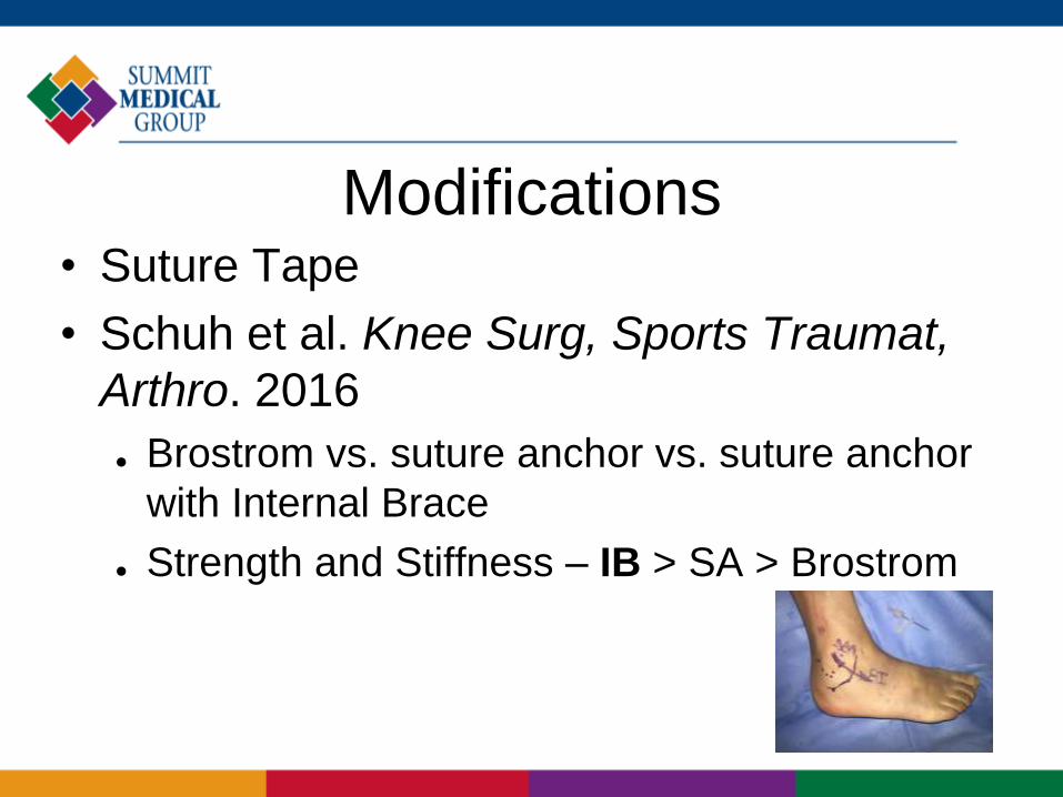
Modifications • Suture Tape
• Schuh et al. Knee Surg, Sports Traumat,
Arthro. 2016
Brostrom vs. suture anchor vs. suture anchor
with Internal Brace
Strength and Stiffness – IB > SA > Brostrom
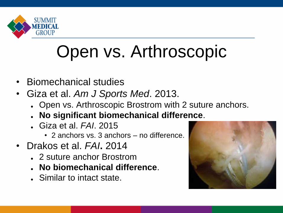
Open vs. Arthroscopic
• Biomechanical studies
• Giza et al. Am J Sports Med. 2013. Open vs. Arthroscopic Brostrom with 2 suture anchors.
No significant biomechanical difference.
Giza et al. FAI. 2015• 2 anchors vs. 3 anchors – no difference.
• Drakos et al. FAI. 2014 2 suture anchor Brostrom
No biomechanical difference.
Similar to intact state.
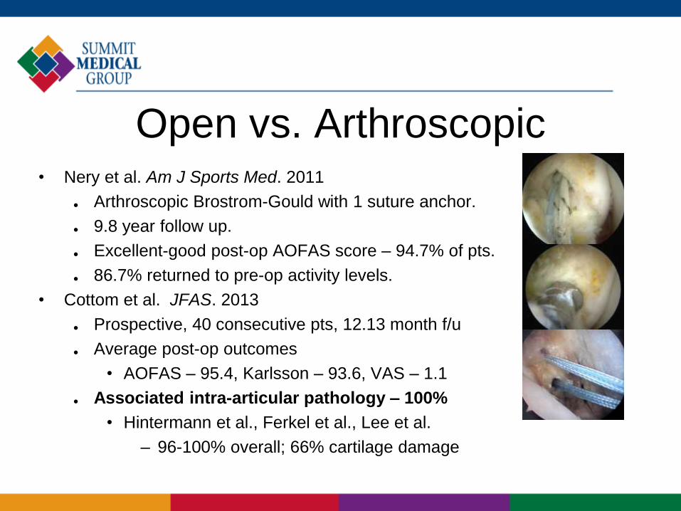
Open vs. Arthroscopic• Nery et al. Am J Sports Med. 2011
Arthroscopic Brostrom-Gould with 1 suture anchor.
9.8 year follow up.
Excellent-good post-op AOFAS score – 94.7% of pts.
86.7% returned to pre-op activity levels.
• Cottom et al. JFAS. 2013
Prospective, 40 consecutive pts, 12.13 month f/u
Average post-op outcomes
• AOFAS – 95.4, Karlsson – 93.6, VAS – 1.1
Associated intra-articular pathology – 100%
• Hintermann et al., Ferkel et al., Lee et al.
– 96-100% overall; 66% cartilage damage

Q & A


