donor-specific indirect pathway analysis reveals a b-cell ... am j... · donor-specific indirect...
TRANSCRIPT

American Journal of TransplantationWiley Periodicals Inc.
C© Copyright 2011 The American Society of Transplantationand the American Society of Transplant Surgeons
doi: 10.1111/j.1600-6143.2011.03869.x
Donor-Specific Indirect Pathway Analysis Reveals aB-Cell-Independent Signature which ReflectsOutcomes in Kidney Transplant Recipients
L. D. Haynesa, E. Jankowska-Gana, A. Shekaa,
M. R. Kellera, M. P. Hernandez-Fuentesb,
R. I. Lechlerb, V. Seyfert-Margolisc,†, L. A. Turkad,
K. A. Newelle and W. J. Burlinghama,*
aUniversity of Wisconsin, Department of Surgery,Transplant Division, Madison, WIbKings College London, MRC Centre for Transplantation,London, UKcFood and Drug Administration, Department of Health andHuman Services, Silver Spring, MDdBeth Israel Deaconess Medical Center (BIDMC), HarvardMedical School, Boston, MA and Immune ToleranceNetwork, Bethesda, MDeEmory University, Department of Surgery and the EmoryTransplant Center, Atlanta, GA*Corresponding author: William J. Burlingham,[email protected]†Formerly at Immune Tolerance Network, Bethesda, MD
To investigate the role of donor-specific indirect path-way T cells in renal transplant tolerance, we analyzedresponses in peripheral blood of 45 patients using thetrans-vivo delayed-type hypersensitivity assay. Sub-jects were enrolled into five groups—identical twin,clinically tolerant (TOL), steroid monotherapy (MONO),standard immunosuppression (SI) and chronic rejec-tion (CR)—based on transplant type, posttransplantimmunosuppression and graft function. The indirectpathway was active in all groups except twins but dis-tinct intergroup differences were evident, correspond-ing to clinical status. The antidonor indirect pathway Teffector response increased across patient groups (TOL< MONO < SI < CR; p < 0.0001) whereas antidonor in-direct pathway T regulatory response decreased (TOL> MONO = SI > CR; p < 0.005). This pattern differedfrom that seen in circulating naı̈ve B-cell numbers andin a cross-platform biomarker analysis, where patientson monotherapy were not ranked closest to TOL pa-tients, but rather were indistinguishable from chroni-cally rejecting patients. Cross-sectional analysis of theindirect pathway revealed a spectrum in T-regulatory:T-effector balance, ranging from TOL patients having pre-dominantly regulatory responses to CR patients hav-ing predominantly effector responses. Therefore, theindirect pathway measurements reflect a distinct as-pect of tolerance from the recently reported elevationof circulating naı̈ve B cells, which was apparent only inrecipients off immunosuppression.
Key words: DTH assay, human, immunological moni-toring, indirect pathway, renal transplantation, trans-plant tolerance
Abbreviations: Breg, B regulatory cell; CR, chronicrejector group; DSA, donor specific antibody; EBV,Epstein Barr virus; MFI, mean fluorescent intensity;MONO, monotherapy group; PBS, phosphate bufferedsaline; PBMC, peripheral blood mononuclear cells; POT,probability of being tolerant; SI, standard immunosup-pression group; Teff, T effector cells; TOL, clinically tol-erant group; Treg, T regulatory cells; TT/DT, tetanustoxoid and diphtheria toxoid pediatric vaccine; tvDTH,trans-vivo delayed-type hypersensitivity assay.
Received 04 August 2011, revised 08 September 2011and accepted for publication 26 September 2011
Introduction
We have previously reported TGFb- or IL10-producing reg-ulatory indirect pathway T-cell responses in patients withlong-term excellent graft function after kidney or liver trans-plantation and withdrawal of all immunosuppression (1).However, the Th3/TGFb-dependent form of tolerance to akidney transplant in monkeys and humans was found to be“metastable” (2–4) and eventually some grafts underwentacute cellular, antibody-mediated or chronic rejection (CR).It is now well established that Th17 responses are inducedwhen cells are exposed to TGFb in the presence of IL6,so metastable tolerance to allografts based on Th3/TGFbcells may be prone to develop into Th17-driven CR, a phe-nomenon most clearly demonstrated in clinical lung trans-plants (5). A more stable form of tolerance may thereforeinvolve both Th3/TGFb- and Tr1/IL10-T regulatory (Treg)-cellresponses (6).
How tolerance at the T- and B-cell levels are interconnectedis not yet clear. Much interest in the possible role of B reg-ulatory cells (Bregs; Ref. 7) has been generated by therecent discovery that renal transplant tolerance in rodents(8,9) and humans (10–12) is characterized by an increasein both intragraft and systemic B cells. Whether this in-crease is a driving force for tolerance induction or whetherit simply helps to stabilize tolerance in the absence of im-munosuppression has not been established.

Haynes et al.
We hypothesized that the regulatory indirect pathwayT-cell response to donor alloantigens in kidney transplantrecipients is a progressive indicator of tolerance, thatis, one which becomes more pronounced in a patientpopulation that receives less immunosuppression whilemaintaining excellent graft function. We studied patientsin categories ranging from most tolerant (identical twin“isograft”) to least tolerant (chronic allograft rejection)and including three “in between” groups: patients withwell-functioning renal allografts who were taking standard,minimal or zero (functionally tolerant) immunosuppressivemedications. We compared the results of indirect path-way monitoring with the analysis of circulating B cells andB–cell-biased “probability of being tolerant” (POT) scores(10,11).
Materials and Methods
Source of cells and reagents
Renal transplant recipients and living donors from three transplant centers(Emory University, Atlanta, GA, USA; University of Wisconsin–Madison,WI, USA and Swedish Medical Center, Seattle, WA, USA) were enrolledin the ITN507ST observational trial, “Identification and Mechanistic Inves-tigations of Tolerant Kidney Transplant Patients” (Clinicaltrials.gov identifierNCT01338779) using institutional review board-approved informed writtenconsent procedures at each institution. Patients were assigned to groupsbased on clinical status as previously described (10). Patients enrolled infour of the groups—identical twin (TWIN—transplant from an identical twindonor), clinically tolerant (TOL—no immunosuppression medication for >1year), steroid monotherapy (MONO, ≤10 mg/day of prednisone) and stan-dard immunosuppression (SI—including any combination of standard post-transplant immunosuppression) all had stable and normal renal function(within 25% of baseline) at the time of enrollment. All patients in the CRgroup had moderately impaired or gradually deteriorating renal function(GFR < 40 mL/min or creatinine >50% above baseline value), had a pre-vious biopsy showing chronic allograft nephropathy (BANFF 1997 criteria)and continued to receive immunosuppressive medications, but were not ondialysis. Peripheral blood samples were collected in citrate and processedusing Ficoll-Hypaque to isolate peripheral blood mononuclear cells (PBMC).PBMC were either used immediately or cryopreserved before analysis.
Antigens used to test for indirect pathway responses included: donor PBMCor spleen cell sonicates, as described elsewhere (1); HLA-A1, HLA-B62 andHLA-B57 single antigen Luminex beads (One Lambda, Inc., Canoga Park,CA, USA), a kind gift of Dr. Junchao Cai, Paul Terasaki Foundation, Los Ange-les, CA, USA and HLA-B allopeptides p37MA (DSDAASPRMAPRAPWIEQ)and p37TE (DSDAASPRTEPRAPWIEQ), described elsewhere (4).
Antibodies co-injected with PBMC: rabbit anti-human TGF-b, goat anti-human IL-10 or control normal rabbit IgG or goat IgG (all R&D Systems, Min-neapolis, MN, USA, all at 25 lg/injection), anti-human IFNc (eBioscience,San Diego, CA, USA, 5 lg/injection) or anti-human IL17 (R&D Systems,5 lg/injection).
Indirect pathway analysis
Human PBMC (7–9 × 10⁶ cells) were mixed with either donor or recall(tetanus/diphtheria toxoid [TT/DT] or Eptstein-Barr virus [EBV]) antigen orwith a combination of donor plus recall antigen and injected into CB.17SCID mouse footpads (1,13). Footpad thickness was measured using aspring-loaded caliper at time 0 and 24 h. Net swelling is determined by
subtracting the background swelling from an injection of the same cellsin phosphate-buffered saline (PBS). This trans-vivo delayed-type hypersen-sitivity (tvDTH) reaction is associated with edema, human cell retentionand mouse neutrophil recruitment in the mouse footpad that is initiated byhuman T cells in response to processed antigens (14). Inhibition of recallresponses in the presence of donor antigens was determined by comparingthe net footpad swelling with each injection using the formula
% Inhibition =[
1 −(Recall + donor antigen
)Recall Alone
]× 100
and is a measure of indirect pathway donor antigen-specific regulatoryactivity (1).
B-cell depletion
B-cell depletion was performed using anti-CD19 immuno-magnetic beads(Miltenyi Biotec, Auburn, CA, USA) and the autoMACS system. Subsequentflow cytometry for CD19+ cells verified removal of 99% of the B cells fromthe PBMC. In the “add-back” experiments, B cells were combined withthe B-cell-depleted sample at the same ratio of B to non-B cells as wasdetermined by flow cytometry of the whole PBMC.
Statistical analysis
Kruskal–Wallis test, a nonparametric equivalent to the one-way analysis ofvariance, was used to compare results across all groups; the Dunn’s correc-tion was used for pair-wise comparisons. For some analyses, an unadjustedMann–Whitney U test was used for individual pair-wise comparisons. Pairedt-tests were used to compare within subject responses. The p-value lessthan 0.05 was considered statistically significant. Statistical analysis wasperformed with Prism GraphPad software.
Results
The spectrum of responses to soluble donor
alloantigens correlates with graft status
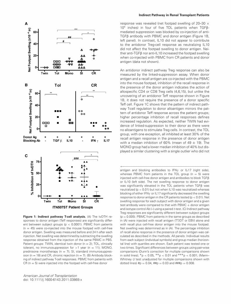
PBMC from half of the subjects (n = 45, selected be-cause of cell availability) enrolled in the ITN507ST trial weretested for donor-specific indirect T effector (Teff) and Tregresponses using the tvDTH assay and donor (or donor HLA-matched surrogate) cell-free lysates. As shown in Figure1A, indirect Teff responses to donor antigens reveals a dis-tinct spectrum across the enrollment groups (p < 0.001);footpad swelling increased as the patients’ clinical statusmoves from those that are tolerant to those that are chron-ically rejecting. Identical twins did not respond to donorantigen whereas patients in the TOL group had a mediannet swelling response of 5 × 10⁻⁴ inches (mean of 4.5 ±4.2). The MONO group had a higher median response of10 × 10−4 inches (mean of 9.3 ± 3.4), as did the SI group(mean of 10.3 ± 7.6). Finally, responses of patients in theCR group were 30 × 10−4 inches (mean of 28.7 ± 10.3),which was significantly higher than all the others. Analy-sis using a single pair-wise comparison of TOL to eitherMONO (p = 0.04) or SI (p = 0.03) groups revealed signifi-cantly lower Teff response in TOL patients.
Antibodies to IFNc and IL17 significantly blocked the indi-rect pathway Teff-cell responses to donor antigens in theCR patients tested (p < 0.01, compared to IgG control, Fig-ure 1B, right panel). In contrast, an indirect pathway Teff
American Journal of Transplantationdoi: 10.1111/j.1600-6143.2011.03869.x
Indirect Pathway in Renal Transplant Patients
Figure 1: Indirect pathway T-cell analysis. (A) The tvDTH re-sponses to donor antigen (Teff responses) are significantly differ-ent between subject groups (p < 0.0001). PBMC from patients(n = 45) were co-injected into the mouse footpad with cell-freedonor antigen. Swelling was measured before and 24 h after eachinjection. Net swelling was determined by subtracting the swellingresponse obtained from the injection of the same PBMC in PBS.Patient groups: TWIN, identical twin donor (n = 2); TOL, clinicallytolerant, no immunosuppression for >1 year (n = 11); MONO,prednisone monotherapy (n = 7); SI, standard immunosuppres-sion (n = 18) and CR, chronic rejection (n = 7). (B) Antibody block-ing of indirect pathway T-cell responses. PBMC from patients withCR (n = 5) were injected into the footpad with cell-free donor
response was revealed (net footpad swelling of 20–30 ×104 inches) in four of five TOL patients when TGFb-mediated suppression was blocked by co-injection of anti-TGFb antibody with PBMC and donor antigen (Figure 1B,left panel). In contrast, IL10 did not appear to contributeto the antidonor Treg-cell response as neutralizing IL10did not affect the footpad swelling to donor antigen. Nei-ther anti-TGFb nor anti-IL10 increased the footpad swellingwhen co-injected with PBMC from CR patients and donorantigen (data not shown).
An antidonor indirect pathway Treg response can also bemeasured by the linked-suppression assay. When donorantigen and a recall antigen are co-injected with the PBMCinto the mouse footpad, inhibition of the recall response inthe presence of the donor antigen indicates the action ofallospecific CD4 or CD8 Treg cells (4,6,15), but unlike theuncovering of an antidonor Teff response shown in Figure1B, it does not require the presence of a donor specificTeff cell. Figure 1C shows that the pattern of indirect path-way T-cell regulation to donor alloantigen mirrors the pat-tern of antidonor Teff response across the patient groups;higher percentage inhibition of recall responses definesincreased regulation. As expected, neither TWIN had evi-dence of linked-suppression to their donor as there wereno alloantigens to stimulate Treg cells. In contrast, the TOLgroup, with one exception, all inhibited at least 30% of therecall antigen response in the presence of donor antigenwith a median inhibition of 60% (mean of 49 ± 19). TheMONO group had a lower median inhibition of 40% but dis-played a similar clustering with a single outlier who did not
←−−−−−−−−−−−−−−−−−−−−−−−−−−−−−−−−−−−−−−−−−−−−−−−−−−antigen and blocking antibodies to IFNc or IL-17 (right side),whereas PBMC from patients in the TOL group (n = 5) wereinjected with cell-free donor antigen and antibodies to block TGFbor IL-10 (left side). The net swelling response to donor antigenwas significantly elevated in the TOL patients when TGFb wasneutralized (p < 0.01) but not when IL10 was neutralized whereasblocking of either IFNc or IL17 significantly decreased the swellingresponse to donor antigen in the CR patients tested (p < 0.01). Netswelling response for each subject with donor antigen and a giventest antibody were compared to that with PBMC + donor antigenand isotype control Ab (–) using a paired t-test. (C) Indirect pathwayTreg responses are significantly different between subject groups(p = 0.005). PBMC from patients in the same groups as describedin (A) were injected with recall antigen (TT/DT or EBV) alone andwith recall plus cell-free donor antigen into the mouse footpad.Net swelling was determined as in (A). The percentage inhibitionof recall alone response in the presence of donor antigen was cal-culated as described in the methods. All panels: Individual resultsfrom each subject (individual symbols) and group median (horizon-tal line) with quartiles are shown. Each patient was tested one ortwo times. Significant differences between groups using pair-wisecomparisons (Dunn’s correction for multiple comparisons shownin solid lines); ∗p < 0.05, ∗∗p < 0.01 and ∗∗∗p < 0.001; (Mann–Whitney U test unadjusted for multiple comparisons shown withdotted lines) #p = 0.04, ##p = 0.03 and ###p = 0.008.
American Journal of Transplantationdoi: 10.1111/j.1600-6143.2011.03869.x

Haynes et al.
regulate (mean of 38 ± 20). The SI group had the same me-dian inhibition as the MONO group; however, similar to theTeff responses (Figure 1C, panel A), the range of indirectpathway regulation in the SI patients was large (mean of41 ± 26). The CR group was significantly lower in its regula-tion scores (mean of 3.3 ± 8) compared to TOL and MONOgroups. Overall, the data in Figure 1 are consistent with aspectrum of increasing cell-mediated immune response todonor soluble antigens and a decrease in regulatory re-sponse over the range of most (TOL) through intermediate(MONO and SI) to least tolerant (CR) patients.
Analysis of naı̈ve B-cell counts and POT index
We verified that the 45 subjects in this study were repre-sentative of the entire 91 subjects in the ITN507ST, specif-ically in regards to the characteristics we and others havepreviously found to describe tolerant renal transplant re-cipients (10–12). As shown in Figure 2A, circulating naı̈veB-cell numbers were significantly different among patientgroups (p = 0.005) with patients in the TOL group havingsignificantly more compared to those in the SI group whensingle pair-wise Mann–Whitney U test was performed. Inthis way, our TOL and SI subgroups are representative ofthose same groups in the complete ITN507ST (10). TheTOL patients also had significantly higher median naı̈veB-cell counts than those in the MONO and CR groups. In-terestingly, the MONO group median most closely resem-bled the CR group median and was significantly lower (nothigher) than the SI median using the unadjusted Mann–Whitney U test.
Three TOL patients scored at the low end of therange of donor-specific indirect pathway Treg-driven linked-suppression (0%, 33% and 40% inhibition, Figure 1C). Cir-culating naı̈ve B cells (138, 2 and 34 cells/lL, respectively,Figure 2A) were also low for two of these three individuals.Importantly, there was no correlation between the naı̈ve B-cell numbers and indirect pathway analysis for patients inany of the groups (data not shown).
The POT scores are based on 14 biomarkers identified froma multiparameter analysis of 11 tolerant renal transplantpatients enrolled in the Indices of Tolerance trial in Europeand 60 subjects in comparison groups (11). Samples frompatients in the ITN507ST study (n = 89; TOL group = 25)were used as a test set and a tolerance signature thresholdof >0.27 was established (11). As shown in Figure 2B, POTscores were available for 9 of the 11 TOL patients analyzedin Figure 1. Although the scores ranged widely, they wereall >0.27. The TOL group had significantly higher medianPOT scores than the MONO, SI and CR groups. Two pa-tients in the SI group but, remarkably, no patients in theMONO group had a POT score above the threshold of 0.27.Thus the data in Figures 1 and 2 reflect distinct aspects ofthe immunologic status of renal transplant recipients. TOLand MONO patients exhibit very similar indirect pathwayresponses whereas the levels of circulating naı̈ve B cellswere very different.
Figure 2: Naı̈ve B-cell counts and cross-platform probability
of being tolerant scores. (A) Naı̈ve B-cell number (CD19+CD27-IgM+IgD+) in fresh peripheral blood of the 45 patients analyzed inFigure 1. (B) Probability of being tolerant, estimated according tothe Indices of Tolerance model of 14 biomarkers, including B-cellparameters and direct pathway T-cell parameters (11). Subjectsin the TWIN group were not tested (n.t.). All panels: Individualresults from each subject (individual symbols) and group median(horizontal line) with quartiles (panel B only) are shown. Signif-icant differences between groups using pair-wise comparisons,∗p < 0.05 and ∗∗p < 0.01; Dunn’s correction for multiple com-parisons shown in solid lines, unadjusted Mann–Whitney U testcomparisons shown with dotted lines.
The tvDTH assay measures indirect pathway T-cell
regulation in a B-cell-independent manner
Because naı̈ve B cells are elevated and linked-suppressionis strongest in the TOL patients, we next sought to de-termine if B cells played any role in linked-suppression ofcell-mediated immunity detected in the tvDTH assay. Wedepleted B cells from patients 002055 (SI group, 75% in-hibition) and 002002 (TOL group, 67% inhibition) and then
American Journal of Transplantationdoi: 10.1111/j.1600-6143.2011.03869.x

Indirect Pathway in Renal Transplant Patients
Figure 3: B cells do not impact tvDTH suppression via indirect
pathway. PBMC from two patients with high circulating naiveB-cell counts, TOL patient 002002 and SI patient 002055 wereassayed directly (PBMC, filled squares), after being depleted ofCD19+ B cells (open triangles) or after depletion and add-back ofB-cell fraction (gray circles). Cell fractions were injected into thefootpad with PBS (negative control, data not shown) with recallantigen (TT/DT or EBV, left panel), recall antigen and self-peptide(p37TE or HA-1R, middle panel) or recall antigen and donor peptide(p37MA, or HA-1H, right panel). Swelling was measured beforeand 24 h after each injection. Net swelling was determined bysubtracting the swelling response obtained from the injection ofthe same cell fractions in PBS.
retested for linked-suppression using donor antigen (datanot shown) or allopeptide (HA1-H or p37MA) in the pres-ence of a recall antigen. As shown in Figure 3, neitherpatient’s inhibitory response to allopeptide was affectedby depletion (open triangles) or add-back of B cells (graycircles). Notably, both of these patients had high numbersof circulating naı̈ve B cells in their PBMC (360 cells/lL for002055 and 142 cells/lL for 002002).
Indirect pathway T-cell responses analyzed using HLA
single-antigen beads
To exclude the possibility that the donor antigen lysatescontained nonspecific factors responsible for either inhi-bition or stimulation of tvDTH responses, we selected in-formative patients and challenged their PBMC with sin-gle antigen HLA-coated beads or with HLA allopeptideantigens rather than whole cell lysates. PBMC from TOLpatient 002002 was obtained 14 years after transplantfrom an HLA class II well-matched and HLA-A-matcheddeceased donor. This subject had not taken any immuno-suppression for 12 years. We previously reported that thispatient had a DQ3-restricted CD4 T-cell response to a singleimmuno-dominant allopeptide, p37MA, found in the heavychain of donor-type HLA-B62; this epitope is shared withHLA-B57 but not with other HLA-A or B proteins (4,16).As shown in Figure 4A, we observed a dose-dependentinhibition of tvDTH response to recall antigen challengewhen PBMC were injected with B62 (open circles) orB57 beads (open squares), resulting in a maximum inhi-bition of 60–63%. In contrast, over the same dose rangeof beads coated with HLA-A1 (filled triangles), a sharedantigen, we found little-to-no inhibition of the footpadswelling response to recall antigen. When self HLA-B37
Figure 4: Donor-specific indirect pathway responses using
HLA-single antigen beads. (A) PBMC of tolerant patient 002002were mixed with varying amounts (0–10 lL) of donor type HLA-B62 (open circle, p37MA+), third party HLA–B57 (open square,p37MA+) or self HLA-A1 (filled triangle, p37MA−)-coated sin-gle antigen beads before addition of TT/DT and injected into themouse footpad. Net swelling was determined as in Figure 1A. Themaximum percentage inhibition of response with recall TT/DT (0beads) versus the response with 10 lL of single antigen beadswas calculated as described in the methods and is 60% with B57beads and 63% with B62 beads. Peptides: PBMC were co-injectedinto the mouse footpad with recall antigen TT/DT and 1000 ngof HLA-B62 allopeptide p37MA (DSDAASPRMAPRAPWIEQ) orHLA-B37 self peptide p37TE (DSDAASPRTEPRAPWIEQ). The per-centage inhibition of recall antigen response with donor peptideis 68%. Patient 002002 = HLA A1, 2; B37, 60; DR 4, 13; donortype: A1, 2; B44, 62; DR 4, 13. (B) Indirect pathway tvDTH re-sponse to single antigen-beads detects stronger sensitization to aDSA+ as compared with DSA− donor HLA antigens. PBMC fromCR patient 002048 were mixed with donor HLA-A2 (circles), HLA-A1 (squares) or HLA-B57 (inverted triangle)-coated single antigenbeads and injected into a CB17 SCID mouse footpad. Net swellingwas determined as in Figure 1A. Mean fluorescent intensity (MFI)values for a contemporaneous serum sample of the same sub-ject tested using HLA-A2 (gray circle)-, HLA-A1 (gray square)- orHLA-B57 (gray inverted triangle)-coated single antigen beads areshown on the right. Patient 002048 = HLA-A11, 25; B18, 51;DR9, 15 Donor = HLA-A1, 2; B7, 57; DR 11,15 (mismatch in bold).
American Journal of Transplantationdoi: 10.1111/j.1600-6143.2011.03869.x
Haynes et al.
peptide p37TE (filled circle) was compared with the al-lopeptide p37MA (filled diamond, co-injection of 1000 ngwith recall antigen), we saw similar strong (68%) inhibitionof the recall antigen response with the p37MA allopeptidebut no effect was seen with the autologous p37TE peptide(Figure 4A, peptide). The equivalent dose-response to theB57- and the B62-bead indicates that the p37MA peptideprovides the critical component for response, and not theintact antigen.
To compare the indirect pathway Teff response with B-cellresponses in the same individual to the identical donorHLA protein, subjects in the CR group were tested intvDTH and Luminex (per clinical follow-up) using HLA sin-gle antigen beads. Data from one such patient (#002048) isshown in Figure 4B. Footpad swelling responses to donorHLA-A1 (squares), HLA-A2 (circles) and HLA-B57 (invertedtriangle) were detected when HLA-coated single antigenbeads were injected with patient PBMC. Donor specific an-tibody (DSA) analysis from the same patient (right panel)revealed a specific humoral response to HLA-A2 (gray cir-cle, mean fluorescent intensity [MFI] = 1168) but not toHLA-A1 (gray square, MFI = 16, Luminex lower limit ofdetection) or HLA-B57 (gray inverted triangle, MFI = 24)mismatched donor antigen. The hierarchy of indirect path-way cellular reactivity, HLA-A2 � HLA-A1 or HLA-B57, re-flected in dose–response to single antigen beads parallelsthe DSA response in serum detected using the same sub-strate, while indicating a broader antigen sensitization atthe cellular versus humoral immunity level.
Discussion
Given the critical importance of indirect pathway alloreac-tivity in tolerance (17,18) and in both cellular and humoral(19) immunity to allografts, many groups including our own(20) have sought to develop in vitro methods of monitor-ing human indirect pathway T cells as a guide to toler-ance induction and successful immunosuppressive drugwithdrawal and the early diagnosis of CR (21). However,weak lymphoproliferative and cytokine responses by lowfrequency indirect pathway T cells have hampered the clin-ical application of indirect pathway assays. Taking advan-tage of the unique characteristics of the tvDTH assay to de-tect both indirect pathway regulation and effector function(15,22–24) and using the original design of the ITN507STcross-sectional trial, we found a pattern of indirect path-way T-cell responses consistent with a spectrum of im-mune responsiveness in 45 renal transplant recipients ofvarious clinical status. At the extreme ends of the spec-trum, identical twins showed no indirect pathway T-cellresponses at all, consistent with having received a kidneyisograft, whereas allosensitized, chronic rejectors demon-strated strong IFNc and IL17-dependent indirect pathwayTeff and B-cell (DSA) responses to donor HLA, with no ev-idence of indirect pathway Treg-cell-mediated bystandersuppression. The TOL group displayed the lowest indi-
rect pathway Teff-cell response to donor and the highestregulatory response. Although not all patients in the TOLgroup were analyzed, the regulatory response appearedto be mainly Th3-type because TGFb and not IL10 block-ing antibodies revealed a latent indirect pathway Teff DTHresponse. This finding is consistent with the TGFb biasin indirect pathway Treg responses to kidney transplantspreviously reported in mouse (25) and monkey (3) toler-ance models. Interestingly, the steroid MONO group had aslightly higher baseline indirect pathway T-cell response todonor antigen and less donor-specific regulation, but oth-erwise closely resembled the TOL group, particularly inthe range and distribution of individual responses. The SIgroup was also in between the TOL and CR groups in bothregulation and Teff antidonor tvDTH assays.
The existence of a progressive spectrum in the indirectpathway means that analysis of this parameter may yieldinsights into potential candidates for partial withdrawal ofimmunosuppression, as has been recently demonstrated(26). It is unclear why a similarly progressive spectrumwas not seen in circulating B-cell counts or in the B-cell-influenced “POT” signature (Figure 2). It is possible thatcorticosteroid monotherapy is toxic to B cells. It is alsopossible that naı̈ve and transitional B cells are required tostabilize tolerance once it is established but are less crit-ical during the development of tolerance. This hypothesispredicts that B cells would accumulate only after some ini-tial tolerance threshold has been reached and the subjectis free of immunosuppression. When we depleted B cellsfrom PBMC of two patients (one in TOL, one in SI) with highnaı̈ve B-cell counts, the strong indirect pathway T-cell regu-lation was unaffected, evidence that the cellular immunity-based indirect pathway function as detected by the tvDTHassay is not directly influenced by the presence of B cells.It is theoretically possible that donor antigen-specific Bregcells acting as an antigen presenting cells could be involvedin suppression of the indirect pathway. This would requireintact donor HLA, not allopeptide. In fact, we could detectno difference in dose–response of inhibition between in-tact donor HLA-B62-coated beads and third party HLA-B57(p37MA+)-coated beads (Figure 4A), arguing against thisidea, at least in one TOL patient. However, there was asuggestive piece of evidence of a connection between thetwo parameters within the TOL group itself—namely, twoof the three TOL patients with the lowest regulation todonor antigen (Figure 1C) also had the lowest number ofB cells (Figure 2A). It is possible that the maintenance oftolerance in these two individuals is independent of eitherof these two factors or that both patients are in a categoryof “unstable tolerance”.
In addition to a significantly elevated CD20 mRNA signalobserved in urine samples of TOL versus SI patients in theITN507ST trial (10), recent data demonstrates a rise in in-tragraft B cells in spontaneous kidney allograft tolerance inmice (8) and in a rat model of allograft tolerance inducedusing LF15–0195, a deoxyspergualine analog (8,9). Weak
American Journal of Transplantationdoi: 10.1111/j.1600-6143.2011.03869.x
Indirect Pathway in Renal Transplant Patients
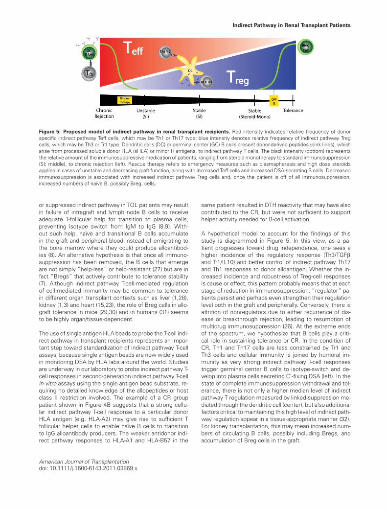
Figure 5: Proposed model of indirect pathway in renal transplant recipients. Red intensity indicates relative frequency of donorspecific indirect pathway Teff cells, which may be Th1 or Th17 type; blue intensity denotes relative frequency of indirect pathway Tregcells, which may be Th3 or Tr1 type. Dendritic cells (DC) or germinal center (GC) B cells present donor-derived peptides (pink lines), whicharise from processed soluble donor HLA (sHLA) or minor H antigens, to indirect pathway T cells. The black intensity (bottom) representsthe relative amount of the immunosuppressive medication of patients, ranging from steroid monotherapy to standard immunosuppression(SI; middle), to chronic rejection (left). Rescue therapy refers to emergency measures such as plasmapheresis and high dose steroidsapplied in cases of unstable and decreasing graft function, along with increased Teff cells and increased DSA-secreting B cells. Decreasedimmunosuppression is associated with increased indirect pathway Treg cells and, once the patient is off of all immunosuppression,increased numbers of naı̈ve B, possibly Breg, cells.
or suppressed indirect pathway in TOL patients may resultin failure of intragraft and lymph node B cells to receiveadequate T-follicular help for transition to plasma cells,preventing isotype switch from IgM to IgG (8,9). With-out such help, naı̈ve and transitional B cells accumulatein the graft and peripheral blood instead of emigrating tothe bone marrow where they could produce alloantibod-ies (8). An alternative hypothesis is that once all immuno-suppression has been removed, the B cells that emergeare not simply “help-less” or help-resistant (27) but are infact “Bregs” that actively contribute to tolerance stability(7). Although indirect pathway T-cell-mediated regulationof cell-mediated immunity may be common to tolerancein different organ transplant contexts such as liver (1,28),kidney (1,3) and heart (15,23), the role of Breg cells in allo-graft tolerance in mice (29,30) and in humans (31) seemsto be highly organ/tissue-dependent.
The use of single antigen HLA beads to probe the T-cell indi-rect pathway in transplant recipients represents an impor-tant step toward standardization of indirect pathway T-cellassays, because single antigen beads are now widely usedin monitoring DSA by HLA labs around the world. Studiesare underway in our laboratory to probe indirect pathway T-cell responses in second-generation indirect pathway T-cellin vitro assays using the single antigen bead substrate, re-quiring no detailed knowledge of the allopeptides or hostclass II restriction involved. The example of a CR grouppatient shown in Figure 4B suggests that a strong cellu-lar indirect pathway T-cell response to a particular donorHLA antigen (e.g. HLA-A2) may give rise to sufficient Tfollicular helper cells to enable naı̈ve B cells to transitionto IgG alloantibody producers. The weaker antidonor indi-rect pathway responses to HLA-A1 and HLA-B57 in the
same patient resulted in DTH reactivity that may have alsocontributed to the CR, but were not sufficient to supporthelper activity needed for B-cell activation.
A hypothetical model to account for the findings of thisstudy is diagrammed in Figure 5. In this view, as a pa-tient progresses toward drug independence, one sees ahigher incidence of the regulatory response (Th3/TGFband Tr1/IL10) and better control of indirect pathway Th17and Th1 responses to donor alloantigen. Whether the in-creased incidence and robustness of Treg-cell responsesis cause or effect, this pattern probably means that at eachstage of reduction in immunosuppression, “regulator” pa-tients persist and perhaps even strengthen their regulationlevel both in the graft and peripherally. Conversely, there isattrition of nonregulators due to either recurrence of dis-ease or breakthrough rejection, leading to resumption ofmultidrug immunosuppression (26). At the extreme endsof the spectrum, we hypothesize that B cells play a criti-cal role in sustaining tolerance or CR. In the condition ofCR, Th1 and Th17 cells are less constrained by Tr1 andTh3 cells and cellular immunity is joined by humoral im-munity as very strong indirect pathway T-cell responsestrigger germinal center B cells to isotype-switch and de-velop into plasma cells secreting C’-fixing DSA (left). In thestate of complete immunosuppression withdrawal and tol-erance, there is not only a higher median level of indirectpathway T regulation measured by linked-suppression me-diated through the dendritic cell (center), but also additionalfactors critical to maintaining this high level of indirect path-way regulation appear in a tissue-appropriate manner (32).For kidney transplantation, this may mean increased num-bers of circulating B cells, possibly including Bregs, andaccumulation of Breg cells in the graft.
American Journal of Transplantationdoi: 10.1111/j.1600-6143.2011.03869.x
Haynes et al.
Acknowledgments
This work was supported by ITN507ST grant from the NIH-supported Im-mune Tolerance Network, by NIH grant #1R01- AI066219 (to WJB) and bythe EU-sponsored One Study (WJB, LDH and MHF). M.H.F. and R.I.L. ac-knowledge support from ITN503ST, EU FP6 grant: QLRT–2002–02127 andfrom the UK Department of Health by National Institute for Health Research(NIHR) comprehensive Biomedical Research Centre award to Guy’s and StThomas’ NHS Foundation Trust in partnership with King’s College Londonand King’s College Hospital NHS Foundation Trust. The authors wish tothank Glen Leverson for his critical help with statistical analysis.
Funding source: No part of this manuscript was prepared by any commer-cial organization nor was it funded by any commercial organization.
Disclosure
The authors of this manuscript have no conflicts of inter-est to disclose as described by the American Journal ofTransplantation.
Note added in proof: While the data indicates that TGF-beta plays a role in the regulation of indirect pathway ob-served in tolerant group patients, we cannot rule out a rolefor IL35 and hence iTR35 T cells.
References
1. VanBuskirk AM, Burlingham WJ, Jankowska-Gan E, et al. Humanallograft acceptance is associated with immune regulation. J ClinInvest 2000; 106:145–155.
2. Lafferty KJ, Babcock SK, Gill RG. Prevention of rejection by treat-ment of the graft: An overview. Prog Clin Biol Res 1986; 224:87–117.
3. Torrealba JR, Katayama M, Fechner JH Jr., et al. Metastable tol-erance to rhesus monkey renal transplants is correlated with al-lograft TGF-beta 1+CD4+ T regulatory cell infiltrates. J Immunol2004; 172:5753–5764.
4. Xu Q, Lee J, Jankowska-Gan E, et al. Human CD4+CD25lowadaptive T regulatory cells suppress delayed-type hypersen-sitivity during transplant tolerance. J Immunol 2007; 178:3983–3995.
5. Burlingham WJ, Love RB, Jankowska-Gan E, et al. IL-17-dependent cellular immunity to collagen type V predisposes toobliterative bronchiolitis in human lung transplants. J Clin Invest2007;117:3498–3506.
6. Cai J, Lee J, Jankowska-Gan E, et al. Minor H antigen HA-1-specific regulator and effector CD8+ T cells, and HA-1 mi-crochimerism, in allograft tolerance. J Exp Med 2004; 199:1017–1023.
7. Iwata Y, Matsushita T, Horikawa M, et al. Characterization of arare IL-10-competent B-cell subset in humans that parallels mouseregulatory B10 cells. Blood 2011; 117:530–541.
8. Wang C, Cordoba S, Hu M, et al. Spontaneous acceptance ofmouse kidney allografts is associated with increased Foxp3 ex-pression and differences in the B and T cell compartments. TransplImmunol 2011; 24:149–156.
9. Le Texier L, Thebault P, Lavault A, et al. Long-term allograft toler-ance is characterized by the accumulation of B cells exhibiting aninhibited profile. Am J Transplant 2011; 11:429–438.
10. Newell KA, Asare A, Kirk AD, et al. Identification of a B cell signa-
ture associated with renal transplant tolerance in humans. J ClinInvest 2010; 120:1836–1847.
11. Sagoo P, Perucha E, Sawitzki B, et al. Development of a cross-platform biomarker signature to detect renal transplant tolerancein humans. J Clin Invest 2010;120:1848–1861.
12. Pallier A, Hillion S, Danger R, et al. Patients with drug-free long-term graft function display increased numbers of peripheral Bcells with a memory and inhibitory phenotype. Kidney Int 2010;78:503–513.
13. Burlingham WJ, Jankowska-Gan E. Mouse strain and injection siteare crucial for detecting linked suppression in transplant recipientsby trans-vivo DTH assay. Am J Transplant 2007; 7: 466–470.
14. Hegde S, Jankowska-Gan E, Roenneburg DA, Torrealba J,Burlingham WJ, Gumperz JE. Human NKT cells promote mono-cyte differentiation into suppressive myeloid antigen-presentingcells. J Leukoc Biol 2009; 86: 757–768.
15. VanBuskirk AM, Wakely ME, Sirak JH, Orosz CG. Patterns of al-losensitization in allograft recipients: Long-term cardiac allograftacceptance is associated with active alloantibody production inconjunction with active inhibition of alloreactive delayed-type hy-persensitivity. Transplantation 1998; 65:1115–1123.
16. Xu Q, Lee J, Keller M, Burlingham WJ. Analysis of indirect path-way CD4+ T cells in a patient with metastable tolerance to akidney allograft: Possible relevance to superior graft survival ofHLA class II closely matched renal allografts. Transpl Immunol2009; 20:203–208.
17. Yamada A, Chandraker A, Laufer TM, Gerth AJ, Sayegh MH,Auchincloss H Jr. Recipient MHC class II expression is re-quired to achieve long-term survival of murine cardiac allo-grafts after costimulatory blockade. J Immunol 2001; 167:5522–5526.
18. Kishimoto K, Yuan X, Auchincloss H Jr., Sharpe AH, MandelbrotDA, Sayegh MH. Mechanism of action of donor-specific transfu-sion in inducing tolerance: Role of donor MHC molecules, donorco-stimulatory molecules, and indirect antigen presentation. J AmSoc Nephrol 2004; 15:2423–2428.
19. Steele DJ, Laufer TM, Smiley ST, et al. Two levels of helpfor B cell alloantibody production. J Exp Med 1996 183:699–703.
20. Burlingham WJ, Fechner JH, DeVito LD, Sollinger HW, KnechtleSJ, Grailer AP. Human interleukin-2 and lymphoproliferative (T-helper cell) responses to soluble HLA class I antigens in vitro:I. Specificity for polymorphic domains. Tissue Antigens 1993;42:35–38.
21. Poggio ED, Clemente M, Riley J, et al. Alloreactivity in renal trans-plant recipients with and without chronic allograft nephropathy.J Am Soc Nephrol 2004; 15:1952–1960.
22. Niederkorn JY, Streilein JW. Alloantigens placed into the an-terior chamber of the eye induce specific suppression ofdelayed-type hypersensitivity but normal cytotoxic T lympho-cyte and helper T lymphocyte responses. J Immunol 1983; 131:2670–2674.
23. Warnecke G, Chapman SJ, Bushell A, Hernandez-Fuentes M,Wood KJ. Dependency of the trans vivo delayed type hypersensi-tivity response on the action of regulatory T cells: Implications formonitoring transplant tolerance. Transplantation 2007; 84:392–399.
24. Molitor-Dart ML, Andrassy J, Kwun J, et al. Developmental expo-sure to noninherited maternal antigens induces CD4+ T regula-tory cells: Relevance to mechanism of heart allograft tolerance.J Immunol 2007;179:6749–6761.
25. Cook CH, Bickerstaff AA, Wang JJ, et al. Spontaneous renal allo-graft acceptance associated with “regulatory” dendritic cells andIDO. J Immunol 2008; 180:3103–3112.
American Journal of Transplantationdoi: 10.1111/j.1600-6143.2011.03869.x
Indirect Pathway in Renal Transplant Patients
26. Jankowska-Gan E, Sollinger HW, Pirsch JD, et al. Successfulreduction of immunosuppression in older renal transplant re-cipients who exhibit donor-specific regulation. Transplantation2009;88:533–541.
27. Callaghan CJ, Rouhani FJ, Negus MC, et al. Abrogationof antibody-mediated allograft rejection by regulatory CD4 Tcells with indirect allospecificity. J Immunol 2007; 178:2221–2228.
28. Jankowska-Gan E, Rhein T, Haynes LD, et al. Human liver allograftacceptance and the “tolerance assay”. II. Donor HLA-A, -B but notDR antigens are able to trigger regulation of DTH. Hum Immunol2002; 63:862–870.
29. DiLillo DJ, Griffiths R, Seshan SV, et al. B lymphocytes differ-entially influence acute and chronic allograft rejection in mice.J Immunol 2011; 186:2643–2654.
30. Zarkhin V, Chalasani G, Sarwal MM. The yin and yang of B cellsin graft rejection and tolerance. Transplant Rev (Orlando) 2010;24:67–78.
31. Martinez-Llordella M, Lozano JJ, Puig-Pey I, et al. Using tran-scriptional profiling to develop a diagnostic test of operationaltolerance in liver transplant recipients. J Clin Invest 2008; 118:2845–2857.
32. Matzinger P, Kamala T. Tissue-based class control: The other sideof tolerance. Nat Rev Immunol 2011;11: 221–230.
American Journal of Transplantationdoi: 10.1111/j.1600-6143.2011.03869.x