donna m. wilcock ph.d. sanders-brown center on … · donna m. wilcock ph.d. sanders-brown center...
TRANSCRIPT

Profiling the neuroinflammatory response in AD — potential for
personalized medicine and therapeutic modulation
Donna M. Wilcock Ph.D. Sanders-Brown Center on Aging
Department of Physiology University of Kentucky

Microglial cells surround plaques and tangles
Plaque
Microglial cells

The original view of inflammation in AD
Adapted from McGeer and McGeer, Exp Gerentol 33:371-378
An autotoxic loop
Initial insult
Some neuronal
death
More rapid neuronal death
Deposition of debris
Activation of microglia (with secretion of toxic materials
Activation of complement cascade (including formation of MAC)
Reinforcing cross-stimulation

Inflammation has a purpose
Time
Leve
l
Potentially damaging proteins – Purpose – to clear debris to destroy invading cells
Non-damaging proteins– Purpose – to repair a wound to decrease damaging proteins
Injury Resolution
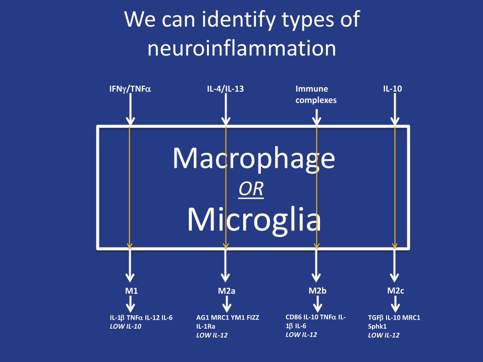
Macrophage
Microglia
IFNγ/TNFα IL-4/IL-13 Immune complexes
IL-10
M1 M2a M2b M2c
IL-1β TNFα IL-12 IL-6 LOW IL-10
AG1 MRC1 YM1 FIZZ IL-1Ra LOW IL-12
CD86 IL-10 TNFα IL-1β IL-6 LOW IL-12
TGFβ IL-10 MRC1 Sphk1 LOW IL-12
We can identify types of neuroinflammation
OR

Determining the inflammatory state of the human Alzheimer’s disease brain
• Many postmortem tissue have used late-stage AD brain samples.
• We hypothesized that there may be differences in the neuroinflammatory state as AD progresses.
• We obtained tissue samples from early AD, late AD and age-matched, non-demented controls.
• Gene expression levels of neuroinflammatory markers measured by qRT-PCR from frozen brain tissue frontal cortex and cerebellum.
• Serum analyzed for predictive markers of the brain neuroinflammatory state.
• Clinical histories of patients examined to determine common non-AD factors that account for differences in the neuroinflammatory states.

Human AD tissue characteristics
Group Age range (yr)
MMSE ApoE4 status
Braak stage PMI (hr)
Early AD N=23
77-100 (mean = 87.2)
20-24 (mean = 22.6)
-/4 = 12 4/4 = 3
3-5 1.0-6.5 (mean = 3.6)
Late AD N= 16
74-88 (mean 85.6)
0-13 (mean 7.25)
-/4 = 6 4/4 = 4
6 1.75-11.0 (mean =5.4)
Control N=37
77-96 (mean = 84.3)
28-30 (mean = 29.1)
-/4 = 10 4/4 = 3
0-2 1.75-8.0 (mean = 4.2)

Cluster analysis revealed two distinct populations in the early AD group
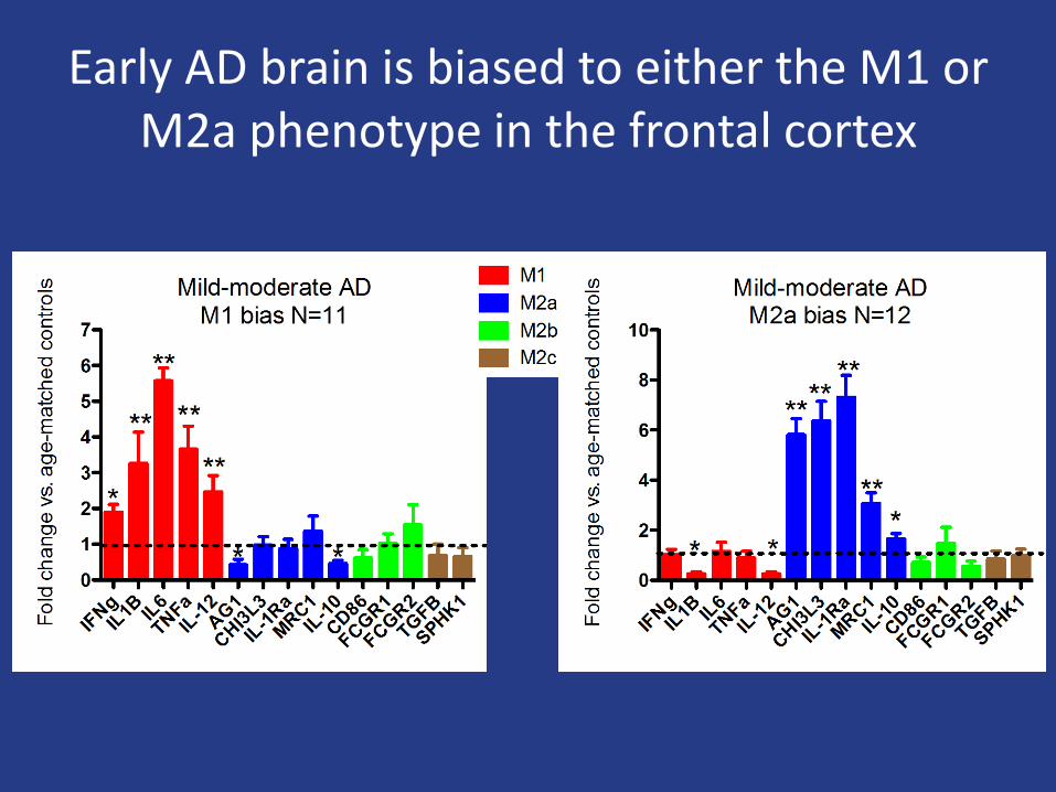
Early AD brain is biased to either the M1 or M2a phenotype in the frontal cortex

In the cerebellum the inflammatory polarization does not exist
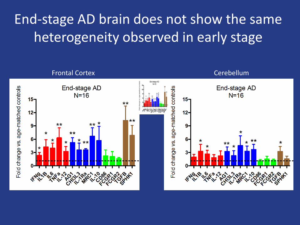
End-stage AD brain does not show the same heterogeneity observed in early stage
Frontal Cortex Cerebellum

Next step….
• Hypothesis: The polarization of the neuroinflammatory state of the early AD brain to either M1 or M2 will significantly influence response to therapy.
• Serum from our samples were run on the Myriad-RBM human inflammation MAP.
• Our goal was to identify serum biomarkers predictive
of the brain neuroinflammatory state.
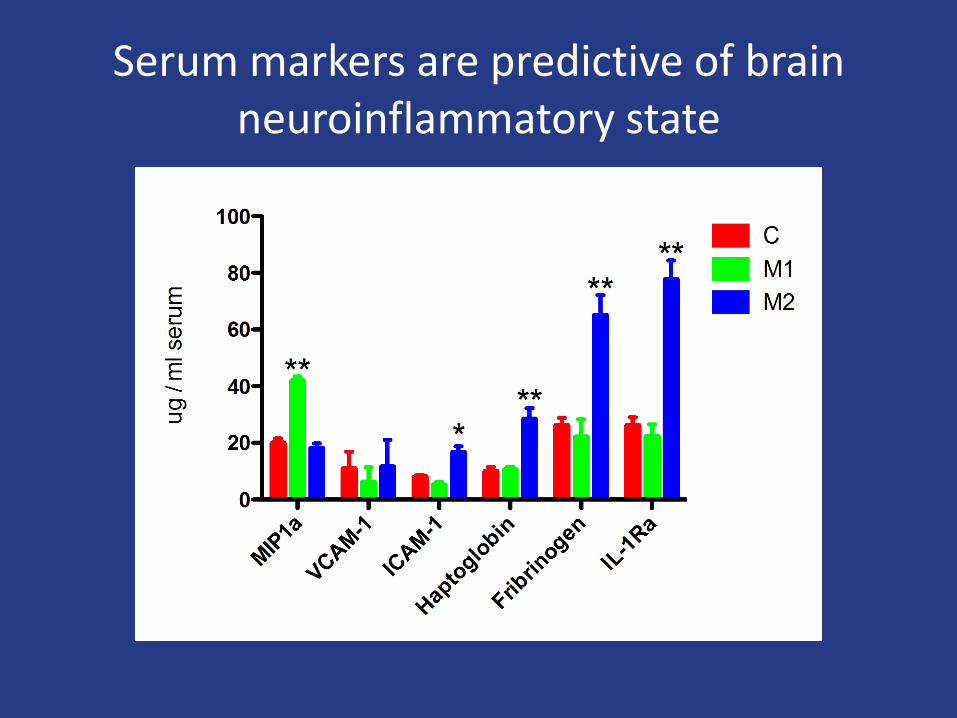
Serum markers are predictive of brain neuroinflammatory state
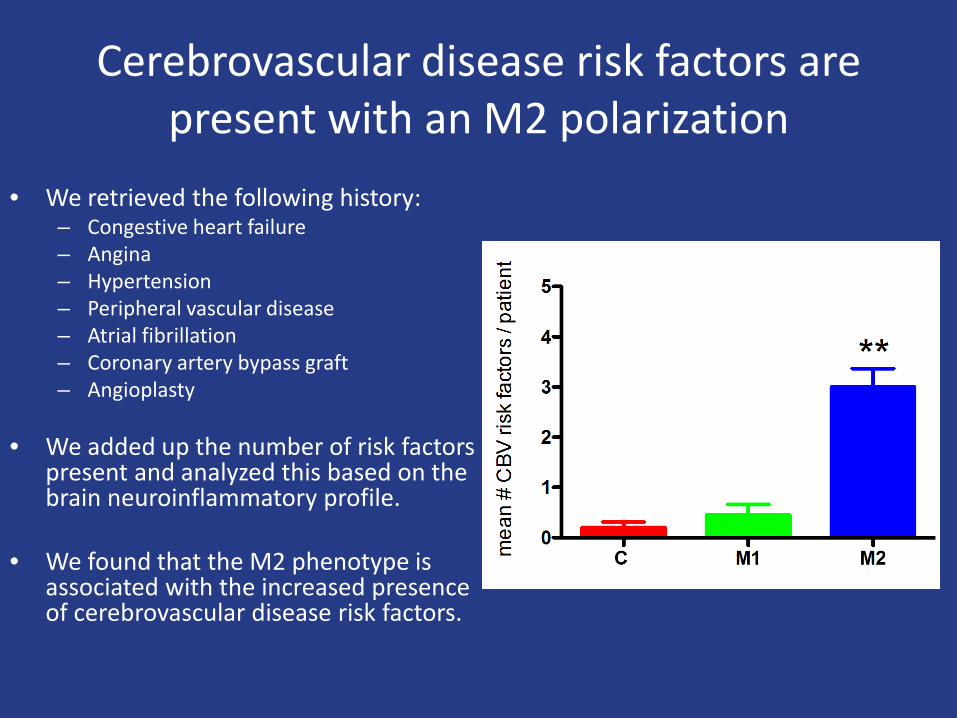
Cerebrovascular disease risk factors are present with an M2 polarization
• We retrieved the following history: – Congestive heart failure – Angina – Hypertension – Peripheral vascular disease – Atrial fibrillation – Coronary artery bypass graft – Angioplasty
• We added up the number of risk factors
present and analyzed this based on the brain neuroinflammatory profile.
• We found that the M2 phenotype is associated with the increased presence of cerebrovascular disease risk factors.
Can we modulate the inflammatory response and influence AD pathology?

Macrophage
Microglia
IFNγ/TNFα IL-4/IL-13
Immune complexes
IL-10
M1 M2a M2b M2c
IL-1β TNFα IL-12 IL-6 LOW IL-10
AG1 MRC1 YM1 FIZZ IL-1Ra LOW IL-12
CD86 IL-10 TNFα IL-1β IL-6 LOW IL-12
TGFβ IL-10 MRC1 Sphk1 LOW IL-12
Modulation to M2b
OR

Study design • 120 seven month old APP/PS1 mice were assigned to one of 4 treatment
groups. – Saline. – Anti-Aβ antibody (6E10, Aβ3-8, IgG1) – IVIg (composed primarily of IgG1 and IgG2) – Mouse IgG (composed primarily of IgG1 and IgG2a).
• Intracranial injections were performed bilaterally in the frontal cortex and
hippocampus.
• The right side was dissected for biochemistry and the left was fixed and processed for histology.
Intracranial injection
1 day 3 day 7 day 14 day 28 day

Aβ is reduced by all treatments but anti-Aβ antibody shows a more rapid reduction

Aβ is reduced by all treatments but anti-Aβ antibody shows a more rapid reduction

Microglial activation is induced by all three antibody injections

Microglial activation peaks later with IVIg than anti-Aβ antibody

Neuroinflammatory phenotypes are modulated by antibody presence in the brain

Summary and conclusions • Neuroinflammatory profiles can be used to phenotype the
inflammatory response in the brain. • Early AD brain exhibits diverse neuroinflammatory phenotypes that
will likely directly influence response to therapeutic interventions.
• M2 inflammatory phenotypes in the brain are associated with elevated cerebrovascular disease risk factors.
• IVIg, when administered intracranially, promotes an M2b phenotype that appears to precede the amyloid reductions.
• We hypothesize that “modulation” of the neuroinflammatory phenotype is a potential therapeutic approach for the treatment of Alzheimer’s disease.

Our working hypotheses
• Neuroinflammatory phenotypes can directly influence Alzheimer’s disease pathology onset and progression.
• The co-morbidity of vascular dementia with Alzheimer’s disease will require different therapeutic approaches than pure AD alone.

Acknowledgements
Funding Sources – National Institutes of Health – NINDS and NIA – Alzheimer’s Association – University of Kentucky CCTS – Baxter BioSciences
• Wilcock laboratory – Holly Brothers PhD
– Tiffany L. Sudduth – Erica Weekman – Kaitlyn Braun – Abigail Greenstein
• MRISC collaborators – David Powell PhD – Peter Hardy PhD
Sanders-Brown collaborators • Elizabeth Head PhD
• Peter Nelson MD,PhD • Sonya Anderson • Ela Patel
• Fred Schmitt PhD • Linda Van Eldik PhD
• Adam Bachstetter PhD
The human studies were funded by a pilot grant awarded by the UK-ADC and the UK-CCTS. IVIg studies were funded by Baxter Biosciences and NINDS.