whole-body sleeping beauty mutagenesis can cause penetrant leukemia/lymphoma and rare high-grade...
TRANSCRIPT
Whole-body Sleeping Beauty mutagenesis can cause penetrantleukemia/lymphoma and rare high-grade glioma withoutassociated embryonic lethality
Lara S. Collier1,2,*, David J. Adams3, Christopher S. Hackett4, Laura E. Bendzick1, KeikoAkagi5,6, Michael N. Davies1, Miechaleen D. Diers1, Fausto J. Rodriguez7, Aaron M.Bender7, Christina Tieu7, Ilze Matise8, Adam J. Dupuy9, Neal G. Copeland10, Nancy A.Jenkins10, J. Graeme Hodgson4, William A. Weiss4, Robert B. Jenkins7, and David A.Largaespada1,*
1Department of Genetics, Cell Biology and Development; Masonic Cancer Center; University ofMinnesota; Minneapolis, MN.3Wellcome Trust Sanger Institute; Hinxton, Cambridge, UK.4University of California; San Francisco, CA.5Mouse Cancer Genetics Program; National Cancer Institute at Frederick; Frederick, MD.7Division of Experimental Pathology; Mayo Clinic; Rochester, MN8Masonic Cancer Center Histopathology Core; University of Minnesota; Minneapolis, MN.9Department of Anatomy and Cell Biology; University of Iowa; Iowa City, IA.10Institute of Molecular and Cell Biology; Singapore
AbstractThe Sleeping Beauty (SB) transposon system has been used as a somatic mutagen to identifycandidate cancer genes. In previous studies, efficient leukemia/lymphoma formation on anotherwise wild-type genetic background occurred in mice undergoing whole-body mobilization oftransposons, but was accompanied by high levels of embryonic lethality. To explore the utility ofSB for large-scale cancer gene discovery projects, we have generated mice that carry combinationsof different transposon and transposase transgenes. We have identified a transposon/transposasecombination that promotes highly penetrant leukemia/lymphoma formation on an otherwise wild-type genetic background, yet does not cause embryonic lethality. Infiltrating gliomas also occurredat lower penetrance in these mice. SB-induced or accelerated tumors do not harbor large numbersof chromosomal amplifications or deletions, indicating that transposon mobilization likelypromotes tumor formation by insertional mutagenesis of cancer genes, and not by promotingwide-scale genomic instability. Cloning of transposon insertions from lymphomas/leukemiasidentified common insertion sites at known and candidate novel cancer genes. These data indicatethat a high mutagenesis rate can be achieved using SB without high levels of embryonic lethality
Authors for correspondence: Dr. Lara Collier, [email protected], Ph:608-890-2149, Fax:608-262-5345 or Dr. David Largaespada,[email protected], Ph:612-626-4979, Fax:612-626-6140.2Present address: School of Pharmacy; University of Wisconsin-Madison; Madison, WI.6Present address: The Ohio State University Comprehensive Cancer Center; Columbus, OH.
Conflicts of interest: In the past, SB technology was exclusively licensed to Discovery Genomics Inc. (DGI), which is co-founded byDAL. DAL has an equity interest in and is an unpaid scientific advisor to DGI. DGI is pursuing the use of SB for human gene therapy.Neither DGI money nor personnel were involved in the work reported here. The University of Minnesota has a pending patent on theprocess of using transposons such as SB for cancer gene discovery. DAL, LSC, AJD, NAJ and NGC are named inventors.
Europe PMC Funders GroupAuthor ManuscriptCancer Res. Author manuscript; available in PMC 2012 December 07.
Published in final edited form as:Cancer Res. 2009 November 1; 69(21): 8429–8437. doi:10.1158/0008-5472.CAN-09-1760.
Europe PM
C Funders A
uthor Manuscripts
Europe PM
C Funders A
uthor Manuscripts
or genomic instability. Furthermore, the SB system can be used to identify new genes involved inlymphomagenesis/leukemiogenesis.
KeywordsTransposon; leukemia; glioma
IntroductionForward somatic cell genetic screens in model organisms are a powerful approach for theidentification and validation of tumor suppressor genes (tsgs) and oncogenes relevant inhuman cancer (1-3). Insertional mutagens such as retroviruses and transposable elements arefrequently used for this purpose because the mutagen itself serves as a molecular tag,allowing rapid identification of mutagenized genomic loci. Candidate cancer genes areidentified by finding regions of the genome that are insertionally mutated in multipleindependent tumors, so-called common insertion sites (CISs).
The SB transposon system has been used as such an insertional mutagen. The SB system isbipartite; consisting of the mobilized piece of DNA, the transposon, and the enzyme thatcatalyzes the transposition reaction, the transposase (4). Different combinations of SBtransposon and transposase transgenics have been used for whole-body somatic cell geneticscreens in vivo (5, 6). For these studies, different lines of mice harboring multiple copies ofthe T2/onc transposon in a head-to-tail arrangement in a chromosomally residentconcatomer were utilized. Lines harboring about 25 copies of T2/onc in the donorconcatomer were designated as low-copy lines (5) while lines harboring greater than 140copies of T2/onc were designated as high-copy lines (6). Two SB transposase transgeniclines were used to mobilize T2/onc throughout the soma. One transgenic was engineeredwith the SB11 version of the transposase “knocked” into the Rosa26 locus (Rosa26–SB11)(6) while one transgenic expresses the SB10 version of the transposase under the control ofthe CAGGS promoter (7) (CAGGS-SB10) (5). Mobilizing T2/onc from low-copy lines byCAGGS-SB10 could not generate tumors on an otherwise wild-type genetic background, yetdid accelerate sarcoma formation in mice deficient for the tsg p19Arf (5). T2/oncmobilization from high-copy lines by Rosa26-SB11 on an otherwise wild-type geneticbackground resulted in high levels of embryonic lethality which limited the number oftransposon;transposase doubly transgenic mice that could be generated (6). All micesurviving to birth eventually succumbed to tumors, primarily lymphocytic lymphoma/leukemia, by 120 days. Medulloblastoma and other hyperplasias/neoplasias were alsoobserved at low penetrance. Cloning insertions from 15 lymphoma/leukemias and onemedulloblastoma identified 33 CISs at known and candidate cancer genes, only a few ofwhich had been previously identified in retroviral screens for lymphoma/leukemia genes (6).
The SB system is two-component (consisting of both transposons and transposase), so thepossibility exists to modify each component individually to determine the effects ontumorigenesis. To this end, we crossed a T2/onc high-copy line to CAGGS-SB10 and twoT2/onc low-copy lines to Rosa26-SB11. We have discovered that a rate of mutagenesissufficient for promoting highly penetrant tumor formation yet insufficient for causingembryonic lethality can be achieved with the SB system. Leukemias/lymphomaspredominate the tumor spectrum in mice undergoing whole-body transposon mutagenesis.Gliomas also occur with reduced penetrance, indicating that this tumor type can be modeledusing SB mutagenesis. Furthermore, widespread genomic instability is not observed in SB-induced or accelerated tumors, suggesting that transposon insertional mutagenesis and notgenomic instability drives tumorigenesis in these models. Transposon insertion sites from
Collier et al. Page 2
Cancer Res. Author manuscript; available in PMC 2012 December 07.
Europe PM
C Funders A
uthor Manuscripts
Europe PM
C Funders A
uthor Manuscripts
SB-induced leukemias/lymphomas identify CISs at both known and candidate novel cancergenes, suggesting that the SB system can reveal a different spectrum of cancer loci thanretroviruses.
Materials and MethodsMice
Mouse work was performed under University of Minnesota IACUC guidelines. All strainshave been described (5, 6, 8). At necropsy, tissues were snap frozen for DNA preparationand formalin fixed/paraffin embedded for pathological analysis at the Masonic CancerCenter Histopathology Core and the Mayo Clinic Tissue and Cell Molecular AnalysisShared Resource. Kaplan-Meier survival analysis was performed using Prism software.
GenotypingTransposase transgenics were PCR genotyped using the following primers:5′GGACAACAAAGTCAAGGTAT3′ and 5′TAACTTGGGTCAAACGTTTC3′. T2/oncmice were genotyped as described (9).
Flow cytometryCell staining and flow cytometry techniques were as described (10, 11). Antibodies usedwere CY5-conjugated anti-CD4, APC-conjugated anti-CD8, FITC-conjugated anti-B220,PE-conjugated anti-TCRβ, PE-conjugated Gr1 and FITC-conjugated Mac1 (BDBiosciences, San Jose, CA). Data were analyzed using FlowJo software.
Linker-mediated PCRFor tumor DNA, linker-mediated PCR was performed as described (12). PCR products fromtumors were shotgun cloned into pCR4-Topo (Invitrogen, Carlsbad, CA). For each PCR, 96bacterial colonies were robot picked, prepped and sequenced on the ABI 3730 platform. Forthe dataset from tail DNA, sequencing was performed on the 454 platform as described (9).
Insertion mapping and CIS analysisMapping of insertion sites to NCBI 36 build of the mouse genome was performed asdescribed (6, 13). CIS analysis was based on published methods (14). Because of thepossibility for transposons to local hop after a prior mobilization, insertions from the sameanimal were not allowed to solely define a CIS. Insertion data is deposited in the RTCGD(13).
Array CGHTumor DNA samples (1 μg each) were labeled with Cy-3-dUTP and control DNA samplesfrom muscle or spleen tissue (from the same animal when possible and from littermates inall other cases) were labeled with Cy-5 d-UTP essentially as described (15), with theomission of the Dpn II digest. Samples were combined and mixed with mouse Cot-1 DNAand hybridized to 1344-element BAC arrays (16) as described. Array images were capturedusing a CCD camera, and automated spot identification and statistical analysis was carriedout using custom software (17) as described (15).
IHCIHC for transposase was performed using the M.O.M. kit (Vector Laboratories, Burlingame,CA). Anti-transposase antibody (R&D Systems, Minneapolis, MN) was used at 1 μg/ml.Immunostain was developed using the ABC Vectastain peroxidase system (Vector
Collier et al. Page 3
Cancer Res. Author manuscript; available in PMC 2012 December 07.
Europe PM
C Funders A
uthor Manuscripts
Europe PM
C Funders A
uthor Manuscripts
Laboratories, Burlingame, CA), and sections were counterstained with hematoxylin. IHCwas performed using anti-GFAP (Dako, Denmark, polyclonal, dilution 1:4000) and anti-synaptophysin antibodies (ICN, Costa Mesa, CA, clone SY38, dilution 1:40) and showed asimilar pattern in ten gliomas examined. Primary antibody incubation was performed for 30minutes, followed by 20 minutes in the Envision+Dual Link detection system on a Dakoautostainer.
Results and DiscussionCombining CAGGS-SB10 with high-copy T2/onc does not result in tumor formation due tolimited transposase expression
To determine if mobilization of T2/onc from high-copy lines by CAGGS-SB10 is sufficientto induce tumors, mice doubly transgenic for a T2/onc high-copy concatomer located onchromosome 4 (6) and the CAGGS-SB10 transgene (8) were generated. No evidence ofembryonic lethality was observed (data not shown). CAGGS-SB10 only controls (n=11) andT2/onc high-copy;CAGGS-SB10 experimental mice (n=9) were aged and monitored fortumor formation for 18 months. No statistical difference in survival was observed (P=.6848,Log rank test) (Figure S1), indicating that the mutagenesis rate achieved by mobilizing T2/onc from a high-copy line by CAGGS-SB10 is insufficient for tumor formation on anotherwise wild-type genetic background.
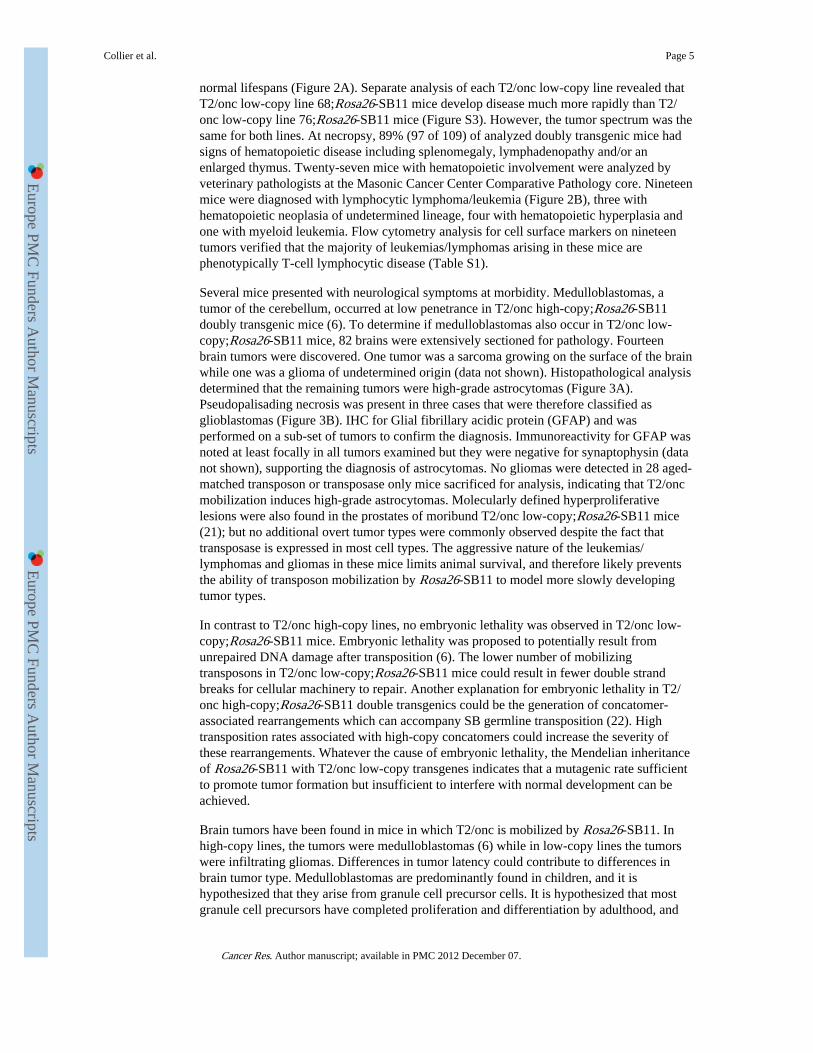
To investigate if transposase expression levels influence mutagenesis rates,immunohistochemistry (IHC) was performed to detect transposase in CAGGS-SB10 andRosa26-SB11 mice. Normal adult tissues were examined, as was a sarcoma from ap19Arf−/−;CAGGS-SB10;T2/onc low-copy mouse (5). Although transposase was detectedin the sarcoma, it was absent from most normal somatic tissues in CAGGS-SB10 mice(Figure 1 and Figure S2). When expression was detected, it occurred in a highly variegatedpattern (liver in Figure 1; kidney in Figure S2). In contrast, transposase was robustlyexpressed in the majority of cell types in Rosa26-SB11 mice (Figure 1 and Figure S2). Thelow level and variegated expression in CAGGS-SB10 mice is potentially due to epigeneticsilencing that is often observed in standard transgenics.
The presence of transposase in a p19Arf−/−;CAGGS-SB10;T2/onc sarcoma indicates thattransposase is expressed in these mice in an appropriate cell type to promotesarcomagenesis. It could be hypothesized that T2/onc high-copy;CAGGS-SB10 mice couldhave developed sarcomas on an otherwise wild-type genetic background due to theavailability of many T2/onc copies for mutagenesis. However, tumor formation was notobserved. In murine models, p19Arf is known to play a role in oncogene-inducedsenescence (18-20). Therefore, in sarcoma-initiating cells in p19Arf+/+ mice, T2/oncmutagenesis of cancer genes could promote Arf-mediated senescence, providing a block totumor formation. This experiment suggests that performing SB-screens in tumor-predisposed genetic backgrounds may be necessary for robust tumor formation in certaintissue types.
Combining T2/onc low-copy lines with Rosa26-SB11 does not cause embryonic lethalitybut promotes tumor formation in otherwise wild-type mice
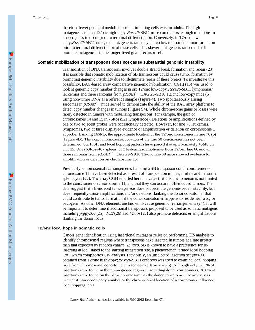
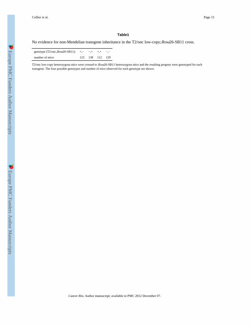
To determine if mobilization from low-copy lines is sufficient for tumor formation, two T2/onc low-copy lines (lines 68 and 76 (5)) were crossed to Rosa26-SB11. Chi square analysisof the resulting progeny (Table 1) revealed no evidence for non-Mendelian inheritance ofthe transgenes (p= 0.4153, 3 degrees of freedom). A large cohort of doubly transgenic miceand single transgenic littermate controls were therefore generated. T2/onc low-copy;Rosa26-SB11 mice became moribund with an average latency of 187 days while controls had
Collier et al. Page 4
Cancer Res. Author manuscript; available in PMC 2012 December 07.
Europe PM
C Funders A
uthor Manuscripts
Europe PM
C Funders A
uthor Manuscripts
normal lifespans (Figure 2A). Separate analysis of each T2/onc low-copy line revealed thatT2/onc low-copy line 68;Rosa26-SB11 mice develop disease much more rapidly than T2/onc low-copy line 76;Rosa26-SB11 mice (Figure S3). However, the tumor spectrum was thesame for both lines. At necropsy, 89% (97 of 109) of analyzed doubly transgenic mice hadsigns of hematopoietic disease including splenomegaly, lymphadenopathy and/or anenlarged thymus. Twenty-seven mice with hematopoietic involvement were analyzed byveterinary pathologists at the Masonic Cancer Center Comparative Pathology core. Nineteenmice were diagnosed with lymphocytic lymphoma/leukemia (Figure 2B), three withhematopoietic neoplasia of undetermined lineage, four with hematopoietic hyperplasia andone with myeloid leukemia. Flow cytometry analysis for cell surface markers on nineteentumors verified that the majority of leukemias/lymphomas arising in these mice arephenotypically T-cell lymphocytic disease (Table S1).
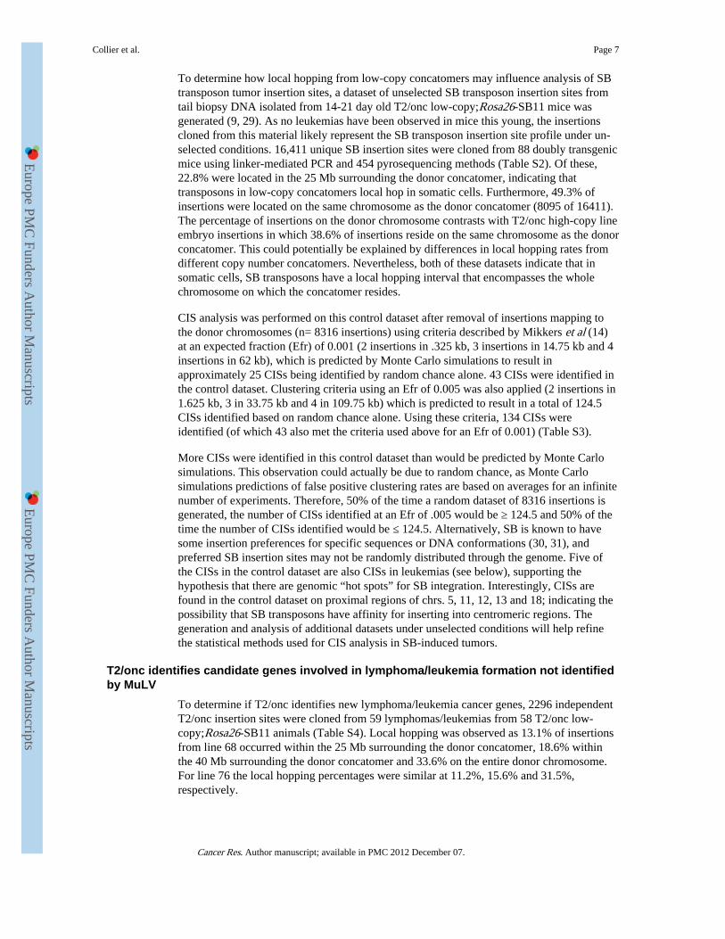
Several mice presented with neurological symptoms at morbidity. Medulloblastomas, atumor of the cerebellum, occurred at low penetrance in T2/onc high-copy;Rosa26-SB11doubly transgenic mice (6). To determine if medulloblastomas also occur in T2/onc low-copy;Rosa26-SB11 mice, 82 brains were extensively sectioned for pathology. Fourteenbrain tumors were discovered. One tumor was a sarcoma growing on the surface of the brainwhile one was a glioma of undetermined origin (data not shown). Histopathological analysisdetermined that the remaining tumors were high-grade astrocytomas (Figure 3A).Pseudopalisading necrosis was present in three cases that were therefore classified asglioblastomas (Figure 3B). IHC for Glial fibrillary acidic protein (GFAP) and wasperformed on a sub-set of tumors to confirm the diagnosis. Immunoreactivity for GFAP wasnoted at least focally in all tumors examined but they were negative for synaptophysin (datanot shown), supporting the diagnosis of astrocytomas. No gliomas were detected in 28 aged-matched transposon or transposase only mice sacrificed for analysis, indicating that T2/oncmobilization induces high-grade astrocytomas. Molecularly defined hyperproliferativelesions were also found in the prostates of moribund T2/onc low-copy;Rosa26-SB11 mice(21); but no additional overt tumor types were commonly observed despite the fact thattransposase is expressed in most cell types. The aggressive nature of the leukemias/lymphomas and gliomas in these mice limits animal survival, and therefore likely preventsthe ability of transposon mobilization by Rosa26-SB11 to model more slowly developingtumor types.
In contrast to T2/onc high-copy lines, no embryonic lethality was observed in T2/onc low-copy;Rosa26-SB11 mice. Embryonic lethality was proposed to potentially result fromunrepaired DNA damage after transposition (6). The lower number of mobilizingtransposons in T2/onc low-copy;Rosa26-SB11 mice could result in fewer double strandbreaks for cellular machinery to repair. Another explanation for embryonic lethality in T2/onc high-copy;Rosa26-SB11 double transgenics could be the generation of concatomer-associated rearrangements which can accompany SB germline transposition (22). Hightransposition rates associated with high-copy concatomers could increase the severity ofthese rearrangements. Whatever the cause of embryonic lethality, the Mendelian inheritanceof Rosa26-SB11 with T2/onc low-copy transgenes indicates that a mutagenic rate sufficientto promote tumor formation but insufficient to interfere with normal development can beachieved.
Brain tumors have been found in mice in which T2/onc is mobilized by Rosa26-SB11. Inhigh-copy lines, the tumors were medulloblastomas (6) while in low-copy lines the tumorswere infiltrating gliomas. Differences in tumor latency could contribute to differences inbrain tumor type. Medulloblastomas are predominantly found in children, and it ishypothesized that they arise from granule cell precursor cells. It is hypothesized that mostgranule cell precursors have completed proliferation and differentiation by adulthood, and
Collier et al. Page 5
Cancer Res. Author manuscript; available in PMC 2012 December 07.
Europe PM
C Funders A
uthor Manuscripts
Europe PM
C Funders A
uthor Manuscripts
therefore fewer potential medulloblastoma-initiating cells exist in adults. The highmutagenesis rate in T2/onc high-copy;Rosa26-SB11 mice could allow enough mutations incancer genes to occur prior to terminal differentiation. Conversely, in T2/onc low-copy;Rosa26-SB11 mice, the mutagenesis rate may be too low to promote tumor formationprior to terminal differentiation of these cells. This slower mutagenesis rate could stillpromote mutagenesis in the longer-lived glial precursor cell.
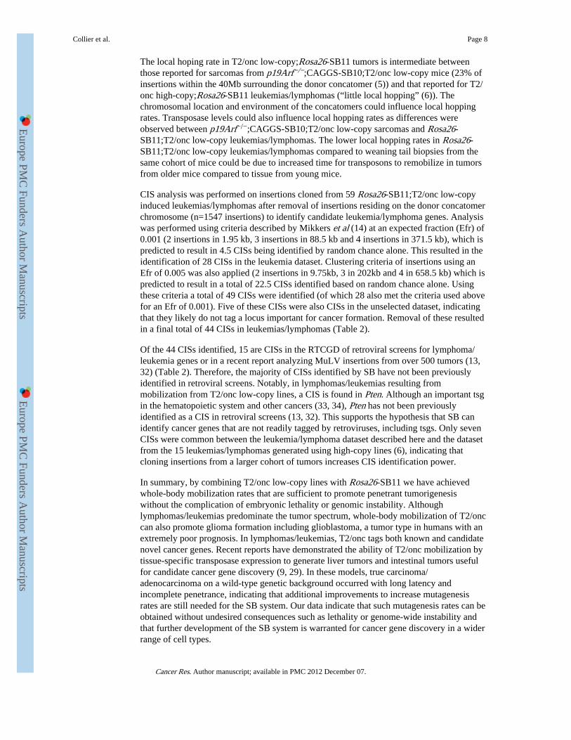
Somatic mobilization of transposons does not cause substantial genomic instabilityTransposition of DNA transposons involves double strand break formation and repair (23).It is possible that somatic mobilization of SB transposons could cause tumor formation bypromoting genomic instability due to illegitimate repair of these breaks. To investigate thispossibility, BAC-based array comparative genomic hybridization (CGH) (16) was used tolook at genomic copy number changes in six T2/onc low-copy;Rosa26-SB11 lymphomas/leukemias and three sarcomas from p19Arf−/−;CAGGS-SB10;T2/onc low-copy mice (5)using non-tumor DNA as a reference sample (Figure 4). Two spontaneously arisingsarcomas in p19Arf−/− mice served to demonstrate the ability of the BAC array platform todetect copy number changes in tumors (Figure S4). Whole chromosome gains or losses wererarely detected in tumors with mobilizing transposons (for example, the gain ofchromosomes 14 and 15 in 76Rosa521 lymph node). Deletions or amplifications defined byone or two adjacent probes were occasionally detected. However, for line 76 leukemias/lymphomas, two of three displayed evidence of amplification or deletion on chromosome 1at probes flanking 166Mb, the approximate location of the T2/onc concatomer in line 76 (5)(Figure 4B). The exact chromosomal location of the line 68 concatomer has not beendetermined, but FISH and local hopping patterns have placed it at approximately 45Mb onchr. 15. One (68Rosa467 spleen) of 3 leukemias/lymphomas from T2/onc line 68 and allthree sarcomas from p19Arf−/−;CAGGS-SB10;T2/onc line 68 mice showed evidence foramplification or deletion on chromosome 15.
Previously, chromosomal rearrangements flanking a SB transposon donor concatomer onchromosome 11 have been detected as a result of transposition in the germline and in normalsplenocytes (22). The array CGH reported here indicates that this phenomenon is not limitedto the concatomer on chromosome 11, and that they can occur in SB-induced tumors. Thedata suggest that SB-induced tumorigenesis does not promote genome-wide instability, butdoes frequently cause amplifications and/or deletions flanking the donor concatomer thatcould contribute to tumor formation if the donor concatomer happens to reside near a tsg oroncogene. As other DNA elements are known to cause genomic rearrangements (24), it willbe important to determine if additional transposons proposed to be used as somatic mutagensincluding piggyBac (25), Tol2 (26) and Minos (27) also promote deletions or amplificationsflanking the donor locus.
T2/onc local hops in somatic cellsCancer gene identification using insertional mutagens relies on performing CIS analysis toidentify chromosomal regions where transposons have inserted in tumors at a rate greaterthan that expected by random chance. In vivo, SB is known to have a preference for re-inserting at loci linked to the starting integration site, a phenomenon termed local hopping(28), which complicates CIS analysis. Previously, an unselected insertion set (n=490)obtained from T2/onc high-copy;Rosa26-SB11 embryos was used to examine local hoppingrates from chromosomal concatomers in somatic cells in vivo (6). Although only 6-11% ofinsertions were found in the 25-megabase region surrounding donor concatomers, 38.6% ofinsertions were found on the same chromosome as the donor concatomer. However, it isunclear if transposon copy number or the chromosomal location of a concatomer influenceslocal hopping rates.
Collier et al. Page 6
Cancer Res. Author manuscript; available in PMC 2012 December 07.
Europe PM
C Funders A
uthor Manuscripts
Europe PM
C Funders A
uthor Manuscripts
To determine how local hopping from low-copy concatomers may influence analysis of SBtransposon tumor insertion sites, a dataset of unselected SB transposon insertion sites fromtail biopsy DNA isolated from 14-21 day old T2/onc low-copy;Rosa26-SB11 mice wasgenerated (9, 29). As no leukemias have been observed in mice this young, the insertionscloned from this material likely represent the SB transposon insertion site profile under un-selected conditions. 16,411 unique SB insertion sites were cloned from 88 doubly transgenicmice using linker-mediated PCR and 454 pyrosequencing methods (Table S2). Of these,22.8% were located in the 25 Mb surrounding the donor concatomer, indicating thattransposons in low-copy concatomers local hop in somatic cells. Furthermore, 49.3% ofinsertions were located on the same chromosome as the donor concatomer (8095 of 16411).The percentage of insertions on the donor chromosome contrasts with T2/onc high-copy lineembryo insertions in which 38.6% of insertions reside on the same chromosome as the donorconcatomer. This could potentially be explained by differences in local hopping rates fromdifferent copy number concatomers. Nevertheless, both of these datasets indicate that insomatic cells, SB transposons have a local hopping interval that encompasses the wholechromosome on which the concatomer resides.
CIS analysis was performed on this control dataset after removal of insertions mapping tothe donor chromosomes (n= 8316 insertions) using criteria described by Mikkers et al (14)at an expected fraction (Efr) of 0.001 (2 insertions in .325 kb, 3 insertions in 14.75 kb and 4insertions in 62 kb), which is predicted by Monte Carlo simulations to result inapproximately 25 CISs being identified by random chance alone. 43 CISs were identified inthe control dataset. Clustering criteria using an Efr of 0.005 was also applied (2 insertions in1.625 kb, 3 in 33.75 kb and 4 in 109.75 kb) which is predicted to result in a total of 124.5CISs identified based on random chance alone. Using these criteria, 134 CISs wereidentified (of which 43 also met the criteria used above for an Efr of 0.001) (Table S3).
More CISs were identified in this control dataset than would be predicted by Monte Carlosimulations. This observation could actually be due to random chance, as Monte Carlosimulations predictions of false positive clustering rates are based on averages for an infinitenumber of experiments. Therefore, 50% of the time a random dataset of 8316 insertions isgenerated, the number of CISs identified at an Efr of .005 would be ≥ 124.5 and 50% of thetime the number of CISs identified would be ≤ 124.5. Alternatively, SB is known to havesome insertion preferences for specific sequences or DNA conformations (30, 31), andpreferred SB insertion sites may not be randomly distributed through the genome. Five ofthe CISs in the control dataset are also CISs in leukemias (see below), supporting thehypothesis that there are genomic “hot spots” for SB integration. Interestingly, CISs arefound in the control dataset on proximal regions of chrs. 5, 11, 12, 13 and 18; indicating thepossibility that SB transposons have affinity for inserting into centromeric regions. Thegeneration and analysis of additional datasets under unselected conditions will help refinethe statistical methods used for CIS analysis in SB-induced tumors.
T2/onc identifies candidate genes involved in lymphoma/leukemia formation not identifiedby MuLV
To determine if T2/onc identifies new lymphoma/leukemia cancer genes, 2296 independentT2/onc insertion sites were cloned from 59 lymphomas/leukemias from 58 T2/onc low-copy;Rosa26-SB11 animals (Table S4). Local hopping was observed as 13.1% of insertionsfrom line 68 occurred within the 25 Mb surrounding the donor concatomer, 18.6% withinthe 40 Mb surrounding the donor concatomer and 33.6% on the entire donor chromosome.For line 76 the local hopping percentages were similar at 11.2%, 15.6% and 31.5%,respectively.
Collier et al. Page 7
Cancer Res. Author manuscript; available in PMC 2012 December 07.
Europe PM
C Funders A
uthor Manuscripts
Europe PM
C Funders A
uthor Manuscripts
The local hoping rate in T2/onc low-copy;Rosa26-SB11 tumors is intermediate betweenthose reported for sarcomas from p19Arf−/−;CAGGS-SB10;T2/onc low-copy mice (23% ofinsertions within the 40Mb surrounding the donor concatomer (5)) and that reported for T2/onc high-copy;Rosa26-SB11 leukemias/lymphomas (“little local hopping” (6)). Thechromosomal location and environment of the concatomers could influence local hoppingrates. Transposase levels could also influence local hopping rates as differences wereobserved between p19Arf−/−;CAGGS-SB10;T2/onc low-copy sarcomas and Rosa26-SB11;T2/onc low-copy leukemias/lymphomas. The lower local hopping rates in Rosa26-SB11;T2/onc low-copy leukemias/lymphomas compared to weaning tail biopsies from thesame cohort of mice could be due to increased time for transposons to remobilize in tumorsfrom older mice compared to tissue from young mice.
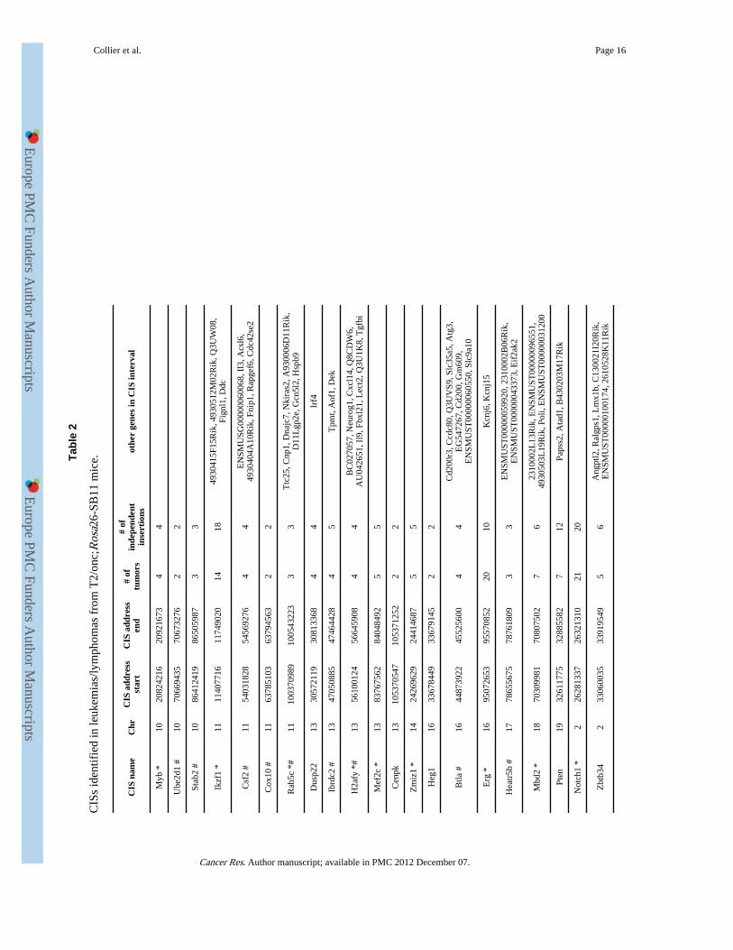
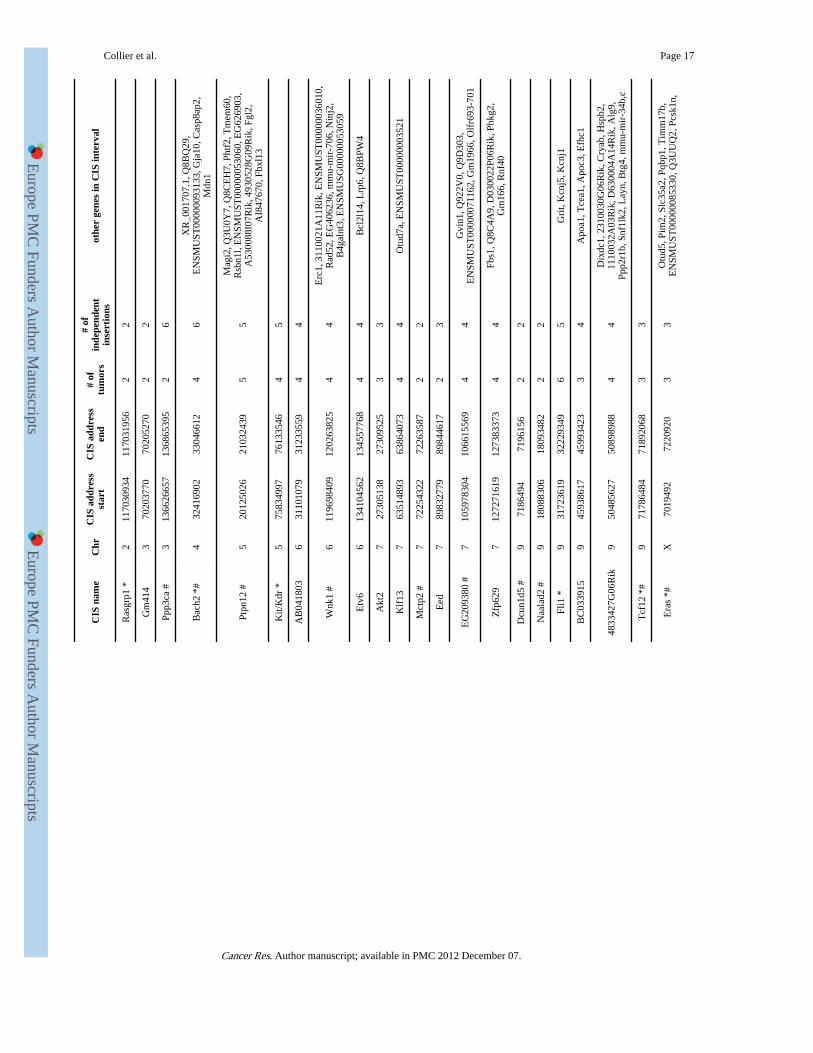
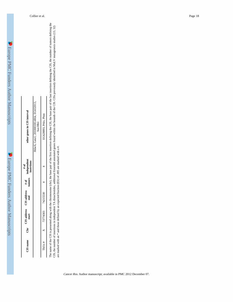
CIS analysis was performed on insertions cloned from 59 Rosa26-SB11;T2/onc low-copyinduced leukemias/lymphomas after removal of insertions residing on the donor concatomerchromosome (n=1547 insertions) to identify candidate leukemia/lymphoma genes. Analysiswas performed using criteria described by Mikkers et al (14) at an expected fraction (Efr) of0.001 (2 insertions in 1.95 kb, 3 insertions in 88.5 kb and 4 insertions in 371.5 kb), which ispredicted to result in 4.5 CISs being identified by random chance alone. This resulted in theidentification of 28 CISs in the leukemia dataset. Clustering criteria of insertions using anEfr of 0.005 was also applied (2 insertions in 9.75kb, 3 in 202kb and 4 in 658.5 kb) which ispredicted to result in a total of 22.5 CISs identified based on random chance alone. Usingthese criteria a total of 49 CISs were identified (of which 28 also met the criteria used abovefor an Efr of 0.001). Five of these CISs were also CISs in the unselected dataset, indicatingthat they likely do not tag a locus important for cancer formation. Removal of these resultedin a final total of 44 CISs in leukemias/lymphomas (Table 2).
Of the 44 CISs identified, 15 are CISs in the RTCGD of retroviral screens for lymphoma/leukemia genes or in a recent report analyzing MuLV insertions from over 500 tumors (13,32) (Table 2). Therefore, the majority of CISs identified by SB have not been previouslyidentified in retroviral screens. Notably, in lymphomas/leukemias resulting frommobilization from T2/onc low-copy lines, a CIS is found in Pten. Although an important tsgin the hematopoietic system and other cancers (33, 34), Pten has not been previouslyidentified as a CIS in retroviral screens (13, 32). This supports the hypothesis that SB canidentify cancer genes that are not readily tagged by retroviruses, including tsgs. Only sevenCISs were common between the leukemia/lymphoma dataset described here and the datasetfrom the 15 leukemias/lymphomas generated using high-copy lines (6), indicating thatcloning insertions from a larger cohort of tumors increases CIS identification power.
In summary, by combining T2/onc low-copy lines with Rosa26-SB11 we have achievedwhole-body mobilization rates that are sufficient to promote penetrant tumorigenesiswithout the complication of embryonic lethality or genomic instability. Althoughlymphomas/leukemias predominate the tumor spectrum, whole-body mobilization of T2/onccan also promote glioma formation including glioblastoma, a tumor type in humans with anextremely poor prognosis. In lymphomas/leukemias, T2/onc tags both known and candidatenovel cancer genes. Recent reports have demonstrated the ability of T2/onc mobilization bytissue-specific transposase expression to generate liver tumors and intestinal tumors usefulfor candidate cancer gene discovery (9, 29). In these models, true carcinoma/adenocarcinoma on a wild-type genetic background occurred with long latency andincomplete penetrance, indicating that additional improvements to increase mutagenesisrates are still needed for the SB system. Our data indicate that such mutagenesis rates can beobtained without undesired consequences such as lethality or genome-wide instability andthat further development of the SB system is warranted for cancer gene discovery in a widerrange of cell types.
Collier et al. Page 8
Cancer Res. Author manuscript; available in PMC 2012 December 07.
Europe PM
C Funders A
uthor Manuscripts
Europe PM
C Funders A
uthor Manuscripts
Supplementary MaterialRefer to Web version on PubMed Central for supplementary material.
AcknowledgmentsWe thank Erin Riley for technical assistance, Paul Marker and Michael Taylor for critical reading of the manuscriptand the members of the Center for Genome Engineering for many helpful discussions.
Funding: K01CA122183 and an American Cancer Society pre-doctoral fellowship (LSC), MN Dept ofEmployment and Economic Development SPAP-05-0013-P-FY06 (DAL and RBJ), R01CA113636-01A1 (DAL),R01NS055750 (WAW), Cancer Research-UK and the Wellcome Trust (DJA).
References1. Uren AG, Kool J, Berns A, van Lohuizen M. Retroviral insertional mutagenesis: past, present and
future. Oncogene. 2005; 24:7656–72. [PubMed: 16299527]
2. Collier LS, Largaespada DA. Transforming science: cancer gene identification. Curr Opin GenetDev. 2006; 16:23–9. [PubMed: 16326095]
3. Callahan R, Smith GH. MMTV-induced mammary tumorigenesis: gene discovery, progression tomalignancy and cellular pathways. Oncogene. 2000; 19:992–1001. [PubMed: 10713682]
4. Ivics Z, Hackett PB, Plasterk RH, Izsvak Z. Molecular reconstruction of Sleeping Beauty, a Tc1-liketransposon from fish, and its transposition in human cells. Cell. 1997; 91:501–10. [PubMed:9390559]
5. Collier LS, Carlson CM, Ravimohan S, Dupuy AJ, Largaespada DA. Cancer gene discovery in solidtumours using transposon-based somatic mutagenesis in the mouse. Nature. 2005; 436:272–6.[PubMed: 16015333]
6. Dupuy AJ, Akagi K, Largaespada DA, Copeland NG, Jenkins NA. Mammalian mutagenesis using ahighly mobile somatic Sleeping Beauty transposon system. Nature. 2005; 436:221–6. [PubMed:16015321]
7. Okabe M, Ikawa M, Kominami K, Nakanishi T, Nishimune Y. ‘Green mice’ as a source ofubiquitous green cells. FEBS Lett. 1997; 407:313–9. [PubMed: 9175875]
8. Dupuy AJ, Fritz S, Largaespada DA. Transposition and gene disruption in the male germline of themouse. Genesis. 2001; 30:82–8. [PubMed: 11416868]
9. Starr TK, Allaei R, Silverstein KA, et al. A transposon-based genetic screen in mice identifies genesaltered in colorectal cancer. Science. 2009; 323:1747–50. [PubMed: 19251594]
10. Kim WI, Matise I, Diers MD, Largaespada DA. RAS oncogene suppression induces apoptosisfollowed by more differentiated and less myelosuppressive disease upon relapse of acute myeloidleukemia. Blood. 2009; 113:1086–96. [PubMed: 18952898]
11. Kim WI, Wiesner SM, Largaespada DA. Vav promoter-tTA conditional transgene expressionsystem for hematopoietic cells drives high level expression in developing B and T cells. ExpHematol. 2007; 35:1231–9. [PubMed: 17560009]
12. Largaespada DA, Collier LS. Transposon-mediated mutagenesis in somatic cells: identification oftransposon-genomic DNA junctions. Methods Mol Biol. 2008; 435:95–108. [PubMed: 18370070]
13. Akagi K, Suzuki T, Stephens RM, Jenkins NA, Copeland NG. RTCGD: retroviral tagged cancergene database. Nucleic Acids Res. 2004; 32:D523–7. [PubMed: 14681473]
14. Mikkers H, Allen J, Knipscheer P, et al. High-throughput retroviral tagging to identify componentsof specific signaling pathways in cancer. Nat Genet. 2002; 32:153–9. [PubMed: 12185366]
15. Hackett CS, Hodgson JG, Law ME, et al. Genome-wide array CGH analysis of murineneuroblastoma reveals distinct genomic aberrations which parallel those in human tumors. CancerRes. 2003; 63:5266–73. [PubMed: 14500357]
16. Hodgson JG, Malek T, Bornstein S, et al. Copy number aberrations in mouse breast tumors revealloci and genes important in tumorigenic receptor tyrosine kinase signaling. Cancer Res. 2005;65:9695–704. [PubMed: 16266989]
Collier et al. Page 9
Cancer Res. Author manuscript; available in PMC 2012 December 07.
Europe PM
C Funders A
uthor Manuscripts
Europe PM
C Funders A
uthor Manuscripts
17. Jain AN, Tokuyasu TA, Snijders AM, Segraves R, Albertson DG, Pinkel D. Fully automaticquantification of microarray image data. Genome Res. 2002; 12:325–32. [PubMed: 11827952]
18. de Stanchina E, McCurrach ME, Zindy F, et al. E1A signaling to p53 involves the p19(ARF) tumorsuppressor. Genes Dev. 1998; 12:2434–42. [PubMed: 9694807]
19. Zindy F, Eischen CM, Randle DH, et al. Myc signaling via the ARF tumor suppressor regulatesp53-dependent apoptosis and immortalization. Genes Dev. 1998; 12:2424–33. [PubMed: 9694806]
20. Palmero I, Pantoja C, Serrano M. p19ARF links the tumour suppressor p53 to Ras. Nature. 1998;395:125–6. [PubMed: 9744268]
21. Rahrmann EP, Collier LS, Knutson TP, et al. Identification of PDE4D as a proliferation promotingfactor in prostate cancer using a Sleeping Beauty transposon-based somatic mutagenesis screen.Cancer Res. 2009; 69:4388–97. [PubMed: 19401450]
22. Geurts AM, Collier LS, Geurts JL, et al. Gene Mutations and Genomic Rearrangements in theMouse as a Result of Transposon Mobilization from Chromosomal Concatemers. PLoS Genet.2006:2.
23. van Luenen HG, Colloms SD, Plasterk RH. The mechanism of transposition of Tc3 in C. elegans.Cell. 1994; 79:293–301. [PubMed: 7954797]
24. Gray YH. It takes two transposons to tango: transposable-element-mediated chromosomalrearrangements. Trends Genet. 2000; 16:461–8. [PubMed: 11050333]
25. Ding S, Wu X, Li G, Han M, Zhuang Y, Xu T. Efficient transposition of the piggyBac (PB)transposon in mammalian cells and mice. Cell. 2005; 122:473–83. [PubMed: 16096065]
26. Balciunas D, Wangensteen KJ, Wilber A, et al. Harnessing a High Cargo-Capacity Transposon forGenetic Applications in Vertebrates. PLoS Genetics. 2006 In press.
27. Zagoraiou L, Drabek D, Alexaki S, et al. In vivo transposition of Minos, a Drosophila mobileelement, in mammalian tissues. Proc Natl Acad Sci U S A. 2001; 98:11474–8. [PubMed:11562481]
28. Luo G, Ivics Z, Izsvak Z, Bradley A. Chromosomal transposition of a Tc1/mariner-like element inmouse embryonic stem cells. Proc Natl Acad Sci U S A. 1998; 95:10769–73. [PubMed: 9724779]
29. Keng VW, Villanueva A, Chiang DY, et al. A conditional transposon-based insertionalmutagenesis screen for genes associated with mouse hepatocellular carcinoma. Nat Biotechnol.2009; 27:264–74. [PubMed: 19234449]
30. Liu G, Geurts AM, Yae K, et al. Target-site preferences of Sleeping Beauty transposons. J MolBiol. 2005; 346:161–73. [PubMed: 15663935]
31. Geurts AM, Hackett CS, Bell JB, et al. Structure-based prediction of insertion-site preferences oftransposons into chromosomes. Nucleic Acids Res. 2006; 34:2803–11. [PubMed: 16717285]
32. Uren AG, Kool J, Matentzoglu K, et al. Large-scale mutagenesis in p19(ARF)- and p53-deficientmice identifies cancer genes and their collaborative networks. Cell. 2008; 133:727–41. [PubMed:18485879]
33. Chow LM, Baker SJ. PTEN function in normal and neoplastic growth. Cancer Lett. 2006;241:184–96. [PubMed: 16412571]
34. Maser RS, Choudhury B, Campbell PJ, et al. Chromosomally unstable mouse tumours havegenomic alterations similar to diverse human cancers. Nature. 2007; 447:966–71. [PubMed:17515920]
Collier et al. Page 10
Cancer Res. Author manuscript; available in PMC 2012 December 07.
Europe PM
C Funders A
uthor Manuscripts
Europe PM
C Funders A
uthor Manuscripts
Figure 1. IHC reveals transposase expression in transgenic miceTransposase is poorly expressed in somatic tissues in CAGGS-SB10 mice (liver shown),while a sarcoma from a p19Arf−/−;CAGGS-SB10;T2/onc low-copy mouse contains manytransposase expressing cells. Most cells in Rosa26-SB11 mice stain positive for transposase(liver shown). Brown indicates antibody staining, nuclei are counter-stained blue. A liverfrom a non-transposase transgenic mouse demonstrates antibody specificity. Scale bar=50microns.
Collier et al. Page 11
Cancer Res. Author manuscript; available in PMC 2012 December 07.
Europe PM
C Funders A
uthor Manuscripts
Europe PM
C Funders A
uthor Manuscripts
Figure 2. Rosa26-SB11; T2/onc low-copy mice are tumor proneA) Kaplan-Meier survival curve for Rosa26-SB11;T2/onc low-copy (SB;T2, triangles),Rosa26-SB11 (SB, squares), and T2/onc low-copy (T2, circles) mice. Rosa26-SB11;T2/onclow-copy mice become moribund more rapidly than controls (p<.001, Logrank test). B)Hematoxylin and Eosin (H&E) stained example of a lymphocytic leukemia/lymphoma froma Rosa26-SB11;T2/onc low-copy mouse. Scale bar=50 microns.
Collier et al. Page 12
Cancer Res. Author manuscript; available in PMC 2012 December 07.
Europe PM
C Funders A
uthor Manuscripts
Europe PM
C Funders A
uthor Manuscripts
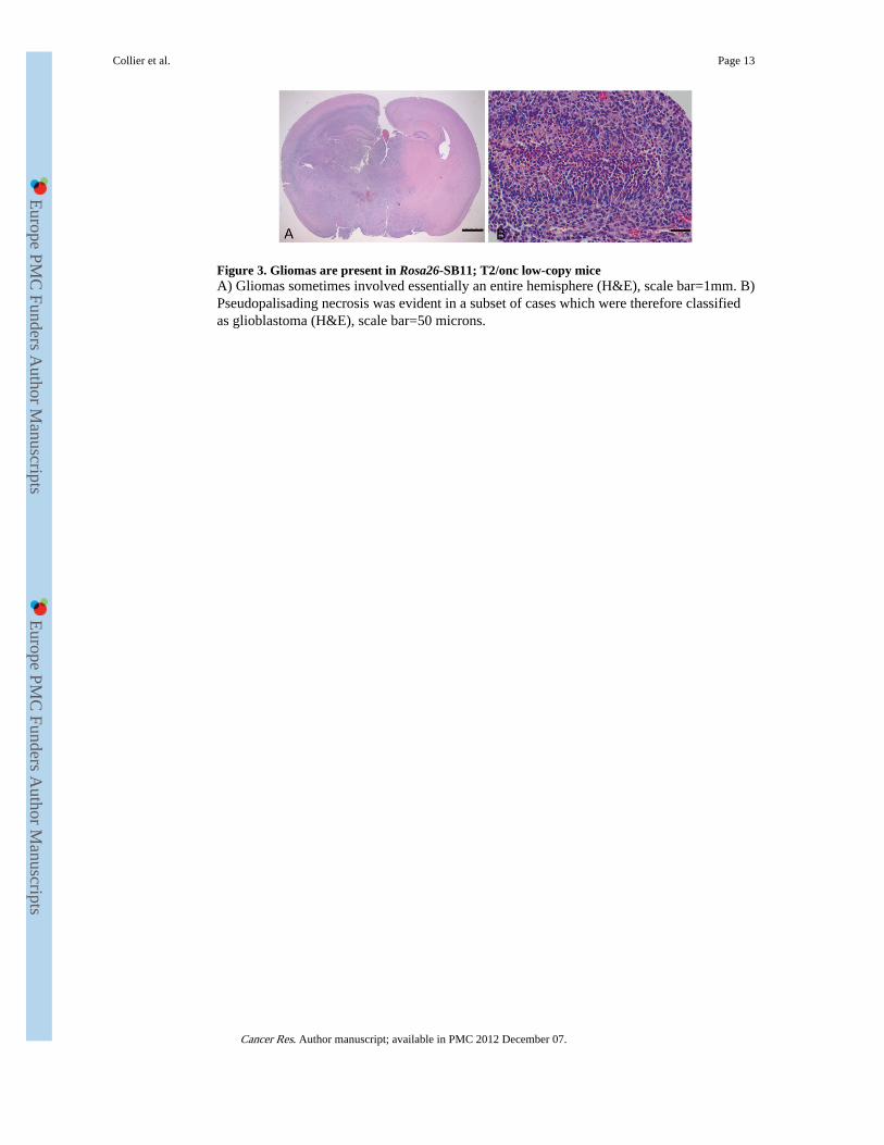
Figure 3. Gliomas are present in Rosa26-SB11; T2/onc low-copy miceA) Gliomas sometimes involved essentially an entire hemisphere (H&E), scale bar=1mm. B)Pseudopalisading necrosis was evident in a subset of cases which were therefore classifiedas glioblastoma (H&E), scale bar=50 microns.
Collier et al. Page 13
Cancer Res. Author manuscript; available in PMC 2012 December 07.
Europe PM
C Funders A
uthor Manuscripts
Europe PM
C Funders A
uthor Manuscripts
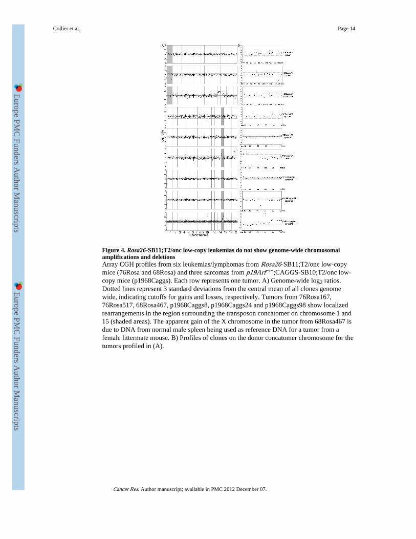
Figure 4. Rosa26-SB11;T2/onc low-copy leukemias do not show genome-wide chromosomalamplifications and deletionsArray CGH profiles from six leukemias/lymphomas from Rosa26-SB11;T2/onc low-copymice (76Rosa and 68Rosa) and three sarcomas from p19Arf−/−;CAGGS-SB10;T2/onc low-copy mice (p1968Caggs). Each row represents one tumor. A) Genome-wide log2 ratios.Dotted lines represent 3 standard deviations from the central mean of all clones genomewide, indicating cutoffs for gains and losses, respectively. Tumors from 76Rosa167,76Rosa517, 68Rosa467, p1968Caggs8, p1968Caggs24 and p1968Caggs98 show localizedrearrangements in the region surrounding the transposon concatomer on chromosome 1 and15 (shaded areas). The apparent gain of the X chromosome in the tumor from 68Rosa467 isdue to DNA from normal male spleen being used as reference DNA for a tumor from afemale littermate mouse. B) Profiles of clones on the donor concatomer chromosome for thetumors profiled in (A).
Collier et al. Page 14
Cancer Res. Author manuscript; available in PMC 2012 December 07.
Europe PM
C Funders A
uthor Manuscripts
Europe PM
C Funders A
uthor Manuscripts
Europe PM
C Funders A
uthor Manuscripts
Europe PM
C Funders A
uthor Manuscripts
Collier et al. Page 15
Table1
No evidence for non-Mendelian transgene inheritance in the T2/onc low-copy;Rosa26-SB11 cross.
genotype (T2/onc,Rosa26-SB11): +,− −,+ +,+ −,−
number of mice: 123 138 112 129
T2/onc low-copy heterozygous mice were crossed to Rosa26-SB11 heterozygous mice and the resulting progeny were genotyped for eachtransgene. The four possible genotypes and number of mice observed for each genotype are shown.
Cancer Res. Author manuscript; available in PMC 2012 December 07.
Europe PM
C Funders A
uthor Manuscripts
Europe PM
C Funders A
uthor Manuscripts
Collier et al. Page 16
Tabl
e 2
CIS
s id
entif
ied
in le
ukem
ias/
lym
phom
as f
rom
T2/
onc;
Ros
a26-
SB11
mic
e.
CIS
nam
eC
hrC
IS a
ddre
ssst
art
CIS
add
ress
end
# of
tum
ors
# of
inde
pend
ent
inse
rtio
nsot
her
gene
s in
CIS
inte
rval
Myb
*10
2082
4216
2092
1673
44
Ube
2d1
#10
7066
9435
7067
3276
22
Stab
2 #
1086
4124
1986
5059
873
3
Ikzf
1 *
1111
4077
1611
7490
2014
1849
3041
5F15
Rik
, 493
0512
M02
Rik
, Q3U
W08
,Fi
gnl1
, Ddc
Csf
2 #
1154
0318
2854
5692
764
4E
NSM
USG
0000
0060
068,
Il3
, Acs
l6,
4930
404A
10R
ik, F
nip1
, Rap
gef6
, Cdc
42se
2
Cox
10 #
1163
7851
0363
7945
632
2
Rab
5c *
#11
1003
7098
910
0543
223
33
Ttc
25, C
np1,
Dna
jc7,
Nki
ras2
, A93
0006
D11
Rik
,D
11L
gp2e
, Gcn
5l2,
Hsp
b9
Dus
p22
1330
5721
1930
8133
684
4Ir
f4
Ibrd
c2 #
1347
0508
8547
4644
284
5T
pmt,
Aof
1, D
ek
H2a
fy *
#13
5610
0124
5664
5908
44
BC
0270
57, N
euro
g1, C
xcl1
4, Q
8CD
W6,
AU
0426
51, I
l9, F
bxl2
1, L
ect2
, Q3U
1K8,
Tgf
bi
Mef
2c *
1383
7675
6284
0484
925
5
Cen
pk13
1053
7054
710
5371
252
22
Zm
iz1
*14
2426
9629
2441
4687
55
Heg
116
3367
8449
3367
9145
22
Btla
#16
4487
3922
4552
5600
44
Cd2
00r3
, Ccd
c80,
Q3U
VS9
, Slc
35a5
, Atg
3,E
G54
7267
, Cd2
00, G
m60
9,E
NSM
UST
0000
0060
550,
Slc
9a10
Erg
*16
9507
2653
9557
0852
2010
Kcn
j6, K
cnj1
5
Hea
tr5b
#17
7865
5675
7876
1809
33
EN
SMU
ST00
0000
5992
0, 2
3100
02B
06R
ik,
EN
SMU
ST00
0000
4337
3, E
if2a
k2
Mbd
2 *
1870
3099
8170
8075
027
623
1000
2L13
Rik
, EN
SMU
ST00
0000
9655
1,49
3050
3L19
Rik
, Pol
i, E
NSM
UST
0000
0031
200
Pten
1932
6117
7532
8855
827
12Pa
pss2
, Ata
d1, B
4302
03M
17R
ik
Not
ch1
*2
2628
1337
2632
1310
2120
Zbt
b34
233
0600
3533
9195
495
6A
ngpt
l2, R
algp
s1, L
mx1
b, C
1300
21I2
0Rik
,E
NSM
UST
0000
0100
174,
261
0528
K11
Rik
Cancer Res. Author manuscript; available in PMC 2012 December 07.
Europe PM
C Funders A
uthor Manuscripts
Europe PM
C Funders A
uthor Manuscripts
Collier et al. Page 17
CIS
nam
eC
hrC
IS a
ddre
ssst
art
CIS
add
ress
end
# of
tum
ors
# of
inde
pend
ent
inse
rtio
nsot
her
gene
s in
CIS
inte
rval
Ras
grp1
*2
1170
3093
411
7031
956
22
Gm
414
370
2037
7070
2052
702
2
Ppp3
ca #
313
6626
657
1368
6539
52
6
Bac
h2 *
#4
3241
6902
3304
6612
46
XR
_001
707.
1, Q
8BQ
29,
EN
SMU
ST00
0000
9313
3, G
ja10
, Cas
p8ap
2,M
dn1
Ptpn
12 #
520
1250
2621
0324
395
5
Mag
i2, Q
3U0Y
7, Q
8CE
H7,
Pht
f2, T
mem
60,
Rsb
n1l,
EN
SMU
ST00
0000
5306
0, E
G62
6903
,A
5300
88I0
7Rik
, 493
0528
G09
Rik
, Fgl
2,A
I847
670,
Fbx
l13
Kit/
Kdr
*5
7583
4997
7613
3546
45
AB
0418
036
3110
1079
3123
3559
44
Wnk
1 #
611
9698
409
1202
6382
54
4E
rc1,
311
0021
A11
Rik
, EN
SMU
ST00
0000
3601
0,R
ad52
, EG
4062
36, m
mu-
mir
-706
, Nin
j2,
B4g
alnt
3, E
NSM
USG
0000
0053
059
Etv
66
1341
0456
213
4557
768
44
Bcl
2l14
, Lrp
6, Q
8BPW
4
Akt
27
2730
5138
2730
9525
33
Klf
137
6351
4893
6386
4073
44
Otu
d7a,
EN
SMU
ST00
0000
0352
1
Mct
p2 #
772
2543
2272
2635
872
2
Eed
789
8327
7989
8446
172
3
EG
2093
80 #
710
5978
304
1066
1556
94
4G
vin1
, Q92
2V0,
Q9D
303,
EN
SMU
ST00
0000
7116
2, G
m19
66, O
lfr6
93-7
01
Zfp
629
712
7271
619
1273
8337
34
4Fb
s1, Q
8C4A
9, D
0300
22P0
6Rik
, Phk
g2,
Gm
166,
Rnf
40
Dcu
n1d5
#9
7186
494
7196
156
22
Naa
lad2
#9
1808
8306
1809
3482
22
Fli1
*9
3172
3619
3222
9349
65
Gri
t, K
cnj5
, Kcn
j1
BC
0339
159
4593
8617
4599
3423
34
Apo
a1, T
cea1
, Apo
c3, E
fhc1
4833
427G
06R
ik9
5048
5627
5089
8988
44
Dix
dc1,
231
0030
G06
Rik
, Cry
ab, H
spb2
,11
1003
2A03
Rik
, D63
0004
A14
Rik
, Alg
9,Pp
p2r1
b, S
nf1l
k2, L
ayn,
Btg
4, m
mu-
mir
-34b
,c
Tcf
12 *
#9
7178
6484
7189
2068
33
Era
s *#
X70
1949
272
2092
03
3O
tud5
, Pim
2, S
lc35
a2, P
qbp1
, Tim
m17
b,E
NSM
UST
0000
0085
330,
Q3U
UQ
2, P
csk1
n,
Cancer Res. Author manuscript; available in PMC 2012 December 07.
Europe PM
C Funders A
uthor Manuscripts
Europe PM
C Funders A
uthor Manuscripts
Collier et al. Page 18
CIS
nam
eC
hrC
IS a
ddre
ssst
art
CIS
add
ress
end
# of
tum
ors
# of
inde
pend
ent
inse
rtio
nsot
her
gene
s in
CIS
inte
rval
Hda
c6, G
ata1
, 201
0001
H14
Rik
, EG
6320
13,
Suv3
9h1
Tbl
1x #
X73
7743
0374
2155
304
4E
G62
8893
, Prk
x, P
bsn
The
nam
e of
the
CIS
is p
rese
nted
alo
ng w
ith th
e ch
rom
osom
e (c
hr),
the
base
pai
r of
the
firs
t ins
ertio
n de
fini
ng th
e C
IS, t
he b
ase
pair
of
the
last
inse
rtio
n de
fini
ng th
e C
IS, t
he n
umbe
r of
tum
ors
defi
ning
the
CIS
, the
num
ber
of in
sert
ions
in in
depe
nden
t TA
din
ucle
otid
es, a
dditi
onal
Ens
embl
ann
otat
ed g
enes
fou
nd w
ithin
the
boun
ds o
f th
e C
IS. C
ISs
prev
ious
ly id
entif
ied
in M
uLV
mut
agen
esis
stu
dies
(13
, 32)
are
mar
ked
with
an
* an
d th
ose
defi
ned
by a
n ex
pect
ed f
ract
ion
(Efr
) of
.005
are
mar
ked
with
a #
.
Cancer Res. Author manuscript; available in PMC 2012 December 07.