welcome to bask 2015 - british association for surgery of the
TRANSCRIPT
Welcome to BASK 2015
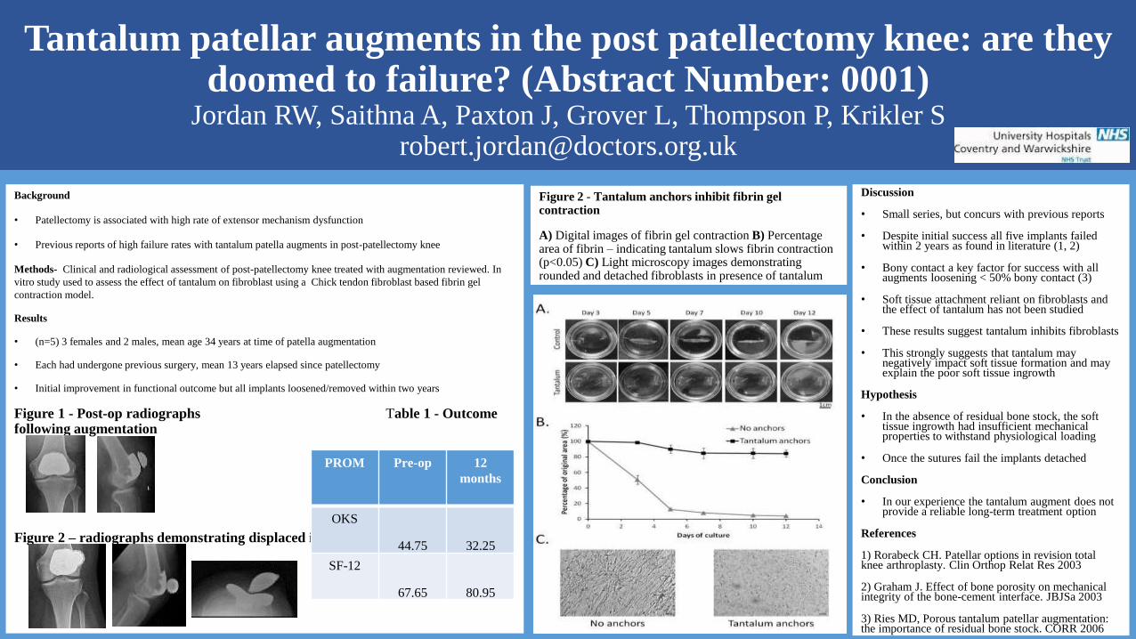
Tantalum patellar augments in the post patellectomy knee: are they doomed to failure? (Abstract Number: 0001)
Jordan RW, Saithna A, Paxton J, Grover L, Thompson P, Krikler S [email protected]
Background
• Patellectomy is associated with high rate of extensor mechanism dysfunction
• Previous reports of high failure rates with tantalum patella augments in post-patellectomy knee
Methods- Clinical and radiological assessment of post-patellectomy knee treated with augmentation reviewed. In
vitro study used to assess the effect of tantalum on fibroblast using a Chick tendon fibroblast based fibrin gel
contraction model.
Results
• (n=5) 3 females and 2 males, mean age 34 years at time of patella augmentation
• Each had undergone previous surgery, mean 13 years elapsed since patellectomy
• Initial improvement in functional outcome but all implants loosened/removed within two years
Figure 1 - Post-op radiographs Table 1 - Outcome following augmentation
Figure 2 – radiographs demonstrating displaced implant
PROM Pre-op 12
months
OKS
44.75 32.25
SF-12
67.65 80.95
Figure 2 - Tantalum anchors inhibit fibrin gel contraction
A) Digital images of fibrin gel contraction B) Percentage area of fibrin – indicating tantalum slows fibrin contraction (p<0.05) C) Light microscopy images demonstrating rounded and detached fibroblasts in presence of tantalum
Discussion
• Small series, but concurs with previous reports
• Despite initial success all five implants failed within 2 years as found in literature (1, 2)
• Bony contact a key factor for success with all augments loosening < 50% bony contact (3)
• Soft tissue attachment reliant on fibroblasts and the effect of tantalum has not been studied
• These results suggest tantalum inhibits fibroblasts
• This strongly suggests that tantalum may negatively impact soft tissue formation and may explain the poor soft tissue ingrowth
Hypothesis
• In the absence of residual bone stock, the soft tissue ingrowth had insufficient mechanical properties to withstand physiological loading
• Once the sutures fail the implants detached
Conclusion
• In our experience the tantalum augment does not provide a reliable long-term treatment option
References
1) Rorabeck CH. Patellar options in revision total knee arthroplasty. Clin Orthop Relat Res 2003
2) Graham J. Effect of bone porosity on mechanical integrity of the bone-cement interface. JBJSa 2003
3) Ries MD, Porous tantalum patellar augmentation: the importance of residual bone stock. CORR 2006
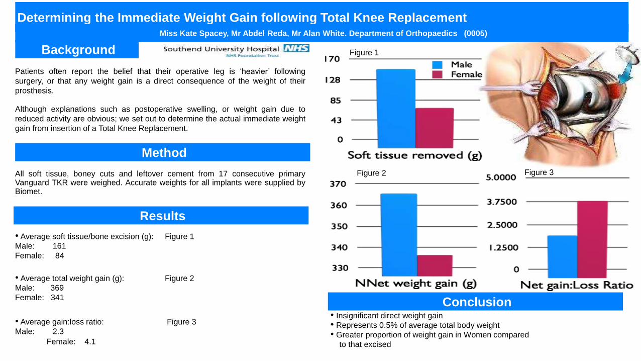
Determining the Immediate Weight Gain following Total Knee Replacement Miss Kate Spacey, Mr Abdel Reda, Mr Alan White. Department of Orthopaedics (0005)
Background
Patients often report the belief that their operative leg is ‘heavier’ following
surgery, or that any weight gain is a direct consequence of the weight of their
prosthesis.
Although explanations such as postoperative swelling, or weight gain due to
reduced activity are obvious; we set out to determine the actual immediate weight
gain from insertion of a Total Knee Replacement.
Method
All soft tissue, boney cuts and leftover cement from 17 consecutive primaryVanguard TKR were weighed. Accurate weights for all implants were supplied byBiomet.
Results
• Average soft tissue/bone excision (g): Figure 1
Male: 161
Female: 84
• Average total weight gain (g): Figure 2
Male: 369
Female: 341
• Average gain:loss ratio: Figure 3
Male: 2.3
Female: 4.1
Figure 1
Figure 2 Figure 3
Conclusion• Insignificant direct weight gain
• Represents 0.5% of average total body weight
• Greater proportion of weight gain in Women compared
to that excised
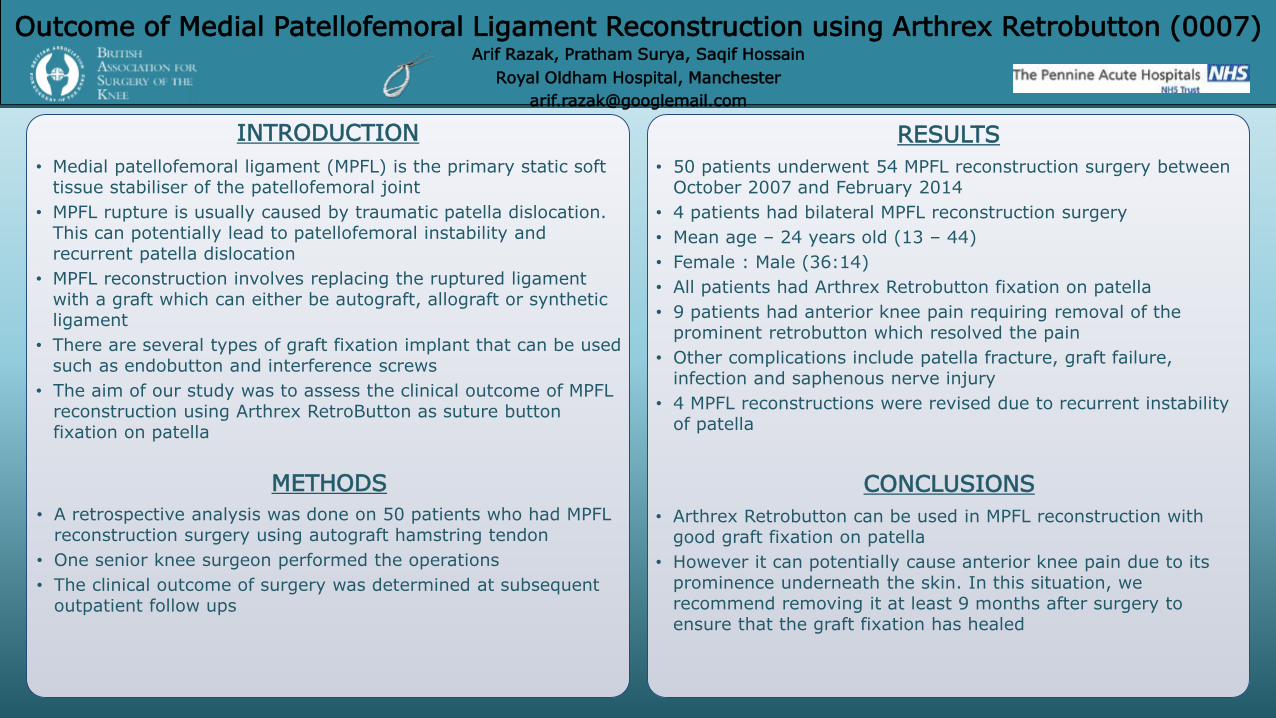
• Medial patellofemoral ligament (MPFL) is the primary static soft tissue stabiliser of the patellofemoral joint
• MPFL rupture is usually caused by traumatic patella dislocation. This can potentially lead to patellofemoral instability and recurrent patella dislocation
• MPFL reconstruction involves replacing the ruptured ligament with a graft which can either be autograft, allograft or synthetic ligament
• There are several types of graft fixation implant that can be used such as endobutton and interference screws
• The aim of our study was to assess the clinical outcome of MPFL reconstruction using Arthrex RetroButton as suture button fixation on patella
INTRODUCTION
METHODS CONCLUSIONS
• A retrospective analysis was done on 50 patients who had MPFL reconstruction surgery using autograft hamstring tendon
• One senior knee surgeon performed the operations
• The clinical outcome of surgery was determined at subsequent outpatient follow ups
Royal Oldham Hospital, Manchester
Arif Razak, Pratham Surya, Saqif Hossain
Outcome of Medial Patellofemoral Ligament Reconstruction using Arthrex Retrobutton (0007)
RESULTS
• 50 patients underwent 54 MPFL reconstruction surgery between October 2007 and February 2014
• 4 patients had bilateral MPFL reconstruction surgery
• Mean age – 24 years old (13 – 44)
• Female : Male (36:14)
• All patients had Arthrex Retrobutton fixation on patella
• 9 patients had anterior knee pain requiring removal of the prominent retrobutton which resolved the pain
• Other complications include patella fracture, graft failure, infection and saphenous nerve injury
• 4 MPFL reconstructions were revised due to recurrent instability of patella
• Arthrex Retrobutton can be used in MPFL reconstruction with good graft fixation on patella
• However it can potentially cause anterior knee pain due to its prominence underneath the skin. In this situation, we recommend removing it at least 9 months after surgery to ensure that the graft fixation has healed
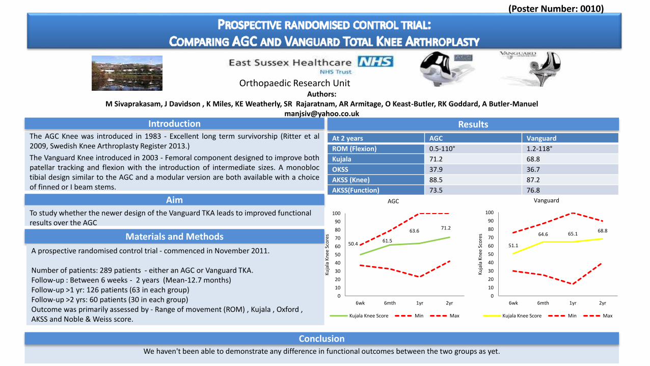
(Poster Number: 0010)
Authors: M Sivaprakasam, J Davidson , K Miles, KE Weatherly, SR Rajaratnam, AR Armitage, O Keast-Butler, RK Goddard, A Butler-Manuel
Materials and Methods
A prospective randomised control trial - commenced in November 2011.
Number of patients: 289 patients - either an AGC or Vanguard TKA.Follow-up : Between 6 weeks - 2 years (Mean-12.7 months) Follow-up >1 yr: 126 patients (63 in each group)Follow-up >2 yrs: 60 patients (30 in each group)Outcome was primarily assessed by - Range of movement (ROM) , Kujala , Oxford , AKSS and Noble & Weiss score.
IntroductionThe AGC Knee was introduced in 1983 - Excellent long term survivorship (Ritter et al2009, Swedish Knee Arthroplasty Register 2013.)
The Vanguard Knee introduced in 2003 - Femoral component designed to improve bothpatellar tracking and flexion with the introduction of intermediate sizes. A monobloctibial design similar to the AGC and a modular version are both available with a choiceof finned or I beam stems.
AimTo study whether the newer design of the Vanguard TKA leads to improved functional results over the AGC
Results
We haven't been able to demonstrate any difference in functional outcomes between the two groups as yet.
Conclusion
50.461.5
63.671.2
0
10
20
30
40
50
60
70
80
90
100
6wk 6mth 1yr 2yr
Ku
jala
Kn
ee S
core
s
AGC
Kujala Knee Score Min Max
51.1
64.6 65.168.8
0
10
20
30
40
50
60
70
80
90
100
6wk 6mth 1yr 2yr
Ku
jala
Kn
ee S
core
s
Vanguard
Kujala Knee Score Min Max
Orthopaedic Research Unit
At 2 years AGC Vanguard
ROM (Flexion) 0.5-110° 1.2-118°
Kujala 71.2 68.8
OKSS 37.9 36.7
AKSS (Knee) 88.5 87.2
AKSS(Function) 73.5 76.8
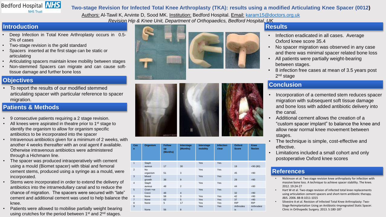
Two-stage Revision for Infected Total Knee Arthroplasty (TKA): results using a modified Articulating Knee Spacer (0012)
Authors: Al-Tawil K, Arvinte D, Sood MK. Institution: Bedford Hospital. Email: [email protected]
Revision Hip & Knee Unit, Department of Orthopaedics, Bedford Hospital, UK
Introduction
• Deep Infection in Total Knee Arthroplasty occurs in 0.5-
2% of cases
• Two-stage revision is the gold standard
• Spacers inserted at the first stage can be static or
articulating
• Articulating spacers maintain knee mobility between stages
• Non-stemmed Spacers can migrate and can cause soft-
tissue damage and further bone loss
Patients & Methods
• 9 consecutive patients requiring a 2 stage revision.
• All knees were aspirated in theatre prior to 1st stage to
identify the organism to allow for organism specific
antibiotics to be incorporated into the spacer
• Intravenous antibiotics given for a minimum of 2 weeks, with
another 4 weeks thereafter with an oral agent if available.
Otherwise intravenous antibiotics were administered
through a Hichmann line.
• The spacer was produced intraoperatively with cement
using a mould (Biomet spacer) with tibial and femoral
cement stems, produced using a syringe as a mould, were
incorporated.
• Stems were incorporated in order to extend the delivery of
antibiotics into the intramedullary canal and to reduce the
chance of migration. The spacers were secured with “late”
cement and additional cement was used to help balance the
knee.
• Patients were allowed to mobilise partially weight bearing
using crutches for the period between 1st and 2nd stages.
ObjectivesConclusion
• Incorporation of a cemented stem reduces spacer
migration with subsequent soft tissue damage
and bone loss with added antibiotic delivery into
the canal.
• Additional cement allows the creation of a
“custom spacer implant” to balance the knee and
allow near normal knee movement between
stages.
• The technique is simple, cost-effective and
effective.
• Limitations included a small cohort and only
postoperative Oxford knee scores
Results
• To report the results of our modified stemmed
articulating spacer with particular reference to spacer
migration.
Cas
e
Organism Follow
up
(Months)
Interstage
(Months)
Interstage
mobility
Infection
clear
Oxford
Score
Knee
flexion
1 Staph
aureus 17 30
Yes Yes
19 <90 (80)
2 No
organism 51 3
Yes Yes
45 >90
3 Mixed
growth 38 9
Yes Yes
29 >90
4 Staph
aureus 48 2
Yes Yes
44 >90
5 Gram +ve
Cocci 48 4
Yes Yes
32 >90
6 None 58 3 Yes Yes 42 >90
7 None 62 5 Yes Yes 37 >90
8 None 5 17 Yes Yes RIP RIP
9
None 56 8
Yes Yes Arthrodes
is
Arthrodesi
s
• Infection eradicated in all cases. Average
Oxford knee score 35.4
• No spacer migration was observed in any case
and there was minimal spacer related bone loss
• All patients were partially weight-bearing
between stages.
• 8 infection free cases at mean of 3.5 years post
2nd stage
References• Nickinson et al. Two stage revision knee arthroplasty for infection with
massive bone loss. A technique to achieve spacer stability. The knee. 2012. 19:24-27
• Hart W et al. Two-stage revision of infected total knee replacements using articulation cement spacers and short-term antibiotic therapy. JBJS. 2006. 88-B:1011-1015
• Silvestre A et al. Revision of Infected Total Knee Arthroplasty: Two-Stage Reimplantation Using an Antibiotic-Impregnated Static Spacer. Clinic in Orthopedic Surgery. 2013. 5:180-187
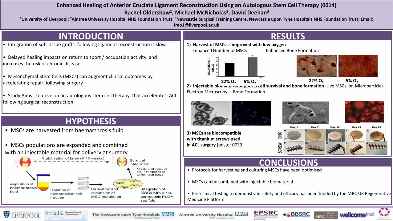
Enhanced Healing of Anterior Cruciate Ligament Reconstruction Using an Autologous Stem Cell Therapy (0014)Rachel Oldershaw1, Michael McNicholas2, David Deehan3
1University of Liverpool; 2Aintree University Hospital NHS Foundation Trust; 3Newcastle Surgical Training Centre, Newcastle upon Tyne Hospitals NHS Foundation Trust; Email: [email protected]
INTRODUCTION• Integration of soft tissue grafts following ligament reconstruction is slow
• Delayed healing impacts on return to sport / occupation activity and increases the risk of chronic disease
• Mesenchymal Stem Cells (MSCs) can augment clinical outcomes by accelerating repair following surgery
• Study Aims : to develop an autologous stem cell therapy that accelerates ACL following surgical reconstruction
HYPOTHESIS• MSCs are harvested from haemarthrosis fluid
• MSCs populations are expanded and combined with an injectable material for delivery at surgery
CONCLUSIONS• Protocols for harvesting and culturing MSCs have been optimised
• MSCs can be combined with injectable biomaterial
• Pre-clinical testing to demonstrate safety and efficacy has been funded by the MRC UK Regenerative Medicine Platform
RESULTS1) Harvest of MSCs is improved with low oxygen
Enhanced Number of MSCs Enhanced Bone Formation
2) Injectable biomaterial supports cell survival and bone formation Live MSCs on MicroparticlesElectron Microscopy Bone Formation
3) MSCs are biocompatiblewith titanium screws used in ACL surgery (poster 0033)
22% O2 5% O222% O2 5% O2
NU
MB
ER O
F M
SCS
0016- The Epidemiology and Financial Burden of Revision Total Knee and Hip Arthroplasty in
England and Wales using NJR Data.A. Patel, G. Pavlou, R. Ahmad, A. Toms- The Exeter Knee Reconstruction Unit.
Introduction.
Total knee arthroplasty (TKA) and Total hip arthroplasty (THA) are well-recognised and proven interventions for patients with advanced arthritis, being both efficacious and cost effective. Studies to date demonstrate a steady increase in the prevalence of primary TKA and revision TKA in the United States between 1990 and 2000. The observed increase in revision surgery has been attributed to a number of factors including a rise in primary procedures, population life expectancy, rates of obesity, and extending the surgical indications to a younger population cohort. US projections estimate that by 2030 the demand for primary TKA will grow by 673% and by 601% for revision TKR from their 2005 level.Projections for hip arthroplasty are more conservative with demand increasing by 174%, with revision hip arthroplastygrowing to 137%. The population of England and Wales continues to increase from 56 million as determined by the mid 2011 census. Estimates from the Office of National Statistics predict a steady rise in population numbers to 64 million by 2030. The increase in total population numbers alongside a rise in older population cohorts is likely to have a direct impact on TKA and THA numbers over the coming years.
Methods.
We analysed data from The National Joint Registry to see if data extrapolated from our registry was comparable to predictions made in the US. We also present the alternative, extreme projection scenario where the incidence rate of female primary operations grows at an ever-increasing rate.
Results.Figure 1: Projected total number of Knee Replacements in England and Wales
0
1000
020
000
3000
0
2010 2015 2020 2025 2030Year
projected
95% CI of projection
observed
Figure 2: Projected total number of Revision Knee Replacements in England and Wales
0
2000
0040
0000
6000
0080
0000
2010 2015 2020 2025 2030Year
Hip replacements Knee replacements
At accelerating proportional increase in incidence rate
Figure: 3 Projected total number of Primary Hip and Knee Replacements in an extreme projection scenario in England and Wales
0
5000
1000
015
000
2000
025
000
2010 2015 2020 2025 2030Year
Hip replacements Knee replacements
At constant proportional increase in incidence rate
Figure 4: Projected total number of Revision Hip and Knee Replacements in England and Wales
0
5000
1000
015
000
2000
025
000
2010 2015 2020 2025 2030Year
Hip replacements Knee replacements
At constant incidence rate
Conclusion.Our study specifically analyses the joint replacement numbers in England and Wales, which we predict to be around 485176 and 720995 in primary knee and hip arthroplasty respectively in 2030. These imply cumulative growth predictions of the same magnitude as previously reported for the US. In keeping with this, we still support the consensus regarding the economic burden of arthroplasty in the future as a real and unaddressed problem. This has significant and serious implications for doctors, healthcare providers, politicians and patients to help plan and accommodate this increased demand for arthroplasty in the future.
Procedure Predicted Growth % (95% CI)
1⁰ TKR 451
1⁰ THR 802
Revision TKR 310
Revision THR 25
Assessing the accuracy of the NJR surgeon and hospital profiles (20)
Nirav K Patel, Sheng Yang Qui, David Ahearne, Vikas Vedi
Department of Trauma and Orthopaedics, Hillingdon Hospital, Uxbridge, Middlesex, UK
Introduction National Joint Registry data for individual consultants are released to the general public in2013: www.njrsurgeonhospitalprofile.org.uk
Contribute to national data on quality and outcome of joint replacements in the UK
Perceived performance of consultants/trusts judged on NJR data
Current Practice
Paper NJR forms completed in theatre after case by consultant/ SpR
Paper booklet left for theatre staff who are registered with the NJR data entry system forNJR database and data is entered into NJR
NJR analyses data and data released to general public
Discussion Joint replacement numbers were under-reported to NJR possibly due to incorrect PID
entry
In addition, hospital admission data did not fully reflect the actual number of operations
performed compared to theatre logbooks
Significant ramifications for the perceived performance of surgeons and hospitals by the
public alongside financial implications to hospitals from coding inaccuracies
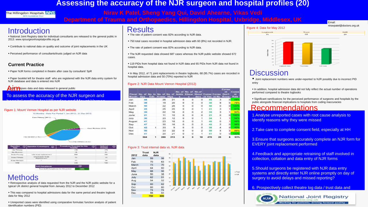
Figure 4: Data for May 2012
Figure 3: Trust internal data vs. NJR data
Results
To assess the accuracy of the NJR surgeon and
hospital profiles for Hillingdon and Mount Vernon
Hospitals
Aim
The rate of patient consent was 82% according to NJR data.
750 total cases recorded in hospital admission data with 60 (8%) not recorded in NJR.
The rate of patient consent was 82% according to NJR data.
The NJR requested data showed 687 cases whereas the NJR public website showed 672
cases.
119 PIDs from hospital data not found in NJR data and 65 PIDs from NJR data not found in
hospital data.
In May 2012, of 71 joint replacements in theatre logbooks, 68 (95.7%) cases are recorded in
hospital admission data and 50 (70%) reported to NJR
1.Analyse unreported cases with root cause analysis to
identify reasons why they were missed
2.Take care to complete consent field, especially at HH
3.Ensure that surgeons accurately complete an NJR form for
EVERY joint replacement performed
4.Feedback and appropriate retraining of staff involved in
collection, collation and data entry of NJR forms
5.Should surgeons be registered with NJR data entry
systems and directly enter NJR online promptly on day of
surgery to avoid delays and missed reporting?
6. Prospectively collect theatre log data / trust data and
compare this with NJR data monthly to identify unreported
cases promptly and for missed cases to be added to NJR
with minimal delay
Recommendations
Figure 2: NJR Data Mount Vernon Hospital (2012)
Methods Retrospective analysis of data requested from the NJR and the NJR public website for a
typical UK district general hospital from January 2012 to December 2012
This was compared to hospital admissions data for the same period and theatre logbook
data for May 2012
Unreported cases were identified using comparative formulaic function analysis of patient
identification numbers (PID)
Figure 1: Mount Vernon Hospital as per NJR website
Email:
Is The Clinical Threshold For Revising A Unicompartmental Knee Arthroplasty Lower Than
For Total Knee Arthroplasty? - A Single Surgeon ExperienceKH Teoh, AP Davies. Morriston Hospital, Swansea SA6 6NL.
[email protected] Poster no: 0021
Methods and Materials:
• Patients who underwent revision of UKA or TKA for
unexplained knee pain between 2008 and 2012 were identified
from the senior surgeon’s arthroplasty database.
• These cases were investigated thoroughly for cause of knee
pain prior to surgery.
• Oxford Knee Score (OKS) and American Knee Society Scores
(AKSS) were recorded prospectively by the senior surgeon pre-
operatively and at each follow up.
• Pre and post op scores were compared in both groups and
statistical analysis of the data was performed.
Conclusion:
• Based on a single surgeon experience, there is weak evidence to suggest that UKA
are revised at a lower clinical threshold than TKA in terms of PROMs.
• However, there are likely to be offered a revision earlier than the TKR group.
• Revised UKR and TKR do equally well as shown by knee scores following surgery.
Results:Introduction
• Implant survival rates for unicompartmental knee arthroplasty
(UKA) are significantly poorer than total knee arthroplasty (TKA)
in worldwide arthroplasty registers
• There is a perception that UKA are revised at a lower clinical
threshold as it is easier to do so
• The aim of the study is to investigate whether the above was
true in a single surgeon series of revision knee arthroplasties by
comparing patient-reported outcome measure (PROMs)
between the two groups
UKR to TKR
(n =12)
TKR to TKR
(n= 14)
Average age (years) 59.8 (48 - 81) 69.0 (60 - 83) UKR younger, P=0.009
Sex 5 males, 7 females 7 males, 7 females
Laterality 6R, 6L 7R, 7L
Time from initial surgery
to revision (months)
29.8 (15 - 43) 47.0 (9 - 130) Time to revision earlier
for UKR, P=0.007
Pre op OKS 33.63 37.50 No diff, p>0.05
Pre op AKSS function 30.83 23.57 No diff, p>0.05
Pre op AKSS knee 44.25 33.79 UKR better, p<0.05
No significant differences in post-op knee scores between the two groups at last follow up (p>0.05).
All scores improved significantly from pre-op values (p<0.05, Wilcoxin-signed ranks test)
Ongoing Knee Pain 1 Year Post TKR
Is Gait Abnormality a Factor? (0022)Mr J P Flanagan The Chelmsford Knee Clinic email: Tina. [email protected]
Up to 20% of patients have chronic pain following TKR.
This study investigates the effect of changing a Quads Avoidance Gait pattern as clinically diagnosed by quads wasting and a lack of full extension at mid-stance.
22 patients with ongoing knee pain and a quads avoidance gait were taught to use their quads to drive their knee back into full extension at mid-stance, having landed on a slightly bent knee.
Oxford Knee Scores (OKS) were taken at initial consultation and at 3 months.
OKS: Initially: Mean 20.9 (sd 9.2)
3 months: Mean 34.1 (sd 8.9) ANOVA p < 0.001
The chronic pain of patients post TKR can be significantly improved by altering their gait pattern.
www.baskonline.com
Improving consenting process for total knee replacement: A complete audit cycle
WC Lee1, S Balasubramanian2, V Kumar2, M Sunkara2, R Reddy2
1Singapore General Hospital, Singapore. [email protected] 2George Eliot Hospital, Nuneaton.
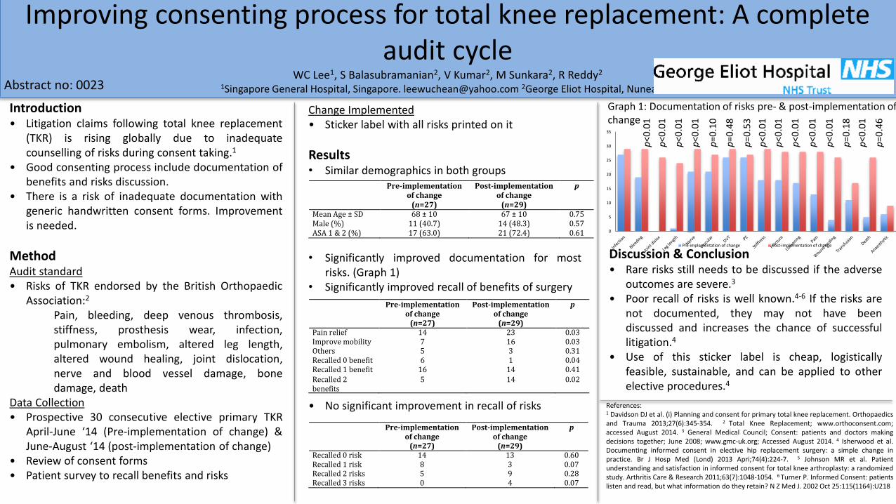
Change Implemented• Sticker label with all risks printed on it
Results• Similar demographics in both groups
• Significantly improved documentation for mostrisks. (Graph 1)
• Significantly improved recall of benefits of surgery
• No significant improvement in recall of risks
0
5
10
15
20
25
30
35
Pre-implementation of change Post-implementation of change
Pre-implementationofchange
(n=27)
Post-implementationofchange
(n=29)
p
MeanAge±SD 68±10 67±10 0.75Male(%) 11(40.7) 14(48.3) 0.57ASA1&2(%) 17(63.0) 21(72.4) 0.61
Pre-implementationofchange
(n=27)
Post-implementationofchange
(n=29)
p
Painrelief 14 23 0.03Improvemobility 7 16 0.03Others 5 3 0.31Recalled0benefit 6 1 0.04Recalled1benefit 16 14 0.41
Recalled2benefits
5 14 0.02
Pre-implementationofchange
(n=27)
Post-implementationofchange
(n=29)
p
Recalled0risk 14 13 0.60Recalled1risk 8 3 0.07Recalled2risks 5 9 0.28Recalled3risks 0 4 0.07
Introduction• Litigation claims following total knee replacement
(TKR) is rising globally due to inadequatecounselling of risks during consent taking.1
• Good consenting process include documentation ofbenefits and risks discussion.
• There is a risk of inadequate documentation withgeneric handwritten consent forms. Improvementis needed.
MethodAudit standard• Risks of TKR endorsed by the British Orthopaedic
Association:2
Pain, bleeding, deep venous thrombosis,stiffness, prosthesis wear, infection,pulmonary embolism, altered leg length,altered wound healing, joint dislocation,nerve and blood vessel damage, bonedamage, death
Data Collection• Prospective 30 consecutive elective primary TKR
April-June ‘14 (Pre-implementation of change) &June-August ‘14 (post-implementation of change)
• Review of consent forms• Patient survey to recall benefits and risks
Graph 1: Documentation of risks pre- & post-implementation of change
p<0
.01
p<0
.01
p<0
.01
p<0
.01
p<0
.01
p<0
.01
p<0
.01
p<0
.01
p<0
.01
p<0
.01
p=0
.10
p=0
.48
p=0
.53
p=0
.18
p=0
.46
Discussion & Conclusion• Rare risks still needs to be discussed if the adverse
outcomes are severe.3
• Poor recall of risks is well known.4-6 If the risks arenot documented, they may not have beendiscussed and increases the chance of successfullitigation.4
• Use of this sticker label is cheap, logisticallyfeasible, sustainable, and can be applied to otherelective procedures.4
References:1 Davidson DJ et al. (i) Planning and consent for primary total knee replacement. Orthopaedicsand Trauma 2013;27(6):345-354. 2 Total Knee Replacement; www.orthoconsent.com;accessed August 2014. 3 General Medical Council; Consent: patients and doctors makingdecisions together; June 2008; www.gmc-uk.org; Accessed August 2014. 4 Isherwood et al.Documenting informed consent in elective hip replacement surgery: a simple change inpractice. Br J Hosp Med (Lond) 2013 Apri;74(4):224-7. 5 Johnson MR et al. Patientunderstanding and satisfaction in informed consent for total knee arthroplasty: a randomizedstudy. Arthritis Care & Research 2011;63(7):1048-1054. 6 Turner P. Informed Consent: patientslisten and read, but what information do they retain? N Z Med J. 2002 Oct 25:115(1164):U218
Abstract no: 0023
A Report of 10 Years Experience with an UnicompartmentalKnee Prosthesis (Poster 0024)
Dr François Gougeon, Hôpital Privé La Louvière, Lille, France Nicolas Hohl* MS Exactech & Dr Roy Harvey* BSc PhD Exactech [email protected] *Employees of Exactech.
• A total of 197 consecutive surgeries were performed by a single senior operator (FG)
• All prostheses were implanted between Dec 2002 and Nov 2013
• Visits @ pre-op, immediate post-op, 3 weeks, 3 , 6 & 12 months thereafter
• The prosthesis is a fixed bearing CoCr femur & all-poly tibia
• 35 lateral & 160 medial Unis; 2 patients received TKA
• 191 implants are still in situ
•Estimated KM survivorship of 97.2% or 0.48 revisions/100 observed years
•Longest follow-up of 11 years
•KM of 2.8% @ mean 4.25 years fu
•Mean Post op flexion 124 Deg
•93% very satisified or satisfiedDisclosure: No funding was provided for this study. Dr Gougeonacknowledges the assistance of his co-authors in the final statistical analysis of data & preparation of this poster
Parameter Pre-operative Post-operative
HKAangle
Medial surgery 173° [165°-179°] 174° [167°-179°]
Lateral surgery 188° [180°-190°] 183° [180°-190°]
KSSscore
Knee score 58 [36-77] 88 [76-100]
Function score 67 [15-100] 85 [71-100]
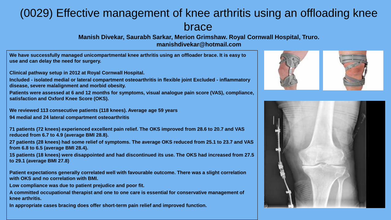
(0029) Effective management of knee arthritis using an offloading knee
braceManish Divekar, Saurabh Sarkar, Merion Grimshaw. Royal Cornwall Hospital, Truro.
We have successfully managed unicompartmental knee arthritis using an offloader brace. It is easy to
use and can delay the need for surgery.
Clinical pathway setup in 2012 at Royal Cornwall Hospital.
Included - isolated medial or lateral compartment osteoarthritis in flexible joint Excluded - inflammatory
disease, severe malalignment and morbid obesity.
Patients were assessed at 6 and 12 months for symptoms, visual analogue pain score (VAS), compliance,
satisfaction and Oxford Knee Score (OKS).
We reviewed 113 consecutive patients (118 knees). Average age 59 years
94 medial and 24 lateral compartment osteoarthritis
71 patients (72 knees) experienced excellent pain relief. The OKS improved from 28.6 to 20.7 and VAS
reduced from 6.7 to 4.9 (average BMI 28.8).
27 patients (28 knees) had some relief of symptoms. The average OKS reduced from 25.1 to 23.7 and VAS
from 6.8 to 6.5 (average BMI 28.4).
15 patients (18 knees) were disappointed and had discontinued its use. The OKS had increased from 27.5
to 29.1 (average BMI 27.8)
Patient expectations generally correlated well with favourable outcome. There was a slight correlation
with OKS and no correlation with BMI.
Low compliance was due to patient prejudice and poor fit.
A committed occupational therapist and one to one care is essential for conservative management of
knee arthritis.
In appropriate cases bracing does offer short-term pain relief and improved function.
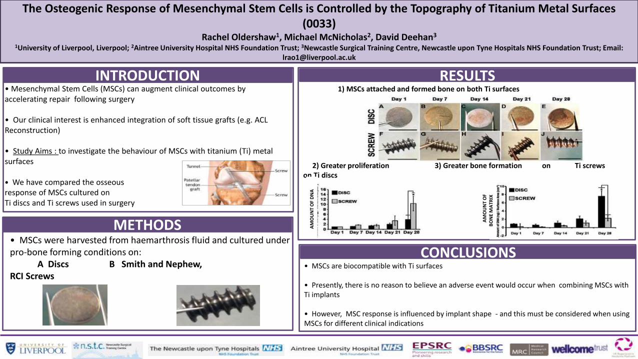
The Osteogenic Response of Mesenchymal Stem Cells is Controlled by the Topography of Titanium Metal Surfaces (0033)
Rachel Oldershaw1, Michael McNicholas2, David Deehan3
1University of Liverpool, Liverpool; 2Aintree University Hospital NHS Foundation Trust; 3Newcastle Surgical Training Centre, Newcastle upon Tyne Hospitals NHS Foundation Trust; Email: [email protected]
INTRODUCTION• Mesenchymal Stem Cells (MSCs) can augment clinical outcomes by accelerating repair following surgery
• Our clinical interest is enhanced integration of soft tissue grafts (e.g. ACL Reconstruction)
• Study Aims : to investigate the behaviour of MSCs with titanium (Ti) metal surfaces
• We have compared the osseous response of MSCs cultured on Ti discs and Ti screws used in surgery
METHODS• MSCs were harvested from haemarthrosis fluid and cultured under pro-bone forming conditions on:
A Discs B Smith and Nephew, RCI Screws
CONCLUSIONS• MSCs are biocompatible with Ti surfaces
• Presently, there is no reason to believe an adverse event would occur when combining MSCs with Ti implants
• However, MSC response is influenced by implant shape - and this must be considered when using MSCs for different clinical indications
RESULTS1) MSCs attached and formed bone on both Ti surfaces
2) Greater proliferation 3) Greater bone formation on Ti screws on Ti discs
AM
OU
NT
OF
DN
A
AM
OU
NT
OF
BO
NE
MA
TRIX
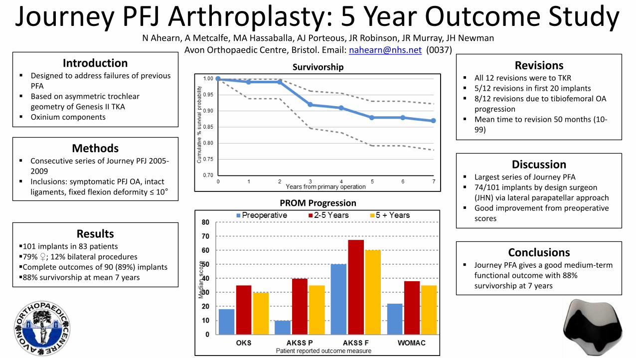
Journey PFJ Arthroplasty: 5 Year Outcome StudyN Ahearn, A Metcalfe, MA Hassaballa, AJ Porteous, JR Robinson, JR Murray, JH Newman
Avon Orthopaedic Centre, Bristol. Email: [email protected] (0037)
PROM Progression
SurvivorshipIntroduction Designed to address failures of previous
PFA Based on asymmetric trochlear
geometry of Genesis II TKA Oxinium components
Methods Consecutive series of Journey PFJ 2005-
2009 Inclusions: symptomatic PFJ OA, intact
ligaments, fixed flexion deformity ≤ 10°
Results101 implants in 83 patients79% ♀; 12% bilateral proceduresComplete outcomes of 90 (89%) implants88% survivorship at mean 7 years
Revisions All 12 revisions were to TKR 5/12 revisions in first 20 implants 8/12 revisions due to tibiofemoral OA
progression Mean time to revision 50 months (10-
99)
Discussion Largest series of Journey PFA 74/101 implants by design surgeon
(JHN) via lateral parapatellar approach Good improvement from preoperative
scores
Conclusions Journey PFA gives a good medium-term
functional outcome with 88% survivorship at 7 years
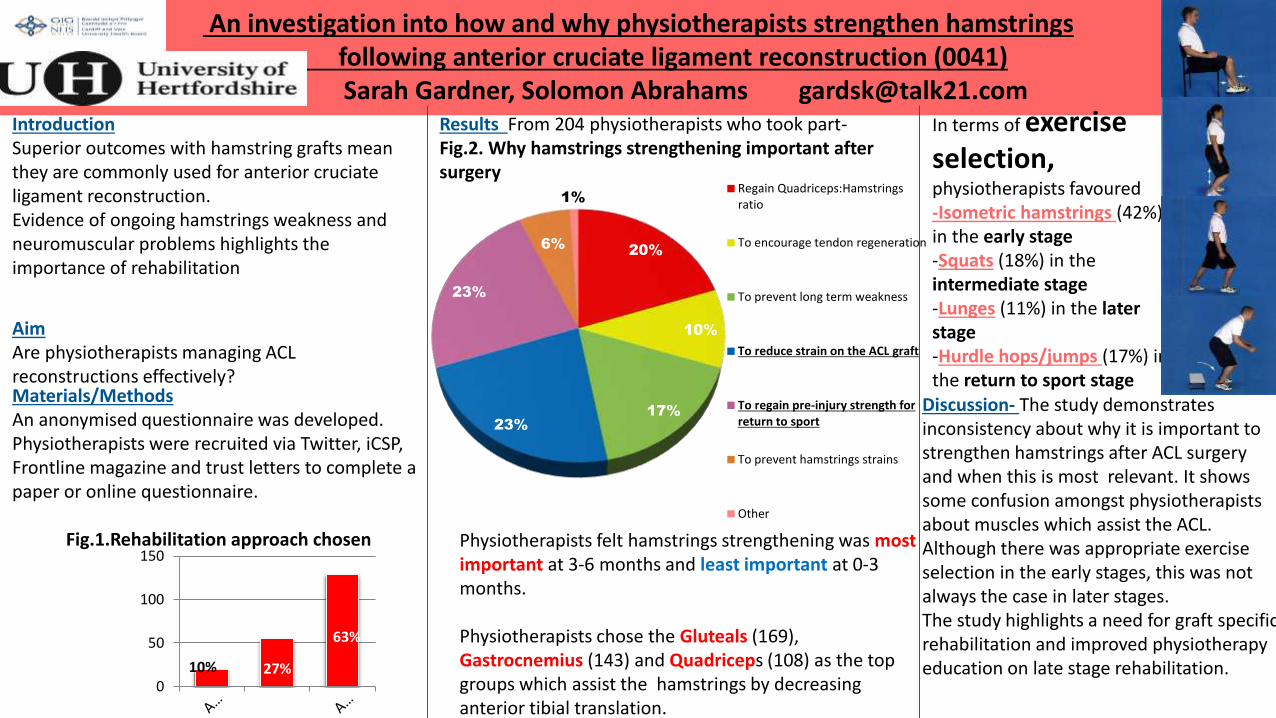
An investigation into how and why physiotherapists strengthen hamstrings following anterior cruciate ligament reconstruction (0041)Sarah Gardner, Solomon Abrahams [email protected]
IntroductionSuperior outcomes with hamstring grafts mean they are commonly used for anterior cruciateligament reconstruction. Evidence of ongoing hamstrings weakness and neuromuscular problems highlights the importance of rehabilitation
AimAre physiotherapists managing ACL reconstructions effectively?Materials/MethodsAn anonymised questionnaire was developed. Physiotherapists were recruited via Twitter, iCSP, Frontline magazine and trust letters to complete a paper or online questionnaire.
Fig.1.Rehabilitation approach chosen
Results From 204 physiotherapists who took part-Fig.2. Why hamstrings strengthening important after surgery
10% 27%
63%
0
50
100
150
20%
10%
17%
23%
23%
6%
1%
Regain Quadriceps:Hamstringsratio
To encourage tendon regeneration
To prevent long term weakness
To reduce strain on the ACL graft
To regain pre-injury strength for return to sport
To prevent hamstrings strains
Other
Physiotherapists felt hamstrings strengthening was most important at 3-6 months and least important at 0-3 months.
Physiotherapists chose the Gluteals (169), Gastrocnemius (143) and Quadriceps (108) as the top groups which assist the hamstrings by decreasing anterior tibial translation.
In terms of exercise selection, physiotherapists favoured -Isometric hamstrings (42%) in the early stage-Squats (18%) in the intermediate stage-Lunges (11%) in the later stage-Hurdle hops/jumps (17%) in the return to sport stage
Discussion- The study demonstrates inconsistency about why it is important to strengthen hamstrings after ACL surgery and when this is most relevant. It shows some confusion amongst physiotherapists about muscles which assist the ACL. Although there was appropriate exercise selection in the early stages, this was not always the case in later stages. The study highlights a need for graft specific rehabilitation and improved physiotherapy education on late stage rehabilitation.
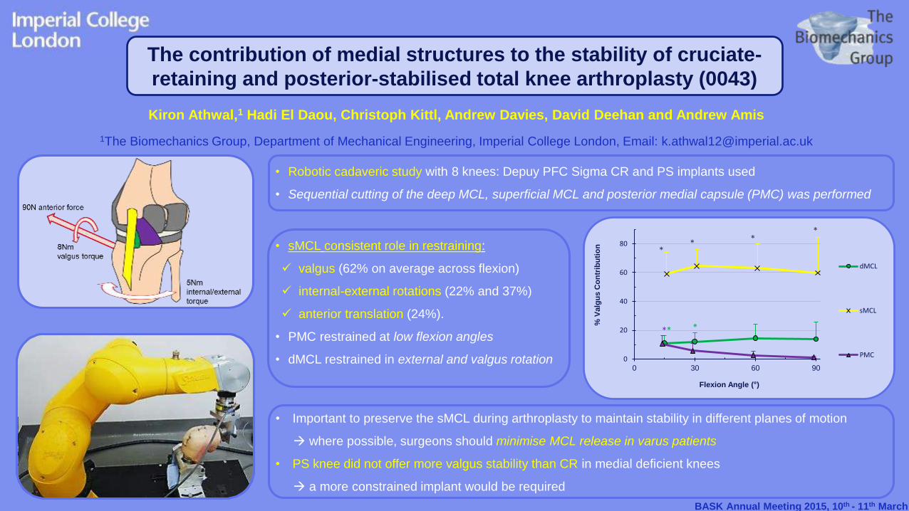
• Robotic cadaveric study with 8 knees: Depuy PFC Sigma CR and PS implants used
• Sequential cutting of the deep MCL, superficial MCL and posterior medial capsule (PMC) was performed
Kiron Athwal,1 Hadi El Daou, Christoph Kittl, Andrew Davies, David Deehan and Andrew Amis
1The Biomechanics Group, Department of Mechanical Engineering, Imperial College London, Email: [email protected]
BASK Annual Meeting 2015, 10th - 11th March
The contribution of medial structures to the stability of cruciate-
retaining and posterior-stabilised total knee arthroplasty (0043)
* *
** *
*
*
0
20
40
60
80
0 30 60 90
% V
alg
us
Co
ntr
ibu
tio
n
Flexion Angle (°)
dMCL
sMCL
PMC
• sMCL consistent role in restraining:
valgus (62% on average across flexion)
internal-external rotations (22% and 37%)
anterior translation (24%).
• PMC restrained at low flexion angles
• dMCL restrained in external and valgus rotation
• Important to preserve the sMCL during arthroplasty to maintain stability in different planes of motion
where possible, surgeons should minimise MCL release in varus patients
• PS knee did not offer more valgus stability than CR in medial deficient knees
a more constrained implant would be required
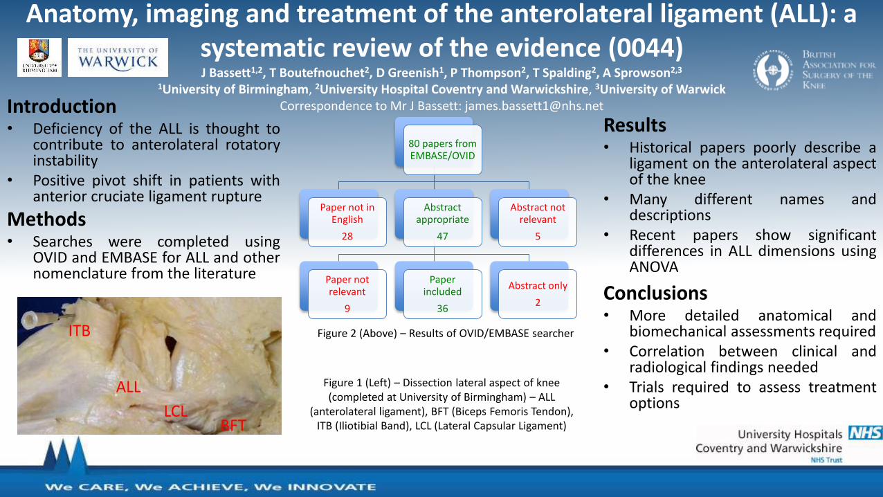
Anatomy, imaging and treatment of the anterolateral ligament (ALL): a systematic review of the evidence (0044)J Bassett1,2, T Boutefnouchet2, D Greenish1, P Thompson2, T Spalding2, A Sprowson2,3
1University of Birmingham, 2University Hospital Coventry and Warwickshire, 3University of WarwickCorrespondence to Mr J Bassett: [email protected]
• Deficiency of the ALL is thought tocontribute to anterolateral rotatoryinstability
• Positive pivot shift in patients withanterior cruciate ligament rupture
Methods• Searches were completed using
OVID and EMBASE for ALL and othernomenclature from the literature
Results• Historical papers poorly describe a
ligament on the anterolateral aspectof the knee
• Many different names anddescriptions
• Recent papers show significantdifferences in ALL dimensions usingANOVA
Conclusions• More detailed anatomical and
biomechanical assessments required• Correlation between clinical and
radiological findings needed• Trials required to assess treatment
options
80 papers from EMBASE/OVID
Paper not in English
28
Abstract appropriate
47
Paper not relevant
9
Paper included
36
Abstract only
2
Abstract not relevant
5
ITB
ALL
LCLBFT
Figure 1 (Left) – Dissection lateral aspect of knee (completed at University of Birmingham) – ALL
(anterolateral ligament), BFT (Biceps Femoris Tendon), ITB (Iliotibial Band), LCL (Lateral Capsular Ligament)
Figure 2 (Above) – Results of OVID/EMBASE searcher
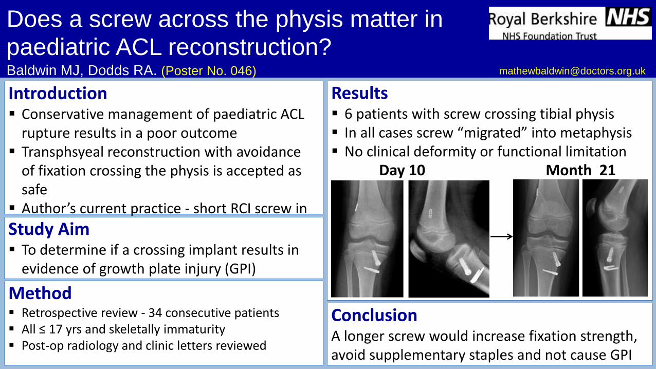
Does a screw across the physis matter in paediatric ACL reconstruction?Baldwin MJ, Dodds RA. (Poster No. 046)
Introduction Conservative management of paediatric ACL
rupture results in a poor outcome Transphsyeal reconstruction with avoidance
of fixation crossing the physis is accepted as safe
Author’s current practice - short RCI screw in tibia supplemented by staple
Method Retrospective review - 34 consecutive patients All ≤ 17 yrs and skeletally immaturity Post-op radiology and clinic letters reviewed
Results 6 patients with screw crossing tibial physis In all cases screw “migrated” into metaphysis No clinical deformity or functional limitation
Study Aim To determine if a crossing implant results in
evidence of growth plate injury (GPI)
ConclusionA longer screw would increase fixation strength, avoid supplementary staples and not cause GPI
Day 10 Month 21
Andover War Memorial Hospital Basingstoke and North Hampshire Hospital Royal Hampshire County Hospital
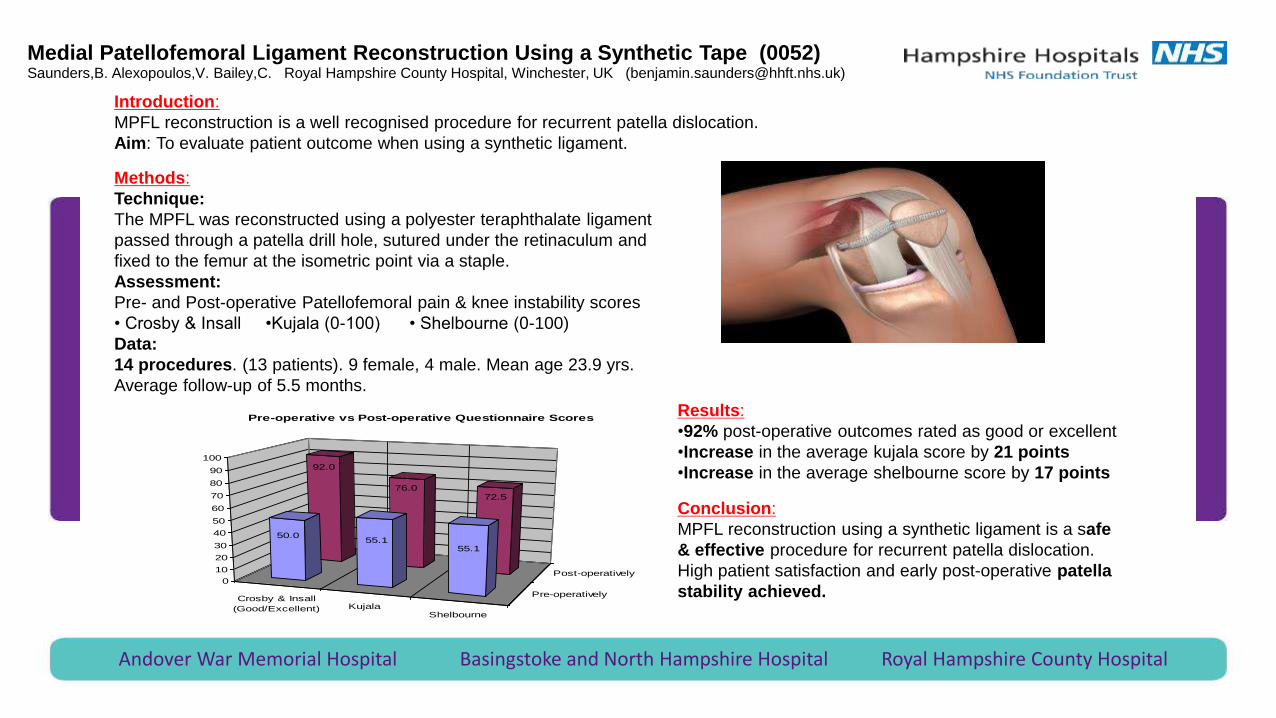
Medial Patellofemoral Ligament Reconstruction Using a Synthetic Tape (0052)
Crosby & Insall
(Good/Excellent) KujalaShelbourne
Pre-operatively
Post-operatively
92.0
76.072.5
50.055.1
55.1
0
10
20
30
40
50
60
70
80
90
100
Pre-operative vs Post-operative Questionnaire Scores
Introduction:
MPFL reconstruction is a well recognised procedure for recurrent patella dislocation.
Aim: To evaluate patient outcome when using a synthetic ligament.
Methods:
Technique:
The MPFL was reconstructed using a polyester teraphthalate ligament
passed through a patella drill hole, sutured under the retinaculum and
fixed to the femur at the isometric point via a staple.
Assessment:
Pre- and Post-operative Patellofemoral pain & knee instability scores
• Crosby & Insall •Kujala (0-100) • Shelbourne (0-100)
Data:
14 procedures. (13 patients). 9 female, 4 male. Mean age 23.9 yrs.
Average follow-up of 5.5 months.
Results:
•92% post-operative outcomes rated as good or excellent
•Increase in the average kujala score by 21 points
•Increase in the average shelbourne score by 17 points
Conclusion:
MPFL reconstruction using a synthetic ligament is a safe
& effective procedure for recurrent patella dislocation.
High patient satisfaction and early post-operative patella
stability achieved.
Saunders,B. Alexopoulos,V. Bailey,C. Royal Hampshire County Hospital, Winchester, UK ([email protected])
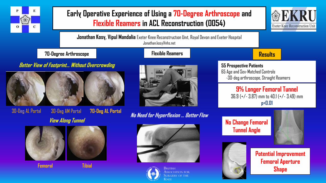
Early Operative Experience of Using a 70-Degree Arthroscope and
Flexible Reamers in ACL Reconstruction (0054)
Jonathan Kosy, Vipul Mandalia Exeter Knee Reconstruction Unit, Royal Devon and Exeter Hospital [email protected]
70-Degree Arthroscope
Better View of Footprint… Without Overcrowding
30-Deg AL Portal 30-Deg AM Portal 70-Deg AL Portal
View Along Tunnel
Femoral Tibial
Flexible Reamers
No Need for Hyperflexion … Better Flow
Results
55 Prospective Patients
65 Age and Sex-Matched Controls
-30-deg arthroscope, Straight Reamers
9% Longer Femoral Tunnel36.9 (+/- 3.87) mm to 40.1 (+/- 3.49) mm
p<0.01
No Change Femoral
Tunnel Angle
Potential Improvement
Femoral Aperture
Shape
ePosters
Available now on the BASK website
www.baskonline.com
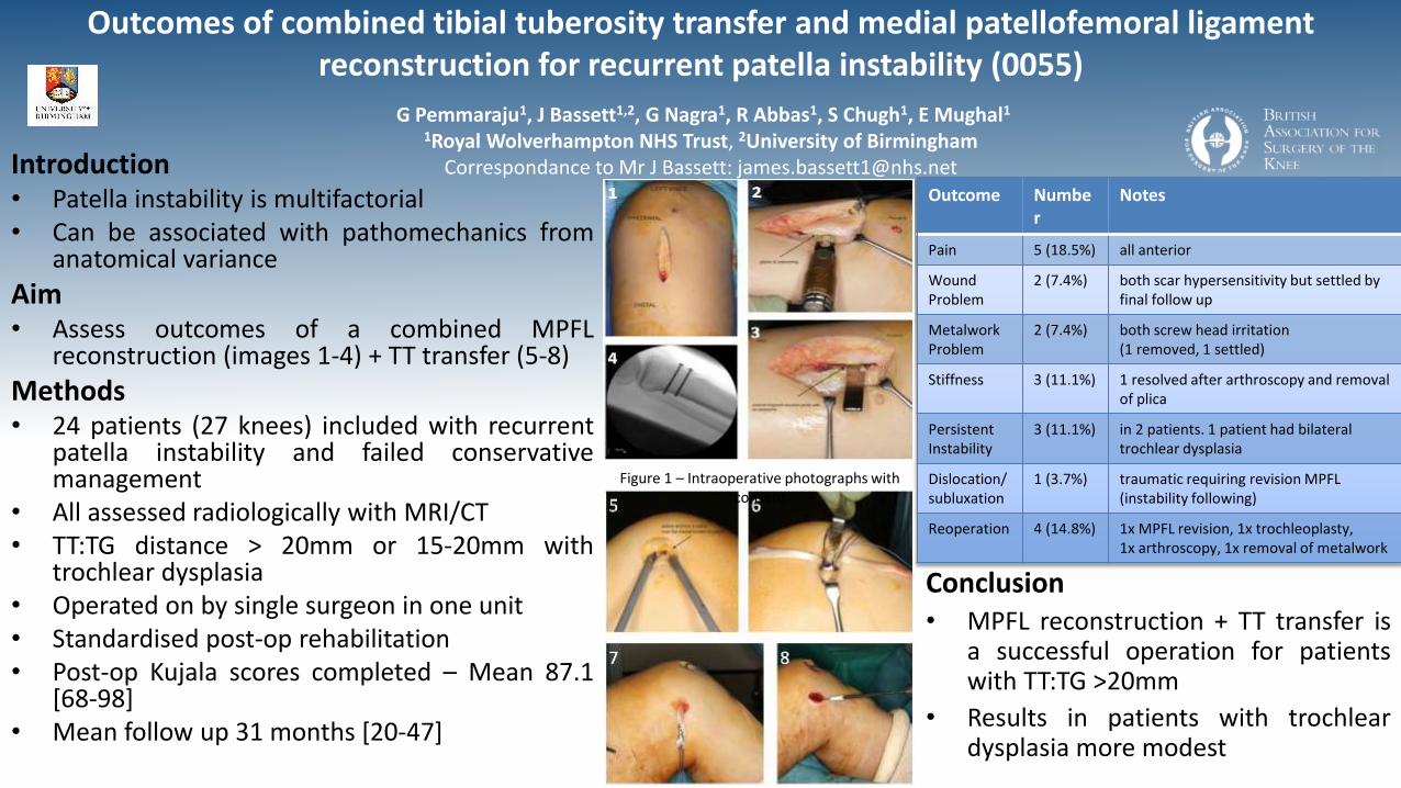
Outcomes of combined tibial tuberosity transfer and medial patellofemoral ligament reconstruction for recurrent patella instability (0055)
G Pemmaraju1, J Bassett1,2, G Nagra1, R Abbas1, S Chugh1, E Mughal1
1Royal Wolverhampton NHS Trust, 2University of BirminghamCorrespondance to Mr J Bassett: [email protected]
• Patella instability is multifactorial• Can be associated with pathomechanics from
anatomical variance
Aim• Assess outcomes of a combined MPFL
reconstruction (images 1-4) + TT transfer (5-8)
Methods• 24 patients (27 knees) included with recurrent
patella instability and failed conservativemanagement
• All assessed radiologically with MRI/CT• TT:TG distance > 20mm or 15-20mm with
trochlear dysplasia• Operated on by single surgeon in one unit• Standardised post-op rehabilitation• Post-op Kujala scores completed – Mean 87.1
[68-98]• Mean follow up 31 months [20-47]
Conclusion• MPFL reconstruction + TT transfer is
a successful operation for patientswith TT:TG >20mm
• Results in patients with trochleardysplasia more modest
Outcome Number
Notes
Pain 5 (18.5%) all anterior
Wound Problem
2 (7.4%) both scar hypersensitivity but settled by final follow up
Metalwork Problem
2 (7.4%) both screw head irritation (1 removed, 1 settled)
Stiffness 3 (11.1%) 1 resolved after arthroscopy and removal of plica
Persistent Instability
3 (11.1%) in 2 patients. 1 patient had bilateral trochlear dysplasia
Dislocation/subluxation
1 (3.7%) traumatic requiring revision MPFL (instability following)
Reoperation 4 (14.8%) 1x MPFL revision, 1x trochleoplasty, 1x arthroscopy, 1x removal of metalwork
Figure 1 – Intraoperative photographs with consent
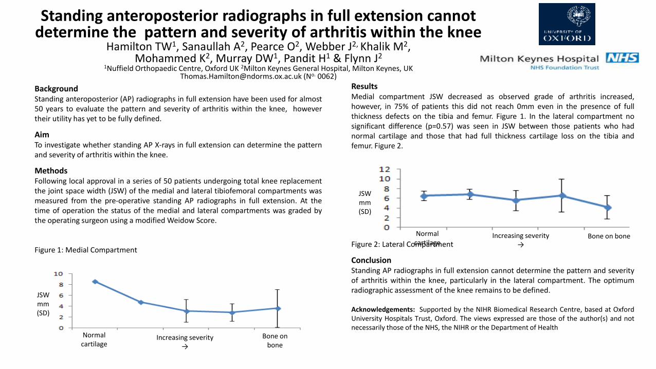
ResultsMedial compartment JSW decreased as observed grade of arthritis increased,however, in 75% of patients this did not reach 0mm even in the presence of fullthickness defects on the tibia and femur. Figure 1. In the lateral compartment nosignificant difference (p=0.57) was seen in JSW between those patients who hadnormal cartilage and those that had full thickness cartilage loss on the tibia andfemur. Figure 2.
Figure 2: Lateral Compartment
ConclusionStanding AP radiographs in full extension cannot determine the pattern and severityof arthritis within the knee, particularly in the lateral compartment. The optimumradiographic assessment of the knee remains to be defined.
Acknowledgements: Supported by the NIHR Biomedical Research Centre, based at OxfordUniversity Hospitals Trust, Oxford. The views expressed are those of the author(s) and notnecessarily those of the NHS, the NIHR or the Department of Health
Bone on bone
Standing anteroposterior radiographs in full extension cannotdetermine the pattern and severity of arthritis within the knee
Hamilton TW1, Sanaullah A2, Pearce O2, Webber J2, Khalik M2,Mohammed K2, Murray DW1, Pandit H1 & Flynn J2
1Nuffield Orthopaedic Centre, Oxford UK 2Milton Keynes General Hospital, Milton Keynes, UK [email protected] (No. 0062)
BackgroundStanding anteroposterior (AP) radiographs in full extension have been used for almost50 years to evaluate the pattern and severity of arthritis within the knee, howevertheir utility has yet to be fully defined.
AimTo investigate whether standing AP X-rays in full extension can determine the patternand severity of arthritis within the knee.
MethodsFollowing local approval in a series of 50 patients undergoing total knee replacementthe joint space width (JSW) of the medial and lateral tibiofemoral compartments wasmeasured from the pre-operative standing AP radiographs in full extension. At thetime of operation the status of the medial and lateral compartments was graded bythe operating surgeon using a modified Weidow Score.
Figure 1: Medial Compartment
Normalcartilage
Bone on boneIncreasing severity→
JSWmm(SD)
Normalcartilage
Increasing severity→
JSWmm(SD)
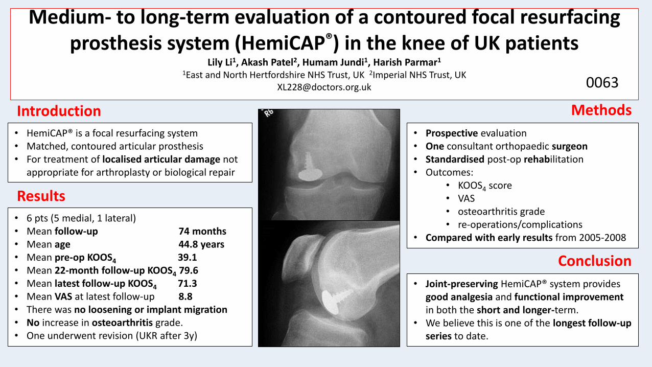
Medium- to long-term evaluation of a contoured focal resurfacing prosthesis system (HemiCAP®) in the knee of UK patients
Lily Li1, Akash Patel2, Humam Jundi1, Harish Parmar1
1East and North Hertfordshire NHS Trust, UK 2Imperial NHS Trust, [email protected] 0063
Introduction
• HemiCAP® is a focal resurfacing system• Matched, contoured articular prosthesis• For treatment of localised articular damage not
appropriate for arthroplasty or biological repair
Methods
• Prospective evaluation• One consultant orthopaedic surgeon • Standardised post-op rehabilitation • Outcomes:
• KOOS4 score• VAS• osteoarthritis grade• re-operations/complications
• Compared with early results from 2005-2008
Results
• 6 pts (5 medial, 1 lateral)• Mean follow-up 74 months• Mean age 44.8 years • Mean pre-op KOOS4 39.1• Mean 22-month follow-up KOOS4 79.6• Mean latest follow-up KOOS4 71.3• Mean VAS at latest follow-up 8.8• There was no loosening or implant migration • No increase in osteoarthritis grade. • One underwent revision (UKR after 3y)
Conclusion
• Joint-preserving HemiCAP® system provides good analgesia and functional improvement in both the short and longer-term.
• We believe this is one of the longest follow-up series to date.
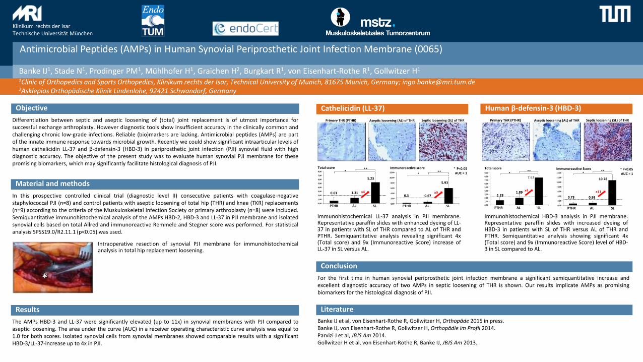
Antimicrobial Peptides (AMPs) in Human Synovial Periprosthetic Joint Infection Membrane (0065)
Klinikum rechts der IsarTechnische Universität München
Banke IJ1, Stade N1, Prodinger PM1, Mühlhofer H1, Graichen H2, Burgkart R1, von Eisenhart-Rothe R1, Gollwitzer H1
1Clinic of Orthopedics and Sports Orthopedics, Klinikum rechts der Isar, Technical University of Munich, 81675 Munich, Germany; [email protected] Orthopädische Klinik Lindenlohe, 92421 Schwandorf, Germany
Objective
Differentiation between septic and aseptic loosening of (total) joint replacement is of utmost importance forsuccessful exchange arthroplasty. However diagnostic tools show insufficient accuracy in the clinically common andchallenging chronic low-grade infections. Reliable (bio)markers are lacking. Antimicrobial peptides (AMPs) are partof the innate immune response towards microbial growth. Recently we could show significant intraarticular levels ofhuman cathelicidin LL-37 and β-defensin-3 (HBD-3) in periprosthetic joint infection (PJI) synovial fluid with highdiagnostic accuracy. The objective of the present study was to evaluate human synovial PJI membrane for thesepromising biomarkers, which may significantly facilitate histological diagnosis of PJI.
Material and methods
In this prospective controlled clinical trial (diagnostic level II) consecutive patients with coagulase-negativestaphylococcal PJI (n=8) and control patients with aseptic loosening of total hip (THR) and knee (TKR) replacements(n=9) according to the criteria of the Muskuloskeletal Infection Society or primary arthroplasty (n=8) were included.Semiquantitative immunohistochemical analysis of the AMPs HBD-2, HBD-3 and LL-37 in PJI membrane and isolatedsynovial cells based on total Allred and immunoreactive Remmele and Stegner score was performed. For statisticalanalysis SPSS19.0/R2.11.1 (p<0.05) was used.
Results
The AMPs HBD-3 and LL-37 were significantly elevated (up to 11x) in synovial membranes with PJI compared toaseptic loosening. The area under the curve (AUC) in a receiver operating characteristic curve analysis was equal to1.0 for both scores. Isolated synovial cells from synovial membranes showed comparable results with a significantHBD-3/LL-37-increase up to 4x in PJI.
Intraoperative resection of synovial PJI membrane for immunohistochemicalanalysis in total hip replacement loosening.
*
Cathelicidin (LL-37) Human β-defensin-3 (HBD-3)
Conclusion
For the first time in human synovial periprosthetic joint infection membrane a significant semiquantitative increase andexcellent diagnostic accuracy of two AMPs in septic loosening of THR is shown. Our results implicate AMPs as promisingbiomarkers for the histological diagnosis of PJI.
Literature
Banke IJ et al, von Eisenhart-Rothe R, Gollwitzer H, Orthopäde 2015 in press.Banke IJ, von Eisenhart-Rothe R, Gollwitzer H, Orthopädie im Profil 2014.Parvizi J et al, JBJS Am 2014.Gollwitzer H et al, von Eisenhart-Rothe R, Banke IJ, JBJS Am 2013.
Immunohistochemical LL-37 analysis in PJI membrane.Representative paraffin slides with enhanced dyeing of LL-37 in patients with SL of THR compared to AL of THR andPTHR. Semiquantitative analysis revealing significant 4x(Total score) and 9x (Immunoreactive Score) increase ofLL-37 in SL versus AL.
Immunohistochemical HBD-3 analysis in PJI membrane.Representative paraffin slides with increased dyeing ofHBD-3 in patients with SL of THR versus AL of THR andPTHR. Semiquantitative analysis showing significant 4x(Total score) and 9x (Immunoreactive Score) level of HBD-3 in SL compared to AL.
PrimaryTHR(PTHR) Asep cloosening(AL)ofTHR Sep cloosening(SL)ofTHR
0,00
2,00
4,00
6,00
8,00
10,00
12,00
pTEP Asep sch Sep sch
0,00
1,00
2,00
3,00
4,00
5,00
6,00
7,00
8,00
pTEP Asep sch Sep sch
***
Totalscore Immunoreac vescore
0.63 1.31
5.23
PTHR AL SL PTHR AL SL
0.3 0.67
5.93
***
*P<0.05AUC=1
x4 x9
PrimaryTHR(PTHR) Asep cloosening(AL)ofTHR Sep cloosening(SL)ofTHR
x4
Totalscore
1.281.89
7.62*
**
1,281,89
7,62
0,00
1,00
2,00
3,00
4,00
5,00
6,00
7,00
8,00
9,00
pTEP Asep sch Sep schPTHR AL SL
0.73 0.98
10.78
*** *P<0.05
AUC=1
Immunoreac veScore
0,73 0,98
10,78
0,00
2,00
4,00
6,00
8,00
10,00
12,00
14,00
pTEP Asep sch Sep schPTHR AL SL
x11
Inconsistencies In The Enhanced Recovery Programmes Across the NHSNagra NS, Hamilton TW, Murray DW, The BONE Collaborative, Pandit HG
Nuffield Orthopaedic Centre, Oxford University Hospitals NHS Trust, Oxford University Clinical Academic Graduate School (OUCAGS)[email protected] (No. 0070)
BackgroundEnhanced Recovery Programmes (ERPs) have been demonstrated to be clinically andcost effective, significantly reducing patient morbidity and mortality across a range ofsurgical specialties.
AimIdentify variations in current ERPs for primary knee arthroplasty across the NHS andestablish adherence to local policies within individual trusts.
MethodsDirected through the British Orthopaedic Network Environment (BONE), twenty NHStrusts offering elective knee arthroplasty completed a pro-forma to establish thecomposition of their knee arthroplasty ERP and performed a fifteen patient (percentre) audit to assess compliance.
Results• Sixteen centres completed the audit. Figure 1.• A large contrast of content and detail of ERPs within different centres was noted.• Pre-operative education was provided in 88% of centres, although content and
delivery varied.• There was wide variation in anaesthetic technique recommended as part of the
ERP with nerve blocks seldom advised.• The use of analgesic, anti-inflammatory and anti-fibrinolytics drugs varied
between centres with regards to dose and timing of delivery in the peri-operativeperiod.
• Gabapentinoids was used in 75% of ERPs.• Tranexamic acid was used in 60% of ERPs.• Peri-articular local anaesthetic infiltration was used in 60% of ERPs.
• Adherence to ERPs varied significantly between centres (XX to XX).
DiscussionSignificant variations exist in the content and delivery of ERPs with manycomponents lacking an evidence base, and poor uptake of those components thathave been demonstrated to be effective. Furthermore, despite guideline compliancewith ERP protocols is poor.
To improve patient care a gold standard evidence-based ERP should beestablished, and used across the NHS.
To become involved please visit the BONE Collaborativewww.bone.ac.uk
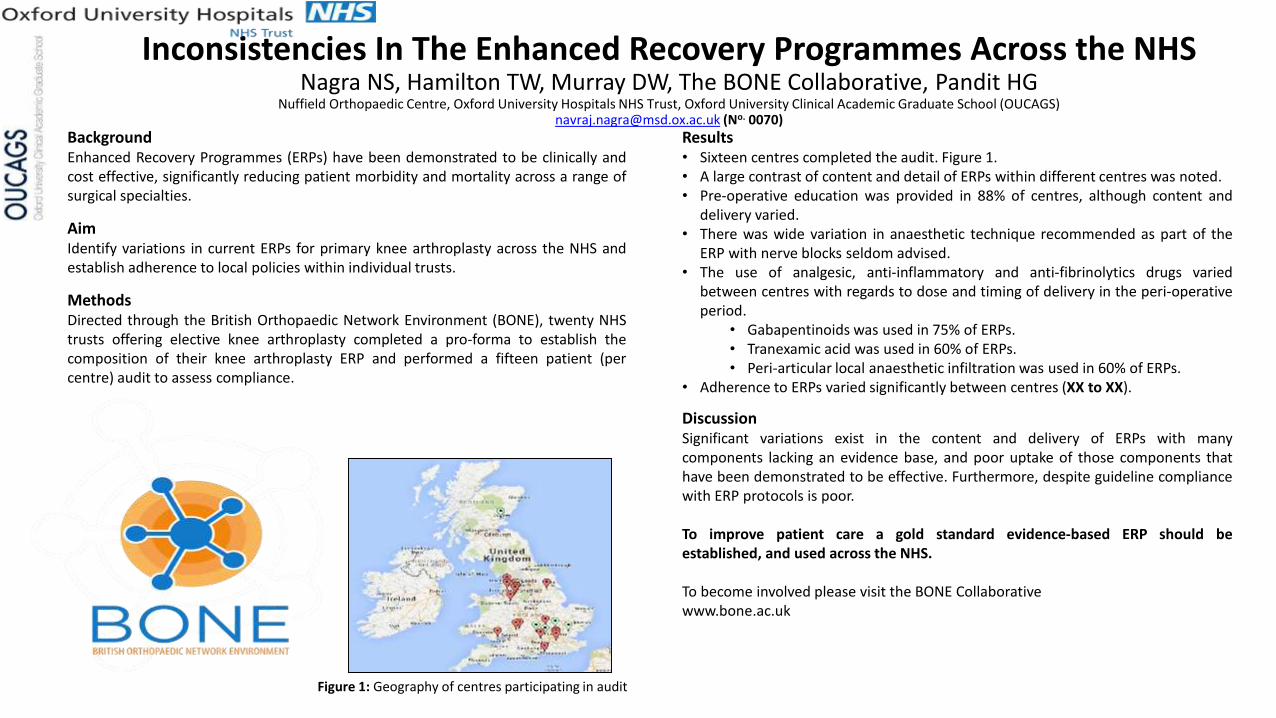
Figure 1: Geography of centres participating in audit
© 2001. Poster provided by Information Design & Imaging, Upper Merion, 8-276-6754
Outcomes of Patients Over 65 Undergoing Knee Arthroscopy in the Presence of Radiological Osteoarthritis
(e-poster number 0072)M.A. Lea, E.W. Yates, G.J. McLauchlan. Lancashire Teaching Hospitals Trust. Email: [email protected]
Introduction
•NICE guidance (IPG230) currently
recommends knee arthroscopy and
debridement but not just washout for the
treatment of OA (1)
•Recent update to NICE guidelines suggests
mechanical symptoms a reason for knee
arthroscopy
•Moseley NEJM 2002 – Randomised controlled
trial of 180 patients. No statistically significant
difference at 2 years when assessing pain relief
and knee function (2)
•Cochrane Review 2009 – Probably does not
improve pain or ability to function compared to
placebo (sham surgery) (3)
Aims
•Describe how effective knee arthroscopy is in
managing symptoms in this population
•Identify any correlation between the x-ray
findings, arthroscopy findings and clinical
outcome
•Show how many patients underwent TKR
within 1 year?
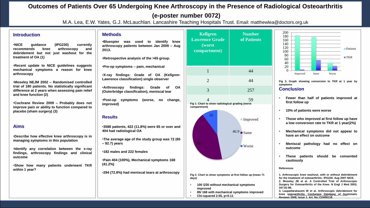
Fig 3. Graph showing conversion to TKR at 1 year by
symptoms
Conclusion
• Fewer than half of patients improved at
first follow up
• 10% of patients were worse
• Those who improved at first follow up have
a low conversion rate to TKR at 1 year(2%)
• Mechanical symptoms did not appear to
have an effect on outcome
• Meniscal pathology had no effect on
outcome
• These patients should be consented
cautiously
References
1. Arthroscopic knee washout, with or without debridement
for the treatment of osteoarthritis: IPG230. Aug 2007 NICE.
2. Moseley JB et al. A Controlled Trial of Arthroscopic
Surgery for Osteoarthritis of the Knee. N Engl J Med 2002;
347:81-88.
3. Laupattarakasem W et al. Arthroscopic debridement for
knee osteoarthritis. Cocharane Database of Systematic
Reviews 2008, Issue 1. Art. No.:CD005118.
Kellgren
Lawrence Grade
(worst
compartment)
Number
of Patients
1 44
2 44
3 257
4 59
Methods
•Bluespier was used to identify knee
arthroscopy patients between Jan 2009 – Aug
2013.
•Retrospective analysis of the >65 group.
•Pre-op symptoms – pain, mechanical
•X-ray findings: Grade of OA (Kellgren-
Lawrence classification) single observer
•Arthroscopy findings: Grade of OA
(Outerbridge classification), meniscal tear
•Post-op symptoms (worse, no change,
improved)
Results
•3586 patients, 422 (11.8%) were 65 or over and
404 had radiological OA
•The average age of the study group was 72 (65
– 92.7) years
•182 males and 222 females
•Pain 404 (100%), Mechanical symptoms 168
(41.2%)
•294 (72.8%) had meniscal tears at arthroscopy
Fig 1. Chart to show radiological grading (worst
compartment)
Fig 2. Chart to show symptoms at first follow up (mean 71
days)
• 100 /235 without mechanical symptoms
improved
• 85/ 168 with mechanical symptoms improved
• Chi-squared 2.55, p=0.11
020406080
100120140160180200
Improved Same Worse
Patients
TKR
46.9
43.1
10Improved
Same
Worse
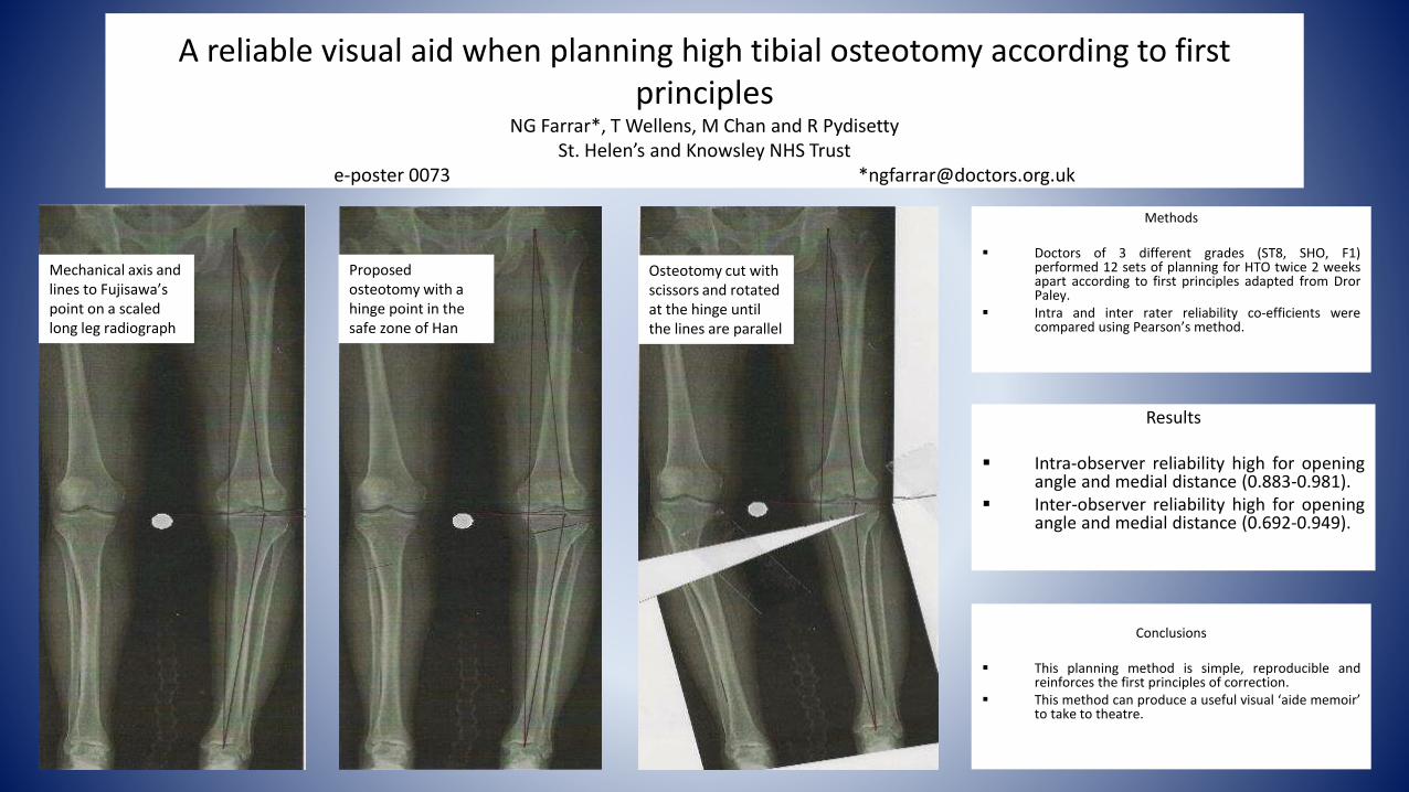
A reliable visual aid when planning high tibial osteotomy according to first principles
NG Farrar*, T Wellens, M Chan and R PydisettySt. Helen’s and Knowsley NHS Trust
e-poster 0073 *[email protected]
Methods
Doctors of 3 different grades (ST8, SHO, F1)performed 12 sets of planning for HTO twice 2 weeksapart according to first principles adapted from DrorPaley.
Intra and inter rater reliability co-efficients werecompared using Pearson’s method.
Mechanical axis and lines to Fujisawa’s point on a scaled long leg radiograph
Proposed osteotomy with a hinge point in the safe zone of Han
Osteotomy cut with scissors and rotated at the hinge until the lines are parallel
Results
Intra-observer reliability high for openingangle and medial distance (0.883-0.981).
Inter-observer reliability high for openingangle and medial distance (0.692-0.949).
Conclusions
This planning method is simple, reproducible andreinforces the first principles of correction.
This method can produce a useful visual ‘aide memoir’to take to theatre.
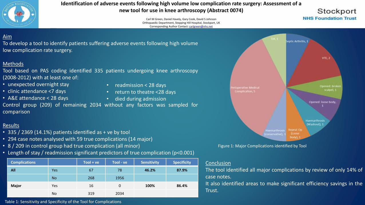
AimTo develop a tool to identify patients suffering adverse events following high volumelow complication rate surgery.
MethodsTool based on PAS coding identified 335 patients undergoing knee arthroscopy(2008-2012) with at least one of:• unexpected overnight stay• clinic attendance <7 days• A&E attendance < 28 daysControl group (209) of remaining 2034 without any factors was sampled forcomparison
Results• 335 / 2369 (14.1%) patients identified as + ve by tool• 294 case notes analysed with 59 true complications (14 major)• 8 / 209 in control group had true complication (all minor)• Length of stay / readmission significant predictors of true complication (p<0.001)
Table 1: Sensitivity and Specificity of the Tool for Complications
Figure 1: Major Complications identified by Tool
ConclusionThe tool identified all major complications by review of only 14% ofcase notes.It also identified areas to make significant efficiency savings in theTrust.
Complications Tool + ve Tool - ve Sensitivity Specificity
All Yes 67 78 46.2% 87.9%
No 268 1956
Major Yes 16 0 100% 86.4%
No 319 2034
• readmission < 28 days• return to theatre <28 days• died during admission
Identification of adverse events following high volume low complication rate surgery: Assessment of a new tool for use in knee arthroscopy (Abstract 0074)
Carl M Green, Daniel Havely, Gary Cook, David S JohnsonOrthopaedic Department, Stepping Hill Hospital, Stockport, UK
Corresponding Author Contact: [email protected]
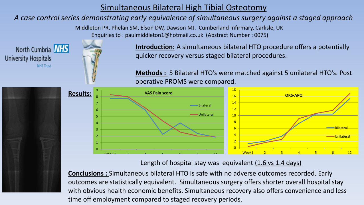
Simultaneous Bilateral High Tibial Osteotomy A case control series demonstrating early equivalence of simultaneous surgery against a staged approach
Middleton PR, Phelan SM, Elson DW, Dawson MJ. Cumberland Infirmary, Carlisle, UKEnquiries to : [email protected] (Abstract Number : 0075)
Introduction: A simultaneous bilateral HTO procedure offers a potentially quicker recovery versus staged bilateral procedures.
Methods : 5 Bilateral HTO’s were matched against 5 unilateral HTO’s. Post operative PROMS were compared.
Conclusions : Simultaneous bilateral HTO is safe with no adverse outcomes recorded. Early outcomes are statistically equivalent. Simultaneous surgery offers shorter overall hospital stay with obvious health economic benefits. Simultaneous recovery also offers convenience and less time off employment compared to staged recovery periods.
Results:
Length of hospital stay was equivalent (1.6 vs 1.4 days)
0
1
2
3
4
5
6
7
8
9
Week 1 2 3 4 5 6 12
VAS Pain score
Bilateral
Unilateral
0
2
4
6
8
10
12
14
16
18
Week1 2 3 4 5 6 12
OKS-APQ
Bilateral
Unilateral
Follow us on Twitter
@baskonline
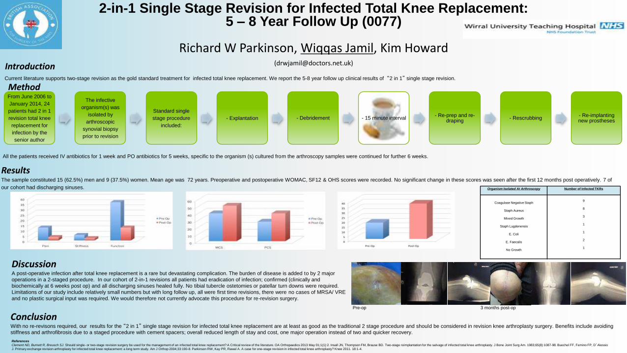
2-in-1 Single Stage Revision for Infected Total Knee Replacement: 5 – 8 Year Follow Up (0077)
Richard W Parkinson, Wiqqas Jamil, Kim Howard([email protected])
References
Clement ND, Burnett R, Breusch SJ. Should single- or two-stage revision surgery be used for the management of an infected total knee replacement? A Critical review of the literature. OA Orthopaedics 2013 May 01;1(1):2. Insall JN, Thompson FM, Brause BD. Two-stage reimplantation for the salvage of infected total knee arthroplasty. J Bone Joint Surg Am. 1983;65(8):1087-98. Buechel FF, Femino FP, D’Alessio
J. Primary exchange revision arthroplasty for infected total knee replacement: a long term study. Am J Orthop 2004;33:190-8. Parkinson RW, Kay PR, Rawal A. A case for one-stage revision in infected total knee arthroplasty? Knee 2011. 18:1-4.
Introduction
From June 2006 to
January 2014, 24
patients had 2 in 1
revision total knee
replacement for
infection by the
senior author
The infective
organism(s) was
isolated by
arthroscopic
synovial biopsy
prior to revision
Standard single
stage procedure
included:
- Explantation - Debridement - 15 minute interval - Re-prep and re-
draping- Rescrubbing
- Re-implanting new prostheses
Method
ResultsThe sample constituted 15 (62.5%) men and 9 (37.5%) women. Mean age was 72 years. Preoperative and postoperative WOMAC, SF12 & OHS scores were recorded. No significant change in these scores was seen after the first 12 months post operatively. 7 of
our cohort had discharging sinuses.
WOMAC Scores SF12 Scores Oxford Knee Scores
Organism Isolated At Arthroscopy Number of Infected TKRs
Coagulase Negative Staph
Staph Aureus
Mixed Growth
Staph Lugdenensis
E. Coli
E. Faecalis
No Growth
9
8
3
1
1
2
1
ConclusionWith no re-revisons required, our results for the “2 in 1” single stage revision for infected total knee replacement are at least as good as the traditional 2 stage procedure and should be considered in revision knee arthroplasty surgery. Benefits include avoiding
stiffness and arthrofibrosis due to a staged procedure with cement spacers; overall reduced length of stay and cost, one major operation instead of two and quicker recovery.
Current literature supports two-stage revision as the gold standard treatment for infected total knee replacement. We report the 5-8 year follow up clinical results of “2 in 1” single stage revision.
All the patients received IV antibiotics for 1 week and PO antibiotics for 5 weeks, specific to the organism (s) cultured from the arthroscopy samples were continued for further 6 weeks.
DiscussionA post-operative infection after total knee replacement is a rare but devastating complication. The burden of disease is added to by 2 major
operations in a 2-staged procedure. In our cohort of 2-in-1 revisions all patients had eradication of infection; confirmed (clinically and
biochemically at 6 weeks post op) and all discharging sinuses healed fully. No tibial tubercle ostetomies or patellar turn downs were required.
Limitations of our study include relatively small numbers but with long follow up, all were first time revisions, there were no cases of MRSA/ VRE
and no plastic surgical input was required. We would therefore not currently advocate this procedure for re-revision surgery.
Pre-op 3 months post-op
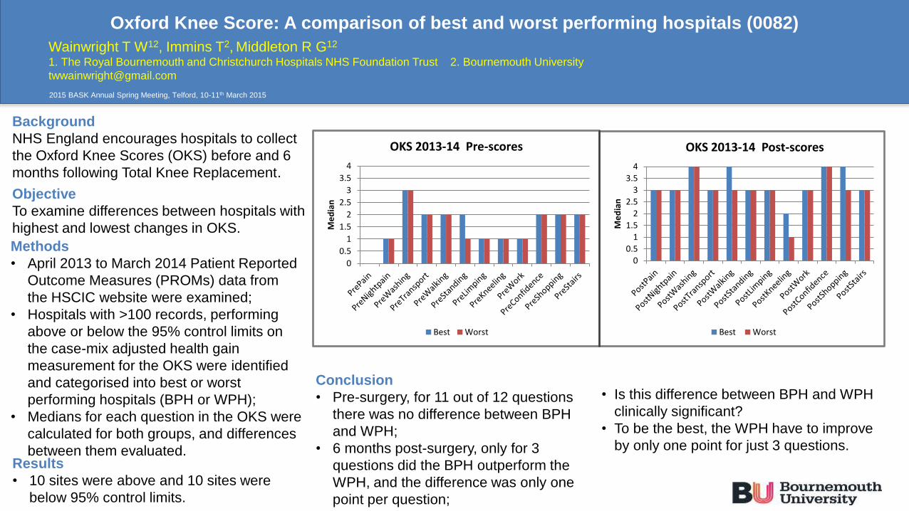
Conclusion
• Pre-surgery, for 11 out of 12 questions
there was no difference between BPH
and WPH;
• 6 months post-surgery, only for 3
questions did the BPH outperform the
WPH, and the difference was only one
point per question;
Oxford Knee Score: A comparison of best and worst performing hospitals (0082)
Background
NHS England encourages hospitals to collect
the Oxford Knee Scores (OKS) before and 6
months following Total Knee Replacement.
Objective
To examine differences between hospitals with
highest and lowest changes in OKS.
Methods
• April 2013 to March 2014 Patient Reported
Outcome Measures (PROMs) data from
the HSCIC website were examined;
• Hospitals with >100 records, performing
above or below the 95% control limits on
the case-mix adjusted health gain
measurement for the OKS were identified
and categorised into best or worst
performing hospitals (BPH or WPH);
• Medians for each question in the OKS were
calculated for both groups, and differences
between them evaluated.Results
• 10 sites were above and 10 sites were
below 95% control limits.
2015 BASK Annual Spring Meeting, Telford, 10-11th March 2015
Wainwright T W12, Immins T2, Middleton R G12
1. The Royal Bournemouth and Christchurch Hospitals NHS Foundation Trust 2. Bournemouth University
• Is this difference between BPH and WPH
clinically significant?
• To be the best, the WPH have to improve
by only one point for just 3 questions.
00.5
11.5
22.5
33.5
4
Me
dia
n
OKS 2013-14 Post-scores
Best Worst
0
0.5
1
1.5
2
2.5
3
3.5
4
Me
dia
n
OKS 2013-14 Pre-scores
Best Worst
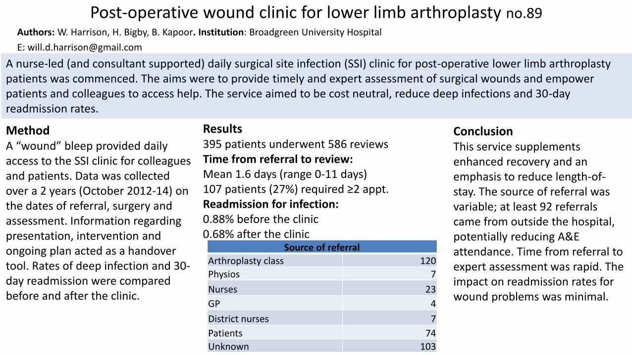
Post-operative wound clinic for lower limb arthroplasty no.89Authors: W. Harrison, H. Bigby, B. Kapoor. Institution: Broadgreen University Hospital
ConclusionThis service supplements enhanced recovery and an emphasis to reduce length-of-stay. The source of referral was variable; at least 92 referrals came from outside the hospital, potentially reducing A&E attendance. Time from referral to expert assessment was rapid. The impact on readmission rates for wound problems was minimal.
A nurse-led (and consultant supported) daily surgical site infection (SSI) clinic for post-operative lower limb arthroplasty patients was commenced. The aims were to provide timely and expert assessment of surgical wounds and empower patients and colleagues to access help. The service aimed to be cost neutral, reduce deep infections and 30-day readmission rates.
MethodA “wound” bleep provided daily access to the SSI clinic for colleagues and patients. Data was collected over a 2 years (October 2012-14) on the dates of referral, surgery and assessment. Information regarding presentation, intervention and ongoing plan acted as a handover tool. Rates of deep infection and 30-day readmission were compared before and after the clinic.
Results395 patients underwent 586 reviewsTime from referral to review: Mean 1.6 days (range 0-11 days)107 patients (27%) required ≥2 appt.Readmission for infection:0.88% before the clinic0.68% after the clinic
Source of referral
Arthroplasty class 120
Physios 7
Nurses 23
GP 4
District nurses 7
Patients 74
Unknown 103
0090
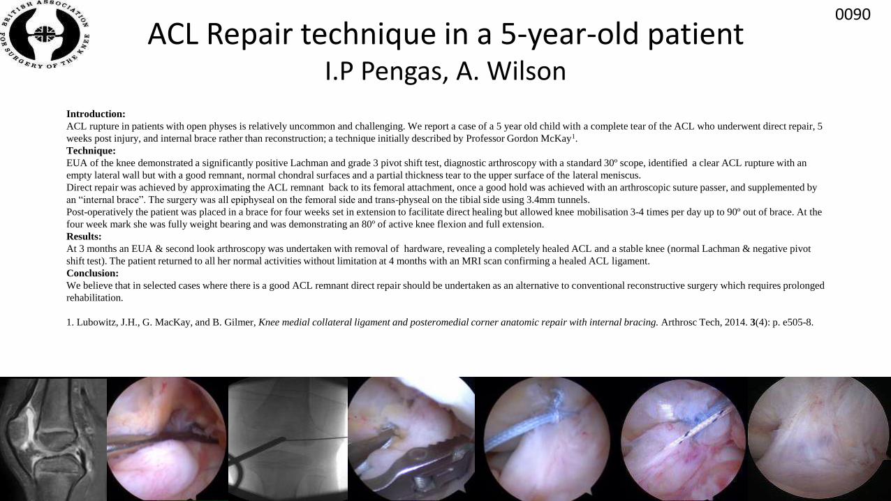
ACL Repair technique in a 5-year-old patientI.P Pengas, A. Wilson
Introduction:
ACL rupture in patients with open physes is relatively uncommon and challenging. We report a case of a 5 year old child with a complete tear of the ACL who underwent direct repair, 5
weeks post injury, and internal brace rather than reconstruction; a technique initially described by Professor Gordon McKay1.
Technique:
EUA of the knee demonstrated a significantly positive Lachman and grade 3 pivot shift test, diagnostic arthroscopy with a standard 30º scope, identified a clear ACL rupture with an
empty lateral wall but with a good remnant, normal chondral surfaces and a partial thickness tear to the upper surface of the lateral meniscus.
Direct repair was achieved by approximating the ACL remnant back to its femoral attachment, once a good hold was achieved with an arthroscopic suture passer, and supplemented by
an “internal brace”. The surgery was all epiphyseal on the femoral side and trans-physeal on the tibial side using 3.4mm tunnels.
Post-operatively the patient was placed in a brace for four weeks set in extension to facilitate direct healing but allowed knee mobilisation 3-4 times per day up to 90º out of brace. At the
four week mark she was fully weight bearing and was demonstrating an 80º of active knee flexion and full extension.
Results:
At 3 months an EUA & second look arthroscopy was undertaken with removal of hardware, revealing a completely healed ACL and a stable knee (normal Lachman & negative pivot
shift test). The patient returned to all her normal activities without limitation at 4 months with an MRI scan confirming a healed ACL ligament.
Conclusion:
We believe that in selected cases where there is a good ACL remnant direct repair should be undertaken as an alternative to conventional reconstructive surgery which requires prolonged
rehabilitation.
1. Lubowitz, J.H., G. MacKay, and B. Gilmer, Knee medial collateral ligament and posteromedial corner anatomic repair with internal bracing. Arthrosc Tech, 2014. 3(4): p. e505-8.
Patient satisfaction following secondary patellar resurfacing after Total Knee Arthroplasty: Results from an arthoplasty register
CJ Thomas1, V Patel2, CN Esler2, RU Ashford2
1. University Hospitals of Coventry & Warwickshire 2. University Hospitals of Leicester
Introduction
There remains a significant lack of consensus regarding the third compartment of the knee
in arthroplasty. Whilst strong evidence supporting primary patellar resurfacing remains
elusive [1,2,3], some studies have demonstrated reduced rates of revision when the
patella is primarily resurfaced [4,5], however the possibility of catastrophic complications
has led some to suggest that selective resurfacing may be the solution [6].
Despite this, anterior knee pain remains a relatively common complication of bi-
compartment TKA [7,8], for which some would consider secondary patellar resurfacing the
next step. A number of relatively small studies have considered the role of secondary
resurfacing for patients who report anterior knee pain following bi-compartment TKA [6, 9,
10, 11, 12, 13, 14] however it is difficult to draw any strong or significant conclusions from
them.
We sought to determine whether patients undergoing secondary resurfacing benefitted
from the procedure.
Methods
We assessed patients’ post-operative satisfaction with secondary patellar resurfacing
following bi-compartment total knee arthroplasty (TKA) via a postal questionnaire. All
patients included in the Trent and Wales Arthroplasty Register (TWAR) for secondary
patellar resurfacing were invited to participate using patient reported outcome measures
including the Oxford Knee Score (OKS), a modified Kujala (patello-femoral) score, the
EuroQual EQ-5D-3L and EuroQual visual analogue score for overall health.
The Trent and Wales Arthroplasty Register
Practically all patients who receive a primary or secondary arthroplastic procedure of the
hip or knee are reported to the Trent and Wales Arthroplasty Register (TWAR). TWAR was
established in 1990. The register now supports 118 surgeons at 31 hospitals. The TWAR
receives all data directly from consultant surgeons at the time of operation, a proportion of
which is independently verified by clerks from the Trent Arthroplasty Audit Group (TAAG).
Participation is by consent only following the 1998 Data Protection Act. Neither pre nor
post-operative patient-related outcome measures (PROMs) are recorded in the register.
Results
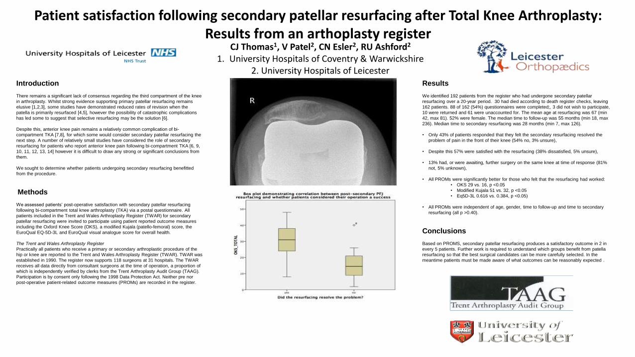
We identified 192 patients from the register who had undergone secondary patellar
resurfacing over a 20-year period. 30 had died according to death register checks, leaving
162 patients. 88 of 162 (54%) questionnaires were completed;, 3 did not wish to participate,
10 were returned and 61 were unaccounted for. The mean age at resurfacing was 67 (min
42, max 81). 52% were female. The median time to follow-up was 55 months (min 18, max
236). Median time to secondary resurfacing was 28 months (min 7, max 126).
• Only 43% of patients responded that they felt the secondary resurfacing resolved the
problem of pain in the front of their knee (54% no, 3% unsure),
• Despite this 57% were satisfied with the resurfacing (38% dissatisfied, 5% unsure),
• 13% had, or were awaiting, further surgery on the same knee at time of response (81%
not, 5% unknown),
• All PROMs were significantly better for those who felt that the resurfacing had worked:
• OKS 29 vs. 16, p <0.05
• Modified Kujala 51 vs. 32, p <0.05
• Eq5D-3L 0.616 vs. 0.384, p <0.05)
• All PROMs were independent of age, gender, time to follow-up and time to secondary
resurfacing (all p >0.40).
Conclusions
Based on PROMS, secondary patellar resurfacing produces a satisfactory outcome in 2 in
every 5 patients. Further work is required to understand which groups benefit from patella
resurfacing so that the best surgical candidates can be more carefully selected. In the
meantime patients must be made aware of what outcomes can be reasonably expected .
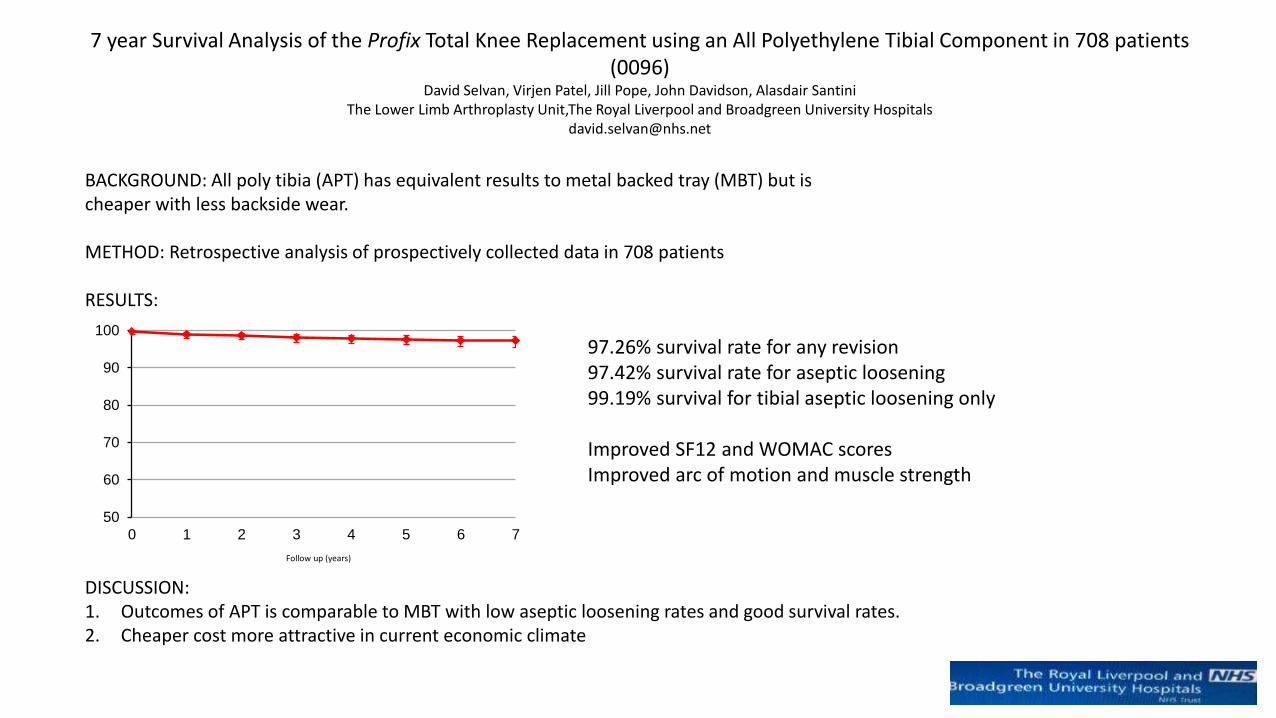
7 year Survival Analysis of the Profix Total Knee Replacement using an All Polyethylene Tibial Component in 708 patients (0096)
David Selvan, Virjen Patel, Jill Pope, John Davidson, Alasdair SantiniThe Lower Limb Arthroplasty Unit,The Royal Liverpool and Broadgreen University Hospitals
BACKGROUND: All poly tibia (APT) has equivalent results to metal backed tray (MBT) but is cheaper with less backside wear.
METHOD: Retrospective analysis of prospectively collected data in 708 patients
RESULTS:
50
60
70
80
90
100
0 1 2 3 4 5 6 7
97.26% survival rate for any revision97.42% survival rate for aseptic loosening99.19% survival for tibial aseptic loosening only
Improved SF12 and WOMAC scoresImproved arc of motion and muscle strength
DISCUSSION: 1. Outcomes of APT is comparable to MBT with low aseptic loosening rates and good survival rates.2. Cheaper cost more attractive in current economic climate
Follow up (years)
How Good Are We At Consenting?Comparing Standards In Practice To National Guidelines
Nagra NS, Street ER, Parker SJ, Kulkarni K, Malik M, Mann BStoke Mandeville Hospital, Buckinghamshire Healthcare NHS Trust, Oxford University Clinical Academic Graduate School (OUCAGS)
[email protected] (No. 0097)
IntroductionProper consenting is an important part of Good Medical Practice and providesprotection for Doctors - ‘If it’s not written down, it didn’t happen’.Litigation is an increasing problem (£14m/5 years in the NHS).Knee arthroscopy has a 4.7% complication rate (infection occurs in 0.84%).
AimTo determine the accuracy of our consenting of patients for complications ofarthroscopic procedures of the knee against BOA national guidelines.
Methods• Retrospective review of 33 electronic patient records and 31 consent forms.• Elements of consent were compared to ‘gold standard’ BOA guidelines.
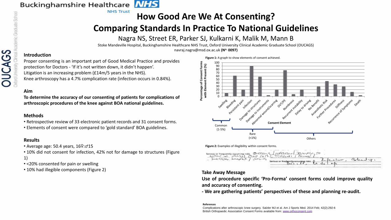
Results• Average age: 50.4 years, 16♀:♂15• 10% did not consent for infection, 42% not for damage to structures (Figure1)• <20% consented for pain or swelling• 10% had illegible components (Figure 2)
Take Away MessageUse of procedure specific ‘Pro-Forma’ consent forms could improve qualityand accuracy of consenting.- We are gathering patients’ perspectives of these and planning re-audit.
ReferencesComplications after arthroscopic knee surgery. Salzler MJ et al. Am J Sports Med. 2014 Feb; 42(2):292-6
British Orthopaedic Association Consent Forms available from: www.orthoconsent.com
0102030405060708090
100
Pe
rce
nta
ge o
f C
on
sen
t Fo
rms
wit
h E
lem
en
t P
rese
nt
(%)
Consent ElementCommon
(1-5%)
Rare(<1%) Others
Figure 1: A graph to show elements of consent achieved.
Figure 2: Examples of illegibility within consent forms.
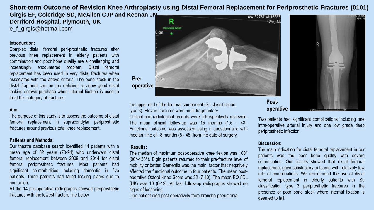
Short-term Outcome of Revision Knee Arthroplasty using Distal Femoral Replacement for Periprosthetic Fractures (0101)Girgis EF, Coleridge SD, McAllen CJP and Keenan JN
Derriford Hospital, Plymouth, UK
Introduction:
Complex distal femoral peri-prosthetic fractures after
previous knee replacement in elderly patients with
comminution and poor bone quality are a challenging and
increasingly encountered problem. Distal femoral
replacement has been used in very distal fractures when
associated with the above criteria. The bone stock in the
distal fragment can be too deficient to allow good distal
locking screws purchase when internal fixation is used to
treat this category of fractures.
Aim:
The purpose of this study is to assess the outcome of distal
femoral replacement in supracondylar periprosthetic
fractures around previous total knee replacement.
Patients and Methods:
Our theatre database search identified 14 patients with a
mean age of 82 years (70-94) who underwent distal
femoral replacement between 2009 and 2014 for distal
femoral periprosthetic fractures. Most patients had
significant co-morbidities including dementia in five
patients. Three patients had failed locking plates due to
non-union.
All the 14 pre-operative radiographs showed periprosthetic
fractures with the lowest fracture line below
the upper end of the femoral component (Su classification,
type 3). Eleven fractures were multi-fragmentary.
Clinical and radiological records were retrospectively reviewed.
The mean clinical follow-up was 15 months (1.5 - 43).
Functional outcome was assessed using a questionnaire with
median time of 18 months (5 - 45) from the date of surgery.
Results:
The median of maximum post-operative knee flexion was 100°
(90°-135°). Eight patients returned to their pre-fracture level of
mobility or better. Dementia was the main factor that negatively
affected the functional outcome in four patients. The mean post-
operative Oxford Knee Score was 22 (7-40). The mean EQ-5DL
(UK) was 10 (6-12). All last follow-up radiographs showed no
signs of loosening.
One patient died post-operatively from broncho-pneumonia.
Two patients had significant complications including one
intra-operative arterial injury and one low grade deep
periprosthetic infection.
Discussion:
The main indication for distal femoral replacement in our
patients was the poor bone quality with severe
comminution. Our results showed that distal femoral
replacement gave satisfactory outcome with relatively low
rate of complications. We recommend the use of distal
femoral replacement in elderly patients with Su
classification type 3 periprosthetic fractures in the
presence of poor bone stock where internal fixation is
deemed to fail.
Post-operative
Pre-
operative
CPD points at BASK
4 CPD points for education +
1 point per hour free papers relevant to practice
Make reflective notes
Complete post meeting SurveyMonkey feedback
No survey = No certificate
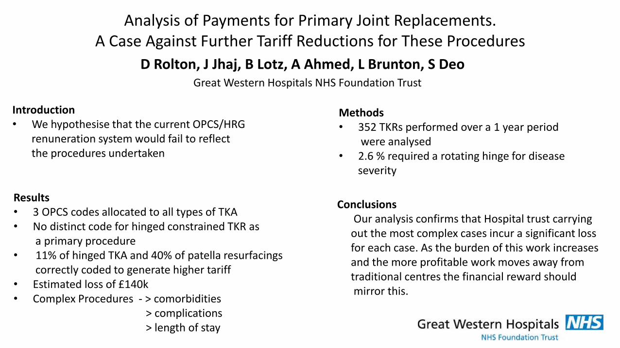
Analysis of Payments for Primary Joint Replacements. A Case Against Further Tariff Reductions for These Procedures
D Rolton, J Jhaj, B Lotz, A Ahmed, L Brunton, S DeoGreat Western Hospitals NHS Foundation Trust
Introduction• We hypothesise that the current OPCS/HRG
renuneration system would fail to reflectthe procedures undertaken
Methods• 352 TKRs performed over a 1 year period
were analysed• 2.6 % required a rotating hinge for disease
severity
Results• 3 OPCS codes allocated to all types of TKA• No distinct code for hinged constrained TKR as
a primary procedure• 11% of hinged TKA and 40% of patella resurfacings
correctly coded to generate higher tariff • Estimated loss of £140k• Complex Procedures - > comorbidities
> complications> length of stay
ConclusionsOur analysis confirms that Hospital trust carrying out the most complex cases incur a significant loss for each case. As the burden of this work increases and the more profitable work moves away from traditional centres the financial reward should mirror this.
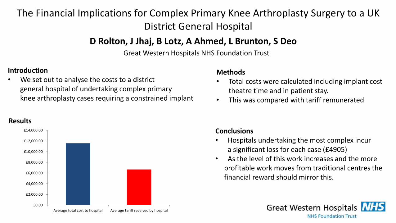
The Financial Implications for Complex Primary Knee Arthroplasty Surgery to a UK District General Hospital
D Rolton, J Jhaj, B Lotz, A Ahmed, L Brunton, S DeoGreat Western Hospitals NHS Foundation Trust
Introduction• We set out to analyse the costs to a district
general hospital of undertaking complex primary knee arthroplasty cases requiring a constrained implant
Methods• Total costs were calculated including implant cost
theatre time and in patient stay.• This was compared with tariff remunerated
Results
Conclusions• Hospitals undertaking the most complex incur
a significant loss for each case (£4905)• As the level of this work increases and the more
profitable work moves from traditional centres the financial reward should mirror this.
£0.00
£2,000.00
£4,000.00
£6,000.00
£8,000.00
£10,000.00
£12,000.00
£14,000.00
Average total cost to hospital Average tariff received by hospital
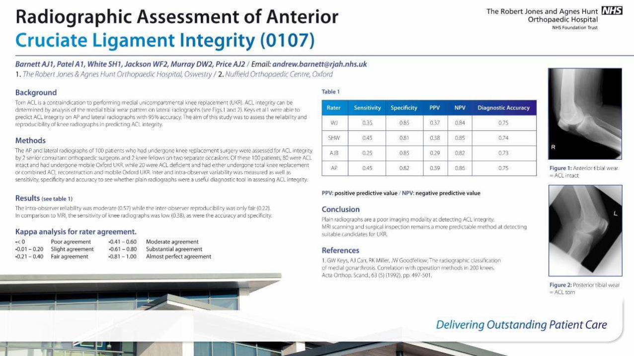
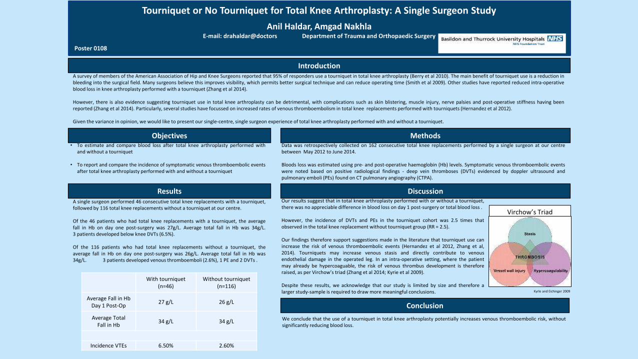
Tourniquet or No Tourniquet for Total Knee Arthroplasty: A Single Surgeon Study
Anil Haldar, Amgad NakhlaE-mail: drahaldar@doctors Department of Trauma and Orthopaedic Surgery
Poster 0108
IntroductionA survey of members of the American Association of Hip and Knee Surgeons reported that 95% of responders use a tourniquet in total knee arthroplasty (Berry et al 2010). The main benefit of tourniquet use is a reduction inbleeding into the surgical field. Many surgeons believe this improves visibility, which permits better surgical technique and can reduce operating time (Smith et al 2009). Other studies have reported reduced intra-operativeblood loss in knee arthroplasty performed with a tourniquet (Zhang et al 2014).
However, there is also evidence suggesting tourniquet use in total knee arthroplasty can be detrimental, with complications such as skin blistering, muscle injury, nerve palsies and post-operative stiffness having beenreported (Zhang et al 2014). Particularly, several studies have focussed on increased rates of venous thromboembolism in total knee replacements performed with tourniquets (Hernandez et al 2012).
Given the variance in opinion, we would like to present our single-centre, single surgeon experience of total knee arthroplasty performed with and without a tourniquet.
MethodsData was retrospectively collected on 162 consecutive total knee replacements performed by a single surgeon at our centrebetween May 2012 to June 2014.
Bloods loss was estimated using pre- and post-operative haemoglobin (Hb) levels. Symptomatic venous thromboembolic eventswere noted based on positive radiological findings - deep vein thromboses (DVTs) evidenced by doppler ultrasound andpulmonary emboli (PEs) found on CT pulmonary angiography (CTPA).
Results
Conclusion
We conclude that the use of a tourniquet in total knee arthroplasty potentially increases venous thromboembolic risk, withoutsignificantly reducing blood loss.
Objectives• To estimate and compare blood loss after total knee arthroplasty performed with
and without a tourniquet
• To report and compare the incidence of symptomatic venous thromboembolic eventsafter total knee arthroplasty performed with and without a tourniquet
A single surgeon performed 46 consecutive total knee replacements with a tourniquet,followed by 116 total knee replacements without a tourniquet at our centre.
Of the 46 patients who had total knee replacements with a tourniquet, the averagefall in Hb on day one post-surgery was 27g/L. Average total fall in Hb was 34g/L.3 patients developed below knee DVTs (6.5%).
Of the 116 patients who had total knee replacements without a tourniquet, theaverage fall in Hb on day one post-surgery was 26g/L. Average total fall in Hb was34g/L. 3 patients developed venous thromboemboli (2.6%), 1 PE and 2 DVTs .
DiscussionOur results suggest that in total knee arthroplasty performed with or without a tourniquet,there was no appreciable difference in blood loss on day 1 post-surgery or total blood loss .
However, the incidence of DVTs and PEs in the tourniquet cohort was 2.5 times thatobserved in the total knee replacement without tourniquet group (RR = 2.5).
Our findings therefore support suggestions made in the literature that tourniquet use canincrease the risk of venous thromboembolic events (Hernandez et al 2012, Zhang et al,2014). Tourniquets may increase venous stasis and directly contribute to venousendothelial damage in the operated leg. In an intra-operative setting, where the patientmay already be hypercoaguable, the risk of venous thrombus development is thereforeraised, as per Virchow’s triad (Zhang et al 2014; Kyrie et al 2009).
Despite these results, we acknowledge that our study is limited by size and therefore alarger study-sample is required to draw more meaningful conclusions.
With tourniquet (n=46)
Without tourniquet (n=116)
Average Fall in Hb Day 1 Post-Op
27 g/L 26 g/L
Average Total Fall in Hb
34 g/L 34 g/L
Incidence VTEs 6.50% 2.60%
Kyrle and Eichinger 2009
Virchow’s Triad
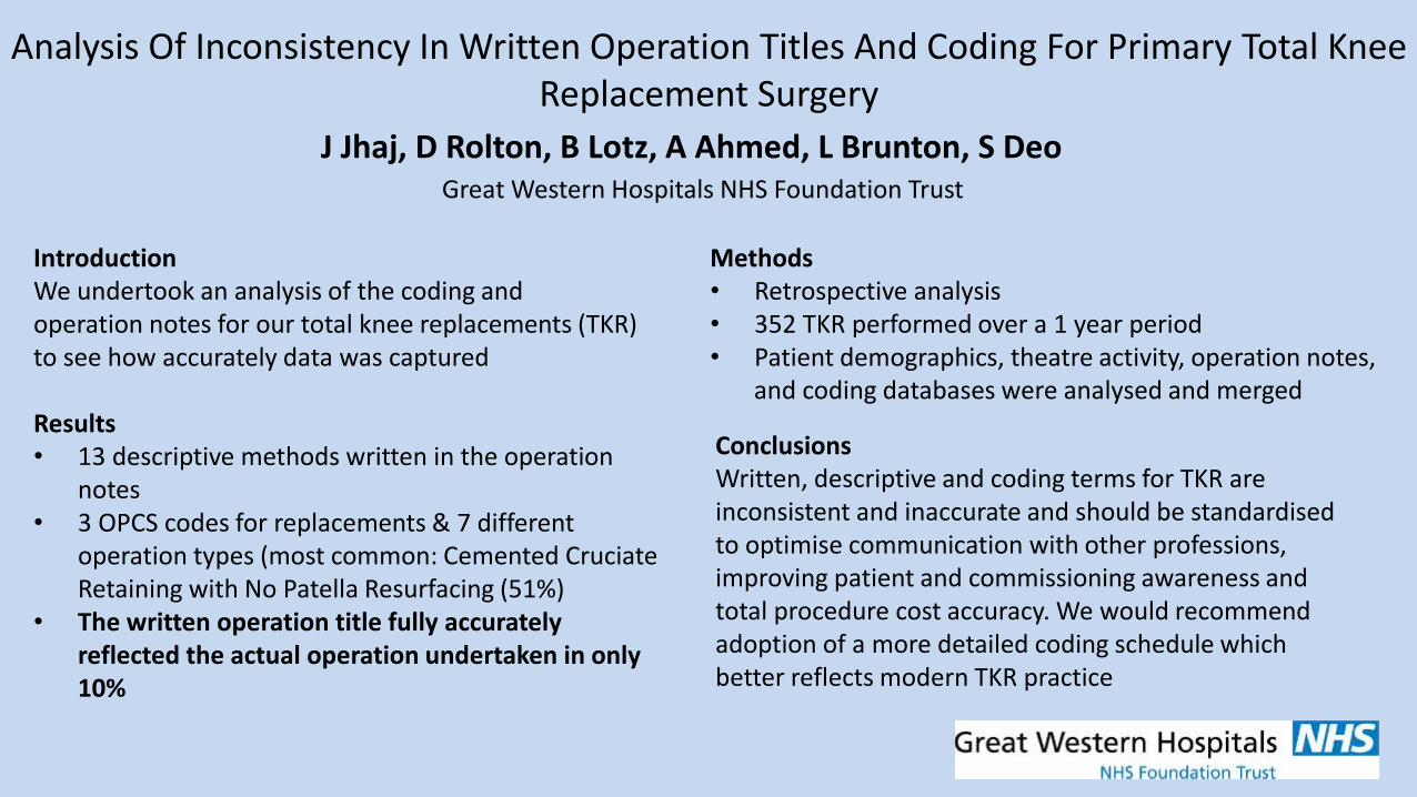
Analysis Of Inconsistency In Written Operation Titles And Coding For Primary Total Knee Replacement Surgery
J Jhaj, D Rolton, B Lotz, A Ahmed, L Brunton, S DeoGreat Western Hospitals NHS Foundation Trust
IntroductionWe undertook an analysis of the coding and operation notes for our total knee replacements (TKR) to see how accurately data was captured
Methods• Retrospective analysis• 352 TKR performed over a 1 year period • Patient demographics, theatre activity, operation notes,
and coding databases were analysed and mergedResults• 13 descriptive methods written in the operation
notes• 3 OPCS codes for replacements & 7 different
operation types (most common: Cemented Cruciate Retaining with No Patella Resurfacing (51%)
• The written operation title fully accurately reflected the actual operation undertaken in only 10%
ConclusionsWritten, descriptive and coding terms for TKR are inconsistent and inaccurate and should be standardised to optimise communication with other professions, improving patient and commissioning awareness and total procedure cost accuracy. We would recommend adoption of a more detailed coding schedule whichbetter reflects modern TKR practice
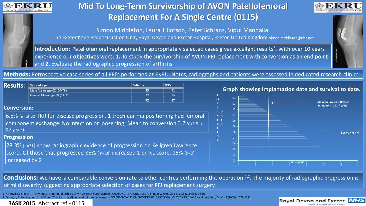
Simon Middleton, Laura Tillotson, Peter Schranz, Vipul Mandalia. The Exeter Knee Reconstruction Unit, Royal Devon and Exeter Hospital, Exeter, United Kingdom [email protected]
Mid To Long-Term Survivorship of AVON PatellofemoralReplacement For A Single Centre (0115)
Introduction: Patellofemoral replacement in appropriately selected cases gives excellent results1. With over 10 years experience our objectives were: 1. To study the survivorship of AVON PFJ replacement with conversion as an end point and 2. Evaluate the radiographic progression of arthritis.
Methods: Retrospective case series of all PFJ’s performed at EKRU. Notes, radiographs and patients were assessed in dedicated research clinics.
Results:
Conversion:
Progression:
Conclusions: We have a comparable conversion rate to other centres performing this operation 1,2. The majority of radiographic progression is of mild severity suggesting appropriate selection of cases for PFJ replacement surgery.
BASK 2015. Abstract ref:- 0115
Sex and age Patients PFJ's
Male Mean age 62 (34-78) 25 32
Female Mean age 59 (41- 82) 47 55
72 87
1. Ackroyd, C. E., et al. "The Avon patellofemoral arthroplasty FIVE-YEAR SURVIVORSHIP AND FUNCTIONAL RESULTS." J of Bone & Joint Surg Br 89.3 (2007): 310-315.2. Starks, I., S. Roberts, and S. H. White. "The Avon patellofemoral joint replacement INDEPENDENT ASSESSMENT OF EARLY FUNCTIONAL OUTCOMES." J of Bone & Joint Surg Br 91.12 (2009): 1579-1582.
6.8% [n=6] to TKR for disease progression. 1 trochlear malpositioning had femoral component exchange. No infection or loosening. Mean to conversion 3.7 y (1.9 to
8.8 years).
0 2 4 6 8 10 12 14
03
04
05
06
07
08
09
10
11
12
13
14
Time years
I
M
P
L
A
N
T
A
T
I
O
N
D
A
T
E
Mean follow up 3.8 years(6 months to 11.7 years)
28.3% [n=21] show radiographic evidence of progression on Kellgren Lawrence score. Of those that progressed 85% [ n=18] increased 1 on KL score, 15% [n=3]
increased by 2
Graph showing implantation date and survival to date.
Converted
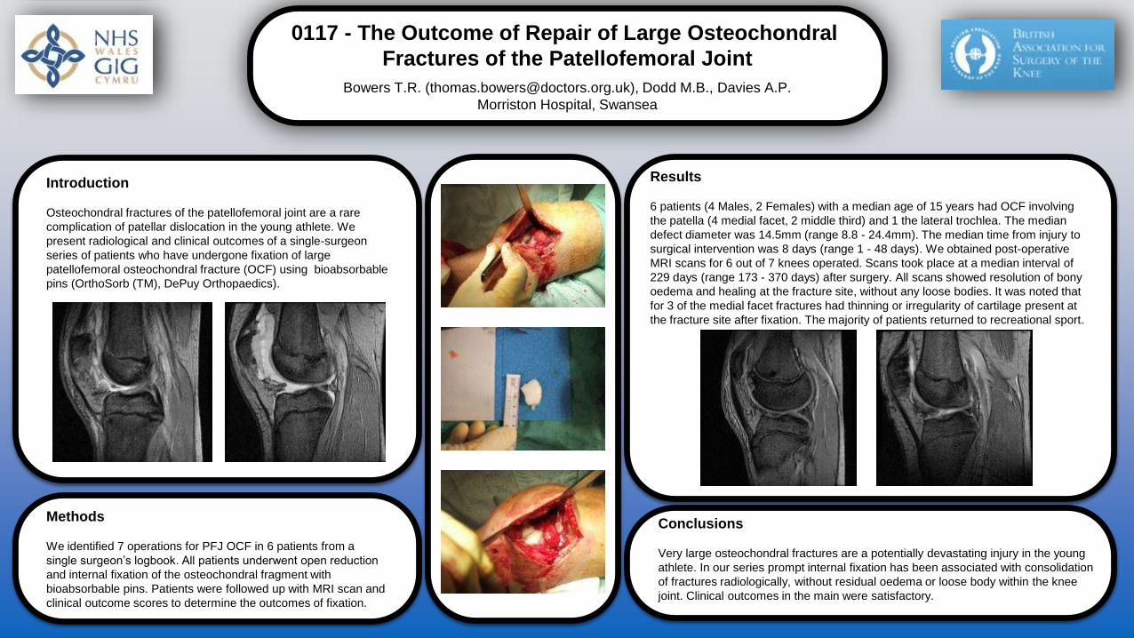
0117 - The Outcome of Repair of Large Osteochondral
Fractures of the Patellofemoral Joint
Bowers T.R. ([email protected]), Dodd M.B., Davies A.P.
Morriston Hospital, Swansea
Introduction
Osteochondral fractures of the patellofemoral joint are a rare
complication of patellar dislocation in the young athlete. We
present radiological and clinical outcomes of a single-surgeon
series of patients who have undergone fixation of large
patellofemoral osteochondral fracture (OCF) using bioabsorbable
pins (OrthoSorb (TM), DePuy Orthopaedics).
Methods
We identified 7 operations for PFJ OCF in 6 patients from a
single surgeon’s logbook. All patients underwent open reduction
and internal fixation of the osteochondral fragment with
bioabsorbable pins. Patients were followed up with MRI scan and
clinical outcome scores to determine the outcomes of fixation.
Results
6 patients (4 Males, 2 Females) with a median age of 15 years had OCF involving
the patella (4 medial facet, 2 middle third) and 1 the lateral trochlea. The median
defect diameter was 14.5mm (range 8.8 - 24.4mm). The median time from injury to
surgical intervention was 8 days (range 1 - 48 days). We obtained post-operative
MRI scans for 6 out of 7 knees operated. Scans took place at a median interval of
229 days (range 173 - 370 days) after surgery. All scans showed resolution of bony
oedema and healing at the fracture site, without any loose bodies. It was noted that
for 3 of the medial facet fractures had thinning or irregularity of cartilage present at
the fracture site after fixation. The majority of patients returned to recreational sport.
Conclusions
Very large osteochondral fractures are a potentially devastating injury in the young
athlete. In our series prompt internal fixation has been associated with consolidation
of fractures radiologically, without residual oedema or loose body within the knee
joint. Clinical outcomes in the main were satisfactory.
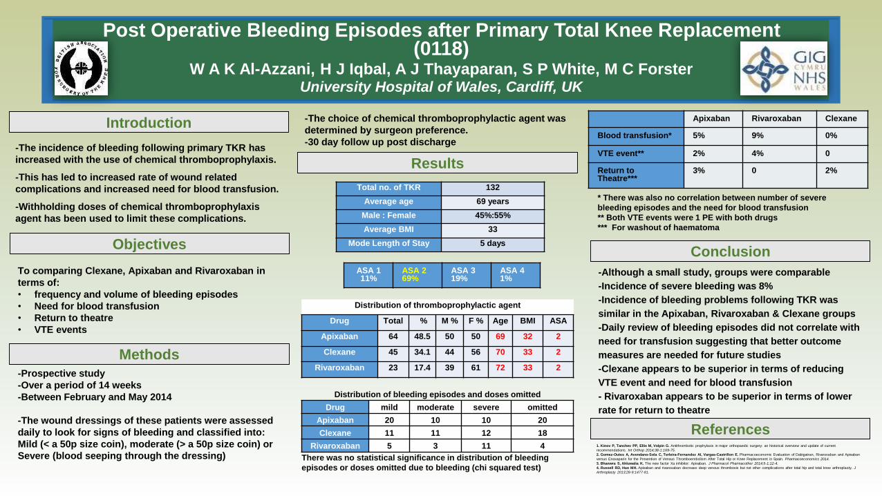
Post Operative Bleeding Episodes after Primary Total Knee Replacement (0118)
W A K Al-Azzani, H J Iqbal, A J Thayaparan, S P White, M C ForsterUniversity Hospital of Wales, Cardiff, UK
Introduction
ConclusionObjectives
References
Results
To comparing Clexane, Apixaban and Rivaroxaban in
terms of:
• frequency and volume of bleeding episodes
• Need for blood transfusion
• Return to theatre
• VTE events
1. Kinov P, Tanchev PP, Ellis M, Volpin G. Antithrombotic prophylaxis in major orthopaedic surgery: an historical overview and update of current
recommendations. Int Orthop 2014;38-1:169-75.
2. Gomez-Outes A, Avendano-Sola C, Terleira-Fernandez AI, Vargas-Castrillon E. Pharmacoeconomic Evaluation of Dabigatran, Rivaroxaban and Apixaban
versus Enoxaparin for the Prevention of Venous Thromboembolism After Total Hip or Knee Replacement in Spain. Pharmacoeconomics 2014.
3. Bhanwra S, Ahluwalia K. The new factor Xa inhibitor: Apixaban. J Pharmacol Pharmacother 2014;5-1:12-4.
4. Russell RD, Huo MH. Apixaban and rivaroxaban decrease deep venous thrombosis but not other complications after total hip and total knee arthroplasty. J
Arthroplasty 2013;28-9:1477-81.
-The choice of chemical thromboprophylactic agent was
determined by surgeon preference.
-30 day follow up post discharge-The incidence of bleeding following primary TKR has
increased with the use of chemical thromboprophylaxis.
-This has led to increased rate of wound related
complications and increased need for blood transfusion.
-Withholding doses of chemical thromboprophylaxis
agent has been used to limit these complications.
-Although a small study, groups were comparable
-Incidence of severe bleeding was 8%
-Incidence of bleeding problems following TKR was
similar in the Apixaban, Rivaroxaban & Clexane groups
-Daily review of bleeding episodes did not correlate with
need for transfusion suggesting that better outcome
measures are needed for future studies
-Clexane appears to be superior in terms of reducing
VTE event and need for blood transfusion
- Rivaroxaban appears to be superior in terms of lower
rate for return to theatre
Methods
-Prospective study
-Over a period of 14 weeks
-Between February and May 2014
-The wound dressings of these patients were assessed
daily to look for signs of bleeding and classified into:
Mild (< a 50p size coin), moderate (> a 50p size coin) or
Severe (blood seeping through the dressing)
Distribution of thromboprophylactic agent
Drug Total % M % F % Age BMI ASA
Apixaban 64 48.5 50 50 69 32 2
Clexane 45 34.1 44 56 70 33 2
Rivaroxaban 23 17.4 39 61 72 33 2
Distribution of bleeding episodes and doses omitted
Drug mild moderate severe omitted
Apixaban 20 10 10 20
Clexane 11 11 12 18
Rivaroxaban 5 3 11 4
There was no statistical significance in distribution of bleeding
episodes or doses omitted due to bleeding (chi squared test)
Total no. of TKR 132
Average age 69 years
Male : Female 45%:55%
Average BMI 33
Mode Length of Stay 5 days
ASA 111%
ASA 2 69%
ASA 3 19%
ASA 4 1%
Apixaban Rivaroxaban Clexane
Blood transfusion* 5% 9% 0%
VTE event** 2% 4% 0
Return to Theatre***
3% 0 2%
* There was also no correlation between number of severe
bleeding episodes and the need for blood transfusion
** Both VTE events were 1 PE with both drugs
*** For washout of haematoma
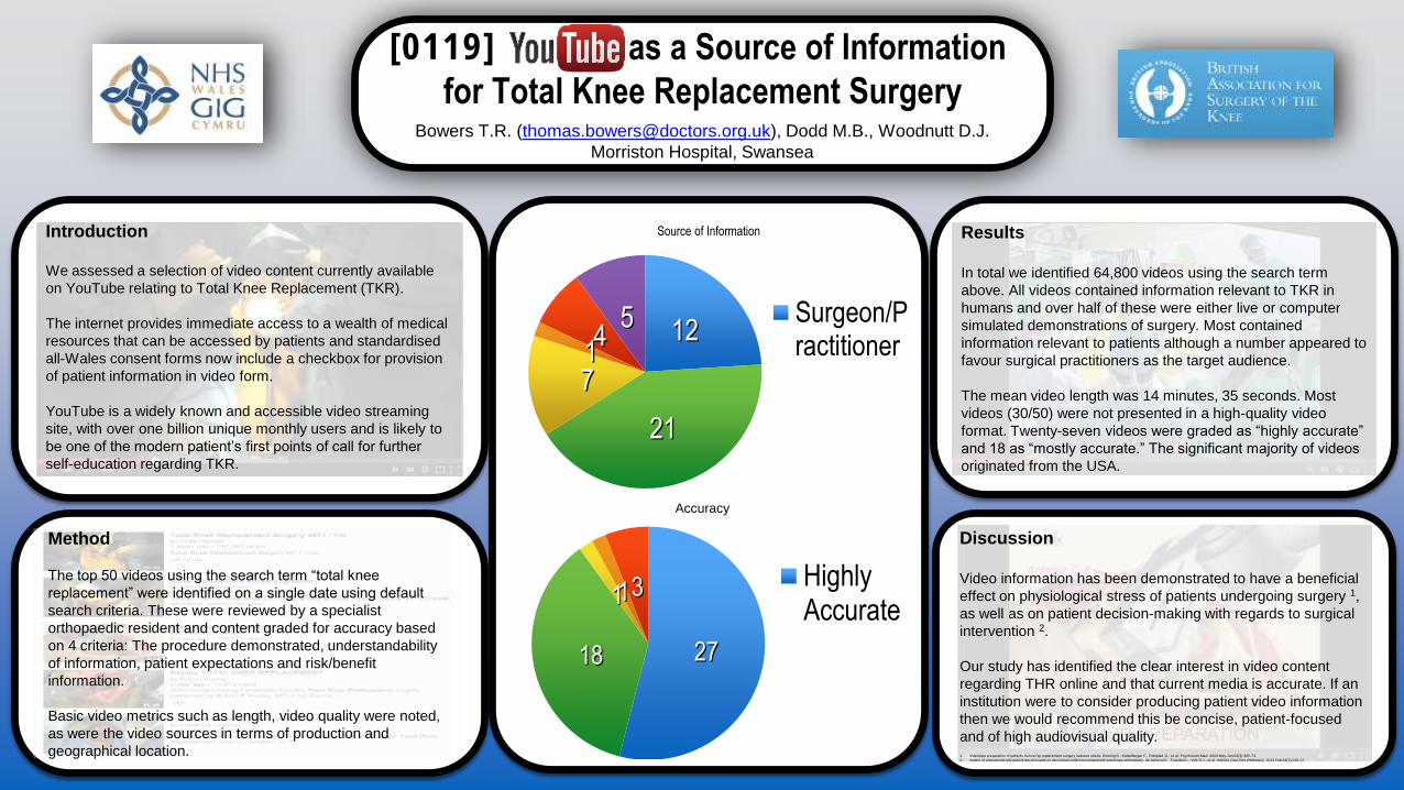
[0119] as a Source of Information
for Total Knee Replacement Surgery
Discussion
Video information has been demonstrated to have a beneficial
effect on physiological stress of patients undergoing surgery 1,
as well as on patient decision-making with regards to surgical
intervention 2.
Our study has identified the clear interest in video content
regarding THR online and that current media is accurate. If an
institution were to consider producing patient video information
then we would recommend this be concise, patient-focused
and of high audiovisual quality.1. Videotape preparation of patients before hip replacement surgery reduces stress. Doering S., Katzelberger F., Rumpled G., et al. Psychosom Med. 2000 May-Jun;62(3):365-73.
2. Impact of educational and patient decision aids on decisional conflict associated with total knee arthroplasty. de Achaval S., Fraenkel L., Volk R.J., et al. Arthritis Care Res (Hoboken). 2012 Feb;64(2):229-37
Results
In total we identified 64,800 videos using the search term
above. All videos contained information relevant to TKR in
humans and over half of these were either live or computer
simulated demonstrations of surgery. Most contained
information relevant to patients although a number appeared to
favour surgical practitioners as the target audience.
The mean video length was 14 minutes, 35 seconds. Most
videos (30/50) were not presented in a high-quality video
format. Twenty-seven videos were graded as “highly accurate”
and 18 as “mostly accurate.” The significant majority of videos
originated from the USA.
Bowers T.R. ([email protected]), Dodd M.B., Woodnutt D.J.
Morriston Hospital, Swansea
2718
113 HighlyAccurate
12
21
714
5 Surgeon/Practitioner
Method
The top 50 videos using the search term “total knee
replacement” were identified on a single date using default
search criteria. These were reviewed by a specialist
orthopaedic resident and content graded for accuracy based
on 4 criteria: The procedure demonstrated, understandability
of information, patient expectations and risk/benefit
information.
Basic video metrics such as length, video quality were noted,
as were the video sources in terms of production and
geographical location.
Accuracy
Source of InformationIntroduction
We assessed a selection of video content currently available
on YouTube relating to Total Knee Replacement (TKR).
The internet provides immediate access to a wealth of medical
resources that can be accessed by patients and standardised
all-Wales consent forms now include a checkbox for provision
of patient information in video form.
YouTube is a widely known and accessible video streaming
site, with over one billion unique monthly users and is likely to
be one of the modern patient’s first points of call for further
self-education regarding TKR.
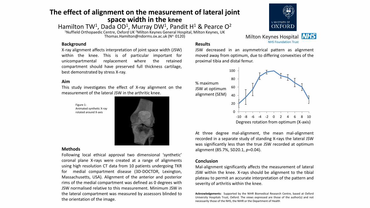
The effect of alignment on the measurement of lateral joint space width in the knee
Hamilton TW1, Dada OD1, Murray DW1, Pandit H1 & Pearce O21Nuffield Orthopaedic Centre, Oxford UK 2Milton Keynes General Hospital, Milton Keynes, UK
[email protected] (No. 0120)
BackgroundX-ray alignment affects interpretation of joint space width (JSW)within the knee. This is of particular important forunicompartmental replacement where the retainedcompartment should have preserved full thickness cartilage,best demonstrated by stress X-ray.
AimThis study investigates the effect of X-ray alignment on themeasurement of the lateral JSW in the arthritic knee.
Figure 1:Animated synthetic X-rayrotated around X-axis
MethodsFollowing local ethical approval two dimensional ‘synthetic’coronal plane X-rays were created at a range of alignmentsusing high resolution CT data from 10 patients undergoing TKRfor medial compartment disease (3D-DOCTOR, Lexington,Massachusetts, USA). Alignment of the anterior and posteriorrims of the medial compartment was defined as 0 degrees withJSW normalised relative to this measurement. Minimum JSW inthe lateral compartment was measured by assessors blinded tothe orientation of the image.
ResultsJSW decreased in an asymmetrical pattern as alignmentmoved away from optimum, due to differing convexities of theproximal tibia and distal femur.
% maximumJSW at optimumalignment (SEM)
Degrees rotation from optimum (X-axis)
At three degree mal-alignment, the mean mal-alignmentrecorded in a separate study of standing X-rays the lateral JSWwas significantly less than the true JSW recorded at optimumalignment (85.7%, SD20.1, p=0.04).
ConclusionMal-alignment significantly affects the measurement of lateralJSW within the knee. X-rays should be alignment to the tibialplateau to permit an accurate interpretation of the pattern andseverity of arthritis within the knee.
Acknowledgements: Supported by the NIHR Biomedical Research Centre, based at OxfordUniversity Hospitals Trust, Oxford. The views expressed are those of the author(s) and notnecessarily those of the NHS, the NIHR or the Department of Health
0
20
40
60
80
100
-10 -8 -6 -4 -2 0 2 4 6 8 10

BASK 2016
30th – 31st March 2016
Liverpool
www.baskonline.com
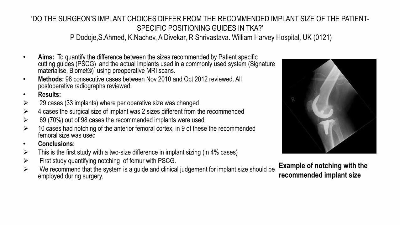
‘DO THE SURGEON’S IMPLANT CHOICES DIFFER FROM THE RECOMMENDED IMPLANT SIZE OF THE PATIENT-
SPECIFIC POSITIONING GUIDES IN TKA?’
P Dodoje,S.Ahmed, K.Nachev, A Divekar, R Shrivastava. William Harvey Hospital, UK (0121)
Example of notching with the
recommended implant size
• Aims: To quantify the difference between the sizes recommended by Patient specific cutting guides (PSCG) and the actual implants used in a commonly used system (Signature materialise, Biomet®) using preoperative MRI scans.
• Methods: 98 consecutive cases between Nov 2010 and Oct 2012 reviewed. All postoperative radiographs reviewed.
• Results:
29 cases (33 implants) where per operative size was changed
4 cases the surgical size of implant was 2 sizes different from the recommended
69 (70%) out of 98 cases the recommended implants were used
10 cases had notching of the anterior femoral cortex, in 9 of these the recommended femoral size was used
• Conclusions:
This is the first study with a two-size difference in implant sizing (in 4% cases)
First study quantifying notching of femur with PSCG.
We recommend that the system is a guide and clinical judgement for implant size should be employed during surgery.
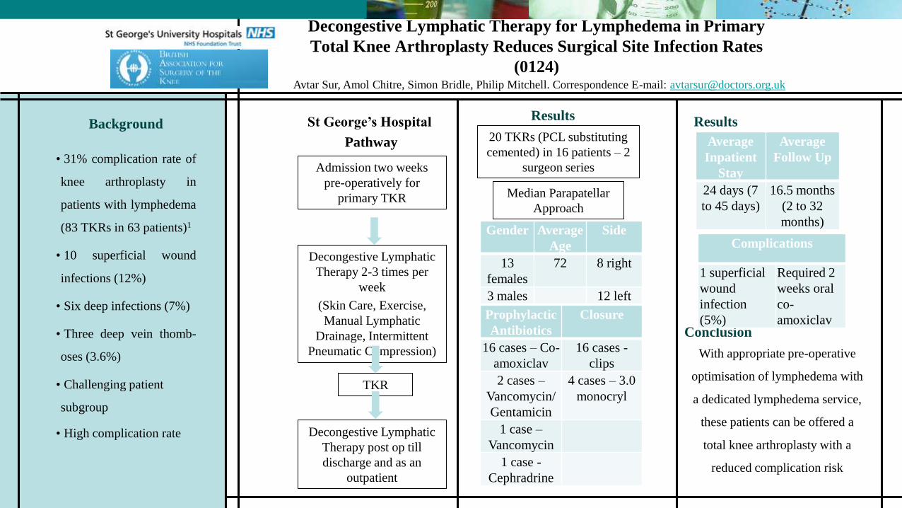
Decongestive Lymphatic Therapy for Lymphedema in Primary
Total Knee Arthroplasty Reduces Surgical Site Infection Rates
(0124)
Background
• 31% complication rate of
knee arthroplasty in
patients with lymphedema
(83 TKRs in 63 patients)1
• 10 superficial wound
infections (12%)
• Six deep infections (7%)
• Three deep vein thomb-
oses (3.6%)
• Challenging patient
subgroup
• High complication rate
Avtar Sur, Amol Chitre, Simon Bridle, Philip Mitchell. Correspondence E-mail: [email protected]
ResultsSt George’s Hospital
Pathway
Admission two weeks
pre-operatively for
primary TKR
Decongestive Lymphatic
Therapy 2-3 times per
week
(Skin Care, Exercise,
Manual Lymphatic
Drainage, Intermittent
Pneumatic Compression)
TKR
Decongestive Lymphatic
Therapy post op till
discharge and as an
outpatient
Gender Average
Age
Side
13
females
72 8 right
3 males 12 left
Prophylactic
Antibiotics
Closure
16 cases – Co-
amoxiclav
16 cases -
clips
2 cases –
Vancomycin/
Gentamicin
4 cases – 3.0
monocryl
1 case –
Vancomycin
1 case -
Cephradrine
20 TKRs (PCL substituting
cemented) in 16 patients – 2
surgeon series
Median Parapatellar
Approach
Results
Conclusion
With appropriate pre-operative
optimisation of lymphedema with
a dedicated lymphedema service,
these patients can be offered a
total knee arthroplasty with a
reduced complication risk
Average
Inpatient
Stay
Average
Follow Up
24 days (7
to 45 days)
16.5 months
(2 to 32
months)
Complications
1 superficial
wound
infection
(5%)
Required 2
weeks oral
co-
amoxiclav
Simplified registration in navigated total knee arthroplasty: a randomised controlled trialNC Sciberras, M Almustafa, BRK. Smith, AH Deakin, DJ Allen, F Picard. Golden Jubilee National Hospital, Clydebank, G81 4DY. [email protected]
Aim
To determine if simplified TKAnavigation software
maintained accuracy of component positioning
decreased registration and operative times
Methods Prospective RCT - ISRCTN:71883082 220 patients randomised to Original
(4.3) or Simplified (Smart) software Registration and operative (skin-to-
skin) time recorded
Results 220 TKAs
• 20 exclusions
Conclusions
Simplified registration
did not alter component accuracy
did shorten registration time
did not reduce overall surgical time
(Poster No. 126)
Introduction Criticism of navigated TKA is that it
increases operation time Focus to make software more efficient
Methods cont. Low dose CT at 6 weeks CT analysed by independent blinded
radiologist• Coronal mechanical alignment
(MFT angle)• Femoral coronal & sagittal angles• Tibial coronal & sagittal angles • Femoral rotation angle
4.3 (n=101) Smart (n=99)
M:F 53:48 50:49
Age 67.9 69.1
BMI 30.8 31.1
Pre-op OKS 17 17
Pre-op alignment
3.6° varus 3.7° varus
Results cont. Mean post-op MFT angles equivalent
• Mean difference - 0.2°
Component positions similar
Mean registration time• Smart - 2 min 30 s• 4.3 - 3 min 23 s
Mean operative time• Both 72 min
p < 0.001
p = 0.855
Patient demographics
p = 0.407
Outcome of primary total knee arthroplasty in morbidly obese patientsAH Deakin, GJ Love. Golden Jubilee National Hospital, Clydebank, G81 4DY. [email protected]
Aim
To determine whether TKApatients with BMI > 40 had similar outcomes to other patients
Methods
All elective primary TKA patients in 2009
Outcomes Length of stay (LoS) Oxford knee score (OKS, 0-48) Patient satisfaction Related problems and complications
(requiring readmission)
Patients divided into three groups BMI>40 (morbidly obese) BMI<=30 (non-obese) BMI between 30-40 (obese)
Results
1014 TKRs 54% female mean age 69 mean BMI 32
Of these 7.7% had BMI > 40
Higher BMI patients had different demographics and outcomes
Conclusion
In this cohort morbidly obese patients had lower OKS and a higher risk of complications after TKA. However there was no increase in patients reporting an unsatisfactory outcome
(Poster No. 127)
BMI<=30 30<BMI<=40 BMI>40 p value
n 381 (35.8%) 555 (54.7%) 78 (7.7%)
Gender 47% female 55% female 78% female <0.001
Mean Age 71 68 64 <0.001
Median LoS 5 5 6 0.042
Mean OKS – 1yr 38 38 34 <0.001
Very satisfied – 1 yr 84% 84% 72% 0.054
Not satisfied – 1 yr 6% 6% 3% 0.635
Any post-op problems 13% 14% 30% 0.001
Related complications 7% 7% 12% 0.303
Alexandra Colby1, Steve Montgomery2
Orthopaedic Clinical Fellow1, Consultant Orthopaedic Surgeon2
University Hospital Aintree, Liverpool
IntroductionDocumented daily senior review is important for:1. Optimum clinical patient care2. Accurate legible management plan3. Timely discharge
Method• Case-note audit• All Orthopaedic wards and outliers • 3 afternoons during October 2014
Results• 62% of patients had a documented daily senior
review• 46.4% of case-notes had completed CUSP sheets
for everyday of admission• 1.4% of case-notes contained a full complement
of up to date CUSP sheets for each day from admission that were completed correctly
• Underperforming areas: No date and time, wrong Consultant, boxes left blank, SHOs signing forms
• Poor documentation most at weekends and operative day
Recommendations• Education needed on CUSP sheet documentation• Up to date CUSP sheets available on all wards• Utilise all Orthopaedic staff in daily senior review
ObjectivesAre all Orthopaedic patients, elective and trauma, receiving a daily senior review and are the Comprehensive Unit-Based Safety Programme Toolkits (CUSP) fit for purpose?
DATE/TIME CONSULTANT
Review all of the following: Notes YES N/A
1 MRSA STATUS/ALERT (NB-teicoplanin prophylaxis for surgery if +ve or alert)
2 MEWS CHART
3 FLUID BALANCE CHART
4 DRUG PRESCRIPTION CHART
5 APPROPRIATE ANTIBIOTICS PRESCRIBED
6 DVT PROPHYLAXIS PRESCRIBED
7 VTE PROFORMA COMPLETED
8 INVASIVE DEVICES PRESENT and ARE APPROPRIATE
9 ANY BIOCHEMICAL / RADIOLOGICAL TESTS FOR REVIEW
10 REVIEW WOUND*/ DRESSINGS and any PRESSURE SORES
11 AMT SCORE DONE (NB HIP FRACTURES pre and post op)
12 DEMENTIA PROFORMA COMPLETED
13 DOCUMENT IF MEDICALLY FIT FOR DISCHARGE
14 Any FOLLOW UP arrangements need organising today?
INTEGRATED WARD ROUND CHECKLIST - ORTHOPAEDIC SURGERY v7
PUT ID LABEL HERE
Results and notes *ALL WOUNDS- IF YOU NEED TO SEE- ONLY A NURSE IS TO REMOVE DRESSING IN TREATMENT ROOM
Friday review: Please list any problems expected or concerns for the weekend team on call
SIGNATURE NAME capitals
Summary: Ward Bed
Alerts
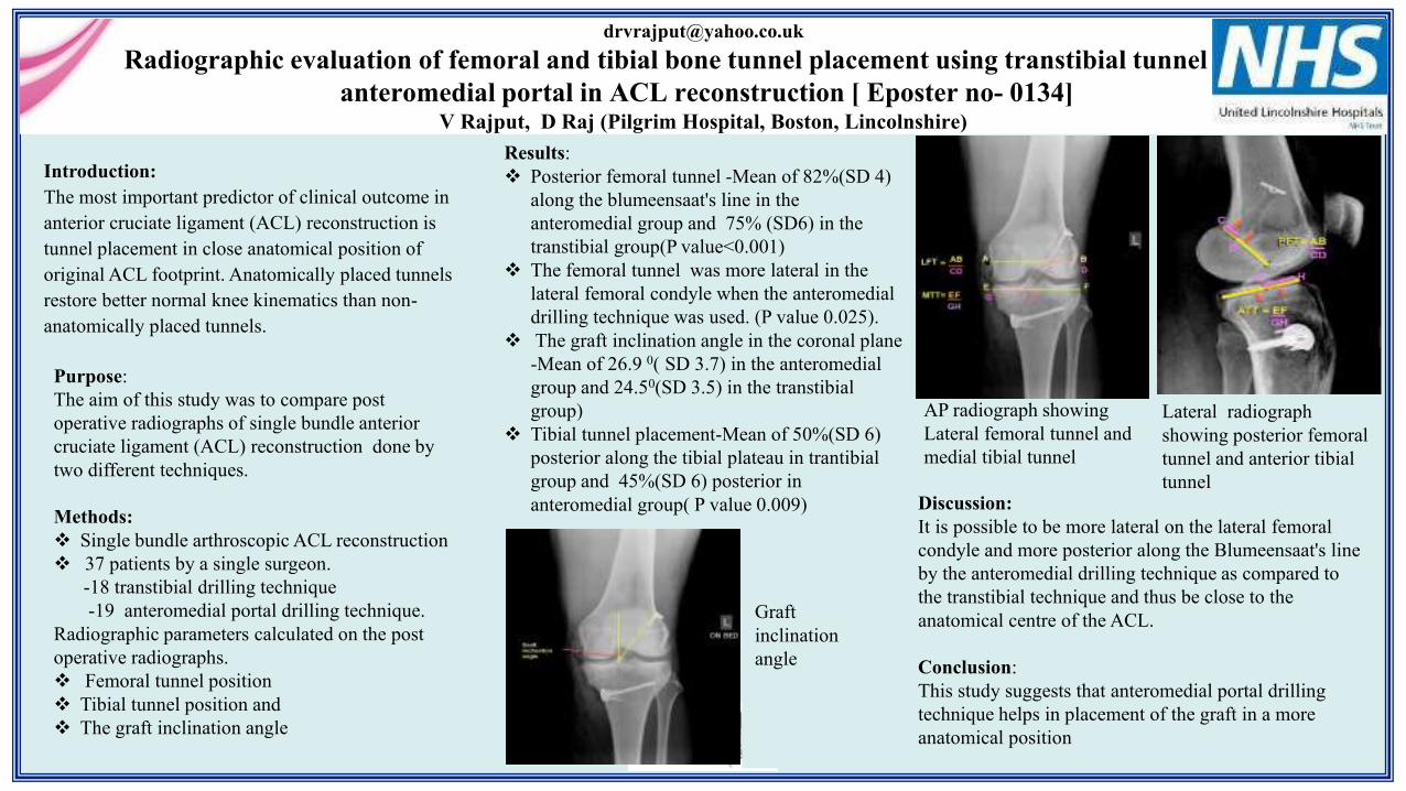
Radiographic evaluation of femoral and tibial bone tunnel placement using transtibial tunnel or the
anteromedial portal in ACL reconstruction [ Eposter no- 0134]V Rajput, D Raj (Pilgrim Hospital, Boston, Lincolnshire)
Discussion:
It is possible to be more lateral on the lateral femoral
condyle and more posterior along the Blumeensaat's line
by the anteromedial drilling technique as compared to
the transtibial technique and thus be close to the
anatomical centre of the ACL.
Conclusion:
This study suggests that anteromedial portal drilling
technique helps in placement of the graft in a more
anatomical position
Results:
Posterior femoral tunnel -Mean of 82%(SD 4)
along the blumeensaat's line in the
anteromedial group and 75% (SD6) in the
transtibial group(P value<0.001)
The femoral tunnel was more lateral in the
lateral femoral condyle when the anteromedial
drilling technique was used. (P value 0.025).
The graft inclination angle in the coronal plane
-Mean of 26.9 0( SD 3.7) in the anteromedial
group and 24.50(SD 3.5) in the transtibial
group)
Tibial tunnel placement-Mean of 50%(SD 6)
posterior along the tibial plateau in trantibial
group and 45%(SD 6) posterior in
anteromedial group( P value 0.009)
Introduction:
The most important predictor of clinical outcome in
anterior cruciate ligament (ACL) reconstruction is
tunnel placement in close anatomical position of
original ACL footprint. Anatomically placed tunnels
restore better normal knee kinematics than non-
anatomically placed tunnels.
Purpose:
The aim of this study was to compare post
operative radiographs of single bundle anterior
cruciate ligament (ACL) reconstruction done by
two different techniques.
Methods:
Single bundle arthroscopic ACL reconstruction
37 patients by a single surgeon.
-18 transtibial drilling technique
-19 anteromedial portal drilling technique.
Radiographic parameters calculated on the post
operative radiographs.
Femoral tunnel position
Tibial tunnel position and
The graft inclination angle
Graft
inclination
angle
AP radiograph showing
Lateral femoral tunnel and
medial tibial tunnel
Lateral radiograph
showing posterior femoral
tunnel and anterior tibial
tunnel
Post operative haemoglobin blood determination following knee joint arthroplasty: Day 1 vs Day 2 blood testing(Poster 0135)
Dr C.Godavitarne1, Dr S.Fraser, Mr M.Khalfaoui2
1.Tunbridge Wells and Maidstone NHS Trust, Kent 2. North West Deanery, [email protected]
ConclusionTo our knowledge no study in the current literature has evaluated the optimal time for post-operative haemoglobin testing following joint arthroplasty surgery. Our study has demonstrated a significant case for 48 hour full blood count testing following knee arthroplasty surgery.
References1.Kalairajah Y, Simpson D, Cossey AJ, Verrall GM, Spriggins AJ. Blood loss after total knee replacement. J Bone Joint Surg Br. 2005;87(11):1480-1482 2. GIlbody J, Dhotar HS, Perruccio AV, Roderick Davey. Topical Tranexamic Acid reduces Transfusion rates in total hip and knee arthroplasty. J Arthroplasty. 2014;29(4):681-6843. Rosencher N, Kerkkamp H, Macheras G, Munuera LM, Menichella G, Barton D, Cremers S, Abraham I. Orthopaedic Surgery Transfusion Hemoglobin European Overview (OSTHEO) study: blood management in elective knee and hip arthroplasty in Europe. Transfusion. 2003; 43:459-469
Results
• 61 patients met the inclusion criteria
• Female to male ratio of 46:15, with an average age of 67.2 years.
• Haemoglobin levels fell from 24 to 48 hours post-surgery on average by 0.39 g/dL (95% CI 0.056-0.725g/dL).
• Using the paired students t-test this was a significant change (p-0.0231)
• The Hb drop resulted in 5 transfusions on the 48hr haemoglobin after controlling for symptomatology
Discussion
Transfusion rates in joint arthroplasty surgery are reported to range from 0% to 67%(2,3).
Thresholds for transfusions are often dependent on a combination of clinical and biochemical variables however most centers will adhere to a haemoglobin range of between 7.0-10.9g/dL as an acceptable indicator for patient transfusion requirement(3).
Whilst Haemoglobin levels are routinely taken on Day 1, our study illustrates the importance of checking haemoglobin on Day 2 as well to identify patients in need of transfusion.
IntroductionPost operative blood tests are conventionally carried out on the first post-operative day. Following joint arthroplasty, the haemoglobin value can fall by upto 53g/dL, highlighting the urgency for this investigation(1). The aim of our study was to determine if there was a significant difference between day 1 and day 2 haemoglobin levels.
Methods
• We conducted a retrospective cohort study undertaken between Dec 2012 and Apr 2013
• All patients who underwent full blood count testing at both 24 and 48 hours consecutively following joint arthroplasty surgery were included in the study
• Data categories included:
1) The actual haemoglobin levels (Day 1 + 2)
2) Whether a transfusion had taken place
3) Indication for the transfusion.
Alexandra Colby1, Clifford Butcher2
Orthopaedic Clinical Fellow1, Consultant Orthopaedic Surgeon2
University Hospital Aintree, Liverpool
Guidelines
Recommendations• Informative poster in clinical areas• Redesign guidelines• Re-audit in 6-12 months time – currently in progress
Objectives• To evaluate current practice for INR reversal against current local guidelines • To assess if there is significant delay to operative treatment if incorrect INR
correction is employed?
Results• 35.3% had correct INR reversal as per guidelines• Of 64.7% who had incorrect INR reversal:
• 27% - incorrect dose of Vitamin K• 82% - no further Vitamin K on reassessment• 27% - no/incorrect Octaplex given • 18% - wrong/unspecified route of Vitamin K
• 1 patient had delay to surgery as a direct result of incorrect INR reversal
For surgery < 12 hours
For surgery 12-24 hours
Method• Retrospective/prospective audit on all Orthopaedic
trauma admissions between August to December 2013• Sample Size = 17 patients
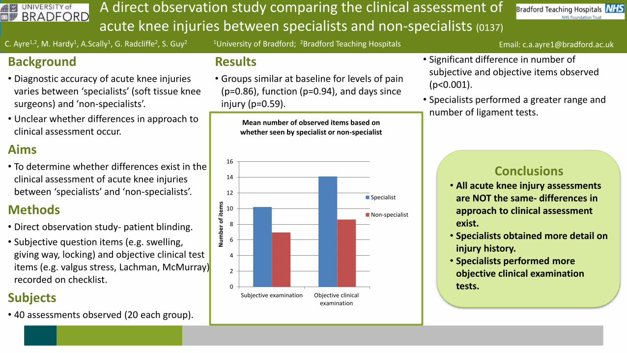
Background• Diagnostic accuracy of acute knee injuries
varies between ‘specialists’ (soft tissue knee surgeons) and ‘non-specialists’.
• Unclear whether differences in approach to clinical assessment occur.
Aims• To determine whether differences exist in the
clinical assessment of acute knee injuries between ‘specialists’ and ‘non-specialists’.
Methods• Direct observation study- patient blinding.
• Subjective question items (e.g. swelling, giving way, locking) and objective clinical test items (e.g. valgus stress, Lachman, McMurray) recorded on checklist.
Subjects• 40 assessments observed (20 each group).
Results• Groups similar at baseline for levels of pain
(p=0.86), function (p=0.94), and days since injury (p=0.59).
• Significant difference in number of subjective and objective items observed (p<0.001).
• Specialists performed a greater range and number of ligament tests.
Conclusions• All acute knee injury assessments
are NOT the same- differences in approach to clinical assessment exist.
• Specialists obtained more detail on injury history.
• Specialists performed more objective clinical examination tests.
A direct observation study comparing the clinical assessment of acute knee injuries between specialists and non-specialists (0137)
C. Ayre1,2, M. Hardy1, A.Scally1, G. Radcliffe2, S. Guy2 1University of Bradford; 2Bradford Teaching Hospitals Email: [email protected]
0
2
4
6
8
10
12
14
16
Subjective examination Objective clinicalexamination
Nu
mb
er
of
ite
ms
Mean number of observed items based on whether seen by specialist or non-specialist
Specialist
Non-specialist
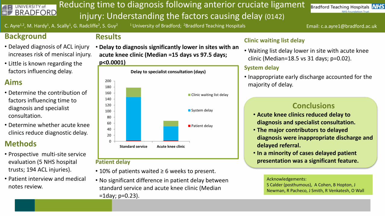
Background• Delayed diagnosis of ACL injury
increases risk of meniscal injury.
• Little is known regarding the factors influencing delay.
Aims• Determine the contribution of
factors influencing time to diagnosis and specialist consultation.
• Determine whether acute knee clinics reduce diagnostic delay.
Methods• Prospective multi-site service
evaluation (5 NHS hospital trusts; 194 ACL injuries).
• Patient interview and medical notes review.
Results• Delay to diagnosis significantly lower in sites with an
acute knee clinic (Median =15 days vs 97.5 days; p<0.0001)
Clinic waiting list delay
• Waiting list delay lower in site with acute knee clinic (Median=18.5 vs 31 days; p=0.02).
System delay
• Inappropriate early discharge accounted for the majority of delay.
Conclusions• Acute knee clinics reduced delay to
diagnosis and specialist consultation.• The major contributors to delayed
diagnosis were inappropriate discharge and delayed referral.
• In a minority of cases delayed patient presentation was a significant feature.
Acknowledgements:S Calder (posthumous), A Cohen, B Hopton, J Newman, R Pacheco, J Smith, R Venkatesh, O Wall
Reducing time to diagnosis following anterior cruciate ligament injury: Understanding the factors causing delay (0142)
C. Ayre1,2, M. Hardy1, A. Scally1, G. Radcliffe2, S. Guy2 1 University of Bradford; 2Bradford Teaching Hospitals Email: [email protected]
Patient delay
• 10% of patients waited ≥ 6 weeks to present.
• No significant difference in patient delay between standard service and acute knee clinic (Median =1day; p=0.23).
0
20
40
60
80
100
120
140
160
180
200
Standard service Acute knee clinic
Delay to specialist consultation (days)
Clinic waiting list delay
System delay
Patient delay
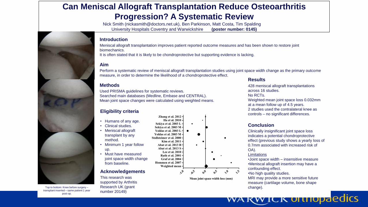
Can Meniscal Allograft Transplantation Reduce Osteoarthritis
Progression? A Systematic Review Nick Smith ([email protected]), Ben Parkinson, Matt Costa, Tim Spalding
University Hospitals Coventry and Warwickshire (poster number: 0145)
Introduction
Meniscal allograft transplantation improves patient reported outcome measures and has been shown to restore joint
biomechanics.
It is often stated that it is likely to be chondroprotective but supporting evidence is lacking.
Aim
Perform a systematic review of meniscal allograft transplantation studies using joint space width change as the primary outcome
measure, in order to determine the likelihood of a chondroprotective effect.
Methods
Used PRISMA guidelines for systematic reviews.
Searched main databases (Medline, Embase and CENTRAL).
Mean joint space changes were calculated using weighted means.
Eligibility criteria
• Humans of any age.
• Clinical studies.
• Meniscal allograft
transplant by any
method.
• Minimum 1 year follow
up.
• Must have measured
joint space width change
from baseline.
Results
428 meniscal allograft transplantations
across 16 studies.
No RCTs.
Weighted mean joint space loss 0.032mm
at a mean follow up of 4.5 years.
2 studies used the contralateral knee as
controls – no significant differences.
Conclusion
Clinically insignificant joint space loss
indicates a potential chondroprotective
effect (previous study shows a yearly loss of
0.7mm associated with increased risk of
OA).
Limitations
•Joint space width – insensitive measure
•Meniscal allograft insertion may have a
confounding effect.
•No high quality studies.
MRI may provide a more sensitive future
measure (cartilage volume, bone shape
change).Top to bottom: Knee before surgery –
transplant inserted – same patient 1 year
post-op.
Acknowledgements
This research was
supported by Arthritis
Research UK (grant
number 20149)
-1.0
-0.5 0.
00.
51.
01.
5
Weighted mean
Hommen et al. 2007
Graf et al. 2004
Rath et al. 2001
Lee et al. 2010
Abat et al. 2013 S
Abat et al. 2013 B
Kim et al. 2011
Stollsteimer et al. 2000
Yoldas et al. 2003 M
Yoldas et al. 2003 L
Sekiya et al. 2003 M
Sekiya et al. 2003 L
Ha et al. 2010
Zhang et al. 2012
Mean joint space width loss (mm)
Join BASK
One of the largest specialist societies
Discounted rates for BASK events
Members only on line content due soon
Application details on website
Proposers must be BASK members
www.baskonline.com
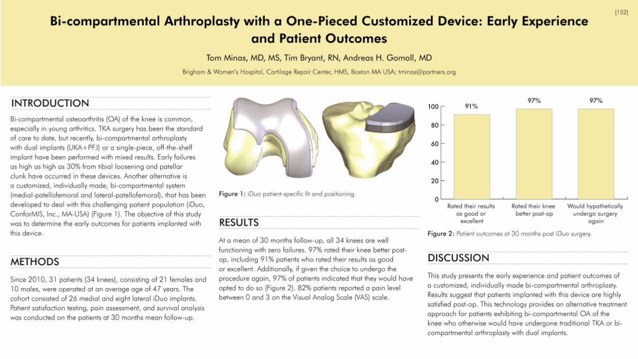
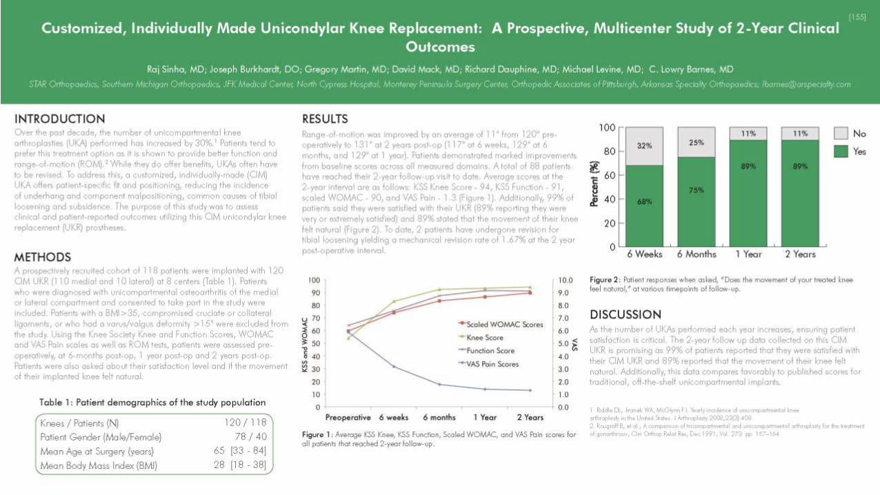
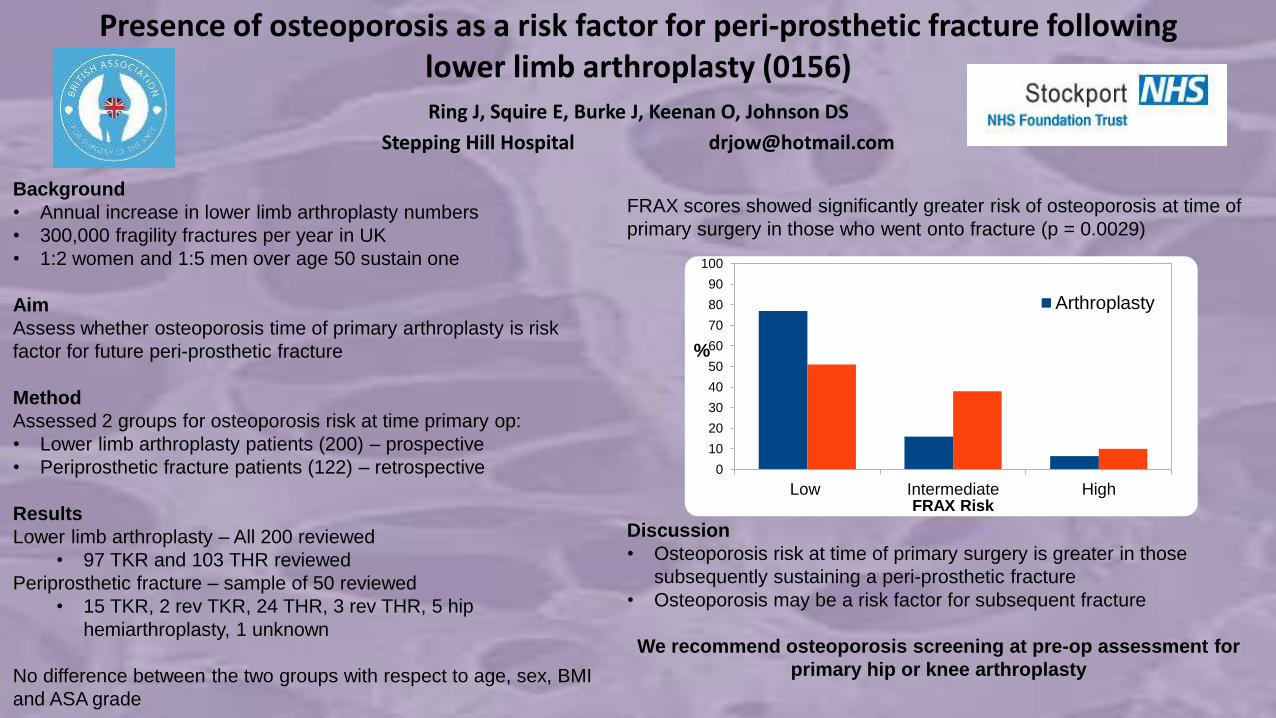
Presence of osteoporosis as a risk factor for peri-prosthetic fracture following lower limb arthroplasty (0156)Ring J, Squire E, Burke J, Keenan O, Johnson DS
Stepping Hill Hospital [email protected]
Background
• Annual increase in lower limb arthroplasty numbers
• 300,000 fragility fractures per year in UK
• 1:2 women and 1:5 men over age 50 sustain one
Aim
Assess whether osteoporosis time of primary arthroplasty is risk
factor for future peri-prosthetic fracture
Method
Assessed 2 groups for osteoporosis risk at time primary op:
• Lower limb arthroplasty patients (200) – prospective
• Periprosthetic fracture patients (122) – retrospective
Results
Lower limb arthroplasty – All 200 reviewed
• 97 TKR and 103 THR reviewed
Periprosthetic fracture – sample of 50 reviewed
• 15 TKR, 2 rev TKR, 24 THR, 3 rev THR, 5 hip
hemiarthroplasty, 1 unknown
No difference between the two groups with respect to age, sex, BMI
and ASA grade
FRAX scores showed significantly greater risk of osteoporosis at time of
primary surgery in those who went onto fracture (p = 0.0029)
Discussion
• Osteoporosis risk at time of primary surgery is greater in those
subsequently sustaining a peri-prosthetic fracture
• Osteoporosis may be a risk factor for subsequent fracture
We recommend osteoporosis screening at pre-op assessment for
primary hip or knee arthroplasty
0
10
20
30
40
50
60
70
80
90
100
Low Intermediate High
%
FRAX Risk
Arthroplasty
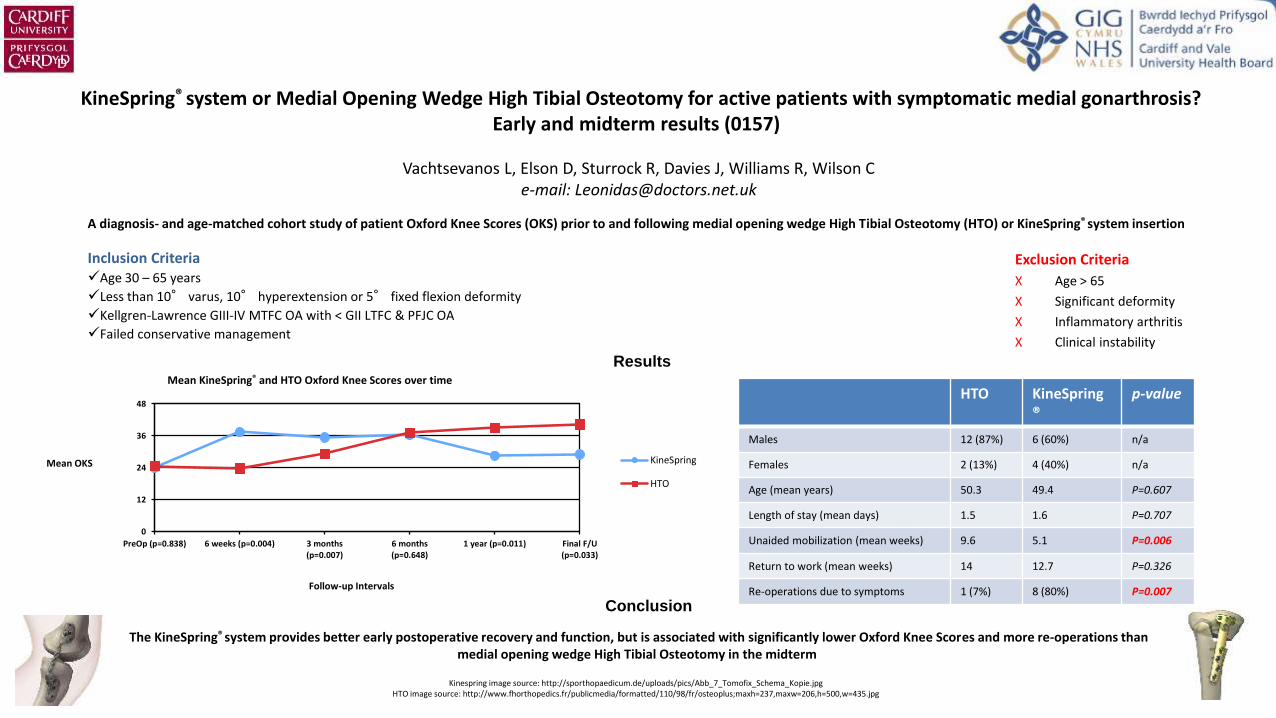
KineSpring® system or Medial Opening Wedge High Tibial Osteotomy for active patients with symptomatic medial gonarthrosis? Early and midterm results (0157)
Vachtsevanos L, Elson D, Sturrock R, Davies J, Williams R, Wilson Ce-mail: [email protected]
A diagnosis- and age-matched cohort study of patient Oxford Knee Scores (OKS) prior to and following medial opening wedge High Tibial Osteotomy (HTO) or KineSpring® system insertion
Inclusion CriteriaAge 30 – 65 years
Less than 10° varus, 10° hyperextension or 5° fixed flexion deformity
Kellgren-Lawrence GIII-IV MTFC OA with < GII LTFC & PFJC OA
Failed conservative management
Exclusion Criteria
X Age > 65
X Significant deformity
X Inflammatory arthritis
X Clinical instability
Kinespring image source: http://sporthopaedicum.de/uploads/pics/Abb_7_Tomofix_Schema_Kopie.jpgHTO image source: http://www.fhorthopedics.fr/publicmedia/formatted/110/98/fr/osteoplus;maxh=237,maxw=206,h=500,w=435.jpg
HTO KineSpring®
p-value
Males 12 (87%) 6 (60%) n/a
Females 2 (13%) 4 (40%) n/a
Age (mean years) 50.3 49.4 P=0.607
Length of stay (mean days) 1.5 1.6 P=0.707
Unaided mobilization (mean weeks) 9.6 5.1 P=0.006
Return to work (mean weeks) 14 12.7 P=0.326
Re-operations due to symptoms 1 (7%) 8 (80%) P=0.007
The KineSpring® system provides better early postoperative recovery and function, but is associated with significantly lower Oxford Knee Scores and more re-operations than medial opening wedge High Tibial Osteotomy in the midterm
Results
0
12
24
36
48
PreOp (p=0.838) 6 weeks (p=0.004) 3 months(p=0.007)
6 months(p=0.648)
1 year (p=0.011) Final F/U(p=0.033)
Mean OKS
Follow-up Intervals
Mean KineSpring® and HTO Oxford Knee Scores over time
KineSpring
HTO
Conclusion
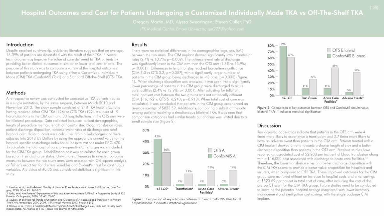
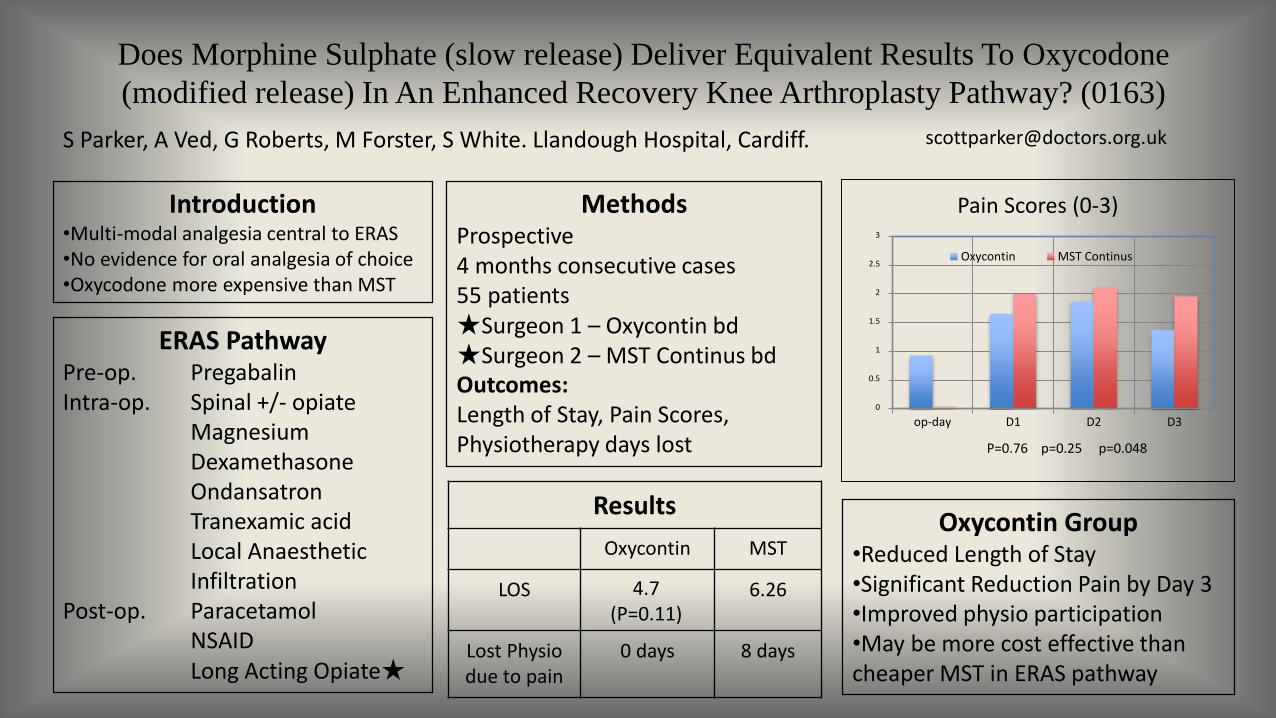
Does Morphine Sulphate (slow release) Deliver Equivalent Results To Oxycodone
(modified release) In An Enhanced Recovery Knee Arthroplasty Pathway? (0163)
S Parker, A Ved, G Roberts, M Forster, S White. Llandough Hospital, Cardiff. [email protected]
Introduction•Multi-modal analgesia central to ERAS•No evidence for oral analgesia of choice•Oxycodone more expensive than MST
MethodsProspective4 months consecutive cases55 patients ★Surgeon 1 – Oxycontin bd ★Surgeon 2 – MST Continus bdOutcomes: Length of Stay, Pain Scores, Physiotherapy days lost
ERAS PathwayPre-op. Pregabalin Intra-op. Spinal +/- opiate
MagnesiumDexamethasoneOndansatronTranexamic acidLocal Anaesthetic Infiltration
Post-op. ParacetamolNSAIDLong Acting Opiate★
0
0.5
1
1.5
2
2.5
3
op-day D1 D2 D3
Pain Scores (0-3)
Oxycontin MST Continus
P=0.76 p=0.25 p=0.048
Results
Oxycontin MST
LOS 4.7(P=0.11)
6.26
Lost Physio due to pain
0 days 8 days
Oxycontin Group•Reduced Length of Stay•Significant Reduction Pain by Day 3•Improved physio participation•May be more cost effective than cheaper MST in ERAS pathway
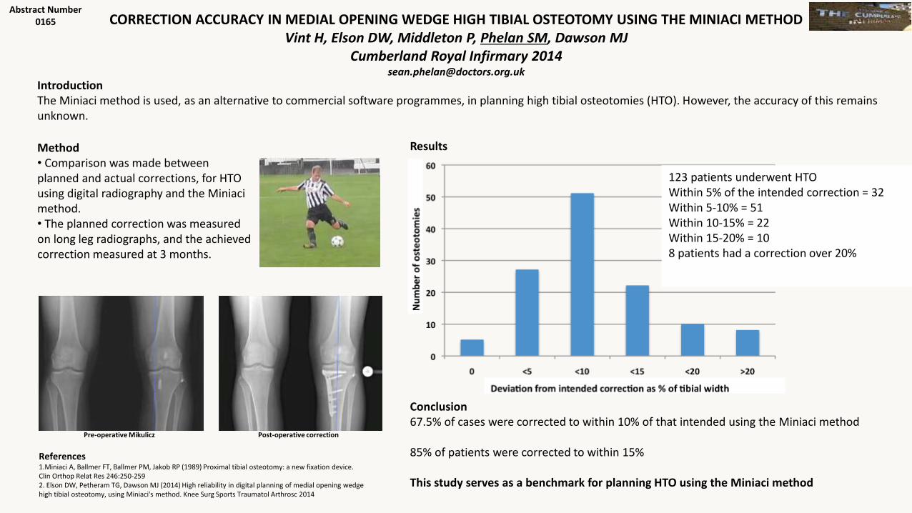
CORRECTION ACCURACY IN MEDIAL OPENING WEDGE HIGH TIBIAL OSTEOTOMY USING THE MINIACI METHODVint H, Elson DW, Middleton P, Phelan SM, Dawson MJ
Cumberland Royal Infirmary [email protected]
IntroductionThe Miniaci method is used, as an alternative to commercial software programmes, in planning high tibial osteotomies (HTO). However, the accuracy of this remains unknown.
Abstract Number0165
References1.Miniaci A, Ballmer FT, Ballmer PM, Jakob RP (1989) Proximal tibial osteotomy: a new fixation device. Clin Orthop Relat Res 246:250-2592. Elson DW, Petheram TG, Dawson MJ (2014) High reliability in digital planning of medial opening wedge high tibial osteotomy, using Miniaci's method. Knee Surg Sports Traumatol Arthrosc 2014
Pre-operative Mikulicz Post-operative correction
Method• Comparison was made between planned and actual corrections, for HTO using digital radiography and the Miniaci method. • The planned correction was measured on long leg radiographs, and the achieved correction measured at 3 months.
Conclusion67.5% of cases were corrected to within 10% of that intended using the Miniaci method
85% of patients were corrected to within 15%
This study serves as a benchmark for planning HTO using the Miniaci method
123 patients underwent HTOWithin 5% of the intended correction = 32Within 5-10% = 51Within 10-15% = 22Within 15-20% = 108 patients had a correction over 20%
Results
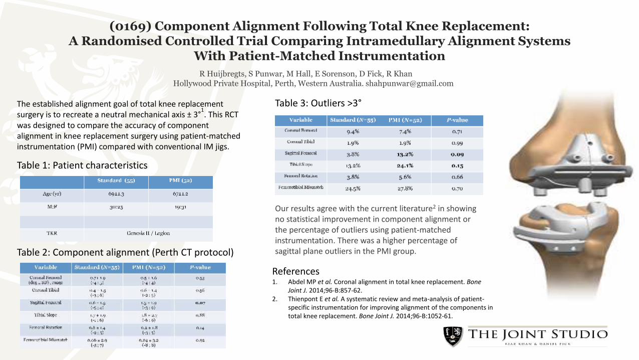
References1. Abdel MP et al. Coronal alignment in total knee replacement. Bone
Joint J. 2014;96-B:857-62.2. Thienpont E et al. A systematic review and meta-analysis of patient-
specific instrumentation for improving alignment of the components in total knee replacement. Bone Joint J. 2014;96-B:1052-61.
(0169) Component Alignment Following Total Knee Replacement: A Randomised Controlled Trial Comparing Intramedullary Alignment Systems
With Patient-Matched Instrumentation
R Huijbregts, S Punwar, M Hall, E Sorenson, D Fick, R KhanHollywood Private Hospital, Perth, Western Australia. [email protected]
Our results agree with the current literature2 in showing no statistical improvement in component alignment or the percentage of outliers using patient-matched instrumentation. There was a higher percentage of sagittal plane outliers in the PMI group.
The established alignment goal of total knee replacementsurgery is to recreate a neutral mechanical axis ± 3°
1. This RCT
was designed to compare the accuracy of componentalignment in knee replacement surgery using patient-matchedinstrumentation (PMI) compared with conventional IM jigs.
Table 2: Component alignment (Perth CT protocol)
Table 3: Outliers >3°
Table 1: Patient characteristics
Get your coding rightWrite clearly
Enter patient details / date on every page
Record all diagnoses / procedures in full
Record co-morbidites every admission
Record organism if infection
Don’t use abbreviations
Don’t use “?” diagnosis – describe symptoms
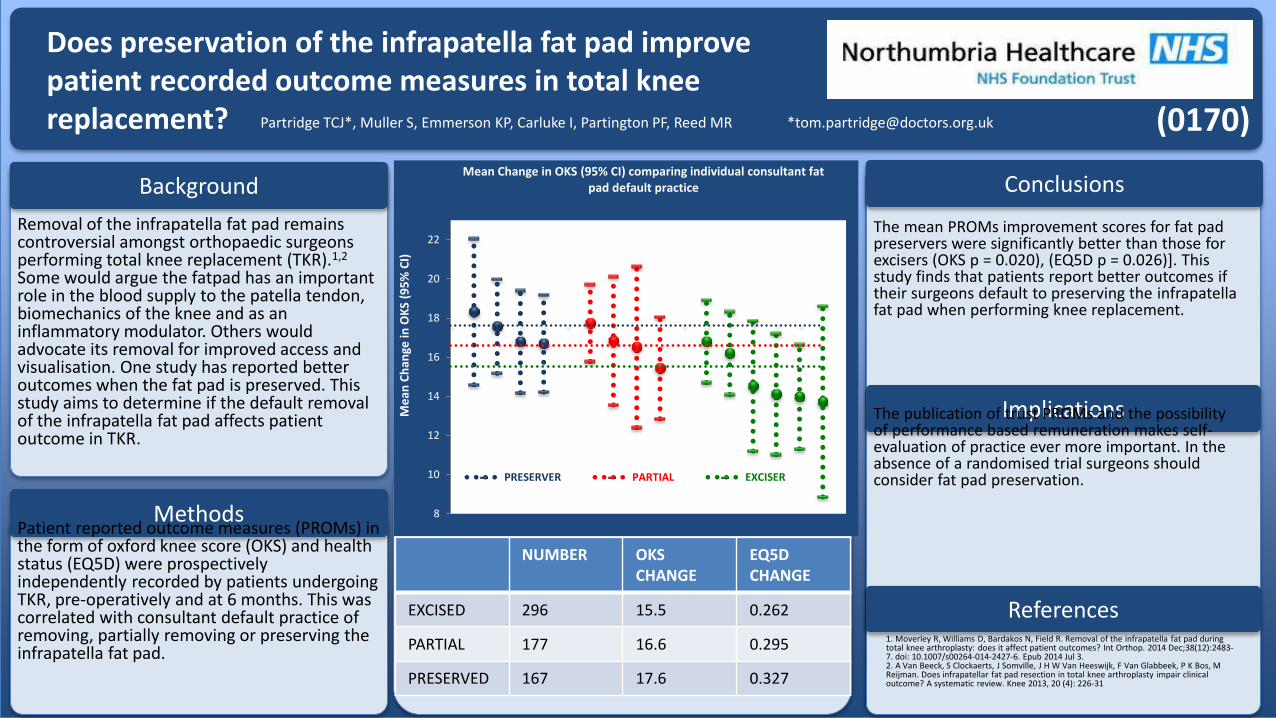
Does preservation of the infrapatella fat pad improve patient recorded outcome measures in total knee replacement? Partridge TCJ*, Muller S, Emmerson KP, Carluke I, Partington PF, Reed MR *[email protected]
Background
Methods
Removal of the infrapatella fat pad remains controversial amongst orthopaedic surgeons performing total knee replacement (TKR).1,2
Some would argue the fatpad has an important role in the blood supply to the patella tendon, biomechanics of the knee and as an inflammatory modulator. Others would advocate its removal for improved access and visualisation. One study has reported better outcomes when the fat pad is preserved. This study aims to determine if the default removal of the infrapatella fat pad affects patient outcome in TKR.
Patient reported outcome measures (PROMs) in the form of oxford knee score (OKS) and health status (EQ5D) were prospectively independently recorded by patients undergoing TKR, pre-operatively and at 6 months. This was correlated with consultant default practice of removing, partially removing or preserving the infrapatella fat pad.
Conclusions
Implications
References
The mean PROMs improvement scores for fat pad preservers were significantly better than those for excisers (OKS p = 0.020), (EQ5D p = 0.026)]. This study finds that patients report better outcomes if their surgeons default to preserving the infrapatellafat pad when performing knee replacement.
The publication of trust PROMs and the possibility of performance based remuneration makes self-evaluation of practice ever more important. In the absence of a randomised trial surgeons should consider fat pad preservation.
NUMBER OKS CHANGE
EQ5D CHANGE
EXCISED 296 15.5 0.262
PARTIAL 177 16.6 0.295
PRESERVED 167 17.6 0.327
1. Moverley R, Williams D, Bardakos N, Field R. Removal of the infrapatella fat pad during total knee arthroplasty: does it affect patient outcomes? Int Orthop. 2014 Dec;38(12):2483-7. doi: 10.1007/s00264-014-2427-6. Epub 2014 Jul 3.2. A Van Beeck, S Clockaerts, J Somville, J H W Van Heeswijk, F Van Glabbeek, P K Bos, M Reijman. Does infrapatellar fat pad resection in total knee arthroplasty impair clinicaloutcome? A systematic review. Knee 2013, 20 (4): 226-31
(0170)
8
10
12
14
16
18
20
22
Me
an C
han
ge in
OK
S (9
5%
CI)
Mean Change in OKS (95% CI) comparing individual consultant fat pad default practice
PRESERVER PARTIAL EXCISER
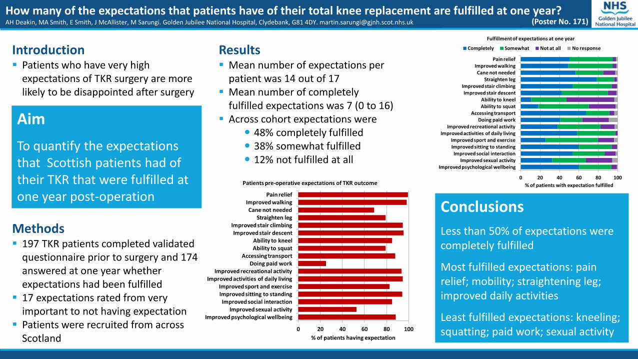
How many of the expectations that patients have of their total knee replacement are fulfilled at one year?AH Deakin, MA Smith, E Smith, J McAllister, M Sarungi. Golden Jubilee National Hospital, Clydebank, G81 4DY. [email protected]
Aim
To quantify the expectations that Scottish patients had of their TKR that were fulfilled at one year post-operation
Methods 197 TKR patients completed validated
questionnaire prior to surgery and 174 answered at one year whether expectations had been fulfilled
17 expectations rated from very important to not having expectation
Patients were recruited from across Scotland
Results Mean number of expectations per
patient was 14 out of 17 Mean number of completely
fulfilled expectations was 7 (0 to 16) Across cohort expectations were
48% completely fulfilled 38% somewhat fulfilled 12% not fulfilled at all
Conclusions
Less than 50% of expectations were completely fulfilled
Most fulfilled expectations: pain relief; mobility; straightening leg; improved daily activities
Least fulfilled expectations: kneeling; squatting; paid work; sexual activity
(Poster No. 171)
Introduction Patients who have very high
expectations of TKR surgery are more likely to be disappointed after surgery
0 20 40 60 80 100
Pain reliefImproved walkingCane not needed
Straighten legImproved stair climbingImproved stair descent
Ability to kneelAbility to squat
Accessing transportDoing paid work
Improved recreational activityImproved activities of daily living
Improved sport and exerciseImproved sitting to standingImproved social interaction
Improved sexual activityImproved psychological wellbeing
% of patients having expectation
Patients pre-operative expectations of TKR outcome0 20 40 60 80 100
Pain reliefImproved walkingCane not needed
Straighten legImproved stair climbingImproved stair descent
Ability to kneelAbility to squat
Accessing transportDoing paid work
Improved recreational activityImproved activities of daily living
Improved sport and exerciseImproved sitting to standingImproved social interaction
Improved sexual activityImproved psychological wellbeing
% of patients with expectation fulfilled
Fulfillment of expectations at one year
Completely Somewhat Not at all No response
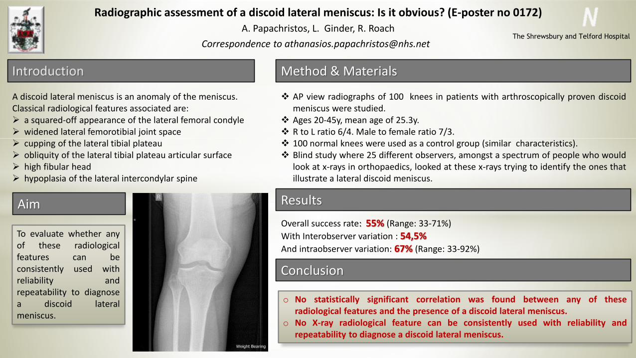
Radiographic assessment of a discoid lateral meniscus: Is it obvious? (E-poster no 0172)A. Papachristos, L. Ginder, R. Roach
Correspondence to [email protected]
A discoid lateral meniscus is an anomaly of the meniscus.Classical radiological features associated are: a squared-off appearance of the lateral femoral condyle widened lateral femorotibial joint space cupping of the lateral tibial plateau obliquity of the lateral tibial plateau articular surface high fibular head hypoplasia of the lateral intercondylar spine
To evaluate whether anyof these radiologicalfeatures can beconsistently used withreliability andrepeatability to diagnosea discoid lateralmeniscus.
AP view radiographs of 100 knees in patients with arthroscopically proven discoidmeniscus were studied.
Ages 20-45y, mean age of 25.3y. R to L ratio 6/4. Male to female ratio 7/3. 100 normal knees were used as a control group (similar characteristics). Blind study where 25 different observers, amongst a spectrum of people who would
look at x-rays in orthopaedics, looked at these x-rays trying to identify the ones thatillustrate a lateral discoid meniscus.
Overall success rate: 55% (Range: 33-71%)
With Interobserver variation : 54,5%And intraobserver variation: 67% (Range: 33-92%)
o No statistically significant correlation was found between any of theseradiological features and the presence of a discoid lateral meniscus.
o No X-ray radiological feature can be consistently used with reliability andrepeatability to diagnose a discoid lateral meniscus.
Method & Materials
Aim Results
Conclusion
The Shrewsbury and Telford Hospital
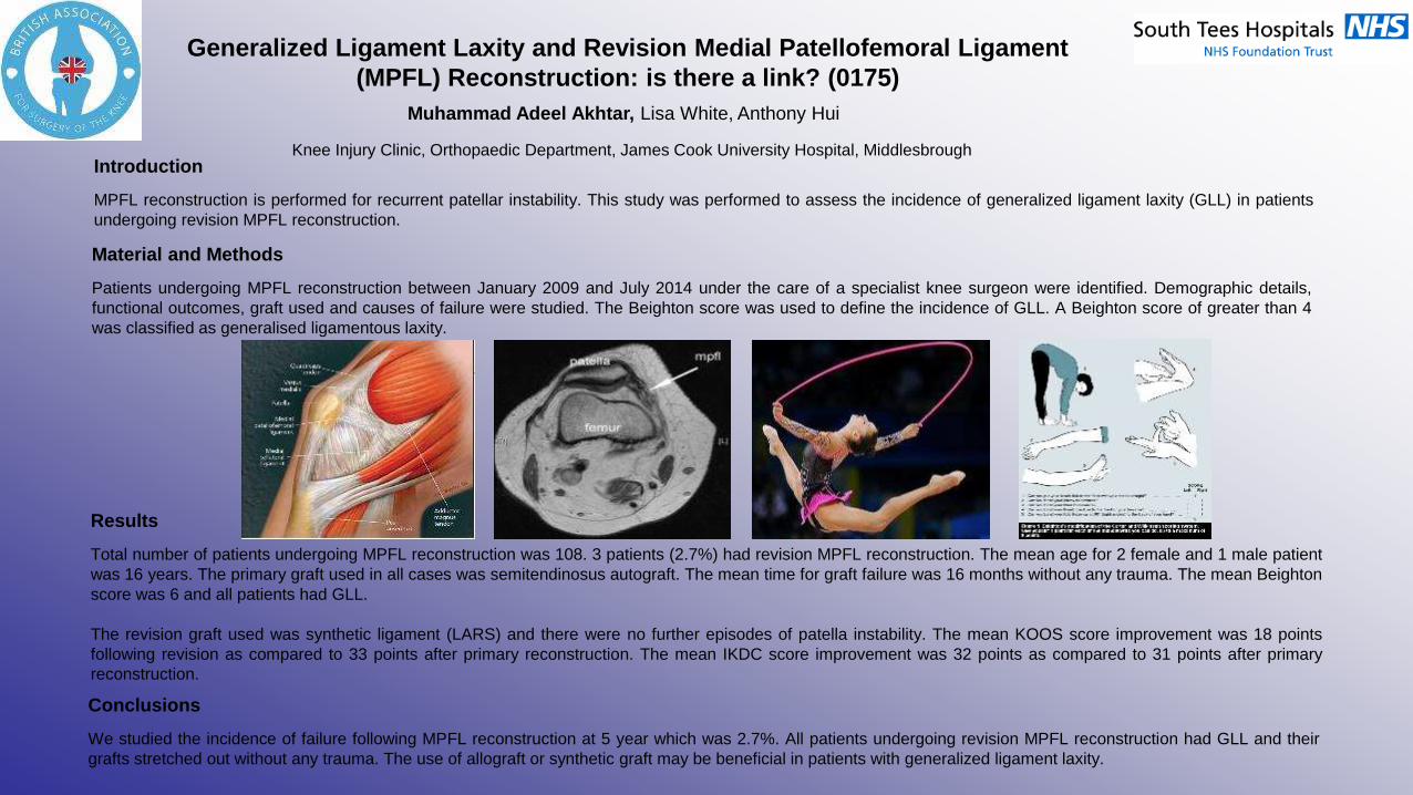
Generalized Ligament Laxity and Revision Medial Patellofemoral Ligament
(MPFL) Reconstruction: is there a link? (0175)
Muhammad Adeel Akhtar, Lisa White, Anthony Hui
Knee Injury Clinic, Orthopaedic Department, James Cook University Hospital, MiddlesbroughIntroduction
MPFL reconstruction is performed for recurrent patellar instability. This study was performed to assess the incidence of generalized ligament laxity (GLL) in patients
undergoing revision MPFL reconstruction.
Results
Total number of patients undergoing MPFL reconstruction was 108. 3 patients (2.7%) had revision MPFL reconstruction. The mean age for 2 female and 1 male patient
was 16 years. The primary graft used in all cases was semitendinosus autograft. The mean time for graft failure was 16 months without any trauma. The mean Beighton
score was 6 and all patients had GLL.
The revision graft used was synthetic ligament (LARS) and there were no further episodes of patella instability. The mean KOOS score improvement was 18 points
following revision as compared to 33 points after primary reconstruction. The mean IKDC score improvement was 32 points as compared to 31 points after primary
reconstruction.
Conclusions
We studied the incidence of failure following MPFL reconstruction at 5 year which was 2.7%. All patients undergoing revision MPFL reconstruction had GLL and their
grafts stretched out without any trauma. The use of allograft or synthetic graft may be beneficial in patients with generalized ligament laxity.
Material and Methods
Patients undergoing MPFL reconstruction between January 2009 and July 2014 under the care of a specialist knee surgeon were identified. Demographic details,
functional outcomes, graft used and causes of failure were studied. The Beighton score was used to define the incidence of GLL. A Beighton score of greater than 4
was classified as generalised ligamentous laxity.
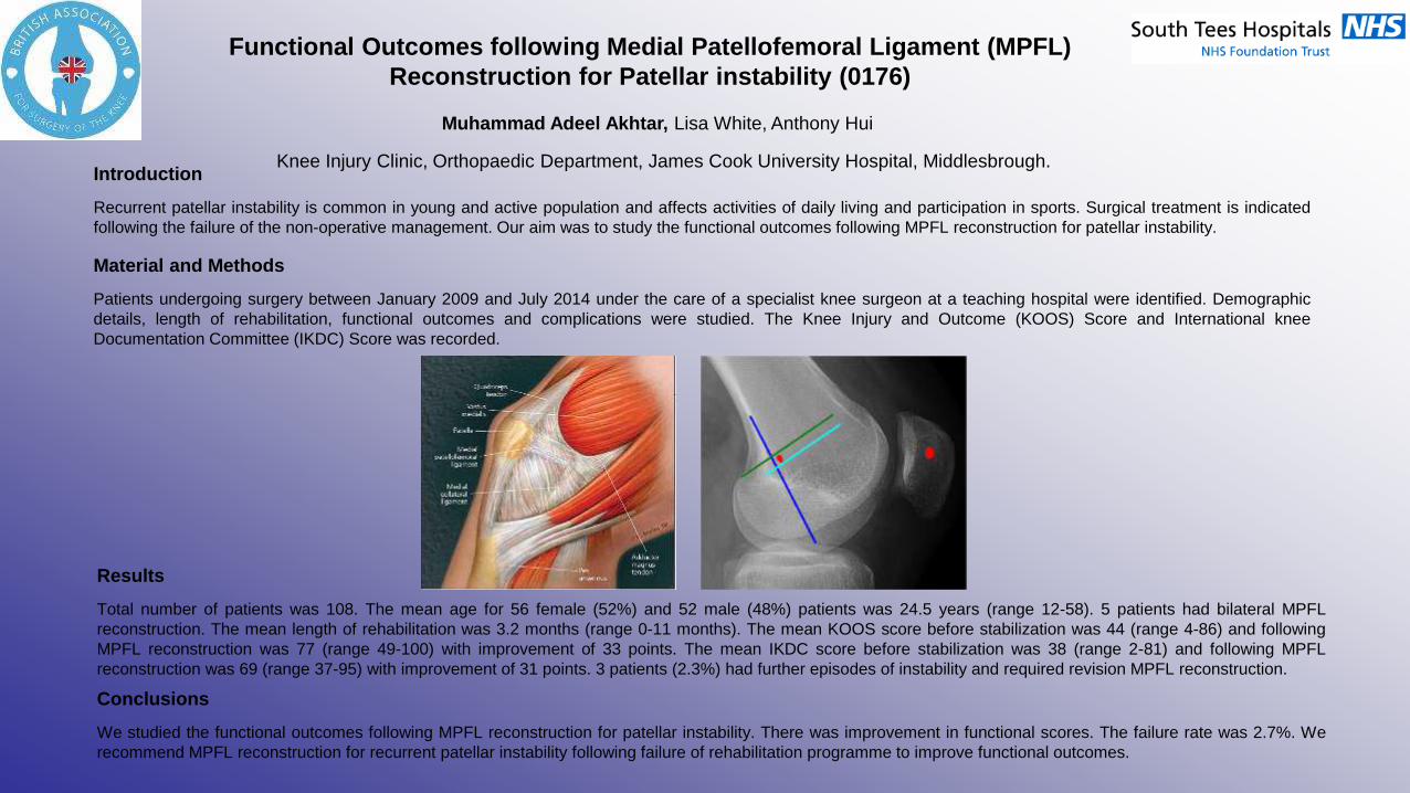
Functional Outcomes following Medial Patellofemoral Ligament (MPFL)
Reconstruction for Patellar instability (0176)
Muhammad Adeel Akhtar, Lisa White, Anthony Hui
Knee Injury Clinic, Orthopaedic Department, James Cook University Hospital, Middlesbrough.Introduction
Recurrent patellar instability is common in young and active population and affects activities of daily living and participation in sports. Surgical treatment is indicated
following the failure of the non-operative management. Our aim was to study the functional outcomes following MPFL reconstruction for patellar instability.
Results
Total number of patients was 108. The mean age for 56 female (52%) and 52 male (48%) patients was 24.5 years (range 12-58). 5 patients had bilateral MPFL
reconstruction. The mean length of rehabilitation was 3.2 months (range 0-11 months). The mean KOOS score before stabilization was 44 (range 4-86) and following
MPFL reconstruction was 77 (range 49-100) with improvement of 33 points. The mean IKDC score before stabilization was 38 (range 2-81) and following MPFL
reconstruction was 69 (range 37-95) with improvement of 31 points. 3 patients (2.3%) had further episodes of instability and required revision MPFL reconstruction.
Conclusions
We studied the functional outcomes following MPFL reconstruction for patellar instability. There was improvement in functional scores. The failure rate was 2.7%. We
recommend MPFL reconstruction for recurrent patellar instability following failure of rehabilitation programme to improve functional outcomes.
Material and Methods
Patients undergoing surgery between January 2009 and July 2014 under the care of a specialist knee surgeon at a teaching hospital were identified. Demographic
details, length of rehabilitation, functional outcomes and complications were studied. The Knee Injury and Outcome (KOOS) Score and International knee
Documentation Committee (IKDC) Score was recorded.
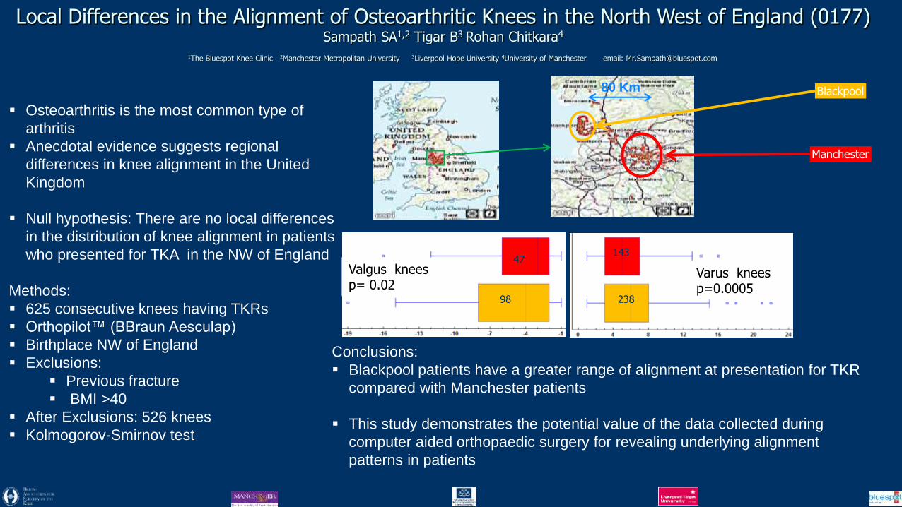
Local Differences in the Alignment of Osteoarthritic Knees in the North West of England (0177) Sampath SA1,2 Tigar B3 Rohan Chitkara4
1The Bluespot Knee Clinic 2Manchester Metropolitan University 3Liverpool Hope University 4University of Manchester email: [email protected]
Osteoarthritis is the most common type of
arthritis
Anecdotal evidence suggests regional
differences in knee alignment in the United
Kingdom
Null hypothesis: There are no local differences
in the distribution of knee alignment in patients
who presented for TKA in the NW of England
Methods:
625 consecutive knees having TKRs
Orthopilot™ (BBraun Aesculap)
Birthplace NW of England
Exclusions:
Previous fracture
BMI >40
After Exclusions: 526 knees
Kolmogorov-Smirnov test
80 Km Blackpool
Manchester
Valgus knees p= 0.02
Varus knees p=0.0005
47
98 238
143
Conclusions:
Blackpool patients have a greater range of alignment at presentation for TKR
compared with Manchester patients
This study demonstrates the potential value of the data collected during
computer aided orthopaedic surgery for revealing underlying alignment
patterns in patients
Tariff 2015/16
Now managed by MONITOR
No agreement yet for prices for 2015/16 – draft rejected by providers
Recent choice of options provided
Enhanced Tariff Option
Default Tariff Rollover
www.baskonline.com for updates
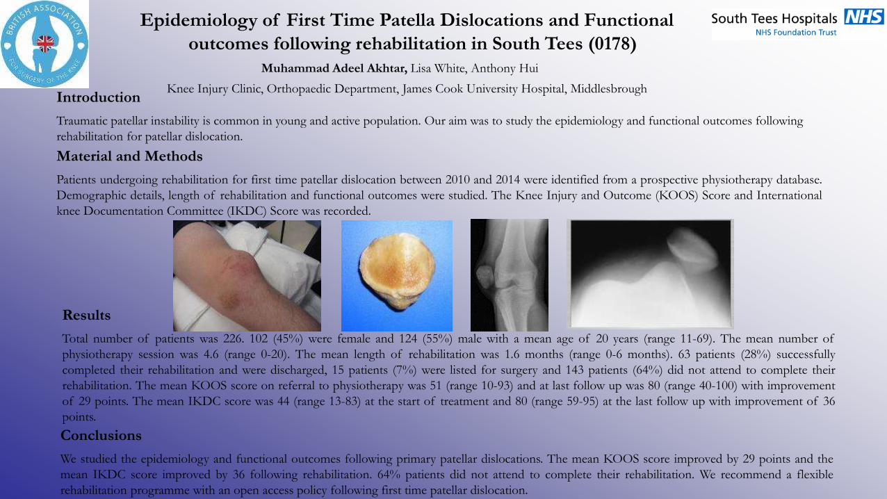
Epidemiology of First Time Patella Dislocations and Functional
outcomes following rehabilitation in South Tees (0178)
Muhammad Adeel Akhtar, Lisa White, Anthony Hui
Knee Injury Clinic, Orthopaedic Department, James Cook University Hospital, MiddlesbroughIntroduction
Traumatic patellar instability is common in young and active population. Our aim was to study the epidemiology and functional outcomes following
rehabilitation for patellar dislocation.
Results
Total number of patients was 226. 102 (45%) were female and 124 (55%) male with a mean age of 20 years (range 11-69). The mean number of
physiotherapy session was 4.6 (range 0-20). The mean length of rehabilitation was 1.6 months (range 0-6 months). 63 patients (28%) successfully
completed their rehabilitation and were discharged, 15 patients (7%) were listed for surgery and 143 patients (64%) did not attend to complete their
rehabilitation. The mean KOOS score on referral to physiotherapy was 51 (range 10-93) and at last follow up was 80 (range 40-100) with improvement
of 29 points. The mean IKDC score was 44 (range 13-83) at the start of treatment and 80 (range 59-95) at the last follow up with improvement of 36
points.
Conclusions
We studied the epidemiology and functional outcomes following primary patellar dislocations. The mean KOOS score improved by 29 points and the
mean IKDC score improved by 36 following rehabilitation. 64% patients did not attend to complete their rehabilitation. We recommend a flexible
rehabilitation programme with an open access policy following first time patellar dislocation.
Material and Methods
Patients undergoing rehabilitation for first time patellar dislocation between 2010 and 2014 were identified from a prospective physiotherapy database.
Demographic details, length of rehabilitation and functional outcomes were studied. The Knee Injury and Outcome (KOOS) Score and International
knee Documentation Committee (IKDC) Score was recorded.
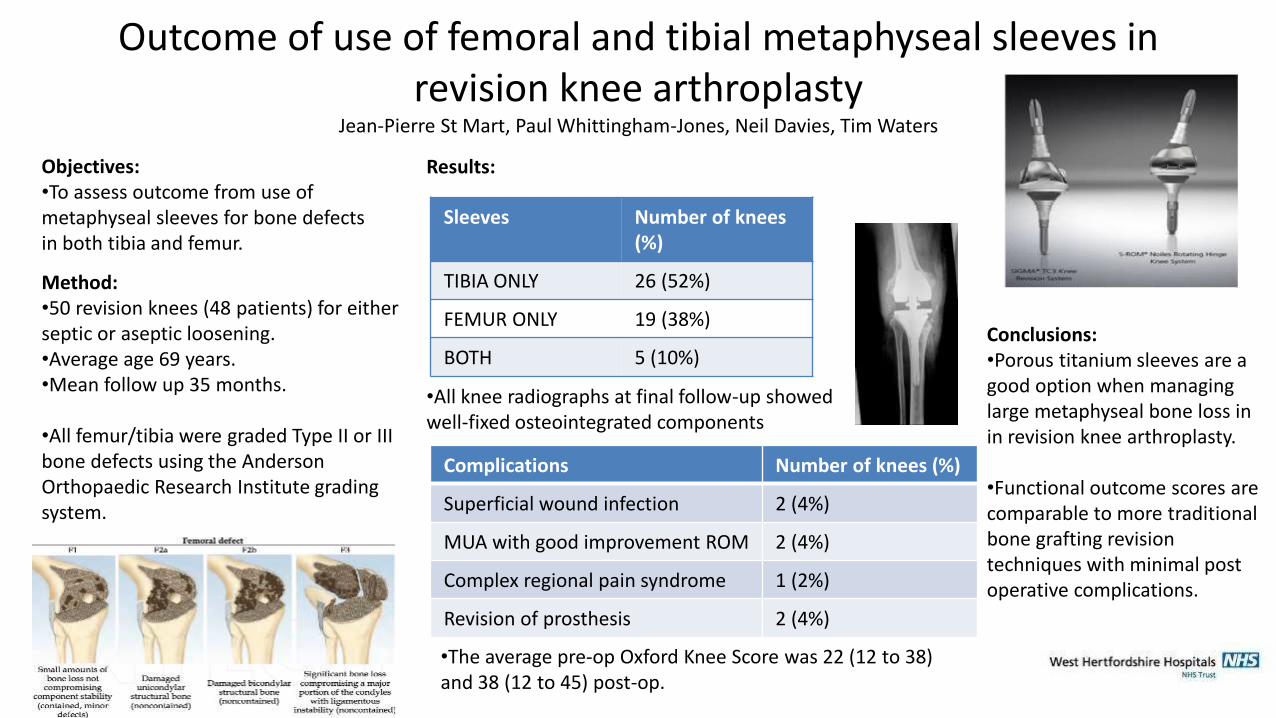
Outcome of use of femoral and tibial metaphyseal sleeves in revision knee arthroplasty
Jean-Pierre St Mart, Paul Whittingham-Jones, Neil Davies, Tim Waters
Objectives:•To assess outcome from use of metaphyseal sleeves for bone defects in both tibia and femur.
Method:•50 revision knees (48 patients) for either septic or aseptic loosening. •Average age 69 years. •Mean follow up 35 months.
•All femur/tibia were graded Type II or III bone defects using the Anderson Orthopaedic Research Institute grading system.
Results:
•All knee radiographs at final follow-up showed well-fixed osteointegrated components
Conclusions:•Porous titanium sleeves are a good option when managing large metaphyseal bone loss in in revision knee arthroplasty.
•Functional outcome scores are comparable to more traditional bone grafting revision techniques with minimal post operative complications.
Sleeves Number of knees (%)
TIBIA ONLY 26 (52%)
FEMUR ONLY 19 (38%)
BOTH 5 (10%)
Complications Number of knees (%)
Superficial wound infection 2 (4%)
MUA with good improvement ROM 2 (4%)
Complex regional pain syndrome 1 (2%)
Revision of prosthesis 2 (4%)
•The average pre-op Oxford Knee Score was 22 (12 to 38) and 38 (12 to 45) post-op.
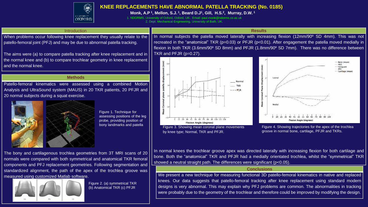
KNEE REPLACEMENTS HAVE ABNORMAL PATELLA TRACKING (No. 0185) Monk, A.P 1, Mellon, S.J. 1, Beard D.J1, Gill, H.S.2, Murray, D.W. 1
1. NDORMS, University of Oxford, Oxford, UK.. Email: [email protected]
2. Dept. Mechanical Engineering, University of Bath, UK.
Introduction
When problems occur following knee replacement they usually relate to the
patello-femoral joint (PFJ) and may be due to abnormal patella tracking.
The aims were (a) to compare patella tracking after knee replacement and in
the normal knee and (b) to compare trochlear geometry in knee replacement
and the normal knee.
Methods
Patello-femoral kinematics were assessed using a combined Motion
Analysis and UltraSound system (MAUS) in 20 TKR patients, 20 PFJR and
20 normal subjects during a squat exercise.
The bony and cartilagenous trochlea geometries from 3T MRI scans of 20
normals were compared with both symmetrical and anatomical TKR femoral
components and PFJ replacement geometries. Following segmentation and
standardized alignment, the path of the apex of the trochlea groove was
measured using customized Matlab software.
Figure 1. Technique for
assessing positions of the leg
probe, providing position of
bony landmarks and patella
Results
In normal subjects the patella moved laterally with increasing flexion (12mm/90º SD 4mm). This was not
recreated in the “anatomical” TKR (p=0.03) or PFJR (p<0.01). After engagement the patella moved medially in
flexion in both TKR (3.6mm/90º SD 8mm) and PFJR (1.8mm/90º SD 7mm). There was no difference between
TKR and PFJR (p=0.27).
In normal knees the trochlear groove apex was directed laterally with increasing flexion for both cartilage and
bone. Both the “anatomical” TKR and PFJR had a medially orientated trochlea, whilst the “symmetrical” TKR
showed a neutral straight path. The differences were significant (p<0.05).
Conclusions
We present a new technique for measuring functional 3D patello-femoral kinematics in native and replaced
knees. Our data suggests that patello-femoral tracking after knee replacement using standard modern
designs is very abnormal. This may explain why PFJ problems are common. The abnormalities in tracking
were probably due to the geometry of the trochlear and therefore could be improved by modifying the design.
Figure 2. (a) symmetrical TKR
(b) Anatomical TKR (c) PFJR
Figure 4. Showing trajectories for the apex of the trochlea
groove in normal bone, cartilage, PFJR and TKRs.Figure 3. Showing mean coronal plane movements
by knee type; Normal, TKR and PFJR.
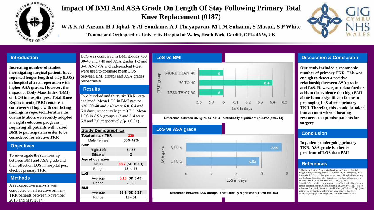
Impact Of BMI And ASA Grade On Length Of Stay Following Primary Total
Knee Replacement (0187)
Introduction
Increasing number of studies
investigating surgical patients have
reported longer length of stay (LOS)
in hospital after an operation with
higher ASA grades. However, the
impact of Body Mass Index (BMI)
on LOS in hospital post Total Knee
Replacement (TKR) remains a
controversial topic with conflicting
findings in reported literature. In
our institution, we recently adopted
a weight reduction program
requiring all patients with raised
BMI to participate in order to be
considered for elective TKR
W A K Al-Azzani, H J Iqbal, Y Al-Soudaine, A J Thayaparan, M I M Suhaimi, S Masud, S P White
Trauma and Orthopaedics, University Hospital of Wales, Heath Park, Cardiff, CF14 4XW, UK
To investigate the relationship
between BMI and ASA grade and
their effect on LOS in hospital post
elective primary THR
A retrospective analysis was
conducted on all elective primary
TKR patients between November
2013 and May 2014
Objectives
Results
Discussion & Conclusion
References
Our study included a reasonable
number of primary TKR. This was
enough to detect a positive
relationship between ASA grade
and LoS. However, our data further
adds to the evidence that high BMI
alone is not a significant factor in
prolonging LoS after a primary
TKR. Therefor, this should be taken
into account when allocating
resources to optimise patients for
surgery
In patients undergoing primary
TKR, ASA grade is a better
predictor of LOS than BMI
Methods
Study DemographicsTotal primary THR 236
Male:Female 58%:42%
Side
Right:Left 64:56
Bilateral 2
Age at operation
Mean 68.7 (SD 10.01)
Range 43 to 96
LoS
Average 6.19 (SD 3.43)
Range 2 - 28
BMI
Average 32.9 (SD 6.33)
Range 19 - 51
BMIgroups Mean Nless than 30 6 80
30 to 40 6.4 125
more than 40 6 31
Total 5.96 122
ASAgroups Mean N
1 to 2 5.82 159
3 to 4 7.59 41
LOS was compared in BMI groups <30,
30-40 and >40 and ASA grades 1-2 and
3-4. ANOVA and independent t-test
were used to compare mean LOS
between BMI groups and ASA grades,
respectively
Two hundred and thirty six TKR were
analysed. Mean LOS in BMI groups
<30, 30-40 and >40 were 6.0, 6.4 and
6.0 days, respectively (p = 0.71). Mean
LOS in ASA groups 1-2 and 3-4 were
5.8 and 7.6, respectively (p < 0.01).
LoS vs BMI
LoS vs ASA grade
Difference between BMI groups is NOT statistically significant (ANOVA p=0.714)
Difference between ASA groups is statistically significant (T-test p=0.04)
Conclusion
1. Halawi, M.J., et al., Preoperative Predictors of Extended Hospital
Length of Stay Following Total Knee Arthroplasty. J Arthroplasty, 2014.
2. Crawford, D.A., et al., Preoperative predictors of length of hospital stay
and discharge disposition following primary total knee arthroplasty at a
military medical center. Mil Med, 2011. 176(3): p. 304-7.
3. Smith, I.D., et al., Pre-operative predictors of the length of hospital stay
in total knee replacement. J Bone Joint Surg Br, 2008. 90(11): p. 1435-40.
4. Lozano, L.M., et al., Severe and morbid obesity (BMI >/= 35 kg/m) does
not increase surgical time and length of hospital stay in total knee
arthroplasty surgery. Knee Surg Sports Traumatol Arthrosc, 2014.
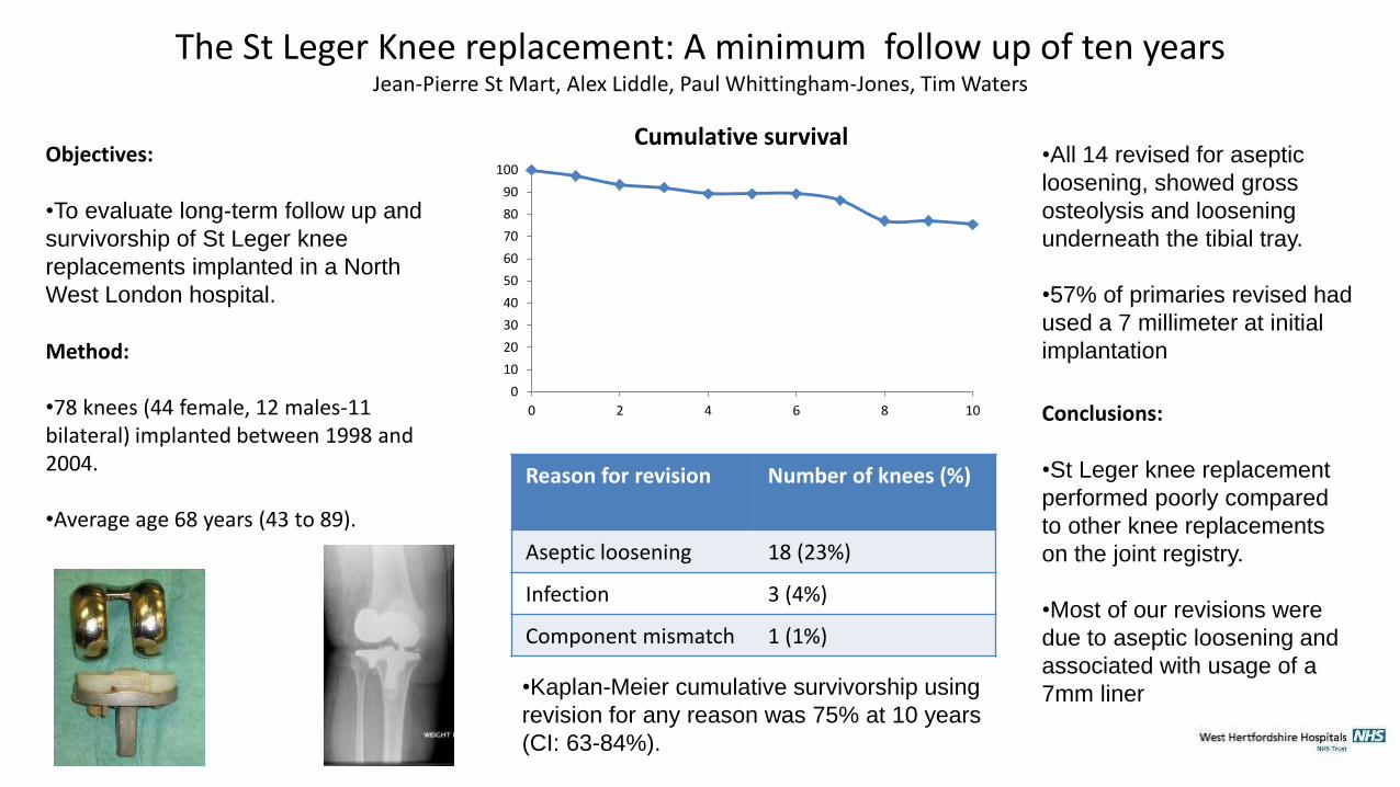
The St Leger Knee replacement: A minimum follow up of ten yearsJean-Pierre St Mart, Alex Liddle, Paul Whittingham-Jones, Tim Waters
Objectives:
•To evaluate long-term follow up and
survivorship of St Leger knee
replacements implanted in a North
West London hospital.
Method:
•78 knees (44 female, 12 males-11 bilateral) implanted between 1998 and 2004.
•Average age 68 years (43 to 89).
Conclusions:
•St Leger knee replacement
performed poorly compared
to other knee replacements
on the joint registry.
•Most of our revisions were
due to aseptic loosening and
associated with usage of a
7mm liner
Reason for revision Number of knees (%)
Aseptic loosening 18 (23%)
Infection 3 (4%)
Component mismatch 1 (1%)
•Kaplan-Meier cumulative survivorship using
revision for any reason was 75% at 10 years
(CI: 63-84%).
0
10
20
30
40
50
60
70
80
90
100
0 2 4 6 8 10
Cumulative survival•All 14 revised for aseptic
loosening, showed gross
osteolysis and loosening
underneath the tibial tray.
•57% of primaries revised had
used a 7 millimeter at initial
implantation
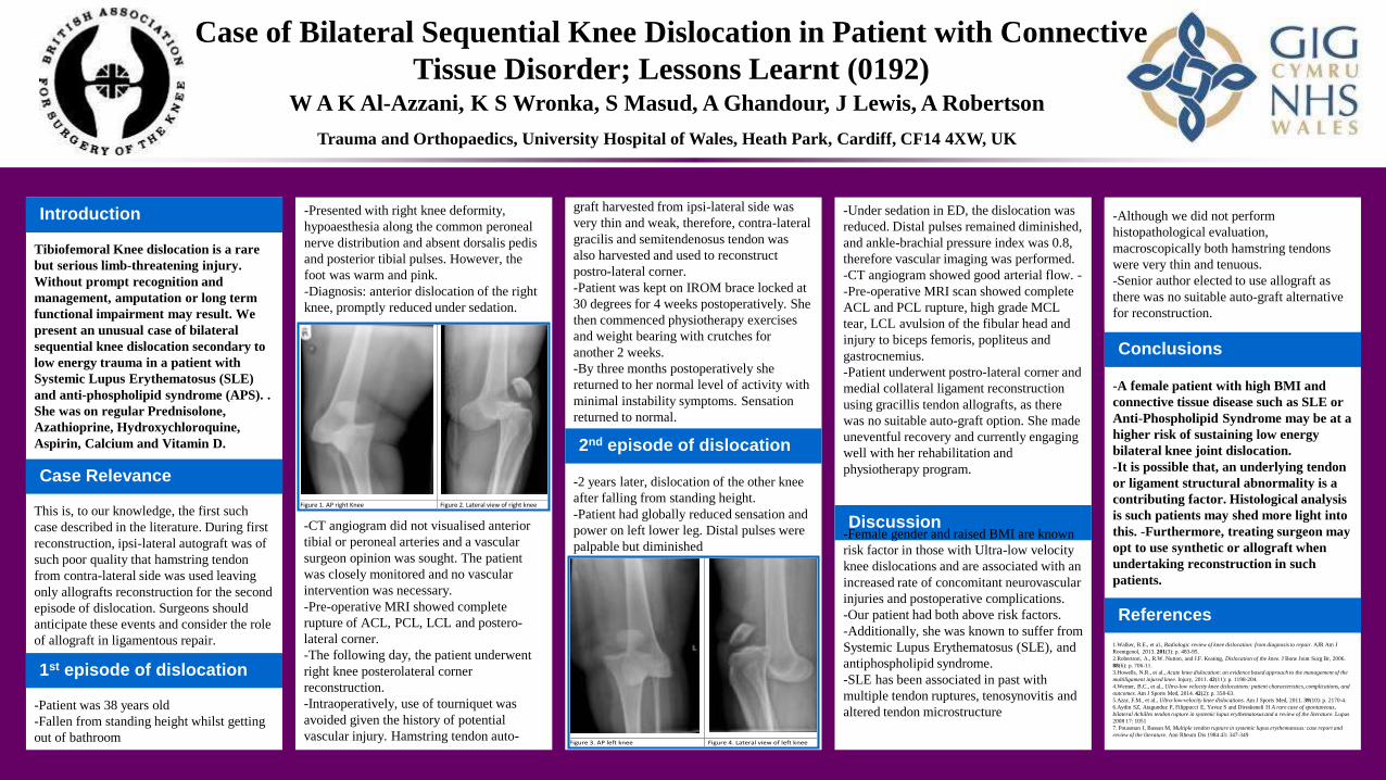
-Although we did not perform
histopathological evaluation,
macroscopically both hamstring tendons
were very thin and tenuous.
-Senior author elected to use allograft as
there was no suitable auto-graft alternative
for reconstruction.
Case of Bilateral Sequential Knee Dislocation in Patient with Connective
Tissue Disorder; Lessons Learnt (0192)
Introduction
Tibiofemoral Knee dislocation is a rare
but serious limb-threatening injury.
Without prompt recognition and
management, amputation or long term
functional impairment may result. We
present an unusual case of bilateral
sequential knee dislocation secondary to
low energy trauma in a patient with
Systemic Lupus Erythematosus (SLE)
and anti-phospholipid syndrome (APS). .
She was on regular Prednisolone,
Azathioprine, Hydroxychloroquine,
Aspirin, Calcium and Vitamin D.
W A K Al-Azzani, K S Wronka, S Masud, A Ghandour, J Lewis, A Robertson
Trauma and Orthopaedics, University Hospital of Wales, Heath Park, Cardiff, CF14 4XW, UK
-Presented with right knee deformity,
hypoaesthesia along the common peroneal
nerve distribution and absent dorsalis pedis
and posterior tibial pulses. However, the
foot was warm and pink.
-Diagnosis: anterior dislocation of the right
knee, promptly reduced under sedation.
This is, to our knowledge, the first such
case described in the literature. During first
reconstruction, ipsi-lateral autograft was of
such poor quality that hamstring tendon
from contra-lateral side was used leaving
only allografts reconstruction for the second
episode of dislocation. Surgeons should
anticipate these events and consider the role
of allograft in ligamentous repair.
-Patient was 38 years old
-Fallen from standing height whilst getting
out of bathroom
1.Walker, R.E., et al., Radiologic review of knee dislocation: from diagnosis to repair. AJR Am J
Roentgenol, 2013. 201(3): p. 483-95.
2.Robertson, A., R.W. Nutton, and J.F. Keating, Dislocation of the knee. J Bone Joint Surg Br, 2006.
88(6): p. 706-11.
3.Howells, N.R., et al., Acute knee dislocation: an evidence based approach to the management of the
multiligament injured knee. Injury, 2011. 42(11): p. 1198-204.
4.Werner, B.C., et al., Ultra-low velocity knee dislocations: patient characteristics, complications, and
outcomes. Am J Sports Med, 2014. 42(2): p. 358-63.
5.Azar, F.M., et al., Ultra-low-velocity knee dislocations. Am J Sports Med, 2011. 39(10): p. 2170-4.
6.Aydin SZ, Atagunduz P, Filippucci E, Yavuz S and Direskeneli H A rare case of spontaneous,
bilateral Achilles tendon rupture in systemic lupus erythematosus and a review of the literature. Lupus
2008 17: 1051
7. Potasman I, Bassan M, Multiple tendon rupture in systemic lupus erythematosus: case report and
review of the literature. Ann Rheum Dis 1984 43: 347-349
Case Relevance
2nd episode of dislocation
Discussion
Conclusions
References
-A female patient with high BMI and
connective tissue disease such as SLE or
Anti-Phospholipid Syndrome may be at a
higher risk of sustaining low energy
bilateral knee joint dislocation.
-It is possible that, an underlying tendon
or ligament structural abnormality is a
contributing factor. Histological analysis
is such patients may shed more light into
this. -Furthermore, treating surgeon may
opt to use synthetic or allograft when
undertaking reconstruction in such
patients.
graft harvested from ipsi-lateral side was
very thin and weak, therefore, contra-lateral
gracilis and semitendenosus tendon was
also harvested and used to reconstruct
postro-lateral corner.
-Patient was kept on IROM brace locked at
30 degrees for 4 weeks postoperatively. She
then commenced physiotherapy exercises
and weight bearing with crutches for
another 2 weeks.
-By three months postoperatively she
returned to her normal level of activity with
minimal instability symptoms. Sensation
returned to normal.
-2 years later, dislocation of the other knee
after falling from standing height.
-Patient had globally reduced sensation and
power on left lower leg. Distal pulses were
palpable but diminished
Figure 1. AP right Knee Figure 2. Lateral view of right knee
1st episode of dislocation
Figure 3. AP left knee Figure 4. Lateral view of left knee
-Under sedation in ED, the dislocation was
reduced. Distal pulses remained diminished,
and ankle-brachial pressure index was 0.8,
therefore vascular imaging was performed.
-CT angiogram showed good arterial flow. -
-Pre-operative MRI scan showed complete
ACL and PCL rupture, high grade MCL
tear, LCL avulsion of the fibular head and
injury to biceps femoris, popliteus and
gastrocnemius.
-Patient underwent postro-lateral corner and
medial collateral ligament reconstruction
using gracillis tendon allografts, as there
was no suitable auto-graft option. She made
uneventful recovery and currently engaging
well with her rehabilitation and
physiotherapy program.
-Female gender and raised BMI are known
risk factor in those with Ultra-low velocity
knee dislocations and are associated with an
increased rate of concomitant neurovascular
injuries and postoperative complications.
-Our patient had both above risk factors.
-Additionally, she was known to suffer from
Systemic Lupus Erythematosus (SLE), and
antiphospholipid syndrome.
-SLE has been associated in past with
multiple tendon ruptures, tenosynovitis and
altered tendon microstructure
-CT angiogram did not visualised anterior
tibial or peroneal arteries and a vascular
surgeon opinion was sought. The patient
was closely monitored and no vascular
intervention was necessary.
-Pre-operative MRI showed complete
rupture of ACL, PCL, LCL and postero-
lateral corner.
-The following day, the patient underwent
right knee posterolateral corner
reconstruction.
-Intraoperatively, use of tourniquet was
avoided given the history of potential
vascular injury. Hamstring tendon auto-
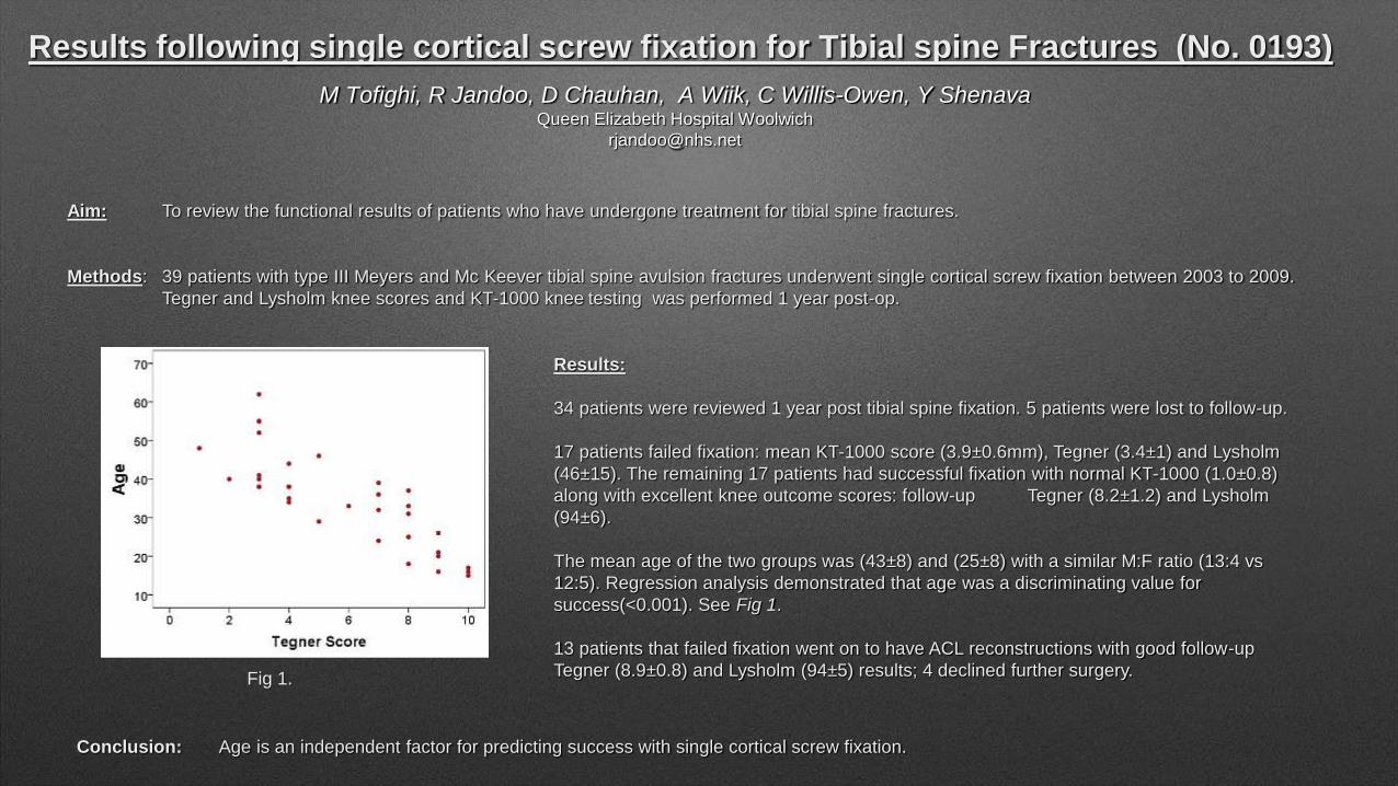
Results following single cortical screw fixation for Tibial spine Fractures (No. 0193)
M Tofighi, R Jandoo, D Chauhan, A Wiik, C Willis-Owen, Y ShenavaQueen Elizabeth Hospital Woolwich
Aim: To review the functional results of patients who have undergone treatment for tibial spine fractures.
Methods: 39 patients with type III Meyers and Mc Keever tibial spine avulsion fractures underwent single cortical screw fixation between 2003 to 2009.
Tegner and Lysholm knee scores and KT-1000 knee testing was performed 1 year post-op.
Results:
34 patients were reviewed 1 year post tibial spine fixation. 5 patients were lost to follow-up.
17 patients failed fixation: mean KT-1000 score (3.9±0.6mm), Tegner (3.4±1) and Lysholm
(46±15). The remaining 17 patients had successful fixation with normal KT-1000 (1.0±0.8)
along with excellent knee outcome scores: follow-up Tegner (8.2±1.2) and Lysholm
(94±6).
The mean age of the two groups was (43±8) and (25±8) with a similar M:F ratio (13:4 vs
12:5). Regression analysis demonstrated that age was a discriminating value for
success(<0.001). See Fig 1.
13 patients that failed fixation went on to have ACL reconstructions with good follow-up
Tegner (8.9±0.8) and Lysholm (94±5) results; 4 declined further surgery.Fig 1.
Conclusion: Age is an independent factor for predicting success with single cortical screw fixation.
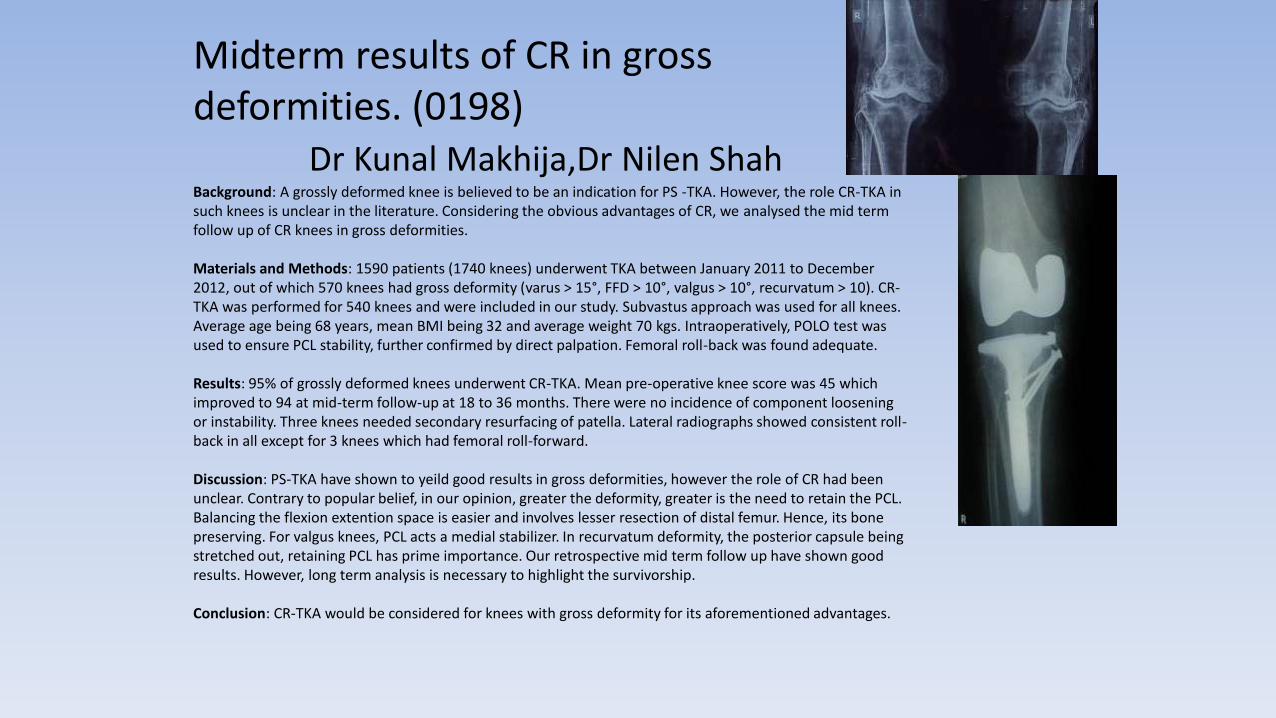
Midterm results of CR in gross deformities. (0198)
Dr Kunal Makhija,Dr Nilen ShahBackground: A grossly deformed knee is believed to be an indication for PS -TKA. However, the role CR-TKA in such knees is unclear in the literature. Considering the obvious advantages of CR, we analysed the mid term follow up of CR knees in gross deformities.
Materials and Methods: 1590 patients (1740 knees) underwent TKA between January 2011 to December 2012, out of which 570 knees had gross deformity (varus > 15°, FFD > 10°, valgus > 10°, recurvatum > 10). CR-TKA was performed for 540 knees and were included in our study. Subvastus approach was used for all knees. Average age being 68 years, mean BMI being 32 and average weight 70 kgs. Intraoperatively, POLO test was used to ensure PCL stability, further confirmed by direct palpation. Femoral roll-back was found adequate.
Results: 95% of grossly deformed knees underwent CR-TKA. Mean pre-operative knee score was 45 which improved to 94 at mid-term follow-up at 18 to 36 months. There were no incidence of component loosening or instability. Three knees needed secondary resurfacing of patella. Lateral radiographs showed consistent roll-back in all except for 3 knees which had femoral roll-forward.
Discussion: PS-TKA have shown to yeild good results in gross deformities, however the role of CR had been unclear. Contrary to popular belief, in our opinion, greater the deformity, greater is the need to retain the PCL. Balancing the flexion extention space is easier and involves lesser resection of distal femur. Hence, its bone preserving. For valgus knees, PCL acts a medial stabilizer. In recurvatum deformity, the posterior capsule being stretched out, retaining PCL has prime importance. Our retrospective mid term follow up have shown good results. However, long term analysis is necessary to highlight the survivorship.
Conclusion: CR-TKA would be considered for knees with gross deformity for its aforementioned advantages.
Subvastus approach for Revision TKA (199) Dr Kunal Makhija , Dr Nilen ShahBackground: Subvastus approach for Total Knee Arthropalsty (TKA) allows a faster recovery. It is traditionally not utilized for revision surgeries because of difficulty in exposure of the knee and eversion of the patella. It is considered to have limited indications. We hypothesized that revision TKA should not really pose a problem as the exposure gained is adequate with added advantage of preserving the extensor mechanism, thereby allowing faster functional recovery. We present an analysis of the use of subvastus approach for revision TKAs.Materials and methods: 50 patients (50 knees) 37 females + 13 males with mean age 68 years underwent revision total knee arthroplasty (TKA) by subvastus approach between January 2006 to January 2013. All patients were prospectively evaluated by pre- and postoperative Knee Society and function score. The average follow-up was 24 months (range from 1 to 3 years) with minimum 1 year follow-up. The indications for revisions were aseptic loosening (20 knees), infection (12 knees), instability (12 knees) and peri-prosthetic fractures (6 knees). Constrained condylar prosthesis (43 knees), hinged prosthesis (6 knees) and custom made prosthesis (1 knee) were fixed using the subvastus approach. Infected knees underwent one or two staged revisions.Results: The approach provided adequate exposure in all revisions. The average Knee Society score improved from 42 to 83 and the function score from 48 to 65. The complications included medial collateral ligament injury (one case), patellar tendon avulsion (one case) and mal-tracking patella (one case). Average hospital stay was 4 days. Average blood lose was 400 ml.Conclusion: Our results compare favourably with other reported series on revision TKA. The subvastus approach can be considered for revision TKAs..