wear of enamel and veneering ceramics after laboratory and chairside finishing procedures
TRANSCRIPT

The wear of dental hard tissues is a natural andunavoidable process.1 However, when opposed byceramic, enamel may be subject to accelerated wear,2 thepattern of which may vary according to the ceramic sys-
tem and its surface characteristics.3,4 A major step wastaken at the end of the last decade, when in vitro studiesdemonstrated the excellent behavior of unshadedcastable glass ceramics (Dicor system) compared withconventional feldspathic materials.4-6 Wear rates, in termsof material removal and vertical height loss, proved to besimilar to gold alloy.7 The advent of machinable ceramicsremoved the “operator” factors that occur during pro-cessing, and provides improved microstructural control.8
Nevertheless, traditional feldspathic ceramics are stillwidely used,9,10 despite the numerous scientific reportsof their harmful behavior with regard to enamelwear.2,3,5,7 This trend is easily explained by the verygood esthetic results achieved with a natural stratifica-tion and the artisan work of the dental ceramist.11 Castor pressed glass ceramics and machined porcelainsalone do not allow such artistry and thus limit their usein esthetic restorative dentistry. In addition, early
Wear of enamel and veneering ceramics after laboratory and chairsidefinishing procedures
Pascal Magne, Dr Med Dent,a Won-Suck Oh, DDS,b Maria R. Pintado, BS, MPH,c and Ralph DeLong,DDS, MS, PhDd
School of Dentistry, University of Minnesota, Minneapolis, Minn.
Purpose. This in vitro study compared the wear of enamel against 3 types of ceramics with high estheticpotential (designed for layering techniques): feldspathic porcelain (Creation), aluminous porcelain (Vitadur α),and low-fusing glass (Duceram-LFC). Laboratory finishing (glazing/polishing) and chairside polishing with aDialite kit were simulated to compare their respective effects on wear.Methods. Tooth-material specimen pairs were placed in an artificial mouth using closed-loop servohy-draulics. Constant masticatory parameters (13.5 N occlusal force, 0.62 mm lateral excursion; 0.23 secondcuspal contact time) were maintained for 300,000 cycles at a rate of 4 Hz. The occlusal surface of each pairwas mapped and digitally recorded before and after each masticatory test. Quantitative changes were mea-sured in terms of depth and volume of wear. Quantitative wear characteristics were assessed by SEM.Results. Significant differences were observed (2-factor ANOVA, P<.05). Duceram-LFC generatedincreased volume loss of enamel (0.197 mm3) compared with Creation (0.135 mm3) and Vitadur α(0.153 mm3). Creation exhibited the lowest ceramic wear and lowest combined volume loss (0.260 mm3;the sum of the data for enamel and the opposing material) compared with Duceram-LFC (0.363 mm3) andVitadur α (0.333 mm3). The most significant differences among materials were observed in volume loss,not in depth of wear. For all 3 ceramic systems, qualitative SEM evaluation revealed an abrasive type ofwear. Wear characteristics of chairside polished specimens were similar to those of laboratory finished speci-mens (glazed and polished).Conclusion. Duceram-LFC was the most abrasive ceramic for the antagonistic tooth. Creation ceramicwas the least abrasive material and most resistant to wear. Defects, brittleness, and the possibly insufficienttoughness of LFC may explain its increased abrasiveness. Laboratory and chairside finishing procedures gen-erated similar results. (J Prosthet Dent 1999;82:669-79.)
This study was supported by the Swiss Foundation for Medical-Bio-logical Grants and in part by the Minnesota Dental ResearchCenter for Biomaterials and Biomechanics Wear Testing and Sur-face Analysis Systems.
aFormerly Visiting Associate Professor, Minnesota Dental ResearchCenter for Biomaterials and Biomechanics, Department of OralScience; and Assistant Professor, Department of Prosthodonticsand Department of Prevention and Therapeutics, School of Den-tal Medicine, University of Geneva, Switzerland.
bProsthodontic Resident, Graduate Prosthodontics, Department ofRestorative Sciences.
cAssociate Professor, Minnesota Dental Research Center for Bioma-terials and Biomechanics, Department of Oral Science.
dProfessor, Minnesota Dental Research Center for Biomaterials andBiomechanics, Department of Oral Science.
DECEMBER 1999 THE JOURNAL OF PROSTHETIC DENTISTRY 669
CLINICAL IMPLICATIONS
There were significant differences in the wear associated with 3 common veneeringceramics. Noncrystalline low-fusing glass appeared to cause more wear of opposingenamel compared with feldspathic or aluminous porcelains. In this study, intraoralfinishing of ceramic restorations was not contraindicated because wear characteristicswere similar to those restorations finished in the laboratory by glazing and polishing.
reports demonstrate that the latest polymers developedfor prosthodontic use (so-called “ceromers”) did notseem to be able to meet their original requirementswith regard to clinical wear and failure rate.12,13
Meanwhile, Duceram-LFC (Ducera, Rosbach, Ger-many) hydrothermal low-fusing glass was introduced. Itis designed to be applied over a conventional ceramiccore and its manufacturer makes impressive claims aboutwear, solubility, and surface properties.14 Unlike mostceramics, the flexural strength and resistance to disinte-gration of Duceram-LFC seem to increase significantlyafter hydrolysis testing.15 Such accelerated aging is sim-ulated by subjecting the material to 4% acetic acid solu-tion at 87°C for 16 hours. However, few scientific stud-ies have investigated its wear properties. Because of itsglassy nature (no crystalline phase), Duceram-LFC isdescribed as a homogeneous structure that shouldreduce wear of antagonistic enamel.14 However, thehigh potential of Duceram-LFC is questioned in light of
recent studies that could not consistently differentiate itswear pattern from other ceramics.16,17
Two possible protocols can be used when placingceramic restorations. The first one corresponds to theclassical prosthodontic approach: A try-in appointmentis usually planned when using traditional full cover-ages.18 This specific chairside step includes meticulousocclusal adjustments, the restoration being returned tothe dental laboratory for eventual corrections, surfacepolishing, and glazing. Such planning optimally allowsthe practitioner to seat and cement the restoration at thenext appointment without further corrections of therestoration surface. These procedures are usually recom-mended because most authors have agreed that the lab-oratory can produce smoother and denser surfaces.19 Asa result, the way ceramic materials are tested often cor-respond to this protocol (laboratory finished surface).However, a different chronology and sequence occurswhen placing bonded restorations such as porcelaininlays, onlays, and veneers. It is not recommended toadjust or check occlusion before the restoration iscemented because of the risk of fracture.20 For this rea-son, the surface of the restoration is often adjusted andrepolished intraorally after cementation.
A variety of commercial kits have been developed toimprove intraoral surface finishing of the ceramic, andhave been extensively studied in the literature.19,21-24
Most of these experiments focused on the ceramic sur-face roughness that can be achieved and compared withan “ideal” glazed surface. It is often speculated that therougher surfaces produced by polishing will generateincreased enamel wear. Only a few studies extendedtheir investigation to the influence of ceramic surfacefinishing on the wear characteristics and wear patternagainst enamel.25,26 Surprisingly, the same conclusionis found in these studies, namely, the absence of a sig-nificant effect of porcelain surface finish.
A problem with wear-related literature is the lack ofstandardization. A wide variety of abrasives, measuringinstruments, and methods of wear testing make it diffi-cult to assess study results that compare dental materi-als.27 Experimental methods have varied from the sim-ple design of the Wig-L-Bug technique28 to a variety ofmore sophisticated systems. There has been no consen-sus about the method of laboratory wear tests or theirclinical meaning. For instance, Mahalick29 used a sim-ple abrading apparatus and measured a 100× increase inmean volume loss for porcelain-to-porcelain whencompared with gold-to-gold specimens. However, anin vivo study by Ekfeldt and Oilo30 reported a volumeloss per unit time for porcelain-to-porcelain that wasonly 5 to 10 times greater than type III or IV gold incontact with identical materials. This finding empha-sizes the importance of using a system that reproduces,in an accurate way, the forces, movements, and physicalenvironment of the human masticatory system. Such a
THE JOURNAL OF PROSTHETIC DENTISTRY MAGNE ET AL
670 VOLUME 82 NUMBER 6
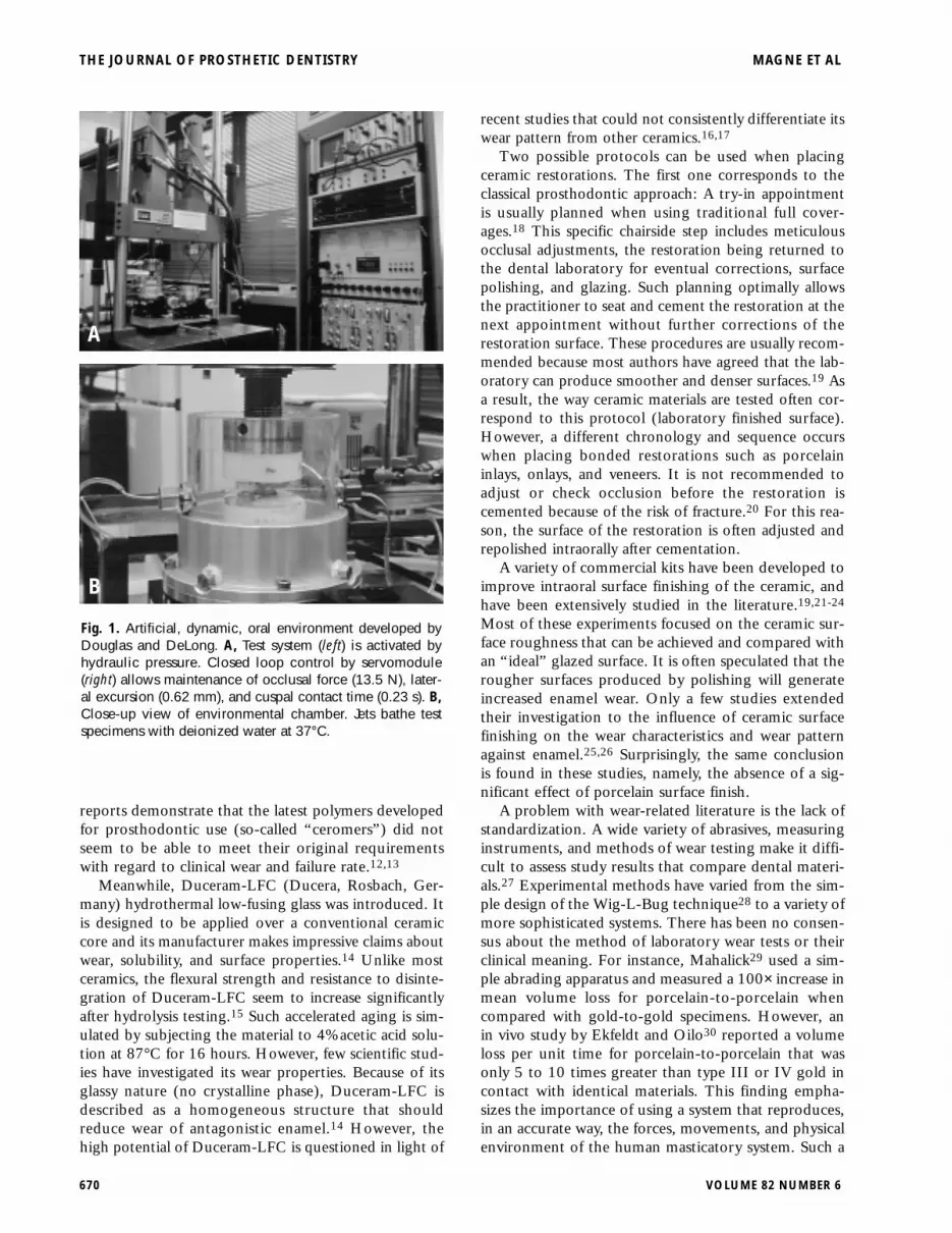
Fig. 1. Artificial, dynamic, oral environment developed byDouglas and DeLong. A, Test system (left) is activated byhydraulic pressure. Closed loop control by servomodule(right) allows maintenance of occlusal force (13.5 N), later-al excursion (0.62 mm), and cuspal contact time (0.23 s). B,Close-up view of environmental chamber. Jets bathe testspecimens with deionized water at 37°C.
A
B

method was proposed in 1983 by DeLong and Dou-glas,31 who developed an artificial oral environmentthat correlated well with clinical occlusal contact wear(Fig. 1, A and B).32,33
This standardized, recognized protocol was used inour study to compare the wear properties of 3 ceramicsdesigned for layering techniques, namely, veneering ofa metal or ceramic core. Special attention was given tothe surface finishing of the ceramic to simulate labora-tory finished surfaces and clinical conditions related toporcelain inlays, onlays, and veneers, namely, surfaceroughening (occlusal adjustment) and the use of anintraoral porcelain finishing kit.
METHOD AND MATERIAL
Three groups of ceramics were tested: (1) low-fusinghydrothermal glass (Duceram-LFC), (2) feldspathicceramic for porcelain-fused-to-metal (Creation, Klema,Meiningen, Austria), and (3) alumina-reinforcedceramic for aluminacore veneering (Vitadur α). Foreach material, 5 disks (12 mm in diameter and 3 mmthick) were produced from a silicon mold and firedover a platinum foil. All specimens were fabricated by acommercial laboratory experienced with the 3 ceramicsystems. This laboratory has routinely used theseceramics for a minimum of 6 years. All ceramic speci-mens were made according to a realistic protocol forfiring conditions and surface finishing. This includedthe reproduction of a distinct surface texture withsuperficial grooves (Fig. 2, A), which was standardizedas much as possible by creating a defined number ofevenly spaced grooves (14 per sample) using constantand reproducible rotation speed of the bur.
For group I (LFC) specimens, the hydrothermalglass is designed to be applied to a core of traditionalfeldspathic material. Therefore, a circular base of Cre-ation porcelain was first constructed (2 firings undervacuum without holding time at 910°C, glazing with-out vacuum without holding time at 910°C) andveneered with 1-mm thick Duceram-LFC (2 firingsunder vacuum without holding time at 660°C, glazingwithout vacuum without holding time at 670°C).Glazing of Duceram-LFC ceramic did not follow man-ufacturer’s recommendations (holding time of 2.5 min-utes at 650°C). On the basis of the clinical experience,this procedure was chosen to prevent excessive translu-cency of the glass. Lack of brightness (insufficient lightreflection) is inherent to Duceram-LFC ceramic andchallenges the dental technician in his layering tech-nique. The “modified” glazing protocol corresponds toa realistic way of using Duceram-LFC for anterior teethwhen high esthetics is essential.
For group II (Creation) specimens, 2 firings undervacuum were performed without holding time at910°C, glazing without vacuum without holdingtime at 910°C. For group III (Vita) specimens, 2 fir-
ings under vacuum were done without holding timeat 950°C, glazing without vacuum without holdingtime at 930°C.
Surface finishing
Two scenarios were simulated for each group ofceramics for surface finishing. The first, laboratory-fin-ished surface, was conducted by glazing, followed bymechanical polishing with pumice and calcium carbon-ate (Sigolin, Thompson Siegel, Dusseldorf, Germany).Calcium carbonate is a common abrasive material usedin dentifrice. It is softer and finer than pumice andallows excellent finishing of the porcelain surface.
The second scenario was an intraorally finished sur-face, which was performed to simulate an intraoraladjustment. All ceramic surfaces were abraded with agreenstone at low speed by a single operator. Repolish-ing was carried out with a 30 fluted tungsten carbidebur (ETUF 9 014, Brasseler, Savannah, Ga.) and acommercial intraoral polishing kit (Dialite, UltraPolishers, Brasseler) consisting of 3 consecutive dia-mond-silicon points (coarse W 16DG-21, mediumW 16DM-21 and fine W 16D-21) and with abundantwater spray at 2000 rpm.
MAGNE ET AL THE JOURNAL OF PROSTHETIC DENTISTRY
DECEMBER 1999 671
Fig. 2. A, Ceramic disk prepared with clinically realistic sur-face texture. B, Tooth specimen positioned in resin.
A
B

Preparation of tooth specimens and wearcharacterization
Thirty extracted maxillary third molars (5 teeth permaterial tested and per finishing procedure) werestored in deionized water and 0.2% azide until use.Teeth were collected from oral surgery clinics and pre-pared by removing the buccal cusps and isolating themesiopalatal cusp. Each tooth was mounted in a flatpolyethylene ring with the use of a chemically curedacrylic resin (Orthodontic resin, batch no. 651006,Dentsply International, Milford, Del.) (Fig. 2, B).
Masticatory movements and forces were simulatedwith an artificial mouth using closed-loop servohy-draulics.31 The chewing cycle was simulated by a modechange from isotonic to isometric contraction (strokecontrol to load control) as required by the physiologyof the mouth. This device has been extensivelydescribed and shown to correlate well with clinicalocclusal contact wear.5,32-34 The ceramic disks werepositioned in a polyethylene ring and used as themandibular member of the system. For each sample,the maxillary member was represented by the palatalcusp of a maxillary third molar (described previously).
Each experimental pair (disk and cusp) was stored indeionized water at 37°C for at least 24 hours beforetesting, then subjected to 300,000 defined masticatorycycles at a chewing rate of 4 Hz. Five specimens of eachmaterial type were placed in the artificial mouth andsubjected to the following masticatory parameters:occlusal force at 13.5 N, lateral excursion at 0.62 mm,and cuspal contact time at 0.23 second. A continuousflow of deionized water was directed on the wear area,maintaining the environmental temperature at 37°C.
Profiling
Paired occlusal surfaces were mapped and digitallyrecorded at the beginning and end of each masticatorytest. The profiling device was designed and built in theMinnesota Dental Research Center for Biomaterialsand Biomechanics (MDRCBB).35 Its design is uniquebecause the tip of the stylus does not move during pro-filing (tip used as a “null point”); instead, the surfacebeing profiled moves. This method is distinct from a
THE JOURNAL OF PROSTHETIC DENTISTRY MAGNE ET AL
672 VOLUME 82 NUMBER 6
Fig. 3. Example of digitally mapped surfaces (ceramic diskand cusp tip with wear facets) in AnSur.
A
B
Fig. 4. A, Volume of material removed (mm3) and B, meandepth of wear (µm) for enamel, ceramic and enamel/ceram-ic combined with both finishing techniques. Thin bars indi-cate standard deviation.
A
B

displacement stylus and was developed specifically tomeet the rigorous challenge of profiling the surface ofteeth with ultrahigh accuracy. The tungsten carbide sty-lus (radius 76 µm) is connected to the movable arm ofan MTS 623.26 extensometer (MTS System Corpora-tion, Eden Prairie, Minn.), the specimen being posi-tioned on 2 computer-controlled sliding tables(Automation Gages, Rochester, N.Y.). Three comput-er-controlled DC servomotors move the x-, y-, andz-axes. The combination of the sample mounted on theplatform of the z-axis slide table, the z-axis servomotor,and the extensometer signal from the stylus contactingthe surface form a closed loop control system. The PCsoftware “Capture,” developed in the MDRCBB, con-trols the surface mapping, corrects the surface data forthe shape of the stylus, and produces a 3-dimensionaldigital image. For this study, the image definition waslimited to 40 profiles (Y-step of 100 µm), each profileconsisting of 150 points (X-step of 50 µm).
The wear area was isolated by fitting the “before”and “after” data with the AnSur surface analysis com-puter graphic software (Regents of the University ofMinnesota, Minneapolis, Minn.) (Fig. 3). Qualitativechanges on occluding surfaces were examined visually,with computer graphics and SEM photomicrographs.
Null hypothesis and statistical analysis
It was hypothesized that the wear characteristics ofenamel and the different veneering ceramics are similar.This null hypothesis was tested using a 2-way analysisof variance (ANOVA). The 2 factors analyzed were thetype of ceramic and finishing technique. After usingANOVA to determine the significance of ceramic type,finishing technique, and the interaction of ceramic typeand finishing technique, the ceramic type was examinedto determine why this effect was significant. The latterwas performed with standard errors from the ANOVAand by applying Bonferroni’s method within the
MAGNE ET AL THE JOURNAL OF PROSTHETIC DENTISTRY
DECEMBER 1999 673
Table I. Means of volume loss, mean depth of wear, and combined wear
Laboratory finish Intraoral finish Average
Volume loss (mm3) Mean depth (µm) Volume loss (mm3) Mean depth (µm) Volume loss (mm3) Mean depth (µm)
EnamelCreation 0.143 (0.026) 79 (17) 0.128 (0.018) 70 (8) 0.135 75LFC 0.211 (0.020) 88 (12) 0.183 (0.022) 84 (11) 0.197 86Vitadur α 0.155 (0.008) 71 (5) 0.152 (0.033) 75 (11) 0.153 73CeramicCreation 0.123 (0.010) 55 (11) 0.125 (0.011) 59 (9) 0.124 57LFC 0.158 (0.038) 65 (11) 0.174 (0.019) 73 (8) 0.166 69Vitadur α 0.177 (0.041) 70 (10) 0.182 (0.034) 75 (8) 0.180 72CombinedCreation 0.266 (0.021) 134 (13) 0.253 (0.021) 129 (15) 0.260 131LFC 0.369 (0.058) 153 (18) 0.357 (0.029) 157 (15) 0.363 155Vitadur α 0.332 (0.048) 140 (14) 0.334 (0.062) 150 (16) 0.333 145
SD are indicated in parentheses.
Table II. Outcome of 2-way ANOVA and description (Bonferroni method). Superscript lines show material that was not statis-tically different in post-hoc tests
Volume loss Mean depth of wear
Factor P value Description P value Description
EnamelFinish .08 .53Material .0000 Creation Vitadur α Duceram-LFC .0362 Post-hoc tests not significantFinish * material .48 .46CeramicFinish .48 .11Material .0006 Creation Duceram-LFC Vitadur α .0035 Creation Duceram-LFC Vitadur αFinish * material .85 .90CombinedFinish .63 .58Material .0001 Creation Duceram-LFC Vitadur α .0078 Creation Vitadur α Duceram-LFCFinish * material .91 .58

ceramic type effect. In comparing the 3 ceramic types,3 comparisons were made, each with a type I error rateof α = .05/3 = .0167. A comparison was declared sig-nificant if P<.0167.
RESULTS
Summaries of material loss (volume and meandepth) are presented in Table I and Figure 4, A and B.Outcome of ANOVAs and corresponding descriptionare listed in Table II. The combined loss of volume andheight was obtained by summing the data for theenamel and opposing material.
Finishing technique
ANOVA failed to show significant differences for thetype of surface finish, either in volume loss or depth ofwear (P>.08). Neither enamel, ceramic, or combinedwear measurements revealed any differences betweensurfaces finished in the laboratory by the dental techni-cian or chairside by the clinician.
Material type
ANOVA was systematically significant when com-paring materials using either volume or mean depthdata. Enamel, ceramic, and combined measurementsrevealed P-values that were always lower for volumeloss (P<.0006) compared with depth of wear(P<.0362). Enamel volume loss generated by Creationand Vitadur α were similar (0.135 and 0.153 mm3,respectively) and differed from enamel volume loss cre-ated by LFC (0.197 mm3). Post hoc tests failed toshow differences between materials for enamel depthof wear (range: 73 to 86 µm). Ceramic wear was con-sistently lower for Creation either in volume loss(0.124 mm3) or depth of wear (57 µm) compared withLFC (0.166 mm3 in volume or 69 µm depth) and
Vitadur α (0.180 mm3 and 72 µm). The same trendoccurred for the combined volume loss. Combineddepth of wear, however, failed to differentiate betweenCreation and Vitadur α.
The interaction between finishing technique andmaterial type was not significant (P>.46); the finishingtechnique had the same effect on the different materials.
Qualitative SEM analysis
The typical aspect of enamel wear facets is depictedin Figure 5. All cusps exhibited deep, well-defined weargrooves. This aspect of enamel wear facets was consis-tent and similar for all test conditions and materials.Typical ceramic surfaces are depicted in Figures 6through 8. Laboratory finished surfaces (Figs. 6, A, 7,A, and 8, A, right side) systematically exhibitedsmoother aspects than chairside finished surfaces (Figs.6, B, 7, B, and 8, B, right side). On average, more airvoids and macroscopic defects were found at the sur-face of Duceram-LFC specimens compared with theothers (Fig. 6, A). However, high magnification viewsof chairside finished Duceram-LFC (Fig. 6, C) tendedto demonstrate fewer microscopic defects than others(Figs. 6, C, 7, C, and 8, C). All ceramic wear facetsdemonstrated grooves characteristic of abrasive wear(Figs. 6, D, 7, D, and 8, D) with a trend for more brit-tle chipping in the case of Duceram-LFC.
DISCUSSION
Dental esthetics are becoming more important, andthe emphasis on quality ceramics is consistent with theimproving skills of dental technicians and the use of lay-ering techniques. Developing veneering ceramics withsophisticated optical properties has also contributed tothe rise in esthetic standards. By design, the “manmade” external layers of ceramics that are responsiblefor the unique and individual beauty of the restorationare carried onto the occluding surfaces and thus influ-ence wear phenomena. Machinable ceramics, eventhough proven to be wear-friendly,8 were not includedin our study because of the focus on veneering porce-lains with high esthetic potential.
Studies in the artificial oral environment havedemonstrated that volumetric wear changes linearlywith time. This has been correlated with clinicaldata31-33 and can be used to predict clinical condi-tions. When comparing the same class of ceramics(porcelain-fused–to–metal), our results show a goodcorrelation with a previous investigation by DeLonget al.5 At 300,000 cycles, those authors found an aver-age enamel volumetric loss of 0.162 ± 0.057 mm3
compared with 0.143 ± 26 mm3 for laboratory-finished Creation ceramic. The similarity of thesevalues indicates the lack of improvement during thelast decade of feldspathic veneering porcelains withregard to wear.
THE JOURNAL OF PROSTHETIC DENTISTRY MAGNE ET AL
674 VOLUME 82 NUMBER 6
Fig. 5. Typical surface of enamel wear facet opposing Duc-eram-LFC material. (Original magnification ×400.)

Volume loss versus depth of wear
Height loss is described in wear studies because of itsease of measurement (no digital devices required) andclinical relevance regarding the vertical dimension ofocclusion. However, volume loss is a more sensitivemethod because it changes linearly with time.36
Accordingly, it was not surprising that the most signif-icant differences between materials were observed involume loss and not in depth of wear. Each of theseresults is consistent with previous in vivo findings.37
The difference between wear measured in volume andin height is largest when opposing surfaces feature cus-pal morphologic structures. This difference is reducedas cuspal structure is removed and opposing surfacesbecome flat.
Effect of material type
There is agreement that veneering porcelains can be
MAGNE ET AL THE JOURNAL OF PROSTHETIC DENTISTRY
DECEMBER 1999 675
Fig. 6. Typical surfaces of Duceram-LFC material. A, Ceramic disk with laboratory finish andB, intraoral finish at edge of wear facet. (Original magnification ×64 and ×65, respectively.)C, Detail of polished surface of B (original magnification ×406); D, detail of ceramic wearfacet (original magnification ×406). Numerous brittle chipping fractures are visible.
A B
C D
associated to an abrasive type of wear.36 Abrasive wearimplies that the abrader (in this situation, ceramics) ismuch harder than the material being abraded (enamel).Interpenetration of the 2 surfaces produces a plough-ing effect, which is characteristic of the abrasive type ofwear.36 This specific wear mechanism was reported forthe 3 materials tested as demonstrated by SEM qualita-tive evaluation (Figs. 6, D, 7, D, and 8, D; with similaraspect of grooves).
Hardness of Duceram-LFC ceramic is claimed to beapproximately 420 VHN,14 which is close to enamel’shardness, 408 VHN.38 Therefore, the Duceram-LFC-enamel wear mechanism was expected to be “soft abra-sive” as defined by Richardson,39 namely, resulting inlower wear rates of enamel when compared with felds-pathic ceramic-enamel couples. The results of our studyclearly show that this hypothesis is not applicable toceramics. Another wear study showed that the extreme-

ly hard In-Ceram core, made of glass infiltrated alumi-na, was less destructive than its corresponding veneer-ing ceramic.40 In fact, hardness and wear appear to bepoorly correlated as demonstrated by Seghi et al.6 Inour study, Duceram-LFC showed a significant increasein volume wear of enamel compared with Creation orVitadur α. The volume loss of enamel opposed to Duc-eram-LFC ceramic exceeded the volume loss of Duce-ram-LFC itself, whereas the loss of ceramic was similarto the loss of antagonistic enamel for both Creationand Vitadur α materials.
On the basis of SEM observations, the main differ-ences among the 3 materials can be first explained by thepresence of air voids at the surface of Duceram-LFCspecimens (Fig. 6, A). Circular fractures were detectedon the corresponding wear facets of Duceram-LFCceramic (Fig. 9). This kind of defect is certainly associat-ed with the presence of porosities just below the ceram-ic surface.36 Many variables can affect the structural
quality of a veneering ceramic, including technicalaspects of the firing process.41 For instance, in an evalu-ation of internal defects of some porcelains, Oilo42 dis-covered that the firing schedule (starting temperature,holding time) was critical to generate an adequate flowof the glass and to limit the number and size of pores.Accordingly, for the 3 tested materials, the main firingswere made exactly according to the manufacturer’s rec-ommendation. However, as reported by the dental lab-oratory in charge, the firing of LFC seems to be lesspredictable than ceramics with high sintering tempera-tures, which may be explained by the fact that regularovens were not originally designed to be accurate in lowtemperatures and are best calibrated to work between900°C and 1000°C. Issues concerning temperature reg-ulation of the ceramic ovens when using low-fusingceramics have been reported previously.43 Furtherresearch is required to determine with precision how aregular oven must be adjusted to optimize the firing of
THE JOURNAL OF PROSTHETIC DENTISTRY MAGNE ET AL
676 VOLUME 82 NUMBER 6
Fig. 7. Typical surfaces of Creation. A, Ceramic disk with laboratory finish and B, intraoral fin-ish at edge of wear facet. (Original magnification ×64.) C, Detail of polished surface of B(original magnification ×406); D, detail of ceramic wear facet (original magnification ×406).
A B
C D

Duceram-LFC ceramic. This problem may have createdthe characteristic macroscopic defects of this material.
However, the most important differences were foundin the microstructure of the tested materials. Figure 10shows the fractured surface of a Duceram-LFC specimen.The noncrystalline nature of Duceram-LFC ceramic isevident when compared with the Creation base. One cananticipate that such differences in microstructure will alsoresult in differences in wear. Duceram-LFC ceramic isclaimed to be friendlier to enamel. High magnificationwith SEM reveals the smoother aspect of LFC betweenthe defects (Fig. 6, C), whereas, on similar views (Fig. 7,C, and 8, C), the “rougher” crystalline nature of theceramic is evident for both Creation and Vitadur α. Thepresence of macroscopic defects might not be the onlyexplanation for the poor performances of LFC. The brit-tleness and the possibly insufficient toughness of the glassmay generate more abrasive particles. As wear proceeds in2-body abrasion, some blunting of the hard asperities or
particles will occur, thus reducing the wear rate. Howev-er, the wear rate can be increased by fracture of brittle par-ticles, resulting in resharpening of the edges of the parti-cle.44 Such a phenomenon is illustrated in Figure 6, D.This “brittle” type of abrasive wear is typical of soda limeglass45 (another low melting temperature glass), andcould presumably affect the performances of the low-fusing hydrothermal glass. Porcelains did not show simi-larly extensive brittle wear.36 Creation ceramic, which isappreciated by dental technicians for its optical properties(iridescence) and natural layering technique, also provedto give the best predicted clinical behavior with regard towear, revealing the lowest average combined wear in bothvolume and depth.
Effect of finishing technique
Intraoral polishing of ceramic restorations is com-mon when placing bonded restorations (inlays, onlays,veneers). As outlined in the introduction, final occlusal
MAGNE ET AL THE JOURNAL OF PROSTHETIC DENTISTRY
DECEMBER 1999 677
Fig. 8. Typical surfaces of Vitadur α. A, Ceramic disk with laboratory finish and, B, intraoralfinish at edge of wear facet (original magnification ×63). C, Detail of polished surface of B(original magnification ×406); D, detail of ceramic wear facet (original magnification ×400).
A B
C D

adjustments cannot be carried out during try-in proce-dures because of the fragility of the unbonded porce-lain piece. Thus, our data have paramount clinicalsignificance considering that (a) the 3 products testedcan be used for the fabrication of inlays, onlays, andveneers, and (b) postbonding occlusal adjustments arealmost unavoidable.
The use of 30-fluted carbide burs in combinationwith a Dialite polishing kit was chosen because ofgood results regarding surface roughness.46 A numberof polishing techniques are described in the literatureand were compared with the “gold standard” given bythe original glaze. Some authors initially demonstratedthe superior smoothness of glazed porcelain.23,47
Divergent work, on the other hand, favors the use ofmechanical polishing.22,48-50 Haywood et al51,52 evenconcluded that intraoral polishing of porcelain canequal or surpass the smoothness of glazed porcelain. Itis recognized that improved esthetic results areobtained by polishing53; however, the degree of suc-cess of any polishing technique is still dependent onhaving a well-condensed porcelain and adequate firingconditions because porosities in the porcelain are notcompletely eliminated by polishing as they are in nat-ural glaze firing.48 Therefore, the combined use ofglazing and polishing can be advocated to improveboth esthetic and surface characteristics. QualitativeSEM analysis showed the superiority of this technique,which was used for the laboratory finish, comparedwith the solely mechanical polishing of the simulatedintraoral finish (Figs. 6 through 8).
The importance of such considerations on the wearcharacteristics might not be as great as speculated bymost authors. Korber et al54 reported that the abra-siveness of rough porcelain was initially greater thanthat of glazed porcelain, but fell to the same level after
a 300-cycle wear-in period against enamel. Similarly,Krejci et al4 showed that after a glazed surface wasworn away, the wear rate was nearly the same for thepolished and the glazed ceramic. The combined resultsof our study are in agreement with these findingsbecause for each material, similar wear rates wereobtained with glazed and polished surfaces, as theywere in the studies by Jagger25 and al-Hiyasat.26
CONCLUSIONS
An artificial oral environment (closed-loop servohy-draulics) was used to compare the wear of enamelagainst 3 types of ceramics designed for layering tech-niques. Laboratory and chairside finishing of theceramic were compared. The most significant differ-ences between materials were observed in volume loss(ANOVA, P<.05):
1. Duceram-LFC was the most abrasive for enameland generated, along with Vitadur α, the highest com-bined loss of enamel and ceramic. Defects, brittleness,and the possibly insufficient toughness of LFC mayexplain its increased abrasiveness.
2. Creation was significantly less abraded and gener-ated the lowest combined loss of enamel and ceramics.
3. For the 3 materials tested, wear characteristics ofintraorally polished specimens were similar to wearcharacteristics of laboratory finished samples (glazedand polished).
4. The same abrasive type of wear was revealed for all3 ceramics.
We express our gratitude to Mr Michel Magne (Montreux,Switzerland) for the fabrication of the ceramic specimens; to DrJames Hodges (Minnesota Oral Health Clinical Research Center,
THE JOURNAL OF PROSTHETIC DENTISTRY MAGNE ET AL
678 VOLUME 82 NUMBER 6
Fig. 9. Typical circular fracture found at wear facet of Duce-ram-LFC specimens. (Original magnification ×1770.)
Fig. 10. General view of fractured surface of Duceram-LFCspecimen. (Original magnification ×49.) Glassy structure(100%) of Duceram-LFC material (upper part) can be differ-entiated from crystalline structure of Creation feldspathicbase (inferior part).

NIH/NIDR P30-DE09737) for the statistical analysis of data; and toBrasseler US for providing Dialite polishing kit.
REFERENCES
1. Luke DA, Lukas PW. The significance of cusps. J Oral Rehabil 1983;10:197-206.
2. Wiley MG. Effects of porcelain on occluding surfaces of restored teeth. JProsthet Dent 1989;61:133-7.
3. Jacobi R, Shillingburg HT Jr, Duncanson MG Jr. A comparison of the abra-siveness of six ceramic surfaces and gold. J Prosthet Dent 1991;66:303-9.
4. Krejci I, Lutz F, Reimer M, Heinzmann JL. Wear of ceramic inlays, theirenamel antagonists, and luting cements. J Prosthet Dent 1993;69:425-30.
5. DeLong R, Sasik C, Pintado MR, Douglas WH. The wear of enamel whenopposed by ceramic systems. Dent Mater 1989;5:266-71.
6. Seghi RR, Rosenstiel SF, Bauer P. Abrasion of human enamel by differentdental ceramics in vitro. J Dent Res 1991;70:221-5.
7. DeLong R, Pintado MR, Douglas WH. The wear of enamel opposing shad-ed ceramic restorative materials: an in vitro study. J Prosthet Dent 1992;68:42-8.
8. Krejci I, Lutz F, Reimer M. Marginal adaptation and fit of adhesive ceram-ic inlays. J Dent 1993;21:39-46.
9. Olin PS, Clay DJ, Look JO. Current prosthodontic practice: a dental labo-ratory survey. J Prosthet Dent 1989;61:742-5.
10. Christensen GJ. The use of porcelain-fused-to-metal restorations in currentdental practice: a survey. J Prosthet Dent 1986:56:1-3.
11. Magne P, Magne M, Belser U. Natural and restorative oral esthetics. PartIII: fixed partial dentures. J Esthet Dent 1994;6:14-21.
12. Depew TE, Sorensen JA. A pilot clinical study on the Artglass system. JDent Res 1998;77:900 (abstract 2150).
13. Christensen GJ. Status report on clinical performances of polymer crowns.CRA Newsletter 1998;22:Issue 10.
14. Komma O. Hydrothermale Dentalkeramik-Systeme. Rosbach: Dokumen-tation von Ducera Dental GmbH; 1993.
15. Risito C, Luthy H, Loeffel O, Schärer P. The chemical solubility and sta-bility of low melting dental porcelains. Schweiz Monatsschr Zahnmed1995;105:611-6.
16. Al-Hiyasat AS, Saunders WP, Sharkey SW, Smith GM. Three-body wear ofhuman enamel against dental ceramics. J Dent Res 1998;77:779 (abstract 1179).
17. Al-Hiyasat AS, Saunders WP, Sharkey SW, Smith GM. The effect of a car-bonated beverage on the wear of human enamel and dental ceramics. JProsthodont 1998;7:2-12.
18. Magne P, Magne M, Belser U. Natural and restorative oral esthetics. PartI: rationale and basic strategies for successful esthetic rehabilitations. JEsthet Dent 1993;5:161-73.
19. Al-Wahadni A, Martin DM. Glazing and finishing dental porcelain: a lit-erature review. J Can Dent Assoc 1998;64:580-3.
20. Magne P, Dietschi D, Holz J. Esthetic restorations for posterior teeth: prac-tical and clinical considerations. Int J Periodont Rest Dent 1996;16:104-19.
21. Patterson CJ, McLundie AC, Stirrups DR, Taylor WG. Refinishing of porce-lain by using a refinishing kit. J Prosthet Dent 1991;65:383-8.
22. Grieve AR, Jeffrey IW, Sharma SJ. An evaluation of three methods of pol-ishing porcelain by comparison of surface topography with the originalglaze. Restorative Dent 1991;7:34-6.
23. Patterson CJ, McLundie AC, Stirrups DR, Taylor WG. Efficacy of a porce-lain refinishing system in restoring surface finish after grinding with fineand extra-fine diamond burs. J Prosthet Dent 1992;68:402-6.
24. Hulterstrom AK, Bergman M. Polishing systems for dental ceramics. ActaOdontol Scand 1993;51:229-34.
25. Jagger DC, Harrison A. An in vitro investigation into the wear effects ofunglazed, glazed, and polished porcelain on human enamel. J ProsthetDent 1994;72:320-3.
26. Al-Hiyasat AS, Saunders WP, Sharkey SW, Smith GM, Gilmour WH. Theabrasive effect of glazed, unglazed, and polished porcelain on the wearof human enamel, and the influence of carbonated soft drinks on the rateof wear. Int J Prosthodont 1997;10:269-82.
27. Eichhold WA, Brown DT. Wear rates of various artificial tooth materials:a literature review. Compend Contin Educ Dent 1996;17:1074-8.
28. Jones DW, Jones PA. A simple abrasion test for composites. J Dent 1972;1:28-34.
29. Mahalick JA, Knap FJ, Weiter EJ. Occlusal wear in prosthodontics. J AmDent Assoc 1971;82:154-9.
30. Ekfeldt A, Oilo G. Wear of prosthodontic materials—an in vivo study. JOral Rehabil 1990;17:117-29.
31. DeLong R, Douglas WH. Development of an artificial oral environmentfor the testing of dental restoratives: bi-axial force and movement control.J Dent Res 1983;62:32-26.
32. DeLong R, Sakaguchi RL, Douglas WH, Pintado MR. The wear of dentalamalgam in an artificial mouth: a clinical correlation. Dent Mater 1985;6:238-42.
33. DeLong R, Sakaguchi RL, Douglas WH, Pintado MR. The wear of a pos-terior composite in an artificial mouth: a clinical correlation. Dent Mater1986;2:235-40.
34. Coffey JP, Goodkind RJ, De Long R, Douglas WH. In vitro study of thewear characteristics of natural and artificial teeth. J Prosthet Dent 1985;54:273-80.
35. DeLong R, Pintado M, Douglas WH. Measurement of change in surfacecontour by computer graphics. Dent Mater 1985;1:27-30.
36. DeLong R, Douglas WH, Sakaguchi RL, Pintado MR. The wear of dentalporcelain in an artificial mouth. Dent Mater 1986;2:214-9.
37. Pintado MR, Anderson GC, De Long R, Douglas WH. Variation in tooth wearin young adults over a two-year period. J Prosthet Dent 1997;77:313-20.
38. Willem SG, Lambrechts P, Braem M, Celis JP, Vanherle G. A classificationof dental composites according to their morphological and mechanicalcharacteristics. Dent Mater 1992;8:310-9.
39. Richardson RC. The wear of metals by relatively soft abrasives. Wear1968;11:245-75.
40. Reeves N, Gore K, Meiers JC, Kelly JR. Enamel wear against In-Ceram andVitadur-N with various surface finishes. J Dent Res 1993;72:182 (abstract665).
41. Claus H. The structure and microstructure of dental porcelain in relation-ship to the firing conditions. Int J Prosthodont 1989;2:376-84.
42. Oilo G. Flexural strength and internal defects of some dental porcelains.Acta Odontol Scand 1988;46:313-22.
43. Mattmuller A, Wassmann J, Biffar R. Hydrothermal ceramic for porcelain-fused-to-metal crowns: an initial experience report from clinical practice.Quintessence Int 1996;27:521-6.
44. Sakaguchi RL. A biophysical analysis of the occlusal wear of dental mate-rials. PhD Dissertation. Minneapolis: University of Minnesota; 1988.
45. Lawn BR. A model for the wear of brittle solids under fixed abrasive con-ditions. Wear 1975;33:269-372.
46. Ward MT, Tate WH, Powers JM. Surface roughness of opalescent porce-lains after polishing. Oper Dent 1995;20:106-10.
47. Campbell SD. Evaluation of surface roughness and polishing techniquesfor new ceramic materials. J Prosthet Dent 1989;61:563-8.
48. Sulik WD, Plekavich EJ. Surface finishing of dental porcelain. J ProsthetDent 1981;46:217-21.
49. Klausner LH, Cartwright CB, Charbeneau GT. Polished versus autoglazedporcelain surfaces. J Prosthet Dent 1982;47:157-62.
50. Scurria MS, Powers JM. Surface roughness of two polished ceramic mate-rials. J Prosthet Dent 1994;71:174-7.
51. Haywood VB, Heymann HO, Kusy RP, Whitley JQ, Andreaus SB. Polish-ing porcelain veneers: an SEM and specular reflectance analysis. DentMater 1988;4:116-21.
52. Haywood VB, Heymann HO, Scurria MS. Effects of water, speed, andexperimental instrumentation on finishing and polishing porcelain intrao-rally. Dent Mater 1989;5:185-8.
53. Brewer JD, Garlapo DA, Chipps EA, Tedesco LA. Clinical discriminationbetween autoglazed and polished porcelain surfaces. J Prosthet Dent1990;64:631-4.
54. Korber KH, Ludwig K, Dunner P. Experimental studies of the abrasioneffect between dental enamel and dental ceramic. [in German] DtschZahnarztl Z 1984;39:2-11.
Reprint requests to:DR PASCAL MAGNE
DEPARTMENT OF PROSTHODONTICS
SCHOOL OF DENTAL MEDICINE
19, RUE BARTHELEMY-MENN
CH-1211 GENEVA 4SWITZERLANDFAX: (41)22-372-94-97E-MAIL: [email protected]
Copyright © 1999 by The Editorial Council of The Journal of ProstheticDentistry.
0022-3913/99/$8.00 + 0. 10/1/102519
MAGNE ET AL THE JOURNAL OF PROSTHETIC DENTISTRY
DECEMBER 1999 679