we are pleased to announce the “targeted proteomics
TRANSCRIPT

1
TPW
IS-2015
WELCOME MESSAGEDear Colleagues,
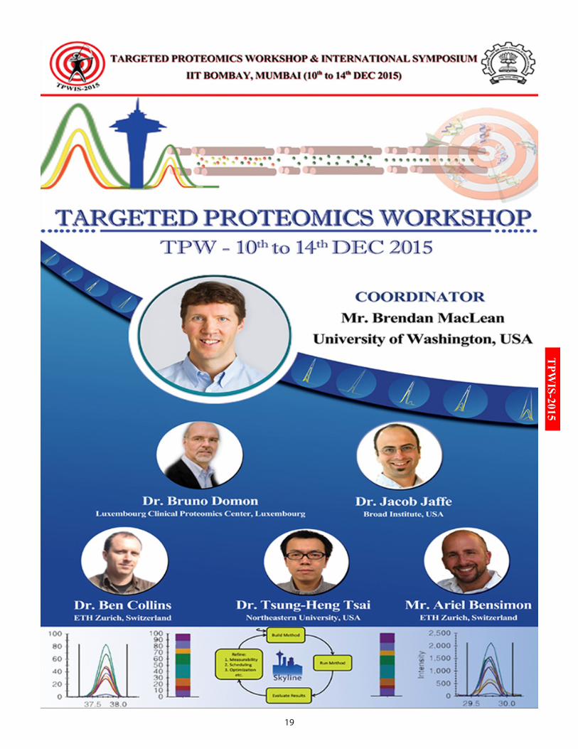
We are pleased to announce the “Targeted Proteomics Workshop and International Symposium (TPWIS-2015)”, to be held at the Indian Institute of Technology (IIT) Bombay, India from 10th to 14th December, 2015.
Targeted proteomics is emerging as a promising tool for the proteomics researchers with interest in quantifying specific proteins in complex mixtures. This not only helps to validate interesting targets but also provides a deep understanding of biology. The Targeted Proteomics Workshop and International Symposium will focus on hypothesis driven proteomics for such validation studies, to know when and how to move from the discovery to a targeted phase of proteomics research, and improve confidence in initial results. The final aim would be to progress to findings with clinical relevance.
Quality control and system suitability plays a critical role in acquiring high-quality quantitative data. This event will cover topics like, targeted methods and statistical tools required for designing a study. Primarily, the focus will be on Single Reaction Monitoring (SRM), Parallel Reaction Monitoring (PRM), Absolute Protein Quantification, popular platforms like Skyline, Trans Proteomic Pipeline, experimental design, quality control, data analysis, downstream processing of data and other subjects that are integral to targeted proteomics. We anticipate that Targeted Proteomics International Symposium (13th - 14th December, 2015) will accelerate the establishment of global standards for data acquisition, analysis, comparison, exchange and verification, which are crucial for reducing the variability and successful translational research. This focused event on “Targeted Proteomics” will undoubtedly be a great opportunity for the students, young researchers and industry experts to get familiarized with recent advances, and significant achievements in targeted proteomics research and integration of discovery based shotgun proteomics.
Two focused workshops, viz., “Targeted Proteomics” and “Trans-Proteomic Pipeline” will be held from 10th to 14th December 2015 in parallel sessions. Mr. Brendan MacLean from the University of Washington, USA will coordinate the Targeted Proteomics Workshop (TPW) and Dr. Robert Moritz from the Institute for Systems Biology, USA, will coordinate the Trans-Proteomic Pipeline (TPP) Workshop. Three days long HR-LC-MS/MS Workshop coordinated by Dr. Mayuri Gandhi from IIT Bombay is scehduled from 10th - 12th December 2015. Evening Innovative Seminars are scheduled from 10th - 12th December, 2015 to provide the latest update of the field to all the workshop participants. These Sessions would cover topics such as Quantitative and Targeted Metabolomics, Biosimilars and Biotherapeutics and Targeted Strategies for the Clinical Biomarker Discovery. Additionally, a student and teacher centric Education Day event is scheduled on 12th December 2015.
This focused international congress on Targeted Proteomics at IIT Bombay, Mumbai will provide an opportunity for the proteomics community to share scientific ideas and discuss the challenges faced in quantitative and targeted proteomics experiments. TPWIS-2015 congress will see 6 parallel events with 60+ internationally renowned speakers/ instructors, 700+ participants from over 10+ countries.
We are looking forward to welcoming all the distinguished scientists and delegates to this highly stimulating Targeted Proteomics congress to be held in the beautiful campus of IIT Bombay.
Sincerely yours,Dr. Sanjeeva SrivastavaConvener, Targeted Proteomics Workshop & International Symposium (TPWIS)-2015 IIT Bombay, Mumbai, India
2
TPWIS-2015
TArGETEd PrOTEOMiCS WOrKSHOP & inTErnATiOnAL SyMPOSiuM (TPWiS-2015)
The advent of state-of-the-art proteomics technologies has en-abled us to obtain enormous scientific information in biomedi-cal research. However, verification of such quantitative data
continues to remain a bottleneck for researchers. For such validation experiments, it is imperative to know when and how to move from the discovery to a more targeted analysis, so as to improve confidence in initial results and progress to findings with clinical relevance. The Targeted Proteomics Workshop and International Symposium will focus on hypothesis driven proteomics. Special emphasis will be given to the proteomics data analysis and interpretation.
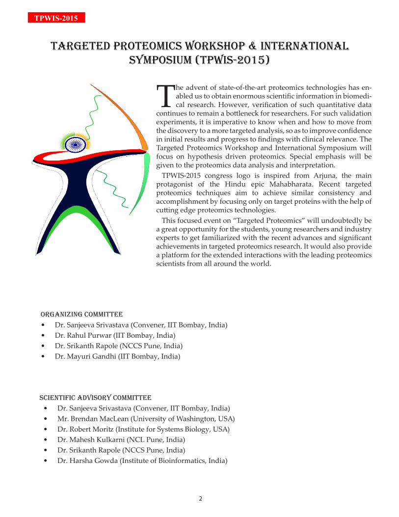
TPWIS-2015 congress logo is inspired from Arjuna, the main protagonist of the Hindu epic Mahabharata. Recent targeted proteomics techniques aim to achieve similar consistency and accomplishment by focusing only on target proteins with the help of cutting edge proteomics technologies.
This focused event on “Targeted Proteomics” will undoubtedly be a great opportunity for the students, young researchers and industry experts to get familiarized with the recent advances and significant achievements in targeted proteomics research. It would also provide a platform for the extended interactions with the leading proteomics scientists from all around the world.
OrGAniZinG COMMiTTEE
Dr. Sanjeeva Srivastava (Convener, IIT Bombay, India)• Dr. Rahul Purwar (IIT Bombay, India)• Dr. Srikanth Rapole (NCCS Pune, India)• Dr. Mayuri Gandhi (IIT Bombay, India)•
SCiEnTiFiC AdViSOry COMMiTTEE
Dr. Sanjeeva Srivastava (Convener, IIT Bombay, India)• Mr. Brendan MacLean (University of Washington, USA)• Dr. Robert Moritz (Institute for Systems Biology, USA)• Dr. Mahesh Kulkarni (NCL Pune, India)• Dr. Srikanth Rapole (NCCS Pune, India)• Dr. Harsha Gowda (Institute of Bioinformatics, India)•
3
TPW
IS-2015
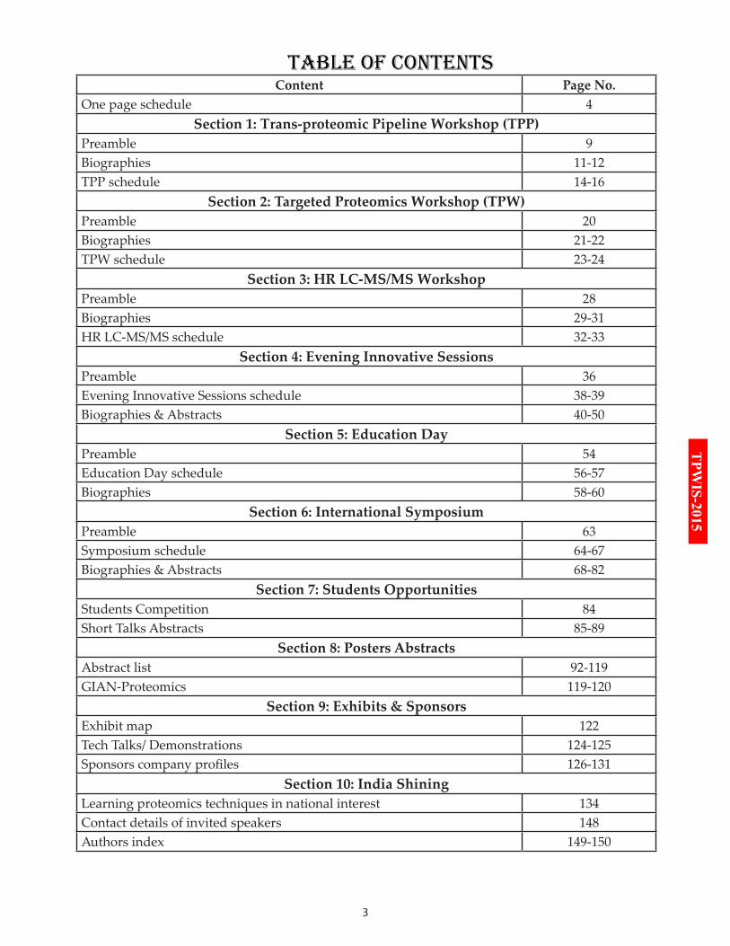
TAbLE OF COnTEnTSContent Page No.
One page schedule 4Section 1: Trans-proteomic Pipeline Workshop (TPP)
Preamble 9Biographies 11-12TPP schedule 14-16
Section 2: Targeted Proteomics Workshop (TPW)Preamble 20Biographies 21-22TPW schedule 23-24
Section 3: HR LC-MS/MS WorkshopPreamble 28Biographies 29-31HR LC-MS/MS schedule 32-33
Section 4: Evening Innovative SessionsPreamble 36Evening Innovative Sessions schedule 38-39Biographies & Abstracts 40-50
Section 5: Education DayPreamble 54Education Day schedule 56-57Biographies 58-60
Section 6: International SymposiumPreamble 63Symposium schedule 64-67Biographies & Abstracts 68-82
Section 7: Students OpportunitiesStudents Competition 84Short Talks Abstracts 85-89
Section 8: Posters AbstractsAbstract list 92-119GIAN-Proteomics 119-120
Section 9: Exhibits & Sponsors Exhibit map 122Tech Talks/ Demonstrations 124-125Sponsors company profiles 126-131
Section 10: India Shining Learning proteomics techniques in national interest 134Contact details of invited speakers 148Authors index 149-150
4
TPWIS-2015
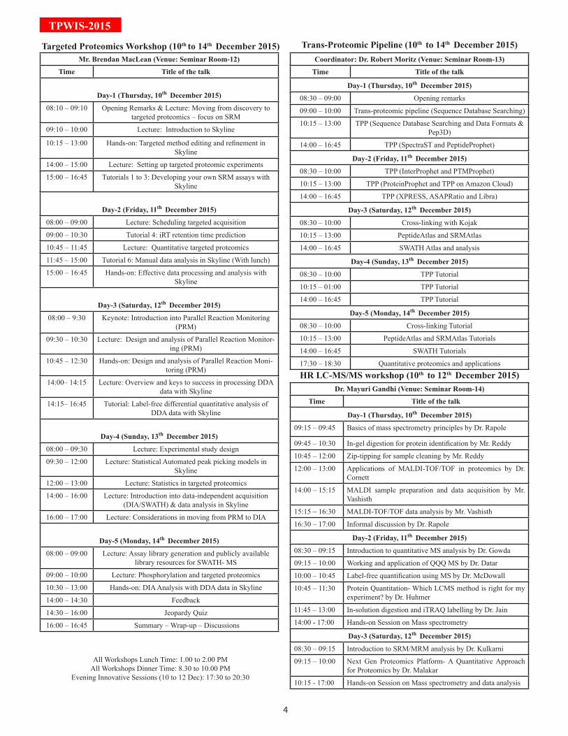
Mr. Brendan MacLean (Venue: Seminar Room-12)
Time Title of the talk
Day-1 (Thursday, 10th December 2015)
08:10 – 09:10 Opening Remarks & Lecture: Moving from discovery to targeted proteomics – focus on SRM
09:10 – 10:00 Lecture: Introduction to Skyline
10:15 – 13:00 Hands-on: Targeted method editing and refinement in Skyline
14:00 – 15:00 Lecture: Setting up targeted proteomic experiments
15:00 – 16:45 Tutorials 1 to 3: Developing your own SRM assays with Skyline
Day-2 (Friday, 11th December 2015)
08:00 – 09:00 Lecture: Scheduling targeted acquisition
09:00 – 10:30 Tutorial 4: iRT retention time prediction
10:45 – 11:45 Lecture: Quantitative targeted proteomics
11:45 – 15:00 Tutorial 6: Manual data analysis in Skyline (With lunch)
15:00 – 16:45 Hands-on: Effective data processing and analysis with Skyline
Day-3 (Saturday, 12th December 2015)
08:00 – 9:30 Keynote: Introduction into Parallel Reaction Monitoring (PRM)
09:30 – 10:30 Lecture: Design and analysis of Parallel Reaction Monitor-ing (PRM)
10:45 – 12:30 Hands-on: Design and analysis of Parallel Reaction Moni-toring (PRM)
14:00– 14:15 Lecture: Overview and keys to success in processing DDA data with Skyline
14:15– 16:45 Tutorial: Label-free differential quantitative analysis of DDA data with Skyline
Day-4 (Sunday, 13th December 2015)
08:00 – 09:30 Lecture: Experimental study design
09:30 – 12:00 Lecture: Statistical Automated peak picking models in Skyline
12:00 – 13:00 Lecture: Statistics in targeted proteomics
14:00 – 16:00 Lecture: Introduction into data-independent acquisition (DIA/SWATH) & data analysis in Skyline
16:00 – 17:00 Lecture: Considerations in moving from PRM to DIA
Day-5 (Monday, 14th December 2015)
08:00 – 09:00 Lecture: Assay library generation and publicly available library resources for SWATH- MS
09:00 – 10:00 Lecture: Phosphorylation and targeted proteomics
10:30 – 13:00 Hands-on: DIA Analysis with DDA data in Skyline
14:00 – 14:30 Feedback
14:30 – 16:00 Jeopardy Quiz
16:00 – 16:45 Summary – Wrap-up – Discussions
All Workshops Lunch Time: 1.00 to 2.00 PMAll Workshops Dinner Time: 8.30 to 10.00 PM
Evening Innovative Sessions (10 to 12 Dec): 17:30 to 20:30
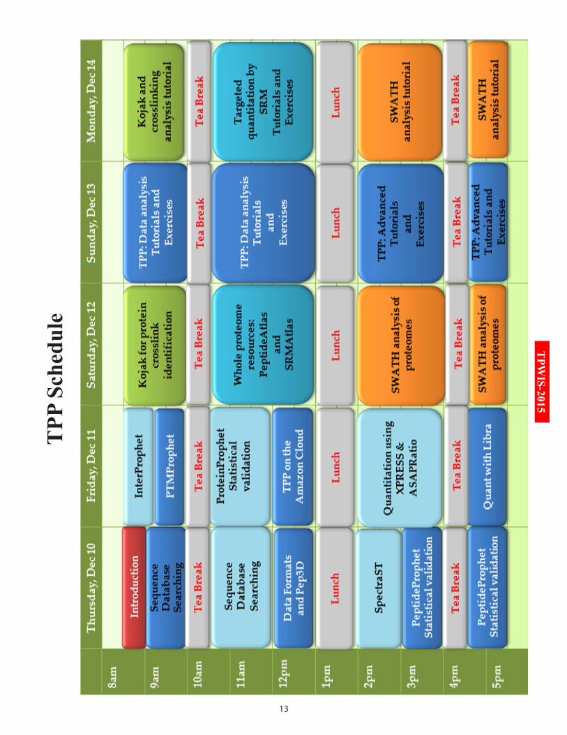
Coordinator: Dr. Robert Moritz (Venue: Seminar Room-13)
Time Title of the talk
Day-1 (Thursday, 10th December 2015)
08:30 – 09:00 Opening remarks
09:00 – 10:00 Trans-proteomic pipeline (Sequence Database Searching)
10:15 – 13:00 TPP (Sequence Database Searching and Data Formats & Pep3D)
14:00 – 16:45 TPP (SpectraST and PeptideProphet)
Day-2 (Friday, 11th December 2015)
08:30 – 10:00 TPP (InterProphet and PTMProphet)
10:15 – 13:00 TPP (ProteinProphet and TPP on Amazon Cloud)
14:00 – 16:45 TPP (XPRESS, ASAPRatio and Libra)
Day-3 (Saturday, 12th December 2015)
08:30 – 10:00 Cross-linking with Kojak
10:15 – 13:00 PeptideAtlas and SRMAtlas
14:00 – 16:45 SWATH Atlas and analysis
Day-4 (Sunday, 13th December 2015)
08:30 – 10:00 TPP Tutorial
10:15 – 01:00 TPP Tutorial
14:00 – 16:45 TPP Tutorial
Day-5 (Monday, 14th December 2015)
08:30 – 10:00 Cross-linking Tutorial
10:15 – 13:00 PeptideAtlas and SRMAtlas Tutorials
14:00 – 16:45 SWATH Tutorials
17:30 – 18:30 Quantitative proteomics and applications
Dr. Mayuri Gandhi (Venue: Seminar Room-14)
Time Title of the talk
Day-1 (Thursday, 10th December 2015)
09:15 – 09:45 Basics of mass spectrometry principles by Dr. Rapole
09:45 – 10:30 In-gel digestion for protein identification by Mr. Reddy
10:45 – 12:00 Zip-tipping for sample cleaning by Mr. Reddy
12:00 – 13:00 Applications of MALDI-TOF/TOF in proteomics by Dr. Cornett
14:00 – 15:15 MALDI sample preparation and data acquisition by Mr. Vashisth
15:15 – 16:30 MALDI-TOF/TOF data analysis by Mr. Vashisth
16:30 – 17:00 Informal discussion by Dr. Rapole
Day-2 (Friday, 11th December 2015)
08:30 – 09:15 Introduction to quantitative MS analysis by Dr. Gowda
09:15 – 10:00 Working and application of QQQ MS by Dr. Datar
10:00 – 10:45 Label-free quantification using MS by Dr. McDowall
10:45 – 11:30 Protein Quantitation- Which LCMS method is right for my experiment? by Dr. Huhmer
11:45 – 13:00 In-solution digestion and iTRAQ labelling by Dr. Jain
14:00 - 17:00 Hands-on Session on Mass spectrometry
Day-3 (Saturday, 12th December 2015)
08:30 – 09:15 Introduction to SRM/MRM analysis by Dr. Kulkarni
09:15 – 10:00 Next Gen Proteomics Platform- A Quantitative Approach for Proteomics by Dr. Malakar
10:15 - 17:00 Hands-on Session on Mass spectrometry and data analysis
Targeted Proteomics Workshop (10th to 14th December 2015) Trans-Proteomic Pipeline (10th to 14th December 2015)
HR LC-MS/MS workshop (10th to 12th December 2015)
5
TPW
IS-2015
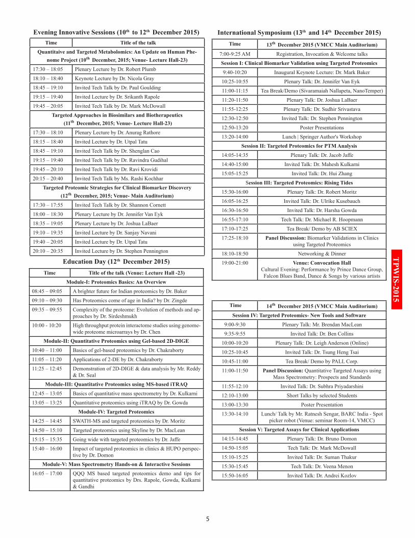
Time 13th December 2015 (VMCC Main Auditorium)
7:00-9:25 AM Registration, Invocation & Welcome talks
Session I: Clinical Biomarker Validation using Targeted Proteomics
9:40-10:20 Inaugural Keynote Lecture: Dr. Mark Baker
10:25-10:55 Plenary Talk: Dr. Jennifer Van Eyk
11:00-11:15 Tea Break/Demo (Sivaramaiah Nallapeta, NanoTemper)
11:20-11:50 Plenary Talk: Dr. Joshua LaBaer
11:55-12:25 Plenary Talk: Dr. Sudhir Srivastava
12:30-12:50 Invited Talk: Dr. Stephen Pennington
12:50-13:20 Poster Presentations
13:20-14:00 Lunch | Springer Author's Workshop
Session II: Targeted Proteomics for PTM Analysis
14:05-14:35 Plenary Talk: Dr. Jacob Jaffe
14:40-15:00 Invited Talk: Dr. Mahesh Kulkarni
15:05-15:25 Invited Talk: Dr. Hui Zhang
Session III: Targeted Proteomics: Rising Tides
15:30-16:00 Plenary Talk: Dr. Robert Moritz
16:05-16:25 Invited Talk: Dr. Ulrike Kusebauch
16:30-16:50 Invited Talk: Dr. Harsha Gowda
16:55-17:10 Tech Talk: Dr. Michael R. Hoopmann
17:10-17:25 Tea Break/ Demo by AB SCIEX
17:25-18:10 Panel Discussion: Biomarker Validations in Clinics using Targeted Proteomics
18:10-18:50 Networking & Dinner
19:00-21:00 Venue: Convocation Hall Cultural Evening: Performance by Prince Dance Group, Falcon Blues Band, Dance & Songs by various artists
Time 14th December 2015 (VMCC Main Auditorium)
Session IV: Targeted Proteomics- New Tools and Software
9:00-9:30 Plenary Talk: Mr. Brendan MacLean
9:35-9:55 Invited Talk: Dr. Ben Collins
10:00-10:20 Plenary Talk: Dr. Leigh Anderson (Online)
10:25-10:45 Invited Talk: Dr. Tsung Heng Tsai
10:45-11:00 Tea Break/ Demo by PALL Corp.
11:00-11:50 Panel Discussion: Quantitative Targeted Assays using Mass Spectrometry: Prospects and Standards
11:55-12:10 Invited Talk: Dr. Subhra Priyadarshini
12:10-13:00 Short Talks by selected Students
13:00-13:30 Poster Presentation
13:30-14:10 Lunch/ Talk by Mr. Ratnesh Sengar, BARC India - Spot picker robot (Venue: seminar Room-14, VMCC)
Session V: Targeted Assays for Clinical Applications
14:15-14:45 Plenary Talk: Dr. Bruno Domon
14:50-15:05 Tech Talk: Dr. Mark McDowall
15:10-15:25 Invited Talk: Dr. Suman Thakur
15:30-15:45 Tech Talk: Dr. Veena Menon
15:50-16:05 Invited Talk: Dr. Andrei Kozlov
Time Title of the talk (Venue: Lecture Hall -23)
Module-I: Proteomics Basics: An Overview
08:45 – 09:05 A brighter future for Indian proteomics by Dr. Baker
09:10 – 09:30 Has Proteomics come of age in India? by Dr. Zingde
09:35 – 09:55 Complexity of the proteome: Evolution of methods and ap-proaches by Dr. Sirdeshmukh
10:00 - 10:20 High throughput protein interactome studies using genome-wide proteome microarrays by Dr. Chen
Module-II: Quantitative Proteomics using Gel-based 2D-DIGE
10:40 – 11:00 Basics of gel-based proteomics by Dr. Chakraborty
11:05 – 11:20 Applications of 2-DE by Dr. Chakraborty
11:25 – 12:45 Demonstration of 2D-DIGE & data analysis by Mr. Reddy & Dr. Seal
Module-III: Quantitative Proteomics using MS-based iTRAQ
12:45 – 13:05 Basics of quantitative mass spectrometry by Dr. Kulkarni
13:05 – 13:25 Quantitative proteomics using iTRAQ by Dr. Gowda
Module-IV: Targeted Proteomics
14:25 – 14:45 SWATH-MS and targeted proteomics by Dr. Moritz
14:50 – 15:10 Targeted proteomics using Skyline by Dr. MacLean
15:15 – 15:35 Going wide with targeted proteomics by Dr. Jaffe
15:40 – 16:00 Impact of targeted proteomics in clinics & HUPO perspec-tive by Dr. Domon
Module-V: Mass Spectrometry Hands-on & Interactive Sessions
16:05 – 17:00 QQQ MS based targeted proteomics demo and tips for quantitative proteomics by Drs. Rapole, Gowda, Kulkarni & Gandhi
Time Title of the talk
Quantitaive and Targeted Metabolomics: An Update on Human Phe-nome Project (10th December, 2015; Venue- Lecture Hall-23)
17:30 – 18:05 Plenary Lecture by Dr. Robert Plumb
18:10 – 18:40 Keynote Lecture by Dr. Nicola Gray
18:45 – 19:10 Invited Tech Talk by Dr. Paul Goulding
19:15 – 19:40 Invited Lecture by Dr. Srikanth Rapole
19:45 – 20:05 Invited Tech Talk by Dr. Mark McDowall
Targeted Approaches in Biosimilars and Biotherapeutics (11th December, 2015; Venue- Lecture Hall-23)
17:30 – 18:10 Plenary Lecture by Dr. Anurag Rathore
18:15 – 18:40 Invited Lecture by Dr. Utpal Tatu
18:45 – 19:10 Invited Tech Talk by Dr. Shenglan Cao
19:15 – 19:40 Invited Tech Talk by Dr. Ravindra Gudihal
19:45 – 20:10 Invited Tech Talk by Dr. Ravi Krovidi
20:15 – 20:40 Invited Tech Talk by Ms. Rashi Kochhar
Targeted Proteomic Strategies for Clinical Biomarker Discovery(12th December, 2015; Venue- Main Auditorium)
17:30 – 17:55 Invited Tech Talk by Dr. Shannon Cornett
18:00 – 18:30 Plenary Lecture by Dr. Jennifer Van Eyk
18:35 – 19:05 Plenary Lecture by Dr. Joshua LaBaer
19:10 – 19:35 Invited Lecture by Dr. Sanjay Navani
19:40 – 20:05 Invited Lecture by Dr. Utpal Tatu
20:10 – 20:35 Invited Lecture by Dr. Stephen Pennington
International Symposium (13th and 14th December 2015)Evening Innovative Sessions (10th to 12th December 2015)
Education Day (12th December 2015)
6
TPWIS-2015
Diamond Sponsor
7
TPW
IS-2015
8
TPWIS-2015
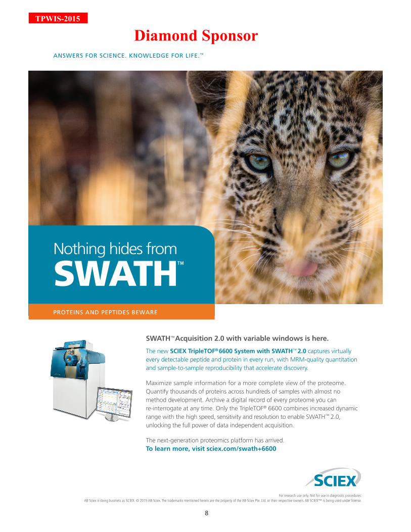
Nothing hides from SWATH™
PROTEINS AND PEPTIDES BEWARE
ANSWERS FOR SCIENCE. KNOWLEDGE FOR LIFE.™
SWATH™ Acquisition 2.0 with variable windows is here.
The new SCIEX TripleTOF® 6600 System with SWATH™ 2.0 captures virtually every detectable peptide and protein in every run, with MRM-quality quantitation and sample-to-sample reproducibility that accelerate discovery.
Maximize sample information for a more complete view of the proteome. Quantify thousands of proteins across hundreds of samples with almost no method development. Archive a digital record of every proteome you can re-interrogate at any time. Only the TripleTOF® 6600 combines increased dynamic range with the high speed, sensitivity and resolution to enable SWATH™ 2.0, unlocking the full power of data independent acquisition.
The next-generation proteomics platform has arrived. To learn more, visit sciex.com/swath+6600
For research use only. Not for use in diagnostic procedures. AB Sciex is doing business as SCIEX. © 2015 AB Sciex. The trademarks mentioned herein are the property of the AB Sciex Pte. Ltd. or their respective owners. AB SCIEX™ is being used under license.
Diamond Sponsor
9
TPW
IS-2015
10
TPWIS-2015
PrEAMbLE TO TrAnS-PrOTEOMiC PiPELinE WOrKSHOP
Democratization of genomics technologies has enabled the rapid determination of genotypes. More recently, the democratization of comprehensive proteomics technologies is enabling the determi-nation of the cellular phenotype and the molecular events that define its dynamic state. Core pro-
teomics technologies include mass spectrometry (MS) to define protein sequence, protein-protein interactions, and protein post-translational modifications. Shotgun Proteomics forms the backbone of proteomics research globally, but is by far a simplified process. In the area of global profiling or discovery based proteomics, sci-entists have proposed numerous tandem MS based work-flows which enable them to paint a holistic picture of the dynamic proteome. Technical variations arising from process design of integral steps in discovery pro-teomics employing shotgun mass spectrometry, differences in instruments and the steps utilized have led to inherent differences in output. A major focal point is to elucidate differences in proteome profiles from the same sample without influences from data processing differences. Data analysis becomes an important step to define the proteome being analyzed and infer changes across biological samples. Providing consistent, high quality data evaluation and reduction for the presentation of statistically valid biological results is key to ensure data quality.
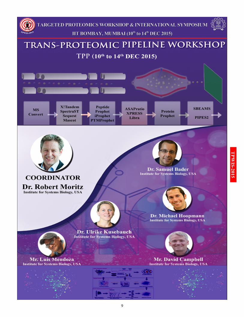
The Institute for Systems Biology has developed the Trans-Proteomic Pipeline (TPP), a suite of softwares aimed at homogenizing a data analysis pipeline for optimal analysis of raw spectral data emerging from any tandem MS based experiment. The TPP supports most of the commonly used quantitative proteomics work-flows and utilizes open, standard data formats to accurately estimate the sensitivity and error rates. The TPP is a robust open-source standardized data processing pipeline for large-scale reproducible quantitative MS proteomics spanning:• Conversion of raw MS/MS data to open formats and standards• Support for searching MS/MS spectra with various search engines, including the bundled Comet and X!Tandem, as well as SEQUEST, Mascot, Crux, OMSSA, MyriMatch, MSGF+, and others• Spectral Library searching and validation with SpectraST• Conversion of search engine results to a uniform open format• Statistical validation of peptide identifications with PeptideProphet and iProphet, and validation of PTM localization sites with PTMProphet• Statistically validated protein identification with ProteinProphet• Quantitative proteomics (SILAC, iCAT, iTRAQ, TMT, etc.) with XPRESS, ASAPRatio, and Libra• Tools for visualization of, and interaction with results• Operation in various modes from desktop to cloud computing
In addition to teaching how to install, use and explore proteomics data produced by the TPP, training will also be provided in advanced systems biology tools on comprehensive cataloguing of proteomes through the PeptideAtlas and SRM repositories called PASSEL. For advanced quantitative proteomics analysis, tutorials will be provided on the use of the SRMAtlas suite of targeted MS assays, and the use of the ISB resources for developing SWATH MS profiles and data analysis through the SWATHAtlas suite of spectral libraries.
Dr. Robert Moritz and his team from Institute for Systems Biology, USA are pioneers in this area who would be addressing a global audience to provide hands-on training in this much needed domain. The Indian Pro-teomics community has established a strong hold in the tandem MS arena and hence will hugely benefit from dedicated training of this nature.
11
TPW
IS-2015
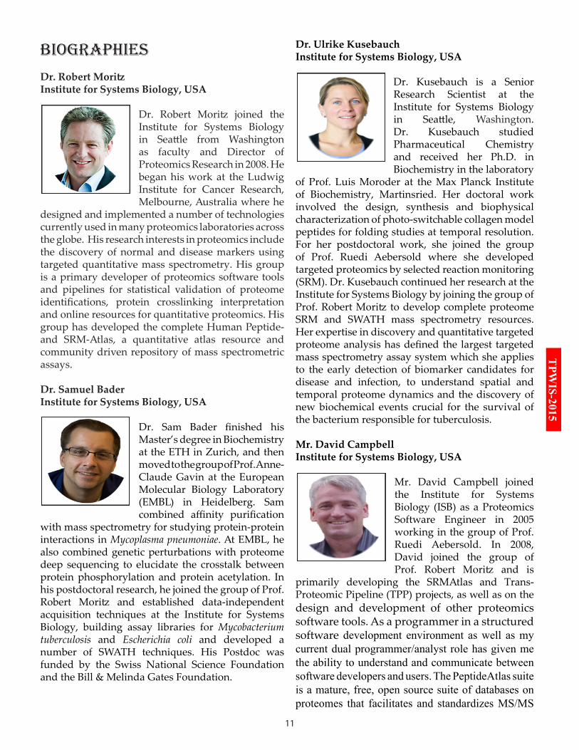
biOGrAPHiES Dr. Robert MoritzInstitute for Systems Biology, USA
Dr. Robert Moritz joined the Institute for Systems Biology in Seattle from Washington as faculty and Director of Proteomics Research in 2008. He began his work at the Ludwig Institute for Cancer Research, Melbourne, Australia where he
designed and implemented a number of technologies currently used in many proteomics laboratories across the globe. His research interests in proteomics include the discovery of normal and disease markers using targeted quantitative mass spectrometry. His group is a primary developer of proteomics software tools and pipelines for statistical validation of proteome identifications, protein crosslinking interpretation and online resources for quantitative proteomics. His group has developed the complete Human Peptide- and SRM-Atlas, a quantitative atlas resource and community driven repository of mass spectrometric assays.
Dr. Samuel BaderInstitute for Systems Biology, USA
Dr. Sam Bader finished his Master’s degree in Biochemistry at the ETH in Zurich, and then moved to the group of Prof. Anne-Claude Gavin at the European Molecular Biology Laboratory (EMBL) in Heidelberg. Sam combined affinity purification
with mass spectrometry for studying protein-protein interactions in Mycoplasma pneumoniae. At EMBL, he also combined genetic perturbations with proteome deep sequencing to elucidate the crosstalk between protein phosphorylation and protein acetylation. In his postdoctoral research, he joined the group of Prof. Robert Moritz and established data-independent acquisition techniques at the Institute for Systems Biology, building assay libraries for Mycobacterium tuberculosis and Escherichia coli and developed a number of SWATH techniques. His Postdoc was funded by the Swiss National Science Foundation and the Bill & Melinda Gates Foundation.
Dr. Ulrike KusebauchInstitute for Systems Biology, USA
Dr. Kusebauch is a Senior Research Scientist at the Institute for Systems Biology in Seattle, Washington. Dr. Kusebauch studied Pharmaceutical Chemistry and received her Ph.D. in Biochemistry in the laboratory
of Prof. Luis Moroder at the Max Planck Institute of Biochemistry, Martinsried. Her doctoral work involved the design, synthesis and biophysical characterization of photo-switchable collagen model peptides for folding studies at temporal resolution. For her postdoctoral work, she joined the group of Prof. Ruedi Aebersold where she developed targeted proteomics by selected reaction monitoring (SRM). Dr. Kusebauch continued her research at the Institute for Systems Biology by joining the group of Prof. Robert Moritz to develop complete proteome SRM and SWATH mass spectrometry resources. Her expertise in discovery and quantitative targeted proteome analysis has defined the largest targeted mass spectrometry assay system which she applies to the early detection of biomarker candidates for disease and infection, to understand spatial and temporal proteome dynamics and the discovery of new biochemical events crucial for the survival of the bacterium responsible for tuberculosis.
Mr. David Campbell Institute for Systems Biology, USA
Mr. David Campbell joined the Institute for Systems Biology (ISB) as a Proteomics Software Engineer in 2005 working in the group of Prof. Ruedi Aebersold. In 2008, David joined the group of Prof. Robert Moritz and is
primarily developing the SRMAtlas and Trans-Proteomic Pipeline (TPP) projects, as well as on the design and development of other proteomics software tools. As a programmer in a structured software development environment as well as my current dual programmer/analyst role has given me the ability to understand and communicate between software developers and users. The PeptideAtlas suite is a mature, free, open source suite of databases on proteomes that facilitates and standardizes MS/MS
12
TPWIS-2015software developers and users. The PeptideAtlas suite is a mature, free, open source suite of databases on proteomes that facilitates and standardizes MS/MS based proteomics data dissemination in a consistent and objective manner. The PeptideAtlas, SRMAtlas, PASSEL and SWATHAtlas are unique databases that provide highly qualified peptide identifications, peptide assays and assay repositories for public dissemination of data. David develops many of these core software tools and provides user support as well as teaching week long proteomics analysis courses at ISB and abroad. David holds an MS and has a long and varied background as researcher and software developer in both academic and biotechnology company backgrounds.
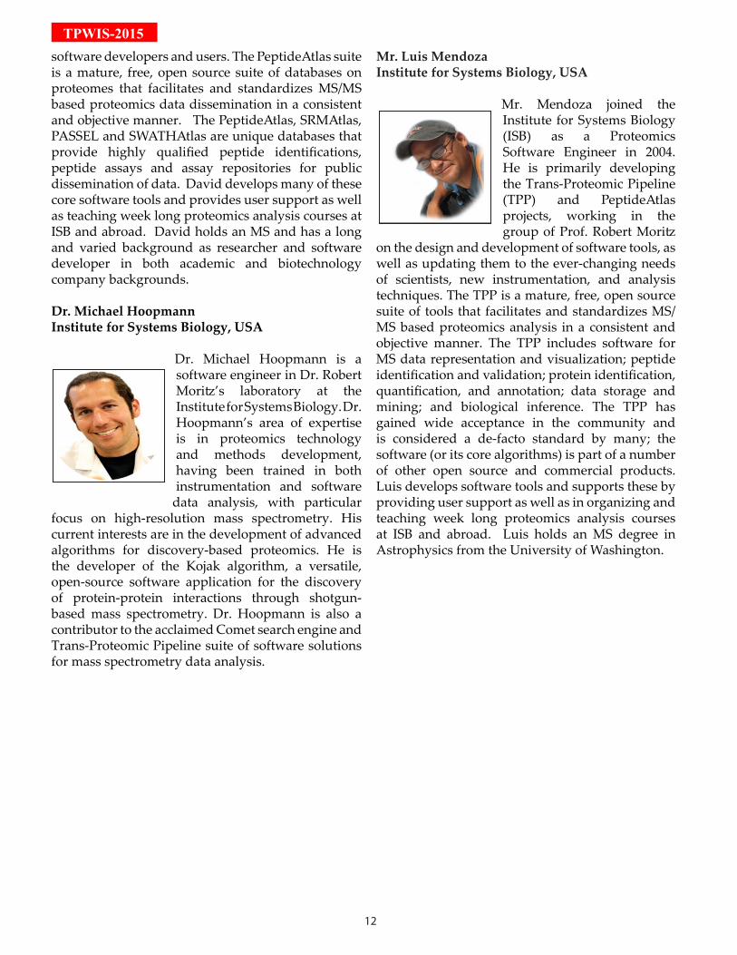
Dr. Michael HoopmannInstitute for Systems Biology, USA
Dr. Michael Hoopmann is a software engineer in Dr. Robert Moritz’s laboratory at the Institute for Systems Biology. Dr. Hoopmann’s area of expertise is in proteomics technology and methods development, having been trained in both instrumentation and software
data analysis, with particular focus on high-resolution mass spectrometry. His current interests are in the development of advanced algorithms for discovery-based proteomics. He is the developer of the Kojak algorithm, a versatile, open-source software application for the discovery of protein-protein interactions through shotgun-based mass spectrometry. Dr. Hoopmann is also a contributor to the acclaimed Comet search engine and Trans-Proteomic Pipeline suite of software solutions for mass spectrometry data analysis.
Mr. Luis MendozaInstitute for Systems Biology, USA
Mr. Mendoza joined the Institute for Systems Biology (ISB) as a Proteomics Software Engineer in 2004. He is primarily developing the Trans-Proteomic Pipeline (TPP) and PeptideAtlas projects, working in the group of Prof. Robert Moritz
on the design and development of software tools, as well as updating them to the ever-changing needs of scientists, new instrumentation, and analysis techniques. The TPP is a mature, free, open source suite of tools that facilitates and standardizes MS/MS based proteomics analysis in a consistent and objective manner. The TPP includes software for MS data representation and visualization; peptide identification and validation; protein identification, quantification, and annotation; data storage and mining; and biological inference. The TPP has gained wide acceptance in the community and is considered a de-facto standard by many; the software (or its core algorithms) is part of a number of other open source and commercial products. Luis develops software tools and supports these by providing user support as well as in organizing and teaching week long proteomics analysis courses at ISB and abroad. Luis holds an MS degree in Astrophysics from the University of Washington.
13
TPW
IS-2015
TPP
Sch
edul
e
14
TPWIS-2015
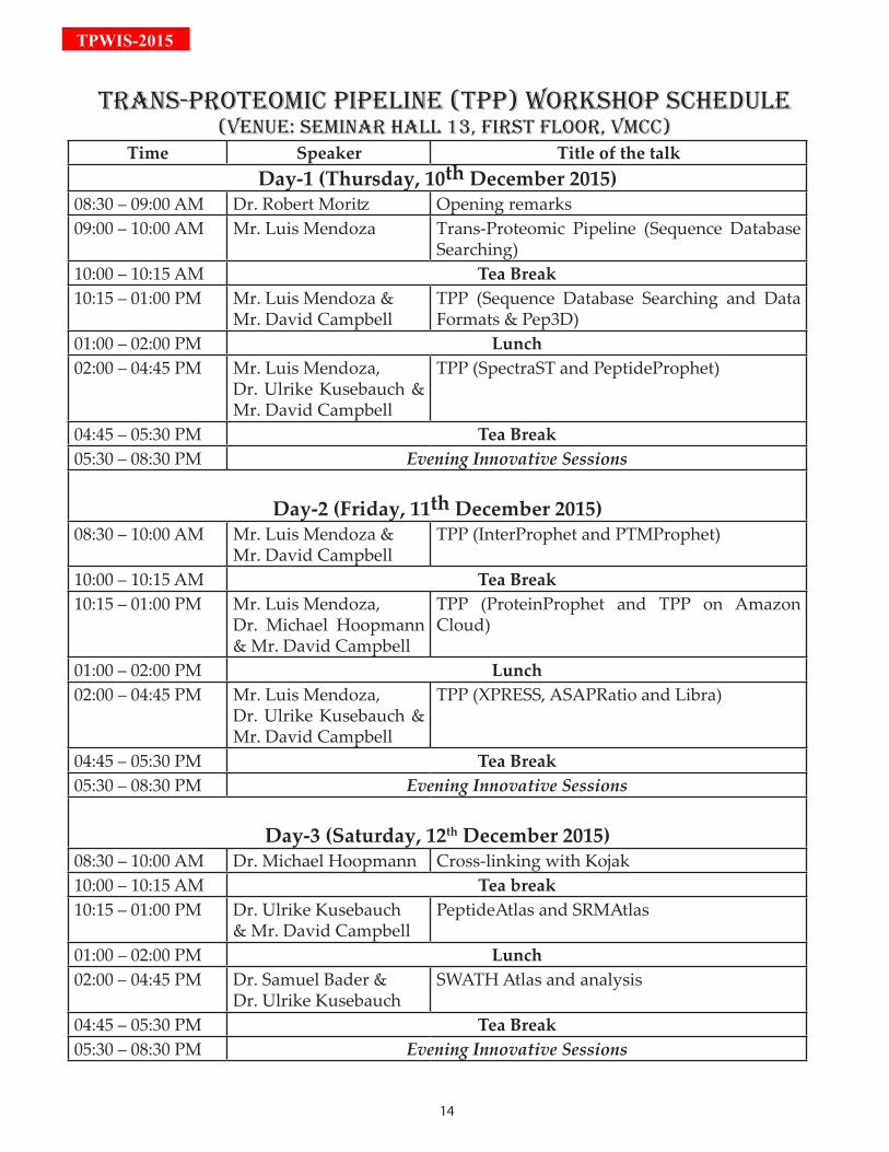
TrAnS-PrOTEOMiC PiPELinE (TPP) WOrKSHOP SCHEduLE(VEnuE: SEMinAr HALL 13, FirST FLOOr, VMCC)
Time Speaker Title of the talkDay-1 (Thursday, 10th December 2015)
08:30 – 09:00 AM Dr. Robert Moritz Opening remarks09:00 – 10:00 AM Mr. Luis Mendoza Trans-Proteomic Pipeline (Sequence Database
Searching)10:00 – 10:15 AM Tea Break10:15 – 01:00 PM Mr. Luis Mendoza &
Mr. David CampbellTPP (Sequence Database Searching and Data Formats & Pep3D)
01:00 – 02:00 PM Lunch02:00 – 04:45 PM Mr. Luis Mendoza,
Dr. Ulrike Kusebauch & Mr. David Campbell
TPP (SpectraST and PeptideProphet)
04:45 – 05:30 PM Tea Break05:30 – 08:30 PM Evening Innovative Sessions
Day-2 (Friday, 11th December 2015)08:30 – 10:00 AM Mr. Luis Mendoza &
Mr. David CampbellTPP (InterProphet and PTMProphet)
10:00 – 10:15 AM Tea Break10:15 – 01:00 PM Mr. Luis Mendoza,
Dr. Michael Hoopmann & Mr. David Campbell
TPP (ProteinProphet and TPP on Amazon Cloud)
01:00 – 02:00 PM Lunch02:00 – 04:45 PM Mr. Luis Mendoza,
Dr. Ulrike Kusebauch & Mr. David Campbell
TPP (XPRESS, ASAPRatio and Libra)
04:45 – 05:30 PM Tea Break05:30 – 08:30 PM Evening Innovative Sessions
Day-3 (Saturday, 12th December 2015)08:30 – 10:00 AM Dr. Michael Hoopmann Cross-linking with Kojak10:00 – 10:15 AM Tea break10:15 – 01:00 PM Dr. Ulrike Kusebauch
& Mr. David CampbellPeptideAtlas and SRMAtlas
01:00 – 02:00 PM Lunch02:00 – 04:45 PM Dr. Samuel Bader &
Dr. Ulrike KusebauchSWATH Atlas and analysis
04:45 – 05:30 PM Tea Break05:30 – 08:30 PM Evening Innovative Sessions
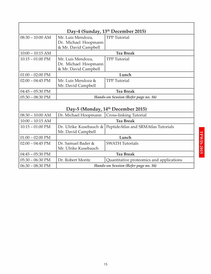
15
TPW
IS-2015
Day-4 (Sunday, 13th December 2015)08:30 – 10:00 AM Mr. Luis Mendoza,
Dr. Michael Hoopmann & Mr. David Campbell
TPP Tutorial
10:00 – 10:15 AM Tea Break10:15 – 01:00 PM Mr. Luis Mendoza,
Dr. Michael Hoopmann & Mr. David Campbell
TPP Tutorial
01:00 – 02:00 PM Lunch02:00 – 04:45 PM Mr. Luis Mendoza &
Mr. David CampbellTPP Tutorial
04:45 – 05:30 PM Tea Break05:30 – 08:30 PM Hands-on Session (Refer page no. 16)
Day-5 (Monday, 14th December 2015)08:30 – 10:00 AM Dr. Michael Hoopmann Cross-linking Tutorial 10:00 – 10:15 AM Tea Break10:15 – 01:00 PM Dr. Ulrike Kusebauch &
Mr. David CampbellPeptideAtlas and SRMAtlas Tutorials
01:00 – 02:00 PM Lunch02:00 – 04:45 PM Dr. Samuel Bader &
Mr. Ulrike KusebauchSWATH Tutorials
04:45 – 05:30 PM Tea Break05:30 – 06:30 PM Dr. Robert Moritz Quantitative proteomics and applications 06:30 – 08:30 PM Hands-on Session (Refer page no. 16)
16
TPWIS-2015
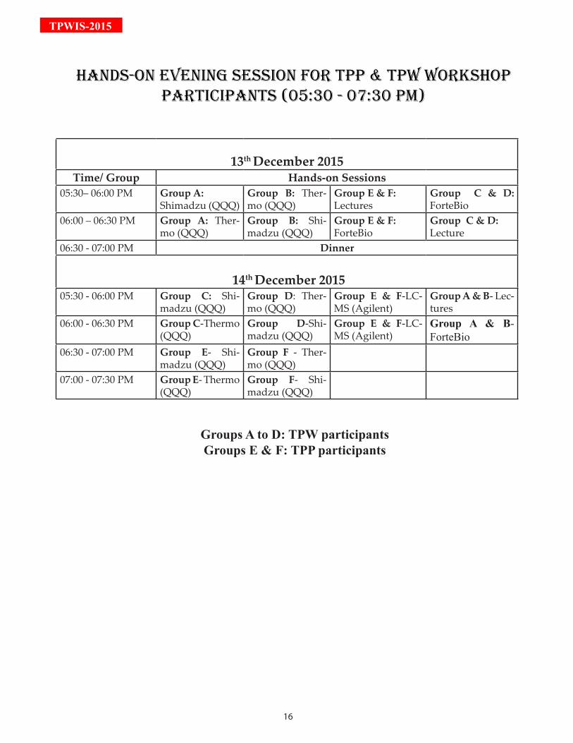
13th December 2015Time/ Group Hands-on Sessions
05:30– 06:00 PM Group A:Shimadzu (QQQ)
Group B: Ther-mo (QQQ)
Group E & F: Lectures
Group C & D: ForteBio
06:00 – 06:30 PM Group A: Ther-mo (QQQ)
Group B: Shi-madzu (QQQ)
Group E & F: ForteBio
Group C & D: Lecture
06:30 - 07:00 PM Dinner
14th December 201505:30 - 06:00 PM Group C: Shi-
madzu (QQQ)Group D: Ther-mo (QQQ)
Group E & F-LC-MS (Agilent)
Group A & B- Lec-tures
06:00 - 06:30 PM Group C-Thermo (QQQ)
Group D-Shi-madzu (QQQ)
Group E & F-LC-MS (Agilent)
Group A & B- ForteBio
06:30 - 07:00 PM Group E- Shi-madzu (QQQ)
Group F - Ther-mo (QQQ)
07:00 - 07:30 PM Group E- Thermo (QQQ)
Group F- Shi-madzu (QQQ)
Groups A to D: TPW participantsGroups E & F: TPP participants
HAndS-On EVEninG SESSiOn FOr TPP & TPW WOrKSHOP PArTiCiPAnTS (05:30 - 07:30 PM)
17
TPW
IS-2015
18
TPWIS-2015
Gold Sponsor
19
TPW
IS-2015
20
TPWIS-2015
APPLyinG TArGETEd PrOTEOMiCS in SySTEMS biOLOGy
In recent years, systems biology has emerged as a new paradigm in life sciences and has been stirring novel technologies that focus on the quantitative measurement of molecules and their contextual re-lationships. Amongst these, several mass spectrometry (MS)-based proteomic approaches have ma-
tured to support systems biology. In most proteomic studies relying on tandem MS, protein samples are digested into peptides which are fur-ther fragmented in the mass spectrometer. Resulting fragment ion signals are assigned to peptide sequences, used to locate modified amino acid residues and to infer the quantity of a peptide. Ideally, to support the systems biology paradigm, MS-based proteomics needs to be able to generate datasets that are complete, re-producible, quantitative and measurable at a reasonable throughput to allow iterations within a study.During the past few years, targeted proteomics has emerged as a complement to the more widely used dis-covery or shotgun proteomics methods. The main MS approach supporting targeted proteomics is Selected Reaction Monitoring (SRM). SRM can be performed at a high level of reproducibility, sensitivity, accuracy and reasonable throughput and thus represents an attractive approach to support systems-level biological questions that are targeted at a pre-defined network of proteins of interest. More recently, for a related ap-proach that utilizes high resolution mass spectrometers instead of triple quadrupoles, the term Parallel Reac-tion Monitoring (PRM), has been introduced. The significance of targeted proteomics for systems biology is increasingly being recognized, highlighted by the selection of targeted proteomics as the method of the year 2012 by the journal “Nature Methods”.In recent years, researchers in this area have introduced extensions of the targeted proteomics method in the category of data independent acquisition (DIA) such as SWATH MS, which relies on targeted data analysis (as opposed to targeted data acquisition). SWATH MS and related DIA approaches vastly extends the number of peptides/proteins quantified per sample, while maintaining the favorable quantitative performance profile of SRM.
The Targeted Proteomics WorkshopMajor advances have been achieved in various facets of targeted proteomics, including advances in instru-mentation, the generation of collections of validated SRM, PRM and SWATH/DIA assay libraries, and the development of multiple computational tools for every step of a targeted proteomics workflow. While these advances have enabled the successful application of these workflows by an increasing number of laboratories worldwide, it has become evident that applying SRM, PRM or SWATH/DIA requires interdisciplinary knowl-edge and experience in three interlaced aspects: designing biological questions which can be investigated by targeted proteomics, harnessing the current technologies to their full capacity and applying a set of compu-tational tools and bioinformatics resources for every step of a targeted proteomics workflow. We believe that overcoming the barriers within and between these aspects would enable a wide and routine application of targeted proteomics in systems biology. Hence, a practical course has been organized aimed to acquaint par-ticipants with targeted proteomics and prepare them to conduct a complete targeted proteomics experiment according to their biological question in their home laboratory. This week long course at IIT Bombay will follow a template which has been initially designed at ETH, Zur-ich and further refined in subsequent related courses at the University of Washington, CRG Barcelona, and Northeastern University. The specific skills, and the detailed program of the upcoming targeted proteomics course have been modified from the initial course focused on SRM to include newly developing approaches. The main topics added to the initial design are PRM and SWATH MS/DIA.
Specific skills for applying a Targeted Proteomics workflowIn order to enable participants to design, measure and analyze their targeted proteomics experiments, we seek to provide them with four distinct skills required to bridge the gap between theory and actual implemen-tation in their own laboratory:1. How to select the targeted proteomics workflow most suited for a biological question of interest?2. How to translate such a biological question of interest to a list of assays, according to the workflow of choice?
21
TPW
IS-20153. How to perform targeted data acquisition for SRM/PRM or data independent acquisition for SWATH MS in a reasonable throughput with high accuracy and sensitivity?4. How to perform statistical analysis of acquired data in order to extract biological meaning?The Targeted Proteomics Workshop at IIT Bombay would offer theoretical introductory lessons, comprehen-sive hands-on training and discussion rounds on each of these skills. The primary data analysis tool used will be Skyline, a freely available and open source Windows client application that supports all stages of the workflows outlined above. We have designed case studies, which will be used throughout most of the practi-cal course to introduce and train participants in applying these workflows. Time will be allocated to discuss participant’s own projects and differences between these projects and the case study. Furthermore, experts in these skills and in some instances the principal developers of the respective methods have been invited to instruct the participants and contribute to their knowledge.
biOGrAPHiES
Mr. Brendan MacLeanUniversity of Washington, USA
Mr. MacLean worked at Microsoft for 8 years in the 1990s where he was a lead developer and development manager for the Visual C++/Developer Studio Project. Since leaving Microsoft, Brendan has been the Vice President of Engineering
for Westside Corporation, Director of Engineering for BEA Systems, Inc., Sr. Software Engineer at the Fred Hutchinson Cancer Research Center, and a founding partner of LabKey Software. In this last position, he was one of the key programmers responsible for the Computational Proteomics Analysis System (CPAS), made significant contributions to the development of X!Tandem and the Trans-Proteomic Pipeline and created the LabKey Enterprise Pipeline. Since August 2008, he has worked as a Sr. Software Engineer within the MacCoss lab and been responsible for all aspects of design, development and support in creating the Skyline Targeted Proteomics environment and its growing worldwide user community.
Dr. Bruno DomonLuxembourg Clinical Proteomics Center,
Luxembourg
Dr. Bruno Domon is an expert in biological mass spectrometry and heading the Luxembourg Clinical Proteomics Center, CRP-Santé, Strassen, Luxembourg since 2010.He joined the Centre de
Recherche Public Santé in Luxembourg to lead the new Clinical Proteomics Center, funded by the Fonds National de la Recherche through a PEARL grant. His main focus is the development of novel mass spectrometry-based proteomics methodologies, and its application to biomarker discovery and evaluation, and to proteomics in general. His current interest is in personalized medicine and personalized therapies and he is collaborating with the clinicians to develop new diagnostic tools in the field of lung cancer.Previously, he was the group leader and principal investigator at the Institute of Molecular Systems Biology at ETH Zurich and headed the mass spectrometry laboratory and the biomarker program (2005 -2009). As Director at Celera Genomics in Rockville MD, USA (2001-2004), he led the proteomics program on drug target discovery in oncology, which resulted in the identification of cell surface proteins for therapeutic development and diagnostics. Prior to that (1998-2001), he was the Associate Director, and headed the mass spectrometry facility at Biogen in Cambridge MA, USA. From 1988-1994, he held different functions at Ciba-Geigy (now Novartis) in Basel, including the position of Head of the mass spectrometry facility.
22
TPWIS-2015Dr. Bruno Domon studied chemistry at the University of Neuchatel, where he obtained a chemical engineering degree in 1980. He graduated from the University of Lausanne, where he received his Ph.D. in 1984 (isolation and structural elucidation of natural products). He then started his mass spectrometry carrier as a post-doctoral fellow at Ciba-Geigy in Basel (1985-86) and at the department of chemistry of the Massachusetts Institute of Technology (MIT, 1986-87). He has more than 70 publications in peer-reviewed journals and has several patents in his name.
Dr. Jacob JaffeBroad Institute, USA
Dr. Jacob D. Jaffe, is the Assistant Director of the Proteomics Platform at the Broad Institute. He obtained his B.A. degree in Biochemistry from the University of Pennsylvania and his Ph.D. from Harvard University,
where he studied with George Church and Howard Berg. Dr. Jaffe has pioneered diverse problems in modern proteomics including large-scale mapping of proteomic data onto genomes, thus allowing their de novo annotation from proteomic evidence, pattern recognition for quantitative proteomics, determination and quantification of epigenetic marks on histone proteins, and high-throughput targeted phosphoproteomics.
Dr. Ben Collins ETH Zurich, Switzerland
Dr. Ben is a native of Ireland where he studied chemistry and applied chemistry at the National University or Ireland, Galway, for his bachelor’s degree. After working as an analytical chemist in Schering-Plough, Ireland, he undertook
an MSc in Molecular Medicine at Trinity College Dublin. His PhD entitled ‘Mass Spectrometry-Based Proteomics to Support Pre-Clinical Pharmaceutical Toxicology Evaluation’ was completed at University College Dublin in 2009 where he remained for 1 year as the Agilent Technologies Newman Fellow (postdoctoral) in Quantitative Proteomics. Ben moved to the Institute of Molecular Systems Biology at ETH Zurich in Autumn 2010 as postdoctoral
researcher under the supervision of Dr. Ruedi Aebersold and Dr. Matthias Gstaiger, where his current research focus is on the application of quantitative interaction proteomics in signaling and the development of SWATH mass spectrometry.
Mr. Ariel BensimonETH Zurich, Switzerland
Ariel was born in Israel and pursued his studies in the Adi Lautman Interdisciplinary Program for Outstanding Students at Tel Aviv University. There after he undertook his Masters study in Medical Sciences at the Department of
Human Molecular Genetics and Biochemistry, Sackler School of Medicine, Tel Aviv University in 2007. During his Master thesis, at the lab of Prof. Yossi Shiloh, he focused on the cellular responses to study DNA double-strand breaks. Ariel joined the lab of Prof. Ruedi Aebersold in 2011 and has been undertaking his doctoral studies under Prof. Aebersold’s supervision at the Institute of Molecular Systems Biology, Department of Biology, ETH Zürich.He has worked as a Research Assistant in distinguished laboratories for over 8 years in broad areas like Molecular Genetics and Systems Biology. His current research interest is focused on mass spectrometry based innovations and validation using Targeted proteomics.
Dr. Tsung-Heng Tsai Northeastern University, USA
Dr. Tsung-Heng Tsai holds a Ph.D. in Electrical Engineering from Virginia Tech. He is now a postdoctoral associate in the lab of Olga Vitek at Northeastern University. His work focuses on developing statistical and computational methods for
quantitative proteomics.
23
TPW
IS-2015
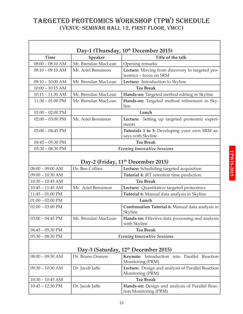
Day-1 (Thursday, 10th December 2015)Time Speaker Title of the talk
08:00 – 08:10 AM Mr. Brendan MacLean Opening remarks08:10 – 09:10 AM Mr. Ariel Bensimon Lecture: Moving from discovery to targeted pro-
teomics – focus on SRM 09:10 – 10:00 AM Mr. Brendan MacLean Lecture: Introduction to Skyline10:00 – 10:15 AM Tea Break10:15 – 11:30 AM Mr. Brendan MacLean Hands-on: Targeted method editing in Skyline11:30 – 01:00 PM Mr. Brendan MacLean Hands-on: Targeted method refinement in Sky-
line01:00 – 02:00 PM Lunch02:00 – 03:00 PM Mr. Ariel Bensimon Lecture: Setting up targeted proteomic experi-
ments03:00 – 04:45 PM Tutorials 1 to 3: Developing your own SRM as-
says with Skyline04:45 – 05:30 PM Tea Break05:30 – 08:30 PM Evening Innovative Sessions
Day-2 (Friday, 11th December 2015)08:00 – 09:00 AM Dr. Ben Collins Lecture: Scheduling targeted acquisition09:00 – 10:30 AM Tutorial 4: iRT retention time prediction10:30 – 10:45 AM Tea Break10:45 – 11:45 AM Mr. Ariel Bensimon Lecture: Quantitative targeted proteomics11:45 – 01:00 PM Tutorial 6: Manual data analysis in Skyline01:00 – 02:00 PM Lunch02:00 – 03:00 PM Continuation Tutorial 6: Manual data analysis in
Skyline03:00 – 04:45 PM Mr. Brendan MacLean Hands-on: Effective data processing and analysis
with Skyline04:45 – 05:30 PM Tea Break05:30 – 08:30 PM Evening Innovative Sessions
Day-3 (Saturday, 12th December 2015)08:00 – 09:30 AM Dr. Bruno Domon Keynote: Introduction into Parallel Reaction
Monitoring (PRM)09:30 – 10:30 AM Dr. Jacob Jaffe Lecture: Design and analysis of Parallel Reaction
Monitoring (PRM)10:30 – 10:45 AM Tea Break10:45 – 12:30 PM Dr. Jacob Jaffe Hands-on: Design and analysis of Parallel Reac-
tion Monitoring (PRM)
TArGETEd PrOTEOMiCS WOrKSHOP (TPW) SCHEduLE(VEnuE: SEMinAr HALL 12, FirST FLOOr, VMCC)
24
TPWIS-2015
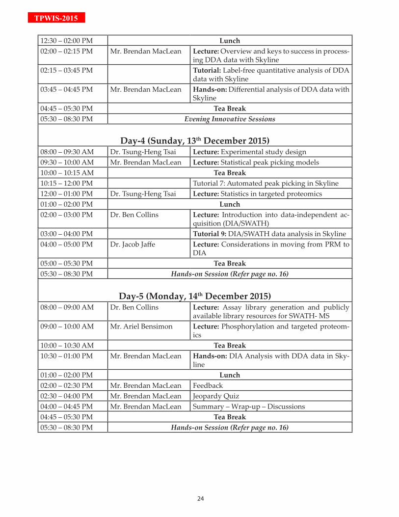
12:30 – 02:00 PM Lunch02:00 – 02:15 PM Mr. Brendan MacLean Lecture: Overview and keys to success in process-
ing DDA data with Skyline02:15 – 03:45 PM Tutorial: Label-free quantitative analysis of DDA
data with Skyline03:45 – 04:45 PM Mr. Brendan MacLean Hands-on: Differential analysis of DDA data with
Skyline04:45 – 05:30 PM Tea Break05:30 – 08:30 PM Evening Innovative Sessions
Day-4 (Sunday, 13th December 2015)08:00 – 09:30 AM Dr. Tsung-Heng Tsai Lecture: Experimental study design09:30 – 10:00 AM Mr. Brendan MacLean Lecture: Statistical peak picking models10:00 – 10:15 AM Tea Break10:15 – 12:00 PM Tutorial 7: Automated peak picking in Skyline12:00 – 01:00 PM Dr. Tsung-Heng Tsai Lecture: Statistics in targeted proteomics01:00 – 02:00 PM Lunch02:00 – 03:00 PM Dr. Ben Collins Lecture: Introduction into data-independent ac-
quisition (DIA/SWATH)03:00 – 04:00 PM Tutorial 9: DIA/SWATH data analysis in Skyline04:00 – 05:00 PM Dr. Jacob Jaffe Lecture: Considerations in moving from PRM to
DIA05:00 – 05:30 PM Tea Break05:30 – 08:30 PM Hands-on Session (Refer page no. 16)
Day-5 (Monday, 14th December 2015)08:00 – 09:00 AM Dr. Ben Collins Lecture: Assay library generation and publicly
available library resources for SWATH- MS09:00 – 10:00 AM Mr. Ariel Bensimon Lecture: Phosphorylation and targeted proteom-
ics10:00 – 10:30 AM Tea Break10:30 – 01:00 PM Mr. Brendan MacLean Hands-on: DIA Analysis with DDA data in Sky-
line01:00 – 02:00 PM Lunch02:00 – 02:30 PM Mr. Brendan MacLean Feedback02:30 – 04:00 PM Mr. Brendan MacLean Jeopardy Quiz04:00 – 04:45 PM Mr. Brendan MacLean Summary – Wrap-up – Discussions04:45 – 05:30 PM Tea Break05:30 – 08:30 PM Hands-on Session (Refer page no. 16)
25
TPW
IS-2015
26
TPWIS-2015
Gold Spon-M
ulti-
met
hod/
wal
k-up
, met
hod
de
velo
pmen
t Val
ve-a
nd
Flex
Cube
-bas
ed w
orkf
low
au
tom
atio
n so
lutio
nsU
HC-s
mal
l mol
ecul
e Ch
ip L
CMS
7100
CE
& C
E/M
S
1290
12
20
1260
SFC
1260
Bio
2100
Bio
anal
yzer
3100
OG
E
1260
1200
Infin
ity S
erie
s HPL
C12
00 In
finity
Ser
ies S
olut
ions
7820
GC
7890
GC
Agile
nt G
C an
d GC
MS
portf
olio
220
GC
Ion
trap
7000
B G
C Q
The
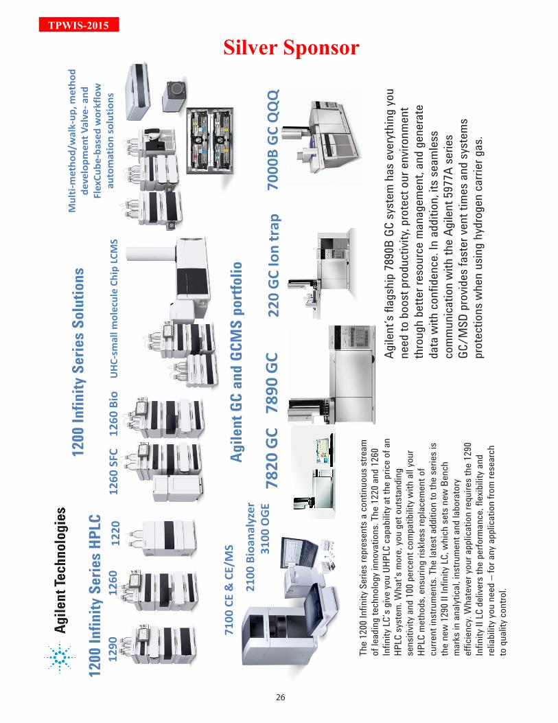
1200
Infin
ity S
eries
repr
esen
ts a
cont
inuo
us st
ream
of
lead
ing
tech
nolo
gy in
nova
tions
. The
122
0 and
126
0 In
finity
LC’s
give
you
UHPL
C ca
pabi
lity a
t the
pric
e of
an
HPLC
syst
em. W
hat's
mor
e, yo
u ge
t out
stan
ding
se
nsiti
vity a
nd 1
00 p
erce
nt co
mpa
tibilit
y with
all y
our
HPLC
met
hods
, ens
urin
g ris
kless
repl
acem
ent o
f cu
rrent
inst
rum
ents
. The
late
st a
dditi
on to
the
serie
s is
the
new
129
0 II I
nfin
ity LC
, whi
ch se
ts n
ew B
ench
m
arks
in a
nalyt
ical, i
nstru
men
t and
labo
rato
ry
effic
iency
. Wha
teve
r you
r app
licat
ion
requ
ires t
he 1
290
Infin
ity II
LC
deliv
ers t
he p
erfo
rman
ce, f
lexib
ility a
nd
relia
bilit
y you
nee
d –
for a
ny a
pplic
atio
n fro
m re
sear
ch
to q
ualit
y con
trol.
Agile
nt’s
flags
hip
7890
B GC
syst
em h
as e
very
thin
g yo
u ne
ed to
boo
st pr
oduc
tivity
, pro
tect
our
env
ironm
ent
thro
ugh
bette
r res
ourc
e m
anag
emen
t, an
d ge
nera
te
data
with
conf
iden
ce. I
n ad
ditio
n, it
s sea
mles
s co
mm
unica
tion
with
the
Agile
nt 5
977A
serie
s GC
/MSD
pro
vides
fast
er ve
nt ti
mes
and
syst
ems
prot
ectio
ns w
hen
usin
g hy
drog
en ca
rrier
gas
.
Silver Sponsor
27
TPW
IS-2015

28
TPWIS-2015
PrEAMbLE TO Hr LC-MS/MS WOrKSHOP
The mass spectrometry workshop aims to teach the basics of three most commonly used proteomic appli-cations using mass spectrometry: protein identification from gels (MALDI-TOF/TOF); protein quantifica-tion using gel-free quantitative techniques (iTRAQ); and validation of targets using Targeted Proteomics
(QQQ).The field of proteomics has witnessed tremendous applications of gel-based proteomics during the past decade. Gel-based proteomic approaches such as two dimensional gel electrophoresis (2-DE) and difference in gel elec-trophoresis (DIGE) along-with mass spectrometry are the most popular and versatile methods. These methods are routinely used in proteomics research for separation, identification and quantification of proteins in various range of biological samples. The protein spots are excised from gel, digested using enzymes and subjected to mass spectrometry analysis. Proper in-gel digestion is the key step for mass spectrometry based proteomics and should be carried out in proper reaction conditions to achieve efficient digestion and to avoid any contamination. This workshop module would acquaint the participants with various post-gel electrophoresis work-flow for in-gel digestion of protein spots (excised from 2-DE gels), matrix selection, sample preparation and data acquisition for MALDI-TOF. The development of soft-ionization techniques has propelled the use of mass spectrometry in proteomics, and with the advances in tagging strategies it has become a powerful platform for quantitative proteomics research. The emergence of LC-MS has paved a way for a revolution in the field of proteomics. It combines the separation ability of HPLC with the superior detection ability of a mass spectrometer. The use of LC-MS has resulted in the emer-gence of omic approaches that have helped quantify various enzymatic activities and has given rise to develop-ment of lipidomics, metabolomics and phosphoproteomics disciplines for studying and understanding biological systems. Recent studies indicate applications of iTRAQ based LC-MS approach for quantitative proteomics, which has helped in understanding the roles of different proteins in various diseases and addressing biological questions. While there are various modifications and technicalities that make up the sophistication of the instrument, the work-flows involved are extremely complex. Proteome of every organism, cells or tissues consist of thousands of proteins with wide dynamic range. For mass spectrometry analysis, it is essential to perform sample fractionation to reduce the complexity of proteins/peptides samples so that maximum number of proteins can be identified. This reduces the masking effect of highly abundant proteins/peptides and thus allows detection of low-abundant proteins. Various methods are available for sample fractionation such as strong cation exchange (SCX) chroma-tography, off-gel electrophoresis, etc. The HR LC-MS/MS Workshop aims at providing a holistic understanding of ESI-Q-TOF mass spectrometry instrumentation, in-solution digestion, iTRAQ labeling, off-gel fractionation, as well as workflows used in proteomic analysis of biological samples. Through this workshop, participants will learn skills, which are essential to perform iTRAQ mass spec based quantification in biological samples to understand biological phenomenon. While the iTRAQ based relative quantification generates an enormous data for further analysis and under-standing targeted proteomics, an emerging and fascinating field of proteomics, is used to quantify the set of pro-teins in complex biological samples in the current age. This workshop would acquaint the participants with triple quadrupole (Q1q2Q3) instrumentation and workflows used for analysis. Participants would also be able to learn the skills essential for designing and analyzing targeted proteomics experiments. Participants would be introduced to the details of selected reaction monitoring (SRM), multiple reaction monitoring (MRM) and data analysis using skyline.The insights obtained from this workshop would enable the understanding of sample handling, separation, data acquisition and bioinformatics data analysis strategies that are employed, in mass spectrometry based experiments and their application towards addressing biologically relevant questions.This workshop is thus a comprehensively designed module for training students and young researchers with the technical know-how of performing mass spectrometry based experiments and analysis independently. Renowned scientists would provide lectures and training to the participants in this highly specialized forum. Learning objectives• To learn in-gel digestion and sample preparation for mass spectrometry (MALDI-TOF) analysis i.e. peptide mass fingerprinting (PMF), MS/MS data acquisition and analysis• To learn iTARQ labeling of biological samples, off-gel fractionation, LC-MS workflow, data acquisition and analysis for peptide quantification• To learn different tools used for targeted analysis and use of Skyline in SRM data analysis
29
TPW
IS-2015
biOGrAPHiES
Dr. Mayuri GandhiIIT Bombay, India
Dr. Mayuri Gandhi has done her Ph.D. in Analytical Chemistry and M.Sc. from Mumbai University in Organic Chemistry. She has joined Indian Institute of Technology (IIT) Bombay in 1984. She has nearly 30 years experience in
Analytical Instruments such as Liquid Chromatography Mass spectrometer(LCMS-QTOF)), Fourier Transform Infra Red Spectrometer Imaging (FTIR-Imaging), Fluorescence Spectrometer, CHNS(O) Analyser, Nuclear Magnetic Resonance Spectrometer(NMR), Inductively Coupled Plasma Mass Spectrometer(ICP-MS) and many other instruments housed in SAIF. Her research area are Synthesis of Nanophosphor (Up converting & down converting), Quantum dots, NIR emitting Nanomaterials, Mesoporous material like Hydroxyapetite for imaging and drug delivery, She is also working in biosensors, Nuclear Scintilators, Quantum cutting materials for solar cell and Biomarker studies with Mass spectrometer in proteomics. She has thirty five publication in international journals and 3 patents and 9 articles in Encyclopedia of Analytical Science. She is guiding 7 Ph.D. Students of CRNTS.
Dr. Srikanth RapoleNational Centre for Cell Science, India
Dr. Srikanth Rapole completed his master’s degree in organic chemistry from Devi Ahilya University. He did his PhD in analytical chemistry from Indian Institute of Chemical technology (IICT), Hyderabad where he developed new mass
spectrometry methods for analysing beta-carbo-peptides. He did his post-doc from University of Massachusetts (UMASS) on metal protein interactions and protein-protein interactions to understand the mechanism of protein aggregation and amyloid formation. After that, he worked as proteomics and mass spectrometry lab director for two years at University of Connecticut. Currently, he is working
as a scientist at NCCS, Pune. His main research interest is to quantitatively identify the protein signatures involving in human diseases including cancer using state-of-the-art and highly sensitive mass spectrometry-based proteomic approaches. In addition, his group is also working to identify and quantify key metabolites and lipids associated with human diseases using mass spectrometry and bioinformatics. He is an active member of Indian society for mass spectrometry, American society for mass spectrometry and proteomics society of India. He has been published more than 50 publications in reputed international journals. He has received best paper award from CSIR in physical sciences. Recently, he has received DBT-Rapid grant for young investigators award.
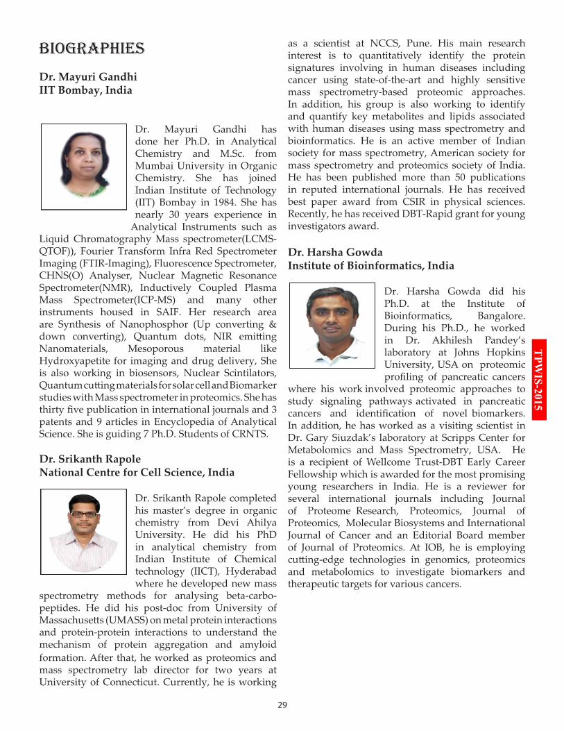
Dr. Harsha GowdaInstitute of Bioinformatics, India
Dr. Harsha Gowda did his Ph.D. at the Institute of Bioinformatics, Bangalore. During his Ph.D., he worked in Dr. Akhilesh Pandey’s laboratory at Johns Hopkins University, USA on proteomic profiling of pancreatic cancers
where his work involved proteomic approaches to study signaling pathways activated in pancreatic cancers and identification of novel biomarkers. In addition, he has worked as a visiting scientist in Dr. Gary Siuzdak’s laboratory at Scripps Center for Metabolomics and Mass Spectrometry, USA. He is a recipient of Wellcome Trust-DBT Early Career Fellowship which is awarded for the most promising young researchers in India. He is a reviewer for several international journals including Journal of Proteome Research, Proteomics, Journal of Proteomics, Molecular Biosystems and International Journal of Cancer and an Editorial Board member of Journal of Proteomics. At IOB, he is employing cutting-edge technologies in genomics, proteomics and metabolomics to investigate biomarkers and therapeutic targets for various cancers.
30
TPWIS-2015Dr. Mahesh KulkarniNational Chemical Laboratory, India
Dr. Mahesh Kulkarni is a scientist at CSIR-National Chemical Laboratory Pune. He obtained his Ph.D. from University of Agricultural Sciences Bangalore. His area of research is Chemical Proteomics, Mass spectrometry,
Diabetes and Aging. He was post Doctoral fellow at CCMB, Hyderabad and Genome Institute of Singapore, Singapore.
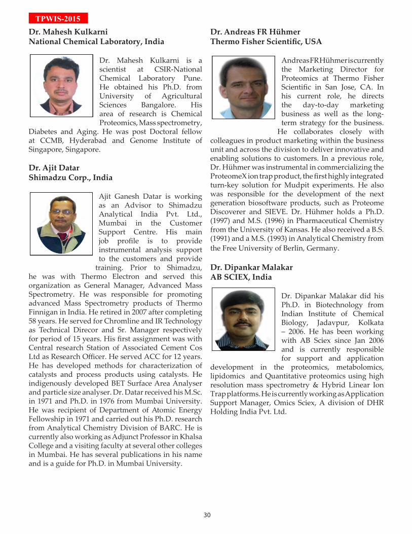
Dr. Ajit DatarShimadzu Corp., India
Ajit Ganesh Datar is working as an Advisor to Shimadzu Analytical India Pvt. Ltd., Mumbai in the Customer Support Centre. His main job profile is to provide instrumental analysis support to the customers and provide
training. Prior to Shimadzu, he was with Thermo Electron and served this organization as General Manager, Advanced Mass Spectrometry. He was responsible for promoting advanced Mass Spectrometry products of Thermo Finnigan in India. He retired in 2007 after completing 58 years. He served for Chromline and IR Technology as Technical Direcor and Sr. Manager respectively for period of 15 years. His first assignment was with Central research Station of Associated Cement Cos Ltd as Research Officer. He served ACC for 12 years. He has developed methods for characterization of catalysts and process products using catalysts. He indigenously developed BET Surface Area Analyser and particle size analyser. Dr. Datar received his M.Sc. in 1971 and Ph.D. in 1976 from Mumbai University. He was recipient of Department of Atomic Energy Fellowship in 1971 and carried out his Ph.D. research from Analytical Chemistry Division of BARC. He is currently also working as Adjunct Professor in Khalsa College and a visiting faculty at several other colleges in Mumbai. He has several publications in his name and is a guide for Ph.D. in Mumbai University.
Dr. Andreas FR HühmerThermo Fisher Scientific, USA
Andreas FR Hühmer is currently the Marketing Director for Proteomics at Thermo Fisher Scientific in San Jose, CA. In his current role, he directs the day-to-day marketing business as well as the long-term strategy for the business.
He collaborates closely with colleagues in product marketing within the business unit and across the division to deliver innovative and enabling solutions to customers. In a previous role, Dr. Hühmer was instrumental in commercializing the ProteomeX ion trap product, the first highly integrated turn-key solution for Mudpit experiments. He also was responsible for the development of the next generation biosoftware products, such as Proteome Discoverer and SIEVE. Dr. Hühmer holds a Ph.D. (1997) and M.S. (1996) in Pharmaceutical Chemistry from the University of Kansas. He also received a B.S. (1991) and a M.S. (1993) in Analytical Chemistry from the Free University of Berlin, Germany.
Dr. Dipankar MalakarAB SCIEX, India
Dr. Dipankar Malakar did his Ph.D. in Biotechnology from Indian Institute of Chemical Biology, Jadavpur, Kolkata – 2006. He has been working with AB Sciex since Jan 2006 and is currently responsible for support and application
development in the proteomics, metabolomics, lipidomics and Quantitative proteomics using high resolution mass spectrometry & Hybrid Linear Ion Trap platforms. He is currently working as Application Support Manager, Omics Sciex, A division of DHR Holding India Pvt. Ltd.
31
TPW
IS-2015Mr. Rajesh VashisthBruker Daltonics, India
Working currently at Bruker Daltonics India as Manager –Technical Application and responsible for Customer training and Application support on Bruker Maldi-Tof and LC-MS/MS system for small molecule and proteomics
applications. He joined Bruker Daltonics in 2002 as Instrumentation Engineer and has been doing the installation and customer Support since then. He started his carrier in 1997 as customer support engineer and handled Mass spectrometer and other analytical instruments such as HPLC, LC-MS etc.
Dr. Ashish PargaonkarAgilent Technologies, India
Dr. Ashish Pargaonkar, is an Applications Engineer for Mass Spectrometry for Agilent’s business in India since 2008. A Microbiologist by education with a Ph.D degree in Botany, he holds more than 14 years of experience. Presently he is based
in Agilent Centre of Excellence in Bangalore and is responsible for applications support for Biopharma, Proteomics and Metabolomics.
Dr. Mark McDowall Waters Corporation, UK
Refer to Evening Innovative Sessions (page No. 42)
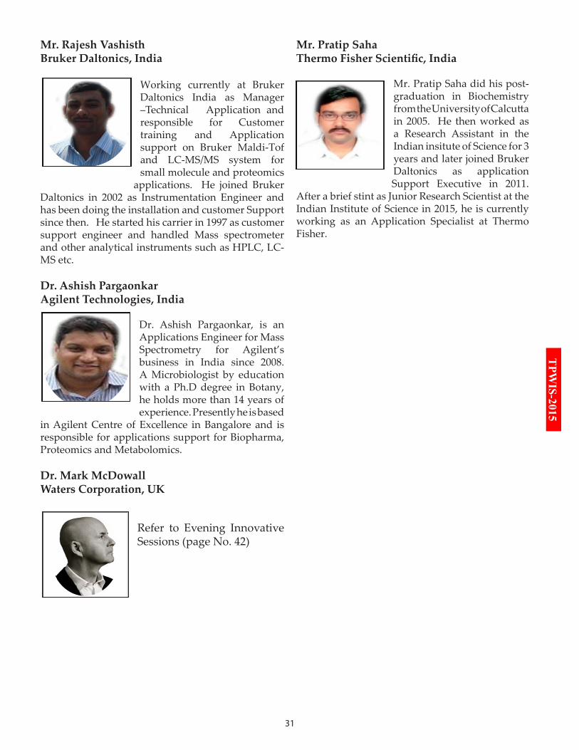
Mr. Pratip SahaThermo Fisher Scientific, India
Mr. Pratip Saha did his post-graduation in Biochemistry from the University of Calcutta in 2005. He then worked as a Research Assistant in the Indian insitute of Science for 3 years and later joined Bruker Daltonics as application Support Executive in 2011.
After a brief stint as Junior Research Scientist at the Indian Institute of Science in 2015, he is currently working as an Application Specialist at Thermo Fisher.
32
TPWIS-2015
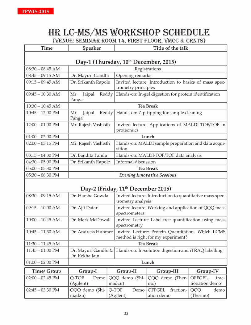
Hr LC-MS/MS WOrKSHOP SCHEduLE(VEnuE: SEMinAr rOOM 14, FirST FLOOr, VMCC & CrnTS)
Time Speaker Title of the talk
Day-1 (Thursday, 10th December, 2015)08:30 – 08:45 AM Registrations08:45 – 09:15 AM Dr. Mayuri Gandhi Opening remarks09:15 – 09:45 AM Dr. Srikanth Rapole Invited lecture: Introduction to basics of mass spec-
trometry principles09:45 – 10:30 AM Mr. Jaipal Reddy
PangaHands-on: In-gel digestion for protein identification
10:30 – 10:45 AM Tea Break10:45 – 12:00 PM Mr. Jaipal Reddy
PangaHands-on: Zip-tipping for sample cleaning
12:00 – 01:00 PM Mr. Rajesh Vashisth Invited lecture: Applications of MALDI-TOF/TOF in proteomics
01:00 – 02:00 PM Lunch02:00 – 03:15 PM Mr. Rajesh Vashisth Hands-on: MALDI sample preparation and data acqui-
sition 03:15 – 04:30 PM Dr. Bandita Panda Hands-on: MALDI-TOF/TOF data analysis04:30 – 05:00 PM Dr. Srikanth Rapole Informal discussion05:00 – 05:30 PM Tea Break05:30 – 08:30 PM Evening Innovative Sessions
Day-2 (Friday, 11th December 2015)08:30 – 09:15 AM Dr. Harsha Gowda Invited lecture: Introduction to quantitative mass spec-
trometry analysis09:15 – 10:00 AM Dr. Ajit Datar Invited lecture: Working and application of QQQ mass
spectrometers10:00 – 10:45 AM Dr. Mark McDowall Invited Lecture: Label-free quantification using mass
spectrometry10:45 – 11:30 AM Dr. Andreas Huhmer Invited Lecture: Protein Quantitation- Which LCMS
method is right for my experiment?11:30 – 11:45 AM Tea Break11:45 – 01:00 PM Dr. Mayuri Gandhi &
Dr. Rekha JainHands-on: In-solution digestion and iTRAQ labelling
01:00 – 02:00 PM Lunch
Time/ Group Group-I Group-II Group-III Group-IV02:00 – 02:45 PM Q-TOF Demo
(Agilent)QQQ demo (Shi-madzu)
QQQ demo (Ther-mo)
OFFGEL frac-tionation demo
02:45 – 03:30 PM QQQ demo (Shi-madzu)
Q-TOF Demo (Agilent)
OFFGEL fraction-ation demo
QQQ demo (Thermo)
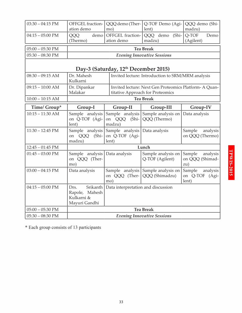
33
TPW
IS-2015
03:30 – 04:15 PM OFFGEL fraction-ation demo
QQQ demo (Ther-mo)
Q-TOF Demo (Agi-lent)
QQQ demo (Shi-madzu)
04:15 – 05:00 PM QQQ demo (Thermo)
OFFGEL fraction-ation demo
QQQ demo (Shi-madzu)
Q-TOF Demo (Agilent)
05:00 – 05:30 PM Tea Break05:30 – 08:30 PM Evening Innovative Sessions
Day-3 (Saturday, 12th December 2015)08:30 – 09:15 AM Dr. Mahesh
KulkarniInvited lecture: Introduction to SRM/MRM analysis
09:15 – 10:00 AM Dr. Dipankar Malakar
Invited lecture: Next Gen Proteomics Platform- A Quan-titative Approach for Proteomics
10:00 – 10:15 AM Tea Break
Time/ Group* Group-I Group-II Group-III Group-IV10:15 – 11:30 AM Sample analysis
on Q-TOF (Agi-lent)
Sample analysis on QQQ (Shi-madzu)
Sample analysis on QQQ (Thermo)
Data analysis
11:30 – 12:45 PM Sample analysis on QQQ (Shi-madzu)
Sample analysis on Q-TOF (Agi-lent)
Data analysis Sample analysis on QQQ (Thermo)
12:45 – 01:45 PM Lunch01:45 – 03:00 PM Sample analysis
on QQQ (Ther-mo)
Data analysis Sample analysis on Q-TOF (Agilent)
Sample analysis on QQQ (Shimad-zu)
03:00 – 04:15 PM Data analysis Sample analysis on QQQ (Ther-mo)
Sample analysis on QQQ (Shimadzu)
Sample analysis on Q-TOF (Agi-lent)
04:15 – 05:00 PM Drs. Srikanth Rapole, Mahesh Kulkarni & Mayuri Gandhi
Data interpretation and discussion
05:00 – 05:30 PM Tea Break05:30 – 08:30 PM Evening Innovative Sessions
* Each group consists of 13 participants

34
TPWIS-2015
Gold SponsorSilver Sponsor

35
TPW
IS-2015
36
TPWIS-2015
PrEAMbLE TO EVEninG innOVATiVE SESSiOnS
The evening innovative seminars are thematic and would feature some of the current developments in the OMICS field. Day-1 (10th December 2015) would highlight Human Phenome Project and Quantita-tive & Targeted Metabolomics. Day-2 (11th December 2015) would discuss Targeted Approaches in Bio-
similars & Biotherapeutics area. Day-3 (12th December 2015) would highlight Targeted Proteomic Strategies for Clinical Biomarker Discovery.
Human Phenome Project and Quantitative & Targeted Metabolomics - In parallel with proteomics, me-tabolomics is receiving immense attention worldwide. Metabolites are the by-products of different metabolic reactions and thus directly reflect the phenotype of an organism. Since metabolites serve as direct signatures of metabolism, their study can be used to investigate the mechanisms of fundamental metabolic processes by linking them to cellular pathways. Thus, metabolomic profiling, combined with high-throughput technolo-gies, provides fast and accurate screening of thousands of biomolecules that are attractive for the identifica-tion of next-generation biomarkers and potential drug/vaccine targets. The evening sessions would feature some of the current developments in Human Phenome Project by the pioneers themselves. Since the launch of the Human Phenome Project, this steadily developing field of metabolomics has gained tremendous mo-mentum.
Targeted Approaches in Biosimilars & Biotherapeutics - Therapeutic proteins are next-generation drugs used in the prevention and treatment of diseases, in particular human critical illness. Biosimilars are a new class of drugs intended to offer comparable safety and efficacy (or clinical equivalence) to their original refer-ence products. Biosimilar medicines are now becoming a reality globally and there exists an incredible op-portunity for the biopharmaceutical sector to capitalize on what is set to become the fastest growing sector of pharmaceutical industry. Several proteomic tools have offered powerful solutions to challenges in biosimilar characterization. Biosimilar development involves optimization of a process to provide a product similar to that of innovator molecule. However, there are several unmet challenges from its development to commer-cialization. Biosimilar development, especially antibodies and drugs, begins with extensive structural and functional characterization, which underpins further product development activities. As regulatory bodies are now being set up to define clear rules, the extensive characterization of Biotherapeutics becomes critical to ensure the patient’s safety and poses great challenge. The omics approaches offer powerful solutions to ad-dress challenges in biosimilar characterization. The advances in targeted proteomics technology can greatly assist biopharma professionals to tackle the development and characterization challenges. Therapeutic de-velopment of lower cost biosimilars will inevitably enter the drug market in the near future, increasing the market competition and patients’ access to the more cost-effective therapies. This discussion will address the issues of concern with the use of biosimilars and the need of appropriate regulations for their approval.
Targeted Proteomic Strategies for Clinical Biomarker Discovery - Apart from the discovery and quantita-tive proteomics, targeted proteomics is emerging as a promising tool for proteomics researchers with interest in validating specific proteins in clinical studies. Specifically in tumor classification, in addition to clinical symptoms and histopathological investigation, protein biomarkers have potential to be considered as prom-ising candidates. In clinical proteomic studies, it becomes necessary to know the absolute difference in the levels of proteins in two different conditions, ideally, a normal/healthy against treated/pathological states. The advent of state-of-the-art proteomic technologies has enabled us to obtain enormous scientific informa-tion in biomedical research. However, validation of such quantitative data continues to remain a bottleneck for researchers. Mass spectrometry based validation experiments thus, often provide robust results with high data confidence. Therefore, for such validation based studies it is imperative to know when and how to move from the discovery to a more targeted analysis, so as to improve confidence in initial results and progress to findings with clinical relevance. Also, the number of samples to be analyzed becomes extremely large to ne-gate any false positive results, especially, when biomarker discovery is a question. For this high throughput requirement, mass spectrometry based targeted quantitative proteomics has emerged as an essential tool in clinical proteomics. During the last decade, targeted proteomics has demonstrated significant impact on vari-ous aspects of clinical research, especially in validation of different drugs and vaccine targets.
37
TPW
IS-2015
38
TPWIS-2015
EVEninG innOVATiVE SESSiOnS SCHEduLE
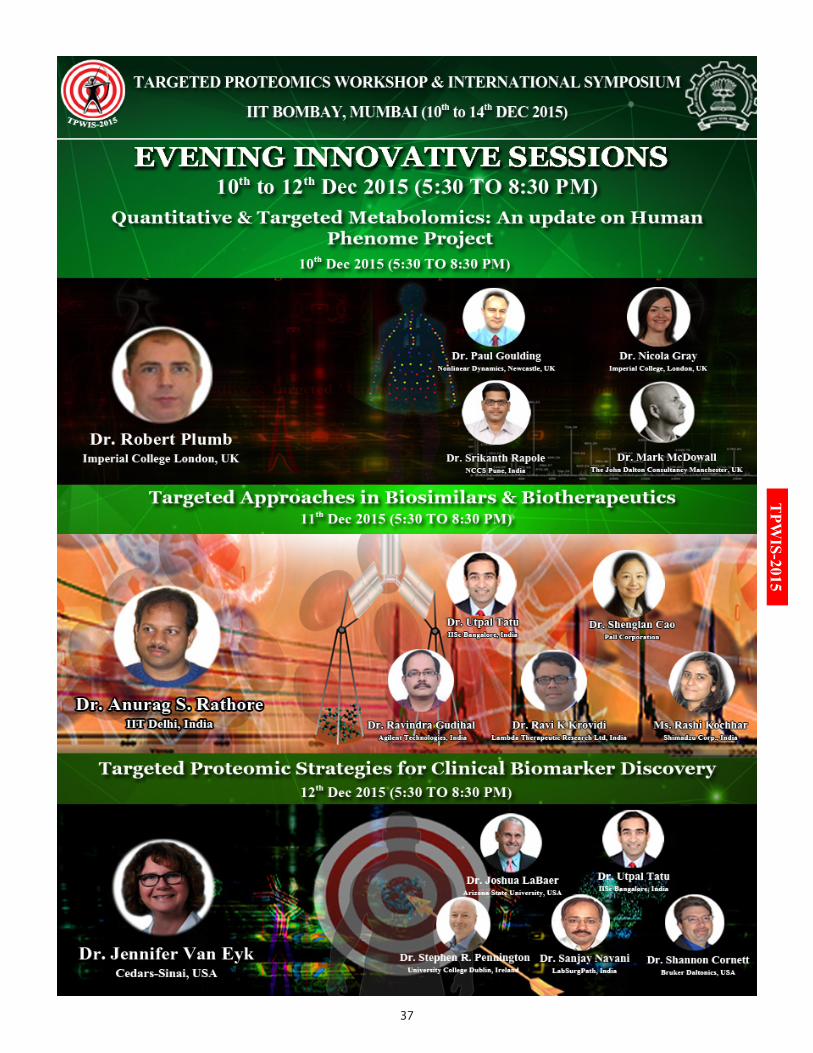
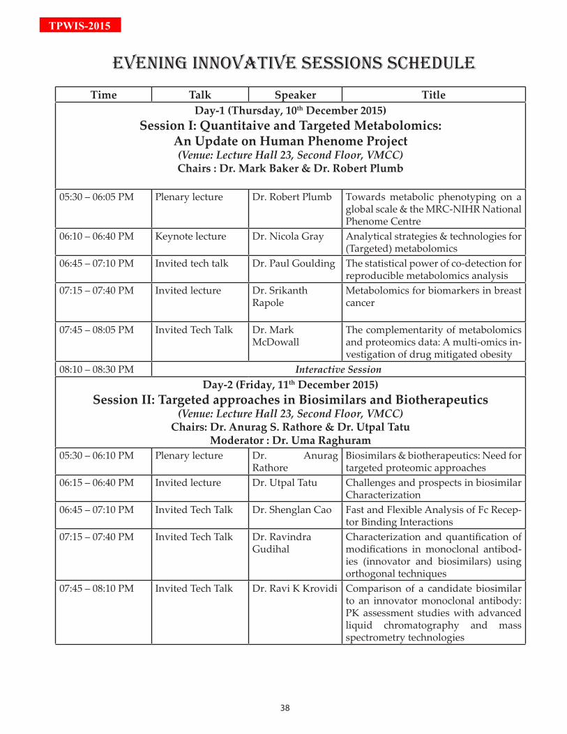
Time Talk Speaker TitleDay-1 (Thursday, 10th December 2015)
Session I: Quantitaive and Targeted Metabolomics: An Update on Human Phenome Project (Venue: Lecture Hall 23, Second Floor, VMCC)Chairs : Dr. Mark Baker & Dr. Robert Plumb
05:30 – 06:05 PM Plenary lecture Dr. Robert Plumb Towards metabolic phenotyping on a global scale & the MRC-NIHR National Phenome Centre
06:10 – 06:40 PM Keynote lecture Dr. Nicola Gray Analytical strategies & technologies for (Targeted) metabolomics
06:45 – 07:10 PM Invited tech talk Dr. Paul Goulding The statistical power of co-detection for reproducible metabolomics analysis
07:15 – 07:40 PM Invited lecture Dr. Srikanth Rapole
Metabolomics for biomarkers in breast cancer
07:45 – 08:05 PM Invited Tech Talk Dr. Mark McDowall
The complementarity of metabolomics and proteomics data: A multi-omics in-vestigation of drug mitigated obesity
08:10 – 08:30 PM Interactive SessionDay-2 (Friday, 11th December 2015)
Session II: Targeted approaches in Biosimilars and Biotherapeutics(Venue: Lecture Hall 23, Second Floor, VMCC)
Chairs: Dr. Anurag S. Rathore & Dr. Utpal TatuModerator : Dr. Uma Raghuram
05:30 – 06:10 PM Plenary lecture Dr. Anurag Rathore
Biosimilars & biotherapeutics: Need for targeted proteomic approaches
06:15 – 06:40 PM Invited lecture Dr. Utpal Tatu Challenges and prospects in biosimilar Characterization
06:45 – 07:10 PM Invited Tech Talk Dr. Shenglan Cao Fast and Flexible Analysis of Fc Recep-tor Binding Interactions
07:15 – 07:40 PM Invited Tech Talk Dr. Ravindra Gudihal
Characterization and quantification of modifications in monoclonal antibod-ies (innovator and biosimilars) using orthogonal techniques
07:45 – 08:10 PM Invited Tech Talk Dr. Ravi K Krovidi Comparison of a candidate biosimilar to an innovator monoclonal antibody: PK assessment studies with advanced liquid chromatography and mass spectrometry technologies
39
TPW
IS-2015
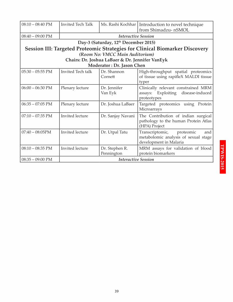
08:10 – 08:40 PM Invited Tech Talk Ms. Rashi Kochhar Introduction to novel technique from Shimadzu- nSMOL
08:40 – 09:00 PM Interactive SessionDay-3 (Saturday, 12th December 2015)
Session III: Targeted Proteomic Strategies for Clinical Biomarker Discovery(Room No: VMCC Main Auditorium)
Chairs: Dr. Joshua LaBaer & Dr. Jennifer VanEykModerator : Dr. Jason Chen
05:30 – 05:55 PM Invited Tech talk Dr. Shannon Cornett
High-throughput spatial proteomics of tissue using rapifleX MALDI tissue typer
06:00 – 06:30 PM Plenary lecture Dr. Jennifer Van Eyk
Clinically relevant constrained MRM assays: Exploiting disease-induced proteotypes
06:35 – 07:05 PM Plenary lecture Dr. Joshua LaBaer Targeted proteomics using Protein Microarrays
07:10 – 07:35 PM Invited lecture Dr. Sanjay Navani The Contribution of indian surgical pathology to the human Protein Atlas (HPA) Project
07:40 – 08:05PM Invited lecture Dr. Utpal Tatu Transcriptomic, proteomic and metabolomic analysis of sexual stage development in Malaria
08:10 – 08:35 PM Invited lecture Dr. Stephen R. Pennington
MRM assays for validation of blood protein biomarkers
08:35 – 09:00 PM Interactive Session
40
TPWIS-2015College London, is the first of its kind facility. Born out of the UK Olympic Legacy its mandate is to provide “high throughput, forensic quality, metabolic phenotyping to support large scale epidemiological studies as well as basic medical research into disease understanding and patient stratification”. As global life-styles change we are seeing increasing cases of obesity, diabetes, and mental health issues. This not only affects a person’s quality of life but also places increased strain on the health-care systems to provide the right treatment whilst managing costs closely. Metabolic Phenotyping offers a valuable and unique insight into the underlying biochemistry of diseases as well as the patients individual biochemistry “phenotype’, diet, health status, age and stress. To deliver this information the analytical data generated in processed via a variety of chemometric modelling and analysis methodologies to deliver the relevant biochemical information. These chemometric platforms employed vary from simple multivariant analysis to highly complex model based analysis and is presented in a format ready for interpretation by medics. This facility comprises of high field NMR instruments, accurate mass LC/MS instruments, tandem quadrupole LC/MS systems as well as dedicated training facility. In this presentation we will discuss the development of analytical platforms both LC/MS and NMR as well as a detailed discussion on the workflow, validation, reporting and decision making process. The presentation will cover the development and validation of the “discovery’ screening methods for polar, non polar metabolites and lipid profiling using LC/MS methodology, as well as describe the use of proton NMR as an initial screen to eliminate contaminated samples. The quantitative targeted LC/MS assays will also be discussed the various compounds classes such as bile acids, amino acids, eicosanoids, and acyl cartanines.
Dr. Nicola Gray Imperial College London, UK
BiographyDr. Nicola Gray is a Senior Scientist with Waters Corporation and an Honorary Research Associate at Imperial College London, where she joined in 2012. Her position involves the development of
LC-MS techniques that will be implemented into
biOGrAPHiES & AbSTrACTSSession I: Quantitaive and Targeted Me-tabolomics: An Update on Human Phe-nome Project
Dr. Robert PlumbImperial College London, UK
BiographyDr. Robert Plumb is the Director of Metabolic Phenotyping and Stratified Medicine in the Waters Health Sciences Business Operations Division, based in Milford, Massachusetts. Dr. Plumb has published
over 100 papers on the subject of HPLC/MS and NMR for bioanalysis, metabolomics and metabolite identification. He is a recognized expert in the use of liquid chromatography with mass spectrometry, capillary scale LC, purifications scale LC and metabonomics, giving many invited papers at international meetings around the world.After obtaining an honors degree in Chemistry from the University of Hertfordshire in 1992, he started work in at Glaxo Research and Development Drug Metabolism Department. During his time at Glaxo and later Glaxo Wellcome he continued his research in liquid chromatography combined with NMR and mass spectrometry for metabolite identification and bioanalysis obtaining his PhD in 1999. Dr. Plumb continued his work for Glaxo Wellcome with the responsibility of metabolite identification using HPLC/MS/NMR and new analytical technology development. In 2001, he moved to Waters Corporation in Milford, MA, USA where he was responsible for the Life Science Chromatography group and laterly LC/MS applications in the Pharmaceutical Market Development Group before becoming the Director of Metabolic Phenotyping. He is currently a visiting Professor in Analytical Chemistry at Kings College London, visiting Professor at Imperial College in the Dept. of Surgery and Cancer and a Fellow of the Royal Society of Chemistry. In 2014, he was awarded as Highly Cited Researcher by Thomson Reuters.
AbstractTowards metabolic phenotyping on a global scale & the MRC-NIHR Nation-al Phenome CentreThe MRC-NIHR National Phenome Centre, Imperial
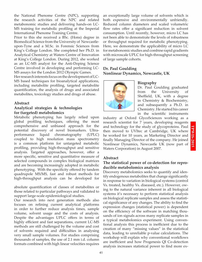
41
TPW
IS-2015the National Phenome Centre (NPC), supporting the research activities of the NPC and related metabonomic studies and delivering hands-on LC-MS training for metabolic profiling at the Imperial International Phenome Training Centre.Prior to this she received a BSc. (Hons) degree in Biomedical Science from the University of Newcastle-upon-Tyne and a M.Sc. in Forensic Sciences from King’s College London. She completed her Ph.D. in Analytical Chemistry at Waters Centre of Innovation at King’s College London. During 2012, she worked as an LC-MS analyst for the Anti-Doping Science Centre involved in developing and performing LC-MS assays for the London 2012 Olympic Games.Her research interests focus on the development of LC-MS based techniques for bioanalytical applications, including metabolite profiling, targeted metabolite quantification, the analysis of drugs and associated metabolites, toxicology studies and drugs of abuse.
AbstractAnalytical strategies & technologies for (targeted) metabolomicsMetabolic phenotyping has largely relied upon global profiling techniques, offering the most comprehensive and unbiased approach for the potential discovery of novel biomarkers. Ultra-performance liquid chromatography (UPLC) coupled to high resolution mass spectrometry is a common platform for untargeted metabolic profiling, providing high-throughput and sensitive analysis. Targeted approaches, however, offer a more specific, sensitive and quantitative measure of selected compounds in complex biological matrices and are becoming increasingly adopted in metabolic phenotyping. With the specificity offered by tandem quadrupole MS/MS, fast and robust methods for high-throughput analysis can be developed for
absolute quantification of classes of metabolites or those related to particular pathways and validated to support large-scale epidemiological studies. Our research into next generation methods also focuses on refining current analytical platforms in order to further reduce analysis times, sample volume, solvent usage and the costs of analysis. Despite the advantages UPLC offers in terms of highly efficient and fast separations, current LC-MS methods are still challenged by the volume and cost of solvents required and difficulties in analysing very small sample volumes. For studies comprising thousands of samples, the use of 2.1 mm i.d. column formats combined with high linear velocities requires
an exceptionally large volume of solvents which is both expensive and environmentally unfriendly. Reduced column diameters and scaled volumetric flow rates offer a significant reduction in solvent consumption. Until recently, however, micro LC has not been able to demonstrate the levels of robustness or throughput required for metabolic phenotyping. Here, we demonstrate the applicability of micro LC for metabonomic studies and combine rapid gradients with microscale UPLC for high-throughput screening of large sample cohorts.
Dr. Paul GouldingNonlinear Dynamics, Newcastle, UK
BiographyDr. Paul Goulding graduated from the University of Sheffield, UK, with a degree in Chemistry & Biochemistry, and subsequently a Ph.D. in Chemistry. He started his career in the scientific instruments
industry at Oxford GlycoSciences working as a research scientist for 7 years, developing reagents and technology for the study of glycoproteins. Paul then moved to UVItec at Cambridge, UK where he worked for 10 years, as Marketing Director and finally Managing Director of the company. He joined Nonlinear Dynamics, Newcastle UK (now part of Waters Corporation) in August 2007.
AbstractThe statistical power of co-detection for repro-ducible metabolomics analysisDiscovery metabolomics seeks to quantify and iden-tify endogenous metabolites that change significantly in response to variation in biological systems (control Vs. treated, healthy Vs. diseased, etc.). However, ow-ing to the natural variance inherent in all biological systems it’s necessary to perform statistical analysis on biological replicate samples and assess the statisti-cal significance of any changes. The ability to find the expression changes (statistical power) is dependent on the efficiency of the software in matching thou-sands of ion signals across many replicate samples in a typical metabolomics experiment. Using conven-tional analysis this process is inefficient due to the creation of many “missing values” in the statistical data, leading to unreliable p-value calculations. The workshop will explain why conventional workflows are inefficient and how Progenesis QI Co-detection analysis increases statistical power to find more ex-
42
TPWIS-2015pression changes than can be found with conven-tional analysis solutions. The statistical power of Co-detection and the simplicity of its deployment within the Progenesis QI environment will be interactively demonstrated.
Dr. Srikanth RapoleNCCS Pune, India
BiographyPlease refer to HR LC-MS/MS workshop (Page No : 29)
AbstractMetabolomics for biomarkers in breast cancerBreast cancer is the most
common malignancy and the leading cause of cancer deaths in women worldwide. Metabolites and lipids are important players in biological systems. Hence, the comparative analyses of the metabolites and lipids of normal, benign and malignant serum samples can provide a better understanding of the molecular events involved in tumour development, and are essential for early diagnosis and prognosis of the disease.In this study, we analysed unbiased metabolomic profile of 75 serum samples and 75 tissue samples of benign, malignantand age- matched healthy controls as well as cell lines using two platforms viz. LC-MS and GC-MS. LC-MS/MS based targeted quantitative metabolite analysis was performed in positive and negative modesusing AB Sciex 4000 QTRAP. GC-MS based untargeted metabolomic profiling was studied using Agilent 7890GC-5977A MSD. For phospholipid profiling, lipids were analysed by in house MRM based platformusing AB Sciex 4000 QTRAP LC-MS/MS.Bioanalytical data obtained were subjected to multivariate statistics using MarkerView and Simca software in order to visualize clusters of cases and to detect the metabolites that are able to differentiate breast cancer patients from healthy individuals. Functional pathway analysis was performed using KEGG and MPP databases to investigate the biological context of the identified metabolites. Several amino acids, fatty acids, phospho lipids, and organic molecules were observed to be differentially expressed in breast cancer when compared with benign and healthy controls. Metabolic sets enrichment analysis of identified differentially expressed metabolites resulted in protein biosynthesis, pyrimidine synthesis, glutamate metabolism, and arginine and proline metabolism amongst top altered pathways.Similarly
pathway analysis of phospholipids depleted their role in Glycerophospholipid metabolism, metabolic pathways, retrograde endocannabinoid pathway, linoleic acid metabolism and alpha linolenic acid metabolism.The results obtained from this study will be presented.
Dr. Mark McDowallWaters Corporation, UK
BiographyDr. Mark McDowall obtained his B.Sc. in Chemistry (1980) and his PhD in Biological Mass Spectrometry (1984) from the University of Wales working on the development and application of ‘primitive’ LC/
MS technologies. He obtained a postdoc fellowship from the Royal Society (of Great Britain) in 1984 to research the mechanism of thermospray ionisation at the University of Bonn (Germany). He joined the LC/MS development team of VG Instruments in 1985 and was involved in the mass spectrometry operations and management of the company for 30 years - throughout it’s evolution to Micromass, where he was Director of Marketing, and more recently Waters Corporation. He served two terms of office on Waters Corporation’s Scientific Advisory Board before he ‘semi-retired’ in March 2014. Today, he continues his career-long association with Waters as a Consultant Mass Spectrometrist.He is a member of the executive management committee of the British Mass Spectrometry Society (BMSS) and a fellow of the Royal Institution (Ri) of Great Britain.
AbstractThe complementarity of metabolomics and proteomics data: A multi-omics investigation of drug mitigated obesityComplementary metabolomic and proteomic studies can provide a multi-dimensional view of biological processes at the molecular level. In this example we combine endogenous metabolite profiling and bottom-up protein profiling to investigate drug mitigated obesity in a laboratory mouse model.Obesity is associated with metabolic syndrome, causing excess body fat to be accumulated, adversely affecting health and life expectancy. It has previously been shown that glucosylceramides play a crucial
43
TPW
IS-2015part in such metabolic syndromes. The manipulation of glucosylceramides with drugs, in mouse models, has shown that symptoms can be negated. Lipid and protein extracts of liver tissue from 3 control and 3 obese mice models were analysed. Protein extracts were proteolysed with trypsin and the resulting peptides separated over a 90-minutereversed-phase nanoscale gradient. Lipid extracts were prepared using 500 µL IPA/water (50:50) and separated over a 20-minute reversed-phase gradient. Data were acquired by data independent acquisition. For protein profiling ion mobility separation was integrated on-line to increase the peak capacity of the analytical system. The acquired data were processed with Progenesis QI software.Randomized proteomic samples (100 ng on-column) were analysed in triplicate to reveal 1200 highly curated proteins across all technical replicates and biological conditions. Over 300 proteins exhibited a significant fold change greater than 2. Randomized lipid extracts 2 µL on-column) were analysed in triplicate. A lipid QC sample, comprised of aliquots of all samples in equal amounts, was injected after every 5 injections. Interrogation of the resulting data revealed over 500 lipid identifications. Unsupervised multivariate analyses showed clear distinction between obese and control groups in both proteomic and lipidomic experiments.
Session II: Targeted Approaches in Biosimilars & Biotherapeutics
Dr. Anurag S. RathoreIIT Delhi, India
BiographyDr. Rathore is a Professor at the Department of Chemical Engineering, Indian Institute of Technology Delhi, India. He is also a consultant of Biotech CMC Issues. His previous roles included management
positions at Amgen Inc., Thousand Oaks, California and Pharmacia Corp., St. Louis, Missouri. His areas of interest include process development, scale-up, technology transfer, process validation, process analytical technology and quality by design. He has authored more than 300 publications and presentations in these areas. He is presently serving as the Editor-in-Chief of Preparative Biochemistry and Biotechnology and Associate Editor for Journal
of Chemical Technology and Biotechnology and PDA Journal of Science and Technology. He also serves on the Editorial Advisory Boards for Biotechnology Progress, BioPharm International, Pharmaceutical Technology Europe and Separation and Purification Reviews. Dr. Rathore has edited books titled Quality by Design for Biopharmaceuticals: Perspectives and Case Studies (2009), Elements of Biopharmaceutical Production (2007), Process Validation (2005), Electrokinetic Phenomena (2004), and Scale-up and Optimization in Preparative Chromatography (2003). He has a Ph.D. in Chemical Engineering from Yale University.
AbstractBiosimilars & Biotherapeutics: Need for targeted proteomic approachesThe ever-increasing cost of healthcare together with our improving understanding of biotech therapeutic drugs has fuelled the rise of biosimilars. Discussion and resolution of the various scientific and regulatory factors that play a role in approval of biosimilars is arguably one of the most significant events over the last decade for biotechnology. Key scientific factors include the complexity of biotech products and processes, use of complex raw materials that are not always well characterized, and our relatively limited understanding of how the numerous quality attributes that define a biotherapeutic impact the product’s safety and/or efficacy in the clinic. A key step towards achieving successful development of a biosimilar is to establish analytical comparability with the innovator drug. This is necessary for the biosimilar manufacturer to avail of the significant reduction in clinical data required for achieving regulatory approval. This talk will discuss key developments that have occurred in the past decade with a focus on addition of more sophisticated platforms to our analytical armoury for characterization of biologics. Limitation of our ability to accurately measure significant quality attributes for a biotech product during production culture in real time will also be discussed. Finally, technology drivers that can alleviate the above mentioned gaps will be discussed. They include mass spectrometry based proteomic approaches and multivariate data analysis (MVDA). This talk will focus on implementation of these approaches in the current paradigm of Quality by Design (QbD) and Process Analytical Technology (PAT).
44
TPWIS-2015Dr. Utpal S. TatuIndian Institute of Science, India
BiographyDr. Tatu is a Professor at the Department of Biochemistry, Indian Institute of Science Bangalore. His research focuses on biology of molecular chaperones with an emphasis on neglected diseases. His
studies have highlighted the potential of exploiting chaperones as drug targets against infectious diseases such as malaria and trypanosomiasis. His lab is also credited with the discovery of a novel trans-splicing event in the expression of heat shock protein 90 gene from Giardia lamblia. His Ph.D. thesis at Indian Insitute of Science received the best Ph.D. thesis award and a gold medal. He did his postdoctoral research at Yale University examining roles of molecular chaperones in protein folding, assembly and secretion. His study on the folding of Influenza virus hemagglutinin protein in the endoplasmic reticulum of mammalian cells has been cited in Molecular Cell Biology book edited by Harvey Lodish. His research contributions have been covered by editorials in Science as well as Nature Medicine and one of his research publications in JBC was selected as the best paper.
AbstractChallenges and prospects in biosimilar characterizationWith the overriding success of Indian Pharma the attention is gradually turning to biopharmaceutical industry in India. Biopharma industry in India is one of the fastest growing in the world and India is also emerging as one of the biggest markets for biopharmaceutical products. The market is still in nascent stage and offers an early advantage to companies, which can get their strategy right. These exciting business opportunities are tied together with certain challenges. Can we deliver quality bipharma products that can compete internationally and gain acceptance in the world market? Answer to this question will lie in the quality control and technical expertise to evaluate biosimilars in the Indian industry. This is increasingly becoming essential for better understanding of structure-function relationship and to study process consistency. I will highlight key technical issues faced by the biosimilar industry in India with top tips to ensure success in the characterization of biopharma products.
Dr. Shenglan Cao Pall Corporation
BiographyDr. Shenglan Cao received her PhD in cell and molecular biology at the School of Biological Sciences of Nanyang Technological University in 2007, under the supervision of Dr.Valerie Lin. Over the years,
she has worked with Sigma-Aldrich and Abbott Molecular in the application and product management fields. She joined the ForteBio team of Pall Corporation in early April this year, taking care of the market development of ForteBio in Asian countries.
AbstractFast and Flexible Analysis of Fc Receptor Bind-ing InteractionsCell mediated effector functions, including the induc-tion of antibody-mediated cell-dependent cytotoxic-ity (ADCC), by a therapeutic antibody depends on its binding affinity to both the biologic target and to Fc-gamma receptors (FcγRs). Throughout drug de-velopment, candidate antibodies are selected, engi-neered, and characterized by their interactions with FcγRs. Here we introduce the Octet® platform as the technology of choice for analyzing Fc gamma recep-tor-antibody binding interactions. Development of FcγR-IgG kinetic assays on the Octet platform is de-scribed, and data presented to demonstrate that these assays can be easily utilized to measure differences in affinity of an FcγR to glycovariants of IgG1.
Dr. Ravindra GudihalAgilent Technologies
BiographyDr. Ravindra Gudihal is currently a Segment Lead and applications scientist (Biopharma) at Agilent Technologies India Pvt. Ltd, Bangalore. At Agilent, he works on application development for
biopharma and proteomics using highly advanced instruments. He received his Ph.D. from Indian Institute of Science, Bangalore, India. During his doctoral studies, he worked on Structure-Functional relationship of Triosephosphate Isomerase, a
45
TPW
IS-2015glycolytic enzyme from Plasmodium falciparum which causes malaria. He developed a fluorescence based assay method for identification of Dehairase enzyme to be used in dehairing of leather from natural sources such as bacterial extract, fungal extract etc. Later, he moved to Purdue University, USA as a postdoc fellow for two and the half years. He worked on new Mass Spectrometric method development to be used in Proteomics and Protein Science at Purdue. He has 19 peer reviewed articles. He also has a MBA in Marketing from Sikkim Manipal University.
AbstractCharacterization and quantification of modifications in monoclonal antibodies (innovator and biosimilars) using orthogonal techniquesMonoclonal antibodies (mAbs) are becoming one of the important classes of biomolecules for treatment of various cancers. Biosimilar mAbs, which are the replica of the licensed innovator product in the market, are also gaining lots of attention. The development of these biosimilars is expanding due to patent expiry of innovator drugs. Many biopharmaceutical companies are involved in the manufacturing of these biosimilars, more so in developing countries. Hence many methods are being developed to characterize these biosimilars in terms of quality and also to quantify the differences. As these are protein molecules they can undergo modification during the biosynthesis (Posttranslational modifications such as glycan content, c-terminal lysine truncations, etc.) or they can get modified during storage, formulation etc. (oxidation, deamidation etc.). In this presentation, use of different methods such as LC, LCMS. Automation etc. will be showcased to show comparability between innovator and biosimilars against modifications.
Dr. Ravi K. Krovidi Lambda Therapeutic Research Ltd, India
BiographyDr. Ravi Krovidi heads Biosimilars, Panomics group at Lambda Therapeutics Research Ltd, India since 2014.His main focus is towards the development of analytical mass spectrometry based proteomics methodologies for various
clinical studies. His group has developed several LC-MS based methods including immunoaffinity strategies, targeted proteomics, and surrogate peptide approach for Pharamcokinetics assessments
of large molecules in ongoing clinical studies. His group is currently offering advanced in-depth protein characterization services to various global biopharma clients, to assess biosimilarity of their products and provide technical data required for regulatory submissions to EMA and US-FDA.Dr. Krovidi holds a Ph.D. (Proteomics and Mass spectrometry) from Max-Planck Institute, Germany and had served the U.S. Government, Department of Energy (DOE) Pacific Northwest National Labs. He has an extensive background in fundamental and applied life science research and has worked in renowned global academia and industry sectors and has over 16 years of experience in proteomics analysis.
Abstract Comparison of a candidate biosimilar to an innovator monoclonal antibody: PK assessment studies with advanced liquid chromatography and mass spectrometry technologiesBiosimilars are biologics containing the same active ingredient as the originator, they have the exact amino acid sequence composition and highly similar glycosylation patterns overlapping with the originator reference product. Biosimilars should demonstrate similarity to the innovator molecule at levels including quality attributes, biological activity, safety and efficacy based on extensive comparability studies. Pharmacokinetic data should demonstrate comparability regarding dose exposure, safety, pharamcodynamics and non clinical and clinical response. LC-MS based methods are currently evaluated for PK assessment of various mAb’s.
Ms. Rashi Kochhar Shimadzu Corp., India
BiographyMs. Rashi Kochhar did her graduation in Biotechnology from the University of Mumbai and post-graduation in Management from Jamanalal Bajaj Institute of Management Studies.
She has been working with Shimadzu Analytical India for over 7 years, catering to two primary techniques viz., LC and LC/MS. She has been shouldering various responsibilities ranging from application support, product management, key customer handling and marketing intiatives. She
46
TPWIS-2015is keenly interested in Biopharma industry and the development of methodologies, which will assist their workflow.
AbstractIntroduction to novel technique from Shimad-zu- nSMOLIn last few years there has been a tremendous in-crease in commercialization of antibody drugs and antibody drug conjugates (ADC), and this segment constitutes major portion of blockbuster drugs.Large number of antibody derived drugsare due for patent expiry within next 5 years. This forecasts a big busi-ness opportunity for pharma industry. However, in process of pre-clinical and clinical studies, quantita-tion of these molecules plays a critical role. Tradition-ally ELISA has been used for quantitation, however establishing ELISA methodology is not only time consuming (to a tune of atleast 6-9 months) but also very expensive, with a high possibility of failure dur-ing large scale study. Triple quadrupole hasbeen a gold standard in quan-titation for pharmaceutical and bioanalytical applica-tions. But unlike small molecules, the sheer size and complexity of these molecules present large number of seen and unseen, unprecedented challenges while quantitation. One of the most commonly seen chal-lenges is selectively identifying and quantitating the signature peptide from the drug.Shimadzu has addressed this challenge of biosimilar, ADC and antibody drugs with a unique methodol-ogy called nano-surface and molecular-orientation limited (nSMOL) proteolysis. It minimizes the sample complexity while maintaining the specificity of the target protein sequences for quantitation by MS. nS-MOL proteolysis is an entirely novel solid–solid pro-teolysis in order to achieve limited proteolysis of the antibody Fab region. This is done by immobilization of antibody in such a way that only Fab region of anti-body is spatially available for selective cleavage with protease immobilized on beads. Hence considerably reducing the digest peptides that are formed and tre-mendously increasing the selectivity since number of peptides formed are limited. Time required for method setup and optimization is drastically reduced. It also makes sample processing very simple and quantitation by triple quadrupole much more sensitive and specific as compared to ELISAWe have already developed and validated for various monoclonal antibody derived drugs in collaboration with National cancer center, Japan. This can be a very useful tool for sample prep for biosimilar manufac-
turers and CRO involved in quantitation of clinical and preclinical antibody derived drugs.
Session III: Targeted Proteomic Strategies for Clinical Biomarker Discovery
Dr. Shannon Cornett Bruker Daltonics, USA
BiographyDr. Shannon Cornett received his Ph.D. in analytical chemistry from the University of Georgia in 1993, working under the mentorship of Jon Amster to develop MALDI applications for FT-ICR and
TOF. Following a Post-doctoral Fellowship at City of Hope National Medical Center, he joined Bruker and held positions of Applications Scientist, TOF R&D Manager and Omniflex Product Manager. In 2002, he moved to Vanderbilt University as a Research Assistant Professor to work with Professor Richard Caprioli, developing new tools and methodologies for the then-emerging field of imaging mass spectrometry. He rejoined Bruker Daltonics in 2009 and now serves as Applications Development Manager for the America and also supports the MALDI-TOF and FT-ICR product lines. He also holds an appointment of Adjunct Research Professor in Biochemistry at Vanderbilt University and continues to be active in MALDI research with more than 25 peer-reviewed publications.
AbstractHigh-throughput Spatial Proteomics of Tissue using rapifleX MALDI Tissue typerMALDI Imaging massspectrometry (IMS) is a unique analytical tool that allows simultaneous label-free visualization of hundreds of endogenous compounds expressed in tissue. In combination with histology MALDI-TOF IMS can reveal correlation of many lipids/ peptides/ proteins to pathological features, creating a multi-dimensional scale for molecular histology that promises to aid in our understanding of disease diagnosis and treatment. Further, MALDI imaging following enzymatic reactions on tissue provide additional depth to the molecular information that can be extracted. TOF mass analyzers provide the widest analyte
47
TPW
IS-2015versatility for MALDI imaging but existing system designs are incapable of meeting the demands for acquiring imaging datasets at a time consistent with clinical relevance. Here we describe a newly designed MALDI-TOF imaging system which offers a significant speed advantage over existing systems and makes possible the goal of tissue typing. The new rapifleX MALDI-TOF platform is capable of acquiring upto 50 spectra per second in imaging mode. It has a 5µm focused laser coupled to a novel mirror system which scans the laser beam while the target is in constant motion. Called smartbeam 3D, this optical system is capable of analyzing square regions of the sample as small as 10µm, the size of which the user chooses via software, which corresponds to the image resolution. RapifleX has acquired images of several hundred thousand pixels in 1-2 hours, a time scale relevant for clinical applications. Areas of the sample that correspond to pixels are square in shape and range in size from 10-200µm. The unique combination of moving laser and sample ensures that true pixels are created that do not overlap as with over sampling techniques while ensuring maximum analytical sampling of the complete pixel area. 10µm resolution MALDI images of phospholipids in excess of 800 k pixels have been acquired in 3hours. 20µm protein images from large biopsies, >1cm, can be acquired in 2-4 hours. Megapixel ion images have been acquired in only a few hours. The new rapifleX MALDI-TOF imaging system is capable of acquiring data ~20x faster than traditional TOF systems. The unique optical system combined with the constantly moving sample provides true square pixels as small as 10µm.
Dr. Jennifer Van Eyk Cedars-Sinai, CA
BiographyDr. Jennifer Van Eyk earned her Ph.D. in Biochemistry at the University of Alberta, Edmonton, Canada and is the current Director of the Johns Hopkins NHLBI Proteomics Center at the Johns Hopkins
School of Medicine in Baltimore, Maryland, Director of Bayview Proteomics Group, and Associate Professor of Medicine in the Division of Cardiology in Biological Chemistry and Biomedical Engineering. Her laboratory is leading in the field of clinical proteomics, which integrates cuttingedge proteomics
and drives innovation in heart and vascular diseases. They have developed technologies to quantify protein isoforms and post-translationally modified proteins in disease pathways in tissue and various body fluids. They have also developed MS-based multiple reaction monitoring assays for a number of membrane proteins and components of signaling pathways to determine their concentration in relevant biological samples. They have also developed assays to determine the extent of phosphorylation (or other PTMs) on key regulatory amino acids as an index of functional activity. In addition to publishing numerous research articles on her work, she has also co-edited books on clinical proteomics including “Clinical Proteomics: From Diagnosis to Therapy” and “Proteomic and genomic analysis of cardiovascular disease”.
AbstractClinically relevant constrained MRM assays: Exploiting disease-induced proteotypesPrecision medicine requires robust circulating biomarkers that precisely quantifies an individual’s pathological status.To achieve this willrequire i) clinically relevant mass spectrometry assays and ii) efficient workflows for sample processing, analysis of 1,000s of samplesand integration of real time quality control measures at all steps. To address the first aspect, we propose that exploiting the quantification of proteotype, disease-induced modifications or protein isoforms that correlate to the clinical phenotype of an individual, would be provide clinically relevant assays that would be well suited for mass spectrometry. We have done so for a number of proteins within to domain of cardiovascular/kidney disease. To increase success in selecting robust proteotypic peptides we have developed an empirical selection workflow, as we have found that many current methods cannot be universally applied. Development of robust constrained MRM assays that target the quantification of disease-specific means that there is forcedselection of a signature peptide with potential weak MRM response or confounded by high background can be problematic. Additional steps that can be optimized to maximize peptide selection and assay performance of constrained MRM assays. To develop effective workflows, we have coupled automation of sample preparation and MS methodhas allowed us to carry out precise measurements on 1000s with CV% under 20%. In cardiovascular/kidney disease, APO L1 and TGF isoforms, natriuretic peptide (BNP), a hormone secreted by the heart and cardiac Troponin I (cTnI) a myofilament structural protein released into blood. We will provide ourworkflows for a 32-peptide
48
TPWIS-2015multiple reaction monitoring (MRM) multiplex for the quantification of disease-induced phosphorylation and proteolytic sites of cTnI, for APO L1 and TGF isoforms using MRM assays as well as intact mass analysis for BNP and its disease-induced proteolytic forms for prognostic risk indications.
Dr. Joshua LaBaerArizona State University, USA
BiographyDr. Joshua LaBaer is one of the nation’s foremost investigators in the rapidly expanding field of personalized diagnostics. His efforts focus on the discovery and validation of biomarkers — unique molecular fingerprints
of disease, which can provide early warning for those at risk of major illnesses, including cancer and diabetes. Formerly Founder and Director of the Harvard Institute of Proteomics, Dr. LaBaer was recruited to ASU’s Biodesign Institute as the first Piper Chair in Personalized Medicine in 2009. The Virginia G. Piper Center for Personalized Diagnostics (VGPCPD) has a highly multidisciplinary staff of molecular biologists, cell biologists, biochemists, software engineers, database specialists, bioinformaticists, biostatisticians, and automation engineers. VGPCPD applies open reading frame clones to the high throughput (HT) study of protein function. In addition, his group invented a novel protein microarray technology, Nucleic Acid Programmable Protein Array, which has been used widely for biomedical research, including the recent discovery of a panel of 28 autoantibody biomarkers that may aid the early diagnosis of breast cancer. He earned his medical degree and a doctorate in Biochemistry and Biophysics, from the University of California, San Francisco. He completed his medical residency at the Brigham and Women’s Hospital and a clinical fellowship in Oncology at the Dana-Farber Cancer Institute, both in Boston. He has contributed more than 140 original research publications, reviews and chapters. He is an Associate Editor of the Journal of Proteome Research, a recent member of the National Cancer Institute’s Board of Scientific Advisors, Co-Chair of the Early Detection Research Network Steering Committee and President of the U.S. Human Proteome Organization.
AbstractTargeted proteomics using Protein Microarrays
Protein microarray provides a high-throughput platform for screening thousands of proteins simultaneously and can be used to study protein-protein interactions, protein-drug interactions, search for enzyme substrates, and as tools to search for disease biomarkers. In particular, recent experiments have focused on using these protein microarrays to search for autoantibody responses in cancer patients. In addition to high-throughput screening, protein microarray approach such as Nucleic acid programmable protein microarray (NAPPA) can be used for targeted proteomics as in to study a set of target proteins of interest. NAPPA array can be prepared for a set of proteins, identified from discovery phase studies and these arrays can be used for validation studies. This suggests application of protein microarray approach for targeted proteomics studies.
Dr. Sanjay NavaniSite Director, The Human Protein Atlas (HPA) Project, India
BiographyDr. Sanjay Navani, Consultant Surgical Pathologist and Immunohistochemist, provides surgical pathology services through his laboratory –LAB SURGPATH. Lab Surgpath is currently the only laboratory
in the country that offers an Immunohistochemistry Stained Slide Service for more than 200 diagnostic and 20,000 research antibodies.Lab Surgpath pathologists have contributed to The Human Protein Atlas (HPA) program by manually annotating approximately 16 million immunohistochemistry images over a period of eight years. The immunohistochemistry images form the backbone of the protein atlas.He finished his MD (Pathology) from Kasturba Medical College, Mangalore University in 1992. Subsequently, he obtained Fellowship in Gynecologic Pathology (1995-96) at Massachusetts General Hospital, Harvard Medical School, Boston, USA under Dr. Robert Young and Dr. Robert Scully. He set up the Immunohistochemistry Section at the Department of Surgical Pathology and Cytology at the Breach Candy Hospital Trust in 1995. He has served as a Surgical Pathologist and Immunohistochemist at Breach Candy Hospital Trust and Indian Cancer Society from 1995 to 2007. He is a Co-Founder of the Group of Gynecologic Pathologists (GGP) and a

49
TPW
IS-2015Founder Member of the Bombay Breast Group.
AbstractThe contribution of Indian surgical pathology to the Human Protein Atlas (HPA) ProjectThe Human Protein Atlas (HPA) Program is an endeavor to map of the human tissue proteome showing the distribution and relative abundance of proteins using immunohistochemistry on tissue microarrays as its backbone. The current version 14 utilizes 25040 antibodies targeting 17005 unique proteins. The data is validated using including immunohistochemistry, mRNA data, Western blot analysis, protein array based assays and immunofluorescent-based confocal microscopy.This web-based database that allows exploration of individual proteins, as well as global expression patterns, in all major tissues and organs in the human bodyand is publically available through the Human Protein Atlas portal (www.proteinatlas.org).Indian surgical pathology has played a pivotal role the HPA. Twenty-six qualified, board-certified Indian surgical pathologists have manually annotated all immunohistochemistry images generated. Specially designed software that enabled each core to be presented individually to the annotator was utilized. The normal tissue atlas contains information and images regarding the expression profiles of human genes both at the mRNA and protein level. The protein expression data is derived from annotation of immunohistochemical staining of cell populations in all major human tissues and organs, including the brain, liver, kidney, lymphoid tissues, heart, lung, skin, gastrointestinal tract, pancreas, endocrine tissues and the reproductive organs. In total, 44 different human tissues are included and contain annotation data for altogether 83 different cell types.The cancer tissue atlas contains a multitude of human cancer specimens representing the 20 most common forms of cancer, including breast-, colon-, prostate-, lung-, urothelial-, skin-, endometrial- and cervical cancer. Altogether 216 different cancer samples are used to generate protein expression profiles for all proteins using immunohistochemistry. The data is presented as pathology-based annotation of protein expression levels in tumor cells, along with the images underlying the annotation.Primary annotation parameters included an evaluation of i) staining intensity (negative, weak, moderate or strong), ii) fraction of stained cells (rare, <25%, 25-75% or >75%) and iii) subcellular localization (nuclear and/or cytoplasmic/membranous).Through the analysis of the data and development
of the portal, there were several challenges that were encountered. The annotation of almost 2 million immunohistochemistry-stained tissue spots per year on tissue microarrays presented a logistical challenge. Approximately 6800 tissue spots needed to be annotated everyday in a six-day week to achieve a target of 2 million images/year. Calibration of displays where the IHC images could be analyzed by the pathologists was an added challenge. In addition to this, satisfactory annotation and curation of data involving platforms like western blot, protein array, comparison with previously published gene/protein characterization data and internally generated RNA-Seq data were other challenges in data annotation and reliable antibody efficacy determination.The Swedish-Indian collaboration for the HPA thus, provided an opportunity to Swedish and Indian researchers and pathologists to be active co-participants in cutting-edge technology, resulting in several joint publications. The database of annotated images has resulted in more than 300 publications by the HPA group. Collaborative efforts between the first world and third-world, in which their respective strengths can be tapped and utilized, are mutually beneficial and therefore, should be encouraged.
Dr. Utpal S. TatuIndian Institute of Science, India
Biography Please refer to Evening Innovative Session II : Targeted Approaches in Biosimilars and Biotherapeutics (Page No : 44)
AbstractTranscriptomic, proteomic
and metabolomic analysis of sexual stage development in MalariaThe malaria parasite experiences a significant amount of stress during its growth in human erythrocytes and heavily relies on secretory functions for pathogenesis. Is the parasite is equipped with machinery to tackle perturbations in the secretory pathway, like the unfolded protein response pathway in higher eukaryotes? Our bioinformatics analysis revealed complete absence of genes involved in the canonical unfolded protein response pathway in Plasmodium falciparum. Accordingly, the parasite was unable to up-regulate endoplasmic reticulum (ER) chaperones or ER-associated degradation in response to ER stress. Global profiling of gene expression, proteomic
50
TPWIS-2015and metabolomic profiling upon redox stress revealed a network of AP2 transcription factors, their targets and specific metabolites being activated and/or upregulated. The overall outcome was up-regulation of genes involved in protein export and the sexual stage of the parasite life cycle culminating in gametocytogenesis. Our results suggest that the malaria parasite uses ER stress as a cue to switch to the transmissible sexual stage.
Dr. Stephen R. PenningtonUniversity College Dublin, Ireland
BiographyDr. Pennington graduated from Imperial College of Science and Technology, University of London with a joint honours degree in Chemistry and Biochemistry before completing a Ph.D. in
Biochemistry at the University of Cambridge. During his Ph.D., he was awarded with an Elmore Medical Research Fellowship and it was during this fellowship that his interests in protein mediated cell signalling and the regulation of the mammalian cell cycle began. Subsequently, he was Wellcome Lecturer in the University of Liverpool before moving to University College Dublin in 2003, where he is Professor of Proteomics in the School of Medicine and the UCD Conway Institute of Biomolecular and Biomedical Research. He was a lead investigator in the Dublin based ‘Prostate Cancer Research Consortium’ and a member of the first Global Action Plan initiative launched by Movember. His research team (www.biomedicalproteomics.org) work closely with clinical colleagues to use a range of proteomics platforms including label-free LC-MS strategies to discover protein biomarkers to meet specific unmet needs in oncology and inflammatory diseases. To progress these biomarkers to potential clinical diagnostic assays, the team has established a dedicated laboratory for targeted multiplexed protein biomarker measurement and clinical evaluation by mass spectrometry.He has been awarded a Beit Memorial Fellowship and received a Sir Henry Wellcome Commemorative Award for Innovative Research. He serves on the editorial boards of several journals, and is currenty Vice-President of the British Society for Proteome Research, a General Council member of the European Proteomics Association and lead organizer of the forthcoming 2017 Annual Congress of the Human
Proteome Organisation (HUPO) which will be held in Dublin.
AbstractMRM assays for validation of blood protein biomarkers The biomarker discovery pipeline through development to clinical utility and value is a seemingly straightforward one. However, the translation of newly discovered protein biomarkers to diagnostic tests of therapeutic relevance remains challenging. Several validation techniques that are currently used including immunoassays often lack reproducibility and hence we here describe mass spectrometric based validation technique which can enhance identification of clinically relevant biomarkers. We are implementing an end-user driven clinically relevant strategy to facilitate the identification of individual candidate protein biomarkers into protein signatures of diagnostic value. This patient focused strategy takes advantage of label-free LC-MS and other proteomic approaches for the identification of biomarkers and the development of multiplexed MS-based protein assays using MRM for clinical evaluation of the protein biomarkers. We are applying this strategy for the development of diagnostically relevant biomarker panel for prostate cancer and psoriatic arthritis. Globally about one in six men is diagnosed with prostate cancer during their lives making it is the most common cancer diagnosed. Conventional treatments include radical prostatectomy, androgen-deprivation therapy, radiotherapy, cryotherapy etc. However, significant challenges remain in deciding which men to treat and how to treat them. Unfortunately, the existing readily available tools for disease diagnosis do not adequately guide the key decisions of whichtreatment to pursue, or more importantly whether ‘active surveillance’ may be more appropriate. The development of a protein test with potential to support these key early clinical decisions will be described. The overall strategy is dependent on the development of robust - reproducible and reliable - MRM based assays and workflows for the development and evaluation of such assays.
51
TPW
IS-2015
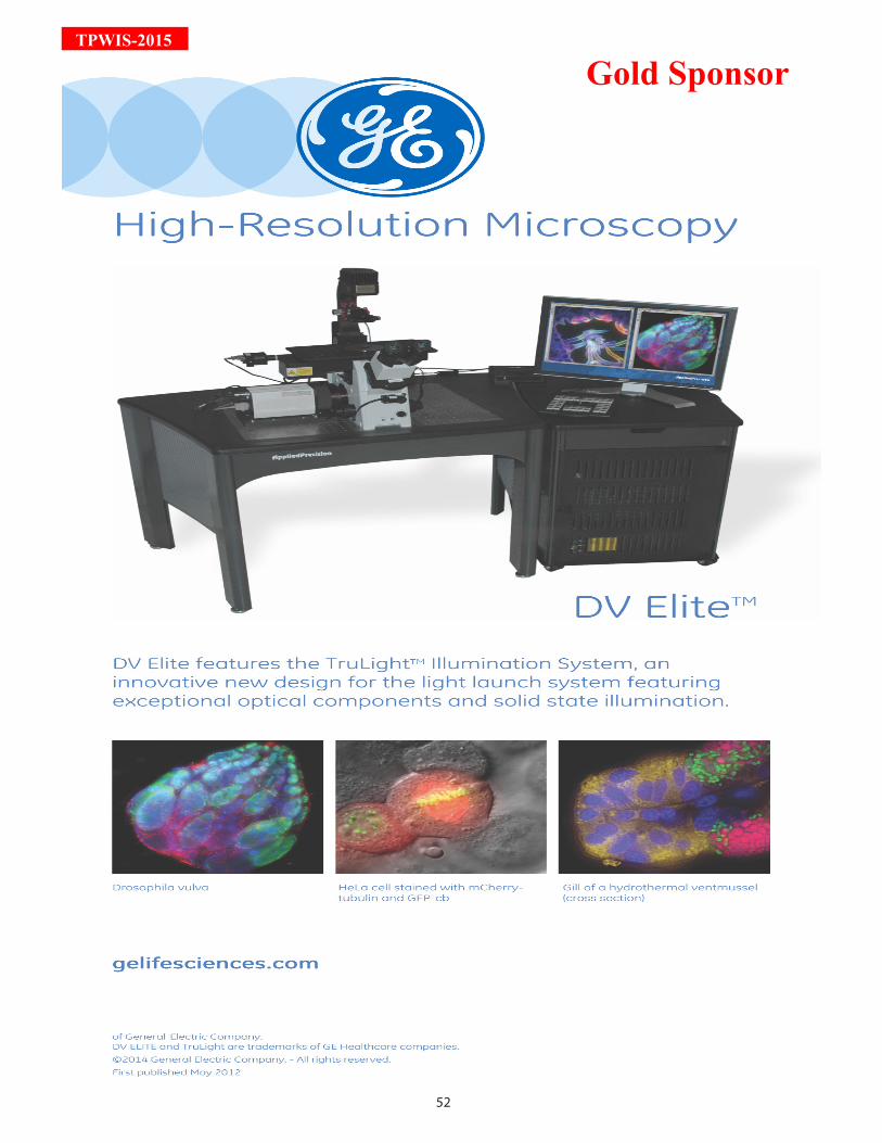
52
TPWIS-2015
Gold Sponsor
53
TPW
IS-2015
54
TPWIS-2015
PrEAMbLE TO EduCATiOn dAy
The Proteomics Education Day is conceptualized to primarily apprise students and teachers with the latest developments in the field of a specialized branch of biosciences called Proteomics. Proteomics is the complete study and characterization of all the proteins encoded from the genetic blueprint in a bio-
logical system. As against the genetic information which is linearly encoded and stored in four nucleotides, a protein is far more analogous and complex, existing in three dimensions, along with varying spatial and temporal configurations. These present fundamental challenges in protein research. However, recently, key technologies have evolved and rapidly accelerated the pace of research in this field. With improvements in the resolving power of gels, to the emergence of mass spectrometry in proteomics research, many powerful technologies have led to a complete face change of the discipline of Proteomics, further resulting in a deeper understanding of biosystems at the functional level. This domain of research is constantly evolving; and re-cent breakthroughs have led to the emergence of powerful and sophisticated technologies in Proteomics.
The advent of state-of-the-art proteomic technologies has thus, enabled us to obtain enormous scientific in-formation in biomedical research; however validation of such quantitative data continues to remain a bottle-neck for researchers. Currently, western blot and ELISA based assays are used for validation studies. These experiments depend on the ease of antibody procurement and have a major drawback of possible antibody cross-reactivity. MS-based validation experiments thus, often provide robust results with high data confi-dence. Therefore, for such validation based studies it is imperative to know when and how to move from the discovery to a more targeted analysis, so as to improve confidence in initial results and progress to findings with clinical relevance.
This event would be divided into five basic modules. The session would start with first building a base from genomics to proteomics and its basics. The Human Genome Project had laid the foundation for various omics studies. Proteomics has in a major way benefitted from existing genome sequences and further studies aim at understanding the dynamic nature of the living systems which forms the basis of any proteomic study. This module would then be taken over by the classical gel based approaches with focus on sensitive platforms like 2D-DIGE and their applications in global proteome analysis. Gel based techniques have been the first stepping stone for proteomics researchers to understand the global proteome of any living system. This field has seen huge transitions from classical one-dimension electrophoresis to the evolved two-dimentional electrophoresis which has made way of covering a greater extent of the proteome. Mass spectrometry, which is the current work-horse of proteomics researchers would be the focus of the next module. The need for stringent valida-tion and sensitive quantification leading to the development of targeted approaches would be discussed in the next module. This would be followed by demo sessions on gel based and mass spectrometry based instru-ments to familiarize students with the emerging platforms in proteomics research. Thus, the schedule of the program is planned such that it overviews the classical and modern proteomics approach in a nut shell and provides a holistic view of the endless possibilities using proteomics research.
This Education Day event on “Proteomics: From Quantitative to Targeted” will essentially be targeted for stu-dents and college teachers. Considering the paucity of resources in terms of easy accessiblity of instruments, this event would serve as an opportunity for the students and college teachers to familiarize themselves with the recent advances in quantitative and targeted proteomics research. Further, the technical training required for specialized streams as these, have not yet reached the masses. We hope that this event would provide a suitable platform for college teachers and students for the extended interactions with the world leading pro-teomics scientists to build their expertise in these areas.
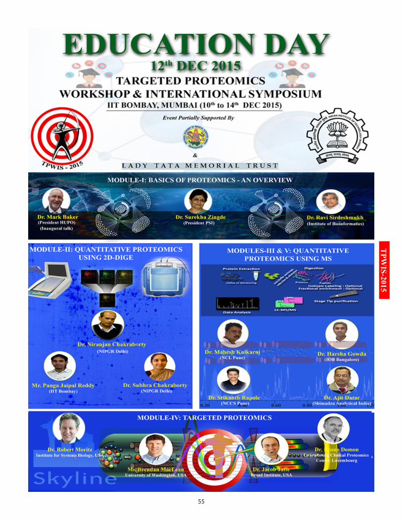
55
TPW
IS-2015
56
TPWIS-2015
Time Speaker Title of the Talk07:30 - 08:30 AM Registrations08:30 – 08:40 AM Dr. Sanjeeva Srivastava Opening address by the Convener
Module-I: Proteomics Basics: An OverviewChair: Dr. K. K. Rao
08:45 – 09:05 AM Dr. Mark S. Baker Opening Remarks: A brighter future for Indian proteomics
09:10 – 09:30 AM Dr. Surekha Zingde Invited Talk: Has Proteomics come of age in In-dia?
09:35 – 09:55 AM Dr. Ravi Sirdeshmukh Invited Talk: Complexity of the proteome: Evo-lution of methods and approaches
10:00 - 10:20 AM Dr. Chien-Sheng Chen Invited Talk: High throughput protein interactome studies using genome-wide proteome microarrays
10:25 – 10:40 AM Tea Break
Module-II: Quantitative Proteomics using Gel-based 2D-DIGEChair: Dr. Tanuja Teni
10:40 – 11:00 AM Dr. Niranjan Chakraborty
Invited Talk: Basics of gel-based proteomics
11:05 – 11:20 AM Dr. Subhra Chakraborty
Invited Talk: Applications of 2-DE
11:25 – 12:45 PM Mr. Panga Jaipal Reddy & Dr. Shubhendu Seal
Hands-on session: Demonstration of 2D-DIGE & data analysis
Module-III: Quantitative Proteomics using MS-based iTRAQChair: Dr. Pramod P. Wangikar
12:45 – 01:05 PM Dr. Mahesh Kulkarni Invited Talk: Basics of quantitative mass spec-trometry
01:05 – 01:25PM Dr. Harsha Gowda Invited Talk: Quantitative proteomics using iTRAQ
01:25– 02:20 PM Lunch
Module-IV: Targeted ProteomicsChair: Dr. Harsha Gowda
02:25 – 02:45 PM Dr. Robert Moritz Invited Talk: SWATH-MS and targeted proteom-ics
EduCATiOn dAy SCHEduLESATurdAy, 12TH dECEMbEr 2015 (VEnuE: LECTurE HALL 23, VMCC)
57
TPW
IS-2015
02:50 – 03:10 PM Mr. Brendan MacLean Invited Talk: Targeted proteomics using Skyline03:15 – 03:35 PM Dr. Jacob Jaffe Invited Talk: Going wide with targeted pro-
teomics03:40 – 04:00 PM Dr. Bruno Domon Invited Talk: Impact of targeted proteomics in
clinics & HUPO perspective
Module-V: Mass Spectrometry Hands-on & Interactive SessionsChairs: Dr. Mahesh Kulkarni & Dr. Rahul Purwar
04:05 – 05:00 PM Dr. Srikanth Rapole, Dr. Ajit Datar,Dr. Mahesh Kulkarni, Dr. Harsha Gowda
Demonstration & Interactive session: QQQ MS based targeted proteomics demo and tips for quantitative proteomics
05:00 – 05:10 PM Dr. Sanjeeva Srivastava Vote of Thanks05:10 – 05:30 PM High Tea05:30 – 09:00 PM Innovative Evening Session: Targeted proteomics strategies for clinical
biomarker discovery
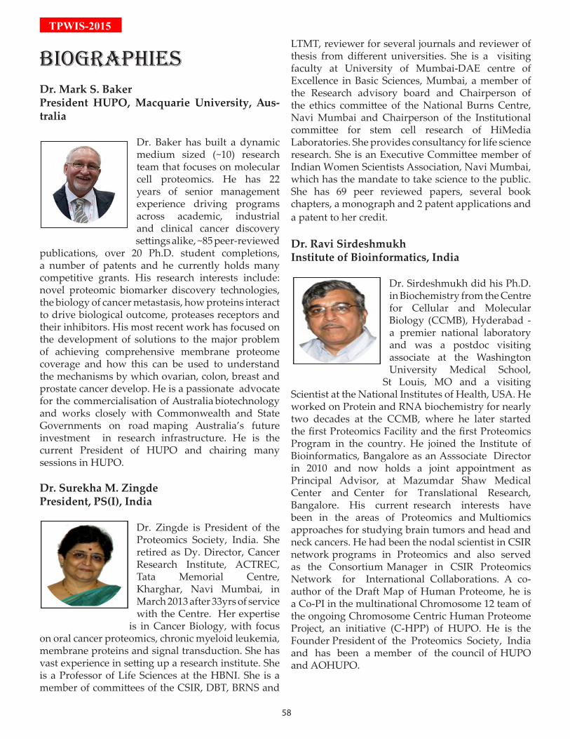
58
TPWIS-2015
biOGrAPHiESDr. Mark S. BakerPresident HUPO, Macquarie University, Aus-tralia
Dr. Baker has built a dynamic medium sized (~10) research team that focuses on molecular cell proteomics. He has 22 years of senior management experience driving programs across academic, industrial and clinical cancer discovery settings alike, ~85 peer-reviewed
publications, over 20 Ph.D. student completions, a number of patents and he currently holds many competitive grants. His research interests include: novel proteomic biomarker discovery technologies, the biology of cancer metastasis, how proteins interact to drive biological outcome, proteases receptors and their inhibitors. His most recent work has focused on the development of solutions to the major problem of achieving comprehensive membrane proteome coverage and how this can be used to understand the mechanisms by which ovarian, colon, breast and prostate cancer develop. He is a passionate advocate for the commercialisation of Australia biotechnology and works closely with Commonwealth and State Governments on road maping Australia’s future investment in research infrastructure. He is the current President of HUPO and chairing many sessions in HUPO.
Dr. Surekha M. ZingdePresident, PS(I), India
Dr. Zingde is President of the Proteomics Society, India. She retired as Dy. Director, Cancer Research Institute, ACTREC, Tata Memorial Centre, Kharghar, Navi Mumbai, in March 2013 after 33yrs of service with the Centre. Her expertise
is in Cancer Biology, with focus on oral cancer proteomics, chronic myeloid leukemia, membrane proteins and signal transduction. She has vast experience in setting up a research institute. She is a Professor of Life Sciences at the HBNI. She is a member of committees of the CSIR, DBT, BRNS and
LTMT, reviewer for several journals and reviewer of thesis from different universities. She is a visiting faculty at University of Mumbai-DAE centre of Excellence in Basic Sciences, Mumbai, a member of the Research advisory board and Chairperson of the ethics committee of the National Burns Centre, Navi Mumbai and Chairperson of the Institutional committee for stem cell research of HiMedia Laboratories. She provides consultancy for life science research. She is an Executive Committee member of Indian Women Scientists Association, Navi Mumbai, which has the mandate to take science to the public. She has 69 peer reviewed papers, several book chapters, a monograph and 2 patent applications and a patent to her credit.
Dr. Ravi SirdeshmukhInstitute of Bioinformatics, India
Dr. Sirdeshmukh did his Ph.D. in Biochemistry from the Centre for Cellular and Molecular Biology (CCMB), Hyderabad - a premier national laboratory and was a postdoc visiting associate at the Washington University Medical School,
St Louis, MO and a visiting Scientist at the National Institutes of Health, USA. He worked on Protein and RNA biochemistry for nearly two decades at the CCMB, where he later started the first Proteomics Facility and the first Proteomics Program in the country. He joined the Institute of Bioinformatics, Bangalore as an Asssociate Director in 2010 and now holds a joint appointment as Principal Advisor, at Mazumdar Shaw Medical Center and Center for Translational Research, Bangalore. His current research interests have been in the areas of Proteomics and Multiomics approaches for studying brain tumors and head and neck cancers. He had been the nodal scientist in CSIR network programs in Proteomics and also served as the Consortium Manager in CSIR Proteomics Network for International Collaborations. A co-author of the Draft Map of Human Proteome, he is a Co-PI in the multinational Chromosome 12 team of the ongoing Chromosome Centric Human Proteome Project, an initiative (C-HPP) of HUPO. He is the Founder President of the Proteomics Society, India and has been a member of the council of HUPO and AOHUPO.
59
TPW
IS-2015Dr. Niranjan ChakrabortyNIPGR, India
Dr. Chakraborty is from the National Institute of Plant Genome Research (NIPGR). His laboratory is developing high-throughput proteomics approaches to study stress perception, signal transduction and metabolic responses of plants to adverse
environmental conditions. His group is focusing on sub-cellular proteomics and using a number of protein mining tools to understand the role of differentially regulated proteins and/or their post-translational modifications in plant physiology. The major focus is on identification of the dynamics of stress-responsive proteins (SRPs), which would not only aid in elucidation of the mechanism underlying stress tolerance, but also would serve as a valuable inventory for crop improvement program.He received ICCR Commonwealth Scholarship and Fellowship; DBT Award of Biotechnology Overseas Associateship and fellow of National Academy of Science, India; Indian National Science Academy, India; National Academy of Agricultural Sciences, India. He is a member of International Plant Proteomics Organization (INPPO); Proteomic Society, India (PS(I)); Society for Biological Chemists (SBC); International Society for Plant Molecular Biology (ISPMB); American Society for Microbiology (ASM), USA; Editorial board member, Scientific Reports; Editorial board member, Rice Science.
Mr. Panga Jaipal ReddyIIT Bombay, India
Mr. Reddy obtained his B.Sc. Degree from Osmania University and completed his Masters in Biochemistry from the University of Pune, India in 2008. He is defending his Ph.D. degree in Proteomics from Department of Biosciences and Bioengineering, Indian
Institute of Technology Bombay. He currently works as a Research Scientist with Prof. Pramod Wangikar at Department of Chemical Engineering at IIT Bombay. He is the author of many scientific publications in reputed journals. He has participated in the development of Virtual Proteomics Laboratory, Clinical Proteomics Remote Triggering Virtual Laboratory, Open Source Courseware Animations
Repository (OSCAR) and National Programme on Technology Enhanced Learning (NPTEL) at IIT Bombay. His current research interests include understanding the regulation of Z-ring assembly and identification of drug targets using proteomics. He has publication sin various peer-reviewed journals like Journal of Proteomics, Proteomics, PlosOne and Expert reviews of Proteomics. He has been involved as an instructor in several workshops on “Gel-based Proteomics” and “Mass Spectrometry Proteomics” held at IIT Bombay.
Dr. Shubhendu SealGE Healthcare, India
Dr. Seal has around 8 years of research experience in the field of molecular biology and diag-nostics. He started his research career in molecular diagnos-tics at DRDE (DRDO) Gwalior, where he worked on the devel-opment of rapid diagnostic kits
for Anthrax and Typhoid. Later he worked at BHU Varanasi for two years in the field of Cytogenetics and Biochemistry. He then became a part of the National Project on Transgenics in Crops (NPTC), ICAR where he has developed genetic trans-formation systems for woody plants and protocols for their molecular assessment. During his research career, he had 17 publications in various national and international journals. In 2010, he started his corpo-rate career as Application Scientist for Affymetrix, Molecular Devices and Wyatt systems. Since 2012, he is working with GE Healthcare as Application Sci-entist for Life-science products, where he developed various assays for Biosimilarity testing and Bio-phar-ma analytics using 2-DE, DIGE and SPR techniques. He also develops the market for Research Products of GE Healthcare- Lifesciences in the west region.
Dr. Subhra ChakrabortyNIPGR, India
Dr. Chakraborty is from the National Institute of Plant Genome Research (NIPGR) and she uses 2-DE gel based approaches to understand the underlying proteomic alterations in plant stress responses. She would illustrate how high quality gels can be
reliably used to study plant proteomics applications.
60
TPWIS-2015
Dr. Mahesh KulkarniNCL Pune, India
Please refer to HR LC-MS/MS workshop (Page No : 30)
Dr. Harsha GowdaInstitute of Bioinformatics, India
Please refer to HR LC-MS/MS workshop (Page No : 29)
Dr. Srikanth RapoleNCCS Pune, India
Please refer to HR LC-MS/MS workshop (Page No : 29)
Dr. Ajit DatarShimadzu Corp. India
Please refer to HR LC-MS/MS workshop (Page No : 30)
Dr. Robert MoritzInstitute for Systems Biology
Please refer to TPP workshop(Page No : 11)
Mr. Brendan MacLeanUniversity of Washington, USA
Please refer to TPW workshop(Page No: 21)
Dr. Bruno DomonLuxembourg Clinical Proteomics Center, Luxembourg
Please refer to TPW workshop(Page No: 21)
Dr. Jacob JaffeBroad Institute, USA
Please refer to TPW workshop(Page No : 22)
Dr. Chien-Sheng ChenNational Central University, Taiwan
(Please refer to Page No : 78)
61
TPW
IS-2015

62
TPWIS-2015
PrEAMbLE TO inTErnATiOnAL SyMPOSiuM
The Targeted Proteomics International Symposium is conceptualized to primarily bring together proteom-ics researchers from around the world, so that participants gets abreast with the latest developments in the field of targeted proteomics. Recently, key technologies have evolved and rapidly accelerated the
pace of research in proteomics. With the emergence of mass spectrometry, many powerful technologies have led to a complete face change of the discipline of proteomics, leading to a deeper understanding of biosystems at the functional level. The advent of state-of-the-art proteomics technologies has enabled us to obtain enor-mous scientific information in biomedical research. However, validation of such quantitative data continues to remain a bottleneck for researchers. Currently, western blot and ELISA based assays are used for valida-tion studies. These experiments depend on the ease of antibody procurement and have a major drawback of possible antibody cross-reactivity. MS-based validation experiments provide robust results with high data confidence. Therefore, for such validation based studies it is imperative to know when and how to move from the discovery to a more targeted analysis, so as to improve confidence in initial results and progress to find-ings with clinical relevance. Targeted Proteomics was declared as the Nature Method of the Year in 2012. It has now established itself as the most sensitive and specific mass spectrometric analysis method to detect and quantitate pre-selected pro-teins in a complex sample matrix. Although targeted proteomics is a recent application of mass spectrometry, it is evolving rapidly as a powerful tool to quantify proteins of interest in complex biological samples. Select-ed reaction monitoring (SRM) has become popular in targeted proteomics in the last decade for quantifying proteins of interest. Skyline has become the standard software in targeted proteomics, and an essential tool in bridging the gap between discovery and targeted proteomics. Skyline is now in use by many research groups around the globe on different mass spectrometry platforms for large-scale targeted proteomics experiments. Also, SRMAtlas, an initiative of Institute for Systems Biology (ISB, Seattle, USA) comprises of a brief summary of targeted proteomics assays for detection and quantification of proteins in a complex protein mixture. For the generation of this database, natural and synthetic peptides were subjected to high quality measurements using a QqQ mass spectrometer. It is intended as a resource for SRM/MRM based proteomics workflow and contains SRM transitions from a variety of model organisms like humans, mouse, rat, etc. Proteomics research has played a crucial role in providing answers to fundamental questions in biology. However, one of the formidable challenges in proteomics field is to study large number of proteins, and iden-tify their interactions and functions. During the last decade, targeted proteomics has demonstrated significant impact on various aspects of clinical research, including the identification of next-generation drugs and vac-cines targets. Considering the paucity of resources in terms of easy accessibility of instrumentation, this event would serve as an opportunity for the students and young researchers in the field to familiarize themselves with the recent advances in targeted proteomics research. Further, the technical training required for special-ized streams as these, has still not reached the masses. Targeted Proteomics is taking increasingly key position in Biology and Biomedical research. This International Symposium would greatly help participants pursuing this domain of promising research which, in all certainty, will make an impact in the area of translational research, in the days to come.
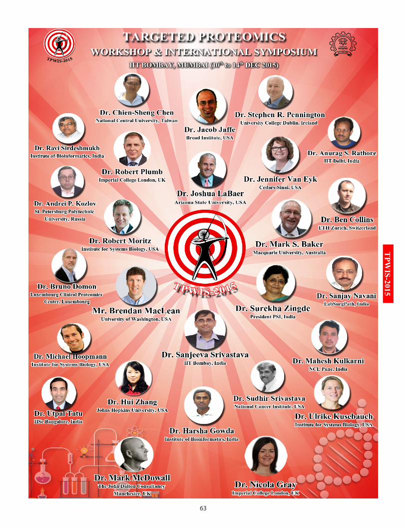
63
TPW
IS-2015
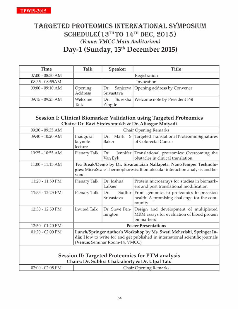
64
TPWIS-2015
Time Talk Speaker Title07:00 - 08:30 AM Registration08:35 - 08:55AM Invocation09:00 - 09:10 AM Opening
AddressDr. Sanjeeva Srivastava
Opening address by Convener
09:15 - 09:25 AM Welcome Talk
Dr. Surekha Zingde
Welcome note by President PSI
Session I: Clinical Biomarker Validation using Targeted ProteomicsChairs: Dr. Ravi Sirdeshmukh & Dr. Aliasgar Moiyadi
09:30 - 09:35 AM Chair Opening Remarks09:40 - 10:20 AM Inaugural
keynote lecture
Dr. Mark S Baker
Targeted Translational Proteomic Signatures of Colorectal Cancer
10:25 - 10:55 AM Plenary Talk Dr. Jennifer Van Eyk
Translational proteomics: Overcoming the obstacles in clinical translation
11:00 - 11:15 AM Tea Break/Demo by Dr. Sivaramaiah Nallapeta, NanoTemper Technolo-gies: MicroScale Thermophoresis: Biomolecular interaction analysis and be-yond
11:20 - 11:50 PM Plenary Talk Dr. Joshua LaBaer
Protein microarrays for studies in biomark-ers and post translational modification
11:55 - 12:25 PM Plenary Talk Dr. Sudhir Srivastava
From genomics to proteomics to precision health: A promising challenge for the com-munity
12:30 - 12:50 PM Invited Talk Dr. Steve Pen-nington
Design and development of multiplexed MRM assays for evaluation of blood protein biomarkers
12:50 - 01:20 PM Poster Presentations01:20 - 02:00 PM Lunch/Springer Author's Workshop by Ms. Swati Meherishi, Springer In-
dia: How to write for and get published in international scientific journals (Venue: Seminar Room-14, VMCC)
Session II: Targeted Proteomics for PTM analysis Chairs: Dr. Subhra Chakraborty & Dr. Utpal Tatu
02:00 - 02:05 PM Chair Opening Remarks
TArGETEd PrOTEOMiCS inTErnATiOnAL SyMPOSiuM SCHEduLE(13TH TO 14TH dEC, 2015)
(Venue: VMCC Main Auditorium)Day-1 (Sunday, 13th December 2015)
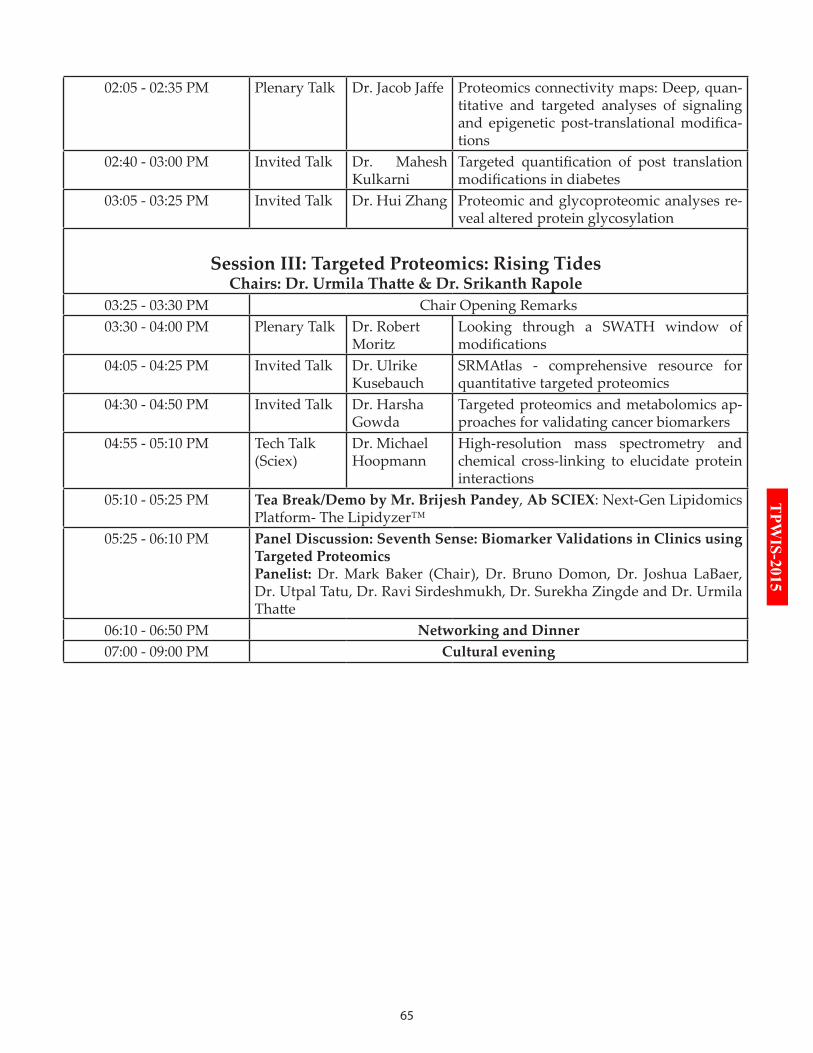
65
TPW
IS-2015
02:05 - 02:35 PM Plenary Talk Dr. Jacob Jaffe Proteomics connectivity maps: Deep, quan-titative and targeted analyses of signaling and epigenetic post-translational modifica-tions
02:40 - 03:00 PM Invited Talk Dr. Mahesh Kulkarni
Targeted quantification of post translation modifications in diabetes
03:05 - 03:25 PM Invited Talk Dr. Hui Zhang Proteomic and glycoproteomic analyses re-veal altered protein glycosylation
Session III: Targeted Proteomics: Rising TidesChairs: Dr. Urmila Thatte & Dr. Srikanth Rapole
03:25 - 03:30 PM Chair Opening Remarks03:30 - 04:00 PM Plenary Talk Dr. Robert
MoritzLooking through a SWATH window of modifications
04:05 - 04:25 PM Invited Talk Dr. Ulrike Kusebauch
SRMAtlas - comprehensive resource for quantitative targeted proteomics
04:30 - 04:50 PM Invited Talk Dr. Harsha Gowda
Targeted proteomics and metabolomics ap-proaches for validating cancer biomarkers
04:55 - 05:10 PM Tech Talk (Sciex)
Dr. Michael Hoopmann
High-resolution mass spectrometry and chemical cross-linking to elucidate protein interactions
05:10 - 05:25 PM Tea Break/Demo by Mr. Brijesh Pandey, Ab SCIEX: Next-Gen Lipidomics Platform- The Lipidyzer™
05:25 - 06:10 PM Panel Discussion: Seventh Sense: Biomarker Validations in Clinics using Targeted ProteomicsPanelist: Dr. Mark Baker (Chair), Dr. Bruno Domon, Dr. Joshua LaBaer, Dr. Utpal Tatu, Dr. Ravi Sirdeshmukh, Dr. Surekha Zingde and Dr. Urmila Thatte
06:10 - 06:50 PM Networking and Dinner07:00 - 09:00 PM Cultural evening
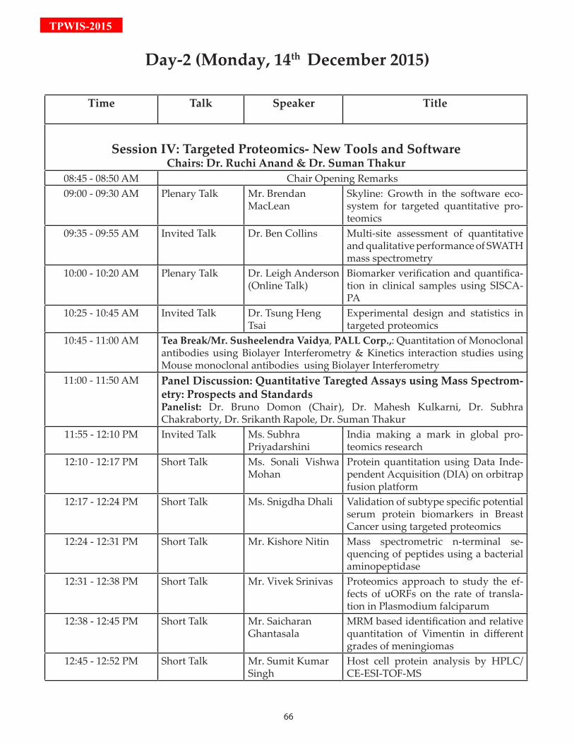
66
TPWIS-2015
Time Talk Speaker Title
Session IV: Targeted Proteomics- New Tools and SoftwareChairs: Dr. Ruchi Anand & Dr. Suman Thakur
08:45 - 08:50 AM Chair Opening Remarks 09:00 - 09:30 AM Plenary Talk Mr. Brendan
MacLeanSkyline: Growth in the software eco-system for targeted quantitative pro-teomics
09:35 - 09:55 AM Invited Talk Dr. Ben Collins Multi-site assessment of quantitative and qualitative performance of SWATH mass spectrometry
10:00 - 10:20 AM Plenary Talk Dr. Leigh Anderson (Online Talk)
Biomarker verification and quantifica-tion in clinical samples using SISCA-PA
10:25 - 10:45 AM Invited Talk Dr. Tsung Heng Tsai
Experimental design and statistics in targeted proteomics
10:45 - 11:00 AM Tea Break/Mr. Susheelendra Vaidya, PALL Corp.,: Quantitation of Monoclonal antibodies using Biolayer Interferometry & Kinetics interaction studies using Mouse monoclonal antibodies using Biolayer Interferometry
11:00 - 11:50 AM Panel Discussion: Quantitative Taregted Assays using Mass Spectrom-etry: Prospects and StandardsPanelist: Dr. Bruno Domon (Chair), Dr. Mahesh Kulkarni, Dr. Subhra Chakraborty, Dr. Srikanth Rapole, Dr. Suman Thakur
11:55 - 12:10 PM Invited Talk Ms. Subhra Priyadarshini
India making a mark in global pro-teomics research
12:10 - 12:17 PM Short Talk Ms. Sonali Vishwa Mohan
Protein quantitation using Data Inde-pendent Acquisition (DIA) on orbitrap fusion platform
12:17 - 12:24 PM Short Talk Ms. Snigdha Dhali Validation of subtype specific potential serum protein biomarkers in Breast Cancer using targeted proteomics
12:24 - 12:31 PM Short Talk Mr. Kishore Nitin Mass spectrometric n-terminal se-quencing of peptides using a bacterial aminopeptidase
12:31 - 12:38 PM Short Talk Mr. Vivek Srinivas Proteomics approach to study the ef-fects of uORFs on the rate of transla-tion in Plasmodium falciparum
12:38 - 12:45 PM Short Talk Mr. Saicharan Ghantasala
MRM based identification and relative quantitation of Vimentin in different grades of meningiomas
12:45 - 12:52 PM Short Talk Mr. Sumit Kumar Singh
Host cell protein analysis by HPLC/CE-ESI-TOF-MS
Day-2 (Monday, 14th December 2015)
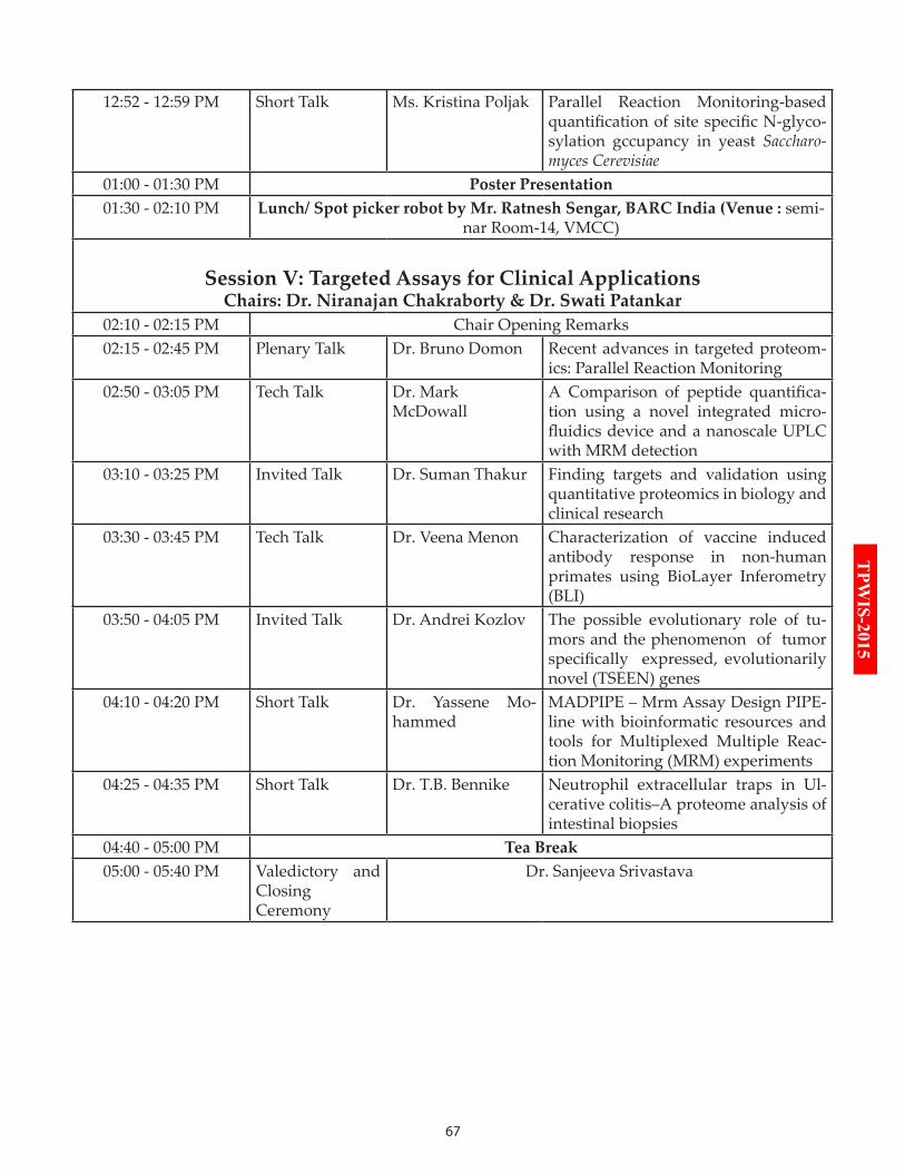
67
TPW
IS-2015
12:52 - 12:59 PM Short Talk Ms. Kristina Poljak Parallel Reaction Monitoring-based quantification of site specific N-glyco-sylation gccupancy in yeast Saccharo-myces Cerevisiae
01:00 - 01:30 PM Poster Presentation 01:30 - 02:10 PM Lunch/ Spot picker robot by Mr. Ratnesh Sengar, BARC India (Venue : semi-
nar Room-14, VMCC)
Session V: Targeted Assays for Clinical ApplicationsChairs: Dr. Niranajan Chakraborty & Dr. Swati Patankar
02:10 - 02:15 PM Chair Opening Remarks02:15 - 02:45 PM Plenary Talk Dr. Bruno Domon Recent advances in targeted proteom-
ics: Parallel Reaction Monitoring02:50 - 03:05 PM Tech Talk Dr. Mark
McDowallA Comparison of peptide quantifica-tion using a novel integrated micro-fluidics device and a nanoscale UPLC with MRM detection
03:10 - 03:25 PM Invited Talk Dr. Suman Thakur Finding targets and validation using quantitative proteomics in biology and clinical research
03:30 - 03:45 PM Tech Talk Dr. Veena Menon Characterization of vaccine induced antibody response in non-human primates using BioLayer Inferometry (BLI)
03:50 - 04:05 PM Invited Talk Dr. Andrei Kozlov The possible evolutionary role of tu-mors and the phenomenon of tumor specifically expressed, evolutionarily novel (TSEEN) genes
04:10 - 04:20 PM Short Talk Dr. Yassene Mo-hammed
MADPIPE – Mrm Assay Design PIPE-line with bioinformatic resources and tools for Multiplexed Multiple Reac-tion Monitoring (MRM) experiments
04:25 - 04:35 PM Short Talk Dr. T.B. Bennike Neutrophil extracellular traps in Ul-cerative colitis–A proteome analysis of intestinal biopsies
04:40 - 05:00 PM Tea Break05:00 - 05:40 PM Valedictory and
Closing Ceremony
Dr. Sanjeeva Srivastava
68
TPWIS-2015
biOGrAPHiES & AbSTrACTS
Dr. Mark S. BakerPresident HUPO, Macquarie University, Aus-tralia
BiographyPlease refer to Education Day (Page No: 58)
AbstractTargeted Translational Pro-teomic Signatures of Col-orectal Cancer
Current methods used for colorectal cancer (CRC) screening (e.g., FOBT, FIT and/or colonoscopy) are grossly inadequate on both sensitivity and specificity grounds. Here, we first report a proteomics discovery program evaluating numerous (e.g., ultradepletion with SWATH-MS and Proseek® Oncology and other multiplexed immunoassays) proteomics biomarker discovery methodologies. In addition, we discuss how proteomics allows iden-tification of interacting membrane proteins (i.e., the metastasome) and demonstrate how proteins in-volved in the metastasome might regulate the cancer invasive phenotype and can be used as clinical bio-markers.Expression of 92 potential plasma cancers biomark-ers were measured in pooled CRC Dukes’ staged (i.e., A-D and controls) EDTA plasmas utilizing Olink’s PEA based Proseek® Multiplex Oncology I kit, where duplicate samples were analysed using Bio-Plex Pro™ human cytokine 27-plex immunoas-says. Expression of CEA (a diagnostic biomarker for CRC) was found to be significantly high in malignant stages C and D, whilst IL 8 and prolactin expression changed significantly between control, benign and malignant stages.We also, employed ultra-SWATH-MS a data indepen-dent acquisition (DIA) method that allows a complete and permanent record of all fragment ions of detect-able peptide precursors from pooled plasmas (n=20 per stage) that were previously ultradepleted (i.e., MARS-12 and our patented ultradepletion) from the same Dukes’ stage A-D CRC patients with age-, sex- and other criteria matched control EDTA-plasmas. The hot-off-the-press results of our ultra-SWATH-MS plasma biomarker studies indicate early detection of CRC and differential separation of Dukes’ stages may be plausible. In addition, shotgun proteomic studies suggest that
integrin (v6) and protease receptor (uPAR) expres-sion are changed in CRC, allowing us to understand changes associated with the metastatic phenotype. An analysis of the interaction between uPAR and v6 has allowed us to develop lead iPEPs that an-tagonise this metastasome. In addition, we report data analysing both proteins (uPAR; epithelially-restrictedv6) as potential intra-stage tissue bio-markers of patient survival in a large retrospective 20 year rectal cancer FFPE tissue biomarker study.
Dr. Jennifer Van Eyk Cedars-Sinai, CA, USA
BiographyPlease refer to Evening Innovative Sessions (Page No: 47)
AbstractTranslational proteomics: overcoming the obstacles in
clinical translation Precision medicine is treating the right disease, the right pathway(s), to correct the underlying pathologi-cal state(s). Precision medicine will or should reduce side affects and increase drug efficiency. Personalized medicine is treating an individual’s disease based on their specific genotype and proteotype. Personalized medicine requires accurate diagnostic assessment not only at the time of medical intervention, but through-out their life. Our premise is an individual’s proteome reflects their past and current health status and thus, will dictate their response to therapeutic intervention. Precise and robust quantification of proteins and their pathological-induced modified forms are required which can only be achieve with technical pipelines able to scale with respect to number of analytes and samples. The question of whether workflows around mass spectrometry-based data independent acquisi-tion (DIA) or multiple reaction monitoring (MRM) have the rigor required for assessing the dynamics of an individual’s proteome needs to be addressed. More people worldwide die of heart disease than any other disease. Yet, currently diagnosis for heart failure is late with 50% mortality within 2-5 years. This is in part due to lack of therapies to treat heart failure. To address this, we are developing DIA and MRM work-flows for both drug screening in preclinical trials and for assessment of potential biomarkers arising based on detailed mechanistic insights. The workflows in-volve automation of sample preparation, mass spec-
69
TPW
IS-2015trometry, quality control and informatics for process assessment. Examples from rat and human studies on HF therapeutics and risk prognosis will be discussed with emphasis on challenges that remain.
Dr. Joshua LaBaerArizona State University, USA
BiographyPlease refer to Evening Innovative Sessions (page No: 48)
AbstractProtein microarrays for studies in biomarkers and
post translational modificationSelf-assembling protein microarrays arrays can be used to study protein-protein interactions, protein-drug interactions, search for enzyme substrates, and as tools to search for disease biomarkers. In particu-lar, recent experiments have focused on using these protein microarrays to search for autoantibody re-sponses in cancer patients. These experiments show promise in finding antibody responses that appear in only cancer patients. New methods using click chemistry-based reagents also allow the application of these arrays for discovering new substrates of post translational modification.
Dr. Sudhir SrivastavaNational Cancer Institute, USA
BiographyDr. Srivastava is Chief of the Cancer Biomarkers Research Group in the Division of Cancer Prevention, National Cancer Institute. He joined the National Cancer Institute in 1988. Since 1990, he has
served as a Program Director in the Division of Cancer Prevention and focused his responsibility in developing molecular signatures of cancer cells for cancer detection research programs with primary emphasis on cancer screening, early detection, risk assessment and informatics.He is an internationally recognized leader in cancer biomarker research. He is best known for his seminal contributions to improving systems approach to biomarker discovery, development and validation. In 2000, He developed and implemented a novel
approach to collaborative clinical research on cancer biomarkers through the establishment of the Early Detection Research Network (EDRN; www.cancer.gov/edrn ), a flagship program at the National Cancer Institute, National Institutes of Health. Under his leadership the network has begun translating biomarkers into clinical tests for early detection and diagnosis, risk assessment, and prognosis. He has spearheaded the role of chemical sciences in oncology by establishing the NCI’s Alliance of Glycobiologists (glycomics.cancer.gov) to study the structure-function relationship of glycans and biomarkers in cancer detection and diagnosis. He has played a key role in conceptualizing and implementing informatics infrastructure for the EDRN in collaboration with NASA (Jet Propulsion Laboratory), a model collaboration being followed elsewhere in NIH.He is best known for his work on developing medical guidelines on the diagnosis of Hereditary Non-polyposis Colorectal Cancer (HNPCC). He played a pivotal role in the development of the Bethesda Guidelines for diagnosing HNPCC, which is in clinical practice worldwide. He has received several honors and awards and is a member of a number of scientific committees world-wide. In 1995, he was elected to the American Joint Committee on Cancer (AJCC) which is responsible for developing staging criteria for cancers for worldwide use and currently serves on the AJCC Executive Committee. He was featured in wired magazine in August 2003 for his leadership in cancer diagnostics. He has been a visiting Professor at several medical and academic institutions, and has delivered several inaugural and keynote addresses. He is Editor-in Chief of the journal Cancer Biomarkers, published by the IOS press and serves as Associate Editors and reviewers for several internationally known journals. He has published more than 200 research papers, review articles and commentaries in peer reviewed journals. He has edited several monographs and book chapters: Early Detection of Cancer: Molecular Markers, published by the Futura Publishing Company in 1995 and Molecular Pathology of Cancer, published by IOS Press, Amsterdam in 1999. Recently, he edited two books on Informatics in Proteomics (2005) published by Francis and Taylor, New York and Translational Pathology of Early Cancer (2012), published by the IOS Press. At present, he is editing another book Translation Research In Cancer Biomarkers to be published by Wiley in 2016.He has received numerous NCI and NIH awards for his leadership in biomarker research and received a Team Science Award for Informatics by the Jet Propulsion Laboratory, NASA for his visionary and
70
TPWIS-2015innovative use of NASA Data System Technologies in biomedical science. Recently, he was invited to give a briefing to the United States Congress on Biomarkers, Bioinformatics and Precision Medicine.
AbstractFrom genomics to proteomics to precision health: A promising challenge for the communityOver the years, a vast array of enabling technologies has been developed to help discover and develop biomarkers, providing opportunities for precision medicine through the identification specific targets for early detection, prevention and treatment. There appears to a synergy among various technologies and the potential to integrate them for biomarker development that clearly demonstrates the value of integrating technologies in delivering a precision detection that is greater than the sum of the individual technology. Integrated genomic and proteomic technologies are yielding a highly innovative strategy for identifying candidate biomarkers for early detection that draws upon the multiple disciplines (i.e., clinical and basic science, technology development, biostatistics and bioinformatics). An efficient and cost effective way to rapidly verify potential candidate biomarkers developed by the National Cancer Institute’s Early Detection Research Network (EDRN) researchers and further refine a biomarker panel in pre-clinical validation studies is provided by employing highly sensitive targeted mass spectrometry-based technologies, such as SRM and PRISM-SRM, before further investment in the development of expensive, clinical-grade immunoassays. The Nucleic Acid-Programmable Protein Array (NAPPA) platform opens the possibility of exploiting the natural tumor-antigen signal amplification provided by autoantibodies to identify novel targets that could be used to develop more sensitive early detection biomarker assays. Some examples will be highlighted to illustrate EDRN’s integrated approach that simply could not have been achieved without collaborations among various disciplines. The fact EDRN investigators work together in the context of integrated workflows, such as those illustrated in my talk, is proof that the collaborative process within the Network is very effective.Integration of genomic, transcriptomic and proteomic data into a single unified picture of tumor biology, including identification of functional processes and pathways that differ between normal and tumor tissues is a transformation that has been a long time coming, but which is currently being pursued by a number of
groups, within and outside of EDRN. If this approach is successful in generating a panel of biomarkers that achieve the necessary sensitivity and specificity for early detection of ovarian cancer, it would be transformative of clinical practice, as ovarian cancer is a disease which has seen no significant decrease in mortality over the past decade. As an example, the Harvard group has created secretome array mapping probe sets that identify genes whose proteins are likely to be present in serum. Bioinformatic-based “validation” as well as results from initial experimental validation supports the utility of the approach. The secretome platform may prove to be invaluable for the discovery phase of serum biomarkers and a helpful method to ensure that efforts and resources are used in validating “appropriate markers.” The down-selection from over 1000 potential biomarkers, based solely on differential expression, to 17, based on genomic data, demonstrates the power of the genomics-based approach. The parallel mass spectrometry-based discovery effort is intriguing in that the protein candidates will apparently be down-selected using similar genomics-based criteria. The integration of multiple data types would likely improve the classification accuracy of the resulting biomarkers, and patients that are likely benefit from precision medicine.
Dr. Stephen R PenningtonUniversity College Dublin, Ireland
BiographyPlease refer to Evening Innovative Sessions(Page No: 50)
AbstractDesign and development of multiplexed MRM assays
for evaluation of blood protein biomarkers The pipeline of biomarker discovery through development to clinical utility and value is a seemingly straightforward one. But, the success in converting newly discovered protein biomarkers to diagnostics test of clinical and patient value is very limited. Possible reasons for this will be introduced.We are implementing an end-user and clinical need driven strategy to progress the identification of individual candidate protein biomarkers into protein signatures of clinical diagnostic value. This patient focused strategy takes advantage of label-free LC-MS and other proteomic approaches for the identification
71
TPW
IS-2015of candidates and the development of multiplexed MS-based protein assays using MRM for clinical evaluation of the protein biomarkers. We are applying this strategy to the development of biomarkers to meet unfilled clinical need in prostate cancer and psoriatic arthritis and our current progress will be described. Taking prostate cancer as a case study for this approach: globally about one in six men will get a diagnosis of prostate cancer during their lives making it is the most common cancer diagnosed. Once diagnosed, for many men prostate cancer can be treated effectively with radical prostatectomy, androgen-deprivation therapy, radiotherapy, cryotherapy or combinations thereof. So, for most the disease is not life threatening. They will die with prostate cancer rather than because of it. However, significant challenges remain in deciding which men to treat and how to treat them. More importantly, it is widely acknowledged that far too many men are treated unnecessarily. For them ‘activesurveillance’ of the disease, allowing them to avoid the often very negative consequences of treatment, would be a betteroption following diagnosis. Unfortunately, the existing readily available tools for disease diagnosis do not adequately guide the key decisions of whichtreatment to pursue, or more importantly whether ‘active surveillance’ may be more appropriate. The development of a protein test with potential to support these key early clinical decisions will be described. The overall strategy is dependent on the development of robust - reproducible and reliable - MRM based assays and workflows for the development and evaluation of such assays.
Dr. Jacob JaffeBroad Institute, USA
BiographyPlease refer to TPW Workshop (Page No: 22)
AbstractProteomics connectivity maps: Deep, quantitative and targeted analyses of
signaling and epigenetic post-translational modificationsWhile transcriptional profiling technology has played a huge role in our understanding of biology and disease, we believe that adding proteomic dimensions to such data could develop even richer explanations of some of the underlying molecular
phenomena in perturbed cellular systems. To that end, we developed targeted proteomic profiling assays in the spaces of signaling (P100) and epigenetics (global chromatin profiling, or GCP). Both assays concentrate on post-translational modifications to proteins that are not easily monitored by DNA-based technologies, and each has a specific focus on an important biological process that underpins the short term and durable responses to changing cellular milieux. Here we present our progress to date on characterizing the proteomic responses to perturbations of epigenetic and signaling systems by drugs in a set of representative biological model systems ranging from cancer to neuronal lineages. We will demonstrate how Proteomic Connectivity Maps can complement existing profiling technologies and provide unique insights for questions focusing on these post-translationally mediated processes.
Dr. Mahesh KulkarniNational Chemical Laboratories, India
BiographyPlease refer to HR LC-MS/MS workshop(Page No: 30)
Abstract:Targeted quantification of post translation
modifications in diabetesDiabetes mellitus (DM) is a metabolic disorder of multiple aetiology characterized by high levels of blood glucose resulting from defects in insulin production, insulin action, or both. Poorly controlled diabetes leads to variety of complications including nephropathy, neuropathy, retinopathy etc. Thus glycemic control is crucial in management of diabetes. Currently diagnosis and management of diabetes heavily relies on detection of levels of N-1-(deoxyfructosyl) valine (DFV) β-hemoglobin (β-Hb), commonly referred as HbA1c, is widely used diagnostic marker in diabetes, believed to provide glycemic status of preceding 90-120 days. However, the turnover of hemoglobin is about 120 days, the DFV-β-Hb, an early and reversible glycation product eventually may undergo irreversible advanced glycation modifications such as carboxymethylation or carboxyethylation. In view of this, deoxyfructosylated, carboxymethylated and carboxyethylated peptides of hemoglobin were quantified in clinical samples from healthy control, pre-diabetes, diabetes and poorly
72
TPWIS-2015controlled diabetes. For the first time we report mass spectrometric quantification of CMV and CEV peptides of β-hemoglobin. Both CMV-β-Hb and CEV-β-Hb peptides showed better correlation with severity of diabetes. Hence, quantification of CMV-β-Hb and CEV-β-Hb could be a useful parameter for assessing the severity of diabetes and may provide better alternative diagnostics.In addition, human serum albumin (HSA), a highly abundant plasma protein, readily undergoes glycation and has been suggested as an alternative diagnostic marker to HbA1c for monitoring glycemic status in diabetes. Therefore, quantification of glycated HSA is of utmost clinical significance. Fragment ion library was constructed for quantification of glycated peptides of albumin. Targeted SWATH analysis of diabetic clinical plasma has led to identification and quantification of 13 glycated peptides of albumin. Five lysine sites namely K549, K438, K490, K88 and K375 were observed to be highly sensitive for glycation modification. Thus peptides involving these lysine sites could be potential novel markers to assess the degree of glycation in diabetes.
Dr. Hui Zhang Johns Hopkins University, USA
BiographyDr. Zhang completed her Ph.D. with Dr. Roland Kallen from University of Pennsylvania in 1999 and joined Cell Signaling Group with Dr. Michael Comb in New England Biolabs as a Product Manager and moved
to Cell Signal Technology as Scientist and Senior Scientist till 2001. After working with Dr. Ruedi Aebersold as Scientist and Senior Scientist at The Institute for Systems Biology, she joined Johns Hopkins University as an Assistant Professor of Pathology in 2006 and was promoted to an Associate Professor of Pathology at Johns Hopkins University in 2011. She established Mass Spectrometry Core Facility of Center for Biomarker Discovery and Translation and serve sas the Director. Her research interests are centered on developing high-throughput technologies to characterize dynamic protein expressions and protein modifications such as phosphorylation, acetylation, and glycosylation. Currently, her research projects focus on understanding the structures and functions of proteins and protein modifications in biology and human diseases using these proteomics technologies. She participates in several research
programs and serves as a principal investigator in the Early Detection Research Network (EDRN), Clinical Proteomic Tumor Analysis Consortium (CPTAC), and mass spectrometry core of Programs of Excellence in Glycosciences (PEG).
AbstractProteomic and glycoproteomic analyses reveal altered protein glycosylationProtein glycosylation is one of the most common forms of protein modifications. Each glycoprotein can be glycosylated at different glycosites and each glycosite may be modified by different glycans. This structural heterogeneity provides additional functions for each glycoprotein in physiological and pathological processes. However, the structural heterogeneity also complicates the studies of structure-function relationships of glycoproteins. To rapidly identify and quantify the glycosylation on each glycosite from complex biological mixtures and to understand the function of protein glycosylation, we developed an integrated approach for global proteomics, glycoproteomics, and glycomics, with which peptides and glycans are isolated and separated by liquid-chromatography followed by quantitative analysis by mass spectrometry. When we performed integrated quantitative analyses to determine protein abundance, glycosylation site occupancy and glycan structures, we showed by specific examples that upon the identification of glycoproteins, glycosites containing specific glycans were readily identifiable and quantifiable. The applications of integrated approach to biological problems facilitate the understanding of glycosylated forms in cellular functions.
Dr. Robert MoritzInstitute for Systems Biology, USA
BiographyPlease refer to TPP workshop(Page No: 11)
AbstractLooking through a SWATH window of modifications Proteomics technology advanc-
es have continued to provide unprecedented levels of both protein identification and quantitation across a multitude of organisms. To capitalize on these ad-vances in comprehensive proteome interrogation, new tools, methods and bioinformatic approaches
73
TPW
IS-2015are required to propel these advances to broad and routine usage. The democratization of these resourc-es enables these tools to be routinely used and enable interrogation of biologically meaningful data so that they can be broadly collected, analyzed and shared within the community. A major goal is to disseminate these developments and applications of proteomic technologies that support comprehensive analysis of all proteins, their isoforms, and post-translational or other modifications contained within the human pro-teome. The advent of new SWATH-MS technologies has heralded new approaches to comprehensively in-terrogate proteomes. I will present advances in novel proteome methods using SWATH-MS that have excit-ing capabilities to comprehensively reveal and quan-titate protein modifications.
Dr. Ulrike KusebauchInstitute for Systems Biology, USA
Biography Redirect to TPP Workshop(Page No: 11)
AbstractSRMAtlas - comprehensive resource for quantitative targeted proteomics
The reliable identification and quantification of proteins is a prerequisite for systems biology and biomedical driven research. Recent advances in mass spectrometry-based proteomics technology have enabled the identification and quantification of proteins at unprecedented comprehensive levels across several organisms, including human. New experimental and computational tools, methods and workflows are required to capitalize on these advances and allow the latest proteomic technologies to be broadly used. To support a rapid implementation and dissemination of the latest techniques, we developed comprehensive, publically available resourcesincluding the Trans-Proteomic-Pipeline, PeptideAtlas, SRMAtlas and PASSEL that enable biologically meaningful data to be collected, analyzed and shared on a proteome wide scale. I will discuss the development of the human SRMAtlas, a compendium of high-quality SRM assays generated in a consistent manner with the use of over 160,000 synthetic peptides for the targeted identification and quantification of the approximately 20,300 human protein-coding genes and its application using multiplexed assays to identify translational biomarkers of kidney fibrosis.
Dr. Harsha GowdaInstitute of Bioinformatics, India
BiographyPlease refer to HR LC-MS/MS Workshop Page(Page No: 29)
AbstractTargeted proteomics and metabolomics approaches
for validating cancer biomarkersCancer is one of the leading causes of cancer deaths worldwide. Several research efforts are focused towards discovery of cancer biomarkers for early detection and monitoring. Over the years, we have carried out several global proteomics and metabolomics studies to identify cancer biomarkers. These discovery studies have resulted in identification of potential biomarkers, some of which have been validated using immunoblots and immunohistochemistry. However, this approach is limited by the availability of good antibodies and costs involved. Mass spectrometry based approaches offer reliable alternative for targeted validation of candidate biomarkers in large number of patient samples. I will discuss multiple reaction monitoring and parallel reaction monitoring approaches that we are employing for targeted validation of cancer biomarkers.
Dr. Michael HoopmannInstitute for Systems Biology, USA
Biography Redirect to TPP Workshop(Page No: 12)
AbstractHigh-resolution mass spectrometry and chemical cross-linking to elucidate protein interactions Chemical cross-linking and mass spectrometry have enabled the analysis of protein-protein interactions and protein topologies. There has been renewed interest in analyzing protein interactions, driven in part by recent advances in technology that
74
TPWIS-2015enable routine acquisition of high-resolution mass spectra. In particular, the availability of diverse cross-linking reagents, coupled with improvements in mass analyzer resolution and throughput, has provided researchers with a formidable arsenal when studying protein interactions. This rapid expansion and diversification of resources has also created significant challenges for the analysis of these data, particularly in computational throughput and statistical validation. I will discuss these challenges and demonstrate solutions we have implemented in the development of our software tools for the identification and validation of cross-linked peptides identified from high-resolution mass spectra.
Mr. Brendan MacLeanUniversity of Washington, USA
BiographyPlease refer to TPW Workshop Page(Page No: 21)
AbstractSkyline: Growth in the software ecosystem for targeted quantitative
proteomicsThe Skyline project started in August, 2008 as a 2-year effort to bring better SRM/MRM software tools to the United States, National Cancer Institute’s CPTAC Verification Working Group that could support the variety of mass spectrometers used by participating laboratories. Over 7 years later, the Skyline project is a thriving proteomics community open-source collaboration supporting 6 mass spec instrument vendors, with over 6,000 registered users worldwide and many thousands of instances started each week. In 2010, development began on extracting chromatograms from full-spectrum data types, with Skyline today providing the broadest available support of data independent acquisition (DIA/SWATH), parallel reaction monitoring (PRM) and MS1 filtering from data independent acquisition (DDA) data. Nature Methods named Targeted Proteomics its method of the year for 2012, noting the importance of Skyline in making this possible. Since then, Skyline has added a framework for integrating with external tools – with its own Tool Store, a web site repository named Panorama for managing and analyzing large collections of data processed in Skyline and direct integration with a cloud data
storage framework named Chorus with fast remote chromatogram extraction for full-spectrum data types. In the past year alone, the Skyline team has added support for targeting small molecules for metabolomics, integrated group comparisons and calibrated quantification, a fully automated pipeline for mass spectrometer quality control called AutoQC and stunning performance for data analysis of DIA and DDA data. This presentation will focus on the recent developments and future directions in the Skyline ecosystem for targeted quantitative proteomics.
Dr. Ben CollinsETH, Zurich, Switzerland
BiographyPlease refer to TPW Workshop Page (Page No: 22)
AbstractMulti-site assessment of quantitative and qualitative
performance of SWATH mass spectrometryReproducibility is a key requirement for scientific research.In recent studies, SWATH-MS which combines data independent acquisition (DIA) and targeted data analysis, was able to quantify thousands of proteins across large sample sets with high data completeness and quantitative accuracy. However, whether SWATH-MS is sufficiently robust to support the generation of reproducible proteomic datasets across laboratories has not been demonstrated. Here we performed a multi-laboratory comparative study in which elevenresearch teams in different locations and countries performed SWATH-MS measurements on standardized samples. All labs performed variable window SWATH acquisition on the TripleTOF 5600. A complex HEK293 matrixwith 30 synthetic peptides dosed in across a broad dynamic range was analyzed. These samples were run repeatedlyduring the course of a week in each lab to investigate the intra- and inter-laboratory reproducibility and detectability of peptides. Thedata were analyzed centrally byOpenSWATHand MultiQuant™.We analyzed the quantitative performance of the 30 synthetic peptides spiked in to the HEK293 matrix. Good linearity was consistently observed across 4.5 orders of magnitude. The number of peptides confidently quantified at each concentration level showed excellent reproducibility between labs. The
75
TPW
IS-2015intra-day reproducibility was ≤10% CV for all labs, and the inter-day CV was typically similar in the majority of labs.We also compared the set of proteins that could be detected from the HEK293 matrix across all samples from all labs by targeted analysis of the SWATH data using a combined human assay library containing peptide coordinates for ~10,000 proteins. Essentially the same set of proteins (~4000+) was detected across all sites. The median Pearson correlation coefficient for protein abundances (log2) across all files from all labs was 0.985.Our data suggest that the reproducibility of SWATH acquisition across labs is sufficiently high to support consistent and large-scale protein quantification across labs.
Dr. Leigh Anderson (Online Talk)SISCAPA Assay Technologies, USA
BiographyDr. Anderson obtained his Bachlor’s in Physics from Yale University and Ph.D. in Molecular Biology from Cambridge University (England). He has published 155 papers and has been granted 40 US Patents. He co-founded
Large Scale Biology, an early proteomics company that IPO’d in 2000, served on the board of directors of Dade Behring (a global diagnostics company), and leads the Plasma Proteome Institute. He is an active member of several scientific advisory boards and is highly sought after as a consultant in the area of proteins, mass spectrometry and diagnostics. Among other distinctions, he received the 1983 Pittsburgh Analytical Chemistry Award; the 2008 Award for Outstanding Contributions to Animal Clinical Chemistry (AACC Division of Animal Clinical Chemistry); and the 2009 HUPO Distinguished Achievement Award in Proteomic Science.
AbstractBiomarker verification and quantification in clinical samples using SISCAPAThe search for new protein biomarkers can be addressed using differential expression analysis via 2-D gels or high resolution mass spectrometry. These methods generate a list of several hundred candidates that are then pared down to a few dozen proteins through tools such as in silico studies and multiplexed ELISAs.
Further validation of the candidate biomarkers is carried out using mass spectrometry of complex biological samples. This requires lengthy chromatographic separations to remove matrix and quantitate the proteins of interest. Enriching for target proteins is an approach to simplify the sample but anti-protein immunocapture enrichment suffers from the same protein- and autoantibody-interferences are are suffered by immunoassays. Thus, tryptic digestion followed by anti-peptide enrichment is the preferred approach.
Dr. Bruno DomonLuxembourg Clinical Proteomics Center, Luxembourg
BiographyPlease refer to TPW Workshop Page (Page No: 21)
AbstractRecent advances in targeted proteomics: Parallel Reac-tion Monitoring
Proteomic biomarker evaluation studies are routinely performed in a targeted manner on triple quadru-pole mass spectrometer operated in selected reaction monitoring (SRM) mode. The low resolution of qua-drupole mass filters results in limited selectivity for complex samples, like bodily fluids. High resolution and accurate mass spectrometers overcome this limi-tation, and have opened new avenues in quantifica-tion. The targeted analyses of clinical samples using a quadrupole-orbitrap mass spectrometer, running in parallel reaction monitoring (PRM) mode, hasshown significant gain in sensitivity and selectivity.The novel acquisition method leverages the internal standards added to the samples to improve the reli-ability and precision of analyses, especially for low abundant components.This technique was applied to clinical assays, using a new data acquisition scheme called internal stan-dard triggered – parallel reaction monitoring (IS-PRM). The method relies on the identification of the internal standards (isotopically labeled peptides),in real-time followed by the adjustment of the acquisi-tion parameters to quantifythe endogenous peptides (corresponding to the proteins of interest). The ac-quisition time is optimized to effectively measure the analytes in a time-scheduled experiment, while keep-ing appropriate cycle times. This approach alternates the acquisition between two PRM modes: a fast, low
76
TPWIS-2015resolution “watch mode”, and a “quantitative mode” using optimized parameters to produce high quality quantitative data. The method has a broad applicabil-ity, and it enables routine quantitative PRM analyses of clinical samples.The analysis of lung cancer mark-ers in plasma samples showed a clear discrimination of the disease stages and subtypes. The approach was also applied to map driver mutations (EGFR, KRAS) in lung cancer tissues samples, with the aim to assist the clinicians to select adequate treatments.
Dr. Mark McDowallWaters Corporation, UK
BiographyPlease refer to Evening Innovative Sessions (Page No: 42)
AbstractA Comparison of peptide quantification using a novel
integrated microfluidics device and a nanoscale UPLC with MRM detectionBiomarker validation is technically challenging re-quiring high throughput, high sensitivity, high reso-lution, wide dynamic range and high selectivity. Min-iaturized LC systems offer improved mass-sensitivity but often lack throughput, robustness and reproduc-ibility. A comparison of a novel integrated micro-fluidic UPLC device with a conventional nanoscale UPLC system, using both high & low resolution MS/MS (MRM) detection (for the quantification of bio-marker peptides) is summarized in this presentation. Stable Isotope Labeled (SIL) peptides, that are ana-logues of putative biomarkers for cardiovascular dis-ease, were spiked at various levels into digested hu-man serum. A reversed phase gradient was employed on both a novel integrated microfluidic UPLC device and a conventional nanoscale UPLC system. MS/MS (MRM) detection using both low resolution tandem quadrupole (QxQ) and high resolution Q-Tof instru-ments was employed.MRM chromatograms, using both high & low reso-lution, showed peptides to be resolved from matrix background over the entire dynamic range for the can-didate biomarkers. For example, <100 amol TAENFR on-column was readily detected with no background matrix interference using the microfluidic UPLC de-vice and low resolution MS/MS (QxQ) detection under high flow and throughput conditions. In comparison, 30 amol of ESDTSYVSLK on-column was detected
using nanoscale UPLC and high resolution MS/MS (Q-Tof) detection. Ultimate sensitivity was<10 amol on-column using conventional nanoscale UPLC with either low or high resolution MRM detection (with a minimum of two transitions per peptide). Typical retention time reproducibility with eitherthe micro-fluidic UPLC device or the nanoscale UPLC equaled 0.02 min standard deviation.1. The novel integrated microfluidic UPLC device en-abled twice the throughput (at reduced sensitivity) compared to the conventional nanoscale UPLC.2. Detection limits were in the 10’s of amol on-column for the novel integrated microfluidic UPLC device and <10 amol on-column for conventional nanoscale UPLC.3. Overall, the optimum combination of throughput, sensitivity, linearity and reproducibility was provid-ed by the novel integrated microfluidic UPLC device (Waters IonKey/MS) coupled to the highest sensitiv-ity QxQ mass spectrometer (Waters Xevo TQ-S).
Dr. Suman ThakurCCMB Hyderabad, India
BiographyDr. Thakur is presently working as a Senior Scientist at the Center for Cellular and Molecular Biology (CCMB), Hyderabad, India. He obtained Ph.D. from University of Delhi in 2002. His professional experience
includes working at Walter Reddy Army Institute of Research, Washington D. C, USA (Postdoc 2002-2004), Indian Institute of Science, Bangalore, India (Postdoc, 2005-2009) and Max Planck Institute of Biochemistry, Munich, Germany (Postdoc 2009-2011).
AbstractFinding targets and validation using Quantitative Proteomics in biology and clinical research Targeted proteomics using mass spectrometry has found its important applications in the field of life sciences and medical biology. Targeted and quantitative proteomics able to find the cause of disease especially narrowing down to particular gene and it is coming closer to transcriptomics and genomics. The quantitation of proteins using stable isotope labeling with amino acids in cell culture (SILAC) would be discussed in details especially to find the target in disease. Notably it also plays an
77
TPW
IS-2015important role to understand the complex process of specific signalling mechanism and its network. Isobaric tags for relative and absolute quantitation (iTRAQ) has been applied to find the targeted biomarker of especially infectious disease and metabolic disorder-diabetes and its complications. Targeted quantitative proteomics is very helpful to understand and validate the biomarker and mode of action of different known and unknown drug. Our group is working to develop novel anticancer drug against different specific cancers. We have investigated successfully the efficiency of various novel natural and synthetic metabolites on targeted oncogene and cancer suppressor gene of different human cancer like retinoblastoma, leukaemia, and melanoma using cell culture and mouse model with the help of quantitative proteomics. Mode of action and targeted signalling mechanism of these novel drugs has been revealed by using quantitative proteomics.
Dr. Veena MenonAmrita University, India
BiographyDr. Menon joined as a Professor in the Department of Pharmacognosy at Amrita in March 2014. She received her Ph.D. in Microbiology & Immunology from Georgetown University, Washington D.C.,
USA. She has about 10 years of research experience in infectious diseases, has worked in various labs across USA – Beckman Research Institute, California USA; Uniformed Services University of the Health Sciences, Bethesda, USA. During the last 4 years, she has been working with leading NIH HIV vaccine investigators in managing and conducting pre-clinical HIV vaccine studies and was actively involved in testing and evaluating new and novel immunogens as HIV vaccine candidates in NHP (non-human primates) models of infection. Her Current Research Focus is on studying the viral and host determinants in HIV transmission, pathogenesis and ART drug resistance. The other major research focus is to elucidate the virological and immunological aspects of the commonly occurring viral diseases in Kerala including HIV, Dengue, Chikungunya for better understanding of mechanisms that can then be harnessed to design effective vaccines.
AbstractCharacterization of vaccine induced antibody response in non-human primates using BioLayer Inferometry (BLI)Characterization of the antibody response elicited by DNA prime-adjuvanated protein boost vaccine with SIV/HIV oligomeric envelopes in non human primates and evaluation of the vaccine efficacy against a mucosal SIV/HIV challenge.
Dr. Andrei KozlovBiomedical Center and Peter the Great St. Petersburg Polytechnic University, Russia
BiographyDr. Kozlov graduated in 1972 from St. Petersburg University. From 1972-1975, he completed his postgraduate (Ph.D.) studies at the N.N. Petrov Research Institute of Oncology. In 1978-1979, Dr. Kozlov served in a tenured Research Training
Fellowship awarded by the International Agency for Research on Cancer at the laboratory of Robert Gallo at the National Cancer Institute. Currently, he holds several positions: Director of the Biomedical Center, Chief, Lab of Molecular Virology, Research Institute of Pure Biochemicals; Chief, Lab of Molecular Virology and Oncology, Peter the Great St. Petersburg Polytechnic University and Principal Investigator for a number of projects. He was among those who discovered the first cases of HIV infection in Russia, performed the first isolation of HIV and field studies of HIV/AIDS epidemic, described the nascent phase of HIV/AIDS epidemic which took place in Russia in 1980s and 1990s and transition to concentrated phase, started first in Russia scientifically based preventive programs in HIV/AIDS and Russian HIV vaccine project. He initiated the MPH program at St. Petersburg University. He is involved in numerous publications which elaborate a national strategy in the field of HIV/AIDS. He has also established a new direction in evolutionary oncology, the theory of positive evolutionarily role of tumors. In 2014, his book “Evolution by Tumor Neofunctionalization” was published by Elsevier/Academic Press. For several years he served as a member of Advisory Board for the Committee of Science and Education of the Russian Parliament. He won the Russian national Chumakov, Vernadsky and Mechnikov awards for research in AIDS, immunology and biotechnology,
78
TPWIS-2015and International Paul Harris Fellowship for his contribution in fighting AIDS and other infectious diseases.
AbstractThe possible evolutionary role of tumors and the phenomenon of tumor specifically expressed, evolutionarily novel (TSEEN) genes Earlier I formulated the hypothesis of the possible evolutionary role of tumors. This hypothesis suggests that heritable tumors supply evolving multicellular organisms with extra cell masses for the expression of newly evolving genes. Evolutionarily novel genes originate in DNA of germ cells. After expression of novel genes in tumor cells, tumors may differentiate in new directions and give rise to new cell types, tissues and organs.The non-trivial prediction on the hypothesis of evolution by tumor neofunctionalization is that evolutionarily novel genes are expressed in tumors.The discussion of experimental confirmation this prediction will include the analysis of evolutionary novelty of tumor-specifically expressed EST sequences; ELFNI – AS1, a human gene with possible microRNA function expressed predominantly in tumors and originated in primates; PBOV1, a human gene of the recent de novo origin with predicted highly tumor-specific expression profile; and the evolutionary novelty of human cancer/testis antigen genes.We also performed the study of the evolutionary novelty of different functional classes of human genes. BLAST-based tools were used to perform this analysis. We found that different classes of genes have different relative evolutionary novelty. The young and novel genes are expressed predominantly in tumors.The conclusion is made that expression of protogenes, evolutionarily young and/or novel genes in tumors might be a new biological phenomenon, a phenomenon of TSEEN (Tumor Specifically Expressed, Evolutionarily New) genes, predicted by the hypothesis of evolution by tumor neofunctionalization.
Ms. Subhra PriyadarshiniNature, India BiographyMs. Priyadarshini is the editor of Nature India, NPG group. She has a bachelor’s degree in Zoology. She pursued law after majoring in Personnel Management and Industrial Relations from Utkal
University, Bhubaneswar, India and a post graduate from the Indian Institute of Mass Communication, she also studied print journalism at the University of Westminster, London. She was a correspondent with
major Indian dailies The Times of India, The Indian Express, The Asian Age, The Telegraph, news agency Press Trust of India and Down To Earth magazine. Nature India is her first online venture, which covers thorough in-depth features and commentaries by leading members of the scientific community.
AbstractIndia making a mark in global proteomics researchIndia had missed the opportunity to contribute scientifically towards the human genome draft published in the last decade. However, a Bangalore-based group more than made up for the missed opportunity through its contribution towards the comprehensive human proteome map, published in the popular journal Nature. This group identified 17,294 protein-coding genes and provided evidence of tissue- and cell-restricted proteins through expression profiling, thereby taking the contribution of the Indian proteomics fraternity to the world.Nature India’s special issue on “Proteomics Research in India”, brought out with the support of the Department of Biosciences and Bioengineering, Indian Institute of Technology Bombay (IITB), is a compendium for researchers around the world since it lists e-learning initiatives, next generation proteomics tools and tips on how to analyse large datasets. The issue also deals with proteomics databases and repositories across the world besides looking closely at the trends in cancer, malaria and plant proteomics
Dr. Chien-Sheng ChenNational Central University, Taiwan
BiographyDr. Chen was born at Kaohsiung, Taiwan in 1974. He completed his doctoral training in the field of Bioanalytical Chemistry at Cornell University in 2005 and then joined Dr. Heng Zhu’s lab at High Throughput
79
TPW
IS-2015Biology Center, Johns Hopkins University School of Medicine as a postdoc fellow. He joined the faculty of Systems Biology and Bioinformatics Institute at National Central University, Taiwan in 2008. His research interests focus on nano/micro-biosensing technology. He developed unique assays for rapid detections and for proteomic research, such as protein function discovery, protein interaction, host-microbe interaction, and biomarker identification using a proteome chip approach. He developed a new high-throughput protein purification protocol to purify ~4000 E. coli proteins within ten hours. The purified proteins were then spotted on glass slides to form the first E. coli proteome chips. He successfully applied them to many projects to facilitate proteomics research and has published several related papers in Nature Methods, PLoS One and Molecular & Cellular Proteomics. He has also developed several unique immunoassays and published them in Biosensors and Bioelectronics, Talanta and PLoS One. He has been an associate editor in Journal of Integrated OMICS and an academic editor in PLoS One. He was awarded as the outstanding new faculty, outstanding research award as well as distinguished professor in National Central University and Academia Sinica Research Award for Junior Research Investigators.
AbstractHigh throughput protein interactome studies using genome-wide proteome microarraysOur lab has a novel high-throughput protein purification technique to purify thousand proteins in one day. These proteins were then spotted onto various glass surfaces to form high-density proteome chips. This novel high-throughput proteomic approach was successfully applied to many projects. First, we attempted to find the intracellular target of natural antimicrobial peptides by our E. coli proteome chips. Our study showed that Lfcin B inhibits the growth of bacteria by influencing the phosphorylation of two component system directly. We have also used E. coli proteome chip to identify bipolar disease biomarkers. The biomarker committee formed by 6 proteins was able to classify biopolar, healthy control and schizophrenic disorder with 75% sensitivity and 80% specificity. In addition to E. coli proteome chip, we also fabricated yeast proteome chips for profiling lipid-protein interactions. We not only recovered many proteins that possessed known PI(3,5)P2-binding domains, but we also found two unknown Pfam domains (Pfam-B_8509 and Pfam-B_10446) that were enriched in our dataset. Among the 162 PI(3,5)P2-BPs, we found a novel motif, HRDIKP[ES]NJLL. A
docking simulation showed that PI(3,5)P2 interacted primarily with lysine or arginine side chains of the newly identified PI(3,5)P2-binding kinases. Recently, we also used human proteome microarrays to identify HCV RNA 5’UTR-binding proteins. We found that hnRNP K interacts with virus RNA 5’-UTR and affect virus RNA replication.
Dr. Tsung Heng TsaiNortheastern University, USA
BiographyPlease refer to TPW Workshop Page(Page No: 22)
Abstarct:Experimental design and statistics in targeted
proteomicsMass spectrometry-based proteomics studies proteins in complex biological mixtures. Stochastic variation and uncertainty are hallmarks of proteomic experiments, and statistical experimental design and analysis are key. To be practical, the statistical methods must accommodate a diverse family of experiment types. To be accurate, they must correctly reflect the specialized stochastic structure of each experimental workflow. This talk will introduce the fundamental concepts of statistical experimental design, and statistical methods for detecting differentially abundant proteins. It will also overview the statistical methods and functionality implemented in MSstats, an open-source software with complex designs, for data processing and summarization of protein abundance, model-based statistical analysis, and sample size calculation in planning future experiments.
Dr. Sanjeeva SrivastavaIndian Institute of Technology Bombay, India
BiographyDr. Sanjeeva Srivastava is Associate Professor and Group Leader of Proteomics Laboratory at the Indian Institute of Technology Bombay India, a guest professor at the Central South University, China and a
Visiting Scientist at the Biodesign Institute, Arizona. He obtained his Ph.D. from the University of Alberta and post-doc from the Harvard Medical School in the area of proteomics, and has specialized expertise in
80
TPWIS-2015applications of data enabled sciences in global health, developing country and resource limited settings. Dr. Srivastava is recipient of several awards including the National Young Scientist Award (Canada), Young Scientist Awards (India) and Apple Research Technology Support Award (UK). The research focus of his group centers on using high-throughput proteomics for the biomarker discovery in Gliomas and infectious diseases. His group is actively working in clinical oncology oriented research focusing on two of the most commonly occurring brain cancers, viz. Glioma and Meningioma. The research focuses on novel biomarker identification using high-throughput proteomics techniques across a wide variety of platforms, including gel-based proteomics, mass spectrometry and protein microarrays. Quantitative profiling to understand proteome alterations along with comprehensive autoantibody profiling of serum and CSF samples in glioma and meningioma have been accomplished. His group also focuses on various neglected tropical diseases malaria, Dengue etc. Additionally, multi-dimensional Omics data are employed for the in silico studies and models. He collaborates actively both across India and internationally to advance this knowledge frontier for the benefit of global health. His vision is to cultivate an ecosystem for clinical proteomics research in India and enable disruptive innovation for biotechnology and global life sciences R&D. To address these grand challenges his vision on data-enabled life sciences (Nature, 2013, 498, 170), biobanking (Nature Reviews Clinical Oncology, 2013, 10, 434-438), innovative ways of funding (EMBO Rep. 2015, 16, 267-71) and e-learning resources for researchers (Nature, 2013, 501, 316) has been appreciated worldwide. Dr. Srivastava serves on executive committee of Proteomics Society, India (PSI) and he is an active member of HUPO and US-HUPO since 2008. He was invited to give a talk in HUPO-2013 conference in Yokohama and in HUPO-2014 in Spain he was invited as panelist of Human Infectious Diseases (HID)-HPP initiative, to chair a session and serve on judging panel of poster presentations. Dr. Srivastava had organized 6th Annual Meeting of PSI, “Proteomics from Discovery to Function” and International Conference from Dec 6th to 11th 2014 in IIT Bombay, Mumbai. This event witnessed many presidents and ex-presidents of HUPO for the first time in India. Dr. Srivastava is very actively contributing in Editorial capacity and currently he is editor for three special issues including a “Proteomics in India” special issue in Journal of Proteomics, “Proteomics research in India” in Nature India and “Protein Arrays” in Proteomics. He is also
associate editor for several international journals. His group has developed E-learning resources such as Virtual Proteomics Laboratory and Open Source Courseware Animations Repository as a community resource. His Proteomics video lectures (40 hours) developed by National Program on Technology Enhanced Learning (NPTEL) is freely accessible for the community. These e-learning resources have been included as tutorial for the International Proteomics Tutorial Programme. Dr. Srivastava’s teaching efforts has provided him Excellence in Teaching Award in IIT Bombay, which is one of the coveted distinctions of this very prestigious institute. Dr. Srivastava envisions designing novel-framework by linking proteomics and big data for the disruptive innovation by collective intelligence and actionable foresight. He believes strongly in trans-generational capacity building in science, bioengineering and 21st century knowledge society to advance the proteomics knowledge for the benefits of global health.
Dr. T.B. BennikeAalborg University, Denmark
BiographyDr. Bennike obtained his Ph.D. from the Laboratory of Medical Mass Spectrometry at Aalborg University (AAU), Denmark. Through his PhD and postdoc positions, he has published several original research papers
in scientific peer-reviewed journals. His research is focused on increasing our understanding of inflammatory diseases, including inflammatory bowel disease, rheumatoid arthritis, and osteoarthritis. He has a background in Nanotechnology (MS) from the Department of Physics and Nanotechnology at Aalborg University (AAU), Denmark. He has held postdoc positions at the University of Southern Denmark, Aalborg University Denmark, and Boston Children’s Hospital, Harvard Medical School, USA.Through an analysis of the proteome, metabolome, and transcriptome of samples from humans and model systems, his objective is to increase our understanding of the underlying biology and triggers of the inflammatory diseases.
Abstract:Neutrophil extracellular traps in ulcerative colitis–A proteome analysis of intestinal biopsiesInflammatory bowel diseases, including Ulcerative
81
TPW
IS-2015colitisaffects 2.5–3 million people in Europe.The life-longconditionssignificantly reduce the quality of life of the patients, and compose a significant economic burden for society.The etiology of these diseases remains incompletely explained.We,therefore, performed the most thorough proteome-based study of ulcerative colitis colon tissue, based on number of analyzed proteins, and have for the first time observed neutrophil extracellular traps in ulcerative colitis, detected by proteomics and verified by microscopy.We performed a comparative proteome analysis of non-inflamed mucosal colon biopsies from 10 patients with ulcerative colitis and 10 controls.The proteome of the biopsies were characterized by high throughput label-free quantitative proteomics, and the biopsy histology was analyzed by microscopy.We quantified 5,711 different proteins in the colon biopsies. Forty-six proteins had a statistically significant difference in abundance between the ulcerative colitis colon tissue and controls. Eleven of the proteins with increased abundances in the ulcerative colitis biopsies were associated with neutrophils and neutrophil extracellular traps. The abundance of calprotectin and lactotransferrin in the tissue correlated with the degree of tissue inflammation as determined by histology. The findings were validated by microscopy where an increased abundance of neutrophils within the mucosal tissue was found. Additionally, we confirmed the presence of neutrophil extracellular traps by extracellular DNA in the ulcerative colitis colon tissue.We found an increased abundance of several proteins hitherto not associated with innate immunity and neutrophils in non-inflamedmucosal colon biopsies from ulcerative colitis patients. The increased abundance of these antimicrobial compounds points to the stimulation of the innate immune system in the etiology of ulcerative colitis. Our findings demonstrate that even though remission has been achieved on the surface of the ulcerative colitis colon tissue, a chronic condition is still present within the morphologically normal tissue.
Dr. Yassene MohammedUniversity of Victoria Proteomics Centre,
Canada
BiographyDr. Mohammed’s work fo-cuses on the Bioinformatics approaches for the design and interpretation of quantitative proteomics experiments using
stable isotope internal standards, as well as on bot-tom-up proteomics. He is leading the Bioinformatics group at the University of Victoria Proteomics Cen-tre, Canada.
AbstractMADPIPE – Mrm Assay Design Pipeline with bioinformatic resources and tools for multi-plexed Multiple Reaction Monitoring (MRM) experimentsMRM-based targeted proteomics is the preferred method for rapid accurate quantitation of proteins in complex biological matrices. LC/MRM-MS experi-ments involve multiple pre-/post-analytical steps. Here we present MADpipe, a pipeline which includes software, a library, and a database for improving and accelerating the design of multiplexed MRM experi-ments. MADpipe includes: • PeptidePicker software for selecting proteotypic peptides, based on information about the protein and the suitability of its tryptic peptides for MRM from six on-line knowledge bases. • PeptidePickerDB, a pre-generated library of all pos-sible peptide surrogates for all human proteins. • PeptideTracker, a knowledge base, containing in-formation on >2500 natural and SIS peptides covering >900 human proteins, and >1800 peptides represent-ing than 800 mouse proteins as well as LC gradients and retention times; >900 peptides representing >500 rat proteins are also included. • Qualis-SIS, for rapid post-analytical calculation and visualization of MRM data. Qualis-SIS determines analyte concentrations in the samples, using CSV files or data imported from other software as input. It includes multiple level-removal algorithms and ac-ceptance criteria, reflecting FDA guidelines.
MADpipe is a workflow designed for using all of these tools and information. If a specific peptide/pro-tein peptides or protein is available in PeptideTrack-er, this eliminates the need for performing additional optimization. If not, it is likely to be found in the pre-generated library, PeptidePickerDB, or PeptidePicker software can be used to find current information on that protein, compiled from major online knowledge bases. Next, the peptide goes through synthesis and optimization steps, and enters PeptideTracker with all the information needed to build an MRM assay. After LC/MRM-MS analysis, the endogenous and labeled peptide responses are imported into Qualis-SIS for evaluation, and interactive accuracy and precision-level adjustment, quality assurance, and calculation
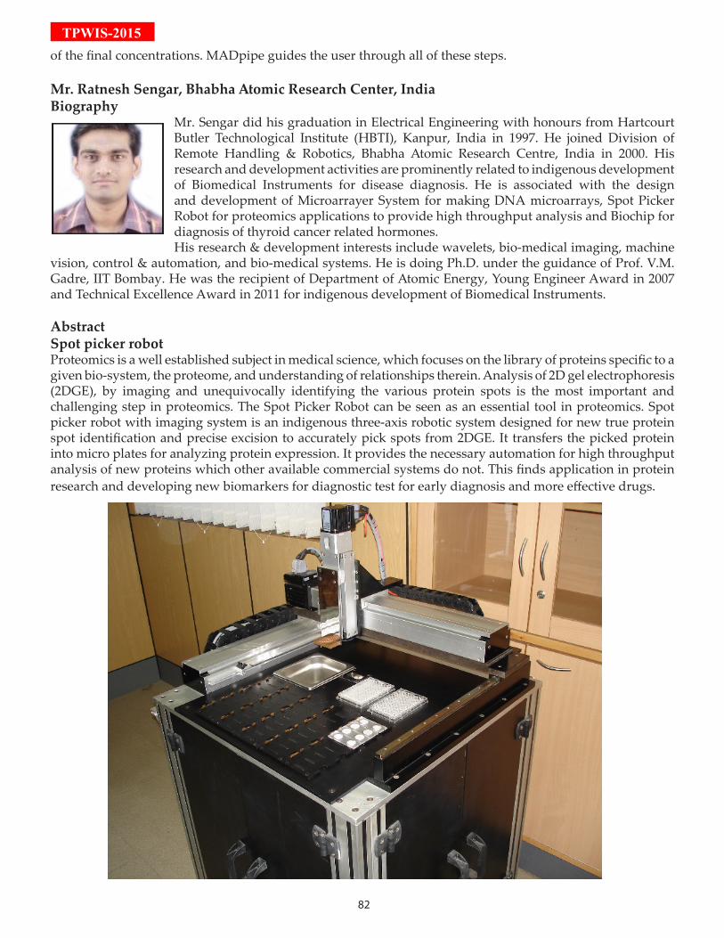
82
TPWIS-2015of the final concentrations. MADpipe guides the user through all of these steps.
Mr. Ratnesh Sengar, Bhabha Atomic Research Center, IndiaBiography
Mr. Sengar did his graduation in Electrical Engineering with honours from Hartcourt Butler Technological Institute (HBTI), Kanpur, India in 1997. He joined Division of Remote Handling & Robotics, Bhabha Atomic Research Centre, India in 2000. His research and development activities are prominently related to indigenous development of Biomedical Instruments for disease diagnosis. He is associated with the design and development of Microarrayer System for making DNA microarrays, Spot Picker Robot for proteomics applications to provide high throughput analysis and Biochip for diagnosis of thyroid cancer related hormones.His research & development interests include wavelets, bio-medical imaging, machine
vision, control & automation, and bio-medical systems. He is doing Ph.D. under the guidance of Prof. V.M. Gadre, IIT Bombay. He was the recipient of Department of Atomic Energy, Young Engineer Award in 2007 and Technical Excellence Award in 2011 for indigenous development of Biomedical Instruments.
Abstract Spot picker robotProteomics is a well established subject in medical science, which focuses on the library of proteins specific to a given bio-system, the proteome, and understanding of relationships therein. Analysis of 2D gel electrophoresis (2DGE), by imaging and unequivocally identifying the various protein spots is the most important and challenging step in proteomics. The Spot Picker Robot can be seen as an essential tool in proteomics. Spot picker robot with imaging system is an indigenous three-axis robotic system designed for new true protein spot identification and precise excision to accurately pick spots from 2DGE. It transfers the picked protein into micro plates for analyzing protein expression. It provides the necessary automation for high throughput analysis of new proteins which other available commercial systems do not. This finds application in protein research and developing new biomarkers for diagnostic test for early diagnosis and more effective drugs.

83
TPW
IS-2015

84
TPWIS-2015
Student Competitions
To encourage students/ participants we have announced few competi-tions and awards for the art, essay, best oral and poster presentations
1. ART COMPETITION Topics• Clinicalbiomarkervalidationusingtargetedproteomics• TargetedproteomicsforPTManalysis• Targetedproteomics:Risingtides• Targetedproteomics:Newtools&software• TargetedAssaysforClinicalApplications
Results: To be announced in Valedictory function during symposium
2. ESSAY COMPETITION Topics• ClinicalBiomarkerValidationusingTargetedProteomics• PrecisionProteomics:Progressmadeand Challengesahead
Results: To be announced in Valedictory function during symposium
3. ORAL PRESENTATION
Results: To be announced in Valedictory function during symposium
4. POSTER PRESENTATION
Results: To be announced in Valedictory function during symposium
All the best to the participants!TPWIS-2015Organising Team
85
TPW
IS-2015
SHOrT TALK AbSTrACTS
Mr. Nitin KishoreIndian Institute of Science Education and Re-search Mohali
BiographyMr. Kishore obtained his B.Sc in Zoology from “TMBU, Bhagalpur. He obtained his M.Sc. in Biochemistry from AAIDU, Allahabad. He is currently pursuing his Ph.D. from Indian Institute of Science Education and
Research Mohali, India under the supervision of Prof. Purnananda Guptasarma and is in his final year. He has experience in “Protein Biochemistry, ” which includes techniques like mass spectrometry, various spectroscopy (absorbance, fluorescence, circular dichroism, infrared), dynamic light scattring (multi-angle), chromatography, electrophoresis and recombinant DNA technology to address various questions in the field of protein folding, stability, aggregation, structure and function. He is using mass spectrometry as a tool for identification through intact mass and peptide mass fingerprinting and analysis, and searching of protein s through database by using raw mass spectrometry data. He is experienced in operating Waters synaptG2S Q-TOF mass spectrometer coupled with LC -ESI /nano-LC-nano-ESI/MALDI. Currently he is using mass spectrometer to know the folding/unfolding of protein through charge state by ESI-MS. He is a life member of Indian society of mass spectrometry (ISMAS) and Proteomics Society of India (PS(I)).
AbstractMass spectrometric N-terminal sequencing of peptides using a bacterial aminopeptidaseAminopeptidases are exo-peptidases that catalyze the hydrolysis of peptide bonds joining the N-terminal amino acid of any peptide to the next amino acid in the sequence. Non- processive aminopeptidases are specific for certain residues and cannot progressively hydrolyze successively placed amino acid residues, whereas processive aminopeptidases tend to be non-specific and can ‘progressively’ cleave up to 30-35 amino acid residues in any peptide. Theoretically, therefore, beginning with a homogenous population of molecules of a defined mass constituting a single peptide of defined amino acid sequence, any processive
aminopeptidase can be used to create a population of peptides of different lengths differing by one amino acid mass, so that the entire population can be studied in a single mass spectrum, to determine the peptide’s amino acid sequence. However, in practice, it is difficult to control the reaction from progressing rapidly and also some peptides have blocked N-termini which prevent facile proteolysis. Sometimes, structure present in the peptide can also interfere. In this study, we describe the use of a non-specific, processive, deblocking Bacillus subtillis-derived aminopeptidase (BsuAP) in a reaction conducted at 70 degrees Centigrade for N-terminus sequencing.Glu fibrino peptide (GFP B) from Waters, USA, was used as substrate. GFP B is used as a standard (lock mass) in mass spectrometry and has a mass of 1570.67 Da. BsuAP was a recombinant protein expressed in, and purified from, E. coli in our own lab. The reaction mixture of enzyme and substrate was prepared by mixing 1 nanomole of the aminopeptidase to 1.6 nanomoles of the substrate (GFP B) in water. The reaction mixture and control reaction (lacking aminopeptidase) were incubated at 700c for 1 hour. After incubation, the reaction-mixture and control reaction were spotted on the MALDI plate. GFP B was used as a lock mass control. For spotting, 1µl of sample was spotted with 1µl CHCA matrix. Samples were analyzed on the Q-TOF Synapt G2S HDMS system (from Waters, USA), with laser desorption achieved by the instrument’s 355nm laser. The spectra analyzed and presented are lock mass-corrected spectra. The GFP B peptide is 14 residues-long. A sufficiently slow reaction would produce a mass spectrum containing the mass corresponding to the complete peptide, and all smaller masses progressively truncating the peptide from its N-terminus, assuming that the reaction does not proceed to completion on all substrate molecules. We found all expected masses down to 480.25 Da in the reaction spectrum. This data suggests that under the conditions used, BsuAP is non-specific and quite processive, progressively digesting 11 amino acid residues in the peptide substrate, beginning with the N-terminal glycine. Masses are seen to have different relative intensities which could be indicative of differences in specificity. It may be noted that the differences in masses of successive peaks (moving leftwards from the control GFP B mass) correspond to masses of the amino acids removed, after addition of the mass of one hydrogen.
86
TPWIS-2015Ms. Kristina PoljakETH Zurich, Switzerland
BiographyMs. Poljak has obtained two M.Sc. degrees, one in Molecular Biology from University of Zagreb, Croatia and the second one in Industrial Biotechnology from University of Orleans, France. She is currently doing her Ph.D. at ETH Zurich,
Switzerland working on yeast oligo-saccharytranferase complex with Dr. Markus Aebi group. Her current research focus is on developing quantitative method to measure N-linked glycosylation site occupancy using targeted proteomics approach.
AbstractParallel Reaction Monitoring-based quantification of site specific N-glycosylation occupancy in yeast Saccharomyces CerevisiaeE.Asparagine (N)-linked protein glycosylation is a complex protein modification. Unlike phosporylation or other modifications, site-specific detection of the modification by antibody-mediated immunoblotting is often challenging for N-glycosylation due to the poor immunogenicity and glycan diversity. However, characterization of N-glycan sites plays a critical role in clinical diagnostics, quality control of therapeutic glycoprotein production and N-glycosylation pathways deduction. Thus, a sensitive, reproducible and quantifiable detection method is required. We are developing combined selected reaction monitoring (SRM) and parallel reaction monitoring (PRM) assays on targeted mass spectroscopic instruments for quantification of 128 glycoproteins in yeast Saccharomyces cerevisiae.
Mr. Saicharan GhantasalaIndian Institute of Technology Bombay, India
BiographyMr. Saicharan completed his Master’s degree (Biotechnology) in the year 2010 from GITAM University, Visakhapatnam. He started his research career under Dr. Girish Maru at the Advanced Centre for Treatment, Research
and Education in Cancer and worked in the area of
Tobacco carcinogenesis. Following this year long stint, he moved to the Proteomics lab at IIT Bombay and has been a part of the Glioma proteomics group eversince. His short research career so far has helped him appreciate the complexity involved in different tumors and the need to better apply the existing proteomics and genomics technologies to solve the challenges that lie ahead in creating a cancer free world.
AbstractMRM based identification and relative quantitation of Vimentin in different grades of meningiomasMeningiomas are tumors originating from meningeal layers of brain and spinal cord, comprising of nearly 30% of the primary CNS tumors. A comparative tissue proteome analysis of low and high grade meningioma using iTRAQ-based quantitative approach coupled to Q-Exactive MS and ESI-quadrupole-TOF revealed 2367 differentially expressed proteins (1%FDR, ≥2 peptides and ≥1.5 fold change in at least one grade). We identified vimentin as one of the interesting targets to be differentially regulated in grade I and II of meningioma with a fold change of 3.28 and 1.95, respectively when compared to controls. Vimentin is a 53kDa intermediate filament protein found to be overexpressed during malignancy and is a known marker for epithelial-mesenchymal transition (EMT) in cases of breast, CNS, gastrointestinal, melanoma and several other malignancies as well. Elucidation of its exact role in EMT and cytoskeletal remodelling still remains an active area of research. Enhanced immunoreactivity of vimentin and cross-reactivity of antibody posed challenge in actual quantification of vimentin using western blots. Therefore, MRM based quantitation emerged out as an alternative approach for comparative analysis of vimentin in different grades of meningioma.LC chromatographic conditions were optimized to achieve separation between different marker peptides of vimentin, which enabled staggering of MRM transitions. Collision energy optimization was carried out for these transitions. Staggered MRM along with optimized collision energies resulted in enhanced sensitivity. Based on the optimized analytical conditions, we report 4 different quantifiable peptides with a minimum of 2 transitions each, which can be used as signature transitions for relative quantitation of vimentin.

87
TPW
IS-2015Ms. Snigdha DhaliNational Centre for Cell Science, India
BiographyMs. Dhali has completed her post graduation from Sathyabama University, Chennai. For the last 7 years, she is working as an operator of Mass Spectrometry instruments in Proteomics Facility at National Centre for Cell Science. She is experienced
in handling triple Quadrapole and MALDI-TOF as well as Liquid chromatography instruments like HPLC, MicroLC and NanoLC.
AbstractValidation of subtype specific potential serum protein biomarkers in Breast Cancer using targeted proteomics Among all other cancers, Breast cancer (BC) has the highest incidence rate in women around the world. Survival rates of patients are high when BC is detected at a very early stage. Being a clinically and genetically heterogeneous disease, breast cancer tumor is subdivided into 4 subtypes viz. luminalA, luminalB, HER2-enriched, basal-like subtype. Hence, there is an urgent need to come up with potential biomarkers which can support the clinicians in early diagnosis as well as to develop targeted therapeutics towards the breast cancer subtypes. In the present study we aim to investigate subtype specific serum potential protein markers in BC.In this work, we used mass spectrometry based iTRAQ, SWATH and complementary 2D-DIGE to identify the differentially expressed proteins in four subtypes of BC against those present in healthy individuals. Identified proteins are validated using in-house developed LC-MS-MRM based assays using 4000 Q-TRAP mass spectrometer coupled to micro LC. Transitions were developed insilico on SKYLINE 3.1 software and selection was based on cross reference from SRM Atlas, MRM Based public libraries and literature.The differentially expressed proteins identified using iTRAQ, SWATH, and 2D-DIGE were interpreted to identify sub-type specific serum protein biomarkers. Interestingly, we were able to identify a panel of differentially expressed subtype specific protein markers which can not only be used to differentiate subtypes but also can give a deep insight towards disease progression in different subtypes. We developed a MRM assay method for these proteins
and validated to propose subtype specific serum protein markers for BC. This is an easy, comparatively less expensive and robust method when compared with other traditional techniques like western blot and ELISA for validation of differentially expressed proteins identified in the discovery phase. These validated subtype specific protein signatures are not only helpful as theranostic biomarkers but also enhance our understanding of the different molecular changes in four subtypes at proteomic level.
Ms. Sonali Vishwa MohanInstitute of Bioinformatics, India
BiographyMs. Mohan obtained her Master’s degree in Chemistry from Indian Institute of Technology (IIT), Delhi. She has worked as a Research Associate at Shantani Proteome Analytics Pvt. Ltd.,
where she was involved in developing Sub Cellular Location Specific Peptide Technology and Unique Polymer Technology for target identification and deconvolution. Currently, she is a Ph.D. student at the Institute of Bioinformatics, Bangalore where she is working on metabolomics and chemical proteomics.
AbstractProtein quantitation using Data Independent Acquisition (DIA) on orbitrap fusion platformData Independent Acquisition (DIA) is a relatively new mass spectrometry based approach for protein identification and quantification. DIA records fragment ions of all precursor ions without subjecting them to abundance based selection. Because of this, it offers the possibility of most comprehensive sampling of peptides in a sample compared to DDA. It also enables identification and fragment ions based quantitation of peptides across sample in a high-throughput manner. Despite gaining some momentum with the advent of the SWATH approach, it is majorly limited to TripleTOF. Here, we adapt an existing method for Orbitrap and employ Skyline & Umpire programs to assess the quantitation performance of DIA on Oribtrap-Fusion Mass Spectrometer. In addition to it, we have also evaluated various mass
88
TPWIS-2015spectrometry parameters and their effect on DIA. Protein digest obtained from human cell line was analyzed in triplicates with different sample amounts from 250ng–2g. DIA acquisition was obtained using MS scans analyzing 400-1000m/z with a resolution of 60k (400m/z). 30 MS/MS scans were acquired with 20m/z isolation window, loop count of 10 and orbitrap resolution of 30k (400m/z). For two fold change, we observed relatively better quantitative accuracy for lower protein amount compared to higher. For higher fold changes, we observed good quantitative accuracy irrespective of protein amount. Although DIA offers several advantages over DDA, informatics tools to deconvolute the data for identification and quantitation need significant improvement.
Mr. Sumit Kumar SinghIndian Institute of Technology Delhi, India
BiographyMr. Singh completed his undergraduate and postgraduate studies (B.Tech and M.Tech degree) from NIT Raipur and NIT Trichy, respectively. Subsequently, he joined Prof. Anurag S. Rathore’s
Bioseparations and Bioprocessing research group at IIT Delhi to pursue his PhD. His doctoral dissertation principally encompasses the broad domain of therapeutic protein characterization. His current endeavours are focused on developing platform approaches by transcriptomic and proteomic profiling tools like LC-MS, CE-MS, NGS and advanced data analytical packages like chemometrics for safety and efficacy assessment of biotherapeutics. The research embodies a two-fold approach: (a) identification of biomarkers that are specific to various product and process related impurities in biotherapeutics; (b) utilization of the chosen biomarkers to develop surrogate testing methods for the existing ADCC/PK/PD assays.
AbstractHost cell protein analysis by HPLC/CE-ESI-TOF-MSHost cell proteins (HCP) analysis has recently emerged as an important analytical requirement in biotherapeutic development workflows. This is primarily due to the perceived impact that HCP can have on a product’s safety and efficacy in the clinic. In addition, since a sub-population of the HCP can be physicochemically near-identical to
the therapeutic product, their accurate quantitation is non-trivial. Typically, a number of orthogonal analytical approaches including immunospecific and non-specific tools are used together for HCP characterization. While immunospecific methods are simple to use, especially when the targeted HCP are present in low levels, nonspecific methods provide a comprehensive assessment of even those HCP that are otherwise difficult to detect by immunospecific methods. The non-specific methods have been evolving over time with each evolution adding a new dimension to the information derived from the previous techniques. Examples to this end include evolution from CEX-RP-MS analysis to RP-RP-MS method of analysis with the latter offering higher peak capacities and selectivity than the former and thus facilitating identification of an increased number of HCP. Despite their analytical advantages, the new method takes considerable time (~10 hours) for analysis. This hampers decision making. In this work, we will report use of unique LC/CE-MS method for HCP characterization. The proposed method, in addition to identifying comparable number of HCP, also reduces the time of analysis considerably. The proposed approach has been used to characterize HCP for three different mAbs produced at different manufacturing facility and also across different downstream process steps.
Mr. Vivek SrinivasIndian Institute of Technology Bombay, India
BiographyMr. Vivek finished his Master degree in Biochemistry from CMRIMS college, Bangalore University. Since 2011, he is working in Molecular parasitology lab under the supervison of Dr. Swati Patankar and Dr. Sanjeeva
Srivastava. His work in the lab involves use of computational and omics approaches to understand mechanism and regulation of protein translation in Plasmodium falciparum; a human malaria parasite. Till now, he has developed an algorithm to predict translatable open reading frames (ORFs). Using this algorithm he has been able to gain insights into the translatable small ORFs and upstream ORFs and also to a smaller extent understand the mechanism of the translation. Currently, he is using proteomics to check some of the hypothesis developed. He is also interested in studying evolution and spread of the
89
TPW
IS-2015parasite infection and he has done a study to show origins of virulence genes in P. falciparum.
Abstract:Proteomics approach to study the effects of uORFs on the rate of translation in Plasmodium falciparumPlasmodium falciparum, a human malaria parasite completes its life cycle in two different hosts and have many distinguishable stages trough out its life cycle. Further, different gene expression has been attributed to me responsible for this feature. However, transcriptional alone does not account for the tight gene regulation observed; due to the paucity of malarial transcriptional regulators identified. This in additional to the fact that there is a lag in mRNA and protein production suggesting the presence of genome wide post transcriptional or translational gene regulation.Moreover, the ribosome profiling shows the 5` UTR have the ability to engage ribosomes; therefore, can hinder the ribosomal complex to reach the downstream CDS and thus reducing the overall rate of translation. Moreover, due to AT rich intergenic regions, the 5` UTR regions also has large frequency of uORFs and studies in mammals have shown that the CDS having higher number of uORFs have relatively low rate of translation ([Protein]/[mRNA]) . This generates three questions: 1) does the parasite uORFs cause reduction in rate of translation? 2) What are the factors (Number of uORFs, translatability of uORFs etc) associated with it? And 3) if uORFs does not down regulate the rate of translation; how does the ribosome skip all the uORFs to translate main CDS? However, detailed understanding of the P. falciparum translation mechanism is needed to answer these questions. In this study we have optimised the methods for measure of absolute protein abundance using label free mass spectrometry approach. Measure absolute protein abundance in P. falciparum and have used this data in conjecture with the available mRNA data to study the effect of uORFs on the rate of translation.
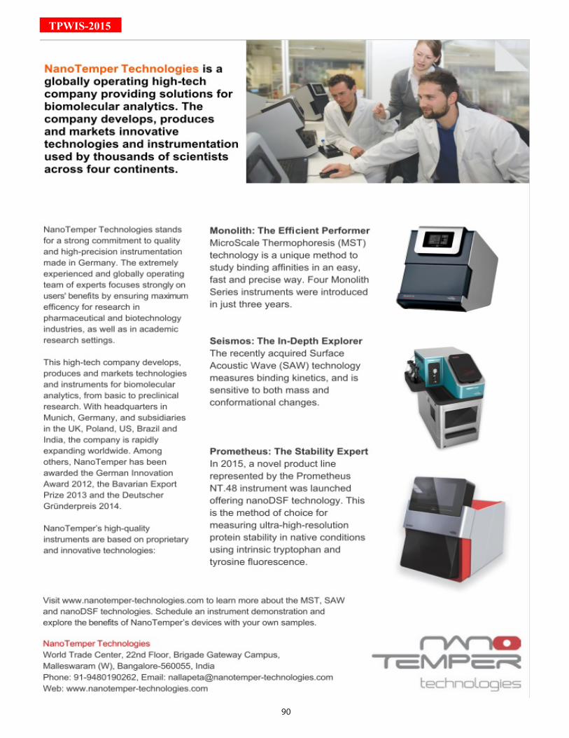
90
TPWIS-2015
91
TPW
IS-2015
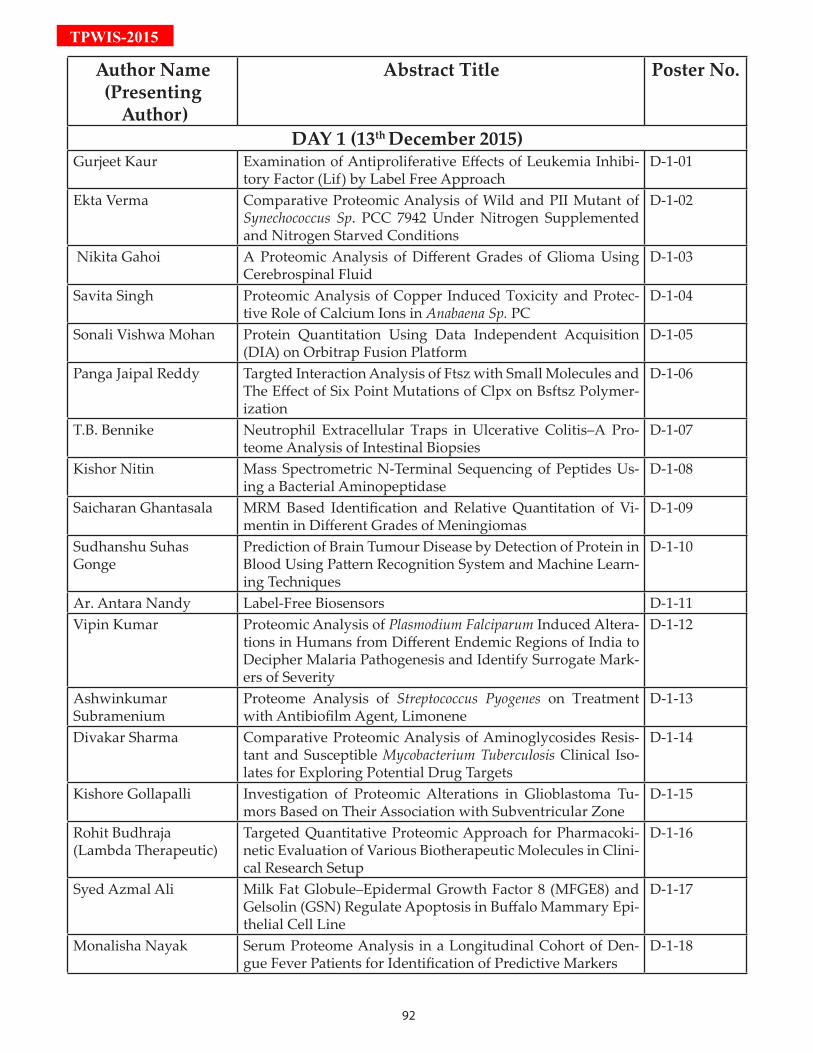
92
TPWIS-2015
Author Name(Presenting
Author)
Abstract Title Poster No.
DAY 1 (13th December 2015)Gurjeet Kaur Examination of Antiproliferative Effects of Leukemia Inhibi-
tory Factor (Lif) by Label Free ApproachD-1-01
Ekta Verma Comparative Proteomic Analysis of Wild and PII Mutant of Synechococcus Sp. PCC 7942 Under Nitrogen Supplemented and Nitrogen Starved Conditions
D-1-02
Nikita Gahoi A Proteomic Analysis of Different Grades of Glioma Using Cerebrospinal Fluid
D-1-03
Savita Singh Proteomic Analysis of Copper Induced Toxicity and Protec-tive Role of Calcium Ions in Anabaena Sp. PC
D-1-04
Sonali Vishwa Mohan Protein Quantitation Using Data Independent Acquisition (DIA) on Orbitrap Fusion Platform
D-1-05
Panga Jaipal Reddy Targted Interaction Analysis of Ftsz with Small Molecules and The Effect of Six Point Mutations of Clpx on Bsftsz Polymer-ization
D-1-06
T.B. Bennike Neutrophil Extracellular Traps in Ulcerative Colitis–A Pro-teome Analysis of Intestinal Biopsies
D-1-07
Kishor Nitin Mass Spectrometric N-Terminal Sequencing of Peptides Us-ing a Bacterial Aminopeptidase
D-1-08
Saicharan Ghantasala MRM Based Identification and Relative Quantitation of Vi-mentin in Different Grades of Meningiomas
D-1-09
Sudhanshu Suhas Gonge
Prediction of Brain Tumour Disease by Detection of Protein in Blood Using Pattern Recognition System and Machine Learn-ing Techniques
D-1-10
Ar. Antara Nandy Label-Free Biosensors D-1-11Vipin Kumar Proteomic Analysis of Plasmodium Falciparum Induced Altera-
tions in Humans from Different Endemic Regions of India to Decipher Malaria Pathogenesis and Identify Surrogate Mark-ers of Severity
D-1-12
Ashwinkumar Subramenium
Proteome Analysis of Streptococcus Pyogenes on Treatment with Antibiofilm Agent, Limonene
D-1-13
Divakar Sharma Comparative Proteomic Analysis of Aminoglycosides Resis-tant and Susceptible Mycobacterium Tuberculosis Clinical Iso-lates for Exploring Potential Drug Targets
D-1-14
Kishore Gollapalli Investigation of Proteomic Alterations in Glioblastoma Tu-mors Based on Their Association with Subventricular Zone
D-1-15
Rohit Budhraja (Lambda Therapeutic)
Targeted Quantitative Proteomic Approach for Pharmacoki-netic Evaluation of Various Biotherapeutic Molecules in Clini-cal Research Setup
D-1-16
Syed Azmal Ali Milk Fat Globule–Epidermal Growth Factor 8 (MFGE8) and Gelsolin (GSN) Regulate Apoptosis in Buffalo Mammary Epi-thelial Cell Line
D-1-17
Monalisha Nayak Serum Proteome Analysis in a Longitudinal Cohort of Den-gue Fever Patients for Identification of Predictive Markers
D-1-18
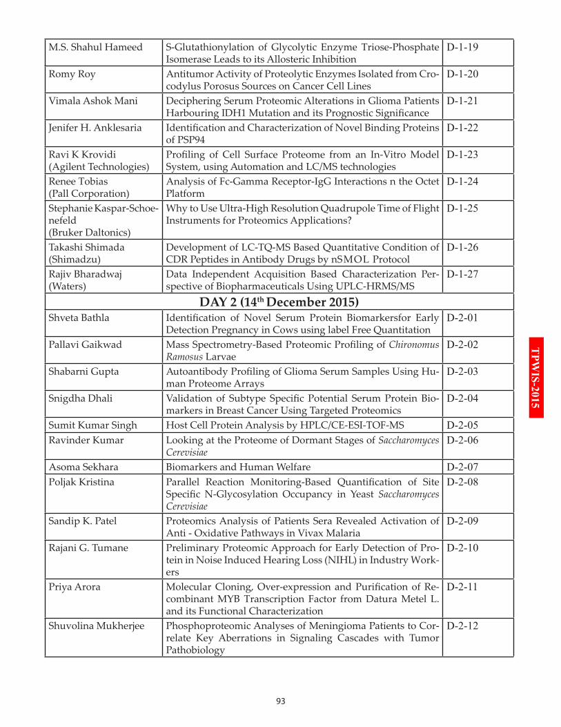
93
TPW
IS-2015
M.S. Shahul Hameed S-Glutathionylation of Glycolytic Enzyme Triose-Phosphate Isomerase Leads to its Allosteric Inhibition
D-1-19
Romy Roy Antitumor Activity of Proteolytic Enzymes Isolated from Cro-codylus Porosus Sources on Cancer Cell Lines
D-1-20
Vimala Ashok Mani Deciphering Serum Proteomic Alterations in Glioma Patients Harbouring IDH1 Mutation and its Prognostic Significance
D-1-21
Jenifer H. Anklesaria Identification and Characterization of Novel Binding Proteins of PSP94
D-1-22
Ravi K Krovidi (Agilent Technologies)
Profiling of Cell Surface Proteome from an In-Vitro Model System, using Automation and LC/MS technologies
D-1-23
Renee Tobias (Pall Corporation)
Analysis of Fc-Gamma Receptor-IgG Interactions n the Octet Platform
D-1-24
Stephanie Kaspar-Schoe-nefeld (Bruker Daltonics)
Why to Use Ultra-High Resolution Quadrupole Time of Flight Instruments for Proteomics Applications?
D-1-25
Takashi Shimada (Shimadzu)
Development of LC-TQ-MS Based Quantitative Condition of CDR Peptides in Antibody Drugs by nSMOL Protocol
D-1-26
Rajiv Bharadwaj (Waters)
Data Independent Acquisition Based Characterization Per-spective of Biopharmaceuticals Using UPLC-HRMS/MS
D-1-27
DAY 2 (14th December 2015)Shveta Bathla Identification of Novel Serum Protein Biomarkersfor Early
Detection Pregnancy in Cows using label Free QuantitationD-2-01
Pallavi Gaikwad Mass Spectrometry-Based Proteomic Profiling of Chironomus Ramosus Larvae
D-2-02
Shabarni Gupta Autoantibody Profiling of Glioma Serum Samples Using Hu-man Proteome Arrays
D-2-03
Snigdha Dhali Validation of Subtype Specific Potential Serum Protein Bio-markers in Breast Cancer Using Targeted Proteomics
D-2-04
Sumit Kumar Singh Host Cell Protein Analysis by HPLC/CE-ESI-TOF-MS D-2-05Ravinder Kumar Looking at the Proteome of Dormant Stages of Saccharomyces
CerevisiaeD-2-06
Asoma Sekhara Biomarkers and Human Welfare D-2-07Poljak Kristina Parallel Reaction Monitoring-Based Quantification of Site
Specific N-Glycosylation Occupancy in Yeast Saccharomyces Cerevisiae
D-2-08
Sandip K. Patel Proteomics Analysis of Patients Sera Revealed Activation of Anti - Oxidative Pathways in Vivax Malaria
D-2-09
Rajani G. Tumane Preliminary Proteomic Approach for Early Detection of Pro-tein in Noise Induced Hearing Loss (NIHL) in Industry Work-ers
D-2-10
Priya Arora Molecular Cloning, Over-expression and Purification of Re-combinant MYB Transcription Factor from Datura Metel L. and its Functional Characterization
D-2-11
Shuvolina Mukherjee Phosphoproteomic Analyses of Meningioma Patients to Cor-relate Key Aberrations in Signaling Cascades with Tumor Pathobiology
D-2-12
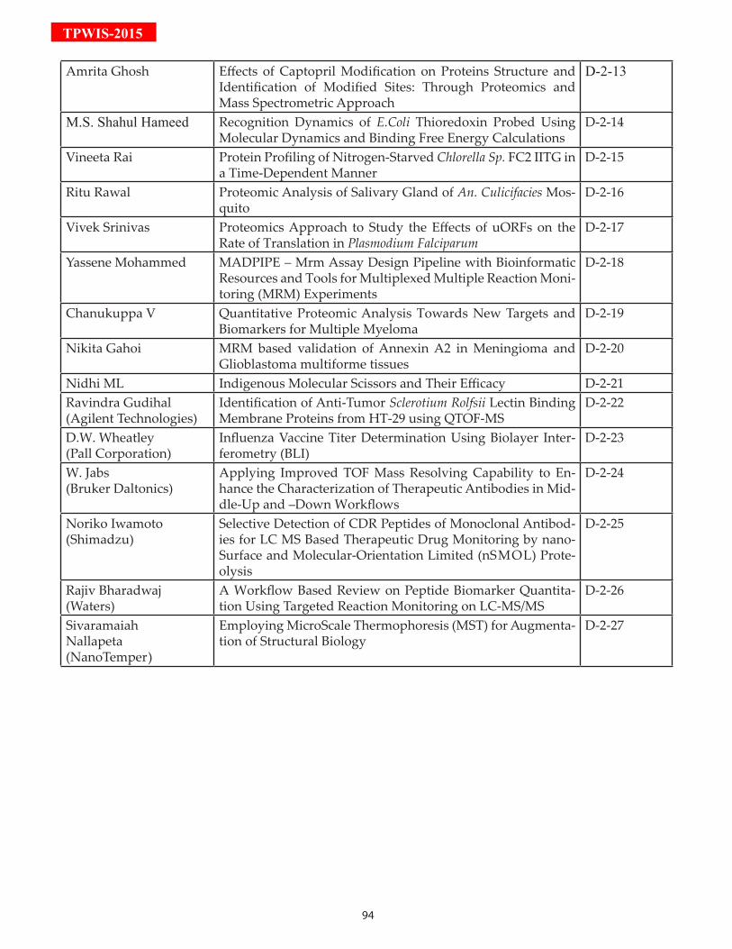
94
TPWIS-2015
Amrita Ghosh Effects of Captopril Modification on Proteins Structure and Identification of Modified Sites: Through Proteomics and Mass Spectrometric Approach
D-2-13
M.S. Shahul Hameed Recognition Dynamics of E.Coli Thioredoxin Probed Using Molecular Dynamics and Binding Free Energy Calculations
D-2-14
Vineeta Rai Protein Profiling of Nitrogen-Starved Chlorella Sp. FC2 IITG in a Time-Dependent Manner
D-2-15
Ritu Rawal Proteomic Analysis of Salivary Gland of An. Culicifacies Mos-quito
D-2-16
Vivek Srinivas Proteomics Approach to Study the Effects of uORFs on the Rate of Translation in Plasmodium Falciparum
D-2-17
Yassene Mohammed MADPIPE – Mrm Assay Design Pipeline with Bioinformatic Resources and Tools for Multiplexed Multiple Reaction Moni-toring (MRM) Experiments
D-2-18
Chanukuppa V Quantitative Proteomic Analysis Towards New Targets and Biomarkers for Multiple Myeloma
D-2-19
Nikita Gahoi MRM based validation of Annexin A2 in Meningioma and Glioblastoma multiforme tissues
D-2-20
Nidhi ML Indigenous Molecular Scissors and Their Efficacy D-2-21Ravindra Gudihal(Agilent Technologies)
Identification of Anti-Tumor Sclerotium Rolfsii Lectin Binding Membrane Proteins from HT-29 using QTOF-MS
D-2-22
D.W. Wheatley(Pall Corporation)
Influenza Vaccine Titer Determination Using Biolayer Inter-ferometry (BLI)
D-2-23
W. Jabs (Bruker Daltonics)
Applying Improved TOF Mass Resolving Capability to En-hance the Characterization of Therapeutic Antibodies in Mid-dle-Up and –Down Workflows
D-2-24
Noriko Iwamoto(Shimadzu)
Selective Detection of CDR Peptides of Monoclonal Antibod-ies for LC MS Based Therapeutic Drug Monitoring by nano-Surface and Molecular-Orientation Limited (nSMOL) Prote-olysis
D-2-25
Rajiv Bharadwaj(Waters)
A Workflow Based Review on Peptide Biomarker Quantita-tion Using Targeted Reaction Monitoring on LC-MS/MS
D-2-26
Sivaramaiah Nallapeta (NanoTemper)
Employing MicroScale Thermophoresis (MST) for Augmenta-tion of Structural Biology
D-2-27
95
TPW
IS-2015D-1-01:Examination of Antiproliferative Effects of Leuke-mia Inhibitory Factor (LIF) Using Label Free Ap-proachKaur G, Kumar S, Ali SA, Pachauri S, Jaswal S, Verma AK, Bathla S, Rawat P, Kumar G, Yadav ML, Kauhsik JK, Malakar D, Mohanty AKNational Dairy Research Institute, Karnal
Leukemia inhibitory factor (LIF) is a pleiotropic gly-coprotein synthesized and secreted in various body tissues. LIF is a molecule that was initially identified through its ability to induce macrophage differentia-tion in the murine M1 myeloid leukemic cell line. We successfully over express the BuLIF protein through transfection of pAcGFP-N1 vector into COS-1 cell line. To examine the antiproliferative effect of LIF in stably SV40 transformed COS1 cell line. Consequently for stably transfected cell line single cell clonal expan-sion and several rounds of selection in the presence of G418 was performed. Highly expressed BuLIF_GFP cells were selected for further studies. The strong ex-pression of BuLIF was observed at 70th passage and cells could grow in the absence of selection pressure without losing GFP signal. Confirmation of genomic integration of BuLIF was performed through PCR amplification and sequencing. The SDS-PAGE and Western blot revealed the identification of transgenic LIF around 65-70 kDa. Mass spectrometry based (Q-TOF), Label Free Quantification (LFQ) approach were used for the functional annotation of antiproliferative effect of LIF. Total of 925 proteins were identified through MaxQuant 1.5.2.8 using NCBI Macaca mu-latta database. LIF and GFP were identified through NCBI Bubalus bubalis and Aequorea victoria data-base respectively. Bioinformatics analysis through STRING and Cytoscape showed the interaction of LIF with LIFR, OSM, OSMR, CLCF1, CSF3, EGK04900 and KCNJ1 proteins. Most of the interacting partners of LIF were involved in apoptosis and anti-proliferative effects. To confirm Apoptotic and anti-proliferative effect of LIF on cancerous kidney cells (SV-40 trans-formed COS-1 cells) Brdu and TUNEL assay was pe-formed. Further validation was performed with the help of qPCR. Till date with our knowledge this is the first study that has been performed on COS-1 cells for apoptotic and anti-proliferative effect of LIF.
D-1-02:Comparative Proteomic Analysis of Wild and PII Mutant of Synechococcus Sp. PCC 7942 Under Ni-trogen Supplemented and Nitrogen Starved Condi-tions
Verma E, Niveshika, Singh S, Mishra AKLaboratory of Microbial Genetics, Department of Botany, Banaras Hindu University, Varanasi-221005 The PII protein is one of the most widely distributed signal transduction protein found in bacteria, archaea and higher plants. PII protein controls the activities of a diverse range of enzymes, transcription factors and membrane transport proteins. This protein is the main nitrogen regulator in cyanobacteria. In this study a fresh water cyanobacterium Synechococcus sp. PCC 7942 and its PII mutant were subjected to nitro-gen starvation and complete proteomic profiling was performed in order to establish pleiotropic responses induced and cellular processes affected by PII protein under nitrogen starvation. Cyanobacteria wild and PII mutant was grown in BG11 medium supplement-ed with 10 mM NaHCO3 and 17 mM NaNO3. For N- deprived condition, NaNO3 supplemented BG11 medium grown exponential stage wild and mutant strain was transferred to –NaNO3 BG11 medium and allowed to grow for 8 days. Protein was isolated and subjected to 2D-PAGE analysis. Further differentially expressed proteins were subjected to MALDI-TOF analysis. By separating soluble extracted proteins on 2-DE gels, more than 300 protein spots were vi-sualized on each colloidal commassiae stained 2-DE gels. Quantitative differences in protein composition were detected by PDQuest software and comparative analysis revealed that several protein spots changed significantly in the PII mutant. Nearly all proteins whose synthesis responded specifically to combine nitrogen deprivation in wild type failed to respond in PII mutant. Some proteins such as alkyl hydroper-oxide reductase, RbcS small subunit of rubisco and chromosome segregation ATPase were found to down expressed in PII mutant. Up-regulated proteins were proteins involved in nitrogen metabolism and fatty acid biosynthesis. Identification of such differentially expressed proteins provides new targets for future studies that will allow assessment of their physiologi-cal roles and significance in cyanobacterial nitrogen regulation.
D-1-03:A Proteomic Analysis of Different Grades of Glio-ma Using Cerebrospinal Fluid Gahoi N1, Malhotra D1, Moiyadi A2, Srivastava S1
1 Department of Biosciences and Bioengineering, Indian Institute of Technology Bombay, Powai, Mumbai-400076, India2 Department of Neurosurgery, Advanced Center for Treatment Research and Education in Cancer, Tata Memo-
96
TPWIS-2015rial Center, Kharghar, Navi Mumbai-410210, IndiaGliomas are the most prevalent form of brain tumors originating from astrocytes and are categorized into four different grades by W.H.O depending upon the aggressiveness of the tumor. Alterations arising in the brain will be reflected in the composition of its proximal fluid i.e., cerebrospinal fluid (CSF) circulat-ing within the central nervous system (CNS). CSF is a reservoir of transported biological substances, toxic excretion and waste of the brain. Therefore, analyz-ing the alterations in the CSF holds immense poten-tial for biomarker discovery. In this study, two com-plementary approaches, 2D-DIGE and iTRAQ were implemented to identify inter-grade proteomic al-terations. Proteins from CSF were extracted using ac-etone precipitation followed by desalting to remove the non-proteinaceous impurities. For 2D-DIGE, la-beled protein samples were separated according to their iso-electric point (pI) and molecular weight. The differentially expressed protein spots were identi-fied using in-gel digestion followed MALDI-TOF/TOF analysis. For iTRAQ analysis buffer exchanged protein samples were subjected to in-solution trypsin digestion followed by labeling of the peptides. The labeled peptides were pre-fractionated based on their iso-electric points, each fraction was then subjected LC-MS/MS. The data was processed using Spectrum-Mill software and the differentially expressed pro-teins were subjected to pathway analysis. Proteins as-sociated with integrin signaling pathway, cytoskeletal regulation, glycolysis, gluconeogenesis, were found to be altered in gliomas. Proteins likevimentin, pro-filin, macrophage-capping protein, selenium-binding protein, protein disulfide-isomerase, peptidyl-prolyl-cis-trans isomerase, etc. were increased with increase intumor grades, while vitronectin, Apolipoprotein C-III, synapsin-1, Superoxide dismutase (Cu-Zn) etc. showed a negative correlation with increase in grade of the tumor. CSF proteomic analysis will provide a mechanistic insight into glioma pathogenesis. A panel of differentially expressed proteinsmay acts as potential biomarker candidates for early diagnosis of gliomas. However, there is a need for further valida-tion of proteins on a larger cohort of glioma patients.
D-1-04:Proteomic Analysis of Copper Induced Toxicity and Protective Role of Calcium Ions in Anabaena Sp. PCSingh S, Verma E, Niveshika, Mishra AKLaboratory of Microbial Genetics, Department of Botany, Banaras Hindu University, Varanasi-221005
Copper (Cu) one of the heavy metals, is also an es-sential microelement involved in numerous physi-ological processes in cyanobacteria. However, when absorbed in excess, it is toxic inducing deleterious effects at morphological, biochemical, physiological and ultrastructural levels. The present investigation deals with detoxifying role of calcium ions towards Copper based inhibition of physiological processes at inhibitory concentration (5 µM CuSO4) in Anabaena sp. PCC 7120. Two dimensional gel electrophoresis (2-DE) followed by MALDI- TOF MSMS of selected protein spots suggest alteration in the proteome. PD-Quest analysis detected protein spots numbering to 111 protein spots in Control (0 µM CuSO4), 117 pro-tein spots (5 µM CuSO4) and 201 protein spots (5 µM CuSO4 + 10 mM CaCl2). Approximately 40 protein spots were differentially expressed under calcium supplemented condition. MALDI TOF MSMS identi-fied most of the expressed protein spots being related to photosynthesis, stress chaperones, TCA cycle and reductases category. The obtained results may be par-ticularly helpful in understanding of acclimation re-sponses of the cyanobacterium to heavy metal stress and how Calcium could play a detoxifying role.
D-1-05:Protein Quantitation Using Data Independent Ac-quisition (DIA) on Orbitrap Fusion PlatformSonali Vishwa Mohan1, Kiran Kumar M.1,2, Manish Kumar1,3, Krishna Patel1, Harsha Gowda1
1 Institute of Bioinformatics, International Tech Park, Bangalore, 560066, India.2 School of Biotechnology, Amrita Vishwa Vidyapeetham, Kollam, 690525 India.3 Manipal University, Madhav Nagar, Manipal, 576104, India.
Data Independent Acquisition (DIA) is a relatively new mass spectrometry based approach for protein identification and quantification. DIA records frag-ment ions of all precursor ions without subjecting them to abundance based selection. Because of this, it offers the possibility of most comprehensive sampling of peptides in a sample compared to DDA. It also en-ables identification and fragment ions based quanti-tation of peptides across sample in a high-throughput manner. Despite gaining some momentum with the advent of the SWATH approach, it is majorly limited to TripleTOF. Here, we adapt an existing method for Orbitrap and employ Skyline & Umpire programs to assess the quantitation performance of DIA on Orib-trap-Fusion Mass Spectrometer. In addition to it, we have also evaluated various mass spectrometry pa-
97
TPW
IS-2015rameters and their effect on DIA. Protein digest ob-tained from human cell line was analyzed in tripli-cates with different sample amounts from 250ng–2µg. DIA acquisition was obtained using MS scans analyz-ing 400-1000m/z with a resolution of 60k (400m/z). 30 MS/MS scans were acquired with 20m/z isolation window, loop count of 10 and orbitrap resolution of 30k (400m/z). For two fold change, we observed rela-tively better quantitative accuracy for lower protein amount compared to higher. For higher fold changes, we observed good quantitative accuracy irrespective of protein amount. Although DIA offers several ad-vantages over DDA, informatics tools to deconvolute the data for identification and quantitation need sig-nificant improvement.
D-1-06:Targted Interaction Analysis of FtsZ with Small Molecules and the Effect of Six Point Mutations of ClpX on BsFtsZ PolymerizationReddy PJ, Khanzode M, Shah VG, Panda D, Srivas-tava SDepartment of Biosciences and Bioengineering, Indian In-stitute of Technology Bombay, Powai, Mumbai-400076, India
Drug resistance is the major concern for healthcare professionals and looking deep into the existing and new chemical libraries to explore the new drugs nov-el mechanism of action. Bacterial cell division process is highly conserved mechanism in prokaryotes and FtsZ is the key player involved in cytokinesis process along with many accessory proteins. Many natural compounds have showed potential antimicrobial activity by targeting the FtsZ polymerization. The protein expression profile of FtsZ has not changed much under curcumin, totarol and plumbagin treat-ment whereas mRNA expression analysis of FtsZ showed significant repression. In the present study, we have performed real-time interaction analysis of both BsFtsZ and EcFtsZ with curcumin, totarol and plumbagin for binding and kinetic analysis. Further, molecular docking has provided the putative bind-ing sites for the small molecules in FtsZ. Besides, we also tested the effect of wild type and six point mu-tants ClpX on BsFtsZ polymerization. Interestingly, mutated ClpX has induced BsFtsZ polymerization in concentration dependent manner however wild type hasn’t changed much. The real-time SPR analysis also showed that both wild type and mutated ClpX binds to BsFtsZ strongly in a concentration dependent man-ner. In summary, our study showed that the men-tioned small molecules did not affect the FtsZ protein
expression levels rather bind with FtsZ at molecular level and regulate the FtsZ polymerization dynamics. This study also provided the KD for both BsFtsZ and EcFtsZ with the small molecules using SPR analysis. In addition, our results demonstrated that both wild type and mutant (six mutations) ClpX leads to inhi-bition of FtsZ polymerization with equal affinity. In depth analysis is necessary for conclusive prospective on role of mutated ClpX on bacterial cell division.
D-1-07:Neutrophil Extracellular Traps in Ulcerative Coli-tis–A Proteome Analysis of Intestinal BiopsiesBennike TB1, Carlsen TG1, Ellingsen T2, Bonderup OK3, Glerup H3, Boegsted M1, Christiansen G4, Birkelund S1, Stensballe A1, Andersen V5,6
1 Department of Health Science and Technology, Aalborg University, Denmark: 2 Department of Rheumatology, Odense University Hos-pital, Denmark 3 Department of Clinical Medicine, Silkeborg Hospital, Denmark 4 Department of Clinical Medicine, Aalborg University Hospital, Denmark 5 Institute of Regional Health Research, University of Southern Denmark 6 Organ Center, Hospital of Southern Jutland, Aabenraa, Denmark
Inflammatory bowel diseases, including Ulcerative colitis affects 2.5–3 million people in Europe. The life-long conditions significantly reduce the quality of life of the patients, and compose a significant economic burden for society. The etiology of these diseases remains incompletely explained. We, therefore per-formed the most thorough proteome-based study of ulcerative colitis colon tissue, based on number of an-alyzed proteins, and have for the first time observed neutrophil extracellular traps in ulcerative colitis, de-tected by proteomics and verified by microscopy.We performed a comparative proteome analysis of non-inflamed mucosal colon biopsies from 10 patients with ulcerative colitis and 10 controls.The proteome of the biopsies were characterized by high through-put label-free quantitative proteomics, and the biopsy histology was analyzed by microscopy.We quantified 5,711 different proteins in the colon biopsies. Forty-six proteins had a statistically sig-nificant difference in abundance between the ulcer-ative colitis colon tissue and controls. Eleven of the proteins with increased abundances in the ulcerative colitis biopsies were associated with neutrophils and neutrophil extracellular traps. The abundance of cal-
98
TPWIS-2015protectin and lactotransferrin in the tissue correlated with the degree of tissue inflammation as determined by histology. The findings were validated by micros-copy where an increased abundance of neutrophils within the mucosal tissue was found. Additionally, we confirmed the presence of neutrophil extracellu-lar traps by extracellular DNA in the ulcerative colitis colon tissue.We found an increased abundance of several proteins hitherto not associated with innate immunity and neutrophils in non-inflamedmucosal colon biopsies from ulcerative colitis patients. The increased abun-dance of these antimicrobial compounds points to the stimulation of the innate immune system in the eti-ology of ulcerative colitis. Our findings demonstrate that even though remission has been achieved on the surface of the ulcerative colitis colon tissue, a chronic condition is still present within the morphologically normal tissue.
D-1-08:Mass Spectrometric N-Terminal Sequencing of Pep-tides Using a Bacterial AminopeptidaseKishor N, Guptasarma PDepartment of Biological Sciences, Indian Institute of Sci-ence Education and Research (IISER) Mohali
Aminopeptidases are exo-peptidases that catalyze the hydrolysis of peptide bonds joining the N-termi-nal amino acid of any peptide to the next amino acid in the sequence. Non- processive aminopeptidases are specific for certain residues and cannot progres-sively hydrolyze successively placed amino acid resi-dues, whereas processive aminopeptidases tend to be non-specific and can ‘progressively’ cleave up to 30-35 amino acid residues in any peptide. Theoretically, therefore, beginning with a homogenous population of molecules of a defined mass constituting a single peptide of defined amino acid sequence, any proces-sive aminopeptidase can be used to create a popula-tion of peptides of different lengths differing by one amino acid mass, so that the entire population can be studied in a single mass spectrum, to determine the peptide’s amino acid sequence. However, in practice, it is difficult to control the reaction from progress-ing rapidly and also some peptides have blocked N-termini which prevent facile proteolysis. Sometimes, structure present in the peptide can also interfere. In this study, we describe the use of a non-specific, pro-cessive, deblocking Bacillus subtillis-derived amino-peptidase (BsuAP) in a reaction conducted at 70 de-grees Centigrade for N-terminus sequencing.Glu fibrino peptide (GFP B) from Waters, USA, was
used as substrate. GFP B is used as a standard (lock mass) in mass spectrometry and has a mass of 1570.67 Da. BsuAP was a recombinant protein expressed in, and purified from, E. coli in our own lab [3]. The reac-tion mixture of enzyme and substrate was prepared by mixing 1 nanomole of the aminopeptidase to 1.6 nanomoles of the substrate (GFP B) in water. The re-action mixture and control reaction (lacking amino-peptidase) were incubated at 700c for 1 hour. After incubation, the reaction-mixture and control reaction were spotted on the MALDI plate. GFP B was used as a lock mass control. For spotting, 1µl of sample was spotted with 1µl CHCA matrix. Samples were ana-lyzed on the Q-TOF Synapt G2S HDMS system (from Waters, USA), with laser desorption achieved by the instrument’s 355nm laser. The spectra analyzed and presented are lock mass-corrected spectra. The GFP B peptide is 14 residues-long. A sufficiently slow reaction would produce a mass spectrum con-taining the mass corresponding to the complete pep-tide, and all smaller masses progressively truncating the peptide from its N-terminus, assuming that the reaction does not proceed to completion on all sub-strate molecules. We found all expected masses down to 480.25 Da in the reaction spectrum. This data sug-gests that under the conditions used, BsuAP is non-specific and quite processive, progressively digesting 11 amino acid residues in the peptide substrate, be-ginning with the N-terminal glycine. Masses are seen to have different relative intensities which could be indicative of differences in specificity. It may be not-ed that the differences in masses of successive peaks (moving leftwards from the control GFP B mass) cor-respond to masses of the amino acids removed, after addition of the mass of one hydrogen atom.
D-1-09:MRM Based Identification and Relative Quanti-tation of Vimentin in Different Grades of Menin-giomasGhantasala Saicharan1, Mukherjee Shuvolina1, Singh Vedita Anand1, Gahoi Nikita1, Datar Ajit2, Bhandarkar Deepti2, Rane Shailendra2, Moyadi Aliasgar3, Srivastava Sanjeeva1
1 Department of Biosciences and Bioengineering, Indian Institute of Technology Bombay, Powai, Mumbai-400076, India2 Shimadzu Analytical (India) Pvt. Ltd., 1 A/B, Rushabh Chambers, Makwana Road, Marol, Andheri East, Mum-bai-400059, India3 Department of Neurosurgery, Advanced Center for Treatment Research and Education in Cancer, Tata Memo-rial Center, Kharghar, Navi Mumbai-410210, India
99
TPW
IS-2015
Meningiomas are tumors originating from meningeal layers of brain and spinal cord, comprising of nearly 30% of the primary CNS tumors. A comparative tis-sue proteome analysis of low and high grade men-ingioma using iTRAQ-based quantitative approach coupled to Q-Exactive MS and ESI-quadrupole-TOF revealed 2367 differentially expressed proteins (1%FDR, ≥2 peptides and ≥1.5 fold change in at least one grade). We identified vimentin as one of the inter-esting targets to be differentially regulated in grade I and II of meningioma with a fold change of 3.28 and 1.95, respectively when compared to controls. Vimen-tin is a 53kDa intermediate filament protein found to be overexpressed during malignancy and is a known marker for epithelial-mesenchymal transition (EMT) in cases of breast, CNS, gastrointestinal, melanoma and several other malignancies as well. Elucidation of its exact role in EMT and cytoskeletal remodelling still remains an active area of research. Enhanced im-munoreactivity of vimentin and cross-reactivity of antibody posed challenge in actual quantification of vimentin using western blots. Therefore, MRM based quantitation emerged out as an alternative approach for comparative analysis of vimentin in different grades of meningioma. LC chromatographic condi-tions were optimized to achieve separation between different marker peptides of vimentin, which enabled staggering of MRM transitions. Collision energy op-timization was carried out for these transitions. Stag-gered MRM along with optimized collision energies resulted in enhanced sensitivity. Based on the opti-mized analytical conditions, we report 4 different quantifiable peptides with a minimum of 2 transi-tions each, which can be used as signature transitions for relative quantitation of vimentin.
D-1-10:Prediction of Brain Tumor Disease by Detection of Protein in Blood Using Pattern Recognition System and Machine Learning TechniquesGonge SSFaculty of Engineering and Technology; Sant Gadge Baba Amravati University, Amravati, India & Department of I.T, Trinity College of Engineering and Research, Pune. Savitribai Phule Pune University, India
21st Century, is the era of digital technology. Due to rapid development in digital technology, there are many advantages for medical and engineering field.There are also many side effects to the environment such as soil pollution, water pollution, air pollution, thermal pollution, etc. which are caused due to glob-
al industrial revolution.These several types of pollu-tion affect the crops and fruits.It reduces the quality and quantity of proteins, vitamines, minerals, etc of food. By taking such food daily in diet, may causes several diseases and affect the human health. There are different types of proteins occurs in human blood.Few of them are Cytoskeletal Proteins, Extracellular Matrix Proteins, Globular Proteins, Plasma Proteins, Protein-C, Protein-S, Protein-Z, Homoproteins, DNA-binding Proteins, Immume System Proteins and vari-ous Enzymes,etc. Many diseases occur due to dieffi-ciency of proteins in human blood such as Marasmus, Kwashiorkor, Cachexia, Ischemic stroke etc. Ischemic stroke is caused due to shortage of Protein-C and Pro-tein-S through blood supply to a part of human brain. It may lead to dis-functioning of brain tissues growth in the region of brain. It may results to give rise to form brain tumour to the patient. To detect and pre-dict this disease, there is a need of expert doctor. Since, India is a developing country with huge population, the number of expert doctors are less. To overcome this problem, there is need to develop an application for detection and prediction of brain tumour disease. It can be correctly predicted by detecting the deficien-cy of Protein-C and Protein-S in blood sample of pa-tient using Pattern Recognition System and Machine Learning Techniques. Machine learning technique contains different algorithms for training and test-ing the datasample. In this work, the standard blood sample database is taken as data for training and test-ing for prediction of tumour using machine learning and pattern recognition system. Pattern recognition system consist of several components such as sen-sor and preprocessors and require feature extractor, classifier etc. Sensor and Preprocessor component of pattern recognition system predicts and detects the blood sample of patient & finds different types of proteins in that blood sample. After completion of this process, the feature extraction component of sys-tem detects and predicts only features of Protein-S & Protein-C from the blood sample. The feature extrac-tion component works on the basis of training given to machine by using supervised learning technique with the help of blood sample data. After calculating the percentage of Protein-C & Protein-S, the classifier component of system classifies that there is deficien-cy or excess of proteins in blood sample. There are different types of classifier used for classification and clustering. Such as Support vector machine, Neural network, Fuzzy logic technique, Decision tree clas-ifier, Rule-based clasifiers, Naïve Bayes classifiers etc. In this research, Support vector machine is used as classifier in pattern recognition system with the help
100
TPWIS-2015of machine learning technique.It results into proper classification of deficiency of protein which helps the doctor to detect and predict patient is suffering from brain tumour or not.
D-1-11:Label-Free BiosensorsNandy AAB.U. Builders & Consultants Pvt. Ltd.
Protein is the major component of the physiological metabolic pathways of the fundamental unit of life, the cells. The term Proteomics refers to a large-scale study and analysis of proteins especially their struc-ture and functions. Proteome is an entity consist-ing of a set of proteins produced or modified by an organism. Its properties are variable with time, re-quirements or stresses undergone by an organism. It provides a confirmation on the presence of proteins providing a quantitative measurement of the same. The fact that proteoms differ from cell to cell and time to time, brings complications in the detection and measurement processes. In order to detect the presence of analytes (molecules), special devices that employ biological or chemical re-ceptors are used. These devices are known as label-free biosensors. Amongst others, biosensors provide information regarding the binding kinetics, enzymes, affinity, selectivity and thermodynamics of the cel-lular interaction. It helps to carry out a screening of biologically active molecules and processes at the cel-lular level. With time, the efficiency and cost-effectiveness of the label-free biosensors have seen considerable im-provements. Beginning with the experimental design and setup, the paper throws light on the pre-estab-lished as well as evolving label-free techniques, assay development and finally data analysis. It is necessary to conduct the study of proteins in the context of the environment. A dialogue on the future uses and ap-plications is also presented to the readers.
D-1-12:Proteomic Analysis of Plasmodium Falciparum In-duced Alterations in Humans from Different En-demic Regions of India to Decipher Malaria Patho-genesis and Identify Surrogate Markers of SeverityKumar V, Ray S, Talukdar A, Srivastava SDepartment of Biosciences and Bioengineering, Indian In-stitute of Technology Bombay, Powai, Mumbai-400076, India
Plasmodium falciparum is major causative organism as-
sociated with malaria related morbidity. This study aims to identify alterations in the human serum pro-teome as a consequence of non-severe and severe in-fections by the malaria parasite Plasmodium falciparum to identify markers related to disease severity and to obtain mechanistic insights about disease pathogene-sis and host immune responses. In discovery phase of the study, a comprehensive quantitative proteomics analysis was performed on three biological replicates of healthy controls (n=90), non-severe falciparum malaria (n=45) and severe falciparum malaria (n=30) patients using gel-based (SyproRuby and 2D-DIGE) and gel-free (iTRAQ) techniques on two independent mass spectrometry platforms (ESI-Q-TOF and Q-Exactive mass spectrometry), and some targets were validated on individual patients. Proteins showing altered serum abundance in falciparum malaria pa-tients revealed the modulation of different physi-ological pathways including chemokine and cytokine signaling, IL-12 signaling and production in mac-rophages, complement cascades, blood coagulation and protein ubiquitination pathways. Some muscle related and cytoskeletal proteins such as titin and galectin-3-binding proteins were found to be disregu-lated in severe malaria patients. Carbonic anhydrase 1 found to be upregulated in severe as compared to non-severe case. It may facilitate the survival of para-site into erythrocytes. Identified proteins including C-reactive protein, Plasma protease C1 inhibitor, al-pha 1-antichymotrypsin and haptoglobin, which ex-hibited alterations in their serum abundance in differ-ent severity levels of malaria, could serve as potential prognostic markers for disease severity and to under-stand in malaria pathogenesis. The first comprehen-sive analysis revealed serum proteomic alternation in severe falciparum infected patients. This study has been done on pooled samples and three individuals. The same study has to perform on large cohorts to strength the potential biomarkers which can monitor severity of disease.
D-1-13: Proteome Analysis Of Streptococcus Pyogenes On Treatment With Antibiofilm Agent, LimoneneSubramenium AK, Rabinson A, Pandian SKDepartment of Biotechnology, Alagappa University, Sci-ence Campus, Karaikudi 630 004, Tamil Nadu, India
Streptococcus pyogenes is an uncompromising human pathogen which targets upper respiratory tract as its main portal of entry. Having equipped with various virulence factors such as M protein, lipoteichoic acid, encapsulation with hyaluronic acid and secretion of
101
TPW
IS-2015exotoxins, the pathogen is at the helm of over 600 mil-lion infections a year, ranging from self-limiting phar-yngitis to life threatening streptococcal toxic shock syndrome. In our previous study, we explored the antibiofilm activity of limonene against S. pyogenes and its minimal biofilm inhibitory concentration was found to be 400µg/ml. The current study is attempted to unveil the mechanism of antibiofilm activity of li-monene against S. pyogenes biofilm by proteomic ap-proach. Proteomic analysis was done initially with SDS-PAGE, to check the quality of the protein and to fig-ure out the obvious differences in the band pattern. Subsequently, two-dimensional gel electrophoresis was performed. Protein spots which showed more than 2 fold difference in the control and treated gels were considered as differentially regulated proteins. The differentially expressed proteins were identi-fied using matrix-assisted laser desorption ionization time-of-flight mass spectrometry. The results showed that limonene altered the protein expression of S. pyogenes. A total of 324 protein spots were found to be matched between control and treat-ed gels. Among the 324 spots, 15 spots were found to be differentially expressed (12 down regulated and 3 up regulated) by more than 2 fold. Most of the identi-fied proteins were found to be involved in metabolic processes of the cells, amino acid metabolism, regula-tion of cell shape and ATP binding cassette. Further validation using Western blot and real time gene ex-pression analysis of the differentially expressed pro-teins is expected to throw more light on the strepto-coccal protein targets of limonene.
D-1-14:Comparative Proteomic Analysis of Aminoglyco-sides Resistant and Susceptible Mycobacterium Tu-berculosis Clinical Isolates for Exploring Potential Drug Targets Sharma D1, Kumar B1, Lata M1, Joshi B2, Venkatesan K1, Shukla S3, Bisht D1
1 Department of Biochemistry, National JALMA Institute for Leprosy and Other Mycobacterial Diseases, Tajganj, Agra-282004, India2 Department of Immunology, National JALMA Institute for Leprosy and Other Mycobacterial Diseases, Tajganj, Agra 282004, India3 School of Studies in Zoology, Jiwaji University, Gwalior, India
Aminoglycosides, amikacin (AK) and kanamycin (KM) are second line anti-tuberculosis drugs used to treat tuberculosis (TB) and resistance to them af-
fects the treatment. Membrane and membrane asso-ciated proteins have an anticipated role in biological processes and pathogenesis and are potential targets for the development of new diagnostics/vaccine/therapeutics. In this study we compared membrane and membrane associated proteins of AK and KM resistant and susceptible Mycobacterium tubercu-losis isolates by 2-DE coupled with MALDI-TOF/TOF-MS and bioinformatic tools. Twelve proteins were found to have increased intensities (PDQuest Advanced Software) in resistant isolates and were identified as ATP synthase subunit alpha (Rv1308), Trigger factor (Rv2462c), Dihydrolipoyl dehydroge-nase (Rv0462), Elongation factor Tu (Rv0685), Tran-scriptional regulator MoxR1(Rv1479), Universal stress protein (Rv2005c), 35kDa hypothetical protein (Rv2744c), Proteasome subunit alpha (Rv2109c), Pu-tative short-chain type dehydrogenase/reductase (Rv0148), Bacterioferritin (Rv1876), Ferritin (Rv3841) and Alpha-crystallin/HspX (Rv2031c). Among these Rv2005c, Rv2744c and Rv0148 are proteins with un-known functions. Docking showed that both drugs bind to the conserved domain (Usp, PspA and SDR domain) of these hypothetical proteins and GPS-PUP predicted potential pupylation sites within them. In-creased intensities of these proteins and proteasome subunit alpha might not only be neutralized/ modu-lated by the drug molecules but also involved in pro-tein turnover to overcome the AK and KM resistance. Besides that Rv1876, Rv3841 and Rv0685 were found to be associated with iron regulation signifying the role of iron in resistance. Further research is needed to explore how these potential protein targets con-tribute to resistance of AK and KM.
D-1-15:Investigation of Proteomic Alterations in Glioblas-toma Tumors Based on Their Association with Sub-ventricular ZoneGollapalli K1, Ghantasala S1, Kumar S1, Srivastava R1, Srikanth R2, Moiyadi A3, Sridhar E3, Srivastava S1
1 Department of Biosciences and Bioengineering, Indian Institute of Technology Bombay, Powai, Mumbai-400076, India2 Proteomics Lab, National Center for Cell Science, Ga-neshkhind, Pune-411007, MH, India.3 Department of Neurosurgery, Advanced Center for Treatment Research and Education in Cancer, Tata Memo-rial Center, Kharghar, Navi Mumbai-410210, India
Glioblastomas are grade-IV gliomas, which are most aggressive among all the four grades of gliomas. Re-
102
TPWIS-2015cent reports suggested the involvement of subven-tricular zone in the survival of glioblastoma (GBM) patients. Subventricular zone (SVZ) is rich in neural stem cells and the tumors associated with this region (SVZ+) are more aggressive than the tumors which are away from the subventricular zone. In order to understand the molecular basis of aggressive nature of these SVZ+ GBM tumors, we performed serum and tissue proteomic analysis using 2D-DIGE and iTRAQ methods. We identified a few proteins which could differentiate SVZ+ tumors from SVZ- tumors. Serum proteomic analysis revealed the significant alteration of lipid binding proteins like apolipoproteins, where as tissue proteomic analysis revealed the significant alteration of various proteins like thymosin beta 4 like protein 3, alpha-1-antitrypsin, cytoskeletal proteins etc. These finding could be helpful in understanding the molecular basis of the aggressive nature of SVZ+ tumors and further development of therapeutic tar-gets.
D-1-16:Targeted Quantitative Proteomic Approach for Phar-macokinetic Evaluation of Various Biotherapeutic Molecules in Clinical Research SetupBudhraja R, Yadav A, Suthar M, Shah M, Shah D, Krovidi RKLambda Therapeutic Research Limited, Gota, Ahmedabad-380 061, Gujarat, India.Quantitative targeted proteomics based approaches deploying state-of-the-art LC-MS technologies have made a significant advancement in biopharmaceuti-cal and clinical research studies and are successfully adopted in various large biomolecule analysis and additionally complement the classical LBA based as-says. Unlike “gold standard”-LBA assays; MS based approaches do not necessarily rely on any specific antibody availability and mitigate the cross reactiv-ity related issues. Surrogate peptide based quanti-fication is performed by spiking a synthetic labeled internal standard (ISTD) of high purity for perform-ing accurate quantification of the target analyte in a given biological matrix. Multiple Reaction Monitor-ing (MRM) based Assays are fully optimized for each of the analytes to ensure maximum sensitivity, selec-tivity and robustness and more widely accepted for global regulatory submissions. Targeted proteomics based approach maintainan appropriate sampling rate and limits the number of analytes measured (Cutler P et al) amidst biological matrix complexity. We have performed a targeted proteomics approach for pharmacokinetic evaluation of various biomol-ecules such as Follicle Stimulating Hormone (FSH),
teripatartide and tested a diverse range of monoclo-nal antibodies including trastuzumab, bevacizumab etc in a clinical trial patient pool. Skyline was used to generate MRM transitions for each analyte and ob-tained transition profile was further interrogated in complex matrices. Intact proteins (FSH, Teriparatide and Trastuzumab) were independently spiked into serum devoid of en-dogenous counterpart and further subjected to albu-min depletion using Agilent albumin/IgG removal cartridge and were tryptic digested. Skyline and Ge-nePattern tools were used to predict MRM/peptide transitions and measured corresponding signature peptides. A suitable surrogate isotope labeled (SIL) peptide was spiked into tryptic peptide mixtures of each digest for accurate quantification. Shimadzu Nexera UHPLC and SCIEX 6500 (QQQ) mass spec-trometer were used for data acquisition. Software suite including MultiQuant® (v3.0), Mascot and Pro-teinPilot were used to analyze the raw data.Information dependent acquisition (IDA) analysis of tryptic digest of each analyte yielded highest se-quence coverage. Signature peptides “ELVYETVR”; “SVSEIQLMHNLGK”; and “FTISADTSK” corre-sponding to FSH, Teriparatide, and Trastuzumab re-spectively yielded MRM transitions 504.772/667.341, 485.926/635.335 and 485.248/721.373 respectively. Peak area ratios of signature peptide and internal standard at various spiked concentration levels was used to quantify and deduce linearity curves with R2 >0.99. Higher sensitivity and reproducible chro-matography were observed along with no detectable interferences across the chromatography retention window overlapping with the signature peptide. Tar-geted proteomics based approach is routinely used for PK assessments of various clinical studies of wide biotherapeutic molecules.
D-1-17:Milk Fat Globule–Epidermal Growth Factor 8 (MF-GE8) and Gelsolin (GSN) Regulate Apoptosis in Buffalo Mammary Epithelial Cell LineAli SA, Verma AK, Kumar S, Jaswal S, Anand V, Singh S, Bathla S, Kaushik JK, Mohanty AKAnimal Biotechnology Centre, National Dairy Research Institute, Karnal, Haryana, 132001
Mammary gland is an exocrine and modified seba-ceous gland, which is made up of a branching net-work of ducts that end in alveoli. It is an ideal system to study the molecular mechanisms associated with cell proliferation, differentiation and oncogenesis. The glycoprotein MFG-E8 mediates phagocytic clear-
103
TPW
IS-2015ance of apoptotic cells and influences theanatomical and physiological cyclic changes taking place during lactation. Recent study shows that MFGE-8 is the part of MFGM and plays a major role in mammary gland development. In our current work we have shown that MFGE8 along with GSN controls the apoptosis process of epithelial cells. We have successfully down regulated the MFGE8 protein through stable transfec-tion of MFGE8 shRNA. Mass spectrometry based (Q-TOF) proteome analysis by Label Free Quantification (LFQ) approach identified 151 down-regulated and 53 up-regulated proteins in stably transfected silenced MFGE8Buffalo mammary epithelial cell line.A total of 540 proteins were identified through MaxQuant 1.5.2.8 using NCBI Bubalusbubalis database. Bioin-formatics analysis performed through online soft-ware tool String 10.0 connected with offline software cytoscape 3.2.1 shows that MFGE 8 makes direct pro-tein-protein interaction with GSN, RALGAPA2, CD9, ITGB5 and ALB. DAVID analysis shows that these proteins are involved in diverse biological processes, cellular functions, molecular functions and path-ways. Furthermore, with the help of Reactome and KEGG pathway database we identified that MFGE 8 and GSN together responsible for regulation of Cas-pase mediated apoptosis. These in silico results were later confirmed by qPCR and Co-Immuno precipita-tion assay. Brdu and TUNEL assay validates that sta-bly transfected cells has more life span compared to normal counterpart. Overall this study supports that MFGE8 together with GSN regulates the apoptosis of cells. Till date to the best of our knowledge, this is the first study that shows the direct interaction of MFGE8 with GSN and their role in regulation of apoptosis process.
D-1-18:Serum Proteome Analysis in a Longitudinal Cohort of Dengue Fever Patients for Identification of Pre-dictive MarkersNayak M, Sharma S, Jadhav M, Srivastava S. Department of Biosciences and Bioengineering, Indian In-stitute of Technology Bombay, Powai, Mumbai-400076, India
Dengue virus infection is a systemic viral disease that has entrenched globally. Several cases of Dengue in-fection are recorded throughout the year covering a wide spectrum of the globe with predominance in tropical and sub tropical regions of the world. The risk of infection is prevalent in India. Morbidity and mortality of Dengue Fever (DF) can be significantly reduced through an early detection followed by sup-
portive treatment. It is thus imperative to develop a robust, efficient and easily available early detection method. This study was conducted to analyze the alterations in serum proteome of patients suffering from DF compared to healthy controls. The samples were collected from disease endemic regions of In-dia with the basis of the undertaken research for the study of viral pathogenesis, host immune response and the identification of potential serum biomarkers. This longitudinal study was performed for two time points (day 1 and day 7) of dengue infected patients using isobaric tags for relative and absolute quanti-tation (iTRAQ) based mass spectrometry. Earlier re-ports have suggested that DF might affect differently in people belonging to different age groups. In this light we have made an attempt to compare protein profiles of DF cases amongst different age groups. Six DF (n=6) patients and eight healthy controls (pooled) were selected for the study. The proteomic analysis revealed 27 differentially expressed proteins(4 up regulated and 23 down regulated) in case of DF pa-tients having age group >45 and 7 differentially ex-pressed protein ( 1 up and 6 downregulated) in case of patients having age group <20. Gene ontology analysis of differentially expressed proteins was done using bioinformatics tools DAVID and PANTHER which revealed that the pathways like complement and coagulation cascades, hemostasis and lipid me-tabolism pathways were significantly altered in DF patients. The study reveals an altered level of comple-ment factor 3, complement factor 5, hemopexin, se-rotransferrin, leucine-rich-alpha-2 glycoprotein. The alterations could help to decipher age related disease pathogenesis and be indicative of disease severity. The study may provide us with potential prognostic markers.
D-1-19:S-Glutathionylation of Glycolytic Enzyme Triose-Phosphate Isomerase Leads to its Allosteric Inhibi-tionHameed MSSDepartment of Biotechnology, Bannari Amman Institute of Technology, Sathyamangalam- 638401, Erode District, Tamil Nadu, India
S-glutathionylation regulates several cellular process-es by modulating protein function. Many enzymes of the glycolytic pathway have been shown to be targets of S-glutathionylation, wherein S-glutathionylation results in inhibition of the pathway. In this study the effects of S-glutathionylation of triose-phosphate isomerase are reported. The kinetics and sites of S-
104
TPWIS-2015glutathionylation of triose-phosphate isomerase were identified using high resolution mass spectrometric analysis and their consequences on enzyme activity were examined by in vitro biochemical assays. Com-bined analysis of data from biochemical assays and mass spectrometry have provided interesting insights into a possible novel mechanism of regulation of this enzyme by S-glutathionylation of cysteine-217 pres-ent in helix G of triose-phosphate isomerase.
D-1-20:Antitumor Activity of Proteolytic Enzymes Isolated From Crocodylus Porosus Sources on Cancer Cell LinesRoy R, Rohini SDepartment of Biotechnology and Biochemical Engineer-ing, Sree Chitra College of Engineering, Pappanamcode, Trivandrum- 695018, Kerala State, India
Biologically active compounds with different modes of action, such as, antiproliferative, antioxidant, an-timicrobial, have been isolated from animal sources. Crocodiles have one of the most efficient immune systems when compared to other animals, which is an advantage for them living in water filled with bacteria and mud. This study has been focused on a novel proteolytic enzyme isolated from Crocodylus porosus sources. Present study explores the antitu-mor activity of Proteolytic enzymes isolated from the pancreas of crocodile on DLA and K 562 cell lines. Findings in the present investigation were encour-aging because Proteolytic enzymes from crocodile source showed significant inhibition of tumour cells. Different techniques such as MTT, Trypan Blue stain-ing, Acridine orange staining, Giemsa staining, DNA laddering was done to determine inhibition rate of cancerous cell when treated with proteolytic enzymes from crocodile. By using MTT assay, inhibition rate at different incubation period was calculated. Stain technique like Tryphan blue was used to determine cell viability. Acridine orange staining was done to identify cell morphology. Giemsa stain a permanent stain used to determine normal and apoptotic cell. DNA laddering was done to determine DNA frag-mentation. Cell cycle analysis was also carried out to show the mitotic inhibition of these enzymes.
D-1-21:Deciphering Serum Proteomic Alterations in Glio-ma Patients Harbouring IDH1 Mutation and its Prognostic SignificanceMani VA1, Manubhai KP1, Epari S2, Moiyadi A2
, Srivastava S1
1 Department of Biosciences and Bioengineering, Indian Institute of Technology Bombay, Powai, Mumbai-400076, India2 Department of Neurosurgery, Advanced Center for Treatment Research and Education in Cancer, Tata Memo-rial Center, Kharghar, Navi Mumbai-410210, India
Glioblastoma multiforme (GBM) is the most preva-lent and fatal tumor of the central nervous system. This malignant glioma is characterized by rapid pro-gression, resistance to chemotherapy and poor prog-nosis with a median survival of about 12 months. Therefore several studies are emerging for detection of early diagnostic and prognostic glioma biomark-ers. Currently, mutation in isocitrate dehydrogenase 1 (IDH1) gene and its association with better progno-sis has been a key focus of research in understanding glioma pathogenesis and disease progression. How-ever, the molecular mechanism responsible for the improved prognosis of glioma patients with IDH1 mutations is poorly studied. In the present study we employed a quantitative proteomic approach to study the proteomic alterations associated with this muta-tion. For doing so, serum samples of the glioma IDH1 wild type (n = 12) and IDH1 mutant (n =20) patients were pooled separately in each grade (grade II, III, IV) and analyzed onLC-MS/MS and quantified using iTRAQ based method. The current study revealed several differentially expressed proteinsinglioma pa-tientsharbouring IDH1 wild type gene in comparison with those harbouring IDH1 mutant gene. Some of the interesting candidates among the differentially expressed proteins were 78 kDa glucose-regulated protein, complement C3, alpha-2-macroglobulin, alpha-1-antitrypsin, antithrombin-III, hemoglobin subunit alpha and pyruvate kinase. Through gene ontology analysis the roles of these proteins in regu-lation of certain important physiological pathways namely apoptosis signaling, blood coagulation and inflammation mediated by chemokine and cytokine signaling and pyruvate metabolism pathways were revealed. The initial findings of this study have been encouraging and further IDH1 studies on larger co-horts of individual patients would unravel the corre-lation between IDH1 mutation and its role in disease pathogenesis and prognosis.
D-1-22:Identification and characterization of Novel Bind-ing Proteins of PSP94Jenifer H. Anklesaria, Bhakti R. Pathak, Smita D. MahaleDivision of Structural Biology, National Institute for Re-
105
TPW
IS-2015search in Reproductive Health (Indian Council of Medi-cal Research), Jehangir Merwanji Street, Parel, Mumbai-400012, India
Prostate secretory protein of 94 amino acids (PSP94) is one of the major constituents found in semen. Its levels in the serum have found to be decreased in prostate cancer and elevated in benign prostatic hy-perplasia. However, the presence of PSP94 in free as well as bound form in serum made it difficult to cor-relate its levels with prostate tumorigenesis. Thus, at-tempts have been made to identify probable binding partners of PSP94 in order to understand its mecha-nism of action. During the chromatographic separa-tion step of proteins from human seminal plasma by reversed phase HPLC we observed that in addition to the main fraction of PSP94, other fractions also showed the presence of detectable amounts of PSP94. This prompted us to hypothesize that PSP94 could be present in the seminal plasma complexed with other protein/s. Using affinity chromatography, western blotting and mass spectrometry experiments, Pros-tatic Acid Phosphatase (PAP) was identified to be one of the proteins binding to PSP94 in the seminal plas-ma. Co-immunoprecipitation experiments were fur-ther employed to confirm the presence of PSP94-PAP complex in the seminal plasma and in silico docking studies were undertaken to delineate the possible mode of interaction. Reports from literature and pre-liminary studies from our group suggest that PSP94 also has a role in sperm function. Thus, affinity chro-matography was used to further identify and charac-terize PSP94 binding proteins on the human sperm as well. Cysteine Rich Secretory Protein-2 (CRISP2) has been identified to be one such protein. Whether this interaction has any effect on sperm function needs to be investigated further.
D-1-23:Profiling of Cell Surface Proteome from an In Vitro Model System, using Automation and LC/MS tech-nologiesRavi K Krovidi1, Arunkumar Padmanaban1, Lynette Lincoln2
1 Agilent Technologies India Pvt. Ltd, Bangalore, India2 Jain University, Bangalore, India
Cell surface membrane proteins play a predomi-nant role in cellular signaling processes. Membrane specific receptor proteins serve as cellular markers, prime drug targets to several pharmaceutical agents. Receptor proteins have been targeted to decipher the
molecular mechanisms for several cancers e.g; breast, epidermoid and lung cancers. Mass spectrometry (MS) is best method for identification and quantifi-cation of cellular proteome. Enrichment of particular protein types is one strategy to study specific sub-proteome. Here, we employ an automated liquid handler to automate a chemical biotinylation enrich-ment methodology, coupled with LC-MS technolo-gies to isolate and analyze the cell surface membrane proteins. Epidermoid carcinoma cell lines-A431 was used as the in-vitro model system for this study. Cell surface proteins were chemically tagged, enriched and were subjected to proteolytic digestion using Trypsin/Lys-C mix. The resulting peptide mixtures were desalted and subjected to a medium to long 45 min linear gradient separations of acetonitrile (ACN) in 0.1% formic acid delivered at 300nL/min over a C18 reverse phase LC system using a microfluidic device. LC-MS/MS data was acquired in both centroid and profile modes. Acquired spectra were then searched with Spectrum Mill search engine against the Homo sapiens, Uniprot FASTA protein database. A mass accuracy of +/- 50 ppm was used for precursor ions and 0.6 Da for product ions. Higher sensitivity levels of peptide detection with dual-stage ion funnel tech-nology have resulted in the identification of several cell surface membrane proteins including extra cel-lular matrix proteins, moderately abundant proteins including pancreatic marker protein, Plectin-1 along with F-Box Leucine rich repeat protein-2, Beta Actin, PGK-2. Our results demonstrate the ability to use au-tomation for the chemical enrichment along with LC/MS profiling to identify the cell surface membrane proteins.
D-1-24:Analysis of Fc-gamma Receptor-IgG Interactions on the Octet PlatformRenee Tobias, Danfeng Yao, and Sriram Kumaras-wamyPall ForteBio, Inc. 1360 Willow Road, Suite 201, Menlo Park, CA 94025
Fc gamma receptors (FcγRs) are membrane glyco-proteins with affinity for the Fc region of immuno-globulin G (IgG). FcγRs expressed on the surface of immune effector cells play a key role in initiating Fc effector functions such as antibody-mediated cell-de-pendent cytotoxicity (ADCC), which is a major mech-anism of action of therapeutic monoclonal antibod-ies. Induction of ADCC by an antibody depends on its binding affinity to both the target and to the FcγR.
106
TPWIS-2015Therefore efforts to analyze and enhance antibody Fc interactions with FcγRs have become an integral part of biotherapeutic and biosimilar development processes. Binding affinities of Fc gamma receptors to monoclonal antibodies can be determined in a high throughput and highly sensitive format using biosen-sor analysis on the Octet platform. Here we introduce the Octet® platform as the technology of choice for analyzing Fc gamma receptor-antibody binding in-teractions. Development of FcγR-IgG kinetic assays on the Octet platform is described, and data present-ed to demonstrate that these assays can be easily uti-lized to measure differences in affinity of an FcγR to glycovariants of IgG1.
D-1-25:Why to Use Ultra-High Resolution Quadrupole Time of Flight Instruments for Proteomics Applica-tionsStephanie Kaspar-Schoenefeld, Markus Lubeck, Annette Michalski, Oliver Raether, Christoph Geb-hardtBruker Daltonics GmbH, Fahrenheitstr. 4, 28359 Bremen, Germany
Shotgun proteomics aims for identification and quan-tification of a large number of peptides and proteins from complex biological samples. From an analytical point of view the very high number of compounds and the high dynamic range remain the major chal-lenges. They can be well addressed by high sequenc-ing speed that UHR QTOF technology provides com-plemented with optimal spectra quality. Accurate and reproducible measurement of peptide and pro-tein ratios is the key for success in proteomics studies. The Impact II UHR QqTOF was developed to address these performance aspects. Here we investigated the quantitative capabilities of the QTOF technology.Label-free quantitation is often used for discovery ap-proaches as it allows comparison of a basically unlim-ited number of samples and does not require specific sample preparation. We investigated the capabilities of the impact II for proteomics using a complex back-ground consisting of yeast spiked with UPS-2 proteins. Results showed regulation ratios close to the expected ratio for the UPS-2 proteins covering a dynamic range of 4 orders of magnitude. Labelled quantitation has the advantage of allowing higher throughput based on multiplexing. Diverse methods are available and supported by the impact II (e.g. iTRAQ 4- and 8plex, TMT 6plex, SILAC). Methods based on quantitation of reporter ions greatly benefit from the broad mass range covered by the instrument. Evaluation of la-
belled quantitation capabilities using iTRAQ 8plex was carried out by setting up a sample consisting of E.coli background spiked with 4 different proteins at different ratios. Results clearly show that quantita-tion of reporter ions can be done accurately and at the same time MS/MS spectra quality for identification is not compromised. The discovered biomarkers are of-ten subsequently validated by Targeted quantitation. This was typically done using MRM-based methods requiring a triple Quadrupole instrument. However hardware improvements of Q-TOF instruments with regard to sensitivity and acquisition speed make them now perfectly suitable for targeted approaches. This could be shown in a study using plasma, which is a very complex matrix with high dynamic range. Results for natural peptide concentration obtained on the impact II platform were comparable to the ones obtained using an MRM approach. Furthermore also the same dynamic range was covered.
D-1-26:Development of LC-TQ-MS Based Quantitative condition of CDR Peptides in Antibody Drugs by nSMOL ProtocolNoriko Iwamoto1, Takashi Shimada1, Akinobu Hamada2
1 Life Sci. Res. Ctr., SHIMADZU Corp., JPN, 2 Clin. Pharm. Transl. Res., National Cancer Center, JPN
Recently, we have reported the novel analysis meth-od for mAb quantitation called nano-surface and molecular-orientation limited proteolysis (nSMOL). This technology is LCMS-based mAbcomplementa-rity-determining region (CDR) peptide quantitation through the Fab-selective proteolysisby the limiting protease access to the substrate.Briefly, immuno-globulin fraction was collected from diluted human plasma with mAb drug spike and immobilized on Protein G resin (pore: 100 nm). And proteolysis was performed by immobilized trypsin on the surface of nanoparticles (diameter: 200 nm). Owing to these two diameter difference, limited proteolysis on antibody Fab region was successful. Using nSMOLprotocol, we have developed the mAb drugs quantitative condi-tion by LC-triple quadrupole (TQ)-MS (SHIMADZU LCMS-8050). The CDR peptide and fragment iden-tification was performed using nSMOL-proteolysed peptides by SHIMADZU LCMS-IT-TOF MS and Mascot analysis. Identified CDR peptides was fol-lowing analyzed by LC-TQ-MS, and determined the optimal multiple reaction monitoring (MRM) condi-tion by LabSolution automated program. The MRM transitions were selected the top three of the y-series
107
TPW
IS-2015ions from the peak intensity order. Furthermore, se-lected MRM transitions were verified the inhibition of plasma matrix. Finally, specific MRM transitions for each mAb drugs were validated the dynamic range and limit of detection (LOD). LCMS condition are described: For MRM analysis solvent A, 0.1% for-mic acid; B, 0.1% formic acid and acetonitrile; flow rate, 0.5 ml/min; analytical time, 8.5 min; column, In-ertSustain C18 (2 x 50 mm); interface, 300 degC; DL, 250 degC; heat block, 450 degC; MRM dwell time, 20 msec. Calibration curves of Bevacizumab peptides demonstrated linearity within 20% relative standard deviation for each MRM transition between concen-trations of 0.5µg/ml and 100µg/ml in plasma. Another mAb drugs such as Trastuzumab or Rituximab were validated in the same way.
D-1-27:Data Independent Acquisition Based Characteriza-tion Perspective of Biopharmaceuticals Using UP-LC-HRMS/MSERajiv Bharadwaj, Raghu Tadala, Anil KurupIndia Technology Centre, Waters India, Bangalore
Biopharmaceuticals encompasses a wide range of pro-tein molecules which include monoclonal antibodies, fusion proteins, pegylated proteins, peptides etc. An-alytical characterization of these therapeutic proteins envisages a single-platform workflow driven method-ologies. An ultra performance liquid chromatographic separation followed by data independent acquisition on a high resolution mass spectrometer with MSE and definitive informatics provides high-throughput analytical information for novel biopharmaceutical characterization and biosimilar comparisons. In this study, we present a glimpse of characterization strat-egy for various segments of bio-therapeutics with key analytical information.
D-2-01:Identification of Novel Serum Protein Biomarkers for Early Detection Pregnancy in Cows using label Free QuantitationBathla S, Rawat P, Kumar S, Baithalu R, Yadav ML, Tiwari A, Singh S, Chopra A, Balhara AK1, Singh I1, Dang AK, Mohanty TK, Kaushik JK, Mohanty AKNational Dairy Research Institute, Karnal Central Insti-tute for Research on Buffaloes
Diagnosis of early pregnancy in cattle is an important management tool. Pregnancy determination can also help to manage feeding to meet the high nutritional
demands of gestation, calv¬ing, lactation and rebreed-ing to maintain a postpartum barren interval close to 60 days. Lack of reliable cow-sideearly pregnancy diagnosis methods further aggravates the situation. Many methods of pregnancy diagnosis, both direct and indirect, are being practiced in bovine species, yet none qualifies as the ideal pregnancy diagnosis method due to the inherent limitations of sensitivity, accuracy, specificity, speed, and ease of performing the test. In the present investigation, our aim is to identify the serological biomarker for detection of early pregnancy using Label Free Quantitation.Blood sample from pregnant and non-pregnant (n=6) Karan friescows were collected on different days of pregnancy (0, 7, 19, 45). Serum was obtained from blood by centrifuging at 4000 rpm for 10min at 4C and stored at -80C until analyzed. Removal of high abundant proteins in pooled sample (500ug) of each condition was done using serum depletion kit from Norgen. 20ug of depleted serum samples were sub-jected to in-solution tryptic digestion followed by desalting using C18 Ziptip and lyophilized. Dried samples were reconstituted in 0.1% formic acid and subjected to Nano LC (NanoAdvance,Bruker), pep-tides were eluted at 400nl/min over 135 gradient followed by MS/MS and MS (3) scans for identifica-tion and quantitation(qTOF,maxisHD,Bruker). Iden-tification of proteins was done using Proteinscape3 Software(Search parameters- Mass range- 50- 2200 m/z, peptide mass tolerance at 50ppm, MS/ MS tol-erance at 0.05Da, FDR 1%, fixed modifications- car-bamidoimethyl (C), variable modification- Oxida-tion (M), one missed cleavage). Advanced bucketing (based on m/z, retention time, intensity of peaks) of peptideswere done using Profile Analysis Software on MS (n=3) data, which was used for quantitation of identified proteins. Student t-test was applied to bucket table to get significant differential abundance (p≥0.05) of proteins. Both MS/MS and MS data were linked in Proteinscape to get the median value for identified differential proteins. Total 365 proteins were identified out of which 120 proteins were differentially expressed. Out of 120 proteins, 71 proteins were having median value≥1.5. Few proteins such as pregnancy zoneprotein, alpha-2-macroglobulin, ceruloplasmin, alpha-2-HS-gly-coprotein precursor etc., were considered to be the potential markers of early pregnancy. Gene ontology study by Panther revealed that proteins are involved in binding and catalytic activity, metabolic and cellu-lar process and blood coagulation. Preliminary vali-dation of few selected peptides was done by targeted proteomics approach using skyline software.
108
TPWIS-2015These results may provide a better understanding of the pregnancy and help in the development of diag-nostic kit for early detection of pregnancy in cows.
D-2-02:Mass Spectrometry - Based Proteomic Profiling of Chironomus Ramosus LarvaeGaikwad P1, Renuse S2, Mukhopadhyaya R3, Nath BB1
1 Department of Zoology, Savitribai Phule Pune Univer-sity, Pune – 4110072 Proteomics Lab, Thermo Fisher Scientific India Pvt. Ltd, Bangalore- 560 0663 Molecular Biology Division, Bhabha Atomic Research Centre, Mumbai-400085, India.
Chironomus ramosus (midge) is an ancient dipteran in-sect with an evolutionary age of approximately 200 million years. Chironomus has proven to be a suit-able model system for stress biology studies. It has also been extensively used for cytogenetic, genotoxic, radiation biology and environmental bio-monitoring studies. Although a few laboratories have embarked on genomics related studies on temperate species of Chironomid midges but till now no investigation has been carried out using proteomics approach. In this connection, the present study has been undertaken to get an insight about the proteome of Chironomus. We have chosen larval samples of C. ramosus because the insect spends three-fourth of its life cycle in aquatic larval stage. Larval protein sample was separated by 8-12 % gradient SDS- PAGE. Subsequently, 15 bands were excised for in-gel trypsin digestion and analysed on Benchtop Q-Exactive Orbitrap Mass spectrometer. As protein database is not currently available for this insect, we performed MS/MS searches with SEQUEST search algorithm and compared it with closely similar clade of insect, Anopheles gambiae. Protein N-termi-nal acetylation and oxidation of methionine was set as a variable modifications while carbamidomethyla-tion of cysteine was set as a fixed modification. Our proteome analysis led to identification of 358 proteins from Anopheles protein database. This study repre-sents first comprehensive proteome of Chironomid midges. The present findings obtained in this study will be useful to identify gamma radiation-induced proteome in near future because C. ramosus has been characterised as one of the radiation-tolerant insects.
D-2-03:Autoantibody Profiling of Glioma Serum Samples using Human Proteome ArraysSyed P1, Gupta S1, Choudhary S3, Pandala NG1, Atak
A1, Richharia A1, Manubhai KP1, Zhu H4, Epari S2, Noronha SB2, Moiyadi A2, Srivastava S1
1 Department of Biosciences and Bioengineering, IIT Bom-bay, Powai, Mumbai-400076, India2 Department of Neurosurgery, Advanced Center for Treatment Research and Education in Cancer, Tata Memo-rial Center, Kharghar, Navi Mumbai-410210, India3 Department of Chemical Engineering, IIT Bombay, Powai, Mumbai-400076, India4 Department of Pharmacology and Molecular Sciences/High-Throughput Biology Center, Johns Hopkins Univer-sity School of Medicine, Baltimore, MD, USA
Glioma is known to be one of the most common CNS tumors and has been associated with heterogeneity and poor prognosis. Early diagnosis and identifica-tion of the underlying sub categories in which glioma is segregated is crucial for therapeutics. The immune system identifies certain aberrant proteins against which it produces auto antibodies, which can serve as putative early diagnostic markers using minimal invasive techniques.Protein microarrays have been utilized to screen the auto antibodies on Human Proteome Arrays harbor-ing more than 17,000 full length human proteins. Stu-dents tt est with p<0.05 and fold change 1.5 fold gave us a list of significantly dysregulated proteins. Sup-port vector machine (SVM) was used to deduce sets of classifier proteins, which could help distinguish various diseased and healthy cohorts.We have deduced sets of 10 classifier proteins from among statistically significant proteins, which could help distinguish healthy from Glioma grade II, III and IV, with 88, 89 and 94% sensitivity and 87, 100 and 73% specificity. IGHG1, PQBP1, EYA1 and SNX1 were significantly dysregulated across all grades. Further subtype analysis of GBM revealed a protein NEDD9 which could be a putative prognostic marker depend-ing on the location of tumor with respect to the sub ventricular zone of the brain. Another subcategory of patients where the IDH1 gene is mutated, are known to have better prognosis as compared to patients car-rying the wild type gene. On a comparison of these two cohorts, we found STUB1 and YWHAH proteins dysregulated in grade II glioma patients. Apart from the pathways commonly associated with tumorigen-esis, we found, enrichment of immunoregulatory and cytoskeletal remodelling pathways which influences cell adhesion and chemotaxis. This is the first investigation, which encompasses a comprehensive screening of 17,000 human proteins to screen the autoantibody response in glioma pa-tients across all grades. The panels of these putative
109
TPW
IS-2015classifier proteins would help diagnose glioma to the extent of identifying the grade or the sub catergory, which would help in therapeutic interventions which otherwise is a challenge due to the heterogeneity pre-sented by the disease. The biochemical alterations also provide us understanding into the pathobiology of the disease.
D-2-04:Validation of Subtype Specific Potential Serum Pro-tein Biomarkers in Breast Cancer Using Targeted Proteomics Dhali S, Gajbhiye A, Dhabi R, Naik V, Rapole SProteomics Lab, National Center for Cell Science, Ganesh-khind, Pune-411007, India
Among all other cancers, Breast cancer (BC) has the highest incidence rate in women around the world. Survival rates of patients are high when BC is detect-ed at a very early stage.Being a clinically and geneti-cally heterogeneous disease, breast cancer tumor is subdivided into 4 subtypes viz. luminalA, luminalB, HER2-enriched, basal-like subtype. Hence, there is an urgent need to come up with potential biomarkers which can support the clinicians in early diagnosis as well as to develop targeted therapeutics towards the breast cancer subtypes. In the present study we aim to investigate subtype specific serum potential pro-tein markers in BC.In this work, we used mass spectrometry based iTRAQ, SWATH and complementary 2D-DIGE to identify the differentially expressed proteins in four subtypes of BC against those present in healthy in-dividuals. Identified proteins are validated using in-house developed LC-MS-MRM based assays using 4000 Q-TRAP mass spectrometer coupled to micro LC. Transitions were developed in silico on SKYLINE 3.1 software and selection was based on cross refer-ence from SRM Atlas, MRM Based public libraries and literature.The differentially expressed proteins identified using iTRAQ, SWATH, and 2D-DIGE were interpreted to identify sub-type specific serum protein biomarkers. Interestingly, we were able to identify a panel of dif-ferentially expressed subtype specific protein markers which can not only be used to differentiate subtypes but also can give a deep insight towards disease pro-gression in different subtypes. We developed a MRM assay method for these proteins and validated to pro-pose subtype specific serum protein markers for BC. This is an easy, comparatively less expensive and ro-bust method when compared with other traditional techniques like western blot and ELISA for validation
of differentially expressed proteins identified in the discovery phase. These validated subtype specific protein signatures are not only helpful as theranos-tic biomarkers but also enhance our understanding of the different molecular changes in four subtypes at proteomic level.
D-2-05:Host Cell Protein Analysis by HPLC/CE-ESI-TOF-MSSingh SK, Nupur N, Narula G, Rathore ASDepartment of Chemical Engineering, IIT Delhi, Hauz-Khas, New Delhi 110016
Host cell proteins (HCP) analysis has recently emerged as an important analytical requirement in biothera-peutic development workflows. This is primarily due to the perceived impact that HCP can have on a product’s safety and efficacy in the clinic. In addition, since a sub-population of the HCP can be physico-chemically near-identical to the therapeutic product, their accurate quantitation is non-trivial. Typically, a number of orthogonal analytical approaches includ-ing immunospecific and non-specific tools are used together for HCP characterization. While immuno-specific methods are simple to use, especially when the targeted HCP are present in low levels, nonspe-cific methods provide a comprehensive assessment of even those HCP that are otherwise difficult to detect by immunospecific methods. The non-specific meth-ods have been evolving over time with each evolu-tion adding a new dimension to the information de-rived from the previous techniques. Examples to this end include evolution from CEX-RP-MS analysis to RP-RP-MS method of analysis with the latter offering higher peak capacities and selectivity than the former and thus facilitating identification of an increased number of HCP. Despite their analytical advantages, the new method takes considerable time (~10 hours) for analysis. This hampers decision making. In this work, we will report use of unique LC/CE-MS meth-od for HCP characterization. The proposed method, in addition to identifying comparable number of HCP, also reduces the time of analysis considerably. The proposed approach has been used to character-ize HCP for three different mAbs produced at differ-ent manufacturing facility and also across different downstream process steps.
D-2-06:Looking at the Proteome of Dormant Stages of Sac-charomyces CerevisiaeKumar R, Ghosh SK, Srivastava S.
110
TPWIS-2015Department of Biosciences and Bioengineering, Indian In-stitute of Technology Bombay, Mumbai-400076, India
In natural environment, survival of microbial cells under stress is one of the most important aspects of cell physiology. Most of the microbial cells on earth exist in a dormant condition commonly known as quiescent state. This state allows cell survival and continuity of species by retaining viability for extend-ed periods of time during hostile environment. The quiescent state is highly regulated and programmed. During entry into the quiescent state, microorganisms undergo morphological and physiological changes that allow them to resist the effects of environmental stresses. Both prokaryotic and eukaryotic cells, such as bacteria, yeast, and neuronal cells can persist in the quiescent state for years. In multicellular eukaryotes, quiescence is regulated by hormones and growth factors. In yeast; quiescence state is induced by star-vation of essential nutrient like carbon, nitrogen, sulphur, and phosphor or by physical factor like tem-perature and so on. Quiescent yeast cells exhibit very low metabolic activity, including low rates of tran-scription and translation suggesting that the cellular abundance of proteins may be similar in quiescent cells. As a result we set to compare the proteome of budding yeast cells from two quiescent state viz G0/stationary phase and tetrads using gel based and gel free iTRAQ based quantative proteomics approach. In our gel based proteomics comparison we observed 14 spots which showed differential abundance in G0/ stationary phase and tetrads. Further to increase the proteome coverage we performed iTRAQ based quantative proteomics and were able to identify 282 proteins which showed differential abundance in G0/stationary phase and tetrads. Only 42 proteins out of 282 showed fold change equal or more than ± 1.5 sug-gesting cellular abundance of proteins does not vary much in these two quiescent stages. We further vali-date proteomics data by western blot and densito-metric analysis of Hsp12, Spg4, bmh1 and Bmh2. Our data showed that level of budding yeast 14-3-3 pro-teins remains similar in both the dormant states. We also showed the role of Hsp12 and Spg4 in meiosis. Our western blot and denstiometric analysis support our proteomics data. Our data showed that Hsp12 and Spg4 express only during stress and cell may use similar mechanism(s) to cope up with different kind of stress. Our FACS data showed that budding yeast cells arrested at G1 stages both in tetrads as well as in G1/stationary phase. We further showed the ef-fect of osmotic stress on meiosis. We also checked the involvement of Hsp12 in sexual reproduction in
meiosis by looking at the sporulation efficiency and spore viability in hsp12∆ strains along with wild type control. Knowledge gleaned about the processes of survival in and exit from stationary-phase may lead to the development of treatment strategies that are in-dependent of pathogen growth. Quiescent yeast cells also may provide an excellent model system for aging because cells in stationary-phase cultures were found to have a shorter replicative life span, mimicking ag-ing in non dividing cells of other organisms.
D-2-07:Biomarkers and Human WelfareSekhara A, Basha PABiochemistry Department, Sri Krishnadevaraya Univer-sity, Anantapuramu
Biological markers have been defined by as “cellular, biochemical or molecular alterations that are mea-surable in biological media such as human tissues, cells, or fluids. In practice, biomarkers include tools and technologies that can aid in understanding the prediction, cause, diagnosis, progression, regression, or outcome of treatment of disease. The biomarker is either produced by the diseased or by the body in response to disease. Biomarkers measure the effect of a drug or other intervention on a biological pro-cess. They measure the direct interaction of a drug with its target molecule, or receptors although some compounds. There are many types of biomarkers: biomarkers of exposure, which are used in risk pre-diction, and biomarkers of disease markers could be used for screening and risk assessment, markers can determine staging, grading, and selection of initial therapy. They are used to monitor therapy, select ad-ditional therapy, or monitor recurrent diseases. Bio-markers can also be used to reduce the time factor and cost for phase I and II of clinical trials by replac-ing clinical endpoints.The application of biomarkers in the diagnosis and management of cardiovascular disease, infections, immunological and genetic disorders, and cancer are used. It is safe and easy to measure, it is cost effect effective, modifiable with treatment and also con-sistent with across gender and ethical groups. It is used predict serious illness. They are useful in cancer detection and drug evelopment. Molecular genetics have already had an impact on neurological practice, leading to improved diagnosis. A biomarker that measures a “biologically effective dose” generally indicates the amount of toxin or chemical measured in the target organ or its surrogate. Most biomark-ers of exposure measure antecedent factors thought

111
TPW
IS-2015to modify the risk of developing the disease investi-gated. Biomarkers are particularly useful in the cross-sectional investigation of acute disease because of the pharmacologic properties of the chemical or toxin. Potential uses of of biomarkers include: identification of individuals destined to become affected or who are in the “preclinical” stages of the illness, reduction in disease heterogeneity in clinical trials or epidemio-logic studies, reflection of the natural history of dis-ease encompassing the phases of induction, latency and detection, and target for a clinical trial.
D-2-08:Parallel Reaction Monitoring-Based Quantification of Site Specific N-Glycosylation Occupancy in Yeast Saccharomyces CerevisiaeKristina P, Elsy N, Markus AETH Zürich, Rämistrasse 101, 8092 Zürich, Switzerland
Asparagine (N)-linked protein glycosylation is a complex protein modification. Unlike phosporylation or other modifications, site-specific detection of the modification by antibody-mediated immunoblotting is often challenging for N-glycosylation due to the poor immunogenicity and glycan diversity. Howev-er, characterization of N-glycan sites plays a critical role in clinical diagnostics, quality control of thera-peutic glycoprotein production and N-glycosylation pathways deduction. Thus, a sensitive, reproducible and quantifiable detection method is required. We are developing combined selected reaction monitor-ing (SRM) and parallel reaction monitoring (PRM) assays on targeted mass spectroscopic instruments for quantification of 128 glycoproteins in yeast Sac-charomyces cerevisiae.
D-2-09:Proteomics Analysis of Patients Sera Revealed Ac-tivation of Anti - Oxidative Pathways in Vivax Ma-lariaPatel SK1, Ray S1, Venkatesh A1, Kumar V1, Chatter-jee G3, Gogtay N2, Thatte U2, Talukdar A4, Kochar SK5, Patankar S1, Srivastava S1
1 Department of Biosciences and Bioengineering, Indian Institute of Technology Bombay, Mumbai-400076, India2 Departments of Clinical Pharmacology, Seth GS Medical College & KEM Hospital, Parel, Mumbai-400012, India3 Dept of Biochemistry, Grant Govt Medical College and Sir JJ Group of Hospitals, Byculla, Mumbai-400008, In-dia4 Medicine Department, Medical College Hospital Kolka-ta, 88, College Street, Kolkata-700073, India5 Department of Medicine, Malaria Research Center, S.P.
Medical College, Bikaner-334003, IndiaVivax malaria is not considered as benign anymore as it causes severity at very low parasitic biomass and mechanism is still obscure. In this study we, have tried to understand mechanism of the disease severity and have identified early diagnostic markers using serum samples. Serum samples from patients diagnosed with severe and non-severe vivax malaria and healthy controls from different endemic regions of India were investigated using different gel-based (2-DE and 2D-DIGE) and MS-based quantitative pro-teomics (iTRAQ and label-free LC-MS/MS) approach-es. In our study, hemopexin, ceruloplasmin, and su-peroxide dismutase-1 were found to be up-regulated in vivax malaria patients. Altered with severitydif-ferential expression of multiple serum proteins, an-tioxidative enzymes and oxidation protein products cumulatively represent the oxidative stress and anti-oxidative status of the patients suffering from malaria and reflect the severity level of the infection.
D-2-10:Preliminary Proteomic Approach for Early Detection of Protein in Noise Induced Hearing Loss (NIHL) in Industry WorkersTumane RG1, Thakkar LR1, Jain RK1 Pingle SK1, Jawade AA1
National Institute of Miners’ Health, JNARDDC Cam-pus, Wadi, Nagpur-440023
Noise induced hearing loss (NIHL) is one of the most persistent health hazards in mine and industries. Ex-cessive occupational noise exposure damages key molecules (protein) present in the micro-machinery of the ear required for the mechanoelectric transduc-tion of sound waves. Specific proteins are known to be associated with hearing loss and related structural and functional disabilities in the human inner, outer hair cells and cochlea. Cochlear protein expression/loss has been attempted in animal models studies but relatively less information is known about the cochle-ar protein expression in human blood samples. This is attempted first here to identify the protein associ-ated with the pathophysiology of NIHL using pro-teomic approaches in human.A total (n=210) samples were selected from mining industries of India. Data were categories on the basis of audiometry analysis. Serum samples were used for 1-D and 2-D electrophoresis. Data were analysed us-ing Quantity one and PD Quest software (Bio-Rad). Protein spots which were detected by 2-D electropho-resis will be subsequently analysed using Matrix As-sisted laser desorption/Ionization time of flight mass
112
TPWIS-2015spectrometry. SDS-PAGE electrophoresis analysis result showed ex-pression of high molecular mass protein were found in huge noise exposed and NIHL group and high and low molecular mass protein were found in both con-trol and NIHL group. 2-DE protein profile detected 170 spots in all groups. It is inferred that identified protein may be used as biomarkers for early detection of NIHL in mine in-dustry workers. These proteins may help us to un-derstand the biochemical processes involved in dis-eases, monitoring of cellular processes.
D-2-11:Molecular Cloning, Over-Expression and Purifi-cation of Recombinant MYB Transcription Factor from Datura Metel L. and its Functional Character-izationArora P1, Rajvanshi PK, Jayabaskaran CDepartment of Biochemistry, Indian Institute of Science, Bangalore 560012, India
MYB proteins are a major group of transcription factors (TFs) in plant-specific processes including secondary metabolism. Here, we report high-level soluble expression of MYB TF from Datura metel in Escherichia coli. DmMYB has been over-expressed by sub-cloning the cDNA ORF in pRSETA vector and transformed in E. coli. The DmMyb expressed as his-tidine-tagged fusion protein was purified using Ni-NTA affinity chromatography. DmMYB so obtained revealed >95 % purity. The expression of soluble Dm-MYB was temperature dependent with a yield of 3–4 mg/l of bacterial culture. The recombinant DmMYB exhibited sequence-specific DNA-binding proper-ties as observed by electrophoretic mobility shift as-say (EMSA). It indicated that the purified recombi-nant DmMYB interacts with three out of five known MBS (MYB Binding Sequence) (1, 2, 3, 4 and 5) i.e., 3 (CCAAATTAATTATTTGAATTGATTTTATTTTT),4(ACATTTCTTTAACAAACTCCATTTGTATTACT)and5(CTGTACGTGCAACAAACTATATCACCATGTATC). In addition, the tryptophan fluorescence of the DmMYB was used to monitor DmMYB-DNA interactions and structural flexibility changes. The fluorescence of native DmMYB was quenched when synthetic duplex DNA oligomer was added to it. Binding constant (Kd) was observed to be 8.1*10-9 M and stoichiometry of interaction was 1:1. Experiments with the dynamic collisional quencher, acrylamide and potassium iodide indicated that the native pro-tein is in folded conformation with tryptophan (trp) residues accessible for quenching. The Stern-Volmer
constant (Ksv values) calculated for KI and acrylam-ide were 12.80 and 17.99 M-1, respectively. The frac-tion of accessible trp (fa) for quenching by potassium iodide (0.41) was comparatively higher than that of acrylamide (0.37). Thus, it can be used to assess the degree of exposure of Trp residues of the protein.
D-2-12:Phosphoproteomic Analyses of Meningioma Pa-tients to Correlate Key Aberrations in Signaling Cascades with Tumor PathobiologyMukherjee S1, Singh VA1, Moiyadi A2, Sridhar E2, Srivastava S1
1Department of Biosciences and Bioengineering, Indian Institute of Technology Bombay, Powai, Mumbai-400076, India2Department of Neurosurgery, Advanced Center for Treat-ment Research and Education in Cancer, Tata Memorial Center, Kharghar, Navi Mumbai-410210, India
Meningiomas are tumors with an occurrence rate of nearly 33% of primary brain and CNS malignan-cies.Although primarily benign, several reports have emerged where the tumor has been recurrent and portray aggressive clinical manifestations. Recent studies have pointed out dysregulation of several signaling cascades like Integrin, Ras, Wnt and vari-ous growth factor mediated pathways in different grades of meningiomas. However, clinical correlation of the key findings to procure potential therapeutic targets remains elusive. Recent studies have pointed out the significance of post translational modifica-tions in cancer progression and identification of these has been highly beneficial for therapeutic interven-tion. The identification of phosphopeptides via mass spectrometry remains challenging owing to the low ionization and less abundance of modified peptides in comparison of the bulk of non-modified peptides obtained from the cells. Here, we summarize the key findings from meningioma tissue extracts run in a TiO2 embedded chip; which has the capability to bind the phosphoproteins and thus enrich the sam-ples prior to ionization via ESI-QTOF. The revelations of this study include identification of phosphorylated variants of Proenkephalin-A, Protein FAM177A1, Trax-interacting protein 1 which occurred in a grade specific manner. Several other proteinswere obtained which are involved in transportation and post trans-lational regulation of adherens junction stability and disassembly. Thus, the current study enabled identi-fication of several phosphopeptides of interest from surgically resected tumor tissue of meningioma pa-tients which may have crucial role in meningioma
113
TPW
IS-2015pathobiology. The protocol for enhanced enrichment of phosphopeptides from the clinical samples needs optimization and further technical upgradation; to the best of our knowledge this is the first attempt to elucidate the phosphoproteome of meningioma pa-tients and would be paramount in building up bet-ter pipelines for identification of PTMs from clinical samples. D-2-13:Effects Of Captopril Modification on Proteins Struc-ture And Identification Of Modified Sites: Through Proteomics And Mass Spectrometric ApproachGhosh A, Kulkarni MJ, Giri APCSIR-NCL, Pune. India
Captopril is an angiotensin converting enzyme (ACE) inhibitor used for the treatment of hyperten-sion and congestive heart failure. It contains different functional groups like ketone, methyl, aromatic ring, carboxylic acid and thiol group. From this study it is found that presence of thiol group in captopril play an important role in inducing structural changes in protein. This study describes the effects of captopril induced protein modification and its influence on the structure through in vitro experiments using insulin, IgG and HSA as model proteins. Striking findings were - captopril modifies and reduces dimeric pro-tein like insulin leading to its segregation. Increasing concentration of captopril increased the intensity of α and β chain peaks of insulin in MALDI-MS analy-sis. A similar result was also obtained for IgG treated with captopril by SDS-PAGE analysis. Captorpil also denatures protein and increase susceptibility towards protease action. Its ability to reduce and denature was compared with DTT (Di thio threitol), a standard re-ducing agent. Captopril modified sites are identified by high resolution accurate mass spectrometer (Q-Ex-active). Furthermore, captopril also induces protein aggregation, as reflected by SDS-PAGE, MALDI-MS and other biophysical assays.
D-2-14:Recognition Dynamics of E.coli Thioredoxin probed using Molecular Dynamics and Binding Free Ener-gy CalculationsHameed MSSDepartment of Biotechnology, Bannari Amman Institute of Technology, Sathyamangalam- 638401, Erode District, Tamil Nadu, India
E.coli thioredoxin has been regarded as a hub protein as it interacts with, and regulates, numerous target
proteins involved in a wide variety of cellular pro-cesses. Thioredoxin can form complexes with a va-riety of target proteins with a wide range of affinity, using a consensus binding surface. In this study an attempt to deduce the molecular basis for the ob-served multispecificity of E.coli thioredoxin has been made. In this manuscript it has been shown that structural plasticity, adaptable and exposed hydro-phobic binding surface, surface electrostatics, closely clustered multiple hot spot residues and conforma-tional changes brought about by the redox status of the protein have been shown to account for the ob-served multispecificity and molecular recognition of thioredoxin. Dynamical differences between the two redox forms of the enzyme have also been studied to account for their differing interactions with some tar-get proteins.
D-2-15:Protein Profiling of Nitrogen-Starved Chlorella Sp. FC2 IITG in a Time-Dependent MannerRai V1, Karthikaichamy A2, Muthuraj M3, Nayak M1, Das D4, Noronha S5, Srivastava S1
1 Department of Biosciences and Bioengineering, Indian Institute of Technology Bombay, Mumbai-400076, India2 IITB-Monash Research Academy, Mumbai-400076, In-dia3 Department of Bioengineering, National Institute of Technology Agartala, Agartala, India4 Department of Biosciences and Bioengineering, Indian Institute of Technology Guwahati, Guwahati, India5 Department of Chemical Engineering, Indian Institute of Technology Bombay, Mumbai, India
During last decade microalgae has acquired attention as potential feedstock for sustainable fuels, but pres-ent understanding of the molecular and regulatory mechanisms controlling oil biosynthesis and storage in microalgae is limited. Indeed, nitrogen starvation exaggerates oil accumulation in the form of lipid droplets via unknown mechanism. Here we report the effects of time-dependent nitrogen-starvation on oil accumulation in a novel oleaginous microalga, Chlorella sp. FC2 IITG. Using isobaric Tags for Rela-tive and Absolute Quantitation (iTRAQ), we have identified 59 nitrogen-starvation associated proteins. Of which 24 were expressed globally in a time-inde-pendent manner during nitrogen deprivation, while the others served as a linker for adaptive transition from one time-point to the other. 2D-DIGE was used to complement iTRAQ, 9 out of 13 protein identified were consistent with iTRAQ data. Among the iden-tified proteins, four protein changes after nitrogen-
114
TPWIS-2015starvation was validated using Western blot analysis. Protein profiling during nitrogen-starvation have revealed interesting molecular mechanism underly-ing oil accumulation, which includes homeostasis of chromatin remodelling via histone proteins, carbon concentrating mechanism (CCMs) including photo-synthesis (light reaction), glycolysis/ gluconeogen-esis, reductive pentose phosphate pathway, lipid ac-cumulation and nitrogen assimilation via proteolysis. The knowledge of nitrogen-starvation proteins has improved our understanding of inherent modulation of the intracellular protein repository and proteome adjustment during nitrogen-starvation and it may be translated to transgenic algae with enhanced oil amassment.
D-2-16:Proteomic Analysis of Salivary Gland of An. Culici-facies MosquitoRawal R, Vijay S, Kadian K, Sharma ANational Institute of Malaria Research, Delhi
Salivary Glands of female mosquitoes play a vital role in maturation and transmission of the infective stage of the malarial parasite to its vertebrate host as sporo-zoites have to accumulate in the secretory ducts of the gland for their successful transmission. Salivary pro-teins like anti-coagulation factors, platelet aggregation inhibition proteins and immunosuppressive proteins help the mosquito to overcome hemostasis and take his blood meal. Anopheles culicifacies, a complex of five isomorphic sibling species, is major vector of ma-laria parasites in rural India. Among these, sub spe-cies A is sensitive and sub species B is poor vector of malaria. In this study, we provide the first proteomic database of salivary gland of An. culicifacies sibling species A (sensitive) and sibling species B (refrac-tory). Both in-solution and in-gel approach followed by LC-MS/MS were used to identify the salivary pro-teins. Total 67 proteins with gel free approach and 45 proteins by in- gel approach were identified in sibling species A and 60 proteins and 49 proteins respectively in sibling species B using SEQUEST/MASCOT algo-rithm. Their functional roles in signal transduction, metabolism, cytoskeleton protein, transport, energy pathways, translational regulation etc. with their sub-cellular location have been depicted by GO analysis, SMART analysis. This sialome studies in both subspe-cies will help us to unravel differentially annotated functional proteins that may elucidate mechanism of parasite killing in refractory species in our An. culici-facies model system. The understanding of refractory mechanisms and host parasite relationship may lead
to the development of novel approaches for malaria control.
D-2-17:Proteomics approach to Study the Effects of uORFs on the Rate of Translation in Plasmodium Falcipar-um Srinivas V, Srivastava S, Patankar S Department of Biosciences and Bioengineering, Indian In-stitute of Technology Bombay, Mumbai-400076, India
Plasmodium falciparum, a human malaria parasite completes its life cycle in two different hosts and has many distinguishable stages trough out its life cycle. Further, different gene expression has been attribut-ed to me responsible for this feature. However, tran-scriptional alone does not account for the tight gene regulation observed; due to the paucity of malarial transcriptional regulators identified. This is addition-al to the fact that there is a lag in mRNA and protein production, suggesting the presence of genome wide post-transcriptional or translational gene regulation. Moreover, the ribosome profiling shows the 5` un-translated regions (UTR) of the parasite mRNA are active and harbors ribosomes. This suggests that the uORFs in the 5` UTR have the ability to engage ribo-somes; therefore, can hinder the ribosomal complex to reach the downstream CDS and thus reducing the overall rate of translation. Moreover, due to AT rich intergenic regions, the 5` UTR regions also has large frequency of uORFs and studies in mammals have shown that the CDS having higher number of uORFs have relatively low rate of translation ([Protein]/[mRNA]). This generates three questions: 1) does the parasite uORFs cause reduction in rate of translation? 2) What are the factors (Number of uORFs, translat-ability of uORFs etc) associated with it? And 3) if uORFs does not down regulate the rate of translation; how does the ribosome skip all the uORFs to translate main CDS? However, detailed understanding of the P. falciparum translation mechanism is needed to an-swer these questions. In this study we have optimised the methods for measure of absolute protein abun-dance using label free mass spectrometry approach. Measure absolute protein abundance in P. falciparum and have used this data in conjecture with the avail-able mRNA data to study the effect of uORFs on the rate of translation.
0D-2-18:MADPIPE–Mrm Assay Design Pipeline with Bioin-formatic Resources and Tools for Multiplexed Mul-tiple Reaction Monitoring (MRM) Experiments
115
TPW
IS-2015Mohammed Y1,2, Smith D1, Domanski D1, Jackson AM1, Percy AJ1, Chambers AG1, Palmer A3, Zhang S3, Borchers CH1,4 1 University of Victoria - Genome British Columbia Proteomics Centre, University of Victoria, Victoria, BC V8Z7X8, Canada 2 Center for Proteomics and Metabolomics, Leiden Uni-versity Medical Center, the Netherlands3 MRM Proteomics Inc., 4464 Markham Street, Suite #2108, Victoria, British Columbia, V8Z 7X8, Canada 4 Department of Biochemistry & Microbiology, Univer-sity of Victoria, Victoria, BC V8P 5C2, Canada
MRM-based targeted proteomics is the preferred method for rapid accurate quantitation of proteins in complex biological matrices. LC/MRM-MS experi-ments involve multiple pre-/post-analytical steps. Here we present MADpipe, a pipeline which includes software, a library, and a database for improving and accelerating the design of multiplexed MRM experi-ments. MADpipe includes: • PeptidePicker software for selecting proteotypic peptides, based on information about the protein and the suitability of its tryptic peptides for MRM from six on-line knowledge bases. • PeptidePickerDB, a pre-generated library of all pos-sible peptide surrogates for all human proteins. • PeptideTracker, a knowledge base, containing in-formation on >2500 natural and SIS peptides covering >900 human proteins, and >1800 peptides represent-ing than 800 mouse proteins as well as LC gradients and retention times; >900 peptides representing >500 rat proteins are also included.• Qualis-SIS, for rapid post-analytical calculation and visualization of MRM data. Qualis-SIS determines analyte concentrations in the samples, using CSV files or data imported from other software as input. It includes multiple level-removal algorithms and ac-ceptance criteria, reflecting FDA guidelines. MADpipe is a workflow designed for using all of these tools and information. If a specific peptide/pro-tein peptides or protein is available in PeptideTrack-er, this eliminates the need for performing additional optimization. If not, it is likely to be found in the pre-generated library, PeptidePickerDB, or PeptidePicker software can be used to find current information on that protein, compiled from major online knowledge bases. Next, the peptide goes through synthesis and optimization steps, and enters PeptideTracker with all the information needed to build an MRM assay. After LC/MRM-MS analysis, the endogenous and labeled peptide responses are imported into Qualis-SIS for
evaluation, and interactive accuracy and precision-level adjustment, quality assurance, and calculation of the final concentrations. MADpipe guides the user through all of these steps.
D-2-19:Quantitative Proteomic Analysis Towards New Tar-gets and Biomarkers for Multiple MyelomaChanukuppa V1, Naik S1, Chatterjee T2, Sharma S2, Manu V2, Pardeshi S2, Rapole S1,1 Proteomics Lab, National Centre for Cell Science, Ga-neshkhind, Pune-411007, India2A rmed Forces Medical College, Pune-411040, India
Multiple myeloma (MM) is a heterogeneous dis-ease and accounts for 1% of all cancers and 14% of all hematological malignancies. MM is a malignant tumor of plasma cells, the major challenge remains the identification of better diagnosis and prognostic biomarkers. Our main aim of this study is to identify potential targets and biomarkers using multipronged proteomic approaches like 2D-DIGE, iTRAQ and la-bel free analysis. In this work we used MM serum, Bone marrow (BM) plasma and BM mono nuclear cells and respected controls. Serum and plasma proteins were extracted and differential proteomic analysis was performed using 2D-DIGE and 4-plex iTRAQ labeling experi-ments. We also performed label free SWATH analysis of BM mono nuclear cells to identify differentially ex-pressed proteins in MM.In the study of serum proteome alterations in MM, our quantitative proteomic analysis using DIGE and iTRAQ resulted 61 differentially expressed proteins in which 30 proteins showed increased expression and 31 proteins showed decreased expression. In case of bone marrow plasma study, we identified 35 differentially expressed proteins out of which 21 pro-teins were found to be up- regulated and 14 proteins were down-regulated. Further, proteomic analysis of mono nuclear cells yielded a total 892 proteins using SWATH analysis in which 222 proteins were found to be statistically differentially expressed. Bioinformat-ics data suggest that DNA replication, angiogenesis, apoptosis, integrin, WNT, CCKR signalling pathways were altered in MM. Our validation data in a different cohort of samples using western blot and LC-MRM-MS/MS are consistent with experimental data. In this study, we employed complementary proteom-ic approaches to identify the protein targets and po-tential biomarker for MM. These protein signatures are not only helpful as diagnostic and prognostic markers but also provide insight disease pathogen-
116
TPWIS-2015esis information of MM.
D-2-20:MRM Based Validation of Annexin A2 in Menin-gioma and Glioblastoma Multiforme TissuesGahoi Nikita1, Gollapalli Kishore1, Mukherjee Shuv-olina1, Singh Vedita Anand1, Ghantasala Saicharan1, Datar Ajit2, Bhandarkar Deepti2, Rane Shailendra2, Moyadi Aliasgar3, Srivastava Sanjeeva1
1 Department of Biosciences and Bioengineering, Indian Institute of Technology Bombay, Powai, Mumbai-400076, India2 Shimadzu Analytical (India) Pvt. Ltd., 1 A/B, Rushabh Chambers, Makwana Road, Marol, Andheri East, Mum-bai-400059, India3 Department of Neurosurgery, Advanced Center for Treatment Research and Education in Cancer, Tata Memo-rial Center, Kharghar, Navi Mumbai-410210, India
Annexins are a family of calcium- dependent phos-pholipid binding proteins found in most cell types and play an important role in processes like endocyto-sis, phagocytosis, stabilization of membrane domains and transduction of extracellular ATP level changes to name a few. Annexin A2, an important member of the Annexin family has been reported to be up regulated in different cancers like pancreatic cancer, renal carci-noma, breast carcinoma and ovarian cancer where it plays a pivotal role in cell adhesion, invasion, migra-tion and angiogenesis. In brain tumors, Annexin A2 has been reported to promote cell invasion and tumor progression.Gliomas and Meningiomas are the most prevalent of all CNS tumors arising from glial cells and menin-ges, respectively. Meningiomas make up 34% of all known primary brain tumors while Glioblastoma mutliforme (WHO grade IV), the most aggressive among gliomas represent 17% of all known primary tumors. Annexin A2 expression levels obtained from tissue proteomic analysis using iTRAQ LC MS/MS in different grades of Meningiomas indicated 6.5 fold up regulation in Grade I and 3.4 fold up regulation in Grade II when compared to peritumoral brain tis-sue. Additionally, Annexin A2 levels obtained from iTRAQ LC MS/MS analysis of a subset of Glioblas-toma multiforme (GBM) based on the tumor involve-ment with Subventricular zone (SVZ) of the brain indicated a 2.4 fold up regulation in SVZ- (tumor away from SVZ) and 1.8 fold up regulation in SVZ+ (tumor in close contact with SVZ) in comparison to peritumoral brain tissues. Validation of Annexin A2 expression levels in meningioma and GBMs using Western blotting revealed a similar trend as observed
in iTRAQ analysis. Further, MRM based validation of Annexin A2 in various grades of meningioma and Glioblastomas was performed on a larger cohort to substantiate the above findings.
D-2-21:Indigenous Molecular Scissors and Their EfficacyNidhi ML, JayaPrada R. Chunduri Biotechnology Department, Mithibai College, Vile Parle (W), Mumbai-400056
Bacterial enzymes are important for their metabolic activities, self protection, survival and sources for the toxin production as well. Most of the bacterial en-zymes are used in textile, food, pharmaceutical and applied in different industries. Bacterial enzymes also play an important role in molecular biology techniques. Molecular scissors are available commer-cially, characterized with known activity. An attempt has been made to isolate and identify certain bacterial enzymes from known laboratory microorganisms, capable of working as molecular scissors during the current study. The procedure found to be easy, pos-sible at lab levels with low production cost and effi-cient performance levels.
D-2-22:Identification of Anti-Tumor Sclerotium Rolfsii Lec-tin Binding Membrane Proteins from HT-29using QTOF-MSRavindra Gudihall,2, Srikanth Barkeer1 Sachin M. Eligar1, Prajna Hegde1, Lu-Gang Yu3, Bale M. Swamy1, 3,Shashikala R. Inamdar1,3
1Department of Studies in Biochemistry, Karnatak Uni-versity, Dharwad-580 003, India2Agilent Technologies India Pvt. Ltd, Bangalore -560048, India3Department of Gastroenterology, Institute of Transla-tional Medicine, University of Liverpool, Liverpool, L69 3BX
Sclerotium rolfsii, a soil borne plant pathogenic fungus secrete a developmental-stage specific lectin (SRL) that displays strong binding to TF antigen and its derivatives. Our earlier studies have shown that SRL specifically binds to human cancerous colon tissues but not to normal ones.Hence, identification of SRL binding receptors on HT29 cells is essential in order to understand the molecular basis of its action. Pro-teomics based workflow was used foridentification of these receptor proteins.HT29 cells membrane proteins were isolated by phase separation and purified by af-finity chromatography on SRL-Sepharose 4B column.
117
TPW
IS-2015SRL pull down membrane proteins were subjected to both in-gel and in-solution trypsin digestion proto-cols. The extracted peptides from gel and in- solution trypsin digested peptides were analyzed on advanced iFunnel QTOF-MS system. The MS/MS data acquired were searched against protein databases to identify the receptor. Validation of the identified receptors was done using western blotting technique.In-solution tyrpsin digestion revealed 18 membrane proteins and the major SRL binding receptor detected were keratins, heat shock proteins, tubulins, pyru-vate kinase, alpha enolase, ADP/ATP translocase 2, annexin A2 and peroxiredoxin-1. All these proteins identified as SRL receptors are known to be involved in physiological process like cell cycle, angiogenesis, stress response, DNA synthesis and glycolysis. Con-sidering the O-Glycan specificity of SRL, the mass spectrometry results were analysedusing NetOGlyc software to detect the presence of potential O-GalNAc binding sites in SRLinteracting membrane proteins. These results suggest that the cytokeratin 1 is the ma-jor SRL binding receptor which may be involved in triggering apoptosis in HT29 cells. Keratin-1 is identi-fied as SRL binding glycoprotein by MS by both in-gel and in-solution tryptic digestion and is confirmed by western blotting analysis using anti keratin antibod-ies. The role of keratins in cancer biology reported in literature supports our observation.
D-2-23:Influenza Vaccine Titer Determination Using Bio-layer Interferometry (BLI)D.W. Wheatley1, D. Saunders1, J.H. Welsh1 , E. Mat-thews2, I. K. Srivastava2, M. M. J. Cox2
1 Pall Life Sciences, Portsmouth/UK2 Protein Sciences Corp, Meriden/USA
Fast, accurate determination of vaccine titer during influenza vaccine manufacture is important in under-standing process performance and correctly scaling each process step. Traditionally Single Radial Im-munodiffusion (SRID) assays have been used as the ‘gold standard’ but the assay requires very skilled operators to obtain reproducible results and is rela-tively low throughput. ELISAs have also been used to determine titer but have lower precision and dynamic range. BLI combines the high throughput characteris-tics of a 96-well plate based ELISA assay in conjunc-tion with improvements in accuracy and repeatability derived from a simpler direct measurement of mass transfer on binding. The assay is based on the bind-ing of the vaccine to polyclonal antibodies that rec-ognise the influenza epitopes presented by the vac-
cine. The polyclonal antibody is bound to a protein G or protein A derivatized biosensor, depending on the animal source of the antibody. This configuration gives increased flexibility by allowing swift changes between vaccines derived from different viral strains by simply binding the paired antibody for the new strain to a biosensor without the need for derivatiza-tion. Hence the assay is suitable for the rapid changes in the viral strains represented in a vaccine. A robust assay, capable of determining vaccine titer from vari-ous process stages has been developed. The assay has been shown to be applicable to both attenuated and synthetic vaccines and is an effective test for vaccine potency.
D-2-24:Applying Improved TOF Mass Resolving Capabil-ity to Enhance the Characterization of Therapeutic Antibodies in Middle-Up and –Down WorkflowsW. Jabs1, A. Resemann1, A. Wiechmann1, W. Imhoff1, W. Evers1, C. Evans2, G. Tremintin3, R. Hartmer1, D. Suckau1, E. Wagner-Rousset4, A. Beck4
1 Bruker, Bremen, Germany2 Bruker, Coventry, UK3 Bruker, Fremont, C4 Pierre Fabre, Saint Julien en Genevois, France
Reducing the inter-chain disulfide bonds of a mAb frees the light (LC) and heavy chains (HC) so their molecular weight and their amino-acid sequences can be analyzed independently by intact mass anal-ysis and by Middle-Down (MD) sequencing. This analysis is routinely carried out to detect amino-acid sequence variations and post-translational modifica-tions (PTMs). Here we extend the specificity of this workflow by deploying MD MALDI-ISD for high quality identification of sequence variations in com-bination with a new ultrahigh resolution (UHR)-ESI QTOF providing Monoisotopic mass determinations of HCs with no speed or dynamic range compro-mise.Four mAbs covering the IgG subclasses on the market were investigated: adalimumab, cetuximab (IgG1), panitumumab (IgG2), and natalizumab (IgG4). mAbs were reduced with TCEP and measured using stan-dard chromatography coupled to a maXis II (Bruker) providing a mass resolution of 80,000. LC and HC raw spectra where deconvoluted using Maximum En-tropy deconvolution. From the deconvoluted spectra Monoisotopic masses were automatically determined using the SNAP algorithm. MALDI Top-Down Se-quencing (TDS) with an Ultraflextreme (Bruker) was applied to localize sequence errors or PTMs detected
118
TPWIS-2015in the middle-up experimentsMeasured masses and isotopic pattern by UHR TOF for the HCs of adalimumab, cetuximab and panitu-mumab are in perfect agreement with the expecta-tions resulting from their amino-acid sequences. For natalizumab an in-house determined sequence was used for theoretical mass and isotopic pattern calcu-lation resulting in significant deviations. High mass accuracy and perfect match of the isotopic patterns for the Wang sequence allowed the unambiguous confirmation of the sequencepublished by Wang et al. (mAbs 1(3), 254-67, 2009). MD-MALDI ISD anal-ysis further validated the Wang sequence as correct sequence Conclusion: A combined platform of UHR TOF LCMS and Maldi Tof/Tof allows to confidently distinguishing Sequence Candidate and PTMs on re-duced mAb level.
D-2-25:Selective Detection of CDR Peptides of Monoclo-nal Antibodies for LC MS Based Therapeutic Drug Monitoring by nano-Surface and Molecular-Orien-tation Limited (nSMOL) ProteolysisNoriko Iwamoto1, Takashi Shimada1, and Akinobu Hamada2
1 Life Sci. Res. Ctr., SHIMADZU Corp.2 Clin. Pharm. Transl. Res., National Cancer Center, JPN
We have report a novel method for the Fab-selective proteolysis to identify and quantify mAb by the lim-iting protease access to the substrate, which we have named nano-surface and molecular-orientation lim-ited (nSMOL) proteolysis. Briefly, immunoglobulin fraction was collected from diluted human plasma with antibody drug spike and immobilized on Pro-tein G resin (pore: 100 nm). And proteolysis was performed by immobilized trypsin on the surface of nanoparticles (diameter: 200 nm). Owing to these two diameter difference, limited proteolysis on antibody Fab region was successful. After nSMOL reaction, the generated peptides were collected by only simple fil-tration, and peptide identification and quantitation were performed by LCMS (SHIMADZU LCMS-IT TOF MS and LCMS-8050). nSMOL proteolysis is an entirely novel solid-solid proteolysis: (1) high-proba-bility of protease-substrate contact by increasing the surface area; (2) Fab region is oriented outward to the solution; and (3) limiting protease access to the substrate making use of the particle and resin pore diameter difference. nSMOL proteolysis enables highly efficient and quantitative detection of comple-mentarity-determining region (CDR) peptides while
decreasing the peptide numbers of the analytical tar-getwithout antibody denaturation.LCMS condition are described: For MRM analysis solvent A, 0.1% for-mic acid; B, 0.1% formic acid and acetonitrile; flow rate, 0.5 ml/min; analytical time, 8.5 min; column, In-ertSustain C18 (2 x 50 mm); interface, 300 degC; DL, 250 degC; heat block, 450 degC; MRM dwell time, 20 msec; MRM transition determination, LabSolution automated program. Calibration curves of Bevaci-zumabpeptides demonstrated linearity within 20% relative standard deviation for each MRM transition between concentrations of 0.5µg/ml and 100 µg/ml in plasma.
D-2-26:A Workflow Based Review On Peptide Biomarker Quantitation Using Targeted Reaction Monitoring on LC-MS/MSRajiv Bharadwaj, Raghu Tadala, Veeranjaneyulu Patnala, Anil KurupIndia Technology Centre, Waters India, Bangalore
Quantitation of peptide biomarkers has been a chal-lenging aspect due its low abundance and lower ion-ization efficiencies. Targeted MRM based quantifica-tion workflows provides a differentiating factor to discover and develop peptide biomarkers in real-time samples. Advances in MRM technology equipped with TOF based MRM (TOF-MRM) and ion mobil-ity based MRM (HD-MRM) enables high-throughput data acquisition for targeted proteomics focused on peptide biomarkers. We present a workflow for tar-geted quantification of peptide biomarkers using high resolution/high volume mass spectrometry fo-cused towards its discovery/screening.
D-2-27:Employing MicroScale Thermophoresis (MST) for Augmentation of Structural Biology Sivaramaiah Nallapetaa, Heide Marie Reschb, Philipp Baaskeb, Stefan Duhrb, Moran Jerabek-Wil-lemsenb a NanoTemper Technologies, World Trade Center, 22nd Floor, Brigade Gateway Campus, Bangalore-560055, In-dia b NanoTemper Technologies GmbH, Flößergasse 4, 81369 Munich, Germany
The analysis of bio-molecular interactions and their quantification in the early stages of the drug discov-ery allows faster and more efficient development of therapeutics. Here we present Microscale Thermo-phoresis (MST), a novel label-free and tether-free
119
TPW
IS-2015technology, for the analysis of the affinity, stoichiometry and binding energetics of biomolecular interactions in a pM to mM affinity range. MST analyzes the directed movement of molecules in optically generated microscopic temperature gradients in working buffers or complex bioliquids, such as cell lysates and blood serum. This thermophoretic movement is determined by the entropy of the hydration shell around the mol-ecules. Almost all interactions and biochemical processes relating to a change in size, charge, and conforma-tion of molecules alter this hydration shell and are thus detectable by MST. Here we show examples of how MST can be used in industry settings by using either fluorescently labeled targets, or in a label-free manner, using the intrinsic tryptophan fluorescence of proteins.
PROTEOMICS: Quantitative and Trans-proteomic Analysis
.........................................................................................................OverviewProteomics is study of protein complements of a given cell at a given time, including the set of all protein isoforms and modifications. Emerging proteomics technologies capable of rapid and ac-curate screening of thousands of proteins are found to be very effective for the multidisciplinary research and these high-throughput technological approaches have propelled their implication in almost all areas of modern life-science research. Various high-throughput integrated pro-teomics technologies capable of fast and accurate screening of thousands of biomolecules are found to be very effective for studying disease pathobiology and identification of next-generation biomarkers and potential drug/ vaccine targets, and therefore considered as valuable tools for multidisciplinary research. Proteomic technologies are rapidly evolving and new technological platforms are coming forward regularly with versatile novel applications; therefore, there is need to keep-up the pace with latest developments in field. In this proteomics course, distin-guished faculty scientists involved in advanced proteomics research will deliver lectures and provide training to the students and young researchers. The overall course will be comprised of 10 days enduring lectures and practical courses with a focus on cutting-edge quantitative and targeted proteomic technologies and trans-proteomic pipeline platform.
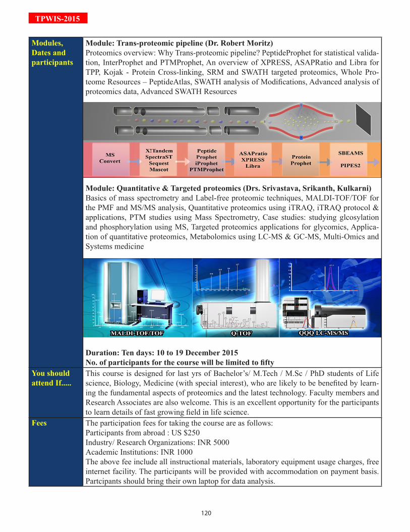
120
TPWIS-2015
Modules, Dates and participants
Module: Trans-proteomic pipeline (Dr. Robert Moritz)Proteomics overview: Why Trans-proteomic pipeline? PeptideProphet for statistical valida-tion, InterProphet and PTMProphet, An overview of XPRESS, ASAPRatio and Libra for TPP, Kojak - Protein Cross-linking, SRM and SWATH targeted proteomics, Whole Pro-teome Resources – PeptideAtlas, SWATH analysis of Modifications, Advanced analysis of proteomics data, Advanced SWATH Resources
Module: Quantitative & Targeted proteomics (Drs. Srivastava, Srikanth, Kulkarni)Basics of mass spectrometry and Label-free proteomic techniques, MALDI-TOF/TOF for the PMF and MS/MS analysis, Quantitative proteomics using iTRAQ, iTRAQ protocol & applications, PTM studies using Mass Spectrometry, Case studies: studying glcosylation and phosphorylation using MS, Targeted proteomics applications for glycomics, Applica-tion of quantitative proteomics, Metabolomics using LC-MS & GC-MS, Multi-Omics and Systems medicine
Duration: Ten days: 10 to 19 December 2015No. of participants for the course will be limited to fifty
You should attend If.....
This course is designed for last yrs of Bachelor’s/ M.Tech / M.Sc / PhD students of Life science, Biology, Medicine (with special interest), who are likely to be benefited by learn-ing the fundamental aspects of proteomics and the latest technology. Faculty members and Research Associates are also welcome. This is an excellent opportunity for the participants to learn details of fast growing field in life science.
Fees The participation fees for taking the course are as follows: Participants from abroad : US $250Industry/ Research Organizations: INR 5000Academic Institutions: INR 1000The above fee include all instructional materials, laboratory equipment usage charges, free internet facility. The participants will be provided with accommodation on payment basis. Partcipants should bring their own laptop for data analysis.

121
TPW
IS-2015
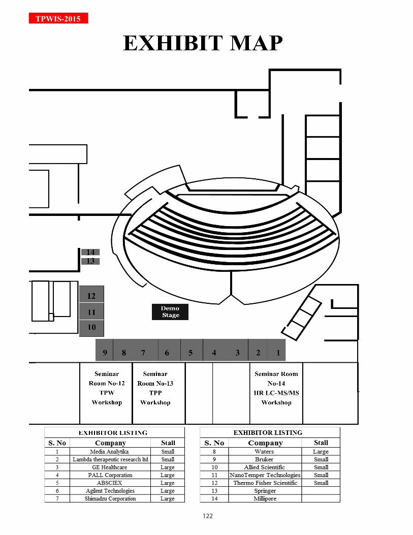
122
TPWIS-2015

123
TPW
IS-2015
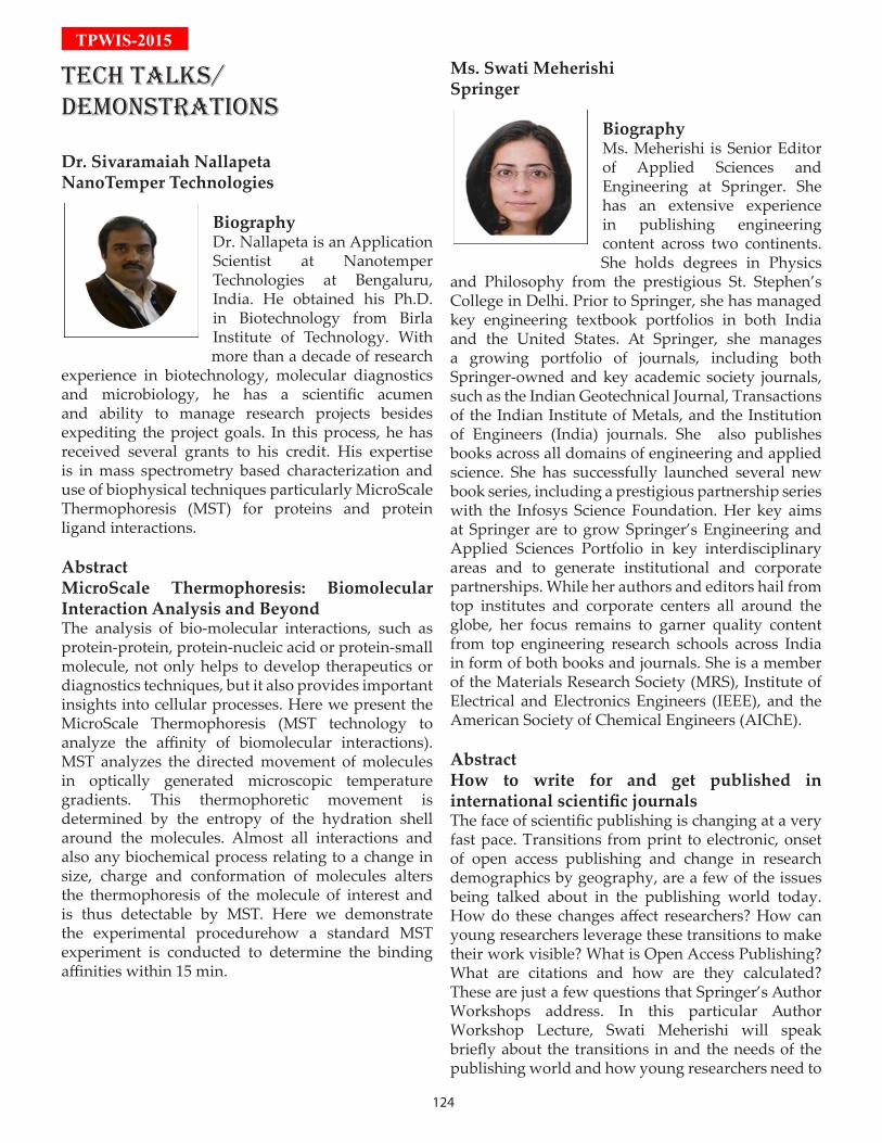
124
TPWIS-2015
TECH TALKS/dEMOnSTrATiOnS
Dr. Sivaramaiah NallapetaNanoTemper Technologies
BiographyDr. Nallapeta is an Application Scientist at Nanotemper Technologies at Bengaluru, India. He obtained his Ph.D. in Biotechnology from Birla Institute of Technology. With more than a decade of research
experience in biotechnology, molecular diagnostics and microbiology, he has a scientific acumen and ability to manage research projects besides expediting the project goals. In this process, he has received several grants to his credit. His expertise is in mass spectrometry based characterization and use of biophysical techniques particularly MicroScale Thermophoresis (MST) for proteins and protein ligand interactions.
AbstractMicroScale Thermophoresis: Biomolecular Interaction Analysis and BeyondThe analysis of bio-molecular interactions, such as protein-protein, protein-nucleic acid or protein-small molecule, not only helps to develop therapeutics or diagnostics techniques, but it also provides important insights into cellular processes. Here we present the MicroScale Thermophoresis (MST technology to analyze the affinity of biomolecular interactions). MST analyzes the directed movement of molecules in optically generated microscopic temperature gradients. This thermophoretic movement is determined by the entropy of the hydration shell around the molecules. Almost all interactions and also any biochemical process relating to a change in size, charge and conformation of molecules alters the thermophoresis of the molecule of interest and is thus detectable by MST. Here we demonstrate the experimental procedurehow a standard MST experiment is conducted to determine the binding affinities within 15 min.
Ms. Swati MeherishiSpringer
BiographyMs. Meherishi is Senior Editor of Applied Sciences and Engineering at Springer. She has an extensive experience in publishing engineering content across two continents. She holds degrees in Physics
and Philosophy from the prestigious St. Stephen’s College in Delhi. Prior to Springer, she has managed key engineering textbook portfolios in both India and the United States. At Springer, she manages a growing portfolio of journals, including both Springer-owned and key academic society journals, such as the Indian Geotechnical Journal, Transactions of the Indian Institute of Metals, and the Institution of Engineers (India) journals. She also publishes books across all domains of engineering and applied science. She has successfully launched several new book series, including a prestigious partnership series with the Infosys Science Foundation. Her key aims at Springer are to grow Springer’s Engineering and Applied Sciences Portfolio in key interdisciplinary areas and to generate institutional and corporate partnerships. While her authors and editors hail from top institutes and corporate centers all around the globe, her focus remains to garner quality content from top engineering research schools across India in form of both books and journals. She is a member of the Materials Research Society (MRS), Institute of Electrical and Electronics Engineers (IEEE), and the American Society of Chemical Engineers (AIChE).
AbstractHow to write for and get published in international scientific journalsThe face of scientific publishing is changing at a very fast pace. Transitions from print to electronic, onset of open access publishing and change in research demographics by geography, are a few of the issues being talked about in the publishing world today. How do these changes affect researchers? How can young researchers leverage these transitions to make their work visible? What is Open Access Publishing? What are citations and how are they calculated? These are just a few questions that Springer’s Author Workshops address. In this particular Author Workshop Lecture, Swati Meherishi will speak briefly about the transitions in and the needs of the publishing world and how young researchers need to

125
TPW
IS-2015prepare for it. This author workshop has been devised specifically as a resource for teaching non-native English-speaking researchers, particularly young scientists how to achieve publication success. Some of the key topics covered during this presentation are:
1. Writing for International Journals: Language, Style and Accuracy2. Selecting a Journal for your Manuscript3. Peer Review and you4. Plagiarism, Citations, Open Access: The Buzzwords of publishing5. Book Writing
Mr. Brijesh PandeyAB SCIEX, India
BiographyHe Has been working with Sciex since Jan 2013 and is currently responsible for support and marketing actvities in the proteomics, metabolomics and lipidomics by using high resolution mass spectrometry, A hybrid technology -Linear
Ion Trap (QTRAP) and Triple Quad systems. Prior to joining Sciex, he has worked with Bio-Rad Laboratories (India) Ltd.in various capacities primarily focusing on 2-D Gel electrophoresis, Imaging system and Sample preparation. He has more than 11 years of experience in promoting Proteomics & Analytical techniques. Currently he is focused on commercializing & promoting mass spectrometry-based workflows for proteomics, metabolomics and lipidomics research. A primary area of focus is the democratization and industrialization of Omics solutions to make them more widely accessible to the life science research community, and most recently, he has focused on establishing cloud computing for multi-Omics analysis in India.
AbstractNext-Gen Lipidomics Platform- The Lipidyzer™A major need to address in complex lipid sample analysis is how to differentiate each member of the lipid classes in presence of isobaric interferences which can increase the selectivity and specificity of identification and accurate quantitation.This problem, coupled with complicated sample preparation techniques and data analysis, highlights the need for a complete solution that addresses these difficulties and provides a simplified method for analysis. A novel lipidomics
platform (Lipidyzer™) is developed by SCIEX and powered by Metabolon®. The Lipidyzer™include simplified sample preparation, automated methods, and streamlined data processing techniques that enable facile, quantitative lipid analysis. It enables labs of all sizes to perform next-generation lipidomics. The all-in-one, turnkey, benchtop platform can run hundreds of samples and quantify over a thousand lipid species quickly, efficiently, and accurately. In addition, the workflow delivers expert data analysis via cloud-based data sharing and access to scientific specialists. There’s simply no faster or more efficient way to go from sample to biologically relevant knowledge.
Mr. Susheelendra VaidyaPALL Corporation
Quantitation of Monoclonal antibodies using Biolayer Interferometry: Label-free technologies offer an ideal solution for reducing the bottlenecks in bioprocessing like early cell culture screening, Protein purification,Cell line selection for optimization of
antibody production, Biologicals manufacturing monitoring to ensure high product quality and improve efficiencies. ForteBio’s Octet® family of instruments provides rapid and accurate analysis of antibody concentrations and other proteins. The BLI-based technology supports each step of bioprocessing, streamlining and speeding your workflow.. Its analytical capabilities provide particular value in applications where existing methods such as HPLC and ELISA have limitations in throughput, performance, workflow, and ease of use. Kinetics interaction studies using Mouse monoclonal antibodies using Biolayer Interferometry: Real-time kinetic measurements offer a direct and more realistic depiction of molecular interactions. Each instrument in the Octet family is a highly capable label-free system for full affinity determination, enabling users to easily and accurately obtain kinetic constants such as ka, kd,KD. The measurement below illustrates a full kinetic characterization using streptavidin biosensors. A titration series of the mouse antibody is measured against an immobilized antigen. The data set is analyzed with global fitting, thereby producing a ka, kd, KD.
126
TPWIS-2015
SPOnSOrS COMPAny PrOFiLES
diAMOnd SPOnSOrS
AB SCIEX URL: http://www.absciex.com/SCIEX helps to improve the world we live in by enabling scientists and labora-tory analysts to find answers to the complex analytical challenges they face. SCIEX's global leadership and world-class service and support in the Cap-illary Electrophoresis and Liquid Chromatography-Mass Spectrometry in-dustry have made it a trusted partner to thousands of the scientists and lab analysts worldwide who are focused on basic research, drug discovery and development, food and environmental testing, forensics and clinical research. With over 40 years of proven innovation, SCIEX excels by listening to and understanding the ever-evolving needs of its customers. We enable scientists and laboratory analysts to find answers to the complex analytical challenges they face. Our instrumentation, software, reagents and workflows allow you to dig deeper into complex biological systems, analyse prospective biomarkers, and deliver results with potential use in clinical applications.
PALL CORPORATIONURL: http://www.pall.com/Pall Corporation has become a global leader in the high-tech filtration, separation, and purification indus-try by meeting the diverse needs of customers across the broad spectrumof life sciences and industry. The company’shighly-engineered process solutions protect people and critical assets, while minimizing emissions and waste. Our single-use systems enable faster,more efficient processes for our customers. Pall Life Sciences caters to the diverse and rapidly expanding biopharmaceu-ticals market. The products and technologies are used from the earliest stages of discovery and development of new drugs, through production and delivery of therapies for the prevention, diagnosis and treatment of disease. ForteBio is a wholly-owned subsidiary of Pall Corp. and a division of Pall Life Sciences.ForteBiodevelop-sanalytical systems that enable real-time analysis of biomolecular interactions (protein quantification and characterization of protein-protein interactions, etc.) in micro-volume sample sizes, providinginformation on affinity, kinetics, and concentration.ForteBio utilizes proprietary Bio-Layer Interferometry (BLI) to create self-calibrating, simple-to-use, and inexpensive instruments that streamline measuring molecular interactions. An entire analysis can be completed in minutes and does not require labeling of the probe or the target. ForteBio’s analytical capabilities provide greater value in applications where existing methods (e.g., HPLC, ELISA, and surface plasmon resonance) have limitations in throughput, performance and cost. Pall Life Sciences brands now include ForteBio®, Octet®, BLItz®, DipandRead™biosensors, and Sidekick™.
GOLd SPOnSOrS
GE HEALTHCARE URL: http://www.gehealthcare.com/GE Healthcare provides technologies and services that are shaping a new age of healthcare. Our broad expertise in medical imaging and information tech-nologies, medical diagnostics, patient monitoring systems, drug discovery, biopharmaceutical manufacturing technologies, performance improvement and performance solutions services help our customers to deliver better care

127
TPW
IS-2015to more people around the world at a lower cost. In addition, we partner with healthcare leaders, striving to leverage the global policy change necessary to implement a successful shift to sustainable healthcare system. Our “healthy imagination” vision for the future invites the world to join us on our journey as we continuously develop innovations focused on reducing costs, increasing access and improving quality around the world. Headquartered in the United Kingdom, GE Healthcare is a unit of General Electric Company (NYSE: GE) Worldwide, GE Healthcare employeesare committed to serving healthcare professionals and their patients in more than 100 countries.
SHIMADZU URL: http://www.shimadzu.com/SHIMADZU Analytical (I) Pvt Ltd is a wholly owned subsidiary of Shimadzu Corporation, Japan - one of the leading Manufacturers Suppliers of Analytical Scientific Instruments such as CHROMATOGRAPHY, MASS-SPECTROME-TRY, SPECTROSCOPY, PHYSICAL TESTING, LIFE SCIENCES, ENVIRON-MENTAL Monitoring instruments. We provide an extensive range of products value added services and customer support Shimadzu has been serving the Indian customers since many years through its established network of distributors namely Toshvin Analytical, Spinco Biotech, Swan Environmental, Saksham Analyti-cal &Amkette Analytics for various product ranges. SHIMADZU Analytical (I) Pvt Ltd reaffirms our con-tinued commitment towards our valued customers. Our key strength has been continuously evolving new applications and providing solutions for challenging scientific queries. We have been relentlessly developing solutions for targeted proteomics, glycomics, mAb analysis giving a complete array of ready to use answers forscientific queries for protein determination and quantitation.
SiLVEr SPOnSOrS
AGILENT TECHNOLOGIESURL: http://www.agilent.com/Agilent Technologies offers a comprehensive and complete portfolio of Pro-teomics products for every stepof the workflow, from sample prep through separation, detection and analysis. Agilent’s innovation & solutions set the standard for precision, generating highly reproducibleand reliable data for yourprotein analysis research. Agilent is the preferred partner for supporting every step of multi-omics integrated Biology projects, from data acquisition to data analysis and visualization. More-over, Agilent is the only provider who can offer such a solution –we continue to lead the industry in the development and implementation of the tools and resources necessary to enable powerful analysis through integrated Biology. Analytical scientists and clinical researchers worldwide relyon Agilent to help fulfill their most complex laboratory demands. Our instruments, software, services and consumable address the full range of scientific and laboratory management needs so our customers can do what they do best: improve the world around us whether a laboratory is engaged in environmental testing, academic, research, medical di-agnostics, pharmaceuticals, petrochemical or food testing. Agilent provides laboratory solutions tomeet their full spectrum of needs. We work closely with customers to help address global trends that impact human health and the environment, and to anticipate future scientific needs. Our solutions improve the efficiency of the entire laboratory, from sample prep to data interpretation and management.
WATERS URL: http://www.waters.com/Waters Corporation creates business advantage for laboratory-dependent organizations by delivering practi-cal and sustainable innovation to enablesignificant advancements in such areas as healthcare delivery, envi-ronmental management, food safety, and water quality worldwide. Pioneering a connected portfolio of separations science, laboratory information management, mass spectrom-etry and thermal analysis, Waters technology breakthroughs and laboratory solutions provide an enduring
128
TPWIS-2015platform for customer success. Waters Corporation’s Ultra Performance LC™, high performance liquid chromatography (HPLC), mass spectrometry (MS), chemistry, software and services are synonymous with quality and perfor-mance in analytical labs around the world.Today, demand for Waters offer-ings has never been greater. Pricing pressures, intense competition and strict regulatory demands are challenging scientists and researchers to make im-portant breakthroughs faster and more efficiently.Increasing productivity in scientific labs has become para-mount. That’s why some oftoday’s highest-growth, highly-regulated industries–pharmaceutical, life science, biochemical, and environmental–have all turned to Waters for help. Waters is a publicly traded corporation (NYSE:WAT) headquartered in Milford, Massachusetts. It is a member of the Standard & Poor’s 500 Index. The company employs more than 5700 with manufacturing facilities located inMilford, Massachusetts; Taun-ton, Massachusetts; Wexford, Ireland; Singapore, and Manchester, England. Waters sells direct in most of the countries it serves. Capsule History An industry pioneer and entrepreneur, James L. Waters founded Waters Corporation in 1958. After stints as a university math teacher, Naval officer and project engineer,Jim started his scientific instruments company in the basement ofa Framingham, Massachusetts police station. In 1963, after the development of several products, Waters Corporation created the GPC 100, the first gel permeation chromatography (GPC) instrument. Later the company adopted ultraviolet (UV) detection and liquid-solid packings in 1967 to create the automated liquid chromatography instrument, ALC-100. In 1969, Jim was introduced to Dimitri D’Arbeloff, then president of Millipore Corporation. Jim invited D’Arbeloff to join Waters’ board of directors. With the help of Millipore, Waters established a firm foothold within the analytical industry. Over the next five years, the company introduced several new products,and Jim gradually removed himself from the day-to-day busi-ness, serving as chairman until Waters Corporation merged with Millipore in 1980. For nearly a decade and a half, the merged companies struggled to find and leverage the synergies between their respective technolo-gies. In 1994, Millipore sold the Waters division to an investor group. Encouraged by increased sales and profits across all product lines, resurgence in economic growth, and corporateR&D spending, Waters man-agement brought the company public in 1995. At the same time, the pharmaceutical industry continued on a prolonged period of investment, making Waters Corporation a chief beneficiary. It was during this period that the company introduced the Alliance® HPLC System, its most successful instrument product. In 1997, Waters Corporation acquired Micromass, Ltd., of Manchester, England, specializing in mass spectrometry. This acquisition proved to be apivotal decision for the company. Together, Waters and Micromass succeeded in combining HPLC and mass spectrometry, a very powerful and increasingly popular analytical technique. The separations power of HPLC coupled with the detection sensitivity and specificity of mass spectrometry is indispensable for many laboratories, particularly those with high-throughput needs.
OTHEr SPOnSOrS
BRUKER URL: http://www.bruker.com/Bruker Corporation is a leading provider of high-performance scientific instruments and solutions for mo-lecular and materials research. Bruker provides a variety of innovative turnkey mass spectrometry systems for academic, pharmaceutical, industrial, clinical and applied markets. Being one of the world’s leading analytical instrumen-tation companies, Bruker is strongly committed to further fully meet its cus-tomers’ needs as well as to continue to develop state-of-the-art technologies and innovative solutions for today’s analytical questions. Bruker systems are expertly designed for performance and utility, and feature a number of unique capabilities and technologies to enhance data quality and facilitate data analysis. Bruker offers a range of standalone and integrated mass spectrometry and analytical system solutions that deliver a variety of levels of information and are ideal for small molecule, protein and peptide or polymer analysis.Bruker delivers a series of innovative, fully inte-
129
TPW
IS-2015grated solutions for Proteomics, Metabolomics and Clinical Research areas, including:Biomarker Discovery & Characterization, Qualitative and quantitative proteomics, PTM analysis, Top-down protein characterization, Molecular Imaging in tissue, Drug Research and Development, Micro-organism identification and Metabolite Discovery and Identification.
NANO TEMPER TECHNOLOGIESURL: http://www.nanotemper-technologies.com/NanoTemper Technologies stands for a strong commitment to quality and high-precision instrumentation made in Germany. The extremely experienced and globally operating team of experts focuses strongly on users’ benefits by ensuring maximum efficiency for research in pharmaceutical and biotechnology industries, as well as in aca-demic research settings. This high-tech company develops, produces and markets technologies and instru-ments for biomolecular analytics, from basic to preclinical research. With headquarters inMunich, Germany, and subsidiaries in the UK, Poland, US, Brazil and India, the company is rapidly expanding worldwide. Among others, NanoTemper has been awarded the German Innovation Award 2012, the Bavarian Export Prize 2013 and the DeutscherGründerpreis 2014. NanoTemper’s high-quality instruments are based on pro-prietary and innovative technologies: Visit www.nanotemper-technologies.com to learn more about the MST, SAW and nanoDSF technologies. Schedule an instrument demonstration and explore the benefits of NanoTemper’s devices with your own samples. NanoTemper Technologies World Trade Center, 22nd Floor, Brigade Gateway Campus, Malleswaram (W), Bangalore-560055, India Phone: 91-9480190262, Email: [email protected] Web: www.nanotemper-technologies.com NanoTemper Technologies is a globally operating high-techcompany providing solutions forbiomolecular analytics. Thecompany develops, producesand markets innovative technologies and instrumentationused by thousands of scientistsacross four continents. Monolith: The Efficient Performer MicroScaleThermophoresis (MST) technology is a unique method tostudy binding affinities in an easy, fast and precise way. Four MonolithSeries instruments were introducedin just three years. Seismos: The In-Depth ExplorerThe recently acquired SurfaceAcoustic Wave (SAW) technologymeasures binding kinetics, and is sensitive to both mass and conformational changes. Prometheus: The Stability Expert In 2015, a novel product linerepresented by the Prometheus NT.48 instrument was launched offeringnanoDSF technology. Thisis the method of choice for measuring ultra-high-resolution protein stability in native conditions using intrinsic tryptophan andtyrosine fluorescence.
EXHibiT SPOnSOrS
THERMO SCIENTIFICURL: http://www.thermoscientific.com/Under the Thermo Scientific brand of Thermo Fisher Scientific, Inc. (NYSE:TMO), we help scientists meet the challenges they face in the lab or in the field every day. From routine analysis to new discoveries, our innovations help professionals do the science they need to do, the way they want to do it. Our high-end analytical instru-ments, laboratory equipment, software, services, consumables and reagents help our customers solve complex analytical challenges, improve patient diagnostics and increase laboratory productivity. The Life Science Re-search Community features information for those researching complex biological systems and deconstructing disease mechanisms at a cellular level. Videos and tools show how new technologies enable researchers to better understand disease origins whichcan lead to the development of new therapies. Sequencing the human
130
TPWIS-2015genome was an important milestone for life science research. Many researchers are overcoming challenges to accurately map complex biological systems and deconstruct disease mechanisms at a cellular level. At the intersection of genomics and proteomics, our technologies enable life science researchers to ask and answer fundamental questions about disease origins and targeted treatments faster. This accelerates development of novel drug targets and new therapies that dramatically improve quality of life and revolutionize healthcare.
MEDI ANALYTIKA INDIA PVT. LTD URL: http://www.medianalytika.com/MediAnalytika India Pvt. Ltd is one of the leading instrumentation companies in India having tie up with several world leading instrumentation companies.
LAMBDA THERUPATIC RESEARCHURL: http://lambda-cro.com/Lambda Therapeutic Research Limited is a leading global Clinical Research Organization (CRO) headquar-tered in Ahmedabad - India, with facilities and operations in Mumbai, Toronto (Canada), Warsaw (Poland), London (UK) and USA. Lambda offers full spectrum clinical trial solutions empowered by more than 14 years of service to the biop-harmaceutical and generic industry. Lambda’s core strengths:• Global presence with capabilities in Indian subcontinent, Europe and North America • More than 1,50,000 active study participants in its global database • More than 850+ validated LC/MS - MS assays available globally • Over 50 successful regulatory audits and inspections globally • More than 40 LC-MS/MS systems • Low temperature storage (2-8 °C, -20 °C and -80 °C) controlled storage facility to store over 3.5 million samples • EDC (paperless execution) of both BA-BE & clinical trials through means of a 21 CFR Part 11 compliant & indigenously developed validated software ‘BizNET’
DSS IMAGE TECH http://www.dssimage.com/Driven by the mantra “Inspired By Technology”, DSS Imagetechendeavours to provide cutting edge technol-ogy solutions to researchers and clinicians in various fields of Life Sciences. DSS is well known in the Indian market for its expertise in Microscopy, Medi-cal Imaging, Genomics, Proteomics& Molecular Diagnostics. The recently established DSS R&D Center has now proved to be a forerun-ner in developing indigenous products for the Indian diaspora.Through its strong association with International market leaders, the DSS Groups activities range across the following segments:
ALLIED SCIENTIFIC PRODUCTSwww.alliedscientific.net/Allied Scientific Products is principally a distributor company for the following products: Antibodies, ELISA kits, assay kits, IHC and ECL kits, proteins and peptides, lysates, biochemical etc. from Abcam; Antibodies, GTPase activation assays, small G-proteins, actin and tubulin proteins, actin visualization kits etc. from Cy-toskeleton Inc.; Antibodies, ELISA kits, RNAi, cDNA clones, siRNA, miRNA, recombinant proteins, qPCR re-agents etc. from Origene; Electron microscopy grade reagents, accessories, diamond knife, tweezer, grid, wa-
131
TPW
IS-2015fer, mesh, adhesive etc. from Electron Microscopy Sciences; Electrophoresis units, blotter, transfer apparatus, power supply, gel dryer, transilluminator, water bath, magnetic stirrer, shaker, rocker etc. from Scie-Plas; Centrifuges, shakers, rockers, incubator shaker, heat block etc. from Benchmark Scientific; Micropipettes from Socorex Swiss.
MERCK MILLIPOREwww.merckmillipore.inMerck is a leader in life science. We aim to solve the toughest problems in the industry by collaborating with the global scientific community, and through that help to increase access to healthcare.
With a catalog of more than 300,000 products, Merck delivers many of the most highly-respected brands in the industry, like Millipore, Milli-Q®, SAFC and BioReliance®. Our offering covers every step of the biotech production chain, creating a complete end-to-end workflow with enhanced customer ser-vice, a simplified interface and a leading distribution platform. Our innovative portfolio, well-balanced geo-graphic reach, and industry leading capabilities, uniquely positions us to anticipate and deliver on customer needs.
At Merck, a global pharmaceutical and chemical company, we take pride in fostering a culture of curiosity. An inquisitive, innovative and solution-oriented mindset can be felt throughout the organization of 50,000 employees, worldwide. Life science at the company, comprised of 19,000 employees in 67 countries, brings together the legacy expertise of Merck Millipore and Sigma-Aldrich, which was acquired by Merck in 2015.
132
TPWIS-2015

133
TPW
IS-2015
134
TPWIS-2015
LEArninG PrOTEOMiCS TECHniquES in nATiOnAL inTErEST
During the last decade, proteomics has demonstrated significant impact on various aspects of clinical research, including understanding of disease pathogenesis, identification of novel diagnostic and prognostic markers, elucidation of mechanisms of drug action and identification of next generation
drugs and vaccines targets. Also, proteomics is increasingly employed in the comprehensive study of the stress response generated by plants and microbes. Thus, proteomics has made significant contribution in all aspects of health care, basic science and physiology research. Being one of the most widely used OMICs approaches, it is taking increasingly key position in Biology and Biomedical research. Precision Medicine is another upcoming initiative which aims at tailoring diagnosis and therapy. Proteomics would have to play a crucial role to ensure such ideas are translated to clinics. Targeted proteomics, in this light, is a timely innova-tion which offers high sensitivity to validate markers and target proteins in biospecimens.
Over the last decade, many Institutes and research groups in India have been using proteomics ap-proaches in their research efforts. The Proteomics Society, India – PS(I), was established in 2009, to promote interactions within the Indian Proteomics community and encourage exchange of ideas, enhance collabora-tions and boost innovations at the national and international level. Active involvement of Indian scientists in research on proteomics of different human cancers and infectious diseases and also on diverse aspects of bacterial, plant and animal proteomics is now becoming prominent on the global stage. Indian researchers played a significant role in two of the three important projects contributing towards the characterization of each and every protein present in the human body. Dr. Pandey and Dr. Kuster’s labs have independently drafted the “Human Proteome Maps” using high-resolution mass spectrometry. Hence, while India was not able to play a vital role in the genome sequencing projects, the country is playing an increasingly significant role in global proteomics research. Besides, Indian researchers have developed various e-learning resources like the virtual lab project dedicated to proteomics developed by the Indian Institute of Technology Bombay, Mumbai, which has now been incorporated as a part of the International Proteomics Tutorial Programme (IPTP), conducted by the Human Proteome Organization (HUPO) and the European Proteomics Association (EuPA). Hence, due to strong international collaborations, government support and enthusiastic scientists with an inter-disciplinary vision, proteomics community in India is growing steadily, leading to a surge in the number of articles published in this area. The publication of a special issue on “Proteomics in India” by the Journal of Proteomics establishes the strength and the infrastructure that is now available in the country to address various biological queries requiring proteomics tools. With this continued growth it seems likely that there will be a bright and exciting future ahead for research within this field.
135
TPW
IS-2015
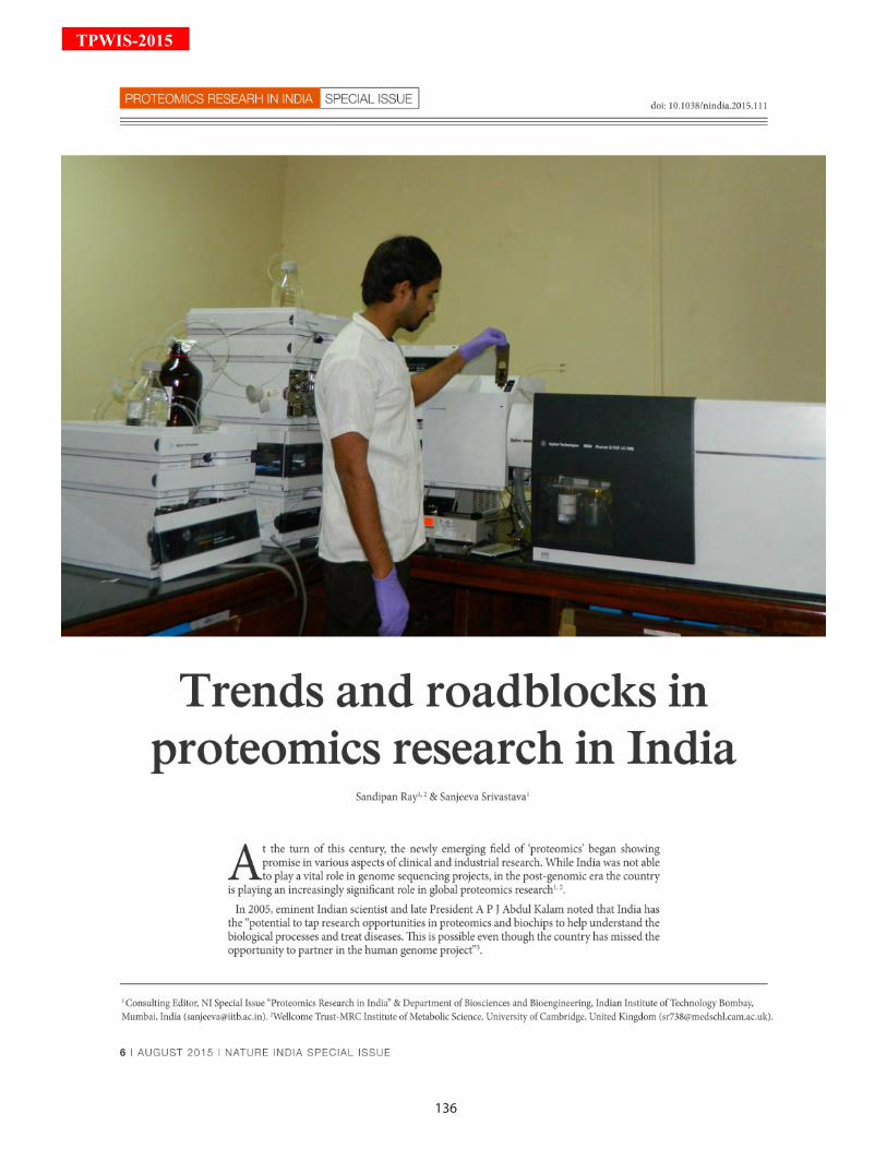
136
TPWIS-2015
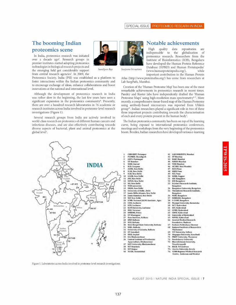
137
TPW
IS-2015
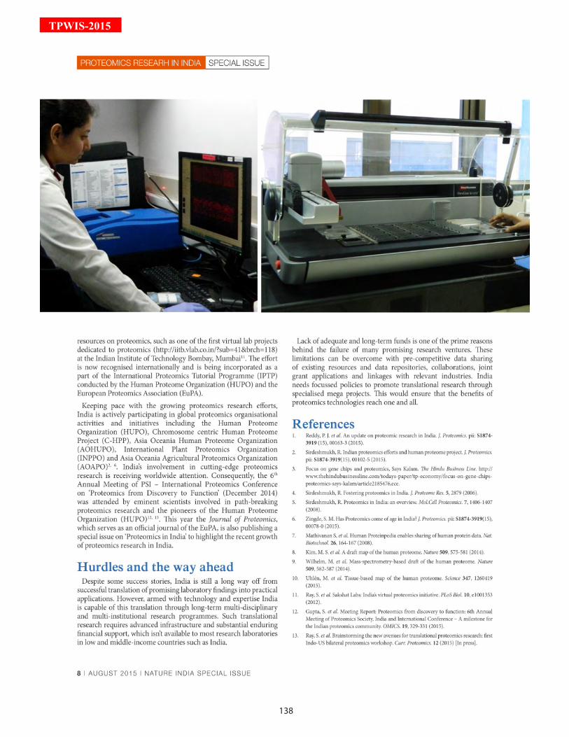
138
TPWIS-2015
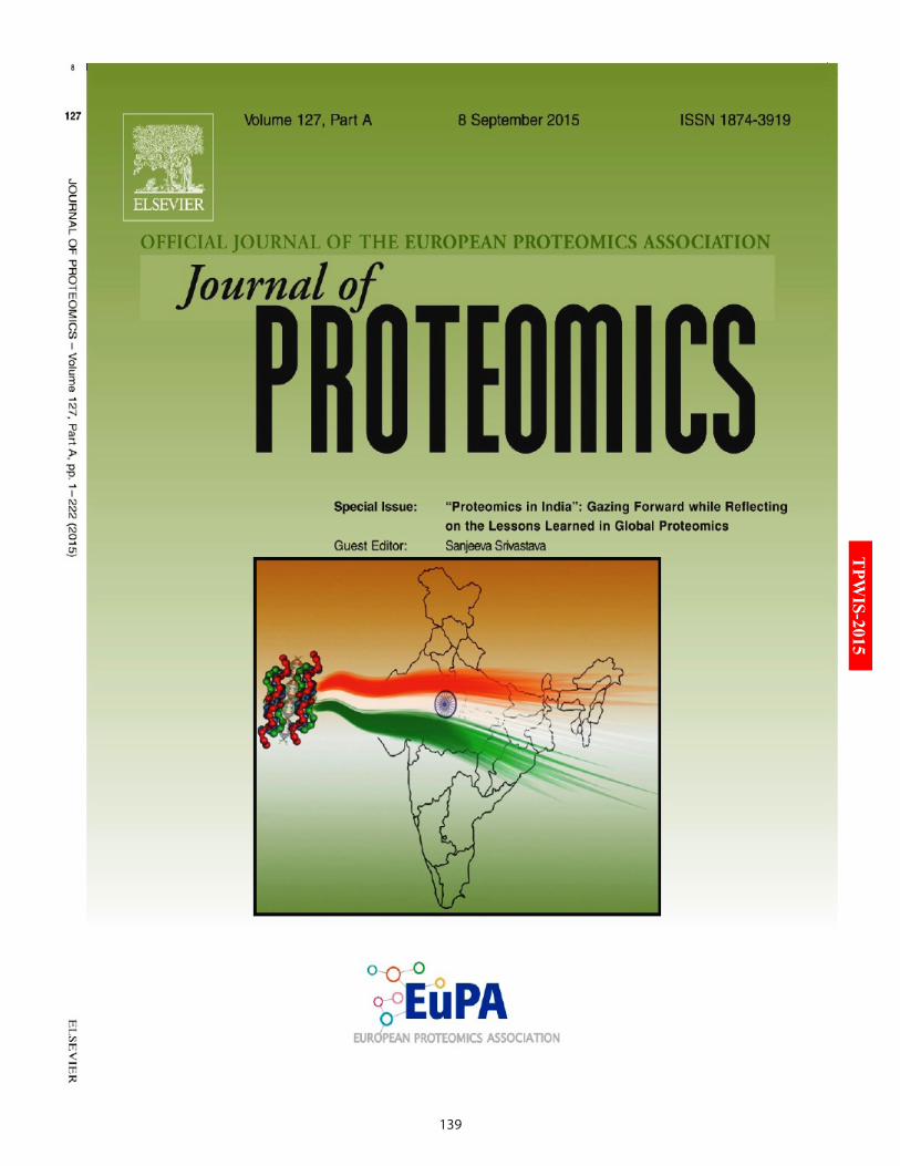
139
TPW
IS-2015
140
TPWIS-2015
141
TPW
IS-2015
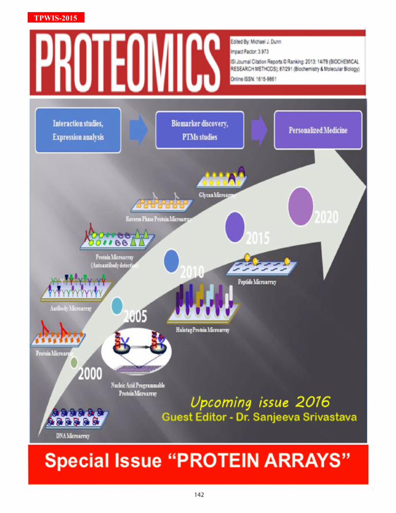
142
TPWIS-2015
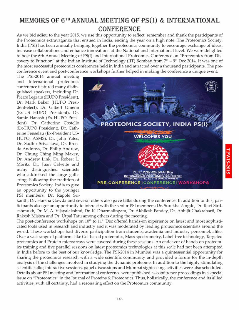
143
TPW
IS-2015
MEMOirS OF 6TH AnnuAL MEETinG OF PS(i) & inTErnATiOnAL COnFErEnCE
As we bid adieu to the year 2015, we use this opportunity to reflect, remember and thank the participants of the Proteomics extravaganza that ensued in India, ending the year on a high note. The Proteomics Society, India (PSI) has been annually bringing together the proteomics community to encourage exchange of ideas, increase collaborations and enhance innovations at the National and International level. We were delighted to host the 6th Annual Meeting of PS(I) and International Proteomics Conference on “Proteomics from Dis-covery to Function” at the Indian Institute of Technology (IIT) Bombay from 7th – 9th Dec 2014. It was one of the most successful proteomics conferences held in India and attracted over a thousand participants. The pre-conference event and post-conference workshops further helped in making the conference a unique event.The PSI-2014 annual meeting and International proteomics conference featured many distin-guished speakers, including Dr. Pierre Legrain (HUPO President), Dr. Mark Baker (HUPO Presi-dent-elect), Dr. Gilbert Omenn (Ex-US HUPO President), Dr. Samir Hanash (Ex-HUPO Presi-dent), Dr. Catherine Costello (Ex-HUPO President), Dr. Cath-erine Fenselau (Ex-President US-HUPO, ASMS), Dr. John Yates, Dr. Sudhir Srivastava, Dr. Bren-da Andrews, Dr. Philip Andrew, Dr. Chung Ching Ming Maxey, Dr. Andrew Link, Dr. Robert L. Moritz, Dr. Juan Calvette and many distinguished scientists who addressed the large gath-ering. Following the tradition of Proteomics Society, India to give an opportunity to the younger PSI members, Dr. Rapole Sri-kanth, Dr. Harsha Gowda and several others also gave talks during the conference. In addition to this, par-ticipants also got an opportunity to interact with the senior PSI members; Dr. Surekha Zingde, Dr. Ravi Sird-eshmukh, Dr. M. A. Vijayalakshmi, Dr. K. Dharmalingam, Dr. Akhilesh Pandey, Dr. Abhijit Chakrabarti, Dr. Rakesh Mishra and Dr. Utpal Tatu among others during the meeting.The post-conference workshops on 10th to 11th Dec offered hands-on experience on latest and most sophisti-cated tools used in research and industry and it was moderated by leading proteomics scientists around the world. These workshops had diverse participation from students, academia and industry personnel, alike. Over a vast range of platforms like Gel-based proteomics, Mass spectrometry, Label-free technology, Targeted proteomics and Protein microarrays were covered during these sessions. An endeavor of hands-on proteom-ics training and five parallel sessions on latest proteomics technologies at this scale had not been attempted in India before to the best of our knowledge. The PSI-2014 in Mumbai was a quintessential opportunity for sharing the proteomics research with a wide scientific community and provided a forum for the in-depth analysis of the challenges involved in studying the dynamic proteome. In addition to the highly stimulating scientific talks; interactive sessions, panel discussions and Mumbai sightseeing activities were also scheduled. Details about PSI meeting and International conference were published as conference proceedings in a special issue on “Proteomics” in the Journal of Proteins & Proteomics. Thus, holistically, the conference and its allied activities, with all certainty, had a resonating effect on the Proteomics community.
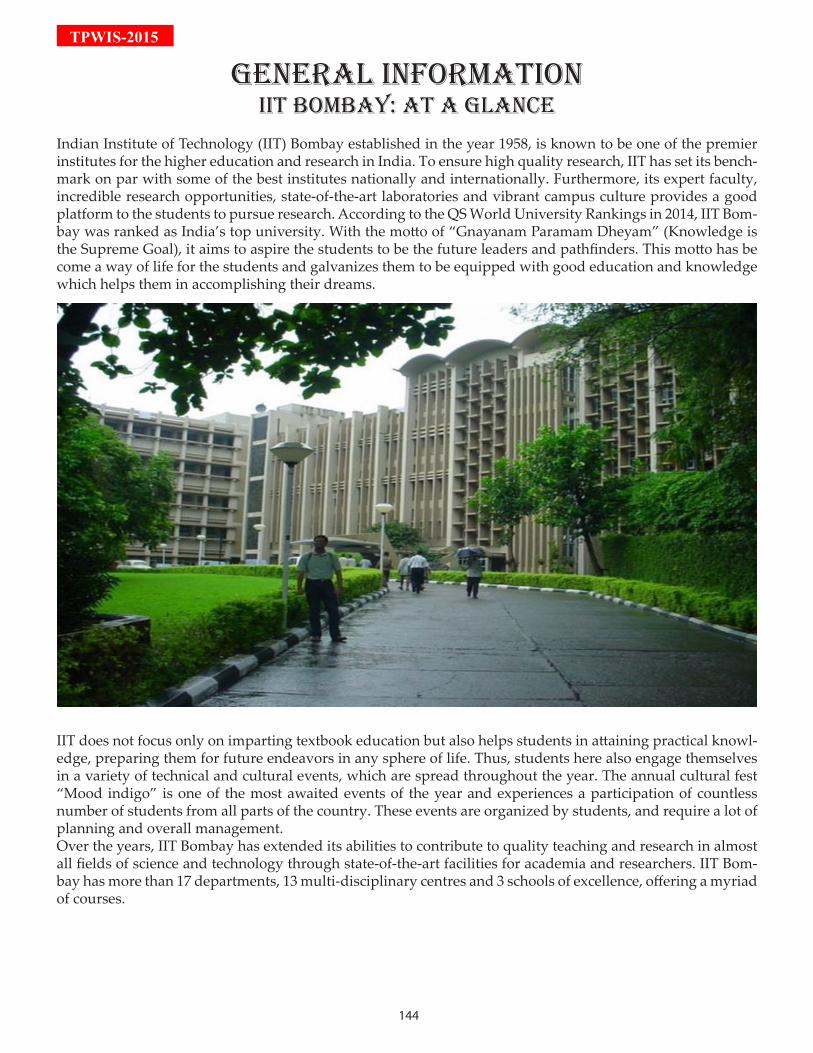
144
TPWIS-2015
GEnErAL inFOrMATiOniiT bOMbAy: AT A GLAnCE
Indian Institute of Technology (IIT) Bombay established in the year 1958, is known to be one of the premier institutes for the higher education and research in India. To ensure high quality research, IIT has set its bench-mark on par with some of the best institutes nationally and internationally. Furthermore, its expert faculty, incredible research opportunities, state-of-the-art laboratories and vibrant campus culture provides a good platform to the students to pursue research. According to the QS World University Rankings in 2014, IIT Bom-bay was ranked as India’s top university. With the motto of “Gnayanam Paramam Dheyam” (Knowledge is the Supreme Goal), it aims to aspire the students to be the future leaders and pathfinders. This motto has be come a way of life for the students and galvanizes them to be equipped with good education and knowledge which helps them in accomplishing their dreams.
IIT does not focus only on imparting textbook education but also helps students in attaining practical knowl-edge, preparing them for future endeavors in any sphere of life. Thus, students here also engage themselves in a variety of technical and cultural events, which are spread throughout the year. The annual cultural fest “Mood indigo” is one of the most awaited events of the year and experiences a participation of countless number of students from all parts of the country. These events are organized by students, and require a lot of planning and overall management.Over the years, IIT Bombay has extended its abilities to contribute to quality teaching and research in almost all fields of science and technology through state-of-the-art facilities for academia and researchers. IIT Bom-bay has more than 17 departments, 13 multi-disciplinary centres and 3 schools of excellence, offering a myriad of courses.
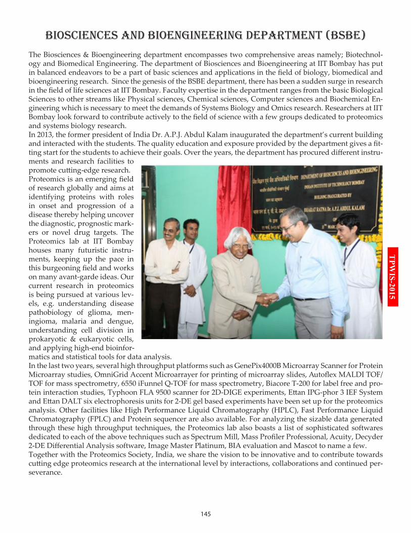
145
TPW
IS-2015
biOSCiEnCES And biOEnGinEErinG dEPArTMEnT (bSbE)
The Biosciences & Bioengineering department encompasses two comprehensive areas namely; Biotechnol-ogy and Biomedical Engineering. The department of Biosciences and Bioengineering at IIT Bombay has put in balanced endeavors to be a part of basic sciences and applications in the field of biology, biomedical and bioengineering research. Since the genesis of the BSBE department, there has been a sudden surge in research in the field of life sciences at IIT Bombay. Faculty expertise in the department ranges from the basic Biological Sciences to other streams like Physical sciences, Chemical sciences, Computer sciences and Biochemical En-gineering which is necessary to meet the demands of Systems Biology and Omics research. Researchers at IIT Bombay look forward to contribute actively to the field of science with a few groups dedicated to proteomics and systems biology research.In 2013, the former president of India Dr. A.P.J. Abdul Kalam inaugurated the department’s current building and interacted with the students. The quality education and exposure provided by the department gives a fit-ting start for the students to achieve their goals. Over the years, the department has procured different instru-ments and research facilities to promote cutting-edge research.Proteomics is an emerging field of research globally and aims at identifying proteins with roles in onset and progression of a disease thereby helping uncover the diagnostic, prognostic mark-ers or novel drug targets. The Proteomics lab at IIT Bombay houses many futuristic instru-ments, keeping up the pace in this burgeoning field and works on many avant-garde ideas. Our current research in proteomics is being pursued at various lev-els, e.g. understanding disease pathobiology of glioma, men-ingioma, malaria and dengue, understanding cell division in prokaryotic & eukaryotic cells, and applying high-end bioinfor-matics and statistical tools for data analysis. In the last two years, several high throughput platforms such as GenePix4000B Microarray Scanner for Protein Microarray studies, OmniGrid Accent Microarrayer for printing of microarray slides, Autoflex MALDI TOF/TOF for mass spectrometry, 6550 iFunnel Q-TOF for mass spectrometry, Biacore T-200 for label free and pro-tein interaction studies, Typhoon FLA 9500 scanner for 2D-DIGE experiments, Ettan IPG-phor 3 IEF System and Ettan DALT six electrophoresis units for 2-DE gel based experiments have been set up for the proteomics analysis. Other facilities like High Performance Liquid Chromatography (HPLC), Fast Performance Liquid Chromatography (FPLC) and Protein sequencer are also available. For analyzing the sizable data generated through these high throughput techniques, the Proteomics lab also boasts a list of sophisticated softwares dedicated to each of the above techniques such as Spectrum Mill, Mass Profiler Professional, Acuity, Decyder 2-DE Differential Analysis software, Image Master Platinum, BIA evaluation and Mascot to name a few.Together with the Proteomics Society, India, we share the vision to be innovative and to contribute towards cutting edge proteomics research at the international level by interactions, collaborations and continued per-severance.

146
TPWIS-2015
MuMbAi: THE LAnd WHErE drEAMS COME TruE
As Salman Rushdie, the famous novelist had once said “You can take the boy out of Bombay; you can’t take Bombay out of the boy, you know”, the words still echo in the backyard of every house in the city. In a city where anything and everything can change, only the sea is a constant. Mumbai, the financial capital of India has a soul of its own, often attracting men and women across the country to pursue their dreams.The city never sleeps and the cosmopolitan milieu further enriches the diverse culture. It has a rich, manifold cultural heritage and houses many traveler’s-favorite spots. The Elephanta caves is one such example. The Elephanta islands, residing on the arms of the Arabian Sea, comprises of two groups of caves; a group of five Hindu caves and a group of two Buddhist caves. The city possesses one of the most valued monolithic monu-ments of India, Gateway of India. It was built in the year 1924 during the British rule. The monument has witnessed three terror attacks since and still stands strong.
Other attractions include Jehangir Art Gallery, National Gallery of Modern Art, Chhatrapati Shivaji Maharaj Vastu Sangrahalaya (formerly known as the Prince of Wales Museum) etc. which house rare ancient exhib-its of Indian history. Also, Town Hall is another majestic structure among the various heritage buildings of Mumbai.This city also houses the famous Bollywood industry and there are many famous film sets located in the heart of city. Film city, Filmistan, Bombay Talkies etc. are some of the few favourite venues for Bollywood film shootings. Also, evening long strolls at beaches like Juhu, Gorai, Marve etc. often helps in rejuvenating the residents after a long, hard day. Another attraction, Marine Drive is one of the most exquisite places to explore in Mumbai. Popularly known as Queen’s Necklace, it is a 3 kilometer stretch running along the coast of Arabian Sea, with a shape of inverted “C”.Among others, EsselWorld, an amusement park stands out from the rest. It is the largest amusement park of the country and also houses Asia’s largest theme water park. Another recent addition to entertainment hubs of the city is Adlabs Imagica. People who visit this place are often seen to be captivated by its charm and de-lightful attractions.List of tourist places in Mumbai:• Elephanta Caves• Jehangir art gallery• National gallery of modern art• Chhatrapati Shivaji Maharaj Vastu Sangrahalaya• Town Hall• Film City• Gateway of India• Juhu Beach• Gorai Beach• Marve Beach• Marine Drive• EsselWorld• Adlabs Imagica• Hanging Gardens of Mumbai• Sanjay Gandhi National Park• Nehru Science Centre• Mount Mary Church, Bandra• Haji Ali Dargah• ISKCON Temple
All they know is that you are trying to get to the city of gold, and that is enough. Come on board, they say. We will adjust. – Suketu Mehta, Author
147
TPW
IS-2015
Contact detailsA
Dr. Ajit DatarShimadzu Corp., [email protected]
Dr. Andreas FR HühmerThermo Fisher Scientific, USA
Dr. Andrei KozlovBiomedical Center and Peter the Great St. Petersburg Polytechnic [email protected]
Dr. Anurag S. RathoreIndian Institute of Technology Delhi, [email protected]
Mr. Ariel BensimonETH Zurich, [email protected]
Dr. Ashish PargaonkarAgilent [email protected]
B
Dr. Ben Collins ETH Zurich, [email protected]
Mr. Brendan MacLeanUniversity of Washington, [email protected]
Dr. Bruno DomonLuxembourg Clinical Proteomics Center, [email protected]
D
Mr. David Campbell Institute for Systems Biology, USA
Dr. Dipankar MalakarABSCIEX, [email protected]
H
Dr. Harsha GowdaInstitute of Bioinformatics, [email protected]
Dr. Hui Zhang Johns Hopkins University, [email protected]
J
Dr. Jacob JaffeBroad Institute, [email protected]
Dr. Jason ChenNational Central University, [email protected]
Dr. Jennifer Van EykCedars-Sinai, [email protected]
Dr. Joshua LaBaerArizona State University, [email protected]
L
Dr. Leigh AndersonSISCAPA Assay Technologies, USA
Mr. Luis MendozaInstitute for Systems Biology, USA
M
Dr. Mahesh KulkarniNational Chemical Laboratory, India [email protected]
Dr. Mark McDowallWaters Corporation, [email protected]
Dr. Mark S. BakerPresident HUPO, Macquarie University, [email protected]
Dr. Mayuri GandhiIIT Bombay, [email protected]
Dr. Michael HoopmannInstitute for Systems Biology, USA
N
Dr. Nicola Gray Imperial College London, [email protected]
Dr. Niranjan ChakrabortyNIPGR, [email protected]
P
Mr. Panga Jaipal ReddyIndian Institute of Technology, Bombay, [email protected]
Dr. Paul GouldingNonlinear Dynamics, Newcastle, [email protected]
Mr. Pratip SahaThermo Fisher Scientific India Pvt Ltd
R
Mr. Rajesh VashishtBruker Daltonics, [email protected]
Ms. Rashi Kochhar Shimadzu Corp., [email protected]
Dr. Ravi K. KrovidiLambda Therapeutic Research Ltd, India [email protected]
Dr. Ravi SirdeshmukhInstitute of Bioinformatics, [email protected]
Dr. Ravindra GudihalAgilent [email protected]
Dr. Robert L. MoritzInstitute for Systems Biology, [email protected]
Dr. Robert PlumbImperial College London, [email protected]
S
Dr. Samuel BaderInstitute for Systems Biology, USA
Dr. Sanjay NavaniLab SurgPath, [email protected]
Dr. Sanjeeva SrivastavaIndian Institute of Technology, Bombay, [email protected]
Dr. Shannon Cornett Bruker Daltonics, [email protected]
Dr. Shenglan Cao Pall [email protected]
Dr. Shubhendu SealGE [email protected]
Dr. Srikanth RapoleNational Centre For Cell Science, [email protected]
Dr. Stephen R. PenningtonUniversity College Dublin, [email protected]
Dr. Subhra ChakrabortyNIPGR, [email protected]
Ms. Subhra PriyadarshiniNature, [email protected]
Dr. Sudhir SrivastavaNational Cancer Institute, [email protected]
Dr. Suman ThakurCentre for Cellular and Molecular Biology, [email protected]
Dr. Surekha ZingdePresident, PS(I), [email protected]
T
Dr. Tsung-Heng Tsai Northeastern University, [email protected]
U
Dr. Ulrike KusebauchInstitute for Systems Biology, USA
Dr. Utpal S. TatuIndian Institute of Science, [email protected]
V
Dr. Veena MenonAmrita University, [email protected]
148
TPWIS-2015
Authors IndexAli Syed Azmal, p 92, 95, 102Anand Ruchi, p 66Anand V, p 102Anderson Leigh, p 66, 75Anderson V, p 97Andrew Philip, p 142Andrews Brenda, p 142Anklesaria Jenifer H., p 93, 104Arora Priya, p 93, 112Atak A, p 108Baaske Philipp, p 118Bader Samuel, p 11, 14, 15, 147Baithalu R, p 107Baker Mark, p 38, 56, 58, 64, 65, 68, 142, 147Balhara AK, p 107Barkeer Srikanth, p 116Basha PA, p 110Bathla Shveta, p 93, 95, 102, 107Beck A., p 117Bennike T.B., p 67, 80, 92, 97Bensimon Ariel, p 22, 23, 24, 147Bhandarkar Deepti, p 98, 116Bharadwaj Rajiv, p 93, 94, 107, 118Birkelund S, p 97Bisht D, p 101Boegsted M, p 97Bonderup OK, p 97Borchers CH, p 115Budhraja Rohit, p 92, 102Calvete Juan, p 142Campbell David, p 11, 14, 15, 147Cao Shenglan, p 38, 44, 147Carlsen TG, p 97Chakrabarti Abhijit, p 142Chakraborty Niranjan, p 56, 59, 67, 147Chakraborty Subhra, p 56, 59, 64, 66, 147Chambers AG, p 115Chanukuppa V, p 96, 117Chatterjee G, p 111Chatterjee T, p 115Chen Jason, p 39, 56, 60, 78, 147Chopra A, p 107Choudhary S, p 108Christiansen G, p 97Chunduri JayaPrada R., p 116Collins Ben, p 22, 23, 24, 66, 74, 147Cornett Shannon, p 39, 46, 147Costello Catherine, p 142Cox M. M. J., p 117Dang AK, p 107Das D, p 113Datar Ajit, p 30, 32, 57, 60, 98, 116, 147Dhabi R, p 109Dhali Snigdha, p 66, 87, 93, 109
Dharmalingam K., p 142Domanski D, p 115Domon Bruno, p 21, 23, 57, 60, 65, 66, 67, 75, 147Duhr Stefan, p 118Eligar Sachin M., p 116Ellingsen T, p 97Elsy N, p 111Epari S, p 104, 108Evans C., p 117Evers W., p 117Fenselau Catherine, p 142Gahoi Nikita, p 92, 94, 95, 98, 116Gaikwad Pallavi, p 93, 108Gajbhiye A, p 109Gandhi Mayuri, p 1, 2, 29, 32, 33, 147Gebhardt Christoph, p 106Ghantasala Saicharan, p 66, 86, 92, 98, 101, 116Ghosh Amrita, p 94, 113Ghosh SK, p 109Giri AP, p 113Glerup H, p 97Gogtay N, p 111Gollapalli Kishore, p 92, 101, 116Gonge Sudhanshu Suhas, p 92, 99Goulding Paul, p 38, 41, 147Gowda Harsha, p 2, 29, 32, 56, 57, 60, 65, 73, 96, 142, 147Gray Nicola, p 38, 40, 147Gudihal Ravindra, p 38, 44, 94, 116, 147Gupta Shabarni, p 93, 108Guptasarma P, p 98Hama¬da Akinobu, p 106, 118Hameed M.S. Shahul, p 93, 94, 103, 113Hanash Samir, p 4, 142Hartmer R., p 117Hegde Prajna, p 116Hoopmann Michael, p 12, 14, 15, 65, 73, 147Hühmer Andreas FR, p 30, 32, 147Imhoff W., p 117Inamdar Shashikala R., p 116Iwamoto Noriko, p 94, 106, 118Jabs W., p 94, 117Jackson AM, p 115Jadhav M, p 103Jaffe Jacob, p 22, 23, 24, 57, 60, 65, 71, 147Jain Rekha, p 32Jain RK, p 111Jaswal S, p 95, 102Jawade AA, p 111Jayabaskaran C, p 112Joshi B, p 101
Kadian K, p 114Kannan MonicaKarthikaichamy A, p 113Kaur Gurjeet, p 92, 95Kaushik JK, p 95, 102, 107Khanzode M, p 97Kishore Nitin, p 66, 85, 92, 98Kochar SK, p 111Kochhar Rashi, p 39, 45, 147Kozlov Andrei, p 67, 77, 147Krovidi Ravi K, p 38, 45, 93, 102, 105, 147Kulkarni Mahesh, p 2, 30, 33, 56, 57, 60, 65, 66, 71, 113, 142, 147Kumar B, p 101Kumar G, p 95Kumar KM, p 96Kumar Manish, p 96Kumar Ravinder, p 93, 109Kumar S, p 95, 102, 107Kumar S, p 101Kumar Vipin, p 92, 100, 111Kumaraswamy Sriram, p 105Kurup Anil, p 106, 118Kusebauch Ulrike, p 11, 14, 15, 65, 73, 147LaBaer Joshua, p 39, 48, 64, 65, 69, 147Lata M, p 101Legrain Pierre, p 142Lincoln Lynette, p 105Link Andrew, p 142Lubeck Markus, p 106MacLean Brendan, p 1, 2, 21, 23, 24, 56, 57, 60, 66, 74, 147Mahale Smita D., p 104Malakar Dipankar, p 30, 33, 95, 147Malhotra D, p 95Mani Vimala Ashok, p 93, 104Manu V, p 115Manubhai KP, p 104, 108Markus A, p 111Matthews E., p 117Maxey Chung Ching Ming, p 142McDowall Mark, p 31, 32, 38, 42, 67, 76, 147Meherishi Swati, p 64, 124Mendoza Luis, p 12, 14, 15, 147Menon Veena, p 67, 77, 147Michalski Annette, p 106Mishra AK, p 95, 96Mishra Rakesh, p 9, 142Mohammed Yassene, p 67, 81, 94, 115Mohan Sonali Vishwa, p 66, 87, 92, 96Mohanty AK, p 95, 102, 107Mohanty TK, p 107Moiyadi Aliasgar, p 64, 95, 98, 101, 104, 108, 112, 116

149
TPW
IS-2015Moritz Robert, p 1, 2, 10, 11, 14, 15, 56, 60, 65, 72, 142, 147Mukherjee Shuvolina, p 93, 98, 112, 116Mukhopadhyaya R, p 108Muthuraj M, p 113Naik S, p 115Naik V, p 109Nallapeta Sivaramaiah, p 94, 118, 124Nandy Ar. Antara, p 92, 100Narula G, p 109Nath BB, p 108Navani Sanjay, p 39, 48, 147Nayak Monalisha, p 92, 103, 113Nidhi ML, p 94, 116Niveshika, p 95, 96Noronha Santhosh, p 108, 110, 113Nupur N, p 109Omenn Gilbert, p 142Pachauri S, p 95Padmanaban Arunkumar, p 105Palmer A, p 115Panda Bandita, p 32Panda D, p 97Pandala NG, p 108Pandey Akhilesh, p 142Pandey Brijesh, p 125Pandian SK, p 100Panga Jaipal Reddy, p 32, 56, 59, 92, 97, 147Pardeshi S, p 115Pargaonkar Ashish, p 31, 147Patankar S, p 67, 111, 114Patel Krishna, p 96Patel Sandip K., p 93, 111Pathak Bhakti R., p 104Patnala Veeranjaneyulu, p 118Pennington Stephen R., p 39, 50, 64, 70, 147Percy AJ, p 115Pingle Shubhangi Kailash, p 111Plumb Robert, p 38, 40, 147Poljak Kristina, p 67, 86, 93, 111Priyadarshini Subhra, p 66, 78, 147Purwar Rahul, p 2, 57Rabinson A, p 100Raether Oliver, p 106 Raghuram Uma, p 38Rai Vineeta, p 94, 113Rajvanshi PK, p 112Ranganathan Shoba p 100Rao K. K., p 56Rapole Srikanth, p 2, 29, 32, 33, 38, 42, 57, 60, 65, 66, 101, 109, 115, 142, 147Rathore Anurag, p 38, 43, 109, 147Rawal Ritu, p 94, 114
Rawat P, p 95, 107Ray S, p 100, 111Renuse S, p 108Resch Heide Marie, p 118Resemann A., p 117Richharia A, p 108Rohini S, p 104Rousset E. Wagner, p 117Roy Romy, p 93, 104Saha Pratip, p 31, 147Saunders D., p 117Schoenefeld Stephanie Kaspar, p 93, 106Seal Shubhendu, p 56, 59, 147Sekhara Asoma, p 93, 110Sengar Ratnesh, p 66, 82Sengupta Shantanu, p 142Shah D, p 102Shah M, p 102Shah VG, p 97Shailendra Rane, p 98, 116Sharma A, p 114Sharma Divakar, p 92, 101Sharma S, p 103Sharma S, p 115Shimada Takashi, p 93, 106, 118Shukla S, p 101Singh I, p 107Singh S, p 102, 107Singh Savita, p 92, 95, 96Singh Sumit Kumar, p 66, 88, 93, 109Singh Vedita Anand, p 98, 112, 116Sirdeshmukh Ravi, p 56, 58, 64, 65, 142, 147Smith D, p 115Sridhar E, p 101, 104, 112Srinivas Vivek, p 66, 88, 94, 114Srivastava I. K., p 117Srivastava R, p 101Srivastava Sanjeeva, p 79, 95, 97, 98, 100, 101, 103, 104, 108, 109, 111, 112, 113, 114, 116, 147Srivastava Sudhir, p 64, 69, 142, 147Stensballe A, p 97Subramenium Ashwinkumar, p 92, 100Suckau D., p 117Suthar M, p 102Swamy Bale M., p 116Syed P, p 108Tadala Raghu, p 106, 118Talukdar A, p 100, 111Tatu Utpal, p 38, 39, 44, 49, 64, 65, 142, 147Teni Tanuja, p 56Thakkar LR, p 111Thakur Suman S., p 66, 67, 76, 147
Thatte Urmila, p 65, 111Tiwari A, p 107Tobias Renee, p 93, 105Tremintin G., p 117Tsai Tsung-Heng, p 22, 24, 66, 79, 147Tumane Rajani G., p 93, 111V Chanukuppa, p 94, 115Van Eyk Jennifer, p 39, 47, 64, 68, 147Vashisth Rajesh, p 31, 32, 147Venkatesan K, p 101Venkatesh A, p 111Verma AK, p 95, 102Verma Ekta, p 92, 95, 96Vijay S, p 114Vijayalakshmi M.A., p 142Wangikar Pramod P., p 56Welsh J.H., p 117Wheatley D. W., p 94, 117Wiechmann A., p 117Willemsen Moran Jerabek, p 118Yadav A, p 102Yadav ML, p 95, 107Yao Danfeng, p 105Yates John, p 142Yu Lu-Gang, p 116Zhang Hui, p 65, 72Zhang S, p 115Zhu H, p 108, 147Zingde Surekha, p 56, 58, 64, 65, 142, 147
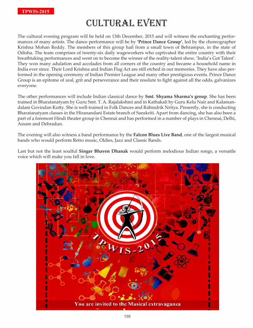
150
TPWIS-2015
CuLTurAL EVEnTThe cultural evening program will be held on 13th December, 2015 and will witness the enchanting perfor-mances of many artists. The dance performance will be by ‘Prince Dance Group’, led by the choreographer Krishna Mohan Reddy. The members of this group hail from a small town of Behrampur, in the state of Odisha. The team comprises of twenty-six daily wageworkers who captivated the entire country with their breathtaking performances and went on to become the winner of the reality-talent show, ’India’s Got Talent’. They won many adulation and accolades from all corners of the country and became a household name in India ever since. Their Lord Krishna and Indian Flag Act are still etched in our memories. They have also per-formed in the opening ceremony of Indian Premier League and many other prestigious events. Prince Dance Group is an epitome of zeal, grit and perseverance and their resolute to fight against all the odds, galvanizes everyone.
The other performances will include Indian classical dance by Smt. Shyama Sharma’s group. She has been trained in Bharatanatyam by Guru Smt. T. A. Rajalakshmi and in Kathakali by Guru Kelu Nair and Kalaman-dalam Govindan Kutty. She is well-trained in Folk Dances and Rabindrik Nritya. Presently, she is conducting Bharatanatyam classes in the Hiranandani Estate branch of Sanskriti. Apart from dancing, she has also been a part of a foremost Hindi theater group in Chennai and has performed in a number of plays in Chennai, Delhi, Assam and Dehradun.
The evening will also witness a band performance by the Falcon Blues Live Band, one of the largest musical bands who would perform Retro music, Oldies, Jazz and Classic Bands.
Last but not the least soulful Singer Bhaven Dhanak would perform melodious Indian songs, a versatile voice which will make you fall in love.