degradative proteomics and disease mechanisms
TRANSCRIPT
REVIEW
Degradative proteomics and disease mechanisms
Francesco Lanucara1,2,3, Philip Brownridge2, Iain S. Young2, Phillip D. Whitfield2
and Mary K. Doherty1
1 The Physiological Laboratory, School of Biomedical Sciences, Faculty of Medicine, University of Liverpool, UK2 Proteomics and Functional Genomics Research Group, Faculty of Veterinary Science, University of Liverpool, UK3 Dipartimento di Chimica e Tecnologie del Farmaco, Universita di Roma La Sapienza, Italy
Received: July 29, 2009
Revised: September 22, 2009
Accepted: October 8, 2009
Protein degradation is a fundamental biological process, which is essential for the main-
tenance and regulation of normal cellular function. In humans and animals, proteins can be
degraded by a number of mechanisms: the ubiquitin-proteasome system, autophagy and
intracellular proteases. The advances in contemporary protein analysis means that proteo-
mics is increasingly being used to explore these key pathways and as a means of monitoring
protein degradation. The dysfunction of protein degradative pathways has been associated
with the development of a number of important diseases including cancer, muscle wasting
disorders and neurodegenerative diseases. This review will focus on the role of proteomics to
study cellular degradative processes and how these strategies are being applied to understand
the molecular basis of diseases arising from disturbances in protein degradation.
Keywords:
Autophagy / Degradative proteomics / Disease / Proteasome / Ubiquitin
1 Introduction
Maintaining cellular homeostasis in response to changing
environmental conditions requires a continual cycle of
synthesis and degradation of intracellular proteins. Protein
synthesis is closely related to the transcriptome, while
protein degradation is regulated by two main downstream
pathways, the ubiquitin-proteasome system (UPS) [1] and
macro-autophagy, typically referred to as autophagy [2, 3].
Short-lived proteins are believed to be degraded predomi-
nantly by the UPS and long-lived proteins and organelles are
degraded in the lysosome by autophagy [4]. However, it has
been observed that there may be overlap between autophagy
and proteasomal degradation and they may indeed be
compensatory [5]. Proteins can also be degraded by a range
of cysteine proteases [6, 7] that cleave proteins into smaller
fragments, which can then be further broken down by other
pathways.
Proteomic strategies are increasingly being used to probe the
structure and function of protein degradative pathways [8, 9].
Proteomics is a key technology for exploring these systems as it
has the ability to define changes in protein expression and
address the technically and conceptually challenging problems
of protein–protein interactions and PTMs. Investigators are also
beginning to adopt proteomic approaches to monitor the
dynamics of protein degradation in cells and complex organ-
isms [10–12]. In addition, proteomics is being used to under-
stand human diseases that result from the dysfunction of
protein degradative pathways. These include cancer [13, 14],
muscle wasting disorders [15, 16] and neurodegenerative
diseases [17, 18]. This review will focus on the role of proteo-
mics to study cellular degradation pathways and how these
strategies are being used to understand the molecular basis of
disease states arising from disturbances in protein degradation.
2 Protein degradative pathways
2.1 UPS
Proteasomal degradation is believed to be a protein-specific
pathway that permits the sensitive and rapid control of
Abbreviations: mdx, x-linked muscular dystrophy; MuRF1,
muscle-specific-RING-finger protein 1; SILAC, stable isotope
labelling of amino acids in cell culture; UPS, ubiquitin-protea-
some system
Correspondence: Dr. Mary K. Doherty, School of Biomedical
Sciences, University of Liverpool, Crown Street, Liverpool L69
3BX, UK
E-mail: [email protected]
Fax: 144-151-794-4989
& 2010 WILEY-VCH Verlag GmbH & Co. KGaA, Weinheim www.clinical.proteomics-journal.com
Proteomics Clin. Appl. 2010, 4, 133–142 133DOI 10.1002/prca.200900159
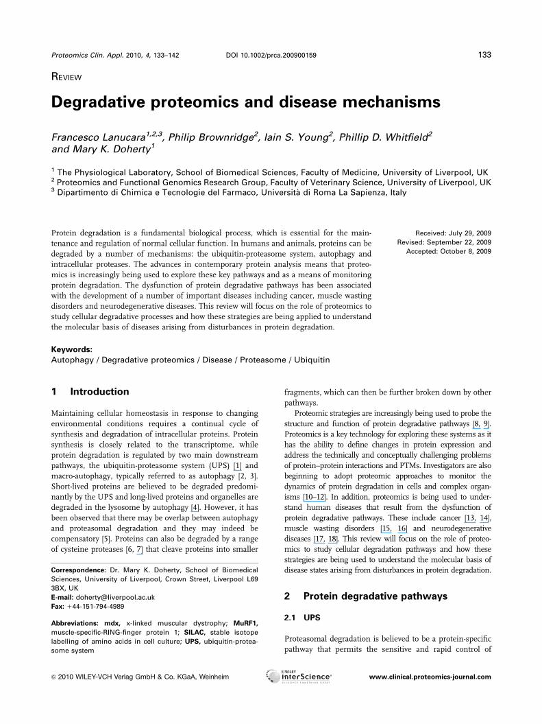
cellular protein composition (Fig. 1). The 26S proteasome is
a multimeric complex (approximately 2.5 MDa), comprising
19S cap structures attached to either end of a barrel-shaped
20S core particle that contains the proteolytic machinery
[19]. In mammalian systems the 20S core particle is
composed of at least 14 subunits, designated a1–7 and b1–7.
The subunits are assembled into four stacked rings, each
containing seven subunits to form the cylindrical abba core.
While the a-subunits are required for structural arrange-
ment and regulation of substrate access, the b-subunits 1, 2
and 5, are catalytically active. The 19S cap is constituted
from at least 18 different subunits. These form a base that
sits adjacent to the 20S core and a lid, which is thought to be
involved in substrate recognition.
Ubiquitin is a small (8.5 kDa) protein and is the primary
regulator of protein degradation by the proteasome. Proteins
are targeted for proteasomal degradation by the attachment
of polyubiquitin chains [20]. A single ubiquitin molecule is
covalently attached via its C-terminal glycine residue to a
specific lysine residue on the substrate protein. This is
mediated by E2 ubiquitin-activating enzymes and multiple
E3 ubiquitin ligases. Ubiquitin chains are subsequently
formed primarily by linkage through Lys48. Recent work
has indicated that proteasomal degradation of proteins may
also be mediated through the linkage of polyubiquitin
chains at Lys11 and Lys63 [21]. Further, there are reports
that proteins are targeted for degradation by the UPS
through ubiquitin linkage to the N-terminal amino acid
[22–24] or by attachment to serine or threonine residues [25].
The polyubiquitinated proteins are then recognised by the
19S cap of the 26S proteasome, and are committed to
degradation in the internal hydrolytic cavity of the 20S
proteasome core. Once a protein has been committed to the
proteasome, degradation to the peptide and amino acid
components is rapid, with the amino acids free for future
protein synthesis.
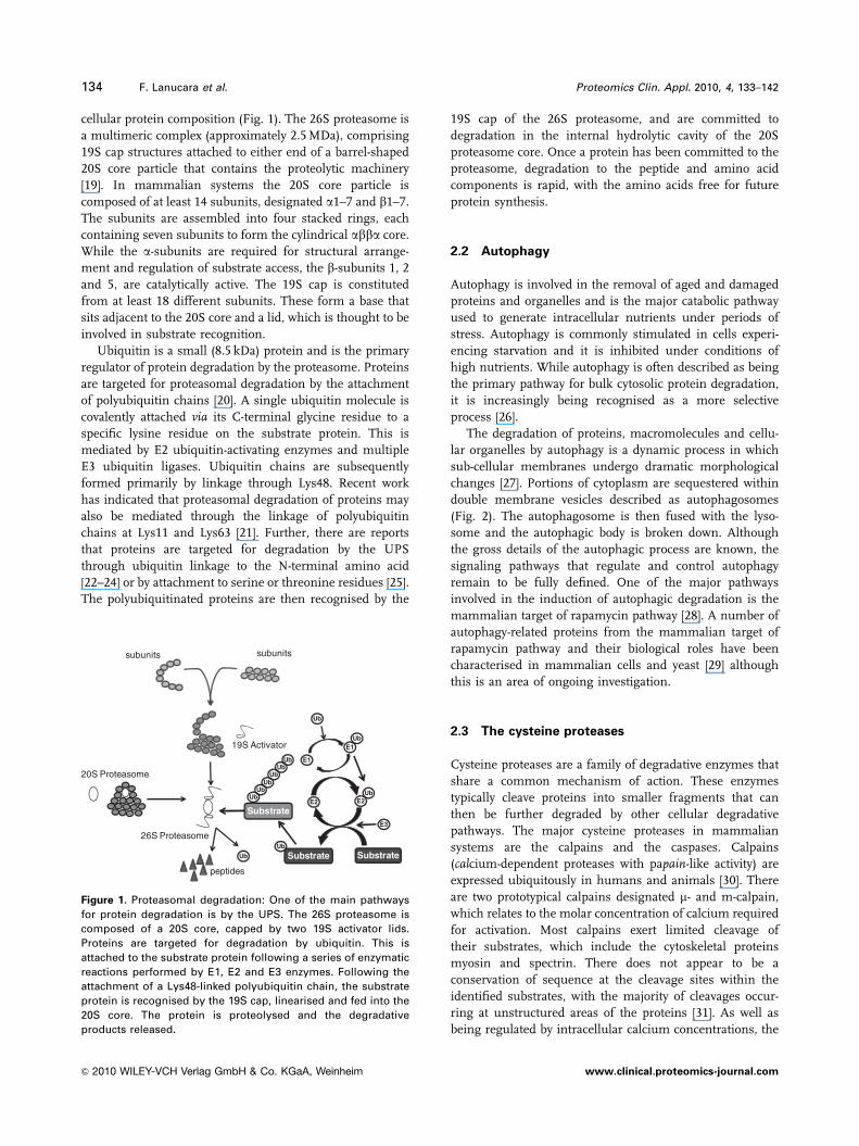
2.2 Autophagy
Autophagy is involved in the removal of aged and damaged
proteins and organelles and is the major catabolic pathway
used to generate intracellular nutrients under periods of
stress. Autophagy is commonly stimulated in cells experi-
encing starvation and it is inhibited under conditions of
high nutrients. While autophagy is often described as being
the primary pathway for bulk cytosolic protein degradation,
it is increasingly being recognised as a more selective
process [26].
The degradation of proteins, macromolecules and cellu-
lar organelles by autophagy is a dynamic process in which
sub-cellular membranes undergo dramatic morphological
changes [27]. Portions of cytoplasm are sequestered within
double membrane vesicles described as autophagosomes
(Fig. 2). The autophagosome is then fused with the lyso-
some and the autophagic body is broken down. Although
the gross details of the autophagic process are known, the
signaling pathways that regulate and control autophagy
remain to be fully defined. One of the major pathways
involved in the induction of autophagic degradation is the
mammalian target of rapamycin pathway [28]. A number of
autophagy-related proteins from the mammalian target of
rapamycin pathway and their biological roles have been
characterised in mammalian cells and yeast [29] although
this is an area of ongoing investigation.
2.3 The cysteine proteases
Cysteine proteases are a family of degradative enzymes that
share a common mechanism of action. These enzymes
typically cleave proteins into smaller fragments that can
then be further degraded by other cellular degradative
pathways. The major cysteine proteases in mammalian
systems are the calpains and the caspases. Calpains
(calcium-dependent proteases with papain-like activity) are
expressed ubiquitously in humans and animals [30]. There
are two prototypical calpains designated m- and m-calpain,
which relates to the molar concentration of calcium required
for activation. Most calpains exert limited cleavage of
their substrates, which include the cytoskeletal proteins
myosin and spectrin. There does not appear to be a
conservation of sequence at the cleavage sites within the
identified substrates, with the majority of cleavages occur-
ring at unstructured areas of the proteins [31]. As well as
being regulated by intracellular calcium concentrations, the
Substrate
subunits subunits
19S Activator
20S Proteasome
26S Proteasome
Ub
E1
E1
E2 E2
SubstrateSubstrate
Ub
peptides
UbUb
UbUb
Ub
Ub
Ub
Ub
Ub
E3
Figure 1. Proteasomal degradation: One of the main pathways
for protein degradation is by the UPS. The 26S proteasome is
composed of a 20S core, capped by two 19S activator lids.
Proteins are targeted for degradation by ubiquitin. This is
attached to the substrate protein following a series of enzymatic
reactions performed by E1, E2 and E3 enzymes. Following the
attachment of a Lys48-linked polyubiquitin chain, the substrate
protein is recognised by the 19S cap, linearised and fed into the
20S core. The protein is proteolysed and the degradative
products released.
134 F. Lanucara et al. Proteomics Clin. Appl. 2010, 4, 133–142
& 2010 WILEY-VCH Verlag GmbH & Co. KGaA, Weinheim www.clinical.proteomics-journal.com
activity of the calpains is also modulated by the specific
inhibitor calpstatin.
Caspases (cysteine proteases that cleave proteins at a
cysteine-aspartic scissile bond) are synthesised as catalyti-
cally inactive proenzymes comprising a number of
domains [32]. Activation of each caspase is induced by
proteolytic cleavage between domains, resulting in the
assembly of the active enzyme complex. The caspases
generally proceed by a cascade mechanism and effect
their proteolytic action by binding the relevant substrate
via a conserved amino acid recognition site. Caspases
have been identified as proteolysing a range of proteins
from the cytoskeleton (e.g. actin and gelsolin), nucleus
(e.g. lamin A and B and associated receptors) and those
involved in cellular signalling such as protein kinases and
cytokines.
3 Understanding degradativemechanisms through proteomics
Proteomics provides a variety of powerful tools with which
to investigate protein degradative pathways. Considerable
research has focused on the UPS [33]; however, investigators
have also begun to apply proteomic techniques to the study
of autophagy [34] and the identification of the primary
cleavage substrates of calpains and caspases [35, 36].
Gel-based approaches have been shown to be of consid-
erable value in visualising degradative products of tissue
proteins [37], while MS strategies have been adopted to
probe the structure of the proteasome and develop models
of its assembly, activation and interaction [38–40]. MS has
also been employed to characterise ubiquitinated and
phosphorylated proteins and to determine the localisation of
these key PTMs. In many studies these analyses have
involved the isolation and enrichment of the proteins of
interest [41–43]. The modifications have then been identified
using bioinformatic searches, which incorporate the corre-
sponding mass shifts. Ubiquitinated peptides have been
putatively identified by searching for the added mass of a
GlyGly tag to a lysine residue [44]. This di-glycine tag is
derived from the tryptic cleavage of the ubiquitin–substrate
complex. However, caution should be taken when using this
approach as a similar mass shift has been observed as an
artefact of alkylation with iodoacetamide [45]. It is proposed
that the use of chloroacetamide in studies focussed on the
identification of ubiquitin modification would minimise
false positive rates as it shows greater selectivity for cysteine
residues. In the case of phosphorylated proteins, phosphate
groups can be easily fragmented in a mass spectrometer
using neutral loss or MS3 strategies [46]. A limitation of this
approach is that the position of the phospho-modification, in
general, has to be manually determined, particularly when
the peptide is multiply phosphorylated or when consecutive
residues are potential modification sites. This restricts the
use of these methods in global profiling experiments.
Recent developments such as electron transfer dissociation
and electron capture dissociation fragment the peptide to
give a different ion series compared with conventional
fragmentation methods [47]. In many instances this allows
labile modifications including phosphate groups to be
retained on the amino acid facilitating more robust assign-
ment of localisation.
MS analyses have also been employed for the quantifi-
cation of modified proteins. These include methods for
the chemical labelling of proteins, e.g. iTRAQ [48] and
stable isotope labelled internal standards, e.g. AQUA [49].
Experimental strategies using stable isotope labelling of
Starvation/extracellular stress Decrease in nutrients/block in nutrient uptake
Decreased intracellular nutrientsSignalling events
nucleation
pre-autophagosome autophagosome autophagolysosome
expansion fusion
lysosomedegradation
TCA cycle
amino acids protein synthesis
fatty acids
Figure 2. Autophagy: Protein degradation in
response to starvation and other extra-
cellular stress is predominantly by autop-
hagy. A decrease in nutrients causes
activation of signalling pathways. Autop-
hagy is initiated by the formation of double-
membraned vesicles that encapsulate the
cellular material to be degraded. This
autophagosome fuses with the lysosome to
form an autophagolysosome in which
breakdown occurs. Amino acids are then
recycled to allow further protein synthesis.
Proteomics Clin. Appl. 2010, 4, 133–142 135
& 2010 WILEY-VCH Verlag GmbH & Co. KGaA, Weinheim www.clinical.proteomics-journal.com

amino acids in cell culture (SILAC)-based approaches have
been extended to permit multiplexed quantification and the
temporal analysis of PTM-modified protein populations [50].
More recently, SILAC has been adapted to allow the direct
quantification of protein synthesis and degradation rates
on a proteome-wide scale [51]. In this ‘‘dynamic SILAC’’
approach, the cultured cells are grown in the presence of
labelled amino acids until full incorporation is achieved. The
cells are then switched to media, which contains only light
amino acids and sampled over a period of time. The relative
loss of label over the ‘‘chase’’ period can be monitored by
MS allowing the rate of degradation to be calculated for
hundreds of proteins. The ability to accurately define rate
constants of degradation on a global scale presents an
exciting prospect for future research into diseases that are
influenced by degradative processes.
4 Protein degradation and disease
Damaged proteins can form aggregates and the prompt
removal of these proteins is essential in maintaining normal
cellular function. Disturbances of protein degradative
systems can therefore lead to the development of a variety of
disease states [52, 53]. The following section highlights
some key examples where proteomic approaches have been
used to investigate diseases that are linked to altered protein
degradation. The research presented aims to demonstrate
how proteomic technologies are providing a more compre-
hensive view of the cellular control mechanisms involved in
these complex pathologies.
4.1 Cancer
Cancer encompasses a group of diseases, which are char-
acterised by uncontrolled growth of cells, invasion of
adjacent tissues, and in many cases the dispersal of
the cells to other locations of the body, metastasis [54].
Proteomics has been used extensively to characterise
the relative differences in protein expression between
cancerous and non-cancerous tissues samples. In one such
investigation, a gel-based approach compared the protein
expression between normal and breast cancer tissues. The
study showed that the abundance of ubiquitin-specific
proteases, proteasome subunits and E3 enzymes was
increased in the cancerous tissue. This indicated that there
was a marked up-regulation in the activity of the UPS in the
disease state [55].
Other cancer proteomic studies have focussed on the role
of the UPS in metastasis. Zhang et al. [56] analysed three
mouse lung adenocarcinoma cell lines with different
metastatic potential. The cells were grown in the presence of35S-methionine and collected at three different time-points.
Proteins were then separated by 2-DE and quantified using
autoradiography. A comparison of the cellular proteomes
revealed that a number of proteins were differentially
expressed, which were then identified by MALDI-MS/MS.
Among these, 28 proteins showed a systematic trend
corresponding to metastatic potential. Bioinformatics
analysis indicated that several of these proteins were
involved in proteasome, cell-cycle and cell communication
pathways. These included keratins, 14-3-3 proteins and
components of the 26S proteasome whose aberrant expres-
sion may be directly or indirectly involved in cancer devel-
opment and metastasis.
A disturbance in autophagy has also been proposed to
play a role in the pathogenesis of cancer. An up-regulation
of autophagy in tumour cells could maintain the viability
and therefore survival of the cancerous cells. Indeed,
autophagy has been reported to be markedly increased
during the tumour formation [57]. Interestingly, tumour
cells can display a deficiency in autophagic degradation
[58, 59]. The impairment of autophagy may also affect the
response of cells to chemotherapy or radiotherapy by redu-
cing the ability of the cell to remove proteins or organelles,
which may have been damaged by the treatment [60, 61]. A
consensus role for autophagy in cancer development is still
lacking, [62] and in this regard proteomic studies may be
helpful in defining the specific correlation between autop-
hagic aberrations and distinct types of cancer.
In a recent study, DIGE was used to probe the link
between defective autophagy and the promotion of
tumourigenesis in immortalised baby mouse kidney cells
[63]. The authors observed that in autophagy-defective cells
subjected to metabolic stress there was an accumulation of
p62, an adaptor protein involved in the targeting of poly-
ubiquitinated proteins to the autophagosome membrane.
There is evidence to suggest that increased expression p62 is
linked to the promotion of tumourigenesis [64]. Moreover,
p62-deficient cells show reduced carcinogenic potential [65].
The study also showed that ER chaperones and proteins
from the protein disulfide isomerase family were elevated.
These proteins are involved in protein refolding, indicating
that in tumour cells there is a deficiency in the management
of protein turnover.
4.2 Diseases related to muscle wasting
Many disease states such as starvation, diabetes mellitus and
sepsis are characterised by accelerated proteolysis of skeletal
muscle. The loss of muscle mass is inextricably linked to
loss of protein, in particular myofibrillar proteins. In muscle
wasting, the increase in proteolysis occurs primarily via the
UPS although the other degradative pathways are involved,
especially where the muscle wastage is rapid [66].
Muscle-specific-RING-finger protein 1 (MuRF1) is an
ubiquitin ligase that is up-regulated during various muscle
wasting conditions. Although the substrates of this enzyme
are not fully defined, MuRF1 binds to large myofibrillar
proteins [67]. To test the hypothesis that MuRF1 promotes
136 F. Lanucara et al. Proteomics Clin. Appl. 2010, 4, 133–142
& 2010 WILEY-VCH Verlag GmbH & Co. KGaA, Weinheim www.clinical.proteomics-journal.com
muscle atrophy, transgenic mouse models over-expressing the
protein were generated [68]. A DIGE comparison of quad-
riceps from the transgenic and wild-type mice failed to reveal
an increase in the multi-ubiquitination of myosin, which has
previously been observed in human patients with muscle
wasting. However, pyruvate dehydrogenase, and its regulator,
PDK2 as well as phosphorylase b and glycogenin were
suppressed. These findings suggest that MuRF1 expression in
skeletal muscle is intimately involved with skeletal muscle
carbohydrate metabolism during metabolic stress.
Muscle wasting is also a feature of neuromuscular
disorders such as Duchenne muscular dystrophy. The lack
of dystrophin is known to initiate the loss of a range of
surface proteins including dystroglycans and sarcoglycans
in this disease state; however, the secondary effects on
global protein expression are poorly defined. To explore
these changes, Ge et al. [69, 70] analysed the proteome of
hind leg muscle from x-linked muscular dystrophy (mdx)
mice at different stages of the disease (1, 3 and 6 months) in
two related studies. Muscle proteins were separated by 2-DE
and identified using MALDI-QTOF. Comparison of the mdx
and control muscle revealed marked differences between the
overall protein patterns. Sixty proteins were found to be
differentially expressed, of which 40 were cytosolic and 20
were microsomal proteins. The authors noted that in the
3- and 6-month old mice, the majority of these proteins were
up-regulated, while at 1 month the same proteins were
down-regulated, which reflected changes in the disease
progression. Of particular note, there was a significant
decrease in the expression of adenylate kinase in mdx mice
(more than fourfold down-regulated). Adenylate kinase is an
important protein in the synthesis and regulation of
nucleotides and this protein may play a critical role in the
impaired regulation of energy metabolism in Duchenne
muscular dystrophy. Myosin light chain 2 was consistently
up-regulated in the mice at all ages and it was suggested that
this may be due to an abnormal state of differentiation in
the disease state.
Another proteomics study compared the diaphragm
muscle from control and mdx mice [71]. A total of 2398
individual protein spots were visualised with 35 being
differentially expressed. Of the proteins that were up-regu-
lated, cardiovascular heat shock protein was shown to have
an eightfold increase in concentration. This finding was
confirmed by Western blotting and was found to be more
pronounced as the mice aged. In addition, visualisation by
immunofluorescent microscopy indicated that the protein
was associated with cytoskeletal components of the muscle
fibre. It was postulated that the protein is involved in the
stress response to damaged fibres and may play an impor-
tant protective role. Interestingly, the Fbxo11 protein, the
substrate recognition component of an E3 ubiquitin–protein
ligase complex involved in ubiquitination, was found to be
down-regulated. A decrease of this protein supports the
hypothesis that the UPS is impaired in muscle-wasting
diseases.
Muscle wasting is also observed in sepsis; however, to
date, few proteomic studies have examined the biochemical
changes associated with this condition. One study has
employed a rat model of cutaneous burn injury with
superimposed infection to investigate changes in protein
expression in skeletal muscle [72]. The only protein that
showed a substantial up-regulation was myosin binding
protein-H. This protein has a strong affinity for myosin with
putative roles in the assembly of myofibrils. As the
concentration of myosin was greatly reduced, it was
proposed that myosin binding protein-H may play a role in
myosin degradation. A number of chaperone proteins,
e.g. HSPb6 and metabolic enzymes including ATP synthase
b-chain were found to be down-regulated, which the authors
suggested could lead to a concomitant decrease in global
protein synthesis following severe injury.
Aging is often accompanied by muscle wasting, a
condition known as sarcopenia. There can be a loss in
muscle mass, arising from both a decrease in the number of
muscle fibres and the atrophy of individual fibres. Structural
and chemical changes in myosin during contraction have
been linked to age-related decreases in specific force and
inhibition of contractility [73]. Aged muscle may also be
more susceptible to oxidative damage, which may contribute
to impaired muscle performance. DIGE has been used to
examine the differences in proteome between skeletal
muscle from young (20–30 years) and elderly, active
(75 years) subjects [74]. A number of proteins involved in
oxidative metabolism were found to be more abundant in
elderly muscle whereas enzymes catalysing reactions
involved in anaerobic metabolism, e.g. creatine kinase,
glycolytic enzymes and transport proteins were of lower
concentration. The expression profile of contractile proteins
was also altered. In elderly individuals there was a decrease
in the expression of the phosphorylated ‘‘fast’’ forms of
myosin light chain kinase. The authors commented that
these changes in expression may be linked to impaired
performance and increased fatigue.
4.3 Neurodegenerative diseases
Deregulation of protein degradative pathways has been
implicated in the pathogenesis of many neurodegenerative
disorders [75, 76]. A key feature of the pathology of many of
these diseases is the formation of insoluble protein aggre-
gates in the brain. The plaques can be made up from
mutant, misfolded or damaged intracellular proteins, which
degradative processing has failed to remove. The case for
examining autophagy in neurological disease states has
been driven by the observation that the disruption of crucial
autophagy genes in mice leads to the accumulation of
polyubiquitinated proteins, which may be particularly
harmful for neurons [77, 78]. Autophagic vacuoles contain-
ing disease-related proteins have also been observed in
regions of the brain affected by Alzheimer’s disease [79].
Proteomics Clin. Appl. 2010, 4, 133–142 137
& 2010 WILEY-VCH Verlag GmbH & Co. KGaA, Weinheim www.clinical.proteomics-journal.com

The UPS is also reported to play a critical role in neurolo-
gical disorders. A mouse model in which a component of
the 26S proteasome, PSMC1, was knocked out showed
impairment of protein degradation by the UPS and forma-
tion of neuronal Lewy-like inclusions [80].
Axonal degeneration is a common pathological feature
of many degenerative disorders; however, the underlying
molecular mechanisms have not yet been fully elucidated.
In a recent study, Goto et al. [81] employed a proteomics
approach to examine protein expression in sciatic nerves
in gracile axonal dystrophy mice. This mouse model used
is a knock-out of ubiquitin C-terminal hydrolase L1, a
de-ubiquitinating enzyme, which has been shown to stabi-
lise mono-ubiquitin in neurons. Depletion of ubiquitin
C-terminal hydrolase L1 leads to a decrease in mono-
ubiquitin and an impairment of proteasomal degradation.
Using DIGE, there was an age-dependent accumulation of
several proteins including GAPDH, 14-3-3, annexin V and
neurofilament L observed in gracile axonal dystrophy mice
compared with wild-type animals. GAPDH, and the
sulphonated form of the protein, was found to aggregate in
axons of the mouse model, indicating an oxidative stress
response.
Proteomic studies have been used to probe the link
between the UPS and Parkinson’s disease. S-nitrosylation of
cysteine residues in the E3 ubiquitin ligase parkin has been
observed both in animal models and in human brain
samples [82]. The PTM leads to an initial promotion of the
E3 ligase activity, resulting in autoubiquitination of parkin
itself. This autoubiquitination in turn results in down-
stream inactivation of the ligase and subsequent proteasome
dysfunction, aberrant protein accumulation and ultimately,
cell death. Moreover, parkin has been shown to be mutated
in Parkinson’s disease [83].
The relevance of PTMs of the 20S components has also
been investigated in Alzheimer’s and normal brain tissues.
Gillardon et al. [84], using DIGE and tandem MS, found that
the a7, a4, b2 and b7 subunits differed in their relative
abundances and showed changes in phosphorylation and
acetylation status. Although the functional relevance of
these modifications is not fully understood, it is possible
that they are involved in the interaction of the 20S subunits
with other proteins. In the case of Alzheimer’s, modification
of the a4 subunit may alter the interaction of the protea-
some with the amyloid peptide and contribute to the aber-
rancies of the neuronal amyloid-b processing [85].
The role of phosphorylation and polyubiquitination in
the impairment of the degradative pathways in neurological
diseases was the focus of another proteomic study on the
microtubule-associated protein Tau [86]. Tau is part of the
degradation-resistant core of neurofibrillary tangles along
with the senile plaques of amyloid-b peptide. The protein
was isolated from brain specimens from patients with
Alzheimer’s and LC-MS/MS was used to analyse the affinity
purified Tau. The study showed that the protein was (poly)-
ubiquitinated at six different identified lysine residues.
These binding sites were localised to the microtubule
binding domain. The polyubiquitin chains were found to be
either Lys48 or Lys6, both of which are involved in protein
degradation. Lys48 polyubiquitination is well-recognised as
the signal of targeting proteins for degradation by the UPS.
It has also been reported that modifications of Lys6 inhibits
ubiquitin-dependent protein degradation [87]. Therefore, the
identification of these modifications indicates that dysfunc-
tion of the UPS may initiate the formation of degradation-
resistant polyubiquitinated Tau tangles in Alzheimer’s. The
finding that Tau is ubiquitinated but not degraded in
Alzheimer’s disease has been confirmed in a proteomic
study by Riederer et al. [88] in which post-mortem brain
samples from middle-aged and elderly subjects with and
without Alzheimer’s disease were analysed.
A recent study has used an MS-based strategy to quantify
polyubiquitin chains in Huntington’s disease [89]. It was
shown that Lys48-linked polyubiquitin chains accumulated
in the brains from both a mouse model and human patients
with Huntington’s disease. Moreover, the accumulation of
other polyubiquitin chains, namely Lys63 and Lys11-linked,
indicated that more global changes in the ubiquitin system
are involved.
4.4 Lysosomal storage disorders
Lysosomes are important degradative compartments of
eukaryotic cells. Defects in lysosomal function are associated
with a group of over 50 inherited diseases known as the
lysosomal storage disorders. Clinically, these diseases
present with a diverse range of symptoms such as progres-
sive neurodegeneration, organomegaly and skeletal
abnormalities. The majority of lysosomal storage disorders
are caused by the deficiency of a single acid hydrolase;
although they can also result from the dysfunction of the
cellular machinery involved in lysosomal protein trafficking
and defective transporters that move the products of lyso-
somal catabolism across the lysosomal membrane.
There has been considerable focus in the application of
proteomic technologies to characterise the soluble and
membrane proteomes of the lysosome [90–92]. Proteomic
approaches have also been used to determine disease-
related disturbances in the protein profiles of body fluids
and tissues from patients with lysosomal storage disorders.
In a pilot study, a label-free LC-MS strategy was adopted to
define changes in the serum protein expression in patients
with type I Gaucher disease [93]. The analyses revealed
alterations in the concentrations of fibrinogens, comple-
ment cascade proteins and high-density lipoproteins in
affected individuals. The differential labelling of proteins
has been used to compare changes in the serum of
Fabry patients before and after a 6-month period of
enzyme replacement therapy [94]. Statistically significant
decreases in the relative concentrations of a2-HS glycopro-
tein, vitamin D-binding protein, transferrin, Ig-a-2 C chain
138 F. Lanucara et al. Proteomics Clin. Appl. 2010, 4, 133–142
& 2010 WILEY-VCH Verlag GmbH & Co. KGaA, Weinheim www.clinical.proteomics-journal.com

and a-2-antiplasmin were observed following enzyme
replacement therapy. More recently, Sleat et al. [95] analysed
mannose-6-phosphorylated proteins, which had been puri-
fied from brain autopsy samples of patients with suspected
lysosomal storage disorders. The authors reported that
spectral counting of peptides detected by MS/MS was able to
provide definitive diagnosis in 8 out of 23 cases and
suggested that this approach may prove to be a comple-
mentary tool to conventional methods used in charactering
lysosomal diseases.
Although a great deal of progress has been made in the
understanding of the metabolic and molecular basis of
lysosomal storage disorders, the mechanisms by which
lysosomal storage leads to cellular dysfunction have yet to be
fully elucidated. There has been an increasing interest in the
possible involvement of autophagy in the pathogenesis of
lysosomal storage disorders [96]. Studies have revealed that
in specific lysosomal diseases there is a deficiency in the
fusion of autophagosomes and lysosomes, leading to the
accumulation of protein aggregates and impaired organelle
turnover. These include Pompe disease [97], mucolipidosis
type IV [98], mucopolysaccharidosis type IIIA and multiple
sulfatase deficiency [99]. The investigation of defective
autophagy in lysosomal storage disorders is still emerging
and proteomic strategies offer the opportunity to further
explore this link and provide additional insights into the
pathophysiology of these disease states.
5 Concluding remarks
Cells continuously adapt to changing environmental condi-
tions by adjusting their protein content to prevailing needs.
This homeostasis involves continuous biosynthesis and
degradative processes. Efficient protein degradation is
essential for the maintenance of cellular health as damaged
proteins commonly develop abnormal intermolecular
interactions resulting in the formation of aggregates. As
such the prompt degradation of irreversibly damaged
proteins is essential to preserve normal cellular function. A
number of disease states have been linked to the malfunc-
tion of cellular degradative pathways, in particular the UPS
and autophagy. While much is known about the regulation
of these pathways under normal conditions in cell culture
system, it is only recently that we are beginning to combine
clinical understanding with fundamental biochemical
knowledge. In many instances, for example, it is not clear
whether the degradative malfunction is a direct cause or a
downstream effect of the disease.
Proteomics is a key strategy in defining the role of
degradative pathways in disease processes. With more
recent advances in proteome quantification and character-
isation of PTMs, it is becoming possible to accurately define
changes in protein pathways due to disease states. In
combination with methods to accurately define individual
protein degradation rates on a large scale, we are moving to
a robust, global understanding of how the cells degradative
mechanisms interact and function in the normal cell and in
diseased conditions. In future, it will be important to inte-
grate data from proteomic-based investigations with other
‘‘omic’’ technologies and with clinical data to fully under-
stand the underlying pathology and biochemistry of these
varied diseases and to develop targeted therapies.
The authors wish to acknowledge the RCUK, The RoyalSociety and BBSRC for supporting research in their laboratories.
The authors have declared no conflict of interest.
6 References
[1] Hershko, A., Ciechanover, A., The ubiquitin system. Annu.
Rev. Biochem. 1998, 67, 425–479.
[2] Meijer, A. J., Codogno, P., Regulation and role of autophagy
in mammalian cells. Int. J. Biochem. Cell Biol. 2004, 36,
2445–2462.
[3] Ohsumi, Y., Protein turnover. IUBMB Life 2006, 58, 363–369.
[4] Ding, W. X., Yin, X. M., Sorting, recognition and activation
of the misfolded protein degradation pathways through
macroautophagy and the proteasome. Autophagy 2008, 4,
141–150.
[5] Pandey, U. B., Nie, Z., Batlevi, Y., McCray, B. A. et al.,
HDAC6 rescues neurodegeneration and provides an
essential link between autophagy and the UPS. Nature
2007, 447, 859–863.
[6] Goll, D. E., Thompson, V. F., Li, H., Wei, W., Cong, J., The
calpain system. Physiol. Rev. 2003, 83, 731–801.
[7] Kuranaga, E., Miura, M., Nonapoptotic functions of caspa-
ses: caspases as regulatory molecules for immunity and
cell-fate determination. Trends Cell Biol. 2007, 17, 135–144.
[8] Xu, P., Peng, J., Dissecting the ubiquitin pathway by
mass spectrometry. Biochim. Biophys. Acta 2006, 1764,
1940–1947.
[9] Kristensen, A. R., Schandorff, S., Hoyer-Hansen, M., Niel-
sen, M. O. et al., Ordered organelle degradation during
starvation-induced autophagy. Mol. Cell. Proteomics 2008,
7, 2419–2428.
[10] Pratt, J. M., Petty, J., Riba-Garcia, I., Robertson, D. H. et al.,
Dynamics of protein turnover, a missing dimension in
proteomics. Mol. Cell. Proteomics 2002, 1, 579–591.
[11] Cargile, B. J., Bundy, J. L., Grunden, A. M., Stephenson,
J. L., Jr., Synthesis/degradation ratio mass spectrometry for
measuring relative dynamic protein turnover. Anal. Chem.
2004, 76, 86–97.
[12] Doherty, M. K., Whitehead, C., McCormack, H., Gaskell,
S. J., Beynon, R. J., Proteome dynamics in complex
organisms: using stable isotopes to monitor individual
protein turnover rates. Proteomics 2005, 5, 522–533.
[13] Botti, J., Djavaheri-Mergny, M., Pilatte, Y., Codogno, P.,
Autophagy signaling and the cogwheels of cancer. Auto-
phagy 2006, 2, 67–73.
Proteomics Clin. Appl. 2010, 4, 133–142 139
& 2010 WILEY-VCH Verlag GmbH & Co. KGaA, Weinheim www.clinical.proteomics-journal.com

[14] Sato, K., Rajendra, E., Ohta, T., The UPS: a promising target
for breast cancer treatment. BMC Biochem. 2008, 9, S2.
[15] Ventadour, S., Attaix, D., Mechanisms of skeletal muscle
atrophy. Curr. Opin. Rheumatol. 2006, 18, 631–635.
[16] Nury, D., Doucet, C., Coux, O., Roles and potential ther-
apeutic targets of the ubiquitin proteasome system in
muscle wasting. BMC Biochem. 2007, 8, S7.
[17] Lim, K. L., Ubiquitin-proteasome system dysfunction in
Parkinson’s disease: current evidence and controversies.
Exp. Rev. Proteomics 2007, 4, 769–781.
[18] Nedelsky, N. B., Todd, P. K., Taylor, J. P., Autophagy and
the ubiquitin-proteasome system: collaborators in neuro-
protection. Biochim. Biophys. Acta 2008, 1782, 691–699.
[19] Eytan, E., Ganoth, D., Armon, T., Hershko, A., ATP-depen-
dent incorporation of 20S protease into the 26S complex
that degrades proteins conjugated to ubiquitin. Proc. Natl.
Acad. Sci. USA 1989, 86, 7751–7755.
[20] Chau, V., Tobias, J. W., Bachmair, A., Marriott, D. et al.,
A multiubiquitin chain is confined to specific lysine in a
targeted short-lived protein. Science 1989, 243, 1576–1583.
[21] Xu, P., Duong, D. M., Seyfried, N. T., Cheng, D. et al.,
Quantitative proteomics reveals the function of unconven-
tional ubiquitin chains in proteasomal degradation. Cell
2009, 137, 133–145.
[22] Breitschopf, K., Bengal, E., Ziv, T., Admon, A., Ciechanover,
A., A novel site for ubiquitination: the N-terminal residue,
and not internal lysines of MyoD, is essential for conjuga-
tion and degradation of the protein. EMBO J. 1998, 17,
5964–5973.
[23] Kuo, M. L., den Besten, W., Sherr, C. J., N-Terminal poly-
ubiquitination of the ARF tumor suppressor, a natural
lysine-less protein. Cell Cycle 2004, 3, 1367–1369.
[24] Ben-Saadon, R., Fajerman, I., Ziv, T., Hellman, U. et al., The
tumor suppressor protein p16(INK4a) and the human
papillomavirus oncoprotein-58 E7 are naturally occurring
lysine-less proteins that are degraded by the ubiquitin
system. Direct evidence for ubiquitination at the N-terminal
residue. J. Biol. Chem. 2004, 279, 41414–41421.
[25] Wang, X., Herr, R. A., Chua, W. J., Lybarger, L. et al.,
Ubiquitination of serine, threonine, or lysine residues on
the cytoplasmic tail can induce ERAD of MHC-I by viral E3
ligase mK3. J. Cell Biol. 2007, 177, 613–624.
[26] Klionsky, D. J., Let’s not forget about non-specific autop-
hagy. Autophagy 2006, 2, 257.
[27] Kim, J., Klionsky, D. J., Autophagy, cytoplasm-to-vacuole
targeting pathway, and pexophagy in yeast and mamma-
lian cells. Annu. Rev. Biochem. 2000, 69, 303–342.
[28] Arsham, A. M., Neufeld, T. P., Thinking globally and acting
locally with TOR. Curr. Opin. Cell Biol. 2006, 18, 589–597.
[29] Klionsky, D. J., Emr, S. D., Autophagy as a regulated path-
way of cellular degradation. Science 2000, 290, 1717–1721.
[30] Croall, D. E., Ersfeld, K., The calpains: modular designs and
functional diversity. Genome Biol. 2007, 8, 218.
[31] Tompa, P., Buzder-Lantos, P., Tantos, A., Farkas, A. et al.,
On the sequential determinants of calpain cleavage. J. Biol.
Chem. 2004, 279, 20775–20785.
[32] Chowdhury, I., Tharakan, B., Bhat, G. K., Caspases - an
update. Comp. Biochem. Physiol. B Biochem. Mol. Biol.
2008, 151, 10–27.
[33] Drews, O., Zong, C., Ping, P., Exploring proteasome
complexes by proteomic approaches. Proteomics 2007, 7,
1047–1058.
[34] Overbye, A., Fengsrud, M., Seglen, P. O., Proteomic analy-
sis of membrane-associated proteins from rat liver autop-
hagosomes. Autophagy 2007, 3, 300–322.
[35] Van Damme, P., Martens, L., Van Damme, J., Hugelier, K.
et al., Caspase-specific and nonspecific in vivo protein
processing during Fas-induced apoptosis. Nat. Methods
2005, 2, 771–777.
[36] Jang, M., Park, B. C., Lee, A. Y., Na, K. S. et al., Caspase-7
mediated cleavage of proteasome subunits during
apoptosis. Biochem. Biophys. Res. Commun. 2007, 363,
388–394.
[37] McLean, L., Young, I. S., Doherty, M. K., Robertson, D. H.
et al., Global cooling: cold acclimation and the expression
of soluble proteins in carp skeletal muscle. Proteomics
2007, 7, 2667–2681.
[38] Sharon, M., Taverner, T., Ambroggio, X. I., Deshaies, R. J.,
Robinson, C. V., Structural organization of the 19S protea-
some lid: insights from MS of intact complexes. PLoS Biol.
2006, 4, e267.
[39] Witt, S., Kwon, Y. D., Sharon, M., Felderer, K. et al.,
Proteasome assembly triggers a switch required for active-
site maturation. Structure 2006, 14, 1179–1188.
[40] Bousquet-Dubouch, M. P., Baudelet, E., Guerin, F.,
Matondo, M. et al., Affinity purification strategy to capture
human endogenous proteasome complexes diversity and
to identify proteasome-interacting proteins. Mol. Cell.
Proteomics 2009, 8, 1150–1164.
[41] Tomlinson, E., Palaniyappan, N., Tooth, D., Layfield, R.,
Methods for the purification of ubiquitinated proteins.
Proteomics 2007, 7, 1016–1022.
[42] Hjerpe, R., Rodriguez, M. S., Efficient approaches for char-
acterizing ubiquitinated proteins. Biochem. Soc. Trans.
2008, 36, 823–827.
[43] Thingholm, T. E., Jensen, O. N., Larsen, M. R., Analytical
strategies for phosphoproteomics. Proteomics 2009, 9,
1451–1468.
[44] Peng, J., Schwartz, D., Elias, J. E., Thoreen, C. C. et al.,
A proteomics approach to understanding protein ubiquiti-
nation. Nat. Biotechnol. 2003, 21, 921–926.
[45] Nielsen, M. L., Vermeulen, M., Bonaldi, T., Cox, J. et al.,
Iodoacetamide-induced artifact mimics ubiquitination in
mass spectrometry. Nat. Methods 2008, 5, 459–460.
[46] Olsen, J. V., Blagoev, B., Gnad, F., Macek, B. et al., Global,
in vivo, and site-specific phosphorylation dynamics in
signaling networks. Cell 2006, 127, 635–648.
[47] Mikesh, L. M., Ueberheide, B., Chi, A., Coon, J. J. et al., The
utility of ETD mass spectrometry in proteomic analysis.
Biochim. Biophys. Acta 2006, 1764, 1811–1822.
[48] Pflieger, D., Junger, M. A., Muller, M., Rinner, O. et al.,
Quantitative proteomic analysis of protein complexes:
140 F. Lanucara et al. Proteomics Clin. Appl. 2010, 4, 133–142
& 2010 WILEY-VCH Verlag GmbH & Co. KGaA, Weinheim www.clinical.proteomics-journal.com
concurrent identification of interactors and their state of
phosphorylation. Mol. Cell. Proteomics 2008, 7, 326–346.
[49] Kirkpatrick, D. S., Gerber, S. A., Gygi, S. P., The absolute
quantification strategy: a general procedure for the quanti-
fication of proteins and post-translational modifications.
Methods 2005, 35, 265–273.
[50] Blagoev, B., Ong, S. E., Kratchmarova, I., Mann, M.,
Temporal analysis of phosphotyrosine-dependent signaling
networks by quantitative proteomics. Nat. Biotechnol. 2004,
22, 1139–1145.
[51] Doherty, M. K., Hammond, D. E., Clague, M. J., Gaskell,
S. J., Beynon, R. J., Turnover of the human proteome:
determination of protein intracellular stability by dynamic
SILAC. J. Proteome Res. 2009, 8, 104–112.
[52] Schwartz, A. L., Ciechanover, A., Targeting proteins for
destruction by the ubiquitin system: implications for human
pathobiology. Annu. Rev. Pharmacol. Toxicol. 2009, 49,
73–96.
[53] Martinet, W., Agostinis, P., Vanhoecke, B., Dewaele, M.,
De Meyer, G. R., Autophagy in disease: a double-edged sword
with therapeutic potential. Clin. Sci. (Lond.) 2009, 116, 697–712.
[54] Hanahan, D., Weinberg, R. A., The hallmarks of cancer. Cell
2000, 100, 57–70.
[55] Deng, S., Zhou, H., Xiong, R., Lu, Y. et al., Over-expression
of genes and proteins of ubiquitin specific peptidases
(USPs) and proteasome subunits (PSs) in breast cancer
tissue observed by the methods of RFDD-PCR and proteo-
mics. Breast Cancer Res. Treat. 2007, 104, 21–30.
[56] Zhang, K., Wrzesinski, K., Stephen, J. F., Larsen, P. M. et al.,
Comparative proteome analysis of three mouse lung
adenocarcinoma CMT cell lines with different metastatic
potential by two-dimensional gel electrophoresis and mass
spectrometry. Proteomics 2008, 8, 4932–4945.
[57] Rez, G., Toth, S., Palfia, Z., Cellular autophagic capacity is
highly increased in azaserine-induced premalignant atypi-
cal acinar nodule cells. Carcinogenesis 1999, 20, 1893–1898.
[58] Gunn, J. M., Clark, M. G., Knowles, S. E., Hopgood, M. F.,
Ballard, F. J., Reduced rates of proteolysis in transformed
cells. Nature 1977, 266, 58–60.
[59] Kisen, G. O., Tessitore, L., Costelli, P., Gordon, P. B. et al.,
Reduced autophagic activity in primary rat hepatocellular
carcinoma and ascites hepatoma cells. Carcinogenesis
1993, 14, 2501–2505.
[60] Abedin, M. J., Wang, D., McDonnell, M. A., Lehmann, U.,
Kelekar, A., Autophagy delays apoptotic death in breast
cancer cells following DNA damage. Cell Death Differ. 2007,
14, 500–510.
[61] Amaravadi, R. K., Yu, D., Lum, J. J., Bui, T. et al., Autophagy
inhibition enhances therapy-induced apoptosis in a myc-
induced model of lymphoma. J. Clin. Invest. 2007, 117,
326–336.
[62] White, E., DiPaola, R. S., The double-edged sword of
autophagy modulation in cancer. Clin. Cancer Res. 2009, 15,
5308–5316.
[63] Mathew, R., Karp, C. M., Beaudoin, B., Vuong, N. et al.,
Autophagy suppresses tumorigenesis through elimination
of p62. Cell 2009, 137, 1062–1075.
[64] Moscat, J., Diaz-Meco, M. T., p62 at the crossroads of
autophagy, apoptosis, and cancer. Cell 2009, 137,
1001–1004.
[65] Duran, A., Linares, J. F., Galvez, A. S., Wikenheiser, K. et al.,
The signaling adaptor p62 is an important NF-kappaB
mediator in tumorigenesis. Cancer Cell 2008, 13, 343–354.
[66] Taillandier, D., Combaret, L., Pouch, M. N., Samuels, S. E.
et al., The role of ubiquitin-proteasome-dependent proteo-
lysis in the remodelling of skeletal muscle. Proc. Nutr. Soc.
2004, 63, 357–361.
[67] Bodine, S. C., Latres, E., Baumhueter, S., Lai, V. K. et al.,
Identification of ubiquitin ligases required for skeletal
muscle atrophy. Science 2001, 294, 1704–1708.
[68] Hirner, S., Krohne, C., Schuster, A., Hoffmann, S. et al.,
MuRF1-dependent regulation of systemic carbohydrate
metabolism as revealed from transgenic mouse studies.
J. Mol. Biol. 2008, 379, 666–677.
[69] Ge, Y., Molloy, M. P., Chamberlain, J. S., Andrews, P. C.,
Proteomic analysis of mdx skeletal muscle: great reduction
of adenylate kinase 1 expression and enzymatic activity.
Proteomics 2003, 3, 1895–1903.
[70] Ge, Y., Molloy, M. P., Chamberlain, J. S., Andrews, P. C.,
Differential expression of the skeletal muscle proteome in
mdx mice at different ages. Electrophoresis 2004, 25,
2576–2585.
[71] Doran, P., Martin, G., Dowling, P., Jockusch, H., Ohlendieck, K.,
Proteome analysis of the dystrophin-deficient MDX
diaphragm reveals a drastic increase in the heat shock protein
cvHSP. Proteomics 2006, 6, 4610–4621.
[72] Duan, X., Berthiaume, F., Yarmush, D., Yarmush, M. L.,
Proteomic analysis of altered protein expression in skeletal
muscle of rats in a hypermetabolic state induced by burn
sepsis. Biochem. J. 2006, 397, 149–158.
[73] D’Antona, G., Pellegrino, M. A., Adami, R., Rossi, R. et al.,
The effect of ageing and immobilization on structure and
function of human skeletal muscle fibres. J. Physiol. 2003,
552, 499–511.
[74] Gelfi, C., Vigano, A., Ripamonti, M., Pontoglio, A. et al., The
human muscle proteome in aging. J. Proteome Res. 2006, 5,
1344–1353.
[75] Rubinsztein, D. C., The roles of intracellular protein-degra-
dation pathways in neurodegeneration. Nature 2006, 443,
780–786.
[76] Upadhya, S. C., Hegde, A. N., Role of the ubiquitin protea-
some system in Alzheimer’s disease. BMC Biochem. 2007,
8, S12.
[77] Hara, T., Nakamura, K., Matsui, M., Yamamoto, A. et al.,
Suppression of basal autophagy in neural cells causes
neurodegenerative disease in mice. Nature 2006, 441,
885–889.
[78] Komatsu, M., Waguri, S., Chiba, T., Murata, S. et al., Loss of
autophagy in the central nervous system causes neurode-
generation in mice. Nature 2006, 441, 880–884.
[79] Dickson, D. W., Ksiezak-Reding, H., Davies, P., Yen, S. H.,
A monoclonal antibody that recognizes a phosphorylated
epitope in Alzheimer neurofibrillary tangles, neurofilaments
Proteomics Clin. Appl. 2010, 4, 133–142 141
& 2010 WILEY-VCH Verlag GmbH & Co. KGaA, Weinheim www.clinical.proteomics-journal.com
and tau proteins immunostains granulovacuolar degen-
eration. Acta Neuropathol. 1987, 73, 254–258.
[80] Bedford, L., Hay, D., Devoy, A., Paine, S. et al., Depletion of
26S proteasomes in mouse brain neurons causes neuro-
degeneration and Lewy-like inclusions resembling human
pale bodies. J. Neurosci. 2008, 28, 8189–8198.
[81] Goto, A., Wang, Y. L., Kabuta, T., Setsuie, R. et al., Proteo-
mic and histochemical analysis of proteins involved in the
dying-back-type of axonal degeneration in the gracile
axonal dystrophy (gad) mouse. Neurochem. Int. 2009, 54,
330–338.
[82] Yao, D., Gu, Z., Nakamura, T., Shi, Z. Q. et al., Nitrosative
stress linked to sporadic Parkinson’s disease: S-nitrosyla-
tion of parkin regulates its E3 ubiquitin ligase activity. Proc.
Natl. Acad. Sci. USA 2004, 101, 10810–10814.
[83] Shimura, H., Hattori, N., Kubo, S., Mizuno, Y. et al., Familial
Parkinson disease gene product, parkin, is a ubiquitin-
protein ligase. Nat. Genet. 2000, 25, 302–305.
[84] Gillardon, F., Kloss, A., Berg, M., Neumann, M. et al.,
The 20S proteasome isolated from Alzheimer’s disease
brain shows post-translational modifications but unchan-
ged proteolytic activity. J. Neurochem. 2007, 101,
1483–1490.
[85] Nixon, R. A., Niemann-Pick Type C disease and Alzheimer’s
disease: the APP-endosome connection fattens up. Am. J.
Pathol. 2004, 164, 757–761.
[86] Cripps, D., Thomas, S. N., Jeng, Y., Yang, F. et al., Alzheimer
disease-specific conformation of hyperphosphorylated
paired helical filament-Tau is polyubiquitinated through
Lys-48, Lys-11, and Lys-6 ubiquitin conjugation. J. Biol.
Chem. 2006, 281, 10825–10838.
[87] Shang, F., Deng, G., Liu, Q., Guo, W. et al., Lys6-modified
ubiquitin inhibits ubiquitin-dependent protein degradation.
J. Biol. Chem. 2005, 280, 20365–20374.
[88] Riederer, I. M., Schiffrin, M., Kovari, E., Bouras, C., Riederer,
B. M., Ubiquitination and cysteine nitrosylation during
aging and Alzheimer’s disease. Brain Res. Bull. 2009, 80,
233–241.
[89] Bennett, E. J., Shaler, T. A., Woodman, B., Ryu, K. Y. et al.,
Global changes to the ubiquitin system in Huntington’s
disease. Nature 2007, 448, 704–708.
[90] Journet, A., Chapel, A., Kieffer, S., Roux, F., Garin, J., Proteo-
mic analysis of human lysosomes: application to monocytic
and breast cancer cells. Proteomics 2002, 2, 1026–1040.
[91] Bagshaw, R. D., Mahuran, D. J., Callahan, J. W., A proteo-
mic analysis of lysosomal integral membrane proteins
reveals the diverse composition of the organelle. Mol. Cell.
Proteomics 2005, 4, 133–143.
[92] Kollmann, K., Mutenda, K. E., Balleininger, M., Eckermann,
E. et al., Identification of novel lysosomal matrix proteins by
proteome analysis. Proteomics 2005, 5, 3966–3978.
[93] Vissers, J. P., Langridge, J. I., Aerts, J. M., Analysis and
quantification of diagnostic serum markers and protein
signatures for Gaucher disease. Mol. Cell. Proteomics 2007,
6, 755–766.
[94] Moore, D. F., Krokhin, O. V., Beavis, R. C., Ries, M. et al.,
Proteomics of specific treatment-related alterations in Fabry
disease: a strategy to identify biological abnormalities.
Proc. Natl. Acad. Sci. USA 2007, 104, 2873–2878.
[95] Sleat, D. E., Ding, L., Wang, S., Zhao, C. et al., Mass spec-
trometry-based protein profiling to determine the cause of
lysosomal storage diseases of unknown etiology. Mol. Cell.
Proteomics 2009, 8, 1708–1718.
[96] Walkley, S. U., Pathogenic cascades in lysosomal disease-
Why so complex? J. Inherit. Metab. Dis. 2009, 32, 181–189.
[97] Fukuda, T., Ewan, L., Bauer, M., Mattaliano, R. J. et al.,
Dysfunction of endocytic and autophagic pathways
in a lysosomal storage disease. Ann. Neurol. 2006, 59,
700–708.
[98] Vergarajauregui, S., Connelly, P. S., Daniels, M. P., Puer-
tollano, R., Autophagic dysfunction in mucolipidosis type IV
patients. Hum. Mol. Genet. 2008, 17, 2723–2737.
[99] Settembre, C., Fraldi, A., Jahreiss, L., Spampanato, C. et al.,
A block of autophagy in lysosomal storage disorders. Hum.
Mol. Genet. 2008, 17, 119–129.
142 F. Lanucara et al. Proteomics Clin. Appl. 2010, 4, 133–142
& 2010 WILEY-VCH Verlag GmbH & Co. KGaA, Weinheim www.clinical.proteomics-journal.com