w8.1 score: ictal findings
TRANSCRIPT

Standardized computer-based organized reporting of EEG:
SCORE*†S�andor Beniczky, ‡Harald Aurlien, ‡Jan C. Brøgger, †Anders Fuglsang-Frederiksen,
§Ant�onioMartins-da-Silva, ¶Eugen Trinka, #Gerhard Visser, *,**Guido Rubboli, *Helle Hjalgrim,
††Hermann Stefan, ‡‡Ingmar Ros�en, §§Jana Zarubova, ¶Judith Dobesberger, *JørgenAlving,
¶¶##Kjeld V. Andersen, ¶¶Martin Fabricius, *Mary D. Atkins, ***Miri Neufeld, †††Perrine Plouin,
‡‡‡PetrMarusic, §§§Ronit Pressler, ¶¶¶RutaMameniskiene, ††R€udigerHopfeng€artner,
#Walter van Emde Boas, and *PeterWolf
*Department of Clinical Neurophysiology, Danish Epilepsy Centre, Dianalund, Denmark; †Department of Clinical Neurophysiology,
University of Aarhus, Denmark; ‡Department of Neurology, University of Bergen, Norway; §Department of Neurological Disorders,
Hospital SantoAnt�onio and ICBAS, University of Porto, Portugal; ¶Department of Neurology, ParacelsusMedical University,
Salzburg, Austria; #Department of Clinical Neurophysiology, Epilepsy Institute in theNetherlands (SEIN), “Meer and Bosch,”
Heemstede, TheNetherlands; **IRCCS Institute of Neurological Sciences, Bellaria Hospital, Bologna, Italy; ††Epilepsy Center,
Neurological Clinic, University Hospital Erlangen, Erlangen, Germany; ‡‡Department of Clinical Neurophysiology, University of
Lund, Sweden; §§Department of Neurology, ThomayerHospital, Prague, Czech Republic; ¶¶Department of Clinical Neurophysiology,
GlostrupHospital, University of Copenhagen, Denmark; ##NørmarkHospital, Ishøj, Denmark; ***EEG and EpilepsyUnit,
Department of Neurology, Tel-Aviv SouraskyMedical Center, Israel; †††Hopital Necker EnfantsMalades, Paris, France;
‡‡‡Department of Neurology, 2nd Faculty ofMedicine, University Hospital Motol, CharlesUniversity in Prague, Prague, Czech
Republic; §§§GreatOrmond Street Hospital for Children, NHSTrust, London, United Kingdom; and ¶¶¶Department of Neurology,
Clinic of Neurology andNeurosurgery, Vilnius University Hospital “Santari�skiuzKlinikos,” Vilnius University, Lithuania1
SUMMARY
The electroencephalography (EEG) signal has a high com-
plexity, and the process of extracting clinically relevant
features is achieved by visual analysis of the recordings.
The interobserver agreement in EEG interpretation is
onlymoderate. This is partly due to themethod of report-
ing the findings in free-text format. The purpose of our
endeavor was to create a computer-based system for EEG
assessment and reporting, where the physicians would
construct the reports by choosing from predefined ele-
ments for each relevant EEG feature, as well as the clinical
phenomena (for video-EEG recordings). A working group
of EEG experts took part in consensus workshops in
Dianalund, Denmark, in 2010 and 2011. The faculty was
approved by the Commission on European Affairs of the
International League Against Epilepsy (ILAE). The work-
ing group produced a consensus proposal that went
through a pan-European review process, organized by the
European Chapter of the International Federation of
Clinical Neurophysiology. The SCORE2 software was
constructed based on the terms and features of the con-
sensus statement and it was tested in the clinical practice.
The main elements of SCORE are the following: personal
data of the patient, referral data, recording conditions,
modulators, background activity, drowsiness and sleep,
interictal findings, “episodes” (clinical or subclinical
events), physiologic patterns, patterns of uncertain signifi-
cance, artifacts, polygraphic channels, and diagnostic sig-
nificance. The following specific aspects of the neonatal
EEGs are scored: alertness, temporal organization, and
spatial organization. For each EEG finding, relevant fea-
tures are scored using predefined terms. Definitions are
provided for all EEG terms and features. SCORE can
potentially improve the quality of EEG assessment and
reporting; it will help incorporate the results of com-
puter-assisted analysis into the report, it will make possi-
ble the build-up of a multinational database, and it will
help in training young neurophysiologists.
KEY WORDS: Assessment, Database, Definitions, EEG,
Semiology, Terms.
The interobserver agreement in electroencephalography
(EEG) interpretation is only moderate (Van Donselaar
et al., 1992; Stroink et al., 2006). The EEG signal has a high
complexity. It depends on the intricate interplay between
the activation of neural networks, localization and orienta-
tion (Wong, 1998) of the source (dipole), and its propaga-
tion throughout the brain (Lopes da Silva & van Rotterdam,
1993 3; Scherg et al., 1999; Flemming et al., 2005). Although
certain aspects (such as spike detection) can be automated,
Accepted January 28, 2013.Address correspondence to S�andor Beniczky, Danish Epilepsy Centre,
Visbys All�e 5, 4293 Dianalund, Denmark. E-mail: [email protected]
Wiley Periodicals, Inc.© 2013 International League Against Epilepsy
1
2
3
4
5
6
7
8
9
10
11
12
13
14
15
16
17
18
19
20
21
22
23
24
25
26
27
28
29
30
31
32
33
34
35
36
37
38
39
40
41
42
43
44
45
46
47
48
49
50
51
52
53
54
55
1
Epilepsia, **(*):1–13, 2013
doi: 10.1111/epi.12135
SPECIAL REPORT
EP
I1
21
35B
Dispa
tch:
28.2.13
Jour
nal:
CE:V
inothk
umar
P.
JournalName
ManuscriptNo.
Autho
rRec
eive
d:No.
ofpa
ges:
13PE
:Sar
avan
an

the whole process of extracting clinically relevant features
cannot be computerized: it requires the assessment and
interpretation of the recording by trained experts. Therefore
it is, to some extent, subjective and dependent on the abili-
ties, training, and experience of the reader (EEGer; Stubbe
et al., 1982). Another factor probably contributing to the in-
terobserver differences is the free-text format of the EEG
descriptions. A recent study has demonstrated that the inte-
robserver agreement was consistently higher when the
observers had to choose mandatory terms than when they
could use optional ones (Gerber et al., 2008). When
assessed for a relatively well-defined and restricted feature
(such as presence or absence of epileptiform discharges) the
interobserver agreement has proved to be beyond 80%
(Stroink et al., 2006). However, the EEG recordings are
much more complex than to be described by a list of binomi-
nal measures. Free-text formats are sufficiently flexible to
reflect the various features extracted and weighted by the
observers, but it can also lead to suboptimal assessment of
the recordings: it does not prevent the observer from miss-
ing important features, it allows the use of terms not widely
accepted (local terminologies), and makes it difficult to
transfer the results of EEG assessment from one laboratory
to another (for building of a multinational database).
The objective of our endeavor was to construct software
for characterizing EEG and ictal clinical events, where the
physician chooses from predefined terms, simultaneously
generating a report and filling information into a database.
We wanted to develop a reliable tool to improve the quality
of EEG assessment, to increase interobserver agreement in
reporting EEG findings, to promote exchange of knowledge
between EEG centers, to construct a multinational database
for further research projects, and to assist in education and
training.
There have been several successful attempts in medicine
to standardize the evaluation and reporting of complex clini-
cal and/or electrographic patterns, for example, the Unified
Parkinson’s Disease Rating Scale and the scoring of the
polysomnography recordings using the AASM4 Manual for
the Scoring of Sleep and Associated Events.
Previous computerized EEG reporting systems have been
published (Aurlien et al., 1999; Finnerup et al., 1999). Rob-
ert R. Young, Keith H. Chiappa, and their colleagues at
Massachusetts General Hospital have in the 1980s used a
locally developed software package for reporting EEG find-
ings. Fixed terms could be selected and a report was semi-
automatically generated (personal communication). Ronald
Lesser and his colleagues at Johns Hopkins have been using
a locally developed software package (“Reporter”) since
1998 for reporting EEG findings. Since then they have pre-
pared 38,000 reports using the software (personal communi-
cation). Although “Reporter” does not construct a formal
database, terms in the EEG descriptions are standardized, so
that “keyword” searches can be used to help with a variety
of needs, including construction of a database. However, the
previous attempts did not reach broad international accep-
tance. One of them seemed to be too time-consuming in the
clinical practice (Finnerup et al., 1999) and it only built a
database, but not a report. Harald Aurlien and his colleagues
have been using software (“Holberg”) for reporting EEG
findings and generating a database since 1998, and a modi-
fied version of this software has been used for reporting all
standard EEG reports at the Danish Epilepsy center since
2009. In total, >36,000 EEG studies have been reported
using this software. The database that was automatically
generated during the reporting and made possible to address
specific issues related to certain aspects of the EEG, and this
led to three additional publications (Aurlien et al., 2004,
2007, 2009). However, these software packages remained
in local use only. Probably the reasons for failing to reach a
wider acceptance were the very different needs and tradi-
tions in different countries. To circumvent these problems,
we tried to make the software as user-friendly as possible,
and we tried to reach an international (in the first step a
European) consensus on the structure and terms necessary
for interpreting and reporting the EEG.
Methods 5
A working group of EEG experts took part in consensus
workshops in Dianalund, Denmark in 2010 and 2011. The
faculty of the workshop was approved by the Commission
on European Affairs of the International League Against
Epilepsy (ILAE), and the event was advertised on the home-
page of EUREPA (the education and research organization
of the CEA 6-ILAE). The SCORE working group, consisting
of 25 clinical neurophysiologists/epileptologists from 15
European countries, elaborated a consensus proposal meant
to reflect the needs and practice in different countries/cen-
ters. This consensus proposal was subsequently submitted
to a pan-European review, organized by the European Chap-
ter of the IFCN 7.
The SCOREworking group followed the widely accepted
international standards: we incorporated the available,
relevant guidelines, consensus statements, and task-force
proposals (Chatrian et al., 1974; Committee, 1981; Daube
et al., 1993; Glimore, 1994; Noachtar et al., 1999; Blume
et al., 2001; Flink et al., 2002; AASM, 2007; ACNS, 2007)
as well as the terms described in authoritative EEG text-
books (Ebersole & Pedley, 2003; Niedermeyer & da Silva,
2005). We added to them or modified them only when abso-
lutely necessary, based on published evidence and/or the
consensus of the group. Because video-EEG recordings
contain data on seizure semiology too, we attempted to
include this into the structured report. The definitions for
the terms used in SCORE are attached as Supporting
Information. In the software these definitions are directly
available for each term.
Based on the consensus statement, the SCORE software
has been developed by a group of programmers at the
Epilepsia, **(*):1–13, 2013doi: 10.1111/epi.12135
2
S. Beniczky et al.
1
2
3
4
5
6
7
8
9
10
11
12
13
14
15
16
17
18
19
20
21
22
23
24
25
26
27
28
29
30
31
32
33
34
35
36
37
38
39
40
41
42
43
44
45
46
47
48
49
50
51
52
53
54
55

Holberg EEG AS8 . The programming work was organized
and supervised by one of the authors (HA), and, during this
process, the content of the software was repeatedly com-
pared and synchronized with the consensus statement by
another author (SB).
The development of the software took 3 years. It was an
iterative process, where successive versions were tested by
scoring EEG recordings in the clinical practice, and the soft-
ware was continuously corrected and adjusted. In total,
>2,000 recordings were included in this process, and four
major revisions were made, in addition to the numerous cor-
rections and trouble-shooting.
A free version of the SCORE software and a detailed
guidance for users can be requested from the following
home-page: http://holbergeeg.com9 .
The software automatically generates a report and saves
the scored features in a local database.
Results
Table 1 shows the main elements of SCORE, constituting
the flowchart of data evaluation and interpretation.
Elements 1–4 can be filled in by the EEG technician
(physiologist), and later checked by the physician who inter-
prets the recording. In the future, connecting the SCORE
software with the patient administrative system of the hospi-
tal/EEG department would help filling in these administra-
tive data. Naturally, several recordings can be listed for
each patient. Each recording has its own referral.
Elements 5–12 contain the main features assessed during
the process of reading the EEG recordings (grouped in the
software under “findings”). The list is long, because it is
meant to contain all clinically relevant aspects that can
occur during a diagnostic EEG recording. However, the
software was designed in such a way that the user does not
have to waste time on those features that do not occur in the
recording. In other words, if an element/feature is not appli-
cable for the recording to be described, one should not open
it from the list. The user chooses the complexity of scoring
according to the clinical setting and the recording.
The last element contains the overall interpretation of the
recording, where the physician specifies the diagnostic
significance. When this is done, a report is automatically
generated, and the features scored by the user are fed into
the database. The terms in the main flowchart are defined in
Appendix S1.
Personal data, referral, and recording conditions
SCORE is installed and run within the hospitals informa-
tion technology system. The users must ensure that the soft-
ware is used according to the local regulations for safety of
personal data. An “anonymization” function will be avail-
able. This will remove all personal data from an entry and
keep only the scored EEG features.
The patient’s personal data (“patient details”) contain
obligatory elements and optional ones. The obligatory ele-
ments are the following: identity number (“identity string” –
in most countries this is given by the social security
number), last name, first name, and date of birth. Optionally
one can record the patient’s address and other details (under
the entry: “notes”). For patients younger than 3 months, the
option of recording the mother’s name instead of the first
name of the patient is offered. For patients younger than
12 months, registering the gestational age is offered as an
option. In the next step, the recording conditions and the
referral data are entered.
If the patient is younger than 3 weeks at the time of the
recording, the software automatically switches to the spe-
cial, neonatal features; for patients between 3 and 5 weeks
of age, the physician can opt to use (or not) the neonatal
reporting-matrix.
The recording conditions contain: start time, duration of
the recording, EEG type (standard/sleep deprived/ambula-
tory recording/short-term video-EEG recording/long-term
video-EEG monitoring/recording in the intensive care unit),
and sensor group (10-20 system, 10-10 system). The sensor
array can be customized for each user site, in the settings.
The name of the technician, physician, and supervision phy-
sician (if any) is selected here. The alertness of the patient is
also registered by selecting item(s) from a multiple-choice
list: awake/oriented/good cooperation/poor cooperation/
disoriented/drowsy/asleep/stupor/comatose. Optionally the
time of the patient’s latest meal can be added. Other aspects
considered important for the recording can be detailed under
“notes.”
The “referral” part contains the following entries: refer-
ring unit, reason(s) for referral, diagnosis at referral (Inter-
national Classification of Diseases, 10th Edition [ICD-10]
list), latest seizure, medication (from the World Health
Organization [WHO] list, with the possibility of specifying
also medication withdrawal and medication administered
during the recording). A list of choices is offered for the
reasons for referral: epilepsy-related indications, other
differential diagnostic questions, specific pediatric indica-
tions, follow-up EEG, assessment of prognosis, and other
Table 1. Themain elements of SCORE: the flowchart
1. The patient’s personal data
2. Referral data
3. Recording conditions
4. Modulators/procedures
5. Background activity
6. Sleep and drowsiness
7. Interictal findings
8. Episodes
9. Physiologic patterns
10. Patterns of uncertain significance
11. EEG artifacts
12. Polygraphic channels
13. Diagnostic significance
Epilepsia, **(*):1–13, 2013doi: 10.1111/epi.12135
3
SCORE
1
2
3
4
5
6
7
8
9
10
11
12
13
14
15
16
17
18
19
20
21
22
23
24
25
26
27
28
29
30
31
32
33
34
35
36
37
38
39
40
41
42
43
44
45
46
47
48
49
50
51
52
53
54
55

indications (Table 2). Several, clinically relevant, non-EEG
data can be entered here.
The options for the “latest seizure” entry are the following:
“undetermined/unknown/<20 min/<1 h/<1 day/<1 week/
<1 month/longer than 1 month.10; 11 ”10; 11
Basic information on brain magnetic resonance imaging
(MRI) (normal/abnormal/not done/no data), functional neu-
roimaging tests (normal/abnormal/not done/no data), and
cognitive impairment (no/mental retardation/dementia due
to unique brain injury/dementia due to progressive disease)
can be included. More detailed information on these aspects
can be added as free text.
The list of provocation methods (“modulators/proce-
dures”) performed during the recording conceptually
belongs to the part with recording conditions. However, for
technical reasons (programming) this is listed in the soft-
ware as the first element of “findings.” This list contains the
following: intermittent photic stimulation (IPS), hyperventi-
lation, sleep deprivation, sleep (induced/natural/after sleep
deprivation), awakening, medication administered during
the recording, manual eye closure/opening, auditory stimu-
lation, nociceptive stimulation, physical effort, cognitive
tasks, and “other modulators and procedures” (specified in
free-text). Choosing hyperventilation prompts to the scoring
of the quality of performance during this (insufficient or suf-
ficient).
Until this point the data can be entered by the technicians/
physiologist.
Scoring the EEG
Elements 5–12 are grouped under the heading “findings”
in the SCORE software (Table 1). While reading the EEG,
the physician “scores” the relevant features of the recordings
using these entries. It follows the way electroencephalogra-
phers describe the EEG recordings. The first two elements
contain the features of the “ongoing” EEG activity during
wake period (“background activity”) and during drowsiness
and sleep. Interictal findings depict all the graphoelements/
EEG patterns that are considered abnormal, and that are not
part of the ongoing (background) activity, and that are not
the EEG manifestation of a seizure/clinical episode or of the
postictal period; the presence of interictal discharges does
not necessarily imply that the patient has epilepsy. The ele-
ment “episodes” contains the clinical and EEG features of
the seizures and other clinical/ictal events. Patterns of uncer-
tain significance, physiologic patterns, artifacts, and poly-
graphic channels can also be scored, if relevant/applicable.
In the end, the electroencephalographer scores the global
interpretation/diagnostic significance of the recording.
Finally a report is automatically generated. Figure 1 shows
the interactive screen of the SCORE software.
Background activity
Background activity contains three main subchapters:
posterior dominant rhythm, other organized rhythms, and
special features. The definition of the terms is detailed in
Appendix S2.
Table 3 shows the features (in bold) that can be scored
for the posterior dominant rhythm (alpha rhythm in adults)
and the corresponding choices for each of them. For each
recording the posterior dominant activity only can be scored
once 12. The electroencephalographer can score here the glo-
bal interpretation of the posterior dominant activity for the
patient (taking into account the age and the state of con-
sciousness). The following choices are available: normal,
abnormal, no definite abnormality, not possible to deter-
mine. To speed up scoring of most recordings, a short-key
redirects the flowchart to the window where frequency of
the posterior dominant rhythm can be specified.
Other organized rhythms (besides the posterior dominant
rhythm) can be selected. Here several entries are permitted
(i.e., several types of “other organized rhythms” can be
selected and successively scored for each recording). First,
one has to choose a name of the rhythm: alpha, beta, mu,
delta, theta. Then the localization has to be specified (see
below). The extent (i.e., percentage of occurrence during
the recording) and the reactivity to external stimuli (yes/no)
can be selected. The electroencephalographer can score here
Table 2. Reasons for referral
Epilepsy-related indications
Clinical suspicion of epilepsy or seizure
Reconsider the initial diagnosis of epilepsy
Classification of a patient diagnosed with epilepsy
Changes in seizure pattern
Suspicion of nonconvulsive status epilepticus
Monitoring of status epilepticus
Monitoring of seizure frequency
Monitoring the effect of medication
Considering stopping AED therapy
Presurgical evaluation
Driver’s license or flight certificate
Other differential diagnostic questions
Psychogenic nonepileptic seizures
Loss of consciousness
Disturbance of consciousness
Encephalopathy
Encephalitis
Dementia
Cerebral vascular disease
Paroxysmal behavioral changes
Other psychiatric
Behavioral symptoms
Coma
Brain death
Specific pediatric indications
Genetic syndrome
Metabolic disorder
Regression
Developmental problems
Follow-up EEG
Assessment of prognosis
Other indication
Epilepsia, **(*):1–13, 2013doi: 10.1111/epi.12135
4
S. Beniczky et al.
1
2
3
4
5
6
7
8
9
10
11
12
13
14
15
16
17
18
19
20
21
22
23
24
25
26
27
28
29
30
31
32
33
34
35
36
37
38
39
40
41
42
43
44
45
46
47
48
49
50
51
52
53
54
55

Figure 1.
The interactive screen for scoring EEG studies. The upper left panel (1) contains the flowchart—the main elements to be scored. If an
element is not relevant for the recording, it is simply skipped (it does not impose any additional burden). The selection panel (2) con-
tains the features to be scored for the element that was chosen in the flowchart (in this case an interictal finding) and the correspond-
ing choices. The upper row of tabs (3) in this panel shows the features available for scoring. In this example, for interictal findings the
available features are: name, location, time-related feature, and modulators. When the “name” feature is active, the selection panel
below displays the list of interictal graphoelements. After choosing “sharp-waves” from the name-list, the software automatically shifts
to the next feature to be scored (in this example: location). The right lower panel (4) contains a summary of the scored items corre-
sponding to the EEG finding that is being scored. The ribbon at the top (5) contains short-key functions (for example selecting “a”
opens directly “Posterior dominant rhythm” with the selection box where frequency values are typed in).
Epilepsia ILAE
Table 3. The features and choices for the posterior dominant rhythm
Frequency (Hz) Amplitude (lV) Reactivity to eye opening
Range (from… to…)
or
Single value (Hz)
<20
20–50
50–100
100–200
>200
Not possible to determine
Yes
Reduced – bilaterally
Reduced on the left side
Reduced on the right side
Not possible to determine
Amplitude asymmetry Frequency asymmetry Caveat
Symmetrical
L < R abnormal left
L < R abnormal right
R < L abnormal left
R < L abnormal right
Not possible to determine
Symmetrical
No. Hz lower on the left side
No. Hz lower on the right side
Only open eyes during the recording
Patient sleep deprived
Drowsy
Absent posterior dominant rhythm because of
Artifacts
Extreme low voltage
Eye closure could not be achieved
Lack of awake period
Lack of compliance
Other (free text)
COLOR
Epilepsia, **(*):1–13, 2013doi: 10.1111/epi.12135
5
SCORE
1
2
3
4
5
6
7
8
9
10
11
12
13
14
15
16
17
18
19
20
21
22
23
24
25
26
27
28
29
30
31
32
33
34
35
36
37
38
39
40
41
42
43
44
45
46
47
48
49
50
51
52
53
54
55

the interpretation of this rhythm (taking into account the age
and the state of consciousness) as normal, abnormal, no def-
inite abnormality.
The last subchapter for the background activity is: “spe-
cial features.” This contains three entities: electrocerebral
inactivity, generalized suppression-burst, and suppression
of the background activity. Selecting “electrocerebral
silence inactivity” makes it impossible to score any
additional feature, except for “artifacts” and “diagnostic
significance.” Selecting “generalized suppression-burst”
prompts to the scoring of the duration of the bursts and
(separately) the duration of the suppression. Selecting
“suppression” prompts to the scoring of the duration of
this. In the next step one can specify whether these entities
are modulated (influenced) by external stimuli/interven-
tions: passive eye opening, auditory stimuli, administered
medication. The possible choices are: increase, decrease,
unmodified, triggered by, stopped by, and “not possible to
determine.”
Scoring the location
Various entries (EEG patterns, graphoelements) have
“location” as an attribute. This is described by laterality
(left/right/midline/bilateral/diffuse) and regions (frontal,
temporal, central, parietal, occipital). Scoring “laterality”
and “regions” prompts (based on the sensor-settings speci-
fied in the recording conditions) a list with the sensors corre-
sponding to the selected side and region. Form this list,
individual electrodes can be unselected, and the maximum
of the field potential can be specified (by choosing one or
more electrode names).
For bilateral localizations, additional two features, the
amplitude asymmetry, and the bilateral synchrony have to
be scored. The choices for amplitude asymmetry are the fol-
lowing: symmetrical, consistently more pronounced on the
left side (>50% difference), consistently more pronounced
on the right side (>50% difference), shifting side-prepon-
derance, not possible to determine). The choices for syn-
chrony are the following: asynchronous, primary bilateral
synchrony, secondary bilateral synchrony (propagation
from left to right/from right to left), and cannot be deter-
mined.
The so-called “generalized” discharges are scored by
choosing: “bilateral” + “synchronous” + name of the
region.
“Diffuse” denotes a location of an EEG pattern (rhythm)
that occurs in all or most of the regions, on both sides, but
asynchronously.
If a graphoelement (transient) is seen in more than two
locations, unrelated to each other (i.e., not part of the same
discharge), “multifocal” can be selected as a descriptor of
localization.
If propagation is seen within a graphoelement, this is
scored in an additional location-window that is automati-
cally added when choosing “propagation.”
Traditionally, the location of the EEG patterns/graphoel-
ements is described by specifying the scalp regions (or elec-
trodes) where the negative potentials are recorded.
However, by visual assessment of the distribution of the
negative and positive potentials on the scalp (voltage map)
the brain region containing the source can be estimated.
Optionally this can be registered as a location-descriptor.
Choices for “brain-regions” (and sub-regions in the brack-
ets) are the following: frontal (perisylvian-superior surface;
lateral; mesial; polar; orbitofrontal), temporal (polar; basal,
lateral-anterior; lateral-posterior; perisylvian-inferior sur-
face), central (lateral convexity; mesial; fissural-anterior,
fissural-posterior; opercular), parietal (lateral-convexity;
mesial; opercular), occipital (lateral; mesial), and insula.
Sleep and drowsiness
The features of the “ongoing” activity during sleep are
scored just following the “background activity.” If abnor-
mal graphoelements (interictal findings) appear, disap-
pear, or change their morphology during sleep, that is not
scored here but at the entry corresponding to that gra-
phoelement (under modulator/sleep). Giving a detailed
description of sleep such as in polysomnography record-
ings is not the scope of this SCORE element. However,
the features considered clinically relevant in an EEG
recording are listed. The following entries can be selected:
sleep architecture, normal sleep patterns, hypnagogic or
hypnopompic hypersynchrony in children, sleep-onset
rapid eye movement sleep (SOREM), abnormal asymme-
try or absence of sleep graphoelements, nonreactive sleep
activity. Appendix S3 contains the definitions of these
terms.
Sleep architecture can be scored as normal/abnormal/not
possible to determine. For “normal sleep patterns” the sleep
stages reached during the recording can be specified (N1;
N2; N3; rapid eye movement [REM]; not possible to deter-
mine). For the absence of sleep graphoelements, the name
of the graphoelement (sleep spindles/vertex sharp tran-
sients/K-complex/POSTS 13/other) and the location where the
graphoelement is absent or reduced (see scoring the locali-
zation) is specified. Significant asymmetry of the sleep spin-
dles can be registered and the location (where it is reduced)
can be specified. Several successive entries can be selected
and scored for “sleep.”
Interictal findings
Each interictal finding is characterized by four attributes
(“features”): name of the graphoelement, localization, time-
related features, and modulators (if any).
The names of the interictal graphoelements are listed in
Table 4 and defined in Appendix S4. They are classified
into four groups to make it easier to find the name in the list.
The location is scored as described above.
The time-related features of the interictal findings are dis-
charge pattern, mode of appearance, and incidence.
Epilepsia, **(*):1–13, 2013doi: 10.1111/epi.12135
6
S. Beniczky et al.
1
2
3
4
5
6
7
8
9
10
11
12
13
14
15
16
17
18
19
20
21
22
23
24
25
26
27
28
29
30
31
32
33
34
35
36
37
38
39
40
41
42
43
44
45
46
47
48
49
50
51
52
53
54
55

“Discharge pattern” characterizes the time-related
features within the discharge. The choices are: single
discharges, rhythmic trains/bursts, and arrhythmic trains/
bursts. The frequency is specified for the rhythmic bursts by
entering the corresponding numbers (Hz). The duration is
specified for the trains/bursts. Because some graphoele-
ments might have more than one type of discharge pattern
within a recording, multiple choices are allowed here.
“Mode of appearance” depicts how the interictal EEG
patterns/graphoelements are distributed throughout the
recording. The choices are: random, periodical, variable,
and not possible to determine. If “periodical” is selected, the
duration of the interdischarge interval can be specified.
“Incidence” characterizes how often the described
interictal finding is seen throughout the recording. For the
single-discharges, the suggested choices for “incidence”
are: only once, <1, 1–3, 4–6/min, >1/10 s, and continuous.
For the trains/bursts the incidence is expressed as the
estimated percentage of the total duration of the bursts dur-
ing the recording (<1%, 1–10%, 10–50%, 50–90%, >90%).
Modulators
Some interictal findings are influenced by external stim-
uli/interventions. These can be described as the fourth fea-
ture: “modulators.” The choices for eye-closure sensitivity
are: yes and no. For the IPS the choices for photoparoxys-
mal response are: posterior stimulus-dependent response,
posterior stimulus independent response (limited to the
stimulus-train/sustained), and generalized photoparoxysmal
response (limited to the stimulus-train/sustained), as sug-
gested by Kasteleijn-Nolst Trenit�e et al. (2001). In addition,
“activation of preexisting epileptogenic area” can be
selected as a choice for IPS (Kasteleijn-Nolst Trenit�e et al.,
2001; Beniczky et al., 2012). For the other modulators the
following choices are available in SCORE: increase,
decrease, unmodified, only during this modulator, not possi-
ble to determine. For “sleep” two additional choices are
available: “change of pattern during sleep”—to make it pos-
sible to describe qualitative changes too (besides the quanti-
tative changes described above), and “continuous during
NREM sleep” to score for CSWS/ESES 15(the percent is
typed in the free-text box). Several, distinct interictal find-
ings can be scored independently/successively.
Episodes
This element of SCORE contains the descriptors of the
clinical episodes and of electrographic seizures.
The main parts of this element are: name of the clinical
episode, timing and context, effect of interventions, and
electroclinical findings.
The names of the episodes can be selected from the list
showed in Table 5 and Appendix S5. The table includes
ILAE seizure classification (Commission, 1981; Berg et al.,
2010). Focal seizures can be further classified according to
the presumed localization, and it can be specified whether
they evolve to bilateral convulsive seizure (impairment of
consciousness is scored in the next step).
“Timing and context” covers the following features: inci-
dence (number of episodes/recording), time at start, dura-
tion of the episode and of the postictal phase, prodrome,
state of wakefulness at the seizure start, impairment of con-
sciousness during the seizure, provocative factors, facilitat-
ing factors, tongue biting, effect of medication, and time
relationship between clinical and EEG start.
To reflect the clinical practice, SCORE makes it possible
to group and describe several clinical episodes (seizures)
under the same heading, if the physician considers them as
manifestation of the same phenomenon. However, as a min-
imum, the seizure onset must identical in all the clinical
episodes described under the same heading. If several clini-
cal episodes are described together, the number of such
episodes during the recording and the time of their start have
to be documented. For the cases where the precise number
of the clinical events cannot be determined, this is included
as a choice (“not possible to determine”).
The duration of the clinical episode (seconds) is regis-
tered. If several clinical events are described under the same
heading, the range is registered (one enters two numbers).
The option “>30 min” is given, if the precise length cannot
be determined.
Table 4. Names of the interictal graphoelements
Epileptiform discharges Special patterns
Polyspikes
Polyspike-and-slow-waves
Runs of rapid spikes
Sharp wave
Sharp-and-slow-waves
Slow sharp wave
Spike
Spike-and-slow-wave
Spike-and-slow-wave runs
Classical 3/s
Slow 1–2.5/s
Fast 4–5/s
Bi-PLEDS
Bursts suppression pattern
Hypsarrhythmia
PLEDs
Periodic complex discharges –
other than PLEDs
SIRPIDs
Triphasic waves
Abnormal slow
activity Neonatal
Delta
Polymorphic delta
Theta
Delta and theta
Intermittent rhythmic
slow activity
Frontal intermittent
rhythmic delta activity
(FIRDA)
Occipital intermittent
rhythmic delta activity
(OIRDA)
Temporal intermittent
rhythmic delta activity
(TIRDA14 )
Alpha bursts
Brief interictal rhythmic discharges
(BIRDS)
Positive Rolandic sharp waves
(PRSW)
Positive temporal sharp-waves
(PTS)
Epilepsia, **(*):1–13, 2013doi: 10.1111/epi.12135
7
SCORE
1
2
3
4
5
6
7
8
9
10
11
12
13
14
15
16
17
18
19
20
21
22
23
24
25
26
27
28
29
30
31
32
33
34
35
36
37
38
39
40
41
42
43
44
45
46
47
48
49
50
51
52
53
54
55

The prodrome (if any) can be selected, and then described
in free text. Prodrome is a preictal phenomenon, and it is
defined as a subjective or objective clinical alteration (e.g.,
ill-localized sensation or agitation) that heralds the onset of
an epileptic seizure but does not form part of it (Blume
et al., 2001). Therefore, prodrome should be distinguished
from aura (which is an ictal phenomenon).
One can specify whether the clinical event started from
sleep or from wake state. The impairment of consciousness
during the seizure (affected/mildly affected/not affected/not
possible to determine) can be scored here.
The facilitating factors (if known) can be selected: alco-
hol, awakening, catamenial, fever, sleep, sleep-depriva-
tion, other (free text). Facilitating factors are defined as
transient and sporadic endogenous or exogenous elements
capable of augmenting seizure incidence (increasing the
likelihood of seizure occurrence). The provocative factors
(if known) can be selected from the list: hyperventilation,
reflex (+free text), other (+free text). For IPS a list is
offered to select the type of photoparoxysmal response
(see under: “Modulators”). Provocative factors are defined
as transient and sporadic endogenous or exogenous ele-
ments capable of evoking/triggering seizures immediately
following the exposure to it.
Tongue biting can be selected and registered.
In this part one can specify whether the clinical start pre-
cedes the EEG start or the other way around. The time (in
seconds) between the clinical and EEG start can be docu-
mented by entering the corresponding number.
Effect of interventions
If medication was administered during the clinical event
(for example, to stop an epileptic seizure) the effect of medi-
cation can be scored: clinical effect (yes/no/not possible to
determine) and the EEG changes (decrease/cessation/no
change/increase/not possible to determine). Duration of the
changes induced by medication administered during the
recording can be entered here.
The electroclinical findings (i.e., the seizure semiology
and the ictal EEG) are divided in three phases: onset, propa-
gation, and postictal. For simple/short seizures the whole
seizure can be described under “onset.”
Within the onset period several clinical signs can be reg-
istered, but this implies that they occurred simultaneously.
For the propagation phase, several clinical signs/ictal EEG
patterns can be selected, and their chronologic order of
appearance can be specified. Because the elements of the
propagation might vary within the group of clinical episodes
described under the same heading, the number of episodes
in which that particular element occurred can be specified.
Otherwise the scoring of the onset and propagation phase is
identical.
The clinical signs are described by a name and the body
localization where it is observed. The list with the names
(Table 6, Appendix S5) corresponds to the ILAE Commis-
sion Report: Glossary of Descriptive Terminology for Ictal
Semiology (Blume et al., 2001).
Somatotopic modifiers describe the part of the body
where the clinical sign is manifested (Blume et al., 2001).
For some of the clinical signs (for example, dacrystic, gelas-
tic) the name determines the body part too. For others this
has to be selected from the list of choices: generalized (yes/
no), laterality (Left/Right/Bilateral – Symmetric/Left >
Right/Right > Left), body part (Eyelid/Face/Arm/Leg/
Trunk/Visceral/Hemi-), and the centricity (Axial/Proximal
limb/Distal limb).
Clinical and behavioral signs of ictal cognitive distur-
bances should be examined and recorded by testing with a
standardized protocol assessing the state of consciousness
(reactivity and orientation), memory, speech or language,
Table 5. Names of episodes
Generalized epileptic seizure
Tonic–clonic (in any combination)
Absence
Typical
Atypical
Absence with special features
Myoclonic absence
Eyelid myoclonia
Myoclonic
Myoclonic
Myoclonic atonic
Myoclonic tonic
Clonic
Tonic
Atonic
Focal seizure
Localization
Frontal/Temporal/Rolandic/Parietal/Occipital
Evolving to bilateral convulsive seizure
Other seizure types
Epileptic spasm
Tonic spasm
Unknown
Subtle seizure
Electrographic seizure
Psychogenic nonepileptic seizure (PNES)
Sleep-related events
Benign sleep myoclonus
Confusional awakening
Periodic limb movement in sleep (PLMS)
REM sleep behavioral disorder (RBD)
Sleep-walking
Pediatric events
Hyperekplexia
Jactatio capitis nocturna
Pavor nocturnus
Stereotypical behavior
Paroxysmal motor event
Other episodes
Syncope
Cataplexy
Other (free text)
Not possible to determine
Epilepsia, **(*):1–13, 2013doi: 10.1111/epi.12135
8
S. Beniczky et al.
1
2
3
4
5
6
7
8
9
10
11
12
13
14
15
16
17
18
19
20
21
22
23
24
25
26
27
28
29
30
31
32
33
34
35
36
37
38
39
40
41
42
43
44
45
46
47
48
49
50
51
52
53
54
55

motor and other neurologic functions of the patient (Velis
et al., 2007).
The ictal EEG pattern is described by its name and the
localization. The names selectable for ictal patterns are
shown in Table 7 and defined in Appendix S5. Where indi-
cated, frequency (Hz) and amplitude (lV) values can be
specified by entering the corresponding numbers.
The localization for these patterns is scored as described
above.
For the postictal phase, the list of clinical signs and the
list of EEG patterns are different from the onset and the
propagation phases. The possible choices are shown in
Table 8. The postictal EEG patterns are defined in Appen-
dix S5. The names of the clinical signs are selected from the
list according to the ILAE Commission Report: Glossary of
Descriptive Terminology for Ictal Semiology (Blume et al.,
2001).
In the postictal period, the clinical signs have as attribute
the somatotopic modifiers. The names of the postictal EEG
patterns have localization as an attribute (similarly to the
onset and propagation phases).
Physiologic patterns and patterns of uncertain
significance
These items are not considered abnormal, and they are
scored only if the physician finds a clinical relevance for it
(for example, emphasizing that they are not abnormal/cor-
recting the previous scoring of a junior physician, and so
on). Patterns of uncertain significance contain graphoele-
ments/EEG patterns that resemble abnormal ones, but in
most of the cases they are not associated with a pathologic
process (“normal variants”).
These items are described by two features: the name and
the localization. The list of the names and their definitions
are in Appendix S6. Localization is described as detailed
above.
Artifacts
In this SCORE element one can document the names
(Appendix S7) and localization of the artifacts, and one can
estimate the consequence of the artifacts on the recording:
not interpretable because of the artifacts/recording of
reduced diagnostic value due to artifacts/does not interfere
with the interpretation of the recording.
Polygraphic channels
In this part one can register the features related to the
additional (polygraphic) sensors: electrocardiography
(ECG), respiration measurements, electromyography (EMG).
The possible choices are shown in Table 9. Values (num-
bers) are entered, where specified, in the brackets.
Diagnostic significance
Before generating the report, the physician has to put the
scored EEG features into the clinical context. The
diagnostic significance of the recording offers three
choices: normal, no definite abnormality, and abnormal. If
Table 7. Names of the ictal EEG patterns
Burst-suppression pattern
DC-shift
Disappearance of ongoing activity
Electrodecremental change
Fast spike activity/repetitive spikes (Hz)
Irregular delta/theta activity (Hz)
Low-voltage fast activity (Hz)
Obscured by artifacts
Polysharp-waves
Polyspikes
Polyspike-and-slow-waves (Hz)
Rhythmic activity (Hz)
Sharp-and-slow-waves (Hz)
Slow wave of large amplitude (lV)
Spike-and-slow-waves (Hz)
Spike-and-slow-wave-runs
Classical 3/s
Slow 1–2.5/s
Fast 4–5/s
Other pattern (free text)
No demonstrable ictal EEG change
Not possible to determine
Table 6. Names of clinical signs during episodes
Elementary motor Automatisms Autonomic
Tonic
Dystonic
Epileptic spasm
Postural
Versive
Myoclonic
Clonic
Jacksonian march
Negative myoclonic
Tonic–clonic
Figure-of-four:
extended elbow:
left/right
Atonic
Astatic
Other (free text)
Dacrystic
Dysphasic
Dyspraxic
Gelastic
Gestural
20 Hyperkinetic
Hypokinetic
Manual or pedal
Mimetic
Oroalimentary
Vocal
Verbal
With preserved
responsiveness
Other (free text)
Cardiovascular
Gastrointestinal
Genital
Hypersalivation
Pupillary
Respiratory/apneic
Sudomotor
Thermoregulatory
Urinary incontinence
Vasomotor
Other (free text)
Sensory Experiential
Motor/behavioral
arrest
Dyscognitive
(free text)
Other clinical
sign (free text)
Not possible to
determine
Auditory
Autonomic
Cephalic/headache
Epigastric
Gustatory
Olfactory
Painful
Somatosensory
Visual
Other (free text)
Affective/emotional
Hallucinatory
Illusory
Mnemonic
d�ej�a-vu
jamais-vu
Other (free-text)
Epilepsia, **(*):1–13, 2013doi: 10.1111/epi.12135
9
SCORE
1
2
3
4
5
6
7
8
9
10
11
12
13
14
15
16
17
18
19
20
21
22
23
24
25
26
27
28
29
30
31
32
33
34
35
36
37
38
39
40
41
42
43
44
45
46
47
48
49
50
51
52
53
54
55

abnormal is selected, one has to specify it in more detail
(Table 10).
For “epilepsy,” further scoring of significance is avail-
able. The entries that can be selected here are not a formal
classification of epilepsies, but rather highlight the addi-
tional diagnostic information the EEG can provide in the
corresponding clinical context, as an element in the diagnos-
tic work-up.
In this part one can score the changes since the last (previ-
ous) EEG: no change/improved/worsened.
Generating the report
When the scoring is done, the report is automatically gen-
erated. The physician can review, edit, or change any part of
the scoring until the report is electronically signed and
saved. In the report-generating window, free-text parts can
be added, and a text-box for “summary” can be filled in.
This entry has a flexible format, where the personal wisdom
of the electroencephalographer can be distilled, and the rea-
sons for even syndrome diagnosis can be explained.
Specific aspects of the neonatal recordings
For newborns (neonatal period = first 28 days after birth)
the gestational age (GA) at birth is specified, and the GA at
the time of the recording is calculated. A specific neonatal
matrix is loaded instead of the “background activity” and
“sleep.” This matrix contains the specific features of the
neonatal ongoing activity and the characteristic transients.
The main elements are: behavioral stages (alertness),
temporal organization, and spatial organization. For each
entry (choice) of the behavioral stage, one can attribute tem-
poral organization and subsequently spatial organization.
These are scored further by their specific features. Temporal
organization characterizes the changes in time of the ongo-
ing EEG activity. The spatial organization codes the charac-
teristic neonatal EEG patterns (also including transients),
and their parameters – including location.
Table 8. Names of the postictal clinical signs and EEG
patterns
Postictal clinical signs
Postictal EEG
patterns
Anterograde amnesia
Aphasia/dysphasia
Behavioral change
Dysphoria
Headache
Hemianopia
Impaired cognition
Nose wiping
Paresis (Todd’s palsy)
Postictal sleep
Quick recovery of
consciousness
Retrograde amnesia
Unconscious
Unilateral motor
phenomena/
myoclonia
Other unilateral
motor
phenomena
Flattening
Increase in the interictal
epileptiform discharges
Periodic epileptiform
discharges
Slowing (Hz)
Other (free text)
Table 9. Features and choices for the polygraphic channels
Type of sensor Description Significance in relation to the clinical event Other features
ECG Normal rhythm
Asystolia (duration: range in seconds)
Bradycardia (beats/min: range)
Extrasystole
Tachycardia (beats/min: range)
Other (free text)
Cause
Consequence
No connected clinical episode
Undetermined
QT period (value)
Respiration Apnea; duration (range in seconds)
Hypopnea; duration (range in seconds)
Apnea-hypopnea index (events/h)
Periodic respiration
Tachypnea (frequency)
Other (free text)
Related to EEG/clinical episode
Unrelated to EEG/clinical episode
Not possible to determine
Saturation (%)
EMG Activity unrelated to the clinical event
Asymmetric activation of EMG (right first/left first)
Decreased activity related to the clinical event
Increased activity related to the clinical event
Myoclonus
Negative myoclonus
Related to EEG paroxysms
Rhythmic
Arrhythmic
Synchronous
Asynchronous
Periodic limb movements in sleep (PLMS)
Spasm
Tonic contraction
Other (free text)
Localization
Side (left, right, bilateral)
Name of muscle(s) (free text)
EOG16 Comments (free-text)
Other sensor (free text)
Epilepsia, **(*):1–13, 2013doi: 10.1111/epi.12135
10
S. Beniczky et al.
1
2
3
4
5
6
7
8
9
10
11
12
13
14
15
16
17
18
19
20
21
22
23
24
25
26
27
28
29
30
31
32
33
34
35
36
37
38
39
40
41
42
43
44
45
46
47
48
49
50
51
52
53
54
55
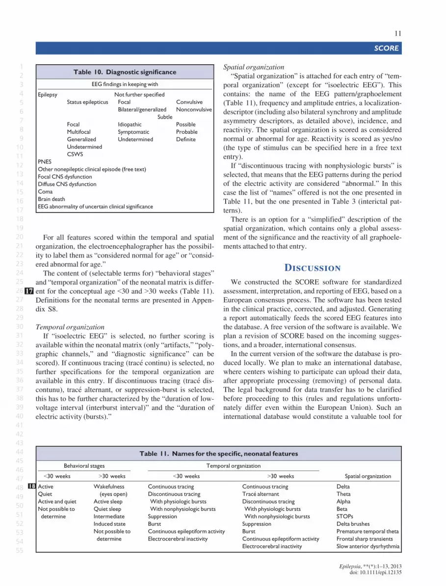
For all features scored within the temporal and spatial
organization, the electroencephalographer has the possibil-
ity to label them as “considered normal for age” or “consid-
ered abnormal for age.”
The content of (selectable terms for) “behavioral stages”
and “temporal organization” of the neonatal matrix is differ-
ent for the conceptual age <30 and >30 weeks17 (Table 11).
Definitions for the neonatal terms are presented in Appen-
dix S8.
Temporal organization
If “isoelectric EEG” is selected, no further scoring is
available within the neonatal matrix (only “artifacts,” “poly-
graphic channels,” and “diagnostic significance” can be
scored). If continuous tracing (trac�e continu) is selected, no
further specifications for the temporal organization are
available in this entry. If discontinuous tracing (trac�e dis-
contunu), trac�e alternant, or suppression-burst is selected,
this has to be further characterized by the “duration of low-
voltage interval (interburst interval)” and the “duration of
electric activity (bursts).”
Spatial organization
“Spatial organization” is attached for each entry of “tem-
poral organization” (except for “isoelectric EEG”). This
contains: the name of the EEG pattern/graphoelement
(Table 11), frequency and amplitude entries, a localization-
descriptor (including also bilateral synchrony and amplitude
asymmetry descriptors, as detailed above), incidence, and
reactivity. The spatial organization is scored as considered
normal or abnormal for age. Reactivity is scored as yes/no
(the type of stimulus can be specified here in a free text
entry).
If “discontinuous tracing with nonphysiologic bursts” is
selected, that means that the EEG patterns during the period
of the electric activity are considered “abnormal.” In this
case the list of “names” offered is not the one presented in
Table 11, but the one presented in Table 3 (interictal pat-
terns).
There is an option for a “simplified” description of the
spatial organization, which contains only a global assess-
ment of the significance and the reactivity of all graphoele-
ments attached to that entry.
Discussion
We constructed the SCORE software for standardized
assessment, interpretation, and reporting of EEG, based on a
European consensus process. The software has been tested
in the clinical practice, corrected, and adjusted. Generating
a report automatically feeds the scored EEG features into
the database. A free version of the software is available. We
plan a revision of SCORE based on the incoming sugges-
tions, and a broader, international consensus.
In the current version of the software the database is pro-
duced locally. We plan to make an international database,
where centers wishing to participate can upload their data,
after appropriate processing (removing) of personal data.
The legal background for data transfer has to be clarified
before proceeding to this (rules and regulations unfortu-
nately differ even within the European Union). Such an
international database would constitute a valuable tool for
Table 10. Diagnostic significance
EEG findings in keeping with
Epilepsy Not further specified
Status epilepticus Focal
Bilateral/generalized
Convulsive
Nonconvulsive
Subtle
Focal
Multifocal
Generalized
Undetermined
Idiopathic
Symptomatic
Undetermined
Possible
Probable
Definite
CSWS
PNES
Other nonepileptic clinical episode (free text)
Focal CNS dysfunction
Diffuse CNS dysfunction
Coma
Brain death
EEG abnormality of uncertain clinical significance
Table 11. Names for the specific, neonatal features
Behavioral stages Temporal organization
Spatial organization<30 weeks >30 weeks <30 weeks >30 weeks
18 Active
Quiet
Active and quiet
Not possible to
determine
Wakefulness
(eyes open)
Active sleep
Quiet sleep
Intermediate
Induced state
Not possible to
determine
Continuous tracing
Discontinuous tracing
With physiologic bursts
With nonphysiologic bursts
Suppression
Burst
Continuous epileptiform activity
Electrocerebral inactivity
Continuous tracing
Trac�e alternant
Discontinuous tracing
With physiologic bursts
With nonphysiologic bursts
Suppression
Burst
Continuous epileptiform activity
Electrocerebral inactivity
Delta
Theta
Alpha
Beta
STOPs
Delta brushes
Premature temporal theta
Frontal sharp transients
Slow anterior dysrhythmia
Epilepsia, **(*):1–13, 2013doi: 10.1111/epi.12135
11
SCORE
1
2
3
4
5
6
7
8
9
10
11
12
13
14
15
16
17
18
19
20
21
22
23
24
25
26
27
28
29
30
31
32
33
34
35
36
37
38
39
40
41
42
43
44
45
46
47
48
49
50
51
52
53
54
55

further research projects, as search criteria can be con-
structed to verify hypothesis or extract relevant information
from the database. In addition, the software makes it possi-
ble to compare the scored features in the report with a scored
“second opinion” from another laboratory on an EEG labo-
ratory. This offers the tools for quality control and audit.
SCORE will be helpful in bridging the gap between the
classical method of visual analysis of the EEG and the
advanced (computerized) analysis methods. The appropri-
ate analysis tools can be attached to the corresponding ele-
ments of SCORE (for example quantitative EEG analysis
method for “background activity”; source analysis methods
for the “localization descriptor,” and so on). The electroen-
cephalographer can enrich with these methods his arma-
mentarium for the analysis and interpretation of EEG
recordings in the clinical practice, by integrating their
results in the standardized EEG report.
Integration of SCORE with the patient administration
systems of the hospitals is going to save considerable time
and increase the feasibility.
The terms/features used in SCORE are provided with a
definition in the current version. The intention of the
SCORE consortium is to provide (besides the definition)
typical examples of EEG samples (screen shots) showing
the various features. Therefore, in addition to the definition,
an EEG sample will be accessible directly (from the feature
in question) in the software—the user will be able to open
this while scoring the EEG. We consider that this has
remarkable potential in training neurophysiologists.
In addition to EEG, MEG data will be integrated for stan-
dardized analysis in collaboration with the European Clini-
cal MEG Society (EMEGS).
At present the following languages are available in the
software: English, Chinese, German, Dutch, Norwegian,
Turkish, and Danish. Translation into eight other languages
is already in progress. One can score a recording using one
language and print out the report in another language.
Unfortunately most of the terms and features of the EEG
report are still largely based on tradition, and systematic
evaluations of their diagnostic significance are not yet avail-
able. An international EEG database would help in further,
evidence-based evaluation (and ultimately selection) of the
features traditionally included in the EEG report. Therefore,
we plan a periodical revision of SCORE, based on these
data, and on additional, incoming suggestions and com-
ments.
Acknowledgments
We would like to express our gratitude for the excellent comments andsuggestions to: Andr�as Fogarasi, Arup Chattopadhyay, Bernhard Steinhoff,Candan Gurses, Christine Soufflet, Fabrizio Monti, Ioana Mindruta, Kris-tina Malmgren, Maria Peltola, Michael Koutroumanidis, P�eter Hal�asz, Ri-itta Lees, Roland Flink, Serap Saygi, Victoria Fernandez, William Szurhaj,and Zarko Martinovic. We are grateful to Svenn Krossnes, Tom Eichele,Emanuel Neto, Haakon Sjursen, and Ina Hjelland for their help with
developing and testing the software. We gratefully acknowledge theNorwegian, Chinese, German, and Turkish translations: Kristian BernhardNilsen (on behalf of The Norwegian National Committee for QualityAssurance in Clinical Neurophysiology), Wu Xintong, Jie Mu, FriedhelmSchmitt, and M. Tansel Kendirli.
Disclosures
HA and JB are shareholders of Holberg EEG AS, and HA is CEO ofHolberg EEG AS. The remaining authors declare no conflicts of interest.We confirm that we have read the Journal’s position on issues involved inethical publication and affirm that this report is consistent with those guide-lines.
References
American Academy of Sleep Medicine (2007) The AASM manual for thescoring of sleep and associated events: rules, terminology and technicalspecifications. Available at: http://www.aasmnet.org.
American Clinical Neurophysiology Society (2007) Guideline twelve:guidelines for long-term monitoring for epilepsy. Available at: http://www.acns.org.
Aurlien H, Gjerde IO, Gilhus NE, Hovstad OG, Karlsen B, Skeidsvoll H.(1999) A new way of building a database of EEG findings. Clin
Neurophysiol 110:986–995.Aurlien H, Gjerde IO, Aarseth JH, Eldøen G, Karlsen B, Skeidsvoll H,
Gilhus NE. (2004) EEG background activity described by a largecomputerized database.Clin Neurophysiol 115:665–673.
Aurlien H, Aarseth JH, Gjerde IO, Karlsen B, Skeidsvoll H, Gilhus NE.(2007) Focal epileptiform activity described by a large computerisedEEG database.Clin Neurophysiol 118:1369–1376.
Aurlien H, Gjerde IO, Eide GE, Brøgger JC, Gilhus NE. (2009)Characteristics of generalised epileptiform activity. Clin Neurophysiol
120:3–10.Beniczky S, Guaranha MS, Conradsen I, Singh MB, Rutar V, Lorber B,
Braga P, Fressola AB, Inoue Y, Yacubian EM, Wolf P. (2012)Modulation of epileptiform EEG discharges in juvenile myoclonicepilepsy: an investigation of reflex epileptic traits. Epilepsia 53:832–839.
Berg AT, Berkovic SF, Brodie MJ, Buchhalter J, Cross JH, van Emde BoasW, Engel J, French J, Glauser TA, Mathern GW, Mosh�e SL, Nordli D,Plouin P, Scheffer IE. (2010) Revised terminology and concepts fororganization of seizures and epilepsies: report of the ILAECommission on Classification and Terminology, 2005–2009. Epilepsia51:676–685.
Blume WT, L€uders HO, Mizrahi E, Tassinari C, van Emde Boas W, EngelJ. (2001) Glossary of descriptive terminology for ictal semiology:report of the ILAE Task Force on Classification and Terminology.Epilepsia 42:1212–1218.
Chatrian GE, Bergamini L, Dondey M, Klass DW, Lennox-Buchtal M,Peters�en I. (1974) Appendix B – a glossary of terms most commonlyused by clinical electroencephalographers. Electroencephalogr Clin
Neurophysiol 37:538–548.Commission on Classification and Terminology of the International
League Against Epilepsy. (1981) Proposal for revised clinical andelectroencephalographic classification of epileptic seizures. Epilepsia22:489–501.
Daube JR, Low PA, Litchy WJ, Sharbrough FW. (1993) Standardspecification for transferring digital neurophysiological data betweenindependent computer systems (ASTM E1467-92). J Clin
Neurophysiol 10:397.Ebersole JS, Pedley TA. (2003) Current practice of clinical
electroencephalography. Lippincott Williams &Wilkins, Philadelphia,PA.
Finnerup NB, Fuglsang-Frederiksen A, Røssel P, Jennum P. (1999) Acomputer-based information system for epilepsy and electroencepha-lography. Int J Med Inform 55:127–134.
Flemming L, Wang Y, Caprihan A, Eiselt M, Haueisen J, Okada Y. (2005)Evaluation of the distortion of EEG signals caused by a hole in the skullmimicking the fontanel in the skull of human neonates. Clin
Neurophysiol 116:1141–1152.
Epilepsia, **(*):1–13, 2013doi: 10.1111/epi.12135
12
S. Beniczky et al.
1
2
3
4
5
6
7
8
9
10
11
12
13
14
15
16
17
18
19
20
21
22
23
24
25
26
27
28
29
30
31
32
33
34
35
36
37
38
39
40
41
42
43
44
45
46
47
48
49
50
51
52
53
54
55

Flink R, Pedersen B, Guekht AB, Malmgren K, Michelucci R, Neville B,Pinto F, Stephani U, Ozkara C; Commission of European Affairs of theInternational League Against Epilepsy: Subcommission on EuropeanGuidelines. (2002) Guidelines for the use of EEG methodology in thediagnosis of epilepsy. International League Against Epilepsy:commission report. Commission on European Affairs: Subcommissionon European Guidelines. Acta Neurol Scand 106:1–7.
Gerber PA, Chapman KE, Chung SS, Drees C, Maganti RK, Ng YT,Treiman DM, Little AS, Kerrigan JF. (2008) Interobserver agreementin the interpretation of EEG patterns in critically ill adults. J Clin
Neurophysiol 25:241–249.Glimore RL; American Electroencephalographic Society Guidelines in
Electroencephalography, Evoked Potentials and Polysomnography.(1994) Guideline eight: guidelines for writing EEG report. J Clin
Neurophysiol 11:30–39.Kasteleijn-Nolst Trenit�e DG, Guerrini R, Binnie CD, Genton P. (2001)
Visual sensitivity and epilepsy: a proposed terminology andclassification for clinical and EEG phenomenology. Epilepsia 42:692–701.
Lopes da Silva F, van Rotterdam A (1993) Biophysical aspects of EEG andMagnetoencephalogram generation. In Niedermeyer E, Lopes da Silva F(Eds) Electroencephalography. Basic principles, clinical applications,
and related fields. Williams andWilkins, Baltimore,MD, pp. 78–91.Niedermeyer E, da Silva FL. (2005) Electroencephalography. Basic
principles, clinical applications and related fields. 5th ed. LippincottWilliams &Wilkins, Baltimore, MD.
Noachtar S, Binnie C, Ebersole J, Magui�ere F, Sakamoto A, WestmorelandB. (1999) A glossary of terms most commonly used by clinicalelectroencephalographers and proposal for the report for the EEGfindings. Electroencephalogr Clin Neurophysiol Suppl 52:21–41.
Scherg M, Bast T, Berg P. (1999) Multiple source analysis of interictalspikes: goals, requirements, and clinical value. J Clin Neurophysiol
16:214–224.Stroink H, Schimsheimer RJ, de Weerd A, Arts WF, Peeters EA, Brouwer
OF, Peters AB, van Donselaar CA. (2006) Interobserver reliability of
visual interpretation of electroencephalograms in children with newlydiagnosed seizures.DevMed Child Neurol 48:374–377.
Stubbe F, Binnie CD, Martins da Silva A (1982) Evaluation of techniquesfor automatic detection of epileptiform discharges in telemetric EEGs.In Stefan H, Burr W (Eds) EEG monitoring. Gustav Fischer, Stuttgart,NewYork, pp. 253–263.
van Donselaar CA, Schimsheimer RJ, Geerts AT, Declerck AC. (1992)Value of the electroencephalogram in adult patients with untreatedidiopathic first seizures. Arch Neurol 49:231–237.
Velis D, Plouin P, Gotman J, da Silva FL; ILAE DMC Subcommittee onNeurophysiology. (2007) Recommendations regarding therequirements and applications for long-term recordings in epilepsy.Epilepsia 48:379–384.
Wong P. (1998) Potential fields, EEG maps, and cortical spike generators.Electroencephalogr Clin Neurophysiol 106:138–141.
Supporting Information
Additional Supporting Information may be found in the
online version of this article:
Appendix S1. Definitions of the terms in the SCORE
main flowchart.
Appendix S2. Background activity.
Appendix S3. Definitions of the terms in the sleep ele-
ment of SCORE.
Appendix S4. Interictal findings.
Appendix S5. Episodes.
Appendix S6. Physiologic patterns.
Appendix S7.Artifacts.
Appendix S8. Specific features of the neonatal EEG 19.
Epilepsia, **(*):1–13, 2013doi: 10.1111/epi.12135
13
SCORE
1
2
3
4
5
6
7
8
9
10
11
12
13
14
15
16
17
18
19
20
21
22
23
24
25
26
27
28
29
30
31
32
33
34
35
36
37
38
39
40
41
42
43
44
45
46
47
48
49
50
51
52
53
54
55

Author Query Form
Journal: EPI
Article: 12135
Dear Author,
During the copy-editing of your paper, the following queries arose. Please respond to these by marking up your proofs with
the necessary changes/additions. Please write your answers on the query sheet if there is insufficient space on the page
proofs. Please write clearly and follow the conventions shown on the attached corrections sheet. If returning the proof by
fax do not write too close to the paper's edge. Please remember that illegible mark-ups may delay publication.
Many thanks for your assistance.
Query reference Query Remarks
1 AUTHOR: Please check author and affiliations and provide city name for
affiliations 2–4, 9, 11, 13 and 17.
2 AUTHOR: plesae spell out SCORE here
3 AUTHOR: Lopes da Silva et al. 1993 has been changed to Lopes da Silva and
van Rotterdam, 1993 so that this citation matches the Reference List. Please
confirm that this is correct.
4 AUTHOR: plesae spell out AASM here at first mention
5 AUTHOR: Please check heading levels.
6 AUTHOR: plesae spell out EUREPA and CEA
7 AUTHOR: plesae spell out IFCN
8 AUTHOR: please clarify AS
9 AUTHOR: Please check all website addresses and confirm that it is correct.
(Please note that it is the responsibility of the author(s) to ensure that all
URLs given in this article are correct and useable.)
10 AUTHOR: one month or greater?
11 AUTHOR: Edit made here. Please check.
12 AUTHOR: or “can be scored only once”?
13 AUTHOR: plesae spell out POSTS
14 AUTHOR: please ensure that all abbreviations that appear in table and that
are not spelled out are spelled out in table footnote
15 AUTHOR: plesae spell out CSWS/ESES and any other abbreviations that
appear in article that are not spelled out at first mention
16 AUTHOR: please spell out EOG
17 AUTHOR: options do not include equal to 30 weeks
18 AUTHOR: ?= 30 weeks?
19 AUTHOR: Please check supporting information captions.
20 AUTHOR: Please check the formatting for Table 6.

O n c e y o u h a v e A c r o b a t R e a d e r o p e n o n y o u r c o m p u t e r , c l i c k o n t h e C o m m e n t t a b a t t h e r i g h t o f t h e t o o l b a r :
S t r i k e s a l i n e t h r o u g h t e x t a n d o p e n s u p a t e x tb o x w h e r e r e p l a c e m e n t t e x t c a n b e e n t e r e d .‚ H i g h l i g h t a w o r d o r s e n t e n c e .‚ C l i c k o n t h e R e p l a c e ( I n s ) i c o n i n t h e A n n o t a t i o n ss e c t i o n .‚ T y p e t h e r e p l a c e m e n t t e x t i n t o t h e b l u e b o x t h a ta p p e a r s .
T h i s w i l l o p e n u p a p a n e l d o w n t h e r i g h t s i d e o f t h e d o c u m e n t . T h e m a j o r i t y o ft o o l s y o u w i l l u s e f o r a n n o t a t i n g y o u r p r o o f w i l l b e i n t h e A n n o t a t i o n s s e c t i o n ,p i c t u r e d o p p o s i t e . W e ’ v e p i c k e d o u t s o m e o f t h e s e t o o l s b e l o w :S t r i k e s a r e d l i n e t h r o u g h t e x t t h a t i s t o b ed e l e t e d .
‚ H i g h l i g h t a w o r d o r s e n t e n c e .‚ C l i c k o n t h e S t r i k e t h r o u g h ( D e l ) i c o n i n t h eA n n o t a t i o n s s e c t i o n .
H i g h l i g h t s t e x t i n y e l l o w a n d o p e n s u p a t e x tb o x w h e r e c o m m e n t s c a n b e e n t e r e d .‚ H i g h l i g h t t h e r e l e v a n t s e c t i o n o f t e x t .‚ C l i c k o n t h e A d d n o t e t o t e x t i c o n i n t h eA n n o t a t i o n s s e c t i o n .‚ T y p e i n s t r u c t i o n o n w h a t s h o u l d b e c h a n g e dr e g a r d i n g t h e t e x t i n t o t h e y e l l o w b o x t h a ta p p e a r s .
M a r k s a p o i n t i n t h e p r o o f w h e r e a c o m m e n tn e e d s t o b e h i g h l i g h t e d .‚ C l i c k o n t h e A d d s t i c k y n o t e i c o n i n t h eA n n o t a t i o n s s e c t i o n .‚ C l i c k a t t h e p o i n t i n t h e p r o o f w h e r e t h e c o m m e n ts h o u l d b e i n s e r t e d .‚ T y p e t h e c o m m e n t i n t o t h e y e l l o w b o x t h a ta p p e a r s .

I n s e r t s a n i c o n l i n k i n g t o t h e a t t a c h e d f i l e i n t h ea p p r o p r i a t e p a c e i n t h e t e x t .‚ C l i c k o n t h e A t t a c h F i l e i c o n i n t h e A n n o t a t i o n ss e c t i o n .‚ C l i c k o n t h e p r o o f t o w h e r e y o u ’ d l i k e t h e a t t a c h e df i l e t o b e l i n k e d .‚ S e l e c t t h e f i l e t o b e a t t a c h e d f r o m y o u r c o m p u t e ro r n e t w o r k .‚ S e l e c t t h e c o l o u r a n d t y p e o f i c o n t h a t w i l l a p p e a ri n t h e p r o o f . C l i c k O K .
I n s e r t s a s e l e c t e d s t a m p o n t o a n a p p r o p r i a t ep l a c e i n t h e p r o o f .‚ C l i c k o n t h e A d d s t a m p i c o n i n t h e A n n o t a t i o n ss e c t i o n .‚ S e l e c t t h e s t a m p y o u w a n t t o u s e . ( T h e A p p r o v e ds t a m p i s u s u a l l y a v a i l a b l e d i r e c t l y i n t h e m e n u t h a ta p p e a r s ) .‚ C l i c k o n t h e p r o o f w h e r e y o u ’ d l i k e t h e s t a m p t oa p p e a r . ( W h e r e a p r o o f i s t o b e a p p r o v e d a s i t i s ,t h i s w o u l d n o r m a l l y b e o n t h e f i r s t p a g e ) .
A l l o w s s h a p e s , l i n e s a n d f r e e f o r m a n n o t a t i o n s t o b e d r a w n o n p r o o f s a n d f o rc o m m e n t t o b e m a d e o n t h e s e m a r k s . .‚ C l i c k o n o n e o f t h e s h a p e s i n t h e D r a w i n gM a r k u p s s e c t i o n .‚ C l i c k o n t h e p r o o f a t t h e r e l e v a n t p o i n t a n dd r a w t h e s e l e c t e d s h a p e w i t h t h e c u r s o r .‚
T o a d d a c o m m e n t t o t h e d r a w n s h a p e ,m o v e t h e c u r s o r o v e r t h e s h a p e u n t i l a na r r o w h e a d a p p e a r s .‚
D o u b l e c l i c k o n t h e s h a p e a n d t y p e a n yt e x t i n t h e r e d b o x t h a t a p p e a r s .