volume 15 number 3 july-september 2021
TRANSCRIPT

Volume 15 Number 3 July-September 2021

Indian Journal of Physiotherapy and Occupational TherapyEditorial Team
EditorArchna Sharma
Ex-Head Dept. of Physiotherapy, G. M. Modi Hospital, Saket, New Delhi - 110 017 Email : [email protected]
Sub-EditorKavita BehalMPT (Ortho)
1. Vikram Mohan, Senior Lecturer – Physiotherapy, Bournemouth University, United Kingdom
2. Angusamy Ramadurai, Principal, Nyangabgwe Referral Hospital, Botswana
3. Faizan Zaffar Kashoo, Lecturer, College Applied Medical Sciences, Al-Majma’ah University, Kingdom of Saudi Arabia
4. Avanianban Chakkarapani, Senior Lecturer, Quest International University Perak, IPOH ,Malaysia
5. Manobhiram Nellutla, Safety Advisor, Fiosa-Miosa Safety Alliance of BC, Chilliwack, BC,
6. Jaya Shanker Tedla Assistant Professor, College of Applied Medical Sciences, Saudi Arabia
7. Salwa El-Sobkey, Associate Professor, King Saud University , Saudi Arabia
8. Saleh Aloraibi, Associate Professor, College of Applied Medical Sciences, Saudi Arabia
9. Rashij M, Faculty - PT Neuro Sciences,College of Allied Health Sciences, UAE
10. Muhammad Naveed Babur, Principle & Associate Professor, Isra University, Islamabad, Pakistan
11. Zbigniew Sliwinski, Professor Jan Kochanowski University in Kielce
12. Mohammed Taher Ahmed Omar, Assistant Professor, Cairo University, Giza, Egypt
13. Ganesan Kathiresan, DBC Senior Physiotherapist, Kuching,Sarawak,Malaysia
14. Ashokan Arumugam, Assistant Professor of Physiotherapy, College of Health Sciences, Gulf Medical University Ajman, United Arab Emirates
15. Veena Raigangar, Lecturer, Dept. of Physiotherapy, University of Sharjah,U.A.E
16. Dr. Jagatheesan A, Assistant Professor, Gulf Medical University, Ajman, UAE.
17. Dr. C.B. Senthilkumar, Assistant Professor-Physical Therapy, Jazan University, Kingdom of Saudi Arabia.
18. Charu Garg, Incharge PT, Sikanderpur Hospital (MJSMRS),Sirsa Haryana, India.
19. Vaibhav Madhukar Kapre, Associate Professor, MGM Institute of, Physiotherapy, Aurangabad (Maharashtra)
20. Amit Vinayak Nagrale, Associate Professor, Maharashtra Institute of Physiotherapy Latur, Maharashtra
21. Manu Goyal, Principal, M.M University Mullana, Ambala, Haryana, India
22. P.Shanmuga Raju, Asst. Professor & I/C Head, Chalmeda Anand Rao Institute of Medical Sciences, Karimnagar, Andhra Pradesh
23. Sudhanshu Pandey, Consultant Physical Therapy and Rehabilitation Department Base Hospital, Delhi
24. Aparna Sarkar, Associate Professor, AIPT, Amity University, Noida
25. Jasobanta Sethi, Professor & Head, Lovely Professional University, Phagwara, Punjab
26. Patitapaban Mohanty, Assoc. Professor & H.O.D, SVNIRTAR, Cuttack , Odisha
27. Suraj Kumar, Asso. Prof. & Head, Department of Physiotherapy, Uttar Pradesh University of Medical Sciences, Saifai,Etawah,UP
28. U.Ganapathy Sankar, Vice Principal, SRM College of Occupational Therapy, Kattankulathur ,Tamil Nadu
29. Hemant Juneja, Head of Department & Associate Professor, Amar Jyoti Institute of Physiotherapy, Delhi
30. Sanjiv Kumar, I/C Principal & Professor, KLEU Institute of physiotherapy, Belgaum, Karnataka
31. Pooja Sharma, Assistant Professor, AIPT , Amity university, Noida32. Nilima Bedekar, Professor, HOD Musculoskeletal Sciences,
Sancheti Institute College of Physiotherapy, Pune33. N.Venkatesh, Principal and Professor, Sri Ramachandra
university, Chennai34. Meenakshi Batra, Senior Occupational Therapist, Pandit Deen
Dayal Upadhyaya Institute for The Physically Handicapped , New Delhi
35. Shovan Saha, T, Associate Professor & head, Occupational therapy School of allied health sciences ,Manipal university, Manipal, Karnataka
36. Akshat Pandey, Sports Physiotherapist Indian Weightlifting Federation / Senior Men and Woman / SAI NSNIS Patiala
37. Maneesh Arora, Professor and as Head of Dept, Sardar Bhagwan ( P.G.) Institute of Biomemical Sciences , Balawala , Dehradun , UK
38. Jayaprakash Jayavelu, Chief Physiotherapist –Medanta The Medicity,, Gurgaon Haryana
39. Deepak Sharan, Medical Director and Sole Proprietor, RECOUP Neuromusculoskeletal Rehabilitation Centre , New Delhi
40. Vaibhav Agarwal, Incharge, Dept of physiotherapy, HIHT, Dehradun
41. Shipra Bhatia, Assistant Professor, AIPT , Amity university, Noida42. Jaskirat Kaur, Assistant Professor, Indian Spinal Injuries Center,
New Delhi43. Prashant Mukkanavar, Assistant Professor, S.D.M College of
Physiotherapy, Dharwad, Karnataka44. Chandan Kumar, Associate professor & HOD Neuro-
physiotherapy, Mahatma Gandhi Mission’s Institute of Physiotherapy, Aurangabad,Maharashtra
45. Satish Sharma, Assistant Professor, I.T.S. Paramedical College Murad Nagar Ghaziabad
46. Richa, Assistant Professor, I.T.S. Paramedical College Murad Nagar Ghaziabad
Editorial Advisory Board

Indian Journal of Physiotherapy and Occupational Therapy
“Indian Journal of Physiotherapy and Occupational Therapy” An essential indexed peer reviewed journal for all physiotherapists &occupational therapists provides professionals with a forum to discuss today’s challenges- identifying the philosophical and conceptualfoundations of the practice; sharing innovative evaluation and treatment techniques; learning about and assimilating new methodologiesdeveloping in related professions; and communicating information about new practice settings. The journal serves as a valuable tool for helping therapists deal effectively with the challenges of the field. It emphasizes articles and reports that are directly relevant to practice.The Journal is registered with Registrar of Newspapers for India vide registration number DELENG/2007/20988.
Print- ISSN: 0973-5666, Electronic - ISSN: 0973-5674, Frequency: Quarterly (4 issues per volume).
Website: www.ijpot.com
© All Rights reserved The views and opinions expressed are ofthe authors and not of the Indian Journal of Physiotherapy andOccupational Therapy. The Indian Journal of Physiotherapy and Occupational Therapy does not guarantee directly or indirectly the quality or efficacy of any products or service featured in the advertisement in the journal, which are purely commercial.
EditorArchna Sharma
Institute of Medico-legal PublicationsLogix Office Tower, Unit No. 1704, Logix City Centre Mall,
Sector- 32, Noida - 201 301 (Uttar Pradesh)
Printed, published and owned byArchna Sharma
Institute of Medico-legal PublicationsLogix Office Tower, Unit No. 1704, Logix City Centre Mall,
Sector- 32, Noida - 201 301 (Uttar Pradesh)
Published atInstitute of Medico-legal Publications
Logix Office Tower, Unit No. 1704, Logix City Centre Mall, Sector- 32, Noida - 201 301 (Uttar Pradesh)
47. Dr. Ashfaque Khan (PT), HOD Physiotherapy, Integral University Lucknow U.P
48. Dr. Dibyendunarayan Bid(PT), SeniorLecturer, The Sarvajanik College of Physiotherapy Rampura, Surat
49. Vijayan Gopalakrishna Kurup, Chief Physiotherapist, Rajagiri Hospital, Aluva, Ernakulam - Kerala
50. Charu Chadha, Assistant Professor, Banarsidas Chandiwala Institute of Physiotherapy Kalka Ji, New Delhi
51. Neeraj Kumar, Programme Chair & Asst. Professor In Galgotias University, Greater Noida
52. Dr. Amandeep Singh, Professor & Head, Department of Physiotherapy, Chandigarh University, Mohali, Punjab.
53. Mohammad Anamul Haque, Physiotherapist,Prince sultan military medical City Riyadh, kingdom of Saudi Arab
54. Baskaran Chandrasekaran, Senior Physiotherapist, PSG Hospitals, Coimbatore
55. Dharam Pandey, Sr. Consultant & Head of Department, BLK Super Speciality Hospital, New Delhi
56. Jeba Chitra, Associate Professor, KLEU Institute of Physiotherapy, Belgaum, Karnataka
57. Deepak B.Anap, Associate Professor, PDVPPF’s, College of Physiotherapy, Ahmednagar. ( Maharashtra)
58. Vijay Batra, Lecturer, ISIC Institute of Rehab. Sciences, New Delhi
59. Ravinder Narwal, Lecturer, Himalayan Hospital, HIHIT Medical University, Dehradun-UK.
60. Abraham Samuel Babu, Assistant Professor, Manipal College of Allied Health Sciences, Manipal
61. Anu Bansal, Assistant Professor and Clinical Coordinator AIPT, Amity university, Noida
62. Bindya Sharma, Assistant Professor, Dr. D. Y .Patil College Of Physiotherapy, Pune
63. Dheeraj Lamba, Associate Professor & Research, Coordinator, School of Physiotherapy, Lovely Professional University, Phagwara (India)
64. Nalina Gupta Singh, Assistant Proessor, Physiotherapy, Amar Jyoti Institute of Physiotherapy, University of Delhi, Delhi
65. Gayatri Jadav Upadhyay, Academic Head, Academic Physiotherapist & Consultant PT, RECOUP Neuromusculoskeletal Rehabilitation Centre, Bangalore
66. Nusrat Hamdani, Asst.Professor and Consultant- Neurophysiotherapy (Rehabilitation Center, Jamia Hamdard), New Delhi
67. Ramesh Debur Visweswara, Assistant Professor, M.S. Ramaiah Medical College & Hospital, Bangalore
68. Nishat Quddus, Assistant Professor , Jamia Hamdard, New Delhi
69. Anand Kumar Singh, Assistant Professor ,RP Indraprast Institute of Medical Sciences Karnal, Haryana
70. Pardeep Pahwa, Lecturer, CompositeRegional Rehabilitation Centre, Sunder-Nagar under NIVH (Ministry of social justice & Empowerment, New Delhi)
71. Dr. Parul Sharma, Assistant Professor School of Physiotherapy Delhi Pharmaceutical Sciences and Research University Government of NCT of Delhi
72. Dr. Jyoti Kataria, Assistant professor Delhi phmaceutical science and research University New Delhi
73. Shilpa Jain Dalal, Assistant Professor in School of Physiotherapy, Delhi Pharmaceutical Sciences & Research University, New Delhi
Editorial Advisory Board

CONTENTS
Indian Journal of Physiotherapy and Occupational Therapy
www.ijpot.com
Volume 15, Number 3 July-September 2021
I
1. Effect of Wii-Based Motor and Cognitive Training on Activities of Daily Living in Patients with Parkinson’s Disease ...................................................................................................................................1Asbin Kafl e, Syed Rais Rizvi
2. Rehabilitation of Stroke Patients in India: An Exploratory Study from a National-Level Survey Data ...8Bhavna Bharati, Kirti Sundar Sahu, Sanghamitra Pati
3. The Psychological well-being of Working Women During Covid-19 Pandemic in India: A Web Based Survey ............................................................................................................................................19Jagruti K Patel, Nijal Parmar
4. The Impact of Physical Therapy Delivered Ergonomics in the Workplace: A Narrative Review ..........27Joshua Prall, Michael Ross
5. Effectiveness of Myofascial Release in Improving Pain, Pain Pressure Threshold and Disability as Compared with Standard Care in Upper Trapezius Myofascial Trigger Points .................................37Kalpana Zutshi, Pankaj Verma, Animesh Hazari
6. Effect of Mandala Art on Psychological Wellbeing among Physiotherapy Undergraduates of Age 18-22 Years ..............................................................................................................................................45Khushbu Shah, Pradeep Borkar
7. The Effect of Back Extension Exercise on H Refl ex in Patients with Lumbosacral Radiculopathy .......54Minal Dhairya Bhavsar
8. Impact of Physical and Mental Training on Overall Performance and Sports Injury Prevention in Female Volleyball Athletes ......................................................................................................................64Neha Bisht, Swati Srivastava
9. Work-Related Musculoskeletal Disorders among Traditional Weavers of Districts of Arunachal Pradesh - A Cross Sectional Study ..........................................................................................................71Pavana, Ngilyang Mica
10. Effectiveness of Core Stability Exercise and Proprioception Exercise on Balance in Subjects with Diabetic Neuropathy- A Randomized Controlled Trial ...........................................................................81Pavana, Nair Anjali Premrajan, Amrita Ghosh
11. Electromyographic Analysis of Gluteus Medius Activity in People with Non-Specifi c Chronic Low Back Pain Compared to Healthy Controls .......................................................................................90Pruthviraj. R, Baby Lalrinkimi, Archana. P, Sneha Banavara Shyamprasad

12. Retrospective Analysis of Scapula Posture Assessment in Patients with sub-acromial Impingement Syndrome in a Tertiary Care Hospital .....................................................................................................97Reema Rajam, Bhavana Mhatre,Vibhawari Wagh, Saraswati Iyer
13. Level of Work Related Depression among Physiotherapists due to COVID-19: An Observational Study ......................................................................................................................................................104Sai Vispute, Neeraj Kumar
14. A Study to Compare the Activation of Vastus Medialis Oblique by EMG for three Different Exercises in Patient with Osteoarthrosis of Knee Joint: An Observational Study ................................110Dhruvi Patel, Rajesh Padnani
15. Effect of Abdominal Resisted Exercises on Pulmonary Function Test Values in Different Temperatures During Summer and Winter Season in Healthy Female Subjects - Longitudinal Study 118Himani Parikh, Premkumar
16. Effectiveness of Pilates and Self-Stretching Exercise on Pain and Quality of Life in Primary Dysmenorrhea” - A Comparative Study ...............................................................................................129Pooja Soni, Devangi Desai
17. A Cross-Sectional Study to Determine Effect of Menopause on Quality of Life and Pain in Women Aged 40-60 Years .....................................................................................................................139Sakina Sadriwala, Neha Rastogi
18. Changes in Usage of Various Study Materials in Physiotherapy from Last Two Decades ..................146Tejas Dilip Sonar, Tejas Borkar
19. Effectivenessof Isometric And Stretching Exercises with and Without Ergonomics in Mechanical Neck Pain among Dental Students ........................................................................................................153Pavana, Chaitanya.M.V
20. Effectiveness of Lumbar Motor Control Exercises in Improving Lumbar Stability among Bharatanatyam Dancers .........................................................................................................................160Pavana , Amrutha SV
21. Assessment of Foot Using Foot Function Index in Taxi Drivers ..........................................................169Sayli Govind Ahire, Suraj Shukla
22. A Study to Compare the Effect of Active Release Technique and Myofascial Release Technique on Pain, Grip Strength & Functional Performance in Subjects with Lateral Epicondylitis ..................173Shilpachandran.K, Nithya Narayanankutty, Saji V.T , Praveena. D, Anjupriya.D, Nishanth Othayoth
23. Normative Data of Grip and Pinch Strengths in Healthy Adults of Indian Population .........................178Sneha Vishwanath, Kavitha Vishal
24. Effects of Plyometric Training on Selected Motor Components in Semi-Professional Kabaddi Players – A Randomised Control Study ................................................................................................184Thirulogachandar Gunasekar, Shivaranjani Balamurugan
II

III25. Awareness of Occupational Therapy among Post-Graduate Resident Doctors and Speciality
Medical Officers in Tertiary Healthcare Centre .....................................................................................193Usha U. Kasar, Tejal P Talankar, Palak V Shah, Musaib I Surya, Sukanya E Shelke, Shreya S Shah
26. Comparision of Active Cycle of Breathing Technique and Autogenic Drainage Technique in Patient Who Had Under Gone Laparotomy ...........................................................................................199Vinal Charpot , Anjali bhise, Neela Soni
27. Efficacy of Three Directional Capsular Stretching Versus Angular Joint Mobilization in Patients with Frozen Shoulder: A Randomized Controlled Trial ........................................................................................ 212Rabia Noureen, Hajra Ameer Shaikh, Muhammad Amir, Nida Waheed, Sumaira Nawaz

Indian Journal of Physiotherapy and Occupational Therapy, July-September 2021, Vol. 15, No. 3 1
Effect of Wii-Based Motor and Cognitive Training on Activities of Daily Living in Patients with Parkinson’s Disease
Asbin Kafle1, Syed Rais Rizvi2
1Postgraduate, 2Associate Professor, KTG College of Physiotherapy, Bangalore, Karnataka, India
Abstract
Background: Parkinson’s disease is considered predominantly a disorder of the basal ganglia. Their vast system of communication allows them to get involved with a variety of functions, including automatic and voluntary motor control, procedural learning relating to routine behaviors, and emotional functions. The association with other cortical areas ensures smoothly orchestrated movement control and motor behaviors.
Method: Experimental Study i.e. Pre to post-test experimental study design with two groups. Conventional sampling of 60 subjects diagnosed with Parkinson’s disease were randomly distributed in two groups i.e. group E and group C. Group C were given global exercise whereas group E were given exercise using WII console. The outcome was assessed in terms of the Berg Balance Scale (BBS), Unified Parkinson’s Disease Rating Scale (UPDRS), Montreal Cognitive Assessment (MoCA), and Unipedal Stance Test (UST).
The subject performed 14 individual 1-hour training sessions, twice a week for 7 weeks.
Conclusion: Based on the statistical analysis, it is concluded that WII based motor and cognitive training have a good impact on Activity of daily living of patients with Parkinson’s disease. WII based training showed slightly better improvement in the Experimental group.
Keywords: Parkinson’s, BBS, UPDRS, ADL, UST, MoCA, QOL, GDS.
Introduction
Parkinson’s is a progressive neurodegenerative disorder characterized by a large number of motor and non-motor features. It is a disorder of the brain that is characterized by tremors and difficulty with walking, movements, and general coordination1.
The cause of Parkinson’s disease is unknown, although complex interactions between genetic and environmental factors are probably involved.
Parkinson’s disease (PD) is amongst the most common neurodegenerative disease in India. The
prevalence rate of Parkinsonism was found to be 33 per 100,000 and 76 per 100,000 in a survey done in 2004.
PD is characterized by a clinical syndrome universally known as Parkinsonism, which includes four cardinal features that are Bradykinesia, resting tremors, rigidity, postural and gait impairment4.
NINTENDO WII FIT AND PARKINSON’S DISEASE:
Recently, the Nintendo Wii FitTM has been proposed as a new tool for balance training for elderly and neurological patients, but its therapeutic effects on patients with Parkinson’s disease have not been established.There is evidence that these patients could benefit from conditions of balance training offered by Wii Fit program rehabilitation which aims to cover weight shifting, and controlled movements near the limits of their stability13.
Corresponding author: Asbin Kafle, Postgraduate, KTG college of Physiotherapy, Bangalore, Karnataka, India. E-Mail: [email protected]
DOI Number: 10.37506/ijpot.v15i3.16157

2 Indian Journal of Physiotherapy and Occupational Therapy. July-September 2021, Vol. 15, No. 3
Materials and Methods
Source of data and Date: KTG Hospital, Bangalore, 2019
Population: Idiopathic Parkinson’s disease treated with Levodopa or its synergists. Aged 60 to 85 years. Hoehn and Yahr stage 1 and 2.
Sample design: Convenient Sampling. Subjects included in the study and were randomly divided into two groups.
Sample Size: 60 Parkinson’s patients.
Study Design: Experimental Study Design
Inclusion criteria:
· Idiopathic Parkinson’s disease treated with Levodopa or its synergists.
· Age 60 to 85 years.
· Hoehn and Yahr stages 1 and 2.
· Good visual and auditory acuity
· Mini-Mental State Examination (MMSE), cut - off 23.
Exclusion criteria:
· Other neurological or orthopedic diseases.
· Dementia.
· Depression ( according to Geriatric Depression Scale (GDS-15) cutoff 6 )
Materials used:
· Assessment Performa’s,
· Stationary
· Outcome Measure Scales
· Nintendo WIITM gaming console
Procedure: Patients with idiopathic PD at Hoehn and Yahr stages 1 and 2 were enrolled in the study. Patients were excluded if they had sensory, visual, cognitive, and/or praxis impairment. Cognitive impairment was assessed by the Mini-Mental State Examination, with a cut off score of 23 points. Patients were excluded if they had changed their PD medication before the study. Only participants who could ambulate at least 30 meters without an assistive device and stand unassisted for at least 15 minutes were enrolled in the study. The participants had to have no prior experience of using a video game console and should not have attended any other rehabilitation before the study. Patients were assigned to the study groups by an independent researcher based on non-consecutive clinic appointment number (first patient into the control group, second patient into the study group, and so on).
Data Analysis and Result
Table-1: Distribution of patients with Parkinson’s disease according to gender over the groups.
Sno GenderGroup
Experimental Control
1 Male 15(50.0%) 13(43.3%)
2 Female 15(50.0%) 17(56.7%)
Chi-Square value=0.268,df=1, p>0.05,NS
The table shows the proportion of patients with Parkinson’s disease according to gender. characteristic of gender is homogeneous in both the groups.

Indian Journal of Physiotherapy and Occupational Therapy, July-September 2021, Vol. 15, No. 3 3
Table-2: Range, mean and SD of back ground variables of patients with Parkinson’s disease in both the groups
Sno Variables
Experimental Control
Unpaired t-test
Range Mean ± SD Range Mean ± SD
1 Age (years) 60-85 72.17±8.19 62-84 72.40±6.71 t=1.121, p>0.05, NS
2 Height (Mtrs) 1.24-1.76 1.47±0.12 1.22-1.66 1.48±0.17 t=0.040, p>0.05, NS
3 Weight(Kg) 35-60 49.56±6.35 33-63 51.33±7.35 t=1.013, p>0.05, NS
4 BMI(Wt/Ht2) 18.12-28.65 22.91±2.78 14.24-29.24 24.12±6.73 t=0.906, p>0.05, NS
The table -2 presents the age in years of the patients with Parkinson’s disease in both the groups. The baseline characteristic of age was similar in both the groups.
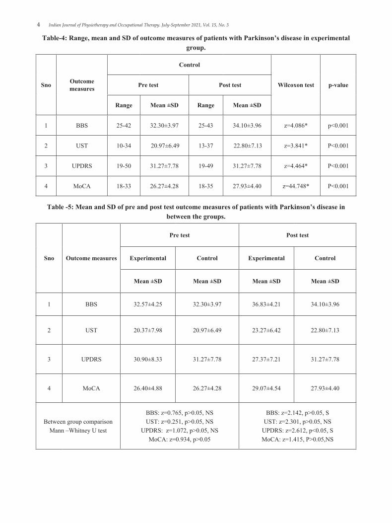
Table-3: Range, mean and SD of outcome measures of patients with Parkinson’s disease in experimental group.
Sno Outcome measures
Experimental
Wilcoxon test p-valuePre test Post test
Range Mean ±SD Range Mean ±SD
1 BBS 24-40 32.57±4.25 29-43 36.83±4.21 z=4.807* p<0.001
2 UST 10-38 20.37±7.98 18-45 23.27±6.42 z=4.079* P<0.001
3 UPDRS 15-51 30.90±8.33 17-46 27.37±7.21 z=4.359* P<0.001
4 MoCA 18-36 26.40±4.88 21-37 29.07±4.54 z=4.641* P<0.001
The above table-3 shows the pre and post test outcome measures of BBS, UST, UPDRS and MoCA of patients with Parkinson’s disease in experimental group.
4 Indian Journal of Physiotherapy and Occupational Therapy. July-September 2021, Vol. 15, No. 3
Table-4: Range, mean and SD of outcome measures of patients with Parkinson’s disease in experimental group.
Sno Outcome measures
Control
Wilcoxon test p-valuePre test Post test
Range Mean ±SD Range Mean ±SD
1 BBS 25-42 32.30±3.97 25-43 34.10±3.96 z=4.086* p<0.001
2 UST 10-34 20.97±6.49 13-37 22.80±7.13 z=3.841* P<0.001
3 UPDRS 19-50 31.27±7.78 19-49 31.27±7.78 z=4.464* P<0.001
4 MoCA 18-33 26.27±4.28 18-35 27.93±4.40 z=44.748* P<0.001
Table -5: Mean and SD of pre and post test outcome measures of patients with Parkinson’s disease in between the groups.
Sno Outcome measures
Pre test Post test
Experimental Control Experimental Control
Mean ±SD Mean ±SD Mean ±SD Mean ±SD
1 BBS 32.57±4.25 32.30±3.97 36.83±4.21 34.10±3.96
2 UST 20.37±7.98 20.97±6.49 23.27±6.42 22.80±7.13
3 UPDRS 30.90±8.33 31.27±7.78 27.37±7.21 31.27±7.78
4 MoCA 26.40±4.88 26.27±4.28 29.07±4.54 27.93±4.40
Between group comparison Mann –Whitney U test
BBS: z=0.765, p>0.05, NSUST: z=0.251, p>0.05, NS
UPDRS: z=1.072, p>0.05, NSMoCA: z=0.934, p>0.05
BBS: z=2.142, p>0.05, SUST: z=2.301, p>0.05, NS
UPDRS: z=2.612, p<0.05, SMoCA: z=1.415, P>0.05,NS

Indian Journal of Physiotherapy and Occupational Therapy, July-September 2021, Vol. 15, No. 3 5
Discussion
The aim of this experiment was to improve ADL and maintain flexibility in subjects with Parkinson’s disease. Our study is in the agreement with the result seen in above mentioned studies both the group showed statistically significant improvement within the group and between the group as well.Result of the present study demonstrate that the WII- based rehabilitation and Global exercises both have effect on the improvement in ADL of person with Parkinson’s disease. WII- based console rehabilitation is somehow simple easy and more effective because of its task oriented approach in terms of rehabilitation and was found to be somehow fun for subjects.
In the following experimental study we found the outcome values were more in subjects with WII-based motor and cognitive training which might be because of the Visual and motor activity working coordinately to give higher values which was not seen in Global exercise because of lack of visual impact and proprioception of the subject in the given training 13.
Conclusion
The present study concludes that WII based motor and cognitive training have a good impact on ADL of patients with Parkinson’s disease. General exercises and WII based training both were effective in Parkinson patient’s rehabilitation. WII based training showed slightly better improvement in the Experimental group. Therefore, it is recommended clinically to consider adding WII-based motor and cognitive training for improving the ADL of patients with Parkinson’s disease.
The table 1 presents distribution of patients with Parkinson’s disease according to gender over the groups.
The table 2 presents the age in years of the patients with Parkinson’s disease in both the groups. In group-A, the subjects were ranging within the age of 60-85 with mean and SD of 72.17±8.19.
The table 3 and 4 presents range, mean and SD of outcome measures of patients with Parkinson’s disease in experimental and control group.
The table 5 presents Mean and SD of pre and post
test outcome measures of patients with Parkinson’s disease in between the groups. The outcome values were seen significantly better in patients with WII based rehabilitation group.
Conflict of Interest: Nil
Source of Funding: Self
Ethical Clearance: The ethical clearance of study was approved by KTG College of physiotherapy, KTG multispecialty hospital.
References
1. J Jankovic Parkinson’s disease: clinical features and diagnosis. Department of Neurology, Parkinson’s disease Centre and Movement Disorders Clinic, Baylor College of Medicine J NeurolNeurosurg Psychiatry 2008;79;368-376. doi:10.1136/jp.2007.131045.
2. ElbazA,MoisanF.Update in the epidemiology of Parkinson’s disease. Current opinion in neurology. 2008 Aug 1;21(4):454-60.
3. GaoJ,NallsMA,Shi M, Joubert BR, Hernanadez DG, Huang X,Hollenbeck A, Singleton AB, Chen H, An exploratory analysis on gene-environment interactions for parkinson’s disease. Neurobiologyof ageing. 2012 Oct 1;33(10):2528-e1.
4. Rodriguez-oroz MC, JahanshahiM,Krack P, LitvanI,Macias R, Bezard E, Obeso JA, Initial clinical manifestation of parkinson’s disease: features and pathophysiological mechanisms. The lancet Neurology. 2009 Dec 1;8(12):1128-39.
5. Bain PG. Dystonic tremor presenting as parkinsonism:long term follow-up of SWEDDs. Neurology.2009 Apr 21:72(16):1443-5.
6. Theodoros DE. Speech disorder in parkinson disease. Communication and swollowing in Parkinson’s disease. 2011 May 20:79-8.
7. EdwardsMJ,BhatiaKP.Functional movement disorders:merging mind and brain. The lancet Neurology.2012 Mar 1;11(3):250-60.
8. MassanoJ,BhatiaKP.Clinical approach to Parkinson’s disease : features, diagnosis and principles of management.Cold spring harbor

6 Indian Journal of Physiotherapy and Occupational Therapy. July-September 2021, Vol. 15, No. 3
prespectives in medicine.2012 Jun 1;2(6):a008870.
9. Pratibhasurathi, KetanJhunjhunwala and Pramodkumar pal: Research in Parkinson’s disease in india, Ann indianacadneurol, 2016 jan- mar,19(1):9-20
10. Dibble L, Christensen J, Ballard D, Foreman K. Diagnosis of fall risk in Parkinson disease: an analysis of individual and collective clinical balance test interpretation. PhysTher 2008;88:323–32.
11. Boonstra TA, van der Kooij H, Munneke M, Bloem BR. Gait disorders and balance disturbances in Parkinson’s disease: clinical update and pathophysiology. CurrOpinNeurol 2008;21: 461–71.
12. Morris ME, Iansek R, Kirkwood B. A randomized controlled trial of movement strategies compared with exercise for people with Parkinson’s disease. MovDisord 2009;24:64–71.
13. Pompeu JE, dos Santos Mendes FA, da silvaKG,Lobo AM, de paulaOliveiraT, Zomignani AP, PiemonteME,Effect of Nintendo Wii based motor and cognitive training on activities of daily living in patients with parkinson’sdisease:arandomised clinical trail. Physiotherapy.2012 Sep 1;98(3):196-204.
14. Smania N, Corato E, Tinazzi M, Stanzani C, Fiaschi A, Girardi P, et al. Effect of balance training on postural instability in patients with idiopathic Parkinson’s disease. Neurorehabil Neural Repair 2010; 9:826–34.
15. Rochester L, Baker K, Hetherington V, Jones D, Willems A, Kwakkel G, et al. Evidence for motor learning in Parkinson’s disease: acquisition, automaticity and retention of cued gait performance after training with external rhythmical cues. Brain Res 2010;1319: 103–11.
16. Mirelman A, Maidan I, Herman T, Deutsch JE, Giladi N, Hausdorff JM. Virtual reality for gait training: can it induce motor learning to enhance complex walking and reduce fall risk in patients with Parkinson’s disease? J Gerontol A BiolSci Med Sci 2011;66: 234–40.
17. Morris ME, Iansek R, Kirkwood B. A randomized controlled trial of movement strategies compared with exercise for people with Parkinson’s disease.
MovDisord 2009;24:64–71.
18. Bateni H. Changes in balance in older adults based on use of physical therapy vstheWii Fit gaming system: a preliminary study. Physiotherapy 2012;98:211–6.
19. Goetz C, Poewe W, Rascol O, Sampaio C, Stebbins G, Counsell C, et al. Movement Disorder Society Task Force Report on the Hoehn and Yahr staging scale: status and recommendations. MovDisord 2004;19:1020–8.
20. Springer BA,Marin R, Cyhan T, Roberts H, Gill NW. Normative values for the Unipedal Stance Test with eyes open and closed. J GeriatrPhysTher 2007;30:8–15.
21. Dalrymple-Alford J, MacAskill M, Nakas C, Livingston L, Graham C, Crucian G, et al. The MoCA well-suited screen for cognitive impairment in Parkinson disease. Neurology 2010;75: 1717–25.
22. Griffin H, Greenlaw R, Limousin P, Bhatia K, Quinn N, Jahanshahi M. The effect of real and virtual visual cues on walking in Parkinson’s disease. J Neurol 2011;258:991–1000.
23. Andrew Siderowf MD, Michael McDermott PhD, Karl Kieburtz MD, MPH,KarenBlindauer MD, Sandra Plumb BA, Ira Shoulson MD. Test–Retest reliability of the Unified Parkinson’s Disease Rating Scale in patients with early Parkinson’s disease: Results from a multicenter clinical trial.Movement Disorders.2002.Volume 17, Issue 4
24. Major MJ,FatoneS,RothEJ. Validity and reliability of the Berg Balance Scale for community-dwelling persons with lower-limb amputation.Archieves of physical medicine and rehabilitation. 2013 Nov 1;94(11):2194-202.
25. Nasreddine, Ziad S.; Phillips, Natalie A.; Bédirian, Valérie; Charbonneau, Simon; Whitehead, Victor; Collin, Isabelle; Cummings, Jeffrey L.; Chertkow, Howard (2005-04-01). The Montreal Cognitive Assessment, MoCA: a brief screening tool for mild cognitive impairment. Journal of the American Geriatrics Society. 53 (4): 695–699
26. KeusSHJ, MunnekeM, NijkrakeMJ, Kwakkel G, Bloem BR. Physical therapy in Parkinson’s disease: evolution and future challenges. MovDisord 2009;24:1–14.

Indian Journal of Physiotherapy and Occupational Therapy, July-September 2021, Vol. 15, No. 3 7
27. Drusini A, Eleazer G, Caiazzo M, et al. One-leg standing balance and functional status in an elderly communitydwelling population in Northeast Italy. Aging ClinExp Res. 2002;14:42-46.
28. Vellas B, Rubenstein L, Ousset P, et al. One-leg standing balance and functional status in a population of 512 community-living elderly persons. Aging. 1997;9:95-98.
29. Jacobs J, Horak F, Tran V, Nutt J. Multiple balance tests improve the assessment of postural stability in subjects with Parkinson’s disease. J Neurosurg Psychiatry. 2006;77:322-326.
30. Goetz CG, Tilley BC, Shaftman SR, Stebbins GT, Fahn S, Martinez- Martin P, et al. Movement Disorder Society-sponsored revision of the Unified Parkinson’s Disease Rating Scale (MDS-UPDRS): scale presentation and clinimetric testing results. MovDisord 2008;23: 2129–70.
31. Ramaker C, MarinusJ,StiggelboutAM,vanHilten BJ. Systematic evaluation of rating scales for impairment and disability in Parkinson’s disease. MovDisord 2002; 17: 867–876.
32. Scalzo PL, Nova IC, PerraciniMR, Sacramento DRC, Cardoso F, FerrazHB, et al. Validation of the Brazilian version of the Berg Balance Scale for patients with Parkinson’s disease. Arq Neuro-
Psiquiatr 2009;67:831–5.
33. Barbara A. Springer, PT, PhD; Raul Marin, MD; Tamara Cyhan, RN; Holly Roberts, MPT; MAJ Norman W. Gill, PT. Normative Values for the Unipedal Stance Test with Eyes Open and Closed. Journal of Geriatric Physical Therapy Vol. 30;1:07.
34. Fujiwara, Yoshinori; Suzuki, Hiroyuki; Yasunaga, Masashi; Sugiyama, Mika; Ijuin, Mutsuo; Sakuma, Naoko; Inagaki, Hiroki; Iwasa, Hajime; Ura, Chiaki (2010-07-01). Brief screening tool for mild cognitive impairment in older Japanese: validation of the Japanese version of the Montreal Cognitive Assessment. Geriatrics & Gerontology International. 10 (3): 225–232.
35. Guo, Qi-Hao; Cao, Xin-Yi; Zhou, Yan; Zhao, Qian-Hua; Ding, Ding; Hong, Zhen (2010-02-01). Application study of quick cognitive screening test in identifying mild cognitive impairment.Neuroscience Bulletin. 26 (1): 47–54.
36. Luis, Cheryl A.; Keegan, Andrew P.; Mullan, Michael (2009-02-01). Cross validation of the Montreal Cognitive Assessment in community dwelling older adults residing in the Southeastern US. International Journal of Geriatric Psychiatry. 24 (2): 197–201.

8 Indian Journal of Physiotherapy and Occupational Therapy. July-September 2021, Vol. 15, No. 3
Rehabilitation of Stroke Patients in India: An Exploratory Study from a National-Level Survey Data
Bhavna Bharati1, Kirti Sundar Sahu1, Sanghamitra Pati2
1Independent Researcher, Bhubaneswar Advanced Rehabilitation Center, Bhubaneswar, India,
2Director, Indian Council of Medical Research (ICMR)-Regional Medical Research Centre, Bhubaneswar, India
Abstract
Background: Stroke is one of the significant public health challenges and one of the critical causes of disability. It has been forecasted that globally age group 6o and above will outnumber children under age 14 in 2047. With the increasing rate of survival, the burden of stroke is going to increase continuously. Currently, old age people suffer from stroke, which limits their activities of daily living. This study explores the determinants of stroke survivors seeking rehabilitation services and the factors affecting them.
Methods: In this study, first wave of Longitudinal aging study in India (LASI) data has been used. The data was collected from all the States and Union Territories across India in 2017-2018. A total number of 72,265 individuals (45 years and above) participated in this study. Subjects who have been ever diagnosed with stroke by any health care professionals were considered as the study population. Subjects were categorized into two groups based on accessing rehabilitation services. For the data analysis, survey-weighted tools have been applied for descriptive statistics and multivariable logistic regression model. The data analysis used R version 4 using R studio.
Results and Discussion: The prevalence of stroke is 1.8% in India. Nearly 40% of them visited Physical therapy or Occupational therapy services for their rehabilitation. Household economical condition, gender, residence, requirement of additional help for daily activities and availability of health insurance are strongly determining visiting rehabilitation services. The summary of this will help to develop our understanding of, why people are not availing of rehabilitation services despite having multiple problems.
Keywords: Activities of daily living, Physical therapy, Occupational therapy, Stroke rehabilitation, After care.
Corresponding author:Bhavna BharatiPlot no 2132 A/6, Nageswar Tangi, Old townBhubaneswar, Odisha, India 751002Email: [email protected]: 9437012395
Introduction
Worldwide stroke is the second leading cause of mortality [1] and one of the significant public health challenges resulting in disability. A study conducted in India by Global Burden of Disease revealed 9.4 million
deaths were only from stroke and 28.5 million lost Disability-Adjusted Life Years (DALYs), which is six times more than malaria [2,3]. The incidence and prevalence of stroke in India vary extensively due to variation in dietary intake, vitamin levels, the air pollution level, cultural and genetic variance [4,5]. Despite availability of various intervention techniques, still Stroke is one of the major reason for the disability worldwide [6]. It has been forecasted that globally people age 6o years and above will outnumber children under age 14 in 2047 [7]. This shows that with the increasing rate of survival, the burden of stroke will increase continuously [6]. Stroke is a complex condition that includes collecting symptoms
DOI Number: 10.37506/ijpot.v15i3.16159

Indian Journal of Physiotherapy and Occupational Therapy, July-September 2021, Vol. 15, No. 3 9
like sensorimotor, cognitive, perceptual, behavioural and motor impairments [1,8,9].
The after-effects of stroke will limit the independence and activity level of stroke survivors not only in activities of daily living (ADL) but also in instrumental activities of daily living (IADL), affecting behavioural and social roles [1]. We should understand the needs of stroke survivors in their participation in daily activities, both ADL and IADL, maintain the socialization, money management and other simple cognitive functions [10]. Here comes the role of a rehabilitation professional, who help the stroke survivors to return to the previous activity level. After stroke, the prominent role of rehabilitation professionals is to focus on overall improvement in motor control and hand function to regain independence in ADL and IADL. Stroke rehabilitation is a multidisciplinary and interdisciplinary approach. Physical Therapists (PT) and Occupational Therapists (OT) play a pivotal role in stroke management [11]. They conduct a detailed assessment of motor functions and other areas like perceptual, cognitive, memory, executive function, participation in the community, work environment, home modifications, physical and task performance like self-care tasks, including bathing, dressing, undressing, toileting, grooming, eating, and domestic or instrumental tasks like meal preparation, shopping, cleaning, laundry, financial and medicines management [7]. Their interdisciplinary roles help in the holistic development of the patient. There is evidence showing that rehabilitated patients after stroke are better in functioning and performance than others [7]. The rehabilitation regime focuses on the essential motor function, teaching compensatory techniques, and includes retraining in other areas like perceptual, cognitive, and visual by graded activities. Moreover, they also focus on training to caregivers, proper handling techniques etc., which eventually minimizes further complications. Treatment overall helps patients regain confidence, perform daily tasks, return to work, and mainstream society like being with their peers. Retraining stroke individuals is a complex task, and the rehabilitation programs help in neurological recovery, teaching ADL and IADL activities through compensatory techniques, social skills and psychological support [12]. A non-discriminatory and equitable health care access is the goal of any health care
program globally [13]. However, certain salient features are key to rehabilitation service, for instance, use of a supportive device or aids like a wheelchair, caregiver who can accompany them for rehabilitation service, health insurance to cover their expenditure (financial barriers) etc.
This study explores the landscape of the burden of stroke, access to therapeutic services to stroke patients in India, and its implications. This study aims 1) To find out the self-reported prevalence of stroke in India for the year 2018, stratified by location, gender, and other sociodemographic factors 2) To estimate the proportion of stroke survivors who availed PT/OT services for their rehabilitation. 3) To explore the range of different forms of disability, including physical, psychological, cognitive, speech, and vision. 4) To estimate the burden of mobility-related impairment in stroke survivors. 5) Evaluate the level of independence in performing ADL/IADL 6) To explore the use of various types of supportive devices and 7) To understand the requirement of external help in the form of caregivers for their day-to-day performance.
Material and Methods
Cross-sectional data were collected in 2017-18 from a nationally representative Longitudinal Ageing Study in India (LASI) Wave-1 [14]. Household and individual Interviews, physical measurement and biomarker data were collected from individuals aged 45 years and above and their spouses, regardless of age, in a household survey. Details of the sampling strategy have been described elsewhere [14]. The study was approved by the Indian Council of Medical Research (ICMR) Ethics Committee, and written informed consent was obtained from participants [14]. The data was collected from all the States and Union Territories across India in 2017 – 2018. A total number of 72,265 individuals participated in this study. This study used unit level first wave of LASI data.
For this study, we have used variables from the Health module of the Individual level questionnaire. Subjects who have been ever diagnosed with stroke by any health care professionals were considered the study population. Eleven relevant questions had been asked to explore the range of problems within that

10 Indian Journal of Physiotherapy and Occupational Therapy. July-September 2021, Vol. 15, No. 3
population. Subjects diagnosed with stroke were again sub-categorized to avail rehabilitation services PT/OT for stroke or its complications. The questions related to our interest are due to stroke any types of physical disability like weakness in arms and legs or their ability to use them, difficulty in speaking, swallowing, vision, thinking or finding the right words to say. There is another module of functional limitations and helpers within individual questionnaires where questions related to physical, or health problems have been asked. The range of questions includes mobility-related impairments (both upper limb lower limb), ADL/ IADL, supportive devices, any types of external help in the form of caregivers for daily activities. Monthly per capita expenditure variable has been used as proxy for the financial status of the household.
For the data analysis, survey-weighted tools have been applied for descriptive statistics and multivariable logistic regression model. The data analysis used R version 4 using R studio. P value less than 0.05 taken as statistically significant for all the results.
Results
A total number of 72,265 individuals participated in this study, out of which 1,220 individuals reported being diagnosed as stroke survivors across India for 2017-2018. The self-reported prevalence of stroke from this study was 1.8% among older adults (45 and above) in India. The prevalence of stroke in rural India was 1.7 %, whereas in urban counterpart was 2%. The male proportion stands at 2.5%, which is nearly double the rate of females (1.3%). Across the 45-59 age group, the proportion was 1%, whereas, in the 60+ age group population, the burden was 2.7%. The mean age of stroke in India stands at 58.5± 11.1 years and the median age 65 (Range 31-98 and IQR 57-72 years). Among stroke survivors, 94% of them have been diagnosed by the mainstream healthcare system (MBBS and above), whereas only 4.2 % were diagnosed by AYUSH (Ayurvedic/Unani/ Homeopathic/ Siddha) system practitioners. A small proportion of 1.7% was diagnosed by others, including Auxiliary Nurse Midwife (ANM), health workers, nurses, private clinics, quack, and registered medical practitioners (RMP) doctors. Nearly a quarter of the subjects have more than one episode of
stroke within their lifetime.
To understand the variability among those who visit therapeutic services for stroke rehabilitation from non-visitors, the following tables have been stratified by the factor of whether anyone visits PT/OT services or not. Of the 1220 individuals with ever diagnosed stroke, 469 (40.1%) availed PT/OT services for their treatment, whereas 749 had not availed PT/OT with two missing information. Table 1 describes demographic variables and their association with PT/OT services. As seen in the table 1, the proportion of stroke survivors is higher in the 60+ age group than the 45-60 age group. Also, there is a significant difference of around 17% between males and females to access the professional rehabilitation services in India for stroke. Similarly, place of residence, having health insurance, dependency on external support and supportive devices are other variables that play a significant role in accessing those services. Despite the difference in accessing rehabilitation services for caste, marital status, education, occupation, and diagnosis by medical professionals, these parameters are found to be not statistically significant.
A stroke results in multiple types of disabilities; table 2 explored the proportion of range of disabilities after stroke. Movement-related problems was the highest among different problems, with nearly 90% of the stroke survivors followed by speech and cognitive problems. There was a variation of these impairments among those who visited PT/OT services for their rehabilitation. As described above mobility-related issues are a significant burden for stroke survivors; following analysis has been emphasized on gross motor function and fine motor activities, respectively.
Among different types of movement-related impairments, stair climbing, and kneeling were maximally affected activities. The proportion of activity related to hand function (picking up a coin from the table) was the least for the stroke survivors (Figure 1).
As presented in the figure 2, most outdoor activities such as gardening, money management while shopping, and finding a new address among stroke survivors are more affected than other basic activities such as eating, bathing, etc.

Indian Journal of Physiotherapy and Occupational Therapy, July-September 2021, Vol. 15, No. 3 11
Table 3 describes the use of various forms of supportive devices to compliment the independence in ADL/IADL. More than half of the stroke survivors using any form of supporting devices for performing gross activities, among which using a walker or walking stick is the most frequently used device. For their daily activities, nearly 46% depend on external help such as family members’ professional help from outside or others.
The multivariate logistic regression analysis has been done to decide who is visiting therapeutic services for the rehabilitation after stroke in India. We found residence, occupation, monthly per capita expenditure, availability of health insurance, use of supportive devices, and need for external help were statistically significant differences in groups who visited PT/OT
services from those who never visited for rehabilitation in India independently. The persons with rural, unemployed, poor economic status, lack of financial security, user of supportive devices, using external help are in disadvantageous position compared to others. After putting these variables for multivariate logistic regression following models have been evolved along with other critical demographic variables following three models have been evolved. Model-1 shows that the richest quintiles, external help, and occupation were significant contributing factors after adjusting for other demographic and other variables. Model-2 describes Economic status as the critical factor to decide, whereas model-3 shows residence, gender and external help requirement are the essential parameters to determine whether anyone will visit for PT/OT services or not for stroke rehabilitation.
Table 1 Demographic variable and its association with stroke survivors
Determinant Response Availing PT/OT services
Yes n(%) No n(%) Total n (%)
Age group(In years)
31-4445-5960+
7 (1.4)123 (27.5)338 (71)
18 (2.6)229 (29.3)499 (68)
25 (2.1)353 (28.7)838 (69.1)
Gender MaleFemale
288 (64.4)181 (35.6)
430 (54.6)319 (45.4)
720 (58.6)500 (41.4)
Place of residenceRuralUrban
250 (56)219 (44)
460 (69.6)289 (30.4)
712 (64.3)508 (35.7)
CasteScheduled caste Scheduled tribe
Other Backward ClassGeneral
80 (17.1)54 (4.2)
175(48.3)138 (30.3)
136 (26)110 (6.5)249 (39.1)218 (28.3)
217 (22.4)164 (5.6)425 (43)356 (29)
MarriageMarried
WidowedOthers
339 (69.4)119 (29.1)11 (1.4)
536 (70.5)195 (28)18 (1.5)
876 (70.1)315 (28.4)29 (1.4)
Ever attended schoolYesNo
281 (53.6)188 (46.4)
424 (52.6)325 (47.4)
706 (53)514 (47)

12 Indian Journal of Physiotherapy and Occupational Therapy. July-September 2021, Vol. 15, No. 3
Occupation Employed with primary work#
UnemployedOthers
135 (28.8)205 (43.7)129 (27.5)
234 (31.2)351 (46.9)164 (21.9)
370 (30.3)557 (45.7)293 (24)
Health insurance YesNo
82 (13.8)381 (86.2)
172 (21.6)573 (78.4)
254 (18.5)956 (81.5)
Use of supportive deviceYesNo
285 (63.7)183 (36.3)
405 (52.1)344 (47.8)
691 (56.9)528 (43.1)
External help for ADL /IADL*
YesNo
196 (59.6)146 (40.4)
190 (36.5)287 (63.4)
386 (46.5)434 (53.5)
Who first diagnosed with stroke
A doctor with MBBS@AYUSH$
Others
453 (96.4)13 (2.9)3 (0.7)
713 (92.4)25 (5.2)11 (2.4)
1167 (94)38 (4.3)14 (1.7)
@MBBS- Bachelor of Medicine, Bachelor of surgery, *ADL – Activities of daily living, IADL- Instrumental activities of daily living, $AYUSH- Ayurvedic/Unani/ Homeopathic/ Siddha, #Employed for primary work: Agri/fishery/ elementary occupation
Table 2 Burden of types of disabilities among stroke survivors stratified by availing PT/OT services.
Response
Availing PT/OT services
Total n(%)Yes n(%) No n(%)
Physical disabilities$YesNo
272(93.6)25(6.3)
289(85.8)50(14.9)
562(89.6)75(10.3)
Speaking or swallowing problems YesNo
169(60.1)128(39.9)
183(57.3)156(42.6)
352(58.4)285(41.6)
Difficulty in thinking or finding the right words to say
YesNo
169(59.9)128(40.1)
175(52)164(48)
345(56)292(44)
Difficulty with vision YesNo
137(44.7)160(55.3)
152(48.3)187(51.7)
289(46.4)348(53.6)
$Physical disabilities: Weakness in your arms and legs or decreased ability to move or use them
Table 3 Use of supportive devices by stroke survivors
Cont... Table 1 Demographic variable and its association with stroke survivors

Indian Journal of Physiotherapy and Occupational Therapy, July-September 2021, Vol. 15, No. 3 13
Availing PT/OT services Totaln(%)Yes n(%) No n(%)
Using any aid or supportive devices
YesNo
285(63.7)183(36.3)
405(52.1)344(47.9)
691(56.8)528(43.1)
Walker/Walking sticksYesNo
100(43.1)185(56.8)
118(27.2)287(72.7)
218(34.2)473(65.7)
WheelchairsYesNo
17(4.3)268(95.7)
8(.3)397(99.7)
25(2)666(98)
Adjustable shower stools /Commodes
YesNo
27(18)258(82)
23(3.5)382(96.5)
50(10.2)641(89.8)
Back/neck collarYesNo
9(2.1)276(97.9)
2(0.4)403(99.6)
11(1.2)680(98.8)
Orthosis/prosthesisYesNo
9(4.8)276(95.2)
8(1.8)397(98.2)
17(3.1)674(96.8)
Table 4 Multivariate logistic regression models describing variables deciding access to rehabilitation services for stroke in India
Model-1 Model-2 Model 3Est. 2.50% 97.50% Est. 2.50% 97.50% Est. 2.50% 97.50%
(Intercept) 2.26 0.08 4.44 2.12 -0.12 4.36 1.71 -0.5 3.92Age groups-45-59 -0.93 -2.91 1.05 -0.91 -2.96 1.13 -0.92 -2.98 1.14
Agegroups-60+ -1.03 -3.01 0.95 -1.18 -3.22 0.85 -1.26 -3.31 0.78Gender-Female 0.43 -0.05 0.91 0.35 -0.08 0.78 0.48 0.06 0.9
Residence-Urban -0.39 -0.88 0.1 -0.4 -0.85 0.05 -0.51 -0.95 -0.07Poorer -0.43 -1.14 0.29 -0.41 -1.12 0.3 -0.41 -1.1 0.27Middle -0.39 -1.06 0.29 -0.6 -1.24 0.05 -0.58 -1.22 0.07Richer -0.63 -1.33 0.07 -0.84 -1.51 -0.17 -0.83 -1.5 -0.16Richest -1.02 -1.71 -0.32 -1.04 -1.69 -0.4 -1.08 -1.71 -0.44
Human help No 0.82 0.38 1.25 0.78 0.35 1.2 0.78 0.36 1.2Health Insurance No -0.45 -0.99 0.08 -0.36 -0.87 0.15Occupation-Others -0.74 -1.32 -0.15 -0.52 -1.07 0.04
Occupation -unemployed -0.05 -0.58 0.47 -0.03 -0.53 0.48Scheduled tribe 0.07 -0.74 0.87
Other backward class -0.24 -0.79 0.3General -0.16 -0.76 0.44AYUSH 0.65 -0.5 1.79 Others 0.24 -1.47 1.96
Aids/assistive devices No -0.1 -0.53 0.33Educated No -0.26 -0.74 0.22

14 Indian Journal of Physiotherapy and Occupational Therapy. July-September 2021, Vol. 15, No. 3
Figure 1 Problem with gross motor function among stroke survivors
Figure 2 Problem in performing ADL/IADL among stroke survivors
Discussion
Developing countries have reported an extensive burden of impairments and disabilities in association with stroke [15,16]. We found a prevalence of 1.8 % of older adults (45 years and above) in India are suffering from stroke. Other studies have illustrated an increased incidence rate from 56/100,000 person-years during 1970-1979 to 117/100,000 person-years during the period 2000-2008 in low, middle-income countries (LMICs) [8]. 60% of the people either die post stroke, disabled or dependent on their ADL/IADL [17]. Studies
shows that stroke is more prevalent in older adults, with males affected more than females [15] which can be reflected in our results. Most stroke survivors result in hemiparesis, for years resulting in functional limitation [18]. Early rehabilitation leads to early motor control which needs professional services to mainstream in daily activities and socialization. Our study states nearly two-fifth are only visiting rehabilitation professionals post stroke.
For the first time, based on our knowledge, a Nationalized survey asked a question related to

Indian Journal of Physiotherapy and Occupational Therapy, July-September 2021, Vol. 15, No. 3 15
receiving PT/OT services for stroke survivors. Physical rehabilitation professionals include mainly PT/OT in India [9]. Several factors decide receiving therapeutic services like demographic, socioeconomic status, residence, occupation, health insurance coverage etc. The result shows the proportion of males receiving this service is higher than females. The reason might be lack of gender empowerment [19], cultural barrier, educational status, health literacy and gender preference. As nearly three fourth of the population of India still resides in rural area, the health care infrastructure is limited to primary care [20,21]. Rehabilitation is a luxury rather than a necessary service in rural India, multiple factors come into play to avail the service. There are barriers like availability, accessibility, affordability, acceptability, difficult geographic terrain etc.[13] for visiting a health care facility. Our study shows that irrespective of educational attainment, there is no statistical difference in availing rehabilitation services, similarly, caste, marital status, occupation affect availing services. However, use of aid and supportive device, dependency on caregiver, having a health insurance has a substantial impact in accessing rehabilitation care.
Among stroke survivors, four salient kinds of difficulty in activities have been assessed like mobility, speaking or swallowing, vision and thinking. In our study, nearly 90% of the stroke survivors reported any form of movement impairments in their upper or lower extremity, which needs professional intervention. Therapeutic methods have shown an effect on improvement in both upper and lower extremities, resulting in improved motor function and hand function [22,23]. Problems like motor function have day-to-day implications in a patient’s life in contrast to other impairments like swallowing, thinking, and difficult vision. Therefore, patients visit a therapist primarily to regain motor function. Apart from motor impairment, other difficulties like speech, recreational etc., also need health care professionals. Rehabilitation is a multidisciplinary approach, so along with PT/OT, other rehab professionals also play a pivotal role in regaining their independence in daily living [9,24]. Policymakers have also realized the essence of teamwork; their skills and knowledge have benefited in overall patient improvement leading to quality of life[9].
Motor impairments can be broadly classified into gross motor and fine motor functions. In this survey, gross motor includes walking 100 yards, sitting for two hours, getting up from a chair, sitting for an extended period, stooping, kneeling, reaching, or extending arm above shoulder level, pulling or pushing a large object, lifting or carrying weight above five kg and picking up a coin from a table. Among these activities, our result shows stair climbing, independent walking, kneeling is more affected than other activities. Studies show that 50 to 70 % of the patients reverse their functional independence [12]. In India, more than 50% of stroke survivors have these motor problems, but are not availing PT/OT services, which is a matter of concern. Within ADL/IADL, activities like dressing, walking across a room, bathing, eating, using the toilet, cooking, shopping, using telephone, taking medications, household chores, gardening, money management and getting around or finding an address in unfamiliar places are asked. Our result shows outdoor functions are severely affected than others. This is not only restricting the individual independence but also the overall family quality of life [25]. In last decade, government policies tried to provide rehabilitation services in all facilities. Hand is always required in day-to-day activities like eating, picking up an object, handwriting, manipulating objects etc. Hand function is directly related to independence and quality of life. There are specific hand therapy principles in Occupational Therapy that improve gross and fine hand function that ultimately helps in ADL/ IADL functions[26].
Besides visiting health care professionals use of supportive devices also plays a major role in the rehabilitation of stroke survivors. In this survey, there is a submodule collecting information about five categories of supportive devices like using walker, or walking sticks, wheelchair, adjustable shower stools or commodes, back or neck collar and use of orthosis or prosthesis. More than half of the stroke survivors using any form of supportive devices. The use of supportive devices is more prevalent who are visiting PT/OT. Wheelchair use among stroke survivors was found to be highly restricted (2%) in our study. The reason might be lack of a barrier-free environment, lack of assessment of an Occupational therapist for home modifications,

16 Indian Journal of Physiotherapy and Occupational Therapy. July-September 2021, Vol. 15, No. 3
acceptance and affording a wheelchair in lower- and middle-income countries like India compared to the developed world. Moreover, there might be another reason, lack of adequate motor function to propel the wheelchair, as most are not attending PT/OT.
As India is progressing towards demographic and epidemiological transition, this prevalence of stroke will increase by manifolds [27]. The risk factors for stroke can be grossly divided into preventable and non-preventable. Low physical activity, high sedentary behaviour, obesity, conditions can lead to stroke in the short term and long term. The journey from the acute stroke phase to the maintenance of routine day-to-day life requires therapeutic services. This rehabilitation process in India is always dependant on several external factors like knowledge of the existence of a mode of therapy, financial strength, availability of health care professionals, referral mechanism, adherence and motivation towards rehabilitation, and trust in available health care professionals [28]. Within the last decade, the government has mainstreamed rehab professionals in health care services to avail the facility with zero out-of-pocket expenditure [29]. Previously only physiotherapists were appointed in government facilities, but nowadays, more emphasis is given to the quality of life, and occupational therapists are also appointed. For the first time, LASI has emphasized rehabilitation care and collected data on the facilities availed by patients. This is a welcome step but can be generalized to other chronic conditions, such as other neurological disorders.
Conclusion
Stroke is a significant public health issue affecting 1.8 % of the Indian population. Gender, financial status of the family, residential area, availability of health insurance, and external help are critical socio-demographic factors that decide whether any person will visit Physical Therapy and Occupational Therapy services for stroke rehabilitation—nearly two-third of the stroke survivors still unable to visit these facilities resulting in poor quality of life. Policy level initiatives should be taken to improve accessing rehabilitation services in India.
Acknowledgement: We would like to thank IIPS
team for the data collection for LASI and staffs of RMRC-Bhubaneswar for getting access to the data.
Funding Resource: This research did not receive any specific grant from funding agencies in the public, commercial, or not-for-profit sectors.
Conflict of Interest : No potential conflict of interest declared.
References
1. Kamalakannan S, Gudlavalleti Venkata M, Prost A, Natarajan S, Pant H, Chitalurri N, et al. Rehabilitation Needs of Stroke Survivors after Discharge from Hospital in India. Archives of Physical Medicine and Rehabilitation 2016;97(9):1526-1532.e9.
2. Dalal P, Bhattacharjee M. Stroke epidemic in India: Hypertension-stroke control programme is urgently needed. The Journal of the Association of Physicians of India 2007;55:689–91.
3. Lozano R, Naghavi M, Foreman K, Lim S, Shibuya K, Aboyans V, et al. Global and regional mortality from 235 causes of death for 20 age groups in 1990 and 2010: a systematic analysis for the Global Burden of Disease Study 2010. The Lancet 2012;380(9859):2095–128.
4. Venugopalan VY, Bhatia R, Pandian J, Khurana D, Kaul S, Sylaja PN, et al. Regional differences in ischemic stroke in India (north vs. south). International Journal of Stroke 2019;14(7):706–14.
5. Lynch EA, Mackintosh S, Luker JA, Hillier SL. Access to rehabilitation for patients with stroke in Australia. Medical Journal of Australia 2019;210(1):21–6.
6. Ranford J, Asiello J, Cloutier A, Cortina K, Thorne H, Erler KS, et al. Interdisciplinary Stroke Recovery Research: The Perspective of Occupational Therapists in Acute Care. Front Neurol 2019;10:1327.
7. Rowland TJ, Cooke DM, Gustafsson LA. Role of occupational therapy after stroke. Annals of Indian Academy of Neurology 2008;11(5):99.
8. Kamalakannan S, Gudlavalleti AashraiSV, Gudlavalleti VenkataSM, Goenka S, Kuper H. Incidence & prevalence of stroke in India: A systematic review. Indian Journal of Medical

Indian Journal of Physiotherapy and Occupational Therapy, July-September 2021, Vol. 15, No. 3 17
Research 2017;146(2):175.
9. Clarke DJ. The role of multidisciplinary team care in stroke rehabilitation. Progress in Neurology and Psychiatry 2013;17(4):5–8.
10. Task Force Members, Schwamm LH, Pancioli A, Acker JE, Goldstein LB, Zorowitz RD, et al. Recommendations for the Establishment of Stroke Systems of Care: Recommendations From the American Stroke Association’s Task Force on the Development of Stroke Systems. Stroke 2005;36(3):690–703.
11. Cieza A, Causey K, Kamenov K, Hanson SW, Chatterji S, Vos T. Global estimates of the need for rehabilitation based on the Global Burden of Disease study 2019: a systematic analysis for the Global Burden of Disease Study 2019. The Lancet 2020;396(10267):2006–17.
12. null null, Schwamm Lee H., Pancioli Arthur, Acker Joe E., Goldstein Larry B., Zorowitz Richard D., et al. Recommendations for the Establishment of Stroke Systems of Care. Stroke 2005;36(3):690–703.
13. Dassah E, Aldersey H, McColl MA, Davison C. Factors affecting access to primary health care services for persons with disabilities in rural areas: a “best-fit” framework synthesis. Global Health Research and Policy 2018;3(1):36.
14. International Institute for Population Sciences (IIPS), NPHCE, MoHFW, Harvard T. H. Chan School of Public Health (HSPH) and the University of, Southern California (USC). Longitudinal Ageing Study in India (LASI) Wave 1, 2017-18, India Report [Internet]. International Institute for Population Sciences, Mumbai.; 2020. Available from: https://www.iipsindia.ac.in/sites/default/files/LASI_India_Report_2020_compressed.pdf
15. Mahak C, Shashi, Yashomati, Hemlata, Manisha N, Sandhya G, et al. Assessment of Utilization of Rehabilitation Services among Stroke Survivors. J Neurosci Rural Pract 2018;9(4):461–7.
16. Feigin VL, Forouzanfar MH, Krishnamurthi R, Mensah GA, Connor M, Bennett DA, et al. Global and regional burden of stroke during 1990–2010: findings from the Global Burden of Disease Study 2010. The Lancet 2014;383(9913):245–55.
17. I. Kneebone I, B. Lincoln N. Psychological Problems after Stroke and Their Management: State of Knowledge. NM 2012;03(01):83–9.
18. Gray V, Rice CL, Garland SJ. Factors That Influence Muscle Weakness Following Stroke and Their Clinical Implications: A Critical Review. Physiotherapy Canada 2011;64(4):415–26.
19. WEF_GGGR_2021.pdf [Internet]. [cited 2021 Apr 5];Available from: http://www3.weforum.org/docs/WEF_GGGR_2021.pdf
20. Reddy KS. Health Care Reforms in India. JAMA 2018;319(24):2477–8.
21. Taqi M, Bidhuri S, Sarkar S, Ahmad WS, Wangchok P. Rural Healthcare Infrastructural Disparities in India: a Critical Analysis of Availability and Accessibility. Journal of Multidisciplinary Research in Healthcare 2017;3(2):125–49.
22. Kwakkel G, Wagenaar RC, Twisk JW, Lankhorst GJ, Koetsier JC. Intensity of leg and arm training after primary middle-cerebral-artery stroke: a randomised trial. The Lancet 1999;354(9174):191–6.
23. McGlinchey MP, James J, McKevitt C, Douiri A, Sackley C. The effect of rehabilitation interventions on physical function and immobility-related complications in severe stroke: a systematic review. BMJ Open 2020;10(2):e033642.
24. Keptner KM, Smyth K, Koroukian S, Schluchter M, Furlan A. Utilization of Rehabilitation Services in Stroke: A Study Utilizing the Health and Retirement Study With Linked Medicare Claims Data. Archives of Physical Medicine and Rehabilitation 2019;100(12):2244–50.
25. Lindley RI, Anderson CS, Billot L, Forster A, Hackett ML, Harvey LA, et al. Family-led rehabilitation after stroke in India (ATTEND): a randomised controlled trial. The Lancet 2017;390(10094):588–99.
26. Sale P, Mazzoleni S, Lombardi V, Galafate D, Massimiani MP, Posteraro F, et al. Recovery of hand function with robot-assisted therapy in acute stroke patients: a randomized-controlled trial. International Journal of Rehabilitation Research 2014;37(3):236–42.

18 Indian Journal of Physiotherapy and Occupational Therapy. July-September 2021, Vol. 15, No. 3
27. Dandona L, Dandona R, Kumar GA, Shukla DK, Paul VK, Balakrishnan K, et al. Nations within a nation: variations in epidemiological transition across the states of India, 1990–2016 in the Global Burden of Disease Study. The Lancet 2017;390(10111):2437–60.
28. Kumar Sg, Roy G, Kar S. Disability and rehabilitation services in India: Issues and challenges. Journal of Family Medicine and Primary Care 2012;1(1):69.
29. Thakur JS, Paika R, Singh S. Burden of noncommunicable diseases and implementation challenges of National NCD Programmes in India. Medical Journal Armed Forces India 2020;76(3):261–7.

Indian Journal of Physiotherapy and Occupational Therapy, July-September 2021, Vol. 15, No. 3 19
The Psychological well-being of Working Women During Covid-19 Pandemic in India: A Web Based Survey
Jagruti K Patel1, Nijal Parmar1
1Assistant Professor, S.S Agrawal Institute of Physiotherapy and Medical Care Education Navsari
Abstract
Background: The current COVID 19 pandemic has had a global effect, affecting almost all. Women, on the other hand, have been affected somewhat differently (if not more severely) around the world, for reasons that go beyond biology. Some of these effects were counteracted by job autonomy and partner support. In general, the pandemic had a greater impact on women’s mental health than it did on men’s. As a result, the study’s aim is to identify potential risk factors that may affect the psychological well-being of working women during this pandemic.
Method: The Perceived Stress Scale and the Generalized Anxiety Disorder Scale were used to assess stress and anxiety levels, while the WHO scale was used to assess psychological well-being. Spearmen’s correlation coefficient was calculated between the descriptive variable and other variables such as PSS, GAD, and WHO to see whether there was any correlation between them.
Results: In a study of 258 working women, nearly 63.95 % of those in the 20 to 30 age group reported mild to moderate stress, while 26.04 % of those in the 31 to 40 age group reported moderate stress, and only a few around 6 to 7 % of women in the 41 to 50 age group reported moderate stress. Women who worked from home had a moderate stress level of 19.56 % and received pay cuts of 40 to 48 %. Women who worked more than 8 hours had a stress level of 40 to 46 %, while women without children had a slight anxiety level of 20 %.
Conclusion: Working women’s psychological health is disrupted by the pandemic, according to the findings, since their stress levels are almost moderate and their anxiety levels are mild. Wage cuts, working in the private sector, and working more than 8 hours were all high risk factors for stress and anxiety in working women.
Keywords: Perceived Stress Scale, Generalized Anxiety disorder Scale, Psychological well-being and Working women.
Corresponding author:Dr. Jagruti PatelAssistant Professor, S.S Agrawal institute of physiotherapy and medical care education,Navsari-396445, [email protected]
Introduction
The current global pandemic’s reach, as well as
novel social conditions such as lockout, isolation, and quarantine, have disproportionately impacted women’s mental health. The following are some possible explanations: Women are more likely to lose their job or financial independence when resources are limited; more likely to lose their school or education as a whole when more resources are available.1
In determining the mental health status of working women, the home and workplace environment played a
DOI Number: 10.37506/ijpot.v15i3.16159

20 Indian Journal of Physiotherapy and Occupational Therapy. July-September 2021, Vol. 15, No. 3
major role. The climate of the workplace, the favorable attitude of colleagues at the workplace and the favorable behavior of husbands / in-laws at home have been found to protect overall mental health. 2
But due to this Pandemic some family / social problems, such as women who encountered work-family conflict or did not engage in family decision-making, were at increased risk of having poor mental health, whereas spending time on social responsibilities and spending some time on yoga / meditation / exercise had good results for mental health.3
Women now care for household tasks, childbearing, and the family around the clock while working from home due to this reason, physical and mental fatigue, as well as stress, can result from the increased workload.4
The cross-sectional analysis found that women with a high percentage of Health Workers were symptoms of stress, depression and anxiety during the early COVID-19 stage. Those who have more than 10 Years of work experiences, among the sociodemographic characteristics, and two or more kids are vulnerable to stress, anxiety and depression. Such health workers may face greater occupational fatigue, family obligations and inequalities in domestic work.5, 6
Due to numerous factors, such as urbanization, industrialization, increased level of education, knowledge of rights, and media power, the role of women in society has changed rapidly. More and more women tend to be working in some kind of job, so that they can contribute to their families financially. However, the attitude towards women, especially married women, and their position in the family has remained the same, as it is still considered their primary duty to take care of the family and children today. A married working woman is therefore overstrained by carrying out duties and obligations both at home and at work, leading to numerous psychological problems such as position conflict, job pressure, mental exhaustion, tension, anxiety, resentment, depression, anger, phobias, and other social and emotional distress.7
Compared to those who do not work, there are few studies finding that women who work have significant mental disorders, but because of COVID 19 they have to work digitally and have to take on home & child duties,
which are the key risk factor for working women in this pandemic. So our aim of the study is to find out whether working having more detrimental effect of COVID 19 on their mental health during pandemic.
Methodology
STUDY DESIGN: Cross -Sectional
POPULATION: Working women in different colleges, universities, and institutions or any sectors
SAMPLING METHOD: Non-probability snowball sampling technique
SAMPLE SIZE: 258
STUDY DURATION: 1 Month
INCLUSION CRITERIA: Working women aged 20-50 years were included.
TOOLS & MATERIAL USED:
o Google form of Infirmed Consent
o Google form for data collection
o Google form of Perceived Stress scale (PSS-10)
o Google form of Generalized Anxiety disorders scale (GAD-7)
o Google form of WHO well-being index (WHO-5)
OUTCOME MEASURES: 1) Perceived Stress scale (PSS-10)
2) Generalized Anxiety disorders scale (GAD-7)
3) WHO well-being index (WHO-5)
DATA COLLECTION PROCEDURE:
Because of the pandemic, the respondents were invited remotely and they only completed the questionnaire in English via an online platform. The questionnaire approach was used to collect data where the Google form was generated and distributed on various platforms such as What’s app groups, Facebook, and Instagram, etc. The consent was also taken from the

Indian Journal of Physiotherapy and Occupational Therapy, July-September 2021, Vol. 15, No. 3 21
subject’s ways of using their data in the study with the aid of Google forms.
Results
Table no.: 1 Level of stress and anxiety experienced by working women on PSS and GAD.
variable category N (%)PSS GAD
MildN (%)
ModerateN (%)
SeverN (%)
MildN (%)
ModerateN (%)
SeverN (%)
Age 20-30 165 63.95% 24(9.30) 124(48.06) 17(6.58) 56(21.70) 32(12.40) 12(4.65)
31-40 69 26.74% 4(1.55) 58(22.48) 7(2.71) 25(9.68) 5(1.93) 6(2.32)
41-50 24 9.30% 2(0.77) 18(6.97) 4(1.55) 6(23.25) 3(1.16) 2(0.77)
Marital Status Single 119 46.12% 19(7.36) 86(33.33) 14(5.42) 41(15.89) 24(9.30) 07(2.71)
Married 139 53.87% 11(4.26) 114(44.18) 14(5.42) 46(17.82) 16(6.20) 13(5.03)
No of children No children 162 62.79% 22(8.52) 124(48.06) 16(6.20) 54(20.09) 27(10.46) 11(4.26)
Having children 96 37.19% 08(3.10) 76(29.45) 12(4.65) 33(12.79) 12(4.65) 09(3.48)
Type of family Nuclear 160 62.01% 21(8.13) 118(45.73) 21(8.13) 46(17.82) 33(12.79) 14(5.42)
Joint 98 37.98% 09(3.48) 82(31.78) 07(2.71) 41(15.89 07(2.71) 6(2.32)
Education Undergraduate 91 35.27% 19(7.36) 60(23.25) 12(4.65) 34(13.17) 12(4.65) 08(3.10)
Post-Graduate 131 50,77% 08(3.10) 111(43.02) 12(4.65) 41(15.89) 24(9.30) 09(3.48)
Doctorate 36 13.95% 03(1.16) 29(11.24) 04(1.55) 12(4.46) 04(1.55) 03(1.16)
Job type Local 224 86.82% 26(10.07) 175(67.82) 23(8.91) 77(29.84) 37(14.34) 15(5.81)
Remote 34 13.17% 04(1.55) 25(9.68) 05(1.93) 10(3.87) 03(1.16) 05(1.93)
Working in Government 45 17.44% 6(2.32) 38(14.72) 1(0.38) 15(5.81) 6(2.32) 2(0.77)
Private 213 82.55% 24(9.30) 162(62.79) 27(0.46) 72(27.90) 34(13.17) 18(6.97)
Working hours <8 134 53.93% 14(5.42) 96(37.20) 14(5.42) 39(15.11) 21(8.13) 09(3.48)
=>8 124 48.06% 16(6.20) 104(40.31) 14(5.42) 48(18.60) 19(7.36) 11(4.26)
Working place Usual 194 75.19% 25(9.68) 14(57.75) 20(7.75) 67(25.96) 31(12.01) 15(5.81)
home 64 24.80% 05(1.93) 51(19.76) 8(3.10) 20(7.75) 9(3.48) 5(1.93)

22 Indian Journal of Physiotherapy and Occupational Therapy. July-September 2021, Vol. 15, No. 3
Current job status Full Pay 142 55.03% 5(1.93) 27(10.46) 5(1.93) 11(4.26) 5(1.93) 4(1.55)
Pay Cut 74 28.68% 22(8.52) 105(40.69) 11(4.26) 46(17.82) 21(8.13) 9(3.48)
Not Working 37 14.34% 3(1.16) 60(23.25) 11(4.26) 28(10.85) 15(5.81) 6(2.32)
Forced to Leave 05 1.93% 0(0) 4(1.55) 1(0.38) 2(0.77) 0(0) 1(0.38)
Current job satisfaction Yes 115 44.57% 7(2.71) 52(20.15) 18(6.97) 42(16.27) 23(8.91) 11(4.26)
No 143 55.42% 23(8.91) 110(42.63) 10(3.87) 45(17.44) 17(6.58) 9(3.48)
Table no.: 2 Correlation between age, marital status, no of children, type of family, education, type of job, working in, working hours, working place current job status, satisfied with job and year of study with of
stress , anxiety and well-being.
Total PSS Total GAD Total WHO
Agep -.077 -.049
.026
r .220 .439.682
Marital statusp -.012 -.045
-.024
r .848 .471.709
No of childrenp -.134* -.080
.068
r .034 .206.281
Type of familyp .118 .156*
-.074
r .061 .013.240
Educationp .018 -.002
-.051
r .774 .974.420
Type of jobp .118 .047
-.079
r .063 .463.214
Working inp -.038 .045
-.013
r .554 .482.838
Working hoursp -.044 .012
-.015
r .493 .857.820
Cont... Table no.: 1 Level of stress and anxiety experienced by working women on PSS and GAD.

Indian Journal of Physiotherapy and Occupational Therapy, July-September 2021, Vol. 15, No. 3 23
Working placep -.044 .012
-.015
r .493 .857.820
Current job statusp .193** .098
-.132*
r .002 .122.036
Satisfied with jobp -.252** -.203**
.221**
r .000 .001.000
** Correlation is significant at the 0.01 level (2-tailed).
* Correlation is significant at the 0.05 level (2-tailed).
Cont... Table no.: 2 Correlation between age, marital status, no of children, type of family, education, type of job, working in, working hours, working place current job status, satisfied with job and year of study with of
Disscussion
Primary aim of this study was to assess level of stress, anxiety and well-being among working women during Covid-19 pandemic by using Perceived Stress scale (PSS-10), Generalized Anxiety disorders scale (GAD-7) and WHO well-being index (WHO-5) respectively which provides information regarding level of stress, anxiety and well-being. A total of 258 working women were assessed for different demographic variables such as Age, marital status, no of children, type of family, education, type of job, working in, working hours, working place current job status and satisfaction with job. Along with it the level of stress was marked on PSS, level of anxiety on GAD and well-being on WHO-5 by working woman themselves.
According to our findings, the overall stress level in the participants is 10.85 % in the severely affected category, 77.51 % in the moderate category, and 11.62 % in the mild category. According to age group, 48.06 % have moderate stress between the ages of 20 and 30 and 22.48 % have moderate stress between the ages of 31 and 40. The study of Abdulah et al. found the highest prevalence of stress at 93.7 %, and the study of Chew et al found the lowest prevalence at 5.2 % 8, 9 While ageing increases the risk of COVID-19 infection and mortality increase, current research shows that anxiety, depression, and stress levels are significantly higher in the age group of 21–30 years during the pandemic.10
The primary explanation for this seems to be because this age demographic is worried about the pandemic’s long-term effects and economic difficulties, since they are key active labour forces in society and are therefore disproportionately impacted by layoffs and business closures.11 Some researchers believe that young people’s increased anxiety is attributable to their increased access to information through social media like what’s app, Instagram and Facebook, which can also cause stress.12
In our findings, the stress level is moderately high (44.18%) in married compared to single working women. Results of one study indicate that in dual-earner couples marital-role quality is significantly negatively associated with psychological distress for women as well as men and that the magnitude of the effect depends little, if at all, on gender or on parental status. These findings challenge the view that marital experiences more significantly influence women’s mental health states than men’s.13
In our study out of 258 working women, 111 women who completed a postgraduate degree reported moderate stress, while only 60 women who completed under graduation reported moderate stress but only 12 working women reported severe stress level in post doctorate degree as well as under graduation level. In one comparative study on stress level among working and housewives reported that working women with a bachelor’s degree have the highest stress levels, followed

24 Indian Journal of Physiotherapy and Occupational Therapy. July-September 2021, Vol. 15, No. 3
by those with a master’s degree.
Working mothers having a children, on average, experience more stress than women who do not have children if a mother works, then childcare has to be arranged. But, in our research, 124 women who do not have children reported a moderate level of stress relative to working mothers. 14
Total of 118 working women who live in nuclear families feel more stress than those 82 women who live in joint family the possible reason might be because of their duties can be shared among family members and their children can be cared for by their in-laws. But only few working women reported severe as well as mild symptoms of stress in these pandemic.13
According to P Abirami’s report, respondents are frustrated with their job’s workload, which could be the primary cause of stress due to working conditions.15 this is close to our study, which found that 162 women working in the private sector have moderate stress levels, whereas only 38 women working in the government sector have moderate stress levels.
Women who work more than 8 hours recorded moderate stress levels in their lives during the pandemic, relative to those who work less than 8 hours. In the current survey, about 40.31 % of women reported moderate stress levels due to longer working hours. Females, in comparison to men, are more likely to perceive similar life experiences as negative, to be more susceptible to interpersonal stressors (such as social rejection), and to ruminate more when they encounter adverse emotions or events, according to Kyungjin LEE et al.16
According to Bhuvaneshwari (2013), stress in married working women is caused by a variety of family and official obligations, workplace abuse, working long hours, and an inappropriate work-life balance. Long-term headaches, hypertension, and obesity are all symptoms of stress in working people.17 the current study found that about 57.75 percent of working women encounter moderate stress as compared to those who work at home, with travel time and infection while travelling being the key causes of moderate stress.
Indeed, studies from the United States suggest that parents are avoiding their children’s scheduled checkups and vaccines in order to prevent COVID-19, which may lead to a rise in childhood illness in the coming years if these appointments are not rescheduled.18 Unless proactive measures to resolve gender dimensions of the pandemic response are made, some women’s lifetime earnings will never recover as a result of all of these factors. So, according to the this report, 40.69 % of women who receive full pay have moderate stress, compared to 23.25% of women who do not receive full pay, and 10.46 % of women have moderate stress as a result of not working in these pandemics because they have quit the job.. Nearly 42.63 % of working women who are dissatisfied with their jobs have moderate stress, while 20.15 % of females who are happy with their jobs have moderate stress.
In this pandemic scenario, out of 258 working women, 33.72 % have mild anxiety, 15.50 % have moderate anxiety, and 7.75 % have extreme anxiety. When comparing women between the ages of 20 and 50, the majority of women between the ages of 20 and 30 suffer from anxiety. However, 56 women in this age group had mild anxiety, while only 32 working women had severe anxiety.
The type of family, including tension, plays a significant role in the psychological well-being of working women. As a result, in the current sample, 46 women in nuclear families experienced mild anxiety, while only 33 women in joint families experienced moderate anxiety. In one study, working women’s graduation was related to a rise in anxiety. However, in the current research, women who have completed a postgraduate degree suffer from mild anxiety in around 15.89 % of cases, while only 13.17 % of women who have completed an undergraduate degree suffer from mild anxiety symptoms .Nearly 46 married women have mild anxiety, and 41 single women have mild anxiety symptoms, although only a few single working women have severe anxiety.
The findings differed from previous research, which showed that women who do not have children suffer from moderate anxiety at a lower rate than working mothers. During these pandemics, almost 54 women

Indian Journal of Physiotherapy and Occupational Therapy, July-September 2021, Vol. 15, No. 3 25
without children have mild anxiety and 27 women have severe anxiety.
Nearly 72 working women in the private sector suffer from mild anxiety, 34 from moderate anxiety, and just 18 from extreme anxiety.
When comparing those who work less than 8 hours to those who work more than 8 hours, 48 women who work more than 8 hours have moderate anxiety. However, 21 females working less than 8 hours have moderate anxiety, while only 19 women working more than 8 hours have moderate anxiety.
Working women’s psychological condition can also be affected by their workplace, since it takes longer to get home, they are more likely to become sick as a result of regular travel, and they are more likely to offer extra time due to full-day working hours plus fatigue than those who work from home. However, relative to those who work from home, 67 women have mild anxiety when they go to work every day, while 31 have moderate anxiety.
In addition, 46 women who are paid full-time have moderate anxiety, while 28 women who are paid half-time have mild anxiety, relative to those who are paid or forced to leave their employment. Women who are happy with their work, and those who are unhappy with their jobs suffer from mild anxiety, which affects nearly 40 to 45 out of 258 working women. One of the major reasons for getting stressed out and anxious in this pandemic because of the lockdown and school closures, women’s access to paid jobs is dwindling, while their unpaid labour is growing. “Domestic tasks, such as childcare, cooking food for the extra children who would otherwise be at school, and caring for ill family members, fall overwhelmingly on women,” Wenham said.19
Conclusion
The psychological condition of working women is disrupted in this pandemic, according to the findings, as their stress level is almost moderate and their anxiety level is mild.
Conflict of Interest-Nil
Source of Funding- Self
Ethical Clearance –Taken from institutional advisory board.
References
1) Women mental Health during COVID 19 and Lock down: A brief narrative and Introspection Tanay Maiti*, Bhavika Rai, Tathagata Biswas.
2) Kopp MS, Stauder A, Purebl G, Janszky I, Skrabski A. Work stress and mental health in a changing society. European Journal of Public Health. 2008 Jun 1;18(3):238-44.
3) Chandola T, Martikainen P, Bartley M, Lahelma E, Marmot M, Michikazu S, Nasermoaddeli A, Kagamimori S. Does conflict between home and work explain the effect of multiple roles on mental health? A comparative study of Finland, Japan, and the UK. International journal of epidemiology. 2004 Aug 1;33(4):884-93.
4) McLaren HJ, Wong KR, Nguyen KN, Mahamadachchi KN. Covid-19 and women’s triple burden: Vignettes from Sri Lanka, Malaysia, Vietnam and Australia. Social Sciences. 2020 May;9(5):87.
5) Li G, Miao J, Wang H, Xu S, Sun W, Fan Y, Zhang C, Zhu S, Zhu Z, Wang W. Psychological impact on women health workers involved in COVID-19 outbreak in Wuhan: a cross-sectional study. Journal of Neurology, Neurosurgery & Psychiatry. 2020 Aug 1;91(8):895-7.
6) Cooke M, Laine C. Gender differences in time spent on parenting and domestic responsibilities. Annals of internal medicine. 2014 Oct 7;161(7):534-5.
7) Panigrahi A, Padhy AP, Panigrahi M. Mental health status among married working women residing in Bhubaneswar City, India: a psychosocial survey. BioMed research international. 2014 Jan 1;2014.
8) Abdulah DM, Musa DH. Insomnia and stress of physicians during COVID-19 outbreak. Sleep Medicine: X. 2020 Dec 1;2:100017.
9) Chew NW, Lee GK, Tan BY, Jing M, Goh Y, Ngiam NJ, Yeo LL, Ahmad A, Khan FA, Shanmugam GN, Sharma AK. A multinational, multicentre study on the psychological outcomes and associated physical symptoms amongst healthcare workers

26 Indian Journal of Physiotherapy and Occupational Therapy. July-September 2021, Vol. 15, No. 3
during COVID-19 outbreak. Brain, behavior, and immunity. 2020 Aug 1;88:559-65.
10) Salari N, Hosseinian-Far A, Jalali R, Vaisi-Raygani A, Rasoulpoor S, Mohammadi M, Rasoulpoor S, Khaledi-Paveh B. Prevalence of stress, anxiety, depression among the general population during the COVID-19 pandemic: a systematic review and meta-analysis. Globalization and health. 2020 Dec;16(1):1-1.
11) Moghanibashi-Mansourieh A. Assessing the anxiety level of Iranian general population during COVID-19 outbreak. Asian journal of psychiatry. 2020 Jun 1;51:102076.
12) Cheng C, Jun H, Baoyong L. Psychological health diathesis assessment system: a nationwide survey of resilient trait scale for Chinese adults. Studies of Psychology and Behavior. 2014 Nov 20;12(6):735.
13) Barnett RC, Brennan RT, Raudenbush SW, Marshall NL. Gender and the relationship between marital-role quality and psychological distress: A study of women and men in dual-earner couples. Psychology of women quarterly. 1994 Mar;18(1):105-27.
14) Harilal A, Santosh VA. A comparative study on stress levels among working women and housewives with reference to the state of Kerala. NMIMS Journal of Economics and Public Policy. 2017 Apr;2(1):29-35.
15) Abirami P. A Study on Stress Level of Women Employees in Private Sector.
16) Lee K, Suh C, Kim JE, Park JO. The impact of long working hours on psychosocial stress response among white-collar workers. Industrial health. 2017 Jan 31;55(1):46-53.
17) Bhuvaneshwari M. A case study on psychological and physical stress undergone by married working women. IOSR Journal of Business and Management. 2013 Nov;14(16):38-44.
18) Goyal P, Choi JJ, Pinheiro LC, Schenck EJ, Chen R, Jabri A, Satlin MJ, Campion Jr TR, Nahid M, Ringel JB, Hoffman KL. Clinical characteristics of Covid-19 in New York city. New England Journal of Medicine. 2020 Jun 11;382(24):2372-4.
19) Burki T. The indirect impact of COVID-19 on women. The Lancet Infectious Diseases. 2020 Aug 1;20(8):904-5.

Indian Journal of Physiotherapy and Occupational Therapy, July-September 2021, Vol. 15, No. 3 27
The Impact of Physical Therapy Delivered Ergonomics in the Workplace: A Narrative Review
Joshua Prall1, Michael Ross2
1Adjunct Professor, Department of Physical Therapy, University of Scranton, Sranton PA, 2Associate Professor, Physical Therapy Department, Daemen College, Amherst, NY
Abstract
With the emergence of work-related musculoskeletal injuries and the associated high cost of injured workers, physical therapists are in a unique position to help employers manage these concerns through injury prevention programs, education, ergonomics, on-site treatment to include exercise instruction and manual intervention, and return to work programs. The purpose of this literature review is to highlight the effectiveness a physical therapist can have on employee health and the prevention of work-related musculoskeletal disorders. In terms of results, when ergonomic interventions were applied, employers saw decreased injuries, increased productivity, decreased costs associated with direct and in-direct costs of work-related injuries, improvements in return-to-work performance, and decrease absenteeism in employees. However, as the interventions described in this report were generally found to be quite heterogeneous, it should also be recognized that general conclusions about the effectiveness of these interventions should be done with care. It is important to ensure that payers, employers and employees are aware of the crucial role physical therapists can play in occupational health. Physical therapists also need to advocate for their role in occupational health and offer expert care to workers in a non-traditional manner.
Keywords: ergonomics, work-related musculoskeletal injuries, injury prevention, risk management.
Introduction
The negative effects of musculoskeletal injuries in the workplace have been well documented and provide a clear understanding for the importance of providing thorough, preventive ergonomic training to all members of the workforce.1-16 Physical therapists play a key role in the occupational health setting at preventing injuries through ergonomics training, workstation modifications, therapeutic exercise, and manual interventions.17,18 With the emergence of work-related musculoskeletal injuries and the associated high cost of injured workers, physical therapists are in an opportunistic position to
help employers manage these concerns through injury prevention programs, education, ergonomics, and return to work programs.19
As established by Liberty Mutual Insurance, statistics have verified that both musculoskeletal disorders (MSDs) and workplace musculoskeletal disorders (WMSDs) have a tremendous economic burden. Their safety index for 2018 illustrates that overexertion injuries in the workplace accounted for 13.7 billion dollars in 2017.21 Additional statistics from The Bureau of Labor Statistics show approximately 2.9 million non-fatal injuries in the private workplace industry in 2016. Of those injuries, 30% were classified as sprains, strains, and tears resulting from overexertion.21
The purpose of this literature review is to highlight the effectiveness a physical therapist can have on employee health and the prevention of work-related musculoskeletal disorders. This review will be divided
Corresponding author:Joshua Prall Adjuct Professor, Department of Physical Therapy, University of Scranton, 237 Jefferson Avenue, Scranton, PA 18510, Phone: 5705615384Email: [email protected]
DOI Number: 10.37506/ijpot.v15i3.16160

28 Indian Journal of Physiotherapy and Occupational Therapy. July-September 2021, Vol. 15, No. 3
into four main parts: (a) the history of ergonomics from the inception of Occupational Safety and Health Administration (OSHA), (b) OSHA employee standards and workplace safety, (c) literature reviews, systematic reviews, and meta-analyses, and (d) conclusion. The aim of this literature review is to also provide the reader with scholarly research to show the effectiveness of physical therapy-delivered ergonomics and work-injury prevention, by focusing on higher levels of evidence seen through literature reviews, systematic reviews, and meta-analyses.
History of Ergonomics
Discussing the history of ergonomics first is germane to this report, as it investigates recordable injuries in the workplace and the effect ergonomics has on decreasing workplace injuries, as well as their negative effects. The study of ergonomics began in 1970 when (OSHA)was instituted in the United States of America. The primary focus at that time was on the responsibilities of companies toward their employees by establishing rules and guidelines that the employers would follow to warrant the safety of every working employee who worked for a particular company by mandating that certain actions be taken to ensure employees can be safe on the job. The main aim of the act was to improve the safety and physical working conditions in the United States for all employees. The progression of musculoskeletal injury prevention in the workplace is the next appropriate step to understanding the history of ergonomics.
OSHA Employee Standards
Currently, there are no mandated Occupational Safety and Health Administration (OSHA) standards for ergonomics and related musculoskeletal injury prevention in the workplace.22 OSHA was officially established on April 28, 1971, with the goal and mission to ensure safe and healthy working conditions for men and women. The mission began by imposing standards and by providing training, outreach, education, and assistance for all workers. It was declared that every workers is entitled to safe and healthy working conditions, and that employers are responsible for handling unsafe work environments, sometimes leading to workers being free of work until working environments were safer and
met the standards laid out for a safe workplace.23 OSHA has standards that are divided into six domains: leverage and accountability, safety and health systems, employee rights, equal protection, framing, and infrastructure. Ergonomics and workplace safety aimed at injury prevention falls under safety and health systems.
The first employee standard aimed at preventing workplace injuries was the Right to Access Worker Medical and Exposure Records. This first step was important because it mandated that employers provide the worker and their doctors access to their medical records and exposure records, including injury logs with work-related musculoskeletal injuries so that the employee has an opportunity to seek care from a healthcare worker. By doing this task, the employer would be exposed if there was an abundance of injuries due to unsafe work environments.22 The second employee standard aimed at musculoskeletal injury prevention was on July 1, 1991, when the state of California started the California Injury Prevention Program Rule. This rule was implemented and included written injury prevention techniques, including safe lifting and bending requirements, consistent breaks, safety meetings, and various other musculoskeletal injury prevention requirements.24 More specifically related to ergonomics, California also adopted ergonomic standards on July 3, 1997. The ergonomic standards included: (a) work-related causation of repetitive motion injuries (RMIs), (b) identifying the cause of RMIs and job processes incurring the highest amount of recordable injuries, (c) medical requirements when someone is injured on the job, and (d) time requirements for reporting injuries to employers and for employers to report those injuries to OSHA.24 Lastly on June 3, 2010, OSHA proposed an initiative to require employers to implement a systematic ergonomic program to help them uncover any safety and health hazards in their workplace and rectify them. California and Minnesota were the first two states to the lead the initiative by mandating this program within their respective states. Since that time, while there are another 13 states which have implemented the ergonomics standards, Pennsylvania is not one of them.22 Since 2010, there have not been any additional OSHA employee standards relating to ergonomics and work-related injury prevention guidelines. This current report offers additional research supporting the need for

Indian Journal of Physiotherapy and Occupational Therapy, July-September 2021, Vol. 15, No. 3 29
ergonomic standards that are nationally mandated for all 50 states.
Theoretical Framework
Because the goal is to find successful interventions that can be applied to a physical therapist’s clinical practice, the knowledge translation theory was used for this investigation.25 Knowledge translation theory is an appropriate theory for research in the healthcare field because the information that is acquired during the research process is now readily available for clinicians to use in their daily practice, regardless of the field. This theory applies concretely with the current study of retrospectively investigating OSHA data that looks at musculoskeletal injuries, productivity, absenteeism, and workers compensation premiums within the workplace before the consultation of ergonomics and after an ergonomics program or consultant has been implemented in the workplace. This theory is germane to the current report as physical therapists will then be able to take the results of this study and apply it while working in the field of occupational health. For physical the rapists that have decided to venture into small business development or entrepreneurialism, they will be able to use the results and outcomes of this report when speaking with employers about business opportunities. The knowledge translation theory is typically found in research involved in the healthcare field as it provides an efficient transition for the reader from the results and outcomes into daily clinical practice.26 The following sections will discuss the current results of case studies, interventional studies, published literature reviews, systematic reviews, and meta-analyses.
Ergonomics and On-Site Physical Therapy in Literature Reviews, Systematic Reviews, andMeta-Analyses
The following section will review studies reducing workplace injuries through ergonomic measures where the primary healthcare provider was a physical therapist (Table 1). The next section will include three parts: (a) literature reviews, (b) systematic reviews, and (c) meta-analyses.
Literature Reviews
There have been three recent literature reviews that have investigated the effects of physical therapy delivered interventions on injuries in the workplace. First, Kim, Chong, and Hong10 examined workstation modifications, postural training, ergonomic education, therapeutic exercises, and the effect on injuries. Thirty-six scholarly journals where a physical therapist was the primary provider of these interventions were included. Studies were excluded from the examination if their injuries were acute and did not occur at work. The researchers analyzed the studies and found that workstation modifications, ergonomic education, postural training, and therapeutic exercises had only short-term positive effects on the workers’ pain and disability. The researchers concluded that ergonomics intervention and education amongst employees could be effective interventions for reducing injuries and pain. The researchers also concluded that there have not been enough high-quality, randomized control trials examining ergonomics on injuries and larger, more high-quality studies should be done to explore the effects of long-term outcomes.10
A second literature review was conducted by Chetty,27 who investigated potential roles in the occupational health setting for physical therapists. There were 29 peer-reviewed studies included in this review, that were identified using BMJ journals, BioMed Central, PEDro, Google Scholar, PubMed, and Cinahl. EVIDEM framework was used to appraise the liability of the included studies. The researcher concluded that a physical therapist’s role in occupational health could be multifactorial and include: (a) assisting human resources with case management, (b) pre-employment screenings, (c) ergonomic training, (d) on-site treatment of injured workers, (e) injury prevention techniques, and (f) return-to-work programs.
The last literature review investigated the role a physical therapist can have in the occupational health setting, specifically return-to-work programs28. The researchers used a stepwise process to identify studies that included a physical therapist providing the return-to-work training and coordination within each company to reduce injuries in the workplace. Researchers

30 Indian Journal of Physiotherapy and Occupational Therapy. July-September 2021, Vol. 15, No. 3
identified 22 articles for inclusion using Medline and Cinahl search engines. The results of the 22 studies identified six domains in which physical therapists can assist in coordinating return-to-work performance: (a) workstation modifications and ergonomic training, (b) clinical screening of injured employees, (c) problem solving with each companies safety professionals, (d) workplace mediation between employees and employers, (e) knowledge of medical conditions within injured employees and (f) knowledge of costs associated with injuries in the workplace. The researchers concluded that physical therapists can play an essential role in coordinating return-to-work programs to reduce costs in the workplace.28
Systematic Reviews
There have been six recent systematic reviews that investigated the effects of physical therapists and ergonomics on overall injuries and costs in the workplace. First, a recent systematic review investigated the effectiveness of physical therapy delivered interventions to combat MSDs among employees with physically demanding work. A search was conducted in databases including PubMed and Web of Science Core Collection for English articles published from 1998 to 2018. The PICO question included randomized controlled trials (RCTs) and non-RCTs in which: (a) participants were adult workers with physically demanding work and MSDs (including specific and non-specific MSDs and musculoskeletal pain, symptoms, and discomfort), (b) ergonomic interventions were initiated and adapted in the physical demanding workplace, (c) a comparison group was included, and (d) an objective measure of musculoskeletal pain, symptoms, prevalence or discomfort was taken. The search of evidence from 54 highest and medium quality studies showed moderate evidence of a positive effect of physical exercise in combating injuries in the workplace. Within this domain, there was strong evidence of a positive effect of workplace strength training, which had the high statistical value. There was limited evidence for ergonomics and strong evidence for no benefit of participatory ergonomics, multifaceted interventions, and stress management. The review of studies did not reveal any negative results in the researched interventions combating MSDs. The
authors concluded that implementing strength training at the workplace can reduce MSD among workers with physically demanding work environments and more quality research needs to be performed to identify a specific intervention that is best fit to reduce injuries in the workplace.29
Second, a systematic review investigating the effects of physical therapy delivered ergonomic interventions on pain and work-related musculoskeletal injuries in office workers was performed. The study was conducted based on the PICO format and included 15 randomized controlled trials where a physical therapist provided the ergonomic interventions to the employees. The inclusion criteria consisted of: (a) studies involving employees who work at a computer for 5 hours or more a day, (b) ergonomic interventions provided to employees involving computer workstations, (c) studies that included reducing work-related injuries as the researcher’s primary outcome, and (d) RCTs. The results of this review showed that when physical therapy delivered ergonomic interventions were delivered to computer workers, they saw a statistically significant reduction in work-related injuries in the upper extremity, lower extremity, and spinal regions. The researchers noted that the primary limitation of these studies is a lack of studies investigating the long-term effects of ergonomic interventions. The researchers suggested that physical therapy delivered ergonomic interventions are effective in reducing work-related injuries amongst computer workers. More studies need to be performed to investigate the long-term effects of interventions and the relationship between ergonomics interventions on absenteeism and workplace productivity.30
Third, a systematic review was performed on physical therapy delivered ergonomic interventions and its effect on upper extremity injuries, pain, and lost work time among employees in any workplace setting. The inclusion criteria for the study included: (a) studies involved a physical therapist delivering the ergonomic intervention, (b) any workplace setting where an employee worked, (c) interventions as long as a physical therapist delivered them, and (d) outcomes that included pain and upper extremity injuries only. There were 36 studies included, and there was a mixed level of

Indian Journal of Physiotherapy and Occupational Therapy, July-September 2021, Vol. 15, No. 3 31
evidence for physical therapy delivered ergonomics. The researchers concluded that moderate evidence showed that physical therapy delivered ergonomics interventions and workstation modifications were better at decreasing upper extremity pain and injuries versus workstation modifications alone. Researchers suggested that more high-quality research needs to be performed to make strong evidence-based suggestions about workplace interventions.9
Fourth, Tompa et al.16 investigated the cost-effectiveness of ergonomic interventions in the workplace. Studies were included in this review if they met the following criteria: (a) studies performed after 1990, and (b) studies needed to be published in peer-reviewed journals. Studies were excluded if: (a) they involved a developing country as the participant population, (b) they were within the military healthcare system, and (c) the interventions only looked at costs instead of including health consequences of ergonomic interventions. To improve the systematic review’s quality assessment, the researchers evaluated studies included in both the healthcare sector or the manufacturing and warehouse sector. The authors concluded that ergonomic interventions were cost-effective in the manufacturing and warehouse industry versus the healthcare sector.
A fifth systematic review performed by Leyshon et al11 investigated the effects that ergonomic interventions had on work-related injuries. The authors also examined how these studies could establish a sound baseline for future studies in this area. The review consisted of 8 articles, all considered high quality, peer-reviewed studies by the authors. The researchers concluded that studies involved in this review showed insufficient to moderate evidence of ergonomic interventions and their effect on injuries in the workplace. The researchers suggested that more high-quality research needs to be performed to set a quality baseline for future research.
Last, Skamagki et al15 investigated the outcome of ergonomic interventions in the workplace on chronic work-related injuries. The review consisted of 8 studies with the following inclusion criteria: (a) chronic
conditions occurring at work, (b) participants in each study were between 18-68 years of age, (c) participants were both males and females, (d) specific therapeutic exercises needed to be presented, and (e) articles were published from 2008 to 2018. Studies were excluded from this review if employees encountered acute injuries; only chronic work-related injuries were included in this study. The researchers concluded that therapeutic exercise, specifically strengthening exercises, can be useful in treating chronic work-related injuries. The primary limitation of this review was the low number of quality studies investigating chronic conditions. Further research needs to be performed to draw more definitive conclusions about the effect ergonomic interventions have on treating and preventing chronic injuries in the workplace.
Meta-Analyses
To date, there has only been one meta-analysis performed on the effects of ergonomics on injuries when the interventions were applied to the employees by a physical therapist. A meta-analysis conducted by Chen et al.3 investigated how physical therapy delivered ergonomic interventions affected neck pain among office workers. Articles for this review were found through Medline, Cinahl, PEDro, and Central databases. Items included in this analysis were: (a) RCTs (b) ergonomic interventions completed at the worksite rather than off-site, (c) employees complaining of neck pain, and (d) a control group with no interventions. After an analysis of 35 studies, 27 studies met the inclusion criteria. The interventions included in this study consisted of therapeutic interventions, hands-on therapy, and ergonomic education. The researchers found moderate evidence for therapeutic exercise and hands-on treatment at reducing neck pain in office workers. Insufficient evidence was found for ergonomic interventions that only addressed neck pain. The authors include in their discussion that generalizing the results to the population is difficult due to current research being limited.

32 Indian Journal of Physiotherapy and Occupational Therapy. July-September 2021, Vol. 15, No. 3
Table 1. Summaries of the scholarly reports that were included in the literature review.
Author Year Study Type and Sample Size
Intervention(s) Measurable Outcomes
Results
Kim, Chun, & Hong
2013 Literature review including 36 scholarly articles
Postural training, ergonomics education, and workplace modification
NA Ergonomic interventions are effective in treating WMSDs.
Chetty 2013 Literature review including 29 scholarly articles
Workplace assessments, ergonomics education, and strengthening exercises
Visual Analog Scale (VAS), Oswestry Low Back
Physical therapists can be effective in reducing injuries through ergonomics education, workplace assessments, and strengthening exercises.
Shaw et al. 2008 Literature review consisting of 22 studies
Return-To-Work programs and ergonomics education as it pertains to the physical therapist role in the industrial workplace
NA Researchers concluded that the role of the physical therapist involves 5 domains: (a) ergonomic training and workstation assessment, (b) employee interviews, (c) discussion of issues amongst employees, (d) discussing health concerns with management,
Table 1. Summaries of the scholarly reports that were included in the literature review.

Indian Journal of Physiotherapy and Occupational Therapy, July-September 2021, Vol. 15, No. 3 33
and (e) medical and musculoskeletal expert.
Sundstrup, Seeberg, Bengtsen, & Andersen Etuknwa & Humphries
2020
2018
Systematic review consisting of 54 RCTs and non-RCTs Systematic review consisting of 13 studies
Ergonomics interventions Ergonomics education and workplace assessments
VAS NA; studies only outcome measure was tracking MSDs
The authors concluded that implementing strength training at the workplace can reduce MSD among workers with physically demanding work environments Ergonomics training and education are effective in reducing WMSDs and MSDs. It also helps mitigate risk of development of a musculoskeletal injury.
Kennedy et al. 2009 Systematic review consisting of 36 scholarly articles
Ergonomics education and workstation adjustment
Disabilities of the Arm, Shoulder, and Hand (DASH), VAS, and posture
Strong evidence that work injury preventions programs reduced injuries
Tompa, et al. 2010 Systematic review consisting of 35 studies
Ergonomics education
Recorded injuries and associated direct and indirect cost of each injury
Moderate evidence to show that an implementation of an ergonomics
Cont... Table 1. Summaries of the scholarly reports that were included in the literature review.

34 Indian Journal of Physiotherapy and Occupational Therapy. July-September 2021, Vol. 15, No. 3
program is cost effective in the industrial workplace.
Leyshon et al. 2010 Systematic review consisting of 8 studies
Ergonomics education
Visual Analog Scale (VAS) and recordable injuries
Insufficient to moderate evidence showing ergonomics is an effective tool at decreasing WMSDs.
Skamagki et al. 2018 Systematic review consisting of 12 studies
Strengthening exercises and ergonomic education
Visual Analog Scale (VAS), Work Ability Index, Disabilities of the Arm, Shoulder, and Hand (DASH)
Strengthening exercises and ergonomics education can decrease pain and injuries in the workplace.
Chen et al. 2018 Meta-analysis consisting of 27 randomized control trials (RCT)
Ergonomics education, stretching, and strengthening exercises
Visual Analog Scale (VAS), Feeling State Questionnaire, Borg CR10, Health Questionnaire Pain Index
There was moderate evidence to show that ergonomics education in addition to strengthening and stretching reduce pain in office workers who were symptomatic.
Review Summary
This review examined the literature on ergonomics intervention delivered by physical therapists and the effects on injuries, absenteeism, presenteeism, ergonomics, the OSHA employee standards, case studies and intervention studies, and meta-analyses, systematic reviews, and literature reviews. Based on a systematic search of the research literature, 10 reviews
were included in this review. It included three literature reviews, six systematic reviews, and one meta-analysis. All papers that were included in this review of the literature had a physical therapist deliver the ergonomic intervention.
In conclusion, when ergonomic interventions were applied, employers saw decreased injuries, increased productivity, decreased costs associated with direct and
Cont... Table 1. Summaries of the scholarly reports that were included in the literature review.

Indian Journal of Physiotherapy and Occupational Therapy, July-September 2021, Vol. 15, No. 3 35
in-direct costs of work-related injuries, improvements in return-to-work performance, and decreased absenteeism in employees. However, as the interventions described in this report were generally found to be quite heterogeneous, it should also be recognized that general conclusions about the effectiveness of these interventions should be done with care. It is important to ensure that payers, employers and employees are aware of the crucial role physical therapists can play in occupational health. Physical therapists also need to advocate for their role in occupational health and offer expert care to workers in a non-traditional manner.
Ethical Clearance: Ethical Clearance was obtained from the Department of Professional Studies in Education at Indiana University of Pennsylvania.
Conflict of Interest: None
Source of Funding: Self
References
1. Ahlstrom L, Hagberg M Dellve L Workplace rehabilitation and supportive conditions at work: A prospective study. Journal of Occupational Rehabilitation. 2012;23(2):248-260. https://doi.org/10.1007/s10926-012-9391-z
2. Alabdulkarim S, Nussbaum MA, Rashedi E, Kim S, Agnew M, Gardner R. Impact of task design on task performance and injury risk: case study of a simulated drilling task. Ergonomics. 2016;60(6):851–866. https://doi.org/10.1080/00140139.2016.1217354
3. Chen X, Coombes BK, Sjøgaard G, Jun D, O’Leary S, Johnston V. Workplace-based interventions for neck pain in office workers: Systematic review and meta-analysis. Physical Therapy. 2018;98(1):40-62. https://doi.org/10.1093/ptj/pzx101
4. Engberg JB, Harris-Shapiro J, Hines D, McCarver P, Liu HH. The impact of worksite clinics on teacher health care utilization and cost, self-reported health status, and student academic achievement growth in a public school district. Journal of Occupational and Environmental Medicine. 2018;60(8):1-78.https://doi.org/10.1097/jom.0000000000001373
5. Esmaeilzadeh S, Ozcan E, Capan N. Effects of ergonomic intervention on work-related upper extremity musculoskeletal disorders among
computer workers: a randomized controlled trial. International Archives of Occupational and Environmental Health. 2012;87(1):73–83. https://doi.org/10.1007/s00420-012-0838-5
6. Etuknwa AB, Humpheries S. A systematic review on the effectiveness of ergonomic training intervention in reducing the risk of musculoskeletal disorder. Journal of Nursing and Health Studies. 2018;3(2):1-10. https://doi.org/10.21767/2574-2825.1000032
7. Hoeben C, Louw Q. Ergonomic chair intervention: Effect on chronic upper quadrant dysfunction, disability and productivity in female computer workers. South African Journal of Physiotherapy. 2014;70(2). doi:10.4102/sajp.v70i2.32
8. Jaromi M, Nemeth A, Kranicz J, Laczko T, Betlehem J. Treatment and ergonomics training of work-related lower back pain and body posture problems for nurses. Journal of Clinical Nursing. 2012;21(11-12):1776-1784.https://doi.org/10.1111/j.1365-2702.2012.04089.x
9. Kennedy CA, Iii BCA, Dennerlein JT, et al. Systematic review of the role of occupational health and safety interventions in the prevention of upper extremity musculoskeletal symptoms, signs, disorders, injuries claims and lost time. Journal of Occupational Rehabilitation. 2009;20(2):127-162. doi:10.1007/s10926-009-9211-2.
10. Kim SE, Chun JC, Hong J. Ergonomics interventions as a treatment and preventative tool for work-related musculoskeletal disorders. International Journal of Caring Sciences. 2013;6(3):339-348.
11. Leyshon R, Chalova K, Gerson L, Savtchenko A, Zakrzewski R, Howie A, Shaw L. Ergonomic interventions for office workers with musculoskeletal disorders: A systematic review. Work. 2010;35:335-348.
12. Mehrparvar AH, Heydari M, Mirmohammadi, SJ, Mostaghaci M, Davari MH, Taheri M period Ergonomic intervention, workplace exercises and musculoskeletal complaints: A comparative study. Medical Journal of the Islamic Republic of Iran. 2014;28(69):1-8.
13. Noh H, Roh H. Approach of industrial physical therapy to assessment of the musculoskeletal system and ergonomic risk factors of the dental hygienist. Journal of Physical Therapy Science.

36 Indian Journal of Physiotherapy and Occupational Therapy. July-September 2021, Vol. 15, No. 3
2013;25(7):821-826.https://doi.org/10.1589/jpts.25.821
14. Shariat A, Cleland JA, Danaee M, Kargarfard M, Sangelaji B, Tamrin SB. Effects of stretching exercise training and ergonomic modifications on musculoskeletal discomforts of office workers: A randomized controlled trial. Brazilian Journal of Physical Therapy. 2018;22(2):144-153.https://doi.org/10.1016/j.bjpt.2017.09.003
15. Skamagki G, King A, Duncan M, Wåhlin C. A systematic review on workplace interventions to manage chronic musculoskeletal conditions.Physiotherapy Research International. 2018;23(4):e1738. https://doi.org/10.1002/pri.1738
16. Tompa E, Dolinschi R, Oliveira CD, Amick BC, Irvin E. A systematic review of workplace ergonomic interventions with economic analyses. Journal of Occupational Rehabilitation. 2018:20(2);220-234. https://doi.org/10.1007/s10926-009-9210-3
17. Bezner JR. Promoting health and wellness: Implications for physical therapist practice. Physical Therapy. 2018;95(10):1433-1444. https://doi.org/10.2522/ptj.20140271
18. Prall J, Ross, M. The management of work-related musculoskeletal injuries in an occupational health setting: The role of the physical therapist. Journal of Exercise Rehabilitation. 2019;15(2):193-199. doi:10.12965/jer.1836636.318
19. Padula RS, Oliveira AB, Carregaro RL, Sato TO. Physical therapy in occupational health and ergonomics: Practical applications and innovative research approaches. Brazilian Journal of Physical Therapy. 2016;20(5):490-492.https://doi.org/10.1590/bjpt-rbf.2014.0193
20. 2019 Workplace Safety Index: The top 10 causes of disabling injuries at work. Liberty Mutual Insurance. https://viewpoint.libertymutualgroup.com/article/top-10-causes-disabling-injuries-at-work-2019/. Published May 28, 2020. Accessed December 31, 2020.
21. Industry Injury and Illness Data. U.S. Bureau of Labor Statistics. https://www.bls.gov/iif/oshsum.htm. Published November 4, 2020. Accessed December 31, 2020.
22. Department of Labor logo UNITED STATESDEPARTMENT OF LABOR. Law and
Regulations | Occupational Safety and Health Administration. https://www.osha.gov/laws-regs. Accessed December 31, 2020.
23. Silverstein M. Getting home safe and sound: Occupational safety and health administration at 38. American Journal of Public Health. 2008;98(3):416–423. https://doi.org/10.2105/ajph.2007.117382
24. Repetitive Motion Injuries. California Occupational Health Website. Retrieved on November 11, 2020. https://www.dir.ca.gov/title8/5110.html.
25. Curran JA, Grimshaw JM, Hayden JA, Campbell B. Knowledge translation research: The science of moving research into policy and practice. Journal of Continuing Education in the Health Professions. 2011;31(3):174-180.https://doi.org/10.1002/chp.20124
26. Estabrooks CA, Thompson DS, Lovely JE, Hofmeyer A. A guide to knowledge translation theory. The Journalof Continuing Education in Health Professions. 2008;26(1):25-26.
27. Chetty L. The role of physiotherapy in occupational health rehabilitation: A review of the literature. Indian Journal of Physiotherapy and Occupational Therapy - An International Journal. 2013;7(4):118-122. https://doi.org/10.5958/j.0973-5674.7.4.133
28. Shaw W, Hong Q, Pransky G, Loisel P. A literature review describing the role of return-to-work coordinators in trial programs and interventions designed to prevent workplace disability. Journal of Occupational Rehabilitation. 2008;18:2-15. https://doi.org/10.1007/s10926-007-9115-y
29. Sundstrup E, Seeberg KG, Bengtsen E, Andersen LL. A systematic review of workplace interventions to rehabilitate musculoskeletal disorders among employees with physical demanding work. Journal of Occupational Rehabilitation. 2020;30(4):588-612. doi:10.1007/s10926-020-09879-x
30. Etuknwa AB, Humpheries S. A systematic review on the effectiveness of ergonomic training intervention in reducing the risk of musculoskeletal disorder. Journal of Nursing and Health Studies. 2018;3(2):1-10. https://doi.org/10.21767/2574-2825.1000032

Indian Journal of Physiotherapy and Occupational Therapy, July-September 2021, Vol. 15, No. 3 37
Effectiveness of Myofascial Release in Improving Pain, Pain Pressure Threshold and Disability as Compared with Standard
Care in Upper Trapezius Myofascial Trigger Points
Kalpana Zutshi1, Pankaj Verma2, Animesh Hazari3
1Assistant Professor Jamia Hamdard, 2Assistant Professor Abhinav Bhindra Sports Medicine and Research Institute, 3Assistant Professor, Gulf Medical University
Abstract
Background and Objective: Pain in the neck region is very common in the area of the upper trapezius muscle. In adults and women population the prevalence of neck pain is very high. It varies with a mean point prevalence of 13% to the lifetime prevalence of 50%. Muscles are very painful after the injury feeling like stiffness and spasm in the muscles. Many of the physiotherapy protocols like MWD, heat modalities, tens, spray, ultrasound, stretch, muscle energy technique (post isometric relaxation); many others included the use of deep friction massage (DFM) and ischemic compression are used in inactivation trigger points in previous studies. But these studies did not find evidence of functional improvements after the treatment. The aim of this study is therefore, to check the effectiveness of Myofascial Release in improving pain, pain pressure threshold and disability as compared with Standard Care in Upper Trapezius Myofascial Trigger Points.
Study Design: Experimental comparative study
Study Setting: Department of Rehabilitation Sciences.
Outcome Measures: VAS, PPT, NPADS, CROM
Materials & Method: The intervention in group A given was U. S and TENS followed by stretching. The patient was in a sitting position. The depth of ultrasound was 1.5 w/cm2 (1 MHz) with a duration of 5 minutes. The parameter for TENS was used as a negative monophasic impulse, low intensity (<10µA), frequency(<10HZ) short duration(10-40µs). This was followed by stretching of upper trapezius 3 repetitions and 90sec hold. Group B DTFM 10 minutes followed by 90-sec stretching.
Results: Within-group A and B there is a significant difference in Pain, PPT, NPADS and CROM as P<0.01 while in between the group there is no significant difference in Pain, PPT, NPADS and CROM as P >0.563, P>0.238, P>0.634 and P>743 respectively.
Conclusion: This present study concluded that MFR and combination of both TENS and ultrasound are equally effective in treating myofascial trigger points statistically. But clinically myofascial release seems to be more effective in pain relief as well as in improving function.
Key words: Trigger points, myofascial release, upper trapezius, musculoskeletal function, neck function.
Corresponding author:Dr. Pankaj Verma (Sports PT)Assistant Professor, Abhinav Bhindra Sports Medicine and Research Institute-752101Address: Utkal signature, 273, NH, Pahal, Bhubaneshwar, Odisha-752101, Ph.no-8899728980Mail: [email protected]
Introduction
Pain in the neck region is very common in the area of the upper trapezius muscle. In adults and women population the prevalence of neck pain is very high. It varies with a mean point prevalence of 13% to the lifetime prevalence of 50%1,2. Muscles are very painful after the
DOI Number: 10.37506/ijpot.v15i3.16161

38 Indian Journal of Physiotherapy and Occupational Therapy. July-September 2021, Vol. 15, No. 3
injury feeling like stiffness and spasm in the muscles. When the main injury is not treated accurately it leads to the formation of muscle knots or points called trigger points. Trigger points are characterized by hyperirritable spots within a taut band of skeletal muscle fibers characterized by referred pain, spasm and limitation of Range of motion(ROM)3,4. There are two types of trigger points identified in the muscles; active trigger points and latent trigger points5. Travell and Simon’s stated that the trigger points are the main cause for the generation of mechanical neck pain6,7. Many histopathological events are related to the formation of trigger points are lack of exercise, vitamin deficiencies, sustain poor posture, sleep disturbances, irregularities in sleep as well as joint problems which leads to micro-trauma and forms trigger point8. There is a lack of well-designed studies, the best available evidence supports that trigger points (TrPs) develop after muscle overuse. The other cause which may play an important role in trigger formation includes eccentric overload, submaximal sustained, and (sub)-maximal concentric contractions. The main factor is the local ischemia, which results in a lowered pH and the following release of several inflammatory mediators in muscle tissue4,9,10. Many of the physiotherapy protocols like MWD, heat modalities, Tens, spray, ultrasound, stretch, MET (post isometric relaxation); many others included the use of deep friction massage (DFM) and ischemic compression are used in inactivation trigger points in previous studies. But these studies did not find evidence of functional improvements after the treatment. Also, there is no sufficient evidence of a comparison of manual and electrotherapeutic approaches in the treatment of trigger points. The aim of this study is therefore, to check the effectiveness of Myofascial Release in improving pain, pain pressure threshold and disability as compared with Standard Care in Upper Trapezius Myofascial Trigger Points.
Method
Study design: Experimental comparative study.
Study setting: The study was conducted in the Department of Rehabilitation Sciences in 2018, Jamia Hamdard, New Delhi.
Sample size: The sample size was calculated based
on a comparison of characteristics means of two groups. A total sample size of 40 participants with 20 in each group.
Participants’ description: A total of forty participants then were recruited who express an interest in taking part given the information about the study. The participants were included in the group A and group B based upon following inclusion and exclusion criteria:
Inclusion criteria:
1. Age: 18-25 years.
2. Both males and females.
3. At least one active trigger point.
4. Positive jump’s sign.
5. Presence of atleast one hypersensitive tender spot in response to 25N pressure.
Exclusion criteria:
1. A patient diagnosed with migraine or they were positive in Weiner’s clinical interpretation.
2. Dermatitis over the back or other skin conditions.
3. Cervical disk syndromes (positive Spurling test).
4. History of blood disorder.
5. Having undergone pain therapy within the past or present month before the study.
Instrumentation:
1.Algometer
2. Cervical goniometer
3. machine sonoplus model number 992
Study procedure:
Study protocol was approved by the Research and ethical committee of Hamdard Institute of Medical Sciences, Jamia Hamdard following participants were given the informed consent. In a single-blinded study,

Indian Journal of Physiotherapy and Occupational Therapy, July-September 2021, Vol. 15, No. 3 39
selected participants were divided into two groups by using the chit system. Group A was a standard care group using TENS and ultrasound following stretching and group B using deep transverse friction massage. The study protocol period was of 4 weeks. Participants were asked to attend the rehabilitation Centre two times per week, resulting in a total eight treatment sessions. The physiotherapist performs all the assessment and baseline measurements on the first day (pre-test score), these were done before starting the treatment. All the patients were asked to be in a sitting position during the study. To check pain intensity and magnitude, a pressure of 25N was exerted on the MTP using the algometer and participants were asked to show their pain on the visual analog scale (VAS). The VAS considered a 10-cm horizontal line which was divided into 10 equal parts11,12. Information about the VAS was given to candidates, the screening took place and pain intensity was recorded. The pain pressure threshold (PPT) was measured with algometer having a 1-cm disk which was pressed vertically down on the myofascial trigger points (MTrP)13.
To incite the patient’s pain, the pressure was raised with a speed of 1kg·cm2. This evaluation was carried out 3 times within 10-second intervals, and the average value which was the final reading determined as the pain pressure threshold ( PPT). For pain pressure threshold (PPT), the algometer was used alone. Evaluations of Pain and disability were carried out according to standardized protocols using neck pain and disability scale (NPAD) which is a valid and reliable tool (Wheeler et al, 1999)12. Patients were asked to properly answer each question by specifying along a 10-cm visual analog scale that belongs to each item less than 5 minutes. Item scores lined from 0 to 5 and the total score was a total of item scores, the maximum total score equal 100, that indicating maximum neck pain and disability. The lesser the total score, the more was the improvement in neck pain and disability14. CROM was assessed by using cervical goniometer. Normal lateral flexion is 45 degree and rotation is between 85-90 degrees.
Group A
The intervention in group A given was U. S and TENS followed by stretching. The patient was in sitting
position. The depth of ultrasound was 1.5 w/cm2 (1 MHz) with a duration of 5 minutes, with the probe placed straight on a trigger point and intensity was elevated until the subject’s pain resistance was reached. It was stayed at that level for 4 times- continuous 5 seconds and then reduced to the half intensity for another 15sec. and change associated with a slow inclination in the intensity with ultrasound, TENS current delivered through two carbon electrodes which were stayed at either end of the muscle belly. The parameter for TENS were used as a negative monophasic impulse, low intensity (<10µA), frequency(<10HZ) short duration(10-40µs)15. This was followed by stretching of upper trapezius 3 repetitions and 90sec hold. To administer two treatments sonoplus model number 992 device is used.
Group B
Group B were received deep transverse Friction Massage with the treatment time of total 10 minutes. The participant was in a relaxed sitting position on an armless chair and both feet firmly planted on the floor. The position of therapist was behind the patient. Thereafter, a gradual gentle friction was applied for 2 minutes followed by 8 minutes’ friction massage to the dominant trigger point using the right thumb with the left thumb reinforcing it from top which was followed by stretching 3 reps 90 sec. hold. The patient was in supine lying while giving stretching to upper trapezius. Next session was given to patient after 48 hours of first intervention3.
Outcome measures:
1.VAS.
2.Pain pressure threshold(PPT).
3.Neck pain and disability scale(NPADS).
4.Cervical range of motion (CROM)
Data Analysis:
All data were examined using SPSS version 17.0. It was used to assess for normality of scores and distribution of all variables was found to be normal. Being a pre and post study design, the subjects were given both interventions Deep friction massage and TENS, U.S.

40 Indian Journal of Physiotherapy and Occupational Therapy. July-September 2021, Vol. 15, No. 3
Group B received deep transverse friction massage 3 minutes slow and 7 minutes vigorous followed by 3 stretches 90 second hold and group A received TENS 10 minutes followed ultrasound 5 minutes. Data were collected and statistically analyzed using pre and post study paired T-test and independent T-Test to test hypothesis and to control both within and between variability. Results were reported as means and standard deviations. For all procedures, significance was accepted
at the alpha level of < 0.05.
Result and Discussion
There were 40 individuals falling age between 18-25 yrs. who reveal their interest in participating in the study. All the participants were included and completed the study and results into the data analysis. (Table 1). The duration of study was 4 weeks followed by post measurement of variables 1 week later.
Table I. Baseline characteristics of participants.
Group AMean ± SD
Group BMean ± SD
Age (Years) 21.5000 ± 1.43270 21.0000 ± 1.80278
Weight (Kgs) 54.5000 ± 10.32422 55.0000 ± 9.18809
Height (Cms) 163.0000 ± 4.57539 163.0000 ±3.77108
Table 2: Comparison of variables VAS, PPT NPADS and CROM within Group A
Group A (Pre-Treatment)Mean ± SD
Group A (Post Treatment)Mean ± SD
P value
Visual Analogue Scale (VAS) 6.8000 ± .69585
2.5500 ± 1.87715<0.01
Pain Pressure Threshold (PPT) 23.3380 ± 4.06223
35.7135 ± 6.16380<0.01
NPADS 42.4000 ±16.96948 14.6250 ±10.67199
<0.01
CROM (Left Flexion) 31.2500 ± 4.8327341.1000 ± 3.89196
<0.01
CROM (Right Rotation) 64.0000 ± 9.26226
73.0000 ± 5.79473<0.01

Indian Journal of Physiotherapy and Occupational Therapy, July-September 2021, Vol. 15, No. 3 41
Table 3: Comparison of variables VAS, PPT, NPADS and CROM within Group B
Group B (Pre-Treatment)Mean ± SD
Group B (Post-Treatment)Mean ± SD
P value
Visual Analogue Scale (VAS) 6.4500 ± .94451 2.5000 ± 1.70139
<0.01
Pain Pressure Threshold (PPT) 23.7130 ± 4.41733 36.6785 ± 6.21798
<0.01
NPADS37.3000 ±14.08471
17.6000 ±15.16974<0.01
CROM (Left Flexion) 30.4000 ± 4.7616942.0500 ± 3.48644
<0.01
CROM (Right Rotation) 59.7500 ± 8.6564571.5500 ± 5.82621
<0.01
Table 4: Comparison of variables PPT, VAS, NPADS and CROM in between Group A and Group B
Group AMean ± SD
Group BMean ± SD
P value
Visual Analogue Scale (VAS) 2.5500 ± 1.87715 2.5000 ± 1.70139 >0.563
Pain Pressure Threshold (PPT) 35.7135 ± 6.16380 36.6785 ± 6.21798 >0.238
NPADS 14.6250 ±10.67199 17.6000 ±15.16974
>0.634
CROM (Left Flexion)41.1000 ± 3.89196 42.0500 ± 3.48644
>0.743
CROM (Right Rotation)73.0000 ± 5.79473 71.5500 ± 5.82621
>0.065
This study was designed to determine the Effectiveness of Myofascial Release in improving pain, pain pressure threshold and disability as compared with Standard Care in Upper Trapezius Myofascial Trigger Points. In the study all the 40 subjects who were having neck pain and had trigger points in the upper fibres of the trapezius muscles in right and/or left sides (unilateral) were included. It was found that both groups shown a very greater significant difference in pain reduction,
reduction in pressure threshold, neck pain and disability scale improvement as well as CROM (lateral flexion and contralateral rotation). Paired T-test within groups was shows highly significant differences in their baseline pain intensity levels among the groups after the first treatment before the eighth treatment and 1 wk. after the eighth treatment. Post data analyses revealed that there was a decrease in the pain intensity in the DTFM group after the first treatment before eighth treatment and

42 Indian Journal of Physiotherapy and Occupational Therapy. July-September 2021, Vol. 15, No. 3
1 week after eighth session p=0.05(P<0.05). The pain intensity reduction in both the TENS and ultra sound group was also significant difference P=0.05(P<0.05) as shown in table 2. Independent T-test in between the both groups reveals that there is no significant difference in improvement of pain p=0.05(p>0.563) as shown in table 4. Paired T-test within groups also reveals that there was a very significant differences in pain pressure threshold values in pre to post reading with a level of significance p=0.05(p<0.05) which was reported before starting first session and secondly reported one week after 8th session i: e (35.7135 ± 6.16380) and (36.6785 ± 6.21798). But independent T-test reveals that there was no as such significance difference in pain pressure threshold values in between groups as p=0.05(P>0.238). There was decrease in baseline reading of neck pain and disability after the first treatment of DTFM group as well TENS and ultrasound group and one week after the 8th week as compare to baseline on first day as P=0.05(<0.05). But if we compare in between group there was no as such significant difference in reduction of neck pain and disability readings both groups show nearly equal improvements based upon p value p=0.05(>0.634). Paired T used to determine the significance related to CROM as increase is very visible after 8th week and 1 week later for post measurement readings p=0.05(<0.05) as in table 2 & table 3. But Independent t-test shows that there not was very significant difference in group DTFM as well as TENS and ultrasound group statistically as p=0.05(>0.065) in table 4. But if you see practically there was greater increase in range of motion and decrease pain in DTFM group as compare to TENS and ultrasound. As per Peter D aker et al the socioeconomic factors plays an important role in the development of neck pain which may be associated with emotional factors as well as psychological factors leads to tightness in the muscle included your upper trapezius16. The result of all these treatments found to had conflicting results that is why it was necessary to found out appropriate approach to treat neck pain patients. Ümit Dündar et at, was conducted a study on effectiveness of ultrasound therapy in cervical myofascial pain syndrome. This study was concluded that the ultrasound therapy is very helpful in treating or inactivating trigger points17. Mukkannavar, P. B studied the effect of combination therapy using TENS & ultrasound and ischemic compression in the treatment
of active myofascial trigger point but the result of the study shows contradictory result compare to this present study3.Thusharika Dilrukshi Dissanayaka et al studied comparison of the effectiveness of transcutaneous electrical nerve stimulation and interferential therapy on the upper trapezius in myofascial pain syndrome. In their study they were found that TENS would lead to better reduction in pain in patient in comparision to IFT, the reason being TENS was applied with the negative electrode (cathode) over the MTrP and the positive electrode (anode) over the attachment on acromial tendon. This enabled the current to precisely target the myofascial trigger points. But in case of IFT it was not like the same process the current was delivered by applying the four electrodes over the area and there was continuous fluctuation of current density across the myofascial trigger point which lead to less effectiveness of IFT over TENS18. So, the evidence of previous study supports the preference of TENS over IFT in this present study. Regarding the myofascial release techniques there were many studies who support the hypothesis of this present study. The study conducted by Caesar Fernandez-de-lass-Pen˜as , based on explanation by cyraix that after injury there was increased in fibroblastic activity which was followed by scaring. The scar which formed was not aligned leads to muscle dysfunction. If myofascial release was applied perpendicular to fibres it results to alignment of fibrils thread of muscles as well as it induced inflammation which helps in the removal of unwanted substance from the injured site and promote healing with proper aligned myofibrils threads and fascia. There were many studies who gave evidence about effectiveness of manual and electrotherapeutic treatment. But these study did find the evidence of level of disability and functional improvement before and after treatment13. In this study both quantitative as well as qualitative measures were used to find out the disability and its impact on function improvements before and after the treatment. Myofascial release was an extensive approach to treat the trigger point followed by stretching to sustain the treatment effect. Also; TENS and ultrasound found to be very effective in relieving pain more rapidly in comparison to myofascial release. However, there were no follow up due to limited time study. So sustained recovery need to be determined for the future scope of studies for better rehabilitation

Indian Journal of Physiotherapy and Occupational Therapy, July-September 2021, Vol. 15, No. 3 43
plan. This present study shows greater improvements in pain, pressure threshold, neck pain and disability scale and CROM (lateral flexion and contralateral rotation) in both the groups. Statistically there were no significant difference in improvements in between the groups.
Scope of future study: It is believed that the conclusion of present study utilizes in the future researches. There is a need for long term studies on MTrP using advanced techniques. Thus future studies should use the multidimensional approach regarding the treatments. In this study female population was more as compare to males. So future studies should have conducted on more male population.
Limitation: Limitation of this study was smaller sample size. There was no follow up after the treatment. Furthermore, studies would recommend the follow ups for long term improvements.
Conclusion: This present study concluded that MFR and combination of both TENS and ultrasound are equally effective in treating myofascial trigger points statistically. But clinically, myofascial release seems to be more effective in trigger point pain and subsequently improve neck function.
Conflict of Interest: There is no conflict of interest associated with this study.
Ethical Clearance: This study involves the human, the clearance has been taken form ethical committee of Hamdard institute of medical science, Jamia Hamdard.
Source of Funding: Self-financed.
References
1. Fejer R, Kyvik KO, Hartvigsen J. The prevalence of neck pain in the world population: A systematic critical review of the literature. Eur Spine J. 2006;15(6):834–48.
2. Gerdle B, Ghafouri B, Ernberg M, Larsson B. Chronic musculoskeletal pain: Review of mechanisms and biochemical biomarkers as assessed by the microdialysis technique. J Pain Res. 2014;7:313–26.
3. Mukkannavar PB. Effect of Combination Therapy [Tens & Ultrasound] and Ischemic Compression in
the Treatment of Active Myofascial Trigger Points. J Exerc Sci Physiother. 2015;8(2):95.
4. Shah JP, Thaker N, Heimur J, Aredo J V., Sikdar S, Gerber L. Myofascial trigger points then and now: A historical and scientific perspective. PM R [Internet]. 2015;7(7):746–61. Available from: http://dx.doi.org/10.1016/j.pmrj.2015.01.024
5. Bron C, Franssen J. Myofascial trigger points: an evidence-informed review. J Man Manip Ther. 2006;14(4):203–21.
6. Chaudhary ES, Shah N, Vyas N, Khuman R, Chavda D, Nambi G, et al. Comparative Study of Myofascial Release and Cold Pack in Upper Trapezius spasm. . Int J Heal Sci Res (IJHSR). 2013;3(December):20–7.
7. Zhang H, Lü JJ, Huang QM, Liu L, Liu QG, Eric OA. Histopathological nature of myofascial trigger points at different stages of recovery from injury in a rat model. Acupunct Med. 2017;35(6):445–51.
8. Bron C, Dommerholt JD. Etiology of myofascial trigger points. Curr Pain Headache Rep. 2012;16(5):439–44.
9. Vázquez-Delgado E, Cascos-Romero J, Gay-Escoda C. Myofascial pain associated to trigger points: A literature review. Part 2: Differential diagnosis and treatment. Med Oral Patol Oral Cir Bucal. 2010;15(4):1–5.
10. Gerwin R. Myofascial pain syndrome: Here we are, where must we go. J Musculoskelet Pain. 2010;18(4):329–47.
11. Sarrafzadeh J, Ahmadi A, Yassin M. The effects of pressure release, phonophoresis of hydrocortisone, and ultrasound on upper trapezius latent myofascial trigger point. Arch Phys Med Rehabil [Internet]. 2012;93(1):72–7. Available from: http://dx.doi.org/10.1016/j.apmr.2011.08.001
12. Wheeler et al 1999 Development of Neck Pain and Disability Scale NPDS.pdf.
13. Fernández-De-Las-Peñas C, Alonso-Blanco C, Fernández-Carnero J, Carlos Miangolarra-Page J. The immediate effect of ischemic compression technique and transverse friction massage on tenderness of active and latent myofascial trigger points: A pilot study. J Bodyw Mov Ther. 2006;10(1):3–9.

44 Indian Journal of Physiotherapy and Occupational Therapy. July-September 2021, Vol. 15, No. 3
14. Pain MN. Ischemic Compression versus Traditional Physical Therapy in Treatment of Chronic Mechanical Neck Pain. Int J Adv Res. 2015;3(01).
15. Sivasankar P. THE INTERNATIONAL JOURNAL OF HUMANITIES & SOCIAL STUDIES Effect of Postisometric Relaxation Technique Versus Conventional Transcutaneous Electrical Nerve Stimulation with Passive Stretching in Reducing Pain in Myofascial Pain Syndrome of the Upper Trapezi. 2014;2(2):16–9.
16. Aker PD, Gross AR, Goldsmith CH, Peloso P. Conservative management ofmechanical neck pain-systematic.PDF. 1996;(November).
17. Dundar U, Solak O, Samli F, Kavuncu V. Effectiveness of Ultrasound Therapy in Cervical Myofascial Pain Syndrome: A Double Blind, Placebo-Controlled Study. Turkish J Rheumatol. 2010;25(3):110–5.
18. Dissanayaka TD, Pallegama RW, Suraweera HJ, Johnson MI, Kariyawasam AP. Comparison of the effectiveness of transcutaneous electrical nerve stimulation and interferential therapy on the upper trapezius in myofascial pain syndrome: A randomized controlled study. Am J Phys Med Rehabil. 2016;95(9):663–72.

Indian Journal of Physiotherapy and Occupational Therapy, July-September 2021, Vol. 15, No. 3 45
Effect of Mandala Art on Psychological Wellbeing among Physiotherapy Undergraduates of Age 18-22 Years
Khushbu Shah1, Pradeep Borkar2
1Intern, Dr. A.P.J. Abdul Kalam College of Physiotherapy, Pravara Institute of Medical Sciences ‘ Deemed to be’ University,, Loni, Maharashtra, India, 2Associate Professor of Department of Orthopaedic Physiotherapy, Dr. A.P.J
Abdul Kalam College of Physiotherapy, Pravara Institute of Medical Sciences ‘ Deemed to be’ University, Loni, Maharashtra, India
Abstract
Objective - To find effect of Mandala Art Therapy on Depression, Anxiety and Stress by DASS- 21 scale.
Methodology – Ethical clearance was obtained from Institutional Ethical Committee.56 participants fulfilling the Inclusion Criteria were selected. The motive and procedure of the study was explained and instructions were given about the protocol of the study. 5 sessions of Mandala Art Therapy were given to the participants with frequency of 2 session per week from 5th January 2021 to 10th march 2021.The measures of 3 main variables (Depression, Anxiety and Stress) were administered at pre and post intervention.
Result: Statistical analysis showed that there was overall decrease in levels of Depression, Anxiety and Stress. Significant results were seen in pre- and post; Depression, Anxiety and Stress Level (P<0.05). The median differences between pre and post Anxiety, Stress and Depression was 0.
Conclusion: This study concludes that Mandala Art have significant effect in reducing the levels of Depression, Anxiety, Stress and improve psychological wellbeing of college undergraduates.
Keywords: Mandala Art Therapy, Psychological wellbeing, Depression, Stress, Anxiety.
Corresponding author: Khushbu ShahIntern, Dr. A.P.J. Abdul Kalam College Of Physiotherapy, Pravara Institute Of Medical Sciences ‘ Deemed to be’ University,, Loni, Maharashtra, India. Email id: [email protected] no: 8999078558
Introduction
Stress is a usual response to the demands and pressures which could be situational, if they are professed as threatening or dangerous (1). A stressor is something, which causes stress. This can be an event, a situation, or an individual. Personal and environmental events that cause stress are identified as stressors(2). The individual’s ability to efficiently cope with stressful situations is mediated by the impact of stressor
experienced. Stress has become an usual part of life, it is very present and becoming more ubiquitous among college going students(3). In India, where economic and social development is under immense pressure which is intensely felt by middle class who are under greater stress to hold their position, protect their social place and to move ahead(4).
A student is subjected to diverse kinds of stressors, such as pressure of academics with a commitment of success, undecided future and difficulties faced for incorporation into system. Social, emotional, physical inconveniences and family problems are faced by students which may influence learning ability and academic performance(4).Insisting that children should achieve good academic performance is a very common observable fact among parents of middle class families(4). It is essential to comprehend other aspects of life which
DOI Number: 10.37506/ijpot.v15i3.16162

46 Indian Journal of Physiotherapy and Occupational Therapy. July-September 2021, Vol. 15, No. 3
may be causing increase in depression, anxiety, and stress. Negative perceptions of body image have been shown to be correlated with increased possibility of depression and anxiety in teenagers. Low satisfaction in life, low self confi dence and feelings of inferiority may result in major impairment of social, occupational and educational functioning (3).
Anxiety disorders are disorders with an ‘abnormal’ experience of fear, which gives rise to sustained distress and / or obstacles in social functioning (5). Anxiety disorders rank high within the list of burden of diseases. According to the Global Burden of Disease study, anxiety disorders are sixth leading cause of disability, in terms of years lived with disability (YLDs), in low-, middle- and high-income countries in 2010.They lead to reduced quality of life and functional impairment, not only in personal life but also at work. They lead to reduced quality of life and functional impairment, not only in personal life but also at work and are associated with substantial personal and societal costs(5). It can also be of assistance for universities to understand what aspects of
life correlate with a decrease in depression, anxiety, and stress symptoms. Studies have shown that those college students who have satisfactory relationships with family and friends are more likely to have overall life (3).
Mandala art therapy is the method of art therapy, which has been successfully used in applied psychology(6). Mandala, a composite word in Sanskrit (a language in ancient India) includes the words “Manda” (meaning centre) and “La” (meaning accomplishment). It refers to reaching individuals about their centre and essence, being sincere with themselves, and achieving a peaceful essence(7). The author of this kind of psychotherapy, Carl Jung recorded his dreams and sketched them in a circle, seeing Mandala as a system that encodes experience of mankind and as a mechanism of synchronization, support, healing worries, correcting anxiety states. Carl Jung saw that Mandalas should be used in diffi cult life circumstances, emotional stress, or crisis. Picture in the Mandala holds power in the safe enclosed space, thereby stabilizing the emotional background, complex personal psychosomatic disorders (6).
Figure 1: Mandala Art

Indian Journal of Physiotherapy and Occupational Therapy, July-September 2021, Vol. 15, No. 3 47
Materials and Methodology
· SOURCE OF DATA: The data was collected from physiotherapy undergraduates of age 18-22.
· METHOD OF COLLECTION OF DATA: The data was collected by online database with help of Google form.
· STUDY SETTING: DR.A.P.J Abdul Kalam College Of Physiotherapy.
· STUDY DESIGN: Descriptive study design
· TYPE OF STUDY: Cross sectional study.
· SAMPLE SIZE: 56 and was determined with the help of previous study comparing mean and SD with open EPI software with confidence interval 95%.
· SAMPLING DESIGN: Convenient Sampling.
· STUDY POPULATION: Physiotherapy undergraduates of age 18-22 years.
· STUDY DURATION: Duration of the study was 6 months.
· INTERVENTION PERIOD: participants received intervention 2 sessions per week (Monday and Thursday) from 5th January 2021 to 10th March 2021.
· MATERIALS USED:
1. Rounder
2. Drawing paper
3. Colors
4. DASS- 21 scale
5. Consent form
6. Patient Information Broacher
7. Data collection sheet
· STATISTICAL TEST: Wilcoxon Signed Rank Test.
· SELECTION CRITERIA:
Inclusion criteria:
Participants included will be:
1. Physiotherapy undergraduates of age 18-22 years.
2. Participants willing to participate.
3. All genders are included.
4. Score of 14 or more for Depression, 10 or more for Anxiety and 19 or more for Stress on DASS-21 scale.
Exclusion criteria:
Participants excluded will be:
1. Students who demonstrated severe psychiatric symptoms (e.g., injuring themselves or others).
2. Any traumatic illness.
3. Vision impairment.
4. Dominant hand upper extremity disability.
PROCEDURE
Ethical clearance was obtained from Institutional Ethical Committee of Dr. A.P.J. Abdul Kalam College Of Physiotherapy, PIMS-DU. The questionnaire was sent to all the physiotherapy undergraduates through Google form along with the consent form by connecting to them through social media within the age group of 18- 22 years of age at Dr. A.P.J Abdul Kalam College Of Physiotherapy. Out of these, 139 people agreed to take part in the intervention. The response rate was 35.6%. The samples were taken according to the inclusion criteria.58 participants were selected out which 8 participants were male and 50 participants were female. Out of 58 participants, 2 people dropped out, they were 2 females. Hence, the final sample size was 56. The study was carried out after receiving the permissions from the participants. The participants in the study were given a sample and a tutorial on how to draw a Mandala. They were asked to draw the Mandala and fill it with either colors or patterns of their choice.
The intervention was given for 5 sessions with 2 sessions per week (Monday and Thursday) from 5th January 2021 to 10th March 2021. Each session lasted

48 Indian Journal of Physiotherapy and Occupational Therapy. July-September 2021, Vol. 15, No. 3
for minimum of 1 hour and maximum of 2 hours. A post assessment DASS 21 scale was sent to the participants to record the scores at the end of 5 sessions. For statistical analysis Wilcoxon Signed Rank Test was used. Mean, Standard Deviation, Difference, Minimum and maximum were obtained.
Outcome Measure
DASS 21 is a set of 3 self report scales designed to measure the emotional states of depression, anxiety and stress (8). The reliableness of DASS 21 has wonderful Cronbach’s alpha values of 0.81, 0.89 and 0.78 for subscales of depression, anxiety and stress respectively. It absolutely was found to possess wonderful internal consistency, discriminative, concurrent and oblique validities (5). Each item is scored on a 4-point Likert scale (ordinal) ranging from 0 (“did not apply to me at all”) to 3 (“applied to
me very much”) (9). Once fi nishing the DASS-21, the respondent is needed to possess the presence of an indication over the previous week. Every item is scored from zero (did not apply to American state) to three (applied to me signifi cantly or most of the time) (5).
Statistical Analysis
The results were analyzed on basis of data obtained pre and post intervention using DASS 21 questionnaire. Descriptive statistics for all outcome measures were expressed as mean, standard deviations and test of signifi cance such as Wilcoxon Signed Rank test was used for comparing the data within each group.
Demographic Data:
There were 56 participants who met the inclusion criteria. The mean age of participants was 20.94 ± 1.56 years. The gender ratio was 48:8 (48 females and 8 males).
Table no 1: Demographic Data
Statistical Data:
Depression:
Pre and post depression scores were recorded and statistical analysis using Wilcoxon Signed Rank test was done. The median of differences between Pre Depression and Post Depression equals 0. (P< 0.05)

Indian Journal of Physiotherapy and Occupational Therapy, July-September 2021, Vol. 15, No. 3 49
Table no 2: Results Obtained From Wilcoxon Signed Rank Test for Depression.Table no 2: Results Obtained From Wilcoxon Signed Rank Test for Depression.
Demo Demographic Data Data Values
Age (years) 18- 22 years
Gender (F:M 48:8
ANXIETY:
Pre and post anxiety scores were recorded and statistical analysis using Wilcoxon Signed Rank test was done. The median difference between Pre Anxiety and Post Anxiety equals 0 (p< 0.05).
Table no 3: Results obtained from Wilcoxon Signed Test for Anxiety
Stress:
Pre and post stress scores were recorded and statistical analysis using Wilcoxon Ranked Sign Test was done.
The median difference between Pre Stress and Post Stress equals 0 (p< 0.05).

50 Indian Journal of Physiotherapy and Occupational Therapy. July-September 2021, Vol. 15, No. 3
Table no 4: Results Obtained From Wilcoxon Signed Rank Test for Stress.
COMPARISION:
Graph no 1: Comparison of mean of the parameters pre and post intervention
Discussion
Previous researches have shown Stress to be one of the most common problem across the globe and estimation of WHO has noted that about 615 million population is suffering from common mental disorders such as anxiety and depression (WHO, 2017). According to the Global Burden of Disease study, anxiety disorders are the sixth leading cause of disability, in terms of years lived with disability (YLDs).
There are also studies which show that psychological wellbeing of students have a correlation with damaging behavior such as smoking and other abnormal experience, fear, distress and obstacles in social functioning.
Stress prevents Long-term potentiation induction and promotes Long-term depression generation in
animal models, as is well known. In humans, LTP-like plasticity follows Hebbian rules and is modulated by glutamate signalling and postsynaptic cell depolarization through the N-methyl-D-aspartate (NMDA) receptor, and we can conclude that pathological glutamatergic neurotransmission had a role in reducing LTP-like neuroplasticity in highly stressed students. Furthermore, stress-related activities such as sleep deprivation, which have been shown to affect immune function, may have had an indirect negative impact on cortical physiology (10).
The most common mental condition in the world is depression. The disease burden of depression ranges beyond functioning and quality of life to include somatic wellbeing. Depression has been related to an increased

Indian Journal of Physiotherapy and Occupational Therapy, July-September 2021, Vol. 15, No. 3 51
risk of cardiovascular disease, stroke, diabetes, and obesity morbidity. These somatic effects may be attributed in part to metabolic, immuno-inflammatory, autonomic, and hypothalamic-pituitary-adrenal (HPA)-axis dysregulations, all of which have been linked to depression. Evidence linking depression to metabolic syndrome abnormalities suggests that depression is particularly linked to the obesity-related elements of the syndrome. In addition, depressed patients have been shown to have systemic inflammation and hyperactivity of the HPA-axis. Autonomic Dysregulation in suicidal patients has been observed in a much less robust manner. Metabolic syndrome and inflammation up regulations tend to be more specific to the atypical depression subtype, whereas hypercortisolemia tends to be more specific to melancholic depression(11).
Mandala art therapy is that the technique of art therapy, that has been successfully employed in applied psychology(7). Mandala, a compound word in Sanskritic language (a language in ancient India) includes the words “Manda” (meaning centre) and “La” (meaning accomplishment).In the present study we have examined the effectiveness of Mandala Art on Psychological wellbeing i.e. Stress, Anxiety and Depression on Undergraduates of age 18-22 years. The result showed that there was significant difference in the level of Depression, Anxiety and Stress after the intervention. The mean baseline value for Depression before the intervention was 21.57±8.22 and after the intervention the baseline values were 5.143 ± 2.975. The mean baseline value for Anxiety before the intervention was 19.39 ± 7.56 and after the intervention the baseline values were 4.78 ± 2.64. The result also showed the mean baseline value for Stress before the intervention was 23.07 ± 7.094 and after the intervention the baseline values were 5.143 ± 7.823.
The psychoanalytic theory supports Jung’s (1973) Mandala approach, which notes that drawing and/or painting a symmetrical Mandala induces beneficial cognitive and emotional benefits due to its ability to ease the “inner turmoil” of individuals’ emotional states. Previous research has shown that coloring Mandalas decreases anxiety in people due to its therapeutic and meditative nature (Henderson, Rosen, & Mascaro;
2007; Sandmire et al., 2012). Mandala art practices are useful because they enable people to convey their inner contradictory feelings and psychological distress through the use of symbols that are often difficult to express verbally supporting Jung’s psychoanalytic theory (Henderson, Rosen, & Mascaro, 2007; Jung, 1973). Furthermore, some studies indicate that coloring Mandalas improves mindfulness, endorsing the mindfulness approach, which encourages people to pay attention and concentrate on the present moment, which is useful in cognitive-based activities and educational settings (Carsley, Heath, & Fajnerova, 2015; Chen et al., 2019; Potash, Yun Chen, & Yan Tsang, 2016) (12).
Mandala therapy helps you to find Cortical Balance. You will find peace and re-center yourself using the Mandala form. You can achieve a state of cerebral equilibrium by coloring it. We have two hemispheres in the neocortex, referred to as the Left Hemisphere and Right Hemisphere. These two hemispheres work in various ways, but neither is “better” than the other; rather, they complement one another. Since we are a part of nature, we, too, are constantly finding equilibrium. In a perfect world, our brain’s two halves can work in tandem. Our Left Brain is mainly used in the worlds of school and work. We must be profitable, creative, effective, and logical, as well as knowledgeable and able to explain anything. There is no space for the Right Brain, which is responsible for intuition and creativity. To combat this, we rejuvenate ourselves by participating in hobbies and activities that engage our Right Brain while allowing our Left Brain to relax. Mandalas help us achieve cerebral balance. You are using your Right Brain, which is the size of imagination and creativity, when you choose your drawing and colors. You are thinking about space and symmetry, applying logic and inference, and thus using your left brain. Connections form in the corpus callosum when both sides of the brain are functioning at the same time, and you can recover energy from the hemisphere you use less on a regular basis. This method helps you to achieve cerebral equilibrium, which is a source of psychological wellbeing (13).
The results of this study clearly indicated that drawing or coloring a Mandala was effective in reducing depression, anxiety and stress below the level of which

52 Indian Journal of Physiotherapy and Occupational Therapy. July-September 2021, Vol. 15, No. 3
they reported.
Conclusion
This study concludes that Mandala Art effectively reduces Depression, Anxiety and Stress. It also suggests that Mandala art therapy can reduce the depression, stress and anxiety for short term as it does not alter or change the main stimuli for the source of Depression, Anxiety and Stress.
Strength and Limitation
The study is among the first to explore whether Mandala Art Therapy can help to balance the parameters of psychological wellbeing like Depression, Anxiety and Stress of physiotherapy undergraduates suffering from moderate level of psychological distress. The study demonstrated the positive effects of Mandala Art Therapy on psychological wellbeing and hence can be quite beneficial for undergraduates and society in reducing the levels of Anxiety, Depression and Stress. The study is limited in assessing the effect of Mandala Art Therapy in smaller sample and psychological outcomes are self measurable.
Future Scope of the Study
The future studies can be performed on larger sample size with long term follow up and with other psychological variables.
It can be done for special population children.
Conflict of Interest: The author declares that there is no conflict of interest concurring the content of present study.
Source of Funding: Nil
References
1) Javaeed A, Zafar MB, Iqbal M, Ghauri SK. Correlation between Internet addiction, depression, anxiety and stress among undergraduate medical students in Azad Kashmir. Pak J Med Sci. 2019 Apr;35(2):506–9.
2) Mathew CP. Stress and coping strategies among college students. IOSR Journal of Humanities and Social Science. 2017;22(8):40-4.
3) Beiter R, Nash R, McCrady M, Rhoades D, Linscomb M, Clarahan M, et al. The prevalence and correlates of depression, anxiety, and stress in a sample of college students. J Affect Disord. 2015 Mar 1;173:90–6.
4) STRESS AMONG PRIVATES SECONDARY SCHOOL STUDENTS INDIA | Test (Assessment) | Schools [Internet]. Scribd. [cited 2021 Mar 24]. Available from: https://www.scribd.com/document /486591196/STRESS-AMONG-P R I V A T E S - S E C O N D A R Y - S C H O O L -STUDENTS-INDIA
5) Coker AO, Coker OO, Sanni D. Psychometric properties of the 21-item Depression Anxiety Stress Scale (DASS-21). 1. 2018 Jul 16;12(2):135–42.
6) Kostyunina NY, Drozdikova-Zaripova AR. Adolescents` School Anxiety Correction by Means of Mandala Art Therapy. :12.
7) Kim H, Kim S, Choe K, Kim J-S. Effects of Mandala Art Therapy on Subjective Well-being, Resilience, and Hope in Psychiatric Inpatients. Archives of Psychiatric Nursing. 2018 Apr;32(2):167–73.
8) Lovibond, S.H. & Lovibond, P.F. (1995). Manual for the Depression Anxiety & Stress Scales. (2nd Ed.)Sydney: Psychology Foundation.
9) Lee E-H, Moon SH, Cho MS, Park ES, Kim SY, Han JS, et al. The 21-Item and 12-Item Versions of the Depression Anxiety Stress Scales: Psychometric Evaluation in a Korean Population. Asian Nurs Res (Korean Soc Nurs Sci). 2019 Feb;13(1):30–7.
10) Concerto C, Patel D, Infortuna C, Chusid E, Muscatello MR, Bruno A, et al. Academic stress disrupts cortical plasticity in graduate students. Stress. 2017 Mar 4;20(2):212–6.
11) Penninx BW, Milaneschi Y, Lamers F, Vogelzangs N. Understanding the somatic consequences of depression: biological mechanisms and the role of depression symptom profile. BMC Medicine. 2013 May 15;11(1):129.
12) Salazar LR. Exploring the effect of coloring mandalas on students’ math anxiety in business statistics courses. Business, Management and Economics Engineering. 2019 Oct 22;17(2):134–51.
13) Body Mind Beauty Health | Your guide to a

Indian Journal of Physiotherapy and Occupational Therapy, July-September 2021, Vol. 15, No. 3 53
happy, healthy & extraordinary life [Internet]. [cited 2021 Mar 25]. Available from: https://bodymindbeautyhealth.wordpress.com/
14) Gujare, Srushti & Borkar, Pradeep. (2020). Effectiveness Of Mindfulness Based Cognitive Therapy Verses Conventional Physiotherapy On Kinesiophobia In Patients With Osteoarthritis Of Knee : A Comparative Study. International Journal of Advanced Research. 8. 897-902. 10.21474/IJAR01/10379.
15) Changes in undergraduate students’ psychological well‐being as they progress through university: Studies in Higher Education: Vol 35, No 6 [Internet]. [cited 2021 Apr 8]. Available from: https://www.tandfonline.com/doi/abs/10.1080/03075070903216643
16) Arguelles, J., & Arguelles, M. (1972). Mandala. Berkeley, CA: Shambala Publications. ASSAGIOLI, R. (1993). Psychosynthesis. New York: Arkana
17) Kasprow MC, Scotton BW. A Review of Transpersonal Theory and Its Application to the Practice of Psychotherapy. J Psychother Pract Res. 1999;8(1):12–23.
18) The Alchemical Mandala a Survey of the Mandala in the Western Esoteric Traditions by Mclean Adam - AbeBooks [Internet]. [cited 2021 Apr 8]. Available from: https://www.abebooks.com/book-search/title/the-alchemical-mandala-a-survey-of-the-mandala-in-the-western-esoteric-traditions/author/mclean-adam/
19) Curry N, Kasser T. Can Coloring Mandalas Reduce Anxiety? Art Therapy: Journal of the American Art Therapy Association. 2005 Jan 1;22.
20) Grossman, F. G. (1981). Creativity as a means of coping with anxiety. Arts in Psychotherapy, 8 (3/4), 185-192. [Internet]. [cited 2021 Apr 8]. Available from: http://www.sciepub.com/reference/65231
21) R. F.C. Hull | Princeton University Press [Internet]. [cited 2021 Apr 8]. Available from: https://press.princeton.edu/our-authors/hull-r-fc
22) Kujirai T, Caramia MD, Rothwell JC, Day BL, Thompson PD, Ferbert A, et al. Corticocortical inhibition in human motor cortex. J Physiol. 1993 Nov;471:501–19.
23) Ludwig AB, Burton W, Weingarten J, Milan F, Myers DC, Kligler B. Depression and stress amongst undergraduate medical students. BMC Med Educ. 2015 Aug 27;15:141.
24) McEwen BS, Bowles NP, Gray JD, Hill MN, Hunter RG, Karatsoreos IN, et al. Mechanisms of stress in the brain. Nat Neurosci. 2015 Oct;18(10):1353–63.
25) Murphy L, Denis R, Ward CP, Tartar JL. Academic stress differentially influences perceived stress, salivary cortisol, and immunoglobulin-A in undergraduate students. Stress. 2010 Jul;13(4):365–70.
26) Behind the Veil: Mandala Drawings by Dementia Patients: Art Therapy: Vol 14, No 3 [Internet]. [cited 2021 Apr 8]. Available from: https://www.tandfonline.com/doi/abs/10.1080/07421656.1987.10759280
27) Duong K, Stargell NA, Mauk GW. Effectiveness of Coloring Mandala Designs to Reduce Anxiety in Graduate Counseling Students. null. 2018 Jul 3;13(3):318–30.
28) Mahmoud JSR, Staten R, Hall LA, Lennie TA. The relationship among young adult college students’ depression, anxiety, stress, demographics, life satisfaction, and coping styles. Issues Ment Health Nurs. 2012 Mar;33(3):149–56.
29) Abbing A, Ponstein A, van Hooren S, de Sonneville L, Swaab H, Baars E. The effectiveness of art therapy for anxiety in adults: A systematic review of randomised and non-randomised controlled trials. PLoS One. 2018;13(12):e0208716.

54 Indian Journal of Physiotherapy and Occupational Therapy. July-September 2021, Vol. 15, No. 3
The Effect of Back Extension Exercise on H Reflex in Patients with Lumbosacral Radiculopathy
Minal Dhairya Bhavsar
Tutor cum Physiotherapist/ Incharge Lecturer in Government Spine Institute and Physiotherapy College, Civil Hospital, Ahmedabad
Abstract
Background: McKenzie back extension exercises from prone position has been widely prescribed for patients with lumbosacral radiculopathy (LSR). It has been reported to reduce radicular symptoms. Such reduction might indicate that decompression effect of this exercise on the compromised nerve root. The aim of this study was to evaluate the effect of McKenzie back extension exercise from prone position on soleus H-reflex of patients with LSR.
Patient and Method: Seventeen male and Thirteen female patients (46.27±10.13 years) with confirmed chronic unilateral LSR participated in the study. All patients had done thirty repetitions of McKenzie back exercise from prone position. The soleus H-reflex was measured before and after the exercise. The Hreflex was elicited by electrical stimulation of the tibial nerve on the popliteal fossa of each legs. The non-involved leg of the patient was used as a control. Subjective pain intensity and sit-to-stand performance were additionally measured before and after back extension exercise using numerical pain analog scale and a stopwatch respectively.
Results: The H-reflex latency within the affected leg showed significant difference after the exercise (p <0.05). The pain intensity and the sit-to-stand test had shown significant changes following exercise (p < 0.05).
Conclusion: This study could indicate that the McKenzie repeated back extension exercises the manner performed in this study had improved the H-reflex, pain intensity or functional level of the patients.
Keywords: H- reflex, back extension exercise, Lumbosacral Radiculopathy
Introduction
Low back pain (LBP) is the commonest musculo-skeletal disorder inflicting largehumanitarian and economical costs.1The chronic LBP is a multidimensional problem including pain and functional disability with its associated socioeconomical consequences.There is increasing proof that the impaired functions can recover with treatment and be restored by active rehabilitation.2Low back pain (LBP) is defined as pain in the lumbar area. It is a common problem which affects the majority of the population.3The lifetime prevalence of LBP varies from Sixty to ninety percent with an annual incidence of 5%. In the majority of cases,
the back problems tend to show the primary symptoms before the age of twenty.4 Usually the pain is acute and heals by itself in less than two months, but most of these cases will experience relapses with each episode becoming worse and worse. Approximately 5 to 10% of cases becomechronic, lasting over two months and creating a seriousmedical challenge.3It is a major cause of work absence and disability [Hestbeak et al, 2005; Burdorf et al, 2006].5,6
Pain radiates into the lower extremity (the thigh, calf, and may spread to the foot) directly along the course of a particular spinal nerve root. The most common symptom
DOI Number: 10.37506/ijpot.v15i3.16163

Indian Journal of Physiotherapy and Occupational Therapy, July-September 2021, Vol. 15, No. 3 55
of Radicular pain is sciatica [MeCmbe et al, 1989].7Most of the chronic low back pain is because of lumber disc herniation. It is seen at L4- L5 and L5- S1 levels with a rate as high as 98%. It frequently develops in the weakest part of the disc which is the posterolateral side. Although it can occur at any age, it is most often encountered in males and at ages between thirty and fifty.Lumber disc herniation can occur in different types and degrees. The herniation is classified into 3 types, median, lateral and posterolateral according to its location. Symptom are developed according to compression and irritation of adjacent nerve root.8,9It can be assessed by X- ray, MRI, CT scan, myelogram, discography and electro diagnostic studies i.e. H reflex, SSEP and F wave.
Neurophysiological testing, especially H-reflex described by Hoffman,10has been used recently to assess the neurophysiological changes in the compromised nerve root and to judge the efficacy of some of non-surgical managements on patients with radiculopathy.11,12,13-16 H-reflex has two parameters, amplitude and latency. The amplitude is used to monitor spinal activity whereas, latency typically assess the sensoryand motor conduction
Clinically, physiotherapytreatment for back pain and radiculopathy is provided in the type of cold, heat, electromagnetic waves, ultrasound, mobilization, manipulation, massage, corset use, traction, electrical stimulation, acupuncture, Maitland technique, Cyriax technique, McKenzie method and homeinstruction.
Back extension exercise was usually used in the treatment of low back pain [Jacqueline et al, 2002; Tudler et al, 2000].17, 18 Several studies found extension of lumbar spine decreased pain more than flexion [Hagen et al 2004; Adams, 2000].19, 20 Centralization phenomenon (CP), first noticed by Robin McKenzie, has been considered as a good prognostic index in recent studies [Werneke and Hart, 2001; wereneke and Hart, 2000].21, 22
In studies conducted in vitro and in vivo,repeated back extension movement have been shown to cause an anterior migration of the nuclear tissue, that conversely displaces posteriorlyduring flexion and prolonged poor sitting in lumber and lumbosacralintervertebral discs.23-
25This may explain the success of RBEE inreducing
posterior protrusions in some intervertebral discs.26-
29Abnormal soleus H-reflex showed good correlation with S1sensory impairment and equally helpful in acute or chronic S1radiculopathy. A prolonged onset latency, absence of or both of Hreflexon the affected side are the most commonly used measures of theH-reflex. H-reflex latency is considered abnormal in case of a side toside difference of more than 2 msec; while a two to four fold side tosidedifference can be seen in H-reflex amplitude.30-33
H-reflex can be used to assess the neurophysiological changes withinthe compromised nerve root and centralization phenomenon after RBEEfrom prone position. Soleus H- reflex study is taken into account as sensitive marker of sensory root dysfunction. In this study attempt was made to evaluate the potential of nerve conduction studies, particularly H- reflex study for diagnosis of radiculopathy.
Aims and Objectives
Aim of this study is to find out the effect of back extension exercises on H reflex in patient with lumbosacral radiculopathy.
Objectives of the study
1) To determine the effect of repeated back extension exercise (RBEE) on H-reflex in patients with lumbosacral radiculopathy.
2) To determine the effect of RBEE on pain intensity and functional ability.
3) To determine the relationship between H-reflex, pain intensity and functional ability in patients with lumbosacral radiculopathy.
Methodology
Samples:
This study consisted of thirty patients of both sexes with mean age of 46.27 ±10.13 years. Patients were collected from the OPD of K. K. Sheth physiotherapy clinic, PDU civil hospital and G. T. sheth orthopedic hospital, Rajkot. Before the participation in this study, patients were explained regarding the procedure and informed consent was taken for their voluntary

56 Indian Journal of Physiotherapy and Occupational Therapy. July-September 2021, Vol. 15, No. 3
participation in this study.After fulfilling the inclusion and exclusion criteria detailed assessment were taken for each patient.
Study design:
Cross sectional Study design
Inclusion criteria:
1. Patient with chronic unilateral lumbosacral radiculopathy (more than 3 months duration)
2. Age:30- 60 years
3. Both male and female
4. Willingness to Participate
Exclusion Criteria:
1. Subjects with lumbosacral Surgery.
2. Subjects with scoliosis & stenosis.
3. Subjects with cancer & cardiac problem.
4. Subjects with peripheral neuropathy.
5 Subjects with history of upper motor neuron lesion.
Measurement procedure:
Sit to stand functional test, pain intensity and soleus H-reflex were measured subsequently before RBEE from prone position. The soleus Hreflex stimulation and recording electrodes were adjusted and fitted immediately after sit-to-stand testing.
After thirty repetitions of back extension exercise, the previousoutcome measures were once more assessed, beginning with pain intensity, H-reflex parameters, then sit-to-stand test to avoid any displacement of the H-reflex recording and stimulating electrodes. The non-involved leg was used as control for H-reflex parameters.
Sit to stand test:
The patient was asked to sit on a standard chair with hips and knees approximately 90° and feet relaxed on the floor. The patient was theninstructed to rise to standing and return to sitting as quickly as possiblefive times with the arm across chest. The time taken for the patient toperform this task was measured with a stopwatch. Sit-to-stand test wasmeasured twice before and after RBEE.
Figure I: A PATIENT DURING SIT TO STAND TEST

Indian Journal of Physiotherapy and Occupational Therapy, July-September 2021, Vol. 15, No. 3 57
Pain intensity recording:
Numerical Pain Intensity Scale was used to measure the patient painintensity before and after the repeated back extension exercises. The scale was composed of the numbers zero to ten. The patient wasasked to assign a number on the scale to the intensity of pain he or shefeels; zero reflecting “No Pain” and ten reflecting the” Worst PainPossible”. A separate scale was filled in before and after RBEE whilepatients in prone position.
H-Reflex Stimulation:
Patient position: - Prone lying with upper extremities positioned symmetrically at side. The distal part of the legs was placed on a comfortable pillow with feet suspended over the edge of the table.
- An electrical stimulation surface bar electrode was placed with coupling gel on the popliteal fossa of both legs with the cathode electrode proximal to the anode electrode and in line with the posterior tibial nerve.
- A recording surface bar electrode was positioned over the soleus muscle 3 cm below the bifurcation of the gastrocnemius tendon; the cathode electrode was proximal to anode with a fixed distance.
- A ground surface metal electrodes positioned midway between the stimulation and recording electrodes. Electrodes were firmly secured with adhesive tape to maximize skin electrode contact.
- The used stimulation parameters were 1.0 ms pulse duration and intensity that elicited H-maximum with minimum and stable M-response. Four readings of the maximum H-reflex and stable minimum M-response with constant intensity were recorded and averaged from each leg. H-reflex recording was measured first from left leg followed by right leg.
- The signals were amplified 500-2000 using differential amplification and filtered at 20-10,000 Hz bandwidth, digitized, stored on computer and printed for analysis.
A PATIENT PERFORMING RBEE WHILE THE ELECTRODES FIXED TO BOTH LEGS
READING OF THE H REFLEX
Repeated Back Extension Exercise
Three sets of ten repetitions were performed in prone position with one min rest between the sets. The patient was asked to reach themaximum extension possible in all attempts and maintain for one second as described by McKenzie.
Result
The mean and SD of age of subjects i.e. 46.27 ± 10.13 and weight of subjects i.e. 62.73 ± 8.82 taken for the study.
Table 1: COMPARISON OF PRE AND POST

58 Indian Journal of Physiotherapy and Occupational Therapy. July-September 2021, Vol. 15, No. 3
INTERVENTION PAIN INTENSITY (PI) ANDSIT TO STAND TEST (STST)
Variable Mean Std Deviation SE of Mean t- value P- Value
PI
PRE TEST 6.13 1.41 .26
4.065 P= 0.000POST TEST 5.67 1.15 .21
STSTPRE TEST 17.80 4.26 .78
4.764 P= 0.000POST TEST 16.63 3.47 .63
The result shows significant difference for pre and post test pain intensity&sit to stand test (t = 4.065, t = 4.764, p < 0.05)
Table 2: COMPARISON OF PRE AND POST INTERVENTION LATENCY OF AFFECTED AND UNAFFECTED LEG
Variable Mean Std Deviation SE of Mean t- value P- Value
LatencyAffected leg
PRE TEST 31.63 3.31 .60
4.021 P= 0.000POST TEST 31.36 3.19 .58
LatencyUnaffected leg
PRE TEST 29.92 2.24 .41
2.710 P= .011POST TEST 29.73 2.25 .41
The result shows significant difference for pre and post test latency (t = 4.021, t = 2.710,p< 0.05).
Table 3: COMPARISON OF PRE AND POST INTERVENTION LATENCY BETWEEN AFFECTED AND UNAFFECTED LEG
Mean difference T value P Value
LAL PRELUL PRE 1.71 2.35 .022
LAL POSTLUL POST 1.63 2.29 .026
The result shows significant difference for pre and post intervention latency between affected and unaffected leg (t = 2.35,t = 2.29, p < 0.05)
Table 4 CORRELATIONS BETWEEN PRE AND POST INTERVENTION PAIN INTENSITY AND

Indian Journal of Physiotherapy and Occupational Therapy, July-September 2021, Vol. 15, No. 3 59
LATENCY OF AFFECTED LEG
PIPRE LALPRE PIPOST LALPOST
PI Pearson Correlation 1 -.093 1 -.001
Sig. (2-tailed) . .623 . .997
N 30 30 30 30
LAL Pearson Correlation -.093 1 -.001 1
Sig. (2-tailed) .623 . .997 .
N 30 30 30 30
There is a negative correlation between pre intrevention pain intensity and latency (r =- 0.093). p- Value which is 0.623 > 0.05 and post intervention pain intensity and latency (r =- 0.001). p- Value which is 0.997 > 0.05 and hence test is insignificant.
Table 5 CORRELATION BETWEEN PRE AND POST INTERVENTION PAIN INTENSITY AND SIT TO STAND TEST
PIPRE STSTPRE PIPOST STSTPOST
PI Pearson Correlation 1 .033 1 .029
Sig. (2-tailed) . .861 . .880
N 30 30 30 30
STST Pearson Correlation .033 1 .029 1
Sig. (2-tailed) .861 . .880 .
N 30 30 30 30
There is a positive correlation between pre intervention pain intensity and sit to stand test. (r = 0.033). p- Value which is 0.861 > 0.05 and post intervention pain intensity and sit to stand test. (r = 0.029). p- Value which is 0.880 > 0.05 and hence test is insignificant.
Table 6 CORRELATION BETWEEN PRE AND POST INTERVENTIONS SIT TO STAND TEST AND LATENCY OF AFFECTED LEG
STSTPRE LALPRE STSTPOST LALPOST
STST Pearson Correlation 1 .148 1 .148
Sig. (2-tailed) . .434 . .436
N 30 30 30 30
LAL Pearson Correlation .148 1 .148 1
Sig. (2-tailed) .434 . .436 .
N 30 30 30 30
There is a positive correlation between pre intervention sit to stand test and latency (r = 0.148). p- Value which

60 Indian Journal of Physiotherapy and Occupational Therapy. July-September 2021, Vol. 15, No. 3
is 0.434 > 0.05 and post intervention sit to stand test and latency (r = 0.148). p- Value which is 0.436 > 0.05 and hence test is insignificant.
Discussion
The results of this study support the experimental hypothesiswhich showed that RBEE had positive neurophysiological effect on the compromised S1 spinal root, pain intensity or functional ability of patients with chronic LSR. The RBEE that was employed in this study looked as it would have a decompression effect over the compromised nerve roots at the level of L5, S1.
The H- reflex latency recorded from the unaffected leg before RBEE was within the normal range.43, 34 The H-reflex latency of theunaffected leg was significantly shorter than what was recorded from the affected side. Such a difference is in line with previous studies. 35The H-reflex latency difference between the affected and unaffected leg was 2 ms. This difference indicates a real pathological delay in conveying impulses through the involved S1 nerve root. These variations between the H-reflex latency of affected andunaffected legs indicated the presence of nerve root compression, demyelination or both, in the involved leg.35 It has been reported that both spinal root compression and demyelination increase H reflex latency.12,13,36 It has been documented that demyelination interrupts the passage of impulses and to prevents salutatory conduction.37 Muscle weakness and diminished tendon reflex of the patients in the present study confirm extensive damage in the compromised nerve root inflicting increase H-reflex latency
The prolongation in H-reflex latency in patients with S1 radiculopathy are documented in many studies.32,38-
41The delay is said to occur in forty-one to hundred percent of radiculopathy patients.The fact that RBEE led to improved Soleus H- reflex latency in LSR patients might be due to the anatomical changes that occurred over the lumber spine throughout extension exercise.
A study of anatomical changes of the spinal canal and intervertebral foramen associated with flexion-extension movement found that the axial tomography scans proved that extension may considerably decrease the canal, midsagittal diameter, and subartical sagittal diameter, that aids in moving the backward protruded
disc anteriorly, that is probably the precursor of nerve root decompressing.42
In this study patient’s pain intensity decreased. The VAS scores demonstrated important improvement after thirty repetition RBEE and H-reflex latency is improved for this study.
Abdulwahab et al81 studied the result of single session of thirty repetitions back extension exercise on H reflex in patients with lumbosacral radiculopathy and concluded that RBEE had no positive neurophysiological effect on the compromised S1 spinal root, pain intensity or functional ability of patients with chronic LSR.This could indicate that thecompromised large fibers of the spinal root have not been sufficiently decompressed after RBEE or have been severely demylinatedand/or degenerated to a degree unable to recover among short period oftime. The result of our study did not support such belief: because the H- reflex, pain intensity and functional test show a significant change after the RBEE. This controversy may attribute to the pathological variations among groups, affecting the movement of the nucleus pulposus.
McKenzie back extension exercises were reported to be effective in reducing acute and chronic radicular pain.43 McKenzie back extension is a progression to lying prone. Lying prone is believed to encourage the nucleus pulposus to move anteriorly away from the compromised nerve root as a result of gravity effect43 and to improve the alignment of the lumber spine at L5-S1.
44Moreover, back extension from prone lying is assumed to have a greater effect in moving the disc content anteriorly away from spinal nerves pathway.45-48 This movements is believed to reduce radicular symptoms of patient with derangement.43The results of our study support such belief; because the H-reflex, pain intensity and functional test show a significant change after the RBEE.
H-reflex latency of the affected leg showed poor correlation withpain intensity and functional performance. This may be explained by the actualfact that pain is multidimensional in nature. The tool that measure oneaspect of the pain experience may not reflect any changes in otheraspects. While pain intensity

Indian Journal of Physiotherapy and Occupational Therapy, July-September 2021, Vol. 15, No. 3 61
scale measures the psychological aspect, sit to stand test measures on aspect of functional activities, the H-reflex isbeing used to measure the neurophysiological aspect of pain.
Limitations of The Study
· This study is proscribed to patients with chronic LSR whom underwent one session of thirty repetitions of McKenzie Back Extension Exercises.
· This study did not investigate if RBEE have a different effect on acute or sub acute LSR.
· The effect of more than one sessions of RBEE has not been investigated.
Further Recommendations
· Larger sample can be recommended for the study.
· Patients with acute and sub-acute symptoms can be included.
· Study can be done with the effect of RBEE on H-reflex of patients with constant and intermittent radiculopathy.
· Study can be done with the effect of a whole McKenzie program on H-reflex in patients with radiculopathies.
· Study can be done with the effect of H- reflex on centralization phenomenon.
Conclusion
McKenzie back extension exercise have Neurophysiological effect on the H-reflex of the compromised S1 nerve root in patients with chronic unilateral LSR. Furthermore, RBEE had significant positive effect on pain intensity or functional level.
Ethical Clearance: -Taken from Ethical committee
Source of Funding: - Self
Conflict of Interest: - Nil
Bibliography
1. Andersson, G.B.Epidemiological features of
chronic low-back pain.Lancet 1999; 354:581-585.
2. Ebenbichler, G.R., Oddsson, L.I., Kollmitzer, J. and Erim, Z. Sensory-motor control of the lower back: implications for rehabilitation. Medicine andScience in Sports and Exercise 2001;33, 1889-1898.
3. Frymoyer, J. Back pain and sciatica. The New England Journal of Medicine 1988; 318: 291-300.
4. Leboeuf-Yde, C. and Kyvik, K. At what age does low back pain become a common problem? A study of 29 434 individuals aged 12-41 years.Spine1998;23: 228-234.
5. Burdorf A, Jansen JP. Predicting the long term course of low back pain and its consequences for sickness absence and associated work disability. Occup EnvironMed 2006; 63:8:522-9.
6. Hestbaek L, Larsen K, Weidick F, Leboeuf-Yde C. Low back pain in military recruits in relation to social background and previous low back. BMCMusculoskeletDisord 2005; 6:25.
7. McCombe PF, Fairbank JCT, Cockersole BC. Reproducibility of physical signs in low back pain. Spine 1989; 14:908-918.
8. Awad JN, Moscovich R. Lumber disc herniations: Surgical Versus nonsurgical treatment. ClinorthopRelat res 2006; 443: 183- 197.
9. Schiphorstpreupera HR, Renemana MF, Boonstrac AM, Dijkstra PU. The relationship beween psychological distress and disability assessed by the symptom checklist- 90- evised and Roland morris Disability Questionnaire in patients with chronic low back pain. Spine J 2007; 7: 525- 530.
10. Catherine Lomen-Hoerth, Michel J. Aminoff. Clinical neurophysiological studies. NeurolClin. 1999; 17(1):65-74.
11. Abdulwahab S. The effect of reading and traction on patient with cervical radiculopathy based on electrodiagnostic testing. Journal of theNeuromusculoskeletal System. 1999; 7(3):91-96.
12. Valezquez-Perez L, Sanchez-cruz G, Perez-Gonzalez RM. Neurophysiological diagnosis of lumbosacral radiculopathy compression syndrome from late responsce. Rev Neurol 2002; 34(9):819-823.

62 Indian Journal of Physiotherapy and Occupational Therapy. July-September 2021, Vol. 15, No. 3
13. Abdulwahab S, Sabbahi M. Neck retraction, cervical root decompression, and radiculopathy. J Ortho Sports Phys Ther.2000; 30(1):4-9.
14. Sabbahi M. Fixing lumbosacral radiculopathy with postural modification: a new method for evaluation and treatment based on electrodiagnostic testing. J NeurolOrthop Med Surg.1997; 17:176-181.
15. Sabbahi M. Electrodiagnosis directed treatment of lumbosacral radiculopathy after spine surgery. NeurolOrthop Med Surg. 1997; 17:181-186.
16. Marin P, Timothy R, Dillingham, Chang A, Praxedes V. Extensor digitorumbrevis reflex in normal and patients with radicuopathies. Muscle & Nerve 1995; 18:52-59.
17. Jacueline H, Suzanne M, David B. Physiotherapy Management for low back pain. Spine2002; 27:4:406-411.
18. Tudler M, Malmivaara A, Esmail R, Koes B. Exercise Therapy for low back pain. 2000; Spine 25:21:2784-2796.
19. Hagen K, Hilde G, Jamtvedt G, Winnem M. Bed rest for acute low back pain and sciatica. Cochrane database Syst Rev 18:2004.
20. Adams MA. Effects of backward bending on lumbar intervertebral discs. Relevance to physical therapy treatments for low back pain.Spine 2000; 25:4:431-438.
21. Wereneke M, Hart DL. Role of centralization phenomenon as a prognostic factor for chronic pain or disability. JOSPT 2000; 30:1:33-96.
22. Wereneke M, Hart DL. Centralization phenomenon as a prognostic factor for chronic low back pain and disability.Spine 2001; 26:758-765.
23. Ponte DJ, Jensen GJ, Kent BE. A preliminary report on the use of the McKenzieprotocol versus Williams’s protocol in the treatment of low back pain. JOrthop Sports PhysTher1984; 6:130-139.
24. Williams MW, Hawley JA, McKenzie RA, Van Wijmen PM. A comparison of the effects of two sitting postures on back and referred pain. Spine 1991; 16:1185-1191.
25. Kopp JR, Alexander AH, Turocy RH, Levrini MG, Lichtman DM. The use of lumbar extension in the evaluation and treatment of patients with acute herniated nucleus pulposus. ClinOrthopRel Res
1986; 202:208-211.
26. Nwuga G, Nwuga V. Relative therapeutic efficacy of the Williams and McKenzie protocols in back pain management. PhysiotherPract 1985; 1:99- 105.
27. Alexander AH, Jones AM, Rosenbaum DH. Non-operative management of herniated nucleus pulposus: patient selection by the extension sign [abstract]. Orthop Trans 1991; 15:674.
28. Delitto A, Cibulka MT, Erhard RE, Bowling RW, Tenhula JA. Evidence for use of an extension mobilization category in low back pain syndrome: A prescriptive validation pilot study. PhysTher 1994; 73:216-228.
29. Donelson R, Grant W, Kamps C, Medcalf R. Pain response to sagittal endrange spinal motion: a prospective, randomized, multicentre trial. Spine 1991; 16:206-212.
30. Dhand UK, Das SK, Chopra JS. Patterns of H-reflex abnormality in patients with low back pain. ElectromygrClinNeurophsiol 1991; 31:209-213.
31. Han T.R., Kim J.H. and Paik N.J. A study on new diagnostic criteria of Hreflex. ElectromyogrClinNeurophysiol 1997; 37:241-250.
32. Braddom RL, Johnson EW, Standardization of H-reflex and diagnostic use in S1 radiculopathy. Arch Phys Med Rehabil 1974; 55:161-166.
33. Fisher M. Electrophysiology of radiculopathy . ClinNeurophysiol 2002; 113: 317-335.
34. Dhand UK, Das SK, Chopra JS. Patterns of H-reflex abnormality in patients with low back pain. ElectromygrClinNeurophsiol 1991; 31:209-213.
35. Kimura J. Electrodiagnosis in disease and muscle: principals and practice .Philadelphia : F.A. Davis, 1989.
36. Peter L. Gray’s Anatomy .Williams & Roger Warwick, Churchill Livingstone, Longman Group Limited, 1980.
37. Mckenzie RA. The cervical and thoracic spine: Mechanical diagnosis and therapy. Waikanae, New Zealand: Spinal Publication Ltd, 1990.
38. Aiello I, Rosta G, Manca M. Diagnosis value of

Indian Journal of Physiotherapy and Occupational Therapy, July-September 2021, Vol. 15, No. 3 63
H-reflex in S1 root compression . J Neurosurg Psych 1981; 44:171-172.
39. Schuchman JA. H-reflex latency in radiculopathy . Arch Phys Med Rehabil 1987; 59:185-187.
40. Fisher MA, Shivde AJ, Teixera C, Grainer LS. Clinical and electrophysiologic appraisal of the significance of radicular injury in back pain. J NeurolNeurosurgpsychiatry 1978; 41:303-306.
41. Torni W. The value and limits of the H-reflex as a diagnostic tool in S1 root compression. ElectromyogrClinNeurophysiol 1983; 23:417-480.
42. Akihiko Inufusa, Howard S, Tae-Hong Lim, Toru Hasegawa, Victor M. Haughton, Bruce H. Nowicki. Anatomical changes of spinal canal and intervertebral foramen associated with flexion-extension movement. Spine 1996; 21:2412-2420.
43. Mckenzie R, May S. The Lumbar Mechanical Diagnosis &Therapy,Vol. Waikanae, New Zealand: Spinal Publication Ltd, 2003
44. Tehranzadeh J, Gabriele OF. The prone position for CT of the lumber spine. Radiology 1984;
152(3):817-818.
45. Beattie PF, Brooks WM, Rothstein JM, et al. Effect of lordosis on the position of the nucleus pulposus in supine subjects. Spine 1994; 19:296-302.
46. Fennell AJ, Jones AP, Hukins DWL. Migration of the nucleus pulposus within the intervertebral disc during flexion and extension of the spine. Spine1996; 21:2753-2757.
47. Schnebel BE, Simmons JW, Chowning J, Davidson R. A digitizing technique for the study of movement of intradiscal dye in response to flexion and extension of lumbar spine. Spine 1988; 13:309-312.
48. Schnebel BE, Watkins RG, Dillin W. The role of spinal flexion and extension in changing nerve root compression in disc herniations. Spine 1989; 14:835-837.

64 Indian Journal of Physiotherapy and Occupational Therapy. July-September 2021, Vol. 15, No. 3
Impact of Physical and Mental Training on Overall Performance and Sports Injury Prevention in Female
Volleyball Athletes
Neha Bisht1, Swati Srivastava2
1Occupational Therapist, Masters of Occupational Therapy (Musculoskeletal), Jamia Hamdard, New Delhi, 2Assistant Professor, Department of Rehabilitation Science, Jamia Hamdard, New Delhi, MOT (Musculoskeletal),
Jamia Hamdard, New Delhi
Abstract
The goal of this study was to assess and investigate the Impact of the Physical & Mental training on the physical fitness, competitive anxiety level and self-confidence of Female Volleyball Athletes in order to prevent the chances of injury during Volleyball and to educate the proper techniques for the sports injury prevention. A total of 35 participants were included in the study after clearing functional movement screening (FMS). The participants were assessed pre and post the study to find the result and prove the hypothesis. The scales used were SCAT, TSCI, AAHPER. The participants were undergone 11 weeks interval training session. The pre and post results were analyzed on T-Test and Percentage analysis. The findings of present study show that in 35 participants 66% of the participants had shown improvement in the confidence level after the training whereas 71 % had shown improvement in the anxiety level after the training. On analyzing 6 components of motor fitness on the basis of AAHPER Motor fitness test, 77% got benefitted in the Sit Ups training, 91 % got benefitted in flexed arm hang training. 66% had shown improvement in shuttle run training. 54 % participants had shown improvement in standing long jump and 600-yard run training whereas 51 % participants had shown improving in 50-yard dash training.
Conclusion: On analyzing the physical fitness through AAHPER scale, the pre post results were highly significant which proves the physical training sessions of 11 weeks were effective in the improvement of the physical fitness of female Volleyball players. The Confidence level were remarkably improved after training sessions as the T test pre post analysis was highly significant. The anxiety level of the participants was shown improvement in the percentage analysis when they were assessed pre and post training sessions through SCAT Scale. On the basis of the findings, it can be concluded that Interval Training session (Physical and Mental) are effective on the overall performance of Female Volleyball Athletes.
Key Words: Female Volleyball Athletes, Confidence, Anxiety, Physical Fitness, Physical training, mental training.
Corresponding Author: Dr. Swati SrivastavaAssistant Professor, Department of Rehabilitation Science, Jamia Hamdard, New Delhi, MOT (Musculoskeletal), Jamia Hamdard, New DelhiMobile no.:9205541476Email id: [email protected]
Introduction
Today, Sports has become cultural phenomenon
of great magnitude and complexity. Performance enhancement in sports environment depends on athlete’s self-confidence, motivation and optimum performance1.It is noteworthy that sport-confidence, anxiety is one of the most common mental factors which results in sports achievement. The importance of competition puts great pressure which makes a competitor disposed to anxiety. When a competitor ‘freezes’ in the crucial moment or commits an incomprehensible mistake, their anxiety leads to the failure. On the other hand, Anxiety
DOI Number:10.37506/ijpot.v15i3.16164

Indian Journal of Physiotherapy and Occupational Therapy, July-September 2021, Vol. 15, No. 3 65
unfolds the expected environment or surrounding which cause nervousness and tension that is related to arousal. These settings put high stress loads on the competitor. The competitive stress draws anxiety in athletes, proves additional element to manage. When anxiety is not unmanageable or unexplained, athletes lose control and invites declining to performance levels. In competitive settings anxiety is the expected happening2. Fear of failure invites anxiety. Volleyball requires comprehensive ability including physical, technical, mental and tactile also depends on skills, training, motivation and physiological factors3.Sports is believed one of the sources of physical, mental change. It is asserted that competitive sports demand physical, mental and emotional toughness. Competitive sports are surrounded in the term of aggressiveness, level of competition, strength and exclusivity. Such sports enable an athlete to make choices moreover it enables an athlete to build confidence in pursuing of acquisition of skills4. The lack in the performance of Indian athletes has been of great concern especially to the coaches, physical educationists, sports scientists and occupational therapist as well. It is a game of power, agility, speed as well as mental flexibility5. Recently, studies have strengthened the relationship between sleep quality, anxiety and sports performance. Research indicates that better sleep quality, besides homeostatic, neuroendocrine and immune regulation influence is an important element contributing to better physical and emotional recovery of the athlete. Athletes with fewer than the ideal number of hours sleep per day (less than 8 hours), were found to have had their sports performance negatively affected. According to Chen et al (2014), biological and mental conditions, daily activities and habits are all factors that can affect sleep duration6. Most people that experience anxiety also experience unpleasant physical sensations regularly. In 12 medical lingos, the fancy term for “physical” is somatic. We all know some of the most common somatic symptoms of anxiety: muscle tension, headaches, backaches, a clenched jaw, feeling keyed up, restless and “on edge” as well as difficulty concentrating. These symptoms are a side effect of our body’s attempts to protect us; blood moves around our body and brain into our large muscles to get us ready to “fight” or to “flee”. This changes the feelings in our bodies. In short, the body is working hard
to protect us which helps us feel calmer and at ease. The goal of relaxation exercises is to change these anxiety symptoms to a lower level7. Numerous interventional methods are employed to improve self-confidence, sport skills performance and satisfaction in athletes and the important role of such techniques in the improvement of athlete’s performance are noted in different studies8.
Materials and Methods
Study Design
Pre & Post Intervention Study
Subjects
A convenient sampling of 35 female volleyball athletes between the age group of 18-29yrs for 11 weeks was done. Subjects were included on the basis of functional movement screening (FMS). FMS score ranging from 0-14 was included. Subjects whose score ranging above 14 were excluded.
Procedure
The proposal was approved by Dissertation Committee of Department of Rehabilitation Sciences and the Ethical Committee of Jamia Hamdard. Permission was taken from the sports authority. Subjects were screened out for inclusion in the study. Informed consent was taken from the candidates. All participants were instructed in detail on how to perform the tests. Selected subjects were assessed with SCAT, AAHPER & TSCI prior to the treatment session. After completing the assessments, subjects were trained physically and mentally according to the proposed treatment protocol. On the end week, subjects were re-assessed with SCAT, AAPHER, TSCI to rule out the impact of physical and mental training.
Outcome measures:
• Functional Movement Screening (FMS)- It is a screening scale used in order to predict sports(volleyball) injuries. It contains 7 components including deep squats, hurdle step, incline lunge, shoulder mobility, active SLR, trunk stability, pushups, rotatory stability. Scoring: Subjects ranging from 0-14 will
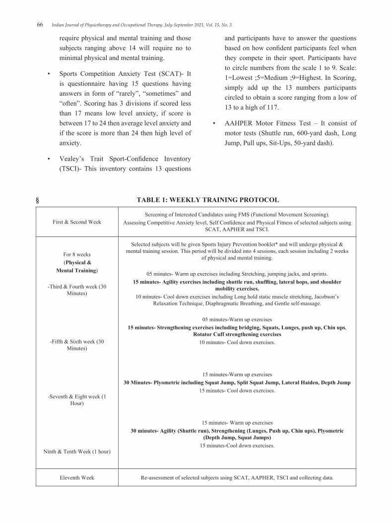
66 Indian Journal of Physiotherapy and Occupational Therapy. July-September 2021, Vol. 15, No. 3
require physical and mental training and those subjects ranging above 14 will require no to minimal physical and mental training.
• Sports Competition Anxiety Test (SCAT)- It is questionnaire having 15 questions having answers in form of “rarely”, “sometimes” and “often”. Scoring has 3 divisions if scored less than 17 means low level anxiety, if score is between 17 to 24 then average level anxiety and if the score is more than 24 then high level of anxiety.
• Vealey’s Trait Sport-Confidence Inventory (TSCI)- This inventory contains 13 questions
and participants have to answer the questions based on how confident participants feel when they compete in their sport. Participants have to circle numbers from the scale 1 to 9. Scale: 1=Lowest ;5=Medium ;9=Highest. In Scoring, simply add up the 13 numbers participants circled to obtain a score ranging from a low of 13 to a high of 117.
• AAHPER Motor Fitness Test – It consist of motor tests (Shuttle run, 600-yard dash, Long Jump, Pull ups, Sit-Ups, 50-yard dash).
§ TABLE 1: WEEKLY TRAINING PROTOCOL
First & Second WeekScreening of Interested Candidates using FMS (Functional Movement Screening).
Assessing Competitive Anxiety level, Self Confidence and Physical Fitness of selected subjects using SCAT, AAPHER and TSCI.
For 8 weeks (Physical &
Mental Training)
-Third & Fourth week (30 Minutes)
-Fifth & Sixth week (30 Minutes)
-Seventh & Eight week (1 Hour)
Ninth & Tenth Week (1 hour)
Selected subjects will be given Sports Injury Prevention booklet* and will undergo physical & mental training session. This period will be divided into 4 sessions, each session including 2 weeks
of physical and mental training.
05 minutes- Warm up exercises including Stretching, jumping jacks, and sprints.15 minutes- Agility exercises including shuttle run, shuffling, lateral hops, and shoulder
mobility exercises.10 minutes- Cool down exercises including Long hold static muscle stretching, Jacobson’s
Relaxation Technique, Diaphragmatic Breathing, and Gentle self-massage.
05 minutes-Warm up exercises15 minutes- Strengthening exercises including bridging, Squats, Lunges, push up, Chin ups,
Rotator Cuff strengthening exercises10 minutes- Cool down exercises.
15 minutes-Warm up exercises30 Minutes- Plyometric including Squat Jump, Split Squat Jump, Lateral Haiden, Depth Jump
15 minutes- Cool down exercises.
15 minutes- Warm up exercises30 minutes- Agility (Shuttle run), Strengthening (Lunges, Push up, Chin ups), Plyometric
(Depth Jump, Squat Jumps)15 minutes-Cool down exercises.
Eleventh Week Re-assessment of selected subjects using SCAT, AAPHER, TSCI and collecting data.

Indian Journal of Physiotherapy and Occupational Therapy, July-September 2021, Vol. 15, No. 3 67
Data Analysis
TABLE 2: PRE-POST DATA ANALYSIS
Variables Mean Standard deviation t-value Significance
level
SITPRE - SITPOST -8.314 2.083 -23.612 .000
FLPRE - FLPOST -7.229 1.629 -26.260 .000
SHPRE - SHPOST 2.943 .802 21.700 .000
STPRE - STPOST -9.143 4.045 -13.372 .000
Y50PRE - Y50POST 1.543 .561 16.282 .000
Y600PRE - Y600POST 2.571 .655 23.238 .000
TSCIPRE - TSCIPOST1 -29.743 9.294 -18.933 .000
SCATPRE- SCATPOST1 26.14 3.533 NS NS
TABLE 3: PERCENTAGE ANALYSIS
TOTAL PARTICIPANTS = 35
SCAT AND TSCI
MEASURESNO. OF
BENEFITTED PARTICIPANTS
NO. OF NON BENEFITTED
PARTICIPANTS
1.CONFIDENCE (Asessed through TSCI) 23 (66%) 12 (34%)
2.ANXIETY (Assessed through SCAT) 25 (71%) 10 (29%)
AAPHER-MOTOR FITNESS TEST
MEASURESNO. OF
BENEFITTED PARTICIPANTS
NO. OF NON BENEFITTED
PARTICIPANTS

68 Indian Journal of Physiotherapy and Occupational Therapy. July-September 2021, Vol. 15, No. 3
1.SIT-UPS-no. 27 (77%) 8 (23%)
2. FLEXED ARM HANG(GIRLS) 32 (91%) 3 (9%)
3. SHUTTLE RUN-10sec 23 (66%) 12 (34%)
4. STANDING LONG JUMP-cm 19 (54%) 16 (46%)
5. 50 -YARD DASH-sec 18 (51%) 17 (49%)
6. 600-YARD RUN-minutes 19 (54%) 16 (46%)
Discussion
This study was set out to investigate and assess the Impact of the Physical & Mental training on the physical fitness of Female Volleyball Athletes and to assess the impact of the physical & mental training on competitive anxiety level and self-confidence of Female Volleyball Athletes in order to prevent the chances of injury during Volleyball and to educate the proper techniques for the sports injury prevention.
A total of 35 participants were included in the study. The participants were assessed pre and post the study to find the result and prove the hypothesis.
The participants were assessed on the basis of three scales. The pre and post results were analysed on T Test and Percentage analysis.
The results were supported by the similar findings in the study: Govind B.Taware, Milind V.Bhutkar, Anil D.Surdi (JKIMSU,2013) conducted a study on A Profile of Fitness Parameters and Performance of Volleyball Players stated that ball games require comprehensive ability including physical ,technical, mental and tactile. Among them, physical abilities exert marked effects on the skill of the players themselves and the tactics of the team1.
On Pre-Post analysis, Confidence level (assessed through TSCI), Motor fitness level (assessed through
Cont... TOTAL PARTICIPANTS = 35 TOTAL PARTICIPANTS = 35 TOTAL PARTICIPANTS = 35
AAHPER), the T- test results were significant which shows that the confidence level and motor fitness level during training were remarkably improved. However, on T test analysis of anxiety level (assessed through SCAT) the result was not significant which shows that the training was not that impactful to enhance anxiety level of the participants.
Similar results were found by Kleinmann (Journal of Sports Psychology,1977) who recognized anxiety into 2 paradigms: facilitating and debilitating they are evaluated on the basis of the outcome they produce. According to Young facilitating anxiety is positive force which is witnessed in form of improved performance whereas debilitating anxiety is negative force which indicates poor performance3. Ethan R.J, Zing Li (International Journal of Physical Education, Sports and Health, 2012) conducted a study on Effect of Anxiety on physical fitness of Female Volleyball Athletes in which they stated that anxiety of a competitive athlete effects their mind which overall leads to decline in their physical performance. Hence, he believed that relaxation exercises are key to athlete’s good performance and their success7.
The different components of the scales used were also analysed through percentage analysis to find the number of benefitted participants through the physical

Indian Journal of Physiotherapy and Occupational Therapy, July-September 2021, Vol. 15, No. 3 69
and mental training. 66% of the participants had shown improvement in the confidence level after the training whereas 71 % had shown improvement in the anxiety level after the training. On analysing 6 components of motor fitness on the basis of AAHPER Motor fitness test, 77% got benefitted in the Sit Ups training, 91 % got benefitted in flexed arm hang training. 66% had shown improvement in shuttle run training. 54 % participants had shown improvement in standing long jump and 600-yard run training whereas 51 % participants had shown improving in 50-yard dash training.
Conclusion
In my pre-post study design, to investigate and assess the Impact of the Physical & Mental training on the physical fitness of Female Volleyball Athletes and to assess the impact of the physical & mental training on competitive anxiety level and self-confidence of Female Volleyball Athletes in order to prevent the chances of injury during Volleyball and to educate the proper techniques for the sports injury prevention.
On analysing the physical fitness through AAHPER scale, the pre post results were highly significant which proves the physical training sessions of 11 weeks were effective in the improvement of the physical fitness of female Volleyball players. The Confidence level were remarkably improved after training sessions as the T test pre post analysis was highly significant. The anxiety level of the participants was shown improvement in the percentage analysis when they were assessed pre and post training sessions through SCAT Scale. On the basis of the findings, it can be concluded that Interval Training session (Physical and Mental) are effective on the overall performance of: Female Volleyball Athletes.
Conflicts of Interest: The authors report no conflicts of interest in this study.
Funding Sources: Self
Ethical Clearance: Ethical approval had been taken from the Ethical Committee of Jamia Hamdard to conduct this study. Verbal and written consent were obtained from all the participants who were elected to participate in this study.
References
1. Govind B.Taware, Milind V.Bhutkar, Anil D.Surdi, A Profile of Fitness Parameters and Performance of Volleyball Players,JKIMSU,2013
2. Lastella J, Sargent, Sleep Quality, Mood and Performance: A study of Elite Brazilian Volleyball Athletes, Journal of Sports Science and Medicine,2016
3. Kleinmann, Anxiety among Female Volleyball Athletes, Journal of Sports Psychology,1977
4. Ali Fattahi,Heydar Sadeghi, Resistance, Plyometrics and combined training in Children and adult Volleyball players: A Review Study, Journal of Exercise Physiology,2011
5. Mohd.Abou. Elmagd, General Psychological factors affecting physical performance and sports, International Journal of Physical Education, Sports and Health,2016
6. Esfahani N and Soflu H.G, The Comparison of pre-competition anxiety and state anger between Female and Male Volleyball Players, (World Journal of Sports Sciences,2010)
7. Ethan R.J, Zing Li, Effects of Anxiety on Physical Fitness of Female Volley ball Athletes, International Journal of Physical Education, Sports and Health, 2012
8. Alexandro Andrade, Danilo R. Coimbra, Sleep Quality, Mood and Performance: A study of elite Brazilian volleyball athletes, Journal of Sports Science and Medicine,2016
9. Kenneth J Hunt, Daniel Hurwit, Incidence and Epidemiology of Foot and Ankle Injuries in Elite Collegiate Athletes, American Journal of Sports Medicine,2016
10. Aagaard, Jorgensen, Augustson, Common Injuries among Female Volleyball Athletes, JPMR,2016
11. Frasson, Ankle and knee injuries among Elite Volleyball Athletes,Journal of Physical, Medicine and Rehabilitation,2005
12. Franciele Marques, Luiz Carlos, Characteristics and contributing factors related to sports injuries in young volleyball player, Journal of BMC,2013
13. Knowles, Powell, Risk of Injuries in Young Volleyball Athletes, Journal of Exercise

70 Indian Journal of Physiotherapy and Occupational Therapy. July-September 2021, Vol. 15, No. 3
Physiology,2004.
14. Knowlesm D. Barber Foss, Jane C. Khoury,A School-based neuromuscular training program and sports related injury incidence: A prospective randomized controlled clinical trial,Journal of Athletic Training,2018
15. Sugimoto D, Myer GD, McKeon JM, Evaluation of the effectiveness of neuromuscular training to reduce ACL injury in female athletes: a critical review of relative risk reduction and numbers needed to treat analyses, British Journal of Sports Medicine,2012
16. Giovanni Monteleone, Chiara Pezzotta, The influence of plantar vault shape on ankle sprain epidemiology in volleyball: preliminary results, (Journal of Sports Medicine,2012
17. Saeko Takahashi, Yasuharu Nagano, A retrospective study of mechanism of anterior cruciate ligament injuries in high school basketball, handball, judo, soccer and volleyball, Journal of Sports Biomechanics,2019
18. Eric C Roux, Evaluation of a sport- specific performance task associated with a lower extremity injury prevention program, Canadian Occupational Therapy Journal,2015
19. Helen C Smith, Pamela Vacek, Risk Factors for Anterior Cruciate Ligament Injury: Hormonal, Genetic, Cognitive Function, Previous Injury and extrinsic risk factors, Journal of Exercise Physiology,2012
20. Stuart M Mcgill, Core Training : Evidence translating to better performance and Injury Prevention, Strength and Conditioning Journal,2010
21. Mario Lamontagne, A Carrafa, Neuromuscular Strategy to prevent Anterior Cruciate Ligament Injury,Journal of Gait and Posture,2009
22. O. Kilic, M. Maas, E. Verhagen, J, Incidence, Aetiology and prevention of musculoskeletal injuries in volleyball: A Systematic Review of the Literature, (European Journal of Sports Science,2019
24. Schmikli SL1, Backx FJ, National Survey on Sports Injuries in the Netherlands: target populations for sports injury prevention programs, Clinical Journal of Sports Medicine,2009
24. Carr K.E, Female Athlete Triad Syndrome in the high school athletes, Archives of Pediatrics and Adolescent Medicine,2011
25. Madison L. Predicting and Preventing Common Volleyball Injuries with Functional Tests, Journal of Physical Medicine and Rehabilitation,2018

Indian Journal of Physiotherapy and Occupational Therapy, July-September 2021, Vol. 15, No. 3 71
Work-Related Musculoskeletal Disorders among Traditional Weavers of Districts of Arunachal Pradesh - A Cross Sectional
Study
Pavana1, Ngilyang Mica2
1Associate Professor, 2Lecturer, Department of Community Physiotherapy, R.V. College of Physiotherapy®, 4th Block Jayanagar, Bangalore, India
Abstract
Background and Objectives: Weaving is the second biggest source of income after agriculture and remains important to India’s economy with roughly 4.3 million people involved, of which 61% are from North-East India. Arunachal Pradesh, one of the North-East states has its own unique method of weaving where it demands long sitting without back support for hours that can develop Musculoskeletal Disorders (MSD). Thus, the study aims to estimate the prevalence of Work-Related Musculoskeletal Disorders (WRMD) and severity of disability due to Low Back Pain (LBP) among traditional weavers of Arunachal Pradesh.
Methods: A cross-sectional study was conducted in five districts of Arunachal Pradesh. 210 traditional weavers were randomly selected. Subjects were screened for Work-Related Musculoskeletal Disorders (WRMD) using Nordic Musculoskeletal Questionnaire (NMQ) following which, the weavers that reported Low Back Pain (LBP) were screened for the severity of disability using Modified Oswestry Low Back Pain Disability Questionnaire (M-OSW). Results obtained were statistically analysed using SPSS 25.0.
Results: The result showed highest prevalence of musculoskeletal disorder in lower back, of which 79.2% had trouble in last 12 months, 69.7% was having trouble during last 7 days and 62.9% were prevented from doing normal activities during last 12 months. A total of 36.6% had mild disability, 60.6% moderate disability and 2.8% severe disability due to low back pain.
Conclusion: This study concluded that, prolong work exposure and awkward work postural demand increases the incidence of musculoskeletal disorders.
Keywords: Traditional weaver, Work-Related Musculoskeletal Disorder, Low Back Pain, Nordic Musculoskeletal Questionnaire, Modified-Oswestry Low Back Disability Questionnaire.
Introduction
Weaving is recognized to be one of the oldest surviving crafts in the world1 and it is the second biggest source of rural employment after agriculture.2 Handloom is an important cottage industry among developing countries like India, Pakistan, Bangladesh, Iran, Turkey and China, where traditional ways of weaving is still significantly practiced.3 India has the highest capacity of loom with a share of 61 percent in the world loom age.4 According to The Fourth All India Handloom
Census 2019-20 by Ministry of Textiles, Government of India, total number of households engaged in weaving activities (weaving and allied activities) is 26.73 lakhs, of which 22.5 lakhs are located in rural areas with 72% of handloom weavers being female.5
Weaving is considered to be one of the most tedious profession, that demands long hours of static work which can develop Musculoskeletal Disorders (MSD) as awkward posture, repetitive movements and contact stress are common.6 It involves numbers of repetition
DOI Number: 10.37506/ijpot.v15i3.16165

72 Indian Journal of Physiotherapy and Occupational Therapy. July-September 2021, Vol. 15, No. 3
in work, causing musculoskeletal strain increasing the possibility of fatigue and insufficient tissue recovery period leading to pain and discomfort.7
Domestic weaving provides livelihood to a considerable group of population exposing weavers to occupational risk factors.8 A high prevalence of musculoskeletal problem was reported among Iranian handloom weavers due to the constraints of working positions, poor design of loom, working times, repetitive work and seat type.9,10 Arunachal Pradesh has a total of 77,600 weavers,5 still preserving its ancient culture and traditions and renowned for its striking garment designs. Weaving is one of the oldest cultural practice being followed from generation to generation. Though modern tool has been introduced with the advent of modernity, the importance of the traditional loom and products made from it have not at all diminished.
“Work-Related Musculoskeletal Disorders” (WRMDs) is a term used to describe a painful or disabling injury to the muscle, tendon or nerves caused or aggravated by work.11 Several studies on handloom weavers have showed that there are a high rate of work-related musculoskeletal problems throughout the World and is a major cause of disability affecting not only individual but the society as a whole.12
Low Back Pain (LBP) is considered to be one of the most common occupational health problems. It is defined by Last JM et al. as “a non-specific condition that refers to complaints of acute or chronic pain and discomfort in or near the lumbosacral spine”.13 Kraemer et al. 1985 stated prolong sitting leads to isometric contraction of back muscles leading to endomuscular pressure restricting blood flow, resulting in ischemia which reduces energy requirements of the muscles causing muscle fatigue.14 Prolong flexion of the spine leads to increase intervertebral joint laxity and fluid loss in the intervertebral discs15 and the intra-discal pressure at the lumbar region is high both in sitting and trunk bent forward posture.16 Insufficient recovery time are the contributing factors to develop Low Back Pain (LBP).17
The weaving method of Arunachal Pradesh also consist of several occupational related risk factors where it demands them to remain in a long sitting, trunk
forward bent position for many hours at a stretch and an activities like pulling, pushing, lifting the tools, working while bent or twisted at the waist and repetitive motions with hands/wrists and gripping is required.
The activity like twisting and bending of vertebral column subject weavers to undue stress while working in sitting position.12,18,19
Study done in Lakhimpur District Assam have reported highest prevalence of discomfort in the waist among weavers. Also, a study done by D. C. Metgud et al20 on women textile workers of Belgaum in the spinning section with similarly long sitting posture without back rest revealed highest pain in the low back. A study done by Geetashree B on weavers also showed that, about 47.5% respondents perceived very severe pain in back and less than 45% respondents made complaint of severe pain in low back.21
Although several studies on Work-Related Musculoskeletal Disorders (WRMDs) and occupational Low Back Pain (LBP) has been conducted in various states of India, the weaving tool and method used by the local weavers in Arunachal Pradesh is different from the mainstream India, their findings and results may show variation or in-applicability to the people of Arunachal Pradesh. Hence, there is a need to establish prevalence of Work-Related Musculoskeletal Disorders (WRMDs) and severity of disability due to Low Back Pain (LBP) among traditional weavers of Arunachal Pradesh for the ergonomic changes to bring about and to assist in planning management strategy including awareness, education and treatment to prevent Work-Related Musculoskeletal Disorders (WRMDs).
Aim and Objectives of the Study
Primary objective:
To estimate the prevalence of Work-Related Musculoskeletal Disorders among traditional weavers of Arunachal Pradesh.
Secondary objective:
To assess the prevalence of severity of disability due to Low Back Pain among traditional weavers of Arunachal Pradesh.

Indian Journal of Physiotherapy and Occupational Therapy, July-September 2021, Vol. 15, No. 3 73
Materials and Method
Research design –A Cross sectional study.
Source of data - Five districts of Arunachal Pradesh (West Kameng, Lower Subansiri, West Siang, East Siang and Namsai).
Study settings - Individuals, self-help groups, societies, private small scale industries and Government textiles and handicraft centres.
Sampling design - cluster sampling technique (randomly recruited).
Sample size (n)=210, from each cluster, sample size (n)= 210/5 = 42.
Inclusion Criteria
Traditional weavers. >14 years of age.
Women weavers.
Weaving experience of more than 12 months and beyond. Should be into weaving phase.
Had no history of MSDs before starting the weaving. Residing within the Study settings.
Willing to participate in the study and sign the informed written consent. Parents willing to sign the informed written consent for minor weavers. Minor weavers willing to sign the informed written assent.
Exclusion Criteria
Musculoskeletal disorder due to trauma or any accident. Musculoskeletal disorder due to neurological disorders. Congenital deformities.
Musculoskeletal degenerative conditions. Any spinal surgeries.
Pregnant and lactating mother.
Procedure
A pilot study was conducted at one of the private weaving industry at Lepakshi, Andhra Pradesh with 10% of the calculated sample size to standardize the questionnaire.
The five districts (West Kameng, Lower Subansiri, West Siang, East Siang and Namsai) of Arunachal Pradesh were randomly selected for the study. These five districts were considered as clusters to give over all representation to the entire State. An approval was obtained from the Institutional Ethics Committee (IEC) of R. V. College of Physiotherapy®. The researcher contacted the Director Textile and Handicrafts, Government of Arunachal Pradesh and permission was obtained to collect data across the five districts. Individuals, self-help groups, societies, private small scale industries and Government textiles and handicraft centers had been visited to screen the weavers population matching inclusion and exclusion criteria. From each cluster 42 subjects were selected based on cluster sampling technique. An informed written consent from the subjects were obtained after explaining to them the purpose of the study in their own language. The researcher collected the data from each selected subjects by direct oral interview technique using the Nordic Musculoskeletal Questionnaire (NMQ)22 including demographic data to estimate the prevalence of Work-Related Musculoskeletal Disorder (WRMD) and the Modified Oswestry Low Back Pain Disability Questionnaire (M-OSW)23,24 were administered to the subpopulation of weavers who reported experiencing low back pain on the NMQ to assess the level of severity of disability due to low back pain. The collected data was analyzed.
Result Analysis
All the categorical variables was presented in the form of frequency tables and graph wherever necessary. The quantitative variables was summarized using mean ± standard deviation with 95% confidence interval. The data was entered using Statistical Software namely SPSS version 25.0, Microsoft Excel 2013 and Microsoft word 2013 was used to draw tables and graphs.
In the present study, the mean (±SD) age of the weavers was 38.79 (±12.844) years.
Maximum weavers (28.1%) were age ranged from 21-30 years (Table 1). The present study shows that majority of the weavers (40.7%) were having high school education.

74 Indian Journal of Physiotherapy and Occupational Therapy. July-September 2021, Vol. 15, No. 3
The analysis shows that maximum weavers (63.3%) belong to middle income level in Standard of Living Index. 25.4% belong to low income level and 11.3% belong to high income level.
Duration of weaving Number (n=221) Percentage (%)
(yrs)
1-10 95 43.0
10-20 62 28.1
20-30 30 13.5
30-40 27 12.2
40-50 5 2.3
50-60 2 0.9
Mean=16.63 ((±12.966)
In this study, the mean (±SD) of working hours distribution was 5.88 (±1.636) hours per day. 61.5% weavers were working for 6-10 hours per day and 38.5% weavers were having 1 to 5 hours of weaving per day.
Present study showed that maximum weavers were affected in Lower Back(79.2%) during the last 12 months. Followed by Upper Back (62.9%), Knee (52.5%), Neck(52.0%), bilateral Shoulder (50.3%), Bilateral Elbow (43.0%), Ankle (43.0%), Bilateral Wrist(42.6%), Hips (36.7%), Right Wrist (7.2%), Right Shoulder (5.4%), Right Elbow (2.7%), Left Shoulder (0,9%), Left Elbow (0.9%) and Left Wrist (0.9%).
Table 2: The Prevalence of Musculoskeletal Disorders in weavers during last 12 months.
Pain in body parts Number (n=221) Prevalence (%)
Neck 115 52.0
Shoulder Right 12 5.4
Left 2 0.9
Both 111 50.3
Elbow Right 6 2.7
Left 2 0.9
Both 95 43.0
Wrist Right 16 7.2
Left 2 0.9

Indian Journal of Physiotherapy and Occupational Therapy, July-September 2021, Vol. 15, No. 3 75
Both 94 42.6
Upper Back 139 62.9
Lower Back 175 79.2
Hips 81 36.7
Knee 116 52.5
Ankle 95 43.0
Among all weavers mostly found in lower back (69.7%) had problem during last 7 days. Upper back was the second highest (49.8%), Knee (45.7%), Neck (38.9%), Bilateral Shoulder (38.5%), Ankle (33.5%), Bilateral Wrist (33.0%), Bilateral Elbow (30.8%), Hips (25.8%), Right Wrist (8.1%), Right Shoulder (6.3%), Left Shoulder (0.5%), Left Elbow (0.5%) and Left Wrist (0.5%) was having trouble due to musculoskeletal disorders during last 7 days.
Table 3: The Prevalence of Musculoskeletal disorders in weavers during last 7 days
Pain in body parts Number (n=221) Prevalence
(%)
Neck 86 38.9
Shoulder Right 14 6.3
Left 1 0.5
Both 85 38.5
Elbow Right 7 3.2
Left 1 0.5
Both 68 30.8
Wrist Right 18 8.1
Left 1 0.5
Cont... Table 2: The Prevalence of Musculoskeletal Disorders in weavers during last 12 months.

76 Indian Journal of Physiotherapy and Occupational Therapy. July-September 2021, Vol. 15, No. 3
Both 73 33.0
Upper Back 110 49.8
Lower Back 154 69.7
Hips 57 25.8
Knee 101 45.7
Ankle 74 33.5
The prevalence of musculoskeletal disorder affecting ADL during last 12 months showed more than a half of the weavers i.e 62.9% were affected by lower back. Less than a half i.e. 42.5% due to knee and 42.1% due to Upper Back. Followed by 36.2% due to shoulder, Wrist (34.4%), Neck (32.6%), Ankle (31.2%), Elbow (29.0%) and Hips (24.4%)
Table 4: The Prevalence of Musculoskeletal disorders affecting ADL during last 12 month in weavers
Pain in body parts Number (n=221) Prevalence
(%)
Neck 72 32.6
Shoulders 80 36.2
Elbows 64 29.0
Wrist 76 34.4
Upper Back 93 42.1
Lower Back 139 62.9
Hips 54 24.4
Knee 94 42.5
Ankle 69 31.2
Cont.. Table 3: The Prevalence of Musculoskeletal disorders in weavers during last 7 days

Indian Journal of Physiotherapy and Occupational Therapy, July-September 2021, Vol. 15, No. 3 77
The present study observed that from the total prevalence of Musculoskeletal Disorders in Low Back 36.6% had minimal disability, 60.6% had moderate disability and 2.8% had severe disability.
Table 5: The Prevalence of Severity of disability due to Low Back Pain
Severity of Low Back Number (n=175) Prevalence
Pain (%)
Minimal Disability (Score 64 36.6
0-20%)
Moderate Disability 106 60.6
(Score 21-40%)
Severe Disability 5 2.8
Discussion
Weaving is one of the most tedious profession, with long hours of static work that develops Musculoskeletal Disorders (MSD).6 In the World of mechanization, the traditional domestic weavers plays a major role in preserving culture with its unique design and method of weaving, providing livelihood to a large section of population.
The numbers of repetition in work causes musculoskeletal strain increasing the possibility of fatigue and insufficient tissue recovery period leading to pain and discomfort.7 Awkward postures adopted by the workers is one of the most important factors of their poor working efficiency and prevalence of Musculoskeletal Disorders (MSD).
The findings of this study confirm the work-related stress on weavers. In the present study, the highest prevalence of musculoskeletal disorder was found in low back, of which 79.2% had trouble in last 12 months, 69.7% was having trouble during last 7 days and 62.9% were prevented from doing normal activities during last 12 months. The prevalence of musculoskeletal disorders during last 12 months revealed that maximum of them
i.e. 79.2% were having pain in lower back. A previous study finding by Naz H et al. showed highest prevalence of pain due to musculoskeletal disorders during last 12 months in Right Wrist (76.56%)3 and in a study by Rahman M et al. the highest prevalence was found in Hips (78.0%).25
The weaving method consists of several occupational related risk factors where it demands them to remain in a long sitting, trunk forward bent position for many hours at a stretch and an activities like pulling, pushing, lifting the tools, working while bent or twisted at the waist and repetitive motions with hands/wrists and gripping is required. The activity like twisting and bending of vertebral column subject weavers to undue stress while working in sitting position.12,18,19
Among all weavers mostly found in lower back (69.7%) had problem during last 7 days. Similarly, in a study by Naz H et al. maximum trouble during last 7 days was found in lower back (59.38%)3. A finding by Rahman M et al. showed 69.72% respondents had trouble in Neck pain.25
In this study it was observed that due to Lower back pain 62.9% weavers were prevented from carrying out

78 Indian Journal of Physiotherapy and Occupational Therapy. July-September 2021, Vol. 15, No. 3
their normal activities during last 12 months . Less than a half i.e. 42.5% due to knee and 42.1% due to Upper Back, followed by 36.2% due to shoulder, Wrist (34.4%), Neck (32.6%), Ankle (31.2%), Elbow (29.0%) and Hips (24.4%) were prevented from their normal activities in last 12 months.
A study done by D. C. Metgud et al20 to outline cardio-respiratory and musculoskeletal profile before, during and at the end of work on women textile workers of Belgaum in the spinning section with similarly long sitting posture without back rest. The study revealed that 91% of the subjects suffered from at least one work-related musculoskeletal pain in relation to length of occupational exposure and region-wise mapping of pain showed that percentage of postural pain in the low back (47%) was highest.
In the present study, among 177 weavers who reported lower back pain in musculoskeletal disorders questionnaire, 29.8% had mild disability, 48.0% moderate disability and 2.3% severe disability . A study done by Bori G et al. on weavers also showed that, about 47.5% respondents perceived very severe pain in back and less than 45% respondents made complaint of severe pain in low back.21 A study conducted by Hossain A et al. to find out the pattern and associated factors of musculoskeletal pain among handloom weavers revealed that 12.9% had mild lumbar spine pain, 55.9% had moderate pain and 31.2 % had severe pain in lumbar spine.26
From the above discussion it can be concluded that traditional weavers of districts of Arunachal Pradesh are equally affected with a high prevalence of musculoskeletal disorders that effects their ADL and productivity. There is an utmost and urgent need of appropriate measures to prevent musculoskeletal disorders among traditional weavers keeping in mind the cultural value of ancient weaving method.
Conclusion
The study found high rate of musculoskeletal disorders among traditional weavers. More than 79.2% of weavers suffered from musculoskeletal pain and Lower Back were the most prevalent site. Frequent breaks during work period and back support would
reduce the Musculoskeletal stress on lumbar region. Based on the observations made, the study concluded that there is ample scope for ergonomic improvement keeping in view the need for maximum comfort to the weavers and the cultural importance of the traditional weaving method to promote their health and well-being and enhance their productivity and quality of work.
Limitations:
Unavailability of scales in local language.
Larger geographical area could have been included (present study included only five districts).
Current study was only determined to find the prevalence of WRMSD and did not include management to prevent WRMSD.
Scope for further study:
A high prevalence of musculoskeletal disorders exists among traditional weavers that effects their ADL, productivity and the quality of work. Further studies are needed to identify the specific risk factors for the ergonomic changes to bring about and to assist in planning management strategy including awareness, education and treatment to prevent Work-Related Musculoskeletal Disorders WRMDs.
Ethical Clearance - taken from R. V. College of Physiotherapy®, Bangalore, Karnataka.
Source of Funding- Self.
Conflict of Interest- Nil
References
1. Pandit S, Kumar P, Chakrabarti D. Ergonomic Problems Prevalent in Handloom Units of North East India. IJSRP. 2013 Jan;3(1):1-7. Available from:http://i3s.net.in/uploads/pdf/Resources/Ergonomic_Problems_Prevalent_in_Handloo m_Units_of_North-East_India.pdf.
2. Government of India, Ministry of Textiles. Annual Report; 2017-18. 281p. Available from: http://texmin.nic.in/sites/default/files/AnnualReport2017-18%28English%29.pdf. Accessed on:2019 Oct 15;22:40.

Indian Journal of Physiotherapy and Occupational Therapy, July-September 2021, Vol. 15, No. 3 79
3. Naz H, Kwatra S, Ojha P. Prevalence of musculoskeletal disorders among handloom weavers of Uttarakhand: an ergonomic study. J. Appl. & Nat. Sci. 2015;7(1):101–4. Available from: https://pdfs.semanticscholar.org/a727/5fbcdd24d73a32d1ea3e7957e6a499c02e1a.pdf.
4. National Skill Development Corporation. Human Resource and Skill Requirements in the Handlooms and Handicrafts Sector (2013-17, 2017-22). 2007. 45p. Available from: https://nsdcindia.org/sites/default/files/Handlooms-Handicrafts.pdf. Accessed on:2019 Oct 15;23:36.
5. Government of India, Ministry of Textiles. Fourth All India Handloom Census 2019-20. 14p. Available from: https://handlooms.nic.in/writereaddata/3736.pdf. Accessed on:2020 Feb 2;15:12.
6. Buckle P, Devereux J. Work-related neck and upper limb musculoskeletal disorders. Luxembourg: Office for Official Publications of the European Communities; 1999. 114p. Available from: https://osha.europa.eu/en/publications/report-work-related-neck-and-upper-limb-musculoskeletal-disorders. Accessed on:2019 Oct 16; 9:20.
7. Tiwari RR, Pathak MC, Zodpey SP. Low Back Pain among Textile Workers. Indian J Occup Environ Med. 2003 Jan;7(1):27-9. Available from: http://medind.nic.in/iay/t03/i1/iayt03i1p27.pdf.
8. Szymańska, R. Coexistence of cervico-brachial neuralgia and backache among textile industry workers. Medycyna pracy. 1988;39(2):125-9.
9. Choobineh A, Lahmi M, Shahnavaz H, Jazani RK, Hosseini M. Musculoskeletal Symptoms as Related to Ergonomic Factors in Iranian Hand-Woven Carpet Industry and General Guidelines for Workstation Design. Int J Occup Saf Ergon. 2004 Jan;10(2):157–68. Available from:https://www.tandfonline.com/doi/pdf/10.1080/10803548.2004.11076604.
10. Choobineh A, Hosseini M, Lahmi M, Jazani RK, Shahnavaz H. Musculoskeletal problems in Iranian hand-woven carpet industry: Guidelines for workstation design. Appl. E r g o n . 2007 Sep;38(5):617-24. Available from: https://www.tums.ac.ir/1392/12/18/83.pdf-hoseinim-2014-03-09-05-55.pdf.
11. Bernard BP, Musculoskeletal Disorders and Workplace Factors. 4676 Columbia Parkway Cincinnati: DHHS (NIOSH); 1997. Available from: https://www.cdc.gov/niosh/docs/97-141/pdfs/97-141.pdf.
12. Kemmlert K. Prevention of occupational musculo-skeletal injuries. Labour Inspectorate investigation. Scand J Rehabil Med. 1996;35:1-34.
13. Last JM, Wallace RB. Public health and preventive medicine. 13th edition, London; 1992.
14. Kraemer J, Kolditz D, Gowin R. Water and electrolyte content of human intervertebal discs under variable load. Spine. 1985 Jan;10(1):69-71. Available from: doi: 10.1097/00007632-198501000-00011.
15. Adams MA, Dolan P, Hutton WC. Diurnal variations in the stresses of the lumbar spine. Spine. 1987 Mar;12(2):130-7. Available from: doi: 10.1097/00007632-198703000-00008.
16. Wilder DG, Pope MH, Frymoyer JW. The biomechanics of lumbar disc herniation and the effect of overload and instability. J. Spinal Disord. 1988;1(1):16-32.
17. Singh MB, Fotedar R, Lakshminarayan J. Occupational Morbidities and their Association with Nutrition and Environmental Factors among Textile Workers of Desert Areas of Rajasthan. J Occup Health. 2005 Oct;47(5):371-7. Available from: https://pdfs.semanticscholar.org/3243/c2fba83f4728c3ece52e3996e9dc3b24cb9c.pdf.
18. Videman T, Nurminen T, Tola S, Kuorinka I, Vanharanta H, Troup JD. Low back pain in nurses and some loading factors of work. Spine. 1984 May-Jun;9(4):400-4. Available from: doi: 10.1097/00007632-198405000-00013.
19. Jayson MIV. The Lumbar Spine and Back Pain. 4th edition. Churchill Livingstone, London: Edinburgh; 1992.
20. Metgud DC, Khatri S, Mokashi MG, Saha PN. An ergonomic study of women workers in a woollen textile factory for identification of health-related problems. Indian J Occup Environ Med. 2008 Apr;12(1):14-19. Available from: http://medind.nic.in/iay/t08/i1/iayt08i1p14.pdf.
21. Bori G. Musculo-skeletal Problems of Women

80 Indian Journal of Physiotherapy and Occupational Therapy. July-September 2021, Vol. 15, No. 3
Weavers in Handloom Industry of Lakhimpur District, Assam. Int J Sci Res. 2016 Dec;6(12):201-11.
22. Rahman M, Khan MH, Hossain I et al. Musculoskeletal Problems among Handloom Workers. Texila int. j. pub health. 2017 Sep;5(3):1-15. Available from: https://www.researchgate.net/publication/322202294_Musculoskeletal_Problems_am ong_Handloom_Workers.
23. Hossain A, Kamrujjaman M, Maleque A. Associated Factors & Pattern of Musculoskeletal Pain among Male Handloom Weavers Residing in Belkuchi, Shirajganj: A Cross Sectional Study. Int J Sci Eng Res. 2018 Oct;9(10):1447-51. Available from: https://www.ijser.org/onlineResearchPaperViewer.aspx?Associated-
Factors-Pattern-of-Musculoskeletal-Pain-among-Male-Handloom-Weavers-Residing-in-Belkuchi-Shirajganj-A-Cross-Sectional-Study.pdf
24. Dickinson CE, Campion K, Foster AF et al.. Questionnaire development: an examination of the Nordic Musculoskeletal questionnaire. Appl Ergon. 1992 Jun;23(3):197-201
25. Fairbank JCT, Pynsent PB. The Oswestry Disability Index. Spine. 2000;25(22):2940-53. Available from:https://pdfs.semanticscholar.org/6716/b3eec3c1da764f636c14d96b823b9fa95106.pdf
26. Fairbank JC, Couper J, Davies JB et al. The Oswestry low back pain questionnaire. Physiotherapy 1980;66:271–3.

Indian Journal of Physiotherapy and Occupational Therapy, July-September 2021, Vol. 15, No. 3 81
Effectiveness of Core Stability Exercise and Proprioception Exercise on Balance in Subjects with Diabetic Neuropathy- A
Randomized Controlled Trial
Pavana1, Nair Anjali Premrajan2, Amrita Ghosh3
1Associate Professor, Department of Community Physiotherapy, Karnataka, 2MPT Student, R.V College of Physiotherapy, Bengaluru, 3Associate Professor, MGM School of Physiotherapy, Navi Mumbai
AbsractBackground and Objectives: Long standing Diabetes Mellitus leads to Diabetic Peripheral Neuropathy in the subjects which alter their daily activities performed. As physical activities are the key elements to prevent the further progress of Diabetes Mellitus. The neuropathy also affects the postural stability leading to impaired balance and risk of falls in the subjects. The integrated activity of the motor and the sensory systems play an important role in maintain the postural stability which depends upon the strength of the core muscles and proper integrated activity of the proprioceptors of the lower limb. The prevalence of the Diabetic Peripheral neuropathy in India is around 10.5% to 32.2%. The Toronto Clinical scoring System is used as a marker to assess the level of neuropathy which has a relation on the stability. Thus, the aim of the study is to find the effectiveness of proprioception exercise or core stability exercise on balance in Diabetic Peripheral Neuropathy subjects.
Objectives: To assess the effectiveness of core stability exercise on balance in Diabetic Neuropathy subjects.To assess the effectiveness of proprioception exercise on balance in Diabetic Neuropathy subjects. To compare the effectiveness of core stability exercise and proprioceptive exercise on balance in Diabetic Neuropathy subjects.
Design: Comparative Randomized Parallel Open Label Study
Methods: Eighty subjects between the age 40-60 years were recruited for the study. Written informed consent and institutional ethical clearance was obtained. Demographic data were obtained and subjects were assigned to one of two groups: Group A: experimental group given Proprioception exercise (n=40) and Group B: control group given Core Stability exercise (n=40). The therapy was given for six weeks with three days in a week and was measured with Berg Balance Scale as an outcome measure at the end of the 6 week protocol. Results: Both the groups showed improvement in the balance according to the Berg Balance Scale. The mean ± SD of pre and post Berg Balance Scale of GROUP A was 37.18 ± 4.81 and 39.41 ± 4.95 respectively and that of GROUP B was 36.95 ± 4.27 and 38.55 ± 4.29 respectively. Conclusion: The results showed a significant improvement within Group A and Group B (P<0.001) but there is no significant improvement between both the groups (P>0.05). Thus the study concluded that proprioception exercise and core stability exercise are worthwhile in improving the balance in subjects with diabetic peripheral neuropathy.
Keywords: Diabetic Peripheral Neuropathy, Proprioception Exercise, Core Stability Exercise, Berg Balance Scale, Toronto Clinical Scoring System
Corresponding Author:Dr. Pavana (PT)
Associate Professor, Department of Community Physiotherapy, R.V College of PhysiotherapyAddress: CA 2/83-3,9th Main Road, 4th Block, JayanagarBengaluru, Karnataka-560011Email: [email protected] number: 8095888027
Introduction
Diabetes Mellitus (DM) is a metabolic disorder due to increased blood sugar (glucose) level which is called (hyperglycemia), type 2 diabetes, the cause is mainly either due to insulin resistance to action on the cells (a hormone that is responsible for the regulation of the
DOI Number: 10.37506/ijpot.v15i3.16166

82 Indian Journal of Physiotherapy and Occupational Therapy. July-September 2021, Vol. 15, No. 3
movement of sugar into the cells) or there is reduction in the production of insulin that maintains the normal glucose level.1 Diabetic peripheral neuropathy (DPN) is a polyneuropathy that may cause a damage to the peripheral nerve fibers, sensorimotor, and autonomic system.2 When there is a progression in the condition it may lead to increased postural sway, gait deviations, abnormal neuromuscular control and increased reaction time, these all will lead to falls as the suffers this due to reduction of balance.4 Postural stability is maintained by the combined action of the sensory feedback of the body that includes vestibular, visual and somatosensory system.8
Somatosensory system consist of muscle, joint, and cutaneous mechanoreceptors, as the proprioception is present in muscle spindle and joint so its affection cause disturbance in balance. 9, 2 These receptors have integration in the central nervous system to produce sensation of joint and its position and movement. 9 DPN affect the proximal and the distal peripheral sensory and motor nerves.8 Previous studies revealed that patients with diabetic peripheral neuropathy suffer from posture instability and balance impairment issues. The affection of the balance is due to the dynamic postural sway and in addition to increased muscular effort to maintain the balance in diabetic patients with neuropathy.11
The risk of fall is increased due to increase in postural sway and as there is increased muscular effort required to maintain the balance of the body. Core stability describes the ability to control the position and movement of the central portion of the body.11 Thus by targeting the muscles deep within the abdomen that assist in the muscles.12 Maintenance of good posture and provide the foundation for all arm and leg movements. Core stability exercises allow the body to maintain its balance by improving the strength of core.11 The aim of the study is to compare the effectiveness of proprioception exercise and core stability exercise to improve balance in Diabetic Peripheral Neuropathy subjects.
Materials and Methods
Informed consent was obtained from each subject after explaining the study and test procedure.
Demographic data was collected and recorded.
Research Design:
Comparative Randomized Parallel Open Label Study
Sources of data:
· Shirdi Sai Baba Hospital
· Conquistar Physios Clinic
Duration – 6 months
Materials Required:
• Toronto Clinical Neuropathy Scoring System Sheet
• Reflex Hammer, Tuning Fork 128 Hz
• 10g monofilament, hot and cold test tubes
• Berg Balance Scale (BBS) Sheet
• One standard chair with arm rest
• Footstool/step
• Stopwatch/ or wristwatch
• 15 foot walkway
• Stationaries
Sample and Sampling Technique:
Sample size: 80 subjects calculated 40 per arm for the study were taken
Sample size calculation: α = 0.05 (type-1 error), d = 15% =0.15 (anticipated error), p = 0.105 (prevalence), q = 0.895 (1-p)
Sampling technique: Convenient sampling
Inclusion Criteria:
· Subjects willing to sign the written informed consent
• Subjects diagnosed with Type 2 DM, DPN
• Subjects with 40 to 60 years
• Subjects with an ability to walk independently
• Subjects with T2DM for ≥ 10 years and DPN for ≥ 1year

Indian Journal of Physiotherapy and Occupational Therapy, July-September 2021, Vol. 15, No. 3 83
• Subjects with BBS score of low and medium risk
• Subjects with mild to moderate neuropathy
Exclusion Criteria:
• Subjects with impaired visual or vestibular impairment and systemic illness
• Subjects unable to provide a proper medical history
• Subjects with neuropathies due to non-diabetic cause
• Subjects with any other neurological condition that can impair balance
• Subjects with any other musculoskeletal conditions that cause pain and instability
• Subjects with severe diabetes with foot ulcers or foot deformities
• Subjects with any kind of lower limb amputation
Measuring Tool:
Berg Balance Scale used as outcome measure tool. In pre- intervention the BBS was used as a screening tool for inclusion criteria to include mild and moderately balance affected subjects into the study. In post-intervention after the 6 months of intervention the BBS was used as a screening tool to note the improvement of balance that occurred due to the interventions given.
Procedure:
Figure 1: Diagrammatic representation of the method implementation
Pre intervention screening for neuropathy was done by TORONTO CLINICAL SCORING SYSTEM and
the balance was screened using BERG BALANCE SCALE.
The therapy was given in the following manner:-
· GROUP A (PROPRIOCEPTION EXERCISE) = 40 subjects
Ø Proprioception Exercise :
Exercises included one leg balance, forward leg swings with knee extension, backward leg swings with knee flexion, toe walking, heel walking, cross-body leg swings right and left sides, partial squat, single-leg squat, blind advanced one leg balance, and side lunge1, Dynamic walking exercise, active range of motion exercise of upper and lower extremity, foot care and dietary advice.1
Ø Duration:
Each session was done for 45 minutes with 1 minute rest for every 5 minute of exercise for 3 times per week for 6 weeks.
· GROUP B ( CORE STABILITY EXERCISE) = 40 subjects
Ø Core Stability Exercise :
Exercise included bridging, plank exercise on knee and elbow, cat curl exercise, abdominal crunch and oblique crunches, fall out exercise, seated marching exercise, lean exercise, twist exercise, Dynamic walking exercise, active range of motion exercise of upper and lower extremity, foot care and dietary advice .11
Ø Duration:
Each session for one hour with 2 minute rest after each exercise for 3 times per week for 6 weeks.
At the end of 6 weeks balance measured using BBS and compared with the pre intervention score to know the effectiveness of the therapies.
Result Analysis
Data has been derived using descriptive and inferential statistics SPSS 23.0. The quantitative variables are presented on Mean ± Standard Deviation

84 Indian Journal of Physiotherapy and Occupational Therapy. July-September 2021, Vol. 15, No. 3
(SD) [Min-Max]. Unpaired t-test has been used to find significance of the study parameters between groups. While paired t-test has been used to find significance of
the study within the groups. Microsoft word and Excel have been used to generate graphs, tables etc.
Table 1: Age distribution of subjects
Age (yrs)Proprioception Exercises Core Stability Exercise
Number Percent Number Percent
41 - 45 0 0 3 7.9
46 - 50 18 46.2 18 47.4
51 - 55 14 35.9 10 26.3
56 - 60 7 17.9 7 18.4
Total 39 100.0 38 100.0
The table 1 shows that majority of study subjects in Proprioception exercise and Core Stability exercise groups are 46.2% and 47.4% in 46-50 years, followed by 35.9% and 26.3% in 51-55 years. In age group of 56-60 years 17.9% and 18.4% are present respectively in both the groups [There were 40 subjects per arm, but in Proprioception exercise one subject drop out and in Core Stability exercise 2 subjects drop out in middle of the study and hence there were 39 and 38 subjects respectively in the groups throughout the analysis].
Table 2: Gender distribution of subjects
GenderProprioception Exercises Core Stability Exercise
Number Percent Number Percent
Male 23 59.0 21 55.3
Female 16 41.0 17 44.7
Total 39 100.0 38 100.0
Table 2 shows that Proprioception exercise group consider at total of 59% males and 41% females whereas in Core Exercise group 55.3% males and 44.7% females respectively.
Study groupsSample
sizeMean SD
Std. Error Mean
t - value P - value
ProprioceptionExercises
39 52.51 3.748 0.600
1.449 0.151Core Stability
Exercise38 51.26 3.818 0.619

Indian Journal of Physiotherapy and Occupational Therapy, July-September 2021, Vol. 15, No. 3 85
Table 3: Age distribution among the groups
The table 3 shows that in this study Mean ± SD of age in Proprioception exercise was 52.51±3.75 years and in Core stability exercise was 51.26±3.82 years, the difference in the mean is not found statistically significant [t=1.449, P>0.151] this shows that there was equal age distribution to both the groups.
Table 4: Pre- and Post- data for outcome variable within Group A
Proprioception Exercises Sample size Mean SDStd. Error
Meant - value P - value
Pre-test BBS score 39 37.18 4.801 0.769
6.771 P < 0.001
Post-test BBS score 39 39.41 4.951 0.793
The table 4 shown above depicts pre and post data for outcome variables within group A. The mean ± SD of pre and post BBS score was 37.18±4.80 and 39.41±4.95 respectively which are statistically highly significant [t= 6.771, P<0.001]. The percentage of improvement within the group is about 6%.
Table 5: Pre and post data for outcome variable within group B
Core StabilityExercises
Sample size
Mean SDStd. Error
Meant - value P - value
Pre-test BBS score 38 36.95 4.268 0.692
5.763 P < 0.001
Post-test BBS score 38 38.55 4.298 0.697
The table 5 shown above depicts pre and post data for outcome variables within Group B. the mean ± SD of pre and post BBS score was 36.95±4.27 and 38.55±4.30 respectively which are statistically highly significant [t=5.763, P<0.001]. The percentage of improvement within the group is about 4%.
Table 6: Difference between pre and post outcome measure of both the groups
Time of measurement Study groups Sample size Mean SD Std. Error Mean t - value P - value
Pre-test BBS score
ProprioceptionExercises
39 37.18 4.81 0.769
0.224 0.823Core Stability
Exercise38 36.95 4.27 0.692
Post-test BBS score
ProprioceptionExercises
39 39.41 4.95 0.793
0.811 0.421Core Stability
Exercise38 38.55 4.29 0.697

86 Indian Journal of Physiotherapy and Occupational Therapy. July-September 2021, Vol. 15, No. 3
The table 6 shows the pre and post values of the outcome measure, with mean ± SD in proprioception exercise was 37.18±4.81 and 39.41±4.95 and for the core stability exercise was 36.95±4.27 and 38.55±4.29, they are not found statistically significant [where for proprioception exercise t=0.224, P=0.823 and for core exercise t=0.811, P=0.421]. This shows that even though the two therapies are significant in improving balance but there was no significant difference between the two therapies.
Discussion
Neuropathy is one of the most common complication present in diabetic individual.2 In diabetes because of the long period of time of high blood sugar level, this can cause damage to the nerve fibers, as diabetes is one of the several major causes of neuropathy.2 When there is a progression in the condition it may lead to increased postural sway, gait deviations, abnormal neuromuscular control and increased reaction time, these all will lead to falls as the suffers this due to reduction of balance.4 DPN affect the proximal and the distal peripheral sensory and motor nerves.8
Balance is an important parameter that is useful in our daily life activity.4 Balance is controlled by the feedback mechanism of the combined muscle action which depends on the postural sway information that is obtained from visual, motor, sensory, and vestibular receptors.2 The impairment in the body balance was found in the anteroposterior plane The affection of the balance is due to the dynamic postural sway and in addition to increased muscular effort to maintain the balance in diabetic patients with neuropathy.11
The present study was designed to compare the effectiveness of 6-weeks intervention of core stability exercise and proprioception exercise on balance in DPN subjects. The main finding of this study suggested a positive effect of both the exercises on balance with no significance between the two statistically. Proprioception exercise mainly focus on the sense of joint position that helps to maintain joint stability and posture that helps to improve somatosensory system which is affected in subjects with DPN.
In the previous study done by Kamtchi found that there was a significant improvement in the balance with proprioceptive training compared to backward walking training. The proprioception exercise in the present study was carried on for 6 weeks. The 6- weeks training duration was chosen as it would take minimum of this period for the neural and muscular adaptations to occur. A similar study done by Abeer-el-Wishy et al concluded that proprioceptive training along with conventional physiotherapy was found in improving proprioception sensation and balance which was carried out for the 6 weeks of intervention.
Here in the present study Proprioception exercise has shown 6% improvement in balance and the Core Stability exercise group has shown 4%improvement in balance, which states that though both are equally effective but proprioception exercise shows more improvement then Core stability exercise in improving balance in Diabetic Peripheral Neuropathy. In this study Mean ± SD of age in Proprioception exercise was 52.51±3.75 years and in Core stability exercise was 51.26±3.82 years, the difference in the mean is not found statistically significant [t=1.449, P>0.151] this shows that there was equal age distribution to both the groups.In the present study both core stability exercise and proprioceptive exercise improve balance at the end of 6 week of training. The mean ± SD of pre and post BBS of Group A was 36.95±4.268 and 38.41± 4.129 and group B was 37.18± 4.801 and 39.41 ± 4.951 respectively. Though statistically the difference seemed to be not significant between the groups, clinically subjects trained with proprioception exercise for 6 weeks, thrice weekly showed better results than the core stability exercise group who underwent for same duration of intervention.
The improvement of balance seen in proprioception training compared to control group due to enhanced training of the lower limbs in various levels of movement in the study enhanced the somatosensory integration for balance improvement. The result of the present study suggested a significant improvement in balance as the p value within the group was P≤0.05. But the effectiveness between the groups was found out to be statistically not significant suggesting both the forms of physical therapy were of similar benefits when done among the DPN

Indian Journal of Physiotherapy and Occupational Therapy, July-September 2021, Vol. 15, No. 3 87
subjects to improve balance. Thus both the therapies help in reducing the risk of fall and thus improve the quality of the life of the individuals with Diabetic Peripheral Neuropathy.
Conclusion
The present study was intended to find the effect of core stability exercise and proprioception exercise in improving balance in diabetic peripheral neuropathy subjects. The results showed there is significant improvement within Group A and Group B (p<0.01) but there is no significant improvement between both the groups (P>0.05). It indicates that both the therapies were equally effective.
Thus, the study concludes proprioception and core stability exercises are worthwhile in improving balance in diabetic peripheral neuropathy subjects.
Limitations:
There were in total three drop-outs in the study [one in Proprioception exercise group because of not appearing for the follow up and two drop-outs in Core Stability exercise group as one was unable to contact and the other could not continue the therapy because of some personal reason]. More intense follow up. Irregularity in the medicine intake used to affect the exercise program protocol.
Further Recommendations:
Studies with more sessions per week and longer follow up along with advice for home exercise program for long term benefits are recommended. Gender specificity can be recommended. Sensitivity and sophisticated outcome measures can be used.
Conflict of Interest: There was no personal or institutional conflict of interest for this study
Source of Funding: Self
Ethical Clearance: Ethical clearance taken from R.V COLLEGE OF PHYSIOTHERAPY, Bengaluru
References
1. K Kamatch , M. Divya. Effect of propriocption training and backward walking training among
diabetic neuropathy patients. The American Journal of Drug and Alcohol Abuse. 25-05-2018; 10(1): 2820-2824.
2. Farag A. Aly, Enas Fawzy, Mohammed Ibrahim. Assessment of stability deficits in patients with diabetic peripheral neuropathy. https://www.researchgate.net/publication/271642377. Jan 2007;12(1): 29-39.
3. Padmanabha UR, Nalam U, Badiger S . Prevalance and Risk Factors of Type 2Diabetes Mellitus in the Rural Population of Mangalore, South India . National Journal of Community Medicine. 2017; 8(8): 456-461.
4. Jaspreet Kaur, Shailendra Kumar Singh. Optimization of efficacy of Core Strengthening exercise protocols on patients suffering from Diabetes Mellitus. Romanian Journal of Diabetes Nutrition & Metabolic Disease. http://rjdnmd.org. 16-12-2017; 25(1): 24-36.
5. S Ashok, M Ramu, R Deepa, V Mohan. Prevalance of neuropathy in Type 2 Diabetic patients attending a Diabetic Center in South India. The journal of the association of physicians of India. April 2002; 50:546-549. https://www.researchgate.net/publication/11223182.
6. Sweety Trivedi, Alak Pandit, Goutam Ganguly, Shyamal Kumar Das. Epidemiology of Peripheral Neuropathy: An Indian Perspective. Ann Indian Acad Neurol. Jul-Sep2017; 20(3): 173-184.
7. Farzaneh Moslemi-Haghighi, Farahnaz Ghafarinejad, Ladan Hemmati, et al . Evaluation of Ankle Joint Proprioception and Balance in Patients with Type 2 Diabetes and Healthy Subjects. Journal of Rehabilitation Sciences and Research. 2015; 2:17-19.
8. Fabio La Porta, MD, Serena Caselli, PT, Sonia Susassi, PT, et al. Is the Berg Balance Scale an Internally Valid and Reliable Measure of Balance Across Different Etiologies in Neurorehabilitation? A Revisited Rasch Analysis Study. Arch Phys Med Rehabil. July 2012; 93: 1209-1216.
9. R.W.M. van Deursen. Foot and Ankle Sensory Neuropathy, Proprioception, and Postural Stability. Journal of Orthopedic & Sports Physical Therapy.1999; 29(12): 718-726.

88 Indian Journal of Physiotherapy and Occupational Therapy. July-September 2021, Vol. 15, No. 3
10. Amirah Mustapa, Maria Justine, Nadia Mohd Mustafah et al. Postural Control and Gait Performance in the Diabetic Peripheral Neuropathy: A Systematic Review. BioMed Research International. 3 February 2016; Volume 2016, Article ID 9305025, http://dx.doi.org/10.1155/2016/9305025: 1-14.
11. Gehan M Ahmed, Hanan A Amer, Walaa M Ragab. Effect of core stability exercises and desensitisation therapy on limit of stability in diabetic peripheral neuropathy patients. International Journal of Therapy and Rehabilitation. March 2018; 25(3): 128-134.
12. University of Health Services, Prinception University, Athletic Medicine. Lumbar/ Core Strength and Stability Exercises. Princeton University Athletic medicine.
13. Michael Fredericson, Venu Akuthota. Core Stability Exercise Principles. The American College of Sports Medicine. January/February 2008; 7(1): 39-44.
14. Patricia Hewston MScOT, Nandini Deshpande PhD. Falls and Balance Impairments in Older Adults with Type 2 Diabetes: Thinking Beyond Diabetic Peripheral Neuropathy. Canadian journal of diabetes. 6 August 2016; 40(1): 6-9.
15. Lusardi, M.M. Functional Performance in Community Living Older Adults. Williams JI, Maki. Berg Balance Scale, Journal of Geriatric Physical Therapy. 2004; 26(3): 14-22.
16. Nima Toosizadeh, Jane Mohler. The influence of Diabetic Peripheral Neuropathy on local postural muscle and central sensory feedback balance control. PLoS ONE. August 10 2015; 10(8):1-12.
17. Lucas Richard Ettinger, Ami Boucher et al. Patient with Type 2 Diabetes Demonstrate Proprioceptive Deficit in the knee. World Journal of Diabetes. 2018 March 15; 9(3): 59-65.
18. Winston Salem, Mamta Jaiswal, Scott Isom. Prevalance of and Risk Factors for Diabetic Peripheral Neuropathy in Youth with Type1 and Type 2 Diabetes: Search for Diabetes in Youth study. Diabetes Care Journal. 2017; 40: 1226-1232. https://doi.org/10.2337/dc17-0179.
19. Vera Brill, Bruce A. Perkins. Validation of the Toronto Clinical Scoring System for Diabetic Polyneuropathy. Diabetes Care. 13 July 2002; 25(11): 2048-2052.
20. Steven J Brown, Joseph C Hondsaker, Andrew J.M. Boulton. Diabetic Peripheral Neuropathy Compromises Balance during Daily Activities. Diabetic Care Journal. 2015; 38(6): 1116-1122.
21. Ru Chen, Yuan Zhang et al. Scoring Systems to screen for diabetic peripheral neuropathy. The Cochrane Colloboration. 2014; Issue 3: 1-31.
22. Sarah S Premraj, Vishwanath Naragond, D Udayashankar. Applicability of Toronto Clinical Neuropathy Scoring and its Correlation and Diabetic Peripheral Neuropathy: A Prospective Cross-sectional Study. Journal of clinical and Diagnostic Research. December 2017; 11(12): 10-13.
23. Andre Lacroix, Kartin Roettger. Department of Training and Movement Science. CIT Effects on Trunk Muscle Strength and Postural Control. Gerontology. 2013; 59: 105-113.
24. Irshad Ahmad, Shalini Verma et al. Sensorimotor and Gait training improves propriocepttion, nerve function, and muscular activation in patients with diabetic peripheral neuropathy: a randomized control trial. Journal of Musculoskeletal and Neuronal Interactions. 2020; 20(2): 234-248.
25. Carolyn Kelly, Adam Fleischer et al. Fear of falling is prevalent in older adults with diabetes mellitus but is unrelated to level of neuropathy. Journal of the American Podiatric Medical association. 2013; 103(6): 480-488.
26. Yuko Chiba, Remi Kodera et al. Risk factors associated with falls in elderly patients with type 2 diabetes. Journal of Diabetes and its Complications. 2015; 29(7): 898-902.
27. Maita M Vaz, Gustavo c Costa et al. postural control and functional strength in patients with type 2 diabetes mellitus with and without peripheral neuropathy. 2013; 94(12): 2465-2470.
28. Lucas Richaed Ettinger, Ami Boucher, Elisabeth Simonovich. Patients with type 2 diabetes demonstrate proprioceptive deficit in the knee. World journal of diabetes. 2018; 9(3): 59-65.

Indian Journal of Physiotherapy and Occupational Therapy, July-September 2021, Vol. 15, No. 3 89
29. Ahmet Mor, Gokhan Iprkoglu et al. The effects of electro stimulation and core exercises on recovery
after high-intensity exercise. International Journal of Applied Exercise Physiology. 2017; 6(4): 46-53.

90 Indian Journal of Physiotherapy and Occupational Therapy. July-September 2021, Vol. 15, No. 3
Electromyographic Analysis of Gluteus Medius Activity in People with Non-Specific Chronic Low Back Pain Compared to
Healthy Controls
Pruthviraj. R1, Baby Lalrinkimi2, Archana. P3, Sneha Banavara Shyamprasad3
1Principal, Professor, 2MPT Student, 3Assistant Professor, R.V. College of Physiotherapy®, Bengaluru, Karnataka.
Abstract
Background and Objectives: The functioning of the hip and trunk muscles has an effect on low back region. Gluteus medius (Gmed) being the primary hip abductor plays a significant role in maintaining the stability of the pelvis. Despite the significance, this muscle activity is least addressed and is also thought to be one among the major causes of pain in the low back. The aim of the study was to compare the activity of Gmed in non-specific chronic low back pain (NSCLBP) and healthy individuals using Surface Electromyography (SEMG).
Methods: 506 subjects were screened, of which 200 subjects were selected. Group A: NSCLBP (n=100) and GROUP B: Healthy controls (n=100). Both groups were instructed to perform maximum voluntary contractions. The mean root mean square (RMS) value of Gmed muscle activity for the exercise task was recorded and analyzed using SEMG.
Results: The results demonstrated that the mean RMS value of the right Gmed muscle activity for GROUP A was lesser (53.00 ± 24.81) than GROUP B (87.9 ± 37.47). The mean RMS value of the left Gmed activity was also lesser for GROUP A (55.99 ± 27.21) compared to GROUP B (89.72 ± 39.83). The difference in means between the two groups showed highly significant outcome (P<0.001).
Conclusion: The study concluded that the muscle activity of Gmed between the groups was lesser in Group A compared to Group B indicating differential activation of the muscle in people with NSCLBP. However, there was no significant difference in mean RMS value of the right and left Gmed muscle activity when compared within the groups respectively.
Key Words: Gluteus Medius, Non-Specific Chronic Low Back Pain, Root Mean Square, Surface Electromyography
Introduction
Low back pain (LBP) is neither a disease nor a diagnostic entity. It is defined as pain localized between
the 12th rib and the inferior gluteal folds associated with or without leg pain.1,2 It is an extremely common problem that about 80% of the population reported to experience LBP at some point in their life.3,4
Chronic low back pain (CLBP) has been considered one of the main health care problems of the modern society.4 Non-specific low back pain is defined as LBP not attributable to a recognizable, known specific pathology. Repetitive trauma is the common cause of chronic mechanical LBP which is often secondary to workplace injury.5,6
Corresponding Author:Dr. Pruthviraj. R (PT)Principal, Professor, R.V. College of Physiotherapy®Address: CA 2/83-3, 9th Main Road, 4th Block, Jayanagar, Bengaluru, Karnataka-560011Email: [email protected] number: 9886590506
DOI Number: 10.37506/ijpot.v15i3.16167

Indian Journal of Physiotherapy and Occupational Therapy, July-September 2021, Vol. 15, No. 3 91
About 5% -15% can be attributed to a specific cause such as neoplasm, osteoporotic fracture or infection for LBP.7,8,9 For the remaining 85% - 95% of cases, the specific cause of LBP is unclear. Experimental studies suggest that LBP may originate from any one of a number of spinal structures and other factors including core strength, lumbosacral alignment and scapular positioning.1,7
LBP leads to functional disability resulting in a loss of number of days at work.10,11 The prevalence of LBP in Indian population is found to range from 6.2% - 92% which is found to be highest in the third decade of life and increases until the age of 60 or 65 years and then declines gradually.3,10 CLBP affects up to 23% of the population worldwide.6
Previous studies have found that biomechanical alterations to the spine caused by mechanical overburdening, incorrect posture, ergonomic characteristics of desks and chairs were the possible risk factors which potentially leads to the development of NSLBP among young adults. Other risk factors include sedentary lifestyle like spending hours with high-tech gadgets along with prolonged sitting in the class.12,13
The hip muscles play an important role in transferring forces to the spine in upright position. The Gmed stabilizes the femur and pelvis during weight bearing especially the stance phase of the gait cycle.14
Therefore, any imbalance in the gluteal muscles can lead to the development of LBP, in which the Gmed muscle being the most important pelvic stabilizer is least addressed, due to under usage by sitting for a prolonged period of time and non-usage of hip abduction related activities in lifestyle like squatting, cross-legged sitting and regular exercise can in-turn lead to pelvic instability which further accounts for LBP.15
SEMG is an important tool of biomechanical analysis which uses surface electrodes. It also facilitates tasks such as recording muscle activity, observes the activation of the musculature in a segment in response to the mobilization of other segments and initial evaluation during a therapeutic treatment or process. The degree of muscular activity reflects the level of muscular effort.16,17
Several studies have shown the activity of Gmed muscle by using SEMG while performing certain therapeutic exercises, but there are very limited studies that evaluated Gmed muscle activity tested in side-lying hip abduction position in subjects with NSCLBP compared with healthy individuals; especially targeted to college going students. Hence, this study aimed to measure Gmed activity in people with NSCLBP using an EMG analysis.
Materials and Methods
Sources of data –
IEM Department, MCA Department, ME Department, R.V. College of Engineering®, Bengaluru, Karnataka
Sampling technique – Purposive Sampling technique
Sample size – n = 200
Group A – Non-specific chronic low back pain (n=100) and GROUP B – Healthy controls (n=100). The sample size was calculated using the following formula:-
n = (Z α/2+Zβ)2p.q/d2
Inclusion Criteria
· Subjects who were willing to sign the written informed consent form and participate in the study.
· Subjects with NSCLBP.
· Subjects age between 20-25 years.
· Subjects with BMI within normal range (18.5 – 24.9 kg/m2).
Exclusion Criteria
· Subjects with neurological problems.
· Subjects with open wounds on the tested area.
· Subjects with any history of spinal surgery.
· Subjects with any hip implants.
· Subjects with impaired sensations.
· Subjects with any congenital anomalies.

92 Indian Journal of Physiotherapy and Occupational Therapy. July-September 2021, Vol. 15, No. 3
· Subjects currently undergoing treatment for LBP.
· Pregnant and lactating women.
PROCEDURE
Informed written consent was obtained from the selected subjects after explaining the purpose and significance of the study in each department.
The researcher recruited the subjects based on inclusion and exclusion criteria with the help of a subjective Self-Reporting Questionnaire (non-validated)to identify subjects with NSCLBP. Demographic data of the subjects were collected and recorded. Total 506 subjects were screened from all the departments, out of which 304 subjects were excluded and 2 subjects denied to participate in the study.
The recruited subjects were divided into two groups; GROUP A consisted of 100 subjects with NSCLBP and GROUP B consisted of 100 healthy controls. Enrolled subjects from both the groups underwent SEMG analysis.
EMG Instrumentation:
EMG Octopus Machine, Clarity Medical Pvt. Ltd., with 2 amplifier channels system was used for the study. It has bipolar surface electrodes which consists of 10mm diameter electrodes, and 30mm diameter ground electrode. All the data recorded from the muscle activity were filtered with a bandwidth of 20Hz – 10KHz.
SEMG Analysis:
Participants were instructed to perform 5 minutes of aerobic warm up before the test. The subjects were then instructed to lie down on the couch in side-lying
position. The subject’s hip area was exposed and cleaned with alcohol wipes.
Electrode Placement:
Gluteus medius activity was recorded using bipolar surface electrodes.18 A hypoallergenic EMG gel was used and the electrodes were placed anterior to the gluteus maximus over the proximal 1/3rd of the distance between the iliac crest and the greater trochanter with an interelectrode distance of 2.5cm, while the ground electrode was placed on the greater trochanter.19,20,21
Gluteus medius Maximum Voluntary Activity:
Each subject’s right and left Gmed activity was elicited with the subject in side-lying hip abduction position approximately at 25 degrees. The physiotherapist offered resistance at the subject’s ankle and instructed the subject to push up/towards the ceiling while performing 3 repetitions of maximum voluntary contractions. Each contractions was sustained for 5 seconds with a rest interval of 1 minute between the trials to avoid fatigue.22,18
A onetime measure of the Gmed activity for all the subjects in both the groups was conducted. The mean RMS data was collected from both the groups and analyzed statistically to find the difference.
Result Analysis
The quantitative variables were summarized by computing the Mean, Standard Deviation and Standard Error of mean. The difference within groups means were tested using Students’ paired t-test where the difference between the groups means were tested using Students’ unpaired t-test. The results were considered statistically significant whenever P ≤ 0.05.
Table 1: Age distribution of subjects studied.Age (years) Group A Group B
20 50 3921 22 3222 13 2323 11 424 2 125 2 1
Total 100 100

Indian Journal of Physiotherapy and Occupational Therapy, July-September 2021, Vol. 15, No. 3 93
Table 2: Gender distribution of subjects studied.
Gender Group A Group B
Male 70 62
Female 30 38
Total 100 100
Table 3: Mean ± SD of age and BMI for Group A and Group B.
Study groups Mean SD SE (Mean)
Age (years)Group A 20.99 1.26 0.13
Group B 20.99 1.02 0.10
BMI (kg/m²)Group A 22.12 1.90 0.19
Group B 21.68 2.00 0.20
Table 4: Comparison of root mean square (RMS) value of right gluteus medius activity and left gluteus medius activity between Group A and Group B.
Comparison between study groups Mean SD SE (Mean) t-value P-value
Right Gluteus Medius - mean RMS value (μV)
Group A 53.00 24.81 2.487.776 P <0.001
Group B 87.94 37.47 3.75
Left Gluteus Medius - mean RMS value (μV)
Group A 55.99 27.21 2.726.992 P <0.001
Group B 89.72 39.83 3.98
Table 5: Comparison of root mean square (RMS) value of right gluteus medius muscle activity and left gluteus medius muscle activity within Group A.
Comparison within Group A Mean SD SE (Mean) t-value P-value
Right Gluteus Medius - mean RMS value (μV) 53.00 24.81 2.48
1.623 0.108
Left Gluteus Medius - mean RMS value (μV) 55.99 27.21 2.72

94 Indian Journal of Physiotherapy and Occupational Therapy. July-September 2021, Vol. 15, No. 3
Table 6: Comparison of root mean square (RMS) value of right gluteus medius muscle activity and left gluteus medius muscle activity within Group B.
Comparison within Group B Mean SD SE (Mean) t-value P-value
Right Gluteus Medius - mean RMS value (μV) 87.94 37.46 3.75
0.621 0.536Left Gluteus Medius - mean RMS value (μV) 89.72 39.83 3.98
Discussion
LBP is a musculoskeletal disorder that affects millions of people with a global lifetime prevalence of about 40%.23,24 Various researches and studies have found that Gmed muscle weakness can be a major contributing factor to LBP.
Gmed plays a significant role in controlling transverse and frontal plane motion of the femur and hip thus providing stability to the lumbopelvic and hip complex. Gmed weakness may eventually lead to loss of dynamic lateral stability of the pelvis and lower back which may lead to the development of LBP.25
To our knowledge based on the literature search, no studies have been done to compare Gmed muscle activity in NSCLBP and healthy individuals measured with the hip abducted in side-lying position. The primary aim of this present study was to compare the difference of Gmed muscle activation in NSCLBP and healthy individuals using SEMG.
SEMG measures muscle electrical activity and is also used as a method to analyze muscle condition during rest and functional activities. The registered EMG signal from the muscle is the collective action potential of all the muscle fibers of the motor unit that fires together as they are innervated by the same motor neuron.26 Subjects with normal range of BMI were recruited since higher BMI increases the chance of crosstalk from the adjacent muscles as fatty tissue layer decreases the EMG signal amplitude.27
Both groups were instructed to perform maximum voluntary isometric contractions in side-lying hip abduction position. During an isometric contraction, motoneuron activity which is modulated is mediated by afferent inputs, Golgi tendon organs, muscle spindles, and small diameter afferents. RMS value was measured because it has been widely utilized for analyzing the muscle electrical signal that is captured as it reflects the physiological activity in the motor unit contraction.26,28
The results of this study showed that the difference in mean RMS value including both right and left Gmed muscle activity respectively was statistically highly significant (P<0.001), as the activity of the muscle was found to be lesser in NSCLBP group compared to the other group which indicated differential activation of the Gmed muscle in people with NSCLBP. However, no significant difference was found within the groups (Group A: P>1.108, Group B: P>0.536) when the muscle activity was compared on the right and left side of the hip.
In contrast to this study, a study done by Tracy Penney et al. on determining the activation of Gmed found that there was greater RMS of gluteus medius muscle on NSCLBP group compared to the control group when tested in unipedal leg stance (P=0.05) which may be likely due to a compensatory mechanism related to weakness of muscles;18 while other studies found there may be adaptation due to fatigue which in turn leads to firing of additional motor units to compensate the decrease in force of contraction.28,29

Indian Journal of Physiotherapy and Occupational Therapy, July-September 2021, Vol. 15, No. 3 95
While a study done by Fernanda G Santos et al., on activation of various targeted muscles during kneeling to half-kneeling task on women also found that Gmed muscle activity was lesser in NSCLBP compared to asymptomatic subjects. The study also mentioned that there was increased activity in lumbar erector spinae for the CLBP group while the other targeted muscles like obliquus internus abdominis along with Gmed were found to be lesser compared to the healthy group. Thus, the results also supported that Gmed muscle activity was comparatively lesser in individuals with NSCLBP.30
Conclusion
The results showed that the difference in mean RMS value of Group A for both right and left Gmed muscle compared to Group B was statistically highly significant. However, the difference in mean RMS value between the right and left Gmed within Group A and Group B did not show any statistical significance. Thus, the study concluded that Gmed activity was lesser in subjects with NSCLBP.
Limitations: The EMG signals processed for RMS value was recorded as the instrumentation does not permit MVIC. Subjects working out on a daily basis or players were not excluded which may have influence the outcome. Gender distribution in both groups were not equally proportional.
Further Recommendations: Association between muscle strength and activation of the specific muscle can be carried out in future research. Specific gender distribution can be recommended. Higher age group population can be included.
Conflict of Interest : Nil.
Source of Funding: Self.
Ethical Clearance: Ethical clearance taken from R.V. COLLEGE OF PHYSIOTHERAPY®, Bengaluru, Karnataka.
References
1. Ehrlich GE. Low back pain. Bull World Health Organ. 2003; 81(9):671-676.
2. Krismer M, van Tulder M. Low back pain (non-
specific). Best Pract Res ClinRheumatol. 2007; 20(1):77-91.
3. Hoy D, Brooks P, Blyth F, Buchbinder R. The epidemiology of low back pain. Best Pract Res ClinRhematol. 2010 Dec; 24(6):769-781.
4. Ruas CA, Vieira A. Do muscle strength imbalances and low flexibility levels lead to low back pain? A brief review. J FunctMorpholKinesiol. 2017; 2(29).
5. Balague F, Mannion AF, Pellise F, Cedraschi C. Non-specific low back pain. The Lancet. 2012 Feb 4; 379(9814):482-491.
6. Will JS, Bury DC, Miller JA. Mechanical low back pain. Am Fam Physician. 2018 Oct 1; 98(7):421-428.
7. Deyo RA, Weintstein JN. Low back pain. N Engl J Med. 2001 Feb 1; 344(5):363-370.
8. Hollingworth W, Todd CJ, King H, Males T, Dixon AK, Karia KR, et al. Primary care referrals for lumbar spine radiography: diagnostic yield and clinical guidelines. Br J Gen Pract. 2002 Jun; 52(479):475-480.
9. Woolf AD, Pfleger B. Burden of major musculoskeletal conditions. Bull World Health Organ. 2003; 81(9):646-656.
10. Bhadauria EA, Gurudut P. Comparative effectiveness of lumbar stabilization, dynamic strengthening, and Pilates on chronic low back pain: randomized clinical trial. J ExercRehabil. 2017 Aug; 13(4):477-485.
11. Melloh M, Elfering A, EgliPresland C, Roeder C, Barz T, RolliSalathé C, et al. Identification of prognostic factors for chronicity in patients with low back pain: a review of screening instruments. IntOrthop. 2009 Apr; 33:301-313.
12. Furtado RNV, Ribeiro LH, AbdoBde A, Descio FJ, Martucci CE Jr, Serruya DC. Non-specific low back pain in young adults: Associated risk factors. Rev Bras Reumatol. 2014; 54(5):371-377.
13. Meman SH, Pais V, Kalal BS. Physical risk factors for low back pain among young sedentary individuals – A prospective study. Indian J Pain. 2017; 31(3):157-163.
14. Reiman MP, Bolgla LA, Loudon JK. A literature review of studies evaluating gluteus maximus and gluteus medius activation during rehabilitation

96 Indian Journal of Physiotherapy and Occupational Therapy. July-September 2021, Vol. 15, No. 3
exercises. Physiother Theory Pract. 2012; 28(4):257-268.
15. Nair P, Gadgerao S, Shyam A, Sancheti P. Is there presence of gluteal muscle weakness in individual with chronic low back pain as compared to healthy individuals? Int J Physiother Res. 2018; 6(3):2726-2731.
16. Massό N, Rey F, Romero D, Gual G, Costa L, Germán A. Surface electromyography applications in the sport. Apunts Med Esport. 2010; 45(165):121-130.
17. Semciw AI, Neate R, Pizzari T. A comparison of surface and fine wire EMG recordings of gluteus medius during selected maximum isometric voluntary contractions of the hip. J ElectromyogrKinesiol. 2014 Dec; 24(6):835-840.
18. Penney T, Ploughman M, Austin MW, Behm DG, Bryne JM. Determining the activation of gluteus medius and the validity of the single leg stance test in chronic, nonspecific low back pain. Arch Phys Med Rehabil. 2014 Oct; 95(10):1969-1976.
19. Bishop BN, Greenstein J, Etnoyer-Slaski JL, Sterling H, Topp R. Electromyographic analysis of gluteus maximus, gluteus medius and tensor fascia latae during therapeutic exercises with and without elastic resistance. Int J Sports PhysTher. 2018 Aug; 13(4):668-675.
20. Blanc Y, Dimanco U. Electrode placement in surface electromyography (sEMG) “Minimal Crosstalk Area” (MCA). Open Rehabil J. 2010; 3:110-126.
21. Anders C, Patenge S, Klaus Sander, Layher F, Biederman U, Kinne RW. Detailed spatial characterization of superficial hip muscle activation during walking: A multi-electrode surface EMG investigation in gluteal region in healthy older adults. PLoS ONE. 2017 Jun 5; 12(6):1-24.
22. Distefano LJ, Blackburn TJ, Marshall SW, Padua DA. Gluteal muscle activation during common therapeutic exercises. J Orthop Sports PhysTher. 2009 Jul; 39(7):532-540.
23. Ringheim I, Indahl A, Roeleveld K. Reduced muscle activity variability in lumbar extensor muscles during sustained sitting in individuals with chronic low back pain. PLoS ONE. 2019; 14(3):1-13.
24. Alsufiany MB, Lohman EB, Daher NS, Gang GR, Shallan AI, Jaber HM. Non-specific chronic low back pain and physical activity: A comparison of postural control and hip muscle isometric strength. Medicine. 2020; 99(5).
25. Sadler S, Cassidy S, Peterson B, Spink M, Chuter V. Gluteus medius muscle function in people with and without low back pain: a systematic review. BMC MusculoskeletDisord. 2019 Oct 22; 20(1):463.
26. Fukuda TY, Echeimberg JO, Pompeu JE, Lucareli PRG, Garbelotti S, et al. Root mean square value of the electromyographic signal in the isometric torque of the quadriceps, hamstrings and brachial biceps muscles in female subjects. The Journal of Applied Research. 2010; 10(1):32-38.
27. Bartuzi P, Torkarski T, Roman-Liu D. The effect of the fatty tissue on EMG signal in young women. ActaBioengBiomech. 2010; 12(2):1-7.
28. Ortega-Auriol PA, Beiser TF, Byblow W, McMorland AJC. Fatigue infleunces the recruitment, but not structure, of muscle synergies. Front Hum Neurosci. 2018 Jun 21; 12(217):1-12.
29. Carpentier A, Duchateau J, Hainaut K. Motor unit behaviour and contractile changes during fatigue in the human first dorsal interosseous. J Physiol. 2001; 534(3):903-912.
30. Santos FG, Carmo CM, Fracini AC, Pereira RRP, Takara KS, Tanaka C. Chronic low back pain in women: muscle activation during task performance. J PhysTher Sci. 2013; 25(13):1569-1573.

Indian Journal of Physiotherapy and Occupational Therapy, July-September 2021, Vol. 15, No. 3 97
Retrospective Analysis of Scapula Posture Assessment in Patients with sub-acromial Impingement Syndrome in a
Tertiary Care Hospital
Reema Rajam1, Bhavana Mhatre2,Vibhawari Wagh1, Saraswati Iyer3
1Post graduate Student, 2Professor, 3Professor and Head, Department of Physiotherapy, Seth GS Medical College & KEM Hospital, Mumbai, India
Abstract
Background: Sub-acromial impingement syndrome is one of the commonest musculoskeletal disorders. Abnormal posture and scapula dyskinesis are considered major risk factors for developing shoulder pain.
Methods: A retrospective cross-sectional study was carried out involving 68 participants. Scapula posture assessed using acromion to plinth distance, distance from T4 to medial border, distance from T7 to inferior angle and observation was analyzed.
Results: The affected side demonstrated a significantly greater acromion to plinth distance (affected side: 6.71±1.27 cm, non-affected side: 6.0±1.3 cm, p<0.0001), lesser distance from T4 to medial border (affected side: 6.33±1.33 cm, non-affected side: 6.60±1.28 cm, p=0.0034) and lesser distance from T7 to inferior angle (affected side: 6.8±1.98 cm, non-affected side: 7.37±2.03 cm, p<0.0001) as compared to the non-affected side. On observation, the superior angle on the affected side was elevated in 62.8% subjects and medial border and inferior angle were closer to midline in 34.2% and 37.1% subjects, respectively.
Conclusion: Scapula posture is altered in the patients with SIS, not only on the symptomatic but also asymptomatic side. Thus, assessing and correcting scapular alignment should be considered as an important aspect in the rehabilitation of patients with sub-acromial impingement syndrome.
Keywords: Acromion to plinth distance, Scapula posture, Shoulder pain, Sub-acromial impingement syndrome
Introduction
Shoulder pain ranks as the third most common musculoskeletal disorder, having a lifetime prevalence of 70%.[1] The common causes for shoulder pain include periarthritis or adhesive capsulitis, subacromial impingement syndrome, rotator cuff injury, glenohumeral arthritis and acromial-clavicular arthritis. Out of these, sub-acromial impingement is the commonest, accounting for 44-65%.[2] A prevalence of 13.8% for subacromial impingement syndrome has been reported in the age group of 23-69 years in a study done amongst the population of Northern India.[3]
Sub-acromial impingement (SIS) has been described by Neer (1972) as the compression of the subacromial tissues due to narrowing of the subacromial space [4]
(normal subacromial space being 7-13mm) [5]. Shoulder impingement syndrome is classified into primary or secondary impingement.[6] Primary impingement occurs because of structural narrowing of the subacromial space due to structural variations in acromion, acromioclavicular (AC) joint or coracoacromial ligament. Secondary impingement may occur due to altered mobility patterns, scapular dyskinesis, insufficient scapular motor control & poor posture.
DOI Number: 10.37506/ijpot.v15i3.16168

98 Indian Journal of Physiotherapy and Occupational Therapy. July-September 2021, Vol. 15, No. 3
Abnormalities in posture and scapula dyskinesis (altered motion of the scapula during coupled scapulohumeral and scapulothoracic movements) are considered as important risk factors for developing shoulder pathologies.[7] Altered scapula posture in the form of downward rotation, excessive internal rotation and anterior tilting may lead to reduced sub-acromial space and thereby lead to further irritation and inflammation of the subacromial structures (supraspinatus, subacromial bursa, long head of biceps tendon).[6] Also, altered upper body posture in the form of forward head posture (FHP) also has been linked to the aetiology of SIS because, it has been associated with an increase in the thoracic kyphosis angle, rounded shoulders and scapulae that are positioned in relatively more elevation, protraction, downward rotation, and anterior tilt. This leads to decrease in range of arm elevation and compression & irritation of supraspinatus tendon.[8] Thus, the scapula plays a key role in shoulder and arm function as a stable base for optimal muscle activation both at rest and during functional tasks.[9] This makes assessment of scapula posture an important aspect of shoulder rehabilitation. A systematic review by Ratcliffe E. et al suggested that, some authors have reported patterns of reduced upward rotation, increased anterior tilting and medial rotation of the scapula. In contrast, others have reported the opposite, and some have identified no difference in motion when compared to asymptomatic controls groups.[10]
To assess this abnormal scapular positioning, most researchers make use of expensive and specialized equipment like motion tracking devices which allow 3-dimensional motion analysis.[11] These methods are accurate, but they are expensive and time-consuming to use in clinical practice. Simple measures like visual observation, acromion to plinth distance, distance from T4 spinous process to medial scapular border & distance from T7 spinous process to inferior angle have been proven to be useful & reliable. Steven M. McPhail et al. investigated the intra-therapist reliability of scapular posture assessment by visual inspection, & found fair to moderate intra-rater reliability (59- 87% agreement).[12] Jo Nijs et al. have reported that intertester reliability coefficient (intraclass correlation coefficients- ICC) was 0.88-0.94 for the measurement of the acromion to plinth
distance, 0.50-0.79 for the measurement of the distance from the medial scapular border to the T4 spinous process, and 0.82-0.96 for measurement of distance from T7 spinous process to inferior angle.[13]
Usually, a greater importance is given to assessment & management of GH joint & assessment and correction of scapula posture is rather neglected. Also, there is paucity of literature regarding assessment of scapula posture in Indian population presenting with SIS. In India, automation is still under process and most work is done manually. We hypothesize that this may affect the general posture and thereby the scapula posture. Hence, this study involves the retrospective analysis of the scapula posture assessment done in a tertiary care hospital in Mumbai. It aims at evaluating the scapula posture seen in patients with SIS which could be addressed if found altered.
Methods
Subjects:
A retrospective cross-sectional study was carried out using data sheets containing routine assessment and treatment details of 68 patients (29 males, 39 females), between 30-59 years of age with SIS, who attended physiotherapy sessions between December 2019 and February 2020. The study received approval from institutional ethics committee of KEM hospital, Mumbai.
The following were included in the study: Unilateral shoulder pain, any three positive tests from the following: pain increased or produced during shoulder flexion and /or abduction of the symptomatic shoulder, Neers’ test, Hawkins Kennedy test, Kinetic Medial Control test, Empty Can test, Painful arc movement between 60-120̊.
Individuals with cervical referred shoulder pain, upper limb neural deficit, upper limb fractures, any shoulder surgery, other shoulder conditions (calcific tendinosis, full thickness rotator cuff tears, adhesive capsulitis, osteoarthritis, etc.), other conditions of elbow, wrist and hand (tennis or golfer’s elbow, de Quervain’s tenosynovitis, trigger finger, TFCC syndrome, etc.) were excluded.

Indian Journal of Physiotherapy and Occupational Therapy, July-September 2021, Vol. 15, No. 3 99
Clinical measures:
· Scapula posture was assessed by the following methods:
(1) Acromion to plinth distance
(2) Distance from T4 to medial border
(3) Distance from T7 to inferior angle
· Observational analysis of superior angle, medial border, inferior angle of the affected side as compared to the non-affected side
Procedure of assessment:
Patient was instructed to position himself in a relaxed standing position with hands by the side of his body, & feet shoulder width apart. For observational analysis, the superior angle, medial border and inferior angle of scapula were observed. The affected side was compared with the non-affected side. From the available literature, normal scapula position is defined as follows: Scapula makes an angle of 30̊ with respect to the frontal plane, the medial border of the scapula is positioned parallel to the spine, the upper edge of the scapula should be located at the T2-3 vertebra, the inferior angle at T7-9, and the scapula of the dominant side is positioned lower and further away from the spine in comparison to the nondominant side. In addition, the inferior angle and medial border of the scapula should be flat against the chest wall. The scapula should be positioned midway between medial and lateral rotation and midway between elevation & depression.[14]
Distance from T4 spinous process to medial border and T7 spinous process to inferior angle was also measured in the standing position. T4 and T7 spinous process, medial border and inferior angle were palpated and a sticking tape was applied. The horizontal distance was measured between them with a measuring tape, respectively.[13,14]
Acromion to plinth distance was measured in the relaxed supine position. Vertical distance between the
posterior border of the acromion and the plinth was measured using a tape measure. These 3 measures were measured bilaterally.[14]
Data Analysis:
MS- Excel 2016 was used to enter the data and statistical analysis was done using GraphPad Prism 8.4.3. Shapiro-Wilk test was used to determine the normality of the data. Acromion to plinth distance and distance from T7 to inferior angle did not pass the normality. Therefore, Wilcoxon matched pairs signed rank test was used to determine differences between affected and non-affected side. Distance from T4 to medial border passed the normality test. Hence, paired t test was used to determine differences between affected and non-affected side. The significance level was set at 95% (p≤0.05).
Results
The characteristics of the subjects are displayed in table 1. 43% subjects were males, and 57% were females. 96% subjects were right-handed dominant while 4% were left-handed dominant. Right shoulder was affected in 62% subjects and left side was affected in 38%.
Table 1: Characteristics of subjects with unilateral subacromial impingement syndrome
Characteristic Mean±SD
Age (years) 48.4±6.4
Height (m) 160.6±14.6
Weight (kg) 65.0±8.5
BMI (kg/m2) 24.8±3.0
Observational analysis of scapula posture is depicted in table 2.

100 Indian Journal of Physiotherapy and Occupational Therapy. July-September 2021, Vol. 15, No. 3
Table 2: Observational Analysis of scapula posture on affected side as compared to non-affected side
Landmarks Elevated Normal Prominent Closer to midline
Away from midline
Superior angle 62.85% 37.14% - - -
Medial border - 21.42 28.57% 34.28% 15.71%
Inferior angle - 2.85 54.28% 37.14% 5.71%
The affected side demonstrated a signifi cantly greater acromion to plinth distance (affected side: 6.71±1.27 cm, non-affected side: 6.0±1.3 cm, p<0.0001), lesser distance from T4 to medial border (affected side: 6.33±1.33 cm, non-affected side: 6.60±1.28 cm, p=0.0034) and lesser distance from T7 to inferior angle (affected side: 6.8±1.98 cm, non-affected side: 7.37±2.03 cm, p<0.0001) as compared to the non-affected side.
Table 3: Descriptive statistics of the outcome on the 3 clinical tests, for assessment of scapular posture: Affected v/s Non-affected side.
TestsAffected side Non-affected side
P valueMean±SD SEM Range Mean±SD SEM Range
Acromion to plinth distance (cm) 6.7+ 1.27 0.15 4.5-12 6.0+ 1.33 0.16 3.6-11.5 <0.0001
T4 to medial border distance (cm) 6.33+1.33 0.15 3.8-10 6.60+1.28 0.15 4.5-10 0.0034
T7 to inferior angle (cm) 6.8+1.98 0.23 2.3-11.3 7.37+2.03 0.24 4-12 <0.0001
Figure 1: Comparison of acromion to plinth, T4 to medial border, T7 to inferior angle distance in affected and non-affected sides.

Indian Journal of Physiotherapy and Occupational Therapy, July-September 2021, Vol. 15, No. 3 101
Discussion
The purpose of the present study was to compare scapula posture on the affected and non-affected sides in subjects with sub-acromial impingement syndrome. The results revealed that acromion to plinth distance was increased on both shoulders, affected side greater than non-affected side. This indicates that scapula was anteriorly tilted and internally rotated. Distance from T4 to medial border and T7 to inferior angle was lesser on the affected side as compared to non-affected side. This indicates that scapula on the affected side was adducted. Also, on observation, superior angle was elevated in 62.8%, and medial border and inferior angle was closer to midline in 34.2% and 37.1% subjects on the affected side, respectively. This indicates that the scapula appeared elevated and adducted on the affected side.
According to Sahrmann, distance between posterior border of acromion should be no greater than 1 inch (2.5cm) from the table.[15] Distance greater than 1 inch indicates that pectoralis minor is short. In our analysis, we found that not only the affected side (6.71±1.27 cm) had a distance >2.5 cm but also the non-affected side (6.0±1.3 cm). This finding in patients with SIS questions the basis of the 2.5 cm measurement as an appropriate clinical guideline. Jo Nijs et al. have reported that acromion to plinth distance is a reliable measure (intertester reliability: ICC-0.88-0.94).[13] A study done by Jeremy Lewis et al, also suggests that this test is reliable (ICC-0.95-0.97), although, the recommended ‘gold standard’ reference of a normal distance of 2.5 cm was not supported in their investigation. Their findings were similar like our findings that subjects without symptoms and subjects with shoulder symptoms were found to have a mean distance of approximately 6 cm, which is over twice the recommended distance. They suggested that this test lacks a diagnostic accuracy to identify subjects that have a shortening of this muscle as a cause of contribution to their symptoms. However, there is a possibility that asymptomatic shoulders with a distance of more than 2.5 cm may develop shoulder and upper quadrant symptoms at some stage in the future.[16] Nevertheless, forward head posture and rounded shoulders have been implicated in the aetiology of SIS because it positions the scapulae in an anteriorly tilted and internally rotated position.[8] This could be the reason
for altered scapula position bilaterally in these patients. But, cannot be concluded upon, as thoracic kyphosis and cervical lordosis angles were not assessed in our study.
Sahrmann has also stated that vertebral border less than 3” (7.5 cm) from spine indicates that the scapula is adducted.[15] In our study, the affected side was adducted as supported by our findings of distance from T4 to medial border (6.33±1.33 cm) and T7 to inferior angle (6.8±1.98 cm) and on observation, the medial border and inferior angle reported to be closer to the midline on the affected side. Although, there is a statistically significant difference between the affected and non-affected sides, the difference between the means for distance between T4 to medial border (0.27 cm) and T7 to inferior angle (0.5 cm) is not so large. Our findings are similar to a study conducted by Jo Nijs et al. which investigated the reliability and clinical importance of 3 clinical tests (distance between the posterior border of the acromion and the table, measurement of the distance from the medial scapular border to the fourth thoracic spinous process, and the lateral scapular slide test) to assess the scapula position in patients with shoulder pain.[13] They reported that the difference in the values between the symptomatic and asymptomatic shoulder was not significant for all the three tests. Hebert et al. have also reported bilateral abnormalities in 3D scapular attitudes in both, symptomatic and asymptomatic shoulders, of subjects with unilateral SIS.[17] An explanation for this could be perturbations in the neuromuscular control. They speculated that inappropriate neuromuscular strategies affecting both the shoulders might have been used, which resulted in higher scapular position on both the sides during arm elevation. A number of muscles attached to the shoulder complex which accomplish the scapular movements are organized functionally in such a way that activation on the symptomatic side also affects the contralateral side. There is evidence which shows that unilateral arm activity increases the tension level of shoulder and neck muscles on the other side of the body. This could explain the altered scapula position on the non- affected side.
However, when comparing the scapular behaviour of SIS patients with healthy subjects, Hébert reported that both shoulders of the patients’ group presented with

102 Indian Journal of Physiotherapy and Occupational Therapy. July-September 2021, Vol. 15, No. 3
abnormal scapular positioning as compared with the healthy subjects.[17] Our retrospective analysis involved only SIS patients and not normal individuals. Thus, further studying of the clinical assessment of scapular positioning, especially in comparison with asymptomatic healthy subjects, is warranted. In addition, the tests used in our study are linear, while scapular movements are 3-dimensional. Thus, further studies to compare the 3-dimensional scapular tests with these 3 clinical tests to evaluate their reliability can be done.
Conclusion
Our study provides evidence that scapula posture is altered in the patients with SIS, not only on the symptomatic but also asymptomatic side. Thus, assessing and correcting scapular alignment should be considered as an important aspect in the rehabilitation of patients with sub-acromial impingement syndrome.
Declaration of Interest: The authors report no conflict of interest.
Source of Funding: Self funded.
References
1. Luime JJ, Koes BW, Hendriksen IJ, et al. Prevalence and incidence of shoulder pain in the general population; a systematic review. Scand J Rheumatol. 2004;33(2):73-81.
2. van der Windt DA, Koes BW, de Jong BA, Bouter LM. Shoulder disorders in general practice: incidence, patient characteristics, and management. Ann Rheum Dis. 1995;54(12):959-964.
3. Sudhir Singh, Sumit Gill, Farid Mohammad, Satyendra Kumar, Dharmendra Kumar, Sanjiv Kumar- Prevalence of shoulder disorders in tertiary care centre, Int J Res Med Sci. 2015 Apr;3(4):917-920.
4. Neer CS II and Welsh RP, The shoulder in sports, Orthop Clin North Am 1977; 8: 583–91.
5. D. Goutallier, P. Le Guillouxb, J.-M. Postel, C. Radierc, J. Bernageaud, S. Zilber, Acromio humeral distance less than six millimeter: Its meaning in full-thickness rotator cuff tear, Orthopaedics & Traumatology: Surgery & Research (2011) 97,
246—251.
6. Lewis, S J, Green, A and Dekel, S, ‘The aetiology of subacromial impingement syndrome’, Physiotherapy, (2001), 87, 9, 458-469.
7. Kibler WB, McMullen J. Scapular dyskinesis and its relation to shoulder pain. J Am AcadOrthop Surg. 2003 Mar-Apr;11(2):142-51. Review. PubMed PMID: 12670140.
8. Jeremy S. Lewis, Ann Green, Christine Wright. Subacromial impingement syndrome: The role of posture and muscle imbalance, J Shoulder Elbow Surg, July/August 2005.
9. Pamela K. Levangie, Cynthia C. Norkin, Joint Structure and Function: A comprehensive analysis, 5th ed., 2011.
10. Elizabeth Ratcliffe, Sharon Pickering, Sionnadh McLean, Jeremy Lewis, Is there a relationship between subacromial impingement syndrome and scapular orientation? A systematic review, Br J Sports Med 2013;0:1–7.
11. Morais NV, Pascoal AG. Scapular positioning assessment: is side-to-side comparison clinically acceptable? Manual Therapy. 2013 Feb;18(1):46-53. DOI: 10.1016/j.math.2012.07.001.
12. Steven M. McPhail, Lars NagelstadDalland, KajaNaess, Marte Lund, Tore Johan Ytre-Hauge, Sigrid Reiersen Holm, Shaun O’Leary, Intratherapist Reliability in the Rating of Scapula Posture in Multiple Planes of Reference, International Scholarly Research Network ISRN Rehabilitation Volume, 2012
13. Jo Nijs, Nathalie Roussel, Kim Vermeulen, Greet Souvereyns, Scapular Positioning in Patients With Shoulder Pain: A Study examining the Reliability and Clinical Importance of 3 Clinical Tests, Arch Phys Med Rehabil Vol 86, July 2005.
14. Jo Nijs, PhD, Nathalie Roussel, Filip Struyf, Sarah Mottram, Romain Meeusen. Clinical Assessment of scapular positioning in patients with shoulder pain: State of the Art, Journal of Manipulative and Physiological Therapeutics, January 2007.
15. Shirley Sahrmann, Diagnosis and treatment of movement impairment syndromes.
16. Jeremy S Lewis and Rachel E Valentine, The pectoralis minor length test: a study of the intra-

Indian Journal of Physiotherapy and Occupational Therapy, July-September 2021, Vol. 15, No. 3 103
rater reliability and diagnostic accuracy in subjects with and without shoulder symptoms, BMC Musculoskeletal Disorders 2007, 8:64.
17. Luc J. He´bert, He´le`neMoffet, Bradford J. McFadyen, Clermont E. Dionne, Scapular Behavior in Shoulder Impingement Syndrome, Arch Phys Med Rehabil Vol 83, January 2002.

104 Indian Journal of Physiotherapy and Occupational Therapy. July-September 2021, Vol. 15, No. 3
Level of Work Related Depression among Physiotherapists due to COVID-19: An Observational Study
Sai Vispute1, Neeraj Kumar2
1MPT, Dept. of Orthopaedic Physiotherapy, Pravara Institute of Medical Sciences, 2Professor & Head of Dept. of Orthopaedic Physiotherapy, Dr. APJ Abdul Kalam College of Physiotherapy, and Vice Principal of Dr. APJ, Abdul
Kalam, College of Physiotherapy, Pravara Institute of Medical Sciences, Loni, Maharashtra, India
Abstract
Introduction: The COVID-19 pandemic has created global crisis & drastic change in living conditions, social life, professional life and economic activity. There is lack of study done yet which examined the presence of work related depression among the physiotherapist due to COVID 19. So the aim of the study is to evaluate the presence or absence of the work relate related depression among physiotherapist due to COVID 19.
Purpose of the Study: To evaluate the level of work related depression and categirize it into mild, moderte, severe levels among physiotherapist due to COVID 19.
Material and Methodology: Between 10/10/2020 to 26/10/2020, 94 Physiotherapist were recruited by online survey through a Google form.88 Participants completed measures of depression (PHQ9) Questionnaire.
Settings and Design: Pravara Institute of Medical Sciences, Dr. APJ Abdul Kalam College of Physiotherapy, Loni.
Type of study- Observational Study with Survey Approach.
Sampling method- A web-based cross-sectional survey
Result: The present study had a targeted sample size of 88 Physiotherapist in which 24 were having no depression, 40 were having mild depression, 18 were having moderate depression and 6 were having moderately severe and no one had severe depression. Meeting the criteria for depression was predicted by clinical practitioners. Work related depression symptoms were also predicted by low income, loss of income, and pre-existing health conditions in self and other.
Conclusion: 88 Physiotherapist responded to the survey based study from all parts of Maharashtra. However, specific COVID-related variables which is associated with psychological distress due to loss of income because of the pandemic& exposure to the virus and high estimates of personal risk, most of physiotherapist from responses were mildly depressed.
Key words: Depression, Patient health questionnaire, physiotherapist.
Corresponding Author:Neeraj KumarAddress: Dr. APJ Abdul Kalam College of Physiotherapy, PIMS, LONI, Maharashtra.Phone numbers 9005411557Facsimile numbers: E-mail address [email protected].
Introduction
The COVID-19 is spreading and changing human life and as we are losing large number of human life worldwide, this situation is getting more challenging to public health, food systems and work1. The economic
DOI Number: 10.37506/ijpot.v15i3.16169

Indian Journal of Physiotherapy and Occupational Therapy, July-September 2021, Vol. 15, No. 3 105
and social disruption caused due to pandemic is devastating. Around 10 million of people are at risk of falling into extreme poverty, while the number of work related problems due to which people are having various psychological issues 2. Same has impacted on clinical physiotherapist and academic physiotherapist as their duties are also affected because of same it has also increased the stress and anxiety among physiotherapists. This situation eventually led to socio-economic crisis and profound psychological distress. From all of those psychological distresses depression is a common disorder which is affecting an individual’s ability to perform life activities. These major stressors can be expected to cause an increased risk of anxiety, stress and depression3.
Hence, people around the world are trying to take the safety measures in order to avoid getting exposed to infections related to COVID 19 such as avoiding travelling, closing down public spaces, and shutting down the entire transportations services like trains, buses and all public transportation. During these period only emergency medical services will be provided was the rule passed by government of India4,5. And as physiotherapists were approaching in a close contact with patients for every treatment sitting more than hour and as there were no definitive methods to diagnose the infected cases and to treat them for the same 6. Thus, many of the physiotherapist were preferring to shut down their OPD’s. Due to which we can assume that they may not able to work efficiently and due to OPD shut down may be depressed and worried about their future7. This pandemic has brought about crises due to reduced job opportunities among people which is resulting in high pressure around the globe. Many studies had also concluded that same is impacting on clinical and academic physiotherapist as their jobs manner and working strategies are also changed. Which are leading to varies psychological pathologies which includes depression, anxiety, stress8.
The experience of lockdown with the uncertainty of unlocking of all those services for academic and professional career has impacts on the mental health and physical health as well. For example, a study was carried out in which was focusing on the effects of quarantine
after the severe acute respiratory syndrome (SARS) pandemic found an association between duration of lockdown with a high prevalence of psychological pathologies among people9.
The ongoing COVID-19 pandemic is creating a stressful situation and the countries is therefore reporting significant rise in physical and mental problems including anxiety, depression, stress, sleep disorder as well as fear among all the people and is impacting on physiotherapists10. All eventually increased the abusive substance uses 10 and sometimes suicidal behavior 11,12. Researchers in China observed that the greater exposure to ‘misinformation’ through social media are more likely contributing to the development of anxiety, depression, and other mental health problems among its population of different socioeconomic background.
Studies before the COVID-19 pandemic also suggested an inverse relationship between media exposure and mental health 11. On the contrary, a study in South Korea during the Middle East respiratory syndrome (MERS) reported a positive relationship between risk perception and media exposure 11. Given the unexpected circumstances, it is crucial to explore the psycho-social experience of university students in Bangladesh, especially during the COVID-19 pandemic 13. Such a study is expected to measure the psychological impacts of an unforeseen emergency on students, as well as to formulate and execute effective interventions and strategies to mitigate the mental health of people at large. This study was designed to address the psychological problems experienced by university students in Bangladesh13. Considering the current pandemic, which has been characterized by unpredictability, lack of control, and increased responsibilities to provide daily perceived stress specific to COVID-19 may be centrally important to study in the context of mental health outcomes. So, the aim of the study is to investigate the presence or absence of work related depression and to categorize among the physiotherapist due to COVID 19.
Need of The Study:
To understanding the psychological outcomes caused by an outbreak on physiotherapist & study mechanism underneath. So that effective intervention

106 Indian Journal of Physiotherapy and Occupational Therapy. July-September 2021, Vol. 15, No. 3
& treatment can be developed & provided to them to improve their psychological wellbeing.
Objectives
1.To evaluate level of work related depression among physiotherapist due to COVID 19 Pandemic.
2.To categorize work related level of depression into mild, moderate, severe among physiotherapist due to COVID 19 Pandemic.
Material and Methodology
A cross sectional online survey was open for all physiotherapist all over India from them 94 responded to it in expected period of time and out of them 88 were included as they were fulfilling the inclusion criteria. Clinical and academicals staff with different range of years of experience in this region were invited to fill the questionnaire. They filled out an online self-administered questionnaire from 10th October 2020 the 26th October 2020. Consent was taken from each one of them in 1st
section itself. Demographic data was collected from each of the participant and the google form was filled by each one of them in 2nd section. In 3rd section PHQ-9 questionnaire was given which was easy to administer, cost- effective, time saving and practically feasible during COVID 19 lockdown period. The questionnaire was sent through the e-mail, WhatsApp messenger, Facebook messenger and the responses were collected from each one of them as they were submitting it back.
Inclusion Criteria
1.Working Academic and Clinical Physiotherapist willing to participate
2.Any age group and open for all genders.
3.Physiotherapist using social media.
Exclusion Criteria
1.Physiotherapist already under psychotherapy.
Chart No.-1

Indian Journal of Physiotherapy and Occupational Therapy, July-September 2021, Vol. 15, No. 3 107
Measurements:
Survey questions included question to collect data like (email, occupation, gender, experience (years), state, country) and variables related to the COVID 19 (changes they are experiencing for 2 weeks) to evaluate the level of work related depression among Physiotherapist. Scale used to evaluate the level of depression was patient health questionnaire-9). It is a quick depression assessment scale. This questionnaire consists of 9 questions, each question has an answer range from 0-3 (0- not at all and 3-nearly every day).
Data Extraction:
A pre- designed data extraction form was used to extract information on the following variables: state, country, gender, average years of experiences, profession. Instrument used to assess depression, response rate and sampling methods.
Statistical Analysis
Descriptive statistics were calculated for sociodemographic characteristics and variables related to depression during COVID 19 pandemic.
Results
100 physiotherapists were invited to the study, 88 completed the questionnaire survey with 1 to 25 years of experience (response rate = 88), from them (66) 75.9 % were female and (22) 24.1% were male and (57) 64.4% were clinicians and (32) 35.6% were academicals staff. When descriptive statistics were calculated it revealed the responses for every question as follow:
From them: I) 42% were not at all, 23.9% several days, 22.5% nearly every day and 11.4% more than half of day were having little interest or pleasure in doing things. ii) 46.6% were not at all, 40.9% were several days, 10.2% were nearly every day and 4% were feeling more than half of day depressed and hopeless. iii) 36.4% were not at all, 36.1% were several days,19.3% were nearly every day and 10.2% were more than half of day facing trouble in falling asleep, or sleeping too much. iv)52.3% were not at all, 23.9% several days, 20.5% nearly every day and 3.3% more than half of day were feeling tired or having little energy. v)45.5 % were not
at all, 30.7% several days, 18.2% nearly every day and 5.6% were having more than half of day poor appetite or overeating. vi)54.5% were not at all, 29.5% several days, 9.1% nearly every day and 6.9% more than half of day were having trouble in concentrating on things such as reading books and watching TV. vii) 52.3%were not at all, 33% several days, 11.4% nearly every day and 3.3% more than half of day were having trouble in concentrating on things such as reading books and watching TV.viii) 67%were not at all, 25% several days, 4% nearly every day and 4% more than half of day were moving or speaking so slow that other people could have noticed. Or the opposite being so fidgety or restless that you have been moving around a lot more than usual. ix)73.9% were not at all, 22.7% several days, 1.7% nearly every day and 1.7% were more than half of day having a thought that they would be better off dead, or to hurting yourself.
Last question was to check for any problems about work and taking care of things at home, or get along with other people for that 35.2% of physiotherapist were facing no difficulty, 54.5% were having somewhat difficulty, for 9.1% it was very difficult and for 1.2% it was extremely difficult. It is also marked according to discriptive data analysis that male physiotherapist have more depression than female physiotherapists.
Figure No.-1
Discussion
A web- based open E- survey was designed with reference to previous online survey. This research paper describes a sample of physiotherapist to report the impact of COVID 19 on their mental health. Web based open E- survey are easy to implement, are less time consuming

108 Indian Journal of Physiotherapy and Occupational Therapy. July-September 2021, Vol. 15, No. 3
method used for gathering information. Convenient sampling was performed in the study, and a reminder related to survey was sent to the various groups of physiotherapy professionals WhatsApp group, Facebook messenger. The survey took hardly 5-10 minutes for completing it. 88 physiotherapist professionals willingly participated in the study through a link sent on email, WhatsApp group, Facebook messenger. This type of study helped us to gather large amount of data from different areas in short period of time.
In present study 88 physiotherapists were screened with the questionnaire and their experiences in the COVID pandemic was analyzed & the study revealed that there is presence of work related depression among physiotherapists, from the gathered responses the level of depression is as follow: (27%) have minimal depression, (44.9%) have mild depression, (20%) have moderate depression and (6.7%) have moderately severe depression. Lack of knowledge & insufficient psychological coping strategies to disease were very obvious under overwhelming circumstances. Secondly, since infrastructure & capability of healthcare systems varies in different countries, psychological reaction of physiotherapist may differ during an outbreak of COVID 19 pandemic14. The applicability of lockdown due to COVID19 pandemic not only affects people’s physical health but also affecting their mental health of physiotherapists10. Depression is a normal reaction to a sudden worsening in living circumstances, involving separation & uncertainty 15. Our study supports the need for integration of mental health consideration into COVID 19 care, including the monitoring of psychological symptoms and social needs within the physiotherapists15.
Conclusion
When the lockdown began, self-isolation, social distancing and quarantine have affected the mental as well as physical health of the population worldwide. As physiotherapist comes in close contact with every patient there was fear and uncertainty among all physiotherapist especially clinicians. From the available data of this study we conclude that 44.9% of physiotherapists are suffering from mild depression due to COVID 19 pandemic We also conclude that
the level of depression is higher among the clinical physiotherapist. Adverse psychological symptoms were prevalent among physiotherapist and screening of same would be beneficial in decreasing psychological impact of COVID 19 pandemic among physiotherapist. Because of which most of physiotherapist shut their OPD for all most two to three months which ultimately landed them up in significant financial pressure which might increase unhealthy conflicts among families which is the reason of depression.
The psychological impact of COVID -19 lockdown is a challenge for an indefinite time, hence at this point it was very important to evaluate the level of work related depression and address the coping strategies and mental health awareness can be spread by means of social media so that one can develop the capacity to cope with stress which will ultimately reduce the major psychological issues. It will be helpful for all to defeat the long term battle with coronavirus.
Limitation
In further studies large sample size can be included to increase the accuracy of the results. More particular variables can be included in further study. Particular age group can be focused.
Conflicting Interest (If present, give more details): No conflicts
Funding : None.
Ethical Clearance: Taken
References
1. World Health Organization. WHO Director-General’s opening remarks at the media briefing on COVID19–11 March 2020 Geneva, Switzerland: World Health Organization; 2020 [cited 2020 18 April.
2. Xiang YT, Yang Y, Li W, Zhang L, Zhang Q, Cheung T, Ng CH. Timely mental health care for the 2019 novel coronavirus outbreak is urgently needed. The Lancet Psychiatry. 2020 Mar 1;7(3):228-9.
3. Greenberg PE, Stiglin LE, Finkelstein SN, Berndt ER. The economic burden of depression in 1990.

Indian Journal of Physiotherapy and Occupational Therapy, July-September 2021, Vol. 15, No. 3 109
The Journal of clinical psychiatry. 1993 Nov.
4. Ahmed MZ, Ahmed O, Aibao Z, Hanbin S, Siyu L, Ahmad A. Epidemic of COVID-19 in China and associated psychological problems. Asian Journal of Psychiatry. 2020; 51:102092. https://doi.org/10.1016/j. ajp.2020.102092 PMID: 32315963.
5. Chen L, Yuan X. China’s ongoing battle against the coronavirus: Why did the lockdown strategy work well? Socio-Ecological Practice Research. 2020:1–6.
6. Maunder R, Hunter J, Vincent L, et al. The immediate psychological and occupational impact of the 2003 SARS outbreak in a teaching hospital. CMAJ. 2003;168(10):1245-1251.PubMedGoogle Scholar
7. Rothe C, Schunk M, Sothmann P, et al. Transmission of 2019-nCoV infection from an asymptomatic contact in Germany [published online January 30, 2020]. N Engl J Med. 2020. doi:10.1056/NEJMc2001468PubMedGoogle Scholar
8. Lai J, Ma S, Wang Y, Cai Z, Hu J, Wei N, Wu J, Du H, Chen T, Li R, Tan H. Factors associated with mental health outcomes among health care workers exposed to coronavirus disease 2019. JAMA network open. 2020 Mar 2;3(3): e203976-.
9. Bai Y, Lin CC, Lin CY, Chen JY, Chue CM, Chou P. Survey of stress reactions among health care workers involved with the SARS outbreak. Psychiatric Services. 2004 Sep;55(9):1055-7.
10. C Tam, E Pang, Lcw Lam, H Chiu Severe acute respiratory syndrome (SARS) in Hong Kong in 2003: Stress and psychological impact among frontline healthcare workers. Psychological
Medicine, volume 34, issue 7, p. 1197 - 204Posted: 2004 workers exposed to coronavirus disease 2019. JAMA Network Open, 3(3), e20397.
11. Chan AO, Huak CY. Psychological impact of the 2003 severe acute respiratory syndrome outbreak on health care workers in a medium size regional general hospital in Singapore. Occupational medicine 2004;54(3):190-6. doi: 10.1093/occmed/kqh027
12. Domagala A, Bala MM, Storman D, et al. Factors Associated with Satisfaction of Hospital Physicians: A Systematic Review on European Data. International journal of environmental research and public health 2018;15(11) doi: 10.3390/ijerph15112546 22. Reid L. Diminishing returns? Risk and the duty to care in the SARS epidemic. Bioethics 2005;19(4):348-61. doi: 10.1111/j.1467-8519.2005.
13. Islam MA, Barna SD, Raihan H, Khan MN, Hossain MT. Depression and anxiety among university students during the COVID-19 pandemic in Bangladesh: A web-based cross-sectional survey. PloS one. 2020 Aug 26;15(8): e0238162.
14. Liu.S, Yang L, Zhang C. Online mental health services in china during the COVID 19 outbreak. Lancet Psychiatry. 2020, 7(4): e 17- e8.
15. Tam CW, Pang EP, Lam LC, et al. Severe acute respiratory syndrome (SARS) in Hong Kong in 2003: stress and psychological impact among frontline healthcare workers. Psychological medicine 2004;34(7):1197-204. doi: 10.1017/s0033291704002247.

110 Indian Journal of Physiotherapy and Occupational Therapy. July-September 2021, Vol. 15, No. 3
A Study to Compare the Activation of Vastus Medialis Oblique by EMG for three Different Exercises in Patient with
Osteoarthrosis of Knee Joint: An Observational Study
Dhruvi Patel1, Rajesh Padnani2
1Assistant Professor, Neotech Institute of Physiotherapy, Virod, Vadodara, 2Associate Professor, Shree K.K.Sheth Physiotherapy College, Rajkot
Abstract
Background and Objectives: Osteoarthritis (OA) also called as Osteoarthrosis or degenerative joint disease,it is the most common form of common disorder of synovial joint.OA represents a major cause of morbidity and disability and as well as it having a significant economic burden on patients and health care resources.Dysfunction of the quadriceps muscle,specially vastus medialis (VM) ,has been hypothesized as cause of OA knee.Various studies evaluating the EMG activity for VMO in straight leg raise,knee extension and weight bearing exercises like single leg stance to strengthen the quadriceps muscle in patient with OA knee but results are different.So,aim of the study is to compare the activation of VMO by EMG for three different exercises in patients with osteoarthritis of knee joint.
Methods: The study was conducted on 100 patients with OA knee with mean age of 49.36+5.58 (SD years).EMG activity of VMO was compared during MVIC with 10 RM in three different positions:1)straight leg raise(SLR),2) terminal knee extension with medial rotation of hip ,3) Single leg stance at 30 degrees knee flexion.
Results : ANOVA and Tukey Kramer Multiple Comparison Test were used for statistical analysis.The result revealed that there is a significant increase in VMO muscle activity when testing position 1 compared with testing position 2 and 3 but there was no significant difference was found between testing position 2 and 3.
Conclusion: The result showed that terminal knee extension with medial rotation of hip and single leg stance at 30 degrees knee flexion significantly activate the VMO than straight leg raise (SLR) .The comparison of these therapeutic exercises have been advanced to strengthen the quadriceps muscles and hence can be used as early rehabilitation program in patient with OA knee.
Keywords: OA knee, VMO,sEMG, SLR ,Knee extension,One leg standing.
Introduction
Osteoarthritis (OA) also called as a Osteoarthrosis or degenerative joint disease, it is the most common form of chronic disorder of synovial joints.1 OA is
more common after 40 years in women than men but the prevalence increases dramatically with age. 45% of women over the age of 65 have symptoms while radiological evidence is found in 70% of those over 65.2
Pain is anteromedial in medial compartment of tibiofemoral joint OA. Other clinical features include crepitus on moving the joint, irregular and enlarged looking joint due to formation of peripheral osteophytes, wasting of quadriceps muscles, stiffness may be present
Corresponding Author:Dhruvi PatelAssistant Professor, Neotech Institute of Physiotherapy, Virod, VadodaraMobile no.-8128680616 [email protected]
DOI Number: 10.37506/ijpot.v15i3.16171

Indian Journal of Physiotherapy and Occupational Therapy, July-September 2021, Vol. 15, No. 3 111
initially due to pain and muscle spasm and later due to capsular contracture.3
In osteoarthrosis patients, quadriceps muscle weakness may contribute to the substantial functional deficits that occur with disease progression.3Knee osteoarthrosis usually affects the medial tibiofemoral joint compartment propably because of the increased load on this compartment during normal walking.4The physiotherapist plays an important role in the health care process of the patients with knee OA.5 Quadriceps strengthening has traditionally been an important component of exercise programs for knee Osteoarthrosis.6
One study mentioned that early atrophy of the VMO is an indicator of general quadriceps weakness.7 Because there is evidence supporting the importance of the VMO, it is essential to design a rehabilitation program that best achieves the goal of regaining and improving the strength of the VMO.8
Exercises described in the literature to activate the VMO are knee extensions, straight leg raise, quad sets, leg press, wall squats, mini squats, step ups, lunges, balance and reach exercises.9
Patients can perform a SLR in a protected and safe manner early in the rehabilitation process of osteoarthrosis of knee joint.10. The VMO functions to control patella alignment by pulling the patella medially during extension and under normal knee function acts as a dynamic medial stabilizer of the patella once the knee reaches terminal extension7 and the final arc of 30 degrees of extension is suggested for strengthening the quadriceps.11several literatures have reported that the only exercise resulted in a higher VM:VL ratio was knee extension with medial rotation of hip(KEMR) compared to lateral rotation of hip(KELR).12Weight bearing exercises like single leg stance are important because these exercises are similar to many functional movement and required to perform many ADLs; however, patients initially lack adequate quadriceps activation to safely perform weight bearing exercise.10These exercise requires greater quadriceps activity with greater knee flexion and benificial for exercises progression in patient with knee OA.13
Debra Kushion et al.,(2012)performed a study and concluded that exercises including short arc quad knee extensions are more effective for both the VMO and VL activation than those incorporating straight leg raises.14
EMG is an appropriate tool for assessing the relative intensity of muscle activity produced during exercises in various positions.15Quadriceps dysfunction in the patients with OA knee has been assessed by diminished EMG activity of the VMO caused by inhibition due to pain, effusion and atrophies.16Multiple research studies were being conducted using the EMG equipment to compare the different quadriceps strengthening exercises for rehabilitation but no studies have concluded one exercise or a group of exercises that isolate the VMO. So, the present study is undertaken with intention to compare the effect of three different exercises on EMG activity of VMO.
Methodology
Source of data: Various physiotherapy OPD setup of Rajkot city
Sample design: Purposive Sampling
Sample size: 100 subjects
Study design: Cross sectional observational study
Criteria for selection:
Inclusion Criteria:
1. Age of 40-60 year.
2. Gender : male and female patients with unilateral OA knee
3. Patient who diagnosed clinically as OA knee with criteria given by Altman R.et al53i.e. knee pain and osteophytes on x-ray or knee pain plus patients age 40 years or older having morning stiffness lasting 30 mins or less and crepitus on motion.
4. Kellgren-Lawrence radiographic grade I, II and III.
5. Unilateral medial compartment tibiofemoral OA without involvement of any other compartment of

112 Indian Journal of Physiotherapy and Occupational Therapy. July-September 2021, Vol. 15, No. 3
knee joint.
Exclusion Criteria:
1. Required an assistive device for ambulation.
2. History of ≥2 falls in the past year.
3. Having history of musculoskeletal injury(meniscal or ligamentous injury or fracture) of lower limb.
4. Having history of total knee arthroplasty.
5. Having neurologic disorders that affected lower-extremity
Materials to be used:
1. RMS EMG PK M-II Software.
2. Bipolar adhesive surface electrode
3. Cotton
4. Spirit
5. Electrode gel
7. Pen
8. Paper
9. Treatment table
10. Pillow
11. Bolster
12. Goniometer
13. Consent form
MEASUREMENT PROCEDURE
Written consent was taken from the subjects, who fulfi lled the inclusion and exclusion criteria.10 RM was found for knee extensors on the affected side in the respective test position by using the de lorme’s shoe prior to the testing procedure for each patients.After the placement of the electrodes, subjects were asked to perform 3 repetitions of the three sets of exercises on the affected side.The EMG signals were acquired and analyzed using the RMS EMG PK M-II software.
Figure 1: Electrode placement of VMO

Indian Journal of Physiotherapy and Occupational Therapy, July-September 2021, Vol. 15, No. 3 113
Results
All Statistical analysis was done by software SPSS 14.0 version. Means and Standard Deviation (SD) were calculated as a measure of central tendency and
measure of dispersion respectively. ANOVA analysis was done for evaluating sEMG activity of VMO in all the three testing positions and Tukey Kramer Mutiple Comparison Test was used to compare the between conditions differences of sEMG activity of VMO .
Table 1: Mean + SD values of %MVIC for VMO muscle
MEAN + SD
Straight leg raise Terminal knee extension with
medial rotation of hip
Single leg stance at 30 degrees knee flexion
VMO 14.88+7.85 24.57+15.059 22.29+12.24
Interpretation: The above table shows VMO maximal activity is found in terminal knee extension with medial rotation of hip and minimal activity is found in straight leg raise position.

114 Indian Journal of Physiotherapy and Occupational Therapy. July-September 2021, Vol. 15, No. 3
Table 2: Multiple comparisons for mean difference of VMO activity in three testing positions in patients with OA knee.
Mean
difference Std.
error Sig.
Straight leg raise
Terminal knee extension with medial rotation of hip
-9.690 1.710 .000
Single leg stance at 30 degrees knee flexion -7.410 1.710 .000
Terminal knee extension with medial rotation of
hip
Straight leg raise 9.690 1.710 .000
Single leg stance at 30 degrees knee flexion 2.280 1.710 .378
Single leg stance at 30 degrees knee flexion
Straight leg raise 7.410 1.710 .000
Terminal knee extension with medial rotation of hip
-2.280 1.710 .378
*The mean difference is significant at the .05 level.
Interpretation: The above table shows the comparison between three testing positions. the result shows significant difference when comparison of straight leg raise with terminal knee extension with medial rotation of hip and single leg stance at 30 degrees knee flexion. but no significant difference in activity between terminal knee extension with medial rotation of hip and single leg stance at 30 degrees knee flexion.
Table 3: ANOVA analysis with post hoc test for VMO between SLR compared with Terminal knee extension with medial rotation of hip and single leg stance at 30 degrees knee flexion for the study
Sum of Squares df Mean
Square F Sig.
Terminal knee extension with medial rotation of hip
Between Groups 17221.477 28 615.053 8.348 .000
Within Groups 5231.033 71 73.677
Total 22452.510 99
Single leg stance at 30 degrees
knee flexion
Between Groups 9861.292 28 352.189 5.014 .000
Within Groups 4987.298 71 70.244
Total 14848.590 99

Indian Journal of Physiotherapy and Occupational Therapy, July-September 2021, Vol. 15, No. 3 115
Interpretation : In above table F value shows significant difference in terminal knee extension with medial rotation of hip (F= 8.348, p<.005), single leg stance at 30 degrees knee flexion(F=5.014, p<.005)
Table 4: ANOVA analysis with post hoc test for VMO between Terminal knee extensions with medial rotation of hip compared with SLR and Single leg stance at 30 degrees knee flexion for the study
Sum of Squares df Mean Square F Sig.
SLR Between Groups
Within Groups Total
5096.961 36 141.582
8.765
.000
1017.599 63 16.152
6114.560 99
Single leg stance at 30 degrees knee flexion
Between Groups
Within Groups Total
9847.778 36 273.549
3.446
.000
5000.812 63 79.378
14848.590 99
Interpretation: In above table F value shows significant difference in straight leg raise
(F= 8.765, p<.005), single leg stance at 30 degrees knee flexion(F=3.446, p<.005)
Table 5: ANOVA analysis with post hoc test for VMO between Single leg stance at 30 degrees knee flexion compared with SLR and Terminal knee extension with medial rotation of hip for the study
Sum of Squares df Mean Square F Sig.
SLR
Between Groups 4823.945 34 141.881 7.146 .000
Within Groups 1290.615 65 19.856
Total 6114.560 99
Terminal knee extension with medial rotation of
hip
Between Groups 16296.748 34 479.316 5.061 .000
Within Groups 6155.762 65 94.704
Total 22452.510 99
Interpretation: In above table F value shows significant difference in straight leg raise
(F= 7.146, p<.005), terminal knee extension with medial rotation of hip (F=5.061, p<.005)

116 Indian Journal of Physiotherapy and Occupational Therapy. July-September 2021, Vol. 15, No. 3
Discussion
The results of the present study supports the experimental hypothesis which shows significant increase in VMO muscle activity during all 3 testing positions (p<0.05) when comparison was made between MVIC straight leg raise and MVIC terminal knee extension with medial rotation of hip and when comparison was made between MVIC straight leg raise and MVIC single leg stance at 30 degrees knee flexion whereas no significant difference(p>0.05) was found in MVIC terminal knee extension with medial rotation of hip and single leg stance at 30 degrees knee flexion.
This result showed that there was a significant increase in the muscle activity of VMO during terminal knee extension with medial rotation of hip. It was assumed that since the lower most fibers of the VMO are attached to the anteromedial aspect of the tibia via the medial extensor aponeurosis, it may act to resist lateral rotation of the tibia. Thus, VMO can be preferentially recruited with active medial rotation of the tibia and it was explained by the study of Ludi Laprade, et al.,(1998)17
The other significant difference found in the muscle activity of VMO during MVIC terminal knee extension with medial rotation of hip when compared to MVIC straight leg raise. Stephen M. et al.,(1994) showed that the vastus medialis oblique and vastus lateralis demonstrated significantly more activity during the 15-00 arc of the short arc knee extension exercise and Short-arc knee extension with hamstring co-contraction exercise than during any portion of the SLR, squat, or Isometric knee co -contraction exercise(ICO) exercises.18 This Study demonstrated that there was a significant increase in the VMO muscle activity during Single leg stance at 30 degrees knee flexion. Weight bearing exercises required varying levels of VM activation.. In the single leg stance with 30 degree knee flexion, represented a more demanding task. In this case, knee flexion increased the applied torque due to gravity resulting in greater VM activation.10Isometric quadriceps contraction in combination with a SLR resulted in significantly greater VM activity than an isolated SLR .10 Study done by Debra cushion et al,(2012)support
the result of the present study and he explained that the short arc quad exercises(SAQ) provide additional VMO activity and thus have a greater impact on strengthening the VMO than do SLRs.14
The SAQ exercise involves dynamic knee extension involving concentric and eccentric activation of the quadriceps muscles, leading to greater recruitment of the VMO and VL.SLR requires the VMO and VL to stabilize the knee in extension, which closely resembles an isometric contraction during flexion and extension of the hip. This type of contraction may require less force from the VMO and VL, and therefore result in a lower activation. Another possible explanation is that as the knee nears extension, the moment arm of the quadriceps decreases and therefore more muscle tension is needed to perform the movement and further increase EMG activation of VMO.14
From the results of this study it can be concluded that VMO shows greater EMG activity due to the anatomy and kinesiology of the muscle during terminal knee extension with medial rotation of hip and single leg stance at 30 degrees knee flexion as compared to straight leg raise. Ultimately, it might be feasible to determine which exercise is able to enhance preferential activation of the VMO for patients with knee OA.
Conclusion
• Exercises including terminal knee extension with medial rotation of hip and single leg stance at 30 degrees knee flexion were equally effective for activation of VMO in patients with osteoarthrosis of knee joint.
• Terminal knee extension with medial rotation of hip and single leg stance at 30 degrees knee flexion are more facilitative than those incorporating straight leg raises for activating the VMO and VL muscles in osteoarthritic patients.
Hence these exercises are effective in improving Quadriceps strength and functional status is better option in osteoarthrosis of knee joint patients.
Conflict of Interest: None
Source of Funding: Self Funding

Indian Journal of Physiotherapy and Occupational Therapy, July-September 2021, Vol. 15, No. 3 117
Ethical Clearance: Taken from Institutional Ethical Committee of Shree K.K. Sheth Physiotherapy College, Rajkot.
References
1. Harsh Mohan, Textbook of pathology fifth edition. Jaypee brothers medical publishers ( P) ltd. ,2005.
2. Deborah symmons. Global burden of osteoarthritis in the year 2000.January 2003,1-26.
3. J Maheswari. Essential orthopaedics. Third edition. New Delhi: Mehta publishers; 2005.
4. Kim L Bennell, et al. Comparison of neuromuscular and quadriceps strengthening exercise in the treatment of varus malaligned knees with medial knee osteoarthritis: a randomised controlled trial protocol. BMC Musculoskeletal Disorders. 2011, 12:276,1-12.
5. Kenneth D. Brandt. Defining osteoarthritis: what it is and what it is not, The journal of musculoskeletal medicine. 2010; 9:27.
6. Osteoarthritis cartilage histology: grading and staging: International cartilage repair and society, Osteoarthritis research society international.2005.
7. Nicole M. Livecchi, Charles W. Armstrong, Mitchell L. Cordova, Mark A. Merrick, and James M. Rankin: Vastus Lateralis and Vastus Medialis Obliqus Activity During a Straight-Leg Raise and Knee Extension With Lateral Hip Rotation. J Sport Rehabil.2002; 11:120-126
8. Barbara D. Campbell, Michelle Kosiara ,Heidi Tolloff. A Comparative Electromyographic Study of the Effect of Four Selected Closed Chain Squat Exercises on Vastus Medialis Oblique and Vastus Lateralis.1998.
9. Cassandra D .VMO Exercises: A Systemic Review.3rd edition. ProQuest;2008.
10. Lori Bolgla, Scott W. Shaffer and Terry R. Malone: Vastus Medialis Activation During Knee Extension
Exercises: Evidence for Exercise Prescription.Journal of Sport rehabilitation. 2008, 16, 1-10.
11. Naoko Aminaka. A Systematic Review of the Effects of Therapeutic Taping on Patellofemoral Pain Syndrome. J Athl Train. 2005 Oct-Dec; 40(4): 341–351.
12. Kay Cerny: Vastus Medialis Oblique/Vastus Lateralis Muscle Activity Ratios for elected Exercises in Persons With and Without Patellofemoral Pain Syndrome. Phys Ther. 1995; 75:672-683.
13. Pamela K Levangie, Cynthia C Norkin. Joint Structure and Function: A Comprehensive Analysis. Fifth edition.F.A.Davis;2011.
14. Debra Kushion, Jessica Rheaume, Kim Kopchitz, Stephen Glass, Gordon Alderinkand, Jann-Huei Jin. EMG Activation of the VastusMedialis Oblique and Vastus Lateralis During Four Rehabilitative Exercises. The Open Rehabilitation Journal.2012, 5, 1-7.
15. UK Misra, J Kalita. Clinical Neurophysiology.2nd ed. Elsevier.2012.
16. Cowan SM, Bennell KL, and McConnell J. Dealyed onset of electromyographic activity of vastus medialis obliquus relative to vastus lateralis in subjects with patellofemoral pain syndrome. Arch phys Med Rehabil.2001,Feb;82(2):183-9.
17. Ludi Laprade, Elsie Culham, Brenda Brouwer, Comparison of Five Isometric Exercises in the Recruitment of the Vastus Medialis Oblique in Persons With and Without Patellofemoral Pain Syndrome. JOSFT . 1998;27(3):197-204.
18. S.M. Gryzlo; R.M. Patek; M. Pink; J. Perry. Electromyographic analysis of knee rehabilitation exercises.Journal of Orthopaedic and Sports Physical Therapy. 1994;20(1):36-4.

118 Indian Journal of Physiotherapy and Occupational Therapy. July-September 2021, Vol. 15, No. 3
Effect of Abdominal Resisted Exercises on Pulmonary Function Test Values in Different Temperatures During
Summer and Winter Season in Healthy Female Subjects - Longitudinal Study
Himani Parikh1, Premkumar2
1Head of the Department, Associate Professor, Neotech Institute of Physiotherapy, Virod, Vadodara,, 2Associate Professor, College of Physiotherapy, Sumandeep Vidyapeeth
Abstract
Background: Resisted Abdominal curl-up exercise training has been used to increase respiratory muscle strength, but no studies have been done to determine whether this type of non - respiratory activity can lead to respiratory fatigue.
Objective: To compare the effect of abdominal resisted exercises on pulmonary function test values during summer and winter season in healthy female subjects.
Procedure: 50 subjects were taken on the basis of criteria of selection and their baseline values were documented. Intervention given were abdominal curl up exercise in form of Grade-3 for 30 repetitions in one minute for one session per day which was carried out for 3 continuous days. Post Intervention PFT Values were taken immediately after Abdominal Resisted Exercises. Pre and post intervention values of 3 days were taken in form of PFT values. This procedure was carried out in summer and winter season for the recruited subjects in temperature of above 350 and below 250.
Results: Thus, it can be analyzed that P≥ 0.05 in case of pre and post values of FVC and MVV in winter season but P≤0.05 in case of pre and post values of FVC and MVV in summer season which is due to increased metabolic activity in summer season.
Conclusion: This study concludes that implementing a primarily non respiratory activity induces fatigue on respiratory muscle strength by reducing FVC and MVV measurements of pulmonary function test more in summer season than in winter season in healthy female subjects.
Key Words: Force vital capacity, Minute ventilation volume, abdominal curl-up, Respiratory muscle strength
Introduction
Few studies have been conducted to evaluate non respiratory exercise and how these affect respiratory muscle strength and endurance. Non-respiratory manoeuvres have been found to activate the diaphragm to varying degrees depending on the type of exercise. Al-Bilbeisi and McCool evaluated the use of sit-ups, bench press, biceps curls and a power lift to examine which had the greatest effect on trans-diaphragmatic
pressure (Pdi); concluding Pdi was at a maximum while inhaling during the sit-up exercise(1). In preliminary work in laboratory, they evaluated several core exercises ranging in difficulty and found a wide range of trans-diaphragmatic pressures elicited, with conventional sit-ups yielding the highest pressures(2).
Depalo et al. conducted a study that examined the effect of a 16-week sit-up and biceps curl training
DOI Number: 10.37506/ijpot.v15i3.16172

Indian Journal of Physiotherapy and Occupational Therapy, July-September 2021, Vol. 15, No. 3 119
program on various respiratory measures including diaphragm thickness, maximum inspiratory pressure (MIP) and maximum expiratory pressure (MEP). Significant increases were seen in all measurements. The increase in diaphragm thickness was a novel finding, providing a solid link between training using a non-respiratory exercise and its direct effect on the hypertrophy of a key inspiratory muscle(3).
Respiratory pattern is generally believed to be determined primarily by a central rhythm generator in the ventro lateral medulla, which integrates with input about blood gases from central and peripheral chemoreceptors, mechanical feedback from the lung and airways, and mechanical and metabolic information from the diaphragm and accessory muscles, before transmitting appropriate motor output to the respiratory muscles via the spinal cord(4). Resting ventilation is highly regulated by predominantly negative-feedback mechanisms and, in normoxia, the maintenance of CO2 levels and pH within a narrow limit is the primary goal. The same regulatory system operates during exercise, but with a few distinctions. The gain of some chemo-receptors increases with exercise, and input from type III and IV afferents in the working muscles and feed forward signals from the motor cortex also contribute to the control of exercise hyperpnoea (5).
Exchange of air between the atmosphere and the alveoli is dependent, in part, upon the mechanical properties and interactions between the lung, the chest wall and the respiratory muscles that act upon them. The work of breathing can be divided into the following two categories: (i) elastic work, related to altering the shape of the anatomical structures involved; and (ii) resistive work, necessary to overcome the resistance to airflow in the airways(6). The hyperventilation of heavy exercise causes substantial increases in both inspiratory and expiratory muscle work and in both the resistive and the elastic work of breathing. The elastic work of inspiration is particularly high if expiratory flow limitation occurs and the subject is made to breathe in a hyperinflated state. Indeed, this has been shown to be the case in highly trained endurance athletes exercising at sea level(6,7).
Several groups have shown that exercise performed in acute hypoxic conditions significantly increases the rate of development of locomotor muscle fatigue relative to normoxic exercise. The substantial pulmonary demands of winter athletes typically occur in cold ambient conditions. This is an important factor if we consider the properties and function of the airways. Inspired air is humidified and warmed by the upper airway mucosa during resting conditions. This process may not be completed during conditions of high ventilation or when the inspired air is cold and dry, which results in desiccation of the respiratory surfaces. Evaporative water loss and heat loss from intrapulmonary airways is believed to be a potent stimulus for exercise-induced bronchoconstriction(8). In acute hypoxia, mitigating the oxygen limitation becomes the priority, and hyperventilation causes hypocapnia and alkalosis. This is mediated by the respiratory control system’s O2 sensors, found in the carotid bodies
Previous studies provide conclusive evidence that abdominal muscles are key muscles in ventilation, but the effect ventilation has on expiratory muscle activity has not been examined to the extent as have inspiratory muscles(9).
The studies by Kyroussis et al. demonstrated fatigue was induced by maximal ventilation. We were interested, however, in another aspect of this question, the idea of using abdominal muscle exercise and examining the effect non respiratory exercise to fatigue have on respiratory muscle strength (MIP, MEP). To our knowledge, this has not been studied. Further, respiratory endurance has not been studied in any way related to abdominal exercise (10). The purpose of the current study was, therefore, to examine the effect of sit-ups on respiratory parameters, forced vital capacity (FVC), MIP, MEP. We hypothesized performing a sit-up bout to fatigue would produce a decrease in MEP (abdominal muscles being primarily expiratory), the incremental breathing test, and the MIP measurement and elicit no change in FVC(10) .

120 Indian Journal of Physiotherapy and Occupational Therapy. July-September 2021, Vol. 15, No. 3
Methodology and Materials
STUDY SETTING: SUMANDEEP VIDYAPEETH
RESEARCH DESIGN: Longitudinal study.
SAMPLING DESIGN: Simple Random sampling.
SAMPLE SIZE: 50
SELECTION CRITERIA :
Girls in the age group of 19-25 years having Body mass index of 18.5 to 23 were selected. They were able to perform 30 abdominal curl ups without resistance i.e. Grade 1 in one minute. The winter temperature was below 250 and summer temperature was above 350.
PROCEDURE: Their baseline values like age and body mass index and pre-intervention PFT values were documented of subjects Intervention were given by
Abdominal curl up exercise in form of Grade-3 for 30 repetitions in one minute for one session per day which was carried out for 3 continuous days. Post Intervention PFT Values were taken immediately after Abdominal Resisted Exercises. Pre and post intervention values of 3 days were taken in form of PFT values. These procedures were carried out in summer and winter season for the recruited subjects in temperature of above 350 and below 250.
OUTCOME MEASURES:
PULMONARY FUNCTION TEST VALUES.
(1) FORCE VITAL CAPACITY
(2) MINUTE VENTILATION VOLUME
DATA ANALYSIS: Data was analysed with student paired and independent t test with SPSS 16.0 software version for windows.
Results
TABLE- 1: PRE AND POST MEAN VALUES OF AVERAGE FVC & MVV DURING WINTER AND SUMMER SEASON
FVC MVV
WINTER SUMMER WINTER SUMMER
MEAN PRE 2.7108 1.5572 11.6458 17.9912
MEAN POST 2.1330 1.4052 11.3160 18.5666
S.D. 0.7436 2.6001 0.7630 5.9559
t value 5.495 0.897 1.409 -0.683
P value 0.000 0.374 0.165 0.498

Indian Journal of Physiotherapy and Occupational Therapy, July-September 2021, Vol. 15, No. 3 121
TABLE 2.1: PRE AND POST VALUES OF FVC AVERAGE AND MVV AVERAGE BETWEEN SUMMER AND WINTER SEASON
Group Statistics
Group N Mean Std. Deviation Std. Error Mean
Win sum pre FVC
1 50 2.7108 2.77417 .39233
2 50 1.5572 4.00662 .56662
Win sum post FVC
1 50 2.1330 2.22185 .31422
2 50 1.4052 3.28186 .46412
Win sum pre MVV
1 50 11.6458 4.92261 .69616
2 50 17.9912 8.28376 1.17150
Win sum post MVV 1 50 11.3160 5.60429 .79257
2 50 18.5666 7.34595 1.03887
TABLE 2.2: t, P values and significance
t-test for Equality of Means
t df Sig. (2-tailed)
Mean Difference
Std. Error Difference
95% Confidence Interval of the Difference
Lower Upper
Win sum pre FVC
1.674 98 .097 1.15360 .68919 -.21407 2.52127
Win sum post FVC
1.299 98 .197 .72780 .56049 -.38447 1.84007
Win sum pre MVV
-4.656 98 .000 -6.3454 1.36274 -9.04971 -3.64109
Win sum post MVV -5.549 98 .000 -7.2506 1.30668 -9.84367 -4.65753

122 Indian Journal of Physiotherapy and Occupational Therapy. July-September 2021, Vol. 15, No. 3
TABLE 3.1: PRE AND POST VALUES OF FVC AND MVV FOR EACH DAY AND SESSION BETWEEN SUMMER AND WINTER SEASON
Group Statistics
Group variable N Mean Std. Deviation Std. Error Mean
pre1FVC1 50 .9362 .54812 .07752
2 50 2.9170 4.03028 .56997
post1FVC1 50 .8526 .46050 .06512
2 50 2.3766 3.38803 .47914
pre1MVV1 50 17.5178 9.20270 1.30146
2 50 10.5036 4.96096 .70159
post1MVV1 50 18.2080 8.08567 1.14349
2 50 9.8330 5.29798 .74925
pre2FVC1 50 1.0328 .55806 .07892
2 50 1.0312 .49755 .07036
post2FVC1 50 1.0064 .48365 .06840
2 50 .9822 .56636 .08009
pre2MVV1 50 19.3508 10.31373 1.45858
2 50 12.2888 8.38156 1.18533
post2MVV1 50 18.8260 8.21259 1.16144
2 50 12.8476 8.30076 1.17390
pre3FVC1 50 2.7386 11.89660 1.68243
2 50 4.1494 4.64443 .65682
post3FVC1 50 2.3754 9.78298 1.38352
2 50 3.1384 3.57598 .50572
pre3MVV1 50 19.8344 8.59965 1.21617
2 50 12.1776 6.38616 .90314
post3MVV 1 50 19.5708 8.97634 1.26945
2 50 11.2086 8.15480 1.15326

Indian Journal of Physiotherapy and Occupational Therapy, July-September 2021, Vol. 15, No. 3 123
TABLE 3.2: t, P values and significance
t-test for Equality of Means
T df Sig. (2-tailed)
Mean Difference
Std. Error Difference
95% Confidence Interval of the Difference
Lower Upper
pre1FVC-3.444 98 .001 -1.9808 .57521 -3.12229 -.83931
post1FVC-3.152 98 .002 -1.5240 .48354 -2.48358 -.56442
pre1MVV4.744 98 .000 7.01420 1.47852 4.08013 9.94827
post1MVV6.126 98 .000 8.37500 1.36709 5.66206 11.08794
pre2FVC.015 98 .988 .00160 .10573 -.20823 .21143
post2FVC.230 98 .819 .02420 .10533 -.18482 .23322
pre2MVV3.757 98 .000 7.06200 1.87949 3.33222 10.79178
post2MVV3.620 98 .000 5.97840 1.65136 2.70133 9.25547
pre3FVC-.781 98 .437 -1.4108 1.80610 -4.99494 2.17334
post3FVC-.518 98 .606 -.76300 1.47305 -3.68623 2.16023
pre3MVV5.055 98 .000 7.65680 1.51484 4.65065 10.66295
post3MVV 4.876 98 .000 8.36220 1.71508 4.95867 11.76573

124 Indian Journal of Physiotherapy and Occupational Therapy. July-September 2021, Vol. 15, No. 3
GRAPH 1: PRE AND POST TEST MEAN VALUES OF AVERAGE FVC & MVV DURING WINTER AND SUMMER SEASON
GRAPH 2: COMPARISION OF PRE AND POST TEST MEAN VALUES OF AVERAGE FVC & MVV DURING WINTER AND SUMMER SEASON

Indian Journal of Physiotherapy and Occupational Therapy, July-September 2021, Vol. 15, No. 3 125
GRAPH 3: COMPARISION OF PRE AND POST TEST MEAN VALUES OF INDIVIDUAL DAY VALUE OF FVC & MVV DURING WINTER AND SUMMER SEASON

126 Indian Journal of Physiotherapy and Occupational Therapy. July-September 2021, Vol. 15, No. 3
PIE CHART 1: DISTRIBUTION OF SUBJECTS ACCORDING TO THEIR AGE.
PIE CHART 2: DISTRIBUTION OF SUBJECTS ACCORDING TO THEIR BMI.

Indian Journal of Physiotherapy and Occupational Therapy, July-September 2021, Vol. 15, No. 3 127
Discussion
This study was conducted to investigate the effect of abdominal resisted exercise on pulmonary function test values in summer and winter season in healthy female subjects. The primary finding of this study was that the implementation of resisted abdominal curl up exercise to exhaustion induced significant decreases in measures of respiratory muscle strength i.e., force vital capacity and minute ventilation volume which are the parameters of pulmonary function test. It was the first time that Christopher et al showed that decreases in respiratory muscle strength and endurance measures were recorded following a primarily non-respiratory manoeuvre (abdominal sit-ups)(2).
Kyroussis et al. demonstrated an important link between maximum ventilation and abdominal muscle fatigue. They showed that two minutes of maximal isocapnic ventilation reduced twitch gastric pressures. In our study, we looked in the opposite direction and showed that abdominal exercise to task failure decreased inspiratory muscle strength. The present findings support the idea respiratory muscles can be fatigued via a primarily non-respiratory maneuver. In this study, sit-ups to exhaustion resulted in significant decreases in achieved strength measures as shown by MIP (6.4% decrease) and MEP (9.4 % decrease). Respiratory muscle endurance measures were also decreased as elicited by the reduction in IBT duration (11.5% decrease)(10).
Although the current study was not a threshold loaded respiratory endurance test, the methods were similar to two different unloaded respiratory endurance tests. The MVV test conducted by Kyroussis et al. and the incremental protocol modelled after Vilozni et al. were two similar unloaded tests and again the incremental protocol appeared to have less intra-individual variability. Although the measures of respiratory muscle strength were volitional the results exhibited were significantly decreased whereas measurements of lung volumes (which were also volitional) showed no change. Since the lung volume measurements are also volitional, this argues that simply global fatigue as a result of the resisted abdominal curl up exercise was not the cause of the decreases in strength seen in this study. Decreases
in the various measures following the abdominal fatigue bout could also be due to the demands of the incremental breathing task itself. This activity does result in some generalized fatigue as reported by the subjects. A connection between abdominal muscle exhaustion and an incremental breathing task has not previously been studied. The diaphragm (being the main inspiratory muscle) is most active during inspiration contracting inferiorly causing the abdominal contents to move anteriorly. Inducing a bout of resisted abdominal curl up exercise where the subject must inhale while performing the exercise increases intra-abdominal pressure creating additional diaphragmatic resistance(10).
Increased intra-abdominal pressure may be one of the underlying factors contributing to the decreases observed in respiratory muscle strength. Diaphragmatic fatigue was likely one of the main causes of the resultant fatigue incurred during the incremental breathing task; the other being related to the respiratory muscles that expand the chest wall. Additional fatigue induced via inhalation during abdominal exercise can prove to be important in various areas of respiratory training. Those in disease states may benefit from this type of training if this activity is implemented during any core exercise they may employ. Further study of the idea (core training with inhalation) needs to be investigated in order to give validity to these claims.
Williams et al showed that moderate-intensity prolonged exercise in a hot environment with ad lib fluid intake results in significant reductions in global inspiratory muscle strength in untrained females(12).
From the results of this study, it can be taken as a note of inducing the resisted abdominal curl up exercise in healthy female subjects reduces the overall respiratory muscle strength in form of force vital capacity and minute ventilation volume. Especially more in summer season than in winter season, the metabolic activity of the body is increasing after the resisted abdominal curl up exercise. And also due to climatic changes i.e., increase in temperature in summer season; the respiratory muscle strength is decreased more than in winter season.

128 Indian Journal of Physiotherapy and Occupational Therapy. July-September 2021, Vol. 15, No. 3
Conclusion
This study concludes that implementing a primarily non respiratory activity (Resisted abdominal curl up exercise) induces fatigue on respiratory muscle strength, consequently eliciting a reduction in respiratory muscle strength via FVC and MVV measurements more in summer season (above350C) than in winter season (below250C). Further investigation is needed in order to examine the effect, the training will have on these respiratory parameters. A larger sample size is recommended for future research.
Conflict of Interest: None
Source of Funding: All expenses whatsoever, related to the study were funded personally by the researcher.
Ethical Clearance: Taken
References
1. AL-BILBEISI F, DENNIS McCOOL F. Diaphragm Recruitment during Nonrespiratory Activities. American Journal of Respiratory and Critical Care Medicine [Internet]. 2000 Aug 14 [cited 2021 Apr 16];162(2):456–9.
2. Strongoli LM, Gomez CL, Coast JR. The effect of core exercises on transdiaphragmatic pressure. Journal of Sports Science and Medicine. 2010;9(2):270–4.
3. DePalo VA, Parker AL, Al-Bilbeisi F, McCool FD. Respiratory muscle strength training with
nonrespiratory maneuvers. Journal of Applied Physiology. 2004;96(2):731–4.
4. Feldman JL, Mitchell GS, Nattie EE. Breathing: Rhythmicity, plasticity, chemosensitivity. Annual Review of Neuroscience. 2003;26:239–66.
5 Kaufman MP, Forster H v. Reflexes Controlling Circulatory, Ventilatory and Airway Responses to Exercise. Comprehensive Physiology. 2011;
6. Johnson BD, Saupe KW, Dempsey JA. Mechanical constraints on exercise hyperpnea in endurance athletes. Journal of Applied Physiology. 1992;73(3):874–86.
7. Guenette JA, Witt JD, McKenzie DC, Road JD, Sheel AW. Respiratory mechanics during exercise in endurance-trained men and women. Journal of Physiology. 2007;581(3):1309–22.
8. Anderson SD, Holzer K. Exercise-induced asthma: Is it the right diagnosis in elite athletes? Journal of Allergy and Clinical Immunology. 2000;106(3):419–28.
9. Suzuki S, Suzuki J, Okubo T. Expiratory muscle fatigue in normal subjects. Journal of Applied Physiology. 1991;70(6):2632–9.
10. Kyroussis D, Mills GH, Polkey MI, Hamnegard CH, Koulouris N, Green M, et al. Abdominal muscle fatigue after maximal ventilation in humans. Journal of Applied Physiology. 1996;81(4):1477–83.
11. Williams JS, O’Keefe KA, Ferris LT. Inspiratory muscle fatigue following moderate-intensity exercise in the heat. Journal of Sports Science and Medicine. 2005;4(3):239–47.

Indian Journal of Physiotherapy and Occupational Therapy, July-September 2021, Vol. 15, No. 3 129
Effectiveness of Pilates and Self-Stretching Exercise on Pain and Quality of Life in Primary Dysmenorrhea” - A
Comparative Study
Pooja Soni1, Devangi Desai2
1Assistant Professor, Neotech Institute of Physiotherapy, Virod, Vadodara, 2Associate Professor, Pioneer Physiotherpy College
Abstract
Background: Dysmenorrhea, defined as painful cramps that occur with menstruation that prevents a woman from performing normal activities. Incidence of Primary dysmenorrhea is very high among the females of reproductive age group. It is responsible for the school, college and work absenteeism because of pain and discomfort.
Objective: The aim of the study was to find out the effectiveness of Pilates and self-stretching on pain and quality of life in primary dysmenorrhea.
Methodology: In this study 38 female subjects meeting inclusion criteria were selected. Subjects were randomly divided into two groups. Group – A was treated with Pilates and Group – B was treated with Self-stretching. Both interventions were given for 30 minutes for 3 days per week for 4 weeks. To assess pain intensity Visual Analogue Scale was used and to assess quality of life Menstrual Distress Questionnaire was used. Both the outcome measures were assessed before and after treatment on the day of maximum pain intensity of menstrual cycle.
Result: Parametric tests were used for statistical analysis. Within group analysis showed statistically significant reduction in pain and improvement in quality of life in both the groups (p≤ 0.005). Between groups analysis showed statistically significant reduction in pain (p≤0.005) in group A (Pilates) and no significant changes were found in quality of life.
Conclusion: Pilates and Self-stretching both interventions are effective in relieving pain and improving quality of life in primary dysmenorrhea. Pilates was found superior to Self-stretching in relieving pain.
Key Words: Dysmenorrhea, Pilates, Self – stretching, VAS, MDQ.
Corresponding author:Pooja SoniAssistant Professor, Neotech Institute of Physiotherapy, Virod, Vadodara, Mobile no: [email protected]
Introduction
Dysmenorrhea is defined as painful menstruation that prevents a woman from performing normal activities.1
Dysmenorrhea literally means painful menstruation,
in realistic and practical definition includes cases of painful menstruation of sufficient magnitude so as to incapacitate day-to-day activities.2
The term “Dysmenorrhea” is derived from the Greek word ‘Dys’ (disorder) or (severe pain) or (abnormality), ‘Meno’ (month) and ‘Rhea’ (flow).3
Along with painful menstruation, it is often accompanied by other symptoms including diarrhea, nausea, vomiting, headache, dizziness, backache, leg
DOI Number: 10.37506/ijpot.v15i3.16173

130 Indian Journal of Physiotherapy and Occupational Therapy. July-September 2021, Vol. 15, No. 3
pain, etc.1, 4
There are two types of Dysmenorrhea,
1. Primary Dysmenorrhea.
2. Secondary Dysmenorrhea.
Primary dysmenorrhea is more common type of dysmenorrhea, which is due to excessive production and release of prostaglandins that make uterine muscles to more contract.1 Its onset shortly after menarche and mostly pain is felt over lower pelvic or abdominal pain that is associated with menstrual flow and lasts 8-72 hours.5 Its prevalence increases during adolescence (15-17 years) and reaches to its highest in 20- 24 years and decreases progressively thereafter.6 Primary dysmenorrhea is one where there is no identifiable pelvic pathology.2
Secondary dysmenorrhea is defined as menstrual pain occurring in the presence of pelvic pathology.2 It results from well-defined gynecological and pelvis conditions.3 Onset occur at any time after menarche (typically after 25 years of age).2
Dysmenorrhea can be treated with non – steroidal anti – inflammatory drugs, oral contraceptives, and vitamins7. Primary dysmenorrhea can also be treated with non-pharmacological treatments like bed rest, exercises, application of heat packs, yoga, aerobics, TENS, Pilates, Connective tissue massage, stretching, warm bath, acupuncture etc7.
Joseph Humbertus Pilates has developed a series of exercises based on progressive movements the body is able to make, currently called Pilates7. Pilates exercises belong to a group of Body – Mind Exercises, where the focus is on controlled movement, posture, and breathing pattern8. Pilates is a dynamic technique aiming at working strength, stretching and flexibility, concerned with maintain ing physiological body curves with the abdomen as the strength center, which constantly works dur ing all Pilates exercises7.Exercises have hormonal effects on the lining of the uterus, or increased level of circulating endorphins9.
Stretching is a general term used to describe any therapeutic maneuver designed to increase the extensibility of soft tissues, thereby improving flexibility by elongating (lengthening) structures that have adaptively shortened and have become hypo mobile overtime. Self-stretching is a type of stretching procedure a patient carries out independently after careful instruction and supervised practice.10 It was believed that contracted ligamentous bands in the abdominal region were the causative factor for physical compression of nerve pathways and their irritation, so the proposed series of stretching exercise was considered very effective as it will increase the blood flow and metabolism of the uterus so reduces dysmenorrheal symptoms.11
Material & Methodology
Inclusion Criteria:
1) Females with Primary dysmenorrhea
2) Age between 18 to 25 years
3) Having regular menstrual cycle of 28 – 35 days
4) VAS more than or equal to 4
5) Spinster
6) Individual with BMI 18.5 to 29.9 kg/m2
Exclusion Criteria:
1) Acute or chronic pelvic pathology
2) Subjects having any history of regular exercises 3days/week [daily average 30-45 min]
3) Attending physical activity like swimming, running or fitness center
4) Who cannot perform exercise
5) Not willing to participate
Study Design: Comparative study
Study Setting: The study was conducted at Physiotherapy OPD, Vadodara
Study Duration: 5 months between November

Indian Journal of Physiotherapy and Occupational Therapy, July-September 2021, Vol. 15, No. 3 131
2019 to March 2020
Sample Size: The calculated sample size came out to be 38. They were divided into two groups; Group A (Pilates) 19 participants and Group B (Self-stretching) 19 participants.
Sample size was calculated using G* Power software version 3.1.9.2
Questionnaire and scale:
· Menstrual Distress Questionnaire
· Visual Analog Scale
Outcome measure:
· Quality of Life
· Pain
Procedure: This study was ethically approved by Institutional Review board. 40 Subjects between the age group of 18 – 25 years were given screening forms. 38 subjects out of 40 subjects who met inclusion criteria were included in this study. Consent was obtained from all the subjects before starting the study. Assessment of each subject was performed before starting the treatment. The participants were randomly divided into two groups.
GROUP A: SUBJECTS WERE TREATED WITH PILATES (N = 19)
GROUP B: SUBJECTS WERE TREATED WITH SELF-STRETCHING (N = 19)
Treatment was started after 4th day of menstruation & given for 3 days per week for 4 weeks to all subjects. To assess pain intensity (VAS) was used and to assess quality of life (MDQ) was used. Both the outcome measures were assessed on the day of maximum pain intensity (i.e., first/second day of menstruation) of the menstrual cycle and reassessed after completion of 12 sessions of the interventions on the day of maximum pain intensity (i.e., first/second day of menstruation) of next menstrual cycle.
GROUP A: PILAES (All exercise should be done in supine lying)
v SINGLE LEG STRETCH: - Inhale as doing Curl head forward and simultaneously bend right knee and pull it in toward chest. Place right hand on the outside of your right ankle, and place left hand on the outside of right knee. Keep left leg fully extended with toes pointed then change legs.
v DOUBLE LEG STRETCH: - Inhale with stretch the body long means reaching arms back by ears and legs are extended. Exhale with pull knees back into chest as arms again hold knees.
v BRIDGING: - Slowly raise the hips off the grounds so that only forearms and heels are touching the ground after that raise one leg straight.
v THE ROLL UP: - Inhale with curl head up by bringing chin towards the chest. Exhale with straighten legs and toes pointed as articulate spine like rolling up off the mat (“c” curve). Inhale as stretching forward. Exhale with begin to roll back revering the sequence of the movement. Imprint spine one vertebrae at a time into mat.
v THE HUNDRED: - Inhale with lift the leg to 90-90 (Hip & knee flexion), pumping the arm till 5 counts.
v PLANK: - Lie prone on the elbow. Raise body upwards with supporting the body’s weight between the forearms and toes.
GROUP B: SELF-STRETCHING
v In this stretching exercise, the subjects were asked to stand, and bend their trunk forward from the hip joint so that the shoulders and back were positioned on a straight line and the upper body was placed parallel to the floor, duration of holding time was 5 sec. with 10 repetitions.
v In this stretching exercise, the subjects were requested to stand and then raise one heel off the floor, then repeat the exercise with the other heel alternatively. The exercise was performed 10 times for each heel.
v In this stretching exercise, the subjects were asked to spread their feet shoulder width apart, place

132 Indian Journal of Physiotherapy and Occupational Therapy. July-September 2021, Vol. 15, No. 3
trunk and hands in forward stretching mode, then completely bend their knees and maintain a squatting position, duration of this position was 5 sec, the subjects then raised their body and repeated the same movement 10 times.
v In this stretching exercise, the subjects were asked to spread their feet wider than shoulder width. Then the subjects were asked to bend and touch right ankle with their left hand while putting right hand in a stretched position above head so that the head was in the middle and head was turned and looked for their right hand, this exercise was repeated for the opposite foot with the same method. The exercise was repeated alternatively 10 times for each side.
v In this stretching exercise, the subjects were asked to lie down in the supine position so that the shoulder, back and feet remain on the floor. In this position the knees were bent with the help of hands and reached to chin, the repetition frequency was 10 times.
v In this stretching exercise, the subjects were asked to stand against a wall and put their hands behind their head and elbows pointed forward in the direction of the eyes, then without bending the verbal column, the abdominal muscle wall was contracted for 10 sec. This exercise was repeated 10 times.
Results
TABLE 1: WITHIN AND BETWEEN GROUP PRE – POST TREATMENT MEAN VAS SCORE
GROUP
PRE – TREATMENT
POST – TREATMENT
t VALUE p VALUE REMARKS
MEANSD
MEAN SD
GROUP – A (PILATES) 7.65 0.73 2.59 0.80 38.885 0.000 SIGNIFICANT
GROUP – B (SELF – STRETCHING) 7.91 0.61 3.72 0.65 29.512 0.000 SIGNIFICANT
GROUPSDIFFERENCE PRE AND
POST TREATMENT t VALUE p VALUE REMARKS
MEAN SD
GROUP – A (PILATES) 4.98
0.52
4.230
0.000
SIGNIFICANT
GROUP–B (SELF- STRETCHING) 4.19 0.61
GRAPH: 1 PRE – POST TREATMENT MEAN VAS SCORE IN GROUP A (PILATES) AND GROUP B (SELF-STRETCHING)

Indian Journal of Physiotherapy and Occupational Therapy, July-September 2021, Vol. 15, No. 3 133
GRAPH 2: MEAN DIFFERENCE IN VAS SCORE IN GROUP A (PILATES) AND GROUP B (SELF – STRETCHING)

134 Indian Journal of Physiotherapy and Occupational Therapy. July-September 2021, Vol. 15, No. 3
TABLE 2: WITHIN GROUP PRE – POST TREATMENT MEAN FOR EIGHT GROUP OF SYMPTOMS OF MDQ IN (I) [GROUP A PILATES] AND (II) [GROUP B SELF – STRETCHING]
(I)
GROUP – A (PILATES)
t VALUE p VALUE REMARKSPRE POST
MEAN SD MEAN SD
PAIN22.78 3.72 12.57 3.06 10.04 0.000 SIGNIFICANT
CONCNTRATION16.15 5.50 10.94 2.32 5.39 0.000 SIGNIFICANT
BEHAVIOURCHANGE
16.36 4.54 9.42 2.00 6.67 0.000 SIGNIFICANT
AUTONOMICREACTION
7.10 2.88 5.05 1.87 4.02 0.001 SIGNIFICANT
WATER RETENTION
8.00 1.59 5.63 2.19 5.29 0.000 SIGNIFICANT
NEGATIVEEFFECT
20.52 5.12 12.57 3.80 7.04 0.000 SIGNIFICANT
AROUSAL8.94 2.52 6.94 2.27 5.23 0.000 SIGNIFICANT
CONTROL 8.31 1.88 7.52 2.06 1.56 0.135NOT
SIGNIFICANT
(II)
GROUP – B (SELF-STRETCHING)
t VALUE p VALUE REMARKSPRE POST
MEAN SD MEAN SD
PAIN22.15 3.25 13.89 2.07 11.66 0.000 SIGNIFICANT
CONCNTRATION 16.00 2.60 12.63 2.36 4.21 0.001 SIGNIFICANT
BEHAVIOURCHANGE
15.84 3.32 10.94 2.52 7.11 0.000 SIGNIFICANT
AUTONOMICREACTION
7.73 1.88 5.68 1.41 5.03 0.000 SIGNIFICANT
WATER RETENTION
7.15 2.58 5.47 0.90 2.96 0.008 SIGNIFICANT
NEGATIVEEFFECT
20.47 6.14 12.31 1.82 5.53 0.000 SIGNIFICANT
AROUSAL 7.89 2.90 6.15 0.95 2.60 0.018 SIGNIFICANT
CONTROL 8.63 1.70 7.00 1.29 5.00 0.000 SIGNIFICANT

Indian Journal of Physiotherapy and Occupational Therapy, July-September 2021, Vol. 15, No. 3 135
GRAPH 3: PRE – POST TREATMENT MEAN FOR EIGHT GROUP OF SYMPTOMS OF MDQ IN GROUP A (PILATES) AND GROUP B (SELF STRETCHING)

136 Indian Journal of Physiotherapy and Occupational Therapy. July-September 2021, Vol. 15, No. 3
TABLE 3: BETWEEN GROUP MEAN DIFFERENCE FOR EIGHT GROUP OF SYMPTOMS OF MDQ SCORE
GROUP – A (PILATES)
GROUP – B (SELF-
STRETCHING)
t VALUE
p VALUE REMARKS
MEAN DIFFERENCE PRE – POST
TREATMENT
MEAN DIFFERENCE PRE – POST
TREATMENT
MEAN SD MEAN SD
PAIN 10.21 4.42 8.26 3.08 1.572 0.125NOT
SIGNIFICANT
CONCNTRATION 5.21 4.21 4.10 2.51 0.982 0.332NOT
SIGNIFICANT
BEHAVIOURCHANGE
7.00 4.49 4.89 2.99 1.698 0.98NOT
SIGNIFICANT
AUTONOMICREACTION
2.15 2.24 2.05 1.77 0.160 0.874NOT
SIGNIFICANT
WATER RETENTION
2.57 1.50 2.05 2.17 0.869 0.391NOT
SIGNIFICANT
NEGATIVEEFFECT
7.94 4.91 8.15 6.42 -0.113 0.910NOT
SIGNIFICANT
AROUSAL 2.21 1.54 2.15 2.58 0.076 0.940NOT
SIGNIFICANT
CONTROL 1.52 1.74 1.78 1.18 -0.544 0.590NOT
SIGNIFICANT
Discussion
Subjects were matched for Age, BMI, Age of menarche and days of menstrual cycle for both the group in order to minimize the difference.
The first objective of this study was to study the effect of Pilates on pain and quality of life. Result showed significant reduction in pain and improvement in quality of life by Pilates. Pilates exercises belong to a group of
Body-Mind Exercises, where the focus is on controlled movement, posture, and breathing. Pilates improves mental and physical wellbeing, increases flexibility and strengthens muscles through controlled movements. The physiological basis for dysmenorrhea is associated with increased levels of prostaglandins, which results in uterine contraction and ischemia. 12 It seems that women who exercise have a reduced incidence of dysmenorrhea. These may be due to exercises have hormonal effects on

Indian Journal of Physiotherapy and Occupational Therapy, July-September 2021, Vol. 15, No. 3 137
the lining of the uterus, or increased level of circulating endorphins.
Dr. Shachi M Paithankar et al (2016)13 studied effects of Pilates and conventional physiotherapy in primary dysmenorrhea on pain and quality of life. They found similar effects as this present study. Neha oswal et al (2017)14 also concluded that Pilates improves quality of life and reduces pain intensity. Even similar result was found in a study conducted by Luana Macêdo (2012).7
The second objective of this study was to study the effects of self-stretching on pain and quality of life. Result showed significant reduction in pain and improvement of quality of life. In quality of life, result showed significant effects in following components of menstrual distress questionnaire like pain, concentration, behavior change, autonomic reaction and negative effect (p<0.005). A study done by Dawood MY (2006)15 has shown that therapeutic exercise can increase the secretion of endorphins from the brain, and these materials in turn raise the pain threshold of the body. Daley AJ (2009)16 believed that contracted ligamentous bands in the abdominal region were the causative factor for physical compression of nerve pathways and their irritation, so the proposed series of stretching exercise was considered very effective.
Reda Mohamed (2016)4 conducted study comparing, stretching versus mefanamic acid tablets. Results of this study found self-stretching exercises as an effective intervention in the management of primary dysmenorrhea.
The third objective of the study was to compare effect of Pilates and self-stretching on pain and quality of life. Result of the present study showed that Pilates was superior to self-stretching in pain reduction and no significant changes were found in quality of life.
Conclusion
Pilates and self-stretching were found effective in reducing pain and improving quality of life in primary dysmenorrhea. Between groups analysis found Pilates more effective in reducing pain than self-stretching.
Hence, Pilates and self-stretching both can be used as a treatment for primary dysmenorrhea.
Conflict of Interest: None
Source of Funding: All expenses whatsoever, related to the study were funded personally by the researcher.
References
1. Charles R. B. Beckmann, Frank W. Ling, William N.P. Herbert, Douglas W.Laube, Roger P. Smith, Robert Casanova et al. Obstetrics and Gynecology. 7th edition. 2014. Chapter 32, Dysmenorrhea and Chronic pelvic pain; p. 327-333.
2. DC Dutta. Textbook of Gynecology. 6th edition. 2013. Chapter 13; p. 178-184.
3. Zeev Harel. Dysmenorrhea in adolescents and young adults: from pathophysiology to pharmacological treatments and management strategies, Expert Opin. Pharmacotherapy. 2008. 9(15) p. 2661-2672.
4. Reda Mohamed-Nabil Aboushady, Tawheda Mohamed Khalefa E-saidy. Effect of Homebased Stretching Exercises and Menstrual Care on Primary Dysmenorrhea and Premenstrual Symptoms among Adolescent Girls. IOSR Journal of Nursing and Health Science. 2016 March-April; Vol 5(2) p. 10-17.
5. Michelle Proctor, Cynthia Farquhar. Diagnosis and management of dysmenorrhea. BMJ. 2006 May; Vol 332(7550) p. 1134-1138.
6. Abbaspour Z, Rostami M, Najjar S. The effect of exercise on primary dysmenorrhea. Journal of Research Health Science 2006; 6(1): p. 26-31.
7. Luana Macêdo de Araújo, José Mário Nunes da Silva, Weltianne Tavares Bastos1, Patrícia Lima Ventura. Pain improvement in women with primary dysmenorrhea treated with Pilates. Rev Dor. São Paulo, 2012 apr-jun; Vol 13(2): p. 119-23.
8. Sylwia Metel, Agata Milert. Joseph Pilates’ method and possibilities of its application in physiotherapy. Medical Rehabilitation. 2007; Vol 11(2), p. 19-28.
9. Hassan Shakeri, Zahra Fathollahi, Noureddin Karimi, Amir M. Arab. Effect of functional lumbar stabilization exercises on pain, disability, and kinesiophobia in women with menstrual low back

138 Indian Journal of Physiotherapy and Occupational Therapy. July-September 2021, Vol. 15, No. 3
pain: a preliminary trial. Journal of Chiropractic Medicine. 2013; Vol 12, p. 160-167.
10. Carolyn Kisner, Lynn Allen Colby. Therapeutic Exercise Foundations and Techniques. 5th edition. 2007. Chapter 4, Stretching for Impaired Mobility. p. 65-108.
11. Kristina S Gamit, Megha S Sheth, Neeta J Vyas. The Effect of Stretching Exercise on Primary Dysmenorrhea in Adult Girls. International journal of Medical Science and Public Health. 2014; Vol 3(5). P. 549-551.
12. K Sembulingam, Prema Sembulingam. Essentials of Medical Physiology. 2012. Sixth edition. Chapter 80 p (482 – 491).
13. Dr. Shachi M Paithankar, Dr. Deepali Hande. Effectiveness of Pilates over Conventional
Physiotherapeutic Treatment in Females with Primary Dysmenorrhea. IOSR Journal of Dental and Medical Sciences. 2016 April; Volume 15(4) p.156-163.
14. Neha Oswal, Dr. AnuradhaSutar, Dr. SnehalGhodey. Effect of Pilates on pain and quality of life in females with primary dysmenorrhea. International Journal of Allied Medical Sciences and Clinical Research (IJAMSCR). Apr - Jun – 2017, Vol. 5(2).
15. Dawood MY. Primary dysmenorrhea: advances in Pathogenesis and management. Obstet Gynecol 2006; 108:428-41.
16. Daley AJ. the role of exercise in the treatment of menstrual disorders: The evidence. Br J Gen Pract 2009; 59:241-2.

Indian Journal of Physiotherapy and Occupational Therapy, July-September 2021, Vol. 15, No. 3 139
A Cross-Sectional Study to Determine Effect of Menopause on Quality of Life and Pain in Women Aged 40-60 Years
Sakina Sadriwala1, Neha Rastogi2
1Assistant Professor, Neotech Institute of Physiotherapy, Virod, Vadodara, 2Associate Professor, Government College of Physiotherapy, Surat
Abstract
Background: Menopause means the permanent cessation of menstruation at the end of reproductive life due to loss of ovarian follicular activity. Menopause has a profound impact on a woman’s physical and mental health as a whole. Quality of life and pain are measured to study and compare the effect of menopause on them.
Objective: The aim of this study was to compare quality of life and pain among pre-, peri- and post-menopausal women and find the effect of menopause on the above-mentioned factors.
Methodology: This cross-sectional study included 258 women between the age group of 40-60 years who were classified according to their menstrual status into premenopause, perimenopause and postmenopause. They were selected on the basis of inclusion and exclusion criteria. All the participants agreed to participate in the study. Outcome measures SF-20, VAS was measured. Statistical analysis of the data was performed using SPSS version 20 through application of descriptive statistics, t-test and multivariate analysis.
Result: The data measure of outcome variables of the premenopausal, perimenopausal and postmenopausal groups was compared using descriptive statistics, multivariate analysis and t-test. The components of SF-20 which were significantly reduced for postmenopausal women as compared to premenopausal were physical functioning (p=0.000), role functioning (p= <0.00001), social functioning (p= <0.00001), mental health (p= 0.0000), health perception (<0.00001). The component of pain was significantly increased (p = 0.0158) in postmenopausal women compared to premenopausal women.
Conclusion: Thus, we can conclude there is significant deterioration in components of quality of life and pain in postmenopausal women as compared to premenopausal women even after controlling for age. These findings will help to add valuable input to current research on menopause.
Keywords: Menopause, Premenopause, Quality of life, Medical Outcomes Study: Short Form-20, pain
Introduction
Menopause means the permanent cessation of menstruation at the end of reproductive life due to loss
of ovarian follicular activity. It is the point of time when last and final menstruation occurs. The clinical diagnosis is confirmed following cessation of menstruation (amenorrhea) for twelve consecutive months without any other pathology(1).
Menopausal status is classified as premenopause, perimenopause or postmenopause according to the PENN-5 menopausal status definition. Premenopausal status was defined by regular cycles with no change in
Corresponding Author:Sakina SadriwalaAssistant Professor, Neotech Institute of Physiotherapy,Virod, Vadodara,Mobile no.:[email protected]
DOI Number: 10.37506/ijpot.v15i3.16174

140 Indian Journal of Physiotherapy and Occupational Therapy. July-September 2021, Vol. 15, No. 3
observed cycle length (21 – 35 days). A woman was considered perimenopausal if she had two menstrual cycles with cycle length changes of at least 7 days or her last menstrual period occurred > 3 but <11 months before the study. Women who had not menstruated within the previous 12 months were categorized as postmenopausal(2) .
At the time of menopause, a woman must readjust her life from one that has been physiologically stimulated by estrogen and progesterone production to one devoid of these hormones(3). These hormonal alterations often result in unpleasant and even harmful physical, psychological, and sexual changes in postmenopausal women, which can have a negative impact on their quality of life(4).
The WHO defined the phrase “quality of life” as an individual’s perception of their position in life in the context of the culture and value systems in which they lie and in relation to their goals, expectations, standards and concerns. The expression “health-related quality of life” refers to an individual’s perception of the correlation between their physical (ability to perform physical activities), psychological (emotional stress, anxiety and depression), and social (family interaction, time for friends and leisure activities) limitations(5).
Pain can also negatively affect quality of life. Those women who were early perimenopausal, late the perimenopausal or postmenopausal reported significantly more aches and pains, compared with premenopausal women. With complete risk factor adjustment, postmenopausal women still reported significantly greater pain symptoms than did premenopausal women(2).
An epidemiological study on Indian women found ‘pain in hands or legs’ as the most prevalent symptom followed by anxiety and physical and mental exhaustion(6). While another study on urban Indian women found the most frequent menopausal symptoms to be in the following order: fatigue, lack of energy, headache, hot flushes, cold sweats, cold hand and feet, weight gain, numbness/tingling and excitability/anxiety(7).
Menopause is not only a biologic process but also a life stage characterized by changing roles such as the end of childbearing potential and children leaving home. A study on women transitioning into menopause found that regardless of the presence of its hallmark symptoms of hot flashes and vaginal dryness, it is associated with a decrement in HRQoL(8).
Mean age at menopause ranges in Indian women from 40.32 to 48.84 years and in developed countries from 48.0 to 51 years(9). The life expectancy of women is around 80 years; this means that women spend more than 30 years in postmenopausal status. The World Health Organization reported that by 2030, there would be 1.2 billion women at and over age of 50. Therefore, although the menopause seems like a natural process, it is a period that must definitely be followed and treated(10).
Material and Methodology
Study Design: Cross-sectional study
Inclusion criteria:
Ø Women aged 40-60 years.
Ø Intact uterus with at least one ovary.
Ø Having entered into menopause naturally without any surgical of medical intervention.
Exclusion Criteria
Ø Currently pregnant of breast feeding.
Ø Using exogenous hormone preparation affecting ovarian or pituitary function in previous 3 months.
Ø Any disorder or disease that may prevent from performing the required tests.
Ø Women who have stopped menstruating as a result of hysterectomy or radiotherapy or chemotherapy.
Ø Women whose menstrual status could not be determined.
Ø Women refusing to participate.
Study Setting: Study was conducted at New Civil Hospital, Surat

Indian Journal of Physiotherapy and Occupational Therapy, July-September 2021, Vol. 15, No. 3 141
Study Duration: 6 months between October 2019 to March 2020
Sample Size: Sample size was calculated from openepi software on basis of prevalence rate of 20% and the resultant sample size for this study was found to be 258.
Sampling Technique: Purposive sampling
Questionnaire and scale
Ø Medical outcome study: 20- item short form survey instrument
Ø Visual Analog Scale
Outcome Measures:
Ø Quality of Life
Ø Pain
Procedure: After obtaining clearance from the
Human Research Ethical Committee of Government Medical College, Surat the present study was initiated. All the women approached were provided with a participant information sheet explaining about the study in a language understood by them and their consent was taken. They were screened to determine their eligibility for the study, according to the inclusion and exclusion criteria. The subjects were enquired about their menopausal status by asking them about their last menstrual cycle and its regularity. Their menopausal status was then determined on the basis of defi nition given by PENN. The participant was administered Medical Outcome Study Short Form-20 questionnaire through interview method. On the visual analog scale, the participant was asked to mark her pain intensity on the line.
Data Analysis: SPSS version 15 was used for the analysis of data. Multivariate model, post-hoc ANOVA, t-test was used for analysis. P-value < 0.05 was considered as signifi cant.
Results
Figure 1 Percentage distribution of type of pain in sample population

142 Indian Journal of Physiotherapy and Occupational Therapy. July-September 2021, Vol. 15, No. 3
Figure 2 Frequency distribution of educational levels of study participants
Figure 3: Frequency distribution of employment status of study participants

Indian Journal of Physiotherapy and Occupational Therapy, July-September 2021, Vol. 15, No. 3 143
Figure 4: Graphical presentation of mean values of Visual Analog Scale
Table 1: Descriptive statistics of Physical Functioning, Pain and Mental Health
Physical Functioning Pain Mental Health N
Mean ±SD
PremenopausalWomen
94.65±7.61 1.62±17.48 74.39±5.68
86PostmenopausalWomen
80.51±13.44 53.48±22.21 67.18±10.77
Table 2.A: Age-adjusted model of Physical Functioning
Parameter B Std. Error t Sig.
Age -0.20699 0.120941 -1.7115 0.0882
Perimenopausal 94.43825** 5.875318 16.0737 0.0000**
Postmenopausal 91.81534** 6.693894 13.7163 0.0000**
Table 2.B: Age-adjusted model for pain
Parameter B Std. Error t Sig.
Age 0.37419084 0.245698 1.5230 0.1290
Perimenopausal 24.22758617* 11.93605 2.0298 0.0434*
Postmenopausal 33.05146075* 13.59904 2.4304 0.0158*
Premenopausal Reference Category

144 Indian Journal of Physiotherapy and Occupational Therapy. July-September 2021, Vol. 15, No. 3
Table 3: Mean comparison of mental health composite
(I)menopausal
status
(J) menopausal
statusMean Difference (I-J) Std. Error Sig.
F
Premenopause Perimenopause 7.9279** 1.324821 0.0000**
21.905Premenopause Postmenopause 7.2093** 1.324821 0.0000**
Perimenopause Postmenopause -0.7186 1.324821 0.8504
Table 4: Comparison of means of role functioning, social functioning and health perception between premenopausal and postmenopausal women
Role Functioning Social Functioning Health Perception
Mean ±SD
PremenopausalWomen
94.9±12.31 93.35±13.65 81.76±10.16
PostmenopausalWomen
66.1± 25.96 77.06±18.16 48.35±23.32
t-value 9.29 6.64 12.17
p-value <0.00001** <0.00001** <0.00001**
Discussion
After using an age-adjusted model for physical functioning in table 2.A, it was found that physical functioning was significantly reduced in postmenopausal women as compared to premenopausal women (p<0.0000). Hess et al(8) concluded that compared to premenopausal women, the physical health composite of the RAND-36 is significantly lower in late perimenopausal, early postmenopausal and late postmenopausal women thus supporting our result of decreased physical functioning of postmenopausal women.
According to table 2.B, after controlling for age, level of pain is significantly more in post-menopausal women as compared to premenopausal women, but the significance is only marginal while Hess et al(8) concluded that the strongest and most consistent effects were seen between menopausal status and pain, menopausal status and general health perceptions.
After applying ANOVA and post-hoc test in table 3, we found highly significant decrease in mental health of postmenopausal women as compared to premenopausal women ( p-value = 0.0000). Supporting these results is a result by Hess et al(8) stating that compared to premenopausal women, the mental health composite of RAND-36 is lower in late perimenopausal, early postmenopausal, and late postmenopausal women. They also concluded that the impact of menopausal status on mental health-related quality of life is at least partially accounted for by menopausal symptom frequency and associated bother while we have focused only on physical strength and functioning.
In table 4, we compared the means of role functioning of premenopausal and postmenopausal women and found the role functioning of postmenopausal women to be significantly less than premenopausal women. This table also shows that the social functioning of postmenopausal women is significantly reduced as compared to premenopausal women, after comparison of their means. For the role functioning component of

Indian Journal of Physiotherapy and Occupational Therapy, July-September 2021, Vol. 15, No. 3 145
SF-20, Hess et al(8) stated that there was no significant impact of menopausal status on role limitation due to emotion problems and social function was worse only in women who had hysterectomies. They concluded that scales that assess interference (role limitation, social functioning and physical functioning) are not as impacted by menopausal stage. They have interpreted that women are not allowing menopause to interfere with most aspects of their lives, despite feeling worse.
Health perception is significantly reduced in postmenopausal women as compared to premenopausal women (p-value=<0.00001). A study by Del Sueldo et al proved that health was perceived as worse in menopausal women (P<0.05) compared with non-menopausal women. Patients younger than 45 years had a better perception of health than older patients(11).
Conclusion
These findings prove that during the postmenopausal phase women have reduced quality of life as compared to premenopausal women. Especially the components of physical functioning and pain of Medical Outcomes Study – Short Form 20, are significantly negatively affected even after controlling for age in postmenopausal women. This study has thrown spotlight on the quality of life and pain levels of postmenopausal women and will help focus on improving the physical and mental health and quality of life of women approaching menopause. We would recommend conducting this study on a national level so that the findings can be generalized for Indian women of all ethnicities and also a longitudinal study on women transitioning from premenopause to post menopause.
Conflict of Interest: None
Source of Funding: All expenses whatsoever, related to the study were funded personally by the researcher.
References
1. Dutta D. Textbook of Gynaecology. 6th ed. Jaypee Brothers; 2013.
2. Gao H, Lin S, Wei Y, Chen Y, Wu Z. The effect of age and menopausal status on musculoskeletal symptoms in Chinese women aged 35-64 years. Climateric. 2013;(16):639–45.
3. Guyton A, Hall J. Textbook of Medical Physiology. 12th ed. Elsevier; 2011.
4. NF H, Gambone J. Essentials of Obstetrics and Gynaecology. 6th ed. Elsevier;
5. Stewart AL, Hays RD, Ware JE. The MOS short-form general health survey: Reliability and validity in a patient population. Medical Care. 1988;26(7):724–35.
6. Joshi M, Nair S. Epidemiological Study to Assess the Menopausal Problems during Menopausal Transition in Middle Age Women of Vadodara, Gujarat, India. Indian Journal of Obstetrics and Gynecology Research. 2015;2(3):163.
7. Sharma S, Tandon VR, Mahajan A. Menopausal symptoms in urban women. JK Science. 2007;9(1):13–7.
8. Hess R, Thurston RC, Hays RD, Chang CCH, Dillon SN, Ness RB, et al. The impact of menopause on health-related quality of life: Results from the STRIDE longitudinal study. Quality of Life Research. 2012;21(3):535–44.
9. Garg R, Rawat R, Pathak M, Lalitha G, Rajwanshi R. Menopausal Symptoms among Postmenopausal Women of North India: A Cross-sectional Study. The Journal of South Asian Federation of Menopause Societies. 2015;3(1):3–5.
10. Teoman N, Özcan A, Acar B. The effect of exercise on physical fitness and quality of life in postmenopausal women. Maturitas. 2004;47(1):71–7.
11. Abad-cardiel M, Marchegiani R, Fernández-pérez C. Health Perception in Menopausal Women. International Journal of Women’s Health. 2018;655–61.

146 Indian Journal of Physiotherapy and Occupational Therapy. July-September 2021, Vol. 15, No. 3
Changes in Usage of Various Study Materials in Physiotherapy from Last Two Decades
Tejas Dilip Sonar1, Tejas Borkar 2 1BPT Student, Dr. A.P.J. Abdul Kalam College of Physiotherapy, 2Associate Professor, Dr. A.P.J. Abdul Kalam
College of Physiotherapy
Abstract
Background: There are various studies done by the researchers which show the use of new m-learning, e-learning and use of digital media, as a study material but there are no studies done to see how this change took place. From the conventional pen-paper and printed study material to digital study material. Hence this research aims to study how this change took place, using a Survey Questionnaire.
Materials and Method: The Questionnaire was sent to 300 physiotherapy students of Dr. A P J Abdul Kalam College of Physiotherapy from the year 2000 to 2020 using simple random sampling. The consent form was included in the questionnaire sent by Google forms by connecting to them through social media. Out of 300 students, 192 students were interested to take part in the survey. They agreed to the informed consent and were included based on inclusion and exclusion criteria. The questionnaire was sent through social platforms. The data was collected from the Google forms, it was documented and interpreted. Statistical analysis was done by tally method and graph presentations.
Result: The questionnaire included various questions related to the usage of study material, usage of devices and changes noticed. The result showed that the use of digital study material increased gradually and later more occasional use of digital study material was noticed. It is also related to increasing portable device usage among participants for study purposes.
Conclusion: As the use of portable devices increased among students for study purposes. The involvement of digital study material increased in the curriculum.
Keywords: Physiotherapy, study material, Two decade, Education, Study.
Introduction
Physiotherapists have increased demands on team-working abilities in settings, where they take autonomous decisions. Individuals with no mortality but more disability are increasing in number. Theory and physical practice are main components of physiotherapy curriculum. Students those who are admitted in physiotherapy program must have developed successful learning strategies, as the requirements of it are relatively high. In order to graduate physiotherapy, students must be able to understand increasingly complex health care system, for which there must be strategies developed that empowers the students as well as the lecturers.
Communication, critical thinking and collaboration within a clinical setting must be learned by the physiotherapy students as the education has traditionally being focused on physical activity, exercises and manual skills.1 In order to provide with an outstanding clinical performance, technology does not provide a shortcut to clinical excellence but it does lower the barrier to building knowledge and developing reasoning skills. 2
There were a large-scale digitalization of books. Every book ever printed was digitalized and converted into a digital library. 3
The Digital transformation of the health care system is progressing, with increase in knowledge about digital
DOI Number: 10.37506/ijpot.v15i3.16175

Indian Journal of Physiotherapy and Occupational Therapy, July-September 2021, Vol. 15, No. 3 147
learning in universities. 5 Now a day’s Digital, social, and mobile technologies (DSMTs) have an increased use in self-directed learning activities. This provides self-learners diverse resources, way of network that support their learning needs and information required in field of physiotherapy. The easy access to mobile technologies and social media has led to a major transformation in knowledge sharing, learning, social interaction, and collective intelligence working as tools that support knowledge sharing and creation. . This supports learners’ ability to acquire knowledge and stay up to date, present their knowledge to others, effectively and quickly communicate with others, receive feedback, and develop a sense of community in a digital format. There is a higher trend of using mobile devices as a tool for references and information management among faculty, students, residents, newer professionals and trainees.6
Medical knowledge has been organized more systematically in digital medical collections. 7
Educational endeavors outcome is dependent on students approach. Approach to the study material taken by the student, teaching and the course vary the response while approaching in pressure. There might be difficulties in getting knowledge depending on the course of studies. Choice of study material can have difficulties when overloaded with studies from vast study material, reduced motivation, reduced perspective of some subjects, problem in recalling previously read topics and difficulty in practical application of knowledge.9 learning outcome can be improved by digital learning. The clue about the potential exam content and important topics is provided to students with easy access by digital technologies in
their curriculum. There is an insufficient understanding of student experience about digital learning. 10
In medical education, instructions are delivered from digital media as a popular medium. This offers unique learning opportunities for medical students. This article try’s to study the present state of digital learning in physiotherapy and how the change took place. This study is to understand the change form printed material to digital material occurred in development of practitioners, interns and students. Learning improves with an interactive study material, according to advances in cognitive sciences. 11
Procedure:
Ethical clearance was obtained from the Institutional Ethical Committee of Dr. A.P.J. Abdul Kalam College of Physiotherapy, PIMS-DU. A Questionnaire was formed by the primary researcher, which was validated by the senior faculty members of Physiotherapy College. The same questionnaire was converted into a Google form and sent to students of Dr. APJ Abdul Kalam College of Physiotherapy from the batch of the year 2000 to 2020. Participants were selected based on a simple random sampling method and inclusion and exclusion criteria. A total of 196 responses were obtained, from which 3 marked no for the consent form and did not participate in the study, where 1 filled the incorrect data. Hence they were excluded from the study. So final sample size was 192, responses were recorded from 192 participants evaluated and statistical analysis was done. Graphs were created depending on the responses of Google Forms.

148 Indian Journal of Physiotherapy and Occupational Therapy. July-September 2021, Vol. 15, No. 3
DATA ANALYSIS AND RESULT:
QUESTIONAIRE ANALYSIS:
Graph no 1. Availability of printed study materials.
As the data provided in the above line graph suggests that the easy availability of printed study material is gradually decreasing from the year 2000 to 2020 according to participant’s responses. Also, there was a gradual increase in the occasional availability of printed study material, whereas none of the participants experienced never or rare availability of printed study material.
Graph no 2. Availability of digital study material.

Indian Journal of Physiotherapy and Occupational Therapy, July-September 2021, Vol. 15, No. 3 149
It is seen that, there was a sudden spike in easy availability of digital study material from the year 2006 to 2015, which continued a gradual increase from the year 2015 to 2020. Also a steep increase in occasional availability from the year 2000 to 2010 followed by a
gradual decrease from the year 2010 to 2020. Also, there was a sharp decrease in rare availability from the year 2000 to 2015, followed by a gradual increase from the year 2015 to 2020, which is represented by the above line graph.
Graph no 3. Use of digital format for studying.
There was a gradual increase in the frequent use of digital study material for study purposes from the year 2000 to 2015, followed by a gradual decrease from 2015 to 2020. A steep decrease in never use of digital format for study purpose is seen from the year 2000 to 2015 as responded by the participants. Fluctuation in occasional
use by the participants is observed with a gradual increase from the year 2000 to 2010, a dizzy decrease from the year 2006 to 2015, followed by a sharp increase from the year 2011 to 2020. Vertical increase and decrease of rare use happened between the years 2006 to 2020. As suggested by the data represented in graph no.5.
Graph no 4. Frequency of using portable devices for study purpose.

150 Indian Journal of Physiotherapy and Occupational Therapy. July-September 2021, Vol. 15, No. 3
The data represented by graph no 10 suggests that the frequent use of portable devices for study purposes took a steep increase from the year 2006 to 2015, followed by a decrease from the year 2011 to 2020. The occasional use increased steadily from the year 2000 to 2010, leading to a gradual decrease from the year 2006 to 2015, also fluctuated for an increase from the year 2011 to 2020. The rare use showed a minor fluctuation between the years 2006 to 2015; whereas a sharp fall was noticed from the year 2000 to 2020 for never using the portable devices based on the participant’s responses.
Discussion
As per the study, out of the 192 participants from the year 2000 to 2020 majority preferred printed study material more in combination with digital study material. This might be due to the availability of printed study material was more initially, gradually the availability become occasional. Also, the availability of digital study material took a steep increase in recent years. This might be due to the digitalization of books on a large scale.3 Many participants gathered their information about studies in pen-paper notes format or using both pen-paper as well as digital. Very few used only digital format for gathering, it can be due to the habit of writing and studying.21 the use of digital format increased in study occasionally over the period. The printed format of study material was a much popular choice among the participants as compared to others. Many students started the use of portable devices during their curriculum. About 80% of participants were using portable devices.6,
16, 18-20
As the use of mobile phones increased in the curriculum the trend of m-learning and e-learning took a growth. Where the majority of participants used mobile phones during their UG and PG curriculum, as their portable device to access digital study material, as the frequency of using portable devices for study purpose increased.2, 4-8, 10, 17-20 There was a lot of change observed by the participants from the initial years, as the years passed the observation of change slightly decreased but is still present, due to a developing shift of education material towards digital as the introduction of e-learning and m-learning is at a rise.17- 23Also, it is seen that a large
number of participants found it easy to study with the printed study material.70.3% of participants did research works. Among who did the research, the majority included printed format in their choice of combinations for study material used but also the digital format was getting a decent start to be included in the research.24 For the last-minute review before the exam or before making any important clinical decision at work, the majority of participants included conventional paper-based notes but also the involvement of digital material is noticed.2 As the participants were accepted based on the year of admission in their UG for physiotherapy. It is observed that Participants from the group of 2000 to 2010 noticed a change in the study material, from their start to the end of the educational curriculum. Whereas from the year 2011 to 2020 there were near to 50-50 observations of change in the study material. This might be due to the introduction of various e-learning, M-learning, Digitalization of books as well as increased use of portable devices for study purposes by the participants, Much likely observed in last year of pandemic leading to change in usage of study material.2,3,6-11,16-20,22
Conclusion
There is a significant increase in the usage of digital study material with the conventional printed study material. As the change in use of devices is increased, so did the use of digital study material too. The changes took a gradual increase over the past 2 decades; it has been observed that during the last decade from the year 2011 to 2020 there is a high rise in usage of digital study material. Most of the participants prefer the conventional pen and paper method in combination with digital. This study shows that the accessibility of new knowledge and a newer dimension of study materials are at a gradual to peek involvement in the learning purpose. It mostly depends on the availability of new ways to access the knowledge and the ease of its availability.
Limitations:
Ø The study was conducted on smaller sample size.
Ø The study was limited to Physiotherapy students

Indian Journal of Physiotherapy and Occupational Therapy, July-September 2021, Vol. 15, No. 3 151
of Dr a P J Abdul Kalam College of Physiotherapy.
Ø The sample size of years varied as compared among them.
Ethical Permission: A Ethical permission was obtained from Institutional Ethical Committee held on 16th December 2020 at Dr APJ Abdul Kalam College of Physiotherapy, Pravara Institute of Medical Sciences, Loni, Maharashtra, India – 413736.
Acknowledgements: Authors are thankful to all the participants who co- operated for the study and all those who directly and indirectly helped for the study.
Ethical Approval Ref. no.: BPT/INT/2020/43
Source of Funding: The source of funding for study is self
Conflict of Interest: None
References
[1] Røe Y, Rowe M, Ødegaard N, Sylliaas H, Dahl-Michelsen T. Learning with technology in physiotherapy education: design, implementation and evaluation of a flipped classroom teaching approach. BMC Medical Education. 2019;19(1).
[2] Manesh R, Dhaliwal G. Digital Tools to Enhance Clinical Reasoning. Medical Clinics of North America. 2018;102(3):559-565.
[3] Coyle K. Mass Digitization of Books. The Journal of Academic Librarianship. 2006;32(6):641-645.
[4] Thoma B, Turnquist A, Zaver F, Hall A, Chan T. Communication, learning and assessment: Exploring the dimensions of the digital learning environment. Medical Teacher. 2019;41(4):385-390.
[5] Haag M, Igel C, Fischer MR, GMA GM. Digital Teaching and Digital Medicine: A national initiative is needed. GMS journal for medical education. 2018;35(3).
[6] Curran V, Matthews L, Fleet L, Simmons K, Gustafson DL, Wetsch L. A review of digital, social, and mobile technologies in health professional education. Journal of Continuing
Education in the Health Professions. 2017 Jul 1;37(3):195-206.
[7] Buendía F, Gayoso-Cabada J, Sierra JL. Generation of standardized e-learning content from digital medical collections. Journal of medical systems. 2019 Jul;43(7):1-8.
[8] Cook DA, Levinson AJ, Garside S, Dupras DM, Erwin PJ, Montori VM. Internet-based learning in the health professions: a meta-analysis. Jama. 2008 Sep 10;300(10):1181-96.
[9] Mayya SS, Krishna Rao A. Learning approaches, learning difficulties and academic performance of undergraduate students of physiotherapy. Internet Journal of Allied Health Sciences and Practice. 2004;2(4):6.
[10] Scott K, Morris A, Marais B. Medical student use of digital learning resources. The clinical teacher. 2018 Feb;15(1):29-33.
[11] Lewis KO, Cidon MJ, Seto TL, Chen H, Mahan JD. Leveraging e-learning in medical education. Current problems in pediatric and adolescent health care. 2014 Jul 1;44(6):150-63.
[12] Koh GC, Khoo HE, Wong ML, Koh D. The effects of problem-based learning during medical school on physician competency: a systematic review. Cmaj. 2008 Jan 1;178(1):34-41.
[13] Newble DI, Entwistle NJ. Learning styles and approaches: implications for medical education. Medical education. 1986 May;20(3):162-75.
[14] Lubawy WC. Evaluating teaching using the best practices model. American Journal of Pharmaceutical Education. 2003 Mar 1;67(1/4):453.
[15] Dunn R, Giannitti MC, Murray JB, Rossi I, Geisert G, Quinn P. Grouping students for instruction: Effects of learning style on achievement and attitudes. The Journal of social psychology. 1990 Aug 1;130(4):485-94.
[16] Klimova, B., and Poulova, P., Learning technologies and their impact on an educational process in the Czech Republic. Proc. Int. Conf. Comput. Sci. Inform. Eng. (CSIE) 2015:429–434, 2015.
[17] Klímová B. Mobile Learning in Medical Education. Journal of Medical Systems.

152 Indian Journal of Physiotherapy and Occupational Therapy. July-September 2021, Vol. 15, No. 3
2018;42(10).
[18] Sung YT, Chang KE, Liu TC. The effects of integrating mobile devices with teaching and learning on students’ learning performance: A meta-analysis and research synthesis. Computers & Education. 2016 Mar 1;94:252-75.
[19] Klimova, B., Mobile learning. Proceedings of Edulearn15 conference, 6th-8th July 2015, pp 2077-2080, 2015.
[20] Park SY, Nam MW, Cha SB. University students’ behavioral intention to use mobile learning: Evaluating the technology acceptance model. British journal of educational technology. 2012 Jul;43(4):592-605.
[21] Williams RL, Eggert AC. Notetaking in college classes: Student patterns and instructional strategies. The Journal of General Education. 2002 Jan 1:173-99.
[22] Kearney M, Schuck S, Burden K, Aubusson P. Viewing mobile learning from a pedagogical perspective. Research in learning technology. 2012;20(1):n1.
[23] Prithishkumar IJ, Michael SA. Understanding your student: Using the VARK model. Journal of postgraduate medicine. 2014 Apr 1;60(2):183.
[24] Mangen A, Olivier G, Velay JL. Comparing comprehension of a long text read in print book and on Kindle: Where in the text and when in the story?. Frontiers in psychology. 2019 Feb 15;10:38.

Indian Journal of Physiotherapy and Occupational Therapy, July-September 2021, Vol. 15, No. 3 153
Effectivenessof Isometric And Stretching Exercises with and Without Ergonomics in Mechanical Neck Pain among Dental
Students
Pavana1, Chaitanya.M.V2
1Associate Professor, 2MPT Student, R.V College of Physiotherapy, Jayanagar, Benaluru, Karnataka
Abstract
Background & Purpose: In the practice of dentistry, stress, tension and postural practices can contribute to neck and lower backache problems. This is due to the limited work area with a limited scope of movement and narrow visual field associated with the oral cavity. The prevalence and severity of various musculoskeletal disorders decreased by performing regular specific exercises among large group of the dentists. The ergonomic hazards, caused by strained posture and prolonged repetitive movements, can induce musculoskeletal disorders. The symptoms begin to appear early in their dental carrier. Objectives: This study is to compare the effectiveness of isometric and stretching exercises with and without ergonomic advice in mechanical neck pain among dental students.
Methods: The investigator personally visited two different dental colleges in Bengaluru, obtained permission from the concerned authorities and screened final year students and interns using standardized Nordic Questionnaire forms. Students with mechanical neck pain were then selected according to the inclusion and exclusion criteria. The selected students from two dental colleges were grouped as Group A and Group B. The students were asked to fill NDI and NPRS forms prior the intervention. Group A were given isometric and stretching exercise along with ergonomic advice, whereas group B were given isometric and stretching exercises only. The students were asked to follow the protocol for 4 weeks. NDI and NPRS were the outcome measures used.
Result: The incidence of mechanical neck pain among 152 dental students screened was 52.63%. 14 out of 60 students who participated in the study were male. Paired t-test and unpaired t test were used for statistical analysis within the group and between the group respectively. Both the groups improved in pain and disability associated with mechanical neck pain. But improvement in neck related complications in group A was more significant than group B in this study. The difference in P value for both NDI and NPRS between both the groups was highly significant.
Conclusion: Isometric and stretching exercises with ergonomic advice were more effectiveness than isometric and stretching exercise without ergonomic advice in mechanical neck pain among dental students.
Keywords: Mechanical Neck Pain, Standardised Nordic Questionnaire, Neck Disability Index, Numeric Pain Rating Scale, Dental Students
Corresponding Author:Dr. Pavana (PT) Associate Professor, R.V College of PhysiotherapyJayanagar, Benaluru,Karnataka-560011Email:[email protected] number:8095888027
Introduction
Musculoskeletal disorders (MSD) are defined as muscular pain or injuries to the human support system that can occur after a single event or cumulative trauma, negatively impacting daily activities. MSD can range from pain in the upper limbs such as the forearm and
DOI Number: 10.37506/ijpot.v15i3.16176

154 Indian Journal of Physiotherapy and Occupational Therapy. July-September 2021, Vol. 15, No. 3
wrist to postural muscles such as the upper and lower back, neck and shoulders as well as lower extremities such as hips, thighs, knees and ankles.1 A common cause of neck pain is muscle strain or tension.2 Prolonged static postures are inherent in dentistry work. Awkward postures that involve forward bending and repeated rotation of the head, neck and trunk to one side are common occurrences during clinical work. As posture deviates more from neutral, the muscles that are responsible for the preferred side of rotating or bending become stronger and the matching antagonistic muscles become elongated and weakened, creating a muscle imbalance. 3 A typical occupational risk factor for developing neck symptoms is prolonged flexion of the cervical spine.4 In the practice of dentistry, stress, tension and postural practices can contribute to neck and lower backache problems. The dentists are at high risk of neck and lower backache problems due to the limited work area with a limited scope of movement and narrow visual field associated with the oral cavity. In comparison with any other health professionals, dental health workers report a higher incidence of work-related MSD. There is a relationship shown between prolonged, static muscle contractions and muscle ischemia or necrosis. Dental surgeons often cannot avoid prolonged static postures. Even in optimal seated postures, more than one-half of the muscles of the body are contracted statically and there is little movement of the vertebral joints. Dental hygienists have been found to have high rates of neck and shoulder disorders, but there is very limited information on risk factors associated with those disorders, the level of risk for students, and the relationship of prior work as dental assistants for dental hygiene students. 5 Symptoms begin to appear early in the career with significant increases upon starting clinical practice. Significant social and economic consequences have been reported including leaving the profession or reducing hours.4 It is important to highlight this issue as WMSD in dentistry might contribute to sick leave, reduced productivity and future possibility of leaving the profession at an early age. The prevalence and severity of various musculoskeletal disorders decreased by performing regular specific exercises among large group of the dentists. The purpose of introducing
ergonomics into the dental curriculum is to train dental students to maintain health and safety in the workplace while maintaining efficiency in their job performance. It improves the quality of work, increase the productivity of the dental team and overall level of satisfaction of work. 6 There is an increasing trend of dentist being treated by physiotherapist for their musculoskeletal problems. The physical therapy advice included posture correction, ergonomic advice, and stretching exercises. 7 To avoid the work-related musculoskeletal disorders among dentists, it is important to create awareness about ergonomic and exercise intervention that can be utilized without much adverse effects8. This study aimed to compare the effectiveness of isometric and stretching exercises with ergonomic advice over isometric and stretching exercises without ergonomic advice.
Methodology :Research design :Comparative study
Source of data :The subjects were recruited from two dental colleges in Bengaluru - Dayananda Sagar Dental college and D A Pandu Memorial RV Dental College.
Total sample size:60 subjects
• Group A(30): Subjects from Dayananda Sagar Dental college who were asked to follow exercise protocol and ergonomic advice.
Group B(30):Subjects from D A Pandu Memorial RV Dental college who were asked to follow exercise protocol only.
SAMPLING TECHNIQUE :Convenient sampling
Inclusion Criteria
• Those who were willing to participate and agreed to sign the written consent form.
• Fourth-year dental students and interns from two different dental colleges in Bangalore.
• Subjects with mechanical neck pain were selected.
• Subjects with NPRS more than 3.
• Students who works at least 4-5 hours per day
Indian Journal of Physiotherapy and Occupational Therapy, July-September 2021, Vol. 15, No. 3 155
since last 6 months.9
Exclusion Criteria
• Subject with recent trauma or RTA (in past 6 months) involving cervical region.
• Subjects with postural deformities like kyphosis, scoliosis.
• Subjects with spinal/neck diseases like spinal cord compression, tumors, fractures, instability, inflammatory diseases and infections, cervical spondylosis and spondylolisthesis
• Subjects with history of any neck surgery.
• Subjects who did not use pillows will be excluded.10
Materials required: Standardized Nordic Questionnaire form , Numerical pain rating scale , Neck disability index form , Informed Consent form Method of collection of data :The investigator personally contacted the two colleges and obtained permission from the concerned authorities. The screening was done using Standardized Nordic questionnaire forms. The subjects were explained about the study, informed written consent form were obtained and then, the study was continued.
PROCEDURE :The Ethical clearance was obtained for the study from the Institutional Ethical Committee. The subjects were screened using Standardized Nordic Questionnaire, and after finding their suitability according to the inclusion and exclusion criteria, the subjects with mechanical neck pain were selected. The demographic data including name, age, gender and working hours were obtained. The subjects were briefed about the nature of study, duration and the intervention protocol. They were encouraged to clarify queries regarding the study, if any. PRE-INTERVENTION TEST: The total duration of the intervention was 4weeks. The pre-intervention test was conducted to assess the intensity of mechanical neck pain before carrying out the intervention. 11
GROUP A: The subjects were asked to fill neck disability index and numeric pain rating scale forms. In the neck disability index form, the subject was asked to
tick the options available. In numeric pain rating scale, the subject was asked to rate pain out of 10 according to pain intensity. The forms were collected on the first day of week 1 of data collection. The exercise protocol and ergonomic corrections were explained to the subjects and the handouts were distributed to the selected subjects on day 2 of week 1.
GROUP B: The subjects were asked to fill neck disability index and numeric pain rating scale forms. In the neck disability index form, the subject was asked to tick the options available. In numeric pain rating scale, the subject was asked to rate pain out of 10 according to pain intensity. The forms were collected on the first day of week 1 of data collection. The exercise protocol was explained to the subjects and the handouts were distributed to the selected subjects on day 2 of week 1.The subjects from both the groups were asked to follow the protocol twice daily for 4 weeks. The subjects were monitored through phone calls and reminded about the exercise protocol and ergonomic correction weekly once. In addition, the subjects of group A were asked to log the exercises and ergonomic advice, whereas subjects of group B were asked to log the exercise protocol.
EXERCISE PROTOCOL: GROUP A: Isometric and stretching exercises were given for neck flexors, extensors, side flexors and rotators. Ergonomic corrections were also taught for the subjects with mechanical neck pain. The handouts both for the exercise protocol and ergonomic corrections were given to the subjects.
GROUP B: Isometric and stretching exercises were given for neck flexors, extensors, side flexors and rotators. The handouts for exercise protocol were given to the subjects.
POST-INTERVENTION TEST: The post-intervention test was conducted to assess the intensity of mechanical neck pain after the intervention. The test was conducted on the last day for week 4 in both the colleges. The subjects of both the groups were asked to fill the Neck disability index and numeric pain rating scale forms and the results were collected and documented the same day.

156 Indian Journal of Physiotherapy and Occupational Therapy. July-September 2021, Vol. 15, No. 3
OUTCOME MEASURES: The outcome of the study was assessed on last day of week 4. Neck disability index and numeric pain rating scales were used as the outcome measures in both group A and group B.
NECK DISABILITY INDEX: The neck disability index was used to know how neck discomfort adversely affect their daily activities like dressing, personal care, driving, recreational activity, etc. The NDI consisted of 10 components and each component were scored from 0 to 5. In the neck disability index, the subjects were asked to tick the options available. The score out of 50 was documented.
Numeric Pain Rating Scale: This scale was used to know the intensity of neck pain. The scale numbered from 0 to 10 were distributed to the subjects. The subjects were asked to score their pain out of 10. The result was then collected and documented.
Statistical Analysis: The data collected for this study was analysed statistically in 2 ways- descriptive and inferential statistics. Inferential statistics done using Mann Whitney U test between the group and Wilcoxon signed rank sum test within the group.The software SPSS 16.0 was used for statistical analysis.
Result Analysis
Figure 1: Gender distribution
Figure 2: Age distribution

Indian Journal of Physiotherapy and Occupational Therapy, July-September 2021, Vol. 15, No. 3 157
COMPARATIVE ASSESSMENT OF NDI AND NPRS WITHIN GROUP A
Table 1: Comparative assessment of NDI and NPRS within group A
Group A Z Value
NDI 4.79
NPRS 4.82
Table 2 : Comparative assessment of NDI and NPRS within group B
GROUP B Z VALUE
NDI 4.65
NPRS 4.49
Table 3: COMPARISON OF NDI BETWEEN THE GROUPS
MEAN RANK PRE NDI POST NDI
GROUP A 30.08 22.28
GROUP B 30.92 38.72
Table 4: COMPARISON OF NPRS BETWEEN THE GROUPS
MEAN RANK PRE NPRS POST NPRS
GROUP A 36.13 24.98
GROUP B 24.87 36.02
Table 5: Comparison of mean, SD, median and IQR of NDI
MethodsStatistical
Parameters
Group A Group BZ-value
Pre-test Post-test Pre-test Post-test
NDI
Mean 8.87 1.93 9.53 5.00 4.79(GROUPA)
SD 3.57 1.34 4.93 3.85 4.65(B)
Median 10 2 7.5 4
IQR 7 2 6 4

158 Indian Journal of Physiotherapy and Occupational Therapy. July-September 2021, Vol. 15, No. 3
Table 6 : Comparison of mean, SD, median and IQR of NPRS
MethodsStatistical
Parameters
Group A Group BZ-value
Pre-test Post-test Pre-test Post-test
NPRS
Mean 5.30 0.90 4.40 1.87 4.82(GROUPA)
SD 1.26 0.84 1.33 1.57 4.49(B)
Median 5 1 4 2
IQR 2 1 2 2
INTERPRETATION:
§ Both the groups improved in pain and disability associated with mechanical neck pain after four weeks of intervention.
§ The study shows that the effectiveness of isometric and stretching exercise with ergonomic advice was much better than that of isometric and stretching exercise only, in reducing the pain and disability associated with mechanical neck pain among dental students.
DISCUSSION: Considering all the musculoskeletal disorders, neck pain was the major concern among the dental students. Most of the neck related complications including pain and disability was associated with their wrong posture while treating patients, using dental chairs inappropriately, repetitive movements, static posture or lack of rest periods. In this study, 60 dental students with mechanical neck pain were grouped into two groups. One group received treatment including isometric and stretching exercise along with ergonomic correction. The other group received isometric and stretching exercises only. Observing the effectiveness of exercises and ergonomic intervention in reducing neck pain 9the present study aimed at combining both of these interventions to explore if exercise together with ergonomic interventions proved to be more beneficial than exercise protocol alone for mechanical neck pain among dental students.
Group B showed improvement which was much ahead of that of group A – consistent with the studies done earlier which had showed that ergonomic advice plays major role in reducing neck related complications like pain and disability in dental students. Dynamic and static activities in dentistry cause musculoskeletal disorders, but dental students do not have sufficient awareness about effects of ergonomic factors on their health. Students’ sitting position and working environment need to be improved and more training is needed in this field. The incidence and severity of mechanical neck pain among dental students can be reduced to a great extent if ergonomic advice along with exercise intervention is introduced in combination. Thus, the study is consistent with the study done by Akshay Bansode and Deepali Hande7, who compared the effects of isometric and stretching exercise with ergonomic advice over ergonomics alone for neck pain in physiotherapists. The group which received ergonomic advice showed better results compared to those without ergonomic advice. The dental students appreciated the ability to overcome the disabling effects of wrong posture and lack of physical exercise which were hindering their work performance. They also improved from psychological stress due to neck pain. The improvement in symptoms is due to improvement in elasticity of muscle and by achieving comfortable muscle tone after stretching. The improvement in muscle strength is due to physiological effect of isometric exercises on neural factors such as increases in activation of motor units

Indian Journal of Physiotherapy and Occupational Therapy, July-September 2021, Vol. 15, No. 3 159
acting synchronously and reducing or counteracting inhibitory impulses.
Limitations: Working hours were the same after the intervention that must have been effected the results. The study lacked supervision of exercises during the intervention. The activities of daily life was not monitored or documented, which must have negatively affected the study.
Conclusion
This study intended to compare the effectiveness of isometric and stretching exercise with and without ergonomic advice in mechanical neck pain among dental students.
Mann- Whitney U test and Wilcoxon signed rank sum test was done for comparing the groups between and within the group respectively.
Though, both the groups showed improvement in pain and disability associated with mechanical neck pain, group A had higher significance compared to group B. Thereby, we can conclude that inclusion of ergonomic advice with exercise protocol can be much beneficial for dental students suffering from mechanical neck pain.
Conflict of Interest
There was no personal or institutional conflict of interest for this study
Source of Funding : Self.
Ethical Clearance: Ethical clearance taken from R.V.COLLEGE OF PHYSIOTHERAPY ,Bengaluru
References
1. Andrew Ng, Melanie J. Hayes and Anu Polster. Musculoskeletal disorders and working posture among dental students and oral health students. Healthcare 2016, 4(1), 13; doi:10.3390/healthcare4010013
2. Soumya M V. Isometric exercises versus dynamic neck exercises in chronic neck pain. IOSR Journal of Nursing and Health Science (IOSR-JNHS) e-ISSN: 2320–1959.p- ISSN: 2320–1940 Volume 3, Issue 2 Ver. I (Mar-Apr. 2014), PP 32-43
3. Lotte Finsen. Biomechanical aspects of occupational neck postures during dental work. International journal of Industrial ergonomics. Volume 23, Issues 5–6, 20 March 1999
4. Bhuvan Deep Gupta et al. Effect of deep cervical flexor training vs. conventional isometric training on forward head posture, pain, NDI in dentists suffering from chronic neck pain. J Clin Diagn Res. 2013 Oct; 7(10): 2261–2264
5. Morse T, Bruneau H, Dussetschleger J. Musculoskeletal disorders of the neck and shoulder in the dental professions. Work (Reading, Mass.)[01 Jan 2010, 35(4):419-429]
6. Saad A Khan, Kwai Yee Chew. Effects of working characteristics and taught ergonomics on the prevalence of musculoskeletal disorders among dental students. BMC Musculoskeletal Disorders201314:118
7. Akshay Bansode and Deepali Hande. The effectiveness of isometric neck exercises and ergonomics over ergonomics alone for neck pain in physiotherapists. Imperial Journal of Interdisciplinary Research(IJIR) Vol-2, Issue 10, 2016 ISSN:2454-1362
8. Betty Ladley Finkbeiner. The biggest ergonomic mistakes the dental professionals make. August 2014
9. Prof. Lydia G. Katrova. Teaching dental ergonomics to undergraduate dental students. June 2017
10. Verhagen A P et al. Ergonomic and physiotherapeutic interventions for treating work-related complaints of the arm, neck or shoulder in adults. Cochrane Database Syst Rev. 2006 Jul 19 ;( 3):CD003471.
11. Niral Chitara and Darja Nishita: Prevalence of neck pain among students in dentistry. International Journal of Health Sciences & Research (www.ijhsr.org). Vol.7; Issue: 8; August 2017

160 Indian Journal of Physiotherapy and Occupational Therapy. July-September 2021, Vol. 15, No. 3
Effectiveness of Lumbar Motor Control Exercises in Improving Lumbar Stability among Bharatanatyam Dancers
Pavana 1, Amrutha SV2
1Associate Professor, Department of Community Physiotherapy, R.V. College of Physiotherapy, 2MPT Student, R.V. College of Physiotherapy, Bengaluru, Karnataka
Abstract
Background and purpose: Low back injuries among Bharatanatyam dancers are said to be over use related and secondary to biomechanical alignment defects. This causes weakness of the deep core muscles resulting in lumbar instability and low back pain. Motor control exercises aim to retrain the optimal control of the deep spinal muscle and maintain normal control during physical and functional tasks. The study is designed to assess the effectiveness of motor control exercises in improving lumbar stability among Bharatanatyam dancers.
Objectives: To find out the strength of the deep abdominal muscle function pre and post intervention. To find out the correlation between lumbar spine curvature in standing and the deep abdominal muscle strength. To assess the effectiveness of lumbar motor control exercises in improving lumbar stability.
Design: Quasi experimental one group pre and post-test design
Methods: A total of 85 subjects (females) aged between 18 to 30 years with Quebec score >20 were recruited using purposive sampling method. Subjects were examined for core muscle activity using Stabilizer pressure biofeedback and were assessed for Lumbar spine curvature using Flexible curve ruler. Pain and disability were scored using Modified Oswestry disability questionnaire (MODQ). The intervention consisted of motor control exercises for 6 weeks. Pre and post-test intervention scores were recorded and analysed. Results: The core muscle activity and Modified Oswestry disability score significantly (p<0.001) improved after the intervention and showed strong significance. The correlation between lumbar lordosis angle and core muscle activity showed positive relation (r = 0.859) and statistically significant (p<0.001). Conclusion: The results suggest that, motor control exercises may be significantly effective in improving lumbar stability among Bharatanatyam dancers.
Keywords: Core Muscle Activity, Flexible Curve Ruler, Low Back Pain, Motor Control Exercises, Stabilizer Pressure Biofeedback.
Corresponding Author:Dr. Pavana (Pt) 1
Associate Professor, Department of Community physiotherapy ,R.V. College of Physiotherapy, Address: CA 2/83-3,9th Main road, 4th Block, Jayanagar Bengaluru, Karnataka - 560011Email: [email protected] number: 8095888027
Introduction
Overuse musculoskeletal injuries or problems are currently found to be an important health issue among the dancers. When the dancers involve in different dancing styles, they expose themselves to musculoskeletal injuries involving back, lower extremities, soft tissue and overuse injury.1 During the course of time, they experience frequent episodes of pain and discomfort due
DOI Number: 10.37506/ijpot.v15i3.16177

Indian Journal of Physiotherapy and Occupational Therapy, July-September 2021, Vol. 15, No. 3 161
to neuromuscular alterations at the affected regions.2
Bharatanatyam, the most ancient Indian classical dance form has undergone enormous changes facing various challenges. It involves linear form and pattern of body movement which is more powerful and dynamic. Their training requires more strength, flexibility, stamina, grace, passion and emotion. When there is lack of flexibility, which is an essential component of normal biomechanical functioning, it can cause early muscle fatigue or altered biomechanics of movement. It has been found that there is prevalence of 34.37% of back injuries in Bharatanatyam dancers. 3,4,5
In a previous study, it was found that during repeated ‘Araimandi’ and ‘Muzhumandi’ positions in Bharatanatyam, there is shortening of iliacus and psoas major muscles resulting in anterior pelvic tilt and imbalances in trunk flexors and extensors which places more stress on the back resulting in increasing lordotic curve in later stages. In case of faulty postures during practice for prolong period can result in permanent structural change.4,5 Core muscle component involves, the local stabilization system providing inter segmental motion and the global stabilization system providing capability for rapid and powerful torques. Insufficient core stability can result in low back pain (LBP) which is more common among performing artists (e.g., dancers). 6
In recent years, there has been focus on motor control exercises (MCEs) that aim to retrain or re-establish the optimal control of deep spinal muscles and maintaining the same during physical and functional tasks.7 This exercise program was designed to improve the specific function of lumbopelvic muscles and to achieve control of posture and movement.8 It is also said to be better than other form of exercises reducing pain and disability.9,
10,11 Many motor control exercises (MCEs) such as Limbering (Cat Camel),12 Bridge,13 Quadruped- bird dog,14 Dead bug,14 Trunk Curl-ups14 and Side bridge,14,15 have been quantified before to enhance spine stability in an environment that imposes low loads on spine.16
Pressure biofeedback unit is reported to be a reliable tool17,18 used clinically both for assessment of deep
local trunk muscles and as an aid in the re-education of stabilization by detecting movements of the lumbar spine associated with a deep abdominal contraction in relation to an air filled reservoir.19,20 Hence the study assesses the deep abdominal muscle function using a Stabilizer Pressure Biofeedback unit. As the evidences suggest, a reliable tool called flexible curve ruler21 is used to assess the lumbar spine curvature. Then the relation between the lumbar spine curvature and deep abdominal muscle activity is also found.
There is a sufficient evidence to suggest that motor control exercise (MCE) therapy targeting the transverse abdominis (TrA) and lumbar multifidus (LM) appears effective in enhancing segmental stability, reducing low back pain, enhancing functional ability and reducing recurrence rate of low back pain.22 However, studies carried out to date have provided little insight in to the effectiveness of motor control exercises among the Bharatanatyam dancers. Thus the aim of the study is to assess the effectiveness of motor control exercises in improving lumbar stability among Bharatanatyam dancers.
Materials and Methods
Source of data: Subjects were recruited from “Samskruti – Temple of Art”, “Natyalaya”, “Natyanjali School of Dance”, “Kechara Academy of Bharatanatyam” dance schools in Bengaluru, Karnataka.
Informed consent was obtained from each subject after explaining the study and test procedure. Demographic data was collected and recorded.
Materials Required
· A flexible curve ruler
· Quebec back pain disability questionnaire form23
· Stabilizer Pressure Biofeedback Unit (PBU, Chattanooga Group)
· Modified Oswestry Disability Questionnaire form24

162 Indian Journal of Physiotherapy and Occupational Therapy. July-September 2021, Vol. 15, No. 3
· Consent form
· Stationery objects
· Stop watch
Sample and sampling technique:
Sample size calculation: p = 0.3437 (prevalence), q = 0.6563 (q = 1 – p), α = 0.05 (type-I error), β = 0.20. Estimated/ anticipated clinical difference, d = 0.15. Taking the power of the test as 80%, Zα/2 = 1.96; Zβ = 0.89.
n = (Zα/2 + Zβ)2 pq ÷ d2 = 85 samples calculated from
prevalence studies.
Sampling technique: Dance schools were selected using simple random sampling technique and subjects were recruited using Purposive sampling technique.
Inclusion Criteria
· Subjects who were willing to participate in the study and sign the written informed consent form.
· Bharatanatyam dancers undergoing training.
· Subjects belonging to age group 18 – 30yrs.
· Subjects with minimum dance experience of 3yrs.4
· Subjects with regular training of at least 3 days a week and at least 1 hour a day.
Exclusion Criteria
· Subjects with known history of upper and lower limb bone fracture, spine trauma or vertebral fracture and/or congenital spine disorders.
· Subjects with any pathological conditions of the spine.
· Subjects with any cardiopulmonary and neurological condition contraindicating the exercise protocol.
· Subjects with chronic low back pain, SI joint dysfunction.
· Pregnant and lactating women.
Outcome measuring tools:
· Modified Oswestry Disability Questionnaire (MODQ)
· Stabilizer Pressure Biofeedback Unit (PBU, Chattanooga group)
PROCEDURE:
LUMBAR SPINE CURVATURE USING FLEXIBLE CURVE RULER:
Subjects were instructed to stand in a relaxed position distributing their weight evenly, with their feet 10 – 15 cm apart. The flexible curve ruler was aligned to the lumbar spine curvature (from T12 to S1) and the obtained shape of the ruler was placed on A4 size white paper and outline of the curve was drawn. A straight line was drawn joining the two tips of the curve and another line drawn perpendicular from deepest point in the curve intersecting the straight line.
Lumbar lordotic angle was then calculated using the formula:
θ = 4 x [arc tan (2H/L)] 4
MODIFIED OSWESTRY LOW BACK PAIN DISABILITY QUESTIONNAIRE: Subjects were asked to complete the questionnaire through which the extent of pain and functional disability experienced were scored. For each section the total possible score is 5: if the first statement is marked the section score = 0; if the last statement is marked, it = 5.
Score: /50 x 100 = ____% points
ABDOMINAL MUSCLE FUNCTION: was assessed using Stabilizer Pressure Biofeedback Unit (Chattanooga group). Subjects were asked to lie in crook lying position. The inflatable cell was placed centrally beneath the abdomen at the level of the Anterior Superior Iliac Spine (ASIS). Subjects were asked to contract or “tighten” their abdominal muscles (abdominal drawing- in maneuver). The readings were taken at full expiration. Subjects were allowed to practice for not more than 6

Indian Journal of Physiotherapy and Occupational Therapy, July-September 2021, Vol. 15, No. 3 163
attempts, to prevent premature fatigue
PBU was zeroed to 40 mm Hg, before each contraction. No feedback was given to the subject at the time of data collection. Readings were taken before and after a 10 sec contraction, which was timed using the stopwatch. 3 consecutive readings were taken. The mean value of the 3 contractions is considered. The depression of the abdominal muscle with neutral pelvis & spine should typically maintain the pressure around 40mmHg.
Then the subjects received motor control exercise training program for 6 weeks. 8, 25
Result Analysis
Data has been described using descriptive and inferential statistics. SPSS v.23 was used. Data was expressed in the form of Mean ± SD with 95% of confidence interval for pre-test and post-test sessions for the outcome measures. Statistical analysis was done using Students’ paired t-test within the group. Spearman’s Correlation coefficient was computed to check the correlation between the Lumbar lordotic angle and Core muscle activity. The non-zero values of ‘r’ between -1 to 0 indicate negative correlation. The non-zero values of ‘r’ between 0 to +1 indicate positive correlation. Scatter diagrams have been plotted for the variables correlated.
Age (Years) Frequency Percent
18 – 20 42 50.6
21 – 25 28 33.7
26 – 30 13 15.7
Total 83 100.0
*in the group, 2 person were dropped out in the middle of the study and thus 83 subjects were included throughout the analysis.
TABLE 1: AGE DISTRIBUTION OF THE SUBJECTS
SCALE OF MEASUREMENT
FOR 18 - 20 YEARS OF AGE
TIME INTERVAL
SAMPLE SIZE MEAN SD
STD. ERROR MEAN
t - VALUE P - VALUE
Core muscle activity (mmHg)
Pre-test 42 10.93 0.94 0.145
62.036 P < 0.001
Post-test 42 3.99 0.61 0.094
Modified Oswestry score (%)
Pre-test 42 37.38 3.52 0.543
49.0325 P < 0.001
Post-test 42 10.71 3.74 0.578

164 Indian Journal of Physiotherapy and Occupational Therapy. July-September 2021, Vol. 15, No. 3
TABLE 2: SCALE OF MEASUREMENT OF CORE MUSCLE ACTIVITY AND MODIFIED OSWESTRY SCORE FOR 18 - 20 YEARS OF AGE
SCALE OF MEASUREMENT
FOR 21 - 25 YEARS OF AGE
TIME INTERVAL
SAMPLE SIZE MEAN SD
STD. ERROR MEAN
t - VALUE P -VALUE
Core muscle activity (mmHg)
Pre-test 28 12.41 0.60 0.114
74.4206 P < 0.001
Post-test 28 4.84 0.56 0.107
Modified Oswestry score (%)
Pre-test 28 42.14 4.28 0.809
24.524 P < 0.001
Post-test 28 13.86 4.77 0.902
TABLE 3: SCALE OF MEASUREMENT OF CORE MUSCLE ACTIVITY AND MODIFIED OSWESTRY SCORE FOR 21 - 25 YEARS OF AGE
SCALE OF MEASUREMENT
FOR 26 -30 YEARS OF AGE
TIME INTERVAL
SAMPLE SIZE MEAN SD
STD. ERROR MEAN
t - VALUE P -VALUE
Core muscle activity (mmHg)
Pre-test 13 13.39 0.63 0.1752
52.6658 P < 0.001
Post-test 13 5.17 0.43 0.1184
Modified Oswestry score (%)
Pre-test 13 49.23 2.65 0.744
36.98 P < 0.001
Post-test 13 17.38 4.11 1.141

Indian Journal of Physiotherapy and Occupational Therapy, July-September 2021, Vol. 15, No. 3 165
TABLE 4: SCALE OF MEASUREMENT OF CORE MUSCLE ACTIVITY AND MODIFIED OSWESTRY SCORE FOR 26 - 30 YEARS OF AGE
FIG 1: GRAPH SHOWING COMPARISON OF CORE MUSCLE ACTIVITY AT PRE AND POST-TEST
FIG 2: GRAPH SHOWING COMPARISON OF MODIFIED OSWESTRY DISABILITY SCORE AT PRE AND POST-TEST

166 Indian Journal of Physiotherapy and Occupational Therapy. July-September 2021, Vol. 15, No. 3
Discussion
The present study was intended to find the effectiveness of lumbar motor control exercises in improving lumbar stability among Bharatanatyam dancers between the age group of 18 to 30 years. It is said that during repeated transitions of half-squatting and full-squatting positions in the Bharatanatyam dance form, there are imbalances in trunk muscles which results in weakness and more stress on lumbar muscles, causing lumbar instability. These structural and physiological changes could probably be one among the common causes of low back pain among the dancers. A total of 85 subjects were recruited out of which there were 2 drop outs and data of 83 subjects were included into the study. Lumbar instability criteria was been taken based on core muscle activity and Modified Oswestry Disability score.
The evidence based studies found that optimal control of the spinal muscles and enhancement of functional activity can be achieved by Motor control exercises and the same was incorporated in the present study. The exercise program aims to maintain a tonic and automatic contraction. They specifically recruit and train the core muscle and thus sub maximally increase the strength of these muscles. It focuses on quality of movement and precise isolation of the relevant core muscles which has been shown to be important in restoring normal motor control in people with LBP. Initially the training is achieved in non-weight bearing positions and subsequent progression is done by activities loading the muscles. The progression involves integration of global muscles of the spine with core muscle during specific functional exercises as well as during strength training of the trunk
In the present study, the pre and post-test mean ± standard deviation for core muscle activity of 83 subjects was 11.816 ± 1.2405mmHg and 4.461 ± 0.7486mmHg respectively (P <0.001). The pre and post-test mean ± standard deviation of Modified Oswestry score was 40.843 ± 5.579% and 12.819 ± 4.789% respectively (P < 0.001). The scale of measurement of core muscle activity and Modified oswestry disability score for 18 – 20 years, 21 – 25 years and 26 – 30 years of age categories also showed statistical significance (P < 0.001). Thus the result of the present study could demonstrate that 6
weeks of motor control exercises training were found effective in reducing non-specific LBP and instability by increasing the function of core muscles which in turn improved functional activity among the dancers.
These results are in consistent with the study by Ibrahim AA et al. assessed the feasibility of implementing Motor Control exercise (MCE) and Patient education program for managing chronic LBP. 30 subjects were included. The study concluded that when compared with the baseline, the Motor control exercises (MCE) plus Patient education group was superior in the management of LBP (P value < 0.05).8 A study conducted by Ferreria ML et al. compared general exercises, motor control exercise and spinal manipulative therapy for chronic LBP. The study found that after 8 weeks of intervention the motor control exercises ( MCE) group had better function (p = 0.004) and perceived there was a better effect of therapy (p = 0.001) than did the general exercise group.26
The current study also found that there was a positive correlation between the lumbar lordosis angle and core muscle activity where r = 0.859 and P = < 0.001. the correlation among 18 – 20 years, 21 – 25 years and 26 – 30 years also showed significant correlation and P = < 0.001. However, Roma S M et al. found a weak negative correlation between lumbar lordosis and lumbar core strength among IT people who complained of pain (r = -0.12), while there was no correlation of lumbar lordosis and lumbar core strength among IT people who had no complains of pain (r = 0.007).27
Conclusion
The current study concludes that there is a positive effect on improving the activity of core muscles (P < 0.001) and there is significant improvement in functional activities and reduction of pain (P < 0.001) among the Bharatanatyam dancers. Thus, the study concludes that there is a significant effect of Lumbar motor control exercises in improving lumbar instability among Bharatanatyam dancers. Awareness in biomechanical and functional impairments due to adaptations of faulty postures during practice sessions can prevent episodes of such non-specific low back pain.

Indian Journal of Physiotherapy and Occupational Therapy, July-September 2021, Vol. 15, No. 3 167
Limitations: The present study included 3 years of minimum practice of dance form but it could have been better if year wise exposure of dance form analysis would give better clinical significance. Larger sample size could have yielded better result for correlation of study variables. Further Recommendations: The present study recruitment was made only among female gender, it could be better if both the genders are included and the variables can be compared on gender basis. Study can be done by adding a control group to compare the effect. Study can be done to find the prevalence rate of non-specific Low back pain among male Bharatanatyam dancers. Long term effects of motor control exercises in improving functional abilities.
Conflict of Interest: There was no personal or institutional conflict of interest for this study
Source of Funding : Self
Ethical Clearance: Ethical clearance obtained from R.V. COLLEGE OF PHYSIOTHERAPY®, Bengaluru.
References
1. Hincapie CA, Morton EJ, Cassidy JD. Musculoskeletal injuries and pain in dancers – A systematic review. Arch Phys Med Rehabil. 2008; 89:1819-29.
2. Kovacsne VB, Makai A, Kiss G, Szilagyi B, Acs P, Jaromi M. The Examination of Muscle Balance in Dancers. Univers J Public Health. 2016; 4(4):171 – 178.
3. Anbarasi V, Rajan David V, Adalarasu K. Analysis of Lower Extremity Muscle Flexibility among Indian Classical Bharathnatyam Dancers. Int. 2012; 6(6):6.
4. Shradha Pawar and Unnati Pandit. Study of Lumbar lordosis and pelvic position in Bharatanatyam dancers. Indian J.Sci.Res. 2015; 6(2):125-130.
5. Sohana Khandekar and Shrikant Mhase. To study the effect of Pilates exercises on low back pain in female Bharatanatyam dancers undergoing training. IJAR. 2018; 4(4): 389 – 393.
6. Rickman AM, Ambegaonkar JP, Cortes N. Core stability: implications for dance injuries. Med Probl Perform Art. 2012 Sep; 27(3):159-64.
7. Akbaria A, Khorashadizadeha S, Abdib G. The effect of motor control exercise versus general exercise on lumbar local stabilizing muscles thickness: Randomized controlled trial of patients with chronic low back pain. J Back Musculoskelet Rehabil. 2008; 21:105–112.
8. Ibrahim AA, Akindele MO, Ganiyu SO. Motor control exercise and patient education program for low resource rural community dwelling adults with chronic low back pain: a pilot randomized clinical trial. J Exerc Rehabil. 2018; 14(5):851-863.
9. Macedo LG, Maher CG, Latimer J, McAuley JH. Motor Control Exercise for Persistent, Nonspecific Low Back Pain: A Systematic Review. Phys Ther. 2009; 89(1):9 – 25.
10. Akbari A, Khorashadizadeh S, Abdi G. The effect of motor control exercises versus general exercise on lumbar local stabilizing muscles thickness: Randomized controlled trial of patients with chronic low back pain. J Back Musculoskelet Rehabil. 2008; 21:105-112.
11. Franca FR, Burke TN, Hanada ES, Marques AP. Segmental stabilization and muscular strengthening in chronic low back pain - a comparative study. Clinics. 2010;65(10):1013-17.
12. Liebenson C. Spinal stabilization-an update. Part 3-training. J Bodyw Mov Ther. 2004; 8:278-285.
13. StevensVK, Bouche KG, Mahieu NN, Coorevits PL, Vanderstraeten GG and Danneels LA. Trunk muscle activity in healthy subjects during bridging stabilization exercises. BMC Musculoskelet Disord. 2006; 07:1-8.
14. McGill SM, Karpowicz A. Exercise for Spine Stabilization: Motion/Motor Patterns, Stability Progressions, and Clinical Technique. Arch phys med rehabil 2009; 90:118-126.
15. Koumantakis GA, Watson PJ, Oldham JA. Trunk Muscle Stabilization Training Plus General Exercise Versus General Exercise Only: Randomized Controlled Trial of Patients With Recurrent Low Back Pain. Phys Ther. 2005 Mar; 85(3):209-25.
16. Liebenson C. Rehabilitation of the spine- A practioners manual. 2/e. USA: Lippincott Williams and Wilkins; 2007.p.633.
17. Pedro Olavo de Paula Limaa, Rodrigo Ribeiro de

168 Indian Journal of Physiotherapy and Occupational Therapy. July-September 2021, Vol. 15, No. 3
Oliveiraa, Leonardo Oliveira Pena Costa, Gloria Elizabeth Carneiro Laurentinoc. Measurement properties of the pressure biofeedback unit in the evaluation of transverse abdominis muscle activity: a systematic review. Physiotherapy. 2011; 97:100-106.
18. Azevedo DC, Lauria AC, Scarpelli Pereira AR, Andrade GT, Ferreira ML, Ferreira PH, Dillen LV. Intraexaminer and Interexaminer Reliability of Pressure Biofeedback Unit for assessing Lumbopelvic stability during 6 Lower limb movement tests. J Manipulative Physiol Ther. 2013; 36:33-43.
19. Cairns MC, Harrison K, Wright C. Pressure biofeedback: A useful tool in the quantification of the abdominal muscular dysfunction?. Physiotherapy 2000 Mar; 86(3):127-138.
20. Garnier KV, Koveker K, Rackwitz B, Kober U, Wilke S, Ewert T, Stucki G. Reliability of a test measuring transversus abdominis muscle recruitment with a pressure biofeedback unit. Physiotherapy. 2009; 95:8-14.
21. Oliveira TS , Candotti CT, Torre ML, Tonin Pelinson PP, Furlanetto TS, Kutchak FM, and Loss JF. Validity and Reproducibility of the Measurements Obtained Using the Flexicurve Instrument to Evaluate the Angles of Thoracic and Lumbar Curvatures of the Spine in the Sagittal Plane. Rehabil Res Pract. 2012; 1-9. doi:10.1155/2012/186156.
22. Moseley L. Combined physiotherapy and education is efficacious for chronic low back pain. Aust J Physiother 2002; 48(4):297-302.
23. Speksnijder CM, Koppenaal T, Knottnerus JA, Spigt M, Staal JB, Terwee CB. Measurement Properties of the Quebec Back Pain Disability Scale in Patients With Nonspecific Low Back Pain: Systematic Review. Phys Ther 2016; 96(11):1816–31.
24. Fritz JM, Irrgang JJ. A Comparison of a Modified Oswestry Low Back Pain Disability Questionnaire and the Quebec Back Pain Disability Scale. Phys Ther. 2001; 81(2):776-88.
25. Rabin A, Shashua A, Pizem K, Dickstein R, Dar G. A clinical prediction rule to identify patients with low back pain who are likely to experience short-term success following lumbar stabilization exercises: a randomized controlled validation study. J Orthop Sports Phys Ther. 2014 Jan; 44(1):6-B13.
26. Ferreira M L, Ferreira P H, Latimer J, Herbert R D, Hodges P W, Jennings M D et al. Comparison of general exercise, motor control exercise and spinal manipulative therapy for chronic low back pain: A randomized trial. Pain. 2007; 31-37.
27. Roma S M, Sanket N, Rachana D, Savita R, Ashok S, Parag S. Assessment of Lumbar Lordosis and Lumbar Core Strength in Information Technology Professionals. Asian Spine J. 2016;10(3):495-500

Indian Journal of Physiotherapy and Occupational Therapy, July-September 2021, Vol. 15, No. 3 169
Assessment of Foot Using Foot Function Index in Taxi Drivers
Sayli Govind Ahire1, Suraj Shukla2
1UG Intern, 2Assistant Professor, DPO’s Nett College of Physiotherapy, Thane, India
Abstract
Aim: To assess foot using foot function index in taxi drivers.
Background: Many taxi drivers with foot pain. Very few studies are been done in assesement of foot pain in taxi drivers. There is a significant difference in left and right foot.
Methodlogy: Primary data collection was done using convenient sampling. 90 male taxi drivers between age group 40-50 year were selected with pain complain. Assessment was done by using foot function index scale.
Results: Taxi drivers showed differences in bilateral foot.
Conclusion: The study concluded that there was significant difference between left and right foot of taxi drivers
Keywords: foot function index, pain, ankle, driver, foot.
Introduction
The feet are flexible structures of bones, joints, muscles, and soft tissues that let us stand upright and perform activities like walking, running, and jumping.1The feet are divided into three sections: The forefoot contains the five toes (phalanges) and the five longer bones (metatarsals).1
The midfoot is a pyramid-like collection of bones that form the arches of the feet.These include the three cuneiform bones, the cuboid bone, and the navicular bone.1The hindfoot forms the heel and ankle. The talus bone supports the leg bones (tibia and fibula), forming the ankle. The calcaneus (heel bone) is the largest bone in the foot.1
Professional drivers are defined as workers whose task is to operate motor vehicles such as bus, truck, taxi, ambulance drivers.2
There is a possibility to have musculoskeletal disorders in foot and ankle joint due to repetitive pedaling operation.2
They have to work in highly uncomfortable conditions like long length of driving,poor design of work place area etc. 4
Drivers control the accelerator or brake pedal to increase or decrease speed with repetitive dorsi flexion and plantar flexion in the ankle.2
The factors that contribute to pain may include prolonged sitting.poor posture,exposure to whole body vibrations,long driving time,heavy lifting,manual materials handling,poor diet or other psychosocial factors.3
In addition, the dorsiflexor muscles (tibialis anterior and tibialis posterior muscles) maintain the foot position
Corresponding Author:Dr. Suraj Shukla (PT), Assistant Professor, DPO’s Nett College of Physiotherapy, Thane, India Phone No.:9164034873
DOI Number: 10.37506/ijpot.v15i3.16178

170 Indian Journal of Physiotherapy and Occupational Therapy. July-September 2021, Vol. 15, No. 3
on the accelerator pedal.2
Static measures of foot posture are regularly used as part of a clinical examination to determine the need for foot level interventions.6
The foot function index is a validated and reliable instrument for measuring foot pain,disability ,and activity restriction.5
This systematic review and meta analysis investigates foot posture as a potential risk factor for lower limb over use injuries.6
The foot function test is a generic measure,its administration and data entry is less time consuming.Thus responsiveness is more exactly assessed in this study.5
Materials and Methods
Study Design
Type of study – conventional study
Tools used-Foot function index
Duration of study – 1 year
Location – churchgate taxi stand,mumbai
SAMPLE DESIGN
Sample size – 90
Sample population – taxi driver
Sampling – Convenient sampling
Inclusion Criteria:
(1)Taxi drivers(2)10 years experienced individuals(3)Willing to participate.
Exclusion Criteria:
(1)Recent foot injuries(2)Recent fractures(3)Diabetes(4)Any foot injuries(5)Neurological issues.
PROCEDURE
A written consent form is taken from the patients in the language best understood by them.
Screening of the patients is done as per the inclusion criteria.
With observation and verbal interaction, a questionnaire including 17 question were handed with the patients.
Before those details about the purpose of study was well explained.
Assurance was given that the information taken would be used only for data collection and would be kept confidential.
Patients were given consent form and foot function index questionnaire
Questions related to their work and posture was asked, after that scoring was done using paired t test and master chart was prepared from the information given by them.
Result
The study concluded that there was significant difference rs.

Indian Journal of Physiotherapy and Occupational Therapy, July-September 2021, Vol. 15, No. 3 171
The above graph shows score using foot function index of left and right foot in taxi drivers.
Discussion
The present study aim to assess the foot using foot function index in taxi drivers.
The present study documents that there is difference in left and right foot in taxi drivers which contradicts the previous study done by Sun Young Kang et al.which shows there is no significant difference in left and right foot range of motion including the pain.2
In study done byKang S-Y et al. involves research on professional drivers regarding seating comfort with less focus on how the foot and ankle are affected by operations of the pedals whereas this study examine pain
score in bilateral foot which found out to have relation with pedals.2
In present study assessment was performed without any difference in locality ,both rural and urban areas were included compared to study done by J.K.Abledu et al.using Nordic musculoskeletal questionnaire to conclude the determinants of musculoskeletal disorders among the urban taxi drivers.8
The foot function index is half the length of many other assesement scale,consists of 17 criteria.Foot function index is a reasonable method to monitor outcomes.7

172 Indian Journal of Physiotherapy and Occupational Therapy. July-September 2021, Vol. 15, No. 3
The study done by sun young kang et al. indicates that the repetitive motion associated with operating brake and accelerated pedals alter ankle where as this study includes specificity of bilateral foot assessment in taxi drivers.2
Acknowledgement
We thank principal, guide and all the staff of DPOS’s Nett College of Physiotherapy, Thane, respected parents for support, suggestions, co-operation, and last but not the least almighty for keeping spirits high and successful attempt throughout the study.
Conflict of Interest – NIL
Source of Funding – SELF
Ethical Clearance- Taken from the ethical committee of Dpo’s Nett College of Physiotherapy.
References
1. Canale, S. Campbells Operative Orthopaedics, Mosby Elsevier, 2007.
2. Kang S-Y, Choung S-D,Jeon H-S.Characteristics of leg and ankle in taxi drivers.Physical Therapy Korea.2014 [Feb] 19;21(1):55-62.
3. Saumya srivastava & U.V Kiran occurance of musculosketal problem with special reference to health hazard in taxi drivers.Issue 4 volume 3,May June 2014.
4. Srivastava,S. and kiran,U.V,2014 Work related musculoskeletal disorder on various body segments in taxi drivers.international journal of science aned research (IJSR),3(6),pp.610-613.
5. Kuyvenhoven MM,Gorter KJ,zuithoff P,Budimanmak E,Conrad KJ,Post MW.The foot function index with verbal rating scales(FFI-5pt):A clinimetric evaluation and comparison with the original FFI.the journal of rheumatology.2002 May ;29(5):1023-8.
6. Neal, B.S., Griffiths, I.B., Dowling, G.J.et al.Foot posture as a risk factor for lower limb injury :a systematic review and meta -analysis. J foot ankle Res 7,55(2014).
7. Soohoo NF,Samimi DB,Vyas RM,Botzler T.Evaluation of the validity of the foot function index in measure outcomes in patient with foot and ankle disorders.Foot and ankle international 2006 Jan;27(1):38-42.
8. Abledu,J.K.,Offei,E.B. and Abledu,G.K.,2014.Occupational and personal determinants of musculoskeletal disorders among urban taxi drivers in Ghana.International scholarly research notices 2014;2014.

Indian Journal of Physiotherapy and Occupational Therapy, July-September 2021, Vol. 15, No. 3 173
A Study to Compare the Effect of Active Release Technique and Myofascial Release Technique on Pain, Grip Strength &
Functional Performance in Subjects with Lateral Epicondylitis
Shilpa Chandran.K1, Nithya narayanankutty2, Saji V.T3, Praveena .D3, Anjupriya.D1, Nishanth Othayoth4
1Assistant Professor ( Physiotherapy Department ) Cooperative Institute of Health Sciences, Thalassery, Kannur, Kerala, India, 2Associate Professor (Physiotherapy Department) Cooperative Institute of Health Sciences,
Thalassery, Kannur, Kerala, India, 3Professor,Cooperative Institute of Health Sciences, Thalassery, Kannur, Kerala, India, 4Registered Physiotherapist, Hamilton Physio and Rehab
Abstract
Background of the Study: Lateral epicondylitis is one of the most common musculoskeletal condition occurring due to repetitive movements. Active release technique and myofascial release technique is used to reduce pain, improve grip strength and functional performance in subjects with lateral epicondylitis .
Methods: Patients in Group A received Active Release Technique (ART) along with ultrasound.
Patients in Group B received Myofascial Release Technique (MFR) along with ultrasound. For a treatment period of about 30- 40min in each session for regular period of 2 to 3 for a week
Outcome Measures : Visual analogue scale, hand dynamometer, Patient rated tennis elbow evaluation score
Results : Myofascial Release Technique was slightly more effective in improving grip strength, reducing pain & disability when compared to Active Release Technique
Conclusion: Active Release Technique and Myofascial Release Technique both along with ultrasound are effective in patients with Lateral Epicondylitis. Myofascial Release Technique demonstrated slight better outcomes than Active Release Technique in the management of Chronic Lateral Epicondylitis.
Keywords : Active release technique, lateral epicondylitis, myofascial release technique
Introduction
Lateral epicondylitis/lateral epicondylalgia, or tennis elbow is a common pathology of both athletes and non-athletes,6It is the most common chronic musculoskeletal pain condition affecting the elbow, causing significant pain, disability and lost productivity.1This condition is
most often associated with overuse or a repetitive stress, as opposed to an acute inflammatory reaction.5The accepted theory of the pathogenesis of lateral epicondylitis is that overuse leads to microscopic tears in the origin of the extensor carpi radialis brevis (ECRB), with subsequent tendon replacement by immature reparative tissue consisting of disorganized collagen, fibroblasts, and vascular elements (agiofibroblastictendinosis).5
According to a study conducted by Bisset LM, Vicenzino B (2015) ,Individuals between the ages of 35 and 54 years are at high risk.The dominant arm is most frequently affected. An incidence of about 1% and a prevalence of 1-3% of adults.1 It is estimated that 10
Corresponding Author:Shilpa Chandran KAssistant Professor, Dept of PhysiotherapyCooperative Institute of Health Sciences ThalasseryKannur Kerala India
DOI Number: 10.37506/ijpot.v15i3.16179

174 Indian Journal of Physiotherapy and Occupational Therapy. July-September 2021, Vol. 15, No. 3
to 50% of people who regularly play tennis do develop this condition at sometime during the career.In tennis players,male players are more often affected than female players in contrast to the general population where the incidence is equal among men and women.2
Myofascial Release Technique (MFR) is being used to treat patients with Lateral Epicondylitis as its more effective.MFR is the application of a low load, long duration stretch to the myofascial complex, intended to restore optimal length, decrease pain and improve functions.3Recent soft tissue technique to gain importance is Active release technique is application of deep digital tension over tenderness and asking the patient to actively move the tissue from the shortened to a lengthened position ART is intended to remove adhesions and restore normal tissue texture.6
Materials and Methodology
Materials
l Patient Rated Tennis Elbow Evaluation scale (PRTEE).
l Hand dynamometer
l Pen, Pencil and Paper.
l Ultrasound
l Visual analogue scale
Methodology
RESEARCH DESIGN: Pre test- post test experimental study
SAMPLES AND SAMPLING TECHNIQUE:
30 subjects (males and females) 15 subjects in each group and purposive sampling technique
STUDY DURATION: 2 months
STUDY METHOD
Subjects with lateral epicondylitis are taken into consideration. From a large number of subjects with elbow pain, the lateral epicondylitis subjects are selected by the proper screening and fulfi lling the inclusive and exclusive criteria and were divided into two groups,group A and group B respectively. Informed consent was taken from each of the subjects prior to participation. Instructions were given to the subjects about techniques performed. A total of 30 subjects was divided equally into two groups by random lottery method [Group A (n=15) and Group B (n=15)].Subjects in group A received ATR and Ultrasound and subjects in group B received MFR and Ultrasound, For a treatment period of about 30- 40min in each session for regular period of 2 to 3 for a week.
Analysis of Data
PRE -TEST POST- TEST ANALYSIS OF PAIN IN GROUP A AND B
Group A group B

Indian Journal of Physiotherapy and Occupational Therapy, July-September 2021, Vol. 15, No. 3 175
PRE -TEST POST- TEST ANALYSIS OF GRIP STRENGTH IN GROUP A AND B
Group A Group B
PRE -TEST POST- TEST ANALYSIS OF FUNCTIONAL ABILITY IN GROUP A AND GROUP B
Group A Group B
Result
When comparing the post test values pain, grip strength and functional ability of both group A and B,through analysis of inter group signifi cance;Paincalculated value t = 2.121 which is less than table value t=2.145(df=14 at p=0.05) which indicates there is no signifi cant difference between the post pain values in group A and group B.For Grip strength it is evident that calculated value t=-4.598 which is less than the table value t=2.145(df=14,p=0.05)which indicates that there is no signifi cant difference post-grip strength score in both group A and group
B.For functional ability calculated value t=2.059 which is less than table value t=2.145(df=14 at p=0.05) which indicates there is no signifi cant difference between post test PRTEE scale score for functional ability in both group A and group B.This shows that both group A and group B shows no signifi cant difference. Hence, we can reject the alternative hypothesis and accept null hypothesis that There is no signifi cant differences between the effect of Active Release Technique and Myofascial Release technique on pain, grip strength & functional performance in subjects with Lateral

176 Indian Journal of Physiotherapy and Occupational Therapy. July-September 2021, Vol. 15, No. 3
Epicondylitis.
Discussion
The purpose of this study is to compare the effect of active release technique and myofascial release technique on pain ,grip strength and functional performance in subjects with lateral epicondylitis.This study was done in 30 subjects who had pain ,loss of grip strength and functional ability due to lateral epicondylitis.They were randomly selected and divided into two groups. Group A received active release technique and ultrasound and group B received myofascial release technique and ultrasound.
According Gliedt JA to Overuse soft-tissue injuries and chronic tendon injuries typically appear in tissues with poor vascularity and repetitive muscle contraction.Upon repetitive use, these tissues produce elastic forces within a tendon that may cause micro-traumatic injury.
According to Jun Ho Kim the active release technique (ART) is a manual therapy for the recovery of soft tissue function that involves the removal of scar tissue, which can cause pain, stiffness, muscle weakness, and abnormal sensations including mechanical dysfunction in the muscles, myofascia, and soft tissue.
According to M.S. Ajimsha Myofascial release technique (MFR) is the application of a low load,long duration stretch to the myofascial complex, intended to restore optimal length, decrease pain, and improve function Myofascial practitioners believe that by restoring the length and health of restricted connective tissue, pressure can be relieved on pain sensitive structures
With tennis elbow, therapeutic ultrasound can increase the efficiency of the normal healing process and enhance the quality of repair. It does this by encouraging blood to circulate to the damaged tendon, which fosters the healing process.
According to this study both Active release technique along with ultrasound and myofascial release along with ultrasound to improve pain, grip strength and functional ability in patients with lateral epicondylitis
show no significant difference.Study shows both ATR and MFR are effective in treating lateral epicondylitis.but MFR shows slight more effectiveness in lateral epicondylitis.
Conclusion
This study leads to following conclusions that after 12 sessions of treatment both active release technique and myofascial release technique were effective in the treatment of chronic lateral epicondylitis but myofascial release technique was found slight superior than active release technique.
Funding - Self
Conflict of Interest - Nil
Ethical Clearance: By Institutional Ethical Committee
References
1. Leanne N Bisset,Physiotherapy management of lateral epicondylalgia 2015
2. Greg w.Johns, treatment of lateral epicondylitis, American Academy of family physician 2007,76:843 -53
3. P.RatanKhuman ,Myofascial release technique in chronic lateral epicondylitis, a randomized controlled study (2013)
4. Christopher van Hofwegen, Epicondylitis in athlete’s elbow(2004)
5. Gouging JP, Rush. Lateral epicondylitis- what is it Really? Current Orthop 2003; 17:386-389
6. Scott D Howitt ,Lateral epicondylosis:a case study of conservative care utilizing ART and rehabilitation(2006)
7. Boyer, Hastings. Lateral tennis elbow: “Is there any science out there? J Shoulder Elbow Surg 1999; 8:481-91.
8. COONRAD R, HOOPER W. Tennis elbow: its course,natural history, conservative and surgical management. The Journal of Bone \& Joint Surgery.1973; 55 (6): 1177—1182.
9. Dimberg L. The prevalence and causation of tennis elbow (lateral humeral epicondylitis) in a

Indian Journal of Physiotherapy and Occupational Therapy, July-September 2021, Vol. 15, No. 3 177
population of workers in an engineering industry. Ergonomics. 1987; 30 (3): 573—579.
10. Briggs CA, Elliot BG. Lateral epicondylitis: A review of structure associated with tennis elbow. Anatclin.1985; 7(3):149-53.
11. Norris C. Sports injuries.3rdedition . Oxford:Butterworth-Heinemann; 1998.
12. Harneet K., Khatri SM, Efficacy of Active Release Technique in Tennis Elbow – A Randomised ControlTrial. Indian Journal of physiotherapy & occupational therapy; 2012; 6(3).
13. Vert Mooney et all, The role of active release manual therapy for upper extremity overuse syndromes –A preliminary report. American physical therapy association; 1997. 14. Ajimsha M, Chithra S, Thulasyammal R. Effectiveness of myofascial release in the management of lateral epicondylitis
15. Smidt N, Van Der Windt D, Assendelft W, Devill\’E W, Korthals-De Bos I, Bouter L. Corticosteroid injections, physiotherapy, or a wait-and-see policy
for lateral epicondylitis: a randomised controlled trial. The Lancet. 2002; 359 (9307): 657—662.
16. Binder A, Hodge G, Greenwood A, HazlemanB, Thomas D. Is therapeutic ultrasound effective in treating soft tissue lesions?. British medical journal (Clinical research ed.). 1985; 290 (6467): 512.
17. Croisier J, Foidart-Dessalle M, Tinant F, CrielaardJ, Forthomme B. An isokinetic eccentric programme for the management of chronic lateral epicondylartendinopathy. British journal of sports medicine. 2007; 41 (4): 269—275.
18. Williamson A, Hoggart B. Pain: a review of three commonly used pain rating scales. Journal ofclinical nursing. 2005; 14 (7): 798—804.
19. Bechtol C. Grip test the use of a dynamometer with adjustable handle spacings. The Journal of Bone & Joint Surgery. 1954; 36 (4): 820—832.
2O. Rompe J, Overend T, Macdermid J. Validation of the patient-rated tennis elbow evaluation questionnaire. Journal of Hand Therapy. 2007; 20 (1): 3—1

178 Indian Journal of Physiotherapy and Occupational Therapy. July-September 2021, Vol. 15, No. 3
Normative Data of Grip and Pinch Strengths in Healthy Adults of Indian Population
Sneha Vishwanath1, Kavitha Vishal2
1Assistant Professor, Alva’s College of Physiotherapy, Moodbidri, Dk-574227, Karnataka, India, 2Associate Professor, Department of Physiotherapy, Father Muller Medical College, Mangalore
Abstract
The purpose of the study was to establish clinical norms for adults aged 20-40 year old on grip and three types of pinch strength. A dynamometer was used to measure grip strength and a pinch gauge to measure pinch strength-tip pinch, key pinch and three fingered pinch. A sample of 118 males and 112 females, aged 20-40 was tested using standard positioning and instructions. Results shows a decline in the grip strength as age increases but no such relation was established between pinch strength and age. Dominance showed no difference. Comparison between males and females ,it shows males have stronger hand strength than females.
Key words: Normative data,grip strength,pinch strength
Corresponding author: Sneha Vishwanath, Assistant Professor,Alva’s College of Physiotherapy,Moodbidri, Dk-574227, Karnataka, India. E-Mail:[email protected] ph no-9739314175
Introduction
The hand is one of the most intricate structure and known for its complexity and utility. Hand strength is of great importance for identifying impairments, and to determine the effectiveness of interventions for patients with hand disabilities or upper extremity injuries or any sports which includes throwing, lifting and catching. Evaluation of hand strength is an important part of hand assessment as being the important parameter in deciding the success of hand rehabilitation and favourable prognosis post surgeries. Hand strength depends on patient’s age, sex, and hand dominance. Grip and pinch strengths are important parameters to determine hand function Normative data is significant to develop a reference range, standards of care- As hand strength varies depending on the measuring method and a patient’s position, reliable and valid evaluation of hand strength is necessary. The normative data cannot be generalized for people living in different social and economic
conditions. Therefore, due to lack of normative data of grip strength for the Indian population, the present study was undertaken to establish the normative data of grip and pinch strengths for healthy Indian adults.
Materials and Methods
This study was approved by the Institutional Review Board and informed consent was obtained. The study included 230 volunteers in the age group 20-40 years. Participants were normal individuals without any diseases or fractures. Anyone who had restriction of movement in the upper limbs, a history of inflammatory joint disease or any neurological disorder or injury involving upper extremities by self-report were excluded.
Before measuring grip and pinch strength, all 230 individuals were asked to complete a standardized questionnaire for demographic information, occupation, current medical status, histories of disease or fracture that were related to hand or upper extremity and any restriction in the range of motion of upper limb especially shoulder, elbow and wrist that interfere with the position of grip strength. Out of 230 volunteers, there were 118 males and 112 female.
DOI Number: 10.37506/ijpot.v15i3.16180

Indian Journal of Physiotherapy and Occupational Therapy, July-September 2021, Vol. 15, No. 3 179
Grip strength was measured using the standard Hand Dynamometer (90.90 kgs) by Baseline® Evaluation Instruments, White Plains New York, USA. For all subjects, handle for Jamar dynamometer was set at the level II for a comfortable grip.4,5 Three pinch strengths were measured using the Baseline® Evaluation Instruments Pinch gauge (13.63 kgs).7,8,9
The grip strength of the right and the left hand (Figure 1) were measured, subsequently the tip pinch of the right and the left hand were measured. Then key pinch and tripod pinch were measured by similar methods. Participants were given a 1 minute break between each measure. After 5 minutes of break, grip strength and three types of pinch strength were measured again in the same manner, and the average strengths were obtained respectively.
The tip pinch is thumb to the index finger, the key pinch is thumb pad to the lateral aspect of middle phalanx of an index finger, and the tripod pinch is thumb pad to pads of the index and middle fingers. The subject was tested in a seated position with the shoulder adducted, not rotated to either side, and the elbow flexed to 90 degrees with the forearm in mid prone position and wrist between 0 degree and 30 degree dorsiflexion and 0 degree and 15 degree ulnar deviation10. The unit of whole values was kilogram (Kg).
Statistical Analysis
The software used for statistical analysis was SPSS-13(Statistical Package for Social Sciences, Version 13. Percentage, mean and standard deviation were calculated for the data according to the age group, gender and hand dominance. Independent t test was applied for comparison between males and females for the variables grip and pinch strength. P-values equal to or less than 0.05 were accepted as significant.
Results
Grip strength: Both right hand 24.65 (S.D±8.88) and left hand mean grip strengths (25.16 (S.D±9.48) increased with age and then declined gradually right hand 23.42 (S.D±7.108) and left hand 23.70(S.D±7.41). It was statistically significant that men’s grip strengths right hand were stronger than women’s (mean 28.09 kg in men and 19.89 kg in women.) (table 1)
Pinch strength: For three types of pinch strengths, the key pinch was the strongest, followed by tripod pinch and tip pinch (table 2)
Tip pinch: Tip pinch increased with age (right hand mean in men 3.14 kg and mean 2.43 kg in women)
Key pinch: Data for both sexes demonstrate that key pinch increased with age. (right hand mean 7.05kg in men and 5.34 kg in women).
Tripod pinch: Tripod pinch increased with age. (right hand mean 5.65kg in men and 4.56kg in women).
Thus this study could not establish any relation between age and pinch strength or comparison between the right hand and the left hand.
Right hand dominant individuals: Comparisons between the left and right hands of right hand dominant individuals demonstrated that the right hand was stronger than the left hand in all measures that we performed (grip strength and 3 types of pinch strengths). Grip strength of right hand (t value=10.549), tip-tip pinch of right hand (t value 3.699) key pinch of right hand (t value 9.139) and three fingered pinch (t value 5.839). The differences were statistically significant via paired t-test (p < 0 .05) (table 3). As subjects for right hand dominance are 226 and left hand dominance are 4, so we ignored the issue of dominance due to the small amount of sample size.

180 Indian Journal of Physiotherapy and Occupational Therapy. July-September 2021, Vol. 15, No. 3
Table 1 Normative data for grip strength
AGE HAND MEAN±SD SE
20-24 R 24.65±8.88 1.15
L 25.16±9.48 1.22
25-29 R 24.08±5.92 0.77
L 24.84±6.36 0.82
30-34 R 24.24±6.42 0.89
L 24.65±6.24 0.86
35-39 R 23.42±7.10 0.93
L 23.30±7.40 0.97
Table 2-Normative data for pinch strength
age hand tip pinch key pinch three fingered pinch
MEAN±SD SE MEAN±SD SE MEAN±SD SE
20-24 R 2.76±0.15 1.19 6.05±1.4 0.17 5.01±1.38 0.17
L 2.57±0.12 1.00 5.87±1.5 0.19 4.63±1.36 0.17
25-29 R 2.99±1.970 0.25 6.51±1.76 0.22 5.49±1.74 0.22
L 2.86±1.704 0.22 6.34±1.80 0.23 5.29±1.66 0.21
30-34 R 2.49±1.06 0.15 6.00±1.69 0.23 5.09±1.34 0.18
L 2.54±1.10 0.15 5.92±1.66 0.23 5.09±1.38 0.19
35-39 R 2.91±1.46 0.19 6.28±1.69 0.22 4.90±1.49 0.19
L 2.75±1.27 0.16 6.13±1.35 0.20 4.80 ±1.47 0.19

Indian Journal of Physiotherapy and Occupational Therapy, July-September 2021, Vol. 15, No. 3 181
Table 3: Comparison of males and females on grip and pinch strength:
gender mean standard deviation standard error t value
grip right M 28.09 7.139 0.657 10.549**
F 19.89 4.185 0.395
grip left M 29.04 7.272 0.669 11.622**
F 19.91 4.132 0.390
tip pinch right M 3.1426 1.57963 0.14542 3.699**
F 2.4397 1.27710 0.12067
tip pinch left M 3.0278 1.37352 0.12644 4.212**
F 2.3274 1.12886 0.10667
key pinch right M 7.054 1.6072 0.1480 9.139**
F 5.345 1.1836 0.1118
key pinch left M 6.932 1.5533 0.1430 9.644**
F 5.171 1.1793 0.1114three fi ngered pinch
right M 5.6580 1.53431 0.14125 5.839**
F 4.5696 1.27241 0.12023
three fi ngered pinch left M 5.5155 1.52585 0.14047 6.193**
F 4.3854 1.21527 0.11483
**-t value signifi cant correlation (p < 0 .05)
Figure 1: Jamar grip strength dynamometer (Hydraulic Hand Dynamometer, and the position of measurement (A) Jamar dynamometer is set at the level II (B) The subject is in a seated position with the
shoulder adducted, not rotated to either side, and the elbow fl exed to 90 degrees with the forearm and wrist in neutral position.

182 Indian Journal of Physiotherapy and Occupational Therapy. July-September 2021, Vol. 15, No. 3
Fig 2.Jamar pinch gauge and the position of measurement (A) Key pinch is thumb pad to lateral aspect of middle phalanx of index fi nger (B) Tip pinch is thumb to index fi nger (C) Tripod pinch is thumb pad to pads
of index and middle fi ngers.
Discussion
The study was aimed to fi nd out the normative data of grip and pinch strength. To our knowledge, there is data available for normative data of grip and pinch strength for the age group 4-19 yr old, 20-75 yr old in the western population. 230 asymptomatic subjects in the age group of 20-40 years were recruited for the study. The subjects were divided in to 4 class intervals of 20-24, 25-29, 30-34 and 35-39 years.The study recruited 118 males and 112 females. This study puts forth a normative data for the age group of 20-40 for grip and pinches strength. Mathiowetz et al 13 conducted a study for normative values of grip strength in the United States. The mean grip strength of the western population was found to be higher when compared to the Indians. Geographic variation and nutritional status may affect grip strength which may account of reduced strength in the Indian population. But however, this study is in agreement with similar studies performed in Indian population. Bansal2 performed a similar study for the age groups 18-25 years and compared dominant hand which shows mean 29.79±9.77 and non dominant hand shows mean 27.88 ± 10.01. Koley et al 14,15found in the age group 18-40 years, sedentary females with right dominant individuals showed grip strength mean for right hand 22.75±3.89 and left hand shows grip strength mean
21.03±3.75.Pinch strength also behaves similarly with grip strength normative data of pinch strength of both right and left hand shows higher pinch strength in the western population compared to the Indian counterparts.
Young et al.6 reported that there was a 12-23% of individual variation in measuring hand strength. So, we measured grip and pinch strengths twice with one minute of a break between each measure and the average scores were used in this study.
Age is an important predictor of grip strength. This is inconsistent with a study by Hanten et al1 where men in the age group 55-64 years had weaker grip strength when compared to men between the age group 20-54 years. Crosby et al 12 performed normative values for hand strength where it shows higher grip strength in the age group 20- 40 years.
Age is one of the strongest predictors of pinch strength. We failed to fi nd any relation with age and pinch strength (tip-tip pinch of right hand p value=0.307, a key pinch of right hand p value=0.323 and three-fi ngered pinch p value=0.167)3.This is supported by Mathiowetz et al.13 who established a normative data for pinch strength in 20-75+ age group and found that tip, key and three fi ngered pinch scores are stable in the age group 20-59 years but a gradual decrease in the age

Indian Journal of Physiotherapy and Occupational Therapy, July-September 2021, Vol. 15, No. 3 183
group 60-79 years, thus a low to moderate correlation between pinch strength and age.
Gender is one of the most important factors where male subjects have strong grip strength than female subjects. Our study supports the fact suggesting higher grip and pinch strength in males better than females across the ages. Grip strength of right hand (t value=10.549), tip-tip pinch of right hand (t value 3.699), key pinch of right hand (t value 9.139), and three fingered pinch (t value 5.839).Therefore it is suggested that males have a stronger grip and pinch strength than females. To support our study in case of gender, Hanten et al.1 also confirmed in his study that males have a stronger grip than females.
The limitation of this study is that we tried to describe grip and pinch strengths with age, sex, and hand dominance. However, they can be influenced by many factors such as hand size, the forearm circumference, height, weight, occupation, level of physical activities. Multivariate analysis for the explanation of grip and pinch strengths with these factors will be challenging.
Conclusion
Thus normative data of healthy adults of Indian population has been established in the age group 20-40 year old. Knowledge of the normative data on hand grip strength will be very helpful for the evaluation and treatment of hand or upper extremities injuries.
Conflict of Interest: Nil
Source of Funding: Self
Ethical Clearance: Taken from Ethical Committee of Father Muller Medical College.
References
1. Hanten WP, Chen WY Austin AA, Brooks RE, Carter HC, Law CA et al. Maximum grip strength in normal subjects from 20-64 years of age. J HandTher. 1999; 12 (3): 193-200
2. Bansal N. Hand grip strength: Normative data for young adults. Indian journal of physiotherapy and occupational therapy.2008; 2(2):29-33.
3. Gunther CM, Burger A, Rickert M , Schulz CU.
Key pinch in healthy adults: Normative values. J Hand Surg .2008; 33(2)
4. Ashford.R, Pasadena, Nagelburg S, Adkins R, Downey. Sensitivity of the Jamar dynamometer in detecting sub maximal grip effort. J Hand Surg.1996.21A:402-405.
5. Mathiowetz V. ,Weber K ,Volland G, Kashman N. Reliability and validity of hand strength evaluation. J Hand Surg. 1984; 9(2):222.
6. Young VL, Pin P, Kraemer BA, Gould RB, Nemergut L, Pellowski M. Fluctuation in grip and pinch strength among normal subjects.J Hand Surg Am. 1989 Jan; 14(1):125-9.
7. Hazel DL, Laura Bix, Kratt A. Interrater reliability of students using hand and pinch dynamometers. Journal of Occupational Therapy.2002; 63(2): 193-7.
8. Reddon JR, Stefanyk WO, Gill DM , Renney C. Hand dynamometer: effects of trials and sessions.Percept Mot Skills. 1985 Dec; 61(3 Pt 2):1195-8.
9. Coldham F, Lewis J, Lee H. The Reliability of one v/s three grip trials in symptomatic subjects. J Hand Ther.2006; 19: 318-27.
10. Balogun JA, Akomolafe CT, Amusa LO. Grip strength: Effects of testing posture and elbow position.Arch Phys Med Rehabil. 1991; 72(5): 280-3.
11. Firrell J, Crain G , Louisville. Which setting of the dynamometer provides maximal grip strength? J Hand Surg.1996;21A:397-401.
12. Crosby CA, Wehbe MA, MawrB.Hand Strength: Normative Values. J Hand Surg. 1994;19: 665-67017.
13. Mathiowetz V, Kashman N, Volland G., Weber.K, Dowe M ,Roger S. Grip and pinch strength: normative data for adults. Arch Phys Med Rehabil. 1985; 66: 69-72
14. Koley.S, Kaur.N, Sandhu J.S. A Study on Hand Grip Strength in Female Labourers of Jalandhar, Punjab, India. J Life Sci.2009; 1(1):57-62.
15. S. Rostamzadeh et al. Normative hand grip strength and prediction models for Iranian office employees. Work 62 (2019) 233–241.

184 Indian Journal of Physiotherapy and Occupational Therapy. July-September 2021, Vol. 15, No. 3
Effects of Plyometric Training on Selected Motor Components in Semi-Professional Kabaddi Players – A Randomised Control
Study
Thirulogachandar Gunasekar1, Shivaranjani Balamurugan2
1Assistant Professor, ACS College of Physiotherapy, Chennai, Tamil Nadu, India, 2Senior Resident (Clinical Physiotherapist), Panimalar Medical College Hospital and Research Institute, Varadharajapuram, Poonamallee,
Chennai, Tamil Nadu, India
Abstract
Background: In Kabaddi, players requirequick reflexes, dynamic balance, agility, neuromuscular coordination and individual proficiency.Plyometric exercises wereutilized to enhance sportsperformance by eliciting numerous positive changes in neural and musculoskeletal system of healthyplayers. The purpose of this study was to evaluate the effects of Plyometric training on selected motor components in semi-professional Kabaddi players.
Methods: 30semi-professional Kabaddi players with an age range of 12-20 years were assigned randomly as Group A performed plyometric training and Group B did their regular training programs. Agility, sprint, explosive power were measured before and after the intervention by agility ‘t’ test, 60m sprint test and sargent jump test respectively.
Results: Group A showed significant improvement in agility time (p=0.021; <0.05), Sprinting time (p=0.0001; <0.05) and jumping performance (p=0.0004; <0.05), and that of group B.
Conclusion: This study proved that the plyometric training is effective in improving Agility, Sprint and Explosive power in semi-professional kabaddi players.
Keywords: Agility, Explosive power, Kabbadi players, Plyometric training, , Sprint,.
Introduction
Kabaddi is a contact sport that originated in Ancient India, the word Kabaddi is derived from a Tamil word Kai – pidi which refers to “(let’s) Hold Hands”, a crucial aspect of play. In India, it is the state game of Tamil Nadu, Punjab and Andhra Pradesh [1]. In Kabaddi, 14 players
(seven on each side) take part without equipment and supervised by a referee, two umpires and a scorer. The toss winning side has the option of choosing a particular side or sending the raiders first. By uttering continuous chant ‘Kabaddi’, the raider takes the maximum possible inspiration and moves to the other side and try to touch one of the defending players. The defenders try to hold the raider within their area and the raider tries to force his way back to his own side without discontinuing the chant. A point is credited to the raider’s group if he is able to come back to his area after touching a defender and the person touched is put out of the game [2].
Corresponding Author: Ms. Shivaranjani BalamuruganMaster of Physiotherapy in OBGSenior Resident (Clinical Physiotherapist)Panimalar Medical College Hospital and Research Institute, Varadharajapuram, Poonamallee,Chennai, Tamil Nadu, India.
DOI Number: 10.37506/ijpot.v15i3.16181

Indian Journal of Physiotherapy and Occupational Therapy, July-September 2021, Vol. 15, No. 3 185
Kabaddi players require motor, physical and physiological components for achieving their goal. Specific fitness with reference to explosive strength, sprint and agility, physical stamina, dynamic balance, neuromuscular coordination, lung capacity, quick reflexes were required for both the attackers and defenders to face the Physiological and Psychological challenges in Kabbadi[3].
Agility is the rapid whole-body movement with change of velocity or direction in response to a stimulus. To be considered an agility task, the movement will not only involve change in speed or direction, but must also be an open skill, wherein a reaction to a stimulus is involved and the movement is not specifically rehearsed[4].
Sprint running and straight sprinting speed were considered as important qualities which contributes to successful performance in many sports. Most of the team sports require players to produce maximal or near maximal sprints of short duration (1-7s) with brief recovery periods, over an extended period of time (60-90 min)[5].
Explosive strength is defined as the individual ability of the neuromuscular system to show signs of strain in the shortest time as possible and is mainly manifested in jumps and in spiking. Explosive leg power constitutes vertical jumping as an integral component of explosive performance and it considered as important component for successful performance in many athletic events[6].
Plyometric training is movement of one muscle to shift from a state of flexibility (the stretch) into a state of shortening (the return to its original position). Stored elastic energy within the muscle is produces more force that can be provided only by a concentric action. The measurement, the extent of the stretch (the degree), determines the use of the strength that allows
flexibility and the transformation of chemical energy into energy used to move muscles [5].Muscle elasticity feature and the myotatic reflex (the stretch reflex) also play an important role in plyometric method. Plyometric training can contribute to improvements in vertical jump performance, acceleration, leg strength, muscular power, increased joint awareness and overall proprioception for kabaddi players [3].
Materials and Methods
Male Semi-professional Kabaddi players with an age between 12-20 years were selected from ChinnaThadagam, Coimbatore district. Study was done on 30 subjects who fulfilled the inclusion criteria were assigned randomly into 2 groups (15 in each group) as Group A (Plyometric training) and Group B (Regular training program). Those Participants who involved in any type of plyometric and strength training before and subjects with any impairments and disabilities were excluded. The purpose and nature of the study were explained to all participants and informed consent was obtained.
Baseline assessment was taken before initiation of training period which includes agility “T” test, 60meters sprint test, Sargent jump test with a training practice for these tests. Players in group A practiced plyometric training for 60 minutes a day, thrice a week for 8 weeks, in which training session includes warm up 15 minutes, plyometric training as mentioned in table 1 were performed for 30minutes, cool down stretches 15 minutes. Players in group B practiced their regular training program for 60 minutes a day which includes thrice a week for 8 weeks which includes warm up 15 minutes, regular training as mentioned in table 2 were performed for 30minutes, cool down stretches 15 minutes. The study methodology flowchart is depicted in figure 3.

186 Indian Journal of Physiotherapy and Occupational Therapy. July-September 2021, Vol. 15, No. 3
Table 1: Plyometric training protocol for Kabbadi players in Group A
WeeksSessions
Plyometric training program Set and Repetitions
1S1-S3
Side to side ankle hops (2X15)Standing jump and reach (2X15)
Front cone hops (5X6)3×6
2 S4-S6Side to side ankle hops (2X15)
Standing long jump(5X6)Lateral jump over barrier(5X6)
3×6
3 S7-S9
Side to side ankle hops (2X12)Standing long jump(4X6)
Lateral jump over barrier(2X12)Double leg hops (3X8)
Lateral cone hops(2X12)
3×6
4 S10-S12
Diagonal cone hops(4X8)Standing long jump with lateral sprint(4X8)
Lateral cone hops(2X12)Single leg bounding(4X7)
Lateral jump single leg(4X6)
3×6
5 S13-S15
Diagonal cone hops(2X7)Standing long jump with lateral sprint(4X7)
Lateral cone hops(4X7)Cone hops with 180 degree turn(4X7)
Single leg bounding(4X7)Lateral jump single leg(4X7)
3×4
6 S16-S18
Diagonal cone hops(2X12)Hexagon drill(2X12)
Cone hops with change of direction sprint(4X6)Double leg hops(3X8)
Lateral jump single leg(4X6)
3×4
7 S19-S21
Diagonal cone hops(4X6)Hexagon drill(4X7)
Cone hops with change of direction sprint(4X7)Double leg hops(2X12)
Lateral jump single leg(2X12)
3×3
8 S22-S24
Diagonal cone hops(2X12)Standing long jump with lateral sprint(2X12)
Lateral cone hops(4X7)Double leg hops(2X12)
3×3

Indian Journal of Physiotherapy and Occupational Therapy, July-September 2021, Vol. 15, No. 3 187
Table 2: Regular training program for kabbadi players in Group B
Weeks Regular training program Distance and Repetitions
8 weeks
Slow jogging 30 meters
Sprint running 30 meters x 2 repetitions
Duck walking 30 meters x 2 repetitions
Duck walk jumping 30 meters x 2 repetitions
One leg jumping (for both legs)
30 meters
Forward jumping 30 meters
Push-ups 20 repetitions
Outcome Measures:
Agility “T” test – used to measure agility for athletes which includes forward, lateral and backward running by placing out four cones (5 yards = 4.57 m, 10 yards = 9.14 m) as mentioned in figure 1. As the command given the subject starts at cone A and sprints to cone B and
touches the base of the cone with their right hand then shuffle sideways to cone C and touches the base with left hand, and then shuffling sideways to the right to cone D and touching the base with the right hand. Finally, they shuffle back to cone B touching with the left hand, and run backwards to cone A. The stopwatch is stopped as they pass cone A. 3 trails can be noted and take the best time as result.
Figure 1 Agility T test

188 Indian Journal of Physiotherapy and Occupational Therapy. July-September 2021, Vol. 15, No. 3
60-meters sprint test – usedto monitor the sprint performance which includes athlete’s acceleration and pick up to full flight.The test was comprises of running a single maximum sprint over 60 meters. Three trials are allowed, and the best time is recorded.
Sargent jump test–used to measure the explosive strength of leg in which the subject was positioned with two feet on the platform, followed by a vertical jump, with free movements of the upper limbs and total freedom in joint flexion of the lower limbs. 3 successful trails were noted with a minimum interval of 45 seconds between the jumps, and the highest value was considered.
Figure 2 Sargent jump test
Data Analysis
In this study Data collected were statistically analysed by Statistical package for the social sciences (SPSS) computer program (version 20) for Windows and the data were reported as mean + SD. Paired‘t’ test is used to analyse the change in agility, sprint, explosive leg power before and after training protocol within groups, were independent‘t’ tests were used to find out the statistical differences between two groups A and B.
Results
Comparison of pre and post-test values of agility, sprint, explosive leg power in group A
Pre-test and post-test values in Group A were analysed using paired ‘t’ test (Table 3). The pre-intervention mean score of agility ‘t’ test is 11.57, 60 meter sprint test is 9.97, sargent jump test is 27.80 were as post-intervention score of agility ‘t’ test is 10.31, 60

Indian Journal of Physiotherapy and Occupational Therapy, July-September 2021, Vol. 15, No. 3 189
meter sprint test is 8.63, sargent jump test is 32.26 was statistically significant (p<0.05) in subjects who followed plyometric training.
Table 3 Comparison of pre and post-test values of agility, sprint, explosive leg power in group A
Outcome measuresPre-test values
Mean + SDPost-test values
Mean + SD‘t’ value Sig (2-tailed) (p<0.05)
Agility ‘T’ test (seconds) 11.57+1.06 10.31+0.61 6.34 0.0001
60 meters Sprint test(seconds) 9.97+0.98 8.63+1.88 11.30 0.0001
Sargent jump test(cm)
27.80+6.32 32.26+-5.78 -10.68 0.0001
Comparison of pre and post-test values of agility, sprint, explosive leg power in group B
Pre-test and post-test values in Group B were analyzed using paired ‘t’ test (Table 4). The pre-intervention mean score of agility ‘t’ test is 11.57, 60 meter sprint test is10.94, sargent jump test is 24.93 were as post-intervention score of agility ‘t’ test is11.17, 60 meter sprint test is10.52, sargent jump test is 25.93 was statistically significant (p<0.05) in subjects who followed their regular training.
Table 4 Comparison of pre and post-test values of agility, sprint, explosive leg power in group B
Outcome measuresPre-test values
Mean + SDPost-test values
Mean + SD‘t’ value Sig (2-tailed) (p<0.05)
Agility ‘T’ test (seconds) 11.57+1.12 11.17+1.17 4.37 0.001
60 meters Sprint test(seconds) 10.94+1.29 10.52+1.16 3.81 0.002
Sargent jump test(cm)
24.93+5.05 25.93+4.97 4.18 0.0009
Comparison of post-test values of agility, sprint, explosive leg power between groupA & B
Post-test values of agility, sprint, explosive leg power of Group A and B were analyzed by unpaired ‘t’ test (Table 5). The post-test mean agility (10.31), sprint (8.63) of Group A is lesser than post-test mean agility
(11.17), sprint (10.52) and explosive leg power (32.27) of group A is greater than explosive leg power (25.93) of Group B, showed a statistical significant change (p<0.05) between groups, favouring Group A which had greater significant improvement in agility, sprint, explosive leg power.

190 Indian Journal of Physiotherapy and Occupational Therapy. July-September 2021, Vol. 15, No. 3
Table 5 Comparison of post-test values of agility, sprint, explosive leg power between group A and group B
Outcome measuresGroup A
Post-test values Mean + SD
Group BPost-test values
Mean + SD‘t’ value Sig (2-tailed) (p<0.05)
Agility ‘T’ test (seconds) 10.31+0.61 11.17+1.17 -2.61 0.014
60 meters Sprint test(seconds) 8.63+1.08 10.52+1.16 -4.59 0.0001
Sargent jump test(cm)
32.27+5.78 25.93+4.67 3.303 0.0026
Discussion
The game Kabaddi combines the actions of wrestling, judo, rugby and gymnastics in which the players also require speed, power and agility in executing the movement in faster manner while riding and catching, hence the desirable anthropometric and physiological characteristics will have a greater advantage in executing a better performance in competition. Plyometric training designed to enhance movement patterns which are essential for motor activities like agility, sprinting and jumping. This exercise will evoke the elastic properties of the muscle fibres and connective tissue which allows the muscle to store energy during the deceleration phase and release that energy during the acceleration period; thereby the muscle is capable of performing more work in the concentric phase[1-6].
This study showed that 8-weeks of plyometric training significantly improved agility, sprint performance and explosive leg power when compared to regular training program among semi-professional kabbadi player. Consistent with our study, Edwin Rimmer et al proved that a sprint-specific plyometric program can improve 40-m sprint performance than standard training program in rugby players[8]. Goran Markovic et al reported that plyometric, either alone or in combination with other training modalities has potential to enhance a wide range of athletic performance such as jump, sprint, agility and endurance performance in children and young adults of both sexes[9]. Michael G.
Miller et al proved that plyometric training can improve athletes agility and there strength and explosiveness. This results also support that agility can be improve in short duration as 6 weeks of plyometric program. This can be useful for athletes during last preparatory phase in competition [ 10]. Ahmet Alptekin et al concluded that 8 weeks plyometric exercise increased explosive and elastic power in adolescent football players. This study also demonstrated that plyometric training was not sufficient to produce best results in 30m sprint following 8-week of training program [11].
Several studies have found that the plyometric training exploits the Stretch-shortening cycle will enhance the contractile properties of the muscle is more likely to be related either the theory of myosin light chain phosphorylation, or by an increased level of excitation of active motor units which results improvement in selected motor components such as the sprint, agility, explosive power and coordination due to neuromuscular adaptations through selective activation of motors units, synchronization, increased activation of synergistic muscles, and increased recruitment of motor units, an increased inhibition of antagonist muscles, a better co-contraction of synergistic muscle, and an inhibition of neural protective mechanisms, and/or an increased motor neuron excitability. Therefore, plyometric training has been recommended for sports that rely on generation of high power output [9, 12 ].

Indian Journal of Physiotherapy and Occupational Therapy, July-September 2021, Vol. 15, No. 3 191
Some of the limitations of this study include small sample size, only Semi-professional kabaddi players were recruited, age group between 12-20 years only were involved and only male players were recruited in this study. Future studies can be done with larger sample size, can include with different age groups and can be done with female kabaddi players and other training regimes such strength training, resistance training, SAQ training and circuit training can be compared.
Conclusion
This study shows that both the groups (A & B) are individually effective in improving Agility, Sprint and Explosive power in semi-professional kabaddi players. However, there is extremely significant difference between the two training groups (group A> Group B) in terms of all three parameters, (i.e., Agility T test, Sprint test and Sargent jump test). Hence, we conclude that the Plyometric training (Group-A) is more effective in improving Agility, Sprint and Explosive power in semi-professional kabaddi players.
Acknowledgments : Authors would like to thank all the participants who took part in this study.
Funding: No funding sources
Conflict of Interest: Nil
Ethical approval: Not required
References
1. Chundu Venkata Rao and Dr.Y.Kishore. Combined Effect of Strength and Plyometric Training Programme on Selected Motor Fitness Components of Male Kabaddi Players. International Journal of Recent Research and Applied Studies, Volume 1, Issue 2(12) July 2014.
2. S. K. Dey, G. L. Khanna and M. Batra. Morphological and physiological studies on Indian national kabaddi players. Br J Sports Med.v.27 (4); 1993 Dec. PMC1332011, 1993 Dec; 27(4): 237–242.
3. Khadijeh-Irandoust and Morteza-Taheri. Effects Of 8-Week Plyometric and Strength training Programs on Selected Physical Fitness Factors of Elite
Kabbadi Players. Indian Journal of Fundamental and Applied Life Sciences ISSN: 2231– 6345, www.cibtech.org/sp.ed/jls/2014/04/jls.htm, 2014 Vol. 4 (S4), pp. 3942-3948
4. J.M. Shepperd and W.B. Young. Agility literature review: Classicification, training and testing. In Journal of Sports Sciences24(9):919-32. DOI:10.1080/02640410500457109.
5. David John Bishop, Matt Spencer, Rob Duffield, Steve Lawrence. The validity of a repeated sprint ability test.March 2001. Journal of Science and Medicine in Sport 4(1):19-29.DOI:10.1016/S1440-2440(01)80004-9
6. Ioannis G. Fatouros, Athansios Z. Jamurtas, D. Leontsini, Kyriakos Taxildaris, N. Aggelousis, N. Kostopoulos and Philip Buckenmeyer, Evaluation of Plyometric Exercise Training, Weight Training, and Their Combination on Vertical Jumping Performance and Leg Strength. The Journal of Strength and Conditioning Research 14(4) · November 2000. DOI: 10.1519/00124278-200011000-0001.
7. Mohemed Abd El-Mawgoud Elsayed. Effects of plyometric training on specific physical Abilities in Long Jump Athletes. World Jouranals of Sports Sciences, 2012; 7(2): 105-108, 2012.
8. Rimmer Edwin and gordon sleivert. Effects of a Plyometric intervention program on Sprint Performance. Journal of Strength and Conditioning Research. 2000; 14(3):295–301.
9. Goran Markovic and Pavle Mikulic. Neuro-Musculokeletal and performance Adaptations to Lower-Extremity Plyometric Training. Research gate sports medicine October 2010. DOI: 10.2165/11318370-00000000-00000.
10. Michael G Miller, Jermy J. Herniman, the effects of 6-week plyometric training program on agility. J Sports Sci Med. 2006 Sep; 5(3): 459–465.
11. Ahmet Alptekin, Ozlem Kilic and Mehmet Mavis. The effects of an 8-week plyometric training program on sprint and jump performance. Serbian Journals of Sports Sciences 2013, 792:45-50. UDC 796.332.015.132, ISSN 1820-6201, ID 199708172.
12. M Lephart, Lephart SM, Abt JP, C M Ferris, Neuromuscular and biomechanical characteristic

192 Indian Journal of Physiotherapy and Occupational Therapy. July-September 2021, Vol. 15, No. 3
changes in high school athletes: a plyometric versus basic resistance program. British Journal of Sports Medicine, 01 Dec 2005, 39(12):932-938.
13. Jeffrey A. Potteiger, Robert H. Lockwood, Mark D. Haub, Brett A. Dolerzal, Khalid S, Almuzaini, Jan M. Schroeder and Carole J. Zebas. Muscle power and fiber characteristics following 8week of plyometric training. Journal of strength and conditioning Research, 1999, 13(3), 275-279.
14. Warren B. Young, Mark H. McDowell and Bentley J. Scarlett. Specificity of sprint and Agility training methods. Journals of strength and conditioning Research (2001) 15(3), 315-319.
15. Toplica Stojanovic and Radmila Kostic. The effects of the plyometric sports training model on the development of the vertical jump of the volleyball players. Physical Education and Sports Vol. 1, N0 9, 2002, pp 11-25.
16. Stewart Bruce –Low and Dave Smith. Explosive Exercises in Sports Training: A critical review. Journals of Exercise Physiology Volume 10 Number 1 Feb 2007, ISSN 1097-9751.
17. William P. Ebben, Timothy J. Suchomel, and Luke R. Garceu. The Effect of Plyometric Training Volume on Jumping Performance. DOI: 10.13140/2.1.1551.4245
18. Mark Vaczi József Tollár, Balázs Meszler, Ivett Juhász and István Karsai Short-Term High Intensity Plyometric Training Program Improves Strength, Power and Agility in Male Soccer Players J Hum Kinet. 36: 17–26. Published online 2013 Mar 28. DOI: [10.2478/hukin-2013-0002]
19. Raj Kumar & Harish Kumar. Effect of Six-Weeks of Plyometric Circuit Training on the Jumping Performance of Female College Players, Journal of
Exercise Science and Physiotherapy, Vol. 1, No.1 & 2: 46-59, 2005.
20. Hammami, Negra Y, Billaut F, Hermassi S, Shephard RJ, Chelly MS Effects of Lower-limb strength training on Agility, repeated sprinting with changes of direction, leg peak power, and neuromuscular adaptations of soccer players. Journal of strength conditioning research: January 24, 2017. Doi: 10.1519/JSC. 0000000000001813.
21. K. Balasubrananian. Effect of speed agility quickness training and plyometric training on selected physical fitness variable among college men Kabaddi players. International journals of physical education, volume 7(1), April 2014, p1-6.
22. Özkan Cdmenld, Hurmuz KOC, Fatma Cimenli and Celil Kacoglu. Effect of Eight-week plyometric training on different surfaces on the jumping performance of male volleyball players. DOI:10.7752/jpes.2016.01026
23. George Davies Bryan L. Riemann and Robert Manske. Clinical Commentary Current Concepts of Plyometric Exercise. The International Journal of Sports Physical Therapy | Volume 10, Number 6 | November 2015 | Page 760.
24. Kamran Ali and Moazzam Hussain Khan. The effects of grass and clay plyometric training on jumping, sprinting and agility in collegiate cricketers. ISSN: 2229-3809 (Online) Journal DOI:10.7439/ijbar.
25. Paulo Gil da Costa Mendes de Salles, Vasconcellos FV, de Salles GF, Fonseca RT, Dantas EH. Validity and Reproducibility of the Sargent Jump Test in the Assessment of Explosive Strength in Soccer Players. Journal of Human Kinetics volume 33/2012, 115-1 DOI: 10.2478/v10078-012-0050-4 115.

Indian Journal of Physiotherapy and Occupational Therapy, July-September 2021, Vol. 15, No. 3 193
Awareness of Occupational Therapy among Post-Graduate Resident Doctors and Speciality Medical Officers in Tertiary
Healthcare Centre
Usha U. Kasar1, Tejal P Talankar2, Palak V Shah2, Musaib I Surya2, Sukanya E Shelke2, Shreya S Shah2
1Assisstant Professor, 2Intern, O.T. Training School & Centre, Seth G.S.M.C. & K.E.M. Hospital, Mumbai
Abstract
Occupational Therapy (OT) is an essential speciality in healthcare system that aims to maximize people’s functional abilities and performance. Knowledge about OCCUPATIONAL THERAPY among healthcare professionals is essential for the delivery of quality care services. Since Occupational Therapy is not a well understood profession, this survey represents assessment of awareness of Occupational Therapy among Post Graduate Residents (PG) and Speciality Medical Officer (SMO) in a tertiary health care center in Mumbai. In this survey there were 145 participants from various specialties and who have Occupational Therapy as a part of patient management team in the hospital. All participants completed a questionnaire regarding awareness and knowledge of Occupational Therapy containing 16 questions. The results revealed that 78.55% had distinctive awareness about Occupational Therapy, 18.75% had average & 2.7% had poor awareness of Occupational Therapy. Maximum awareness was found in departments of General Medicine&Psychiatry (79%), while poor awareness was found in department of Obstetrics & Gynecology (51%). The preliminary findings suggest that Most participants perceived that Occupational Therapy domain of practice only includes orthopaedics, mental health, hand rehabilitation and adult and paediatric neurology while overlooking the areas of wellness and gerontology. Such a result reflects lack of communication channels between Occupational Therapists and other healthcare professionals which can restrict interdisciplinary work. Therefore, more interprofessional interactions and collaborative activities needs to be incorporated within the curriculum and placements of all healthcare professionals.
Keywords: Occupational Therapy awareness, knowledge of post graduate residents, India
Introduction
Occupational Therapy (OT) is an essential speciality in healthcare system that can be defined as enabling people to engage in everyday activities, such as self-care, productivity, and leisure to improve their health and well-being. It aims to maximize people’s functional abilities and performance. Knowledge about Occupational Therapy among healthcare professionals is essential to guarantee the delivery of comprehensive, holistic, and quality care services. Occupational Therapy is a holistic evidence-based client-centered profession of modern health care system, with primary focus on purposeful goal-oriented activity. Enhanced with the use of latest technological systems for evaluation, diagnosis,
education and treatment of the patients of any age group, whose functions are impaired by physical, psychosocial and cognitive impairments; be it developmental disorders, or the ageing process affecting their quality of life, Occupational Therapy aims to prevent disability, promote health & well-being and return to optimum occupational roles.
Generally, the conceptualization of Occupational Therapy has been vague among the public (3) and even among healthcare professionals (4). Healthcare professionals need to understand other team member’s roles so that appropriate referrals can be made and role confusion can be prevented. Patients can sometimes be deprived of Occupational Therapy services because
DOI Number: 10.37506/ijpot.v15i3.16183

194 Indian Journal of Physiotherapy and Occupational Therapy. July-September 2021, Vol. 15, No. 3
healthcare professionals lack knowledge about the services provided by occupational therapists.
Occupational Therapy services are rendered to patients with all kinds of physical, mental, psychological, and social impairment. The role of Occupational Therapy in the Neurology, Musculoskeletal, Mental, Pediatric and Developmental disabilities, cardio-pulmonary rehabilitation is inevitable and indispensable.
The lack of knowledge of healthcare professionals about Occupational Therapy reduces the opportunities of engaging in mutual planning and reasoning with Occupational Therapy professionals which reflects negatively on the quality of care (9).
In year 2018 a study performed Wesam Barakat Darawsheh, found that 48% of the sample had poor or no knowledge about Occupational Therapy, while 28.3% were unaware of it. Also, Occupational Therapy was commonly (50%) perceived to be exclusively targeting people with disabilities (PWDs) and neurological and physical conditions (58% and 53%, resp.) in addition to exclusively providing services for the rehabilitation of the upper extremity (48%). Common misconceptions associated with Occupational Therapy were that Occupational Therapist prescribe medication (43%) and Occupational Therapist are physiotherapists (44%) (2).
Olumide Ayoola Olaoye Chidozie Emmanuel conducted a study in year 2016 and concluded that Nigerian medical and health sciences undergraduates had high awareness, but poor to moderate knowledge about the Occupational Therapy profession, roles, and work settings. Level of awareness and knowledge about Occupational Therapy were significantly influenced by the rehabilitation-related course of survey and lower level of survey. (8)
Areej Habib Meny and Aalia Akhtar Hayat in year 2017 concluded that health care professionals in Makkah lack adequate knowledge about Occupational Therapy, its goals and treatment methods. They also suggested that knowledge of Health Care Professional requires to be addressed in order to improve quality of life of patients with the help of Occupational Therapy. (6)
The level of knowledge of post graduate residents & SMO’s about Occupational Therapy affects their acceptance and referral to Occupational Therapy services. The existing literature suggests that there is a lack of awareness about Occupational Therapy amongst health care professionals in different parts of the world. Unfortunately, no such survey has been done in Indian scenario so far and there is lack of scientific data in and around the globe. The lack of knowledge of healthcare professionals about Occupational Therapy can be a source of potential barricade in identification of deserving patients in need and their subsequent referral. Therefore, the aim of the current survey was to explore the level of knowledge and awareness about Occupational Therapy in the healthcare professionals i.e. post graduate residents and SMO’s from the various specialties who also refer patients for Occupational Therapy services in tertiary health care setup.
Materials and Methods
Study design: Survey.
Sampling Technique: Convenient sampling.
Study setting: The survey was conducted in various departments of tertiary healthcare centre in Mumbai in year 2019-20.
Participants:
Post graduate residents and Speciality Medical Officers from department of General Medicine, General Surgery, Orthopedics, Psychiatry, Pediatrics, Dermatology, ENT, Ophthalmology, Community Medicine and Obstetrics & Gynecology from tertiary healthcare Centre were invited to participate in this survey using a convenient sampling technique. Occupational Therapy, Physiotherapy, Nursing, Super-speciality, non-clinical and para-clinical departments were excluded from participating in this survey.
Assessment Tool/Instrumentation:
A Questionnaire was specifically developed for the survey targeting the general areas of Occupational Therapy awareness. A draft of the questionnaire was reviewed by a group of four experienced Occupational
Indian Journal of Physiotherapy and Occupational Therapy, July-September 2021, Vol. 15, No. 3 195
therapists working in the field. Their comments, suggestions, and feedback were taken into account to finalize the questionnaire. There were 16 specific questions assessing general awareness of Occupational Therapy and all questions were compulsory. The questions which covered areas of source of knowledge about Occupational Therapy, aims and services of Occupational Therapy, Occupational Therapy domain of practice, scope of Occupational Therapy, Occupational Therapy treatment and intervention methods and common misconceptions about Occupational Therapy. All questions were close ended. Scoring of questionnaire was as following: two types of questions were asked Objective and Informative. For objective questions which had responses as YES/MAYBE/NO were scored as correct response = 2, maybe = 1, incorrect response = 0 except for questions which had responses as YES/NO were scored response = 1, incorrect response = 0. For informative questions 1 score was given for each correct response ranging from 1-5. The maximum score was 34 and minimum score was 0. The final scoring was graded into three classes viz. Poor 0-11, Average 12-24, Distinctive 25-34 and participants were graded accordingly.
Data Analysis: Data analysis was done using IBM SPSS, excel and google form.
Results and Discussion
The results revealed that 78.55% had distinctive awareness about Occupational Therapy, 18.75% had average & 2.7% had poor awareness of Occupational Therapy. Maximum awareness was found in departments of General Medicine (79%), Psychiatry (79%), Orthopedics (76%), General Surgery (75%), Community Medicine (75%), Dermatology (73%), ENT (70%), Pediatrics (61%), Ophthalmology (59%) and Obstetrics & Gynecology (51%).
The following subsections will present further interpretation on the source and implications of this main result in light of the literature.
GENERAL AWARENESS & KNOWLEDGE ABOUT OCCUPATIONAL THERAPY:
98.6% participants have heard of Occupational Therapy services before, while 1.4% had never heard of Occupational Therapy affects which comprises of responses from ophthalmology and obstetrics and gynaecology department.
Out Of 145 participants 98.6 % who responded yes, majority know about Occupational Therapy because they have an occupational therapy department at work place (84%g), they have worked with Occupational Therapist (28.5), they have consulted an Occupational Therapist for themselves/family and friends(23.6%), through seminars/workshops/internet (19.4%) and they have a family member/ friend who is occupational therapist (22.9%). 42% participants were aware that the term occupation in Occupational Therapy refers to any activity that includes self-care, leisure and/or work. 45% participants were aware about Occupational Therapy course duration and almost all the participants were aware of the subjects included in Occupational Therapy course curriculum.
DOMAIN OF PRACTICE, SERVICES AND TREATMENT & INTERVENTION METHODS PROVIDED BY OCCUPATIONAL THERAPY:
Out of 145 participants 37.26% participants thought that Orthopedics, hand therapy plastic surgery rehabilitation, Adult and pediatric neurology, Adult and child psychiatry, General medicine and gerontology are practice domains of Occupational Therapy practice. While 27.58% thought only Orthopedics, hand therapy plastic surgery rehabilitation are the practice domains of an occupational therapist. 17.24% participants thought only Adult and pediatric neurology are the domains of Occupational Therapy practice. 10.34% and 7.58% participants respectively thought only adult and child psychiatry, general medicine and gerontology is practice domain of Occupational Therapy. Only 0.08% participants thought none of the above mentioned area are practice domain of Occupational Therapy. 92% participants feel that it is necessary to have both Occupational Therapy and Physiotherapy departments in every health care and rehabilitation setup. 13% participants have never referred any patients

196 Indian Journal of Physiotherapy and Occupational Therapy. July-September 2021, Vol. 15, No. 3
to occupational therapy services and of 87% who have referred, 11.72% referred for splints and brace fabrication, 11.03% for exercises and mobilisation, 6.89% restoration of function and 70.36% for all of the
above. Majority of participants which consists of 84% were aware of Occupational Therapy goals like Early mobilization, prevent chest complications and Promote independence in activities of daily living in acute care.
Table-1: Participants response (In percentage) for Occupational Therapy domain of practice
Domain of Practice Percentage
Orthopaedics, hand therapy plastic surgery rehabilitation. 27.58%
Adult and paediatric neurology 17.24%
Adult and child psychiatry 10.34%
General med and gerontology 7.58%
All of the above areas 37.26%
None of the above areas 0.08%
Roles of Occupational Therapy:
79.2% participants think that occupational therapist has role in work fitness evaluation while 78.5% participants think that occupational therapist has role in Assessment and calculation of disability percentage. Only 65.3% participants think that occupational therapist has role in vocational counselling while 60.4% think that occupational therapist has role in learning disability assessment.74.3% participants feel that Occupational Therapy services are required in acute care setup, 9.7% don’t feel the need, whereas the remaining 16% are unsure of the need of Occupational Therapy in acute care.78% participants feel that Occupational Therapist have a role in discharge planning.
Scope:
53.1% feel that an Occupational Therapist can become researcher, academician, diplomat, counsellor while 4.13% feel that Occupational Therapist can become none of the above.
Areas of Practice Outside Hospital:
88.2% participants think that occupational therapist has role in sports rehabilitation while 66% participants think Occupational therapist has role in private clinics. 47.9%, 38.2% and 12.5% participants respectively think Occupational Therapist has role in Industries and corporate firms, Schools and Law firms.
Table-2: participants responses (In percentage) for fields of occupational therapy practice other than hospital setup.
Fields Percentage
Sports rehabilitation 88.2%
Private clinics 66%
Industries and corporate firms 47.9%
Law firms 12.5%
Schools 38.2 %

Indian Journal of Physiotherapy and Occupational Therapy, July-September 2021, Vol. 15, No. 3 197
Misconceptions:
When asked is Occupational Therapy and Physiotherapy same? Out of 145 participants 79.9% responded that services offered by Occupational Therapy and Physiotherapy are different, 4% feel that services offered by Occupational Therapyand Physiotherapy are same (which include responses from orthopaedic, psychiatry, gynaecology and general surgery departments) and 16% were unsure. 7.6% participants feel that Occupational Therapy services are for people who have trouble finding a job due to their illness.8.32% participants feel that Occupational Therapy is a branch of Physiotherapy, while 21.3% were unsure.
Limitations:
Due to heavy OPD’s and busy schedule Not all PG residents and SMO’s responded from every department. More detailed face to face interviews could have conducted more information. This data was collected from only one tertiary care hospital so data from other tertiary care hospitals and other different types of setups should be considered.
Conclusion
There have been studies on awareness of OT in general population, medical practitioners, medical undergraduates and nurses or rehab team members, but no study has focused on the Medical residents and Speciality Medical Officers. These are professionals who take active role in overseeing the medical care of the patient and the functions performed by the medical staff. They participate directly in care when services are initially implemented. They also help, assess, and diagnose needs and plan of action for individual and families.
This survey was conducted to investigate about awareness of Occupational Therapy among Post Graduate resident doctors and Speciality Medical Officers in tertiary healthcare centre. Although the desired sample size was 260 (all the PG residents and SMOs of all the clinical departments), only 145 participants consented because of busy schedule of participants, heavy OPDs and time constraints.
On analysis, we found that a few participants from departments of gynaecology, ophthalmology and ENT had never heard of Occupational Therapy services before. While majority participants were aware of Occupational Therapy, they had misconceptions and confusion regarding Occupational Therapy roles in health care and rehabilitation. A common misconception among participants was that services offered by Occupational Therapy and Physiotherapy are same and few also feel that Occupational Therapy is a branch of Physiotherapy.
Few participants feel that it is enough to refer patients to either one of the two departments based on their convenience and not necessarily depending on patients’ specific needs and that a Physiotherapy can be a substitute for Occupational Therapy and vice versa This survey has shown that participants acknowledged the value of Occupational Therapy in tertiary healthcare center, but the value of Occupational Therapy perceived by participants was not based on a genuine understanding of the uniqueness of the role and services provided.
This confusion can be due to overlapping roles and areas of practice of both the professions and also because the trend is to address both the departments together as OT/PT, with no clear distinction concerning the reason for referral. Maintaining uniqueness of the profession using the original philosophy of OT that is treating, remediating and adapting to the new Occupational Roles of a patient using functional approaches along with help of professional bodies in preserving OT identities and preventing role confusion should be done.
Although majority participants are aware of the course duration and course curriculum of Occupational Therapy, some feel that only areas such as orthopaedics, mental health, hand rehabilitation and adult and paediatric neurology fall under Occupational Therapy domain of practice overlooking the areas of wellness, gerontology and general medicine. Such a result reflects lack of communication channels between Occupational Therapists and other healthcare professionals which is restricting interdisciplinary work. Thus, the curriculum of all health care professionals should include topics which directly familiarise students with rehabilitation

198 Indian Journal of Physiotherapy and Occupational Therapy. July-September 2021, Vol. 15, No. 3
specialities and concept of interdisciplinary work.
In this survey it was found that role of Occupational Therapy is unacknowledged in school-based setting, industries, corporate sector, and law firms. This indicates that Occupational Therapists needs a stronger representation of their work roles outside hospital settings.
The residents and speciality medical officers are also professionals who, in future will be taking up important posts in Academic or Clinical practice in various set ups. Hence sensitizing them towards the role and importance of Occupational Therapy intervention is necessary and will be beneficial to the patients’ and profession as well.
Hence, we concludethat it is not unawareness of Occupational Therapy but rather misconceptions about Occupational Therapy which are a potential barricade in delivering holistic health care services to clients.
Conflict of Interest: Nil
Source of Funding: Self-funded
Ethical Clearance: Since it was an internship project ethical permission was not mandatory.
References
1. Alotaibi NM, Manee FS, Murphy LJ, Rassafiani M.Knowledge about and Attitudes of Interdisciplinary Team Members toward Occupational Therapy Practice: Implications and Future Directions. Medical Principles and Practice. 2019;28(2):158-66.
2. Darawsheh WB. Awareness and knowledge about occupational therapy in Jordan. Occupational therapy international. 2018 May 21;2018.
3. Jackman M, Stagnitti K. Fine motor difficulties: The need for advocating for the role of occupational therapy in schools. Australian Occupational Therapy Journal. 2007 Sep;54(3):168-73.
4. Jamnadas B, Burns J, Paul S. Understanding occupational therapy: Nursing and physician assistant students’ knowledge about occupational therapy. Occupational Therapy in Health Care. 2002 Jan 1;14(1):13-25.
5. Kraeger MM, Walker KF. Attrition, burnout, job dissatisfaction and occupational therapy managers. Occupational Therapy in Health Care. 1992 Jan 1;8(4):47-62.
6. Meny AH, Hayat AA. Knowledge about occupational therapy in Makkah, Saudi Arabia. Where do health care professionals stand. Int Ann Med. 2017;1(11):6.
7. Moore K, Cruickshank M, Haas M. Job satisfaction in occupational therapy: a qualitative investigation in urban Australia. Australian Occupational Therapy Journal. 2006 Mar;53(1):18-26.
8. Olaoye OA, Emechete AA, Onigbinde AT, Mbada CE. Awareness and knowledge of occupational therapy among Nigerian medical and health sciences undergraduates. Hong Kong Journal of Occupational Therapy. 2016 Jun;27(1):1-6.
9. Shafaroodi N, Kamali M, Parvizy S, Mehraban AH, O’Toole G. Factors affecting clinical reasoning of occupational therapists: a qualitative study. Medical journal of the Islamic Republic of Iran.2014;28:8.

Indian Journal of Physiotherapy and Occupational Therapy, July-September 2021, Vol. 15, No. 3 199
Comparision of Active Cycle of Breathing Technique and Autogenic Drainage Technique in Patient Who Had Under
Gone Laparotomy
Vinal Charpot 1, Anjali bhise2, Neela Soni3
1Lecturer, Physiotherapist, 2 Principal, Lecturer At Government Physiotherapy College and Government Spine Institute, Civil Hospital, Ahmadabad, 3Academic Director, SKUM Physiotherapy College, Ahmedabad. Gujrat
Abstract
Objective: To Find Out Effectiveness of AD and ADT in the Patients Who Had Under Gone Laprotomy
Method: Study design: Comparative Study
Study Setting: It Was Concluded In General Surgical Wards In Civil Hospital, Ahmadabad.
Study Duration: 3 months. The duration of treatment programmed for each subjects was 3-to 4 days.
Sample selection: Patients (between 25 years to50 years) who had under gone laprotomy were selected for this study, total 40 patients were selected for this study on the basis of inclusion and exclusion criteria.
Results: There was significant improvement in parameters of the patients of two groups after treatment by autogenic drainage and active cycle of breathing technique within the surgical wards.
Conclusion: This study suggests that there is no significant deference in effects of obtained with autogenic drainage and active cycle of breathing techniques. It was also observed that individually autogenic drainage and active cycle of breathing techniques are significantly effective.
Keywords: AD, ACBT, laprotomy, PFT, Chest Expansion
Introduction
Upper abdominal surgeries are predominantly major surgeries and the exploratory surgeries held higher risk of development of post operative complication such as Post operative hypoxia
- Lack of alveolar ventilation
- Bronchospasm
- Pneumothorax
- Atelectasis
- Pneumonia
- Pulmonary oedema
Atelectasis :Hypoxaemia often seen during the first 48 hours after most major operations. 1
Due to a reduction in functional residual capacity and
- Significant atelectasis is more often seen with upper abdominal incisions rather than the lower abdominal incisions. 2
- The basic mechanisms leading to atelectesis are
· Increased volume of bronchial secretions.
· Increased viscosity of secretions.
· Reduced tidal volume and ability to cough.
Bronchitis, confleuentbroncho-pneamonia are
DOI Number: 10.37506/ijpot.v15i3.16184

200 Indian Journal of Physiotherapy and Occupational Therapy. July-September 2021, Vol. 15, No. 3
common complications occurring post operatively.3
Of these certain complications can be attributed to affect of general anesthesia in spite of recent technological advancement one of the bronchial secretions resulting in difficult mucolysis and mucokinesis ultimately leading to retention of secretion and partial blockage of the airways. 4
Other changes in respiratory system (Post-operatively) also, occur as a effects of analgesics changes in the lung volumes diaphragmatic — dysfunction. Changes in the pattern of ventilators and alterations in gas exchange and responses to carbon dioxide and oxygen concentrations. Several authors have described postoperative, pulmonary complications (PPC) as being the largest cause of post operative morbidity and mortality.5 The incident in post-operative pulmonary complications (P.P.Cs) can very between 20%-95% in laparotomic surgery(upper abdominal surgery) as a result of the high incidence of these complications (including mortality) and the cost during hospital stay, efforts have been the last, decade to identify, those patients who have a higher changes of developing complications and to find techniques to prevent such complications.’ 6
After surgery there is decrease in forced vital capacity (FVC) expiratory volume in first second of forced expiration (FEV1), peak expiratory flow rate (PEFR) and maximum voluntary ventilation. 7
Laparotomice surgery associated with intense post-operative pain during lst post-operatively couching moving or turning out of the bed anddeep breathing especially give high pain score patient may be unable to breath deeply and inspiratory capability also, limit patient ability to cough deeply and effectively result in ineffective airway clearance.8
Physiotherapy is often prescribed after abdominal surgery as well in an attempt to counter the negative pathophysiological changes that occur in the post operative period.9
Adult patient undergoing upper abdominal surgery show lung function changes after surgery that may persist for up to two weeks. Chest physiotherapy is effective
in clearing secretions from the lung of the patients with copious secretions. 10
The conventional treatment for many years was postural drainage with percussion deleterious effect has been associated with manual technique including arterial desaturation, bronchospasm, atelectasis, increase oxygen consumption. 11
In recent years technique have been developed which are effective less uncomfortable and can be used independent of an assistant. Autogenic drainage and active cycle of breathing techniques are among these new methods. 12 , 13
Autogenic drainage refer to a series of breathing exercises devised by the Belgian physiotherapist Jean chevalier autogenic drainage was developed in Belgian in 1960 and involves related breathing and air pressure principles. It uses breaths of different sizes to perform different roles :
· Unsticking the musuc. 14
· Collecting the mucus. 15
· Evacuating the mucus. 16
Autogenic drainage is an airway clearance technique using expiratory airflow throughout the whole range of breathing from residual volume to total lung capacity. The aim of autogenic drainage is to achieve an optional expiratory flow progressively through causing dynamic airway collapse. Assisted autogenic drainage not yet capable of carrying out this technique actively themselves with autogenic drainage, it is important to adjust how fast you breathe out at each level, so that you reduce airway compression (tight lung) when exhaling. The goal is to achieve a mucous “rattle” rather than a whistling “wheeze” which would mean your airways are getting tight. 17
This method can also be adjusted somewhat for people with poor lung function. That subject can expand all the air sacs in their lungs by simply taking a number of deep breaths, holding their breath briefly with each. When they let go of the breath the mucus is encouraged to move in the direction of the air-up towards their

Indian Journal of Physiotherapy and Occupational Therapy, July-September 2021, Vol. 15, No. 3 201
mouth. 18
Autogenic drainage can be adapted to each person’s particular lung and mucus problems. The techniques is said to be less likely to cause uncontrolled coughing than others. 19Active cycle of breathing technique, developed by New-zeland pulmonary physiotherapist. Jennifer Prior in the 1970s and now used quite widely in Australian. 20 ,21 , 21
Most physiotherapist and all pulmonary physiotherapist have been trained to teach this technique to people who have trouble getting mucus up. It is simple to learn, safe and takes only 15-20 minutes. It involves a special pattern of three different types of breathing. 22
v Relaxed normal breathing expanding the abdomen when, breathing
v Deep breaths which are held in a few seconds and
v Huffing or forced expiration.
ACBT has been combined with postural drainage in the head down Position at various angles, but this has no advantages over ACBT done lying horizontally and is certainly less comfortable (cecins, et al 1999). The angle makes no difference to the amount of mucus produced. So subject may as well be comfortable. 23The ACBT consists of a cycle of huffs from mid to low lung volume interspersed with deep breathing and relaxed abdominal breathing. 24
It is a cycle of breathing control thoracic expansion exercise and forced expiration technique. The original studies on the forced expiration technique (FET) used this cycle of techniques, but people began to use a regimen of huffing alone or other variations on the FET. 25,26
Aims and Objectives
AIM: To study of comparision of ACBT and AOT in pt who had under gone laparotomy
Objectives:
1) To study the effect of ACBT in pt who had under
gone laparotomy to present post operative pulmonary complication
2) To study the effect of ADT in pt who had under gone laparotomy to present post operative pulmonary complications.
3) Comparativaty effectiveness of ACBT & ADT
Null hypothesis (Ho):
There is no significant difference in effect of pre and post, ACBT and autogenic drainage technique in patient who had under gone laprotomy.
Alternative hypothesis (H1):
1) To assess the effect of active cycle of breathing technique and autogenic drainage technique for prevention of post operative pulmonary complication in patient who had under gone laprotomy.
2) To utilize the above understanding for helping mankind life free from post operative pulmonary complications after laprotomy.
Methodology
Study Design :
It is comparative study to find out difference between the effect of autogenic drainage technique and active cycle of breathing technique in patients who had under gone laparotomy.
· Study Setting :
This study was concluded in general surgical words in civil hospital, asarva, ahmedabad.
· Duration of study :
Total duration of the study was 3 months. The duration of treatment programme for each subject was 3-4 days.
· Sample Selection :
For this study a convenient sample of 43 patients with laparotomic surgery were selected on basis of inclusion, exclusion criterions. All the subjects are admitted general

202 Indian Journal of Physiotherapy and Occupational Therapy. July-September 2021, Vol. 15, No. 3
surgical wards, civil hospital. Explanation regarding the objective of the study was given. All of them took part in the study on a voluntary basis after signing consent form.
Inclusion Criteria :
· Patient with abdominal surgery or exploratory laparotomy.
· Patient with supra umbilical midline incision or above umbilicus transverse incision or pera-midline incision also included.
· Age of pt bet 25 years to 50 years. Pt with laparotomy within one week of surgery having lung congestion. Willing to participate.
Exclusion Criteria :
· Patient with tracheotomy
· Pt with history of co-existing medical problems like
· Angina
· Neurological Deficits
· Orthopedic abnormality
· Uncontrolled diabetes
· Hypertension
· T.B.
· Patient with Preexisting lung disease
· Chronic smokers
· Indication for ventilatory support
· Homodynamic instability
· Pulmonary embolism
· Pneumo_thorax
· CHF were excluded from this study.
· Unconscious and uncooperative patients
· Patient with 2nd time laparotic surgery.
· Patient with history of any lung surgery.
· Patient Hypersensitive patient
· Patients with age <25yr, >50yrs, very old, periodic patient.
Material
· Measure tape
· Couch
· Pen pencil & paper
· Bed sheets
· Pillow
· Consent form
· Digital camera
· Stethoscope
· Sputum mug
· Thick cotton gauge piece
· An ISO 9001 company RMS (Medspiror) recorders and Medicare systems Pvt
OUTCOME MEASURES
1. FVC, FEVI, PEFR measured using P.F.T machine.
2. Chest expansion or excursion measured using measure tape.
· Instruments
1. P.F.Testing Machine
RMS (Medspiror) Recorder and Medicare Systems Pvt. Ltd.
2. Measure tape:
Having both centimeters and inch markings, it’s150 centimeters long.

Indian Journal of Physiotherapy and Occupational Therapy, July-September 2021, Vol. 15, No. 3 203
FIGURE 1

204 Indian Journal of Physiotherapy and Occupational Therapy. July-September 2021, Vol. 15, No. 3
· Parameters.
Following parameters were made to know the effectiveness of the active cycle of breathing technique and autogenic drainage technique in patients who had undergone laparotomy.
1. FVC, FEV1, PEFR : was measured with the help of pulmonary function testing machine. The patient was made to do the vital capacity test. The patient was made to sit in bed half lying in comfortable position and was explained about the procedure of the test. first of all patient have to take a deep breath as much as he can ,than mouth piece was kept inside his mouth, and nose clip was used to close the nose and than exhale forcefully completely and than again breath in deeply. The test was carried out thrice and the maximal value was taken into consideration.27
2. Chest expansion / excursion : The patients was in sitting position in bed and chest measurement was taken with minimal clothing as possible. The chest expansion was taken at two different levels. i.e. at axilla and at xiphoid. The measurement were taken thrice at each level and the mean was taken into consideration. The patient was asked to exhale out completely and on reaching the residual volume reading on measure tape was noticed. Then patient was asked to inhale slowly and as deep as possible and reading on themeasure tape was noticed. The difference between two reading shoes the chest excursion. 28
Procedure:
Ethical clearance was obtained from the ethical clearance committee of Government Physiotherapy College and general surgical wards prior to study. The procedure was explained to all the subjects a written informed consent of all 40 patients was taken. All patients were evaluating on 1st post operative day with. P.F.T. and chest expansion to know the condition of patient before the treatment given to them. Then for two days that is 2nd & 3rd post-operative day, the treatment was given that is in form of two technique autogenic drainage and active cycle of breathing techniques and
measurement are taken again on the 4th postoperative day in surgical wards.29
After completely the physical therapy examination, treatment was introduced. Treatment program was tailored for each subject depending on their evaluation.30
· Two group.
Group-1 Autogenic drainage the technique was given for 3 to 4 days.
Group-2 Active cycle of breathing technique was given for 3 to 4 days.
GROUP-1 AUTOGENIC DRAINAGE THE TECHNIQUE WAS
GIVEN FOR 3 to 4 DAYS.
· 20 subjects were there in group 1, who were treated by autogenic drainage for two days that is 2nd and 3rd post-operative day.
· That patient can perform the technique alone in sitting position or half lying position it is a “Concentration intensive” technique useful for people with cystic fibrosis bronchiectasis or other lung conditions that produce a large volumes of retained secretions. Autogenic drainage requires one or two initial one hour session with an instructor one to three, 30-45 minutes fallow-up teaching sessions may be needed. mucus
· Autogenic drainage utilized expiratory airflow to mobilize muss from the smaller airways first and large airways last.
· There are three phases of the breathing exercises.
· “Unsticking”, the muscles in smaller airways by breathing at low lung volumes.
· “Collecting” the mules from the middle airways by breathing at low lung volume to mid lung volumes.
· “Evaluating” the museles from central airways by breathing at mid to high lung volumes.

Indian Journal of Physiotherapy and Occupational Therapy, July-September 2021, Vol. 15, No. 3 205
·
FIGURE 2
GROUP-2 ACTIVE CYCLE OF BREATHING TECHNIQUE WAS
GIVEN FOR 3 to 4 DAYS.
· Group 2 also include 20 subjects who were treated by active cycle of breathing technique.
FIGURE 3
Results
· There was significant improvement in all parameter of the patients of two groups after treatment by autogenic drainage technique and active cycle of breathing technique within the surgical wards
· All four parameters that is FVC, FEV, PEFR and chest expansion were analyzed with t- test.
Group-1 PFT AND CHEST EXPANTION
· In all patient the pre Ad, FVC mean was 2.80 ± 0.32 and ad was of j, tool+ 3.08± 0.32 and there was significant difference between,]( t=0.54 , p<0.001)
· In all Patient the pre As FEV1, mean was 2.61± 0.44 and post ad was 2.93 ± 0.44 and there was also significant difference between pre and post values. ( t= 0.97 , p<0.001)
· In all patient the pre Ad PEFR mean was 263.5 ± 24.96 and post Ad was 294.2± 24.96 and there was

206 Indian Journal of Physiotherapy and Occupational Therapy. July-September 2021, Vol. 15, No. 3
also significant difference between pre and post values. ( t= 0.84 , p<0.01)
Table 1: Outcome measures
P.F.T. T values P values
FEV1 0.54 0.001
FVC 0.97 0.001
PEFR 0.84 0.001
T (values) P. Value (<)
Chest expansion 0.20 0.001
In all patients the pre ad, chest expansion mean was 0.21 and 1.58 and post ad, chest expansion mean was 1.25 and 1.58 and there was also significant difference between pre and post values. (t = 0.20, p<0.001)
Group-2 PFT AND CHEST EXPANTION
· In all patients the pre ACBT, FVC mean was 2.71 to 0.52 and post ACBT was 3.08 to 0.52 and there was significant difference between pre & post values (t= 0.38, p<0.001)
· In all patients the pre ACBT, FEV1, mean was 2.54 to 0.18 and post ACBT, was 2.78 to 0.18 and there
was significant difference between pre and post values. (t= 0.77, p<0.001)
· In all patients the pre ACBT, PEFR values mean was 279.8 to 19.80 and Post ACBT was 307.9 to 19.80 and there was significant difference between pre and post values. (t= 1.03, p<0.001)
· In all patient the precise chest expansion mean was 0.8 to 0.4 and post exercise mean was 1.4 t o.4 and there was significant difference between pre and post values. (t= 0.86, p<0.001)
Table 2
P.F.T T values P. values
FVC 0.38 0.001
FEV1 0.77 0.001
PEFR 1.03 0.001
T value P values (<)
Chest expansion 0.86 0.001
· All above results shows that this two techniques that is autogenic drainage and active cycle of breathing techniques has a significant role in improving patient’s
condition after the laparotomy.
· There is no significant difference between two techniques. Both techniques are equally effective. But, it

Indian Journal of Physiotherapy and Occupational Therapy, July-September 2021, Vol. 15, No. 3 207
was observed that individually both techniques are significantly effective.
· DATA ANALYZED WITH THE HELP OF MICROSOFT EXCEL USING t-test AND THE RESULT REVELS THE FOLLOWING
TABLE 3
NO GROUPOUTCOME MEASURES
PRE MEAN POST MEAN T VALUE P VALUE
1 GROUP 1
FVCFEV1PEFR
CEST EXPANSION
2.802.61263.50.21
3.082.93294.21.25
0.540.940.840.20
<0.001<0.001<0.001<0.001
2 GROUP 2
FVCFEV1PEFR
CEST EXPANSION
2.712.54279.80.8
3.062.78307.91.4
0.380.771.030.86
<0.001<0.001<0.001<0.001
Table -4 Demographic data
Age In Year AD ACBT
25-30 6 4
31-35 4 11
36-40 10 5
TOTAL 20 20
Sex AD ACBT
Male 11 7
Female 9 13
Total 20 20

208 Indian Journal of Physiotherapy and Occupational Therapy. July-September 2021, Vol. 15, No. 3
TABLE 5: Average value of FVC, FEV1 , PEFR and chest expantion of all patients in both group.
PFT AD ACBT
FVC Pre 2.80 2.71
Post 3.08 3.08
FEV1 Pre 2.61 2.54
Post 2.93 2.77
PEFR Pre 263.5 279.8
Post 294.2 307.9
AD ACBT
CHEST EXPANTION pre 0.21 0.8
post 1.25 1.4
Discussion
To compare the effectiveness of the autogenic drainage and active cycle of breathing technique in patient who had under gone laprotomy.
According to rezaiguia S Jayre; that pulmonary complications subjected to abdominal operations clearly indicates that the lungs are the are the most endangered organ during the post operative period . 31,32
According to Sampson H, and leavard H, they said that the rate of pulmonary complication is directly dependant on the type of surgical method with less severe impairment to abdominal wall and allowing better patient’s mobility which is of almost important for the prevention of respiratory complication. 33,34
According to Remake BM Ringer S. J Oomja kalkman Co said that post –operative respiratory insufficiency develops include quite breathing because of deficient respiratory excursions caused by the presence of post –operative deep breathing and expectoration.35,36,37
These all things authors have also described post –operative complications as being the largest cause of post operative morbidity and mortality. The incident in post –operative pulmonary complications can vary between 20% -90% in abdominal surgery.38,39
As a result of high incidence of these complications and the high cost during hospital stay, efforts have been made during the last decade to identify those patients who have a higher change of developing complications and to find techniques to prevent such complications, physiotherapy has an important role not only before surgery but also during post –operative care.
The airway clearance techniques have been to maintain and improve pulmonary function and to prevent post –operative pulmonary complications.
There is a decrease in FVC, FEV1 and PEFR and also decreases in chest expansion .so these are taken as an outcome measurement.
The main aims of airway, clearance techniques are to mobilize the secretion and remove out it.
The mean FVC, FEV1, PEFR and chest expansion of AD and ACBT were 3.08, 2.93, 294.2 and 1.25, 3.09, 2.77, 307.95, 1.4 respectively. This difference between two treatments was statistically significant at p< 0.001.
The result clearly demonstrated that there was no, overall difference between result obtained. In this study both treatment technique found equally effective.
Hall DO, Clayton compared the AD and ACBT with PD and concluded that AD was found to be as

Indian Journal of Physiotherapy and Occupational Therapy, July-September 2021, Vol. 15, No. 3 209
good as ACBT at clearing mucus in patients with cystic fibrosis and therefore and effective method of home physiotherapy.40
Similar results were obtained by savci et al. they studied the effects of AD and ACBT in 30 male COPD patients and concluded that AD is as effective as ACBT in clearing and improving lung functions 41.42
Limitation of this study included that present study has been carried out on 40 patients for getting a universally acceptable result. This study should be carried out in different categories and class of people.
Limitations:
1) Sample size small
2) here, measure tape was used measure chest expansion instead chest caliper, could be a better tool to measure chest expansion.
3) Medical treatment was not stopped and natural resolution or recovery of the condition is also possible so that it can alter the results as the sample size is too small.
4) Duration of surgical ward stay may be different in different hospitals.
4) P.F.T only 3 values are taken, it would more appropriate if more values can be taken.
Conclusion
A study of comparison between effectiveness of AUTOGENIC DRAINAGE and ACTIVE CYCLE OF BREATHING TECHNIQUE in patients who had undergone laprotomy was carried out .there was total
43 patients who were selected for the study. From the group 1, one patient was excluding, and from group 2, two patients were excluded. So study was carried out at general surgical wards at civil hospital, Ahmadabad. All 40 patients had taken treatment as per format.
This study suggests that there is no significant difference in effects obtained with autogenic drainage and active cycle of breathing techniques.
It was observed that individually autogenic drainage and active cycle of breathing techniques are significantly effective.
Summary
In this study of comparison between effectiveness of AUTOGENIC DRAINAGE and ACTIVE CYCLE OF BREATHING TECHNIQUE in patients who had undergone laprotomy, the total 40 patients were assessed by pulmonary function tests and chest expansion.
The results were analyzed with the help of Microsoft excel using t-test and results showed that there was increase in all parameters which are significant.
Here two techniques were to improve post –operative status of the patient. Autogenic drainage and active cycle of breathing techniques were given. It helps to mobilize and loosening the secretions and then expel out it which is necessary to prevent further pulmonary complications .it also help to patients to regain near normal physical and mental health which affect after surgical wards stay .
So, these two techniques are useful for better patient post –operative outcome in hospital and early recovery from operation.
Table 6 : After comparison between GROUP1 and GROUP 2
NO PFT t- value p- value
1.2.3.
FVCFEV1PEFR
3.151.140.31
<0.001<0.001<0.001
1. CHEST EXPANSION 0.73 <0.001
No significant difference between Autogenic drainage technique and and Active cycle of breathing technique in patient who had under gone laprotomy.

210 Indian Journal of Physiotherapy and Occupational Therapy. July-September 2021, Vol. 15, No. 3
Ethical Clearence: The study was approved by
The Institutional Ethics Committee
B.J MEDICAL COLLEGE, CIVIL HOSPITAL, AHMEDABAD.
Written patient consent was obtained prior to publication of this study.
Source of Funding: Self
Conflict of Intrest: Nil
Refferences
1. Jak, W. A Post-Operative Pulmonary Complications. Br. J, Med—1986, 55-58-91.
2. Rezaiguai, S.; Jayr, C., Respiratory Complications After Abdominal Surgery; Vol. 15; Anesthesia Reanim: Ann Fr., 1998, pp 632–646.
3. The Management of Abdominal Operation, Rodney, MaingotVol -1, 2nd ed, pp 268–282.
4. Cigarini, J.; Lamy, M. Paen and Pulmonary Disfunctions After Laprotomic Surgery. Anesthesiology 1991. 75-122-129.
5. A Concise Textbook of Surgery, S.Das, 3rd ed.
6. WWW, Surgical .tutor, org. U.K.
7. Charles Weissrman, M.D Pulmonary Function After Theoretic Surgery, Anesthanalg 1999, 88, 1272.
8. Men’s Health Treatment Details (Public Information) Virtual Medical Centre- Copy Right 2002–2007. Last Update Jul 1 2007.
9. Indian J. Physiother. Occup. Ther. Oct–Dec 2006, Vd -1.
10. Adc, B. Mj.com.
11. Savci Janifer, 200. J. Cardiopulm. Rehabil., 20 (1), 37–43, Sem A.P. Denie, Lanal MS, PT.
12. www.neahs, nsw, gov, au.
13. Belgian Physiotherapist, Jean Chevaillar, Autogenic Drainage technique 1991.
14. David, Airway Clearance technique 1991.
15. Dab and Alexander Schone Autogenic Drainage technique 1989.
16. Mcilwaine, al, M. The Effect of Chest Physiotherapy
by Postural Drainage and Autogenic Drainage Technique on Chest Expansion in Cystic Fibrosis. Pediat, Pulmonary Supple 1991, 6, 291.
17. New-Zeeland, Pulmonary Physiotherapist, ACBT Jennifer Pnjor; Australia, 1970.
18. Pryor, W., Active Cycle of Breathing Technique, 1979 Airway Clearance technique 1995.
19. Thompson; Webber; Pryor, Lung Function Improve Without Hypoxemia, 1986.
20. Webber, B. A.; Hofmeyer, effect of postural drainage and forced expiratory technique in patient of cystic fibrosis. Dis. Chest 1986, 80-353-359.
21. Cecins et al, Active Cycle of Breathing Technique with Postural Drainage, 1999.
22. http://www.unwefeenter.org/acc/acbt.html.
23. Hofmeyer; Pyror; F. E. T., 1986.
24. Falk, a., 1984. Recisman et al, 1988.
25. American College of Physicians Pre-Operative Pulmonary Function Testing. Ann. Intern. Med. 1990, 112-793-4.
26. Jerusalam, I. — 91120, Charles Weissrman, M.D, Pulmonary Function, After Surgery, Department of anesthesiology and critical care, Medicine Hadassah; University Hospital: Kiryat, DOB 1200.
27. Lawrence, V. A.; Page, C. p. Harris, GD Pre-Operative Spirometry Before and After Abdominal Operation: A Critical Appraisal of Its Predictive Value. Intern. Med. 1989, 5, 149–280.
28. SmiddyFg Diagnosis of Management of Pulmonary Complications Following Abdominal, Surgery. In surgery Vol-1, Tutorials, 2nd ed; Pitman: England, 1977, pp 23–29.
29. Singer, M.; Vermat, S. Hall of Et Al 1994 Hemodynamic effects of manual hyperinflation in critically ill mechanically ventilated patients chest, 106, 1182–1187.
30. Ciesla, N. D. Chest Physical Therapy for Patients in the Incentive Care Unit. Physiotherapy 1996, 96, 609–625.
31. King Crowe, J 1998 Mobilization practices in Canadian critical care units, physiotherapy can, so, 5, 206–211.
32. Leith, D. E., cough, phys, ther-48, 439, 1968, 3.

Indian Journal of Physiotherapy and Occupational Therapy, July-September 2021, Vol. 15, No. 3 211
33. Starr, J. A., Manual Techniques of Chest Physical Therapy and Airway Churchill–Livingstone New York, 1992.
34. Tidy’s Physiotherapy, Thomson, A., Alison Skinner, Joan Piercy, 12th ed.
35. Airway Physidogy Respiratory Care the Science Journal of the American Association of Respiratory Care, Jul 2002, Volume — 47 number — 7; Page, 778, Caraig, D.; Lapin, M. D.
36. Basmajan, J. V.; Deluca, C. J., Muscles Alive, 5th ed, Williams andwilkin’ s; Baltimore, 1985.
37. Lord, B. Post-Operative Chest Complications in Abdominal Operations. In Abdominal Operation, Radeymaingot Editor, 7th ed New gourk, Appleton,
Century crofts, co, Ltd. 1983, PP-491-496; Vol. 1.
38. Schwartz, S. Complications. In Principles of Surgery S. Shwartz editor, 4th ed; King Keongprint Co. Pvt. Ltd.: Singapore, 1983, PP-461-463.
39. SmiddyFui Diagnosis of Management of Pulmonary Complications Following Abdominal Surgery. In surgery” Vol-1, Tutorials, 2nd ed, Engl. pitman 1977, pp 23–29.
40. Do, M. H.; Nelson, D. Thorax 50, R1995, 165–169.
41. Savci. J. Cardiopulm. Rehabil. 2000, 20 (1).
42. Jennifer, A., Pryor. S Ammani Prasad, Physiotherapy for Respiratory and Cardiac Problems, 3rd ed, 2002.

212 Indian Journal of Physiotherapy and Occupational Therapy. July-September 2021, Vol. 15, No. 3
Efficacy of Three Directional Capsular Stretching Versus Angular Joint Mobilization in Patients with Frozen Shoulder:
A Randomized Controlled Trial
Rabia Noureen1, Hajra Ameer Shaikh2, Muhammad Amir3, Nida Waheed4, Sumaira Nawaz5
1Physiotherapist, Department of Physical Therapy, Almurtaza Hosital, Pakistan, 2 Lecturer, Department of Physical Therapy and Rehabilitation Sciences, Indus University, Pakistan, 3Incharge, Orthopedic Workshop, JPMC,
Pakistan, 4 Lecturer, Institute of Physical Therapy and Rehabilitation, JSMU, Pakistan, 5Physiotherapist, Medical Center Fauji Fertilizer Company, Mirpur Mathelo, Pakistan
Abstract
Background: The evidence regarding efficacy of the new concept of Angular Joint Mobilization technique (AJM) is scare hence this study aimed to compare the effectiveness of AJM versus three dimensional Capsular Stretching exercises (CS) in reducing shoulder pain, functional limitations, shoulder disability and in improving ROM of FS survivors.
Methodology: A randomized controlled, parallel group design study was conducted on FS patients. Participants with the history of FS and aged 35 to 60 years were included however subjects with glenohumeral arthritis, trauma, infection or congenital abnormality of shoulder were excluded. Four outcome measures were assessed including Numeric pain rating scale (NPRS), Simple Shoulder Test (SST), Shoulder Pain and Disability Index (SPADI) and goniometer. 50 volunteers were equally divided into AJM and CS group.
Result: The mean comparison of post NPRS in AJM group was 3.4 and in CS group was 4.4, SST score in AJM was 10.4 and CS was 9.4, SPDAI and ROM in all the direction showed significant improvement in AJM as compared to CS group at p-value <0.05.
Conclusion: AJM is significantly more effective in reducing shoulder pain, functional limitations and shoulder disability, also in improving ROM of FS patients as compared to CS.
Keywords: Adhesive capsulitis, angular joint mobilization, capsular stretching, frozen shoulder
Introduction
Frozen Shoulder (FS) also commonly known as adhesive capsulitis is a painful disorder of shoulder joint1. Evidence reported that this condition occurs in approximately 2% to 5% of the general population
2 however a higher prevalence of about 10% to 22% has been documented in diabetes mellitus (DM) patients3. A study reported that in Lahore, Pakistan the estimated prevalence of FS in patients with DM is 41.3%. Another study claimed that women are more commonly affected furthermore positive family history and co-morbids such as uncontrolled glucose level increases the risk of FS4.
FS is characterized as primary which is idiopathic or secondary. Secondary FS is defined as one associated with any trauma, cardiovascular disease, rotator cuff disorder, shoulder impingement, hemiparesis or DM, however some school of thoughts classify FS with
Corresponding Author:Hajra Ameer Shaikh Lecturer, Department of Physical Therapy and Rehabilitation Sciences, Indus University.ST-2D, Block-17, Gulshan-e-Iqbal, Adjacent to National Stadium Rd, National Stadium, [email protected]+923422332987
DOI Number: 10.37506/ijpot.v15i3.16185

Indian Journal of Physiotherapy and Occupational Therapy, July-September 2021, Vol. 15, No. 3 213
diabetes as primary. FS in individuals with diabetes is documented in 10% to 36% 5.
FS is considered to be a self-limiting condition with symptoms lasting as early as 6 months or may last up to 11 years, however some authors concluded that the symptoms may never fully subside in many patients even after the treatment6. Commonly occurs between the ages of 35 and 60 years, usually occurs unilaterally effecting dominant shoulder7. Although evidence about prognosis of FS is scare, and the course of the disorder might be prolonged in few cases8. A recent study reported that FS negatively impacts the quality of life9.
Angular Joint Mobilization technique (AJM) which is mobilization with joint axis shift is a new concept for treatment of FS. Evidence suggested that capsular stretching exercises (CS) reduces intra-articular pressure and promotes separation of the articular surfaces also enhances the release of synovial fluids hence facilitate capsule nourishment10.
A systematic review reported that physiotherapy (PT) is effective and safe for the treatment of FS11. Another recent study added that addition of home exercise programs for rotator cuff strengthening plays a significant role in the recovery12.
Although a huge literature is available regarding the efficacy of various physiotherapy techniques for FS however the efficacy of the new concept of AJM is scare hence this randomized controlled trial (RCT) aimed to compare the effectiveness of AJM versus three dimensional CS in reducing shoulder pain, functional limitations and shoulder disability and also in improving ROM of FS survivors.
Methodology
A two-arm parallel-group RCT was conducted on the patients suffering from FS in the physiotherapy department of Jinnah postgraduate medical Centre (JPMC), Karachi, Pakistan. After the approval of synopsis the study was completed in the duration from August 2020 to January 2021. Participants with the history of FS, aged between 35 to 60 years and were
cognitively stable were included in the study. However subjects with glenohumeral arthritis, trauma, infection, tumor, fracture or congenital abnormality of shoulder joint were excluded. Four outcome measures were assessed Numeric pain rating scale (NPRS) was used to examine pain intensity, Simple Shoulder Test (SST) to evaluated functional limitations, Shoulder Pain and Disability Index (SPADI) was used to assess shoulder disability and goniometer to determine range of motion (ROM) of the shoulder. All the tools establish good validity and reliability. The sample size of 50 participants was calculated by using WHO online software OPEN EPI version 3.
The voluntary participants who gave written informed consent were divided into two groups (Group: AJM and Group: CS) using simple random sampling. Each group consists of 25 participants. Participants of both the groups attended 6 sessions over a period of two weeks (3/week). The session started with patient education and explanation of procedure. The overall duration of each session was 30 minutes for both the groups.
Group: AJM underwent AJM therapy. Before applying AJM for flexion, abduction, external rotation, and internal rotation, the direction of the joint shift was identified by the therapist and patient. Grade II AJM was applied in all 6 sessions, in which rotary oscillations were applied with overpressure but stopped before joint pathological limit. Joint tissues were slack at the beginning of the arc of movement and joint stretching occurred from mid to end range. For flexion AJM, inferior shift was applied on the first to the three sessions. Posterior shift was applied from the forth to the sixth sessions. For abduction AJM, posterior shift was applied from the first to the three sessions; rotational shift was applied on the fourth session; posterior shift was applied on the sixth session. For external rotation AJM at approximately 57° abduction angle, posterior shift was applied from the first session; inferior shift was applied on the second session; posterior shift applied from the third session; rotational shift applied on the fourth session; and posterior shift applied on the fifth to sixth session. For internal rotation AJM at approximately 57°

214 Indian Journal of Physiotherapy and Occupational Therapy. July-September 2021, Vol. 15, No. 3
abduction angle, posterior shift was applied from the first to the sixth session. Comparatively group: CS received shoulder three dimensional capsular stretching therapy. The therapist performed inferior capsular stretch, posterior and anterior capsular stretching exercises were performed by patients. Both the groups were given home exercise program of self-shoulder strengthening exercises on alternate days of treatment by therapist. All the outcome measures were assessed pre and post 2 weeks of intervention.
Data were analyzed using IBM SPSS version 23.0. Counts with percentages were reported for baseline characteristics of studied variables between two
treatment groups AJM and CS. Mean and standard deviation reported for NPRS, SST, SPDAI scores and ROM outcomes. Independent sample t-test was also used to compare the post treatment outcomes of AJM and CS treatments. P-values less than 0.05 were considered as significant.
Result
This RCT was conducted on fifty FS patients which were randomly divided into AJM= 25 and CS= 25 groups. Most of the participants were females i.e. 60%. The mean age of the participants were 44.52 in AJM group and 47.44 in CS group (figure 1).
Figure 1: Baseline characteristics of studied samples.
The mean and SD of NPRS in AJM was 3.4±1.1 and in CS it was 4.4±1.7, p-value less than 0.05 showed a significant reduction in post NPRS in AJM treatment group (table 1).
Table 1: Mean Comparison of Post NPRS score in AJM and CS Group
ScoreAJM CS
p-valueMean SD Mean SD
NPRS 3.4 1.1 4.4 1.7 <0.01*
*p<0.05 was considered significant using Independent sample t-test
The mean and SD of SST in AJM treatment was 10.4±1.3 and in CS it was 9.4±1.2, p-value <0.05 showed a significant increase in post SST in AJM treatment group (table 2).

Indian Journal of Physiotherapy and Occupational Therapy, July-September 2021, Vol. 15, No. 3 215
Table 2: Mean Comparison of Post Simple Shoulder Test score in AJM and CS Group
ScoreAJM CS p-value
Mean SD Mean SD
SST 10.4 1.3 9.4 1.2 <0.01*
*p<0.05 was considered significant using Independent sample t-test
Table 3 reports the SPDAI score of samples in AJM and CS group, the mean SPDAI pain in AJM was 29.4±4.6 and in CS treatment it was 39.1±4.2, a significant reduction in post SPDAI pain scale in the AJM treatment. The mean post SPDAI disability in AJM treatment was 38.1±4.9 and in CS treatment it was 49.0±5.2, showed a significant reduced in post SPDAI disability in the AJM treatment, similarly the mean post SPDAI in AJM treatment was 33.9±4.6 and in CS treatment it was 43.8±4.8, showed a significant reduced in post SPDAI in the AJM treatment.
Table 3: Mean Comparison of Post SPDAI outcomes in AJM and CS Group
ScoreAJM CS
p-valueMean SD Mean SD
SPDAI Pain Scale 29.4 4.6 39.1 4.2 <0.01*
SPDAI Disability Scale 38.1 4.9 49.0 5.2 <0.01*
SPDAI 33.9 4.6 43.8 4.8 <0.01*
*p<0.05 was considered significant using Independent sample t-test
The mean comparison of post ROM outcomes in AJM and CS group shows that AJM group more significant improvements ROM in all the directions as compare to CS group at p-value <0.05 (table 4).
Table 4: Mean Comparison of Post ROM outcomes in AJM and CS Group
ROMAJM CS
p-valueMean SD Mean SD
Flexion 165.0 7.5 139.6 7.9 <0.01*
Extension 58.6 3.1 50.0 7.3 <0.01*
Abduction 149.0 12.7 124.8 8.6 <0.01*
Internal Rotation 55.0 8.1 42.8 4.2 <0.01*
External Rotation 59.0 8.5 46.4 5.2 <0.01*
*p<0.05 was considered significant using Independent sample t-test

216 Indian Journal of Physiotherapy and Occupational Therapy. July-September 2021, Vol. 15, No. 3
Discussion
In regards to the main objectives of this RCT the results reveals that the new concept of AJM is significantly more effective in decreasing shoulder pain, functional limitations and shoulder disability, also in improving ROM of FS survivors as compared to three dimensional CS.
The kaltenborn arthrokinematics convex-concave rule is widely acknowledge and practiced in PT. It is the simple way to identify the direction of translatoric joint mobilization during angular displacement of the joint13. This present study reported that AJM (Kaltenborn concept) is significantly effective in improving pain, disability and ROM of FS in contras another recent pilot study conducted in India in 2020 highlighted that Gong’s mobilization (Maitland’s concept) have significant effects in regards to these outcomes measures14.
The findings of this RCT is supported by a previous case report conducted in America in 2017 and documented that AJM is effective and produce immediate relive in pain, shoulder disability and improving ROM in FS survivors15.
Although literature shows that CS is effective in improving the ROM in FS patients however this study reported that when compared to AJM, CS is less effective. Similarly a recent study added that as each segment of joint capsule effect ROM in all directions of shoulder movement hence in order to regain joint motion whole-joint capsular release will produce desire effects in FS patients16. Another study suggested that insufficient length of the antero-inferior capsule might be the main factor for shoulder ache. Therefore, in the case of FS the capsule (inferior portion) which gets adhered and causes capsular insufficiency needs to be addressed 17.
Some of the studies focused on capsular tightness and functional outcomes when compared with pain and stiffness 18, 19. Literature shows that various studies highlighted the effect of different physiotherapy techniques like dry needling, ultrasound, manual therapy, stretching and other exercises for a FS 20,21 however this study focused on a new concept i.e. AJM
To our knowledge this is the first RCT comparing the effect of AJM with three dimensional CS in FS patients using gold standard tools. However some limitations associated with the study includes some sample size, short follow-up duration also patients taking medicine for pain relieve was not addressed hence might potentially affected the study results.
Further studies with large sample size also determining the frequency and duration of treatment that might be more effective in the management of complications associated with FS. Also long term effect of treatment should be addressed.
Conclusion
This experimental study concluded that AJM, is significantly more effective in reducing shoulder pain, functional limitations and shoulder disability also in improving ROM of FS patients as compared to three dimensional CS.
Acknowledgement: We acknowledge all the participants of this study.
Conflict of Interest: The authors declared no conflicting interest.
Source of Funding: Self-funded.
Ethical Clearance: Taken from institution ethical committee.
References
1. Whelton C, Peach CA. Review of diabetic frozen shoulder. European Journal of Orthopaedic Surgery & Traumatology. 2018 Apr 1;28(3):363-71.
2. Zreik NH, Malik RA, Charalambous CP. Adhesive capsulitis of the shoulder and diabetes: a meta-analysis of prevalence. Muscles, ligaments and tendons journal. 2016 Jan;6(1):26.
3. Malavolta EA, Gracitelli ME, Pinto GD, Silveira AZ, Assunção JH, Ferreira Neto AA. Asian ethnicity: a risk factor for adhesive capsulitis?. Revista Brasileira de Ortopedia. 2018 Oct;53(5):602-6.
4. Neviaser AS, Neviaser RJ. Adhesive capsulitis of the shoulder. JAAOS-Journal of the American

Indian Journal of Physiotherapy and Occupational Therapy, July-September 2021, Vol. 15, No. 3 217
Academy of Orthopaedic Surgeons. 2011 Sep 1;19(9):536-42.
5. Cho CH, Koo TW, Cho NS, Park KJ, Lee BG, Shin D, Choi S, Cho SH, Kim MS. Demographic and clinical characteristics of primary frozen shoulder in a Korean population: a retrospective analysis of 1,373 cases. 2015;18(3).
6. Fields BK, Skalski MR, Patel DB, White EA, Tomasian A, Gross JS, Matcuk GR. Adhesive capsulitis: review of imaging findings, pathophysiology, clinical presentation, and treatment options. Skeletal radiology. 2019 Aug 1:1-4.
7. Inayat F, Ali NS, Shahid H, Younus F. Prevalence and determinants of frozen shoulder in patients with diabetes: a single center experience from Pakistan. Cureus. 2017 Aug;9(8).
8. Wong CK, Levine WN, Deo K, Kesting RS, Mercer EA, Schram GA, Strang BL. Natural history of frozen shoulder: fact or fiction? A systematic review. Physiotherapy. 2017 Mar 1;103(1):40-7.
9. Molvær AK, Iversen MM, Igland J, Peyrot M, Tell GS, Holte KB, Berg TJ. Higher levels of bodily pain in people with long‐term type 1 diabetes: associations with quality of life, depressive symptoms, fatigue and glycaemic control–the Dialong study. Diabetic Medicine. 2020 May 23.
10. Paul A, Rajkumar JS, Peter S, Lambert L. Effectiveness of sustained stretching of the inferior capsule in the management of a frozen shoulder. Clinical Orthopaedics and Related Research®. 2014 Jul 1;472(7):2262-8.
11. Tang HY, Wei W, Yu T, Zhao Y. Physical therapy for the treatment of frozen shoulder: A protocol for systematic review of randomized controlled trial. Medicine. 2019 Aug;98(32).
12. Oh BH, Kim SY. The Effect of Home Exercise Programs for Rotator Cuff Strengthening on Pain, Range of Motion, Disability Level, and Quality of Life in Patients With Adhesive Capsulitis. Physical Therapy Korea. 2020;27(1):19-29.
13. Schomacher J. The convex-concave rule and the lever law. Manual therapy. 2009 Oct;14(5):579.
14. Shrestha M, Joshi DD. Effect of gong’s mobilization on pain, range of motion and disability in frozen shoulder: A pilot study.2020.
15. Kim Y, Lee G. Immediate Effects of Angular Joint Mobilization (a New Concept of Joint Mobilization) on Pain, Range of Motion, and Disability in a Patient with Shoulder Adhesive Capsulitis: A Case Report. The American journal of case reports. 2017;18:148.
16. Hagiwara Y, Kanazawa K, Ando A, Sekiguchi T, Koide M, Yabe Y, Suzuki K, Itoi E. Effects of joint capsular release on range of motion in patients with frozen shoulder. Journal of Shoulder and Elbow Surgery. 2020 May 12.
17. Düzgün İ, Baltacı G, Atay ÖA. Manual therapy is an effective treatment for frozen shoulder in diabetics: an observational study. Joint Diseases and Related Surgery. 2012;23(2):094-9.
18. Gupta S, Raja K, Manikandan N. Impact of adhesive capsulitis on quality of life in elderly subjects with diabetes: A cross sectional study. International journal of diabetes in developing countries. 2008 Oct;28(4):125.
19. Jewell DV, Riddle DL, Thacker LR. Interventions associated with an increased or decreased likelihood of pain reduction and improved function in patients with adhesive capsulitis: a retrospective cohort study. Physical therapy. 2009 May 1;89(5):419-29.
20. Kelley MJ, Mcclure PW, Leggin BG. Frozen shoulder: evidence and a proposed model guiding rehabilitation. Journal of orthopaedic & sports physical therapy. 2009 Feb;39(2):135-48.
21. Gursel YK, Ulus Y, Bilgic A, Dincer G, van der Heijden GJ. Adding ultrasound in the management of soft tissue disorders of the shoulder: a randomized placebo-controlled trial. Physical therapy. 2004 Apr 1;84(4):336-43.

Our Contact Info:Institute of Medico-Legal Publications
Logix Office Tower, Unit No. 1704, Logix City Centre MallSector- 32, Noida - 201 301 (Uttar Pradesh)
Mob: 09971888542, Ph.: +91 120 429 4015, E-mail: [email protected], Website: www.ijpot.com

Send all payment to :Institute of Medico-Legal Publications
Logix Offi ce Tower, Unit No. 1704, Logix City Centre MallSector- 32, Noida - 201 301 (Uttar Pradesh)
Mob: 09971888542, +91 120 429 4015, E-mail: [email protected], Website: www.ijpot.com
Journal Title Print Only
Indian Journal of Physiotherapy and Oc-cupational Therapy INR 9000
NOTE FOR SUBSCRIBERS
• Advance payment required by cheque/demand draft in the name of “Institute of Medico-Legal Publications“ payable at New Delhi.
• Cancellation not allowed except for duplicate payment.• Claim must be made within six months from issue date.• A free copy can be forwarded on request.

Registered with Registrar of Newspapers for India (Regd. No. DELENG/2007/20988)