vocal cord and pharyngeal weakness with autosomal dominant distal myopathy: clinical description and...
TRANSCRIPT

Am. J. Hum. Genet. 63:1732–1742, 1998
1732
Vocal Cord and Pharyngeal Weakness with Autosomal Dominant DistalMyopathy: Clinical Description and Gene Localization to 5q31Howard Feit,1 Alice Silbergleit,1 Lori B. Schneider,1 Jorge A. Gutierrez,2 Reine-Paule Fitoussi,4Cecile Reyes,4 Guy A. Rouleau,5 Bernard Brais,5 Charles E. Jackson,3 Jacques S. Beckmann,4and Eric Seboun4
Departments of 1Neurology, 2Pathology, and 3Genetics, Henry Ford Hospital, Detroit; 4CNRS-URA 1922/Genethon Laboratory, Evry, France;and 5Centre for Research in Neurosciences, McGill University, Montreal General Hospital, Montreal
Summary
Distal myopathy refers to a heterogeneous group of dis-orders in which the initial manifestations are weaknessand atrophy of the hands and feet. We report a familysegregating an autosomal dominant distal myopathy,with multiple affected individuals in whom vocal cordand pharyngeal weakness may accompany the distal my-opathy, without involvement of the ocular muscles. Toour knowledge, this pedigree displays a distinct distalmyopathy with the added features of pharyngeal andvocal cord dysfunction (VCPDM) that has not been pre-viously reported. We mapped the MPD2 gene forVCPDM to chromosome 5q within a 12-cM linkageinterval between markers D5S458 and D5S1972 in alarge pedigree (a maximum LOD score of 12.94 at arecombination fraction of 0 for D5S393) and combinedgenome screening and DNA pooling successfullyadapted to fluorescent markers. This technique providesfor the possibility of fully automated genome scans.
Introduction
Distal myopathy refers to a heterogeneous group of dis-orders in which the initial manifestations are weaknessand atrophy of the hands and feet (Griggs and Markes-bery 1994). The inheritance can be dominant or reces-sive, for both childhood- and adult-onset forms. In somedistal myopathies, the muscle biopsy may display arimmed vacuolar myopathy with trichrome stains. Thereare some autosomal dominant distal myopathies that
Received August 5, 1998; accepted for publication October 12,1998; electronically published November 10, 1998.
Address for correspondence and reprints: Dr. Eric Seboun, CNRS-URA 1922/Genethon, 1 Rue de l’Internationale, 91002 Evry, France.E-mail: [email protected]
� 1998 by The American Society of Human Genetics. All rights reserved.0002-9297/98/6306-0020$02.00
display inclusion bodies (Askanas and Engel 1998; Far-deau and Tome 1998). We report a family segregatingan autosomal dominant distal myopathy, with multipleaffected individuals in whom vocal cord and pharyngealweakness may accompany the distal myopathy, withoutinvolvement of the ocular muscles. This form of distalmyopathy has not been previously recognized and is dis-tinct from other myopathies with pharyngeal or vocalweakness. We localized the gene responsible for thisdisorder to chromosome 5q31 by combining genomescreening and DNA-pooling strategies that use fluores-cent microsatellite markers.
Family and Methods
Family Collection
The study was approved by the review board of theHenry Ford Hospital. Informed consent was obtainedfrom family members.
DNA Isolation, Pooling, and Genotyping
DNA was isolated and purified from peripheral bloodby use of standard protocols. We adapted the DNA-pooling genome-scanning method (Sheffield et al. 1994;Carmi et al. 1995) to fluorescent markers. Two DNApools, one for the healthy individuals and one for theaffected members, were prepared. DNA concentrationwas determined by optical density (OD) reading at 260nm. DNA concentration was set to 100 mg/ml beforepooling. DNA quality and concentration were controlledby PCR amplification. Each pool was made by the com-bination of seven DNA samples. After pooling, DNAconcentration was checked to be ∼100 mg/ml by use ofOD reading. Because age at onset for the disease initiallyappeared to be 35–57 years, the pool of healthy indi-viduals was prepared with DNA from unaffected mem-bers 157 years of age.
Genotyping was performed with fluorescent end-la-beled microsatellite markers from the Genethon map;average spacing was 15 cM. PCR reactions were per-

Feit et al.: VCPDM Gene Mapped to 5q31 by DNA Pooling 1733
Figure 1 Pedigree of the initial branch of the kindred studied. Blackened circles and squares indicate members reported to be affected ordefinitely affected, determined on the basis of neurological findings.
formed in 15 ml containing 20 ng genomic DNA, 0.33mM each primer, 0.16 mM dNTPs, 10 mM Tris-HCl,pH 9, 50 mM KCl, 1.5 mM MgCl2, 0.1% Triton, and0.5 U Taq polymerase. DNA was denatured for 5 minat 96�C followed by 35 cycles of amplification (dena-turation for 40 s at 94�C, and annealing and elongationfor 30 s at 55�C). After the last cycle of amplification,ends were repaired by the incubation of the PCR prod-ucts for 30 min at 37�C, with 0.3 U T4 DNA polymerase.PCR products were combined by series of 8–12, depen-dent on fluorophore and expected allele size. Aliquotswere denatured for 5 min at 94�C before being loadedonto a 5% denaturing polyacrylamide gel (Vignal et al.1993). Alleles were separated by electrophoresis per-formed with 373 ABI sequencers. Genotyping data wereprocessed with GENOTYPER (ABI), GENSCAN (ABI),and MARKSYN (Genethon) softwares.
Statistical Analysis of Linkage Data
LOD-score analysis was computed by use of FAST-LINK (Schaffer 1996), under a dominant mode of in-heritance and isofrequent alleles. The frequency of thedisease allele was set to .0001. Age-dependent pene-trance was evaluated by use of the affected members ofthe family.
Haplotypes were constructed by use of GENE-HUNTER (Kruglyak et al. 1996). Because of the size of
the family and computation time limits, the pedigree wasbroken into overlapping subfamilies, to ensure fast andconsistent haplotype construction.
Results
Clinical Features
The family’s pedigree is shown in figure 1, and theclinical features of 12 affected individuals are summa-rized in table 1. The family is Caucasian and originatesfrom the vicinity of southeastern Tennessee. Twelve of37 reportedly affected individuals were examined clin-ically. An autosomal dominant inheritance is likely inthis four-generation pedigree. The typical age at onsetis 35–57 years (average 45.7 years). The muscle weak-ness usually involves the feet and ankles first but maystart in the hands. In two individuals (V-13 and V-14),voice change was the initial manifestation. Asymmetricinvolvement at onset is very typical, but eventually asymmetric distal weakness of the hands and legs prevails.The weakness in the legs often has a peroneal distri-bution, but it eventually involves inversion of the ankle,rendering the gait very unstable. The gastrocnemiusmuscle is usually relatively spared. The use of ankle-footorthotic braces and/or canes may become necessary, butambulation is generally preserved. In the hands, the pat-tern of weakness is very characteristic. The extensors of

1734 Am. J. Hum. Genet. 63:1732–1742, 1998
Table 1
Clinical Features of 12 Affected Individuals in the Kindred
IndividualCurrent Age
(years)Age at Onset
(years) Sex Distal WeaknessShoulderWeakness
Swallowing or VocalDysfunction CPK (# Normal)
IV-2 56 45 M L leg* 1 RR hand � L
R 1 L � 2 (45)
IV-4 48 44 M R hand* 1 LR leg � L
R � WNL (45)
IV-6 65 57 M L leg* 1 R ) � WNLIV-12 50 45 M R leg* 1 L
R hand 1 L) � 1.3 (45)
IV-52 60 48 M R leg* 1 LL hand 1 R
) ) 2
IV-38 72 54 F Hands*Legs
R 1 L � WNL
V-12 51 40 M Legs*R hand � L
) � 2
V-13 42 41 M R leg � LR hand
) �* 2
V-14 58 52 F Hands � legs ) �* )V-17 52 35 M L leg* 1 R
R hand � LR � L ) 2
V-18 50 35 M R leg* 1 L ) ) 8V-19 47 47 F Hands
Legs) � WNL
NOTE.—An asterisk (*) indicates the first symptom. CPK values are listed in relation to the upper limits of normal for the particularlaboratory. If the CPK value was obtained at an age differing from the current age, then that age (in years) is given in parentheses.WNL � within normal limits.
the fingers are each affected to a varying degree. If thehand is placed on a table, some fingers can be liftedwhereas others cannot. The variable weakness affectsthe position of the fingers differently when the patientis asked to extend the fingers and wrist (Brooke 1986).The initial weakness in the hand also selectively involvesthe abductor pollicis brevis, which is often strikinglyatrophied but without other symptoms of median nerveentrapment.
The vocal cord and pharyngeal weakness can be pres-ent at the onset of the distal-extremity weakness. At first,the voice has a hypophonic, breathy quality, but thismay slowly progress to a wet, gurgling, hoarse voicewith hypernasal resonance and difficulty with swallow-ing and aspiration. Activities such as gargling becomeimpossible. In some cases, laryngoscopy reveals bowingof the vocal cords, with secretions constantly flowingpast as a result of incomplete closure of the glottis andpharyngeal muscle weakness. The vocal cords can bevoluntarily opened but will never close properly. Aspi-ration may be reduced by the injection of agents thatadd bulk and act as stiffeners (teflon, gel foam, or fat)or by bilateral silastic-implant medializations of the vo-cal folds. Vocal cord dysfunction or swallowing disorderwas present in 9 of 12 affected individuals and was re-ported to have occured in most of the affected deceasedrelatives, at older ages.
Shoulder weakness was the only proximal muscle in-volvement noted. The weakness was usually asymmetricand predominantly affected the deltoid. Four of 12 in-dividuals had shoulder weakness. Progressive externalophthalmoplegia was not found. Significant ptosis wasnot noted, except in one individual (IV-38) who reportedptosis requiring corrective surgery at age 43 years. Onepatient (V-12) was oxygen-dependent with restrictivepulmonary disease and left-ventricular dysfunction withejection fraction of 45%. One patient (IV-50) was re-ported to have cardiomyopathy together with coronaryartery disease. Frank unexplained cardiomyopathy wasnot observed.
Electrodiagnostic and Muscle Biopsy Studies
Electromyographic (EMG) and nerve conduction–velocity (NCV) data were available for 7 of 12 individ-uals (table 2). The compound–muscle action potentialsin limb muscles could be either myopathic or neuro-pathic. In 3 of 7 individuals, the NCV was mildly orborderline slow. Electrodiagnostic studies of the vocaland pharyngeal muscles in two individuals (IV-6 and IV-12) showed myopathic potentials (table 3).
Muscle biopsy was performed on 6 of 12 individuals.Patient IV-6 had quadriceps and gastrocnemius biopsiesthat disclosed a chronic noninflammatory myopathy
Feit et al.: VCPDM Gene Mapped to 5q31 by DNA Pooling 1735
Table 2
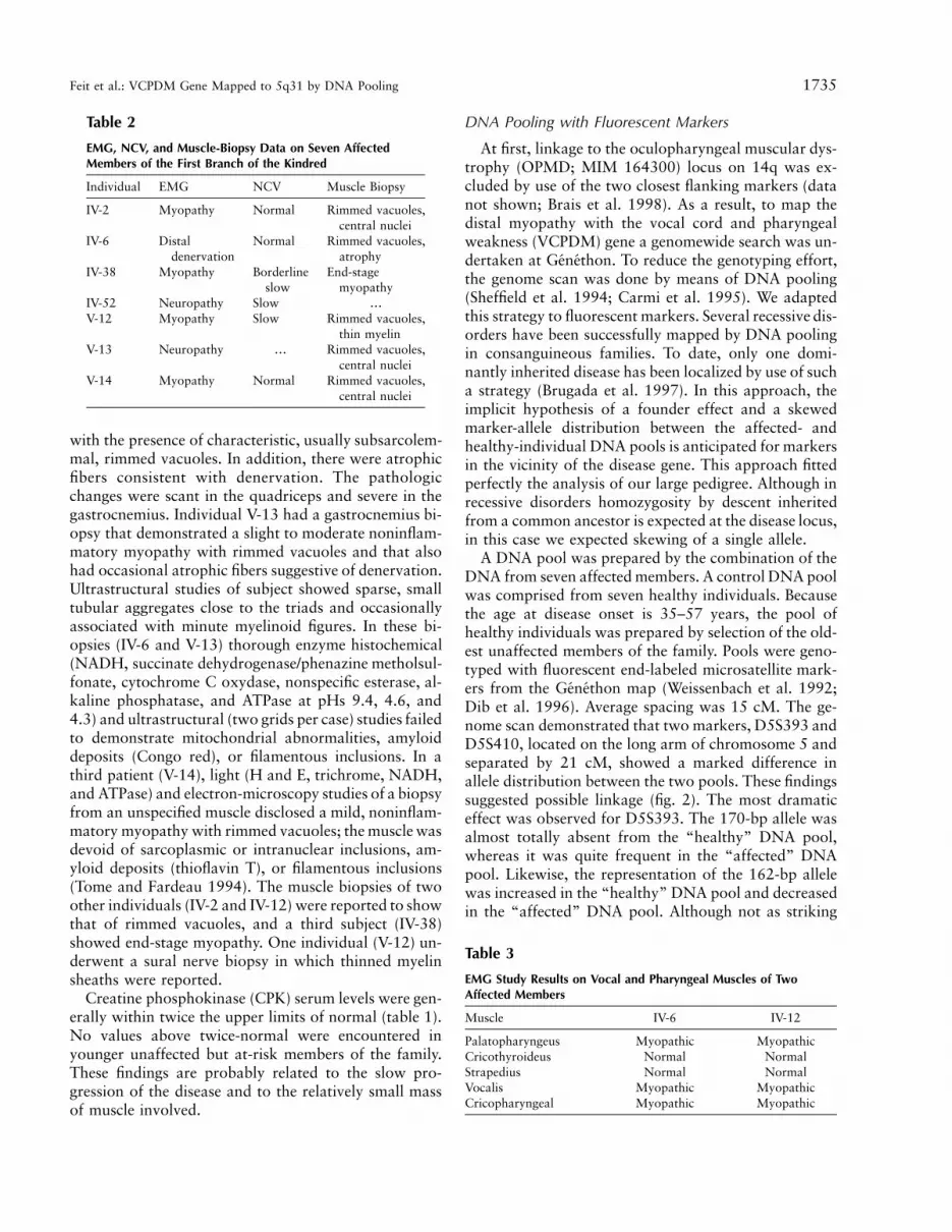
EMG, NCV, and Muscle-Biopsy Data on Seven AffectedMembers of the First Branch of the Kindred
Individual EMG NCV Muscle Biopsy
IV-2 Myopathy Normal Rimmed vacuoles,central nuclei
IV-6 Distaldenervation
Normal Rimmed vacuoles,atrophy
IV-38 Myopathy Borderlineslow
End-stagemyopathy
IV-52 Neuropathy Slow )V-12 Myopathy Slow Rimmed vacuoles,
thin myelinV-13 Neuropathy ) Rimmed vacuoles,
central nucleiV-14 Myopathy Normal Rimmed vacuoles,
central nuclei
Table 3
EMG Study Results on Vocal and Pharyngeal Muscles of TwoAffected Members
Muscle IV-6 IV-12
Palatopharyngeus Myopathic MyopathicCricothyroideus Normal NormalStrapedius Normal NormalVocalis Myopathic MyopathicCricopharyngeal Myopathic Myopathic
with the presence of characteristic, usually subsarcolem-mal, rimmed vacuoles. In addition, there were atrophicfibers consistent with denervation. The pathologicchanges were scant in the quadriceps and severe in thegastrocnemius. Individual V-13 had a gastrocnemius bi-opsy that demonstrated a slight to moderate noninflam-matory myopathy with rimmed vacuoles and that alsohad occasional atrophic fibers suggestive of denervation.Ultrastructural studies of subject showed sparse, smalltubular aggregates close to the triads and occasionallyassociated with minute myelinoid figures. In these bi-opsies (IV-6 and V-13) thorough enzyme histochemical(NADH, succinate dehydrogenase/phenazine metholsul-fonate, cytochrome C oxydase, nonspecific esterase, al-kaline phosphatase, and ATPase at pHs 9.4, 4.6, and4.3) and ultrastructural (two grids per case) studies failedto demonstrate mitochondrial abnormalities, amyloiddeposits (Congo red), or filamentous inclusions. In athird patient (V-14), light (H and E, trichrome, NADH,and ATPase) and electron-microscopy studies of a biopsyfrom an unspecified muscle disclosed a mild, noninflam-matory myopathy with rimmed vacuoles; the muscle wasdevoid of sarcoplasmic or intranuclear inclusions, am-yloid deposits (thioflavin T), or filamentous inclusions(Tome and Fardeau 1994). The muscle biopsies of twoother individuals (IV-2 and IV-12) were reported to showthat of rimmed vacuoles, and a third subject (IV-38)showed end-stage myopathy. One individual (V-12) un-derwent a sural nerve biopsy in which thinned myelinsheaths were reported.
Creatine phosphokinase (CPK) serum levels were gen-erally within twice the upper limits of normal (table 1).No values above twice-normal were encountered inyounger unaffected but at-risk members of the family.These findings are probably related to the slow pro-gression of the disease and to the relatively small massof muscle involved.
DNA Pooling with Fluorescent Markers
At first, linkage to the oculopharyngeal muscular dys-trophy (OPMD; MIM 164300) locus on 14q was ex-cluded by use of the two closest flanking markers (datanot shown; Brais et al. 1998). As a result, to map thedistal myopathy with the vocal cord and pharyngealweakness (VCPDM) gene a genomewide search was un-dertaken at Genethon. To reduce the genotyping effort,the genome scan was done by means of DNA pooling(Sheffield et al. 1994; Carmi et al. 1995). We adaptedthis strategy to fluorescent markers. Several recessive dis-orders have been successfully mapped by DNA poolingin consanguineous families. To date, only one domi-nantly inherited disease has been localized by use of sucha strategy (Brugada et al. 1997). In this approach, theimplicit hypothesis of a founder effect and a skewedmarker-allele distribution between the affected- andhealthy-individual DNA pools is anticipated for markersin the vicinity of the disease gene. This approach fittedperfectly the analysis of our large pedigree. Although inrecessive disorders homozygosity by descent inheritedfrom a common ancestor is expected at the disease locus,in this case we expected skewing of a single allele.
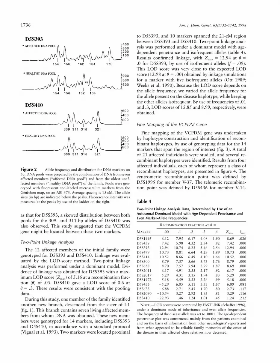
A DNA pool was prepared by the combination of theDNA from seven affected members. A control DNA poolwas comprised from seven healthy individuals. Becausethe age at disease onset is 35–57 years, the pool ofhealthy individuals was prepared by selection of the old-est unaffected members of the family. Pools were geno-typed with fluorescent end-labeled microsatellite mark-ers from the Genethon map (Weissenbach et al. 1992;Dib et al. 1996). Average spacing was 15 cM. The ge-nome scan demonstrated that two markers, D5S393 andD5S410, located on the long arm of chromosome 5 andseparated by 21 cM, showed a marked difference inallele distribution between the two pools. These findingssuggested possible linkage (fig. 2). The most dramaticeffect was observed for D5S393. The 170-bp allele wasalmost totally absent from the “healthy” DNA pool,whereas it was quite frequent in the “affected” DNApool. Likewise, the representation of the 162-bp allelewas increased in the “healthy” DNA pool and decreasedin the “affected” DNA pool. Although not as striking
1736 Am. J. Hum. Genet. 63:1732–1742, 1998
Figure 2 Allele frequency and distribution for DNA markers on5q. DNA pools were prepared by the combination of DNA from sevenaffected members (“affected DNA pool”) and from the oldest unaf-fected members (“healthy DNA pool”) of the family. Pools were gen-otyped with fluorescent end-labeled microsatellite markers from theGenethon map, on an ABI 373. Average spacing is 15 cM. The allelesizes (in bp) are indicated below the peaks. Fluorescence intensity wasmeasured at the peaks by use of the ladder on the right. Table 4
Two-Point Linkage Analysis Data, Determined by Use of anAutosomal Dominant Model with Age-Dependent Penetrance andEven Marker-Allele Frequencies
RECOMBINATION FRACTION AT v �
MARKER .00 .1 .2 .3 .4 Zmax vmax
D5S1995 6.12 7.95 6.17 4.08 1.90 8.69 .026D5S458 7.42 5.98 4.32 2.54 .82 7.42 .000D5S393 12.94 10.74 8.23 5.46 2.54 12.94 .000D5S479 10.73 8.81 6.64 4.29 1.89 10.73 .000D5S414 10.52 8.66 6.49 4.10 1.64 10.52 .000D5S500 8.79 7.37 5.66 3.75 1.76 8.79 .000D5S658 8.70 7.57 5.94 3.99 1.87 8.69 .000D5S2011 6.17 4.91 3.55 2.17 .92 6.17 .000D5S2017 5.29 4.31 3.15 1.94 .83 5.29 .000D5S1972 5.18 4.59 3.53 2.26 .99 5.18 .000D5S436 �5.29 6.05 5.11 3.53 1.67 6.09 .081D5S638 �6.88 2.71 2.45 1.70 .80 2.73 .117D5S2090 �10.54 3.27 2.92 1.95 .81 3.29 .116D5S410 �22.93 .46 1.24 1.01 .45 1.24 .212
NOTE.—LOD scores were computed by FASTLINK (Schaffer 1996),under a dominant mode of inheritance and even allele frequencies.The frequency of the disease allele was set to .0001. The age-dependentpenetrance plot was constructed mainly from the patients examinedand on the basis of information from other neurologists’ reports andfrom what appeared to be reliable family memories of the onset ofthe disease in their affected close relatives now deceased.
as that for D5S393, a skewed distribution between bothpools for the 309- and 311-bp alleles of D5S410 wasalso observed. This study suggested that the VCPDMgene might be located between these two markers.
Two-Point Linkage Analysis
The 12 affected members of the initial family weregenotyped for D5S393 and D5S410. Linkage was eval-uated by the LOD-score method. Two-point linkageanalysis was performed under a dominant model. Evi-dence of linkage was obtained for D5S393 with a max-imum LOD score (Zmax) of 5.16 at a recombination frac-tion (v) of .05. D5S410 gave a LOD score of 0.6 at
. These results were consistent with the poolingv � .3data.
During this study, one member of the family identifiedanother, new branch, descended from the sister of I-1(fig. 1). This branch contains seven living affected mem-bers from whom DNA was obtained. These new mem-bers were genotyped for 14 markers, including D5S393and D5S410, in accordance with a standard protocol(Vignal et al. 1993). Two markers were located proximal
to D5S393, and 10 markers spanned the 21-cM regionbetween D5S393 and D5S410. Two-point linkage anal-ysis was performed under a dominant model with age-dependent penetrance and isofrequent alleles (table 4).Results confirmed linkage, with atZ � 12.94 v �max
for D5S393, by use of isofrequent alleles ( )..0 f � .09This LOD score was very close to the expected LODscore (12.98 at ) obtained by linkage simulationsv � .00for a marker with five isofrequent alleles (Ott 1989;Weeks et al. 1990). Because the LOD score depends onthe allele frequency, we varied the allele frequency forthe allele present on the disease haplotype, while keepingthe other alleles isofrequent. By use of frequencies of .01and .3, LOD scores of 15.85 and 8.99, respectively, wereobtained.
Fine Mapping of the VCPDM Gene
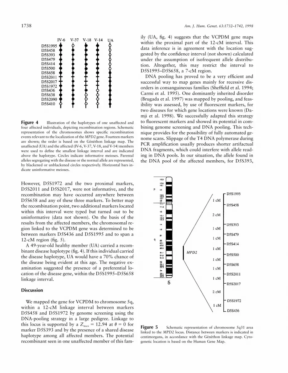
Fine mapping of the VCPDM gene was undertakenby haplotype construction and identification of recom-binant haplotypes, by use of genotyping data for the 14markers that span the region of interest (fig. 3). A totalof 21 affected individuals were studied, and several re-combinant haplotypes were identified. Results from fouraffected individuals, each of whom represent a class ofrecombinant haplotypes, are presented in figure 4. Thecentromeric recombination point was defined byD5S1995 for member V-37. The telomeric recombina-tion point was defined by D5S436 for member V-14.

Figu
re3
Hap
loty
pes
of14
DN
Am
arke
rson
5qin
affe
cted
mem
bers
from
both
bran
ches
ofth
eki
ndre
d.D
NA
sam
ples
used
inth
eD
NA
-poo
ling
stra
tegy
are
indi
cate
dby
tiny
blac
kene
dsq
uare
sne
xtto
the
mem
ber
num
ber.
Hap
loty
pes
wer
ein
ferr
edby
use
ofG
EN
EH
UN
TE
R(K
rugl
yak
etal
.199
6).R
econ
stru
cted
hapl
otyp
esar
ein
ital
icty
pe.T
oen
sure
fast
and
cons
iste
ntha
plot
ype
cons
truc
tion
,th
epe
digr
eew
asbr
oken
into
over
lapp
ing
subf
amili
es.
The
dise
ase
hapl
otyp
eis
circ
led.
1738 Am. J. Hum. Genet. 63:1732–1742, 1998
Figure 4 Illustration of the haplotypes of one unaffected andfour affected individuals, depicting recombination regions. Schematicrepresentation of the chromosomes shows specific recombinationevents relevant to the localization of the MPD2 gene. Fourteen markersare shown; the order is based on the Genethon linkage map. Theunaffected (UA) and the affected (IV-6, V-37, V-18, and V-14) memberswere used to define the smallest linkage interval and are indicatedabove the haplotype. Circles indicate informative meioses. Parentalalleles segregating with the disease or the normal allele are represented,by blackened or unblackened circles respectively. Horizontal bars in-dicate uninformative meioses.
Figure 5 Schematic representation of chromosome 5q31 arealinked to the MPD2 locus. Distance between markers is indicated incentimorgans, in accordance with the Genethon linkage map. Cyto-genetic location is based on the Human Gene Map.
However, D5S1972 and the two proximal markers,D5S2011 and D5S2017, were not informative, and therecombination may have occurred anywhere betweenD5S658 and any of these three markers. To better mapthe recombination point, two additional markers locatedwithin this interval were typed but turned out to beuninformative (data not shown). On the basis of theresults from the affected members, the chromosomal re-gion linked to the VCPDM gene was determined to bebetween markers D5S436 and D5S1995 and to span a12-cM region (fig. 5).
A 49-year-old healthy member (UA) carried a recom-binant disease haplotype (fig. 4). If this individual carriedthe disease haplotype, UA would have a 70% chance ofthe disease being evident at this age. The negative ex-amination suggested the presence of a preferential lo-cation of the disease gene, within the D5S1995–D5S658linkage interval.
Discussion
We mapped the gene for VCPDM to chromosome 5q,within a 12-cM linkage interval between markersD5S458 and D5S1972 by genome screening using theDNA-pooling strategy in a large pedigree. Linkage tothis locus is supported by a at forZ � 12.94 v � 0max
marker D5S393 and by the presence of a shared diseasehaplotype among all affected members. The potentialrecombinant seen in one unaffected member of this fam-
ily (UA, fig. 4) suggests that the VCPDM gene mapswithin the proximal part of the 12-cM interval. Thisdata inference is in agreement with the location sug-gested by the confidence interval (not shown) calculatedunder the assumption of isofrequent allele distribu-tion. Altogether, this may restrict the interval toD5S1995–D5S658, a 7-cM region.
DNA pooling has proved to be a very efficient andsuccessful way to map genes mainly for recessive dis-orders in consanguineous families (Sheffield et al. 1994;Carmi et al. 1995). One dominantly inherited disorder(Brugada et al. 1997) was mapped by pooling, and feas-ibility was assessed, by use of fluorescent markers, fortwo diseases for which gene locations were known (Da-mji et al. 1998). We successfully adapted this strategyto fluorescent markers and showed its potential in com-bining genome screening and DNA pooling. This tech-nique provides for the possibility of fully automated ge-nome scans. Slippage of the T4 DNA polymerase duringPCR amplification usually produces shorter artifactualDNA fragments, which could interfere with allele read-ing in DNA pools. In our situation, the allele found inthe DNA pool of the affected members, for D5S393,
Feit et al.: VCPDM Gene Mapped to 5q31 by DNA Pooling 1739
Table 5
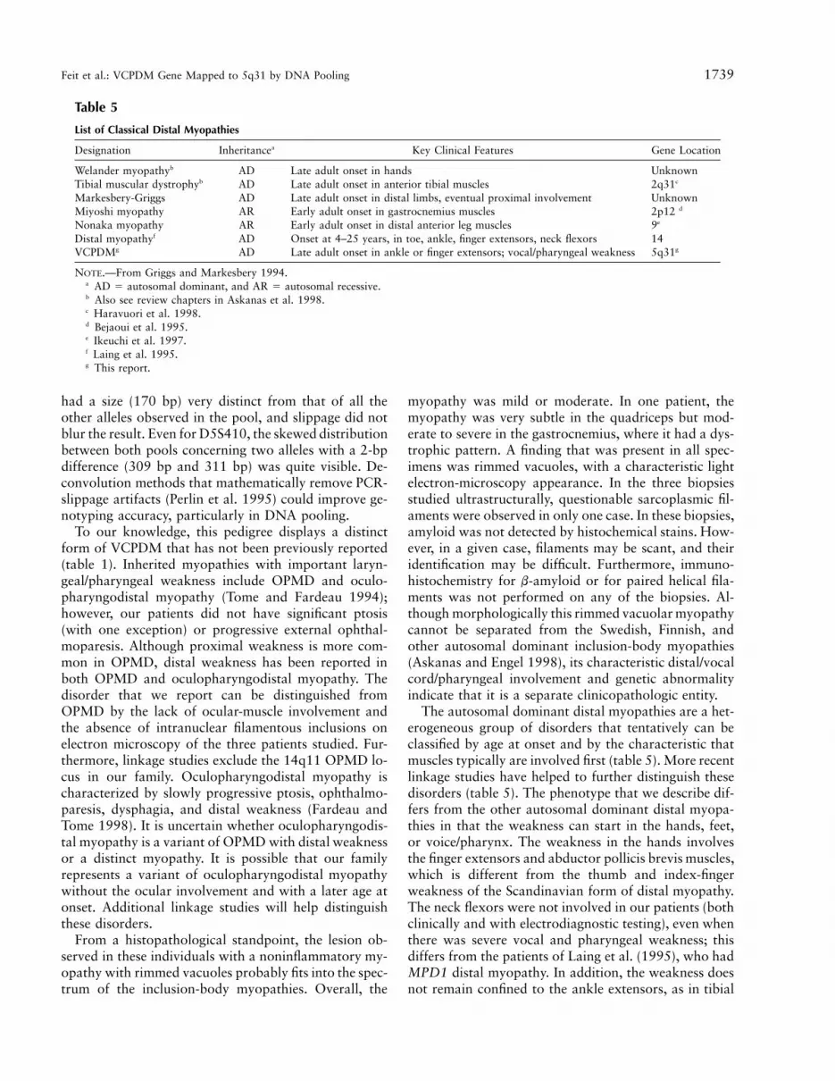
List of Classical Distal Myopathies
Designation Inheritancea Key Clinical Features Gene Location
Welander myopathyb AD Late adult onset in hands UnknownTibial muscular dystrophyb AD Late adult onset in anterior tibial muscles 2q31c
Markesbery-Griggs AD Late adult onset in distal limbs, eventual proximal involvement UnknownMiyoshi myopathy AR Early adult onset in gastrocnemius muscles 2p12 d
Nonaka myopathy AR Early adult onset in distal anterior leg muscles 9e
Distal myopathyf AD Onset at 4–25 years, in toe, ankle, finger extensors, neck flexors 14VCPDMg AD Late adult onset in ankle or finger extensors; vocal/pharyngeal weakness 5q31g
NOTE.—From Griggs and Markesbery 1994.a AD � autosomal dominant, and AR � autosomal recessive.b Also see review chapters in Askanas et al. 1998.c Haravuori et al. 1998.d Bejaoui et al. 1995.e Ikeuchi et al. 1997.f Laing et al. 1995.g This report.
had a size (170 bp) very distinct from that of all theother alleles observed in the pool, and slippage did notblur the result. Even for D5S410, the skewed distributionbetween both pools concerning two alleles with a 2-bpdifference (309 bp and 311 bp) was quite visible. De-convolution methods that mathematically remove PCR-slippage artifacts (Perlin et al. 1995) could improve ge-notyping accuracy, particularly in DNA pooling.
To our knowledge, this pedigree displays a distinctform of VCPDM that has not been previously reported(table 1). Inherited myopathies with important laryn-geal/pharyngeal weakness include OPMD and oculo-pharyngodistal myopathy (Tome and Fardeau 1994);however, our patients did not have significant ptosis(with one exception) or progressive external ophthal-moparesis. Although proximal weakness is more com-mon in OPMD, distal weakness has been reported inboth OPMD and oculopharyngodistal myopathy. Thedisorder that we report can be distinguished fromOPMD by the lack of ocular-muscle involvement andthe absence of intranuclear filamentous inclusions onelectron microscopy of the three patients studied. Fur-thermore, linkage studies exclude the 14q11 OPMD lo-cus in our family. Oculopharyngodistal myopathy ischaracterized by slowly progressive ptosis, ophthalmo-paresis, dysphagia, and distal weakness (Fardeau andTome 1998). It is uncertain whether oculopharyngodis-tal myopathy is a variant of OPMD with distal weaknessor a distinct myopathy. It is possible that our familyrepresents a variant of oculopharyngodistal myopathywithout the ocular involvement and with a later age atonset. Additional linkage studies will help distinguishthese disorders.
From a histopathological standpoint, the lesion ob-served in these individuals with a noninflammatory my-opathy with rimmed vacuoles probably fits into the spec-trum of the inclusion-body myopathies. Overall, the
myopathy was mild or moderate. In one patient, themyopathy was very subtle in the quadriceps but mod-erate to severe in the gastrocnemius, where it had a dys-trophic pattern. A finding that was present in all spec-imens was rimmed vacuoles, with a characteristic lightelectron-microscopy appearance. In the three biopsiesstudied ultrastructurally, questionable sarcoplasmic fil-aments were observed in only one case. In these biopsies,amyloid was not detected by histochemical stains. How-ever, in a given case, filaments may be scant, and theiridentification may be difficult. Furthermore, immuno-histochemistry for b-amyloid or for paired helical fila-ments was not performed on any of the biopsies. Al-though morphologically this rimmed vacuolar myopathycannot be separated from the Swedish, Finnish, andother autosomal dominant inclusion-body myopathies(Askanas and Engel 1998), its characteristic distal/vocalcord/pharyngeal involvement and genetic abnormalityindicate that it is a separate clinicopathologic entity.
The autosomal dominant distal myopathies are a het-erogeneous group of disorders that tentatively can beclassified by age at onset and by the characteristic thatmuscles typically are involved first (table 5). More recentlinkage studies have helped to further distinguish thesedisorders (table 5). The phenotype that we describe dif-fers from the other autosomal dominant distal myopa-thies in that the weakness can start in the hands, feet,or voice/pharynx. The weakness in the hands involvesthe finger extensors and abductor pollicis brevis muscles,which is different from the thumb and index-fingerweakness of the Scandinavian form of distal myopathy.The neck flexors were not involved in our patients (bothclinically and with electrodiagnostic testing), even whenthere was severe vocal and pharyngeal weakness; thisdiffers from the patients of Laing et al. (1995), who hadMPD1 distal myopathy. In addition, the weakness doesnot remain confined to the ankle extensors, as in tibial

1740 Am. J. Hum. Genet. 63:1732–1742, 1998
muscular dystrophy. The assignment of this disorder toa locus on chromosome 5q also distinguishes this dis-order from other forms of dominantly inherited distalmyopathy. In keeping with the precedent started byLaing et al. (1995), the gene on 5q for this form of distalmyopathy is designated “MPD2.”
The phenotype that we describe in this family closelyresembles that reported by Young and Harper (1980) asan autosomal dominant distal atrophy with vocal cordparalysis. In the family reported by Young and Harper,EMGs were obtained for two of nine affected individualsand showed spontaneous fibrillation and either “reducedinterference pattern” or “reduced volitional activity.”Muscle biopsies were not obtained. In our family, bothneurogenic and myopathic muscle potentials were ob-served. Thus, we cannot exclude the possibility that thephenotype reported by Young and Harper (1980) as adistal atrophy with vocal cord paralysis was possibly adistal myopathy instead. Linkage studies of the familiesreported by Young and Harper (1980) may clarify thisissue. The MPD2 phenotype is also easily confused withthe neuronal form of Charcot-Marie-Tooth disease if theEMG results for an individual patient suggest a neuro-genic disorder with normal conduction velocities. Thediagnosis of VCPDM does not require voice and pha-ryngeal changes, since some affected individuals in ourkindred did not show these changes at the time of theirexaminations. Attention to the presentation and subse-quent course in other family members is important. Mus-cle biopsies that include an involved distal muscle andexaminations of other affected members of the familyare recommended.
Two diseases have been mapped to this chromosomalregion: an autosomal recessive (LGMD2F; MIM601287) and an autosomal dominant (LGMD1A; MIM15900) limb-girdle muscular dystrophy. LGMD2F hasbeen mapped to chromosome 5q33-34 (Passos-Bueno etal. 1996) and is a result of a mutation in the d-sarcog-lycan gene (Nigro et al. 1996). This gene is telomeric tothe linkage interval we defined and is therefore excluded.LGMD1A has been mapped within a 2.5-Mb intervalflanked by D5S479 and D5S594 (Bartoloni et al. 1997).Our linkage interval encompasses the LGMD1A locus;however, both disorders are clinically quite different. Theproximal weakness in LGMD1A is always much moresevere than the distal weakness; the opposite is observedin VCPDM. Age at onset is earlier in LGMD1A, andanticipation is also suspected (Speer et al. 1998). In con-trast, there is no evidence of anticipation in VCPDM.Both diseases could actually be caused by distinct mu-tations affecting either the same gene or syntenic genes.At this point, these hypotheses cannot be resolved. Itshould be remembered that both studies were based onthe examination of a single large family that representsa founder effect and in which a single pathogenic mu-
tation is segregating. Hence, it is difficult at this stageto generalize the clinical description to other mutationswithin these genes. It will be of interest to review theclinical properties once other independent VCPDM andLGMD1A families have been identified. A similar situ-ation has been observed for LGMD2B (LGMD2B; MIM253601) and the distal Miyoshi type of muscular dys-trophy (ARDMD; MIM 254130), which both mappedto chromosome 2p (Bashir et al. 1994; Bejaoui et al.1995). Recent results have shown that both diseases seg-regated in the same family, which suggests that they wereboth a result of the same allele (Illarioshkin et al. 1996;Weiler et al. 1996).
The protein coded by the autosomal dominant genefor VCPDM is unknown but is presumed to be expressedin skeletal muscle. A search of the Human Gene Mapdatabase for the D5S1995–D5S658 linkage interval re-vealed 161 expressed sequence tags (ESTs) with noknown function in most instances. Among the ESTs witha putative or known function, one (locus HSRNASMAP;European Molecular Biology Library accession numberX87613) codes for a putative transcription factor thatcontains a bromodomain highly expressed in skeletalmuscle (Nielsen et al. 1996). This gene is located be-tween D5S393 and D5S500 and represents a potentialcandidate gene. An autosomal dominant form of pro-gressive hearing loss (DFNA15; MIM 602459) resultsfrom an 8-bp deletion in the transcription factorPOU4F3 (Vahava et al. 1998). POU4F3 is located onchromosome 5q31 telomeric to the VCPDM linkage in-terval. In OPMD, the trinucleotide (GCG)8–13, coding fora polyalanine tract is amplified in the poly(A)-bindingprotein 2 gene (PABP2) on chromosome 14q. ThePABP2 protein is highly expressed in the nuclei of skel-etal-muscle fibers (Brais et al. 1998). HSRNASMAPtherefore represents a good candidate gene for VCPDM.Its possible role in VCPDM is presently being in-vestigated.
Acknowledgments
We would like to thank Dr. Michael S. Benninger(Otolaryngology Department, Henry Ford Hospital, Detroit),Cindy Grywalski, Ralph Thompson, the patients and theirfamily; and the many physicians who provided clinical data.C.E.J. and E.S. were supported by grants from l’AssociationFrancaise contre les Myopathies.
Electronic-Database Information
Accession numbers and URLs for data in this article are asfollows:
Genethon, http://www.genethon.fr (for fluorescent end-labeledmicrosatellite markers and GENOTYPER (ABI), GEN-SCAN (ABI), and MARKSYN software)

Feit et al.: VCPDM Gene Mapped to 5q31 by DNA Pooling 1741
Human Gene Map Database, http://www.ncbi.nlm.nih.gov/SCIENCE96/ (for ESTs in D5S1995–D5S658 interval)
Online Mendelian Inheritance in Man (OMIM), http://www.ncbi.nlm.nih.gov/Omim/ (for OPMD [MIM 164300],LGMD2F [MIM 601287], LGMD1A [MIM 15900],LGMD2B [MIM 253601], ARDMD [MIM 254130], andDFNA15 [MIM 602459])
References
Askanas V, Engel WK (1998) Newest approaches to diagnosisand pathogenesis of sporadic inclusion-body myositis andhereditary inclusion-body myopathies, including molecular-pathologic similarities to Alzheimer disease. In: Askanas V,Serratrice G, Engel WK (eds) Inclusion-body myositis andmyopathies. Cambridge University Press, Cambridge, pp3–78
Askanas V, Serratrice G, Engel WK (eds) (1998) Inclusion-bodymyositis and myopathies. Cambridge University Press,Cambridge
Bartoloni L, Horrigan SK, Zhang Y, Viles K, Gilchrist JM,Vance JM, Yamaoka LH, et al (1997) Limb-girdle musculardystrophy 1A: refinement of the 5q31 localization and aphysical and genetic map of the interval. Am J Hum GenetSuppl 61:A267
Bashir R, Strachan T, Keers S, Stephenson A, Mahjneh I, Mar-coni G, Nashef L, et al (1994) A gene for autosomal recessivelimb-girdle muscular dystrophy maps to chromosome 2p.Hum Mol Genet 3:455–457
Bejaoui K, Hirabayashi K, Hentati F, Haines JL, Ben HamidaC, Belal S, Miller RG, et al (1995) Linkage of Miyoshi my-opathy (distal autosomal recessive muscular dystrophy) tochromosome 2p12. Neurology 45:768–772
Brais B, Bouchard JP, Xie YG, Rochefort DL, Chretien N,Tome FM, Lafreniere RG, et al (1998) Short GCG expan-sions in the PABP2 gene cause oculopharyngeal musculardystrophy. Nat Genet 18:164–167
Brooke MH (1986) A clinician’s view of neuromuscular dis-eases. Williams & Wilkins, Baltimore, pp 174–178
Brugada R, Tapscott T, Czernuszewicz GZ, Marian AJ, IglesiasA, Mont L, Brugada J, et al (1997) Identification of a geneticlocus for familial atrial fibrillation. N Engl J Med 336:905–911
Carmi R, Rokhlina T, Kwitek-Black AE, Elbedour K, Nishi-mura D, Stone EM, Sheffield VC (1995) Use of a DNApooling strategy to identify a human obesity syndrome locuson chromosome 15. Hum Mol Genet 4:9–13
Damji KF, Gallione CJ, Allingham RR, Slotterbeck B, Gutt-macher AE, Pasyk KA, Vance JM, et al (1998) QuantitativeDNA pooling to increase the efficiency of linkage analysisin autosomal dominant disease. Hum Genet 102:207–212
Dib C, Faure S, Fizames C, Samson D, Drouot N, Vignal A,Millasseau P, et al (1996) A comprehensive genetic map ofthe human genome based on 5,264 microsatellites. Nature380:152–154
Fardeau M, Tome F (1998) Inclusion body myopathies. In:Askanas V, Serratrice G, Engel WK (eds) Inclusion-bodymyositis and myopathies. Cambridge University Press, Cam-bridge, pp 252–260
Griggs RC, Markesbery WR (1994) Distal myopathies. In:
Engel AG, Franzini-Armstrong C (eds) Myology basic andclinical. McGraw-Hill, New York, pp 1246–1257
Haravuori H, Makela-Bengs P, Udd B, Partanen J, PulkkinenL, Somer H, Peltonen L (1998) Assignment of the tibialmuscular dystrophy locus to chromosome 2q31. Am J HumGenet 62:620–626
Ikeuchi T, Asaka T, Saito M, Tanaka H, Higuchi S, TanakaK, Saida K, et al (1997) Gene locus for autosomal recessivedistal myopathy with rimmed vacuoles maps to chromosome9. Ann Neurol 41:432–437
Illarioshkin SN, Ivanova-Smolenskaya IA, Tanaka H, Veresh-chagin NV, Markova ED, Poleshchuk VV, Lozhnikova SM,et al (1996) Clinical and molecular analysis of a large familywith three distinct phenotypes of progressive muscular dys-trophy. Brain 119:1895–1909
Kruglyak L, Daly MJ, Reeve-Daly MP, Lander ES (1996) Par-ametric and nonparametric linkage analysis: a unified mul-tipoint approach. Am J Hum Genet 58:1347–1363
Laing NG, Laing BA, Meredith C, Wilton SD, Robbins P,Honeyman K, Dorosz S, et al (1995) Autosomal dominantdistal myopathy: linkage to chromosome 14. Am J HumGenet 56:422–427
Nielsen MS, Petersen CM, Gliemann J, Madsen P (1996) Clon-ing and sequencing of a human cDNA encoding a putativetranscription factor containing a bromodomain. BiochimBiophys Acta 1306:14–16
Nigro V, de Sa Moreira E, Piluso G, Vainzof M, Belsito A,Politano L, Puca AA, et al (1996) Autosomal recessive limb-girdle muscular dystrophy, LGMD2F, is caused by a mu-tation in the delta-sarcoglycan gene. Nat Genet 14:195–198.
Ott J (1989) Computer-simulation methods in human linkageanalysis. Proc Natl Acad Sci USA 86:4175–4178
Passos-Bueno MR, Moreira ES, Vainzof M, Marie SK, ZatzM (1996) Linkage analysis in autosomal recessive limb-gir-dle muscular dystrophy (AR LGMD) maps a sixth form to5q33-34 (LGMD2F) and indicates that there is at least onemore subtype of AR LGMD. Hum Mol Genet 5:815–820
Perlin MW, Lancia G, Ng SK (1995) Toward fully automatedgenotyping: genotyping microsatellite markers by decon-volution. Am J Hum Genet 57:1199–1210
Schaffer AA (1996) Faster linkage analysis computations forpedigrees with loops or unused alleles. Hum Hered 46:226–235
Sheffield VC, Carmi R, Kwitek-Black A, Rokhlina T, Nishi-mura D, Duyk GM, Elbedour K, et al (1994) Identificationof a Bardet-Biedl syndrome locus on chromosome 3 andevaluation of an efficient approach to homozygosity map-ping. Hum Mol Genet 3:1331–1335
Speer MC, Gilchrist JM, Stajich JM, Gaskell PC, WestbrookCA, Horrigan SK, Bartoloni L, et al (1998) Evidence foranticipation in autosomal dominant limb-girdle musculardystrophy. J Med Genet 35:305–308
Tome FMS, Fardeau M (1994) Oculopharyngeal muscular dys-trophy. In: Engel AG, Franzini-Armstrong C (eds) Myologybasic and clinical. McGraw-Hill, New York, pp 1233–1245
Vahava O, Morell R, Lynch ED, Weiss S, Kagan ME, AhituvN, Morrow JE, et al (1998) Mutation in transcription factorPOU4F3 associated with inherited progressive hearing lossin humans. Science 279:1950–1954
Vignal A, Gyapay G, Hazan J, Nguyen S, Dupraz C, Cheron

1742 Am. J. Hum. Genet. 63:1732–1742, 1998
N, Becuwe N, et al (1993) A non-radioactive multiplex pro-cedure for genotyping of microsatellite markers. In: AdolphKW (ed) Methods in molecular genetics: gene and chro-mosome analysis. Academic Press, San Diego, 211–221
Weeks DE, Ott J, Lathrop GM (1990) SLINK: a general sim-ulation program for linkage analysis. Am J Hum GenetSuppl 47:A204
Weiler T, Greenberg CR, Nylen E, Halliday W, Morgan K,Eggertson D, Wrogemann K (1996) Limb-girdle muscular
dystrophy and Miyoshi myopathy in an aboriginal Canadiankindred map to LGMD2B and segregate with the same hap-lotype. Am J Hum Genet 59:872–878
Weissenbach J, Gyapay G, Dib C, Vignal A, Morissette J, Mil-lasseau P, Vaysseix G, et al (1992) A second-generation link-age map of the human genome. Nature 359:794–801
Young ID, Harper PS (1980) Hereditary distal spinal muscularatrophy with vocal cord paralysis. J Neurol Neurosurg Psy-chiatry 43:413–418