viruses of pet fish
TRANSCRIPT

Vet Clin Exot Anim 8 (2005) 67–84
Viruses of pet fish
Barbara D. Petty, DVMa,*, William A. Fraser, BSb
aDepartment of Large Animal Clinical Sciences, University of Florida,
7922 NW 71 Street, Gainesville, FL 32653, USAbAnimal Disease Diagnostic Laboratory, Division of Animal Industry, Department of
Agriculture and Consumer Services, P.O. Box 458006, Kissimmee, FL 34745, USA
Many veterinarians who work with pet fish may see viruses infrequentlybecause adverse environmental conditions are responsible for most fishhealth problems. It is important, however, for veterinarians to be cognizantof the few viruses of ornamental fish that have serious implications. Thisarticle is a guide to the most common viruses that affect fish that are kept aspets or display animals. Some of the viruses that are included here arepresented simply as case reports, given that the incidence and confirmationof viral disease in pet fishes occur sporadically. Where more information isavailable, clinical findings are discussed in more detail.
Although several fish cell lines are available, many viruses of pet fishes willnot grow in them. Diagnosis of such viruses is limited to electron microscopicexamination of tissues. Other diagnostic methods, such as immunofluores-cence, ELISA, and virus neutralization, are not available for many of the viraldiseases that arementionedhere, but some laboratoriesmay be able to providesuch tests. Polymerase chain reaction (PCR) methods for several fish virusesare available; for some viruses (eg, koi herpesvirus), PCR may be a morereliable detection method than virus isolation or electron microscopy.
Viruses that affect many species
Iridoviruses have been found in many species of fishes and are large,enveloped DNA viruses. Of the four genera, only two, Lymphocystivirus andRanavirus, cause disease in fishes [1]. Some iridoviruses, such as largemouthbass virus (LMBV) and gourami iridovirus, do not cause disease except inadverse environmental conditions. Several iridoviral diseases are listed by
* Corresponding author.
E-mail address: [email protected] (B.D. Petty).
1094-9194/05/$ - see front matter � 2005 Elsevier Inc. All rights reserved.
doi:10.1016/j.cvex.2004.09.003 vetexotic.theclinics.com

68 PETTY & FRASER
the Office International des Epizooties as notifiable diseases, but they arelimited to foodfish such as red sea bream and white sturgeon which are notkept commonly in home aquaria. Many iridoviral isolates from fish havelow pathogenicity.
Lymphocystis
Lymphocystis disease, caused by an iridovirus, is the most common viraldisease that is seen in pet fish. It affects many different fish species—fresh-water and marine—and has worldwide distribution. The most commonlyaffected ornamental fishes are the glassfish (Chanda ranga) and the marineangels (Pomacanthidae). Other susceptible fishes include cichlids, butterfly-fishes (Chaetodontidae), and sunfishes (Centrarchidae). Catfish andcyprinids are not susceptible.
Infection with either of two viruses, lymphocystis disease virus type 1 ortype 2, will result in similar clinical signs [1]. These viruses are the onlymembers of the genusLymphocystivirus and are the largest of the iridoviruses;they range in size from 130 nm to 330 nm.
Both viruses infect the dermal fibroblasts and result in hypertrophy thatgives rise to the lesions that are visible grossly. The skin of susceptible fish thatare infected with either virus will have multiple focal to coalescing irregulargray orwhitemasses (Fig. 1). Fins are affected commonly, although the lesionsalso may occur on the body. Infrequently, lymphocystis has been found toinfect internal organs of some marine fishes [2].
Wet mounts of skin scrapings reveal the hypertrophied cells, as willhistopathology of an infected site (Fig. 2). Lymphocystivirus is the onlyvirus in fish that can be diagnosed confidently with wet mount preparationsof skin scrapings and histopathology of affected areas.
It is uncommon for fish to die from this viral infection. Frequently, thelesions disappear after a period of time; however, if lesions are located nearthe mouth, feeding may be disrupted with subsequent starvation.
Fig. 1. A marine angelfish (Centropyge fisheri) displaying the typical gray masses of
lymphocystis. (Courtesy of Stephen A. Smith, DVM, PhD, Blacksburg, VA.)

69VIRUSES OF PET FISH
The virus is transmitted horizontally, when infected skin cells rupture andrelease infective particles into the water. The virus gains entry to a susceptiblefish at skin and gills. Dense fish populations that are subject to regularhandling (such as at a wholesale or retail facility) are at high-risk forlymphocystis transmission. Even in such situations, it is not unusual toobserve infections on only certain species.
Experimentally, the incubation time that was required for lesions todevelop in infected fish decreased as water temperature increased. Lesionsdeveloped in 5 to 12 days at 20�C to 25�C [3]. In wild marine fish populations,incidence is frequently the highest in spring and summer.
Neither of the lymphocystiviruses is grown easily in cell culture becauseof the long incubation period that is required. Cell lines that have been usedsuccessfully include bluegill fry (BF-2) and red drum dorsal fin cells.Electron microscopy of negative stains of the masses reveal viral particles(Fig. 3). Stress reduction via maintenance of good water quality conditionsand use of gentle handling techniques may minimize impact of the virus andaid resolution of the lesions.
Viruses that affect cyprinids
Spring viremia of carp virus
Spring viremia of carp virus (SVCV) was officially recognized first by Fijanin 1971 as the causative agent of an acute hemorrhagic infection that primarilyaffects carp, although fish that exhibited typical signs of SVCV infection werefirst described in 1904 [4]. Infection with this virus has resulted in 30% to 70%mortality rates of cultured andwild carp in Europe. Although there have beenno reports of infected fish in Asia, SVCV-positive fish were identified ina shipment of koi and goldfish that was sent to England fromChina in the late
Fig. 2. Tissue section of juvenile clownfish (Amphiprion sp). Note the greatly hypertrophied
cells (arrows) due to infection with lymphocystis virus (hematoxylin-eosin, original magnifica-
tion �40).

70 PETTY & FRASER
1990s [5]. Two separate cases of SVCV occurred in the United States in 2002.One case was identified as the cause of low mortality in koi carp at a koiproduction facility in North Carolina. The other case occurred in feral carp inCedar Lake, inWisconsin. InApril 2004, a positive case was identified in a koihobbyist pond in Washington. Another case was identified in July 2004 ata production facility in Missouri in koi that had been transported fromMinnesota. Ongoing surveillance programs that are performed by the U.S.Fish andWildlife Service and the U.S. Department of Agriculture (USDA) ofwild and cultured susceptible fishes, respectively, have not revealed anySVCV-positive fish. Trade in ornamental koi of unknown SVCV statuscontinues, so it is possible that more cases will occur in the United States. TheUSDA is considering the implementation of a health certification requirementfor import of SVCV-susceptible species. This program would require that allsusceptible fish that enter the United States originate from a certified SVCV-free facility/zone/country and that a certificate of veterinary inspection thatattests to the negative SVCV status accompanies the fish.
The causative agent of spring viremia of carp is an RNA virus (formerlyRhabdovirus carpio) that is tentatively placed in the genus vesiculovirus inthe Rhabdoviridae family. It is one of several rhabdoviruses that causedisease in fish, although SVCV is the only rhabdoviral disease of ornamentalfish. At the time of this writing, spring viremia of carp is the only notifiableviral disease of ornamental fish. Any positive case in the United States mustbe reported to the state veterinarian. Susceptible fishes are koi/common carp(Cyprinus carpio), grass carp (Ctenopharyngodon idella), bighead carp
Fig. 3. Electron micrograph of lymphocystis viral particles from a negatively stained skin mass
from a clownfish (Amphiprion sp) (negative stain phosphotungstic acid (PTA), original
magnification �50,000).

71VIRUSES OF PET FISH
(Hypophthalmichthys nobilis), silver carp (Aristichthys nobilis), Crucian carp(Carassius carassius), and goldfish (Carassius auratus) [4].
Clinical signs are variable and can be compounded by concurrentinfections (parasitic, bacterial, or fungal). Infected fish frequently exhibitlethargy, decreased respiration, loss of equilibrium, exophthalmia, pale gills,hemorrhages, ascites, protruding vent, and skin ulcers. Common internalfindings include edema, inflammation, and pin-point hemorrhages (espe-cially swim bladder). Multiple white foci on the atrium were present inSVCV-infected fish in Switzerland [6].
Perivasculitis in hepatic vessels and multi-focal necrosis in liver andpancreas are common findings on histopathology. Excretory and hemato-poietic tissue of kidney also may be affected. Perivasculitis, desquamation ofepithelium, and villar atrophy may be observed in the intestine.
Transmission primarily is horizontal, with infective particles found infeces, gill and skin mucus, urine, and exudate from skin blisters. Verticaltransmission may occur, but is not common [7]. The virus can be trans-mitted by vectors, such as blood-sucking parasites, fish-eating birds, or fish-handling equipment.
The virus enters by way of the gills, which are the primary site ofreplication. Temperature is the most important factor in the determinationof whether fish exhibit clinical signs. Disease occurs when the watertemperature is 15�C to 20�C. If susceptible fish are exposed to SCVC and thewater temperature is warmer than 20�C, they typically do not exhibit clinicalsigns.
The virus only can be isolated from infected fish when the watertemperature is colder than 20�C. Therefore, it is important to determine ifthe water temperature is at or below 20�C when collecting suspect SVCVspecimens.
Identification methods include cell culture of liver, spleen, and kidney,using epithelioma papillosum of carp (EPC) or fathead minnow (FHM) celllines, immunofluorescent antibody technique, ELISA, virus neutralization,and PCR. On cell cultures, cytopathic effect (CPE) consists of widespreadcell rounding followed by release of cells from plate surface [8]. Virusneutralization is the confirmatory test for SVCV at the time of this writing.Veterinarians who work with SVCV-susceptible fish should ascertain thatthe laboratory that is used for viral diagnostics is approved by the USDA toperform diagnostic tests for SVCV.
There is no treatment for SVCV; if it is diagnosed in a fish facility in theUnited States, all exposed fish, including fish species that are not susceptible toSVCV, must be depopulated. The virus can survive in the environmentwithout a host for up to 42 days inmud, so disinfection and sanitationmust bedirected at eradicating it from the environment. SVCV is destroyed easily withdisinfectants (eg, chlorine, formalin, sodium hydroxide) and pH extremes ofless than 4 and greater than 10. Ozonation and ultraviolet irradiation areeffective methods to disinfect potentially infected water sources.

72 PETTY & FRASER
Ideally, all susceptible species should come from an SVCV-free source. Ifthis is not possible, they should be quarantined for a minimum of 30 days.Manipulation of water temperature to exacerbate clinical signs of diseasemay be useful during the quarantine period.
To qualify as an SVCV-free source, a facility must undergo SVCV testing.A random group of 150 susceptible fish is selected for testing, only when thewater temperature is in the appropriate range for expression of clinical signs(10�C–18�C), by an accredited veterinarian. The testsmust be conducted twiceyearly. After 2 years of negative tests, the facility can be declared to be free ofSVCV. To maintain that designation, the facility must be tested annually.
The potential impact of this disease is significant. If a facility hasa positive fish and does not have good animal movement records, all fish atthat facility may be depopulated. The loss of valuable fish, potential loss ofsales, labor costs for disinfection, and other costs can be a devastating blowto a business.
Koi herpesvirus
The first case of koi herpesvirus (KHV) was diagnosed as the cause ofmassive mortality of ornamental koi in the United States in 1998 [9]. Sincethat time, Israel, Europe, and Asia also have reported cases in ornamentalkoi. In 2003, massive mortalities that were due to KHV occurred in wildcommon carp in Japan.
The only susceptible species to KHV infection is common carp/koi carp(Cyprinus carpio). Clinical signs ofKHV infection are nonspecific, but includepatchy white areas on the gills that resemble columnaris infections andproduction of excess mucus on the body. KHV-infected fish also may havesecondary parasitic and bacterial infections. Necrosis of internal organs isa common finding on internal examination. Infected fish may swim near thesurface of the water and act lethargic.
The agent of KHV, a DNA virus, is classified as a member of theHerpesviridae family (Fig. 4) [10]. It is transmitted horizontally by way ofcontact with infected fish or water that is contaminated with the virus. Thevirus is infective in water for up to 4 hours [11]. It is killed easily withstandard disinfectants.
If an infected fish is introduced into a population of susceptible fish, clinicalsigns of disease may be exhibited 14 days later. Mortality rates of 80% to100% may occur in susceptible fish when the water temperature is 18�C to27�C.Thesemortality ratesmay be decreased or halted by increasing thewatertemperature to 30�C; however, this is not recommended because surviving fishmay become a carrier population. Another herpesvirus of fish, channel catfishvirus (CCV), serves as a good example. Catfish that survive CCV outbreakswere proven to be a covert source of virus to susceptible catfish [3]. Furtherresearch is needed to determine if KHV is similar; until proven it is best toassume that a carrier state can exist.

73VIRUSES OF PET FISH
TodiagnoseKHV, gill, spleen, and kidney samples should be submitted forPCR and cell culture. KHV will grow on the koi fin cell line (KF-1) and carpbrain cell line [12]. Typical CPE consists of vacuole formation and eventualdetachment of the cells (Fig. 5). The PCRmethod is amore sensitive and rapiddiagnostic method than virus isolation.
Histopathologic lesions that are caused by KHVmay be extensive. Fusionof secondary lamellae and hyperplasia, hypertrophy, and necrosis of epithelialcells have been observed in the gills. Focal necrosis and intranuclear inclusionsoccurred in the splenic parenchyma and in the hematopoietic cells of thekidney.
New koi should be quarantined for at least 30 days and the watertemperature should be maintained between 18�C and 27�C during thequarantine period to promote expression of KHV infection, if present.
Fig. 5. KF-1 cell line displaying typical signs of KHV infection. Note the vacuole formation
(arrow) (original magnification �100).
Fig. 4. Electron micrograph of koi herpesvirus from infected koi fin–1 cell line (negative stain
PTA, original magnification �140,000).

74 PETTY & FRASER
Methods for determining previous exposure to KHV, such as in situhybridization and serology for anti-KHV antibodies, are being developed(A. Goodwin, PhD, Pine Bluff AR, personal communication, September2004).
Cyprinid herpesvirus
Cyprinid herpesvirus (CHV-1), which also is known as carp pox, fish pox,or epithelioma papillosum, is a benign, hyperplastic condition that is limitedto the epidermis and primarily affects adult common carp/koi carp(Cyprinus carpio). The disease is common to all areas of carp culture.
In contrast to lymphocystis lesions which appear nodular and rough, thelesions that are caused by carp pox are smooth and waxlike in appearance.The lesions are opaque and raised and may cover most of the body and fins.
Mortalities that are due to CHV-1 are rare in older fish, but mortalityrates may be high in juvenile fish. In older fish, the lesions may appearseasonally, usually when the water temperature is colder than 15�C. Lesionsregress as the water temperature is warmed to 20�C. After lesion regression,the genome of the virus has been found in spinal nerves, cranial nerveganglia, and subcutaneous tissue.
The virus is transmitted horizontally by way of cohabitation as a result ofrelease of viral particles from ruptured skin cells. Although the virus hasbeen detected in the eggs of infected female carp, evidence of disease in fryfrom infected eggs has not been observed. CHV-1 has been transmittedexperimentally by way of intracoelomic and intramuscular injections.
The lesions that are caused by CHV-1 can be differentiated from those thatare caused by lymphocystis virus by light microscopy. CHV-1 causeshyperplasia of the epidermal cells, whereas lymphocystis virus causeshypertrophy. In juvenile carp, necrosis of the kidney, liver, and intestinemay be observed.
CHV-1 is caused by Herpesvirus cyprini, which readily grows on EPC orFHM cells. CPE includes vacuole formation, rounding of the cells, and focalpyknosis. Clinical signs of infection can be managed by manipulating thewater temperature to reduce the lesions.
Grass carp hemorrhagic virus
Grass carp hemorrhagic virus (GCHV) is primarily a disease of grasscarp in China. Goodwin’s [13] comparative analysis of GCHV and goldenshiner reovirus revealed that the viruses are one and the same. Becauseveterinarians may be asked to assist with diagnostic testing of golden shiners(Notemigonus crysoleucas), which are used commonly as baitfish, in-formation on GCHV and its effect on golden shiners are included here.
An aquareovirus, GCHV was first isolated from golden shiners in theUnited States in 1977 [3]. It is an icosahedral RNA virus (Fig. 6). It has beenassociated with disease in golden shiners only when the water temperature

75VIRUSES OF PET FISH
exceeds 25�C. A high stocking rate may facilitate progression of disease.Losses may be as high as 50% if stocking density is high.
The virus is transmitted horizontally, with an incubation period of 1 to 2weeks. It is infective in water for 15 days at 4�C to 30�C [7]. Infected fishmay act lethargic and swim near the surface. Occasionally, petechialhemorrhage in the skin may be observed.
Kidney and spleen are the optimum organs for virus isolation. GCHVgrows on FHM at 28�C to 30�C. The severity of the disease can be reduced bydecreasing the stocking rate and decreasing water temperature, if possible.
Viruses that affect cichlids
Iridovirus-associated disease in angelfish
In the late 1980s and early 1990s, mortalities in freshwater angelfish(Pterophyllum scalare) soared. Hobbyists, retail stores, and producersexperienced higher losses than normal. Rumors abounded that an unknownagent was responsible for ‘‘angelfish plague’’ or ‘‘angelfish AIDS.’’Researchers at the University of Florida analyzed multiple cases of mortalityin angelfish during that period. Water quality problems were the mostcommon source of disease in the fish that were submitted for diagnostictesting. Ectoparasites were the next most common source of disease.Iridoviruses were seen in a few cases; however, none could be isolated in cellculture (Fig. 7) [14].
Fig. 6. Electron micrograph of golden shiner reovirus particles (negative stain PTA, original
magnification �140,000).

76 PETTY & FRASER
Schuh and Shirley [15] reported a case of an iridovirus in one juvenileangelfish. Extensive necrosis and hemorrhage of the hematopoietic tissue inthe kidney and spleen and pancreatic necrosis were observed on histology.Hypertrophied cells, with an occasional inclusion body, were seen inassociation with the necrosis. On examination by transmission electronmicroscopy, the hypertrophied cells contained intracytoplasmic iridovirusparticles.
In contrast, in 2003, a commercial producer of freshwater angelfish beganto experience increased mortality rates that were limited to two vats of thefacility. Fish were submitted to a laboratory for examination. The affectedfish had enlarged abdomens and abnormal body posture (head angled up).Moderate numbers of flagellated protozoans were observed in the distalintestine, but no other parasite was observed. Bacterial cultures of spleen,kidney, and brain were negative. Electron microscopic examination ofspleen and kidney revealed iridovirus particles; however, there was noassociated pathology on light microscopy. In this case, the virus seems tohave been an incidental finding. The producer elected to depopulate the fishin the facility, disinfected the systems, and purchased new fish. No similarlosses have been reported (unpublished data).
Herpesvirus-associated disease in angelfish
In addition to the finding of iridovirus in angelfish that experiencedmortality during the 1990s, a herpesvirus also was associated with increasedmortality—mostly in juveniles—during movement from ponds to tanks.
Fig. 7. A hallmark of iridovirus disease is the presence of swollen, virus infected cells. In
lymphocystis, the ballooned cells occur in the skin and the result usually is not fatal. In this
electron micrograph (negative stain), an iridovirus infected cell (arrow) in an angelfish kidney is
many times larger than its uninfected partners. When such abnormally large cells occur
internally, the result frequently is fatal (epoxy section, lead citrate, and uranyl acetate, original
magnification �3200).

77VIRUSES OF PET FISH
Excess mucus on the skin was a common finding in the diseased fish. Bacteriaand parasites were ruled out as pathogens and there were no detectable waterquality problems. Several cases with similar histories all had confirmedherpesvirus infections; this strongly implicated the virus as the cause of death.Attempts to isolate the virus in cell culture were unsuccessful, although viralparticles were observed on electron microscopy of the skin (Fig. 8).
Retrovirus-associated disease in angelfish
Frequently, retroviruses cause neoplasia in fish and other vertebrates [1].Enveloped RNA viruses, they are linked to several neoplastic diseases infish—walleye dermal sarcoma virus, damselfish neurofibromatosis virus,salmon leukemia virus, and angelfish retrovirus. Occasionally, angelfish(P scalare) may present with large nodular masses around the mouth. Thetumors are unsightly, but usually do not result in mortality; however theability to take up food particles may be impaired and subsequent weight lossmay occur.
Francis-Floyd et al [16] reported that histologic examination of thetumors revealed dense fibrovascular tissue that was covered by thickenedstratified squamous epithelium. Retrovirus-like particles were seen in thecytoplasm of neoplastic stromal cells on ultrastructural examination.
Attempts to infect healthy angelfish by injection with cell-free ultra-filtrates or topical application of the ultrafiltrate to abraded lips were notsuccessful. The fibromas can be removed surgically and there usually is norecurrence [17].
Iridovirus-associated disease in oscars
After a heater malfunctioned in one system at an oscar (Astronotusocellatus) production facility in Florida, water temperature increased from
Fig. 8. Electron micrograph of herpesviral particles (arrows) in skin of angelfish. (negative
stain; epoxy section, lead citrate, and uranyl acetate; original magnification �20,000).

78 PETTY & FRASER
82�F to 94�F (28�C to 37�C) during a 2- to 3- day period [18]. The heaterwas repaired and the water temperature returned to normal. Several daysafter the temperature increase, fish in the system began to die; mortalityrates ranged from 20% to 100% in the different tanks of the system.
Clinical signs of the fish that were submitted to a diagnostic laboratoryincluded exophthalmia, darkening of the skin, and skin ulcerations.Behavioral abnormalities included hanging at the surface, lying in lateralrecumbency at the bottom, and spinning.
Although numerous motile bacteria were observed on skin of the fish withskin ulcerations, no significant pathogen was identified on wet mountexamination of external and internal organs or bacterial culture of posteriorkidney and brain. Acute, diffuse, severe coagulative necrosis with intra-cytoplasmic inclusions in the kidney was the primary tissue lesion that wasobserved in fixed specimens. In addition, intracytoplasmic inclusions wereobserved in other tissues, including endothelial cells of the blood vessels of thegills and in atrial tissue, and interstitial cells in the esophagus, skeletal muscle,and bone. Ultrastructural examination of the affected tissues revealediridoviral particles. Viral isolation was not attempted. The producer de-populated and disinfected the entire facility and purchased new broodstock.No similar problem has been reported since.
Viruses that affect anabantoids
Gourami iridovirus
In the early 1990s, several gourami producers in Florida began toexperience mortality rates that varied from 10% to as high as 70% in thesummer months. The three spot gourami (Trichogaster trichopterus),a popular gourami in the pet trade, was the most common species thatwas affected. Iridoviruses also have been observed in dwarf gouramis (Colisalalia), kissing gouramis (Helostoma temminckii), and pearl gouramis(T leeri), but it is unknown if those viruses are the same as the one inthree spot gouramis.
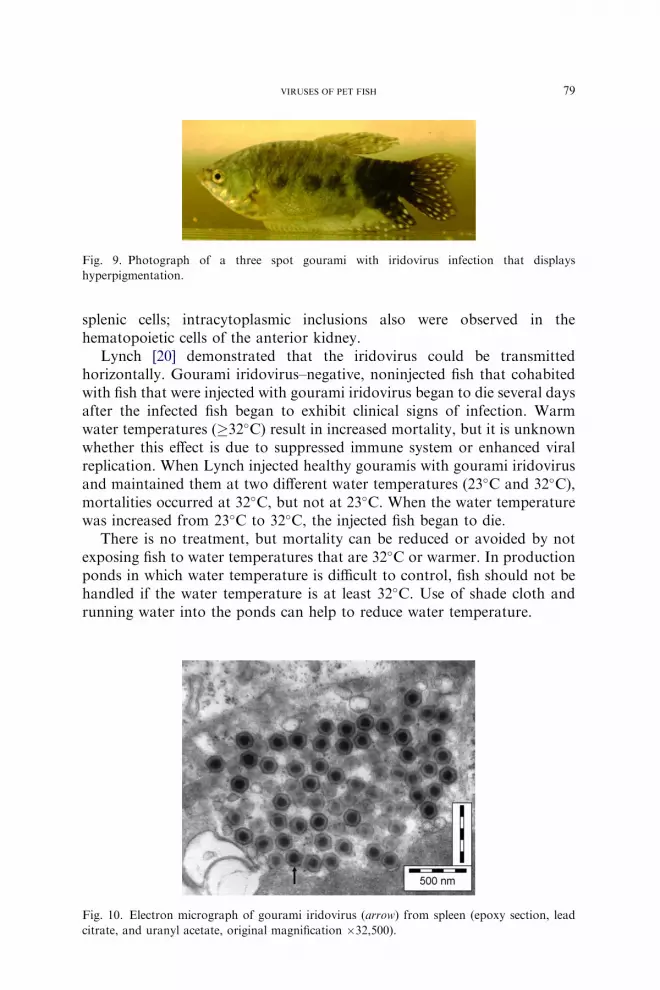
Affected fish displayed patchy hyperpigmentation and were lethargic(Fig. 9). Some fish also had ascites. On internal examination, the spleens ofaffected fish were enlarged and a clear, amber fluid was present in the coelom.No parasitic or bacterial pathogen was isolated.
On electron microscopic examination of internal tissues, iridovirusparticles were observed in the spleen, intestine, and anterior kidney(Fig. 10). Viral isolation was attempted by culturing spleen and intestine onFHM and tilapia heart (TH) cells. CPE occurred in the TH cells 3 to 5 dayspostinoculation. Electron microscopic examination of the TH culturesrevealed iridovirus particles [19].
On histopathology, the primary lesion was diffuse necrosis of the splenicparenchyma. Intracytoplasmic and intranuclear inclusions were present in
79VIRUSES OF PET FISH
splenic cells; intracytoplasmic inclusions also were observed in thehematopoietic cells of the anterior kidney.
Lynch [20] demonstrated that the iridovirus could be transmittedhorizontally. Gourami iridovirus–negative, noninjected fish that cohabitedwith fish that were injected with gourami iridovirus began to die several daysafter the infected fish began to exhibit clinical signs of infection. Warmwater temperatures (�32�C) result in increased mortality, but it is unknownwhether this effect is due to suppressed immune system or enhanced viralreplication. When Lynch injected healthy gouramis with gourami iridovirusand maintained them at two different water temperatures (23�C and 32�C),mortalities occurred at 32�C, but not at 23�C. When the water temperaturewas increased from 23�C to 32�C, the injected fish began to die.
There is no treatment, but mortality can be reduced or avoided by notexposing fish to water temperatures that are 32�C or warmer. In productionponds in which water temperature is difficult to control, fish should not behandled if the water temperature is at least 32�C. Use of shade cloth andrunning water into the ponds can help to reduce water temperature.
Fig. 9. Photograph of a three spot gourami with iridovirus infection that displays
hyperpigmentation.
Fig. 10. Electron micrograph of gourami iridovirus (arrow) from spleen (epoxy section, lead
citrate, and uranyl acetate, original magnification �32,500).

80 PETTY & FRASER
Viruses that affect catfishes
Pleco herpesvirus
Herpesvirus like particles have been seen in two species of armoredsuckermouth catfishes, the blue-eyed pleco (Panaque suttoni) and thecommon pleco (unidentified loricariid).
A veterinarian who worked with an ornamental production andwholesale facility observed that blue-eyed plecos (an imported fish)occasionally arrived with pale, blotchy lesions on the skin [21]. Overa period of several days, the lesions coalesced and appeared to be ulcerativeand the affected fish died within days of the appearance of the lesions(Fig. 11). Herpesvirus like particles were observed by electron microscopicexamination of the skin lesions. This species is not imported commonly andno new cases have been seen.
The common pleco is a popular fish that is kept by hobbyists for algaecontrol and scavenging leftover food. The eggs are collected from creeks,canals, and rivers and sold to farmers who hatch and grow the plecos toa sellable size. Some farmers noticed the development of blotchy lesions onthe skin of the plecos when the water temperature was lower than theoptimum temperature for the species. Affected plecos were submitted fordiagnostic testing; herpesvirus particles were seen on negative stains of theskin lesions. Attempts to isolate the virus in cell culture were not successful.
Viruses that affect centrarchids
Largemouth bass iridovirus
Although largemouth bass (Micropterus salmoides) usually are notconsidered a pet fish, they are kept frequently as display animals in baitshops and outdoor equipment stores. Largemouth bass virus (LMBV) was
Fig. 11. Blue-eyed plecos infected with a herpesvirus. Note the vesicles and coalesced lesions.
(Courtesy of Roy P.E. Yanong, VMD, Ruskin, FL.)

81VIRUSES OF PET FISH
identified first in healthy, as well as diseased, largemouth bass during aninvestigation of a fish kill in a Florida lake in 1991. No pathology wasassociated with the virus in any of the fish [22]. In 1995, LMBV was isolatedfrom two moribund largemouth bass from a large fish kill in the Santee-Cooper Reservoir in South Carolina [23]. Although LMBV has beenimplicated in sporadic fish kills during the summer months around thesoutheast United States, no fish kills have been attributed to the presence ofLMBV in Florida where approximately 30% of the lakes that were sampledhad largemouth bass that were positive for LMBV. Recent studies suggesta link between high water temperature and expression of clinical signs ofLMBV infection [24].
In addition to largemouth bass, susceptibility to LMBV is reported inFlorida largemouth bass (M salmoides floridanus), a subspecies of large-mouth bass. Other centrarchids, such as bluegill (Lepomis macrochirus), arereported to be susceptible to LMBV, but do not show signs of illness. Thevirus is spread by way of horizontal transmission; Woodland et al [25] hadlimited success when they attempted to transmit the virus orally. There is noevidence that vertical transmission occurs.
LMBV has been blamed for fish kills of trophy-sized largemouth bass;however, injection of LMBV into adult largemouth bass did not result indisease, but 100% of juvenile largemouth bass died following injection[23,26]. Reports indicate that the swim bladder of infected fish may be filledwith an exudate or may be inflamed and overfilled [23].
LMBV easily grows in FHM (Fig. 12) or BF-2 cells. CPE consists ofpyknotic rounded cells that eventually detach. Serology and PCR methodsalso are available. More research is required to demonstrate that LMBV isa causative agent in largemouth bass kills.
Fig. 12. Iridovirus particles (arrow) in FHM cell culture from the first isolation of LMBV.
Virus was grown from fish tissues, but virions were never found in the original tissue which
suggested that they were present in low numbers (epoxy section, lead citrate, and uranyl acetate,
original magnification �32,500).
82 PETTY & FRASER
Other viruses
Picornavirus in rainbowfish
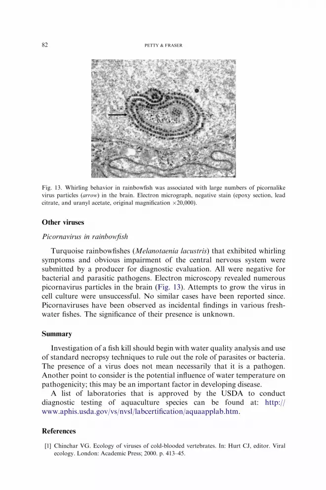
Turquoise rainbowfishes (Melanotaenia lacustris) that exhibited whirlingsymptoms and obvious impairment of the central nervous system weresubmitted by a producer for diagnostic evaluation. All were negative forbacterial and parasitic pathogens. Electron microscopy revealed numerouspicornavirus particles in the brain (Fig. 13). Attempts to grow the virus incell culture were unsuccessful. No similar cases have been reported since.Picornaviruses have been observed as incidental findings in various fresh-water fishes. The significance of their presence is unknown.
Summary
Investigation of a fish kill should begin with water quality analysis and useof standard necropsy techniques to rule out the role of parasites or bacteria.The presence of a virus does not mean necessarily that it is a pathogen.Another point to consider is the potential influence of water temperature onpathogenicity; this may be an important factor in developing disease.
A list of laboratories that is approved by the USDA to conductdiagnostic testing of aquaculture species can be found at: http://www.aphis.usda.gov/vs/nvsl/labcertification/aquaapplab.htm.
References
[1] Chinchar VG. Ecology of viruses of cold-blooded vertebrates. In: Hurt CJ, editor. Viral
ecology. London: Academic Press; 2000. p. 413–45.
Fig. 13. Whirling behavior in rainbowfish was associated with large numbers of picornalike
virus particles (arrow) in the brain. Electron micrograph, negative stain (epoxy section, lead
citrate, and uranyl acetate, original magnification �20,000).

83VIRUSES OF PET FISH
[2] Colorni A, Diamant A. Splenic and cardiac lymphocystis in the red drum, Sciaenops
ocellatus (L.). J Fish Dis 1995;18:467–71.
[3] Plumb JA. Health maintenance and principal microbial diseases of cultured fish. Ames (IA):
Iowa State University Press; 1999.
[4] Wolf K. Fish viruses and fish viral diseases. Ithaca (NY): Cornell University Press; 1988.
[5] Hoole D, BuckeD, Burgess P, et al. Diseases of carp and other cyprinid fishes. Oxford (UK):
Fishing News Books; 2001.
[6] Bernet D, Wahli T. First report of SVC infection in koi carp in Switzerland. Bull Eur Assoc
Fish Pathol 2002;22(3):225–9.
[7] Fijan N. Spring viraemia of carp and other viral diseases and agents of warm-water fish. In:
Woo PTK, Bruno DW, editors. Fish diseases and disorders: viral, bacterial, and fungal
infections, vol. 3. New York: Cab International; 1999. p. 177–246.
[8] Goodwin AE. First report of spring viremia of carp (SVCV) in North America. J Aquat
Anim Health 2002;14(3):161–4.
[9] Hedrick RP, Gilad O, Yun S, et al. A herpesvirus associated with mass mortality of juvenile
and adult koi, a strain of common carp. J Aquat Anim Health 2000;12(1):44–57.
[10] Waltzek TB, Kelley GO, Yun SC, et al. Relationships of koi herpesvirus (KHV) to herpes-
like viruses of fish and amphibians. Presented at the 35th Annual Conference, International
Association for Aquatic Animal Medicine. Galveston, Texas, April 4–9, 2004.
[11] Perelberg A, Smirnov M, Hutoran M, et al. Epidemiological description of a new viral
disease afflicting culturedCyprinus carpio in Israel. The Israeli Journal of Aquaculture 2003;
55(1):5–12.
[12] Gray WL, Mullis L, LaPatra SE, et al. Detection of koi herpesvirus DNA in tissues of
infected fish. J Fish Dis 2002;25:171–8.
[13] Goodwin AE. Molecular, physical and clinical evidence that golden shiner virus (GSV) and
grass carp reovirus (GCR) are variants of the same virus. J Aquat Anim Health 2003;15(4):
257–63.
[14] Francis-Floyd R, Fraser W, Yanong R. Iridoviruses in Florida fisheries. Presented at the
First Joint Meeting, International Association of Aquatic Animal Medicine and American
Association of Zoo Veterinarians, New Orleans, September 17–21, 2000.
[15] Schuh JCL, Shirley IG. Viral hematopoietic necrosis in an angelfish (Pterophyllum scalare).
Jour Zoo Wild Med 1990;21(1):95–8.
[16] Francis-Floyd R, Bolon B, Fraser W. Lip fibromas associated with retrovirus-like particles
in angel fish. J Am Vet Med Assoc 1993;202(3):427–9.
[17] Harms CA, Lewbart GA. Surgery in fish. Veterinary Clin North AmExot Anim Pract 2000;
3(3):759–74.
[18] Yanong RPE. Iridoviral-associated disease in oscars (Astronotus ocellatus). Presented at the
34thAnnual Conference, InternationalAssociation ofAquaticAnimalMedicine.Waikoloa,
Hawaii, May 10–14, 2003.
[19] Fraser WA, Keefe TJ, Bolon B. Isolation of an iridovirus from farm-raised gouramis. J Vet
Diagn Invest 1993;5:205–53.
[20] Lynch MJ. Preliminary investigation of gourami iridovirus infections [master’s thesis].
Gainesville (FL): University of Florida; 1998.
[21] Yanong RPE. Possible herpesvirus-associated disease in the blue-eyed plecostomus,
Panaque suttoni. Presented at the 26th Annual Conference, International Association of
Aquatic Animal Medicine. Mystic, Connecticut, May 6–10, 1995.
[22] Grizzle JM, Altinok I, Fraser WA, et al. First isolation of largemouth bass virus. Dis Aquat
Organ 2002;50:233–5.
[23] Plumb JA, Grizzle JM, Young HE, et al. An iridovirus isolated from wild largemouth bass.
J Aquat Anim Health 1996;8(4):265–70.
[24] Grant EC, Philipp DP, Inendino KR, et al. Effects of temperature on the susceptibility
of largemouth bass to largemouth bass virus. J Aquat Anim Health 2003;15(3):
215–21.

84 PETTY & FRASER
[25] Woodland JE, Brunner CJ, Noyes AD, et al. Experimental oral transmission of largemouth
bass virus. J Fish Dis 2002;25:669–72.
[26] Plumb JA, Zilberg D. The lethal dose of largemouth bass virus in juvenile largemouth
bass and the comparative susceptibility of striped bass. J Aquat Anim Health 1999;11:
246–52.