vcam-1 expression on dystrophic muscle vessels has a critical role in the recruitment of human...
TRANSCRIPT

STEM CELLS IN HEMATOLOGY
VCAM-1 expression on dystrophic muscle vessels has a critical role in therecruitment of human blood–derived CD133� stem cellsafter intra-arterial transplantationManuela Gavina, Marzia Belicchi, Barbara Rossi, Linda Ottoboni, Fabio Colombo, Mirella Meregalli, Maurizio Battistelli, Laura Forzenigo,Piero Biondetti, Federica Pisati, Daniele Parolini, Andrea Farini, Andrew C. Issekutz, Nereo Bresolin,Franco Rustichelli, Gabriela Constantin, and Yvan Torrente
Recently our group demonstrated themyogenic capacity of human CD133� cellsisolated from peripheral blood when deliv-ered in vivo through the arterial circula-tion into the muscle of dystrophic scid/mdx mice. CD133� stem cells express theadhesion molecules CD44, LFA-1, PSGL-1,�4-integrins, L-selectin, and chemokinereceptor CCR7. Moreover these cells ad-here in vitro to VCAM-1 spontaneouslyand after stimulation with CCL19. Impor-tantly, after muscle exercise, we found
that the expression of VCAM-1 is stronglyup-regulated in dystrophic muscle ves-sels, whereas the number of rolling andfirmly adhered CD133� stem cells signifi-cantly increased. Moreover, human dys-trophin expression was significantly in-creased when muscle exercise wasperformed 24 hours before the intra-arterial injection of human CD133� cells.Finally, treatment of exercised dystrophicmice with anti–VCAM-1 antibodies led toa dramatic blockade of CD133� stem cell
migration into the dystrophic muscle. Ourresults show for the first time that theexpression of VCAM-1 on dystrophicmuscle vessels induced by exercise con-trols muscle homing of human CD133�
stem cells, opening new perspectives fora potential therapy of muscular dystro-phy based on the intra-arterial delivery ofCD133� stem cells. (Blood. 2006;108:2857-2866)
© 2006 by The American Society of Hematology
Introduction
Attempts to repair muscle damage in patients with Duchennemuscular dystrophy (DMD) by transplanting myogenic progenitorsdirectly into muscles are hampered by the problem of cell survivaland the limited migration of these cells in the muscles. The deliveryof myogenic stem cells to the sites of muscle lesions through thesystemic circulation is a potential alternative approach to treat thisdisease, but injected cells may become trapped intravenously inother organs (eg, liver, spleen, lung) so that only a small portionenters the muscle microvasculature and migrates to dystrophicmuscle. Our recent work supports the idea that stem cells can reachthe site of muscle regeneration and can contribute to muscle repairand to replenishment of the satellite cell pool after intra-arterialinjection, suggesting that this technique might be particularlysuited for treating muscle dystrophy.1,2 This protocol succeededprimarily because of the widespread distribution of donor stemcells through the muscle capillary network, a distinct advantage ofthis strategy over previous approaches. Elucidation of the mecha-nisms involved in the muscle homing of stem cells will aid thedevelopment of a potential therapy for muscular dystrophy basedon the systemic delivery of stem cells. It has been shown recentlythat VLA-4–VCAM-1 interactions efficiently mediate rolling and
arrest in vivo in blood vessels from bone marrow or underpathologic conditions characterized by an activated endothelium.3-5
In the present study, we focused our attention on the molecularmechanisms involved in human CD133� cells homing to dystro-phic muscle and efficiently mediating their transplantation.
Materials and methods
Isolation and characterization of human blood–derived CD133�
cell fraction for FACS analysis
Human circulating CD133� cells were isolated from blood mononucleatedcells, as previously described.1 The enrichment of CD133� cells wasfollowed by a second round of cell sorting for the CD133 antigen with adual-laser FACSVantage SE (Becton Dickinson, Franklin Lakes, NJ).Sorted CD133� cells were double labeled with anti–CD133-phycoerythrin(anti–CD133-PE) (Miltenyi Biotech, Bergisch Gladbach, Germany), anti–VLA4-FITC, anti–CD44-FITC, anti–LFA1-FITC, anti–L-selectin–FITC(BD Pharmingen, San Diego, CA), anti–PSGL1-FITC (MBL International,Woburn, MA), anti–CCR3-FITC, anti–CCR5-FITC, anti–CCR7-FITC,anti–CXCR3-FITC, and anti–CXCR4-FITC (R&D Systems, Minneapolis,MN). Isotype control was performed using fluorescein isothiocyanate
From the Stem Cell Laboratory, Department of Neurological Sciences,Fondazione Istituto di Ricovero e Cura a Carattere Scientifico (IRCCS),Ospedale Maggiore Policlinico, Centro Dino Ferrari, University of Milan, Italy;the Department of Pathology, Division of General Pathology, University ofVerona, Italy; the Fondazione IRCCS Ospedale Maggiore Policlinico, Instituteof Nuclear Medicine, Milan, Italy; the Radiology Unit, Fondazione IRCCSOspedale Maggiore Policlinico of Milan, Italy; the Department of Pediatrics,Pathology, and Microbiology-Immunology, Dalhousie University, Halifax, NS,Canada; and the Department of Sciences Applied to Complex Systems,Polytechnic University of Marche, Ancona, Italy.
Submitted April 24, 2006; accepted June 15, 2006. Prepublished online asBlood First Edition Paper, June 29, 2006; DOI 10.1182/blood-2006-04-018564.
Supported by the Association Francaise contre les Myopathies; the ItalianMinistry of Health (Ricerca Finalizzata); the IRCCS Fondazione PoliclinicoHospital (Progetto a Concorso); Cariverona Foundation; Fondo IncentivazioneRicerca di Base (FIRB); and Fondazione Italiana Sclerosi Multipla (FISM).
Reprints: Yvan Torrente, Department of Neurological Science, University ofMilan, Padiglione Ponti, Ospedale Policlinico, via Francesco Sforza 35, 20122Milan, Italy; e-mail: [email protected].
The publication costs of this article were defrayed in part by page chargepayment. Therefore, and solely to indicate this fact, this article is herebymarked ‘‘advertisement’’ in accordance with 18 U.S.C. section 1734.
© 2006 by The American Society of Hematology
2857BLOOD, 15 OCTOBER 2006 � VOLUME 108, NUMBER 8

(FITC)–conjugated mouse IgG1, R-phycoerythrin (R-PE)–conjugated ratIgG2a. To ensure the quality of our analysis, we also investigated sortedCD133� cells for the expression of molecules, as previously indicated. Wealso characterized the adhesion molecules and chemokines receptor expres-sion of sorted CD133� cells after stimulation with 5 ng TNF-� in RPMI1640 medium (Gibco, Invitrogen, Carlsbad, CA) enriched with insulin, 20ng/mL epidermal growth factor (EGF), and 10 ng/mL basic fibroblasticgrowth factor (bFGF). The cells were analyzed 4 to 5 hours afterstimulation and after overnight incubation with TNF-�.
In vitro adhesion assay on ICAM-1 and VCAM-1
We evaluated the functionality of LFA-1 and VLA-4 expressed by CD133�
cells using an in vitro spontaneous adhesion assay for human ICAM-1 andVCAM-1 (R&D Systems).6 After isolation, sorted CD133� cells wereresuspended in PBS, CaCl2, MgCl2 1 mM, and 10% FCS (pH 7.2) at2.5 � 106/mL, and 25 �L of this suspension was plated on 18-well glassslides for 30 minutes. The slides were coated with recombinant 1 �g/mLICAM-1 and 1 �g/mL VCAM-1 (R&D Systems), as previously described.6
Five microliters of CCL19/MIP3� (R&D Systems) was added (1 �M) tothe cell-containing wells for 3 minutes at 37°C. This chemokine has beenshown to induce the adhesion of human lymphocytes to endothelial cells orendothelial cell adhesion ligands. Cells were fixed in 1.5% glutaraldehydefor 20 minutes. The number of adherent cells in a 0.2-mm2 field wascounted using cell-counting software.
Selectin chimera binding assays
The functionality of PSGL-1 expressed on sorted blood-derived CD133�
and CD133� cells was demonstrated with a cytofluorimetric assay.Approximately 5 � 105 cells were resuspended in 100 �L standard medium(SM [DMEM, 0.1% BSA, 0.1% NaN3]) and were incubated for 30 minutesat 4°C with E- and P-selectin chimeras (R&D Systems). Cells were washedtwice, resuspended in 100 �L SM, and incubated with anti–human IgGFITC (diluted 1:32; Sigma, St Louis, MO) for 20 minutes at 4°C. After 2washes in SM, cells were pelleted and resuspended in fresh DMEMimmediately before FACS analysis. Nonspecific staining was established bythe addition of EDTA (10 mM) to staining reactions conduced with theselectin chimeras.
In vivo staining of endothelial adhesion molecules
To characterize the adhesion molecules expressed by murine musclevessels, we analyzed 3 groups (n � 5 each) of 3-month-old scid/mdx mice:group 1, mice under basal conditions; group 2, mice 4 to 5 hours afterstimulation with 1 �g TNF-� injected intravenously; group 3, mice aftermuscle stress from 1 hour of swimming and analysis of adhesion moleculeexpression 24 hours later. Monoclonal antibodies (mAbs) anti–E-selectin(RME-1), anti–P-selectin (RMP-1), anti–MAdCAM-1 (MECA-367), anti–ICAM-1 (YN1.1.7.4), anti–VCAM-1 (M.K.2.7), and isotype-matchedcontrol antibodies were labeled using the Alexa Fluor 568 labeling kit(Molecular Probes, Eugene, OR). Striate muscle vessels were visualizedusing intravital microscopy, as described.1 Intravital visualization of theAlexa Fluor 568 (Molecular Probes) staining was confirmed by fluores-cence analysis of the cryosectioned hind limbs of each treated animal.Immunocytochemistry for the expression of CD31 and �SMA was alsocarried out to identify venules and arterioles, respectively. Intravitalmicroscopy images were captured with a BXW0WI microscope (Olympus,Tokyo, Japan) equipped with an Olympus Achroplan water immersionobjective (20�/0.5 NA; focal distance, 3.3 mm).
Intravital microscopy
With the intravital microscopy model,7 we verified the muscle homing andvascular adhesion properties of CD133� stem cells into the pectoral andiliopsoas muscles of scid/mdx mice after muscle exercise (n � 6) and afteradministration of anti–VCAM-1 mAb (n � 6). Animals were anesthetized,and the pectoral muscle was analyzed with an intravital microscope(BX50WI; Olympus, Tokyo, Japan). A dextran dye (155 kDa; Sigma) was
injected to visualize the blood vessels. CD133� stem cells (106) werefluorescently labeled with green CMFDA (5-chloromethylfluorescein diac-etate; Molecular Probes) and injected by a digital pump at flow rate of 0.13to 1 �L/s.
Characterization of muscle homing of CD133� stem cellsby HMPAO labeling
We tested the muscle selectivity of human circulating CD133� cells bylabeling them with technetium Tc 99m-labeled hexamethylpropylene amineoxime (HMPAO), a radioactive marker previously described.8 Cells wereincubated for 2 hours at 37°C in RPMI medium supplemented with 1�Ci/mL (0.037 MBq) HMPAO. Labeling efficiency was measured with adose calibrator (Atomlab 100; Biodex Medical, Shirley, NY). Cell vitalitywas assessed by trypan blue and by double staining with propidium iodide(PI) and annexin V. HMPAO-labeled CD133� (10 � 105) or CD133� cellswere injected into the right femoral artery of 2-month-old scid/mdx mice inbasal condition (n � 5 each) and after swimming (n � 5 each). Wecompared the distribution of radioactivity of the first series of experimentswith animals pretreated intraperitoneally with 250 �g selective blockingantibodies against VCAM-1 (n � 4) and ICAM-1 (n � 4) (generous giftsfrom Eugene C. Butcher, Laboratory of Immunology and Vascular Biology,Department of Pathology, Stanford University School of Medicine, Stan-ford, CA) 1 hour before stem cell injection. Animals were killed 24 hourslater, tissue from muscle, lung, kidney, liver, and brain was collected, andradioactive content was measured in a �-counter (LS1801; BeckmanCoulter, Hialeah, FL) as previously described.8
Microfil perfusion
PkH2-labeled CD133� cells (5 � 105) were injected into the right femoralartery of scid/mdx mice in basal conditions (n � 3) and 24 hours afterexercise (n � 3). After cell injection, the mice were perfused with 10 mLPBS and then 10 mL liquid silicon rubber (Microfil; Flow Tech, Carver,MA). Hind limbs of treated mice were isolated in toto and placed underrefrigeration at 4°C overnight to allow polymerization. On the followingday, specimens were carefully dissociated and placed in a 50% mixture ofwater and glycerin. At successive 24-hour intervals, the glycerin concentra-tion was raised to 75%, then to 85%, and finally to pure glycerin. Statisticalanalysis using the Student t test was also performed. Immunofluorescenceimages were captured with a DMIRE2 microscope (Leica Microsystems,Cambridge, United Kingdom) and a Leica DC350 camera. The microscopewas equipped with a 40�/0.75 NA dry objective. Images were acquired andprocessed with Leica Qfluoro software.
Labeling of CD133� cells with iron oxide
To better characterize the migratory capacity of CD133� cells in mousedystrophic animal models, we used live cell tracking methods such asmagnetic resonance imaging (MRI) after labeling with nanoparticles of ironoxide (Endorem; Guerbet, Sulzbach, Germany).9,10 Human CD133� stemcells were labeled with 250 �g/mL Endorem (Guerbet) in RPMI 1640medium enriched with 20 ng/mL EGF and 10 ng/mL bFGF for 12 or 24hours. We tested different cell suspensions (100 000, 50 000, 20 000, 5000,500 cells) in 1.7% gelatin. For in vivo experiments, CD133� cells wereinjected intra-arterially in the scid/mdx dystrophic animal model and werevisualized with MRI. Images were obtained using a 4.7-T spectrometer(Burker, KooWeeRup, VIC, Australia) equipped with a surface coil made inhouse. Single sagittal, coronal, and transversal images were obtained by afast gradient-echo sequence to localize the subsequent T2-weighted trans-verse images, as measured by a standard turbo spin-echo sequence. Thesame muscles monitored by MRI were isolated and characterized byimmunohistochemistry for the expression of CD133 antigen and Prussianblue staining.9
Histochemistry and immunocytochemistry of injecteddystrophic muscles
For human dystrophin detection, unexercised (n � 5) and exercised (n � 6)scid/mdx mice were injected intra-arterially with 500 000 human CD133�
2858 GAVINA et al BLOOD, 15 OCTOBER 2006 � VOLUME 108, NUMBER 8

stem cells, as previously described.1 Sixty days later, muscle sections werecharacterized using the anti–human nuclear lamin A/C (Novocastra, 1:200),anti–human dystrophin (1:50; Chemicon, Temecula, CA), anti–�-smoothmuscle actin (DAKO, Carpinteria, CA), and anti-CD31 (1:100; Chemicon)antibodies, as previously described.1
Results
Characterization of adhesion molecules andchemokine receptors
To evaluate the migratory capacity of human circulating CD133�
cells, we first studied by flow cytometry the adhesion moleculesand chemokine receptors expressed on their surfaces. In theseexperiments, we sorted CD133� and CD133� cells, and wecharacterized both populations by flow cytometry analysis.Sorted CD133� cells (99% � 1% [mean � SD]) coexpressedCD44 and LFA-1. In particular, we observed 2 subpopulationsof CD133 on the basis of LFA-1dim and LFA-1bright expression. Inaddition, 83% � 2% of sorted CD133� cells expressed PSGL-1,44% � 3% expressed VLA-4, and 60% � 5% expressed L-selectin. Chemokine receptor analysis showed very low expres-sion of CXCR3 and CXCR4 (4% � 3%), whereas 44% � 5.4%of CD133� cells were positive for CCR7. Less than 2% of sortedCD133� cells expressed CCR3 (1.6% � 1.3%) or CCR5(1.7% � 1.5%). We observed that sorted CD133� cells expressed98% � 1% CD44, 87% � 1% LFA-1, 63.5% � 1% L-selectin,53.5% � 1% CCR7, 19% � 1% CXCR3, 3.5% � 1% CXCR4,1.2% � 1% CCR3, and 0.09% � 1% CCR5. In contrast to CD133�
cells, very low expression of VLA-4 (0.6% � 0.5%) and reducedexpression of PSGL-1 (7.5% � 1%) were observed on sorted
CD133� cells. Although the expression of chemokine receptors andadhesion molecules was regulated in leukocytes by the inflamma-tory microenvironment, human circulating CD133� cells did notseem to be particularly influenced by this condition. In fact, after 5and 20 hours of stimulation with TNF-� in culture medium, onlythe percentage of cells expressing L-selectin was decreased from60% to 4%. The other adhesion molecules and chemokine recep-tors showed no modification in their expression as a result ofexposure to TNF-� (Figure 1). All these data demonstrate thathuman CD133� cells express a complete pattern of adhesionmolecules and chemokine receptor CCR7, an important moleculeusually involved in the activation and migration of leukocytesunder physiologic and pathologic conditions.
Evaluation of adhesion capability of CD133� cells in vitro
The adhesion capacity of CD133� cells to integrin ligands wasevaluated in vitro by adhesion assays to VCAM-1 and ICAM-1before and after stimulation with MIP3� (CCL19, a ligand forCCR7 receptor). Rapid adhesion assays showed that CD133� cellsspontaneously adhered to VCAM-1 and ICAM-1, suggesting thatVLA-4 and LFA-1 integrins expressed on CD133� cells were, inpart, already activated and able to bind their counter ligandswithout chemoattractant stimulation. Interestingly, the number ofcells spontaneously adherent to VCAM-1 was 178 � 28, whichwas more than 10 times greater than the number of cells thatadhered to ICAM-1 (15.7 � 3.1), showing that nonactivatedCD133� cells displayed higher binding activity of VLA-4 thanLFA-1. Stimulation with MIP3� improved significantly the num-ber of adherent cells to VCAM-1, and, though the number ofadherent cells remained low, an increase was also observed inICAM-1 (Figure 2A). Taken together, these results suggest that
Figure 1. FACScan immunophenotyping of sorted human circulating CD133� and CD133� cells. Human circulating CD133� cells obtained from the peripheral blood ofhealthy donors were characterized after FACSVantage sorting for the expression of adhesion molecules and chemokine receptors. The expression of the adhesion moleculeswas also evaluated in sorted CD133� cells and after stimulation of sorted CD133� cells with 5 ng/mL TNF-�. CD133� cells (99% � 1%) coexpressed CD44 and LFA-1. Inparticular, we observed 2 subpopulations of CD133 on the basis of LFA-1dim and LFA-1bright expression. In addition, 83% � 2% of CD133� cells expressed PSGL-1, 44% � 3%expressed VLA-4, and 60% � 5% expressed L-selectin. Analysis of chemokine receptors showed a very low expression of CXCR3 and CXCR4 (4% � 3%), whereas44% � 5.4% of CD133� cells were positive for CCR7. Less than 2% of total CD133� cells expressed CCR3 (1.6% � 1.3%) or CCR5 (1.7% � 1.5%). We observed that sortedCD133� cells expressed 98% � 1% CD44, 87% � 1% LFA-1, 63.5% � 1% L-selectin, 53.5% � 1% CCR7, 19% � 1% CXCR3, 3.5% � 1% CXCR4, 1.2% � 1% CCR3, and0.09% � 1% CCR5. Very low expression of VLA-4 (0.6% � 0.5%), CCR3 (1.2% � 1%), and CCR5 (0.09% � 1%) was observed. After cytokine stimulation of sorted CD133�
cells, the expression of L-selectin decreased from 60% to 4%. The other adhesion and chemokine molecules showed similar decreases.
VCAM-1 PROMOTES HUMAN STEM CELL MUSCLE HOMING 2859BLOOD, 15 OCTOBER 2006 � VOLUME 108, NUMBER 8

CD133� cells preferentially adhere in vitro using VLA-4 integrin.To demonstrate specificity, the assays were performed in thepresence of anti–VCAM-1 and anti–ICAM-1 antibodies (R&DSystems), and the blockade of binding was greater than 95% (datanot shown). We also performed flow cytometry assays using E- andP-selectin chimeras on the sorted CD133� and CD133� fractionsof blood-derived mononucleated cells. Results demonstrated thatapproximately 40% of human CD133� cells bound to E-selectin,whereas approximately 10% of these cells bound to P-selectin.These results suggested that unstimulated CD133� cells expressedfunctional ligands mainly for E-selectin (Figure 2B). Moreover, thebinding of CD133� cells for E- and P-selectin was, respectively,1.1% � 0.3% and 1.9% � 0.2% (Figure 2C), showing that thesecells displayed low binding activity of PSGL-1.
In vivo staining of adhesion molecules in dystrophic vessels
To support the results obtained with binding assays, we performedan in vivo staining study aimed at identifying the endothelialadhesion molecules potentially involved in dystrophic musclehoming. For this reason, scid/mdx mice were monitored in basalconditions (n � 5), after 1 hour of swimming exercise (n � 5), andafter intravenous injection of 1 �g TNF-� (n � 5). Through the useof fluorescent antibodies and intravital microscopy, we observedthat vessels of 3-month-old scid/mdx mice were positive for the
expression of P-selectin, whereas MAdCAM-1, ICAM-1, andE-selectin were not detected. Positivity for anti–ICAM-1 andanti–MAdCAM-1 antibodies was found in Peyer patches, asexpected. P-selectin–positive vessels were visible in pectoral andhind limb muscles with tortuous morphology and regions ofnarrowing, and very low expression of VCAM-1 was observed insome vessels. After stimulation with TNF-� and 24 hours aftermuscle exercise, no variation in the expression of E-selectin andMAdCAM-1 was observed, whereas VCAM-1 in vivo stainingshowed an increase in the number of positive vessels and anenhanced intensity of the positive vessels (Figure 3). Surprisingly,and in contrast to the VCAM-1 results, ICAM-1 expression was notup-regulated in dystrophic muscle after TNF-� injection, though inthe same animals we found positivity for ICAM-1 in Peyer patchesand brain. No detectable differences were observed betweenarteriolar and venular staining. In vivo staining performed witha control mAb anti–human Ras showed no in vivo staining ofdystrophic muscle (data not shown). In vivo staining data wereconfirmed by immunofluorescence for CD31 and �SMA per-formed on cryosections of perfused mice. It was, in fact,possible to detect several CD31� muscle venules and �SMA-positive arterioles that coexpressed P-selectin or VCAM-1(Figure 3) in the cryosections obtained from the same musclespreviously monitored by in vivo staining.
Intravital microscopy performed in dystrophic muscle vessels
Muscle homing and vascular adhesion properties of CD133� cellswere next verified in the pectoral and gastrocnemius muscles of3-month-old scid/mdx mice under flow conditions. For all in vivoexperiments, we used blood-derived CD133� cells obtained withmagnetic column separation. Even if the purity obtained with thismethod was just greater than 90%, the contaminating fraction ofCD133� cells did not affect our results. In fact, the characterizationof adhesion molecules and the adhesion assays of sorted CD133�
cells showed a low capacity of these cells to interact with the mainendothelial adhesion molecules involved in the recruitment fromthe bloodstream described in our experiments. When injected intothe aortic arch (through the right carotid), few human CD133� cellswere able to firmly adhere to the murine muscle vessels, but mostinteracting cells performed rolling (21 � 5.4 for rolling interac-tions and 0.8 � 0.3 for firm adhesion; mean � SEM). We observedthat some cells were mechanically trapped in blood capillaries andin proximity to narrowing vessels. This low capacity of CD133�
cells to firmly adhere to the dystrophic vessel endotheliumprobably resulted because the expression of P-selectin, as a rollingreceptor, in the muscle vessels of scid/mdx was insufficient tomediate adhesion interactions necessary for efficient firm arrest.We thus extended these experiments injecting the CD133� cells inscid/mdx mice, which, after 1 hour of swimming exercise, dis-played increased expression of VCAM-1. Rolling events weremore frequent (increase of 50%; P .04) in vessels stimulatedwith exercise than in vessels in basal condition (31.5 � 3.6)(Figure 4E). We also analyzed the quality and strength of rolling bymeasuring the rolling velocities (Vrolls) of interacting humanCD133� cells (Figure 4F). Median Vroll in basal conditions andafter swimming was 49.9 �m/s and 24.9 �m/s, respectively. Inaddition, the distribution of Vroll in velocity classes showed alarger number of cells with higher Vroll under basal conditions,suggesting that different molecular mechanisms might mediaterolling interactions after swimming compared with basal condi-tions. Importantly, we observed a 6-fold increase in the number of
Figure 2. CD133� cells express functional adhesion molecules and CCR7receptor. We evaluated the functionality of adhesion molecules expressed byCD133� cells with in vitro adhesion assays on their ligands, and we tested the effectof chemokines on this binding (A). Unstimulated CD133� cells adhered to humanVCAM-1 in vitro. This binding was improved by stimulation with CCR7 ligand MIP3�.Only a reduced number of cells adhered to human ICAM-1, and a chemokineresponse to stimulation led to increased cell adhesion. Adhesion was completelyabsent in uncoated wells and extremely low in wells coated with FCS (backgroundadhesion). Results are expressed as mean � SD of adherent cells per 0.2 mm2. Thenumber of spontaneously adhered cells was compared with the number of cells thatadhered after stimulation with CCL19 by using 2-tailed Student t test. *P .01;**P .001. The functionality of PSGL1 was evaluated with a cytofluorimetric assay;40% of CD133� cells were able to bind E-selectin, but only 10% were able to bindP-selectin (B). CD133� showed very low ability to bind E- and P-selectin (1.1% � 0.3%and 1.9% � 0.2%, respectively) (C).
2860 GAVINA et al BLOOD, 15 OCTOBER 2006 � VOLUME 108, NUMBER 8
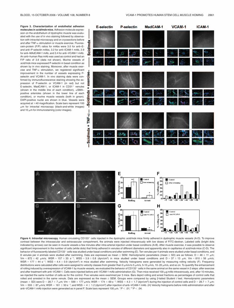
Figure 3. Characterization of endothelial adhesionmolecules in scid/mdx mice. Adhesion molecule expres-sion on the endothelium of dystrophic muscle was evalu-ated with the use of in vivo staining followed by observa-tion with intravital microscopy and on cryosections beforeand after TNF-� stimulation or muscle exercise. Fluores-cein-protein (F/P) ratios for mAbs were 3.0 for anti–E-and anti–P-selectin mAbs, 3.2 for anti–ICAM-1 mAb, 2.9for anti–MAdCAM-1 mAb, and 3.4 for anti–VCAM-1 mAb.An anti–human Ras mAb was used as control and had anF/P ratio of 3.6 (data not shown). Murine vessels ofscid/mdx mice expressed P-selectin in basal condition asshown by in vivo staining. Moreover, after muscle exer-cise and TNF-� stimulation, we registered significantimprovement in the number of vessels expressing P-selectin and VCAM-1. In vivo staining data were con-firmed by immunofluorescence staining showing the ex-pression of P-selectin or VCAM-1 (in red) but notE-selectin, MadCAM-1, or ICAM-1 in CD31� venules(shown in the middle line of each condition), �SMA–positive arterioles (shown in the lower line of eachcondition), or murine muscle vessels (both in green).DAPI-positive nuclei are shown in blue. Vessels wereacquired at �40 magnification. Scale bars represent 100�m for intravital microscopy (black-and-white images)and 10 �m for immunostaining (color images).
Figure 4. Intravital microscopy. Human circulating CD133� cells injected in the dystrophic scid/mdx mice firmly adhered in dystrophic muscle vessels (A-D). To improvecontrast between the intravascular and extravascular compartment, the animals were injected intravenously with low doses of FITC-dextran. Labeled cells (bright dotsindicated by arrows) can be seen in muscle vessels a few minutes after intra-arterial injection under basal conditions (A-B). After muscle exercise, it was possible to observesignificant improvement in the number of cells (white dots) that firmly adhered in venules of different diameters and apparently also in capillaries of scid/mdx mice (C-D). Thebehavior of fluorescently labeled CD133� cells was studied under basal conditions and after swimming (E). Ten venules per 4 animals were studied under basal conditions, and8 venules per 4 animals were studied after swimming. Data are expressed as mean � SEM. Hemodynamic parameters (mean � SD) are as follows: D � 46 � 11 �m;Vm � 872 � 42 �m/s; WSR � 157 � 33 s�1; WSS � 3.9 � 0.8 (dyn/cm2) in mice studied under basal conditions and D � 37 � 13 �m; Vm � 810 � 56 �m/s;WSR � 177 � 44 s�1; WSS � 4.4 � 0.9 (dyn/cm2) in mice studied after swimming. Velocity histograms were generated by measuring rolling velocity (F). Frequencydistributions were calculated after cells were assigned to velocity classes from greater than 0 �m/s to 5 �m/s, 5-10 �m/s, 10- 20 �m/s, and so on. To quantify the enhancementof rolling induced by overexpression of VCAM-1 by muscle vessels, we evaluated the behavior of CD133� cells in the same animal on the same vessel in 2 steps: after exerciseand after treatment with anti–VCAM-1. Cells were injected before anti–VCAM-1 mAb administration (G). Then mice received 100 �g mAb intravenously, and, after 10 minutes,we injected the same number of cells as for the control. Five venules were examined per 3 mice. Bars depict rolling and arrest fractions as percentages of control cells thatrolled and arrested in the same venule. Data are expressed as the mean � SEM. Groups were compared by using 2-tailed Student t test. Hemodynamic parameters(mean � SD) were D � 28.7 � 7 �m; Vm � 599 � 177 �m/s; WSR � 174 � 69 s�1; WSS � 4.3 � 1.7 (dyn/cm2) during the injection of control cells and D � 28.7 � 7 �m;Vm � 555 � 87 �m/s; WSR � 161 � 50 s�1; and WSS � 4 � 1.2 (dyn/cm2) after injection of anti–VCAM-1 mAb. (H) Velocity histograms before mAb administration and afteranti–VCAM-1 mAb injection were generated as in panel F. Scale bars represent 100 �m. *P .01; **P .04.
VCAM-1 PROMOTES HUMAN STEM CELL MUSCLE HOMING 2861BLOOD, 15 OCTOBER 2006 � VOLUME 108, NUMBER 8

CD133� cells able to firmly adhere in capillaries, small postcapil-lary venules, and larger venules (5.5 � 1.9) (Figure 4E). Theincreased recruitment after swimming could be explained by thenatural capacity of CD133� cells to bind VCAM-1 because of thepresence on their membrane of the integrin very late antigen(VLA-4). Taken together, these results show that the activation ofdystrophic vessel endothelium by exercise improves the adhesionof human CD133� cells in vivo.
We next sought to identify the molecular mechanisms ofrolling and arrest in dystrophic muscle venules after swimming.Rolling interactions were not inhibited after the administrationof anti–VCAM-1 mAb. In contrast, almost 60% of firm adhesion/arrest was blocked by the use of anti–VCAM-1 mAb (Figure4G). Median Vroll was 23.8 for control cells and 24.5 afteranti–VCAM-1 administration. However, though not statisticallysignificant, the mean � SD value of Vroll was slightly increasedafter VCAM-1 blockade: 31 � 21 for control cells comparedwith 39 � 42 after mAb blockade. In addition, the distributionof Vroll in velocity classes showed a slightly larger number ofcells with higher Vroll after the administration of anti–VCAM-1mAb (Figure 4H), suggesting that VCAM-1 might contribute toroll strengthening of some CD133� cells. An isotype-matchedcontrol antibody (anti–human Ras) had no effect on rolling orarrest (data not shown). These results clearly show an importantrole for VCAM-1 in the recruitment of CD133� cells indystrophic muscle after muscle exercise. Hemodynamic param-eters for intravital microscopy experiments are depicted inFigure 4E,G.
Detection of intra-arterially injected CD133� stem cellsby fluorescence and microfil perfusion
To confirm the results obtained by intravital microscopy andquantify firmly adhered cells with a different method, animalsinjected intra-arterially with CD133� stem cells were perfusedwith microfil. This technique offers the possibility of eliminat-ing transient adhered cells, observing the 3-dimensional cast ofdystrophic muscle vessels, and identifying injected cells distri-bution in toto.
In basal conditions, the transillumination of microfil-perfusedvessels of intra-arterially injected scid/mdx mice rarely showedPKH2 CD133�-labeled cells in large and small vessels of dystro-phic muscles (2 � 1.41 cells/field at 20� magnification) (Figure5A, C). In contrast, after muscle exercise, we observed a 4- to5-fold increase of the number of adhered cells in large vessels anddistributed in the capillary network (10 � 2.51 cells/field at 20�magnification; P .01) (Figure 5B, D-E).
MRI analysis
To test the migration capacity of human circulating CD133�
stem cells and to monitor the fate of implanted cells using anoninvasive method such as MRI, we labeled these cells with acontrast agent based on dextran-coated superparamagnetic ironoxide nanoparticles clinically approved as a blood pool agent(Endorem; Guerbet). In vitro results demonstrated that CD133�
stem cell iron incorporation leads to MRI visualization. The ironoxide nanoparticles in cultured CD133� stem cells were ob-served as blue spots after Prussian blue staining (Figure 6F-G).Cell counting of Prussian blue–stained cells revealed that after
12 hours of labeling with Endorem (Guerbet), the percentage oflabeled cells was 44.2% � 7.4% and that after 24 hours oflabeling, the percentage was increased to 85.6% � 8.3%. Longerlabeling did not increase either the number of labeled cells or thenumber of iron particles inside the cells. A colorimetric assay forthe quantification of cell proliferation and cell viability based onthe cleavage of the tetrazolium salt WST-1 (Roche MolecularBiochemicals, Indianapolis, IN) by mitochondrial dehydroge-nases in viable cells did not show any differences betweennanoparticle-labeled and nonlabeled cell groups. Moreover,MRI images showed a clear hypointense signal at all concentra-tions of more than 500 cells (Figure 6A-E). At 24 hours afterintra-arterial transplantation, we observed a hypointense signalobtained by MRI evaluation in the quadriceps, tibialis anterior,and soleus muscle tissues. No recognizable hypointense signalin the muscle of uninjected leg was detected. The hypointensesignal in muscle remained visible during the first 24 hours afterintra-arterial injection with no change in shape. The histology ofthese hypointense areas showed that a large number of Prussianblue–positive cells had entered into the muscle and that most ofthem coexpressed the CD133 antigen around vessels, suggestingmigration from the arterial circulation (Figure 6I, J).
Figure 5. Evaluation of the effect of muscle exercise on the recruitment ofhuman CD133� cells. CD133� stem cells were incubated with PkH2 green to allowdetection by fluorescence microscopy with a perfusion technique that showed thevasculature in 3 dimensions. Intra-arterially injected CD133� stem cells were rarelyfound in the vessels of dystrophic muscles before exercise (A, C). In contrast, afterswimming exercise intra-arterially injected scid/mdx mice showed an increasednumber of adhered cells in the capillary network (B, D) and to large vessels (E). (E)Large vessels perfused with a rubber silicon fluid were detected in transillumination.(inset) Cells were not evident in other fluorescence acquisitions, confirming thatpositively registered cells in vessels were not artefacts. (F) Statistical evaluation byStudent t test of the number of CD133� cells adhered to the vessel and confirmedsignificant improvement in recruitment after exercise. *P .05. Scale bars represent100 �m (A-C), 200 �m (D), and 25 �m (E). In panels A-E, arrows indicate the labeledcells. Error bars in panel F indicate standard deviation.
2862 GAVINA et al BLOOD, 15 OCTOBER 2006 � VOLUME 108, NUMBER 8

In vitro labeling of CD133� stem cells with 99mTc-labeledHMPAO and evaluation of labeled cell accumulationin dystrophic scid/mdx mice
To test the efficiency of labeling, CD133� stem cells wereincubated with 15 MBq HMPAO in serum-free RPMI between30 and 120 minutes. A time-dependent incorporation of HMPAOwas observed with maximum efficiency after 60 minutes,resulting in an overall labeling efficiency of 61% � 11%. Next,we investigated whether the labeling affected cell viability orfunction. Up to 120 hours after HMPAO labeling, trypan blueuptake in the radiolabeled CD133� stem cells was not signifi-cantly different from that in controls, with 97.1% � 2.1% ofcells trypan blue negative after labeling (n � 3). The viability oflabeled CD133� cells was also confirmed by cytofluorimetricanalysis. In fact, annexin V and PI staining showed a vitality of97% � 2% (Figure 7A). Similarly, the functional capacity ofCD133� stem cells to migrate in response to vascular endothe-lial growth factor in a modified Boyden chamber assay was notaffected by radiolabeling when radiolabeled CD133� stem cellswere compared with their unlabeled controls (data not shown).To determine the leakage of HMPAO into the supernatant, wechecked the activity of HMPAO in the supernatants and in theadherently growing CD133� stem cells. We found that32.4% � 5.2% of the HMPAO incorporated into CD133� stemcells was retained after 24 hours (data not shown). We thusinvestigated the distribution of radioactively labeled humanCD133� stem cells after their intra-arterial injection intoscid/mdx mice. In these experiments, we compared the distribu-tion of radioactivity obtained after the injection of CD133� cellsin exercised mice pretreated with anti–ICAM-1 (n � 4) oranti–VCAM-1 (n � 4) or without pretreatment (n � 5). Ascontrol, we characterized the radioactivity distribution obtainedafter intra-arterial injection of radiolabeled CD133� cells inbasal condition (n � 5) and after exercise (n � 5). Twenty-four
hours after the injection of CD133� cells, a high traceraccumulation was found in muscle tissues of mice withoutpretreatment (70.1% � 18.3% of the injected activity). Themuscle tracer distribution did not show major changes afteranti–ICAM-1 mAb pretreatment (71.2% � 9.1% of whole bodyactivity). However, we observed reduced accumulation ofCD133� cells in other organs, such as the spleen, in which it isknown that the blood vessel endothelium expresses ICAM-1. Incontrast to the results obtained with anti–ICAM-1 antibody, weregistered a marked reduction (approximately 10-fold) of theradioactivity in the muscle of dystrophic mice pretreated withanti–VCAM-1 mAb (Figure 7B). The specific radioactivity inisolated muscles after swimming exercise and no pretreatmentcompared with anti–ICAM-1 pretreatment were, respectively,68 047.9 � 5048.36 cpm/g tissue and 90 337.1 � 6874.11 cpm/gtissue. After anti–VCAM-1 pretreatment, specific total muscleactivity decreased significantly to 8152.4 � 1507.36 cpm/g(P .05). All these data suggest a critical role of VCAM-1 inthe recruitment of CD133� stem cells in exercised dystrophicmuscle tissues. However, an important interaction was observedin control mice injected with CD133� cells. In fact, theradioactivity detected after the injection of these labeled cellswas 79 721.5 � 1060.66 cpm/g tissue in basal condition, whereasa decrement of 3-fold (to 31 533 � 1342.07 cpm/g tissue)was observed after exercise. These data demonstrate that theimprovement observed in the recruitment of radiolabeled cellsafter swimming exercise is related to the CD133� fraction, andthese observations are not affected by the contaminating nega-tive cells.
Human dystrophin expression in injected scid/mdx mice
We then investigated the in vivo ability of CD133� stem cells todifferentiate into the myogenic lineage after intra-arterial injec-tion. As shown by radioactivity studies to contain donor cells,
Figure 6. T2-weighted images of phantoms and injectedmuscle leg tissues and implanted CD133� stem cells. MRimages of phantoms formed by a set of test tubes containing asuspension of Endorem-labeled cells in gelatin: (A) 500, (B) 5000,(C) 20 000, (D) 50 000, and (E) 100 000 cells. Sample containsgelatin only. Inhomogeneities in the phantom images were causedby the moderate sedimentation of cells while the gelatin wassetting. Sequence parameters were repetition time (TR), 2000msec; effective echo time (TE), 42.5 msec; turbo factor, 4; numberof acquisitions (AC), 16; field of view (FOV), 3.5 cm; matrix,256 � 256; slice thickness, 0.5 mm; slice separation, 1 mm. Twosets of interleaved transversal images were measured to coverthe whole muscle. For in vitro experiments, phantoms of sterileagarose 2% containing labeled cells were measured by a similarsequence with different geometry: FOV, 6 cm; matrix, 256 � 256;slice thickness, 1 mm. Only one slice was measured for phan-toms. Prussian blue staining of samples containing 50 000 (F) and100 000 (G) cells. (H) Several areas of hypointense signal wereseen 24 hours after intra-arterial grafting by MRI (white quadrant).(J) Histologic examination performed on biopsy specimens ofMRI-positive areas confirmed positive Prussian blue–stained cellsthat coexpressed the CD133 antigen around muscle vessels.Scale bars represent 100 �m (A-E); 25 �m (F-G, I, inset); 3 mm (H).
VCAM-1 PROMOTES HUMAN STEM CELL MUSCLE HOMING 2863BLOOD, 15 OCTOBER 2006 � VOLUME 108, NUMBER 8

tissues were evaluated for the expression of human dystrophin 2months after CD133� stem cell engraftment. Human dystrophin–positive myofibers were counted in 5 nonadjacent cross-sectionsof the intra-arterially injected muscles, and the longitudinaldimension of the positive area was approximately 600 �m. Inunexercised animals that received intra-arterial injection, weobserved that the highest number of dystrophin-positive fibersper cross-section was detected in the quadriceps muscle of theinjected leg (roughly 0.5% to 1.0% of total fibers in a givencross-section) (Figure 7E; Table 1). However, after muscleswimming/exercise, the percentage of human dystrophin in-creased 4-fold in all observed muscle tissues of the injected leg(Figure 7F; Table 1). In all injected muscles, many of the humandystrophin–positive fibers expressed the anti–human lamin A/C(Figure 7C-D). Moreover, human dystrophin–positive myofiberswere clustered primarily near �SMA-positive muscle arteries(Figure 7E-F). Indeed, positive human lamin A/C cells were foundnear the human dystrophin–positive myofibers. All these datasuggest that muscle exercise increases the number of maturemyofibers expressing the human dystrophin after the intra-arterialtransplantation of blood-derived CD133� stem cells.
Discussion
Recently we demonstrated that human circulating cells express-ing the CD133 antigen behave as a stem cell population capable
of commitment to hemopoietic, endothelial, and myogeniclineages.1 The discovery of the mechanisms involved in themuscle homing of stem cells will aid in improving a potentialtherapy for muscular dystrophy based on the systemic deliveryof such stem cells.
Almost all CD133� cells coexpressed CD44 and LFA-1, with aparticular distribution of the latter antigen in 2 subpopulations,LFA-1dim and LFA-1bright, suggesting the existence of a lessactivated and a more activated subpopulation of CD133� cells.More than 40% of the CD133 cells expressed PSGL-1, VLA-4,L-selectin, and CCR7, suggesting that part of CD133� cells mightmigrate in secondary lymphoid organs.11 Moreover, the expressionof these chemokine receptors and adhesion molecules of CD133�
cells do not seem to be particularly influenced by exposure toTNF-�. All these data demonstrate that freshly isolated humanCD133� cells already express a pattern of adhesion moleculespotentially able to mediate migration through the blood vessel wall.Thus, we investigated the ability of stem cells to bind endothelialadhesion molecules such as ICAM-1, VCAM-1, P-selectin, andE-selectin using in vitro functional assays. The results obtainedsuggest that VLA-4 and LFA-1 integrins expressed by CD133�
cells are functional, at least in part, and are able to spontaneouslybind VCAM-1 and ICAM-1, respectively. However, we observedthat the capacity of VLA-4 to bind VCAM-1 was more than 10-foldhigher than the capacity of LFA-1 to bind ICAM-1, suggesting thatCD133� cells might preferentially migrate using VLA-4. Inaddition, unstimulated CD133� cells express functional ligands for
Figure 7. HMPAO-labeled CD133� cells and measurement of the distribution of radioactivity by �-counter in dystrophic injected mice. (A) Viability of radiolabeledcells. Flow cytometry analysis with PI and annexin V confirmed the 97% viability observed with trypan blue staining. (B) When we blocked the VCAM-1 molecule with theanti–VCAM-1 antibody, we obtained a significant decrease in radioactivity 12 hours after intra-arterial injection in the muscles and in the different organs, such as brain, lungs,kidneys, spleen, and liver, compared with values obtained from untreated scid/mdx mice. A decrement in radioactivity in organs of mice treated with anti–ICAM-1 was alsodetected, whereas in the muscle we registered a significant improvement in counts per minute per gram. Animals injected with CD133� cells used as control showed adecrement in the recruitment of intra-arterial injected cells after exercise (C-F). Human dystrophin expression 60 days after intra-arterial injection of human CD133� stem cellsis shown. Error bars indicate standard deviation. (C-D) Colocalization of the human dystrophin (red), human lamin A/C nuclei (green), and Hoechst (blue) demonstrated theformation of normal human myofibers in dystrophic muscles after transplantation of the human blood–derived CD133� cells. The number of human dystrophin–positivemyofibers in quadriceps of exercised mice (D, F) was higher than muscle of unexercised mice (C, E). Low magnification revealed mature human dystrophin muscle fibers (red)near �SMA-positive muscle arteries (green) after the intra-arterial transplantation of CD133� stem cells into unexercised (E) and exercised (F) dystrophic scid/mdx mice. Scalebars represent 100 �m (C), 75 �m (D), and 50 �m (E-F).
2864 GAVINA et al BLOOD, 15 OCTOBER 2006 � VOLUME 108, NUMBER 8

E-selectin, but not for P-selectin, suggesting preferential binding toE-selectin during rolling interactions in vivo. Using intravitalmicroscopy, it was possible to characterize the expression ofmurine endothelial adhesion molecules potentially involved indystrophic muscle homing. Unexpectedly, some blood vessels ofscid/mdx mice in basal conditions were positive for the expressionof P-selectin, whereas MAdCAM-1, ICAM-1, and E-selectin werenot detected. Very low expression of VCAM-1 was detected onsome vessels. Moreover, when injected into the circulation ofscid/mdx mice, few human circulating CD133� cells were able tofirmly adhere to the murine muscle vessels. In fact, CD133� cellsbind only E-selectin, but not P-selectin, chimera, whereas dystro-phic muscle vessels express P-selectin but not E-selectin. This lackof matching between endothelium and CD133� cells may explainthe low capacity of stem cells to interact with the blood vessel wallin dystrophic muscles. P- and E-selectin on endothelial cells areprimary adhesion molecules for capture and the initiation ofrolling.12 E- and P-selectin rolling was previously described forhematopoietic progenitor cells (HPCs) in bone marrow vessels.3
Selectin-independent rolling of HPCs can be also mediated by�4-integrins, which interact with endothelial VCAM-1.3 However,constitutive expression of P-selectin and very low expression ofVCAM-1 in some dystrophic vessels were not sufficient to mediateefficient recruitment of CD133� cells in our experimental models.After treatment of animals with TNF-� or after swimming muscleexercise, E-selectin and MAdCAM-1 expression were not up-regulated in dystrophic muscle vessels, whereas a massive increasein the expression of VCAM-1 was observed. Surprisingly, and incontrast to VCAM-1 data, ICAM-1 expression was not up-regulated in dystrophic muscle, though in the same animals wefound positivity for anti–ICAM-1 mAb in Peyer patches and brain.4
Abnormal P-selectin expression in dystrophic muscle and the lack
of ICAM-1 and E-selectin up-regulation after TNF-� administra-tion support the hypothesis that dystrophic muscle might representa unique milieu able to selectively recruit blood cells expressingpeculiar combinations of adhesion molecules. Intravital micros-copy performed in the dystrophic vessels of scid/mdx mice,enriched in VCAM-1 and P-selectin after exercise, showed anincrease in the number of cells able to roll and firmly adhere. Thelack of E-selectin expression on scid/mdx endothelium and the lackof binding of P-selectin chimera by CD133� cells suggest thatrolling events and firm arrest of human stem cells in dystrophicmuscle vessels might have been mediated by VCAM-1 and VLA-4in our experimental models. By using CD133� cells labeled withnanoparticles of iron oxide and MRI, it was possible to demonstratethe presence of migrated stem cells into the muscle after intra-arterial injection. In addition, muscle swimming exercise increasedthe percentage of human dystrophin–positive myofibers by 4-foldin all observed muscle tissues, documenting that exercise increasesthe recruitment of CD133� cells into the dystrophic muscle. Theincrease of CD133� recruitment after exercise was also confirmedby microfil perfusion. To demonstrate the importance of VCAM-1in the recruitment of human circulating CD133� cells, we per-formed blocking experiments with radiolabeled cells. We regis-tered a marked reduction of approximately 10-fold in the radioactiv-ity in the muscle of dystrophic mice injected with anti–VCAM-1mAb before CD133� cell administration. These results clearlydemonstrate that VCAM-1 blockade prevented CD133� accumula-tion in the dystrophic muscle and that the VCAM-1/VLA-4adhesion receptor pair has a critical role in the recruitment of stemcells to dystrophic muscle vessels. Our results are supported byrecent studies showing that VCAM-1 acts as an “endothelialhoming molecule” with a role in the recruitment of stem cells in
Table 1. Blood-derived CD133� cells yield human Dys3� myofibers within injected dystrophic muscles after intra-arterialtransplantation of unexercised and exercised scid/mdx mice
Positive myofibers
Quadriceps Soleus Tibialis anterior
Unexercised Exercised Unexercised Exercised Unexercised Exercised
Mouse 1
Lamin A/C� 11 � 3 45 � 1* 7 � 5 52 � 3* 4 � 2 32 � 1*
Dys� fibers/s 26 � 13 73 � 5* 14 � 9 61 � 10* 10 � 2 55 � 11*
Dys/lamin A/C� fibers 15 � 61 45 � 1* 9 � 6 47 � 7* 5 � 8 32 � 22*
Mouse 2
Lamin A/C� 10 � 8 35 � 10 15 � 2 21 � 14 7 � 3 37 � 5*
Dys� fibers/s 12 � 5 59 � 9* 9 � 2 48 � 11* 18 � 11 91 � 32*
Dys/lamin A/C� fibers 9 � 3 45 � 4* 3 � 1 27 � 12* 9 � 7 72 � 18*
Mouse 3
Lamin A/C� 24 � 11 101 � 17* 10 � 7 70 � 24* 17 � 4 33 � 13
Dys� fibers/s 16 � 11 68 � 11* 6 � 2 49 � 16* 14 � 10 46 � 7*
Dys/lamin A/C� fibers 8 � 3 44 � 12* 3 � 1 31 � 21* 8 � 2 29 � 13*
Mouse 4
Lamin A/C� 19 � 6 36 � 19 8 � 5 24 � 8 15 � 7 53 � 11*
Dys� fibers/s 10 � 6 61 � 21* 4 � 3 41 � 13* 9 � 8 57 � 31*
Dys/lamin A/C� fibers 7 � 4 51 � 15* 1 � 2 35 � 20* 2 � 1 31 � 16*
Mouse 5
Lamin A/C� 18 � 9 81 � 21* 10 � 4 72 � 12* 11 � 4 43 � 23*
Dys� fibers/s 20 � 11 89 � 22* 9 � 1 53 � 12* 14 � 11 61 � 15*
Dys/lamin A/C� fibers 7 � 3 67 � 19* 5 � 3 26 � 8* 5 � 3 37 � 25*
Results are expressed as number of human lamin A/C-, Dys-, and Dys/lamin A/C-positive cells per section of muscle tissue (� SD) in a longitudinal dimension ofapproximately 600 �m.
Dys indicates dystrophin.*P .01.
VCAM-1 PROMOTES HUMAN STEM CELL MUSCLE HOMING 2865BLOOD, 15 OCTOBER 2006 � VOLUME 108, NUMBER 8

inflamed central nervous system.13 However, further studies areneeded to enhance the migratory ability of CD133� cells and tounderstand other mechanisms of stem cell homing, such aschemokines and chemokine receptors controlling integrin activa-tion, leading to arrest in dystrophic vessels and chemotaxis in the
damaged muscle. In conclusion, our results show that the inductionof VCAM-1 expression on the dystrophic muscle endotheliumrepresents a key mechanism of delivery of CD133� cells, allowingfor improvement in potential therapy for muscular dystrophy basedon the intra-arterial administration of stem cells.
References
1. Torrente Y, Belicchi M, Sampaolesi M, et al. Hu-man circulating AC133(�) stem cells restore dys-trophin expression and ameliorate function indystrophic skeletal muscle. J Clin Invest. 2004;114:182-195.
2. Sampaolesi M, Torrente Y, Innocenzi A, et al. Celltherapy of alpha-sarcoglycan null dystrophic micethrough intra-arterial delivery of mesoangioblasts.Science. 2003;301:487-492.
3. Mazo IB, Gutierrez-Ramos JC, Frenette PS,Hynes RO, Wagner DD, von Andrian UH. Hema-topoietic progenitor cell rolling in bone marrowmicrovessels: parallel contributions by endothelialselectins and vascular cell adhesion molecule 1.J Exp Med. 1998;188:465-474.
4. Piccio L, Rossi B, Scarpini E, et al. Molecularmechanisms involved in lymphocyte recruitmentin inflamed brain microvessels: critical roles forP-selectin glycoprotein ligand-1 and heterotri-
meric G(i)-linked receptors. J Immunol. 2002;168:1940-1949.
5. Battistini L, Piccio L, Rossi B, et al. CD8� T cellsfrom patients with acute multiple sclerosis displayselective increase of adhesiveness in brainvenules: a critical role for P-selectin glycoproteinligand-1. Blood. 2003;101:4775-4782.
6. Lawrence MB, Springer TA. Leukocytes roll on aselectin at physiologic flow rates: distinction fromand prerequisite for adhesion through integrins.Cell. 1991;65:859-873.
7. Laudanna C, Constantin G. New models of intra-vital microscopy for analysis of chemokine recep-tor-mediated leukocyte vascular recognition.J Immunol Methods. 2003;273:115-123.
8. Gianolli L, Dosio F, Matarrese M, et al. 99mTc-2GAM: a tracer for renal imaging. Nucl Med Biol.1996;23:927-933.
9. Jendelova P, Herynek V, De Croos J, et al. Im-aging the fate of implanted bone marrow stro-mal cells labeled with superparamagneticnanoparticles. Magn Reson Med. 2003;50:767-776.
10. Sykova E, Jendelova P. Magnetic resonancetracking of implanted adult and embryonic stemcells in injured brain and spinal cord. Ann N YAcad Sci. 2005;1049:146-160.
11. Butcher EC, Picker LJ. Lymphocyte homing andhomeostasis. Science. 1996;272:60-66.
12. Ley K, Kansas GS. Selectins in T-cell recruitmentto non-lymphoid tissues and sites of inflamma-tion. Nat Rev Immunol. 2004;4:325-335.
13. Pluchino S, Zanotti L, Rossi B, et al. Neuro-sphere-derived multipotent precursors promoteneuroprotection by an immunomodulatorymechanism. Nature. 2005;436:266-271.
2866 GAVINA et al BLOOD, 15 OCTOBER 2006 � VOLUME 108, NUMBER 8