value of digital mammography in predicting lymphovascular
TRANSCRIPT
RESEARCH ARTICLE Open Access
Value of digital mammography inpredicting lymphovascular invasion ofbreast cancerZhuangsheng Liu1†, Ruqiong Li1†, Keming Liang1, Junhao Chen1, Xiangmeng Chen1, Xiaoping Li2, Ronggang Li3,Xin Zhang4, Lilei Yi5 and Wansheng Long1*
Abstract
Background: Lymphovascular invasion (LVI) has never been revealed by preoperative scans. It is necessary to usedigital mammography in predicting LVI in patients with breast cancer preoperatively.
Methods: Overall 122 cases of invasive ductal carcinoma diagnosed between May 2017 and September 2018 wereenrolled and assigned into the LVI positive group (n = 42) and the LVI negative group (n = 80). Independent t-testand χ2 test were performed.
Results: Difference in Ki-67 between the two groups was statistically significant (P = 0.012). Differences in interstitialedema (P = 0.013) and skin thickening (P = 0.000) were statistically significant between the two groups. Multiplefactor analysis showed that there were three independent risk factors for LVI: interstitial edema (odds ratio [OR] =12.610; 95% confidence interval [CI]: 1.061–149.922; P = 0.045), blurring of subcutaneous fat (OR = 0.081; 95% CI:0.012–0.645; P = 0.017) and skin thickening (OR = 9.041; 95% CI: 2.553–32.022; P = 0.001).
Conclusions: Interstitial edema, blurring of subcutaneous fat, and skin thickening are independent risk factors forLVI. The specificity of LVI prediction is as high as 98.8% when the three are used together.
Keywords: Lymphovascular invasion, Breast cancer, Digital mammography
BackgroundBreast cancer metastasizes through lymphatic and bloodvessels, which makes the patients susceptible to distantmetastasis, postoperative recurrence and even death.Lymphovascular invasion (LVI) has never been revealedby preoperative scans. Diagnostic histopathology isneeded to reveal LVI of cancer cells. Preoperative identi-fication of LVI can help to predict the prognosis of pa-tients with breast cancer, especially those with axillary
node-negative breast cancer, and to develop adjuvanttreatment plans in clinical settings [1, 2].Digital mammography is one of the important imaging
tools for breast cancer screening and diagnosis. Therehave been many reports on prediction of axillary lymphnode metastasis [3–5] and on digital mammographyscreening [6–9]. However, there has been no report onpredicting LVI of breast cancer based on imaging patternsof digital mammography, except a little literature in whichMRI findings were used for prediction [2, 10–12].It is necessary to use digital mammography, a more
readily available imaging tool, in preoperative predictionof LVI in patients with breast cancer, and to evaluate thepredictive specificity of the imaging findings and certainbiomarkers detected by immunohistochemistry so as to
© The Author(s). 2020 Open Access This article is licensed under a Creative Commons Attribution 4.0 International License,which permits use, sharing, adaptation, distribution and reproduction in any medium or format, as long as you giveappropriate credit to the original author(s) and the source, provide a link to the Creative Commons licence, and indicate ifchanges were made. The images or other third party material in this article are included in the article's Creative Commonslicence, unless indicated otherwise in a credit line to the material. If material is not included in the article's Creative Commonslicence and your intended use is not permitted by statutory regulation or exceeds the permitted use, you will need to obtainpermission directly from the copyright holder. To view a copy of this licence, visit http://creativecommons.org/licenses/by/4.0/.The Creative Commons Public Domain Dedication waiver (http://creativecommons.org/publicdomain/zero/1.0/) applies to thedata made available in this article, unless otherwise stated in a credit line to the data.
* Correspondence: [email protected]†Zhuangsheng Liu and Ruqiong Li contributed equally to this work.1Department of Radiology, Jiangmen Central Hospital, Affiliated JiangmenHospital of Sun Yat-Sen University, No. 23 Haibang Street, Jiangmen 529000,Guangdong, ChinaFull list of author information is available at the end of the article
Liu et al. BMC Cancer (2020) 20:274 https://doi.org/10.1186/s12885-020-6712-z
better foresee disease progression and develop targetedtreatment plans.
MethodsClinical dataThis single-center retrospective study enrolled 122 cases ofinvasive ductal carcinoma diagnosed between May 2017and September 2018. Since the data were obtained from apicture archiving and communication system, patients’ in-formed consent was not required for this study. The inclu-sion criteria were: (1) modified radical mastectomy orbreast-conserving surgery + axillary lymph node dissection;(2) diagnosis of invasive breast cancer confirmed by routinehistopathological and immunohistochemical examinations;(3) no previous history of breast tumors or primary tumorsin other locations; and (4) LVI status confirmed by immu-nohistochemistry. Patients were excluded if they hadobscured lesions revealed by digital mammograms, werebreastfeeding, had underwent lumbar puncture orradiotherapy prior to diagnosis, and had incompleteclinical data.Mammographic lesions were confirmed either by core
needle biopsies or by surgical pathology. All the patientsunderwent digital mammography preoperatively. All ofthem were female and aged between 26 and 77 with amedian age of 45. They were assigned into the LVI posi-tive group (n = 42) and the LVI negative group (n = 80).
Digital mammographyGE (Senographe DS) and IMS (GIOTTO IMAGE) sys-tems were used. Four standard body positions were im-aged under automatic exposure conditions, and theywere the right craniocaudal (RCC) view, left craniocau-dal (LCC) view, right mediolateral oblique (RMLO) viewand left mediolateral oblique (LMLO) view. When le-sions were found in the axillary tail of Spence or nearthe cleavage, amplified imaging in these locations wasdone to reveal the lesions fully. The flat-panel detectorof internal and external obliques was parallel to the pec-toralis major muscle. According to the patients’ bodyshape, the projecting angle ranged from 40° to 65° andwas usually 60°. The glands were fully unfolded, and theskin folds below the breasts and upper abdomen werewithin the mammographic field.
Image analysisThe imaging characteristics of breast cancer lesions weredescribed according to the 5th edition of ACR BI-RADS®Atlas published in 2013 [13]. The lesions were classifiedinto four types: mass, calcification, architectural distor-tion, and asymmetry. There could be one or moremasses. The masses could be round, lobulated, and ir-regular. Clear and sharp edges meant well circumscribedmargins, clear and sharp edges obscured by glands
meant obscured margins, while obscured and spiculatededges meant indistinct margins. Amorphous and finepolymorphic calcifications were included, while typicallybenign calcifications were excluded. Architectural distor-tion referred to abnormal deformation of breasts withoutany mass clearly revealed by imaging. History of traumaand surgery must be ruled out under this circumstance.Focal asymmetry was seen in two images, but lacks theoutward border or a mass. There were only few cases ofarchitectural distortion and focal asymmetry (both n <5). Those cases were excluded to avoid inaccurate statis-tical results. And the lesion type was classified as massand calcification only.Associated features included nipple discharge, intersti-
tial edema, blurring of subcutaneous fat layer, skin thick-ening, and axillary adenopathy (lymph nodes measuring> 1 cm in the long axis diameter with absence of hilus. Ifenlarged lymph nodes are new, they need to be clinicallycombined and further examined). Due to its absence inthe LVI negative group (n = 0), nipple discharge was notincluded as a variable. Thickening of the skin means thatthe affected breast has localized or diffuse thickening ofthe skin greater than 2 mm in thickness. Interstitialedema and subcutaneous fat layer blurring are caused bythe filling and dilation of lymphatic vessels and bloodvessels in the breast, while the performance of the wholebreast including subcutaneous fat layer is blurring, andmultiple cord shadows are seen.
PathologySurgically resected breast cancer specimens were fixed in10% formaldehyde for 24 h, then were dehydrated and em-bedded in paraffin wax. Sections were prepared. StandardHE stain and streptavidin-peroxidase-biotin (SP) immuno-histochemical method were performed. DAB detectionsystems were used. A score of 3 and more indicated Her-2overexpression. Positive FISH result of Her-2 gene ampli-fication also indicated overexpression when the score was2 and more. Ki67 proliferative index (PI) of <10% indi-cated low expression level, while that of > 30% indicatedhigh expression level. Ki67 PI of 10 to 30% indicated inter-mediate expression level [14].The gold standard of this study was that LVI was de-
fined as the intravenous tumor emboli and lymphatictumor emboli detected by immunohistochemistry. Thesetwo kinds of tumor emboli were clinically referred to asintralymphovascular tumor emboli due to the difficultyof distinguishing them with pathological sections. Thespecimens were read by a senior pathologist who hadbeen working on breast cancer for 21 years.
Statistical analysisThe data were analyzed using SPSS Version 19.0. Thequantitative data were expressed as means ± SDs. The
Liu et al. BMC Cancer (2020) 20:274 Page 2 of 7
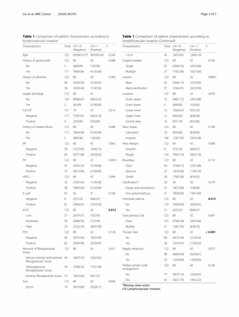
Table 1 Comparison of patient characteristics according tolymphovascular invasion
Characteristics Total LVI = 0(Negative)
LVI = 1(Positive)
P
Age 122 49.99(10.17) 48.93(9.34) 0.566
History of giving birth 122 80 42 0.488
No 5 4(80.00) 1(20.00)
Yes 117 76(64.96) 41(35.04)
History of abortion 122 80 42 0.560
No 86 55(63.95) 31(36.05)
Yes 36 25(69.44) 11(30.56)
nipple discharge 122 80 42
No 120 80(66.67) 40(33.33)
Yes 2 0(0.00) 2(100.00)
CA153a 121 79 42 0.514
Negative 117 77(65.81) 40(34.19)
Positive 4 2(50.00) 2(50.00)
History of related illness 122 80 42 0.488
No 117 76(64.96) 41(35.04)
Yes 5 4(80.00) 1(20.00)
ER 122 80 42 0.062
Negative 39 21(53.85) 18(46.15)
Positive 83 59(71.08) 24(28.92)
PR 122 80 42 0.0591
Negative 47 26(55.32) 21(44.68)
Positive 75 54(72.00) 21(28.00)
HER-2 122 80 42 0.994
Negative 32 21(65.63) 11(34.38)
Positive 90 59(65.56) 31(34.44)
E-cada 87 56 31 0.100
Negative 6 2(33.33) 4(66.67)
Positive 81 54(66.67) 27(33.33)
Ki-67 122 80 42 0.012
Low 27 20(74.07) 7(25.93)
Moderate 36 29(80.56) 7(19.44)
High 59 31(52.54) 28(47.46)
P53 122 80 42 0.126
Negative 40 30(75.00) 10(25.00)
Positive 82 50(60.98) 32(39.02)
Amount of fibroglandulartissue
122 80 42 0.431
Almost entirely fat/Scatteredfibroglandular tissue
49 33(67.35) 16(32.65)
Heterogeneousfibroglandular tissue
54 37(68.52) 17(31.48)
Extreme fibroglandular tissue 19 10(52.63) 9(47.37)
Size 122 80 42 0.094
≤2cm 79 56(70.89) 23(29.11)
Table 1 Comparison of patient characteristics according tolymphovascular invasion (Continued)
Characteristics Total LVI = 0(Negative)
LVI = 1(Positive)
P
>2cm 43 24(55.81) 19(44.19)
Single/multiple 122 80 42 0.746
Single 95 63(66.32) 32(33.68)
Multiple 27 17(62.96) 10(37.04)
Lesions 122 80 42 0.8855
Mass 65 43(66.15) 22(33.85)
Mass/calcification 57 37(64.91) 20(35.09)
Location 122 80 42 0.879
Outer upper 73 49(67.12) 24(32.88)
Outer lower 5 4(80.00) 1(20.00)
Lower inner 15 10(66.67) 5(33.33)
Upper inner 15 9(60.00) 6(40.00)
Central area 14 8(57.14) 6(42.86)
Mass shape 122 80 42 0.160
Lobulated 16 8(50.00) 8(50.00)
Irregular 106 72(67.92) 34(32.08)
Mass Margin 122 80 42 0.088
Smooth 6 2(33.33) 4(66.67)
Rough 116 78(67.24) 38(32.76)
Boundary 122 80 42 0.714
Clear 69 47(68.12) 22(31.88)
Obscure 27 16(59.26) 11(40.74)
Shield 26 17(65.38) 9(34.62)
Calcificationa 56 36 20 0.279
Vague and amorphous 25 18(72.00) 7(28.00)
Fine polymorphous 31 18(58.06) 13(41.94)
Interstitial edema 122 80 42 0.013
No 110 76(69.09) 34(30.91)
Yes 12 4(33.33) 8(66.67)
Subcutaneous fat 122 80 42 0.697
Clear 101 67(66.34) 34(33.66)
Muddy 21 13(61.90) 8(38.10)
Thicken Skin 122 80 42 < 0.001
No 86 65(75.58) 21(24.42)
Yes 36 15(41.67) 21(58.33)
Nipple retraction 122 80 42 0.073
No 98 68(69.39) 30(30.61)
Yes 24 12(50.00) 12(50.00)
Axillary lymph nodeenlargement
122 80 42 0.166
No 77 54(70.13) 23(29.87)
Yes 45 26(57.78) 19(42.22)aMissing value existsLVI Lymphovascular invasion
Liu et al. BMC Cancer (2020) 20:274 Page 3 of 7
independent t-test was done for intergroup comparisons.The count data were expressed as frequencies or rates,and the χ2 test or Fisher’s method was performed. P <0.05 indicated statistically significant difference. Thecount data whose values were 0 were excluded fromstatistical analysis and listed only in table(s).
ResultsGeneral data and biomarkersTable 1 presents the data about childbearing history,miscarriage history, history of other breast diseases, nip-ple discharge, CA153, age, ER, PR, HER-2, E-CAD, P53,and Ki-67. Difference in Ki-67 between the LVI positivegroup and the LVI negative group was statistically sig-nificant (P = 0.012), while no statistically significant dif-ferences were observed in the other factors mentioned.
Digital mammography findingsDetails are shown in Table 1. Differences in interstitialedema (P = 0.013, Figs. 1 and 2) and skin thickening(P = 0.000, Figs. 1 and 2) between the two groups werestatistically significant. No statistically significant inter-group differences were seen in other imaging features,such as fibroglandular tissue density (P = 0.431), radio-logical diameter (P = 0.094), mass number (P = 0.746), le-sion number (P = 0.8855), location (P = 0.879), massshape (P = 0.160), mass margin (P = 0.088), boundary (P = 0.714), calcification (P = 0.279), subcutaneous fat
(P = 0.697), nipple retraction (P = 0.073) and axillary ade-nopathy (P = 0.166).Risk factor analysis results are shown in Table 2.
Multiple factor analysis showed that there are three in-dependent risk factors for LVI: interstitial edema (oddsratio [OR] = 12.610; 95% confidence interval [CI]: 1.061–149.922; P = 0.045), subcutaneous fat (OR = 0.081; 95%CI: 0.012–0.645; P = 0.017) and skin thickening (OR =9.041; 95% CI: 2.553–32.022; P = 0.001).Table 3 shows the sensitivity, specificity, accuracy,
positive predictive value and negative predictive value ofthe three independent risk factors in LVI prediction.The specificity of LVI prediction was as high as 98.8%when they were applied together.
DiscussionLVI or intralymphovascular tumor emboli is closely re-lated to the adverse outcome of many malignant tumors[15–17]. As a risk factor for recurrent breast cancer fol-lowing modified radical mastectomy, lymphovasculartumor emboli, especially lymphatic tumor emboli, hasbeen included in the St Gallen consensus for breast can-cer [18]. Karlsson et al. [19] reported that the failure rateof chemotherapy was higher in breast cancer patientswith LVI than those without. Shen et al. [20] found thatlymphovascular tumor emboli promoted recurrence anddistant metastasis of local tumors. Therefore, presence
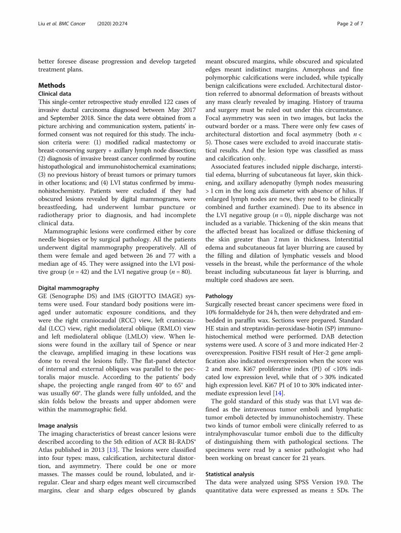
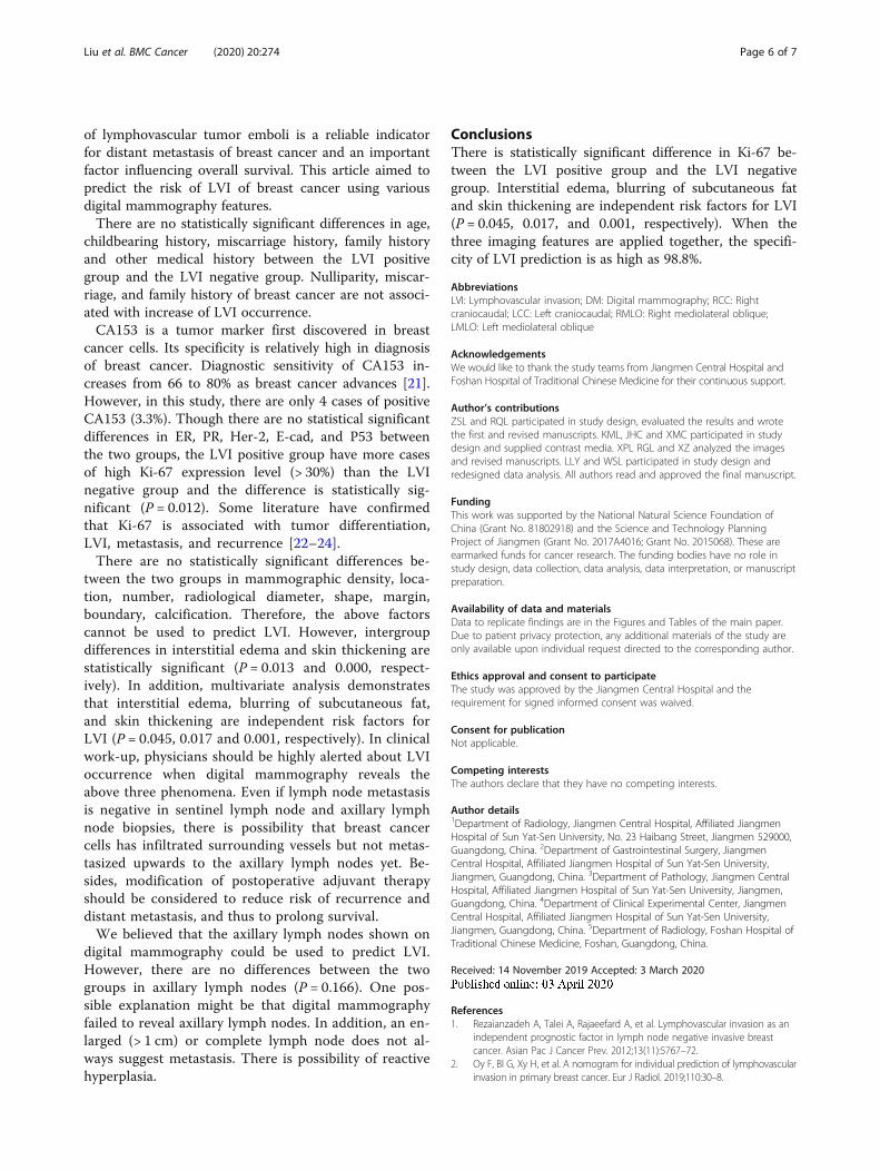
Fig. 1 Female, LVI positive. The MLO position (a) and CC position (b)of digital mammography showed an irregular upper mass withblurred boundaries in the upper outer quadrant of the right breast.Interstitial edema (green arrow), blurring of subcutaneous fat layer(yellow arrow), skin thickening (red arrow), and axillary adenopathy(white arrow) were observed in the right breast
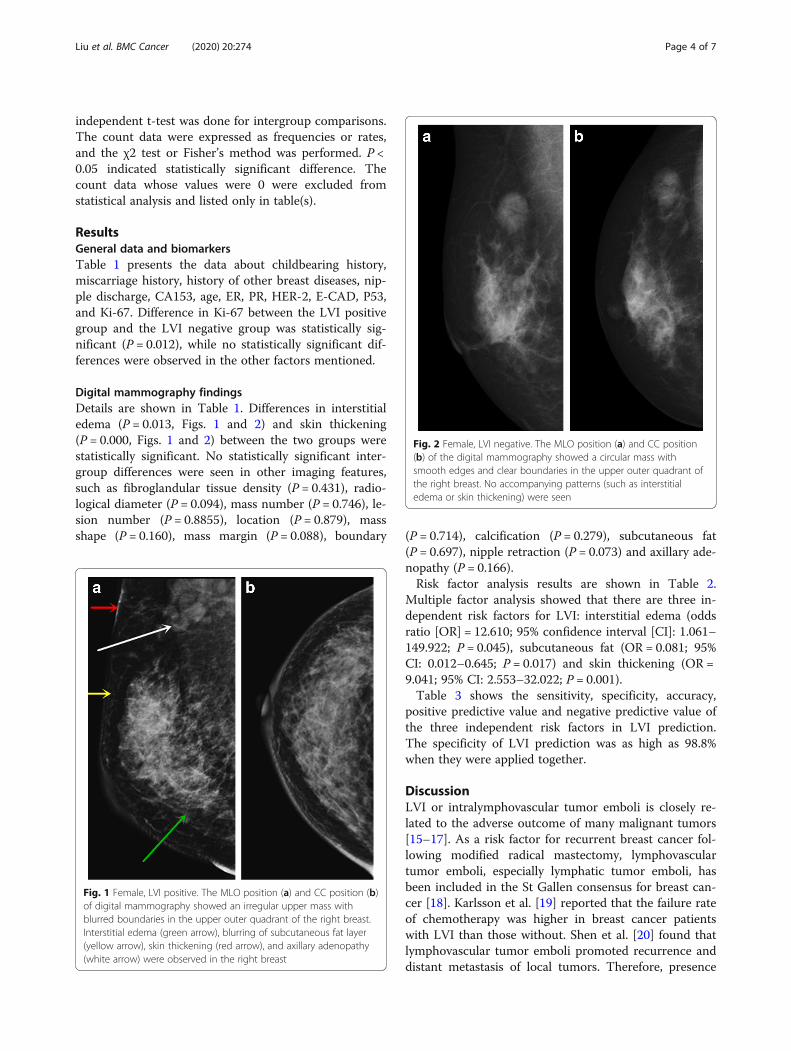
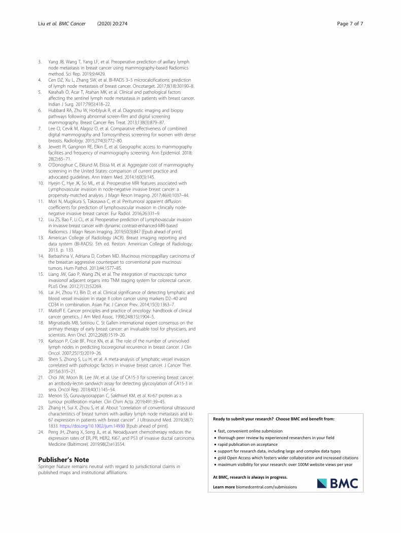
Fig. 2 Female, LVI negative. The MLO position (a) and CC position(b) of the digital mammography showed a circular mass withsmooth edges and clear boundaries in the upper outer quadrant ofthe right breast. No accompanying patterns (such as interstitialedema or skin thickening) were seen
Liu et al. BMC Cancer (2020) 20:274 Page 4 of 7
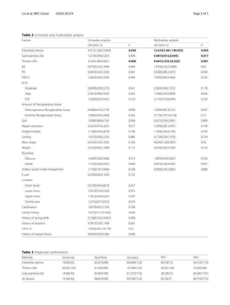
Table 2 Univariate and multivariate analysis
Factors Univariate analysis Multivariate analysis
OR (95% CI) P OR (95% CI) P
Interstitial edema 4.471(1.260,15.864) 0.020 12.610(1.061,149.922) 0.045
Subcutaneous fat 1.213(0.458,3.207) 0.698 0.081(0.012,0.645) 0.017
Thicken Skin 4.333(1.899,9.891) 0.000 9.041(2.553,32.022) 0.001
ER 0.475(0.216,1.044) 0.064 1.595(0.252,10.084) 0.62
PR 0.481(0.224,1.034) 0.061 0.508(0.085,3.035) 0.458
HER-2 1.003(0.429,2.345) 0.994 1.436(0.462,4.466) 0.532
Ki-67
Moderate 0.690(0.209,2.273) 0.541 0.382(0.094,1.551) 0.178
High 2.581(0.948,7.022) 0.063 1.298(0.350,4.809) 0.696
P53 1.920(0.827,4.457) 0.129 2.115(0.739,6.050) 0.163
Amount of fibroglandular tissue
Heterogeneous fibroglandular tissue 0.948(0.414,2.179) 0.899 1.439(0.491,4.215) 0.507
Extreme fibroglandular tissue 1.856(0.630,5.469) 0.262 3.773(0.707,20.154) 0.12
Size 1.928(0.890,4176) 0.096 0.921(0.294,2.891) 0.889
Nipple retraction 2.267(0.914,5.621) 0.077 1.299(0.281,5.991) 0.738
Single/multiple 1.158(0.476,2.819) 0.746 1.183(0.336,4.165) 0.793
Lesions 1.057(0.500,2.233) 0.885 0.712(0.256,1.979) 0.514
Mass shape 0.472(0.163,1.365) 0.166 0.624(0.128,3.047) 0.56
Margin 0.224(0.043,1.389) 0.112 0.424(0.032,5.630) 0.516
Boundary
Obscure 1.469(0.586,3.684) 0.413 1.483(0.439,5.007) 0.526
Shield 1.131(0.436,2.935) 0.800 0.997(0.246,4.043) 0.997
Axillary lymph node enlargement 1.716(0.797,3.694) 0.168 0.909(0.295,2.805) 0.868
E-cad 0.250(0.043,1.452) 0.122
Location
Outer lower 0.510(0.054,4.819) 0.557
Lower inner 1.021(0.314,3.320) 0.973
Upper inner 1.361(0.434,4.267) 0.597
Central area 1.531(0.477,4.913) 0.474
Calcification 1.857(0.601,5.734) 0.166
Family history 1.927(0.117,31.602) 0.646
History of giving birth 2.158(0.233,19.947) 0.498
History of abortion 0.781(0.339,1.799) 0.561
CA15–3 1.925(0.261,14.179) 0.52
History of related illness 0.463(0.050,4.284) 0.498
Table 3 Diagnostic performance
Methods Sensitivity Specificity Accuracy PPV NPV
Interstitial edema 19.0(8/42) 95.0(76/80) 68.9(84/122) 66.7(8/12) 69.1(76/110)
Thicken Skin 50.0(21/42) 81.3(65/80) 70.5(86/122) 58.3(21/36) 75.6(65/86)
Subcutaneous fat 19.0(8/42) 83.8(67/80) 61.2(75/122) 38.1(8/21) 66.3(67/101)
All factors 14.3(6/42) 98.8(79/80) 69.7(85/122) 85.7(6/7) 68.7(79/115)
Liu et al. BMC Cancer (2020) 20:274 Page 5 of 7
of lymphovascular tumor emboli is a reliable indicatorfor distant metastasis of breast cancer and an importantfactor influencing overall survival. This article aimed topredict the risk of LVI of breast cancer using variousdigital mammography features.There are no statistically significant differences in age,
childbearing history, miscarriage history, family historyand other medical history between the LVI positivegroup and the LVI negative group. Nulliparity, miscar-riage, and family history of breast cancer are not associ-ated with increase of LVI occurrence.CA153 is a tumor marker first discovered in breast
cancer cells. Its specificity is relatively high in diagnosisof breast cancer. Diagnostic sensitivity of CA153 in-creases from 66 to 80% as breast cancer advances [21].However, in this study, there are only 4 cases of positiveCA153 (3.3%). Though there are no statistical significantdifferences in ER, PR, Her-2, E-cad, and P53 betweenthe two groups, the LVI positive group have more casesof high Ki-67 expression level (> 30%) than the LVInegative group and the difference is statistically sig-nificant (P = 0.012). Some literature have confirmedthat Ki-67 is associated with tumor differentiation,LVI, metastasis, and recurrence [22–24].There are no statistically significant differences be-
tween the two groups in mammographic density, loca-tion, number, radiological diameter, shape, margin,boundary, calcification. Therefore, the above factorscannot be used to predict LVI. However, intergroupdifferences in interstitial edema and skin thickening arestatistically significant (P = 0.013 and 0.000, respect-ively). In addition, multivariate analysis demonstratesthat interstitial edema, blurring of subcutaneous fat,and skin thickening are independent risk factors forLVI (P = 0.045, 0.017 and 0.001, respectively). In clinicalwork-up, physicians should be highly alerted about LVIoccurrence when digital mammography reveals theabove three phenomena. Even if lymph node metastasisis negative in sentinel lymph node and axillary lymphnode biopsies, there is possibility that breast cancercells has infiltrated surrounding vessels but not metas-tasized upwards to the axillary lymph nodes yet. Be-sides, modification of postoperative adjuvant therapyshould be considered to reduce risk of recurrence anddistant metastasis, and thus to prolong survival.We believed that the axillary lymph nodes shown on
digital mammography could be used to predict LVI.However, there are no differences between the twogroups in axillary lymph nodes (P = 0.166). One pos-sible explanation might be that digital mammographyfailed to reveal axillary lymph nodes. In addition, an en-larged (> 1 cm) or complete lymph node does not al-ways suggest metastasis. There is possibility of reactivehyperplasia.
ConclusionsThere is statistically significant difference in Ki-67 be-tween the LVI positive group and the LVI negativegroup. Interstitial edema, blurring of subcutaneous fatand skin thickening are independent risk factors for LVI(P = 0.045, 0.017, and 0.001, respectively). When thethree imaging features are applied together, the specifi-city of LVI prediction is as high as 98.8%.
AbbreviationsLVI: Lymphovascular invasion; DM: Digital mammography; RCC: Rightcraniocaudal; LCC: Left craniocaudal; RMLO: Right mediolateral oblique;LMLO: Left mediolateral oblique
AcknowledgementsWe would like to thank the study teams from Jiangmen Central Hospital andFoshan Hospital of Traditional Chinese Medicine for their continuous support.
Author’s contributionsZSL and RQL participated in study design, evaluated the results and wrotethe first and revised manuscripts. KML, JHC and XMC participated in studydesign and supplied contrast media. XPL RGL and XZ analyzed the imagesand revised manuscripts. LLY and WSL participated in study design andredesigned data analysis. All authors read and approved the final manuscript.
FundingThis work was supported by the National Natural Science Foundation ofChina (Grant No. 81802918) and the Science and Technology PlanningProject of Jiangmen (Grant No. 2017A4016; Grant No. 2015068). These areearmarked funds for cancer research. The funding bodies have no role instudy design, data collection, data analysis, data interpretation, or manuscriptpreparation.
Availability of data and materialsData to replicate findings are in the Figures and Tables of the main paper.Due to patient privacy protection, any additional materials of the study areonly available upon individual request directed to the corresponding author.
Ethics approval and consent to participateThe study was approved by the Jiangmen Central Hospital and therequirement for signed informed consent was waived.
Consent for publicationNot applicable.
Competing interestsThe authors declare that they have no competing interests.
Author details1Department of Radiology, Jiangmen Central Hospital, Affiliated JiangmenHospital of Sun Yat-Sen University, No. 23 Haibang Street, Jiangmen 529000,Guangdong, China. 2Department of Gastrointestinal Surgery, JiangmenCentral Hospital, Affiliated Jiangmen Hospital of Sun Yat-Sen University,Jiangmen, Guangdong, China. 3Department of Pathology, Jiangmen CentralHospital, Affiliated Jiangmen Hospital of Sun Yat-Sen University, Jiangmen,Guangdong, China. 4Department of Clinical Experimental Center, JiangmenCentral Hospital, Affiliated Jiangmen Hospital of Sun Yat-Sen University,Jiangmen, Guangdong, China. 5Department of Radiology, Foshan Hospital ofTraditional Chinese Medicine, Foshan, Guangdong, China.
Received: 14 November 2019 Accepted: 3 March 2020
References1. Rezaianzadeh A, Talei A, Rajaeefard A, et al. Lymphovascular invasion as an
independent prognostic factor in lymph node negative invasive breastcancer. Asian Pac J Cancer Prev. 2012;13(11):5767–72.
2. Oy F, Bl G, Xy H, et al. A nomogram for individual prediction of lymphovascularinvasion in primary breast cancer. Eur J Radiol. 2019;110:30–8.
Liu et al. BMC Cancer (2020) 20:274 Page 6 of 7
3. Yang JB, Wang T, Yang LF, et al. Preoperative prediction of axillary lymphnode metastasis in breast cancer using mammography-based Radiomicsmethod. Sci Rep. 2019;9:4429.
4. Cen DZ, Xu L, Zhang SW, et al. BI-RADS 3–5 microcalcifications: predictionof lymph node metastasis of breast cancer. Oncotarget. 2017;8(18):30190–8.
5. Karahallı Ö, Acar T, Atahan MK, et al. Clinical and pathological factorsaffecting the sentinel lymph node metastasis in patients with breast cancer.Indian J Surg. 2017;79(5):418–22.
6. Hubbard RA, Zhu W, Horblyuk R, et al. Diagnostic imaging and biopsypathways following abnormal screen-film and digital screeningmammography. Breast Cancer Res Treat. 2013;138(3):879–87.
7. Lee CI, Cevik M, Alagoz O, et al. Comparative effectiveness of combineddigital mammography and Tomosynthesis screening for women with densebreasts. Radiology. 2015;274(3):772–80.
8. Jewett PI, Gangnon RE, Elkin E, et al. Geographic access to mammographyfacilities and frequency of mammography screening. Ann Epidemiol. 2018;28(2):65–71.
9. O’Donoghue C, Eklund M, Elissa M, et al. Aggregate cost of mammographyscreening in the United States: comparison of current practice andadvocated guidelines. Ann Intern Med. 2014;160(3):145.
10. Hyejin C, Hye JK, So ML, et al. Preoperative MRI features associated withLymphovascular invasion in node-negative invasive breast cancer: apropensity-matched analysis. J Magn Reson Imaging. 2017;46(4):1037–44.
11. Mori N, Mugikura S, Takasawa C, et al. Peritumoral apparent diffusioncoefficients for prediction of lymphovascular invasion in clinically node-negative invasive breast cancer. Eur Radiol. 2016;26:331–9.
12. Liu ZS, Bao F, Li CL, et al. Preoperative prediction of Lymphovascular invasionin invasive breast cancer with dynamic contrast-enhanced-MRI-basedRadiomics. J Magn Reson Imaging. 2019;50(3):847 [Epub ahead of print].
13. American College of Radiology (ACR). Breast imaging reporting anddata system (BI-RADS). 5th ed. Reston: Ameirican College of Radiology;2013. p. 133.
14. Barbashina V, Adriana D, Corben MD. Mucinous micropapillary carcinoma ofthe breast:an aggressive counterpart to conventional pure mucinoustumors. Hum Pathol. 2013;44:1577–85.
15. Liang JW, Gao P, Wang ZN, et al. The integration of macroscopic tumorinvasionof adjacent organs into TNM staging system for colorectal cancer.PLoS One. 2012;7(12):52269.
16. Lai JH, Zhou YJ, Bin D, et al. Clinical significance of detecting lymphatic andblood vessel invasion in stage II colon cancer using markers D2–40 andCD34 in combination. Asian Pac J Cancer Prev. 2014;15(3):1363–7.
17. Matloff E. Cancer principles and practice of oncology: handbook of clinicalcancer genetics. J Am Med Assoc. 1990;248(15):1904–5.
18. Mignatiadis MB, Sotiriou C. St Gallen international expert consensus on theprimary therapy of early breast cancer: an invaluable tool for physicians, andscientists. Ann Oncl. 2012;26(8):1519–20.
19. Karlsson P, Cole BF, Price KN, et al. The role of the number of uninvolvedlymph nodes in predicting locoregional recurrence in breast cancer. J ClinOncol. 2007;25(15):2019–26.
20. Shen S, Zhong S, Lu H, et al. A meta-analysis of lymphatic vessel invasioncorrelated with pathologic factors in invasive breast cancer. J Cancer Ther.2015;6:315–21.
21. Choi JW, Moon BI, Lee JW, et al. Use of CA15-3 for screening breast cancer:an antibody-lectin sandwich assay for detecting glycosylation of CA15-3 insera. Oncol Rep. 2018;40(1):145–54.
22. Menon SS, Guruvayoorappan C, Sakthivel KM, et al. Ki-67 protein as atumour proliferation marker. Clin Chim Acta. 2019;491:39–45.
23. Zhang H, Sui X, Zhou S, et al. About “correlation of conventional ultrasoundcharacteristics of breast tumors with axillary lymph node metastasis and ki-67 expression in patients with breast cancer”. J Ultrasound Med. 2019;38(7):1833. https://doi.org/10.1002/jum.14930 [Epub ahead of print].
24. Peng JH, Zhang X, Song JL, et al. Neoadjuvant chemotherapy reduces theexpression rates of ER, PR, HER2, Ki67, and P53 of invasive ductal carcinoma.Medicine (Baltimore). 2019;98(2):e13554.
Publisher’s NoteSpringer Nature remains neutral with regard to jurisdictional claims inpublished maps and institutional affiliations.
Liu et al. BMC Cancer (2020) 20:274 Page 7 of 7