utility of p16 immunohistochemistry for the identification of lynch syndrome
TRANSCRIPT
UTILITY OF P16 IMMUNOHISTOCHEMISTRY FOR THEIDENTIFICATION OF LYNCH SYNDROME
Artemio Payá(*),1,2, Cristina Alenda(*),1,2,3, Lucía Pérez-Carbonell1, Estefanía Rojas1, José-Luis Soto3, Carmen Guillén3, Adela Castillejo3, Victor M. Barberá3, Alfredo Carrato3, AntoniCastells4, Xavier Llor5, Montserrat Andreu6, Jim Koh7, Greg H. Enders8, SusanaBenlloch1, and Rodrigo Jover1,91Unidad de Investigación. Hospital General Universitario de Alicante, Alicante, Spain.2Servicio de Anatomía Patológica. Hospital General Universitario de Alicante, Alicante, Spain3Unidad de Investigación. Hospital General Universitario de Elche, Elche, Spain.4Department of Gastroenterology, Institut de Malalties Digestives, Hospital Clínic, IDIBAPS,CIBERehd, University of Barcelona, Barcelona, Spain.5Digestive Diseases and Nutrition Section. University of Illinois at Chicago, Chicago, IL, USA.6Department of Gastroenterology, Hospital del Mar, Barcelona, Spain.7Duke University, Durham, NC, USA.8Fox Chase Cancer Center. Philadelphia, PA, USA.9Unidad de Gastroenterología. Hospital General Universitario de Alicante, Alicante, Spain.
AbstractImmunohistochemistry for mismatch repair proteins has shown utility in the identification of Lynchsyndrome, but majority of tumours with loss MLH1 expression are due to sporadic hypermethylationof the MLH1 promoter. These tumours can also show epigenetic silencing of other genes, such asp16. The aim of our study is to evaluate the utility of p16 immunohistochemistry in the predictionof MLH1 germline mutations.
METHODS: p16 immunohistochemistry was appropriately evaluated in 79 colorectal cancers withloss of MLH1 expression. Methylation of MLH1 and p16 were quantitatively studied using real timePCR assay Methylight. BRAF V600E mutation in tumour tissue was also investigated. Genetic testingfor germline mutation of MLH1 was made on 52 patients.
RESULTS: Loss of p16 expression was seen in 21 out of 79 samples (26,6%). There was foundstatistically significant association between p16 expression and p16 methylation (p<0.001), MLH1methylation (p<0.001) and BRAF mutation (p<0.005). All tumours with loss of p16 expressionshowed hypermethylation of p16 (21/21), 95.2% (20/21) showed MLH1 methylation and 71.4%(15/21) were mutated for BRAF V600E Mutational analysis showed pathogenic germline mutations
CORRESPONDING AUTHOR: Dr. Rodrigo Jover, Unidad de Gastroenterología, Hospital General Universitario de Alicante. PintorBaeza, 12. 03010 ALICANTE. SPAIN.(*)These authors contributed equallyThe Corresponding Author has the right to grant on behalf of all authors and does grant on behalf of all authors, an exclusive licence (ornon-exclusive for government employees) on a worldwide basis to the BMJ Publishing Group Ltd and its Licensees to permit this article(if accepted) to be published in Gut and any other BMJPGL products to exploit all subsidiary rights, as set out in our licence(http://gut.bmj.com/ifora/licence.pdf).
NIH Public AccessAuthor ManuscriptClin Cancer Res. Author manuscript; available in PMC 2010 May 1.
Published in final edited form as:Clin Cancer Res. 2009 May 1; 15(9): 3156–3162. doi:10.1158/1078-0432.CCR-08-3116.
NIH
-PA Author Manuscript
NIH
-PA Author Manuscript
NIH
-PA Author Manuscript
in 8 of the patients, harbouring 10 tumours. All 10 of these tumours showed normal staining of p16in the immunochemical analysis.
CONCLUSIONS: p16 immunohistochemistry is a good surrogate marker for p16 and MLH1epigenetic silencing due to hypermethylation, and is useful as screening tool in the selection ofpatients for genetic testing in Lynch syndrome.
Keywordscolorectal cancer; Lynch syndrome; p16; immunohistochemistry; diagnosis
BACKGROUNDLynch syndrome is an autosomal dominant disorder that accounts for approximately 3-4% ofall colorectal cancers (CRC) (1). Lynch syndrome is caused by germline mutations in the DNAmismatch repair (MMR) genes, mainly MLH1, MSH2, MSH6 and PMS2 (1). Defects in thispathway lead to changes in the length of nucleotide repeat sequences, a phenomenon calledmicrosatellite instability (MSI), which constitutes the molecular hallmark of HNPCC (2).These tumours can also be identified by immunohistochemical loss of MMR proteins (3;4).The presence of MSI may be observed in up to 10-15% of sporadic CRC. In these cases, MMRimpairment is caused by epigenetic silencing of MLH1, due to MLH1 promoter methylation(5).
Since molecular characterization of Lynch syndrome was established, the identification of genecarriers has become a critical issue. Identification of patients with Lynch syndrome hasimportant clinical implications because surveillance for CRC and other cancers in thispopulation is able to reduce cancer mortality and is cost-effective (6). A previous study fromour group established that fulfilment of revised Bethesda criteria (7), followed by either MSItesting or MMR proteins immunohistochemistry is a sensible approach to pre-select patientsfor genetic testing (4). Patients having tumours with loss of expression of MSH2 or MSH6 aresuspected carriers of germline mutations of any of these genes, but patients whose tumoursshow loss of MLH1 may either have hereditary or sporadic disease. The majority of sporadictumours with loss of MLH1 expression belong to a group of colorectal cancers that arehypermethylated at multiple genetic loci. These CRC have been described as displaying theCpG Island Methylator Phenotype (CIMP) (8;9), and a panel of markers has been proposedfor its diagnosis (10). One of the loci frequently methylated in these CIMP tumours isCDKN2A (p16). Presumably, some of the tumours with loss of MLH1 caused by epigeneticsilencing through aberrant methylation, should also have a silenced p16, and, therefore,immunohistochemical loss of staining of this protein. The aim of our study is to evaluate thevalue of p16 immunohistochemistry in the prediction of MLH1 germline mutations in patientswith tumours that show loss of MLH1.
METHODSSubjects
Immunohistochemical analysis of MLH1 was performed in 2401 CRC tumours. Tumour tissuewas collected from a series of 2,246 non-selected surgical CRC specimens from theEPICOLON study (n=1.281) (11) and from the Pathology Department of the Hospital GeneralUniversitario of Alicante, collected between the years 1999-2007 (n=965). The remaining 155tumours were collected from patients of the Genetic Counselling in Cancer Department of theHospital General Universitario of Elche. Demographic, clinical, and tumor-relatedcharacteristics of probands, as well as a detailed family history were obtained using a pre-established questionnaire, as described elsewhere (4). Loss of MLH1 expression was found in
Payá et al. Page 2
Clin Cancer Res. Author manuscript; available in PMC 2010 May 1.
NIH
-PA Author Manuscript
NIH
-PA Author Manuscript
NIH
-PA Author Manuscript
124 tumors (5.2%), from 120 patients. All these tumours showed normal expression of MSH2and MSH6. In 32 cases there was not enough tissue to perform immunohistochemical ormolecular studies and they were excluded from this study. Finally, the study was performedin 92 tumours from 88 patients that showed loss of MLH1 immunohistochemical expression.Eighty-three tumours were non selected population-based CRC specimens and 9 were fromthe Genetic Counselling Unit. Figure 1 shows a flow chart of the molecular analysis performedon the samples.
ImmunohistochemistryImmunohistochemical analysis of MLH1, MSH2, MSH6 and PMS2 was performed in blocksof formalin-fixed paraffin-embedded tumour tissue as previously described (4;12).
Immunohistochemical analysis of p16 expression was performed on tissue microarray (TMA).One of the requirements for inclusion in the study was that enough tumour tissue was presentwithin the block of wax-embedded tissue to facilitate subsequent TMA construction. Therepresentative tumour regions were identified and marked on the H&E stained slides andsubsequently identified on the corresponding tissue blocks. Tissue cylinders of diameter of 1mm were punched out from the marked areas of each block and incorporated into a recipientparaffin block using a precision instrument—the tissue arrayer (Beecher Instruments, Durviz).A total of 6 TMAs were constructed for the study. TMAs contained between 30 and 50 coresof 1 mm needle size. For inclusion in the study, at least two evaluable cores of tumour tissuewere required per case. Four-micrometer-thick sections were cut from TMAs. The slides wereput on a TechMate 500 immunostainer and incubated for 30 minutes at room temperature withthe mouse monoclonal antibody JC2, which recognizes the first ankyrin repeat of p16 (providedby Dr. Jim Koh, Duke University, Durham, NC, USA) (13). The antibody was detected by theEnvision+ technique (Dako). Processed immunohistochemical slides were evaluated by twopathologists. A tumour was considered to have normal expression for p16 when unequivocalnuclear staining was seen in some neoplastic epithelial cells, with or without cytoplasmaticstaining. Cases with loss of expression included those cases with lack of expression in tumourcells in presence of internal positive control (stromal cells or blood vessels). Samples wereconsidered not scored when no staining of internal control was seen.
MLH1 and CDKN2A methylation analysisGenomic DNA was extracted from tumour paraffin-embedded tissue blocks. Two tissuecylinders of 1 mm of diameter were punched out with the tissue arrayer from the previouslyselected tumour areas. QiaAmp DNA Mini kit for DNA extraction was used according to themanufacturer's protocol after removal of paraffin by xylene.
The MLH1 and CDKN2A (p16) methylation analysis was performed by real time PCR assayMethylight as previously described (Applied Biosystems, Foster City, CA, USA) (14).Bisulphite conversion was made with the EZ DNA methylation-Gold kit as described by themanufacturer (Zymo Research, Orange, CA, USA). Quantitative PCR was performed by ABI7500 (Applied Biosystems, Foster City, CA, USA). Primers and a probe, designed to detectbisulphite converted fully methylated MLH1 and p16 DNAs, have been described and usedpreviously (10;15-17). The PCR reactions were performed according to the protocols (16;18).
In order to calculate the percentage of methylated reference (PMR) we established thedichotomization threshold at PMR= 4, to obtain a bimodal distribution in the MLH1 andCDKN2A methylation loci. Methylation positive (PMR > 4) MLH1 and CDKN2A samplescould be distinguished from negative (PMR ≤ 4) ones.
Payá et al. Page 3
Clin Cancer Res. Author manuscript; available in PMC 2010 May 1.
NIH
-PA Author Manuscript
NIH
-PA Author Manuscript
NIH
-PA Author Manuscript
BRAF V600E MutationV600E BRAF mutation was detected using specific TaqMan probes by real time PCR (ABIPRISM 7500, Applied Biosystems, Foster City, CA, USA) and the allelic discriminationsoftware (Applied Biosystems) as previously described by Benlloch et al. (19)
MLH1 germline genetic testingGermline genetic alteration studies were performed on genomic DNA isolated from peripheralblood leukocytes or from non-tumour colon tissue as previously described (4). Point mutationanalysis of MLH1 gene was done by polymerase chain reaction (PCR) amplification and directsequencing of the entire coding region and the exon-intron boundaries. PCR primers andconditions have been described elsewhere (20-22). Large genomic rearrangements (insertionsand/or deletions) in MLH1 loci were screened by multiplex ligation-dependent probeamplification (MLPA) according to the manufacturer protocols (Salsa MLPA Kit P003 andP008; MRC-Holland, Amsterdam, The Netherlands).
Data management and analysisData were collected and entered into the computer using MICROSOFT ACCESS software forstorage and initial analysis. Further analysis was done using SPSS software (SPSS 15.0). Forcontinuous variables relevant measures of central tendency (means for normally distributeddata and medians and interquartile ranges for skewed data) were used to explore data. TheChi2 test was used for comparison of qualitative variables. A Student's t test was used forcomparison of normally distributed continuous variables and a Mann-Whitney U test was usedfor unpaired comparison of non-normally distributed continuous variables. A p value of lessthan 0.05 was considered significant.
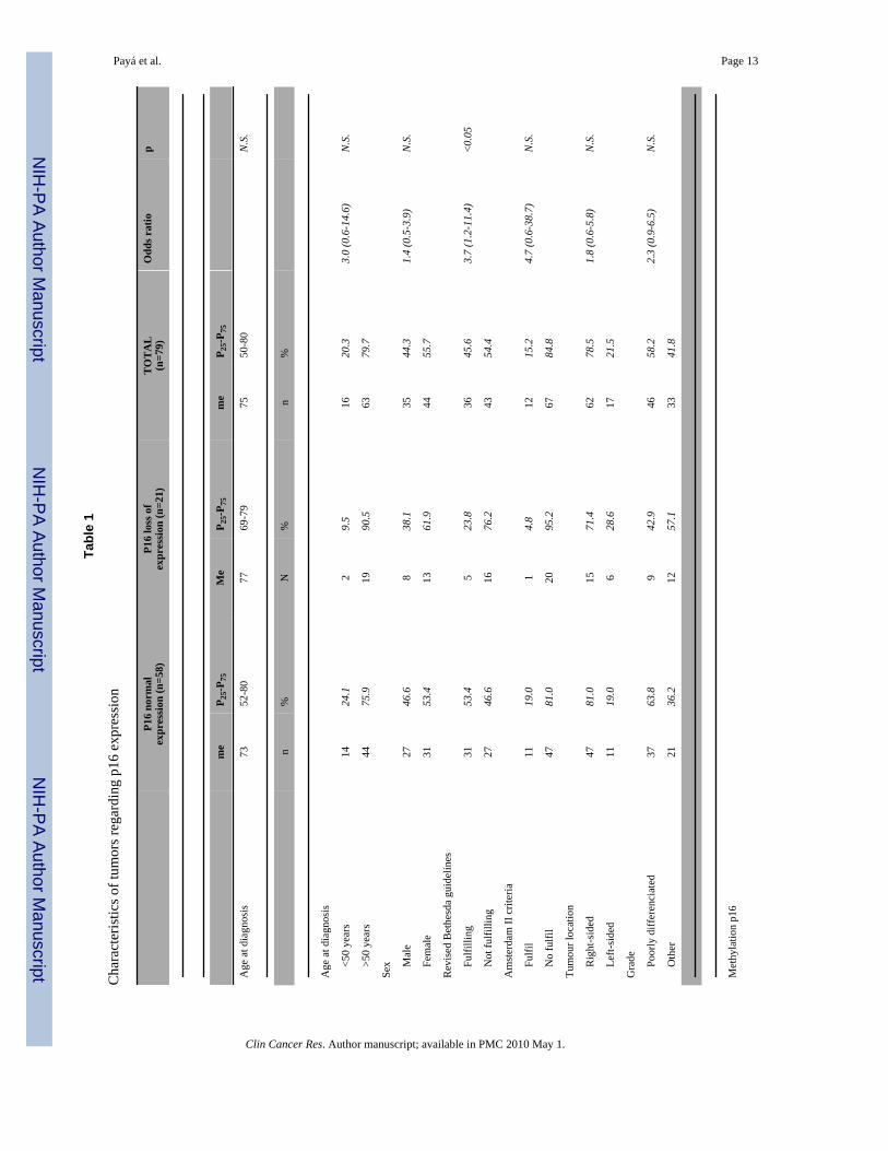
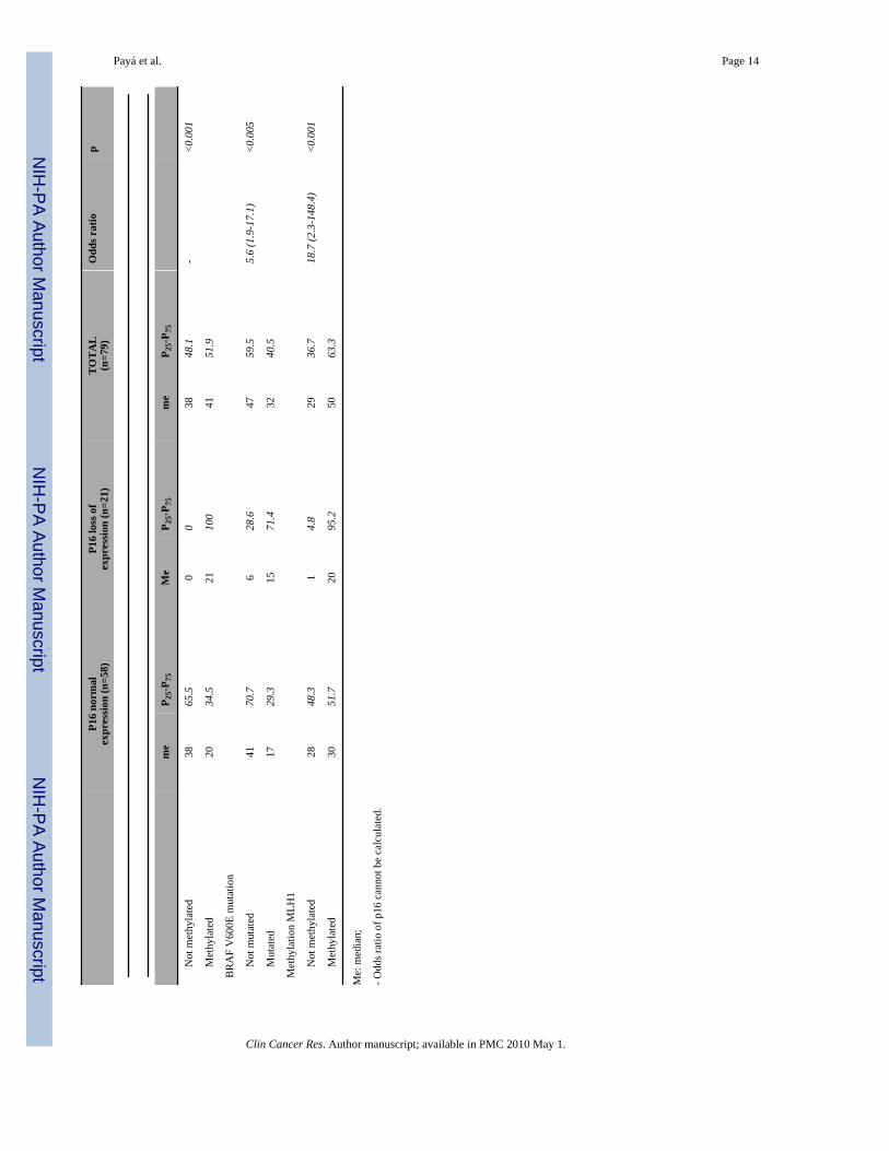
RESULTSp16 immunohistochemistry was performed in 92 tumours with loss of MLH1 expression(Figure 2) from 88 patients. In 13 of the tumours, p16 immunohistochemistry could not beconfidently assessed and was classified as not scored, due to absence of clear staining in stromalcells, which served as internal positive controls. Loss of p16 expression was seen in 21 out of79 samples (26,6%) (Fig.2). Characteristics of tumours according to p16 expression status canbe seen in table 1. There was a statistically significant association between p16 expression andp16 methylation (p<0.001), MLH1 methylation (p<0.001) and BRAF mutation (<0.005). Alltumours with loss of p16 expression showed hypermethylation of p16 (21/21), 95.2% (20/21) showed MLH1 methylation and 71.4% (15/21) were mutated for BRAF V600E (table 1).However, 20 out of 41 (50%) of the tumours with p16 methylation retained p16 expression(Table 1). Tumours with loss of p16 expression showed more frequently poor differentiation.p16 immunohistochemistry was also performed in 60 sporadic tumours with normal expressionof MLH1 and microsatellite stability, loss of p16 expression was seen in only 5 of these tumours(3%).
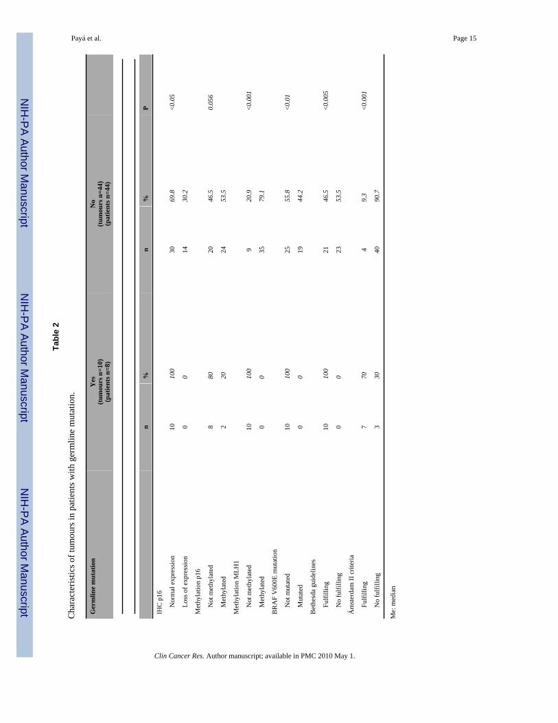
Mutational analysis of MLH1 was performed in 52 out of 88 patients whose tumours showedloss of MLH1 staining. Fifty-four CRC from these 52 patients were analyzed. Thirty of thesepatients fulfilled some of the revised Bethesda criteria and 11 fulfilled Amsterdam II criteria.Mutational analysis showed pathogenic mutations in 8 of the patients, harbouring 10 tumours.All 10 of the tumours analyzed from the 8 patients with germline pathogenic mutations inMLH1 showed normal staining of p16. All patients with germline mutations met Bethesdacriteria. Moreover, all tumours from patients with germline mutations showed non-mutatedBRAF and non-methylated MLH1 (table 2).
Payá et al. Page 4
Clin Cancer Res. Author manuscript; available in PMC 2010 May 1.
NIH
-PA Author Manuscript
NIH
-PA Author Manuscript
NIH
-PA Author Manuscript
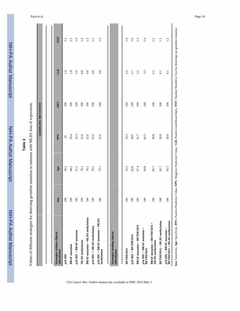
Table 3 shows values of sensitivity, specificity, positive and negative predictive value, andpositive Likelihood ratio for Bethesda criteria, BRAF mutation, MLH1 methylation, p16immunohistochemistry. Different combinations of these variables for the prediction ofgermline MLH1 mutation can also be seen in table 3. Values for p16 immunohistochemistryand BRAF mutation are similar, and combination of these techniques improves separatelyobtained results.
DISCUSSIONSelection of patients for genetic testing in Lynch syndrome is frequently difficult in clinicalpractice. The use of Amsterdam criteria is capable to detect Lynch Syndrome with a highspecificity but with very low sensitivity. When clinical presentation and family history aremost compelling, the yield of mutational testing is often no better than 50%, and even in thebest case-scenario, when Amsterdam criteria are met and a tumour shows high MSI and lossof MMR protein expression, the likelihood of germline mutation detection is approximately70-80% (23). Other strategies, such as the revised Bethesda criteria (7), improve the sensitivitybut with a high lack of specificity. With this approach, a high number of patients are sent forgenetic testing based only on clinical criteria, with the subsequent expending of resources andthe consequent generation of anxiety to patients and their families. Several approaches havebeen used for refining the selection of patients for genetic testing. The observation that patientswith Lynch syndrome show a characteristic phenotype with microsatellite instability promptedto use these markers as a first pre-screening modality. Then, the demonstration of the role ofthe immunohistochemistry and its equivalence to microsatellite instability analysis in thediagnostic algorithm of Lynch syndrome (4) improved the availability of these tools and itsgeneralization in clinical practice, due to the possibility of performing immunohistochemistryin any pathology department. Moreover, patients with tumours showing MSH2 or MSH6 lackof expression should be directly sent for genetic testing, because it is a strong indicator formutation in these genes (23). However, this clinical-molecular strategy have had somedetractors, because a number of patients with Lynch syndrome might not fulfil revised Bethesdacriteria (24). Sometimes family history is difficult or even impossible to obtain. Furthermore,recent studies show that, even among patients with a known high risk for Lynch syndrome,there is a marked underutilization of MSI analysis (25). For these reasons, some authorsadvocate for routine molecular screening of patients with CRC for Lynch syndrome usingimmunohistochemistry (24). Another fact that can support routine immunohistochemical studyof CRC is the recognized better prognosis of mismatch repair deficient tumours (26), and thedifferent response to 5-fluorouracil based chemotherapy that these tumours have (27-29). Inour study, we included only patients with MLH1 loss of expression, and compared molecularonly with clinical-molecular approaches for diagnosis, showing that combinations of onlymolecular tests are at least as good as strategies that include clinical data (Table 3). Our resultsshow that p16 immunohistochemistry can improve the results of this strategy, avoidinggermline genetic testing in approximately a third of patients with loss of MLH1 expression.
Instruments for the refinement of the selection of patients with loss of MLH1 for genetic testinghave been proposed. Mutation V600E in the oncogene BRAF has been suggested ascharacteristic of sporadic colorectal tumours with MSI and this mutation is not detected intumours from patients with germline mutations in MLH1 or MSH2 genes (30;31). Severalstudies have demonstrated that detection of BRAF V600E mutation could simplify the selectionof CRC patients for genetic testing for Lynch syndrome (32;33). However, the use of BRAFmutational analysis in clinical practice has been limited, probably due to the need of molecularbiology resources for its implementation. The main strength of p16 immunohistochemistry forclinical use in selection of suspected Lynch syndrome patients for genetic testing is itsfeasibility, in contraposition to other methylation markers such as V600E BRAF mutation orMLH1 methylation (34), that are time consuming and not available for the majority of clinical
Payá et al. Page 5
Clin Cancer Res. Author manuscript; available in PMC 2010 May 1.
NIH
-PA Author Manuscript
NIH
-PA Author Manuscript
NIH
-PA Author Manuscript
centresAberrant promoter hypermethylation associated with transcriptional silencing ofmultiple tumour suppressor genes has been proposed as a mechanistic component in theevolution of multiple cancers (35). Tumours with a critical degree of aberrant methylation havethe CpG island methylator phenotype (CIMP). CIMP tumours show promoterhypermethylation in multiple genes, including p16, p14, MGMT and MLH1 among others.Loss of the INK4a/ARF/INK4b locus is among the most frequent cytogenetic events in humancancer. The products of this locus p15INK4a, p16INK4b and ARF play widespread andindependent roles in tumour suppression (36). Specific somatic loss of p16, through pointmutation or small deletion, has been reported in human cancer (37), but epigenetic silencingthrough aberrant promoter methylation is the most common mechanism of inactivation (36;38). p16 loss of expression provokes increase in proliferation and vascularisation in coloncancer cells (13;39).
Limitations of our study are the small number of patients with MLH1 germline mutations thatwe included. However, the excellent sensitivity of p16 expression for MLH1 methylation, withvirtually all the cases with loss of p16 expression being methylated, makes p16immunohistochemistry a robust marker for this event. Most samples include abundant stromalcomponents that stain for p16, providing an internal positive control to verify adequate tissuepreservation and technical success of the staining. Another limitation is the existence of caseswith p16 hypermethylation that showed normal p16 staining. This fact may be caused, at leastin part, by the target region analyzed for the p16 methylation. The Methylight system (primersand probe) used here has been described elsewhere, being useful to characterize the CpG islandmethylator phenotype (18). The amplicon sequence analyzed is located at exon 1α. Using invitro models, Gonzalgo et al (1998) observed that p16 expression could occur in the presenceof a relatively heavily methylated coding domain (exon 1α, named as region D). Methylationof certain regions upstream of the p16 exon 1α may be more critical for transcription activity(particularly region C) (40). Exonic CpG islands are more susceptible to de novo methylationthan promoter islands. The cancer-specific promoter methylation might be a result of spreadingfrom exonic foci and selection of cells whose growth is deregulated by the gene inactivation(41).
In conclusion, our results suggest that the immunohistochemical study of p16 could improvethe selection of patients for genetic testing of germline mutations in MLH1. Patients with CRCand MLH1 loss of expression, whose tumours also show loss of p16 expression can bereasonably excluded for genetic testing, because this loss of expression indicates, with highpossibility, aberrant hypermethylation and epigenetic silencing of both p16 and MLH1 genes.
STATEMENT OF TRANSLATIONAL RELEVANCE
The main contribution of this manuscript is the utility of p16 immunohistochemistry in theidentification of patients with colorectal cancer and high level of suspicion of Lynchsyndrome. Patients with tumours showing loss of MLH1 expression can be hereditary orsporadic. In this study we demonstrate that p16 immunohistochemistry is a good surrogatemarker for both p16 and MLH1 hypermethylation. Patients whose tumours have loss ofboth MLH1 and p16 expression have hypermethylated colorectal cancer and, therefore,their tumours are sporadic. These patients can be confidently excluded for genetic testingof MLH1. P16 immunohistochemistry is easy to perform and available for every pathologydepartment, taking advantage over other more exigent techniques such as BRAF mutation.
Reference List1. Rustgi AK. The genetics of hereditary colon cancer. Genes Dev 2007;21:2525–38. [PubMed:
17938238]
Payá et al. Page 6
Clin Cancer Res. Author manuscript; available in PMC 2010 May 1.
NIH
-PA Author Manuscript
NIH
-PA Author Manuscript
NIH
-PA Author Manuscript
2. Ionov Y, Peinado MA, Malkhosyan S, Shibata D, Perucho M. Ubiquitous somatic mutations in simplerepeated sequences reveal a new mechanism for colonic carcinogenesis. Nature 1993;363:558–61.[PubMed: 8505985]
3. Jover R, Paya A, Alenda C, Poveda MJ, Peiro G, Aranda FI, Perez-Mateo M. Defective mismatch-repair colorectal cancer: clinicopathologic characteristics and usefulness of immunohistochemicalanalysis for diagnosis. Am J Clin Pathol 2004;122:389–94. [PubMed: 15362369]
4. Pinol V, Castells A, Andreu M, Castellvi-Bel S, Alenda C, Llor X, Xicola RM, Rodriguez-Moranta F,Paya A, Jover R, Bessa X. Accuracy of revised Bethesda guidelines, microsatellite instability, andimmunohistochemistry for the identification of patients with hereditary nonpolyposis colorectalcancer. JAMA 2005;293:1986–94. [PubMed: 15855432]
5. Wheeler JM, Bodmer WF, Mortensen NJ. DNA mismatch repair genes and colorectal cancer. Gut2000;47:148–53. [PubMed: 10861278]
6. Jarvinen HJ, Mecklin JP, Sistonen P. Screening reduces colorectal cancer rate in families withhereditary nonpolyposis colorectal cancer. Gastroenterology 1995;108:1405–11. [PubMed: 7729632]
7. Umar A, Boland CR, Terdiman JP, Syngal S, de la CA, Ruschoff J, Fishel R, Lindor NM, Burgart LJ,Hamelin R, Hamilton SR, Hiatt RA, Jass J, Lindblom A, Lynch HT, Peltomaki P, Ramsey SD,Rodriguez-Bigas MA, Vasen HF, Hawk ET, Barrett JC, Freedman AN, Srivastava S. Revised BethesdaGuidelines for hereditary nonpolyposis colorectal cancer (Lynch syndrome) and microsatelliteinstability. J Natl Cancer Inst 2004;96:261–8. [PubMed: 14970275]
8. Hawkins N, Norrie M, Cheong K, Mokany E, Ku SL, Meagher A, O'Connor T, Ward R. CpG islandmethylation in sporadic colorectal cancers and its relationship to microsatellite instability.Gastroenterology 2002;122:1376–87. [PubMed: 11984524]
9. Samowitz WS, Albertsen H, Herrick J, Levin TR, Sweeney C, Murtaugh MA, Wolff RK, Slattery ML.Evaluation of a large, population-based sample supports a CpG island methylator phenotype in coloncancer. Gastroenterology 2005;129:837–45. [PubMed: 16143123]
10. Weisenberger DJ, Siegmund KD, Campan M, Young J, Long TI, Faasse MA, Kang GH,Widschwendter M, Weener D, Buchanan D, Koh H, Simms L, Barker M, Leggett B, Levine J, KimM, French AJ, Thibodeau SN, Jass J, Haile R, Laird PW. CpG island methylator phenotype underliessporadic microsatellite instability and is tightly associated with BRAF mutation in colorectal cancer.Nat Genet 2006;38:787–93. [PubMed: 16804544]
11. Pinol V, Andreu M, Castells A, Paya A, Bessa X, Rodrigo J. Frequency of hereditary non-polyposiscolorectal cancer and other colorectal cancer familial forms in Spain: a multicentre, prospective,nationwide study. Eur J Gastroenterol Hepatol 2004;16:39–45. [PubMed: 15095851]
12. Xicola RM, Llor X, Pons E, Castells A, Alenda C, Pinol V, Andreu M, Castellvi-Bel S, Paya A, JoverR, Bessa X, Giros A, Duque JM, Nicolas-Perez D, Garcia AM, Rigau J, Gassull MA. Performanceof different microsatellite marker panels for detection of mismatch repair-deficient colorectal tumors.J Natl Cancer Inst 2007;99:244–52. [PubMed: 17284719]
13. Dai CY, Furth EE, Mick R, Koh J, Takayama T, Niitsu Y, Enders GH. p16(INK4a) expression beginsearly in human colon neoplasia and correlates inversely with markers of cell proliferation.Gastroenterology 2000;119:929–42. [PubMed: 11040180]
14. Eads CA, Danenberg KD, Kawakami K, Saltz LB, Blake C, Shibata D, Danenberg PV, Laird PW.MethyLight: a high-throughput assay to measure DNA methylation. Nucleic Acids Res 2000;28:E32.[PubMed: 10734209]
15. Fiegl H, Gattringer C, Widschwendter A, Schneitter A, Ramoni A, Sarlay D, Gaugg I, Goebel G,Muller HM, Mueller-Holzner E, Marth C, Widschwendter M. Methylated DNA collected bytampons--a new tool to detect endometrial cancer. Cancer Epidemiol Biomarkers Prev 2004;13:882–8. [PubMed: 15159323]
16. Ogino S, Kawasaki T, Brahmandam M, Cantor M, Kirkner GJ, Spiegelman D, Makrigiorgos GM,Weisenberger DJ, Laird PW, Loda M, Fuchs CS. Precision and performance characteristics ofbisulfite conversion and real-time PCR (MethyLight) for quantitative DNA methylation analysis. JMol Diagn 2006;8:209–17. [PubMed: 16645207]
17. Widschwendter M, Siegmund KD, Muller HM, Fiegl H, Marth C, Muller-Holzner E, Jones PA, LairdPW. Association of breast cancer DNA methylation profiles with hormone receptor status andresponse to tamoxifen. Cancer Res 2004;64:3807–13. [PubMed: 15172987]
Payá et al. Page 7
Clin Cancer Res. Author manuscript; available in PMC 2010 May 1.
NIH
-PA Author Manuscript
NIH
-PA Author Manuscript
NIH
-PA Author Manuscript
18. Ogino S, Cantor M, Kawasaki T, Brahmandam M, Kirkner GJ, Weisenberger DJ, Campan M, LairdPW, Loda M, Fuchs CS. CpG island methylator phenotype (CIMP) of colorectal cancer is bestcharacterised by quantitative DNA methylation analysis and prospective cohort studies. Gut2006;55:1000–6. [PubMed: 16407376]
19. Benlloch S, Paya A, Alenda C, Bessa X, Andreu M, Jover R, Castells A, Llor X, Aranda FI, MassutiB. Detection of BRAF V600E mutation in colorectal cancer: comparison of automatic sequencingand real-time chemistry methodology. J Mol Diagn 2006;8:540–3. [PubMed: 17065421]
20. Hampel H, Frankel W, Panescu J, Lockman J, Sotamaa K, Fix D, Comeras I, La Jeunesse J, NakagawaH, Westman JA, Prior TW, Clendenning M, Penzone P, Lombardi J, Dunn P, Cohn DE, CopelandL, Eaton L, Fowler J, Lewandowski G, Vaccarello L, Bell J, Reid G, de la CA. Screening for Lynchsyndrome (hereditary nonpolyposis colorectal cancer) among endometrial cancer patients. CancerRes 2006;66:7810–7. [PubMed: 16885385]
21. Kolodner RD, Tytell JD, Schmeits JL, Kane MF, Gupta RD, Weger J, Wahlberg S, Fox EA, Peel D,Ziogas A, Garber JE, Syngal S, Anton-Culver H, Li FP. Germ-line msh6 mutations in colorectalcancer families. Cancer Res 1999;59:5068–74. [PubMed: 10537275]
22. Wahlberg SS, Schmeits J, Thomas G, Loda M, Garber J, Syngal S, Kolodner RD, Fox E. Evaluationof microsatellite instability and immunohistochemistry for the prediction of germ-line MSH2 andMLH1 mutations in hereditary nonpolyposis colon cancer families. Cancer Res 2002;62:3485–92.[PubMed: 12067992]
23. Lynch HT, Boland CR, Rodriguez-Bigas MA, Amos C, Lynch JF, Lynch PM. Who should be sentfor genetic testing in hereditary colorectal cancer syndromes? J Clin Oncol 2007;25:3534–42.[PubMed: 17687158]
24. Hampel H, Frankel WL, Martin E, Arnold M, Khanduja K, Kuebler P, Nakagawa H, Sotamaa K,Prior TW, Westman J, Panescu J, Fix D, Lockman J, Comeras I, de la CA. Screening for the Lynchsyndrome (hereditary nonpolyposis colorectal cancer). N Engl J Med 2005;352:1851–60. [PubMed:15872200]
25. van Lier MG, de Wilt J, Wagemakers J, Dinjens WN, Damhuis R, Kuipers EJ, van Leerdam ME.Poor compliance with MSI-analysis in patients with colorectal cancer at high risk for Lynchsyndrome. Gastroenterology 2008;134 Ref Type: Abstract.
26. Popat S, Hubner R, Houlston RS. Systematic review of microsatellite instability and colorectal cancerprognosis. J Clin Oncol 2005;23:609–18. [PubMed: 15659508]
27. Carethers JM, Smith EJ, Behling CA, Nguyen L, Tajima A, Doctolero RT, Cabrera BL, Goel A,Arnold CA, Miyai K, Boland CR. Use of 5-fluorouracil and survival in patients with microsatellite-unstable colorectal cancer. Gastroenterology 2004;126:394–401. [PubMed: 14762775]
28. Jover R, Zapater P, Castells A, Llor X, Andreu M, Cubiella J, Pinol V, Xicola RM, Bujanda L, ReneJM, Clofent J, Bessa X, Morillas JD, Nicolas-Perez D, Paya A, Alenda C. Mismatch repair status inthe prediction of benefit from adjuvant fluorouracil chemotherapy in colorectal cancer. Gut2006;55:848–55. [PubMed: 16299036]
29. Ribic CM, Sargent DJ, Moore MJ, Thibodeau SN, French AJ, Goldberg RM, Hamilton SR, Laurent-Puig P, Gryfe R, Shepherd LE, Tu D, Redston M, Gallinger S. Tumor microsatellite-instability statusas a predictor of benefit from fluorouracil-based adjuvant chemotherapy for colon cancer. N Engl JMed 2003;349:247–57. [PubMed: 12867608]
30. Deng G, Bell I, Crawley S, Gum J, Terdiman JP, Allen BA, Truta B, Sleisenger MH, Kim YS. BRAFmutation is frequently present in sporadic colorectal cancer with methylated hMLH1, but not inhereditary nonpolyposis colorectal cancer. Clin Cancer Res 2004;10:191–5. [PubMed: 14734469]
31. Wang L, Cunningham JM, Winters JL, Guenther JC, French AJ, Boardman LA, Burgart LJ,McDonnell SK, Schaid DJ, Thibodeau SN. BRAF mutations in colon cancer are not likely attributableto defective DNA mismatch repair. Cancer Res 2003;63:5209–12. [PubMed: 14500346]
32. Bessa X, Balleste B, Andreu M, Castells A, Bellosillo B, Balaguer F, Castellvi-Bel S, Paya A, JoverR, Alenda C, Tito L, Martinez-Villacampa M, Vilella A, Xicola RM, Pons E, Llor X. A prospective,multicenter, population-based study of BRAF mutational analysis for Lynch syndrome screening.Clin Gastroenterol Hepatol 2008;6:206–14. [PubMed: 18096441]
33. Domingo E, Laiho P, Ollikainen M, Pinto M, Wang L, French AJ, Westra J, Frebourg T, Espin E,Armengol M, Hamelin R, Yamamoto H, Hofstra RM, Seruca R, Lindblom A, Peltomaki P, Thibodeau
Payá et al. Page 8
Clin Cancer Res. Author manuscript; available in PMC 2010 May 1.
NIH
-PA Author Manuscript
NIH
-PA Author Manuscript
NIH
-PA Author Manuscript

SN, Aaltonen LA, Schwartz S Jr. BRAF screening as a low-cost effective strategy for simplifyingHNPCC genetic testing. J Med Genet 2004;41:664–8. [PubMed: 15342696]
34. Bettstetter M, Dechant S, Ruemmele P, Grabowski M, Keller G, Holinski-Feder E, Hartmann A,Hofstaedter F, Dietmaier W. Distinction of hereditary nonpolyposis colorectal cancer and sporadicmicrosatellite-unstable colorectal cancer through quantification of MLH1 methylation by real-timePCR. Clin Cancer Res 2007;13:3221–8. [PubMed: 17545526]
35. Herman JG, Baylin SB. Gene silencing in cancer in association with promoter hypermethylation. NEngl J Med 2003;349:2042–54. [PubMed: 14627790]
36. Kim WY, Sharpless NE. The regulation of INK4/ARF in cancer and aging. Cell 2006;127:265–75.[PubMed: 17055429]
37. Forbes S, Clements J, Dawson E, Bamford S, Webb T, Dogan A, Flanagan A, Teague J, Wooster R,Futreal PA, Stratton MR. COSMIC 2005. Br J Cancer 2006;94:318–22. [PubMed: 16421597]
38. Esteller M, Corn PG, Baylin SB, Herman JG. A gene hypermethylation profile of human cancer.Cancer Res 2001;61:3225–9. [PubMed: 11309270]
39. Gibson SL, Boquoi A, Chen T, Sharpless NE, Brensinger C, Enders GH. p16(Ink4a) inhibits histologicprogression and angiogenic signaling in min colon tumors. Cancer Biol Ther 2005;4:1389–94.[PubMed: 16322687]
40. Gonzalgo ML, Hayashida T, Bender CM, Pao MM, Tsai YC, Gonzales FA, Nguyen HD, NguyenTT, Jones PA. The role of DNA methylation in expression of the p19/p16 locus in human bladdercancer cell lines. Cancer Res 1998;58:1245–52. [PubMed: 9515812]
41. Nguyen C, Liang G, Nguyen TT, Tsao-Wei D, Groshen S, Lubbert M, Zhou JH, Benedict WF, JonesPA. Susceptibility of nonpromoter CpG islands to de novo methylation in normal and neoplasticcells. J Natl Cancer Inst 2001;93:1465–72. [PubMed: 11584062]
Payá et al. Page 9
Clin Cancer Res. Author manuscript; available in PMC 2010 May 1.
NIH
-PA Author Manuscript
NIH
-PA Author Manuscript
NIH
-PA Author Manuscript
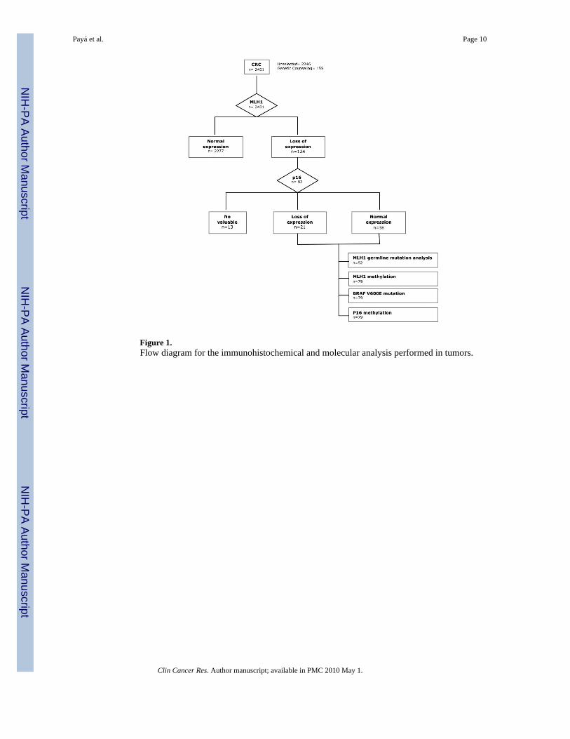
Figure 1.Flow diagram for the immunohistochemical and molecular analysis performed in tumors.
Payá et al. Page 10
Clin Cancer Res. Author manuscript; available in PMC 2010 May 1.
NIH
-PA Author Manuscript
NIH
-PA Author Manuscript
NIH
-PA Author Manuscript
Payá et al. Page 11
Clin Cancer Res. Author manuscript; available in PMC 2010 May 1.
NIH
-PA Author Manuscript
NIH
-PA Author Manuscript
NIH
-PA Author Manuscript
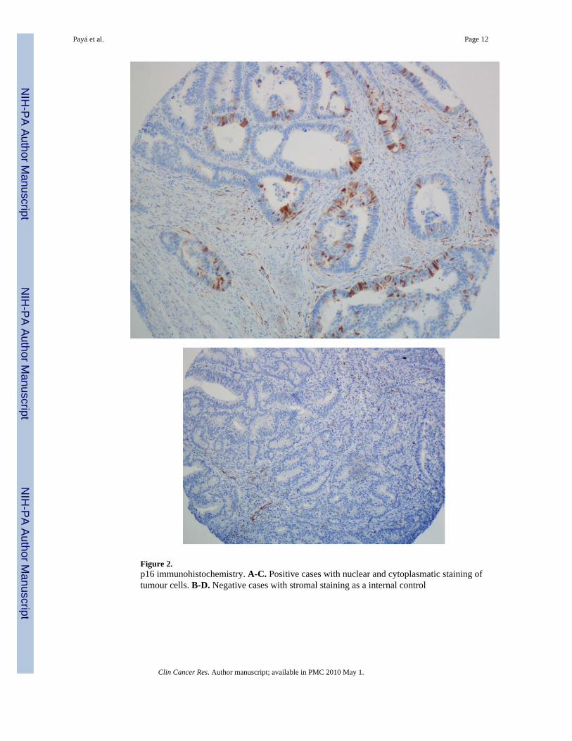
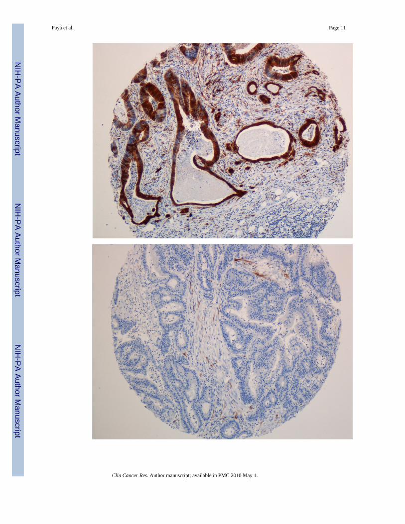
Figure 2.p16 immunohistochemistry. A-C. Positive cases with nuclear and cytoplasmatic staining oftumour cells. B-D. Negative cases with stromal staining as a internal control
Payá et al. Page 12
Clin Cancer Res. Author manuscript; available in PMC 2010 May 1.
NIH
-PA Author Manuscript
NIH
-PA Author Manuscript
NIH
-PA Author Manuscript
NIH
-PA Author Manuscript
NIH
-PA Author Manuscript
NIH
-PA Author Manuscript
Payá et al. Page 13
Tabl
e 1
Cha
ract
eris
tics o
f tum
ors r
egar
ding
p16
exp
ress
ion
P16
norm
alex
pres
sion
(n=5
8)P1
6 lo
ss o
fex
pres
sion
(n=2
1)T
OT
AL
(n=7
9)O
dds r
atio
p
me
P 25-
P 75
Me
P 25-
P 75
me
P 25-
P 75
Age
at d
iagn
osis
7352
-80
7769
-79
7550
-80
N.S
.
n%
N%
n%
Age
at d
iagn
osis
<5
0 ye
ars
1424
.12
9.5
1620
.33.
0 (0
.6-1
4.6)
N.S
.
>5
0 ye
ars
4475
.919
90.5
6379
.7
Sex
M
ale
2746
.68
38.1
3544
.31.
4 (0
.5-3
.9)
N.S
.
Fe
mal
e31
53.4
1361
.944
55.7
Rev
ised
Bet
hesd
a gu
idel
ines
Fu
lfilli
ng31
53.4
523
.836
45.6
3.7
(1.2
-11.
4)<0
.05
N
ot fu
lfilli
ng27
46.6
1676
.243
54.4
Am
ster
dam
II c
riter
ia
Fu
lfil
1119
.01
4.8
1215
.24.
7 (0
.6-3
8.7)
N.S
.
N
o fu
lfil
4781
.020
95.2
6784
.8
Tum
our l
ocat
ion
R
ight
-sid
ed47
81.0
1571
.462
78.5
1.8
(0.6
-5.8
)N
.S.
Le
ft-si
ded
1119
.06
28.6
1721
.5
Gra
de
Po
orly
diff
eren
ciat
ed37
63.8
942
.946
58.2
2.3
(0.9
-6.5
)N
.S.
O
ther
2136
.212
57.1
3341
.8
Met
hyla
tion
p16
Clin Cancer Res. Author manuscript; available in PMC 2010 May 1.
NIH
-PA Author Manuscript
NIH
-PA Author Manuscript
NIH
-PA Author Manuscript
Payá et al. Page 14
P16
norm
alex
pres
sion
(n=5
8)P1
6 lo
ss o
fex
pres
sion
(n=2
1)T
OT
AL
(n=7
9)O
dds r
atio
p
me
P 25-
P 75
Me
P 25-
P 75
me
P 25-
P 75
N
ot m
ethy
late
d38
65.5
00
3848
.1-
<0.0
01
M
ethy
late
d20
34.5
2110
041
51.9
BR
AF
V60
0E m
utat
ion
N
ot m
utat
ed41
70.7
628
.647
59.5
5.6
(1.9
-17.
1)<0
.005
M
utat
ed17
29.3
1571
.432
40.5
Met
hyla
tion
MLH
1
N
ot m
ethy
late
d28
48.3
14.
829
36.7
18.7
(2.3
-148
.4)
<0.0
01
M
ethy
late
d30
51.7
2095
.250
63.3
Me:
med
ian;
- Odd
s rat
io o
f p16
can
not b
e ca
lcul
ated
.
Clin Cancer Res. Author manuscript; available in PMC 2010 May 1.
NIH
-PA Author Manuscript
NIH
-PA Author Manuscript
NIH
-PA Author Manuscript
Payá et al. Page 15
Tabl
e 2
Cha
ract
eris
tics o
f tum
ours
in p
atie
nts w
ith g
erm
line
mut
atio
n.
Ger
mlin
e m
utat
ion
Yes
(tum
ours
n=1
0)(p
atie
nts n
=8)
No
(tum
ours
n=4
4)(p
atie
nts n
=44)
n%
n%
P
IHC
p16
N
orm
al e
xpre
ssio
n10
100
3069
.8<0
.05
Lo
ss o
f exp
ress
ion
00
1430
.2
Met
hyla
tion
p16
N
ot m
ethy
late
d8
8020
46.5
0.05
6
M
ethy
late
d2
2024
53.5
Met
hyla
tion
MLH
1
N
ot m
ethy
late
d10
100
920
.9<0
.001
M
ethy
late
d0
035
79.1
BR
AF
V60
0E m
utat
ion
N
ot m
utat
ed10
100
2555
.8<0
.01
M
utat
ed0
019
44.2
Bet
hesd
a gu
idel
ines
Fu
lfilli
ng10
100
2146
.5<0
.005
N
o fu
lfilli
ng0
023
53.5
Ám
ster
dam
II c
riter
ia
Fu
lfilli
ng7
704
9.3
<0.0
01
N
o fu
lfilli
ng3
3040
90.7
Me:
med
ian
Clin Cancer Res. Author manuscript; available in PMC 2010 May 1.
NIH
-PA Author Manuscript
NIH
-PA Author Manuscript
NIH
-PA Author Manuscript
Payá et al. Page 16
Tabl
e 3
Val
ues o
f diff
eren
t stra
tegi
es fo
r det
ectin
g ge
rmlin
e m
utat
ion
in tu
mou
rs w
ith M
LH1
loss
of e
xpre
ssio
n.
GE
RM
LIN
E M
UT
AT
ION
Stra
tegi
es w
ithou
t clin
ical
info
rmat
ion
Sen
Spe
PPV
NPV
+LR
NN
T
p16
IHC
100
30.2
2510
01.
43.
1
BR
AF
mut
atio
n10
044
.229
.410
01.
82.
3
p16
IHC
+ B
RA
F m
utat
ion
100
51.2
32.3
100
2.0
1.9
ML
H1
met
hyla
tion
100
79.1
52.6
100
4.8
1.4
BR
AF
mut
atio
n +
ML
H1
met
hyla
tion
100
79.1
52.6
100
4.8
1.3
p16
IHC
+ M
LH
1 m
ethy
latio
n10
079
.152
.610
04.
81.
2
p16
IHC
+ B
RA
F m
utat
ion
+ M
LH
1m
ethy
latio
n10
079
.152
.610
04.
81.
2
Stra
tegi
es n
eedi
ng c
linic
alin
form
atio
n
BE
TH
ESD
A10
053
.533
.310
02.
21.
9
p16
IHC
+ B
ET
HE
SDA
100
62,8
38,5
100
2.7
1.6
BR
AF
mut
atio
n +
BE
TH
ESD
A10
067
.441
.710
03.
11.
5
p16
IHC
+ B
RA
F m
utat
ion
+B
ET
HE
SDA
100
69.8
43.5
100
3.3
1.4
BR
AF
mut
atio
n +
BE
TH
ESD
A +
ML
H1
met
hyla
tion
100
83.7
58.8
100
6.1
1.2
BE
TH
ESD
A +
ML
H1
met
hyla
tion
100
83.7
58.8
100
6.1
1.2
p16
IHC
+ B
RA
F m
utat
ion
+B
ET
HE
SDA
+ M
LH
1 m
ethy
latio
n10
083
.758
.810
06.
11.
2
Sen:
Sen
sitiv
ity, S
pe: S
peci
ficity
, PPV
: Pos
itive
Pre
dict
ive
Val
ue, N
PV: N
egat
ive
Pred
ictiv
e V
alue
, +L
R: P
ositi
ve L
ikel
ihoo
d R
atio
, NN
T: N
umbe
r Nee
ded
to T
est f
or d
etec
ting
one
germ
line
mut
atio
n
Clin Cancer Res. Author manuscript; available in PMC 2010 May 1.