differential expression of p16 ink4aprotein and s100a4 protein in gastric carcinoma
TRANSCRIPT
Differential expression of p16 INK4a protein and S100A4 protein in gastric carcinoma.
Ghada A. Abd El-Fattah (MD), Mohebat H. Gouda (MD), and AdelZ. El-Saediy (Ass.Prof.) Pathology Department, Faculty of
Medicine, Benha University
Abstract : PURPOSE: Gastric cancer (GC) is an extremely commondisease worldwide. The p16 INK4a protein, is a tumorsuppressor protein that inhibits CDK4 and CDK6, whichphosphorylate the RB protein. S100A4 is known to beinvolved in cancer cell motility by virtue of its abilityto activate non-muscle myosin. The present study aims atinvestigating role of p16 INK4a, and S100A4 in progressionof gastric carcinoma by analyzing p16 INK4a and S100A4protein expression in gastric carcinomas and correlatingtheir expression with clinicopathological findings.
Patients and Methods: Forty cases including 30 non-consecutiveretrospective selected gastric carcinomas (4 GI, 16 GII, 10 GIII), and 10 cases of normal gastric tissue at resection margins of peptic ulcers, were taken as control. Cases were collected in the period 2009-2012, selected from files of pathology department, Faculty of Medicine- Benha University and Egyptian National Cancer Institute (NCI). Follow up of theselected cases was recorded for 18 months. Correlations between S100A4 and p16 INK4a immunoreactivity and clinicopathological characteristics were evaluated.
Results: showed significant inverse correlation between p16 INK4a expression and grade of carcinoma (P < 0.05) as well as high significant correlation with type of gastriccarcinoma (P < 0.01). S100A4 was positively correlated with tumor grade, lymph node metastasis, distant metastasis and tumor-node-metastasis (TNM) staging
1
(P<0.05). S100A4 expression was also significantly correlated with poor patient survival. The worst survivalwas correlated to cases with low p16INK4a/high S100A4 expression (P < 0.01).
Conclusion: The immunohistochemical expression of bothp16INK4a and S100A4 in gastric carcinoma is associatedsignificantly with tumor grade. Expression of S100A4 issignificantly associated with lymph node and distantmetastases, and poor prognosis. Estimation of bothmarkers can be used in planning the therapy and patient’sfollow up.
Key words:
p16INK4a, S100A4, tumor suppressor gene (TSG), gastric carcinoma (GC), immunohistochemistry (IHC).
Introduction
Gastric cancer is the fourth most common
cancer in the world with relative frequency of
7.8% of all cancers. It is the second leading
cause of cancer-related mortality worldwide,
accounting for 11.3% of cancer deaths (de Martel
C et al., 2012). In Egypt, cancer stomach is in
the eleventh rank constituting 2.1% of all
cancers with male to female ratio 1.55 and median
age of 53 year (El-Bolkainy et al., 2013).
2
However, the worldwide incidence of gastric
cancer has declined rapidly over the recent few
decades. Part of the decline may be due to the
recognition of certain risk factors such as H.
pylori and other dietary and environmental risks
(Hannah, et al; 2012).
The current staging classifications of
gastric carcinomas do not produce accurate
predictions of patient outcomes. Molecular
biomarkers may account for this diversity and
several prognostic factors have been identified
(Fareed, et al; 2009). However, none of these
methods have been proven to be robust enough to
be incorporated into routine practice.
A critical point in the cell cycle is the
G1/S transition checkpoint which is frequently
altered in tumors. This is controlled by cyclins
and cyclin-dependent kinases (CDK), which complex
to induce the progression of cells into the S-
phase by phosphorylating retinoblastoma protein.
INK4 comprising p16INK4a, p15INK4B, p18INK4C, and
p19INK4D are family of CDK inhibitors and binds
3
specifically to CDK4 and CDK6, thereby preventing
kinase activities (Hanan, et al; 2009).
P16, a 156-amino acid protein, is encoded by
the INK4A (CDKN2A, MTS1) gene located on
chromosome 9p21. It exerts its function by
competing with cyclin D in binding to CDK4
preventing the activation of this kinase and
inhibiting its productive interaction. Thus, p16
protein can specifically associate with CDK4 and
disrupt the formation of active kinase complexes
preventing transition of cells from G1 to S phase
(Osanai, et al; 2011). Therefore, inactivation of
p16INK4a leads to activation of cyclin/CDK
complexes, resulting in cell cycle progression.
Alterations of p16INK4a gene are known to occur in
many primary tumors (Hannah, et al; 2012).
S100A4 protein is the well-studied member of
S100 family. It is localized in the nucleus,
cytoplasm, and extracellular space and
possesses a wide range of biological
functions. It appears to take part in the
homeostasis of growth with apparent
involvement in growth factor signal transduction
4
and apoptotic cell death (House et al., 2011).
Overexpression of the S100 calcium-binding
protein A4(S100A4) is involved in
epithelial-to-mesenchymal transition, oncogenic
transformation, angiogenesis, cytoskeletal
integrity and cancer metastasis. ( Cabezo´n et
al., 2007 & Stein, et al ,2011 and Zhao, et al,
2013).S100A4 is also named as metastasis-
associated Mts1 and fibroblast-specific protein 1
(FSP1) because of its close association with
cancer metastasis and constitutive expression in
fibroblasts ( Malashkevich et al., 2008).
Therefore, S100A4 protein expression is
associated with patient outcome in a number of
tumor types (House et al., 2011). The present
study aims at investigating role of p16 INK4a, and
S100A4 in progression of gastric carcinoma by
analyzing p16 INK4a and S100A4 protein expression
in gastric carcinomas and correlating their
expression with clinicopathological findings.
5
Materials and methods:
Clinical investigations and tissue samples:
The present study was based on 40 cases including
ten cases of normal gastric tissue at resection
margins of peptic ulcer (non-neoplastic lesion)
were taken as control and 30 non-consecutive
retrospective selected gastric carcinomas.
Carcinoma cases included 18 cases of
adenocarcinoma, 6 cases of mucinous carcinoma and
6 cases of signet ring carcinoma. Cases were
collected in the period 2009-2012. They were
selected from files of pathology department,
Faculty of Medicine- Benha University and
Egyptian National Cancer Institute (NCI). Cases
were selected according the availability of
clinical and follow-up data for at least 18
months. Only patients with primary gastric cancer
who had not undergone previous irradiation or
chemotherapeutic treatment were included in the
study.
Staging was carried out according to the TNM
Classification System (Rosai, 2011).
6
Formalin-fixed, paraffin-embedded gastric
tissues were used. Three sections of 4 micron
thickness were obtained from each case. One
section was H & E stained for diagnosis and
grading. Two experienced pathologists blindly and
independently confirmed the histological
diagnosis of each gastric lesion and agreed on
the grading. Other sections were mounted on
positively- charged slides, immunohistochemically
stained with antibodies against S100A4 and p16 INK4a using the Ultra Vision Detection System
(Anti-polyvalent, HRP/DAB, ready-to-use, Lab
Vision corporation).
Immunohistochemical staining: tissue sections
mounted on positively- charged slides, were
heated at 60°C for 30 minutes then deparaffinized
and rehydrated through a series of xylene and
alcohol before staining. After antigen retrieval
with microwave treatment in 10mM citrate buffer
(Neo-Markers, Cat. ♯ AP-9003), pH 6.0, endogenous
peroxidase was blocked with 3% hydrogen peroxide
for 20 minutes. Sections were washed 3 times with
cold 0.01 M phosphate buffered saline (PBS).
After blocking with 10% normal rabbit serum,
7
sections were incubated with polyclonal antibody
against p16INK4a (clone 16P07 Neomarkers; dilution
1:100) and pre-diluted ready to use S100A4
(Neomarker, LAB VISION, USA). Both were incubated
overnight at 4° C. The prepared DAB-substrate-
chromogen solution was applied and incubated for
5-15 minutes until color intensity has been
reached. Lastly, sections were counterstained
with Mayer's Hematoxylin. Samples of endometrial
adenocarcinomas known to overexpress p16 INK4a were
used as positive controls. A tissue section of
breast cancer was used as positive control for
S100A4. Negative controls were performed by
omitting the primary antibody step.
Interpretation and evaluation of p16INK4a
immunohistochemical staining: The stains were
interpreted as positive if there was distinct
diffuse cytoplasmic or nuclear reactivity in the
neoplastic cells, greater than any background
staining in the matched non-neoplastic gastric
tissue, in more than 20% of the cells within the
different cases (Chen, et al, 2013)
8
Interpretation and evaluation of S100A4
immunohistochemical staining: S100A4 was stained
yellow-brown in the cytoplasm and nucleus. The
degree of immunostaining based on the staining
intensity and percentage of positive cells. The
intensity grading scale was according to the
following criteria: 0 (no staining), 1 (weak
staining, light yellow), 2 (moderate staining
yellow-brown) and 3 (strong staining, brown).
Moderate and strong staining indicated tumors
with high S100A4 expression, while no and weak
staining indicated low S100A4 expression (Li, et
al, 2013).
Statistical analysis: Statistical analysis wasperformed using the SPSS (version 16.0 for windows) software package according to Sperman's correlation coefficient. Correlation between several variables was computed using Fisher's exact test. P value less than 0.05 (<0.05) was considered significant and <0.01 was highly significant.
Results
A- Immunohistochemical results of p16 INK4a
staining":
9
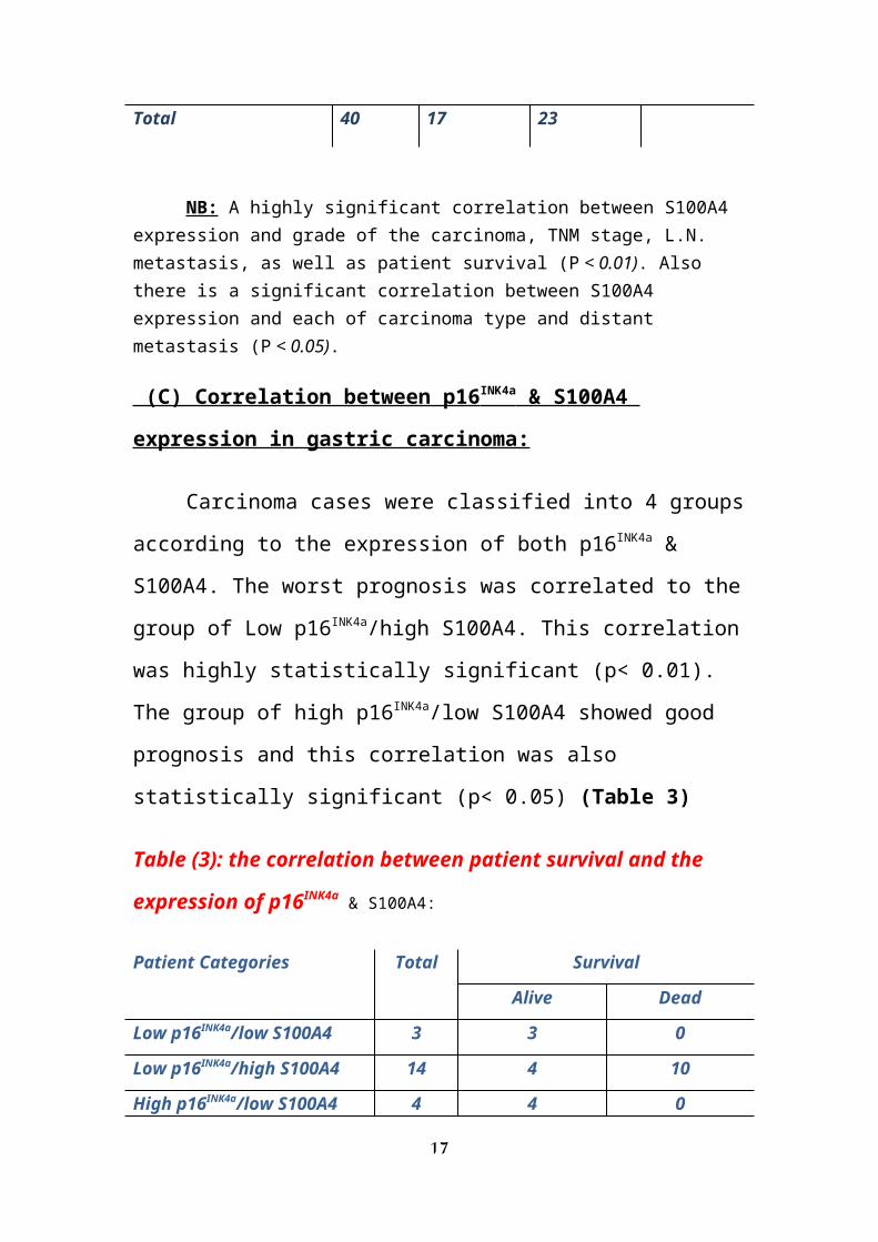
All 10 cases (100%) of normal gastric tissue
showed positive cytoplasmic expression of p16INK4a,
while among 30 cases of gastric carcinoma, 13
cases (43.3%) were positive and 17 cases (56.7%)
were negative. This difference between expression
of p16INK4a in normal and malignant neoplastic
gastric tissue was statistically highly
significant (P<0.01) (Fig 1).
As regarding the type of cancer, 11 cases
(61.1%) of adenocarcinoma were positive for
p16INK4a, while all cases (100%) of signet ring
carcinoma showed negative expression. Out of 6
mucinous carcinomas, only 2 cases (33.3%) were
positive (P<0.01).
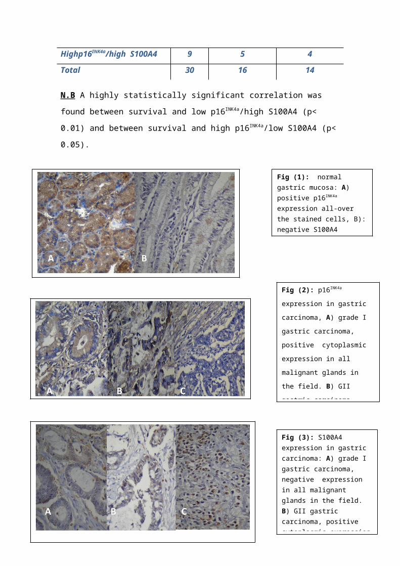
In relation to the tumor grade, all cases
(100%) of gastric carcinoma grade 1 showed
positive p16INK4a expression. Among cases of grade
II, 7 out of 16 cases (43.75%) were positive, and
9 cases (56.25%) were negative. Among the 10
cases of gastric carcinoma GIII, only 2 cases
(20%) were positive for p16INK4a expression and 8
cases (80%) were negative and this relationship
is statistically significant (P <0.05) (Fig 2).
10
All stage I cases (100%) showed positive
p16INK4a expression, while only one case (12.5%) of
stage II was positive. Six cases (75%) of stage
III and 2 cases (20%) of stage IV were positive
for p16INK4a expression, this relationship is
statistically insignificant. (P>0.05).
- Concerning to state of lymph node
metastasis, 4 out of the 10 lymph node negative
cases (40%) showed positive p16INK4a expression,
while positivity was seen in 9 of the 20 lymph
node positive cases (45%). This relationship was
statistically insignificant (P>0.05).
Insignificant correlation was also detected
between p16INK4a expression and distant metastasis
(P>0.05). Eleven (55%) out of 20 metastasis free
cases were positive for p16INK4a expression and 2
(20%) out of 10 cases with distant metastasis
were positive.
Correlation with patient survival was also
statistically insignificant (P>0.05). Half of the
cases of living patients (50%) showed positive
p16INK4a expression and 5 dead cases were positive
(35.7%). (Table 1).
11
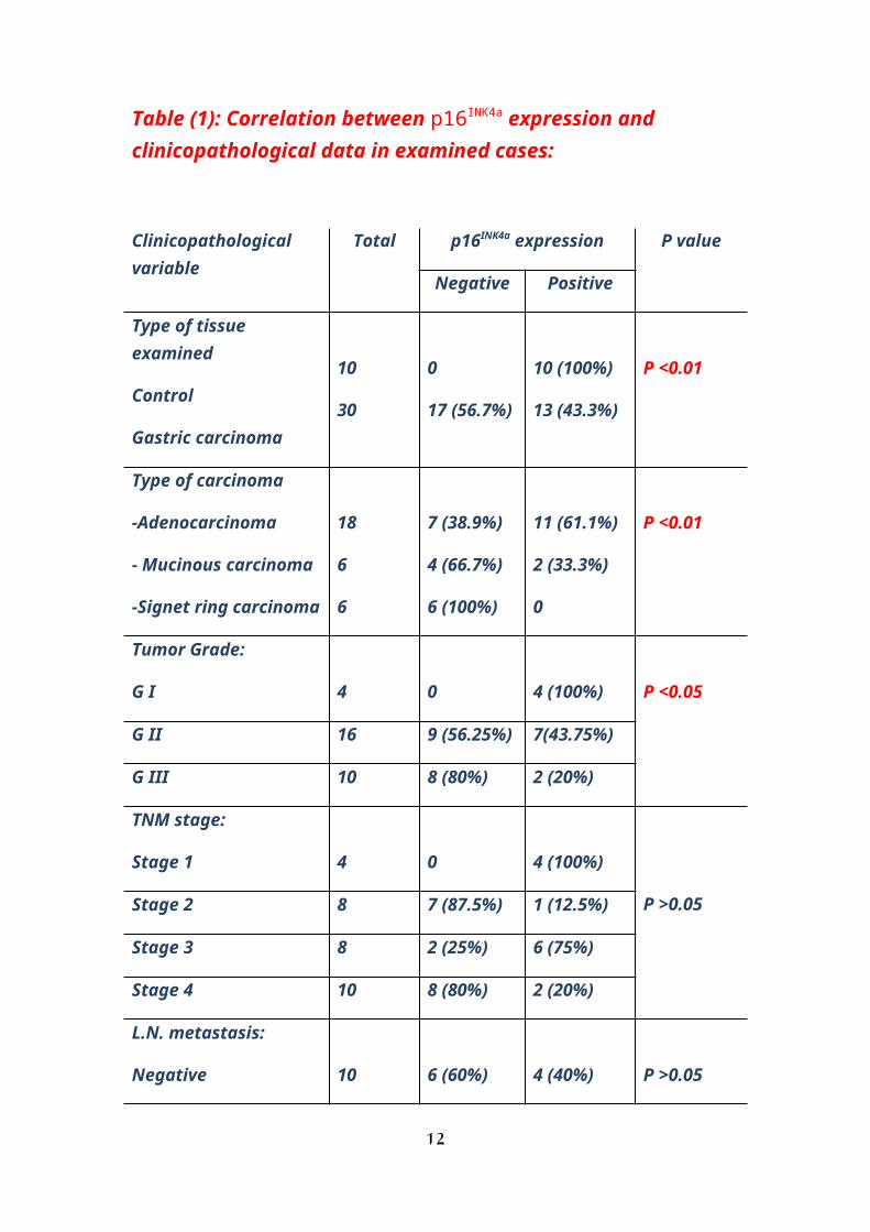
Table (1): Correlation between p16INK4a expression and clinicopathological data in examined cases:
Clinicopathological variable
Total p16INK4a expression P value
Negative Positive
Type of tissue examined
Control
Gastric carcinoma
10
30
0
17 (56.7%)
10 (100%)
13 (43.3%)
P <0.01
Type of carcinoma
-Adenocarcinoma
- Mucinous carcinoma
-Signet ring carcinoma
18
6
6
7 (38.9%)
4 (66.7%)
6 (100%)
11 (61.1%)
2 (33.3%)
0
P <0.01
Tumor Grade:
G I 4 0 4 (100%) P <0.05
G II 16 9 (56.25%) 7(43.75%)
G III 10 8 (80%) 2 (20%)
TNM stage:
Stage 1 4 0 4 (100%)
P >0.05Stage 2 8 7 (87.5%) 1 (12.5%)
Stage 3 8 2 (25%) 6 (75%)
Stage 4 10 8 (80%) 2 (20%)
L.N. metastasis:
Negative 10 6 (60%) 4 (40%) P >0.05
12
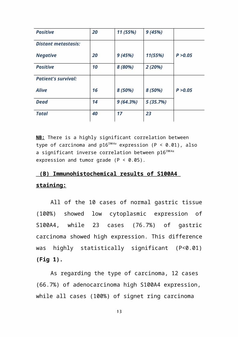
Positive 20 11 (55%) 9 (45%)
Distant metastasis:
Negative 20 9 (45%) 11(55%) P >0.05
Positive 10 8 (80%) 2 (20%)
Patient's survival:
Alive 16 8 (50%) 8 (50%) P >0.05
Dead 14 9 (64.3%) 5 (35.7%)
Total 40 17 23
NB: There is a highly significant correlation between type of carcinoma and p16INK4a expression (P < 0.01), also a significant inverse correlation between p16INK4a expression and tumor grade (P < 0.05).
(B) Immunohistochemical results of S100A4
staining:
All of the 10 cases of normal gastric tissue
(100%) showed low cytoplasmic expression of
S100A4, while 23 cases (76.7%) of gastric
carcinoma showed high expression. This difference
was highly statistically significant (P<0.01)
(Fig 1).
As regarding the type of carcinoma, 12 cases
(66.7%) of adenocarcinoma high S100A4 expression,
while all cases (100%) of signet ring carcinoma
13
showed low expression. Among 6 cases of mucinous
carcinoma only 1 case (16.7%) was reported with
low expression of S100A4 (P< 0.05).
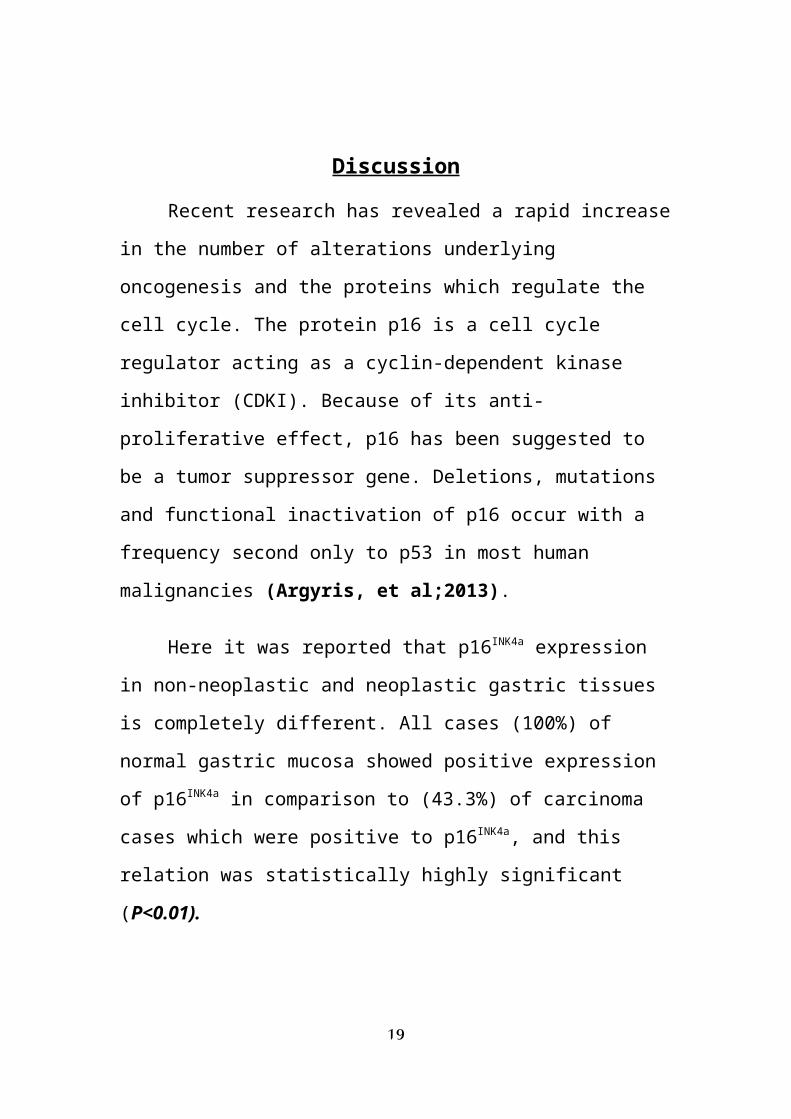
In relation to the tumor grade, the
expression of S100A4 was increased together with
loss of differentiation. Only one case (25%) of
grade 1 showed high expression while 12 cases of
G2 (75%) and all cases of G3 (100%) showed high
expression, and this relationship is
statistically highly significant (P< 0.05) (Fig
3).
As regarding the stage of gastric carcinoma,
one case (25%) of stage I showed high expression.
In stage II and III, high S100A4 expression was
shown in 6 cases (75%). All cases (100%) of stage
IV cases showed high S100A4 expression. This
relationship is statistically highly significant.
(P<0.05).
- Concerning the state of lymph node , S100A4
expression is markedly among cases with positive
L.N. metastases. All of these cases, (100%)
showed high expression. In the 10 lymph node
negative cases (70%) showed low S100A4
14
expression. This relationship was statistically
highly significant (P<0.01).
- All cases (100%) of those with distant
metastasis showed high expression. In absence of
distant metastasis, 13 cases (65%) showed high
expression. A statistically significant
correlation was found between S100A4 expression
and occurrence of distant metastasis (P < 0.05).
Regarding patient survival, the expression of
S100A4 was related to poor prognosis. All of dead
cases (100%) showed high expression, while 9
cases (56.25%) of living patients showed high
expression. This was a statistically highly
significant relationship (P < 0.01). (Table 2).
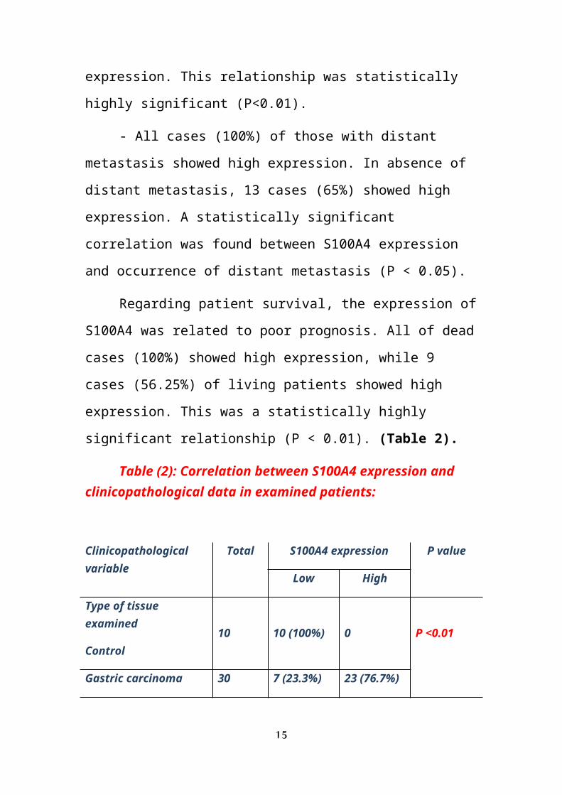
Table (2): Correlation between S100A4 expression and clinicopathological data in examined patients:
Clinicopathological variable
Total S100A4 expression P value
Low High
Type of tissue examined
Control 10 10 (100%) 0 P <0.01
Gastric carcinoma 30 7 (23.3%) 23 (76.7%)
15
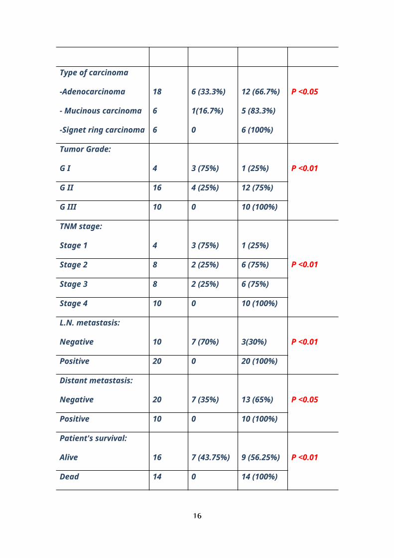
Type of carcinoma
-Adenocarcinoma
- Mucinous carcinoma
-Signet ring carcinoma
18
6
6
6 (33.3%)
1(16.7%)
0
12 (66.7%)
5 (83.3%)
6 (100%)
P <0.05
Tumor Grade:
G I 4 3 (75%) 1 (25%) P <0.01
G II 16 4 (25%) 12 (75%)
G III 10 0 10 (100%)
TNM stage:
Stage 1 4 3 (75%) 1 (25%)
P <0.01Stage 2 8 2 (25%) 6 (75%)
Stage 3 8 2 (25%) 6 (75%)
Stage 4 10 0 10 (100%)
L.N. metastasis:
Negative 10 7 (70%) 3(30%) P <0.01
Positive 20 0 20 (100%)
Distant metastasis:
Negative 20 7 (35%) 13 (65%) P <0.05
Positive 10 0 10 (100%)
Patient's survival:
Alive 16 7 (43.75%) 9 (56.25%) P <0.01
Dead 14 0 14 (100%)
16
Total 40 17 23
NB: A highly significant correlation between S100A4 expression and grade of the carcinoma, TNM stage, L.N. metastasis, as well as patient survival (P < 0.01). Also there is a significant correlation between S100A4 expression and each of carcinoma type and distant metastasis (P < 0.05).
(C) Correlation between p16 INK4a & S100A4
expression in gastric carcinoma:
Carcinoma cases were classified into 4 groups
according to the expression of both p16INK4a &
S100A4. The worst prognosis was correlated to the
group of Low p16INK4a/high S100A4. This correlation
was highly statistically significant (p< 0.01).
The group of high p16INK4a/low S100A4 showed good
prognosis and this correlation was also
statistically significant (p< 0.05) (Table 3)
Table (3): the correlation between patient survival and the
expression of p16INK4a & S100A4:
Patient Categories Total Survival
Alive Dead
Low p16INK4a/low S100A4 3 3 0
Low p16INK4a/high S100A4 14 4 10
High p16INK4a/low S100A4 4 4 0
17
Highp16INK4a/high S100A4 9 5 4
Total 30 16 14
N.B A highly statistically significant correlation was
found between survival and low p16INK4a/high S100A4 (p<
0.01) and between survival and high p16INK4a/low S100A4 (p<
0.05).
18
Fig (1): normal gastric mucosa: A) positive p16INK4a expression all-over the stained cells, B):negative S100A4
Fig (2): p16INK4a
expression in gastric
carcinoma, A) grade I
gastric carcinoma,
positive cytoplasmic
expression in all
malignant glands in
the field. B) GII
gastric carcinoma,
Fig (3): S100A4 expression in gastric carcinoma: A) grade I gastric carcinoma, negative expression in all malignant glands in the field. B) GII gastric carcinoma, positive cytoplasmic expression
Discussion
Recent research has revealed a rapid increase
in the number of alterations underlying
oncogenesis and the proteins which regulate the
cell cycle. The protein p16 is a cell cycle
regulator acting as a cyclin-dependent kinase
inhibitor (CDKI). Because of its anti-
proliferative effect, p16 has been suggested to
be a tumor suppressor gene. Deletions, mutations
and functional inactivation of p16 occur with a
frequency second only to p53 in most human
malignancies (Argyris, et al;2013).
Here it was reported that p16INK4a expression
in non-neoplastic and neoplastic gastric tissues
is completely different. All cases (100%) of
normal gastric mucosa showed positive expression
of p16INK4a in comparison to (43.3%) of carcinoma
cases which were positive to p16INK4a, and this
relation was statistically highly significant
(P<0.01).
19

These results were in agreement with Osanai,
et al; (2011) who found that the
immunohistochemical expression of p16 was
observed in only 32.4% of the carcinoma cases.
Xiu-Sheng ,et al;(2001) found that the positive
rate of P16 protein expression in gastric
carcinoma was significantly lower than that in
normal gastric mucosa and dysplastic gastric
mucosa (P<0.05).
On the contrary, Tsujie, et al;(2000) who
reported that less than 10% of non-tumor gastric
mucosal cells were p16INK4a positive, whereas the
expression of p16INK4a in gastric cancer cells
varied widely from 0 to 100% (mean, 24.5%). Also
Rocco, et al; (2002) found that in non-cancerous
gastric tissues the immunostaining of p16 was
weak and limited to antral glands. Lesnikova, et
al; (2009) found that p16INK4a expression was not
seen in normal cervix tissue. Similarly Zhao, et
al;(2003) reported that the frequency of loss of
P16 protein expression in the gastric cancer
tissue, adjacent non-tumor tissue, and distal
normal tissue was 77.5 %, 55.0 % , and 17.5 % ,
20
respectively (P<0.005). This difference in the
results may be attributed to difference in the
type or grade of carcinoma cases in each study.
In the current study, a significant inverse
correlation (p<0,05) between p16INK4a expression
and grade of gastric carcinoma cases was found,
all cases of grade I, (43.75%) of grade II, and
(20%) of grade III gastric carcinoma cases
showed positive p16INK4a expression. These results
were parallel to results reported by Tsujie, et
al; (2000) who reported a clinicopathologic
survey indicated that a low or no expression of
p16INK4a was associated with poorly differentiated
carcinoma (p = 0.0133). Rocco, et al;(2002) found
that the intensity of immunostaining was
inversely related to the grade of differentiation
of these tumors. The loss of expression of p16INK4a
in high grades of carcinomas means loss of its
anti-proliferative activity. This may be an
important factor in uncontrolled gastric cell
proliferation and progression of the tumor
towards high grades.
21
On the contrary to the current results
Osanai, et al;(2011) reported that There was no
statistically significant relationship between
the immunohistochemical expression of p16 and the
degree of histological differentiation of tumor,
when analyzed the relation to immunoreactivity
score (p = 0.81). Also, Xiu-Sheng ,et al;(2001)
reported that the positive rate of P16 protein
expression in mucoid carcinoma 10.00% was
significantly lower than that in poorly
differentiated carcinoma 51.22%, undifferentiated
carcinoma 57.69% and signet ring cell carcinoma
62.50% (P<0.05). The key differences between
this study and similar previously reported IHC
studies are firstly, the number of cases
analyzed; secondly the absence of p16 in the
lowest grade of differentiation may reflect
clonal expansion of the cells with a more
aggressive phenotype.
As regarding type of carcinoma, positive
p16INK4a expression was reported in 61.1% of
adenocarcinomas, 33.3% of mucoid carcinomas,
while all cases of signet ring carcinoma were
22
negative. This was a highly statistically
significant correlation (p<0.01). These results
could be explained by the behavior of different
types of gastric carcinomas; 66.7% of mucinous
carcinomas which are considered a less
differentiated carcinomas and signet ring
carcinomas which are always considered a high
grade cancer, and this explains their negative
expression for p16 which is found to be negative
in high grade tumors.
Rocco ,et al, (2002) reported that the
overexpression of p16 seems to be a common event
in the development of both intestinal and diffuse
type of gastric cancer and it is likely that it
may be driven by features of the neoplastic
state. Also, Xiu-Sheng He , et al, (2001)
reported that the positive rate of P16 protein
expression in mucoid carcinoma 10.00% was
significantly lower than that in poorly
differentiated carcinoma 51.22%,
undifferentiated carcinoma 57.69% and signet ring
cell carcinoma 62.50% (P<0.05).
23
Regarding lymph node metastasis, positive
p16INK4a expression was reported in 40% of cases
with absent lymph node metastases and in 45% of
cases with positive lymph node metastases. This
relationship was statistically insignificant
(P>0.05).
Concerning distant metastases in this study,
80% of gastric carcinomas with distant metastases
were negative to p16INK4a expression and 55% of
cases without distant metastases were positive to
p16INK4a. This relationship was also statistically
insignificant (P>0.05).
As regard the stage of gastric carcinoma, all
stage I cases (100%) showed positive p16INK4a
expression, while (12.5%) of stage II cases were
positive, (75%) of stage III cases and (20%) of
stage IV cases were positive for p16INK4a
expression, this relationship is statistically
insignificant. (P > 0.05).
These results were supported by results
reported by Osanai, et al;(2011) who reported
that Statistical analysis showed no significant
24
relationship between staging and p16INK4a protein
expression (p = 0.485).
In this study, the correlation between p16INK4a
protein expression and patient’s survival was
statistically insignificant (p> 0.05). This
agrees with Tsujie, et al, (2000), who reported
that the level of p16 expression did not
correlate with patients' prognosis. Chen, et al,
(2013) also did not identify a correlation
between p16 levels and patient survival.
Different studies showed that S100A4 plays a
role in tumor growth , motility and invasion
suggesting that it is directly linked to the
progression of human carcinoma as in colorectal
carcinoma (Boye et al., 2010) and in prostatic
carcinoma (Yong-Wook et al.,2010). In this study,
it was reported that all cases (100%) of non-
neoplastic gastric tissue showed low S100A4
expression while (76.7%) of gastric carcinoma
cases showed high expression of S100A4 and this
difference was statistically significant
(P<0.01).
25
This agrees with Yonemura , et al, (2000) who
reported that S100A4 expression was detected in
51 (55%) of 92 primary gastric cancers and Li, et
al, (2013) who reported that 53 (62.35%) of
gastric carcinoma cases exhibited S100A4
overexpression, in which immunostaining was
observed in the cytoplasm or the nucleus of the
tumor cells.
In relation to the tumor grade, the
expression of S100A4 was increased together with
loss of differentiation. (25%) of grade 1 showed
high expression, (75%) of G2 and all cases of G3
(100%) showed high expression, and this
relationship is statistically highly significant
(P< 0.05).This agreed with Yonemura , et al,
(2000) who reported a strong relationship between
S100A4 expression and histological
differentiation of gastric adenocarcinomas. In
their meta-analysis on colorectal carcinoma, Liu,
et al, (2013) detected a higher S100A4 expression
with poor differentiation. This could be
explained by the highly variable feature of
S100A4 expression which might indicate the
26
influence(s) of cell cycle regulators, and
especially epigenetic factor(s) in the
transcription of this gene.
Concerning the state of lymph node
metastasis, we found that all cases with positive
L.N. showed high S100A4 expression. In the 10
lymph node negative cases, 7 cases (70%) showed
low expression. This relationship was
statistically highly significant (P<0.01).
Also in this study we reported strong S100A4
expression in 100% of cases with distant
metastasis. In absence of distant metastasis,
(65%) showed low S100A4 expression. A
statistically significant correlation was found
between S100A4 expression and occurrence of
distant metastasis (P < 0.05).
In this work, it was found that the S100A4
expression is increased in relation to the stage
of gastric carcinoma. (75%) of stage I showed low
expression while (100%) of stage IV cases showed
high expression. This relationship is
statistically highly significant. (P<0.01).
27
Comparing to other studies, Li, et al, (2013)
reported that S100A4 overexpression was closely
associated with the gastric LN metastasis
(P=0.000) and distant metastasis (P=0.024). Zhao,
et al, (2013) found that gastric S100A4 was
positively correlated with lymph node metastasis
and tumor-node-metastasis (TNM) staging (P<0.05).
The same were the results of Wang, et al, (2010)
who reported that gastric Expression of S100A4 in
gastric cancer is associated significantly with
lymph node and distant metastases
In the meta-analysis on colorectal cancer done
by Liu, et al, (2013), the results suggested a
significant association between high S100A4
expression and advanced TNM stage, as well as the
presence of lymph node metastasis. Pooled data
also suggested an evident trend towards higher
S100A4 expression with poor differentiation and
distant metastases.
As regarding endometrioid carcinoma, Xie, et
al, (2007) reported that S100A4 expression was
significantly higher in stage III and IV tumors
compared with stage I.
28
These results could be explained by the
function of S100A4. It stimulates cell motility,
invasion, angiogenesis and participates in the
regulation of cell death. Invasion and motility
is probably promoted through induction of
epithelial to mesenchymal transition EMT. Cell
motility, invasion, and angiogenesis all
contribute to stimulation of metastasis. S100A4
enhances the turnover of myosin IIA filaments at
the leading edge of migrating cells, resulting in
increased motility, which could contribute to an
increased metastatic capacity of cancer cells,
Then S100A4-stimulated plasmin activation may
also contribute to the observed activation of
MMP-2 and MMP- 13 which help tumor cells to
travel through the surrounding stroma (Boye and
Mælandsmo, 2010).
In this study, it was found that the expression
of S100A4 was related to poor prognosis. All of
dead cases (100%) showed high expression, while
(43.75%) of living patients showed low
expression. This was a statistically highly
significant relationship (P < 0.01).
29
In their studies on gastric carcinoma, Zhao,
et al, (2013) & Wang, et al, (2010) reported
S100A4 as a marker for poor prognosis. Also
Yonemura , et al, (2000) reported that patients
with S100A4-positive tumors survived
significantly poorer than did those with S100A4-
negative tumors
On other studies Liu, et al, (2013) detected
that more than twelve studies investigated the
relation between S100A4 and patient survival and
practically demonstrated a significant
association between S100A4 overexpression and
worse prognosis in CRC patients. Stein, et al
(2011) study was carried upon both colorectal and
gastric cancer and they reported that a high
S100A4 expression correlates with aggressive
tumor growth and poor prognosis in colorectal
cancer. Overexpression of S100A4 is also related
to aggressiveness and metastasis in gastric
cancer. This correlation to poor prognosis could
be explained by the strong correlation between
high S100A4 levels and advanced tumor stage.
30
On the contrary, in their study, Pedersen, et
al (2002) did not find any association between
the expression levels of S100A4 and clinical
outcome. But they thought that this conflict
occurred because they investigated snap-frozen,
acetone-fixed tumor biopsies, while others
examined archival formalin-fixed, paraffin-
embedded specimens. Such differences in
preservation and fixation could possibly affect
the results.
The classification of patients into 4 groups
according to expression of both markers reveals
that the worst prognosis was related to the group
of low p16INK4a/ high S100A4 expression and this
was highly statistically significant (p< 0.01).
This could be explained by the relation between
advanced tumor stage and high S100a4 levels plus
the relation between the negative p16INK4a and loss
of tumor differentiation.
In conclusion, this study suggests that p16INK4a is related to the differentiation of the gastric carcinoma while S100A4 upregulation is positivelyassociated with the growth, invasion, metastasis and differentiation of gastric carcinomas. p16INK4a
and S100A4 may be useful markers to predict
31
progression of gastric carcinoma, while S100A4 may be a promising marker for detection of progression, aggressive behavior and prognosis ofgastric carcinomas. Estimation of both markers could be valuable in planning the therapy and patient’s follow up.
References
Argyris PP, Kademani D, Pambuccian SE, et al;Comparison between p16 INK4A immunohistochemistry and human papillomaviruspolymerase chain reaction assay in oral papillary squamous cell carcinoma. J Oral Maxillofac Surg.;71(10):1676-82, 2013.
Boye K and Mælandsmo G: Mechanisms involved
in tumor progression. Am J of Pathol, Vol.
176, No. 2., 2010
Boye K, Nesland JM, Sandstad B,et al :
Nuclear S100A4 is a novel prognostic marker in
colorectal cancer. Eur J Cancer. ;46(16):2919-
25 .2010
Cabezo´n T, Celis JE, Skibshoj I, et
al:Expression of S100A4 by a variety of cell
32
types present in the tumor microenvironment of
human breast cancer. Int J Cancer, 121:1433–
1444.2007.
Chen Y, Chu H, and Lewis BC. p16 Stimulates
CDC42-Dependent Migration of Hepatocellular
Carcinoma Cells. PLoS One, 8(7), 2013.
de Martel C, Ferlay J, Franceschi S, et al.
Global burden of cancers attributable to
infections in 2008: a review and synthetic
analysis. Lancet Oncol; 13(6):607, 2012.
EL-Bolkainy M, Akram M, Gouda I, EL-Bolkainy
T, Badawy O. Pathology of Cancer,4thed,
gastrointestinal cancers;200:204, 2013.
Fareed KR, Kaye P, Soomro IN, et al.
Biomarkers of response to therapy in oesophago-
gastric cancer. Gut.;58:127–143., 2009.
Hanan M. Abd El-Moneim, and Dalia M. Abd El-
Rehim: Immunohistochemical and Molecular Study
of p16 INK4aExpression in Pituitary Adenoma.
Journal of the Egyptian Nat. Cancer Inst., Vol.
21, No. 4,: 351-360, 2009.
Hannah H. and Wong, PC: Immunohistochemical
features of the gastrointestinal tract tumors.
33
Journal of Gastrointestinal Oncology.Vol 3, No
3, 2012.
House RP., Maria et al : Two Functional
S100A4 Monomers Are Necessary for Regulating
Nonmuscle Myosin-IIA and HCT116 Cell Invasion.
Biochemistry; 50(32): 6920–6932, 2011.
Lesnikova I, Lidang M, Hamilton S: p16 as a
diagnostic marker of cervical neoplasia: a
tissue microarray study of 796 archival
specimens. Diagnostic Pathology , 4:22, 2009 .
Li H, Liu Z, Xu C, et al,. Overexpression of
S100A4 is closely associated with the
progression and prognosis of gastric cancer in
young patients. Oncol Lett.; 5(5): 1485–1490,
2013.
Liu Y, Tang W, Wang J, et al.
Clinicopathological and prognostic
significance of S100A4 overexpression in
colorectal cancer: a meta-analysis.
Diagnostic Pathology, 8: 181, 2013.
Malashkevich VN, Varney KM, Garrett SC, et
al: Structure of Ca2_-bound S100A4 and its
interaction with peptides derived from non-
34
muscle myosin-IIA. Biochemistry , 47:5111–
5126.2008.
Osanai MH; Edelweiss MI; Meurer L: Protein
p16 INK4aimmunohistochemical expression in
adenocarcinoma of the esophagus. ABCD, arq.
bras. cir. dig. vol.24 no.4, 2011.
Pedersen KB, Nesland JM, Fodstad Ø, et al:
Expression of S100A4, E-Cadherin, α- and β-
Catenin in breast cancer biopsies. Br. J.
Cancer, 87: 1281-1286, 2002.
Rocco A, Schandl L, Nardone G,et al: Loss of
expression of tumor suppressor p16(INK4)
protein in human primary gastric cancer is
related to the grade of differentiation. Dig
Dis. ; 20(1):102-5, 2002.
Rosai J: Gastrointestinal tract. In Rosai and
Ackerman's Surgical Pathology, 10th ed. Mosby.
P: 627-635, 2011.
Stein U , Burock S , Herrmann P, et al.:
Diagnostic and prognostic value of metastasis
inducer S100A4 transcripts in plasma of
colon, rectal, and gastric cancer patients. J
Mol Diagn . 13(2):189-98. 2011.
35
Tsujie M, Yamamoto H, Tomita N, et al:
Expression of tumor suppressor gene p16
(INK4) products in primary gastric cancer.
Oncology ; 58(2):126-36, 2000.
Wang YY , Ye ZY, Zhao ZS, et al. High-level
expression of S100A4 correlates with lymph
node metastasis and poor prognosis in
patients with gastric cancer. Annals of
surgical oncology 17:1, 89-97, 2010.
Xie R, Loose DS, Shipley GL, et al.
Hypomethylation-induced expression of S100A4
in endometrial carcinoma. Modern Pathology ,
(20): 1045–1054, 2007.
Xiu-Sheng He, Qi Su, Zhu-Chu Chen:
Expression, deleton and mutation of p16 gene
in human gastric cancer. World J
Gastroenterol;7(4):515-521, 2001.
Yonemura Y, Endou Y, Kimura K, et al. Inverse
expression of S100A4 and E-Cadherin is
associated with metastatic potential in
gastric cancer. Clin Cancer Res., (6): 4234,
2000.
36
Yong-Wook K , In Ho C, Kyung Do K, et al:
Significance of S100A2 and S100A4 Expression
in the Progression of Prostate
Adenocarcinoma. Korean J Urol. ;51(7):456-62.
2010
Zhao GH, Li TC, Shi LH, et al. Relationship
between inactivation of p16 gene and gastric
carcinoma. World J Gastroenterol. ;9(5):905-
9, 2003.
Zhao Y , Zhang T, Wang Q.: S100 calcium-
binding protein A4 is a novel independent
prognostic factor for the poor prognosis of
gastric carcinomas. Oncol Rep 30(1):111-8.
2013.
37