unzipping and binding of small interfering rna with single
TRANSCRIPT
1
Unzipping and binding of small interfering RNA with single walled Carbon
Nanotube: a platform for small interfering RNA delivery
Mogurampelly Santosh1,3
, Swati Panigrahi2, Dhananjay Bhattacharyya
2, A. K. Sood
3 and Prabal
K Maiti *1,3
1 Centre for Condensed Matter Theory, Indian Institute of Science, Bangalore 560012, India.
2 Biophysics Division, Saha Institute of Nuclear Physics, Kolkata 700064
3Department of Physics, Indian Institute of Science, Bangalore 560012, India.
Abstract
In an effort to design efficient platform for siRNA delivery, we combine all atom classical and
quantum simulations to study the binding of small interfering RNA (siRNA) by pristine single wall
carbon nanotube (SWCNT). Our results show that siRNA strongly binds to SWCNT surface via
unzipping its base-pairs and the propensity of unzipping increases with the increase in the diameter
of the SWCNTs. The unzipping and subsequent wrapping events are initiated and driven by van der
Waals interactions between the aromatic rings of siRNA nucleobases and the SWCNT surface.
However, MD simulations of double strand DNA (dsDNA) of the same sequence show that the
dsDNA undergoes much less unzipping and wrapping on the SWCNT in the simulation time scale
of 70 ns. This interesting difference is due to smaller interaction energy of thymidine of dsDNA
with the SWCNT compared to that of uridine of siRNA, as calculated by dispersion corrected
density functional theory (DFT) methods. After the optimal binding of siRNA to SWCNT, the
complex is very stable which serves as one of the major mechanisms of siRNA delivery for
biomedical applications. Since siRNA has to undergo unwinding process with the effect of RNA-
induced silencing complex, our proposed delivery mechanism by SWCNT possesses potential
advantages in achieving RNA interference (RNAi).
Keywords: siRNA, CNT, Unzipping, Wrapping, van der Waals Interaction and MD Simulations.
*Correspoding Author: [email protected]
2
1. Introduction
RNA interference (RNAi) is a powerful technology for controlling the expression of genes in
biomedical applications. Small interfering ribonucleic acid (siRNA) molecules (typically 21 to 23
nucleotides) are being actively studied due to their potential influence on cell functionality and
applications in medicine 1,2
. RNAi is a cellular process in which a double stranded RNA (dsRNA)
reduces a specific gene expression. The base-pairing of siRNA with messenger RNA (mRNA)
sequence silences the encoded protein. The mechanism of RNAi involves RNA-induced silencing
complex (RISC) comprising of Dicer, Argonaute2 and siRNA binding protein that induces
unzipping of siRNA into two single strand RNAs 3-7
. One of these two strands acts as a guiding
strand to specifically base-pair with mRNA. Generally for efficient gene silencing, chemically
unmodified siRNAs are rapidly degraded in serum and hence siRNAs have to be bound with
transfecting carriers 8. We show the unwinding enhanced siRNA binding to carbon nanotube (CNT)
and propose siRNA delivery to target cell for achieving RNAi without degradation. Possible
transfecting carriers are linear or branched cationic polymers (dendrimers) 9-13
, cationic lipids 14,15
,
carbon nanotubes (CNTs) 16-19
, cell penetrating peptides 20,21
and few proteins 22,23
. Though siRNA
molecules are proved to be potential silencers of gene expression that can have extraordinary
treatment capabilities of HIV, hepatitis and cancer 24-30
, efficient delivery of these molecules to the
target cell is a big challenge today.
In this work, we address the question of using SWCNT as efficient carrier. CNTs functionalized
with polymers such as Polyethylene glycol (PEG), CONH-(CH2)(6)-NH3+Cl- or single stranded
DNA (ssDNA) are efficient transporters of siRNA into human T cells and primary cells 17,19,31-33
.
However, the structural and energetic changes taking place in siRNA while binding to the SWCNT
surface are not known. In comparison, wrapping of single stranded DNA (ssDNA) 34-37
and double
stranded DNA (dsDNA) 38
on the SWCNT surface have been studied recently. With this in view, we
have studied the thermodynamics and conformational properties of siRNA-SWCNT complex
through all atom molecular dynamics (MD) simulations and ab-initio quantum calculations.
2. Computational details
2.1 Molecular Dynamics Simulations
Molecular dynamics simulations were carried out using AMBER9 suite of programs 39
using the
AMBER 2003 (along with ff99) 40
force fields and the TIP3P model 41
for water. Latest
improvements of torsion angle parameters are reported for RNA in a new force field called
parmbsc0 and its variants 42,43
. However we have used ff99 for siRNA instead of parmbsc0 since the
3
conformational changes of siRNA reported here are not artefacts of the wrong torsion angle
parameterization in ff99. Further, we intend to compare the present results with our previous
simulations on siRNA that used ff99 11-13
. Notwithstanding this, we have also performed a few
simulations with parmbsc0 and obtained similar conformational changes as reported here which are
shown in the supplementary Fig. S1 and S2 44
and, therefore, conclude that the ff99 parameters are
reliable (for example see 45-47
). The initial structure of the siRNA was taken from the protein data
bank (PDB code: 2F8S) 48
. The sequence of the siRNA used is UUr(AGA CAG CAU AUA UGC
UGU CU)2UU with sticky ends of sequence UU on the two ends of the strands. We have built
armchair SWCNT of various diameters; (5, 5) (d = 6.68 Å), (6, 6) (d = 8.02 Å), (7, 7) (d = 9.36 Å)
and (8, 8) (d = 10.69 Å). We choose carbon nanotubes to be 142 Å in length to ensure sufficient
sliding length for the siRNA before stable binding. In the initial configuration, the siRNA is placed
on the nanotube surface such that the nanotube axis and siRNA helix axis are nearly parallel to each
other. The siRNA-SWCNT complexes were then solvated separately with TIP3P water box 41
of
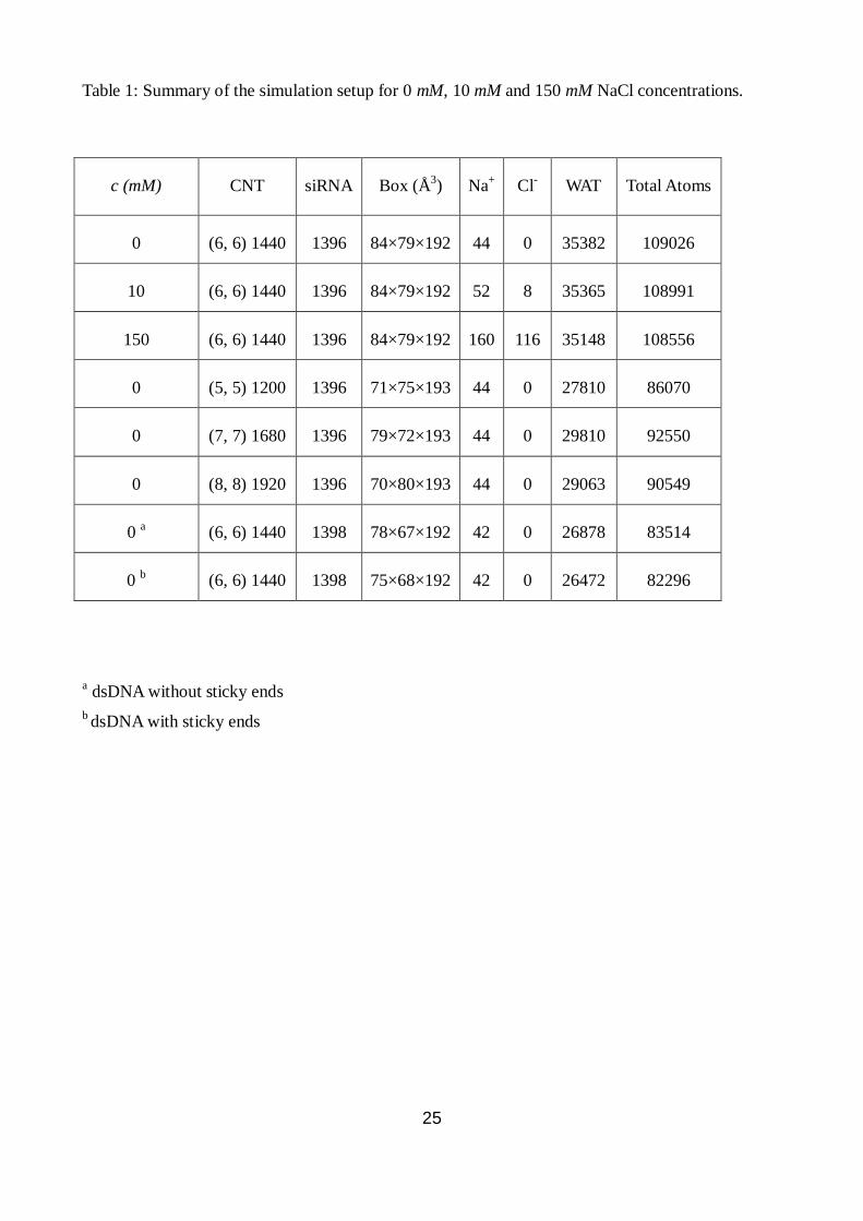
dimensions as shown in Table 1 using the LEaP module in AMBER9. The box dimensions were
chosen such that there is at least 20 Å solvation shell in all directions from the surface of siRNA-
CNT complex during the entire simulation. In addition, some water residues were replaced by 44
Na+ counterions to neutralize the negative charge on the phosphate backbone groups of the siRNA
structure. The initial system containing siRNA-(6, 6) CNT with added water and neutralizing
counterions is shown in Fig. 1. In separate simulation runs, additional NaCl residues were added to
prepare system at 10 mM and 150 mM salt concentrations. These three salt concentrations (0 mM,
10 mM and 150 mM) were studied in view of the importance of electrostatic screening in binding
mechanisms of siRNA with the SWCNT surface. For comparison, we have also simulated dsDNA
adsorption on (5, 5) and (6, 6) CNTs of the same length at 300 K. The dsDNA has the same length
and sequence as the siRNA where the nucleobase Uracil (U) is replaced by nucleobase Thymine
(T), i.e., TTd(AGA CAG CAT ATA TGC TGT CT)2TT. In another simulation, we choose a random
sequence of dsDNA as given by d(GCA TGA AAT GCT TAA AGC TTA C)2. The full details of the
various systems studied, number of NaCl residues, water residues, box dimensions and total number
of atoms are summarized in Table 1.
For simulating the CNT, carbon atoms are modelled as uncharged Lennard-Jones particles with sp2
hybridization according to the parameters from AMBER03 force field (type CA). In addition,
bonded interactions viz., stretching, torsion and dihedral terms were also included. We have used
the same force field for the CNTs earlier in the context of water transport through them 49
. To keep
the CNT fixed during simulations, all the atom positions were constrained with harmonic potential
4
of spring constant of 1000 kcal/mol-Å2. The translational centre of mass motions were removed
every 1000 steps. The long range electrostatic interactions were calculated with the Particle Mesh
Ewald (PME) method 50
using a cubic B-spline interpolation of order 4 and a 10-5
tolerance set for
the direct space sum cutoff. A real space cutoff of 9 Å was used both for the long range electrostatic
and short range van der Waals interaction with a non-bond list update frequency of 10. We have
used periodic boundary conditions in all three directions and the bond lengths involving bonds to
hydrogen atoms were constrained using SHAKE algorithm 51
. This constraint enabled us to use a
time step of 2 fs for obtaining a long trajectory. During the minimization, the siRNA-SWCNT
complex structures were fixed in their starting conformations using harmonic constraints with a
force constant of 500 kcal/mol-Å2. This allowed the water molecules to reorganize which eliminates
bad contacts with the siRNA and the CNT structures. The minimized structures were then subjected
to 40 ps of MD, using 1 fs time step for integration. During the constant volume - constant
temperature (NVT) MD, the system was gradually heated from 0 to 300 K using weak harmonic
restrains of 20 kcal/mol-Å2
on the solute to its starting structure. This allows slow relaxation of the
siRNA-CNT structure. Subsequently, simulations were performed under constant pressure -
constant temperature conditions (NPT), with temperature regulation achieved using the Berendsen
weak coupling method 52
(0.5 ps time constant for heat bath coupling and 0.5 ps pressure relaxation
time). NPT-MD was used to get the correct (experimental) solvent density. Finally, for analysis of
structures and properties, we have carried out 50-100 ns of NVT MD with 2 fs integration time step
using a heat bath coupling time constant of 1 ps. The trajectories were saved at a frequency of 2 ps.
2.2 Quantum Chemical Calculations
We have also carried out quantum chemical analysis with dispersion correction (DFT-D) to
understand the interactions of the CNT with the DNA and RNA. The valencies of the carbon atoms
at the ends of the (6, 6) CNT were satisfied by adding necessary hydrogen atoms. The structural
features that distinguish RNA from DNA are the presence of uracil base and 2’-OH groups of the
ribose sugars. The four systems modeled by us are as follows: (i) (6, 6) CNT with one uracil
nucleobase, (ii) (6, 6) CNT with one thymine nucleobase, (iii) (6, 6) CNT with one uridine
nucleoside (uracil attached with C3’-endo ribose sugar) and (iv) (6, 6) CNT with thymidine
nucleoside (thymine nucleobase attached with C2’-endo deoxyribose sugar) as shown in Fig. S3(a).
These initial structures were built using MOLDEN 53
software. Initially the nucleobases were
placed in parallel to the CNT surface. Free geometry optimization of all the four systems discussed
above were carried out using Gaussian09 54
without any constraints using density functional theory
with WB97XD/6-31g(d,p) basis set 55
, which includes dispersion correction, giving rise to energy
5
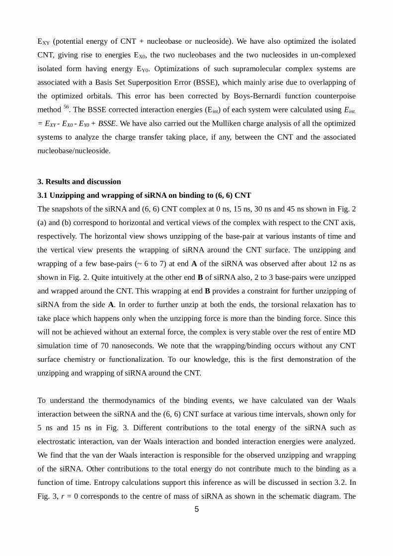
EXY (potential energy of CNT + nucleobase or nucleoside). We have also optimized the isolated
CNT, giving rise to energies EX0, the two nucleobases and the two nucleosides in un-complexed
isolated form having energy EY0. Optimizations of such supramolecular complex systems are
associated with a Basis Set Superposition Error (BSSE), which mainly arise due to overlapping of
the optimized orbitals. This error has been corrected by Boys-Bernardi function counterpoise
method 56
. The BSSE corrected interaction energies (Eint) of each system were calculated using Eint.
= EXY - EX0 - EY0 + BSSE. We have also carried out the Mulliken charge analysis of all the optimized
systems to analyze the charge transfer taking place, if any, between the CNT and the associated
nucleobase/nucleoside.
3. Results and discussion
3.1 Unzipping and wrapping of siRNA on binding to (6, 6) CNT
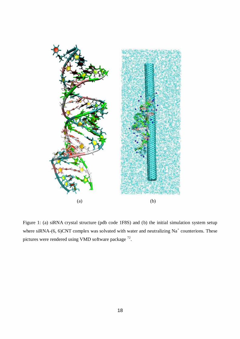
The snapshots of the siRNA and (6, 6) CNT complex at 0 ns, 15 ns, 30 ns and 45 ns shown in Fig. 2
(a) and (b) correspond to horizontal and vertical views of the complex with respect to the CNT axis,
respectively. The horizontal view shows unzipping of the base-pair at various instants of time and
the vertical view presents the wrapping of siRNA around the CNT surface. The unzipping and
wrapping of a few base-pairs (~ 6 to 7) at end A of the siRNA was observed after about 12 ns as
shown in Fig. 2. Quite intuitively at the other end B of siRNA also, 2 to 3 base-pairs were unzipped
and wrapped around the CNT. This wrapping at end B provides a constraint for further unzipping of
siRNA from the side A. In order to further unzip at both the ends, the torsional relaxation has to
take place which happens only when the unzipping force is more than the binding force. Since this
will not be achieved without an external force, the complex is very stable over the rest of entire MD
simulation time of 70 nanoseconds. We note that the wrapping/binding occurs without any CNT
surface chemistry or functionalization. To our knowledge, this is the first demonstration of the
unzipping and wrapping of siRNA around the CNT.
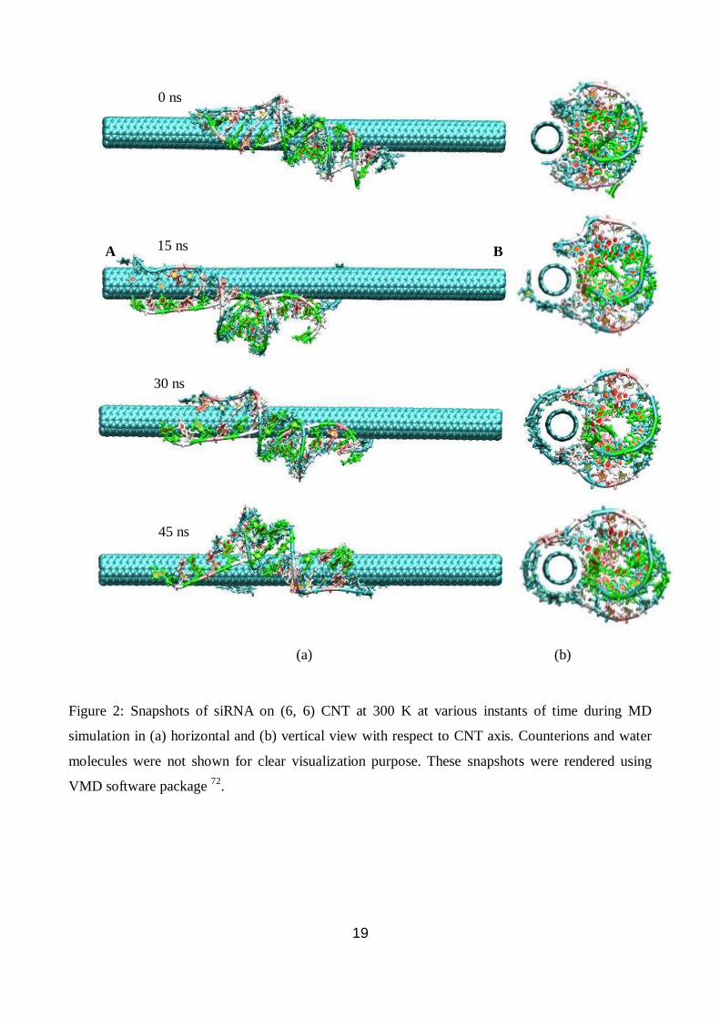
To understand the thermodynamics of the binding events, we have calculated van der Waals
interaction between the siRNA and the (6, 6) CNT surface at various time intervals, shown only for
5 ns and 15 ns in Fig. 3. Different contributions to the total energy of the siRNA such as
electrostatic interaction, van der Waals interaction and bonded interaction energies were analyzed.
We find that the van der Waals interaction is responsible for the observed unzipping and wrapping
of the siRNA. Other contributions to the total energy do not contribute much to the binding as a
function of time. Entropy calculations support this inference as will be discussed in section 3.2. In
Fig. 3, r = 0 corresponds to the centre of mass of siRNA as shown in the schematic diagram. The
6
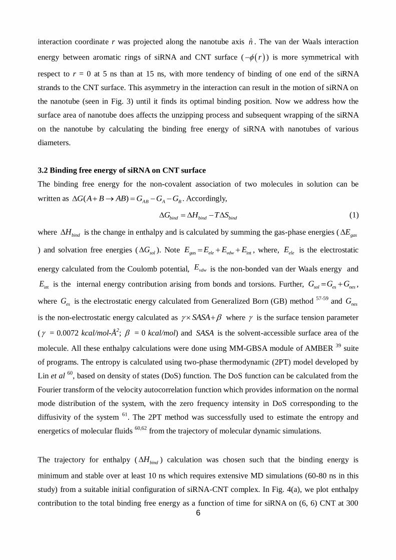
interaction coordinate r was projected along the nanotube axis n̂ . The van der Waals interaction
energy between aromatic rings of siRNA and CNT surface ( r ) is more symmetrical with
respect to r = 0 at 5 ns than at 15 ns, with more tendency of binding of one end of the siRNA
strands to the CNT surface. This asymmetry in the interaction can result in the motion of siRNA on
the nanotube (seen in Fig. 3) until it finds its optimal binding position. Now we address how the
surface area of nanotube does affects the unzipping process and subsequent wrapping of the siRNA
on the nanotube by calculating the binding free energy of siRNA with nanotubes of various
diameters.
3.2 Binding free energy of siRNA on CNT surface
The binding free energy for the non-covalent association of two molecules in solution can be
written as ( ) AB A BG A B AB G G G . Accordingly,
bind bind bindG H T S (1)
where bindH is the change in enthalpy and is calculated by summing the gas-phase energies ( gasE
) and solvation free energies ( solG ). Note intgas ele vdwE E E E , where, eleE is the electrostatic
energy calculated from the Coulomb potential, vdwE is the non-bonded van der Waals energy and
intE is the internal energy contribution arising from bonds and torsions. Further, sol es nesG G G ,
where esG is the electrostatic energy calculated from Generalized Born (GB) method 57-59
and nesG
is the non-electrostatic energy calculated as SASA where is the surface tension parameter
( = 0.0072 kcal/mol-Å2; = 0 kcal/mol) and SASA is the solvent-accessible surface area of the
molecule. All these enthalpy calculations were done using MM-GBSA module of AMBER 39
suite
of programs. The entropy is calculated using two-phase thermodynamic (2PT) model developed by
Lin et al 60
, based on density of states (DoS) function. The DoS function can be calculated from the
Fourier transform of the velocity autocorrelation function which provides information on the normal
mode distribution of the system, with the zero frequency intensity in DoS corresponding to the
diffusivity of the system 61
. The 2PT method was successfully used to estimate the entropy and
energetics of molecular fluids 60,62
from the trajectory of molecular dynamic simulations.
The trajectory for enthalpy ( bindH ) calculation was chosen such that the binding energy is
minimum and stable over at least 10 ns which requires extensive MD simulations (60-80 ns in this
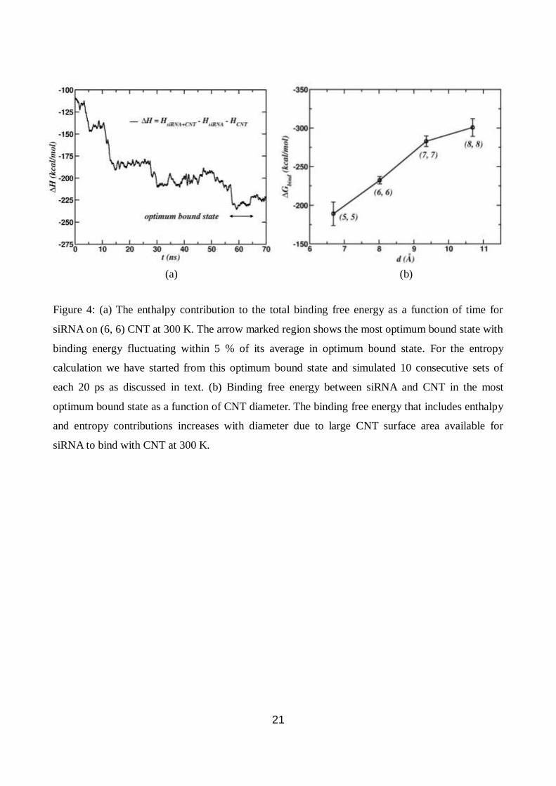
study) from a suitable initial configuration of siRNA-CNT complex. In Fig. 4(a), we plot enthalpy
contribution to the total binding free energy as a function of time for siRNA on (6, 6) CNT at 300
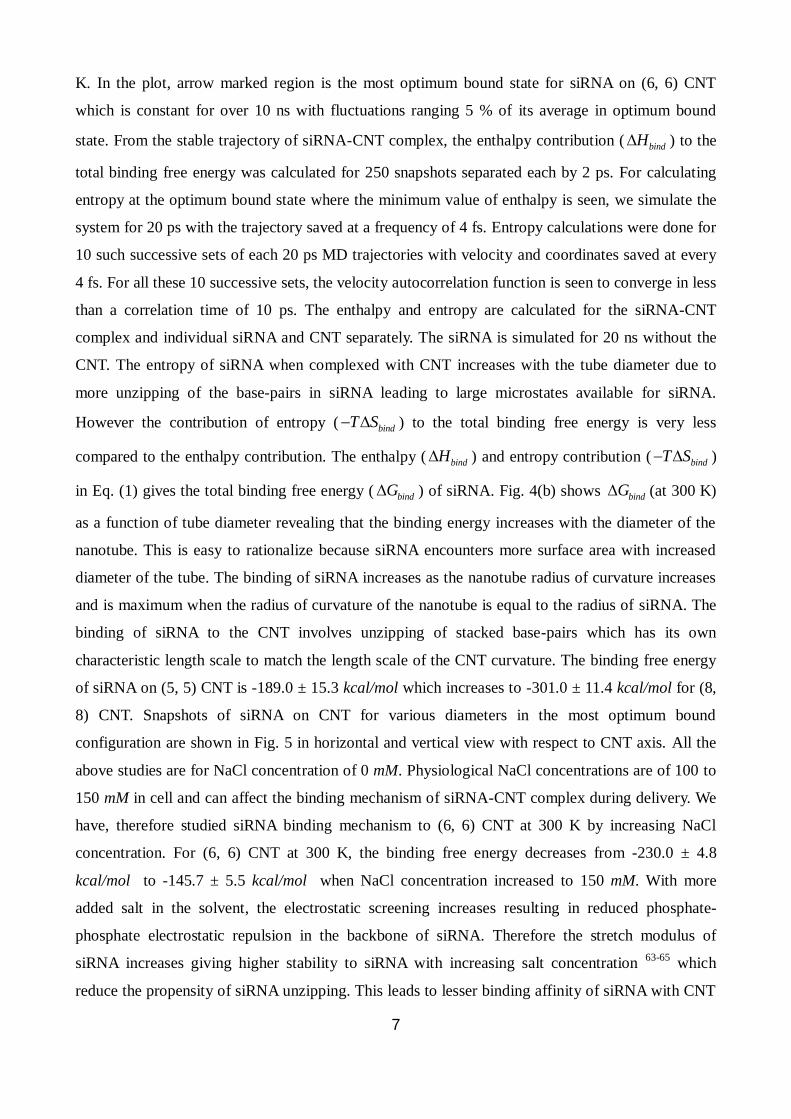
7
K. In the plot, arrow marked region is the most optimum bound state for siRNA on (6, 6) CNT
which is constant for over 10 ns with fluctuations ranging 5 % of its average in optimum bound
state. From the stable trajectory of siRNA-CNT complex, the enthalpy contribution ( bindH ) to the
total binding free energy was calculated for 250 snapshots separated each by 2 ps. For calculating
entropy at the optimum bound state where the minimum value of enthalpy is seen, we simulate the
system for 20 ps with the trajectory saved at a frequency of 4 fs. Entropy calculations were done for
10 such successive sets of each 20 ps MD trajectories with velocity and coordinates saved at every
4 fs. For all these 10 successive sets, the velocity autocorrelation function is seen to converge in less
than a correlation time of 10 ps. The enthalpy and entropy are calculated for the siRNA-CNT
complex and individual siRNA and CNT separately. The siRNA is simulated for 20 ns without the
CNT. The entropy of siRNA when complexed with CNT increases with the tube diameter due to
more unzipping of the base-pairs in siRNA leading to large microstates available for siRNA.
However the contribution of entropy ( bindT S ) to the total binding free energy is very less
compared to the enthalpy contribution. The enthalpy ( bindH ) and entropy contribution ( bindT S )
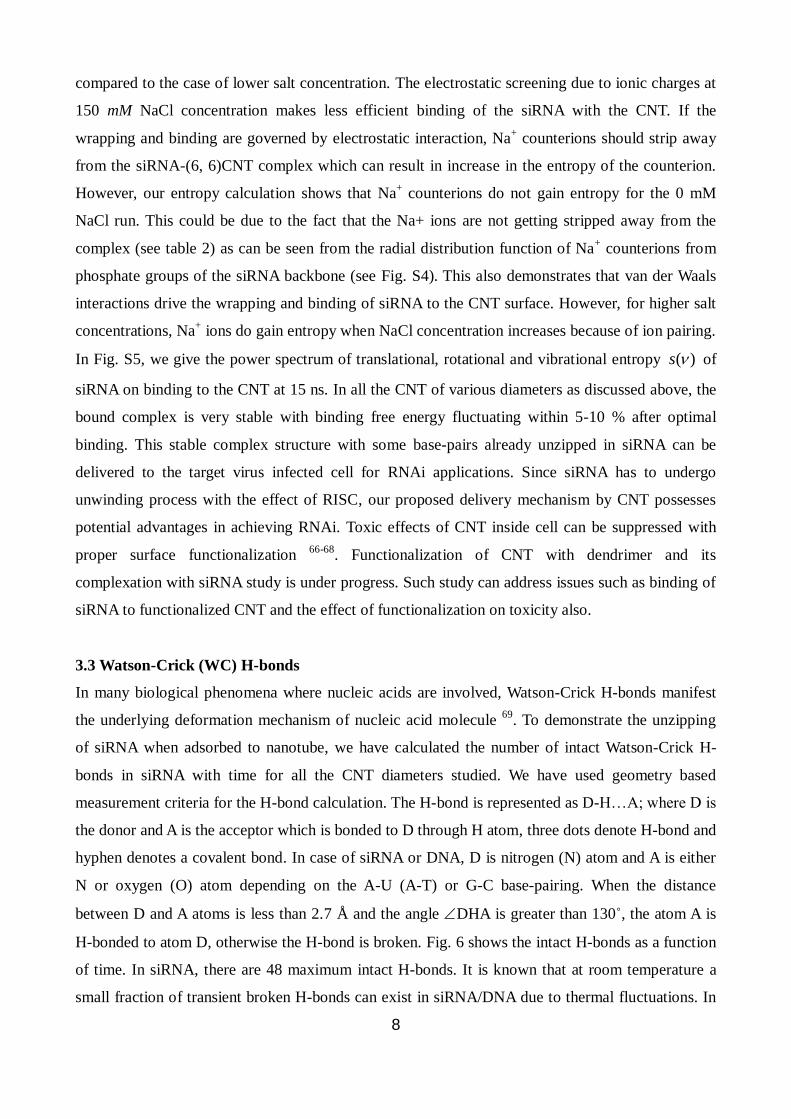
in Eq. (1) gives the total binding free energy ( bindG ) of siRNA. Fig. 4(b) shows bindG (at 300 K)
as a function of tube diameter revealing that the binding energy increases with the diameter of the
nanotube. This is easy to rationalize because siRNA encounters more surface area with increased
diameter of the tube. The binding of siRNA increases as the nanotube radius of curvature increases
and is maximum when the radius of curvature of the nanotube is equal to the radius of siRNA. The
binding of siRNA to the CNT involves unzipping of stacked base-pairs which has its own
characteristic length scale to match the length scale of the CNT curvature. The binding free energy
of siRNA on (5, 5) CNT is -189.0 ± 15.3 kcal/mol which increases to -301.0 ± 11.4 kcal/mol for (8,
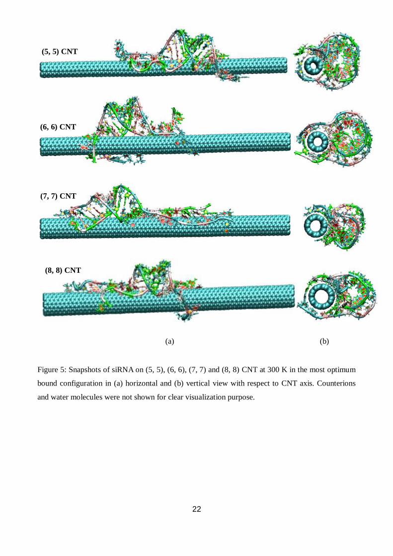
8) CNT. Snapshots of siRNA on CNT for various diameters in the most optimum bound
configuration are shown in Fig. 5 in horizontal and vertical view with respect to CNT axis. All the
above studies are for NaCl concentration of 0 mM. Physiological NaCl concentrations are of 100 to
150 mM in cell and can affect the binding mechanism of siRNA-CNT complex during delivery. We
have, therefore studied siRNA binding mechanism to (6, 6) CNT at 300 K by increasing NaCl
concentration. For (6, 6) CNT at 300 K, the binding free energy decreases from -230.0 ± 4.8
kcal/mol to -145.7 ± 5.5 kcal/mol when NaCl concentration increased to 150 mM. With more
added salt in the solvent, the electrostatic screening increases resulting in reduced phosphate-
phosphate electrostatic repulsion in the backbone of siRNA. Therefore the stretch modulus of
siRNA increases giving higher stability to siRNA with increasing salt concentration 63-65
which
reduce the propensity of siRNA unzipping. This leads to lesser binding affinity of siRNA with CNT
8
compared to the case of lower salt concentration. The electrostatic screening due to ionic charges at
150 mM NaCl concentration makes less efficient binding of the siRNA with the CNT. If the
wrapping and binding are governed by electrostatic interaction, Na+ counterions should strip away
from the siRNA-(6, 6)CNT complex which can result in increase in the entropy of the counterion.
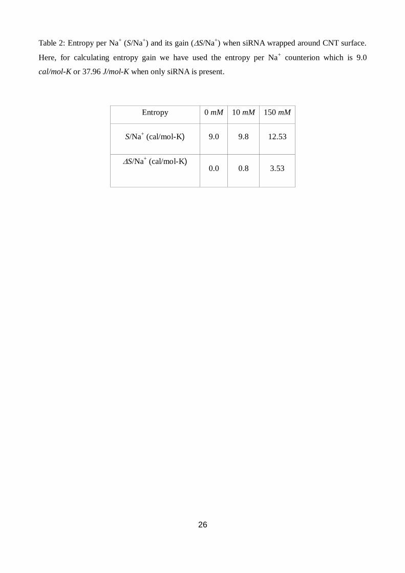
However, our entropy calculation shows that Na+ counterions do not gain entropy for the 0 mM
NaCl run. This could be due to the fact that the Na+ ions are not getting stripped away from the
complex (see table 2) as can be seen from the radial distribution function of Na+ counterions from
phosphate groups of the siRNA backbone (see Fig. S4). This also demonstrates that van der Waals
interactions drive the wrapping and binding of siRNA to the CNT surface. However, for higher salt
concentrations, Na+ ions do gain entropy when NaCl concentration increases because of ion pairing.
In Fig. S5, we give the power spectrum of translational, rotational and vibrational entropy ( )s of
siRNA on binding to the CNT at 15 ns. In all the CNT of various diameters as discussed above, the
bound complex is very stable with binding free energy fluctuating within 5-10 % after optimal
binding. This stable complex structure with some base-pairs already unzipped in siRNA can be
delivered to the target virus infected cell for RNAi applications. Since siRNA has to undergo
unwinding process with the effect of RISC, our proposed delivery mechanism by CNT possesses
potential advantages in achieving RNAi. Toxic effects of CNT inside cell can be suppressed with
proper surface functionalization 66-68
. Functionalization of CNT with dendrimer and its
complexation with siRNA study is under progress. Such study can address issues such as binding of
siRNA to functionalized CNT and the effect of functionalization on toxicity also.
3.3 Watson-Crick (WC) H-bonds
In many biological phenomena where nucleic acids are involved, Watson-Crick H-bonds manifest
the underlying deformation mechanism of nucleic acid molecule 69
. To demonstrate the unzipping
of siRNA when adsorbed to nanotube, we have calculated the number of intact Watson-Crick H-
bonds in siRNA with time for all the CNT diameters studied. We have used geometry based
measurement criteria for the H-bond calculation. The H-bond is represented as D-H…A; where D is
the donor and A is the acceptor which is bonded to D through H atom, three dots denote H-bond and
hyphen denotes a covalent bond. In case of siRNA or DNA, D is nitrogen (N) atom and A is either
N or oxygen (O) atom depending on the A-U (A-T) or G-C base-pairing. When the distance
between D and A atoms is less than 2.7 Å and the angle DHA is greater than 130˚, the atom A is
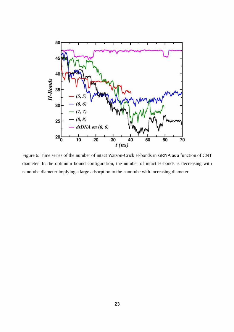
H-bonded to atom D, otherwise the H-bond is broken. Fig. 6 shows the intact H-bonds as a function
of time. In siRNA, there are 48 maximum intact H-bonds. It is known that at room temperature a
small fraction of transient broken H-bonds can exist in siRNA/DNA due to thermal fluctuations. In
9
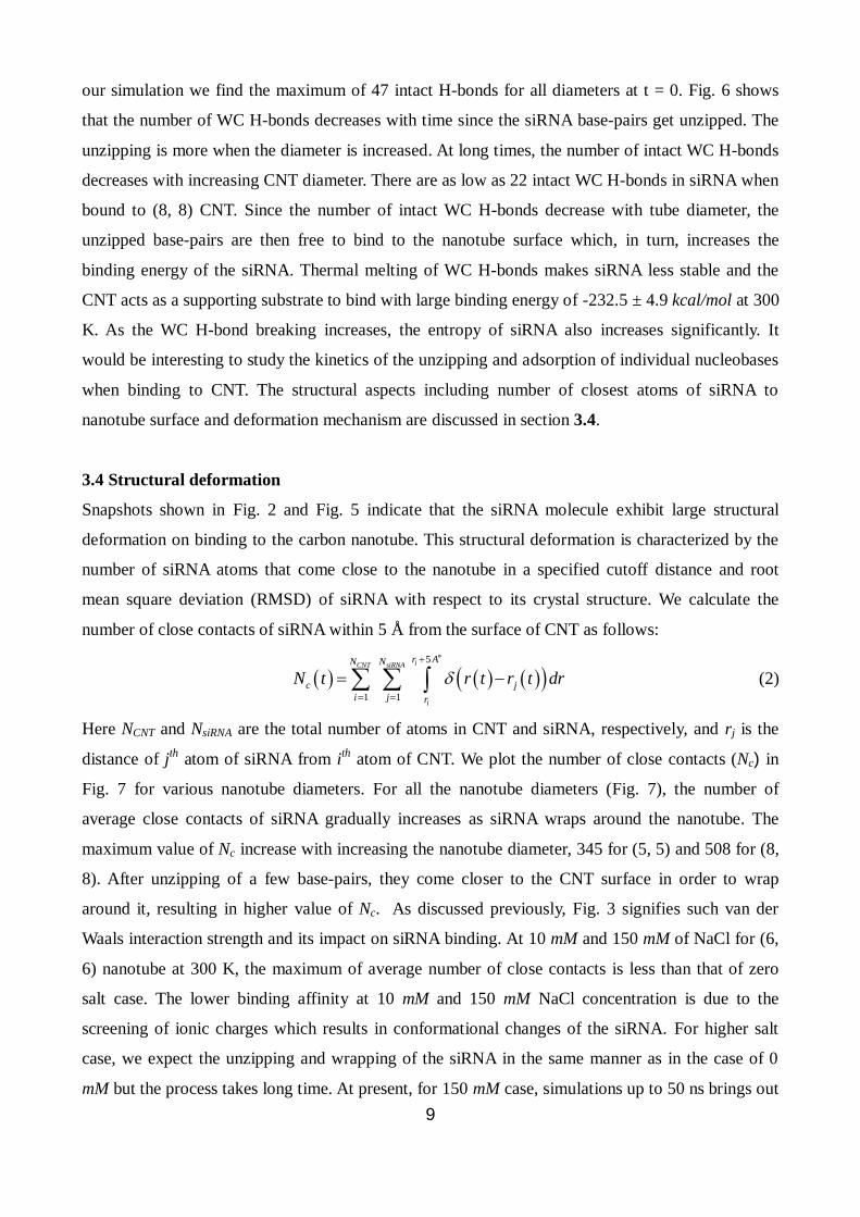
our simulation we find the maximum of 47 intact H-bonds for all diameters at t = 0. Fig. 6 shows
that the number of WC H-bonds decreases with time since the siRNA base-pairs get unzipped. The
unzipping is more when the diameter is increased. At long times, the number of intact WC H-bonds
decreases with increasing CNT diameter. There are as low as 22 intact WC H-bonds in siRNA when
bound to (8, 8) CNT. Since the number of intact WC H-bonds decrease with tube diameter, the
unzipped base-pairs are then free to bind to the nanotube surface which, in turn, increases the
binding energy of the siRNA. Thermal melting of WC H-bonds makes siRNA less stable and the
CNT acts as a supporting substrate to bind with large binding energy of -232.5 ± 4.9 kcal/mol at 300
K. As the WC H-bond breaking increases, the entropy of siRNA also increases significantly. It
would be interesting to study the kinetics of the unzipping and adsorption of individual nucleobases
when binding to CNT. The structural aspects including number of closest atoms of siRNA to
nanotube surface and deformation mechanism are discussed in section 3.4.
3.4 Structural deformation
Snapshots shown in Fig. 2 and Fig. 5 indicate that the siRNA molecule exhibit large structural
deformation on binding to the carbon nanotube. This structural deformation is characterized by the
number of siRNA atoms that come close to the nanotube in a specified cutoff distance and root
mean square deviation (RMSD) of siRNA with respect to its crystal structure. We calculate the
number of close contacts of siRNA within 5 Å from the surface of CNT as follows:
5
1 1
iCNT siRNA
i
r AN N
c j
i j r
N t r t r t dr
(2)
Here NCNT and NsiRNA are the total number of atoms in CNT and siRNA, respectively, and rj is the
distance of jth
atom of siRNA from ith
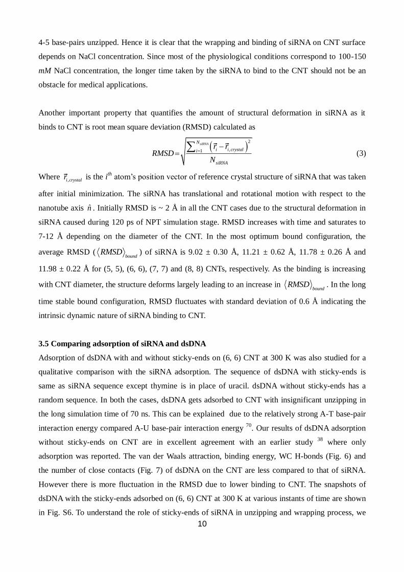
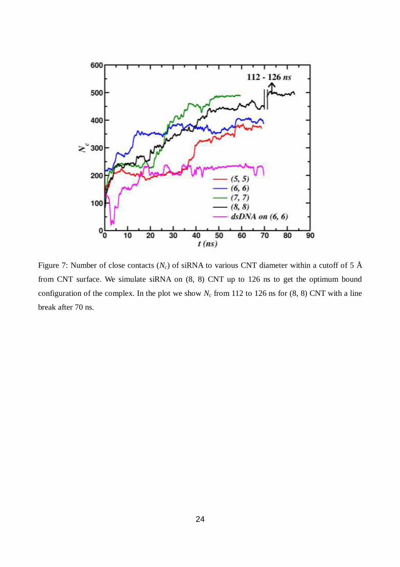
atom of CNT. We plot the number of close contacts (Nc) in
Fig. 7 for various nanotube diameters. For all the nanotube diameters (Fig. 7), the number of
average close contacts of siRNA gradually increases as siRNA wraps around the nanotube. The
maximum value of Nc increase with increasing the nanotube diameter, 345 for (5, 5) and 508 for (8,
8). After unzipping of a few base-pairs, they come closer to the CNT surface in order to wrap
around it, resulting in higher value of Nc. As discussed previously, Fig. 3 signifies such van der
Waals interaction strength and its impact on siRNA binding. At 10 mM and 150 mM of NaCl for (6,
6) nanotube at 300 K, the maximum of average number of close contacts is less than that of zero
salt case. The lower binding affinity at 10 mM and 150 mM NaCl concentration is due to the
screening of ionic charges which results in conformational changes of the siRNA. For higher salt
case, we expect the unzipping and wrapping of the siRNA in the same manner as in the case of 0
mM but the process takes long time. At present, for 150 mM case, simulations up to 50 ns brings out
10
4-5 base-pairs unzipped. Hence it is clear that the wrapping and binding of siRNA on CNT surface
depends on NaCl concentration. Since most of the physiological conditions correspond to 100-150
mM NaCl concentration, the longer time taken by the siRNA to bind to the CNT should not be an
obstacle for medical applications.
Another important property that quantifies the amount of structural deformation in siRNA as it
binds to CNT is root mean square deviation (RMSD) calculated as
2
,1
siRNAN
i i crystali
siRNA
r rRMSD
N
(3)
Where ,i crystalr
is the i
th atom’s position vector of reference crystal structure of siRNA that was taken
after initial minimization. The siRNA has translational and rotational motion with respect to the
nanotube axis n̂ . Initially RMSD is ~ 2 Å in all the CNT cases due to the structural deformation in
siRNA caused during 120 ps of NPT simulation stage. RMSD increases with time and saturates to
7-12 Å depending on the diameter of the CNT. In the most optimum bound configuration, the
average RMSD (bound
RMSD ) of siRNA is 9.02 ± 0.30 Å, 11.21 ± 0.62 Å, 11.78 ± 0.26 Å and
11.98 ± 0.22 Å for (5, 5), (6, 6), (7, 7) and (8, 8) CNTs, respectively. As the binding is increasing
with CNT diameter, the structure deforms largely leading to an increase in bound
RMSD . In the long
time stable bound configuration, RMSD fluctuates with standard deviation of 0.6 Å indicating the
intrinsic dynamic nature of siRNA binding to CNT.
3.5 Comparing adsorption of siRNA and dsDNA
Adsorption of dsDNA with and without sticky-ends on (6, 6) CNT at 300 K was also studied for a
qualitative comparison with the siRNA adsorption. The sequence of dsDNA with sticky-ends is
same as siRNA sequence except thymine is in place of uracil. dsDNA without sticky-ends has a
random sequence. In both the cases, dsDNA gets adsorbed to CNT with insignificant unzipping in
the long simulation time of 70 ns. This can be explained due to the relatively strong A-T base-pair
interaction energy compared A-U base-pair interaction energy 70
. Our results of dsDNA adsorption
without sticky-ends on CNT are in excellent agreement with an earlier study 38
where only
adsorption was reported. The van der Waals attraction, binding energy, WC H-bonds (Fig. 6) and
the number of close contacts (Fig. 7) of dsDNA on the CNT are less compared to that of siRNA.
However there is more fluctuation in the RMSD due to lower binding to CNT. The snapshots of
dsDNA with the sticky-ends adsorbed on (6, 6) CNT at 300 K at various instants of time are shown
in Fig. S6. To understand the role of sticky-ends of siRNA in unzipping and wrapping process, we
11
have performed a simulation of RNA with the same sequence as siRNA but without any sticky-
ends. Interestingly, RNA unzipping and wrapping around CNT is very less compared to the case
where sticky-ends are present in siRNA. The RNA stays adsorbed on the CNT surface with linear
translational motion along the CNT axis during the entire 50 ns long simulation. Therefore, the
sticky-ends enhance the unzipping and wrapping of siRNA on CNT. On the other hand, sticky-ends
do not help dsDNA in unzipping due to relatively stronger A-T base-pair energy compared to the A-
U base-pair energy. Snapshots of RNA without sticky-ends adsorbed on nanotube are shown in Fig.
S7.
3.6 siRNA vs dsDNA: insights from Quantum mechanical calculations
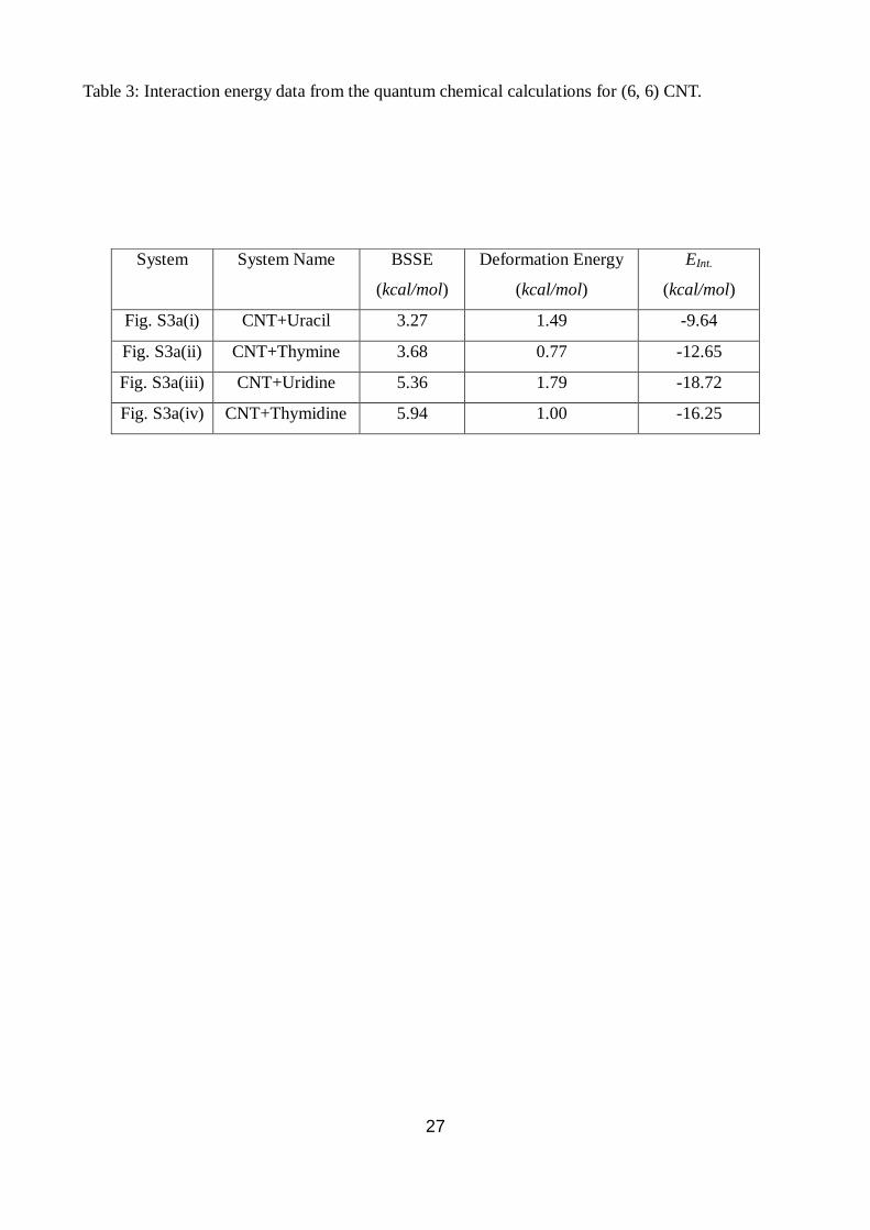
We find interaction energy of the CNT-uracil nucleobase complex is -9.64 kcal/mol whereas that of
the CNT-thymine nucleobase complex is about -12.25 kcal/mol. Interaction energy between
thymine and CNT was calculated earlier using Hartree-Fock and related methods giving significant
attraction between the two 71
. The energies using more robust density functional theory which
includes dispersion correction (DFT-D), the value of interaction energy of thymine obtained in our
method are quite similar to that of the previous estimate (-11.3 kcal/mol), without dispersion and
electron correlation effects. The difference in energy between thymine and uracil bases obtained in
our method may arise due to the stronger non-polar interaction between the methyl groups of
thymine and the carbon atoms of the CNT. Since sugar backbone plays a crucial role in maintaining
the structure and stability of RNA or DNA, we have also optimized the CNT-uridine and CNT-
thymidine complexes including sugars attached to the nucleobases. This now represents a more
accurate model of CNT-siRNA complex. The interaction energy values shown in Table 3 indicate
that CNT with uridine has stronger binding (-18.72 kcal/mol), than that of the CNT with thymidine
(-16.25 kcal/mol). This is due to possibility of weak hydrogen bond formation between the three -
OH groups of uridine molecule with carbon atoms of CNT. We believe this favours the siRNA
unzipping and subsequent wrapping on CNT whereas only adsorption of dsDNA on CNT is
observed. The uridine molecule forms two H-bonds involving O5’-H5’ of ribose sugar with two
carbon atoms at edge-1 (Fig. S3 (a(iii))) of CNT with H-bond distances of 2.62 Å and 2.52 Å and
associated O-H...C angles of 150.81o and 166.16
o, respectively. Another H-bond is found between
the O3’-H3’ of the ribose sugar ring with one of the carbon atom that lies towards the middle of the
CNT with H-bond distance and angle of 2.50 Å and 154.39o, respectively. Therefore, uridine is able
to form three good H-bonds with the CNT and stabilize the system provided the carbon atoms have
sufficient negative charges.
12
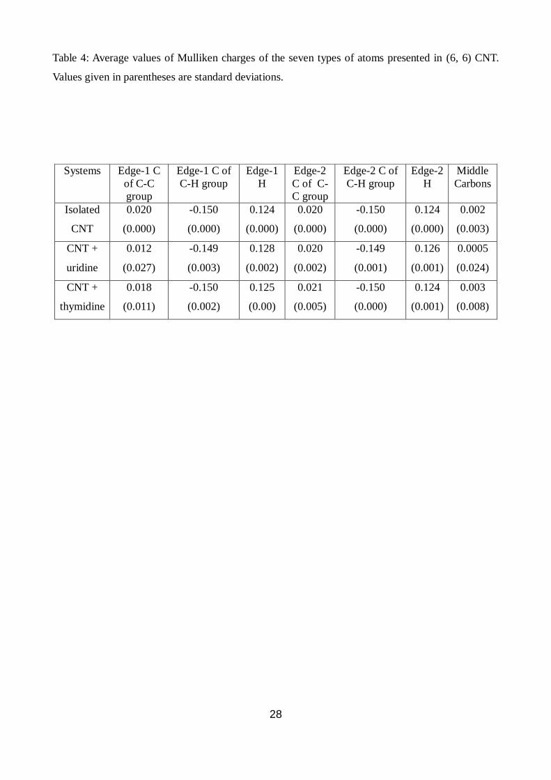
We have analysed partial charge of all the atoms of CNT calculated by Mulliken population
analysis. As expected the terminal C-H groups are slightly polar, the carbons not bonded to
hydrogen are neutral (carbons of C-C group, those lies inwardly at the terminal edges, Fig. S3 (b))
and the central carbons present at the middle region of the CNT have nearly zero charge. Moreover
all the charges at the two edges are symmetrically distributed with zero standard deviations when it
is not complexed with any nucleobase/nucleoside. In presence of thymine or uracil bases, the
properties of the CNT remain nearly unchanged. A presence of nucleoside residues, particularly the
uridine nucleoside, breaks the symmetry of the CNT significantly (shown in Table 4), as detected
from the larger standard deviations of the charges of different groups of atoms. We have classified
the CNT atoms into the following types (Fig. S3 (b)): i) carbon atoms at the edges which are not
bonded to any hydrogen C-C carbons, ii) carbon atoms of C-H group and iii) carbon atoms which
lie in the central region of CNT. The terminal atoms can be further classified into edge-1 and edge-
2, depending on proximity to the binding nucleoside. When uridine binds to CNT, partial charges of
the C-C carbon atoms at the edge-1 change significantly. In this case, the average charge of the
carbon atom decreases and the standard deviation increases, which signifies delocalisation of the
charges. In presence of the polar uridine, the electrons of the CNT move significantly through the
extended conjugation and accumulate near the uridine. The thymidine also alters charges of these
carbon atoms in CNT but to a lesser extent. We notice that the atoms, which are far away from the
nucleoside (those of the edge-2), do not undergo any noticeable changes in both the cases. The
partial charges of the central carbon atoms also alter significantly, particularly in case of uridine,
which is reflected in larger standard deviations (partial charge of carbon changes from 0.002 to –
0.060 for acting as H-bond acceptor). The other carbon atoms of CNT that are forming H-bonds
with the O-H groups of uridine acquire Mulliken charges (in units of electron charge ‘e’) of -0.063
and -0.154 from 0.020 and -0.149, respectively. This signifies that the uridine can polarize CNT and
has strong binding affinity with CNT than that of the thymidine, which is correlated with its
interaction energy data also.
4. Conclusions
Using combination of all atom molecular dynamics simulations and ab-initio quantum mechanical
calculations we report unzipping and wrapping of a small interfering RNA molecule on the carbon
nanotube to study the binding mechanism of siRNA on the CNT surface. To the best of our
knowledge this is the first theoretical demonstration of the unzipping and wrapping of siRNA on the
CNT surface. In the process of understanding siRNA delivery mechanism to achieve RNAi by
13
CNTs at a microscopic level, we attempt to study the thermodynamic and energetic properties of
siRNA-CNT complex. The binding mechanism of siRNA on various CNT diameters has been
studied. Our simulations show that a few base-pairs at both the strands of siRNA get unzipped and
wrap around CNT surface with strong binding affinity. The binding energy increases with the CNT
diameter due to van der Waals forces between siRNA aromatic rings and CNT surface. In order to
wrap around CNT, siRNA has to be very flexible. Since siRNA molecule is double stranded with
two sticky-ends whose persistence length is much larger than that of single stranded RNA, we may
naively expect that it will be difficult to wrap. But interestingly, siRNA gets unzipped and
eventually wraps around the CNT surface within a few nanoseconds for all the CNT diameters
studied in this paper. The unzipping and subsequent binding processes were initiated and driven by
van der Waals (dispersion) interaction between aromatic rings of siRNA and CNT surface,
facilitated by two sticky-ends on both the strands. siRNA gains entropy on binding to the CNT
surface due to unzipping of a few base-pairs. More surface area of CNT for large diameters
enhances the interaction with siRNA and improves binding. An increase in broken WC H-bonds,
RMSD and number of close contacts indicate large structural deformation of siRNA with respect to
its starting crystal structure. The siRNA-CNT complex is very stable after the optimal binding to the
CNT. The dsDNA of the same sequence adsorption on the same CNT show that dsDNA has very
less unzipping and wrapping around the CNT in the observed simulation time scale of 70 ns.
Considering the unzipping and wrapping process is guided by uridine-CNT interaction, we have
carried out detail quantum chemical analysis of the two comparative systems, CNT-uridine and
CNT-thymidine. Our quantum chemistry results indicate that CNT has better propensity to bind to
uridine due to its additional O-H groups which form strong H-bond with the CNT. The H-bond
formation requires charge accumulation towards some carbons of the CNT, which also takes place
due to extended conjugation of the CNT. We also studied effect of salt concentration on the siRNA-
CNT interaction. Our results suggest that at large NaCl concentration, the screening of ionic charges
make it less efficient for binding compared to charge neutral case. However the unzipping and
wrapping of siRNA happens very slowly in this case. The adsorbed siRNA can be delivered to virus
infected cell via endocytosis to reduce the expression of specific unwanted genes to achieve RNAi
effect. In RNAi therapy, siRNA delivery to the target cell involves siRNA unzipping into two single
strands that is mediated by RISC loading complex 3. We have shown the unwinding enhanced
siRNA binding to carbon nanotube (CNT) and propose siRNA delivery to target cell for achieving
RNAi without degradation. In a subsequent work we study the binding mechanism of siRNA on
graphene for more efficient and sophisticated delivery method. The intrinsic toxicity effects caused
by CNT to the cell functionality was shown to have less effect by proper functionalization of CNT
14
66-68. Studies on cell penetrating membrane mechanism of siRNA-CNT complex and solubility of
CNT after siRNA delivery are among future perspectives in this vast area.
Acknowledgments
PKM and AKS thank DBT, India for the financial support. We thank Prof. M. Muthukumar for
stimulating discussions. We acknowledge computational resource supported by the DST Centre for
Mathematical Biology at IISc. We also thank Prof. S. Ramaswamy for allowing us to use his
computational facility. MS thank UGC, India for senior research fellowship.
15
Refereces
1 C. Napoli, C. Lemieux, and R. Jorgensen, Plant Cell 2, 279-289 (1990).
2 A. Fire, S. Q. Xu, M. K. Montgomery, S. A. Kostas, S. E. Driver, and C. C. Mello, Nature
391, 806-811 (1998). 3
Y. Tomari, C. Matranga, B. Haley, N. Martinez, and P. D. Zamore, Science 306, 1377-1380
(2004). 4
P. D. Zamore and B. Haley, Science 309, 1519-1524 (2005). 5
G. Hutvagner and P. D. Zamore, Current Opinion in Genetics & Development 12, 225-232
(2002). 6
M. Ghildiyal and P. D. Zamore, Nature Reviews Genetics 10, 94-108 (2009). 7
P. D. Zamore, T. Tuschl, P. A. Sharp, and D. P. Bartel, Cell 101, 25-33 (2000). 8
G. Meister and T. Tuschl, Nature 431, 343-349 (2004). 9
A. Tsubouchi, J. Sakakura, R. Yagi, Y. Mazaki, E. Schaefer, H. Yano, and H. Sabe, Journal
of Cell Biology 159, 673-683 (2002). 10
Y. Z. Huang, M. W. Zang, W. C. Xiong, Z. J. Luo, and L. Mei, Journal of Biological
Chemistry 278, 1108-1114 (2003). 11
V. Vasumathi and P. K. Maiti, Macromolecules 43, 8264-8274 (2010). 12
B. Nandy and P. K. Maiti, Journal of Physical Chemistry B 115, 217-230 (2011). 13
P. K. Maiti and B. Bagchi, Nano Letters 6, 2478-2485 (2006). 14
S. M. Elbashir, J. Harborth, W. Lendeckel, A. Yalcin, K. Weber, and T. Tuschl, Nature 411,
494-498 (2001). 15
D. Yang, F. Buchholz, Z. D. Huang, A. Goga, C. Y. Chen, F. M. Brodsky, and J. M. Bishop,
Proceedings of the National Academy of Sciences of the United States of America 99, 9942-
9947 (2002). 16
Q. Lu, J. M. Moore, G. Huang, A. S. Mount, A. M. Rao, L. L. Larcom, and P. C. Ke, Nano
Letters 4, 2473-2477 (2004). 17
Z. Liu, M. Winters, M. Holodniy, and H. J. Dai, Angewandte Chemie-International Edition
46, 2023-2027 (2007). 18
Z. Liu, K. Chen, C. Davis, S. Sherlock, Q. Z. Cao, X. Y. Chen, and H. J. Dai, Cancer
Research 68, 6652-6660 (2008). 19
Z. Liu, S. Tabakman, K. Welsher, and H. J. Dai, Nano Research 2, 85-120 (2009). 20
M. C. Morris, P. Vidal, L. Chaloin, F. Heitz, and G. Divita, Nucleic Acids Research 25,
2730-2736 (1997). 21
F. Simeoni, M. C. Morris, F. Heitz, and G. Divita, Nucleic Acids Research 31, 2717-2724
(2003). 22
I. Puebla, S. Esseghir, A. Mortlock, A. Brown, A. Crisanti, and W. Low, Journal of
Biotechnology 105, 215-226 (2003). 23
Y. Minakuchi, F. Takeshita, N. Kosaka, H. Sasaki, Y. Yamamoto, M. Kouno, K. Honma, S.
Nagahara, K. Hanai, A. Sano, T. Kato, M. Terada, and T. Ochiya, Nucleic Acids Research 32
(2004). 24
N. S. Lee, T. Dohjima, G. Bauer, H. T. Li, M. J. Li, A. Ehsani, P. Salvaterra, and J. Rossi,
Nature Biotechnology 20, 500-505 (2002). 25
J. M. Jacque, K. Triques, and M. Stevenson, Nature 418, 435-438 (2002). 26
D. H. Kim and J. J. Rossi, Nature Reviews Genetics 8, 173-184 (2007). 27
J. Kurreck, Angewandte Chemie-International Edition 48, 1378-1398 (2009). 28
P. D. Zamore and N. Aronin, Nature Medicine 9, 266-267 (2003). 29
B. Urban-Klein, S. Werth, S. Abuharbeid, F. Czubayko, and A. Aigner, Gene Therapy 12,
16
461-466 (2005). 30
D. Bumcrot, M. Manoharan, V. Koteliansky, and D. W. Y. Sah, Nature Chemical Biology 2,
711-719 (2006). 31
N. W. S. Kam, Z. A. Liu, and H. J. Dai, Angewandte Chemie-International Edition 45, 577-
581 (2006). 32
Z. H. Zhang, X. Y. Yang, Y. Zhang, B. Zeng, Z. J. Wang, T. H. Zhu, R. B. S. Roden, Y. S.
Chen, and R. C. Yang, Clinical Cancer Research 12, 4933-4939 (2006). 33
J. DeRouchey, C. Schmidt, G. F. Walker, C. Koch, C. Plank, E. Wagner, and J. O. Radler,
Biomacromolecules 9, 724-732 (2008). 34
M. Zheng, A. Jagota, E. D. Semke, B. A. Diner, R. S. McLean, S. R. Lustig, R. E.
Richardson, and N. G. Tassi, Nature Materials 2, 338-342 (2003). 35
M. Zheng, A. Jagota, M. S. Strano, A. P. Santos, P. Barone, S. G. Chou, B. A. Diner, M. S.
Dresselhaus, R. S. McLean, G. B. Onoa, G. G. Samsonidze, E. D. Semke, M. Usrey, and D.
J. Walls, Science 302, 1545-1548 (2003). 36
S. G. Chou, H. B. Ribeiro, E. B. Barros, A. P. Santos, D. Nezich, G. G. Samsonidze, C.
Fantini, M. A. Pimenta, A. Jorio, F. Plentz, M. S. Dresselhaus, G. Dresselhaus, R. Saito, M.
Zheng, G. B. Onoa, E. D. Semke, A. K. Swan, M. S. Unlu, and B. B. Goldberg, Chemical
Physics Letters 397, 296-301 (2004). 37
R. R. Johnson, A. T. C. Johnson, and M. L. Klein, Nano Letters 8, 69-75 (2008). 38
X. Zhao and J. K. Johnson, Journal of the American Chemical Society 129, 10438-10445
(2007). 39
D. A. Case, T. A. Darden, T. E. Cheatham III, C. L. Simmerling, J. Wang, R. E. Duke, R.
Luo, K. M. Merz, D. A. Pearlman, M. Crowley, R. C. Walker, W. Zhang, B. Wang, S. Hayik,
A. Roitberg, G. Seabra, K. F. Wong, F. Paesani, X. Wu, S. Brozell, V. Tsui, H. Gohlke, L.
Yang, C. Tan, J. Mongan, V. Hornak, G. Cui, P. Beroza, D. H. Mathews, C. Schafmeister, W.
S. Ross, and P. A. Kollman, (University of California, San Francisco, 2006). 40
Y. Duan, C. Wu, S. Chowdhury, M. C. Lee, G. M. Xiong, W. Zhang, R. Yang, P. Cieplak, R.
Luo, T. Lee, J. Caldwell, J. M. Wang, and P. Kollman, Journal of Computational Chemistry
24, 1999-2012 (2003). 41
W. L. Jorgensen, J. Chandrasekhar, J. D. Madura, R. W. Impey, and M. L. Klein, Journal of
Chemical Physics 79, 926-935 (1983). 42
A. Perez, I. Marchan, D. Svozil, J. Sponer, T. E. Cheatham, C. A. Laughton, and M. Orozco,
Biophysical Journal 92, 3817-3829 (2007). 43
P. Banas, D. Hollas, M. Zgarbova, P. Jurecka, M. Orozco, T. E. Cheatham, J. Sponer, and M.
Otyepka, Journal of Chemical Theory and Computation 6, 3836-3849 (2010). 44
See Supplementary Material Document No. xxx. 45
J. Romanowska, J. A. McCammon, and J. Trylska, Plos Computational Biology 7 (2011). 46
I. Besseova, M. Otyepka, K. Reblova, and J. Sponer, Physical Chemistry Chemical Physics
11, 10701-10711 (2009). 47
K. Reblova, Z. Strelcova, P. Kulhanek, I. Besseova, D. H. Mathews, K. Van Nostrand, I.
Yildirim, D. H. Turner, and J. Sponer, Journal of Chemical Theory and Computation 6, 910-
929 (2010). 48
Y. R. Yuan, Y. Pei, H. Y. Chen, T. Tuschl, and D. J. Patel, Structure 14, 1557-1565 (2006). 49
B. Mukherjee, P. K. Maiti, C. Dasgupta, and A. K. Sood, Journal of Chemical Physics 126
(2007). 50
T. Darden, D. York, and L. Pedersen, Journal of Chemical Physics 98, 10089-10092 (1993). 51
J. P. Ryckaert, G. Ciccotti, and H. J. C. Berendsen, Journal of Computational Physics 23,
327-341 (1977). 52
H. J. C. Berendsen, J. P. M. Postma, W. F. Vangunsteren, A. Dinola, and J. R. Haak, Journal
of Chemical Physics 81, 3684-3690 (1984). 53
G. Schaftenaar and J. H. Noordik, Journal of Computer-Aided Molecular Design 14, 123-
17
134 (2000). 54
M. J. Frisch, G. W. Trucks, H. B. Schlegel, G. E. Scuseria, M. A. Robb, J. R. Cheeseman, G.
Scalmani, V. Barone, B. Mennucci, G. A. Petersson, H. Nakatsuji, M. Caricato, X. Li, H. P.
Hratchian, A. F. Izmaylov, J. Bloino, G. Zheng, J. L. Sonnenberg, M. Hada, M. Ehara, K.
Toyota, R. Fukuda, J. Hasegawa, M. Ishida, T. Nakajima, Y. Honda, O. Kitao, H. akai, T.
Vreven, J. Montgomery, J. A., J. E. Peralta, F. Ogliaro, M. Bearpark, J. J. Heyd, E. Brothers,
K. N. Kudin, V. N. Staroverov, R. Kobayashi, J. Normand, K. Raghavachari, A. Rendell, J.
C. Burant, S. S. Iyengar, J. Tomasi, M. Cossi, N. Rega, N. J. Millam, M. Klene, J. E. Knox,
J. B. Cross, V. Bakken, C. Adamo, J. Jaramillo, R. Gomperts, R. E. Stratmann, O. Yazyev, A.
J. Austin, R. Cammi, C. Pomelli, J. W. Ochterski, R. L. Martin, K. Morokuma, V. G.
Zakrzewski, G. A. Voth, P. Salvador, J. J. Dannenberg, S. Dapprich, A. D. Daniels, Ö.
Farkas, J. B. Foresman, J. V. Ortiz, J. Cioslowski, and D. J. Fox, Revision A.1 ed. (Gaussian,
Inc., Wallingford CT, 2009). 55
J. D. Chai and M. Head-Gordon, Journal of Chemical Physics 128 (2008). 56
S. F. Boys and F. Bernardi, Molecular Physics 19, 553-& (1970). 57
B. Jayaram, K. J. McConnell, S. B. Dixit, and D. L. Beveridge, Journal of Computational
Physics 151, 333-357 (1999). 58
B. Jayaram, Y. Liu, and D. L. Beveridge, Journal of Chemical Physics 109, 1465-1471
(1998). 59
B. Jayaram, D. Sprous, and D. L. Beveridge, Journal of Physical Chemistry B 102, 9571-
9576 (1998). 60
S. T. Lin, M. Blanco, and W. A. Goddard, Journal of Chemical Physics 119, 11792-11805
(2003). 61
P. H. Berens, D. H. J. Mackay, G. M. White, and K. R. Wilson, Journal of Chemical Physics
79, 2375-2389 (1983). 62
T. A. Pascal, W. A. Goddard, and Y. Jung, Proceedings of the National Academy of Sciences
of the United States of America 108, 11794-11798 (2011). 63
C. G. Baumann, S. B. Smith, V. A. Bloomfield, and C. Bustamante, Proceedings of the
National Academy of Sciences of the United States of America 94, 6185-6190 (1997). 64
J. R. Wenner, M. C. Williams, I. Rouzina, and V. A. Bloomfield, Biophysical Journal 82,
3160-3169 (2002). 65
Z. J. Tan and S. J. Chen, Biophysical Journal 90, 1175-1190 (2006). 66
C. W. Lam, J. T. James, R. McCluskey, and R. L. Hunter, Toxicological Sciences 77, 126-
134 (2004). 67
G. Jia, H. F. Wang, L. Yan, X. Wang, R. J. Pei, T. Yan, Y. L. Zhao, and X. B. Guo,
Environmental Science & Technology 39, 1378-1383 (2005). 68
D. X. Cui, F. R. Tian, C. S. Ozkan, M. Wang, and H. J. Gao, Toxicology Letters 155, 73-85
(2005). 69
D. Voet and J. G. Voet, Biochemistry, Second ed. (John Wiley & Sons, Inc., New York,
1995). 70
J. Sponer, P. Jurecka, and P. Hobza, Journal of the American Chemical Society 126, 10142-
10151 (2004). 71
N. Varghese, U. Mogera, A. Govindaraj, A. Das, P. K. Maiti, A. K. Sood, and C. N. R. Rao,
Chemphyschem 10, 206-210 (2009). 72
W. Humphrey, A. Dalke, and K. Schulten, Journal of Molecular Graphics 14, 33-& (1996).
18
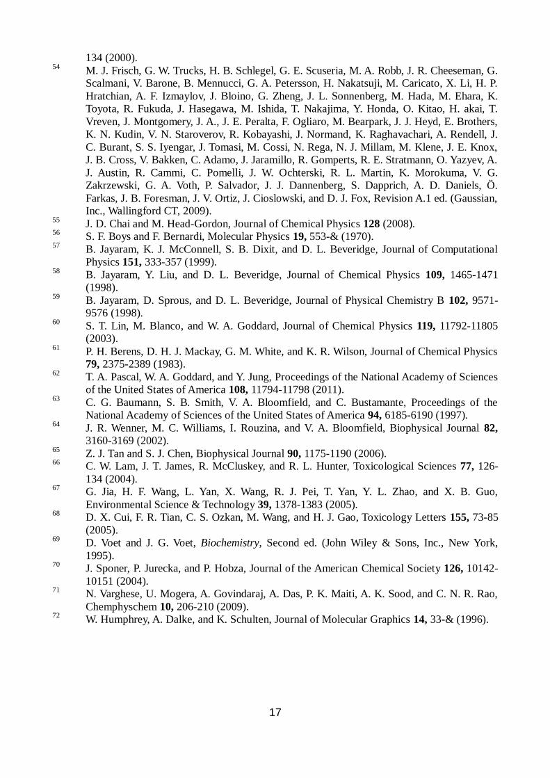
Figure 1: (a) siRNA crystal structure (pdb code 1F8S) and (b) the initial simulation system setup
where siRNA-(6, 6)CNT complex was solvated with water and neutralizing Na+ counterions. These
pictures were rendered using VMD software package 72
.
(a) (b)
19
0 ns
15 ns
30 ns
45 ns
A B
Figure 2: Snapshots of siRNA on (6, 6) CNT at 300 K at various instants of time during MD
simulation in (a) horizontal and (b) vertical view with respect to CNT axis. Counterions and water
molecules were not shown for clear visualization purpose. These snapshots were rendered using
VMD software package 72
.
(a) (b)
20
Figure 3: van der Waals interaction energy between siRNA and (6, 6) CNT at 300 K. See the text
for details of the calculation. The siRNA strongly binds to CNT at 15 ns compared to 5 ns with an
increment in van der Waals energy of about 133 kcal/mol or 225.4 kBT.
21
Figure 4: (a) The enthalpy contribution to the total binding free energy as a function of time for
siRNA on (6, 6) CNT at 300 K. The arrow marked region shows the most optimum bound state with
binding energy fluctuating within 5 % of its average in optimum bound state. For the entropy
calculation we have started from this optimum bound state and simulated 10 consecutive sets of
each 20 ps as discussed in text. (b) Binding free energy between siRNA and CNT in the most
optimum bound state as a function of CNT diameter. The binding free energy that includes enthalpy
and entropy contributions increases with diameter due to large CNT surface area available for
siRNA to bind with CNT at 300 K.
(a) (b)
22
Figure 5: Snapshots of siRNA on (5, 5), (6, 6), (7, 7) and (8, 8) CNT at 300 K in the most optimum
bound configuration in (a) horizontal and (b) vertical view with respect to CNT axis. Counterions
and water molecules were not shown for clear visualization purpose.
(a) (b)
(5, 5) CNT
(6, 6) CNT
(7, 7) CNT
(8, 8) CNT
23
Figure 6: Time series of the number of intact Watson-Crick H-bonds in siRNA as a function of CNT
diameter. In the optimum bound configuration, the number of intact H-bonds is decreasing with
nanotube diameter implying a large adsorption to the nanotube with increasing diameter.
24
Figure 7: Number of close contacts (Nc) of siRNA to various CNT diameter within a cutoff of 5 Å
from CNT surface. We simulate siRNA on (8, 8) CNT up to 126 ns to get the optimum bound
configuration of the complex. In the plot we show Nc from 112 to 126 ns for (8, 8) CNT with a line
break after 70 ns.
25
Table 1: Summary of the simulation setup for 0 mM, 10 mM and 150 mM NaCl concentrations.
a dsDNA without sticky ends
b dsDNA with sticky ends
c (mM) CNT siRNA Box (Å3) Na
+ Cl
- WAT Total Atoms
0 (6, 6) 1440 1396 84×79×192 44 0 35382 109026
10 (6, 6) 1440 1396 84×79×192 52 8 35365 108991
150 (6, 6) 1440 1396 84×79×192 160 116 35148 108556
0 (5, 5) 1200 1396 71×75×193 44 0 27810 86070
0 (7, 7) 1680 1396 79×72×193 44 0 29810 92550
0 (8, 8) 1920 1396 70×80×193 44 0 29063 90549
0 a (6, 6) 1440 1398 78×67×192 42 0 26878 83514
0 b (6, 6) 1440 1398 75×68×192 42 0 26472 82296
26
Table 2: Entropy per Na+ (S/Na
+) and its gain (S/Na
+) when siRNA wrapped around CNT surface.
Here, for calculating entropy gain we have used the entropy per Na+ counterion which is 9.0
cal/mol-K or 37.96 J/mol-K when only siRNA is present.
Entropy 0 mM 10 mM 150 mM
S/Na+ (cal/mol-K) 9.0 9.8 12.53
S/Na+ (cal/mol-K)
0.0 0.8 3.53
27
Table 3: Interaction energy data from the quantum chemical calculations for (6, 6) CNT.
System System Name BSSE
(kcal/mol)
Deformation Energy
(kcal/mol)
EInt.
(kcal/mol)
Fig. S3a(i) CNT+Uracil 3.27 1.49 -9.64
Fig. S3a(ii) CNT+Thymine 3.68 0.77 -12.65
Fig. S3a(iii) CNT+Uridine 5.36 1.79 -18.72
Fig. S3a(iv) CNT+Thymidine 5.94 1.00 -16.25
28
Table 4: Average values of Mulliken charges of the seven types of atoms presented in (6, 6) CNT.
Values given in parentheses are standard deviations.
Systems Edge-1 C
of C-C
group
Edge-1 C of
C-H group
Edge-1
H
Edge-2
C of C-
C group
Edge-2 C of
C-H group
Edge-2
H
Middle
Carbons
Isolated
CNT
0.020
(0.000)
-0.150
(0.000)
0.124
(0.000)
0.020
(0.000)
-0.150
(0.000)
0.124
(0.000)
0.002
(0.003)
CNT +
uridine
0.012
(0.027)
-0.149
(0.003)
0.128
(0.002)
0.020
(0.002)
-0.149
(0.001)
0.126
(0.001)
0.0005
(0.024)
CNT +
thymidine
0.018
(0.011)
-0.150
(0.002)
0.125
(0.00)
0.021
(0.005)
-0.150
(0.000)
0.124
(0.001)
0.003
(0.008)