untitled - annals of jinnah sindh medical university
TRANSCRIPT
The AJSMU is published biannually by Jinnah Sindh Medical University.Editorial Correspondence should be addressed to: Editor-in-Chief, AJSMU, Jinnah Sindh MedicalUniversity, Rafiqui H.J. Shaheed Road, Karachi 75510; Tel: 99204776; 35223811-15 Ext:1076, 1087Email: [email protected]; Website: www.ajsmu.com; Fax: 99201372Annual subscription: Pakistan Rs.450, Bangladesh & India: Rs.600, UK 15, U.S.A and other countries: US$ 15 Published by: The Registrar, Jinnah Sindh Medical University, Rafiqui H.J. Shaheed Road, Karachi.
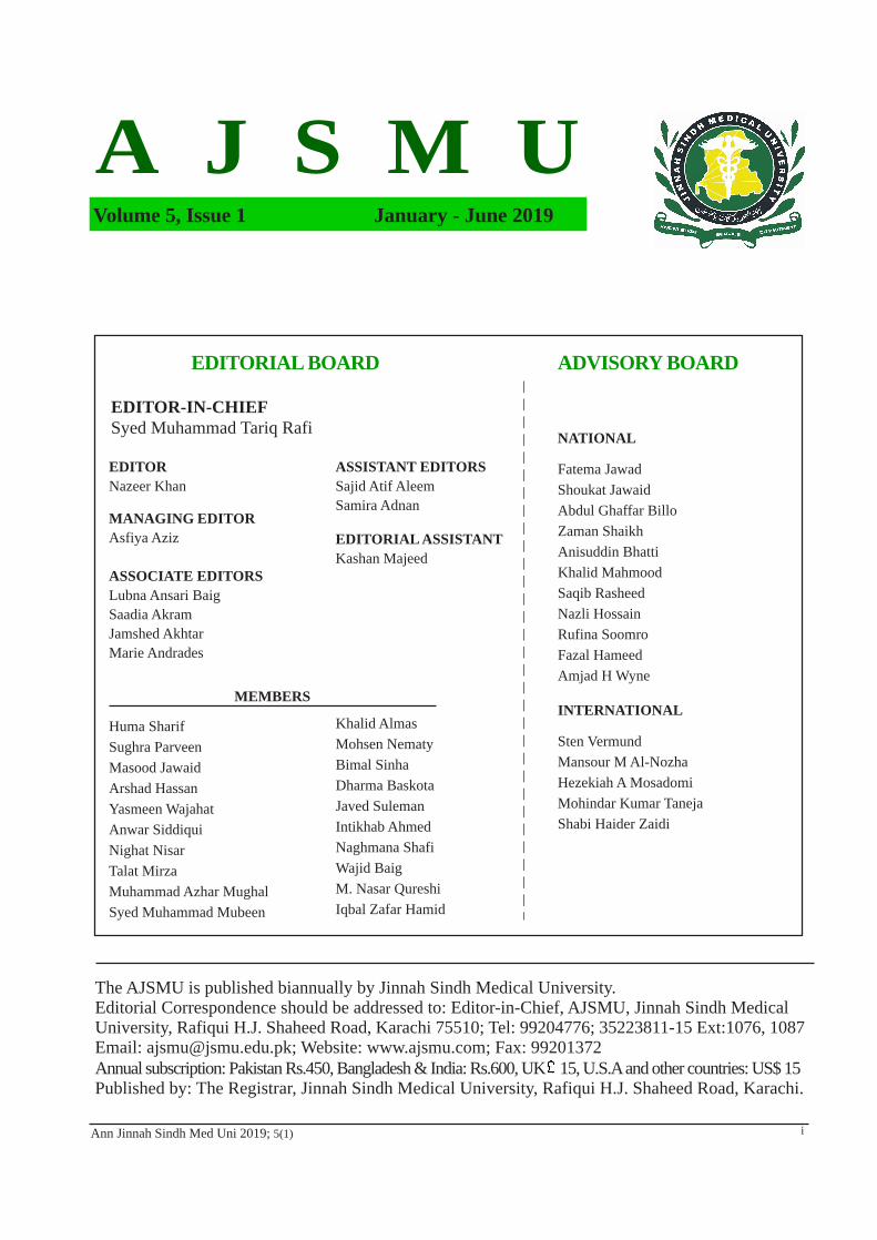
EDITORIAL BOARD ADVISORY BOARD
NATIONAL
Fatema JawadShoukat JawaidAbdul Ghaffar BilloZaman ShaikhAnisuddin BhattiKhalid MahmoodSaqib RasheedNazli HossainRufina SoomroFazal HameedAmjad H Wyne
INTERNATIONAL
Sten VermundMansour M Al-NozhaHezekiah A MosadomiMohindar Kumar TanejaShabi Haider Zaidi
A J S M U
Ann Jinnah Sindh Med Uni 2019; 5(1) i
Volume 5, Issue 1 January - June 2019
Khalid AlmasMohsen NematyBimal SinhaDharma BaskotaJaved SulemanIntikhab AhmedNaghmana ShafiWajid BaigM. Nasar QureshiIqbal Zafar Hamid
Huma Sharif Sughra ParveenMasood Jawaid Arshad HassanYasmeen Wajahat Anwar SiddiquiNighat Nisar Talat MirzaMuhammad Azhar MughalSyed Muhammad Mubeen
MEMBERS
EDITORNazeer Khan
MANAGING EDITORAsfiya Aziz
ASSOCIATE EDITORSLubna Ansari BaigSaadia AkramJamshed AkhtarMarie Andrades
ASSISTANT EDITORSSajid Atif AleemSamira Adnan
EDITORIAL ASSISTANTKashan Majeed
EDITOR-IN-CHIEFSyed Muhammad Tariq Rafi
Page No.
1
3
10
15
21
26
31
35
39
47
iii
iv
v
Ann Jinnah Sindh Med Uni 2019; 5(1)ii
A J S M UVolume 5, Issue 1 January - June 2019
EDITORIAL
Family Medicine: Indispensable for An Effective Healthcare System
ORIGINAL ARTICLE
Statistical Assessment of Drug Release Kinetics and Formulations Attributes of Ranitidine Tablets Available in Karachi, Pakistan
Genetic Variability of Omentin-1 Gene in Apparently Healthy Population
Impact of Oxidative Stress on Hypertension in Patients on Maintenance Haemodialysis
Comparative Study of Mean Corpuscular Volume Between Lacto-vegetarian and Non-vegetarian Populations of Tharparkar Village
Knowledge and Frequency of Needle Stick Injury Among Dental Students and House Officers of Bhitai Medical and Dental College, Mirpur Khas
Self-Image and Its Impact on Academic Performance of Undergraduate Medical Students in Karachi
Risk Factors and Frequency of Hypocalcaemia in Unintentional Parathyroid Gland Removal During Thyroid Surgical Interventions
REVIEW ARTICLE
Phytomedicine: Silybum marianum (Silymarin) as an Effective Hepato-protective Source from Nature
COMMUNICATION
Molecular Diagnostics: A Paradigm Shift
From the Desk of the Editor
Acknowledgement of Reviewers
Instructions to Authors
Marie Andrades
Shaheen Parveen, Huma Ali, Maria Sodagar, Amber Nawab, Anum Tariq, and Fozia Israr
Shazia Nazar, Ambreen Qamar, Sara Rafique, and Shayan Zufishan
Sadia Rehman, Santosh Kumar, Abdul Manan Junejo, Fatima Mehboob, Hasan Ali, and Noorun-nisa Memon
Suresh Kumar, Asma Shaikh, Zareen Irshad, Vinita Kumari, Salma Parween, and Shahida Kashif
Irfan Ali, Faisal Hameed, Ali Maqbool, Muhammad Kazim, Muhammad Aqeel Aslam, Saad Uddin Siddiqui, and Nabeel Hafeez
Rahat Naz, Fatima Abid, Talat Naz, Sohaila Tariq, and Sajid Atif Aleem
Hurtamina Khan, Shireen Ramzan, Zahid Mehmood, Razzaq Dogar, and Javed Jamali
Huma Shareef, Erum Zaheer, Saima Abedin, and Hina Hassam
Asma Shabbir and Syed Mehmood Hassan
Family Medicine: Indispensable for An Effective Healthcare System
EDITORIAL
Pakistan is beset with enormous healthcare problems including the quadruple burden of both communicable and non-communicable diseases posing a major public health challenge for all stakeholders1. The existing healthcare system consists of three tiers of primary, secondary and tertiary care in the public sector composed of dispensaries, basic health units (BHU), rural health centres, district hospitals, and larger tertiary care hospitals2. Despite a well-defined structure of the public health care system, 80% of the general public uses the private sector for healthcare incurring out of pocket expenditure. The low utilization of public services could be driven by multiple factors like deficiency of appropriately trained healthcare personnel resulting in poor quality and poor access, lack of availability of personnel and resources like medicines, cost, and distrust in the government healthcare system. As a result, BHUs are underutilized, whereas the tertiary care system is overwhelmed with patients who could easily be dealt with at the primary care BHU level.
Strong primary healthcare can address up to 90% of a population’s health needs. Our BHUs are currently manned by medical officers with no clear training in primary care leading to treatment and management which may be inappropriate for primary care. Family medicine is a primary care specialty geared towards providing ccomprehensive holistic healthcare for the individual and family across all ages, genders, and diseases. It is based on knowledge of the patient in the context of the family and the community, emphasizing not just curative but also preventive, promotive, and rehabilitative healthcare3. The trained family physician is able to manage majority of the health problems in the community preventing such patients from requiring tertiary care. A trained family physician acts as a healthcare leader with a role in not just disease management but also in supervision, capacity building of the team, clinical governance, and championing community-oriented primary care.
Strong evidence exists to show the impact of Family Medicine on health systems. Barbara Starfield4 led a multi-country analysis of health systems concluding that a country’s strong primary care/family medicine system is associated with improved population health outcomes for all-cause mortality, all-cause premature mortality, infant mortality, low birth rate, mortality from heart disease, cancer, stroke, improved self-reported health, reduced healthcare spending, and greater patient satisfaction. Further evidence indicates that trained family physicians have a positive impact, on the quality of clinical processes, health services performance, and healthcare team building5. Countries where chronic diseases are the principal health burden, family doctors manage 95% of the health problems while absorbing only 5% of the health budget5.
Realizing the evidence and the need to reduce the burden on tertiary care with greater emphasis on disease prevention, the federal government has incorporated family physicians as an essential element in the health system at primary care level. This is in line with the need to meet the target of the United Nation’s 17th
Sustainable Development Goal: Good Health6. In addition, World Health Organization Eastern Mediterranean Region endorsed a resolution to incorporate Family Practice approach into primary care as a central strategy to achieve Universal Health Coverage7.
In Pakistan, there are a total of 11,530 Primary Health Care centers with only 18 postgraduate certified family physicians working at these facilities7. Currently, less than 2000 doctors are qualified family physicians in the country. Many of them are working abroad primarily due to lack of sufficient jobs for their qualification resulting in an even greater dearth of trained family physicians available to serve the country’s needs. Ironically, Pakistan has a total of 108 medical colleges with Family Medicine as a department in only 11 of them and eight Family Medicine postgraduate education sites, of which three are in the public sector.
Recognizing Family Medicine as an urgent priority, Pakistan Medical and Dental Council (PM&DC) has now accepted Family Medicine as a separate specialty with teaching hours mandated in the undergraduate
Ann Jinnah Sindh Med Uni 2019; 5(1): 1-2
How to cite this article: Andrades M. Family medicine: Indispensable for an effective healthcare system. Ann Jinnah Sindh Med Uni 2019; 5(1): 1-2
Correspondence: Professor Marie Andrades. Head, Family Medicine Department, Jinnah Sindh Medical University, Karachi, PakistanEmail: [email protected]
Marie Andrades
1
curriculum emphasizing teaching in the final year of medical school.
Considering the enormous need for trained family physicians in Pakistan, the greatest challenge is overcoming the dearth of qualified human resource. In light of this need, the following solutions are suggested.
There are currently 186,980 doctors registered with PM&DC for their basic medical degree MBBS8. Many of them are practicing as general practitioners (GPs) and medical officers (MOs) in the private and public sector providing primary care to the population. Upgrading these GPs and MOs knowledge and skills in Family Medicine through mandatory Continuing Professional Development (CPD) programmes mandated by the PM&DC will help in fulfilling the gap in primary care. These CPD certified physicians can then be developed as Family Medicine trainers to fulfill the dearth of teaching faculty at primary care centers. The centers where they work can be accredited based on fulfilling certain criteria as training sites for undergraduate and postgraduate teaching. The postgraduate trainees will help in overcoming the dearth of human resource for primary care while fulfilling their training needs. These organizations can work with both the government and WHO to provide access to the GPs registered in their database.
Medical colleges should initiate departments of Family Medicine separately or in conjunction with community medicine (where financial resources are constrained) hiring the major and minor diplomas in Family Medicine. Community Medicine and Internal Medicine faculty can also be developed as Family Medicine trainers through training of trainer programmes. Enhancing employment opportunities will help in retention of qualified family physicians within the country.
Short one-year diploma programmes in Family Medicine may be introduced through medical universities for non-practicing doctors (many of whom are women) and practicing GPs to enhance the pool of qualified family physicians. In this regard, three public sector universities have already initiated this step, with Jinnah Sindh Medical University shortly to follow suite.
In conclusion, there is no denying the fact that Family Medicine is the cure for the ailing healthcare system in Pakistan and indispensable for provision of effective primary care in Pakistan’s health sector. The dearth of trained family physicians is a stumbling block in this regard. Using a multipronged approach will help in overcoming this challenge.
References
1. Nishtar S, Bhutta ZA, Jafar TH, Ghaffar A, Akhtar T, Bengali K, et al. Health reform in Pakistan: a call to action. Lancet. 2013; 381(9885):2291-7
2. Basharat S, Shaikh BT. Healthcare system in Pakistan. In: Rout HS (Ed.) Healthcare system - A global survey. New Delhi: New Century Publications; 2011.434-54
3. WONCA Europe 2011 Edition. Available from https://www.woncaeurope.org/sites/default/files /documents/Definition%203rd%20ed%202011%20 with%20revised%20wonca%20tree.pdf [Accessed 10th
May 2019]4. Starfield B, Shi L, Macinko J. Contribution of Primary
Care to Health Systems and Health. Milbank Q. 2005;83(3):457-502
5. Chan M. The rising importance of family medicine. Available from https://www.who.int/dg/speeches/2013/ family_medicine_20130626/en/ [Accessed 10th May 2019]
6. Sustainable Development Goals. World Health Organization. Available at https://www.who.int/sdg/en/ [Accessed 10th May 2019]
7. Salah H, Kidd M. Family Practice in the Eastern Mediterranean Region. Pakistan: CRC press Taylor and Francis Group; 2019.307-16
8. Statistics. Pakistan Medical and Dental Council. Available at www.pmdc.org.pk/Statistics/tabid/103/Default.aspx [Accessed 10th May 2019]
Ann Jinnah Sindh Med Uni 2019; 5(1): 1-22
Marie Andrades
1 Institute of Pharmaceutical Sciences, Jinnah Sindh Medical University, Karachi, Pakistan2 Faculty of Pharmacy, Ziauddin University, Karachi, Pakistan 3 Faculty of Pharmacy, Jinnah University for Women, Karachi, PakistanCorrespondence: Prof. Huma Ali, Institute of Pharmaceutical Sciences, Jinnah Sindh Medical University, Karachi, PakistanEmail: [email protected]
Shaheen Parveen1, Huma Ali1, Maria Sodagar2, Amber Nawab3, Anum Tariq2, and Fozia Israr1
Statistical Assessment of Drug Release Kinetics and Formulations Attributes of Ranitidine Tablets Available in Karachi, Pakistan
ORIGINAL ARTICLE
Ann Jinnah Sindh Med Uni 2019; 5(1): 3-9
ABSTRACTObjective: To elucidate the in vitro equivalence of Ranitidine brands with other quality attributes to provide drug fate for interchangeability or replacement during prescription writingMethodology: In the present study, quality assessment including range of physico-chemical parameters were evaluated for six selected brands of ranitidine (RT-1 to RT-6). Result: Results were observed to be in satisfactory points of confinement. Additionally, disintegration profiles of all brands were resolved utilizing phosphate buffer pH 6.8. Information was investigated by factual strategies as recommended by FDA, for example, similarity factor (f2) and difference factor (f1) and one-way ANOVA technique. Consequences of one-way ANOVA showed no huge variation among the dissolution profiles of reference and test brands. Conclusion: Correspondingly, results of f1 and f2 showed similar profiles of test and reference products. In addition, all the brands were found to be best fitted in Weibull model. Key words: Ranitidine, Weibull model, Dissolution profiles, physico-chemical evaluation, pharmaceutical
How to cite this article: Parveen S, Ali H, Sodagar M, Nawab A, Tariq A, Israr F. Statistical assessment of drug release kinetics and formulations attributes of ranitidine tablets available in Karachi, PakistanAnn Jinnah Sindh Med Uni 2019; 5 (1): 3-9
conditions by blocking the acid secretion like in gastroesophageal reflux disease (GERD), peptic ulcer, gastritis, Zollinger-Ellison1-3. It is very soluble in water and less permeable to cell membrane. Ranitidine is crystalline in nature, has a whitish to pale yellowish colour with good solubility character in methanol, water, and few organic solvents4,5. Ranitidine is categorised in class III drug as defined by BCS recommended via FDA. It is well tolerable and shows atypical interactions and adverse effects and is approximately 50% bioavailable having 300-500 ng/ml serum level with dose of 150 mg observed after 2-3 hours of taken dose and 6% approximately excreted in urine6,7.
INTRODUCTIONRanitidine belongs to H2 receptor antagonist in pharmacological class of drug, utilized for the management of gastric and duodenal pathological
3
Currently, dissolution test data was used for drug profile comparison. In vitro evaluation of basic drugs plays an important role in bio-waiver assessment and alleviates the regulatory trouble of pharmaceutical industries for product development. UV spectrophotometer was used for analysis of ranitidine samples. Validated method demonstrates that dissolution test is appropriate for the assessment of ranitidine within pharmaceutical solid dosage form during in vitro studies explaining linearity, precision, and accuracy8-10. Use of statistical similarity methods helps in conclusion, which is based on concurrence or subjective assessments, but somewhat on scientific facts by controlling predefined maximum error probability i.e. significance limit11-14.
In developing countries such as Pakistan, where a significant stretch of the population cannot afford to manage the cost of essential medical healthcare services, availability of substandard and spurious pharmaceutical formulations may exacerbate the situation. Studies like ours contribute importantly in prescription writing for alternative drugs at reasonable price. No such study concerning the pharmaceutical equivalence of ranitidine has been conducted in Pakistan. Therefore, this investigation is meant to explain the quality and dissolution effectiveness for correlations of different ranitidine brands available in the market in Karachi, Pakistan.
METHODOLOGYSanofi (Pvt) Ltd gifted the ranitidine reference. Sodium hydroxide, methanol, petroleum spirit, and potassium dihydrogen phosphate were used as analytical grade (Merck, Germany). In the present study, reference was chosen as RT-1 product amongst selected brands owing to its excellent physicochemical traits whilst RT-2 to RT-6 were designated as trial/test brands. Hardness tester (OSK Fujiwara, Ogawa Seiki Co. Ltd., Japan), and friabilator (H. Jurgens GmbH and Co., Germany). Thickness, weight, and diameter variation assessments were performed using vernier calliper and analytical balance (AUW-220, UNI Blog, Shimadzu, corp.) Basket Rack Assembly was utilized to perform the disintegration test (Erweka ZT-2 Husenstamn, Germany) (USP, 2003). UV-Visible spectrophotometer (UV-1800, Shimadzu Corp., Japan).
Evaluation for Pharmaceutical Equivalence and Quality Attributes of Ranitidine Tablets
Identification Test (IR Spectrum Technique): An amount was shacked of the powdered tablets containing 25 mg of ranitidine in 5 mL of methanol was soaked for a period of 5 minutes. The filtrate was separated
and dried. Petroleum spirit 1 ml was added to the deposit, vessels sides were scratched to incite crystallization, dissipated to dryness. The infrared range of the dried deposit, as per Appendix II, is in good compliance with the reference range of RT15.
Physicochemical Properties Evaluation: In current investigation, variety of parameters including weight variation, diameter, thickness, hardness, and friability were calculated for the six selected brands of ranitidine (RT-1 to RT-6).
Disintegration Test: Basket rack assembly was used to determine the disintegration time of all formulations. Tablets were introduced in each tube of assembly (N=6) and disintegration was observed. After completion of disintegration of all tablets (when no residue of tablet left on mesh of tube), time was noted in minutes. All brands were tested in similar manner16.
Assay and Content Uniformity of Ranitidine HCl: Twenty tablets from each brand were accurately weighed. Mean weight of each brand was calculated and ground to powder form. An equivalent quantity of 150 mg of each sample was transferred to volumetric flask of 100 ml, methanol was added to make up the volume and samples were sonicated for 10 minutes and filtered. Filtrate was diluted with methanol to obtain 15 ìg/ml of ranitidine. The absorbance of each sample was observed at 325 nm. Standards were also prepared in the same concentration to calculate the percentage assay of each brand17. Content uniformity was also performed in similar way using 10 individual samples of each brand and %RSD values were calculated.
In Vitro Dissolution Study: In addition, RT-1 to RT-6 brands were also estimated for drug release potential by dissolution test. For this, dissolution apparatus II was used at 370C + 0.50C; 50 rpm with 900 ml of phosphate buffer pH 6.8. Percentage amount of release contents were measured spectrophotometrically with UV-1800 Shimadzu Corporation Japan. Wave length was 325 nm for the set of experiment.
Comparison of Dissolution Profiles of Different Brands of Ranitidine: Ranitidine reference (RT-1) and test (RT-2 – RT-6) formulations were evaluated by multiple point dissolution method using apparatus II, at 50 rpm speed of rotation in 900 ml of pH 6.8 phosphate buffer. Temperature was adjusted at 37 + 0.50C throughout the experiment. Samples collection time was up to 120 minutes (5, 10, 15, 25, 30, 45, 60, 90 and 120 min). Ten ml samples were withdrawn at every point of sampling and consequently added with 10 ml fresh medium (previously maintained at 37 +
Ann Jinnah Sindh Med Uni 2019; 5(1): 3-94
Shaheen Parveen, Huma Ali, Maria Sodagar, Amber Nawab, Anum Tariq, Fozia Israr
0.50C) in dissolution basket. Drug contents released were approximated by using spectrophotometer at 325 nm.
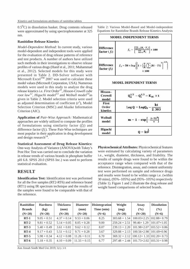
Ranitidine Release KineticsModel-Dependent Method: In current study, various model-dependent and independent tools were applied for the evaluation of drug release patterns of reference and test products. A number of authors have utilized such methods in their investigations to observe release profiles of various drugs (Hanif et al., 2011; Muhammad et al., 2012). Selected models for this study were presented in Table 2. DD-Solver software with Microsoft ExcelTM 2007 was used to calculate these model values (Microsoft Corporation, USA). Numerous models were used in this study to analyze the drug release kinetics i.e. First Order11, Hixson-Crowell cube root law12, Higuchi model13 and Weibull model14as given in Table 2. Model selection criteria were used as adjusted determination of coefficient (r2), Model Selection Criterion (MSC) and Akaike Information Criterion (AIC).
Application of Pair-Wise Approach: Mathematical approaches are widely utilized to compute the profiles of formulations using similarity factor (f2) and difference factor (f1). These Pair-Wise techniques are most popular in their application in drug development and design research14.
Statistical Assessment of Drug Release Kinetics: One-way Analysis of Variance (ANOVA)with Tukey’s Post Hoc Test was carried out to conclude the variation in release trends of various brands in phosphate buffer pH 6.8. SPSS 20.0 (SPSS Inc.) was used to perform statistical evaluation.
RESULTIdentification Test: Identification test was performed for all the five samples (RT2-RT6) and reference brand (RT1) using IR spectrum technique and the results of the samples were found to be comparable with that of the reference.
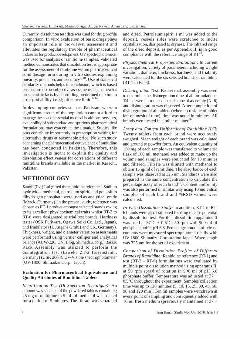
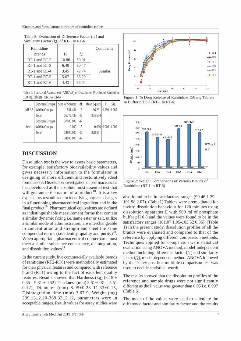
Physicochemical Attributes: Physicochemical features were estimated by calculating variety of parameters i.e., weight, diameter, thickness, and friability. The results of sample drugs were found to be within the acceptance range when compared with that of the reference. Disintegration, assay, and content uniformity test were performed on sample and reference drugs and results were found to be within range i.e. (within 30 mins), (95%–105%) and (95%–105%) respectively (Table 1). Figure 1 and 2 illustrate the drug release and weight based comparisons of selected brands.
Ann Jinnah Sindh Med Uni 2019; 5(1): 3-9
Ranitidine Brands (N=20)RT-1RT-2RT-3RT-4RT-5RT-6
Hardness (kg)
(N=20)9.05 + 0.519.81 + 0.525.40 + 0.499.17 + 0.435.98 + 0.345.18 + 0.35
Thickness(mm)
(N=20)4.37 + 0.145.14 + 0.053.61 + 0.815.51 + 0.124.12 + 0.084.10 + 0.09
Diameter (mm)
(N=20)9.53 + 0.068.05 + 0.289.62 + 0.129.71 + 0.2811.26 + 0.1111.33 + 0.15
Disintegration Time (min)
(N=6)8.259.008.073.676.706.77
Weight (mg)
(N=20)303.68 + 1.54259.24 + 2.51239.13 + 2.20329.88 + 2.13369.32 + 2.12297.48 + 2.44
Assay (%)
(N=20)100.03+2.2599.46+ 1.29101.98+2.07100.56+2.98100.12+ 1.33101.73+2.02
Dissolution(%)
(N=6)102.88+ 0.79101.07+1.05103.52+ 0.86101.69+0.96102.20+ 0.73103.31+ 0.98
Table 2: Various Model-Based and Model-independent Equations for Ranitidine Brands Release Kinetics Analysis
Order
5
Kinetics and formulations attributes of ranitidine tablets
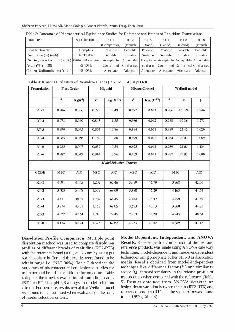
Dissolution Profile Comparison: Multiple point dissolution method was used to compare dissolution profiles of different brands of ranitidine (RT2-RT6) with the reference brand (RT1) at 325 nm by using pH 6.8 phosphate buffer and the results were found to be within range i.e. (NLT 80%). Table 3 describes the outcomes of pharmaceutical equivalence studies for reference and brands of ranitidine formulations. Table 4 depicts the kinetics evaluation of ranitidine brands (RT-1 to RT-6) at pH 6.8 alongwith model selection criteria. Furthermore, results reveal that Weibull model was found to be best fitted when evaluated on the basis of model selection criteria.
Model-Dependant, Independent, and ANOVA Results: Release profile comparison of the test and reference products was made using ANOVA-one way technique, model-dependent and model-independent techniques using phosphate buffer pH 6.8 as dissolution media. Results obtained from model-independent technique like difference factor (f1) and similarity factor (f2) showed similarity in the release profile of test products when compared with the reference. (Table 5) Results obtained from ANOVA detected an insignificant variation between the test (RT2-RT6) and reference product (RT1) as the value of p was found to be 0.997 (Table 6).
Ann Jinnah Sindh Med Uni 2019; 5(1): 3-9
Table 3: Outcomes of Pharmaceutical Equivalence Studies for Reference and Brands of Ranitidine FormulationsParameters
Identification TestDissolution (%) (n=6)Disintegration Test (min) (n=6)Assay (%) (n=20)Content Uniformity (%) (n=20)
Specifications
CompliesNLT 80%
Within 30 minutes95-105%95-105%
RT-1(Comparator)
PassableSuitable
AcceptableConformedAdequate
RT-2(Brand)PassableSuitable
AcceptableConformedAdequate
RT-3(Brand)PassableSuitable
AcceptableconformAdequate
RT-4(Brand)PassableSuitable
AcceptableConformedAdequate
RT-5(Brand)PassableSuitable
AcceptableConformedAdequate
RT-6(Brand)PassableSuitable
AcceptableConformedAdequate
Table 4: Kinetics Evaluation of Ranitidine Brands (RT-1 to RT-6) at pH 6.8
6
Shaheen Parveen, Huma Ali, Maria Sodagar, Amber Nawab, Anum Tariq, Fozia Israr
DISCUSSIONDissolution test is the way to assess basic parameters, for example, satisfactory bioavailability values and gives necessary information to the formulator in designing of more efficient and restoratively ideal formulations. Dissolution investigation of pharmaceuticals has developed as the absolute most essential test that will guarantee the nature of a product18. It is a key explanatory test utilized for identifying physical changes in a functioning pharmaceutical ingredient and in the final product19. Pharmaceutical equivalents are defined as indistinguishable measurement forms that contain a similar dynamic fixing i.e. same ester or salt, utilize a similar mode of administration, are interchangeable in concentration and strength and meet the same compendial norms (i.e. identity, quality and purity)20. When appropriate, pharmaceutical counterparts must meet a similar substance consistency, disintegration, and dissolution values21.
In the current study, five commercially available brands of ranitidine (RT2-RT6) were methodically estimated for their physical features and compared with reference brand (RT1) owing to the fact of excellent quality features. Results showed that Hardness (kg) (5.18 ± 0.35 – 9.81 ± 0.52), Thickness (mm) 3.61±0.81 – 5.51 0.12), Diameter (mm) 8.05±0.28–11.33±0.15, Disintegration time (min) 3.67-9, Weight (mg) 239.13±2.20-369.32±2.12, parameters were in acceptable ranges. Result values for assay studies were
also found to be in satisfactory ranges (99.46 1.29 – 101.98 2.07). (Table1) Tablets were premeditated for invitro dissolution behaviour for 120 minutes using dissolution apparatus II with 900 ml of phosphate buffer pH 6.8 and the values were found to be in the satisfactory ranges (101.07 1.05-103.52 0.86). (Table 1) In the present study, dissolution profiles of all the brands were evaluated and compared to that of the reference by applying different comparison methods. Techniques applied for comparison were statistical evaluation using ANOVA method, model-independent method including difference factor (f1) and similarity factor (f2), model-dependent method. ANOVA followed by the Tukey post hoc multiple comparison test was used to decide statistical worth.
The results showed that the dissolution profiles of the reference and sample drugs were not significantly different as the P value was greater than 0.05 i.e. 0.997 (Table 6).
The mean of the values were used to calculate the difference factor and similarity factor and the results
Ann Jinnah Sindh Med Uni 2019; 5(1): 3-9
Table 5: Evaluation of Difference Factor (f1) and Similarity Factor (f2) of RT-1 to RT-6
RanitidineBrands
RT-1 and RT-2RT-1 and RT-3RT-1 and RT-4RT-1 and RT-5RT-1 and RT-6
f110.866.403.455.674.43
f250.0160.4772.7463.2966.04
Comments
Similar
Table 6: Statistical Assessment (ANOVA) of Dissolution Profiles of Ranitidine 150 mg Tablets (RT-1 to RT-6)
pH 6.8
time
Between GroupsWithin GroupsTotalBetween GroupsWithin GroupsTotal
Sum of Squares831.454
36772.43337603.887
0.00034800.00034800.000
df5
42475
4247
Mean Square166.291875.534
0.000828.571
F0.190
0.000
Sig.0.965
1.000
Figure 1: % Drug Release of Ranitidine 150 mg Tablets in Buffer pH 6.8 (RT-1 to RT-6)
Figure 2: Weight Comparisons of Various Brands of Ranitidine (RT-1 to RT-6)
7
Kinetics and formulations attributes of ranitidine tablets
for f1 and f2 were found to be in order of (3.45-10.86) and (50.01-72.74), respectively (Table 5). f1 values equal to 15 (0-15) and f2 range of 50-100 guarantees similarity or proportionality of the two brands and subsequently the sameness of the test and reference22. In case, if the estimation of f2 is 50, 90% comparability in the profile was shown and the value up to 40, then 80% likeness might be demonstrated. Thus, the outcomes from this investigation uncovered similitude in the medication release.
The slope and coefficient of determination (r2) values were identified using each model. For First Order and Higuchi models, the r2 values were in the range of 0.973-0.990 and 0.639-0.867 respectively. Using Hixson-Crowell model, values of r2 lied in the range of 0.925-0.994. Weibull model gave best curve fitting with highest values of coefficient of determination (0.984-0.989). The determination of the fitting model in the medication discharge behaviour is important to guarantee the viability of the investigation. Different criteria for the choice of the numerical models which depend on the factual treatments are reported in multiple literatures. The most generally utilized strategy uses the coefficient of assurance, r2 to determine the best fit equation condition. This strategy can be utilized when the parameters of the model conditions are comparable19,21,22.
Other widely accepted techniques include Model Selection Criteria (MSC) and Akaike Information Criterion (AIC). The AIC, as characterized above, is reliant on the extent of the data points and additionally the quantity of perceptions. What is more, the most fitting model is the one with the littlest estimation of the AIC. The MSC will give an indistinguishable ranking between models from the AIC and has been standardized with the goal that it is autonomous of the scaling of the information focuses. Besides, the most fitting model will be that with the biggest MSC (to boost the "data content" of the model)24.
As observed from Table 4, Weibull model proves to be the best fit model followed by First-Order, Hixson-Crowell, and Higuchi models. The values of AIC and MSC for Weibull model are in the range of (40.64-44.64) and (3.860-4.259) respectively. AIC values for First-Order, Hixson-Crowell, and Higuchi models are found to be (39.57-51.48), (35.32-58.28) and (64.45-72.45), respectively. Hixson-Crowell model gave MSC value in the range of (2.228-4.944) whereas the MSC values observed for First Order and Higuchi model are in the range of (3.403-4.471) and (1.202-1.710) respectively. Other investigations conducted by Ali et al. and Naqvi et al. also reported Weibull as prominent
model for description of drug release of Gatifloxacin tablets22,23.
CONCLUSIONAll the selected products (RT-1 to RT-6) of ranitidine brands verified the adequate physico-chemical characteristics and confirmed the satisfactory in vitro drug release profiles. Such studies not only offer exceptional avenues for choice of superior alternatives accessible in drug market as prominent products but also assist in the most favourable care of patients in developing countries, where ease of access and affordability of these products influence swift healthcare provision.
Authors’contributions: Prof. Huma Ali conceived the idea, worked on data collection, data analysis and review, and also worked on introduction and discussion. Dr Shaheen Parveen and Dr Fozia Israr worked on literature search, results, and discussion. Dr Maria Sodagar and Dr Amber Nawab reviewed the literature, worked on discussion and edited the manuscript. Dr Anum Tariq reviewed the literature, result and conclusion in the discussion. All authors contributed to the final manuscript.
References
1. Garcia RS, Belafsky PC, Della MA, Osborn JM, Pypendop BH, Pierce T, et al. Prevalence of Gastroesophageal Reflux in Cats During Anesthesia and Effect of Omeprazole on Gastric pH. J Vet Intern Med.2017; 31(3): 734-742
2. Chang YX, Qiu YQ, Du LM, Li CF, Guo M. Determination of ranitidine, nizatidine, and cimetidine by a sensitive fluorescent probe. Analyst. 2011; 136(20): 4168-73
3. VaniaMaslarska, Determination of ranitidine hydrochloride in pharmaceutical preparations by direct Potentiometriy; Inter J Pharm Pharmaceut Sci; 2014; 6(1): 538-540
4. Ashiru Diane AI, Patel R, Basit AW, Simple and universal HPLC-UV method to determine cimetidine, ranitidine, famotidine and nizatidine in urine: Application to the analysis of ranitidine and its metabolites in human volunteers; J Chromatogr B. 2007; 860(2): 235–240.
5. Júnior AS, Barbosa AI, Santos VL, Silva RL, Junior EC. Test of dissolution and comparison of in vitro dissolution profiles of coated ranitidine tablets marketed in Bahia, Brazil. Braz J Pharm sci. 2014; 50(1): 83-89
6. FDA, Center for Drug Evaluation and Research. Guidance for Industry-Immediate release solid dosage forms: Scale-up and postapproval changes (SUPAC-IR): Chemistry, manufacturing, and controls, in vitro dissolution testing and in vivo bioequivalence test documentation. 1995
Ann Jinnah Sindh Med Uni 2019; 5(1): 3-98
Shaheen Parveen, Huma Ali, Maria Sodagar, Amber Nawab, Anum Tariq, Fozia Israr
7. Alsayegh AM, Alshirifi AN, Jaafer S, New spectrophotometric determination of Ranitidine Hydrochloride in different pharmaceutical samples. Irq Nat J Chem. 2014; 56: 357-366
8. Hussain A, Hanif M, Shoaib MH, Yousuf RI, Ali T, Muhammad IN, et.al. Comparative Studies of Ciprofloxacin 250 mg Tablets Available in Pakistani Market. Lat Am J Pharm. 2013; 32 (4): 484-89
9. Karmarkar AB, Gonjari ID, Hosmani AH, Dhabale PN & Bhise SB, Dissolution rate enhancement of fenofibrate using liquisolid tablet technique. Lat Am J Pharm. 2009; 28(2): 219-25.
10. Hanif M, Shoaib MH, Yousuf RI, Sattar S, Nadeem M, Hussain L, et,al. Formulation development of intermediate release Nimesulide tablets by CCRD for IVIVC studies. Pak J Pharm Sci. 2014; 27(4): 785-92
11. Zafar F, Shoaib MH, Yousuf RI, Development and Evaluation of fast dispersible ketoprofen 100mg tablets. Asian J Pharm Rese. 2012; 2 (1): 1-9
12. Zafar F, Ali H, Shabana NS, Naveed S, Siddiqui S, Quality assessment and dissolution profile comparison studies on 250mg mefenamic acid tablets available in local market of Karachi. J Chin Pharm Sci. 2015; 24 (10): 673–677
13. Ali H, Shoaib MH, Zafar F, Bushra R, Yasmin R, Siddiqui S, et,al. Intermediate release formulations of diclofenac potassium tablets for IVIVC. Pak J Pharm Sci. 2016; 29(4): 1287-98
14. Bushra R, Shoaib MH, Ali H, ZAFAR F, Shafiq Y & Aslam N. In vitro drug analysis and stability studies of optimized formulations of aceclofenac (100 mg) Tablets. Lat Am Pharm. 2016; 35(4): 695-704
15. British Pharmacopeia. Pharmacopeial Convention 2013
16. United States Pharmacopeia 27. Rockville: US Pharmacopeial Convention 2003
17. Wadulkar RD, Kalyankar TM, Pawar AA, Anitha K, Stability Indicating UV Spectrophotometric Assay Method Development for Simultaneous Determination of Ranitidine and Dicyclomine in Bulk and Pharmaceutical Dosage Form. Asian J Pharm Tech Innov. 2016; 4 (20):26-33
18. Arshad HM, Shoaib MH, Ghayas S, Yousuf RI, Hanif M, et,al. Pharmaceutical quality control studies on Gatifloxacin 200 mg tablets available in the Pakistani market. Lat Am J Pharm. 2011; 30(10): 1922-26.
19. Muhammad IN, Shoaib MH, Yousuf RI, Hanif M, Jabeen S, Ali T. Formulation Development and optimization of Cefuroxime Axetil tablets by directcompression method and its stability studies. Lat Am J Pharm. 2012; 31(2): 271-78
20. Moore JW, Flanner HH. Mathematical Comparison of Curves with an Emphasis on In-Vitro Dissolution Profiles. Pharm Technol. 1996; 20(6): 64–74
21. Naqvi GR, Baqir SN, Harris MS, Huma A, Farya Z, Shaheen P, et,al. Pharmaceutical Surveillance Study of Moxifloxacin Formulations: Therapeutic Perspective in Terms of Quality and Efficacy. Lat Am J Pharm. 2018; 37 (6): 1104-14
22. Rasul A, Iqbal M, Murtaza G, Waqas MK, Hanif M, Khan SA, et, al. Design, development and in – vitro evaluation of metoprolol tartrate tablets containing xanthan – tragacanth. Acta Pol Pharm. 2010 ; 67 (5): 517-22
23. Ali H, Farya Z, Huma S, Fozia I, Shaheen P, Amber N, et,al. Validation of Spectroscopic Dissolution Methodof Gatifloxacin: Statistical Comparison of In Vitro Therapeutic Equivalence and Quality Attributes. LatAm J Pharm. 2018;37 (6): 1231-43
24. Traple MAL, Okamoto RT, Yamamoto RN, Pinto TA and Lourenço FR. A new multivariate similarity factorfor in vitro therapeutic equivalence assessment. Afr JPharm Pharmacol, 2014; 8(6): 185-93
Ann Jinnah Sindh Med Uni 2019; 5(1): 3-9 9
Kinetics and formulations attributes of ranitidine tablets
Shazia Nazar1, Ambreen Qamar1, Sara Rafique1, and Shayan Zufishan2
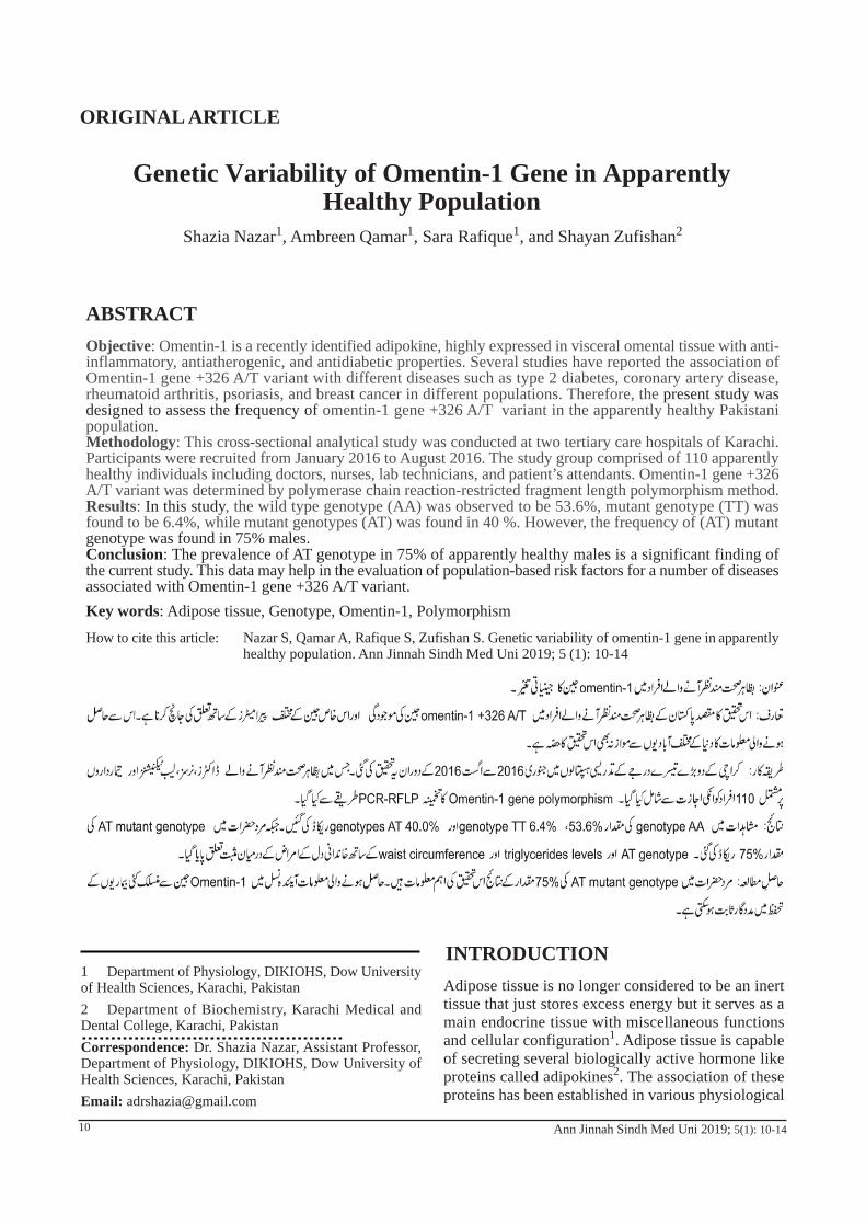
Genetic Variability of Omentin-1 Gene in Apparently Healthy Population
ORIGINAL ARTICLE
Ann Jinnah Sindh Med Uni 2019; 5(1): 10-14
ABSTRACTObjective: Omentin-1 is a recently identified adipokine, highly expressed in visceral omental tissue with anti-inflammatory, antiatherogenic, and antidiabetic properties. Several studies have reported the association of Omentin-1 gene +326 A/T variant with different diseases such as type 2 diabetes, coronary artery disease, rheumatoid arthritis, psoriasis, and breast cancer in different populations. Therefore, the present study was designed to assess the frequency of omentin-1 gene +326 A/T variant in the apparently healthy Pakistani population.Methodology: This cross-sectional analytical study was conducted at two tertiary care hospitals of Karachi. Participants were recruited from January 2016 to August 2016. The study group comprised of 110 apparently healthy individuals including doctors, nurses, lab technicians, and patient’s attendants. Omentin-1 gene +326 A/T variant was determined by polymerase chain reaction-restricted fragment length polymorphism method.Results: In this study, the wild type genotype (AA) was observed to be 53.6%, mutant genotype (TT) was found to be 6.4%, while mutant genotypes (AT) was found in 40 %. However, the frequency of (AT) mutant genotype was found in 75% males. Conclusion: The prevalence of AT genotype in 75% of apparently healthy males is a significant finding of the current study. This data may help in the evaluation of population-based risk factors for a number of diseases associated with Omentin-1 gene +326 A/T variant.Key words: Adipose tissue, Genotype, Omentin-1, Polymorphism
How to cite this article: Nazar S, Qamar A, Rafique S, Zufishan S. Genetic variability of omentin-1 gene in apparently healthy population. Ann Jinnah Sindh Med Uni 2019; 5 (1): 10-14
1 Department of Physiology, DIKIOHS, Dow University of Health Sciences, Karachi, Pakistan2 Department of Biochemistry, Karachi Medical and Dental College, Karachi, PakistanCorrespondence: Dr. Shazia Nazar, Assistant Professor, Department of Physiology, DIKIOHS, Dow University of Health Sciences, Karachi, PakistanEmail: [email protected]
INTRODUCTIONAdipose tissue is no longer considered to be an inert tissue that just stores excess energy but it serves as a main endocrine tissue with miscellaneous functions and cellular configuration1. Adipose tissue is capable of secreting several biologically active hormone like proteins called adipokines2. The association of these proteins has been established in various physiological
10
processes including inflammation, hunger, energy metabolism, insulin resistance, immunity, and angiogenesis3. There is a greater possibility of developing cardiac disease, diabetes mellitus, and other metabolic disorders with expansion of visceral fats as compared to subcutaneous fats; however, both types of this tissue are involved in production of a unique profile of adipokines4. Omentin-1, a 34kDa protein consisting of 295 amino acids, is a newly identified adipokine which is mainly secreted by stromal vascular cells of visceral omental fat5,6. Omentin-1 is an anti-inflammatory protein that is highly expressed in epicardial adipose tissue (EAT) around the heart and coronary arteries7,8. The expression of omentin-1 in the heart, lungs, ovary, and placenta has also been reported, but its role in these organs is not yet completely known. Omentin protein exists in two forms: omentin-1 and omentin-2; however the major circulating isoform is omentin-1. Its plasma level is 100 ng/ml to 1 ug/ml8,9. Omentin-1 has anti-inflammatory, anti-atherogenic, and anti-diabetic properties. It may improve insulin sensitivity in human adipocyte, myocytes, and hepatocytes via activation of AMPK/Akt pathway10. Furthermore, in vitro studies have shown that omentin-1 causes inhibition of tumor necrosis factor Alfa (TNFá) induced degradation of IkB and NF-kB (nuclear factor kappa B) activity to reduce inflammation11. Omentin-1 serum level is observed to be lower in obese subjects12 and type 2 diabetic patients13. The Omentin-1 gene contains 1269bp, with 8 exons and 7 introns, localized at 1q22-q23 position14. In 2007, Val109Asp single nucleotide polymorphism in exon 4 of omentin-1 gene was reported and +326 A/T nucleotide was declared to be polymorphic15. Several studies have discussed the association of this single nucleotide polymorphism (SNP) in different diseases such as type 2 diabetes15, coronary artery disease20, rheumatoid arthritis19, psoriasis9, and breast cancer17. The frequency of +326 A/T nucleotide variant has been described from several world populations so, it is quite important to know the prevalence of such a clinically significant gene polymorphism in the healthy population of Pakistan. Current study is the first study, to the best of our knowledge, that has observed the prevalence omentin-1 gene +326 A/T variant in apparently healthy and non-symptomatic individuals of Pakistan. The data will help in future studies in finding other disorders associated with omentin-1 gene +326 A/T variant.
METHODOLOGY The current study was approved from the ethical review board of Dr. Abdul Qadeer Khan Institute of Biotechnology and Genetic Engineering (KIBGE). A
total of 110 healthy individuals were recruited in the Civil hospital Karachi (CHK) and the Karachi Institute of Heart Diseases (KIHD) from January 2016 to August 2016. Sample size was calculated using open epi software. Convenient sampling was used to select the participants. All the selected healthy participants, including doctors, nurses, lab technicians, and patients’ attendants, were informed about the methods and significance of study. Written Consents were taken from all participants. Information about age, gender, family history, ethnicity, and past medical history was recorded through proforma. The blood pressure of the patients was measured via defined protocol. Weight, height, and waist circumference were also measured. Weight and height were measured to the nearest kilogram and centimeter, respectively. BMI was calculated by standard formula (kg/m2). Blood sample was drawn from brachial vein in early morning and transferred to blood collection tube containing anticoagulant EDTA (ethylene diamine tetra acetate).
Subjects with history of infectious diseases in the previous four weeks, taking anti-inflammatory drugs, statins, diabetes mellitus, heart disease, malignancy, known renal or hepatic disorder were excluded from the study.
DNA Extraction: Whole blood was used to extract genomic DNA via salting out extraction method. Nano-drop (Thermo Scientific USA) was used for quantifying the extracted DNA whereas integrity was checked by horizontal gel system by resolving 2 l genomic DNA samples on 0.8% agarose gel.
PCR Analysis: Polymerase chain reaction16 was p e r f o r m e d u s i n g F - p r i m e r 5 - GAGCCTTTAGGCCATGTCTCT-3' and the R- primer 5'- CTCTCCTTCTTCTCCAGCCCAT-3'15. Total volume of 50 l was prepared for PCR, consisting of genomic DNA, 2 units of Taq DNA polymerase, 1.5mM MgCl, 0.2 mM dNTPs, and 1X PCR buffer of pH-8.3. Initial genomic DNA pre-denaturation was done at the temperature of 94°C for 5 minutes. Denaturation phase consisted of 35 cycles at 94°C, annealing phase had 40 seconds at 62°C followed by 60 second extension phase at 72°C. The final extension time was 5 minutes at 72°C. Amplified PCR product of 471 bp was resolved on 2% of agarose gel and visualized by gel doc system (Fig. 1).
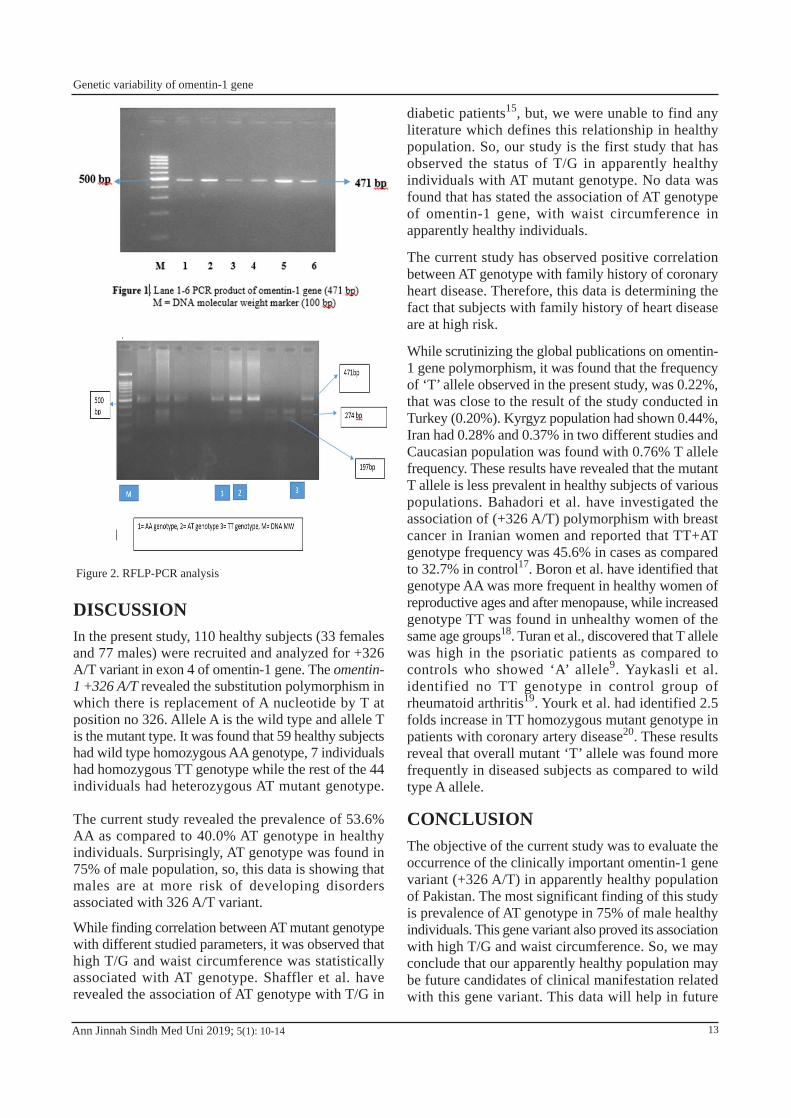
Restriction Fragment Length Enzyme Analysis: PCR products were purified and treated with 10U of restriction enzyme AccI (Molecul-on, New- Zeeland) and placed in incubator for 16 hours at 37 o C. Digested product of PCR was analyzed by Gel documentation system. TT homozygous individuals had shown two
Ann Jinnah Sindh Med Uni 2019; 5(1): 10-14 11
Genetic variability of omentin-1 gene
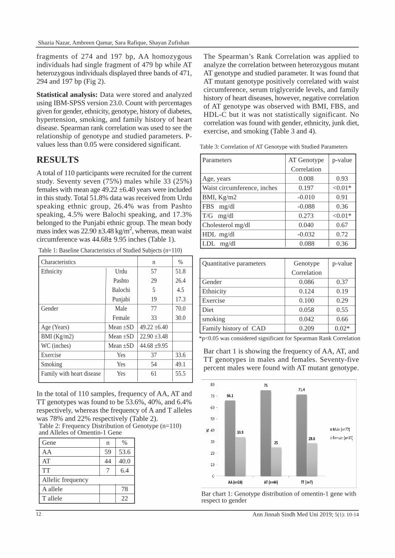
fragments of 274 and 197 bp, AA homozygous individuals had single fragment of 479 bp while AT heterozygous individuals displayed three bands of 471, 294 and 197 bp (Fig 2).
Statistical analysis: Data were stored and analyzed using IBM-SPSS version 23.0. Count with percentages given for gender, ethnicity, genotype, history of diabetes, hypertension, smoking, and family history of heart disease. Spearman rank correlation was used to see the relationship of genotype and studied parameters. P-values less than 0.05 were considered significant.
RESULTSA total of 110 participants were recruited for the current study. Seventy seven (75%) males while 33 (25%) females with mean age 49.22 ±6.40 years were included in this study. Total 51.8% data was received from Urdu speaking ethnic group, 26.4% was from Pashto speaking, 4.5% were Balochi speaking, and 17.3% belonged to the Punjabi ethnic group. The mean body mass index was 22.90 ±3.48 kg/m2, whereas, mean waist circumference was 44.68± 9.95 inches (Table 1).
In the total of 110 samples, frequency of AA, AT and TT genotypes was found to be 53.6%, 40%, and 6.4% respectively, whereas the frequency of A and T alleles was 78% and 22% respectively (Table 2).
Ann Jinnah Sindh Med Uni 2019; 5(1): 10-14
The Spearman’s Rank Correlation was applied to analyze the correlation between heterozygous mutant AT genotype and studied parameter. It was found that AT mutant genotype positively correlated with waist circumference, serum triglyceride levels, and family history of heart diseases, however, negative correlation of AT genotype was observed with BMI, FBS, and HDL-C but it was not statistically significant. No correlation was found with gender, ethnicity, junk diet, exercise, and smoking (Table 3 and 4).
Bar chart 1 is showing the frequency of AA, AT, and TT genotypes in males and females. Seventy-five percent males were found with AT mutant genotype.
Table 1: Baseline Characteristics of Studied Subjects (n=110)
CharacteristicsEthnicity
Gender
Age (Years)BMI (Kg/m2)WC (inches)ExerciseSmokingFamily with heart disease
UrduPashtoBalochiPunjabi
MaleFemale
Mean ±SDMean ±SDMean ±SD
YesYesYes
n57295197733
49.22 ±6.4022.90 ±3.4844.68 ±9.95
37 5461
% 51.826.44.517.370.030.0
33.649.155.5
Table 2: Frequency Distribution of Genotype (n=110) and Alleles of Omentin-1 GeneGeneAAATTTAllelic frequencyA alleleT allele
n59447
%53.640.06.4
7822
Table 3: Correlation of AT Genotype with Studied Parameters
Quantitative parameters
GenderEthnicityExerciseDietsmokingFamily history of CAD
Genotype Correlation
0.0860.1240.1000.0580.0420.209
p-value
0.370.190.290.550.660.02*
*p<0.05 was considered significant for Spearman Rank Correlation
Parameters
Age, yearsWaist circumference, inchesBMI, Kg/m2FBS mg/dlT/G mg/dlCholesterol mg/dlHDL mg/dlLDL mg/dl
AT Genotype Correlation
0.0080.197-0.010-0.0880.2730.040-0.032 0.088
p-value
0.93<0.01*
0.910.36
<0.01*0.670.720.36
Bar chart 1: Genotype distribution of omentin-1 gene with respect to gender
12
Shazia Nazar, Ambreen Qamar, Sara Rafique, Shayan Zufishan
DISCUSSIONIn the present study, 110 healthy subjects (33 females and 77 males) were recruited and analyzed for +326 A/T variant in exon 4 of omentin-1 gene. The omentin-1 +326 A/T revealed the substitution polymorphism in which there is replacement of A nucleotide by T at position no 326. Allele A is the wild type and allele T is the mutant type. It was found that 59 healthy subjects had wild type homozygous AA genotype, 7 individuals had homozygous TT genotype while the rest of the 44 individuals had heterozygous AT mutant genotype.
The current study revealed the prevalence of 53.6% AA as compared to 40.0% AT genotype in healthy individuals. Surprisingly, AT genotype was found in 75% of male population, so, this data is showing that males are at more risk of developing disorders associated with 326 A/T variant.
While finding correlation between AT mutant genotype with different studied parameters, it was observed that high T/G and waist circumference was statistically associated with AT genotype. Shaffler et al. have revealed the association of AT genotype with T/G in
diabetic patients15, but, we were unable to find any literature which defines this relationship in healthy population. So, our study is the first study that has observed the status of T/G in apparently healthy individuals with AT mutant genotype. No data was found that has stated the association of AT genotype of omentin-1 gene, with waist circumference in apparently healthy individuals.
The current study has observed positive correlation between AT genotype with family history of coronary heart disease. Therefore, this data is determining the fact that subjects with family history of heart disease are at high risk.
While scrutinizing the global publications on omentin-1 gene polymorphism, it was found that the frequency of ‘T’ allele observed in the present study, was 0.22%, that was close to the result of the study conducted in Turkey (0.20%). Kyrgyz population had shown 0.44%, Iran had 0.28% and 0.37% in two different studies and Caucasian population was found with 0.76% T allele frequency. These results have revealed that the mutant T allele is less prevalent in healthy subjects of various populations. Bahadori et al. have investigated the association of (+326 A/T) polymorphism with breast cancer in Iranian women and reported that TT+AT genotype frequency was 45.6% in cases as compared to 32.7% in control17. Boron et al. have identified that genotype AA was more frequent in healthy women of reproductive ages and after menopause, while increased genotype TT was found in unhealthy women of the same age groups18. Turan et al., discovered that T allele was high in the psoriatic patients as compared to controls who showed ‘A’ allele9. Yaykasli et al. identified no TT genotype in control group of rheumatoid arthritis19. Yourk et al. had identified 2.5 folds increase in TT homozygous mutant genotype in patients with coronary artery disease20. These results reveal that overall mutant ‘T’ allele was found more frequently in diseased subjects as compared to wild type A allele.
CONCLUSION The objective of the current study was to evaluate the occurrence of the clinically important omentin-1 gene variant (+326 A/T) in apparently healthy population of Pakistan. The most significant finding of this study is prevalence of AT genotype in 75% of male healthy individuals. This gene variant also proved its association with high T/G and waist circumference. So, we may conclude that our apparently healthy population may be future candidates of clinical manifestation related with this gene variant. This data will help in future
Ann Jinnah Sindh Med Uni 2019; 5(1): 10-14
Figure 2. RFLP-PCR analysis
13
Genetic variability of omentin-1 gene
studies searching for other disorders associated with omentin-1 gene +326 A/T variant. Further studies are needed to find out whether there is a change in omentin-1 protein physiological function with mutant AT genotype of omentin-1 polymorphism among healthy individuals. Author’s contribution: Dr Shazia Nazar conceived the idea, worked on literature search, data collection, data analysis and review, worked on introduction, discussion and result, drew the conclusion from the discussion and edited the manuscript. Dr Ambreen Qamar and Dr Shayan Zoufishan worked on literature search, results, and discussion. Dr Sara Rafique reviewed the literature, worked on discussion, and edited the manuscript. All authors discussed the results and contributed to the final manuscript.
References1. Smitka K, Maresova D. Adipose tissue as an endocrine
organ: An update on pro-inflammatory and anti-inflammatory microenvironment. Prague Med Rep. 2015; 116(2): 87-111
2. Pepe J, Cipriani C, Cilli M, Colangelo L, & Minisola S. Adipokines and bone metabolism: an interplay to untangle. J Endocrinol Invest. 2016; 39(11): 1359-13661
3. Kajiya M, Miyoshi T, Doi M, Usui S, Iwamoto M, Takeda K. Serum adipocyte fatty acid binding protein is independently associated with complex coronary lesions in patients with stable coronary artery disease. Heart vessels. 2013; 28(6): 696-703
4. Korner A, Kratzsch J, Gausche R, Schaab M, Erbs S, Kiess W. New predictors of the metabolic syndrome in children- role of adipocytokines. Pediatr Res. 2007; 61(6):640-645
5. Isakova ZT, Talaibekova ET, Asambaeva DA, Kerimkulova AS, Lunegova OS, Aldasheva NM, et.al. A polymorphic marker Val109Asp in the omentin gene are associated with abdominal obesity in the Kyrgyz population. J Endocrinol.2016; 62(3):4-8
6. Zhou JY, Chan L, Zhou SW. Omentin: linking metabolic syndrome and cardiovascular disease. Curr vas pharma. 2014; 12(1): 136-143
7. Naruim T, Watanabe T, Kadowaki S, Kinoshita D, Honda Y, Otaki Y,et.al. Impact of Omentin-1 levels on cardiac prognosis in patients with heart failure. Cardiovasc Diabetol. 2014; 13:84
8. Pan HY, Guo L, Li Q. Changes of serum omentin-1 levels in normal subjects and in patients with impaired glucose regulation and with newly diagnosed and untreated type 2 diabetes. Diabetes Res Clin Pract. 2010; 88(1): 29-33
9. Turan H, Yaykasli KO, Soguktas H, Yaykasli E, Aliagaoglu C, Erdem T et.al. Omentin serum levels and omentin gene Val109Asp polymorphism in patients with psoriasis. Int J Dermatol. 2014; 53(5): 601-605
10. Yang RZ, Lee MJ, Hu H, Pray J, Wu HB, Hansen BC, et.al.. Identification of omentin as a novel depot –specific adipokines in human adipose tissue. Amm J Physiol Endocrinol. 2006; 290(6):1253-1261
11. Yamawaki H, Tsubaki N, Mukohda M, Okada M, Hara Y. Omentin , a novel adipocytokine induces vasodilation in rat isolated blood vessels. Biochem Biophys Res Commun. 2010; 393(4): 668-672
12. Batista CM, Yang RZ, Lee MJ, Glynn NM, et.al. Omentin plasma levels and gene expression are decreased in obesity. J Diabetes. 2007; 56(6): 1655-1661
13. Kohan L, Safarpur M, Abdollahi H. Omentin-1 rs2274907 and resistin rs1862513 polymorphisms influence genetic susceptibility to nonalcoholic fatty liver disease. Mol Biol Res Commun. 2016; 5(1): 11-17
14. El-Mesallamy HO, El-Derany MO, Hamdy NM. Serum omentin-1 and chemerin levels are interrelated in patients with type 2 diabetes mellitus with or without ischaemic heart disease. Diabet Med, 2011;28(10):1194-200
15. Schäffler A, Zeitoun M, Wobser H, Buechler C, Aslanidis C, Herfarth H. Frequency and significance of the novel single nucleotide missense polymorphism Val109Asp in the human gene encoding omentin in Caucasian patients with type 2 diabetes mellitus or chronic inflammatory bowel diseases. Cardio diabetol. 2007; 6(1): 1
16. Karabulut S, Afsar CU., Karabulut M, Alis H, Bozkurt MA, Aydogan F,et.al. Clinical significance of serum omentin-1 levels in patients with pancreatic adenocarcinoma. BBA Clin. 2016;15(6):138-142
17. Bahadori M, Kohan L, Farzan M, Aliakbari S, Panah MM. An increased risk of breast cancer associated with Val109Asp polymorphism in omentin gene. IJB. 2014; 5(1): 429-434
18. Boron D, Czerny B, Bartkowiak-Wieczorek J, Sieron D, Wolski, H. Omentin Polymorphism and its Relations to Bone Mineral Density in Women. Arch med res. 2015; 46(3): 173-180
19. Yaykasli KO, Yaykasli E. The Frequency Of Omentin Val109asp Polymorphism And The Serum Level Of Omentin In Patients With Rheumatoid Arthritis. Acta Medica Mediterranea. 2013; 29(3): 52-56
20. Yörük Ü, Yaykasli KO, Özhan H, Memisogullari R, Karabacak A, Bulur S, et.al. Association of omentin Val109Asp polymorphism with coronary artery disease. Anadolu Kardiyol Derg. 2014; 14(6): 511-514
21. Soguktas H, Yaykasli KO, Turan H, Kaya E, Yaykasli E. Omentin Val/Val genotype increases predisposition to acne vulgaris without changing omentin serum level. Cell Mol Biol (Noisy le grand). 2018; 64(12).81-86
22. Splichal Z, Bienertova-Vasku J, Novak J, Zlamal F, Tomandl J, Tomandlova M. et.al. The common polymorphism Val109Asp in the omentin gene is associated with daily energy intake in the Central-European population. Nutr neurosci. 2015;18(1):41-8
23. Bahadori M, Kohan L, Jafari N. Association of assessment between Val109Asp omentin gene andobesity in Iranian women. Iran J Diabetes Metab. 2015;14(2):127–132
Ann Jinnah Sindh Med Uni 2019; 5(1): 10-1414
Shazia Nazar, Ambreen Qamar, Sara Rafique, Shayan Zufishan
Sadia Rehman1, Santosh Kumar2, Abdul Manan Junejo2, Fatima Mehboob3, Hasan Ali1, and Noorun-nisa Memon4
Impact of Oxidative Stress on Hypertension in Patients on Maintenance Haemodialysis
ORIGINAL ARTICLE
Ann Jinnah Sindh Med Uni 2019; 5(1): 15-20
ABSTRACTObjective: To assess the association of oxidative stress with hypertension and its correlation with the duration of haemodialysisMethodology: It was a case control study conducted in a public sector tertiary care hospital in 2017. The study participants were recruited from the nephrology ward while the healthy controls were taken from participant neighborhood through frequency matching. Non probability consecutive sampling technique was employed. Cases included were suffering from chronic renal failure and were receiving maintenance haemodialysis. Exclusion criteria was patients suffering from any other chronic illness other than chronic renal failure (pulmonary disease and hepatic insufficiency). Detailed analysis was done with application of ANOVA, Pearson correlation and independent sample t test. P value less than 0.05 was taken as significant.Results: Highly significant difference was observed in mean serum malondialdehyde, mean plasma superoxide dismutase, mean systolic blood presure, and mean BMI among the cases and controls (p value < 0.001). Positive linear correlation was found between blood presure and serum malondialdehyde i.e. (r = 0.4) while on the other hand strong negative correlation was found between blood presure and plasma superoxide dismutase i.e. (r = -0.73).Conclusion: Oxidative stress worsens with progression of haemodialysis and leads to development of hypertension.Keywords: Oxidative stress, serum malondialdehyde, hypertension, plasma SOD, BMI, haemodialysis
How to cite this article: Rehman S, Kumar S, Junejo AM, Mehboob F, Ali H, Memon NN. Impact of oxidative stress on hypertension in patients on maintenance haemodialysis. Ann Jinnah Sindh Med Uni 2019; 5 (1): 15-20
1 Department of Biochemistry, Bahria University Medicaland Dental College, Karachi, PakistanDepartment of Nephrology2 / Department of Physiology4, Jinnah Sindh Medical University Karachi, Pakistan3 Department of Medicine, Sharif Medical and Dental College, Lahore, PakistanCorrespondence: Dr Sadia Rehman, Lecturer, Department of Biochemistry, Bahria University Medical and Dental College, Karachi, PakistanEmail: [email protected]
INTRODUCTIONThe main risk of mortality in patients of end stage renal disease (ESRD) is the cardiovascular diseases1.
The US renal data system and European Registry of patients, both have highlighted that the incidence of developing a cardiac event is almost five times higher in chronic dialysis patients as compared to the healthy population2. Cardiovascular events in ESRD are basically a sequel of hypertension and diabetes as these two are termed number one risk factors for renal failure3-4. In chronic renal failure, there is a loss of equilibrium between pro-oxidant and anti-oxidant capacities and a state of increased oxidative stress is evident. The factors which are held responsible for this shift are not only cardiovascular causes but also factors which are more associated to uremia5-11.
15
The role of oxidative stress is evident in the pathogenesis of hypertension, which has high prevalence in chronic renal failure patients and is considered as the number one risk factor for the progression of cardiovascular diseases12-13.
Oxidative stress promotes vascular smooth muscle proliferation and deposition of collagen leading to an increase in the intima media thickness ratio and hence causing narrowing of the vascular lumen12-18. In addition to this, oxidative stress causes imbalance between the endothelium dependent vascular relaxation and vascular contractile activity by stimulating endothelial injury and decreasing Nitric oxide availability13-16. All these factors contribute to the development of hypertension.
Malondialdehyde, a lipid peroxidation end product is produced as a result of the attack by the free radicals on the polyunsaturated fatty acids on the surface of the cell membranes. Malondialdehyde, if produced in high numbers, is a marker of systemic oxidation14-18.
The term antioxidant refers to a molecule which is capable of stabilizing or deactivating free radicals before they attack normal cells19. Endogenous antioxidants are crucial for maintaining optimal cellular functions resulting in systemic health and well-being20. However, in conditions promoting oxidative stress, dietary antioxidants may be necessary to maintain the cellular function on optimal levels as endogenous antioxidants may prove to be insufficient.
Glutathione peroxidase, catalase, and superoxide dismutase are the most efficient enzymatic antioxidants. Mitochondria, being the major site of free radical generation, contain a variety of antioxidants present on both sides of their membranes in order to minimize ROS induced stress21. Superoxide dismutase is also among the most effective enzymatic antioxidants. Superoxide dismutases (SODs) defend against oxidative damage by enzymatically converting O2- to H2O2. According to a recent study, SOD is a major antioxidant enzyme in the regulation of oxidative stress during progressive renal injury22.
When oxidative stress occurs, it triggers the oxidation of molecules such as lipids, proteins, and carbohydrates, leading to lipid peroxidation and accumulation of advanced glycation end products which cause severe damage to the endothelium11-15. Moreover, nitric oxide which causes endothelial smooth muscle relaxation is rapidly degraded by the oxygen derived free radical superoxide anion15-20. There is also a correlation between oxidative stress and renin activation32. Hence oxidative stress is involved in the pathogenesis of
various conditions including hypertension, inflammation, and the progression of chronic kidney disease to end stage renal disease12-16.
The main objective of the study was to assess the association of oxidative stress with hypertension and its correlation with duration of haemodialysis. This study emphasizes on the magnitude and complications of oxidative stress in haemodialysis patients. This study will help the nephrologists to identify oxidative stress as a major causative factor of hypertension and other cardiovascular complications and to change the typical treatment regimes by adding antioxidants to lower the oxidative stress.
METHODOLOGYA Case Control study was conducted in the Nephrology ward of a public sector hospital of Karachi in 2017. Sample size was calculated by open epi website calculator. (REFERENCE STUDY: Locatelli F, Canaud B, Eckardt KU, Stenvinkel P, Wanner C, Zoccali C. Oxidative stress in end-stage renal disease: an emerging threat to patient outcome. Nephrology Dialysis Transplantation. 2003 Jul 1; 18 (7):1272-80.)
A sample size of 90 subjects was calculated which was further subdivided into three groups::
Group A: Healthy control group comprised 30 subjects from the neighborhood who volunteered for the study, and were matched on age, gender, and socio-economic status. Their routine laboratory investigations were within normal ranges.
Group B: Thirty subjects who had been on haemodialysis for up to three years
Group C: Thirty subjects who were on haemodialysis for more than three years
Inclusion criteria for the cases with chronic renal failure was taking haemodialysis therapy for more than two months. Exclusion criteria was the same for all the groups and comprised omission of patients with hepatic insufficiency, chronic pulmonary disease, and diabetes mellitus. Non-probability consecutive sampling technique was utilized for selection of participants. Biochemistry lab investigations and oxidative stress biomarkers were measured in all three groups. Hypertensive cutoff was taken for systolic BP 140 mmHg and for diastolic BP at 90 mmHg. Ethical permission for the present study was taken from the Institutional Review Committee, Jinnah Postgraduate Medical Centre (JPMC), Karachi diary no: NO.F.2-81-IRB/2018- GENL/5173/JPMC. Data which was obtained during the study was kept confidential.
Ann Jinnah Sindh Med Uni 2019; 5(1): 15-2016
Sadia Rehman, Santosh Kumar, Abdul Manan Junejo, Fatima Mehboob, Hasan Ali, Noorun-nisa Memon
BMI was calculated from weight and height measurements which were obtained through calibrated apparatus available in the wards. Blood pressure was measured in supine position after allowing the participant to relax for 10 minutes. Later on, 10 ml blood samples were collected before dialysis therapy in patients on haemodialysis.
The malondialdehyde (MDA) was estimated in the form of thiobarbituric acid reacting substances (TBARS) by the method of Okhawa et al, 1979. Levels of SOD were measured by using reagent method (method of Kono, 1978).
Data was entered on SPSS version 21. Mean and standard deviation were taken out for all numeric variables, whereas frequencies and percentages were taken out for categorical variables. One-way ANOVA was applied for finding difference in mean between the three groups after fulfilling the assumptions of normality and homogeneity through Shapiro Wilk test and QQ plot and Levenne test. Post hoc analysis was done through Tukey’s test. Pearson correlation was applied for finding association of blood pressure with serum malondialdehyde and plasma SOD. Two sample t-test was applied for finding difference in means of serum malondialdehyde and plasma SOD on the basis of gender. P value <0.05 was taken as significant.
RESULTSA total of n=90 participants were recruited in three groups. Males were n=55 (61%) and females were n=35 (39%). The mean age of the participants was 38±8 years.
The group A comprised n=30 healthy participants. The mean age of the group was 35±7.7 years. The mean systolic BP was 108±10mmHg. Mean serum malondialdehyde (nmol/ml) was 10.87±3.04. Plasma superoxide dismutase (µ/l) was 108.5±19.4. The mean BMI of the group was 23.4±3.3 kg/m2.
Group B, n=30 corresponded to participants who were on haemodialysis for the last three years. Mean age of the group was 36±8 years. Mean systolic blood pressure
Ann Jinnah Sindh Med Uni 2019; 5(1): 15-20
was 136±11mmHg. Mean serum malondialdehyde (nmol/ml) was 15.7±3. Plasma superoxide dismutase (µ/l) was 85±16. The mean BMI of the group was 22.6±3.7.
Group C, n=30 corresponded to participants who were on haemodialysis for more than three years. Mean age of the group was 43±4 years. Mean systolic blood pressure was 159±12mmHg. Mean serum malondialdehyde (nmol/ml) was 31±8. Plasma superoxide dismutase (µ/l) was 46±19. The mean BMI of the group was 20.8±4.6.
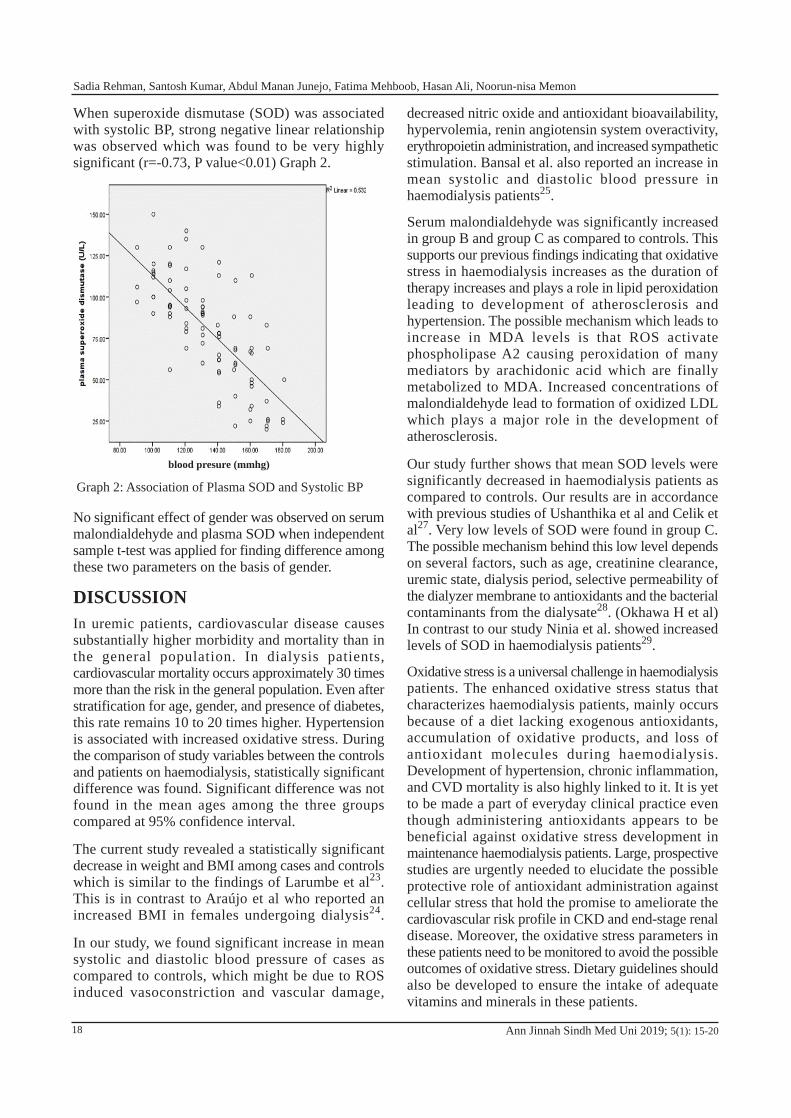
Table 1 summarizes the significant differences which were observed when ANOVA was applied for serum malondialdehyde, SOD, systolic BP, and BMI means difference among the three groups. Post hoc analysis was done by the Tukey HSD test. Significant differences were observed between all groups when mean serum malondialdehyde, SOD, and systolic BP was compared. Borderline significance was observed only in Group A and C when mean BMI was compared in between groups through Tukey HSD.
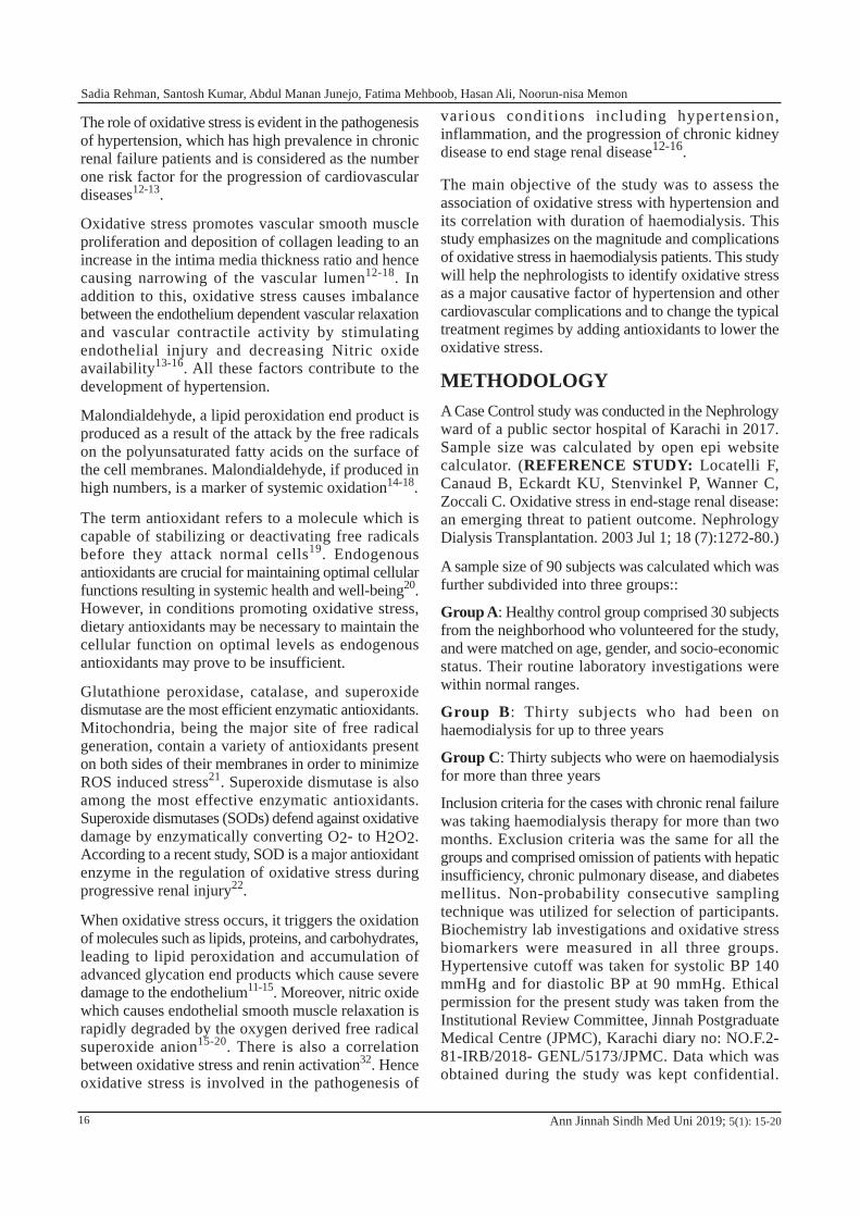
When serum malondialdyde was associated with systolic BP, very highly significant mild positive linear correlation was seen (r=0.4, p value<0.01) Graph 1.
Table 1: Differences in Mean Numeric Variables Between the Three Groups (N=90)
Groups
ABC
P-Value*
n
303030-
SerumMalondialdehyde (nmol/ml)
Mean±Std.dev10.9±3.0315.7±2.9
31.01±8.48<0.01
SODMean±Std.dev
108.5±485±1646±19<0.01
Systolic BPMean±Std.dev
108±10136±2
159.6±12.3<0.01
BMIMean±Std.dev
23.4±3.322.6±3.720.8±4.0
0.037
* ANOVA
Graph 1: Association of Serum Malondialdehyde and Systolic BP
17
Oxidative stress on hypertension on maintenance haemodialysis
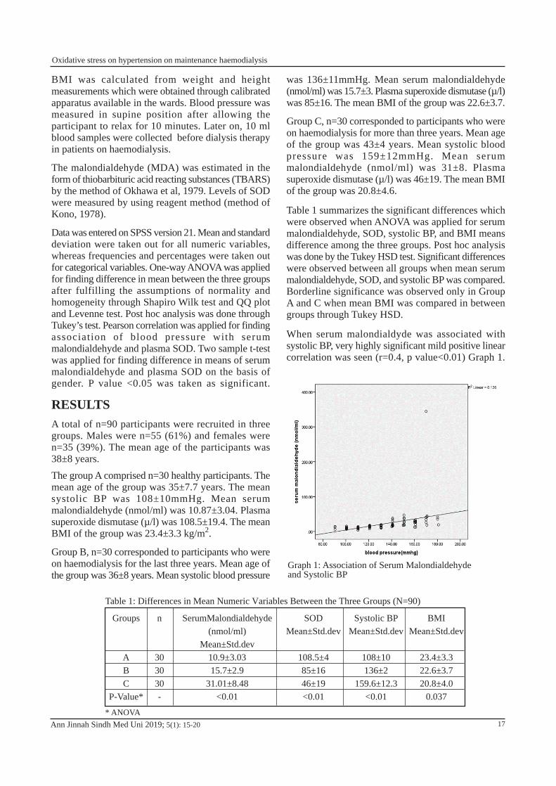
When superoxide dismutase (SOD) was associated with systolic BP, strong negative linear relationship was observed which was found to be very highly significant (r=-0.73, P value<0.01) Graph 2.
No significant effect of gender was observed on serum malondialdehyde and plasma SOD when independent sample t-test was applied for finding difference among these two parameters on the basis of gender.
DISCUSSIONIn uremic patients, cardiovascular disease causes substantially higher morbidity and mortality than in the general population. In dialysis patients, cardiovascular mortality occurs approximately 30 times more than the risk in the general population. Even after stratification for age, gender, and presence of diabetes, this rate remains 10 to 20 times higher. Hypertension is associated with increased oxidative stress. During the comparison of study variables between the controls and patients on haemodialysis, statistically significant difference was found. Significant difference was not found in the mean ages among the three groups compared at 95% confidence interval.
The current study revealed a statistically significant decrease in weight and BMI among cases and controls which is similar to the findings of Larumbe et al23. This is in contrast to Araújo et al who reported an increased BMI in females undergoing dialysis24.
In our study, we found significant increase in mean systolic and diastolic blood pressure of cases as compared to controls, which might be due to ROS induced vasoconstriction and vascular damage,
decreased nitric oxide and antioxidant bioavailability, hypervolemia, renin angiotensin system overactivity, erythropoietin administration, and increased sympathetic stimulation. Bansal et al. also reported an increase in mean systolic and diastolic blood pressure in haemodialysis patients25.
Serum malondialdehyde was significantly increased in group B and group C as compared to controls. This supports our previous findings indicating that oxidative stress in haemodialysis increases as the duration of therapy increases and plays a role in lipid peroxidation leading to development of atherosclerosis and hypertension. The possible mechanism which leads to increase in MDA levels is that ROS activate phospholipase A2 causing peroxidation of many mediators by arachidonic acid which are finally metabolized to MDA. Increased concentrations of malondialdehyde lead to formation of oxidized LDL which plays a major role in the development of atherosclerosis.
Our study further shows that mean SOD levels were significantly decreased in haemodialysis patients as compared to controls. Our results are in accordance with previous studies of Ushanthika et al and Celik et al27. Very low levels of SOD were found in group C. The possible mechanism behind this low level depends on several factors, such as age, creatinine clearance, uremic state, dialysis period, selective permeability of the dialyzer membrane to antioxidants and the bacterial contaminants from the dialysate28. (Okhawa H et al) In contrast to our study Ninia et al. showed increased levels of SOD in haemodialysis patients29.
Oxidative stress is a universal challenge in haemodialysis patients. The enhanced oxidative stress status that characterizes haemodialysis patients, mainly occurs because of a diet lacking exogenous antioxidants, accumulation of oxidative products, and loss of antioxidant molecules during haemodialysis. Development of hypertension, chronic inflammation, and CVD mortality is also highly linked to it. It is yet to be made a part of everyday clinical practice even though administering antioxidants appears to be beneficial against oxidative stress development in maintenance haemodialysis patients. Large, prospective studies are urgently needed to elucidate the possible protective role of antioxidant administration against cellular stress that hold the promise to ameliorate the cardiovascular risk profile in CKD and end-stage renal disease. Moreover, the oxidative stress parameters in these patients need to be monitored to avoid the possible outcomes of oxidative stress. Dietary guidelines should also be developed to ensure the intake of adequate vitamins and minerals in these patients.
Ann Jinnah Sindh Med Uni 2019; 5(1): 15-20
Graph 2: Association of Plasma SOD and Systolic BP
blood presure (mmhg)
18
Sadia Rehman, Santosh Kumar, Abdul Manan Junejo, Fatima Mehboob, Hasan Ali, Noorun-nisa Memon
The strength of our study was the comparison of two groups of dialysis with healthy controls. However, the limitation was the small sample size with selection of study participants through probability sampling technique.
CONCLUSIONThe study results have clearly demonstrated a significant increase in oxidative stress marker (malondialdehyde) and a decrease in the antioxidants in patients receiving maintenance haemodialysis as compared to controls. The oxidative stress increases as the duration of dialysis increases showing a positive correlation with hypertension.
Authors’ contributions: Dr Sadia Rehman conceived the study, searched for literature, contributed in data collection, analysis and review, and worked on introduction and discussion. Dr Santosh kumar and Dr Abdul Manan worked on literature search, results and discussion. Dr Fatima Mehboob and Dr Hasan Ali reviewed the literature, contributed to the discussion and edited the manuscript. Dr Noor un Nisa reviewed the literature, results and conclusion. All authors contributed to the final manuscript.
References1. Agarwal R, Flynn J, Pogue V, Rahman M, Reisin E
and Weir MR. Assessment and management of hypertension in patients on dialysis. J Am Soc Nephrol. 2014; 25(8):1630-1646
2. Alam A, Amanullah F, Baig-Ansari N, Lotia-Farrukh I, Khan FS. Prevalence and risk factors of kidney disease in urban Karachi: baseline findings from a community cohort study. BMC Res Notes. 2014; 7(1):179
3. Arici M and Walls J. End stage renal disease, atherosclerosis and cardiovascular mortality: is C-reactive protein the missing link? Kidney Int. 2001; 59(2):407-414
4. Asamiya Y, Yajima A, Tsuruta Y, Otsubo S and Nitta K. Oxidised LDL/LDL-cholesterol ratio and coronary artery calcification in haemodialysis patients. Nut Metab Cardiovas Dis. 2013; 23(7):619-627
5. Celik G, Capraz I, Yontem M, Bilge M, Unald M and Mehmetoglu I. The relationship between the antioxidant system oxidative stress and dialysis-related amyloidosis in hemodialysis patients. Saudi J Kidney Dis Transplant. 2013; 24(6):1157-1164
6. Chobanian AV. Joint National Committee on Prevention, Detection, Evaluation, and Treatment of High Blood Pressure. National Heart, Lung, and Blood Institute. Hypertension, 2003; 42:1206-52
7. Coombes JS, Fassett RG. Antioxidant therapy in hemodialysis patients: a systematic review. Kidney Int. 2012; 81(3):233-46
8. Cozzolino M, Galassi A, Pivari F, Ciceri P, Conte F. The Cardiovascular Burden in End-Stage Renal Disease. In Expanded Hemodial. 2017; 191:44-57
9. Dai L, Golembiewska E, Lindholm B, Stenvinkel P. End-stage renal disease, inflammation and cardiovascular outcomes. In Expanded Hemodial. 2017; 191:32-43
10. Descamps-Latscha B, Drüeke T, Witko-Sarsat V. Dialysis-induced oxidative stress: biological aspects, clinical consequences, and therapy. In Seminars Dial. 2001; 14(3):193-199
11. Förstermann, U, Xia N, and Li H. Roles of vascular oxidative stress and nitric oxide in the pathogenesis of atherosclerosis. Circulation Res. 2017; 120(4):713-735
12. Fukai T, Ushio-Fukai M. Superoxide dismutases: role in redox signaling, vascular function, and diseases. Antioxid Redox Signal. 2011; 15(6):1583-606
13. Furukawa S, Suzuki H, Fujihara K, Kobayashi K, Iwasaki H, Sugano Y and Shimano H. Malondialdehyde-modified LDL-related variables are associated with diabetic kidney disease in typoe 2 diabetes. Dia Res Clin Prac.2018; 141:237-243
14. Himmelfarb J. Hemodialysis complications. American Journal of Kidney Diseases. 2005; 45(6):1122-31
15. Khoubnasabjafari M, Ansarin K, Jouyban A. Reliability of malondialdehyde as a biomarker of oxidative stress in psychological disorders. BioImpacts: BI. 2015; 5(3):123
16. Kono Y. Generation of superoxide radical during autoxidation of hydroxylamine and an assay for superoxide dismutase. Arch Biochem Biophy. 1978; 186(1):189-195
17. Kuchta A, Pacanis A, Kortas-Stempak B, Çwiklinska A, Zietkiewicz M, Renke M, Rutkowski B. Estimation of oxidative stress markers in chronic kidney disease. Kidney and Blood Press Res. 2011; 34(1):12-19
18. Kundoor N, Mohanty S, Narsini RK and Kumar TN. Pro-oxidants and antioxidants levels in chronic renal failure patients treated by dialysis. Asian J Pharmaceu Res Health Care; 2017; 9(2):71-74
19. Liakopoulos V, Roumeliotis S, Gorny X, Dounousi E, Mertens PR. Oxidative stress in hemodialysis patients: A review of the literature. Oxid Med Cell Longev. 2017; 2017:3081856
20. Libetta C, Sepe V, Esposito P, Galli F, Dal Canton A. Oxidative stress and inflammation: implications in uremia and hemodialysis. Clin Biochem. 2011:31; 44(14- 15):1189-98
21. Locatelli F, Canaud B, Eckardt KU, Stenvinkel P, Wanner C, Zoccali C. Oxidative stress in end-stage renal disease: an emerging threat to patient outcome. Nephrol Dial Transplant. 2003; 18(7):1272-80
22. Marjani A, Velayeti J, Mansourian AR and Dahmardeh N. Evaluation of oxidative stress and thyroid hormone status in hemodialysis patients in Gorgan. Indian J Physiol Pharmacol. 2017; 61(3):278-284
Ann Jinnah Sindh Med Uni 2019; 5(1): 15-20 19
Oxidative stress on hypertension on maintenance haemodialysis
23. Modaresi A, Nafar M, Sahraei Z. Oxidative stress in chronic kidney disease. Iran J KidneyDis. 2015; 9(3):165-179
24. Mollace V, Gliozzi M, Musolino V, Carresi C, Muscoli S, Mollace R,et.al. Oxidized LDL attenuates protective autophagy and induces apoptotic cell death of endothelial cells: Role of oxidative stress and LOX-1 receptor expression. Intern J Cardiol. 2015; 184:152-58
25. Nakamura Y, Inagaki M, Kenmotsu S, Yamadera S, Ohsawa I, Gotoh H et.al. Significance of Cu/Zn-Superoxide dismutase levels in hemodialysis patients: A mini review. Modern Res Inflam. 2017; 6(2):9-13
26. Nimse SB, Pal D. Free radicals, natural antioxidants, and their reaction mechanisms. Rsc Advances. 2015; 5(35):27986-8006
27. Ninic A, Sopic M, Munjas J, Spasojevic-Kalimanovaska V, Kotur-Stevuljevic J, Bogavac-Stanojevic N et.al. Association between superoxide dismutase isoenzyme gene expression and total antioxidant status in End-Stage renal disease patients on hemodialysis. Balkan Med J. 2018; 35(6):431-436
28. Okhawa H, Ohishi N, Yagi K. Reaction of linoleic acid hydroperoxides with thiobarbituric acids. Anal Biochem. 1979; 95:351-354
29. Perrotta I, Aquila S. The role of oxidative stress and autophagy in atherosclerosis. Oxid Med Cell Longev. 2015; 2015: 130315
30. Pisoschi AM, Pop A. The role of antioxidants in the chemistry of oxidative stress: a review. Eur J Med Chem. 2015; 97:55-74
31. Popolo A, Autore G, Pinto A, Marzocco S. Oxidativestress in patients with cardiovascular disease and chronicrenal failure. Free Radic Res. 2013; 47(5):346-56
32. Pickering TG. Diagnosis and evaluation of renovascularhypertension: Indications for therapy. Circulation.1991;83(2):I147–54
Ann Jinnah Sindh Med Uni 2019; 5(1): 15-2020
Sadia Rehman, Santosh Kumar, Abdul Manan Junejo, Fatima Mehboob, Hasan Ali, Noorun-nisa Memon
Suresh Kumar1, Asma Shaikh1, Zareen Irshad1, Vinita Kumari1,Salma Parween1, and Shahida Kashif2
Comparative Study of Mean Corpuscular Volume Between Lacto-vegetarian and Non-vegetarian Populations of
Tharparkar Village
ORIGINAL ARTICLE
Ann Jinnah Sindh Med Uni 2019; 5(1): 21-25
ABSTRACTObjective: Anaemia caused due to micronutrient deficiencies is commonly found in our country. Mean Corpuscular Volume (MCV) is the average volume of red cells and largely depends upon micronutrients like B12, folic acid, and iron present in diet. Deficiency of these micronutrient, affects MCV and can cause anaemia. This study aimed to compare the MCV of lacto-vegetarian and non-vegetarian adults living in Tharparkar-Sindh village.Methods: Case control analytical descriptive study. One hundred apparently healthy strict lacto-vegetarian and non-vegetarian subjects were selected from the same village of Tharparkar. After written consent and preliminary physical examination, blood samples were collected under sterilized condition in two tubes—one containing EDTA for CBC and the second for serum for B12 and folate. Peripheral smear was made at the research field site and fixed with 70% methanol to maintain cellular morphology. Samples were analyzed for complete blood count, serum B12, and folate.Results: The mean age in vegetarian group was 30.5 years (±8.36) and 30.13(±9.22) in non-vegetarian group. Male to female ratio was 3.4:1. On direct questioning, 54 vegetarians and 24 non-vegetarians agreed to have felt fatigue and lethargy. Most of the subjects in both groups relied on wheat, pulses, vegetables, and milk products as staple food whereas non-vegetarians often consumed eggs and meat as well. In vegetarian group, 83% and in non-vegetarian group, 66% subjects were found to be B12 deficient, while 7% vegetarians and 23% non-vegetarians had low folate level. Nine vegetarians and 22 non-vegetarians were found to be anaemic. A total of 20 vegetarian subjects and 4 non-vegetarian subjects were found with definite high MCV i.e. more than 100.Conclusion: Majority of the vegetarians and more than half of the non-vegetarians had vitamin B12 deficiency while folate levels were normal in most of the subjects of both groups. MCV is a poor indicator of the severity of B12 and folate deficiency anaemia and normal MCV does not exclude B12 or folate deficiency. Clinicians need to be aware of the low sensitivity of the MCV while screening.
Key words: Anaemia, MCV, Lactovegetarian, Macrocytic
How to cite this article: Kumar S, Shaikh A, Irshad Z, Kumari V, Parween S, Kashif S. Comparative study of mean corpuscular volume between lacto-vegetarian and non-vegetarian populations of a Tharparkar village. Ann Jinnah Sindh Med Uni 2019; 5 (1): 21-25
1 Department of Pathology, Sindh Medical College, Jinnah Sindh Medical University, Karachi, Pakistan2 Liaquat College of Medicine and Dentistry, Karachi, Pakistan
Correspondence: Dr Suresh Kumar, Department of Pathology, Sindh Medical College, Jinnah Sindh Medical University, Karachi, PakistanEmail: [email protected]
21
INTRODUCTIONTharparkar is a neglected district of the province of Sindh, inhabited by two communities living together for centuries. One of these communities is lacto-vegetarian and the other is non-vegetarian. The lack of basic facilities, education and some religious customs, have placed the population of this area at a high risk of dietary deficiency. Anaemia is defined as a clinical condition characterized by haemoglobin concentration below normal for age, sex, physiological condition, and altitude above the sea level1. It is a global concern affecting poor populations in the developing countries mainly due to micronutrient deficiency in their diet. Lack of knowledge of the dietary sources of B-complex vitamin, ignorance of the importance of these vitamins’ daily consumption and poor socioeconomic conditions are major contributors of low biochemical levels of these essential nutrients in Pakistani population.
Among other micronutrients, vitamin B12 and folate play the most important role in the maturation of blood cells and stability of neuron cells2. Severe and irreversible damage of brain and neuron system are reported to be due to vitamin B12 and folate deficiency3.
Vegetarians diet is vitamin B12 deficient because the major source of this essential vitamin is animal products while it is found in a negligible amount in dairy product. Hence, vegetarians are found to be more deficient in vitamin B124. On the other hand, folate can be found in both animal and plant sources and its deficiency has been rarely reported5. Deficiency of vitamin B12 and folate leads to macrocytic anaemia6.
Vitamin B12 and folic acid are essential dietary components for humans, because they are required for DNA synthesis. Hematopoietic cells are especially sensitive to deficiencies of folate and vitamin B12. With derangement of DNA synthesis due to deficiencies of these vitamins, megaloblastic anaemia ensues7. Vitamin B12 and folate deficiencies are quite common among Pakistani individuals and may be a leading cause of megaloblastic anaemia in our population8.
Mean Corpuscular Volume (MCV) is the average volume of red cells. In specimen, MCV is elevated or decreased in accordance with average red cell size i.e. normal MCV indicates normocytic, low MCV indicates microcytic, and high MCV indicates macrocytic anaemia. This is used as a morphological basis of anaemia9. The reference range for MCV is 80-95 fl in adults10. Macrocytosis is generally defined as an MCV greater than 100fl.
MCV largely depends upon micronutrients like B12, folic acid, and iron present in diet. Deficiency of these
micronutrients affects MCV and can cause anaemia11. When the peripheral smear indicates megaloblastic anaemia, the most likely cause is vitamin B12 or folate deficiency12. Macrocytosis occurs in approximately 3% of general population worldwide13. Megaloblastic anaemia is frequently observed in clinical practice in Pakistan. However, this paper is based on a unique comparative study of MCV in two communities only differing in their dietary pattern.
METHODOLOGY
This descriptive cross-sectional study was conducted on n=100 lacto-vegetarian and n=100 non-vegetarian apparently healthy subjects of both genders, in the rural area of Tharparkar in 2012. The study was conducted after receiving ethical approval from the ethical committee of the Dow University of Health Sciences. Subjects were introduced to this project at a local assembly place. Informed consent was taken before enrolling the participants in the study. The literacy rate in this population was 35 % so the project was explained in detail to the uneducated subjects and their thumb impressions or signatures were obtained on the consent form. The subjects aged between 14 and 55 year. Those with strict lacto-vegetarian diet, were grouped as group 1 and non-vegetarians of the same age were included in group 2. Apparently healthy subjects were included and those who were taking multivitamin preparations orally or parenterally, subjects with history of blood transfusion, history of diarrhoea, worm infestation and those aged less than 14 years were excluded from study. After taking aseptic measures, anti-coagulated whole blood samples were collected for assessing multiple parameters of blood i.e. Complete Blood Count (CBC) for Mean Corpuscular Volume (MCV), vitamin B12 and folate level.
Peripheral blood smear was made at the research field site and fixed with 70% methanol, so that cellular morphology could be maintained. The sample slides and tubes were coded with the subject’s serial numbers and full names. After collection of required quantity, the samples were stored in two iceboxes having 4°C and 0°C temperatures. Samples were safely transported within 8–10 hours to Dow Diagnostic Research Laboratory, Karachi for analysis using an automated cell counter TAC–alpha (5 parts) for CBC and serum B12 and folate levels. The peripheral blood smear was stained with Leishman stain. Peripheral smear morphology was observed by using conventional microscopy.
Ann Jinnah Sindh Med Uni 2019; 5(1): 21-2522
Suresh Kumar, Asma Shaikh, Zareen Irshad, Vinita Kumari, Salma Parween, Shahida Kashif
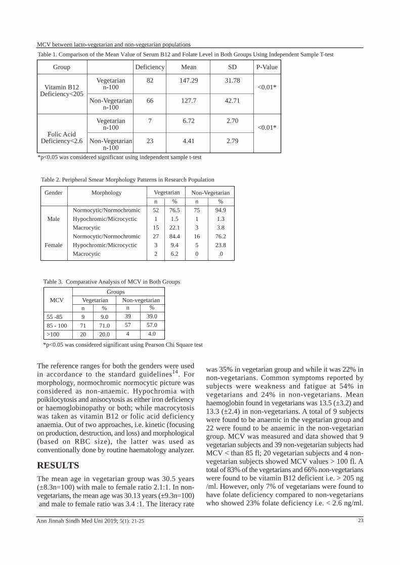
The reference ranges for both the genders were used in accordance to the standard guidelines14. For morphology, normochromic normocytic picture was considered as non-anaemic. Hypochromia with poikilocytosis and anisocytosis as either iron deficiency or haemoglobinopathy or both; while macrocytosis was taken as vitamin B12 or folic acid deficiency anaemia. Out of two approaches, i.e. kinetic (focusing on production, destruction, and loss) and morphological (based on RBC size), the latter was used as conventionally done by routine haematology analyzer.
RESULTSThe mean age in vegetarian group was 30.5 years (±8.3n=100) with male to female ratio 2.1:1. In non-vegetarians, the mean age was 30.13 years (±9.3n=100) and male to female ratio was 3.4 :1. The literacy rate
Ann Jinnah Sindh Med Uni 2019; 5(1): 21-25
Table 1. Comparison of the Mean Value of Serum B12 and Folate Level in Both Groups Using Independent Sample T-test
Group
Vitamin B12Deficiency<205
Folic AcidDeficiency<2.6
Vegetariann-100
Non-Vegetariann-100
Vegetariann-100
Non-Vegetariann-100
Deficiency
82
66
7
23
Mean
147.29
127.7
6.72
4.41
SD
31.78
42.71
2.70
2.79
P-Value
<0.01*
<0.01*
was 35% in vegetarian group and while it was 22% in non-vegetarians. Common symptoms reported by subjects were weakness and fatigue at 54% in vegetarians and 24% in non-vegetarians. Mean haemoglobin found in vegetarians was 13.5 (±3.2) and 13.3 (±2.4) in non-vegetarians. A total of 9 subjects were found to be anaemic in the vegetarian group and 22 were found to be anaemic in the non-vegetarian group. MCV was measured and data showed that 9 vegetarian subjects and 39 non-vegetarian subjects had MCV < than 85 fl; 20 vegetarian subjects and 4 non-vegetarian subjects showed MCV values > 100 fl. A total of 83% of the vegetarians and 66% non-vegetarians were found to be vitamin B12 deficient i.e. > 205 ng /ml. However, only 7% of vegetarians were found to have folate deficiency compared to non-vegetarians who showed 23% folate deficiency i.e. < 2.6 ng/ml.
*p<0.05 was considered significant using independent sample t-test
Table 2. Peripheral Smear Morphology Patterns in Research Population
Gender
Male
Female
Morphology
Normocytic/NormochromicHypochromic/MicrocycticMacrocyticNormocytic/NormochromicHypochromic/MicrocycticMacrocytic
n521152732
%76.51.522.184.49.46.2
n75131650
%94.91.33.876.223.8.0
Vegetarian Non-Vegetarian
Table 3. Comparative Analysis of MCV in Both Groups
MCV
55 -8585 - 100>100
n97120
% 9.071.020.0
n39574
% 39.057.04.0
Vegetarian Non-vegetarianGroups
*p<0.05 was considered significant using Pearson Chi Square test
23
MCV between lacto-vegetarian and non-vegetarian populations
The mean B12 and folate level in vegetarians was 190.24 and 6.73, and 226.71 and 13.9, respectively in non-vegetarians (Table 1).
DISCUSSIONThe present study is unique in the sense that (a) to my knowledge, it was conducted for the first time in a rural area of Pakistan which is inhabited equally by the Muslims and the Hindus; (b) It focused on the principle of strict vegetarianism which is the chief feature of the studied Hindu population and non-vegetarians which are the Muslim population; (c) It explored the consequences of vitamin B12 and folate deficiencies on MCV of both groups.
In our study, the mean age of the vegetarians who were found to be vitamin B12 deficient was 30 years, this is different from the study done by Hashim and Tahir in Pakistan in 2006 who found the mean age to be 55 years14. In this study, male to female ratio was 2.1:1 in vegetarians and 3.7:1 in non-vegetarians. This difference in genders is due to the practice of veiling of the female population in Tharparkar village. Although all subjects (vegetarian and non-vegetarian) recruited in the study were apparently healthy and did not present any signs and symptoms, but on direct questioning, 54% vegetarians reported history of fatigue and weakness, while 24 % of non-vegetarians reported having unexplained lethargy. These findings are similar to the study done by Stable and colleagues in 199015. These workers suggested that fatigue and lethargy are the most initial symptoms experienced by those who developed macrocytic anaemia. The mean B12 levels were found to be 190.2 (normal 205 ng/ml and above) in vegetarians and 226.7 in non-vegetarians which also showed a preponderance of B12 deficiency among the vegetarian group. The correlation coefficient between B12 and age was positive i.e. 0.173 with significant p value 0.014.
In case of folate level analysis, we also found the most expected results, like only 7% subjects in the vegetarian group and 23% subjects in non-vegetarian group were deficient in folate, with a highly significant p-value less than 0.0001. The mean folate levels were 6.7 ng/ml (normal above 2.6 ng/ml) and 13.9 ng/ml respectively. The correlation co-efficient between folic acid and age was negative i.e. 0.027 with an insignificant p-value 0.70. The prevalence of vitamin B12 deficiency in the present study is much higher than other studies while folate deficiency is also significantly on the higher side.
MCV is the most sensitive index in diagnosis of vitamin B12 /folate deficiency16. The degree of anaemia varies but macrocytic and hyper-segmented neutrophils are
considered important laboratory findings in many studies17. In our studies, we also observed early changes in RBC morphology. The blood films of most of the studied population were found to be normocytic and normochromic (Table 3). These findings matched with the study done by Khanduri and Sharma in 200718. We found that MCV in vegetarian population was 93.6 (±9.9) and 87.6 (±8.2) in non-vegetarians. Maximum range was 118 fl. Seventy-one vegetarians and fifty-seven non-vegetarians had MCV between 85–100 fl in vegetarians with p-value less than 0.001. The most probable cause of borderline increase or definite increase in MCV in vegetarians is vitamin B12 or folate deficiency. This ecological influence on MCV was discussed in the study done by Graves and Lee in 201219.
Another intricacy regarding the importance of MCV in vitamin B12/folate deficiency states is addressed in the study done by Spirak in 198220. It says that if iron deficiency and thalassemia coexist with vitamin B12 and folate deficiency, than MCV will not be increased and may decrease in spite of vitamin B12 /and folate deficiency21. MCV can increase in many conditions such as in alcoholism, hypothyroidism, liver disease, pregnancy, and reticulocytosis so, we suggest excluding all these condition before labeling and deciding on vitamin B12/folate deficiency21. In our study, we had excluded notable conditions just indicated and we found increased MCV and serum vitamin B12/folate deficiency in 20% subjects in vegetarian group and only 4% in non-vegetarian group. Such poor correlation has been reported in the study done by Ward in 200223.
CONCLUSIONThe vegetarian and non-vegetarian populations of Tharparkar district are sailing in the same boat because of poor economy, harsh environmental and sociocultural conditions. In this study, both groups exhibited severe vitamin B12 deficiency predominately in the vegetarian group, but that was not truly reflected from MCV. It seems to be a poor indicator of the severity of B12 and folate deficiency anaemia, hence, normal MCV does not exclude B12 or folate deficiencies. Clinicians need to be aware of the low sensitivity of the MCV for screening and the upper limit of MCV should not be more than 95 fl.
Authors’ contributions: Dr Suresh Kumar conceived the idea, worked on literature search, data collection, data analysis and review, introduction and discussion. Dr Asma Sheikh and Dr Zareen Irshad worked on literature search, results and discussion. Dr Vinita kumari and Dr Salma Parween reviewed the literature,
Ann Jinnah Sindh Med Uni 2019; 5(1): 21-2524
Suresh Kumar, Asma Shaikh, Zareen Irshad, Vinita Kumari, Salma Parween, Shahida Kashif
worked on discussion and edited the manuscript. All authors discussed the results and contributed to the final manuscript.
References1. Hofbrand Av, Herbert V. Nutrition anemias. Semin
Hematol. 1999;36:13-23
2. Mannan M, Anwar M, Saleem M, Wigar A, Ahmad M. A study of serum vitamin B12 and folate. Levels in patients of megaloblastic anemia in North Pakistan.J Pak Med Assoc.1995;45:187-88
3. Heil SG, Hogeveen M, Kluijtymans LA, Morava E, de Berg GB,Blom HJ, et al. Morforoid features in a child with combined methylmalonic aciduria and hemocystinuria. J Inherit Metab Dis. 2007;30(5):811
4. Gupta AK, Damji A, Uppaluri A. Vit B12 deficiency. Prevalence among South Asians at a Toronto clinic. Can Fam Physician.2004;50:743-7
5. Snow CF, Laboratory diagnosis of vit B12 and folate deficiency: A guide to primary care physcian. Arch Inter Med. 1999;159:1289-98
6. Aslinia F, Mazza JJ and Yale SH. Megaloblastic anemia and other causes of macrocytosis. J Med Res. 2006; 4(3):236-41
7. Gibson RS. Principle of nutritional assessment, oxford: Oxford University Press; 1990: 461-86
8. Kakepoto GN, Iqbal MP. Iqbal SP. Megaloblastic anaemia is a hospital-based population. Med Sci Res 2000; 28:45- 7
9. Gerardo Colon- Otero MD, C. Christopher Hook MD, David Menek MD. A practical approach to differential diagnosis and evolution of adult patient with macrocytic anemia. Med Clin North Am. 1992; 76(3): 581-597
10. Stabler SP, Allen RH. Megaloblastic anemias in.22nd Edition, Goldman editor. WB Saunders company Cecil text book of medicine; 2004
11. Iqbal SP, Kakepoto GN, Iqbal SP. Vitamin B12 deficiency a major cause of megaloblastic anemia in patients attending a tertiary care hospital. J Ayub Med Coll Abbotabad. 2009;21(3):92-4
12. Hashim H, Tahir F, Frequency of vitamin B12 and folic acid deficiency among patients of megaloblastic anemia. Ann Pak Med Sci. 2006; 2(3):192-4
13. Veda P, Evaluation of Macrocytosis in Routine hemograms. Indian J Hematol Blood Tranfus. 2013; 29(1):26-30
14. Stabler SP, Allen RH, Savage DG, Lindenbaum J, Clinical spectrum and diagnosis of cobalmin deficiency. Blood. 1990:76(5):871-881
15. Chan CW, Liu SY, Kho CS, Lau KH, Liang YS, Chu WR. et al. Diagnostic clues to megaloblastic anemias without macrocytosis. Int J Lab Hem. 2007; 29:163-171
16. Naeem MA, Utrra GM, Etiology of incidence of megaloblastic anemia in district Gilgit. Pak J Path. 2007; 18(1):15-6
17. Khanduri U, sharma A. Megaloblastic anemia: prevalence and causative Factors. Natl Med J India. 2007; 20(4):172-175
18. Freeland-Graves JH, Lee JJ. Global multiplicity of dietary standards for trace elements. J Trace Elem Med Biol. 2012; 26(2-3):61-65 doi:10.1016/j.jtemb.
19. Spivak JL. Masked Megaloblastic Anemia. Arch Intern Med. 1982; 142(12): 2111-14
20. Ceylan C, Miskioglu M, Colak H, Kiliccioglu B, Ozdemir E. Evaluation of reticulocyte parameter in iron deficiency vitamin B(12) deficiency and beta-thalassemia minor patients. Int J Lab Hematol. 2007; 29(5):327-334
21. Maruyama S, Hirayama C, Yamamoto S, Koda M, Udagawa A, Kadowaki Y, et al. Red blood status inalcoholic and non-alcoholic liver disease. J Lab ClinMed. 2001; 138(5):332-337
22. Ward PC, Modern approaches to the investigation ofvitamin B(12) deficiency. Clin Lab Med. 2002;22(2):435-445
Ann Jinnah Sindh Med Uni 2019; 5(1): 21-25 25
MCV between lacto-vegetarian and non-vegetarian populations
6 Dental Surgeon, Civil Hospital Karachi, Pakistan7 Oral & Maxillofacial Surgery, Mohammad Bin Qasim Medical and Dental College, Karachi, Pakistan
Correspondence: Dr Faisal Hameed, Assistant Professor, Oral & Maxillofacial Surgery, Sindh Institute of Oral Health Sciences, Jinnah Sindh Medical University, Karachi, Pakistan Email: [email protected]
Oral & Maxillofacial Surgery1 / Department of Oral Biology3/ Oral Medicine5, Bhittai Medical and Dental College, Sciences, Karachi, Pakistan2 Oral & Maxillofacial Surgery, Sindh Institute of Oral Health Sciences, Jinnah Sindh Medical University, Karachi, Pakistan4 Senior Lecturer, Department of Prosthodontic, Fatima Jinnah Dental College, Karachi, Pakistan
Irfan Ali1, Faisal Hameed2, Ali Maqbool3, Muhammad Kazim4, Muhammad Aqeel Aslam5,Saad Uddin Siddiqui6, and Nabeel Hafeez7
Knowledge and Frequency of Needle Stick Injury Among Dental Students and House Officers of Bhitai Medical and
Dental College, Mirpur Khas
ORIGINAL ARTICLE
Ann Jinnah Sindh Med Uni 2019; 5(1): 26-30
ABSTRACT
Objective: To evaluate the knowledge and experience of Needle Stick Injury (NSI) among dental students and house officers at Bhitai Dental and Medical College, Mirpur Khas. Methodology: This descriptive cross-sectional study was conducted among dental students and house officers. The number of needle stick injuries and post exposure reporting in the past 12 months was assessed by a questionnaire-based survey. SPSS 19 was used to analyze the data. Result: A total of 330 dental students and house officers participated in this study in which 76.3% were dental students and 23.3% were house officers. As many as 63% had experienced NSI. As many as 31% reported receiving NSI during re-capping of the needles, 21% during administering local anaesthesia injection, and 19% reported getting injured during the scaling procedure. A total of 78.6% of the participants did not report the exposure incident. Conclusion: The most prevalent of causes reported was receiving injury during the recapping of needle. The reporting of NSIs was found to be insufficient, hence evaded proper follow up because the respondents feared stigmatization and discrimination. Furthermore, awareness was low regarding the means of prevention and protocol.
Key words: Needle stick Injury, Dentistry, Occupational Hazards, Post Exposure Prophylaxis, Blood-borne PathogensHow to cite this article: Ali I, Hameed F, Maqbool A, Kazim M, Aslam MA, Siddiqui SU, Hafeez N. Incidence of
needle stick injury (NSI) among dental students and dental house officers of Bhitai Medicaland Dental College, Mirpur Khas. Ann Jinnah Sindh Med Uni 2019; 5 (1): 26-30
26
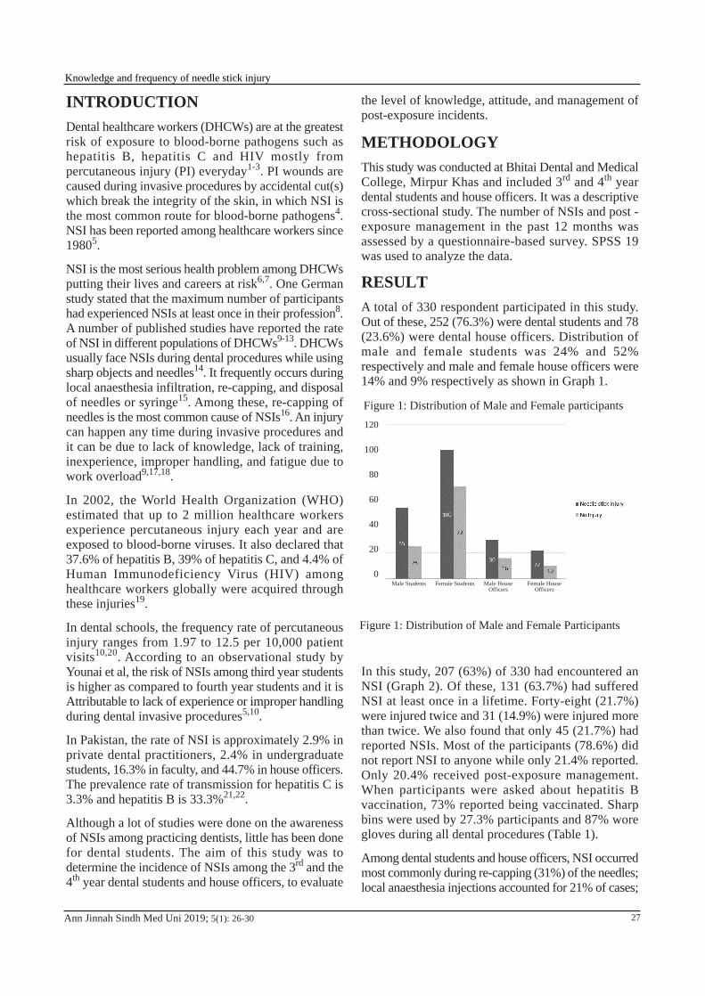
INTRODUCTIONDental healthcare workers (DHCWs) are at the greatest risk of exposure to blood-borne pathogens such as hepatitis B, hepatitis C and HIV mostly from percutaneous injury (PI) everyday1-3. PI wounds are caused during invasive procedures by accidental cut(s) which break the integrity of the skin, in which NSI is the most common route for blood-borne pathogens4. NSI has been reported among healthcare workers since 19805.
NSI is the most serious health problem among DHCWs putting their lives and careers at risk6,7. One German study stated that the maximum number of participants had experienced NSIs at least once in their profession8. A number of published studies have reported the rate of NSI in different populations of DHCWs9-13. DHCWs usually face NSIs during dental procedures while using sharp objects and needles14. It frequently occurs during local anaesthesia infiltration, re-capping, and disposal of needles or syringe15. Among these, re-capping of needles is the most common cause of NSIs16. An injury can happen any time during invasive procedures and it can be due to lack of knowledge, lack of training, inexperience, improper handling, and fatigue due to work overload9,17,18.
In 2002, the World Health Organization (WHO) estimated that up to 2 million healthcare workers experience percutaneous injury each year and are exposed to blood-borne viruses. It also declared that 37.6% of hepatitis B, 39% of hepatitis C, and 4.4% of Human Immunodeficiency Virus (HIV) among healthcare workers globally were acquired through these injuries19.
In dental schools, the frequency rate of percutaneous injury ranges from 1.97 to 12.5 per 10,000 patient visits10,20. According to an observational study by Younai et al, the risk of NSIs among third year students is higher as compared to fourth year students and it is Attributable to lack of experience or improper handling during dental invasive procedures5,10.
In Pakistan, the rate of NSI is approximately 2.9% in private dental practitioners, 2.4% in undergraduate students, 16.3% in faculty, and 44.7% in house officers. The prevalence rate of transmission for hepatitis C is 3.3% and hepatitis B is 33.3%21,22.
Although a lot of studies were done on the awareness of NSIs among practicing dentists, little has been done for dental students. The aim of this study was to determine the incidence of NSIs among the 3rd and the 4th year dental students and house officers, to evaluate
the level of knowledge, attitude, and management of post-exposure incidents.
METHODOLOGYThis study was conducted at Bhitai Dental and Medical College, Mirpur Khas and included 3rd and 4th year dental students and house officers. It was a descriptive cross-sectional study. The number of NSIs and post -exposure management in the past 12 months was assessed by a questionnaire-based survey. SPSS 19 was used to analyze the data.
RESULTA total of 330 respondent participated in this study. Out of these, 252 (76.3%) were dental students and 78 (23.6%) were dental house officers. Distribution of male and female students was 24% and 52% respectively and male and female house officers were 14% and 9% respectively as shown in Graph 1.
In this study, 207 (63%) of 330 had encountered an NSI (Graph 2). Of these, 131 (63.7%) had suffered NSI at least once in a lifetime. Forty-eight (21.7%) were injured twice and 31 (14.9%) were injured more than twice. We also found that only 45 (21.7%) had reported NSIs. Most of the participants (78.6%) did not report NSI to anyone while only 21.4% reported. Only 20.4% received post-exposure management. When participants were asked about hepatitis B vaccination, 73% reported being vaccinated. Sharp bins were used by 27.3% participants and 87% wore gloves during all dental procedures (Table 1).
Among dental students and house officers, NSI occurred most commonly during re-capping (31%) of the needles; local anaesthesia injections accounted for 21% of cases;
Ann Jinnah Sindh Med Uni 2019; 5(1): 26-30
Figure 1: Distribution of Male and Female Participants
120
100
80
60
40
20
0
Figure 1: Distribution of Male and Female participants
Male Students Female Students Male House Officers
Female House Officers
27
Knowledge and frequency of needle stick injury
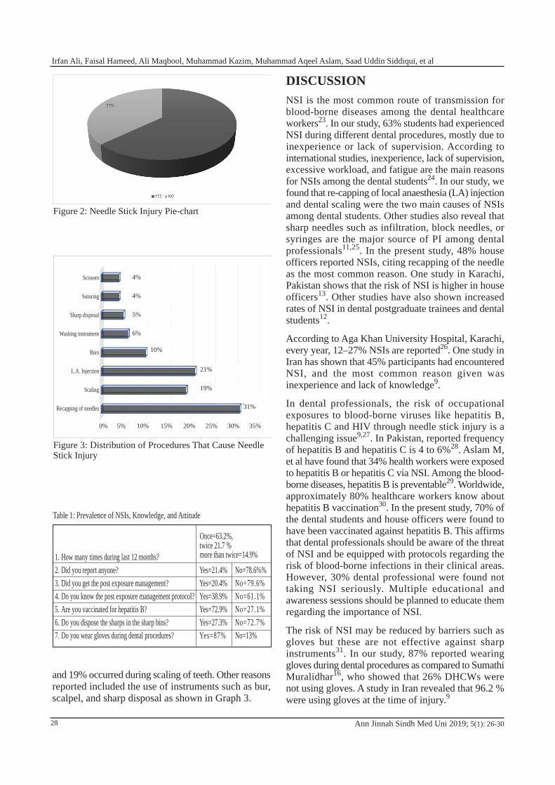
and 19% occurred during scaling of teeth. Other reasons reported included the use of instruments such as bur, scalpel, and sharp disposal as shown in Graph 3.
DISCUSSIONNSI is the most common route of transmission for blood-borne diseases among the dental healthcare workers23. In our study, 63% students had experienced NSI during different dental procedures, mostly due to inexperience or lack of supervision. According to international studies, inexperience, lack of supervision, excessive workload, and fatigue are the main reasons for NSIs among the dental students24. In our study, we found that re-capping of local anaesthesia (LA) injection and dental scaling were the two main causes of NSIs among dental students. Other studies also reveal that sharp needles such as infiltration, block needles, or syringes are the major source of PI among dental professionals11,25. In the present study, 48% house officers reported NSIs, citing recapping of the needle as the most common reason. One study in Karachi, Pakistan shows that the risk of NSI is higher in house officers13. Other studies have also shown increased rates of NSI in dental postgraduate trainees and dental students12.
According to Aga Khan University Hospital, Karachi, every year, 12–27% NSIs are reported26. One study in Iran has shown that 45% participants had encountered NSI, and the most common reason given was inexperience and lack of knowledge9.
In dental professionals, the risk of occupational exposures to blood-borne viruses like hepatitis B, hepatitis C and HIV through needle stick injury is a challenging issue9,27. In Pakistan, reported frequency of hepatitis B and hepatitis C is 4 to 6%28. Aslam M, et al have found that 34% health workers were exposed to hepatitis B or hepatitis C via NSI. Among the blood-borne diseases, hepatitis B is preventable29. Worldwide, approximately 80% healthcare workers know about hepatitis B vaccination30. In the present study, 70% of the dental students and house officers were found to have been vaccinated against hepatitis B. This affirms that dental professionals should be aware of the threat of NSI and be equipped with protocols regarding the risk of blood-borne infections in their clinical areas. However, 30% dental professional were found not taking NSI seriously. Multiple educational and awareness sessions should be planned to educate them regarding the importance of NSI.
The risk of NSI may be reduced by barriers such as gloves but these are not effective against sharp instruments31. In our study, 87% reported wearing gloves during dental procedures as compared to Sumathi Muralidhar16, who showed that 26% DHCWs were not using gloves. A study in Iran revealed that 96.2 % were using gloves at the time of injury.9
Ann Jinnah Sindh Med Uni 2019; 5(1): 26-30
Figure 2: Needle Stick Injury Pie-chart
Table 1: Prevalence of NSIs, Knowledge, and Attitude
1.2.3.4.5.6.7.
How many times during last 12 months?Did you report anyone?Did you get the post exposure management?Do you know the post exposure management protocol?Are you vaccinated for hepatitis B?Do you dispose the sharps in the sharp bins?Do you wear gloves during dental procedures?
Yes=21.4%Yes=20.4%Yes=38.9%Yes=72.9%Yes=27.3%Yes=87%
Once=63.2%, twice 21.7 % more than twice=14.9%
No=78.6%%No=79.6%No=61.1%No=27.1%No=72.7%No=13%
Figure 3: Distribution of Procedures That Cause Needle Stick Injury
10%
21%
19%
31%
Scissors
Suturing
Sharp disposal
Washing instrument
Burs
L.A. Injection
Scaling
Recapping of needles
4%
4%
5%
6%
0% 5% 10% 15% 20% 25% 30% 35%
28
Irfan Ali, Faisal Hameed, Ali Maqbool, Muhammad Kazim, Muhammad Aqeel Aslam, Saad Uddin Siddiqui, et al
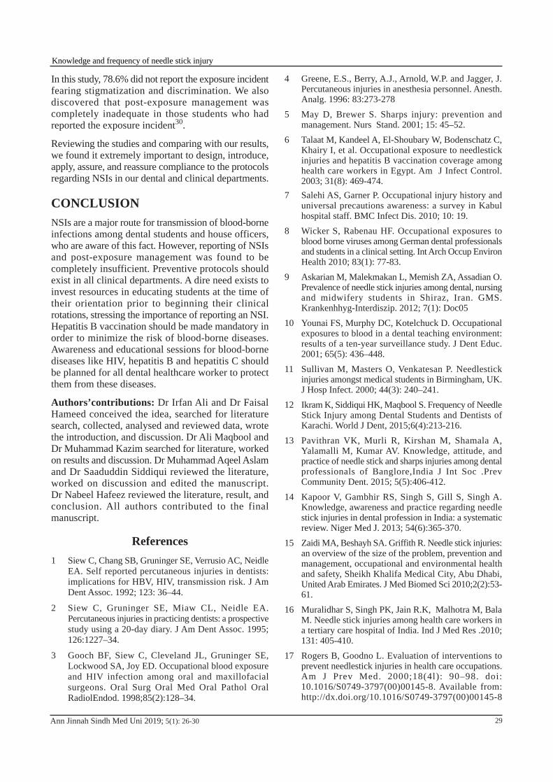
In this study, 78.6% did not report the exposure incident fearing stigmatization and discrimination. We also discovered that post-exposure management was completely inadequate in those students who had reported the exposure incident30.
Reviewing the studies and comparing with our results, we found it extremely important to design, introduce, apply, assure, and reassure compliance to the protocols regarding NSIs in our dental and clinical departments.
CONCLUSIONNSIs are a major route for transmission of blood-borne infections among dental students and house officers, who are aware of this fact. However, reporting of NSIs and post-exposure management was found to be completely insufficient. Preventive protocols should exist in all clinical departments. A dire need exists to invest resources in educating students at the time of their orientation prior to beginning their clinical rotations, stressing the importance of reporting an NSI. Hepatitis B vaccination should be made mandatory in order to minimize the risk of blood-borne diseases. Awareness and educational sessions for blood-borne diseases like HIV, hepatitis B and hepatitis C should be planned for all dental healthcare worker to protect them from these diseases.
Authors’contributions: Dr Irfan Ali and Dr Faisal Hameed conceived the idea, searched for literature search, collected, analysed and reviewed data, wrote the introduction, and discussion. Dr Ali Maqbool and Dr Muhammad Kazim searched for literature, worked on results and discussion. Dr Muhammad Aqeel Aslam and Dr Saaduddin Siddiqui reviewed the literature, worked on discussion and edited the manuscript. Dr Nabeel Hafeez reviewed the literature, result, and conclusion. All authors contributed to the final manuscript.
References1 Siew C, Chang SB, Gruninger SE, Verrusio AC, Neidle
EA. Self reported percutaneous injuries in dentists: implications for HBV, HIV, transmission risk. J Am Dent Assoc. 1992; 123: 36–44.
2 Siew C, Gruninger SE, Miaw CL, Neidle EA. Percutaneous injuries in practicing dentists: a prospective study using a 20-day diary. J Am Dent Assoc. 1995; 126:1227–34.
3 Gooch BF, Siew C, Cleveland JL, Gruninger SE, Lockwood SA, Joy ED. Occupational blood exposure and HIV infection among oral and maxillofacial surgeons. Oral Surg Oral Med Oral Pathol Oral RadiolEndod. 1998;85(2):128–34.
4 Greene, E.S., Berry, A.J., Arnold, W.P. and Jagger, J. Percutaneous injuries in anesthesia personnel. Anesth. Analg. 1996: 83:273-278
5 May D, Brewer S. Sharps injury: prevention and management. Nurs Stand. 2001; 15: 45–52.
6 Talaat M, Kandeel A, El-Shoubary W, Bodenschatz C, Khairy I, et al. Occupational exposure to needlestick injuries and hepatitis B vaccination coverage among health care workers in Egypt. Am J Infect Control. 2003; 31(8): 469-474.
7 Salehi AS, Garner P. Occupational injury history and universal precautions awareness: a survey in Kabul hospital staff. BMC Infect Dis. 2010; 10: 19.
8 Wicker S, Rabenau HF. Occupational exposures to blood borne viruses among German dental professionals and students in a clinical setting. Int Arch Occup Environ Health 2010; 83(1): 77-83.
9 Askarian M, Malekmakan L, Memish ZA, Assadian O. Prevalence of needle stick injuries among dental, nursing and midwifery students in Shiraz, Iran. GMS. Krankenhhyg-Interdiszip. 2012; 7(1): Doc05
10 Younai FS, Murphy DC, Kotelchuck D. Occupational exposures to blood in a dental teaching environment: results of a ten-year surveillance study. J Dent Educ. 2001; 65(5): 436–448.
11 Sullivan M, Masters O, Venkatesan P. Needlestick injuries amongst medical students in Birmingham, UK. J Hosp Infect. 2000; 44(3): 240–241.
12 Ikram K, Siddiqui HK, Maqbool S. Frequency of Needle Stick Injury among Dental Students and Dentists of Karachi. World J Dent, 2015;6(4):213-216.
13 Pavithran VK, Murli R, Kirshan M, Shamala A, Yalamalli M, Kumar AV. Knowledge, attitude, and practice of needle stick and sharps injuries among dental professionals of Banglore,India J Int Soc .Prev Community Dent. 2015; 5(5):406-412.
14 Kapoor V, Gambhir RS, Singh S, Gill S, Singh A. Knowledge, awareness and practice regarding needle stick injuries in dental profession in India: a systematic review. Niger Med J. 2013; 54(6):365-370.
15 Zaidi MA, Beshayh SA. Griffith R. Needle stick injuries: an overview of the size of the problem, prevention and management, occupational and environmental health and safety, Sheikh Khalifa Medical City, Abu Dhabi, United Arab Emirates. J Med Biomed Sci 2010;2(2):53-61.
16 Muralidhar S, Singh PK, Jain R.K, Malhotra M, Bala M. Needle stick injuries among health care workers in a tertiary care hospital of India. Ind J Med Res .2010; 131: 405-410.
17 Rogers B, Goodno L. Evaluation of interventions to prevent needlestick injuries in health care occupations. Am J Prev Med. 2000;18(4l): 90–98. doi: 10.1016/S0749-3797(00)00145-8. Available from: http://dx.doi.org/10.1016/S0749-3797(00)00145-8
Ann Jinnah Sindh Med Uni 2019; 5(1): 26-30 29
Knowledge and frequency of needle stick injury
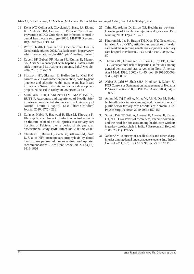
18 Kohn WG, Collins AS, Cleveland JL, Harte JA, Eklund KJ, Malvitz DM, Centers for Disease Control and Prevention (CDC) Guidelines for infection control in dental health-care settings--2003. MMWR Recomm Rep. 2003;52(17):1–61
19 World Health Organization. Occupational Health-Needlestick injuries 2002. Available from: https://www. whi.int/occupational_health/topics/needinjuries/en/.
20 Zuberi BF, Zuberi FF, Hasan SR, Kumar R, Memon SA, Afsar S. Frequency of acute hepatitis C after needle stick injury and its treatment outcome. Pak J Med Sci. 2009;25(5): 766-769
21 Sjostrom HT, Skyman E, Hellström L, Med KM, Grinevika V. Cross-infection prevention, basic hygiene practices and education within nursing and health care in Latvia: a Swe- dish-Latvian practice development project. Nurse Educ Today 2003;23(6):404-411
22 MUNGURE E.K, GAKONYO J.M, MAMDANI Z , BUTT F, Awareness and experience of Needle Stick injuries among dental students at the University of Nairobi, Dental Hospital. East African Medical Journal.2010; 87(5): 211
23 Zafar A, Habib F, Hadwani R, Ejaz M, Khowaja K, Khowaja R, et al. Impact of infection control activities on the rate of needle stick injuries at a tertiary care hospital of Pakistan over a period of six years: an observational study. BMC Infect Dis. 2009; 9: 78-80.
24 Cleveland JL, Barker L, Gooch BF, Beltrami EM, Cardo D. Use of HIV postexposure prophylaxis by dental health care personnel: an overview and updated recommendations. J Am Dent Assoc. 2002, 133(12): 1619-1626
25 Trim JC, Adams D, Elliott TS. Healthcare workers’ knowledge of inoculation injuries and glove use. Br J Nursing 2003; 12(4): 215–221.
26 Khurram M, Ijaz K, Bushra TH, Khan NY. Needle stick injuries. A SURVEY, attitudes and practices of health care workers regarding needle stick injuries at a tertiary care hospital in Pakistan. J Pak Med Assoc 2008;58:57-60
27 Thomas DL, Gruninger SE, Siew C, Joy ED, Quinn TC. Occupational risk of hepatitis C infections among general dentists and oral surgeons in North America. Am J Med. 1996; 100(1):41–45. doi: 10.1016/S0002-9343(96)90009-1
28 Abbas Z, Jafri W, Shah SHA, Khokhar N, Zuberi SJ. PGS Consensus Statement on management of Hepatitis B Virus Infection 2003. J Pak Med Assoc. 2004; 54(3): 150-58
29 Aslam M, Taj T, Ali A, Mirza W, Ali H, Dar M, Badar N. Needle stick injuries among health care workers of public sector tertiary care hospitals of Karachi. J Col Physic Surg, Pakistan 2010;20(3):150-153.
30 Sukriti, Pati NT, Sethi A, Agrawal K, Agrawal K, Kumar GT, et al. Low levels of awareness, vaccine coverage, and the need for boosters among health care workersin tertiary care hospitals in India. J Gastroenterol Hepatol.2008; 23(11): 1710-5
31 Jabbar AM, A survey of needle sticks and other sharp injuries among dental undergraduate students Int J InfectControl 2011, 7(3): doi:10.3396/ijic.V7i1.022.11
Ann Jinnah Sindh Med Uni 2019; 5(1): 26-3030
Irfan Ali, Faisal Hameed, Ali Maqbool, Muhammad Kazim, Muhammad Aqeel Aslam, Saad Uddin Siddiqui, et al
Rahat Naz1, Fatima Abid2, Talat Naz2, Sohaila Tariq3, and Sajid Atif Aleem4
Self-Image and Its Impact on Academic Performance of Undergraduate Medical Students in Karachi
ORIGINAL ARTICLE
Ann Jinnah Sindh Med Uni 2019; 5(1): 31-34
ABSTRACTObjective: To evaluate the extent to which students are concerned about their physical appearance and how much self-perception affects themMethodology: A cross sectional observational study was conducted at the Institute of Health & Business Management and Social Sciences, Jinnah Sindh Medical University (IOH&BMSS), Karachi. A structured online questionnaire was designed and distributed through random sampling among students of medical sciences to collect the data. Data was analyzed using SPSS version 16.Results: Out of the total of 160 participants in this study, 44 (27.5%) were male while 116 (72.5%) were female. Mean age was 18.24+ 1.68 years while the mean Body Mass Index (BMI) was 21.56+ 4.8. Frequency of depression due to physical appearance found in Grade A and A+ students was 0 while 27.5%, 20.8%, 40.7% was noted among students who got B, B+, B- and 30.4% and 14.3% depression due to physical appearance was noted in C, C- achieving students respectively.Conclusion: Students who achieved A and A+ grade had no depression due to physical appearance while mild to moderate depression was noted among students who secured lower grades. Furthermore, no relationship was observed between overweight and academic achievements.Key words: Self-image, academic achievements, students, BMIHow to cite this article: Naz R, Abid F, Naz T, Tariq S. Aleem SA. Self-image and its impact on academic performance.
Ann Jinnah Sindh Med Uni 2019; 5 (1): 31-34
Incharge CME and Additional Director HR1 / Department of Physiology2 / Department of Research4, Jinnah Sindh Medical University, Karachi, Pakistan3 Consultant Sonologist, Mehdi Manji Laboratories, Karachi, Pakistan
Correspondence: Dr Rahat Naz, Incharge CME and Additional Director HR, Jinnah Sindh Medical University, Karachi, PakistanEmail: [email protected]
INTRODUCTIONSelf-image is a mental representation of one’s own personality or is the structured declarative knowledge
about one’s self that could guide social behaviour. Self-image is the manner in which we take into account our own physical, psychological, and emotionalcharacteristics, the dimensions of our ego and social position. Accurate self-perception is very important to know about attitudinal abilities and personal behavioural knowledge1. Teenage is a time in life when structuring one’s self-image determines cognitive, affective, and physical development. Self-image stimulates affective qualities to gain behavioural safety and detachment.
31
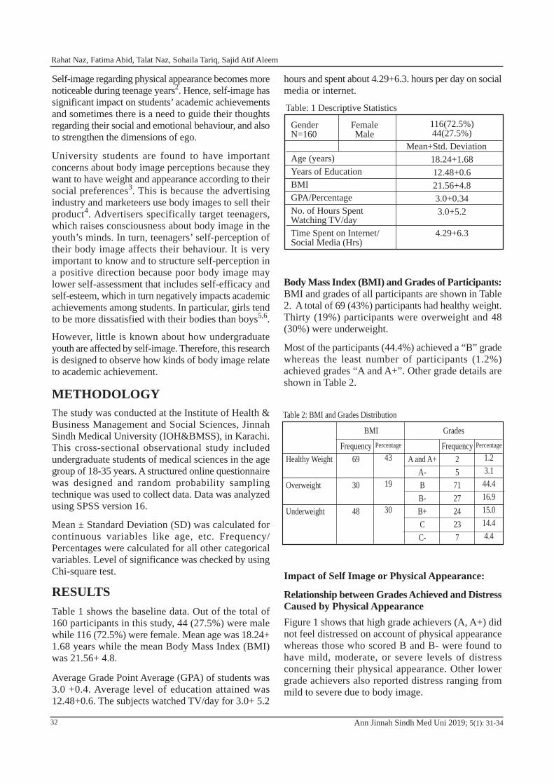
Self-image regarding physical appearance becomes more noticeable during teenage years2. Hence, self-image has significant impact on students’ academic achievements and sometimes there is a need to guide their thoughts regarding their social and emotional behaviour, and also to strengthen the dimensions of ego.
University students are found to have important concerns about body image perceptions because they want to have weight and appearance according to their social preferences3. This is because the advertising industry and marketeers use body images to sell their product4. Advertisers specifically target teenagers, which raises consciousness about body image in the youth’s minds. In turn, teenagers’ self-perception of their body image affects their behaviour. It is very important to know and to structure self-perception in a positive direction because poor body image may lower self-assessment that includes self-efficacy and self-esteem, which in turn negatively impacts academic achievements among students. In particular, girls tend to be more dissatisfied with their bodies than boys5,6.
However, little is known about how undergraduate youth are affected by self-image. Therefore, this research is designed to observe how kinds of body image relate to academic achievement.
METHODOLOGYThe study was conducted at the Institute of Health & Business Management and Social Sciences, Jinnah Sindh Medical University (IOH&BMSS), in Karachi. This cross-sectional observational study included undergraduate students of medical sciences in the age group of 18-35 years. A structured online questionnaire was designed and random probability sampling technique was used to collect data. Data was analyzed using SPSS version 16.
Mean ± Standard Deviation (SD) was calculated for continuous variables like age, etc. Frequency/ Percentages were calculated for all other categorical variables. Level of significance was checked by using Chi-square test.
RESULTSTable 1 shows the baseline data. Out of the total of 160 participants in this study, 44 (27.5%) were male while 116 (72.5%) were female. Mean age was 18.24+ 1.68 years while the mean Body Mass Index (BMI) was 21.56+ 4.8.
Average Grade Point Average (GPA) of students was 3.0 +0.4. Average level of education attained was 12.48+0.6. The subjects watched TV/day for 3.0+ 5.2
hours and spent about 4.29+6.3. hours per day on social media or internet.
Body Mass Index (BMI) and Grades of Participants:BMI and grades of all participants are shown in Table 2. A total of 69 (43%) participants had healthy weight. Thirty (19%) participants were overweight and 48 (30%) were underweight.
Most of the participants (44.4%) achieved a “B” grade whereas the least number of participants (1.2%) achieved grades “A and A+”. Other grade details are shown in Table 2.
Impact of Self Image or Physical Appearance:
Relationship between Grades Achieved and Distress Caused by Physical AppearanceFigure 1 shows that high grade achievers (A, A+) did not feel distressed on account of physical appearance whereas those who scored B and B- were found to have mild, moderate, or severe levels of distress concerning their physical appearance. Other lower grade achievers also reported distress ranging from mild to severe due to body image.
Ann Jinnah Sindh Med Uni 2019; 5(1): 31-34
Table: 1 Descriptive Statistics
GenderN=160
Age (years)Years of EducationBMIGPA/PercentageNo. of Hours Spent Watching TV/dayTime Spent on Internet/Social Media (Hrs)
FemaleMale
116(72.5%)44(27.5%)
Mean+Std. Deviation18.24+1.6812.48+0.621.56+4.83.0+0.343.0+5.2
4.29+6.3
Healthy Weight
Overweight
Underweight
Frequency69
30
48
Percentage
43
19
30
A and A+A-BB-B+CC-
Frequency25
712724237
Percentage
1.23.1
44.416.915.014.44.4
BMI Grades
Table 2: BMI and Grades Distribution
32
Rahat Naz, Fatima Abid, Talat Naz, Sohaila Tariq, Sajid Atif Aleem
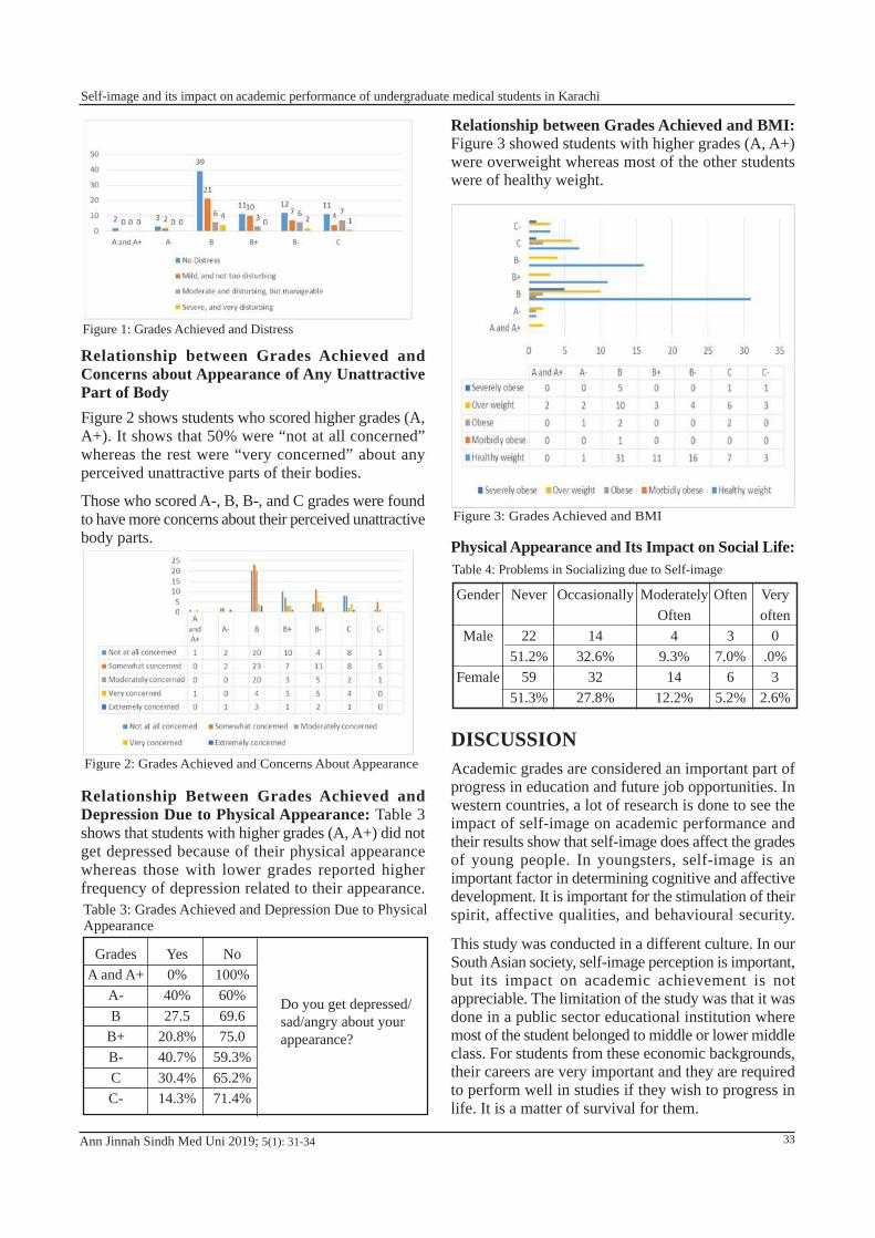
Relationship between Grades Achieved and Concerns about Appearance of Any Unattractive Part of BodyFigure 2 shows students who scored higher grades (A, A+). It shows that 50% were “not at all concerned” whereas the rest were “very concerned” about any perceived unattractive parts of their bodies.
Those who scored A-, B, B-, and C grades were found to have more concerns about their perceived unattractive body parts.
Relationship Between Grades Achieved and Depression Due to Physical Appearance: Table 3 shows that students with higher grades (A, A+) did not get depressed because of their physical appearance whereas those with lower grades reported higher frequency of depression related to their appearance.
Ann Jinnah Sindh Med Uni 2019; 5(1): 31-34
Relationship between Grades Achieved and BMI:Figure 3 showed students with higher grades (A, A+) were overweight whereas most of the other students were of healthy weight.
Physical Appearance and Its Impact on Social Life:
DISCUSSIONAcademic grades are considered an important part of progress in education and future job opportunities. In western countries, a lot of research is done to see the impact of self-image on academic performance and their results show that self-image does affect the grades of young people. In youngsters, self-image is an important factor in determining cognitive and affective development. It is important for the stimulation of their spirit, affective qualities, and behavioural security.
This study was conducted in a different culture. In our South Asian society, self-image perception is important, but its impact on academic achievement is not appreciable. The limitation of the study was that it was done in a public sector educational institution where most of the student belonged to middle or lower middle class. For students from these economic backgrounds, their careers are very important and they are required to perform well in studies if they wish to progress in life. It is a matter of survival for them.
Table 3: Grades Achieved and Depression Due to PhysicalAppearance
Do you get depressed/sad/angry about your appearance?
GradesA and A+
A-B
B+B-CC-
Yes0%40%27.5
20.8%40.7%30.4%14.3%
No100%60%69.675.0
59.3%65.2%71.4%
Table 4: Problems in Socializing due to Self-image
Gender
Male
Female
Never
2251.2%
5951.3%
Occasionally
1432.6%
3227.8%
ModeratelyOften
49.3%
1412.2%
Often
37.0%
65.2%
Very often
0.0%
32.6%
Figure 2: Grades Achieved and Concerns About Appearance
Figure 3: Grades Achieved and BMI
33
Self-image and its impact on academic performance of undergraduate medical students in Karachi
Figure 1: Grades Achieved and Distress
Cultural difference is also an important factor. Although some institutions are coeducational, but there is minimum interaction between boys and girls. In a conservative society, most of the females wear either hijab or loose clothes which hide their physique. Table 1 showed a majority of females from the total of 160 participants with only 44 males. Therefore, the general population of the study comprised mainly females with average age around 20 years.
In Table 2, the BMI and grades of all participants are shown. This indicated that most of the participants had healthy weight (43%), while 19% were overweight. Thirty per cent participants were underweight. Most participants had scored ‘B’ grade (44.4%) whereas ‘A and A+’ were achieved by only 1.2%.
Figure 1 shows the relationship between the grades and distress due to physical appearance. The high achievers were convinced that physical appearance was unable to impact their grades. Those participants who scored B grades (30%) reported mild disturbance due to their physical appearance.
Figure 3 also confirmed that participants with high grades felt no impact on physical appearance or they managed it very well. The study also showed that those few students who scored A grades were overweight. It can be attributed to cultural differences that students in an eastern society like Pakistan do not appear to be as conscious of their looks as their counterparts in the West, and are able to manage their self-esteem despite being overweight.
The hypothesis is partially supported by the study. Those who scored good grades were the ones who were able to manage the discomfort caused by poor perception of physical appearance. Further studies are required to see the impact of self-image on academic grades in students of private medical colleges which usually enroll students from higher income families, who may be more conscious about their perceptions of self-image.
This study also highlighted the need of physical health and availability of gymnasium and sports facilities for students. The overall health of students is important as a healthy body is required for a healthy mind, which impacts the perception of self-image. It is recommended that educational institutions should have sports facilities where students can take care of their physical health.
Authors’ contributions: Dr Rahat Naz conceived the idea, worked on literature search, data collection, data analysis and review, introduction and discussion, reviewed the literature, results, and drew the conclusion from discussion. Dr Fatima Abid worked on literature search, results and discussion. Dr Sohaila Tariq and Miss Talat Naz reviewed the literature, worked on discussion and edited the manuscript. All authors discussed the results and contributed to the final manuscript.
References
1. Bãban, A. Educational counseling, a methodological guide for counseling classes, Psinet Publishing house, Cluj-Napoca. 2001; 66.
2. Fung MSC, Yuen M. Body image and eating attitudes among adolescent Chinese girls in Hong Kong. Percept Mot Skills.2003; 96(1): 57-66.
3. Yahia N, El-Ghazale H, Achkar A, Rizk S. Dieting practices and body image perception among Lebanese university students. Asia Pac J Clin Nutr. 2011; 20(1): 21-28.
4. Thompson, K J, Heinberg, LJ. The media’s influence on body image disturbance and eating disorders: We’ve reviled them, now can we rehabilitate them? J Soc Issus. (1999); 55(2): 339-353.
5. Page RÌ, Lee CM, Miao NF. Self-perception of body weight among high school students in Taipei, Taiwan.Int J Adolesc Med Health. 2005; 17(2): 123-36.
6. Chen LJ, Fox KR, Haase AM. Body shape dissatisfaction and obesity among Taiwanese adolescents. Asia Pac J Clin Nutr. 2008; 17(3): 457-60.
Ann Jinnah Sindh Med Uni 2019; 5(1): 31-3434
Rahat Naz, Fatima Abid, Talat Naz, Sohaila Tariq, Sajid Atif Aleem
Hurtamina Khan1, Shireen Ramzan2, Zahid Mehmood1, Razzaq Dogar2, and Javed Jamali2
Risk Factors and Frequency of Hypocalcaemia in Unintentional Parathyroid Gland Removal During
Thyroid Surgical Interventions
ORIGINAL ARTICLE
Ann Jinnah Sindh Med Uni 2019; 5(1): 35-38
ABSTRACTObjectives: To identify the risk factors and to determine the frequency of hypocalcaemia in unintentional parathyroid gland removal during thyroid surgical interventions at a tertiary care hospital in KarachiMethodology: Retrospective study of two years was conducted at Jinnah Postgraduate Medical Centre (JPMC), Karachi from April 2016 to April 2018. The Head & Neck department’s records were accessed and analyzed using a proforma. Results: Total 86 patients were identified. Twenty-four (24) cases from the total of 86 were found to be malignant, which were treated accordingly, resulting in temporary hypocalcaemia in seven (7) cases. Six (6) Cases of Papillary Ca (total thyroidectomy with central neck dissection) resulted in temporary hypocalcaemia in two (2) cases. Seven (7) multinodular goiter (total thyroidectomy) resulted in temporary hypocalcaemia in two (2) cases. In eight (8) cases, lobectomies/ FNAC was done initially, but then their histopathological reports turned out as Papillary Ca. Therefore, completion thyroidectomy with central neck dissection was performed in seven (7) cases and total thyroidectomy with modified neck dissection in one (1) case, which resulted in temporary hypocalcaemia in three (3) cases. Three (3) cases of Follicular Ca in which total thyroidectomy was performed, resulted in no cases of temporary hypocalcaemia.Conclusion: Re-operated surgeries, malignant thyroid pathology with neck dissection (central/ modified neck dissection) are associated with high chances of unintentional parathyroid gland removal leading to hypocalcaemia. The operating surgeon should consider these risk factors during recurrent malignant surgery with neck dissection.
How to cite this article: Khan H, Ramzan S, Mehmood Z, Dogar R, Jamali J. Risk factors and incidence of hypocalcemia in unintentional parathyroid gland removal during thyroid surgical interventionsa. Ann Jinnah Sindh Med Uni 2019; 5 (1): 35-38
1 Jinnah Postgraduate Medical Centre, Karachi, Pakistan2 Dow University of Health Sciences, Karachi, PakistanCorrespondence: Hurtamina Khan, Jinnah Postgraduate Medical Centre, Karachi, PakistanEmail: [email protected]
INTRODUCTION
Parathyroid glands were first identified in humans by Ivar Sandstorm1. These glands are closely associated with lateral lobes of thyroid and have a short line of embryologic descent but function independently of
thyroid gland2. Lobectomies and thyroidectomies are the most frequent operations for patients with benign and malignant pathology of thyroid gland/glands3,4. Parathyroid injury during unilateral lobectomy is unlikely to result in hypocalcaemia5. The operating surgeon should also keep in mind the anatomical variations of the parathyroid glands6-8. During thyroidectomies, preservation of the parathyroid glands can be achieved by careful dissection directly on the thyroid capsule separating the parathyroid glands gently from the thyroid capsule4. In the 21st century, thyroidectomy has become safe and effective with improved outcomes and minimal morbidity9,10.
35
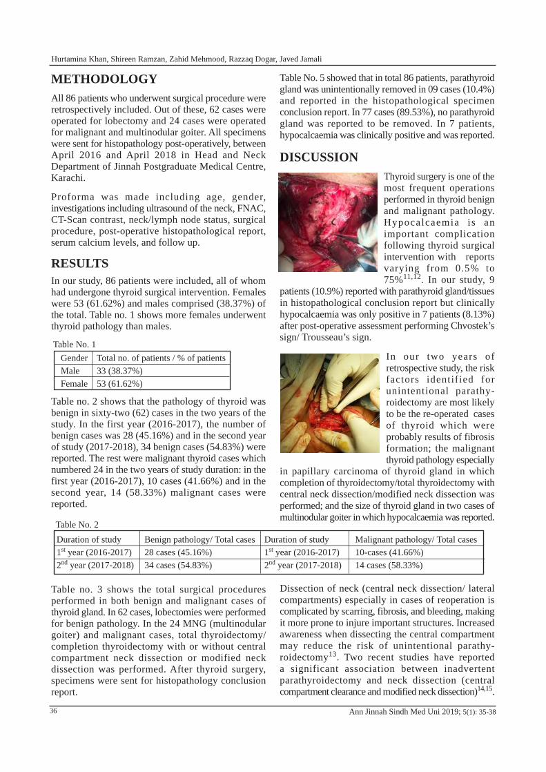
METHODOLOGYAll 86 patients who underwent surgical procedure were retrospectively included. Out of these, 62 cases were operated for lobectomy and 24 cases were operated for malignant and multinodular goiter. All specimens were sent for histopathology post-operatively, between April 2016 and April 2018 in Head and Neck Department of Jinnah Postgraduate Medical Centre, Karachi.
Proforma was made including age, gender, investigations including ultrasound of the neck, FNAC, CT-Scan contrast, neck/lymph node status, surgical procedure, post-operative histopathological report, serum calcium levels, and follow up.
RESULTSIn our study, 86 patients were included, all of whom had undergone thyroid surgical intervention. Females were 53 (61.62%) and males comprised (38.37%) of the total. Table no. 1 shows more females underwent thyroid pathology than males.
Table no. 2 shows that the pathology of thyroid was benign in sixty-two (62) cases in the two years of the study. In the first year (2016-2017), the number of benign cases was 28 (45.16%) and in the second year of study (2017-2018), 34 benign cases (54.83%) were reported. The rest were malignant thyroid cases which numbered 24 in the two years of study duration: in the first year (2016-2017), 10 cases (41.66%) and in the second year, 14 (58.33%) malignant cases were reported.
Table no. 3 shows the total surgical procedures performed in both benign and malignant cases of thyroid gland. In 62 cases, lobectomies were performed for benign pathology. In the 24 MNG (multinodular goiter) and malignant cases, total thyroidectomy/ completion thyroidectomy with or without central compartment neck dissection or modified neck dissection was performed. After thyroid surgery, specimens were sent for histopathology conclusion report.
Table No. 5 showed that in total 86 patients, parathyroid gland was unintentionally removed in 09 cases (10.4%) and reported in the histopathological specimen conclusion report. In 77 cases (89.53%), no parathyroid gland was reported to be removed. In 7 patients, hypocalcaemia was clinically positive and was reported.
DISCUSSIONThyroid surgery is one of the most frequent operations performed in thyroid benign and malignant pathology. Hypocalcaemia is an important complication following thyroid surgical intervention with reports varying from 0.5% to 75%11,12. In our study, 9
patients (10.9%) reported with parathyroid gland/tissues in histopathological conclusion report but clinically hypocalcaemia was only positive in 7 patients (8.13%) after post-operative assessment performing Chvostek’s sign/ Trousseau’s sign.
In our two years of retrospective study, the risk factors identified for unintentional parathy-roidectomy are most likely to be the re-operated cases of thyroid which were probably results of fibrosis formation; the malignant thyroid pathology especially
in papillary carcinoma of thyroid gland in which completion of thyroidectomy/total thyroidectomy with central neck dissection/modified neck dissection was performed; and the size of thyroid gland in two cases of multinodular goiter in which hypocalcaemia was reported.
Dissection of neck (central neck dissection/ lateral compartments) especially in cases of reoperation is complicated by scarring, fibrosis, and bleeding, making it more prone to injure important structures. Increased awareness when dissecting the central compartment may reduce the risk of unintentional parathy-roidectomy13. Two recent studies have reported a significant association between inadvertent parathyroidectomy and neck dissection (central compartment clearance and modified neck dissection)14,15.
Ann Jinnah Sindh Med Uni 2019; 5(1): 35-38
Table No. 1GenderMaleFemale
Total no. of patients / % of patients33 (38.37%)53 (61.62%)
Table No. 2
Duration of study1st year (2016-2017)2nd year (2017-2018)
Benign pathology/ Total cases28 cases (45.16%)34 cases (54.83%)
Duration of study 1st year (2016-2017)2nd year (2017-2018)
Malignant pathology/ Total cases10-cases (41.66%)14 cases (58.33%)
36
Hurtamina Khan, Shireen Ramzan, Zahid Mehmood, Razzaq Dogar, Javed Jamali
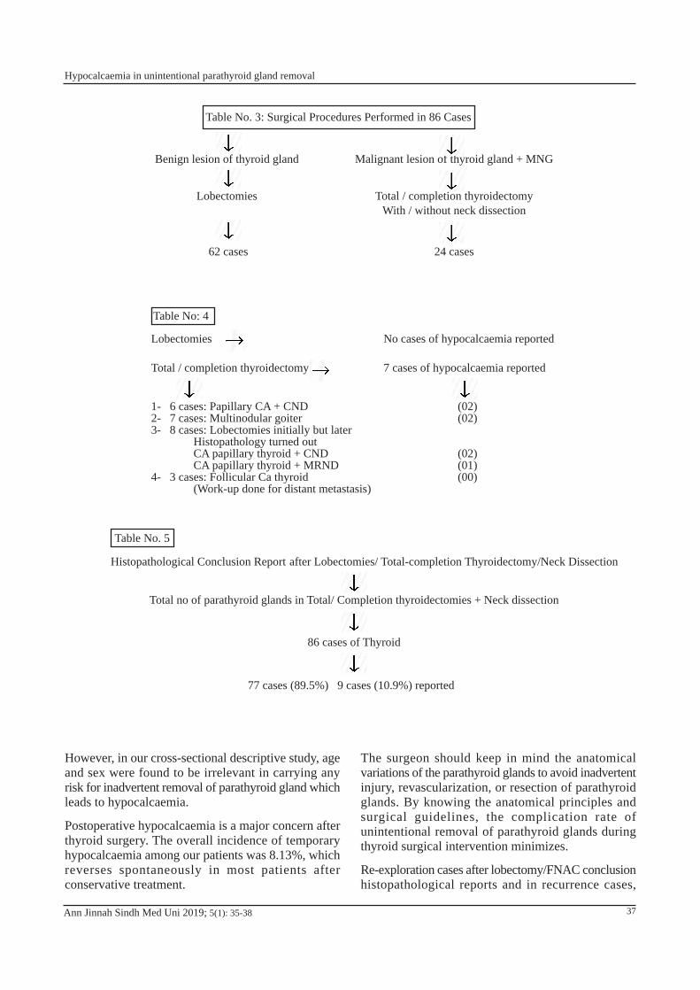
However, in our cross-sectional descriptive study, age and sex were found to be irrelevant in carrying any risk for inadvertent removal of parathyroid gland which leads to hypocalcaemia.
Postoperative hypocalcaemia is a major concern after thyroid surgery. The overall incidence of temporary hypocalcaemia among our patients was 8.13%, which reverses spontaneously in most patients after conservative treatment.
Ann Jinnah Sindh Med Uni 2019; 5(1): 35-38
The surgeon should keep in mind the anatomical variations of the parathyroid glands to avoid inadvertent injury, revascularization, or resection of parathyroid glands. By knowing the anatomical principles and surgical guidelines, the complication rate of unintentional removal of parathyroid glands during thyroid surgical intervention minimizes.
Re-exploration cases after lobectomy/FNAC conclusion histopathological reports and in recurrence cases,
Table No: 4
Lobectomies
Total / completion thyroidectomy
1- 6 cases: Papillary CA + CND2- 7 cases: Multinodular goiter3- 8 cases: Lobectomies initially but later
Histopathology turned outCA papillary thyroid + CNDCA papillary thyroid + MRND
4- 3 cases: Follicular Ca thyroid(Work-up done for distant metastasis)
No cases of hypocalcaemia reported
7 cases of hypocalcaemia reported
(02)(02)
(02)(01)(00)
Histopathological Conclusion Report after Lobectomies/ Total-completion Thyroidectomy/Neck Dissection
Total no of parathyroid glands in Total/ Completion thyroidectomies + Neck dissection
86 cases of Thyroid
77 cases (89.5%) 9 cases (10.9%) reported
Table No. 5
Table No. 3: Surgical Procedures Performed in 86 Cases
Benign lesion of thyroid gland
Lobectomies
62 cases
Malignant lesion of thyroid gland + MNG
Total / completion thyroidectomyWith / without neck dissection
24 cases
37
Hypocalcaemia in unintentional parathyroid gland removal
malignant thyroid pathology with neck dissection, has a strong correlation with unintentional removal of parathyroid glands.
CONCLUSIONRe-operated surgeries after lobectomy and for malignant thyroid pathology with neck dissection are strongly associated with high risk for unintentional removal of parathyroid glands during thyroid surgical interventions which may cause hypocalcaemia in patients. Surgeon should be fully aware and must consider these risk factors during reoperative surgery of thyroid and malignant pathology with central/modified neck dissection.
Authors’ contributions: Dr Hurtamina Khan conceived the idea, worked on literature search, data collection, data analysis and review, and the introduction. Dr Shireen Ramzan and Dr Zahid Mehmood worked on literature search, results, and discussion. Dr Razzaq Dogar reviewed the literature, worked on discussion, and edited the manuscript. Dr Javed Jamali reviewed the article. All authors discussed the results and contributed to the final manuscript.
References1. Sandstorm, I.On a new gland in man and several
mammals (glandulaeparathyriodea). Upps Lak Forh. 1880;15:441-471
2. Bliss RD,Gauger PG, Delbridge LW. Surgeons approach to the thyroid gland surgical anatomy and the importance of technique. World J Surg. 2000;24:891-897
3. Bellatone R, Lombardi CP, Bossola M, Boscherini M, Alesina P, Traini, et al. Total thyroidectomy for management for benign thyroid disease. World J Surg. 2002; 26 (12):1468-71
4. Giles Y, Boztepe H, Terzioglu T, Tezelman S. The advantage of total thyroidectomy to avoid reoperation for incidental thyroid cancer in multinodular goiter. Arch Surg.2004; 139(2):179-82.
5. Bhattacharya N, Fried MP. Assessment of the morbidity and complications of total thyroidectomy. Arch Otolaryngol Head Neck Surg. 2002;128(4):389-392. doi:10.1001/archotol.128.4.389
6. Khairy GA, Al-Saif A. Incidental parathyriodectomy during thyroid resection: incidence ,risk factors, and outcome. Ann Saudi Med. 2011;31(3):274-278
7. Akerstrom G, Malamaeus J, Bergstrom R. Surgical anatomy of human parathyroid glands. Surgery. 1984; 95(1):14-21
8. Abboud B. Topographic anatomy and arterial vascularization of the parathyroid glands: Practical application. Presse Med. 1996; 25(25):1156-61.
9. Koyuncu A, Dokmetas HS, Turan M, Aydin C, Karadayi K, Budak E, et al. Comparison of thyroidectomy techniques for benign thyroid disease. Endocr J. 2003; 50(6):723-7
10. Friguglietti CU, Lin CS, Kulesar MA. Total thyroidectomy for benign thyroid disease. Laryngoscpe. 2003; 113(10):1820-6.
11. Fahmy FF, Gillet D, Lolen Y, Shotton JC. Management of serum calcium levels in post-thyroidectomy patients. Clin Otolaryngol Allied Sci. 2004;29(6):735-9.
12. Sturniolo G, Lo Schiavo MG, Tonante A, D’Alia C, Bonano L. Hypocalacemia and hypoparathyroism after total thyroidectomy: a clinical biological study and surgical considerations. Int J Surg Ivestig. 2000;2(2):99-105
13. Yousuf T, Gaballah G, Abd-Elaad E, EL-Dosky E. Assessment of riskfactors pf incidental parathyroi-dectomy during thyroid surgery: A prospective study. Inter J surg. 2010;8(3):207-211
14. Sasson AR, Pinpank JF, JR, Wetherington RW, Hanlon AL, Ridge JA. Incidental parathroidectomy during thyroid surgery doesnot cause transient symptomatic hypocalcemia. Arch Otolaryngol Head Neck Surg.2001; 127(3):304-8
15. Lin DT, Patel SG, Shah AR, Singh B, Shah JP. Incidenceof inadvertent parathyroid removal during thyroidectomy.Laryngoscope. 2002;112(4):608-11
Ann Jinnah Sindh Med Uni 2019; 5(1): 35-3838
Hurtamina Khan, Shireen Ramzan, Zahid Mehmood, Razzaq Dogar, Javed Jamali
Huma Shareef1, Erum Zaheer2, Saima Abedin3, and Hina Hassam3
Phytomedicine: Silybum marianum (Silymarin) as an Effective Hepato-protective Source from Nature
REVIEW ARTICLE
Ann Jinnah Sindh Med Uni 2019; 5(1): 39-46
ABSTRACTObjective: To review the efficacy and safety of milk thistle for management of hepatic disordersMethodology: A thorough literature survey was carried out for the article. Following key words (Silybum marianum, Liver) were used to search the articles on Google Scholar and PubMed. More than 50 articles published between 2000 and 2018 were taken into consideration.Result: The review explored the main constituents of milk thistle and its properties to protect liver against various toxins, alcohol effects, and viral attacks. Silymarin is capable of regenerating liver and bring the hepatic markers, Alanine transaminase (ALT), Aspartate transaminase (AST) and others, to normal levels. Reported toxicities of silymarin are few. Conclusion: Silybum marianum has proved its higher efficacy and safety in hepatic disorders, as compared to other therapies and therefore is an agent of choice. Furthermore, its cost effectiveness and ability to be developed as various dosage forms, like emulsion and nanoparticles, undoubtedly ensures its extensive use in future.Key words: Silybum marianum, Liver
How to cite this article: Shareef H, Zaheer E, Abedin S, Hassam H. Phytomedicine silybum marianum (silymarin) as an effective hepato-protective source from nature. Ann Jinnah Sindh Med Uni 2019; 5 (1): 39-46
Department of Pharmacognosy1 / Department of Pharmaceutical Chemistry2 / Department of Pharmaceutics3, Institute of Pharmaceutical Sciences, Jinnah Sindh Medical University, Karachi, PakistanCorrespondence: Prof. Huma Shareef, Department of Pharmacognosy, Institute of Pharmaceutical Sciences, Jinnah Sindh Medical University, Karachi, PakistanEmail: [email protected]
INTRODUCTIONSilybum marianum L, commonly known as milk thistle or Marian thistle belongs to botanical family Asteraceae.
It is an annual or biannual specie found all over the globe especially in the Southern Europe and Asia (Figure I). Milk thistle has been famous since the olden days, to cure hepatic and gall bladder ailments such as hepatitis B and C, liver cirrhosis, cancers, jaundice, and alcoholic and non-alcoholic fatty liver disease. In addition to its traditional uses, it has also clinically proven its efficacy to combat certain poisoning conditions which include snake and insect stings, alcohol as well as mushroom Amanita poisoning1. Its role also encompasses the stimulation of biliary secretion and production of breast milk2. Chinese traditional medicine and Indian herbal practitioners
39
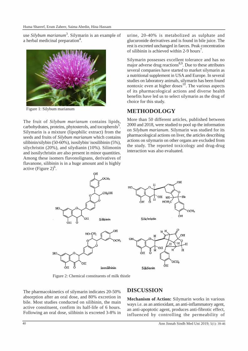
use Silybum marianum3. Silymarin is an example of a herbal medicinal preparation4.
The fruit of Silybum marianum contains lipids, carbohydrates, proteins, phytosterols, and tocopherols5. Silymarin is a mixture (lipophilic extract) from the seeds and fruits of Silybum marianum which contains silibinin/silybin (50-60%), isosilybin/ isosilibinin (5%), silychristin (20%), and silydianin (10%). Silimonin and isosilychristin are also present in minor quantities. Among these isomers flavonolignans, derivatives of flavanone, silibinin is in a huge amount and is highly active (Figure 2)6.
The pharmacokinetics of silymarin indicates 20-50% absorption after an oral dose, and 80% excretion in bile. Most studies conducted on silibinin, the main active constituent, confirm its half-life of 6 hours. Following an oral dose, silibinin is excreted 3-8% in
urine, 20-40% is metabolized as sulphate and glucuronide derivatives and is found in bile juice. The rest is excreted unchanged in faeces. Peak concentration of silibinin is achieved within 2-9 hours7.
Silymarin possesses excellent tolerance and has no major adverse drug reactions8,9. Due to these attributes several companies have started to market silymarin as a nutritional supplement in USA and Europe. In several studies on laboratory animals, silymarin has been found nontoxic even at higher doses10. The various aspects of its pharmacological actions and diverse health benefits have led us to select silymarin as the drug of choice for this study.
METHODOLOGYMore than 50 different articles, published between 2000 and 2018, were studied to pool up the information on Silybum marianum. Silymarin was studied for its pharmacological actions on liver, the articles describing actions on silymarin on other organs are excluded from the study. The reported toxicology and drug-drug interaction was also evaluated.
DISCUSSIONMechanism of Action: Silymarin works in various ways i.e. as an antioxidant, an anti-inflammatory agent, an anti-apoptotic agent, produces anti-fibrotic effect, influenced by controlling the permeability of
Ann Jinnah Sindh Med Uni 2019; 5(1): 39-46
Figure 1: Silybum marianum
Figure 2: Chemical constituents of milk thistle
40
Huma Shareef, Erum Zaheer, Saima Abedin, Hina Hassam
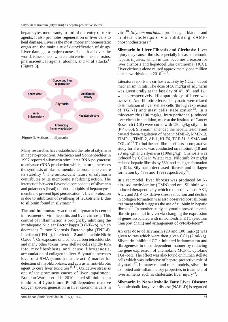
hepatocytes membrane, to forbid the entry of toxic agents. It also promotes regeneration of liver cells to heal damage. Liver is the most important homeostatic organ and the main site of detoxification of drugs. Liver damage, a major cause of death all over the world, is associated with certain environmental toxins, pharmaceutical agents, alcohol, and viral attacks11
(Figure 3).
Many researches have established the role of silymarin in hepato-protection. Machicao and Sonnenbichler in 1997 reported silymarin stimulates RNA polymerase to enhance rRNA production which, in turn, increases the synthesis of plasma membrane proteins to ensure its stability11. The antioxidant nature of silymarin contributes to its membrane stabilizing action. The interaction between flavonoid components of silymarin and polar ends (head) of phospholipids of hepatocytes’ membrane prevent lipid peroxidation12. Liver protection is due to inhibition of synthesis of leukotriene B due to silibinin found in silymarin13.
The anti-inflammatory action of silymarin is central in treatment of viral hepatitis and liver cirrhosis. This control of inflammation is brought by inhibiting the intrahepatic Nuclear Factor kappa B (NF-kb), which decreases Tumor Necrosis Factor-alpha (TNF-á), Interferon (IFN-g), Interleukin-2 and inducible Nitric Oxide14. On exposure of alcohol, carbon tetrachloride, and many other toxins, liver stellate cells rapidly turn into myofibroblasts and cause fibrogenesis, accumulation of collagen in liver. Silymarin increases level of á-SMA (smooth muscle actin) marker for detection of myofibroblasts, and acts as an anti-fibrotic agent to cure liver toxicities15-17. Oxidative stress is one of the prominent causes of liver impairment. Brandon Warner et al in 2010 stated silibinin as an inhibitor of Cytochrome P-450 dependent reactive oxygen species generation in liver carcinoma cells in
Ann Jinnah Sindh Med Uni 2019; 5(1): 39-46
vitro18. Silybum marianum protects gall bladder and hinders cholestasis via inhibit ing cAMP-phosphodiesterase19.
Silymarin in Liver Fibrosis and Cirrhosis: Liver injury may cause fibrosis, especially in case of chronic hepatic injuries, which in turn becomes a reason for liver cirrhosis and hepatocellular carcinoma (HCC). Liver cirrhosis alone caused approximately one million deaths worldwide in 201020,21.
Literature reports the cirrhosis activity by CCl4 induced mechanism in rats. The dose of 50 mg/kg of silymarin was given orally at the last day of 4th, 8th, and 12th
weeks respectively. Histopathology of liver was assessed. Anti-fibrotic effects of silymarin were related to stimulation of liver stellate cells (through expression of TGF-â) and mast cells stabilization22. In a thiocetamide (100 mg/kg, intra peritoneal)-induced liver cirrhotic condition, mice at the Institute of Cancer Research (ICR) were cured with 150mg/kg silymarin (P < 0.05). Silymarin amended the hepatic lesions and caused down-regulation of hepatic MMP-2, MMP-13, TIMP-1, TIMP-2, AP-1, KLF6, TGF-â1, á-SMA and COL-á123. To find the anti-fibrotic effects a comparative study for 8 weeks was conducted on nilotinib (10 and 20 mg/kg) and silymarin (100mg/kg). Cirrhosis was induced by CCl4 in Wistar rats. Nilotinib 20 mg/kg reduced hepatic fibrosis by 68% and collagen formation by 49%. Silymarin decreased fibrosis and collagen formation by 47% and 18% respectively24.
In a rat model, liver fibrosis was produced by N-nitrosodimethylamine (DMN) and oral Silibinin was induced therapeutically which reduced levels of AST, ALT, and ALP. Oxidative stress reduction and decline in collagen formation was also observed post silibinin treatment which suggests the use of silibinin in hepatic fibrosis25. In another study, silymarin proved its anti-fibrotic potential in vivo via changing the expression of genes associated with mitochondrial ETC (electron transport chain) and arrangement of cytoskeleton26.
An oral dose of silymarin (20 and 100 mg/kg) was given to rats which were then given CCl4 (2 ml/kg). Silymarin inhibited CCl4 initiated inflammation and fibrogenesis in dose-dependent manner by reducing the gene expression of chemokine MCP-1, cytokine TGF-beta. The effect was also found on human stellate cells which was indicative of hepato-protective role of silymarin27. In many rat and mice models, silymarin exhibited anti-inflammatory properties in treatment of liver ailments such as cholestatic liver injury28.
Silymarin in Non-alcoholic Fatty Liver Disease: Non-alcoholic fatty liver disease (NAFLD) is regarded
Figure 3: Actions of silymarin
41
Silybum marianum (silymarin) as hepato-protective source
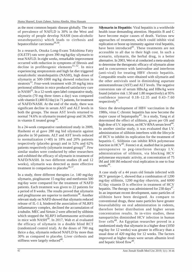
as the most common hepatic disease globally. The rate of prevalence of NAFLD is 30% in the West and majority of people develop NASH (non-alcoholic steatohepatitis) which leads to cirrhosis and hepatocellular carcinoma29,30.
In a research, Otsuka Long-Evans Tokishima Fatty (OLETF) rats were given 390 mg/kg/day silymarin to treat NAFLD. In eight weeks, remarkable improvement occurred with reduction in symptoms of fibrosis and decline in profibrogenic elements31,32. In mice experiencing streptozotoin and high fat diet triggered nonalcoholic steatohepatitis (NASH), high doses of silymarin at 500-1000 mg/kg showed reduction in steatosis33. Four-week treatment with 20 mg/kg intra peritoneal silibinin in mice produced satisfactory cure in NASH32. In a 12-week open label comparative study, silymarin (70 mg three times a day) was compared with vitamin E (400 IU/day) in 71 patients, for treatment of NAFD/NASH. At the end of the study, there was significant decline in serum AST and ALT levels in both the groups. The mean AST levels returned to normal 74.6% in silymarin treated group and 56.30% in vitamin E treated group34.
In a 24-week comparative randomized study in 2009, Hashemi et al gave 280 mg bid silymarin against placebo in 50 patients. ALT and AST levels reduced to normalization (<40) in 18% and 20% patients respectively (placebo group) and in 52% and 62% patients respectively (silymarin treated group)35. Few similar studies were conducted by other researchers and exhibited the efficacy of silymarin in treatment of NAFD/NASH. In two different studies (8 and 12 weeks), silymarin was detected as more effective treatment in comparison to placebo36,37.
In a study, three different therapies i.e. 140 mg/day silymarin, pioglitazone 15 mg/day and metformin 500 mg/day were compared for the treatment of NAFD patients. Each treatment was given to 22 patients for a period of 8 weeks. The results proved that silymarin and pioglitazone are superior to metformin38. Another relevant study on NAFD showed that silymarin reduced release of IL-1 â, hindered the association of NLRP3 inflammatory complex, declined the accumulation of á-tubulin. MEC and Sirtuin 2 were affected by silibinin which stopped the NLRP3 inflammasome activation in mice with NASH39. In 2017, Wah et al evaluated the efficacy of silymarin in a double blind RCT (randomized control trial). At the doses of 700 mg thrice a day, silymarin reduced NAFLD by more than 30% as compared to placebo. Liver cirrhosis and stiffness were largely reduced40.
Silymarin in Hepatitis: Viral hepatitis is a worldwide health issue demanding attention. Hepatitis B and C have become major causes of death. Various new approaches of treatment, which could inhibit viral replication or boost up immunity against viral hepatitis, have been introduced41. These treatments are not accessible to all due to their high cost. In such a scenario, silymarin, the herbal drug, is a better alternative. In 2003, Wei et al conducted a meta-analysis to determine the therapeutic efficacy of silymarin alone and in concurrence with lamivudine and interferon (anti-viral) for treating HBV chronic hepatitis. Comparable results were obtained with silymarin and the other antivirals used in diminishing aspartate aminotransferase (AST) and ALT levels. The negative conversion rate of serum HBsAg and HBeAg were found (relative risk 1.50 and 1.80 respectively) at 95% confidence interval 0.18–12.35 and 0.43–7.60 respectively42.
Since the development of HBV vaccination in the 1980s, virus C chronic hepatitis has now become the major cause of hepatopathy43. In a study, Yang et al determined the effect of silibinin, given per OS and high dose of I.V. injection, on HCV-RNA serum level44. In another similar study, it was evaluated that I.V. administration of silibinin interferes with the lifecycle of HCV to inhibit its replication for the treatment of hepatitis C. Silibinin can inhibit RNA polymerase function in HCV45. Ferenci et al, studied that in patients unresponsive to peg-interferon therapy I.V. administration of silibinin, via blocking HCV polymerase enzymatic activity, at concentration of 75 ìM and 100 ìM reduced viral replication in one to four weeks46.
A case study of a 44 years old female infected with HCV genotype-1, showed that a combination of 1200 mg/day silibinin, 1200 mg/day ribavirin, and 6000 IU/day vitamin D is effective in treatment of HCV hepatitis. The therapy was administered for 238 days47. In an important recent development, nano particles of silibinin have been designed. As compare to conventional drugs, these nano particles have greater bioavailability on oral administration in rodents, therefore better distribution and higher serum concentration results. In in-vitro studies, these nanoparticles diminished HCV infection in human liver cells48. An Egyptian researcher proved in a randomized study that silymarin in a higher dose (1050 mg/day for 12 weeks) was greater in efficacy than a usual dose of 420 mg/day for 12 weeks. The factors improved at higher doses were serum albumin level and hepatic blood flow49.
Ann Jinnah Sindh Med Uni 2019; 5(1): 39-4642
Huma Shareef, Erum Zaheer, Saima Abedin, Hina Hassam
Silymarin in Hepatocellular Carcinoma: Hepatocellular carcinoma (HCC) has become a major reason of death all over the world50, causing approximately 0.75 million deaths every year51. HCC is a consequence of hepatic inflammation, viral hepatitis, NASH, and contact with toxins like alcohol52. The scanty availability of treatments has urged the healthcare professionals to explore herbal products for therapeutic values53-56.
Mastron et al in 2015 proved that silymarin/silibinin reduce oxidative stress and induce arrest at various stages of cell cycle to treat hepatic cancers57. Bosch-Barrera et al in 2017 studied that silymarin reduced toxicity of anti-cancer drugs through inhibition of signal transducers58. Hepatocellular carcinoma was induced by N-nitrosodiethylamine. The treatment with silymarin considerably attenuated the changes in levels of ALT, AST, alpha fetoprotein, and decreased MDA-DNA adduct formation, hence proved itself as a likely chemotherapeutic agent59. In another study, N-nitrosodiethylamine (NDEA), 10 mg/kg for 12 weeks, was used to induce HCC. The parameters assessed were ALT, AST, MDA, GSH, GR, and others. The group of mice treated with silymarin showed a reduction in MDA and enhancement in GS, GR, and SOD levels. Histopathology was improved and AST and ALT were brought to normal60. Nano emulsion of silymarin was developed and optimized. Studies against HCC (Hepatocellular carcinoma) showed that nano emulsion enhanced ROS intensity and declined cell viability (p<0.05)61.
Toxicity Studies of Silymarin: The toxicity studies, after I.V. infusion of silymarin, indicated LD50 of 400, 385, and 140 mg/kg in mice, rats, and rabbits respectively whereas at slow rate of I.V. infusion, LD50 reached 2 g/kg. After an oral dose, LD50 was 10g/kg62.
As stated in ‘Milk Thistle report NTP TR 565’, silymarin has great safety margin63,64. For two years, 50 rats and mice of both genders were given 1.25%, 2.5%, and 5% of milk thistle extract. No occurrence of toxicity and carcinogenicity was observed. When determining the toxicity of silymarin in humans, no more than 2.4% and 1% occurrence of adverse effects were reported, in blinded and open clinical studies respectively. The main adverse event was a laxative effect. Other effects comprised nausea, vomiting, dyspepsia, urticaria, inflammation like pruritus and arthralgia. In all published studies of silymarin toxicity, only one serious adverse reaction is reported which led to collapse in a 57 years old female65.
Drug-Drug Interaction Studies on Silymarin : In a 2-week study on Chinese volunteers, the administration of silymarin (140 mg tid) and talinolol (a substrate of multidrug resistant P-glycoprotein) was done concurrently. It was observed that plasma AUC of talinolol was increased by 36% by silymarin66. In a simulated study, the interaction of silibinin with warfarin (CYP2C9 substrate) and midazolam (CYP3A substrate) were studied. At higher dose (1,650 mg/day), silymarin was predicted to enhance midazolam and warfarin AUC approximately by 5% and 4% respectively. A clinical study suggested 9% and 13% increase in plasma AUC of midazolam and warfarin at concurrent use of high doses of silymarin67.
CONCLUSIONThe review concluded that silymarin is highly effective in treatment of various liver ailments and is free from any major toxicity. Silymarin because of its low cost, easy availability, and greater therapeutic value, has become a drug of choice. Exploration of other herbal drugs is suggested to counter the side effects of antibiotics and the emerging resistance against them.
Authors’ contributions: Prof. Huma Shareef conceived the idea, edited and supervised the manuscript. Ms. Erum zaheer wrote the manuscript. Ms. Saima and Ms. Hina searched the literature. All authors discussed the results and contributed to the final manuscript.
References
1. Rambaldi A, Jacobs BP, Laquinto G, Gluud C. Milk thistle for alcoholic and/or hepatitis B or C liver diseases – a systematic Cochrane Hepato-Biliary Group Review with metaanalyses of randomized clinical trials. Am J Gastroenterol. 2005; 100(11): 2583–2591.
2. Pradhan SC, Girish C. Hepatoprotective herbal drug, silymarin from experimental pharmacology to clinical medicine. Indian J Med Res.2006; 124(5): 491–504.
3. Abenavoli L, Bardazzi G, Cracolici F, Quaranta C, Santini G, Graziosi S. et al. Complementary therapies for treating alcoholism, First Annual meeting by Complementary Medicine Research Group of the Italian Society for Alcohol Studies-May 5, 2006, Florence, Italy. Fitoterapia. 2008; 79(2): 142–147. doi: 10.1016/j. fitote.2007.11.016.
4. Abenavoli L, Bellentani S.Milk thistle to treat non-alcoholic fatty liver disease: Dream or reality? Expert Review of Gastroent & Hepat. 2014; 7(8): 677–679.
5. Wu JW, Lin LC, Tsai TH. Drug–drug interactions of silymarin on the perspective of pharmacokinetics. J Ethnopharmacol. 2009; 121(2): 185–193.
6. Abenavoli L, Capasso R, Milic N, Capasso F. Review Milk Thistle in Liver Diseases: Past, Present, Future Phytother. Res. 2010; 24(10): 1423–1432
Ann Jinnah Sindh Med Uni 2019; 5(1): 39-46 43
Silybum marianum (silymarin) as hepato-protective source
7. Saller R, Brignoli R, Melzer J, Meier R. An updated systematic review with meta-analysis for the clinical evidence of silymarin. Forsch Komplementmed. 2008; 15(1): 9–20.
8. Machicao F, Sonnenbichler J. Mechanism of the stimulation of RNA synthesis in rat liver nuclei by silybin.Hoppe Seyler Z Physiol Chem. 1997; 358(2):141-147.
9. Erlejman AG, Verstraeten SV, Fraga CG, Oteiza PI. The interaction of flavonoids with membranes: potential determinant of flavonoid antioxidant effects. Free Radic Res. 2004; 38(12): 1311-1320.
10. Dehmlow C, Erhard J, de Groot H. Inhibition of Kupffer cell functions as an explanation for the hepatoprotective properties of silibinin. Hepatol. 1996; 23(4):749-54.
11. Upadhyay G, Tiwari MN, Praksh O, Jyoti A. Involvement of multiple molecular events in pyrogallol-induced hepatotoxicity and silymarin-mediated protection: Evidence from gene expression profiles. Food Chem Toxicol. 2010; 48(6): 1660-1670.
12. Tsai JH, Liu JY, Wu TT,Ho PC, Huang CY, Shyu JC. et al. Effects of silymarin on the resolution of liver ?brosis induced by carbon tetrachloride in rats. J Viral Hepat. 2008. 15(7): 508–14.
13. Shaker ME, Shiha GE, Ibrahim TM, Comparison of early treatment with low doses of nilotinib, imatinib and a clinically relevant dose of silymarin in thioacetamide–induced liver fibrosis. Eu J of Pharmacol. 2011; 670(2-3):593-600.
14. Lieber CS, Leo MA, Cao Q Ren C, DeCarli LM. Silymarin retards the progression of alcohol-induced hepatic ? brosis in baboons. J Clin Gastroenterol 2003; 37(4): 336–339.
15. Brandon-Warner E, Sugg JA, Schrum LW, Mckillop LH, Silibinin inhibits ethanol metabolism and ethanol dependent cell proliferation in an vitro model of hepatocellular carcinoma. Cancer Lett.2010; 291(1): 120-29.
16. Crocenzi FA, Basiglio CL, Pérez LM, Portesio MS, Pozzi EJ, Roma MG. Silibinin prevents cholestasis-associated retrieval of the bile salt export pump, Bsep, in isolated rat hepatocyte couplets: possible involvement of cAMP. Biochem Pharmacol. 2005; 69(7):1113-20.
17. Mokdad AA, Lopez AD, Shahraz S, Lozano R, Mokdad AH, Stanaway J. et al. Liver cirrhosis mortality in 187 countries between 1980 and 2010: a systematic analysis. BMC medicine. 2014; 12: 145.
18. Schuppan D, Afdhal NH. Liver cirrhosis. Lancet (London, England). 2008; 371(9615):838–51.
19. Jeong DH, Lee GP, Jeong WI, Do SH, Yang HJ, Yuan DW. et al. Alterations of mast cells and TGF-beta1 on the silymarin treatment for CCl(4)-induced hepatic fibrosis. World J Gastroenterol. 2005;11(8):1141–8.
20. Chen IS, Chen YC, Chou CH, Chuang RF, Sheen LY, Chiu CH. Hepatoprotection of silymarin against thioacetamide-induced chronic liver fibrosis. J Sci Food Agric. 2012; 92(7):1441–7.
21. Shaker ME, Zalata KR, Mehal WZ, Shiha GE, Ibrahim TM. Comparison of imatinib, nilotinib and silymarin in the treatment of carbon tetrachlorideinduced hepatic oxidative stress, injury and fibrosis. Toxicol Appl Pharmacol. 2011;252(2):165–75.
22. Ezhilarasan D, Karthikeyan S, Vivekanandan P. Ameliorative effect of silibinin against N- nitrosodimethylamine-induced hepatic fibrosis in rats. Environ Toxicol Pharmacol. 2012;34(3): 1004–13.
23. Li CC, Hsiang CY, Wu SL, Ho TY. Identification of novel mechanisms of silymarin on the carbon tetrachloride-induced liver fibrosis in mice by nuclear factor-kappaB bioluminescent imaging-guided transcriptomic analysis. Food chem toxicol. 2012;50(5): 1568–75.
24. Mahli A, Koch A, Czech B, Peterburs P, Lechner A, Haunschild J, et al. Hepatoprotective effect of oral application of a silymarin extract in carbon tetrachloride-induced hepatotoxicity in rats. Clinical Phytoscience. 2015;1: 5.
25. Alaca N, Özbeyli D, Uslu S, Þahin HH, Yiðittürk G, Kurtel, H,Treatment with milk thistle extract (Silybum marianum), ursodeoxycholic acid, or their combination attenuates cholestatic liver injury in rats: Role of the hepatic stem cells. Turk J of Gastroenterol. 2017; 28(6): 476–84.
26. Charlton MR, Burns JM, Pedersen RA, Watt KD, Heimbach JK, Dierkhising RA. Frequency and outcomes of liver transplantation for nonalcoholic steatohepatitis in the United States. Gastroenterol. 2011;141(4): 1249–53.
27. Wong RJ, Aguilar M, Cheung R, Perumpail RB, Harrison SA, Younossi ZM, et al. Nonalcoholic steatohepatitis is the second leading etiology of liver disease among adults awaiting liver transplantation in the United States. Gastroenterol. 2015;148(3): 547–55.
28. Kim M, Yang SG, Kim JM, Lee JW, Kim YS, Lee JI. Silymarin suppresses hepatic stellate cell activation in a dietary rat model of non-alcoholic steatohepatitis: analysis of isolated hepatic stellate cells. Int J Mol Med. 2012; 30(3): 473–9.
29. Salamone F, Galvano F, Cappello F, Mangiameli A, Barbagallo I, Li VG. Silibinin modulates lipid homeostasis and inhibits nuclear factor kappa B activation in experimental nonalcoholic steatohepatitis. Transl res: the journal of Lab. and Cl. med. 2012; 159(6): 477– 86.
30. Pais P, D’Amato M. In vivo efficacy study of milk thistle extract (ETHIS-094) in STAM model of nonalcoholic steatohepatitis. Drugs R D. 2014; 14(4): 291–9.
31. Hajiani E, Hashmi SJ. Comparison of therapeutic effects of silymarin and vitamin E in nonalcoholic fatty liver disease: results of an open labele, prospective, randomized study Jundishapur. J of Nat Ph. Prod. 2009;4: 8–14
Ann Jinnah Sindh Med Uni 2019; 5(1): 39-4644
Huma Shareef, Erum Zaheer, Saima Abedin, Hina Hassam
32. Hashemi SJHE, Sardabi EH. A placebo-controlled trial of silymarin in patients with nonalcoholic fatty liver disease. Hepat Mon. 2009; 9:265–70.
33. Masoodi MRA, Panahian M, Vojdanian M. Effects of silymarin on reducing liver aminotransferases in patients with nonalcoholic fatty liver diseases. GOVARESH. 2013; 18: 181–5.
34. Solhi H, Ghahremani R, Kazemifar AM, Hoseini YZ. Silymarin in treatment of non-alcoholic steatohepatitis: a randomized clinical trial. Caspian J of Intern Med. 2014; 5(1): 9–12.
35. Hajiaghamohammadi AA, Ziaee A, Oveisi S, Masroor H. Effects of metformin, pioglitazone, and silymarin treatment on non-alcoholic fatty liver disease: a randomized controlled pilot study. Hepat Mon. 2012; 12(8): e6099.
36. Zhang B, Xu D, She L, Wang Z, Yang N, Sun R, et.al. Silybin inhibits NLRP3 inflammasome assembly through the NAD(+)/SIRT2 pathway in mice with nonalcoholic fatty liver disease. FASEB J. 2018; 32(2): 757–67.
37. Wah Kheong C, Nik Mustapha NR, Mahadeva S. A randomized trial of silymarin for the treatment of nonalcoholic steatohepatitis. Clin Gastroenterol Hepatol. 2017; 15(12): 1940–49.
38. Stanaway JD, Flaxman AD, Naghavi M, Fitzmaurice C, Vos T, Abubakar I. et al. The global burden of viral hepatitis from 1990 to 2013: Findings from the Global Burden of Disease Study 2013. Lancet.2016; 388 (10049): 1081–88.
39. Wei F, Liu SK, Liu XY, Li ZJ, Li B, Zhou YL. et al. Meta-analysis: Silymarin and its combination therapy for the treatment of chronic hepatitis B. Eur J Clin Microbiol Infect Dis. 2013; 32(5): 657–69
40. Petruzziello A, Marigliano S, Loquercio G, Cacciapuoti C. Hepatitis C virus (HCV) genotypes distribution: An epidemiological up-date in Europe. Infect Agents Cancer. 2016; 11: 53-66
41. Ghany MG, Strader DB, Thomas DL, Seeff LB. American Association for the Study of Liver Diseases. Diagnosis, management, and treatment of hepatitis C: An update. Hepatology 2009; 49(4): 1335–74.
42. Ahmed-Belkacem A, Ahnou N, Barbotte L, Wychowski C, Pallier C, Brillet R. et al. Silibinin and related compounds are direct inhibitors of hepatitis C virus RNA-dependent RNA polymerase. Gastroenterology. 2010; 138(3):1112–22.
43. Ferenci P, Scherzer TM, Kerschner H, Rutter K, Beinhardt S, Hofer H. et al. Silibinin is a potent antiviral agent in patients with chronic hepatitis C not responding to pegylated interferon/r ibavir in therapy. Gastroenterology. 2008; 135(5): 1561–67.
44. Dahari H, Shteingart S, Gafanovich I, Cotler SJ, D’Amato M, Pohl RT. et al. Sustained virological response with intravenous silibinin: Individualized IFNfree therapy via real-time modelling of HCV kinetics. Liver Int. 2015; 35(2): 289–94.
45. Liu CH, Lin CC, Hsu WC, Chung CY, Lin CC, Jassey A, et.al. Highly bioavailable silibinin nanoparticles inhibit HCV infection. Gut. 2017; 66(10): 1853–61.
46. Fathalah WF, Abdel Aziz MA, Abou El, Soud NH, El Raziky MES. High dose of silymarin in patients with decompensated liver disease: A randomized controlled trial. J Interferon Cytokine Res.2017; 37(11): 480–87.
47. Dimitroulis D, Damaskos C, Valsami S, Davakis S, Garmpis N, Spartalis E. et al. From diagnosis to treatment of hepatocellular carcinoma: An epidemic problem for both developed and developing world. World J Gastroenterol. 2017; 23(29): 5282–94.
48. Prasad V, Goldstein JA. US news and world report cancer hospital rankings: Do they reflect measures of research productivity? PLoS ONE 2014; 9: e107803.
49. Ghouri YA, Mian I, Rowe JH. Review of hepatocellular carcinoma: Epidemiology, etiology, and carcinogenesis. J Carcinog. 2017; 16: 1.
50. Bae K, Park JH, Kim J, Cho CK, Oh B, Costa D. et al. Traditional oriental herbal medicine and natural killer cells for cancer patients: A systematic review and meta-analysis. Phytother Res. 2017; 31(4): 519–32.
51. Chen SR, Qiu HC, Hu Y, Wang Y, Wang YT. Herbal medicine offered as an initiative therapeutic option for the management of hepatocellular carcinoma. Phytother Res. 2016; 30(6): 863–77.
52. Safe S, Kasiappan R. Natural products as mechanism-based anticancer agents: Sp transcription factors as targets. Phytother Res. 2016; 30(11):1723–32.
53. Tariq A, Sadia S, Pan K, Ullah I, Mussarat S, Sun F, et al. A systematic review on ethnomedicines of anti-cancer plants. Phytother Res. 2017; 31(2):202–64.
54. Mastron JK, Siveen KS, Sethi G, Bishayee A. Silymarin and hepatocellular carcinoma: A systematic, comprehensive, and critical review. Anticancer Drugs. 2015; 26(5): 475–86.
55. Bosch-Barrera J, Queralt B, Menendez JA. Targeting STAT3 with silibinin to improve cancer therapeutics. Cancer Treat Rev. 2017; 58: 61–69
56. Ramakrishnan G,Augustine TA,Jagan S,Vinodhkumar R, Devaki T. Effect of silymarin on N-nitrosodiethylamine induced hepatocarcinogenesis in rats. Exp Oncol. 2007; 29(1): 39-44
57. Mesallamy HOE, Metwally NS, Soliman MS, Ahmed KA, Moaty MMA. The chemopreventive effect of Ginkgo biloba and Silybum marianum extracts on hepatocarcinogenesis in rats. Cancer Cell Int. 2011; 11(1):38 (1-12)
58. Ahmed U, Akhtar J, Singh SP, Badruddeen, Ahmed FJ, Siddiqui S. Silymarin nanoemulsion against human hepatocellular carcinoma: development and optimization. Artif Cells Nanomed Biotechnol. 2018; 46(2): 231-41
59. Ghosh AGT, Jain S. Silymarin-a review on pharmacodynamics and bioavailability enhancement. J of Pharm Sci and Tech. 2010; 2: 348–55.
Ann Jinnah Sindh Med Uni 2019; 5(1): 39-46 45
Silybum marianum (silymarin) as hepato-protective source
60. Dunnick JK, Nyska A, Bishop JB, Bucher JR, Chhabra RS, Foster PM. et al. Toxicology and carcinogenesis studies of milk thistle extract (CAS No. 84604-20-6) in F344/N rats and B6C3F1 mice (Feed Studies). Natl Toxicol Program tech rep ser. 2011;565:1–177.
61. Dunnick JK, Nyska A. The toxicity and pathology of selected dietary herbal medicines. Toxicol Pathol. 2013; 41(2): 374–86.
62. Polyak SJ, Ferenci P, Pawlotsky JM. Hepatoprotective and antiviral functions of silymarin components in hepatitis C virus infection. Hepatology. 2013; 57(3):1262–71.
63. Han Y, Guo D, Chen Y, Tan ZR, Zhou HH. Effect of continuous silymarin administration on oral Talinolol pharmacokinetics in healthy volunteers. Xenobiotica;the fate of foreign compounds in biological systems. 2009(9); 39: 694–9.
64. Brantley SJ, Gufford BT, Dua R, Fediuk DJ, Graf TN,Scarlett YV. et al. Physiologically based pharmacokineticmodeling framework for quantitative prediction of anherb-drug interaction. CPT Pharmacomet. and systems pharma. 2014; 3: e107.
Ann Jinnah Sindh Med Uni 2019; 5(1): 39-4646
Huma Shareef, Erum Zaheer, Saima Abedin, Hina Hassam
1 Jinnah Sindh Medical University, Karachi, Pakistan Correspondence: Dr. Asma Shabbir, Assistant Professor, Department of Pathology, Sindh Medical College, Jinnah Sindh Medical University, Karachi, Pakistan Email: [email protected]
Asma Shabbir1 and Syed Mehmood Hassan1Molecular Diagnostics: A Paradigm Shift
COMMUNICATION
Ann Jinnah Sindh Med Uni 2019; 5(1): 47-48
INTRODUCTIONEffective care given to the patient by a healthcare practitioner depends upon accurate ‘diagnosis’ and ‘treatment’ of the ailment. The first line laboratory diagnostic applications like gram staining, hematoxylin and eosin staining, complete blood count, and other biochemical tests have been available for decades. However, they have significant limitations. Molecular diagnostics has offered a dynamic transformation to the field of diagnostics. It is a branch which provides a set of techniques to analyze biological markers in the genetic code (genome) and protein expressed by the genes (proteome). Its role in human diseases widely tags in mutation and pathogen detection. Continuous availability of new methods and new applications has helped molecular diagnostics to be the most rapidly growing field in the laboratory medicine.
With ground breaking developments in molecular biology, we are trying to understand the ways in which diseases develop in an individual. Moreover, it also provides us an idea of disease resistance, immunity, and specific therapy to the patient. Molecular diagnostics is widely being used in hospitals, reference laboratories, and blood banks. The major technologies involved are polymerase chain reaction (PCR), blotting techniques, fluorescent in situ hybridization (FISH), microarray, and mass spectrometry amongst others. These tests are extensively being used in clinical applications including infectious diseases, genetic disease screening, pharmacogenomics, oncology and human leukocyte antigen typing1.
PCR is the most commonly used technique in major laboratories. It has revolutionized the field of scientific research and medicine. Approximately more than twelve modified types of PCR are now available with
significant advantages over one another. It is playing an emerging role in infectious diseases like tuberculosis, HIV, hepatitis etc. Though the diagnosis of infectious agent can be done through traditional lab tests but PCR remains an exquisite choice as it is a more rapid, sensitive, and specific technique. Similarly, it has also gained importance in the field of histopathology particularly to understand the biology of lymphomas and leukaemia. Translocations in aggressive tumours like sarcomas are now being identified through RT-PCR. DNA profiling is also being utilized in forensic sciences, genetic, and paternity testing2.
FISH technique has extended the field of cytogenetics, being more rapid and accurate as compared to the conventional method of karyotype analysis. It has become a vital tool in oncology to detect molecularmarkers and translocations in various cancers including leukaemia, breast carcinoma, prostate cancer, cholangiocarcinoma, and melanoma. However, literature shows that developing countries have limited use of FISH which might be due to lack of knowledge or unavailability of the technique3. Microarray is another upcoming technique which is not yet routinely used in diagnostics due to its cost and because it also requires skillful hands to work. But, it promises to be as reliable and sensitive as other molecular diagnostic facilities.
Other than the medical field, molecular techniques are also being utilized in agricultural and industrial applications. Moreover, research in gene therapy is ongoing and in the limelight. Approximately 19,000 to 20,000 protein coding genes are estimated to be present in a human genome4. In this era of personalized medicine, it is expected that complete human genome will be recognized soon through varieties of molecular techniques. Development and new insights in the field of molecular biology has provided a paradigm shift from routine diagnostic tests to the advancement in diagnosis and treatment of a number of diverse diseases.
47
How to cite this article: Shabbir A, Hassan SM. Molecular diagnostics: a paradigm shift. Ann Jinnah Sindh Med Uni 2019; 5 (1): 47-48
CONCLUSIONHence, we conclude that knowledge about molecular biology and diagnostics has become imperative for every medical practitioner. In this period of modern medicine, medical students as well should obtain awareness of molecular diagnostics. Moreover, focus should be on including molecular diagnostics in the medical curriculum.
Authors’ contributions: Asma Shabbir conceived the idea and wrote the manuscript. Syed Mehmood Hasan did the critical review. Both the authors approved the manuscript.
References1. Grody WW, Nakamura RM, Strom CM, Kiechle
FL(eds). Molecular Diagnostics: Techniques and Applications for the Clinical Laboratory. Academic Press, Inc; Boston, MA. 2010.
2. Kumar A, Rani R, Goel A, Role of PCR in diagnostics. Bio Evolution, 2014; 23-24
3. Ratan ZA, Zaman S, Mehta V, Haidere MF, Runa NJ, Akter N, Application of Fluorescence In Situ Hybridization (FISH) Technique for the Detection of Genetic Aberration in Medical Science. Cureus, 2017; 9(6):1325, doi:10.7759/cureus.1325, PMID:28690958
4. Ezkurdia I, Juan D, Rodriguez JM, Frankish A, Diekhans M, Harrow J, et.al. Multiple evidence strands suggest that there may be as few as 19,000 human protein-coding genes. Hum Mol Genet. 2014; 23(22):5866–78.doi: 10.1093/hmg/ddu309.
Ann Jinnah Sindh Med Uni 2019; 5(1): 47-4848
Asma Shabbir, Syed Mehmood Hassan
Ann Jinnah Sindh Med Uni 2019; 5(1): iii
During recent years, there has been an emerging, upward trend in research and publication of studies on the effects of Ramadan fasting on the healthy population as well as on the populace diagnosed with various diseases and conditions. To publicize the outcomes of these research works, many awareness programs, symposia and conferences are being organized globally. International coordination groups, such as ‘Diabetes and Ramadan: International Alliance’ have been established to coordinate the efforts and investigations of researchers involved in this work. Periodicals like ‘Journal of Nutrition, Fasting and Health of Iran’ are playing a pivotal role in disseminating research outcomes related to the effects of Ramadan fasting. Guidelines for fasting are being developed for patients of various diseases, such as ‘Diabetes and Ramadan: Practical Guidelines’ devised by the International Diabetes Federation. ‘New guidelines on diabetes management during Ramadan’ has been published recently by Baqai Institute of Diabetes and Endocrinology (BIDE), Pakistan. Departments and study groups are being established in institutions to facilitate researchers in conducting studies on the effect of Ramadan fasting on different diseases.
Jinnah Sindh Medical University (JSMU), Karachi under the visionary leadership of the Honorable Vice Chancellor, Prof. Syed Muhammad Tariq Rafi, annually organizes, just before the month of Ramadan, an awareness program regarding diet and drug adjustment for conditions such as pregnancy, diabetes, renal diseases, coronary artery disease etc. Furthermore, every alternate year, JSMU organizes an ‘International Biennial Conference’ on Ramadan and Health. These conferences are organized to disseminate studies
FROM THE DESK OF THE EDITOR*
3rd Pakistan International Biennial Conference on Ramadan and Health
Prof. Nazeer KhanEditor, Annals of Jinnah Sindh Medical UniversityProfessor of Biostatistics, Chairman of the Conference,Department of Research Jinnah Sindh Medical University,Rafiqui H R. Shaheed Road Karachi 75510, [email protected]: +92 21 99204776/ext 1087
conducted on the effects of Ramadan fasting and these studies are presented by Pakistani speakers as well as by invited foreign speakers. The third issue of the conference, titled ‘3rd Pakistan International Conference on Ramadan and Health’ will be held from 23rd to 25th
August’ 2019.
Four foreign speakers: Dr. Mohsen Nematy, Mashhad University of Medical Sciences, Mashhad, Iran; Dr. Abdolreza Norouzy, Mashhad University of Medical Sciences, Mashhad, Iran; Prof. Mafauzy Mohamed, University of Sains, Malaysia and Dr. Kamran Mahmood Ahmed A.Aziz, Ministry of Health, Abha, Saudi Arabia, have consented to present their studies in this conference. Fourteen invited speakers from Pakistan will also present their work related to various aspects of the effects of Ramadan fasting.
The theme of the conference is ‘Ramadan: Bridge between Health and Spiritual Path’. The inauguration session will be held in Marriott Hotel, Karachi on the evening of 23rd August’ 2019. The honourable Governor of the province of Sindh, Mr Imran Ismail and Chairman of Hilton Pharma, Dr. Sardar Muhammad Yasin Malik, have consented to be the Chief Guest and Guest of Honor, respectively, for this ceremony. Prof. Anis Ahmed, a well-known scholar and Vice Chancellor of Riphah University, Islamabad will deliver a talk on the theme of the conference. Scientific sessions would be held on the 24th and 25th of August in Sindh Medical College campus of JSMU. There will be five plenary sessions on Ramadan and Diabetes, Ramadan and Drug Management, Ramadan and Medical Education and Ramadan and Medicine; and four sessions for contributed papers. On the evening of 24th August’ 2019, dinner at a beachfront restaurant along the coastline of the Arabian Sea will be hosted for the delegates and the organizing committee. The conference will culminate with closing remarks and suggestions from the delegates in the afternoon of 25th August. We cordially invite all interested delegates to come to Karachi and be part of this educational and enlightening experience. Meanwhile, we are eagerly waiting for our guests and friends to join us.
* This announcement might also be published in other medical journals.
Nazeer Khan
1. Azfar Siddiqi, Centre for Disease Control, United States of America
2. Ashraf Jahngeer, Dow University of Health Sciences, Karachi, Pakistan
3. Kaleem Sayyed, United States of America
4. Atta Chandio, Shaheed Mohtarma Benazir Bhutto Medical University, Larkana, Pakistan
5. Huba Atiq, Aga Khan University, Karachi, Pakistan
6. Naghmana Shafi, United States of America
7. Naresh Kumar, Dow University of Health Sciences, Karachi, Pakistan
8. Fareed Mohsin, Saudi Arabia
9. Syed Muhammad Mubeen, Hamdard University, Karachi, Pakistan
10. Muhammad Siddiqui, Saudi Arabia
11. Umar Farooq, Dow University of Health Sciences, Karachi, Pakistan
12. Imran Bakar, Hamdard University, Karachi, Pakistan
13. Durr e Sameen, Dow University of Health Sciences, Karachi, Pakistan
14. Jewraj Maheshwari, United States of America
15. Dr Tariq Ali, Dow University of Health Sciences, Karachi, Pakistan
16. Zeeshan Feroz, King Saud Bin Abdulaziz University for Health Sciences, Saudi Arabia
17. Nighat Nisar, Dow University of Health Sciences, Karachi, Pakistan
18. Maha Ali, Bahria University, Karachi, Pakistan
19. Dileep Nanik, United Arab Emirates
20. Nadia Aziz Ather, Altamash Dental Hospital, Karachi, Pakistan
21. Hina Zahid, Dow University of Health Sciences, Karachi, Pakistan
22. Shazia Jamshed, International Islamic University of Malaysia
23. Shadman Andalib, Jamjoom Pharma, Saudi Arabia
24. Saira Mirza, University of Toronto, Canada
25. Syeda Kausar Ali, Jinnah Sindh Medical University, Karachi, Pakistan
26. Nadeem Rizvi, Jinnah Postgraduate Medical Centre, Karachi, Pakistan
27. Nusrat Shah, Dow University of Health Sciences, Karachi, Pakistan
28. Anwar Mian, Dow University of Health Sciences, Karachi, Pakistan
29. Shiraz Burki, Australia
Ann Jinnah Sindh Med Uni 2019; 5(1):IV
July 2018 to June 2019The Editorial Board of AJSMU is indebted to the following members of the medical community for their time and effort
in reviewing articles and sending valuable feedback.
ACKNOWLEDGEMENT OF REVIEWERS
The Annals of Jinnah Sindh Medical University (AJSMU)is an open access, peer reviewed, bi-annual publication.Authors are invited to submit articles with a wide spectrumof coverage reporting original work, epidemiology, publichealth, indigenous disease and standards of health care fromPakistan and overseas. Basic research with clear clinicalimplications will also be considered. Review articles ofcurrent interest and high standard are welcomed forconsideration. There are also sections for Case Reports,Short Communication and letter to editors. To encouragethe young investigators, AJSMU also includes a ‘StudentSection’. All submissions must conform to the UniformRequirements for Biomedical Journals of InternationalCommittee of Medical Journals Editors (ICMJE).
AJSMU is copyright under the Berne Convention. All rightsreserved. AJSMU is an Open Access journal and will notcharge readers, or their institutions for access. From theBudapest Open Access initiative (BOAI) definition of openaccess, the reader has the right to “read, download, copy,distribute, print, search, or link to the full text of the articles”.Articles published in AJSMU are distributed under the termsof the Creative Commons Attribution-NonCommercialLicense (CC BYNC). Readers may copy, distribute, display,and perform the work, and make derivative works based onit for noncommercial purposes with the proper citation ofthe original work. Please note, however, if an article containsa figure from another source that is protected by copyright,the republication is not allowed without seeking permissionfrom the original source of publication.
Submission of Manuscripts: Manuscripts, or the essenceof their content, must be previously unpublished and shouldnot be under simultaneous consideration by another journal.The authors should also declare if any similar work has beensubmitted to or published by another journal. They shouldalso declare that it has not been submitted/published elsewherein the same form, in English or in any other language,without the written consent of the Publisher.
We are happy to consider articles that have been previouslypublished in another language, or in a local journal withlimited distribution, on the condition that there is anappropriate citation included in the references, and writtenconsent is obtained.
If any article is already made publicly available on an institutewebsite or repository, this will not affect our decision topublish, but we do need to know this. Failure to disclosethis is unethical. Following publication the author shouldupdate the repository and include a citation to the publishedwork. Furthermore, if any study is already presented orallyor in poster form in an international conference and theabstract or full paper is available in the conference ‘abstractbook’ or in proceedings, can be consider for publication in AJSMU.
INSTRUCTIONS TO AUTHORSPrior to submission, authors are requested to visit our website (www.jsmu.edu.pk/ajsmu) where the full instructions
and guidelines for authors, together with the Uniform Requirements for Biomedical Journals and submission checklistscan be found. Manuscripts that do not conform to the Instructions to Authors may be subject to delay.
The authors should also declare that the paper is the originalwork of the author(s) and not copied (in whole or in part)from any other work. All papers will be automaticallychecked for duplicate publication and plagiarism usingTURNITIN software. If detected, appropriate action will betaken in accordance with guidelines of Higher EducationCommission’s plagiarism policy. All Tables and Figuresmust be original and not adapted from other work unlessspecified and with the appropriate references and copyrightpermission. By virtue of the submitted manuscript, thecorresponding author acknowledges that all the co-authorshave seen and approved the final version of the manuscript.The corresponding author should provide all co-authors withinformation regarding the manuscript, and obtain theirapproval before submitting any revisions. Participation solelyin the acquisition of funding or the collection of data doesnot justify authorship. Manuscripts are only accepted forpublication on the understanding that the authors will permiteditorial amendments, though galley proofs will always besubmitted to the corresponding author before being sentfinally to press. There is no submission or publication chargesfor AJSMU to avoid any extra financial burden on theinvestigator. The authors should be able to provide uponrequest the raw material of any study or the quality assuranceassessment of any study under publication. To avoid anydelay in processing manuscripts, prior to submission, allauthors should refer to the appropriate checklists accordingto the type of the manuscript and comply with their guidelines:
Randomized controlled trials: CONSORT ChecklistObservational studies in epidemiology: STROBE ChecklistDiagnostic accuracy studies: STARD ChecklistSystematic reviews and meta-analyses: PRISMA ChecklistQualitative research: COREQ Checklist.
Prior to the initial submission of a new manuscript, pleasecarefully consider that all authors’ names are included asno change to authors’ details will be permitted after theinitial submission.
Ethical standards: AJSMU is committed to upholding thehighest standards of research, editorial, and publicationethics, and follows international guidelines, procedures, andpolicies (eg. Committee of Publication Ethics [COPE], andOffice of Research Integrity [ORI]) when dealing with anycases of suspected ethical misconduct. If such cases arise,the journal may share relevant information with necessarythird parties (for example, authors’ institutes, PakistanAssociation of Medical Editors or Higher EducationCommission). All information will be treated in a confidential,factual, and non-judgmental manner. AJSMU will also retainthe right to pursue any issues of ethical misconduct evenafter rejection or withdrawal of a manuscript from the journal.
Electronic submission of Manuscripts: Manuscripts canbe submitted electronically on www.ajsmu.com
Ann Jinnah Sindh Med Uni 2019; 5(1): v-viii v
Electronic submission saves time and postage costs, and allows the manuscript to be handled in electronic form throughout the review process.
Figures and Illustrations should be provided in jpeg format(resolution 300 DPI). Titles and detailed explanations belongin the legends for illustrations (on a separate sheet), not onthe illustrations themselves. Upon acceptance of themanuscript for publication, authors are required to providethe original copy of the assignment of copyright dully signedby all authors.
Manuscript Preparation: Manuscripts including tables,references and figure legends, must be typewritten on 8 1/2x 11 inch (21.5 x 28 cm) or size A4 paper, with margins ofat least 1 inch (2.5 cm). Pages should be numberedconsecutively, beginning with the title page and continuingthrough the last page of typewritten material. Manuscriptsmust be accompanied by a covering letter signed by theauthor and all co-authors. All Case Reports must include atleast one figure. The journal recommends that authors shouldconsider having their manuscripts professionally edited priorto submission; even more so for authors for whom Englishis a second language.
Word Limits: Original articles should be no longer than3000 words, (excluding abstract and references). Shortreview could have up to 1500 words, systematic reviewcould have 4000 words and expert review will be free fromword limitation. Short and systematic review articles mayinclude up to 50 references, and Original articles may includeup to 25 references. Articles may include a maximum of 6tables and/or figures in total.
Urdu Text: An Urdu translation must be submitted for: (1)title of the article, (2) each author’s name and affiliation,and (3) abstracts.
Title Page: The title page must contain (1) title of the articlein Urdu and English, (2) first name, middle initial and familyname of each author plus highest degrees, not more than 2,in that specific order (3) Urdu translation for all authorsnames (4) any disclaimers, and (5) a short running title ofno more than 40 characters (count letters and spaces). Thesecond page should include in English and Urdu (1) nameand address of the department(s) and institution(s) fromwhere the research was carried out for each author; affiliationaddress should be a record of where an author is currentlyworking. If the study was previously carried out at anotherinstitute this should appears as “formerly of ....” (2) currentposition and affiliation address for the corresponding authorand (3) first name, address, telephone number, fax numberand E-mail address of author to whom correspondenceshould be sent if it differs from the first author. Reviewarticles should consist of one or 2 authors, only ClinicalReview or Meta-analysis may include multiple authors. Case Reports should preferably not exceed 4 authors.
Abstracts: For original articles a structured abstract of 250words should include the following subheadings: Objectives(may include background in one line if pertinent),Materials/Subjects/Patients and Methods (place and durationof study, design, patients/respondents, brief methodology),Results (key findings only), Conclusion. For review articles
and case reports the abstract should be unstructured withnot more than 150 words. The abstracts should also includeUrdu translation however if author(s) are unable to do so,then the editorial board of the journal will provide thetranslation service free of cost.
Introduction: For original research articles the introductionshould be fully referenced with not more than threeparagraphs and a maximum of 15 pertinent references. Theintroduction should end with the rationale of the studyleading to the research question/study objectives. For casereports the introductory section should include a maximumof 10 pertinent references and end with the objective andreason of presenting the case.
Methods: This section should include setting, study design,sampling technique and sample size estimation. In studiesof diagnostic accuracy, the methodology should be detailedand include the inclusion and exclusion criteria of patientsinvolved in the study together with information on patientrecruitment. Textual re-use of portions of an author’s previouswork in the methods section will be considered, providingthat an explanatory note is included with appropriatereferencing: “The methods are exactly as published in theprevious publication.....”
Statistical Analysis: The author(s) should adequatelydescribe or reference all statistical procedures used in aparagraph at the end of the methods section. It is expectedthat the statistical tests used are appropriately selected andapplied, with an indication of the related assumptions andhow they have been tested. The ambiguous use of statisticalterms should be avoided such as random with the meaningof haphazard, correlation instead of association, etc. Exactp-values and confidence intervals are to be used. Standardguidelines must be used in reporting the results of clinicaltrials, studies assessing diagnostic tests, etc. The statisticalsoftware package used must be specified and properlyreferenced.
Tables: Tables should be double spaced on a separate sheetof paper. Do not submit tables as photographs. Numbertables consecutively in the order of their first citation in thetext and supply a brief “stand-alone” title for each. Giveeach column a short or abbreviated heading. Place explanatorymatter in footnotes, not in the heading. Explain in footnotesall non-standard abbreviations that are used in each table.For footnotes use the following symbols, in this sequence*, †, ‡, §, **, ††, ‡‡, §§ etc. Bar graphs and pie charts shouldonly be used where absolutely indicated and should beprovided in pattern. If the author is interested to print thegraphs in color, he/she has to pay color printing. Wherepossible the information should be presented in table format.
Discussion: Discussion should include the comparison andcritical analysis of the study results with the contemporaryreferences. The references cited in the introduction shouldbe made use of in the discussion. This section should alsoinclude the comments of the author(s) on their study andshould not be a repetition of the results. Discussion shouldnot be more than six paragraphs.
Conclusion and Recommendations: Conclusions shoulddirectly relate to the objectives of the study and not
Ann Jinnah Sindh Med Uni 2019; 5(1): v-viiivi
Instructions to authors
hypothetical statements by the author(s). The conclusionsmay include recommendations (if any), however it is not arequirement of the journal.
Acknowledgment: Acknowledgment should conform tothe Uniform Requirements for Biomedical Journals whichstates: List all contributors who do not meet the criteria forauthorship, such as a person who provided purely technicalhelp, writing assistance, or a department chair who providedonly general support. Financial and material support shouldalso be acknowledged. Groups of persons who havecontributed materially to the paper but whose contributionsdo not justify authorship may be listed under a heading suchas “clinical investigators” or “participating investigators,”and their function or contribution should be described, forexample, “served as scientific advisors,” “critically reviewedthe study proposal,” “collected data,” or “provided and caredfor study patients.” Because readers may infer theirendorsement of the data and conclusions, all persons musthave given written permission to be acknowledged.
References: For research reports a maximum of 25 pertinentreferences and for review papers a maximum of 50 pertinentreferences can be cited. References should be numberedconsecutively in the order, in which they are first mentionedin the text, not alphabetically, and formatted according toVancouver style. References must be superscripted throughout the text of the manuscript. List all authors when there are 6 or fewer; when there are 7 or more, list only the first 6 and add “et al”. References older than ten years should be updated to the most recent. Acceptable references are from the last ten years with a minimum of two from the last five years, however, one or 2 historical references may be used if necessary. All references must be cited in the text or tables. Where references are cited in tables only, the first reference number used in the table should follow on numerically from the last reference number used in the main text of the article. Where previous studies are mentioned in a table the authors’ names should appear in Vancouver style, with the names and reference numbers appearing in one column, and any other necessary information appearing in a separate column. Unpublished data and personal communications may be cited as references. Please try to avoid using conference papers or abstracts as references, these can only be allowed if published by journals included in Index Medicus or a well-known publishing company and are within one year from the submission date of the manuscript. Only 1-2 up to date references should be used for each particular point in the text. Only few website references can be accepted. The download dates should be mentioned in such references.
References to journal articles should include, in this order:(1) authors, (2) title, (3) journal name (as abbreviated inIndex Medicus, if not included in Index Medicus journaltitle should be given in full), (4) year, (5) volume, (6) pagenumbers (start and end). Volume and edition numbers andspecific page numbers should be included when appropriate.Secondary references are not acceptable. The author isresponsible for the accuracy and completeness of thereferences and for their correct textual citation. When acitation is referred to in the text by name, the accompanyingreference must be from the original source. Upon acceptance
of a paper all authors must be able to provide the full paperfor each reference cited upon request at any time up topublication. Failure to do so may result in the paper beingwithdrawn from the journal. Example of correct referenceform is given as follows:
Journal: Khan NB. Treatment needs for dental caries inschoolchildren of Riyadh: A-follow-up study of the OralHealth Survey of Saudi Arabia. Saudi Med J 2003; 24 (10):1081-1086.
Al-Nozha MM, Al-Maatouq MA, Al-Mazrou YY, ArafahMR, Khalil MZ, Khan NB, et al. Diabetes mellitus in SaudiArabia. Saudi Med J 2004; 25(11): 1603-1610.
Book Chapter: Thilander B. Ronning O. Introduction toOrthodontics. Stockholm: Gothia; 1995. p. 43-49.
Review Articles should include an extended bibliographyup to 100 references. Original Articles and Case Reportsshould include up to date references, preferably not exceeding40 and 15 respectively. Brief Communication should includea maximum of 5 references.
Illustrations: All figures or photographs should be submittedin a high resolution (minimum 300 DPI) electronic versionsaved in jpeg format. Original hard copies of all figures maybe requested when necessary.
Photographs will be accepted at the discretion of the EditorialBoard. All lettering, arrows, or other artwork must be doneby an artist or draftsman. If arrows are used please ensurethey appear in a different color to the background color,preferably black with a white border, or white with a blackborder. If arrows distinguish different items on the figurethen different arrow styles should be used ie. long, short,wide, narrow. Written informed consent for publication mustaccompany any photograph in which the subject can beidentified. Written copyright permission, from the publishers,must accompany any illustration that has been previouslypublished. All illustrations (“figures”) must be numberedas cited in the text in consecutive numeric order. With colorillustrations, the author must bear part of the expense forcolor reproduction. Titles and detailed explanations belongin the legends for illustrations, (on a separate sheet), not onthe illustrations themselves. If the authorship does not includea radiologist, please ensure that all radiology figures arereviewed and written approval submitted from a radiologist.Please note, this will not qualify for authorship, however,an acknowledgment may be included.
Units of Measurement: Le Systerne International d’Unites(SI) is preferred Equivalent values in traditional units shouldbe given if thought to be necessary.
Abbreviations and symbol: Use only accepted internationalabbreviations. Avoid abbreviations in the title and abstract.The full term for which an abbreviation stands should precedeits first use in the text unless it is a standard unit ofmeasurement. The following is a list of abbreviations whichmay be used without expansion. CD; CD-ROM;
Ethical consent: All manuscripts reporting the results ofexperimental investigations involving human subjects shouldinclude a statement confirming that informed consent wasobtained from each subject or subject’s guardian. All
Ann Jinnah Sindh Med Uni 2019; 5(1): v-viii vii
Instructions to authors
manuscript must include an approval letter from ethicscommittee or institutional review board, even the studydoes not include human or animal subjects. Ethical approvalis necessary not only for patient consent, but to avoidduplication of work from the same institute, and confirmsthat the institute gives approval to release the data. Whenreporting experiments on animals, authors should indicatewhether the institutional and national guide for the care anduse of laboratory animals was followed.
Conflict of interest: Authors are responsible for recognizingand disclosing financial and other conflicts of interest thatmight bias their work. They should acknowledge in themanuscript all financial support for the work and otherfinancial or personal connections to the work. Conflict ofinterest of reviewers, editors and staff can be seen in Conflictof Interest page.
Corrections, retractions and expressions of concern aboutresearch findings: Editors must assume initially that authorsare reporting work based on honest observations. Nevertheless, two types of difficulty may arise.Firstly, errors may be noted in published articles that requirethe publication of a correction or erratum of part of the work.
It is conceivable that an error could be so serious as to vitiatethe entire body of the work, but this is unlikely and shouldbe handled by editors and authors on an individual basis.Such an error should not be confused with inadequaciesexposed by the emergence of new scientific information inthe normal course of research. The latter require no corrections or withdrawals. The second type of difficulty is scientific fraud. If substantial doubts arise about the honesty of work, either submitted or published, it is the editors’ responsibility to ensure that the question is appropriately pursued (including possible consultation with the authors). However, it is not the task of editors to conduct a full investigation or to make a determination; that responsibility lies with the institution where the work was carried out or with the funding agency.
The editor should be promptly informed of the final decisions,and, if a fraudulent paper has been published, the journalmust print a retraction. If this method of investigation doesnot result in a satisfactory conclusion, the editor may chooseto publish an expression of concern, with an explanation.
The retraction or expression of concern, so labeled, shouldappear on a numbered page in a prominent section of thejournal, be listed in the contents page, and include in itsheading the title of the original article. It should not simplybe a letter to the editor. Ideally, the first author should bethe same in the retraction as in the article, although undercertain circumstances the editor may accept retractions byother responsible people. The text of the retraction shouldexplain why the article is being retracted and include abibliographic reference to it.
If a published paper is found later to be extensively plagiarized and is found to be a duplicate or redundant publication, a note of retraction will be published, the indexing services will be notified, and copies of the correspondence will be sent to the authors’ head of institute,
Pakistan Association of Medical Editors and Higher Education Commission.
The validity of previous work by the author of a fraudulentpaper cannot be assumed. Editors may ask the authors’institution to assure them of the validity of earlier workpublished in their journals or to retract it. If this is not donethey may choose to publish an announcement to the effectthat the validity of previously published work is not assured.
Permission to Reprint: Whenever a manuscript containsmaterial (text, tables dosages, figures etc.), which is protectedby copyright, it is the obligation of the author to securewritten permission from the holder of the copyright.
Galley Proofs: The research team of any study should assignone of the authors as the corresponding author. The EditorialOffice will send proofs of the manuscript to the correspondingauthor for final proof reading and it will be the responsibilityof the corresponding author to return the galley proofmaterials appropriately corrected within the stipulated time.Proofs will not be accepted from any other of the authorswithout an accompanying authorization letter from thecorresponding author. After the corresponding author hassigned the galley proof, they will bear the burden ofresponsibility for any publication errors missed on the galleyproof found after publication. No major changes such asdeletion, shortening or expansion of sentences in the textwill be accepted at this time. During the proofing process,no addition of information is allowed, however, if there newrelevant information to be added to the manuscript, this canbe included as an addendum to the article. The correspondingauthor(s) is required to sign on each page of the galley proofindicating their approval of any Editorial amendments andagree that the meaning of their article has not been altered.The author should review this carefully, as he/she isresponsible for all changes in his work, including changesmade by the copy editor. It is the duty of the correspondingauthor to respond promptly to any query from the EditingDepartment as failure to do so may result not only in delayof the article but also return of the article to the authorwithout publication. Papers will be published only whenfinally accepted manuscript signed by the correspondingauthor or designated corresponding author is received in theEditorial Office. If a manuscript is sent out for proofing andno response is received from the corresponding author, thismanuscript will be deferred for one issue only. The proofwill then be resent after four months and if there is still noresponse from the corresponding author at that time thepaper may be withdrawn from the Journal. If the EditorialOffice is able to proofread the article and answer anyoutstanding queries, it will be at the Editor’s discretion toproceed with the publishing of the paper including a statementthat this has not been proofread by the corresponding author.If there are a substantial number of unresolved queries thenthe paper may be withdrawn from the Journal.
Submit manuscripts electronically only at: www.ajsmu.comPhone: 021-99205185 Ext: 1076, 1087
Ann Jinnah Sindh Med Uni 2019; 5(1): v-viiiviii
Instructions to authors