unnatural amino acid incorporation into virus-like particles
TRANSCRIPT
Unnatural Amino Acid Incorporation into Virus-Like Particles
Erica Strable†, Duane E. Prasuhn Jr.†, Andrew K. Udit†, Steven Brown†, A. James Link‡,John T. Ngo‡, Gabriel Lander§, Joel Quispe§, Clinton S. Potter§, Bridget Carragher§, DavidA. Tirrell‡, and M. G. Finn*,†Department of Chemistry and The Skaggs Institute of Chemical Biology, and National Resource forAutomated Molecular Microscopy and Department of Cell Biology, The Scripps Research Institute,La Jolla, California 92037, and Division of Chemistry and Chemical Engineering, California Instituteof Technology, Pasadena, California 91125. Received October 21, 2007; Revised ManuscriptReceived January 14, 2008
AbstractVirus-like particles composed of hepatitis B virus (HBV) or bacteriophage Qβ capsid proteins havebeen labeled with azide- or alkyne-containing unnatural amino acids by expression in a methionineauxotrophic strain of E. coli. The substitution does not affect the ability of the particles to self-assemble into icosahedral structures indistinguishable from native forms. The azide and alkynegroups were addressed by Cu(I)-catalyzed [3 + 2] cycloaddition: HBV particles were decomposedby the formation of more than 120 triazole linkages per capsid in a location-dependent manner,whereas Qβ suffered no such instability. The marriage of these well-known techniques of sense-codon reassignment and bioorthogonal chemical coupling provides the capability to constructpolyvalent particles displaying a wide variety of functional groups with near-perfect control ofspacing.
INTRODUCTIONA variety of strategies and expression systems have been employed to cotranslationallyincorporate unnatural amino acids into proteins. The three most common approaches are theuse of a nonsense or rare codon (1-8), the use of a larger (quadruplet) codon (4,9), or thereassignment of a sense codon (10-17). In the last approach, codons for methionine (11-15,18,19), leucine (20), isoleucine (21), phenylalanine (22), tryptophan (23,24), and proline (25)have been used for incorporation of unnatural amino acids. Here we employ this technique(10,16,26) to replace methionine with the unnatural amino acids azidohomoalanine (1, Figure1) and homopropargyl glycine (2) for the first time in proteins that spontaneously assembleinto virus-like particles in the host E. coli cell.
The incorporation of organic azides and terminal alkynes introduces highly energetic functionalgroups that are inert to biological molecules under physiological conditions. The azide group's“bio-orthogonality” has been particularly prized, due to its participation in the Staudingerreaction with phosphines (27,28), cycloaddition with strained-ring alkynes (27,29-31), and
© 2008 American Chemical Society* E-mail: [email protected]..†Department of Chemistry and The Skaggs Institute of Chemical Biology,The Scripps Research Institute.‡California Institute of Technology.§National Resource for Automated Molecular Microscopy and Department of Cell Biology, The Scripps Research Institute.Supporting Information Available: Details of protein expression, characterization of alkyne-labeled particles, and summary ofpreviously reported yields of proteins incorporating unnatural amino acids. This material is available free of charge via the Internet athttp://pubs.acs.org.
NIH Public AccessAuthor ManuscriptBioconjug Chem. Author manuscript; available in PMC 2009 July 20.
Published in final edited form as:Bioconjug Chem. 2008 April ; 19(4): 866–875. doi:10.1021/bc700390r.
NIH
-PA Author Manuscript
NIH
-PA Author Manuscript
NIH
-PA Author Manuscript
copper(I)-mediated cycloaddition with terminal alkynes (32,33). We have developed the lastof these into a robust tool for making connections with biological molecules (34) at reasonableconcentrations (35). The process has been used by us and others for the selective modificationof enzymes (6,36), cells (14,15,37), virus particles (7,34,35,38,39), newly synthesized proteins(40-43), and tissue lysates (44,45). The cotranslational incorporation of azides and alkynes intoself-assembled virus-like particles enables their use in the chemoselective preparation ofpolyvalently labeled structures. In addition to providing near-perfect control over thepositioning of desired groups on the virus surface, the genetic incorporation and subsequentchemical addressing of azides and alkynes allows the independent use of other bioconjugationtechniques without protecting group manipulations or concerns about cross-reactivity.
The inner protein shell (core antigen) of hepatitis B virus is composed of either 180 (maximumdiameter 318 Å) or 240 (maximum diameter 348 Å) copies of the coat protein (Figure 2) (46,47). We will use the abbreviation HBV to refer to the latter structure, which is the predominant(46) particle used here; the designation HBcAg also appears in the literature. The native capsidprotein is 183 amino acids in length; we employed the assembly domain composed of the first149 amino acids (Cp149), which is largely α-helical and produces more than 95% of the 240-subunit particle. While a variety of recombinant protein expression systems have been usedsuccessfully to produce the HBV core antigen (48,49), by far the most common has been E.coli (50,51).
The E. coli bacteriophage Qβ is composed of 180 copies of the coat protein assembled into aT = 3 icosahedral virion (average diameter 270 Å, Figure 2) (52,53), encapsidating a positive-sense RNA genome (54). The capsid protein is composed of 132 amino acids of mostlyantiparallel β-sheet structure (52,55). For a nonthermophile, the capsid is unusually stabletoward extremes of pH, temperature, and chemical reagents, due to a combination of a largenoncovalent contact area within capsid dimers. Furthermore, cysteine residues at positions 74and 80, located at the five- and threefold axes of symmetry, respectively, form disulfidelinkages to the corresponding residues of neighboring subunits. As a result, rings of fivesubunits are formed at the icosahedral fivefold symmetry axis, and rings of six protein subunitsare formed around the threefold axis, with each protein dimer being linked to the capsid byfour disulfide bonds. The coat protein of Qβ is tolerant of genetic manipulation and can berecombinantly expressed in high yields (56-58), making it desirable for a variety ofapplications. All mention below of particles or virions refer to the noninfectious, self-assembled virus-like particles (VLPs) of either HBV or Qβ, encapsidating random cellularRNA.
RESULTS AND DISCUSSIONUnnatural Amino Acid Incorporation
We have incorporated the unnatural amino acids azidohomoalanine (1) andhomopropargylglycine (2) into HBV and Qβ using reassignment of the methionine sensecodon, which provides global replacement of all methionines with the unnatural amino acid.Thus, genetic engineering is usually required to place Met residues where the unnatural aminoacids are desired. The presence of N-terminal methionine, required by the translationalmachinery, is variable for both HBV Cp149 and Qβ, as it is for many proteins expressed in E.coli (60). HBV contains one additional Met at position 66, situated halfway up the side of thefour helix bundle and therefore accessible to solution-phase reactants (Figure 2A). The Qβsequence has no other methionines, so mutants K16 M and T93 M were generated. The formerplaces the new amino acid at the most exposed location of the capsid exterior, while the latterinstalls Met (or its surrogate) on the interior surface in a somewhat hindered position (Figure2C).
Strable et al. Page 2
Bioconjug Chem. Author manuscript; available in PMC 2009 July 20.
NIH
-PA Author Manuscript
NIH
-PA Author Manuscript
NIH
-PA Author Manuscript
For the efficient incorporation of unnatural amino acids, the protein must be expressed undertight inducible control. Native and mutant HBV and Qβ coat protein sequences were clonedinto the pQE-60 plasmid (Qiagen) and the methionine auxotroph E. coli strain M15(pREP4)MA was used for expression. The transformed cells were grown in minimal mediasupplemented with all 20 naturally occurring amino acids. When the desired mass of cells wasachieved, methionine was removed from the media by pelleting, washing, and resuspendingthe cells in minimal media containing all the natural amino acids except methionine. The cellswere incubated at 37 °C for 30−60 min to allow the bacteria to deplete their intracellular stocksof methionine. Finally protein expression was initiated by distributing the cells in minimalmedia containing the 19 natural amino acids, 1 mM isopropyl-β-D-1-thiogalactopyranoside(IPTG), and 80 mg/L of racemic 1 or 2.
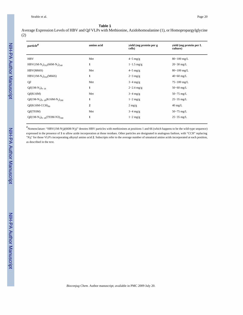
A series of control experiments established that HBV or Qβ protein was produced only whenthe minimal media was supplemented by Met, 1, or 2, and only in the presence of IPTG. Inmedia supplemented with 1 or 2, the yield of isolated HBV or Qβ particles was approximately20−60% of the amount of protein produced in normal media under the same induction by IPTG(Table 1), representing some of the highest yields of unnatural amino acid-containing proteinsreported thus far (see Supporting Information).
HBV and Qβ VLPs containing 1 or 2 were indistinguishable in all physical characteristics fromthe analogous particles incorporating methionine (Figure 3). In each case, ultracentrifugationthrough sucrose gradients (not shown) and analysis on size-exclusion chromatographyproduced sharp virus bands at the same position as native capsids. Negative-stainedtransmission electron microscopy images of the particles revealed homogeneous samples oficosahedral nonaggregated particles. (The HBV Cp149 particles containing 1 do not containnucleic acid, and thus exhibit stain uptake with dark centers. Qβ particles are packed full ofrandom cellular nucleic acid and have light centers in the negative stained images.) SDS-PAGEanalyses revealed the same high purities of the capsids containing unnatural amino acids asthose obtained with methionine. Last, the structures of both the HBV and Qβ virus-like particleswith 1 were confirmed by cryo-electron microscopy image reconstruction. The electron densitymaps overlaid well with the published X-ray crystal structures for the native capsids.
The incorporation of 1 and 2 in place of methionine was confirmed by trypsin digestion andanalysis of the resulting peptides using MALDI mass spectrometry, with the resultssummarized in Figure 4. Sequence coverage of the wild-type and 1-labeled HBV Cp149particle was nearly complete, with all but two residues included (Ile97, Arg98). Followingnormal expression in the presence of all 20 amino acids, Met was found at positions 1 and 66,the former in 70−90% of the protein, varying from batch to batch. The same result was observedwhen 1 was substituted for Met in the protocol described above, with 1 incorporated at the N-terminus in 70−90% of the protein produced, and completely (>95%) at position 66. Themutation M66S was also constructed, and found to contain the same 70−90% of 1 at the N-terminus when expressed with the unnatural amino acid.
Analysis of Qβ capsids required shorter trypsin digestion times to ensure limited proteolysisof the amino-terminal region. A coverage map that included all peptide fragments larger thanthree amino acids was obtained (Figure 4). Post-translational processing at the N-terminus wasfound to vary with the nature of the residue installed at that position. Under normal (Met-containing) expression, Met1 was never detected in either wild-type or mutant particles,showing that it was processed away during protein production (therefore, the amino acidnumbering eliminates this residue). When 1 was substituted for Met in any form of the Qβsequence, approximately 10% of the capsids were found to have the unnatural amino acid atthe N-terminus. However, in approximately one of every five batches this level of incorporationspiked to as much as 90%. It was therefore necessary to analyze each preparation of Qβ capsids
Strable et al. Page 3
Bioconjug Chem. Author manuscript; available in PMC 2009 July 20.
NIH
-PA Author Manuscript
NIH
-PA Author Manuscript
NIH
-PA Author Manuscript
to determine the number of azide-containing residues available for conjugation. Incorporationof 1 at either of the internal positions tested (16 or 93) was uniformly excellent (>95%). Thesame type of analysis (not shown) revealed 50% incorporation of 2 into Qβ K16 M at position16, with the remainder being Met. No homopropargyl glycine was detected at position 1.
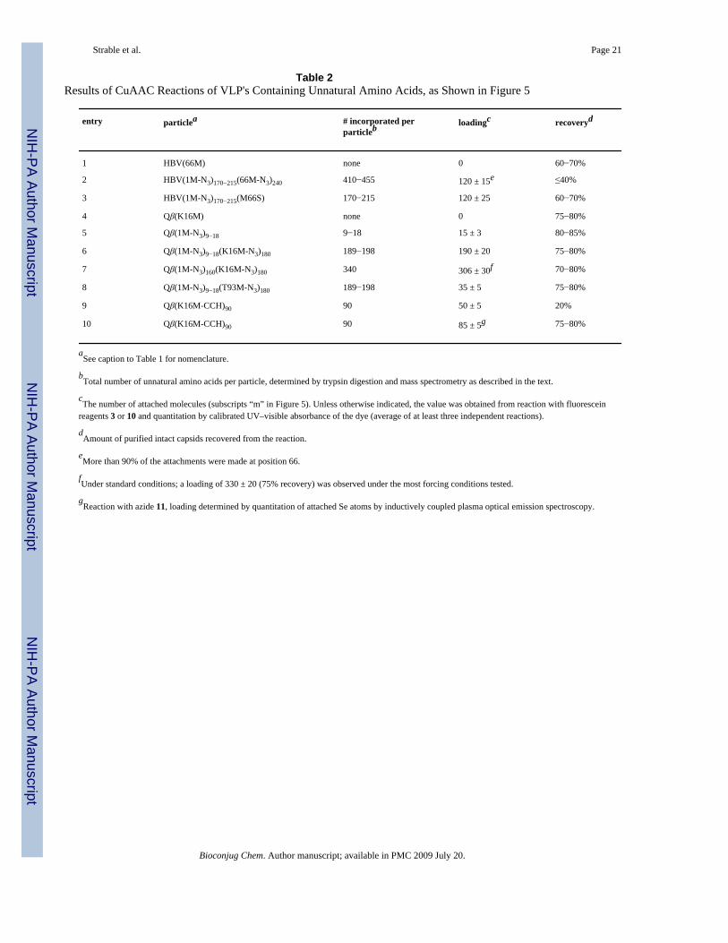
Conjugation to Genetically Incorporated Azide and Alkyne GroupsSince the expression of HBV in the presence of 1 provided 70−90% incorporation at position1 and complete incorporation at position 66, a maximum of approximately 410−460 azides perVLP should be available for conjugation. We employed these particles as reagents in thecopper-catalyzed azide-alkyne cycloaddition (CuAAC) reaction, using CuOTf andaccelerating ligand 4 under inert atmosphere as has been previously described (61,62). Thepurified virus was pelleted out of solution, taken into an inert-atmosphere glovebox, andresuspended in degassed 0.1 M Tris buffer (pH 8.0) to prepare for reaction with fluoresceinalkyne 3 under our standard conditions, as shown in Figure 5. After 16 h at room temperature,intact capsids could neither be detected by size-exclusion chromatography (Figure 6) norrecovered from sucrose gradients. However, gel electrophoresis (Figure 6) showed strongfluorescein labeling of the protein. Trypsin digestion and MALDI analysis in the mannerdescribed above revealed that approximately 50% of the sites at position 66 were labeled withfluorescein (equal intensities for the 56−81 fragment, m/z = 2919.3, and its labeled analoguewith an increase in mass of 413 Da). Interestingly, fewer than 10% of the azide groups atposition 1 were addressed.
HBV particles lacking incorporated azide groups were found to be stable toward the reactionconditions without coupling to fluorescein, ruling out nonspecific adsorption of the dye (Table2, entry 1). All HBV particles were stable to the CuAAC reaction conditions (copper, ligand,buffer, and sample handling) and also toward extended incubation with fluorescein alkyne 3in the absence of CuAAC catalyst. However, the particles did decompose when either ligand4 or the previously used tris(triazolylmethyl)amine 5 was employed in the CuAAC process,showing that the nature of the ligand made no difference to particle stability. When the reactiontime was varied, or the amount of catalyst was reduced, virus-like particles were recovered inyields inversely proportional to the amount of CuAAC reaction that had occurred. The use ofrhodamine, biotin, or Gd(DOTA) reagents (6, 7, or 8) in place of fluorescein alkyne gave similarresults. We therefore conclude that the decomposition of the particle was caused by the covalentattachment of these structures. We have previously observed other particles such as cowpeamosaic virus (CPMV) to be similarly sensitive to the covalent tethering of flat, hydrophobicspecies such ferrocene, tetramethylrhodamine, Fe(III) protoporphyrin IX, Ru(II) bipyridinecomplexes, and doxorubicin. However, the nature of the attached species varied somewhat inthis case, and so it may well be that the triazole linkage, which is common to each of theseexamples, is responsible for the instability of the virus-like HBV particle. CPMV and Qβ haveboth been shown to be stable toward high levels of coverage by triazoles (34,35,37,38,62).
We attempted to strike a balance between covalent attachment and stability for HBV by varyingthe concentrations of CuOTf, ligand 4, fluorescein-alkyne 3, and protein. The use of 250 μMCuOTf with 500 μM ligand 4 slowed the reaction enough to allow the recovery of a maximumof 40% of the starting amount of capsids as intact icosahedra, bearing an average of 120fluorescein molecules per capsid (Table 2, entry 2). The vast majority of these were attachedto the M66 position and not to the N-terminal azidohomoalanine residue, as shown by trypsindigestion/MS. The loading value was determined by UV–vis absorbance spectroscopy offluorescein (495 nm) in samples of purified labeled particles, with calibration using knownsamples under similar conditions (fluorescein in the presence of protein; protein concentrationsdetermined by Lowry protein assay). It should also be noted that the attachment reaction wasinhibited by the use of ligand 4 in greater than a 2:1 molar ratio with respect to Cu, consistent
Strable et al. Page 4
Bioconjug Chem. Author manuscript; available in PMC 2009 July 20.
NIH
-PA Author Manuscript
NIH
-PA Author Manuscript
NIH
-PA Author Manuscript
with previous observations (61), and that higher concentrations of Cu catalyst 4 gave rise togreater amounts of capsid decomposition as the number of reactions climbed above the 120-per-particle level.
The site of bioconjugation was moved from the spike to the “floor” of the HBV structure bymutation of M66 to serine. Expression of the M66S Cp149 mutant in the presence ofazidohomoalanine 1 allowed the isolation of a particle having reactive azide only at the N-terminus (70−90% incorporation, Table 1). Reaction with fluorescein-alkyne 3 under theconditions shown in Figure 5 gave rise to the same approximate loading of 120 per particle,but isolated in significantly higher yield (60−70%, which represents the maximum possiblerecovery at this scale due to losses in sample handling from sucrose gradient purification; Table2, entry 3). Since the HBV particle bearing azidohomoalanine at both positions 1 and 66 showedpredominant labeling at the latter, we conclude that the more exposed position on the 4-helixbundle is both more reactive and more deleterious to capsid stability when labeled.Decomposition of the particle results in denaturation and precipitation, and thus removal ofprotein from the labeling reaction. The greater capsid stability in the M66S case may derivefrom the fact that the N-terminus, now the only site bearing azide groups, is relatively far fromthe interface between subunit dimers. Compounds attached by flexible linkers to M66, incontrast, are positioned to easily interact with the hydrophobic interior of each 4-helix bundle.The potential for small molecules to affect HBV capsid assembly and stability by binding atsubunit interfaces has been previously illustrated for certain dihydropyrimidine derivatives(63,64).
Qβ proved to be much more amenable to CuAAC derivatization. Each of the few azide groupsin the wild-type virus-like particle containing 1 at 5−10% of the N-terminal sites was addressedwith alkyne reagent 4 (Table 2, entry 5). When 1 was additionally installed at each of the highlyexposed K16 M positions, the particle was also completely labeled with fluorescein, causingno apparent decomposition (Table 2, entry 6). A rare sample in which azidohomoalanine waslargely retained at the N-terminus, designated Qβ (1M-N3)160(K16M-N3)180, took on 306 ±30 dyes under standard conditions, and 330 ± 20 dyes in the presence of higher concentrationsof protein and catalyst, all with excellent particle yield and stability (Table 2, entry 7). Loadingvalues were determined with calibrated measurements of fluorescein absorbance and proteinconcentration determined by a modified Lowry assay, since the Qβ VLP contains no tryptophanresidues, making for poor UV–vis absorbance characteristics. The resulting particles andprotein were characterized in the same manner as HBV above (see Figure 3 for size-exclusionchromatography and gel electrophoresis). Trypsin digestion and mass spectrometry confirmedthe loading values by showing the expected peptide fragments (and only those fragments) tobe increased by 413 Da.
The accessibility of the K16M-N3 insertion was further demonstrated by the chemoselectiveconjugation of a protein to the Qβ(1M-N3)9−18(K16M-N3)180 particle. Transferrin, an 80 kDiron transport protein, was derivatized with N-propargylbromoacetamide and purified to give9 (Figure 5), in analogous fashion to a maleimide-alkyne derivative previously described(35). The CuAAC reaction of 9 with Qβ (1M-N3)9−18(K16M-N3)180 gave a particle bearingcovalently attached transferrin, as shown by gel electrophoresis and TEM (Figure 6).
Qβ (1M-N3)9−18(T93M-N3)180, in which the nonterminal unnatural amino acid is on theinterior surface of the capsid, was not nearly as reactive as the K16 M derivative (Table 2, entry8). While all of the N-terminal residues were coupled to 3 under standard CuAAC conditions,only 30−40 dyes were attached to the particle in total, which means that only 20−30 of the 180azide-labeled T93 M sites participated in the reaction. As before, yields of intact particles weregood. The same result was observed when alkyne 8 was used. Position 93 has both a more
Strable et al. Page 5
Bioconjug Chem. Author manuscript; available in PMC 2009 July 20.
NIH
-PA Author Manuscript
NIH
-PA Author Manuscript
NIH
-PA Author Manuscript
congested protein environment than position 16, and may well be occluded by associated RNApackaged inside the particle, giving rise to sluggish reactivity.
Lastly, the alkyne-containing particle Qβ (K16M-CCH)90 proved to be sensitive to theattachment of fluorescein azide 10 (Table 2, entry 9), giving rise to low recoveries of partiallyconjugated material. Note that azide 10, and not alkyne 3, induces instability in the resultingQβ conjugate (each attached to unnatural amino acids installed at the same site). This ispresumably a function of the longer tether between the reactive group and the fluoresceinmoiety in 10, allowing the polycyclic aromatic structure to more easily interact withhydrophobic subunit interfaces in the particle. The use of selenomethionine derivative 11, onthe other hand, provided clean and complete triazole formation with no loss of particle integrity(Table 2, entry 10), showing again that triazole moieties are well tolerated by the Qβ capsid.
CONCLUSIONSWe describe here the first successful site-specific incorporation and subsequent reaction ofunnatural amino acids into virus-like particles. The substitutions of azide- and alkyne-containing structures for methionine had no apparent effect on the ability of the resultingproteins to self-assemble into icosahedral capsids that are indistinguishable in structure fromthe native forms. Tight control over protein expression in methionine auxotrophic cells resultedin high yields of the assembled capsids. The degree of post-translational removal of unnaturalamino acid introduced in position 1 in place of Met was variable, and was characterized bytrypsin digestion and mass spectrometry analysis.
These novel capsids can be chemically addressed with perfect chemoselectivity by the CuAACreaction. HBV was found to be sensitive to the coupling of molecules to the 4-helix-bundlespikes, while Qβ suffered from much fewer limitations in this regard. The low concentrationsof azide and alkyne reagents required for coverage of most or all of the inserted azide andalkyne groups are made possible by the efficiency of the CuAAC process. Because the CuAACreaction is insensitive to the substituents present on the azide and alkyne reactants and similarlyunreactive with natural protein functional groups, the two-step process of genetically directedincorporation of azides or alkynes followed by CuAAC ligation provides a functionalequivalent to the installation of almost any unnatural (triazole-linked) amino acid one canimagine.
EXPERIMENTAL PROCEDURESCP149 HBV Plasmid Construction
A pET11c plasmid containing the Cp149 sequence was gratefully received from Prof. AdamZlotnick of the Oklahoma University Health Sciences Center. In order to provide tight induciblecontrol overexpression (pET11c was found to be constitutively expressed to an unacceptabledegree), the HBV gene was amplified using the following primers: Cp149F 5′-AAG AAGGAG GAT ATA GGT CTC AC ATG GAC ATT GAC CCT-3′ and Cp149R 5′-TCG GGCTTT GTT AGC AGC CGG AAG CTT ATA CTA AAC AAC CGT-3′ The PCR product wassequentially digested with Hind III and Bsa I, and then ligated into pQE-60 plasmid (Qiagen)which had been sequentially digested with Hind III and Nco I. The resulting plasmid wastransformed into competent M15MA cells harboring the pREP4 plasmid yielding theexpression cells M15MA(pQE-60/HBV).
Site-Directed Mutagenesis of Cp149 HBVMutations were introduced into the coat protein of HBV by standard overlap PCR to removethe methionine at position 66 (M66S). The following general forward and reverse primers were
Strable et al. Page 6
Bioconjug Chem. Author manuscript; available in PMC 2009 July 20.
NIH
-PA Author Manuscript
NIH
-PA Author Manuscript
NIH
-PA Author Manuscript
used: Forward 5′-AGA ATT CAT TAA AGA GGA GAA ATT AAC-3′, Reverse 5′-CCA AGCTCA GCT AAT GCT TAT CCT-3′, M66SF 5′-GGA GAC TTA TCT ACT CTA GCT-3′,M66SR 5′-AGC TAG AGT AGA TAA GTC TCC-3′. The PCR products were sequentiallydigested with Hind III and NcoI and ligated into pQE-60 that had previously been digestedwith Hind III and Nco I. The resulting plasmids were transformed into competent M15MAcells yielding the following expression construct: M15MA(pQE-60/M66S HBV).
Insertion of the K16 M Qβ Coat Protein Gene into pQE-60The coat protein gene for Qβ containing the point mutation K16 M was originally created inthe pET-28b plasmid, which is not compatible with expression in M15 based Met auxotrophicE. coli cells. Therefore, the Qβ coat protein gene was amplified from the pET-28b plasmidusing the following primers: QBF 5′-AAA GAG GAG AAA TTA AGG TCT CAC ATG GCAAAA TTA GAG ACT-3′ and QBR 5′-CCA AGC TCA GCT AAT TAA GCT TTA TTAATA-3′ The PCR product was sequentially digested with Hind III and Bsa I. The digested PCRproduct was ligated into pQE-60 plasmid (Qiagen), which had been sequentially digested withHind III and Nco I. The resulting plasmid was transformed into competent M15MA cellsharboring the pREP4 plasmid yielding the expression cells M15MA(pQE-60/Qβ K16M).
Site-Directed Mutagenesis of K16 M QβThe following primers were used to introduce Met at position 93 (T93M), using standardoverlap PCR as above: Forward 5′-AGA ATT CAT TAA AGA GGA GAA ATT AAC-3′,Reverse 5′-CCA AGC TCA GCT AAT GCT TAT CCT-3′, WTF 5′-GGA GAC TTA TCTACT CTA GCT-3′, WTR 5′-AGC TAG AGT AGA TAA GTC TCC-3′. The PCR productswere sequentially digested with Hind III and NcoI and ligated into pQE-60 that had previouslybeen digested with Hind III and Nco I. The resulting plasmids were transformed into competentM15MA cells yielding the following expression construct: M15MA(pQE-60/WT Qβ) andM15MA(pQE-60/T93 M Qβ).
Synthesis of Unnatural Amino Acids (1 and 2)Several short routes to azidohomoalanine (1) have been reported in the literature (15,65-67).We chose a simplified version of the method of Rappoport and co-workers (68), since thestarting lactone is commercially available in racemic form at modest cost and an enantiospecificsynthesis is not required because E. coli incorporates only the L-isomer during proteinexpression. α-Amino-γ-butyrolactone hydrobromide (5.02 g, 27.6 mmol) was refluxed in 1:1HBr/glacial HOAc (250 mL) overnight (17−24 h). The HBr and acetic acid were then removedby rotary evaporation, yielding crude bromohomoalanine. The crude material was thendissolved in a solution of NaN3 (9.00 g, 138 mmol) in water (125 mL) and refluxed againovernight. The solution was evaporated to dryness and the residual tan solid resuspended in aminimal volume of 0.1 M HCl. The acidic solution was passed through a column of Dowex50WX4−100 ion-exchange resin, washing with water and eluting the product with 1 MNH4OH. The collected fractions were evaporated to dryness, yielding an oil. Compound 1 wasobtained as a tan solid (1.49 g, 10.3 mmol, 37%) upon resuspending in a minimal volume ofwater and lyophilizing. The purity of the solid was determined by NMR using pyridine as aninternal standard and generally was >85% (overall yields 32−38%). 1H NMR (D2O, 200 MHz,d1 = 10 s) δ 3.44 (t, 2H, CHCH2CH2N3), 3.07 (dd, 1H, CHCH2CH2N3), 1.58 (m, 2H,CHCH2CH2N3). Homopropargylglycine (2) was prepared as previously described (10).
Incorporation of 1 into HBV and QβA single of colony of cells expressing the desired HBV or Qβ construct was used to inoculate5 mL of SOB media containing carbenicillin (100 μg/mL) and kanamycin (50 μg/mL) and wasgrown overnight. The resulting culture was transferred into 500 mL of M9 minimal media
Strable et al. Page 7
Bioconjug Chem. Author manuscript; available in PMC 2009 July 20.
NIH
-PA Author Manuscript
NIH
-PA Author Manuscript
NIH
-PA Author Manuscript
supplemented with all 20 amino acids, carbenicillin (100 μg/mL), and kanamycin (100 μg/mL)and allowed to grow for 8 h. Aliquots (25 mL) of the resulting culture were transferred intoeach of 11 flasks containing 500 mL of fresh minimal media supplemented with all 20 aminoacids, carbenicillin (100 μg/mL) and kanamycin (100 μg/mL) and allowed to grow overnight.In the morning the cells were pelleted and resuspended in 500 mL of fresh M9 minimal mediasupplemented with all of the natural amino acids minus methionine. The cells were agitated at37 °C for 30−40 min, and then transferred into new flasks of M9 minimal media supplementedwith all of the natural amino acids minus methionine, IPTG (1 mM), carbenicillin (100 μg/mL), kanamycin (100 μg/mL), and azidohomoalanine (80 mg of racemate). After 6 h at 37 °C, the cells were harvested and stored at −80 °C.
For each preparation, 30−60 g of cells were allowed to thaw at room temperature and wereresuspended in 50 mL distilled water. Cold lysis buffer (200 mL, 50 mM HEPES pH 8.0, 500mM NaCl, 0.1 mg/mL DNase1, and 0.1 mg/mL RNaseA) was added and the cells weresubjected to three cycles of sonication (2 min sonication and 2 min rest per cycle). Lysozyme(1 mg per mL of lysis buffer) was added and the solution was stirred in the cold room for 1 h.Insoluble cell debris was removed by centrifugation at 10 000 rpm for 30 min in a JA-10 rotor(Beckman).
For HBV, ammonium sulfate was added to the supernatant to a final concentration of 40% ofsaturation, and allowed to stir at room temperature for 30 min. The precipitated HBV coatprotein was separated by centrifugation (10 000 rpm, 30 min, JA-10 rotor), and then wasresuspended in 0.1 M potassium phosphate buffer (50 mL, pH 7.0). Any remaining insolublematerial was removed by centrifugation (10 000 rpm, 15 min, JA-17 rotor). Sodium chloridewas added to a final concentration of 0.5 M and the HBV coat proteins were allowed to assembleovernight. The resulting assembled VLPs were separated from smaller proteins byultrapelleting (42 000 rpm, 6 h, 4 °C, 50.2Ti rotor, L90K ultracentrifuge). The pelleted materialwas resus-pended in 3−10 mL of 0.1 M potassium phosphate buffer (pH 7.0). Furtherpurification was accomplished by the use of two successive sucrose gradient sedimentations(10−40% sucrose gradients in SW28 rotor at 28 000 rpm for 6 h at 4 °C.
For Qβ, PEG 8000 was added to the supernatant to a final concentration of 10% and the mixturewas allowed to stir at room temperature for 30 min. The precipitated Qβ capsids were separatedby centrifugation (10 000 rpm, 30 min, JA-10 rotor), and then resuspended in 0.1 M potassiumphosphate buffer (50 mL, pH 7.0). Insoluble material was removed by centrifugation (10 000rpm, 15 min, JA-17 rotor), and VLPs were isolated and purified as for HBV above, with finalconcentration by ultrapelleting.
Characterization of HBV and Qβ ParticlesAll protein preparations were analyzed by denaturing gel electrophoresis (4−12% NuPAGEBis-Tris gel, Invitrogen) to estimate their purity; in all cases, the anticipated coat protein bandconstituted >95% of the intensity visualized by densitometry after Coomassie blue staining.HBV concentration was determined by absorbance at 280 nm, 1 mg/mL providing anabsorbance value of 1.74. The concentrations of modified HBV and all Qβ particles weredetermined with the Modified Bradford Assay (Pierce, Inc.) and a BSA standard curve. SinceHBV VLPs do not contain significant quantities of RNA, the ratio of absorbances at 260 nmvs 280 nm was 0.6−0.7. Qβ VLPs package random cellular nucleic acid, giving rise to A260/A280 values of 1.8−1.9. The presence of individual, aggregated, and disassembled particles wasdetermined by size-exclusion chromatography on a Superose 6 column using an Akta Explorer(GE Healthcare) fast protein liquid chromatography (FPLC) instrument. Transmission electronmicroscopy was performed by applying particles at a concentration of 0.2 mg/mL to a carbon-coated Formvar transmission electron grid. The grids were stained with 2% uranyl acetate, andvisualized in a Phillips CM120 transmission electron microscope.
Strable et al. Page 8
Bioconjug Chem. Author manuscript; available in PMC 2009 July 20.
NIH
-PA Author Manuscript
NIH
-PA Author Manuscript
NIH
-PA Author Manuscript
VLP samples were processed for determination of derivatization site(s) by MALDI massspectrometry as follows. Samples (100 μL, 1−2 mg/mL protein) were mixed with 300 μL of 8M urea and 30 μL of 1 M DTT, and incubated at 37 °C for 1 h to allow the protein to denature.Iodoacetamide (1 M, 50 μL) was added to cap any free cysteines and the sample was againincubated at 37 °C for 1 h. DTT (30 μL) was added to quench unreacted capping reagent, andthe samples were diluted to a final volume of 1.9 mL using 25 mM ammonium bicarbonate(pH 8.0). Each sample was digested with 30 μg of either trypsin or Glu-C protease overnightat 37 °C. The samples were then concentrated to approximately 300 μL, and urea was removedwith Zip-tips (Microcon) prior to MALDI analysis.
Cryoelectron Microscopy and AnalysisSamples were prepared for CryoEM analysis by preservation in vitreous ice via rapid-freezeplunging onto plasma cleaned Cflat carbon film grids using a Vitrobot (FEI Co). Data collectionwas performed on Tecnai F20 electron microscopes (FEI Co.) operating at 120 keV using adose of ∼20e−/Å2 and a nominal underfocus of 0.8 to 3 μm utilizing the Leginon data collectionsoftware (69). For the reconstructions, 735 micrographs of azidohomoalanine-incorporatedHBV were collected at a nominal magnification of 80 000× at a pixel size of 0.14 nm at thespecimen. 125 micrographs of azidohomoalanine-incorporated Qβ particles were collected ata nominal magnification of 50 000× at a pixel size of 0.23 nm at the specimen. All micrographswere collected on a 4000 × 4000 CCD camera (Gatan Inc.). CryoEM analysis of all other Qβvariants, including the transferrin-alkyne conjugated particles, was performed under the sameconditions as the azidohomoalanine-incorporated HBV particles.
The contrast transfer function (CTF) for each micrograph was estimated using the AutomatedCTF Estimation (ACE) package (70). 8906 HBV particles and 11 882 Qβ particles wereextracted from the collected data at a box size of 304 × 304 pixels and 180 × 180 pixels,respectively. The HBV particles were binned by a factor of 2 for the reconstruction. Phasecorrection of the single particles and subsequent three-dimensional refinement was carried outwith the EMAN software package (71). The amplitudes of the resulting refined structures wereadjusted with the SPIDER software package (72). Resolution of the final HBV and Qβ densitieswere determined to be around 8 Å and 10 Å (respectively) according to 0.5 FSC criteria. Rigid-body docking of crystal structures into the reconstruction density and graphical representationswere produced by the Chimera visualization software package (73,74).
Copper Catalyzed Azide-Alkyne Cycloaddition ReactionsPurified azide-containing HBV or Qβ particles were pelleted out of solution (42 000 rpm, 6h), the supernatant was removed, and residual buffer was allowed to drain away from the pellet.The viral pellet was then taken into a nitrogen-filled glovebox (without exposure to vacuum)and resuspended in a minimal volume of degassed 0.1 M Tris at pH 8.0. A small aliquot of thevirus solution was removed from the glovebox and used to determine virus concentration. Atypical conjugation reaction employed a VLP concentration of 2 mg/mL in 0.1 M Tris (pH 8.0)in a round-bottomed 2 mL Eppendorf tube (rather than conical tubes that do not provide goodmixing upon gentle agitation). Fluorescein alkyne 6 (final concentration 1 mM) was added,followed by a freshly prepared buffer solution of Cu(I) triflate and 2 equiv of ligand 4 or 5.Final concentrations of Cu were either 100 μM or 250 μM for HBV, 500 μM or 1 mM forQβ. The tube was sealed, placed in a secondary sealed container (usually a small round-bottomed flask), removed from the glovebox, and attached to a slow tumbler arm for agitationat room temperature for 16−18 h. After completion of the reaction, intact virus-like particleswere separated from excess labeling reagent using a combination of 10−40% sucrose gradients(38 000 rpm, SW41 rotor, 4 h for HBV, 3 h for Qβ) and ultrapelleting.
Strable et al. Page 9
Bioconjug Chem. Author manuscript; available in PMC 2009 July 20.
NIH
-PA Author Manuscript
NIH
-PA Author Manuscript
NIH
-PA Author Manuscript
ACKNOWLEDGMENTThis work was supported by the NIH (AI056013, RR021886, GM62523), the David & Lucille Packard FoundationInterdisciplinary Science Program, and the Canadian Institutes of Health Research (postdoctoral fellowship toA.K.U.).Cryo-electron microscopy was performed at the National Resource for Automated Molecular Microscopywhich is supported by the NIH NCRR P41 program (RR17573).
LITERATURE CITED1. Noren CJ, Anthony-Cahill SJ, Griffith MC, Schultz PG. A general method for site-specific
incorporation of unnatural amino acids into proteins. Science 1989;244:182–188. [PubMed: 2649980]2. Cornish VW, Benson DR, Altenbach CA, Hideg K, Hubbell WL, Schultz PG. Site-specific
incorporation of biophysical probes into proteins. Proc. Natl. Acad. Sci. USA 1994;91:2910–2914.[PubMed: 8159678]
3. Wang L, Brock A, Herberich B, Schultz PG. Expanding the genetic code of Escherichia coli. Science2001;292:498–500. [PubMed: 11313494]
4. Anderson JC, Schultz PG. Adaptation of an orthogonal archaeal leucyl-tRNA and synthetase pair forfourbase, amber, and opal suppression. Biochemistry 2003;42:9598–9608. [PubMed: 12911301]
5. Chin JW, Cropp TA, Anderson JC, Mukherji M, Zhang Z, Schultz PG. An expanded eukaryotic geneticcode. Science 2003;301:964–967. [PubMed: 12920298]
6. Deiters A, Cropp TA, Mukherji M, Anderson JC, Schultz Peter G. Adding amino acids with novelreactivity to the genetic code of Saccharomyces cerevisiae. J. Am. Chem. Soc 2003;125:11782–11783.[PubMed: 14505376]
7. Tian F, Tsao M-L, Schultz PG. A phage display system with unnatural amino acids. J. Am. Chem. Soc2004;125:15962–15963. [PubMed: 15584720]
8. Ryu Y, Schultz PG. Efficient incorporation of unnatural amino acids into proteins in Escherichiacoli. Nat. Methods 2006;3:263–265. [PubMed: 16554830]
9. Anderson JC, Wu N, Santoro SW, Lakshman V, King DS, Schultz PG. An expanded genetic code witha functional quadruplet codon. Proc. Natl. Acad. Sci. USA 2004;101:7566–7571. [PubMed: 15138302]
10. Van Hest JCM, Kiick KL, Tirrell DA. Efficient incorporation of unsaturated methionine analoguesinto proteins in vivo. J. Am. Chem. Soc 2000;122:1282–1288.
11. Kiick KL, Tirrell David A. Protein engineering by in vivoincorporation of non-natural amino acids:Control of incorporation of methioonine analogues by methionyl tRNA synthetase. Tetrahedron2000;56:9487–9493.
12. Kiick K,L, Weberskirch R, Tirrell DA. Identification of an expanded set of translationally activemethionine analogues in Escherichia coli. FEBS Lett 2001;502:25–30. [PubMed: 11478942]
13. Kiick KL, Saxon E, Tirrell DA, Bertozzi CR. Incorporation of azides into recombinant proteins forchemoselective modification by the Staudinger ligation. Proc. Natl. Acad. Sci. USA 2002;99:19–24.[PubMed: 11752401]
14. Link AJ, Tirrell DA. Cell surface labeling of Escherichia coli via copper(I)-catalyzed [3 + 2]cycloaddition. J. Am. Chem. Soc 2003;125:11164–11165. [PubMed: 16220915]
15. Link AJ, Vink MKS, Tirrell DA. Presentation and detection of azide functionality in bacterial cellsurface proteins. J. Am. Chem. Soc 2004;126:10598–10602. [PubMed: 15327317]
16. Link AJ, Tirrell DA. Reassignment of sense codons in vivo. Methods 2005;36:291–298. [PubMed:16076455]
17. Link AJ, Vink MKS, Agard NJ, Prescher JA, Bertozzi CR, Tirrell DA. Discovery of aminoacyl-tRNAsynthetase activity through cell-surface display of non-canonical amino acids. Proc. Natl. Acad. Sci.USA 2006;103:10180–10185. [PubMed: 16801548]
18. van Hest JCM, Tirrell DA. Efficient introduction of alkene functionality into proteins in vivo. FEBSLett 1998;428:68–70. [PubMed: 9645477]
19. Kiick KL, Van Hest JCM, Tirrell DA. Expanding the scope of protein biosynthesis by altering themethionyl-tRNA synthetase activity of a bacterial expression host. Angew. Chem., Int. Ed2000;39:2148–2152.
Strable et al. Page 10
Bioconjug Chem. Author manuscript; available in PMC 2009 July 20.
NIH
-PA Author Manuscript
NIH
-PA Author Manuscript
NIH
-PA Author Manuscript
20. Tang Y, Tirrell DA. Biosynthesis of a highly stable coiled-coil protein containing hexafluoroleucinein an engineered bacterial host. J. Am. Chem. Soc 2001;123:11089–11090. [PubMed: 11686725]
21. Wang Q, Chan TR, Hilgraf R, Fokin VV, Sharpless KB, Finn MG. Bioconjugation by copper(I)-catalyzed azide-alkyne [3 + 2] cycloaddition. J. Am. Chem. Soc 2003;125:3192–3193. [PubMed:12630856]
22. Yoshikawa E, Fournier MJ, Mason TL, Tirrell DA. Genetically Engineered fluoropolymers. Synthesisof repetitive polypeptides containing p-fluorophenylalanine residues. Macromolecules1994;27:5471–5475.
23. Budisa N, Alefelder S, Bae JH, Golbik R, Minks C, Huber R, Moroder L. Proteins with β-(thienopyrrolyl)alanines as alternative chromophores and pharmaceutically active amino acids.Protein Sci 2001;10:1281–1292. [PubMed: 11420430]
24. Bae JH, Alefelder S, Kaiser JT, Friedrich R, Moroder L, Budisa N. Incorporation of β-seleno(3,2-b)pyrrolyl-alanine into proteins for phase determination in protein X-ray crystallography. J. Mol. Biol2001;309:925–936. [PubMed: 11399069]
25. Renner C, Alefelder S, Bae JH, Budisa N, Huber R, Moroder L. Fluoroprolines as tools for proteindesign and engineering. Angew. Chem., Int. Ed 2001;40:923–925.
26. Link AJ, Mock ML, Tirrell DA. Noncanonical amino acids in protein engineering. Curr. Opin.Biotechnol 2003;14:603–609. [PubMed: 14662389]
27. Saxon E, Bertozzi Carolyn R. Cell surface engineering by a modified Staudinger reaction. Science2000;287:2007–2010. [PubMed: 10720325]
28. Vocadlo DJ, Bertozzi CR. A strategy for functional proteomic analysis of glycosidase activity fromcell lysates. Angew. Chem., Int. Ed 2004;43:5338–5342.
29. Agard NJ, Baskin JM, Prescher JA, Lo A, Bertozzi CR. A comparative study of bioorthogonalreactions with azides. ACS Chem. Biol 2006;1:644–648. [PubMed: 17175580]
30. Mahal LK, Yarema KJ, Bertozzi Carolyn R. Engineering chemical reactivity on cell surfaces througholigosaccharide biosynthesis. Science 1997;276:1125–1128. [PubMed: 9173543]
31. Agard NJ, Prescher Jennifer A, Bertozzi Carolyn R. A strain-promoted (3 + 2) azide-alkynecycloaddition for covalent modification of biomolecules in living systems. J. Am. Chem. Soc2004;126:15046–15047. [PubMed: 15547999]
32. Rostovtsev VV, Green LG, Fokin VV, Sharpless KB. A stepwise Huisgen cycloaddition process:Copper(I) catalyzed regioselective “ligation” of azides and terminal alkynes. Angew. Chem., Int. Ed2002;41:2596–2599.
33. Tornøe CW, Christensen C, Meldal M. Peptidotriazoles on solid phase: [1,2,3]-triazoles byregiospecific copper(I)-catalyzed 1,3-dipolar cycloadditions of terminal alkynes to azides. J. Org.Chem 2002;67:3057–3062. [PubMed: 11975567]
34. Wang Q, Chan TR, Hilgraf R, Folkin VV, Sharpless KB, Finn MG. Bioconjugation by copper(I)-catalyzed azide-alkyne (3 + 2) cycloaddition. J. Am. Chem. Soc 2003;125:3192–3193. [PubMed:12630856]
35. Sen Gupta S, Kuzelka J, Singh P, Lewis WG, Manchester M, Finn MG. Accelerated bioorthogonalconjugation: a practical method for the ligation of diverse functional molecules to a polyvalent virusscaffold. Bioconjugate Chem 2005;16:1572–1579.
36. Deiters A, Cropp TA, Summerer D, Mukherji M, Schultz PG. Site-specific PEGylation of proteinscontaining unnatural amino acids. Bioorg. Med. Chem. Lett 2004;14:5743–5745. [PubMed:15501033]
37. Kaltgrad E, Sen Gupta S, Punna S, Huang C-Y, Chang A, Wong C-H, Finn MG, Blixt O.Anticarbohydrate antibodies elicited by polyvalent display on a viral scaffold. ChemBioChem2007;8:1455–1462. [PubMed: 17676704]
38. Prasuhn JDE, Yeh RM, Obenaus A, Manchester M, Finn MG. Viral MRI contrast agents: coordinationof Gd by native virions and attachment of Gd complexes by azide-alkyne cycloaddition. Chem.Commun 2007;12:1269–1271.
39. Singh P, Prasuhn DEJ, Yeh RM, Destito G, Rae CS, Osborn K, Finn MG, Manchester M.Biodistribution, toxicity and pathology of cowpea mosaic virus nanoparticle in vivo. J. ControlledRelease 2007;120:41–50.
Strable et al. Page 11
Bioconjug Chem. Author manuscript; available in PMC 2009 July 20.
NIH
-PA Author Manuscript
NIH
-PA Author Manuscript
NIH
-PA Author Manuscript

40. Beatty KE, Xie F, Wang Q, Tirrell DA. Selective dye-labeling of newly synthesized proteins inbacterial cells. J. Am. Chem. Soc 2005;127:14150–14151. [PubMed: 16218586]
41. Dieterich DC, Link AJ, Graumann J, Tirrell DA, Schuman EM. Selective identification of newlysynthesized proteins in mammalian cells using bioorthogonal noncanonical amino acid tagging(BONCAT). Proc. Natl. Acad. Sci. USA 2006;103:9482–9487. [PubMed: 16769897]
42. Beatty KE, Liu JC, Xie F, Dieterich DC, Schuman EM, Wang Q, Tirrell David A. Fluorescencevisualization of newly synthesized proteins in mammalian cells. Angew. Chem., Int. Ed2006;45:7364–7367.
43. Dieterich DC, Lee JJ, Link AJ, Graumann J, Tirrell David A, Schuman EM. Labeling, detection, andidentification of newly synthesized proteomes with bioorthogonal non-canonical amino acid tagging.Nat. Prot 2007;2:532–540.
44. Speers AE, Cravatt BF. Profiling enzyme activities in vivo using click chemistry method. Chem. Biol2004;11:535–546. [PubMed: 15123248]
45. Speers AE, Adam GC, Cravatt BF. Activity based protein profiling in vivo using a copper(I)-catalyzedazide-alkyne cycloaddition. J. Am. Chem. Soc 2003:125.
46. Kenney JM, von Bonsdorff C-H, Nassal M, Fuller SD. Evolutionary conservation in the hepatitis Bvirus core structure: comparison of human and duck cores. Structure 1995;3:1009–1019. [PubMed:8589996]
47. Wynne SA, Crowther RA, Leslie AG. The crystal structure of human hepatitis B virus. Mol. Cell1999;6:771–780. [PubMed: 10394365]
48. Hilditch CM, Rogers LJ, Bishop DHL. Physicochemical analysis of the hepatitis B virus core antigenproduced by a baculovirus expression vector. J. Gen. Virol 1990;71:2755–2759. [PubMed: 2254755]
49. Allen M, Bulte JWM, Liepold L, Basu G, Zywicke HA, Frank JA, Young M, Douglas T. Paramagneticviral nanoparticles as potential high-relaxivity magnetic resonance contrast agents. Magn. Res. Med2005;54:807–812.
50. Cohen BJ, Richmond JE. Electron microscopy of hepatitis B core antigen synthesized in E. coli.Nature 1982;296:677–678. [PubMed: 7040981]
51. Wingfield PT, Stahl SJ, Williams RW, Steven AC. Hepatitis core antigen produced in Escherichiacoli: subunit composition, conformational analysis, andin vitro capsid assembly. Biochemistry1995;34:4919–4932. [PubMed: 7711014]
52. Vallegaard K, Fridborg K, Liljas L. Crystallization and preliminary x-ray diffraction studies of thebacteriophage Qβ. Acta Crystallogr., Sect. D: Biol. Cryst 1994;D50:105–109.
53. Tars K, Bundule M, Fridborg K, Liljas L. The crystal structure of bacteriophage GA and a comparisonof bacteriophages belonging to the major groups of Escherichia coli leviviruses. J. Mol. Biol1997;271:759–773. [PubMed: 9299325]
54. Overby LR, Barlow GH, Doi RH, Jacob M, Spiegelman S. Comparison of two serologically distinctribonucleic acid bacteriophages. J. Bacteriol 1966;91:442–448. [PubMed: 5903109]
55. Golmohammadi R, Fridborg K, Bundule M, Valegaard K, Liljas L. The crystal structure ofbacteriophage Qβ at 3.5 Å resolution. Structure 1996;4:543–554. [PubMed: 8736553]
56. Kozlovska TM, Cielens I, Dreilinna D, Dislers A, Baumanis V, Ose V, Pumpens P. RecombinantRNA phage Qβ capsid particles synthesized and self-assembled in Escherichia coli. Gene1993;137:133–137. [PubMed: 7506687]
57. Vasiljeva I, Kozlovska T, Cielens I, Strelnikova A, Kazaks A, Ose V, Pumpens P. Mosaic Qβ coatsas a new presentation model. FEBS Lett 1998;431:7–11. [PubMed: 9684855]
58. Freivalds J, Dislers A, Ose V, Skrastina D, Cielens I, Pumpens P, Sasnauskas K, Kazaks A. Assemblyof bacteriophage Qβ virus-like particles in yeast Saccharomyces cerevisiae and Pichia pastoris. J.Biotechnol 2006;123:297–303. [PubMed: 16406160]
59. Shepherd CM, Borelli IA, Lander G, Natarajan P, Siddavanahalli V, Bajaj C, Johnson JE, Brooks CLIII, Reddy VS. VIPERdb: a relational database for structural virology. Nucleic Acids Res2006;34:D386–D389. [PubMed: 16381893]
60. Hirel P-H, Schmitter J-M, Dessen P, Fayat G, Blanquet S. Extent of N-terminal methionine excisionfrom Escherichia coli proteins is governed by the side-chain length of the penultimate amino acid.Proc. Natl. Acad. Sci. USA 1989;86:8247–8251. [PubMed: 2682640]
Strable et al. Page 12
Bioconjug Chem. Author manuscript; available in PMC 2009 July 20.
NIH
-PA Author Manuscript
NIH
-PA Author Manuscript
NIH
-PA Author Manuscript
61. Lewis WG, Magallon FG, Fokin VV, Finn MG. Discovery and characterization of catalysts for azide-alkyne cycloaddition by fluorescence quenching. J. Am. Chem. Soc 2004;126:9152–9153. [PubMed:15281783]
62. Sen Gupta S, Raja KS, Kaltgrad E, Strable E, Finn MG. Virus-glycoploymer conjugates by copper(I)-catalysis of atom transfer radical polymerization and azide-alkyne cycloaddition. Chem. Commun2005:4315–4317.
63. Stray SJ, Bourne CR, Punna S, Lewis WG, Finn MG, Zlotnick A. A heteroaryldihydropyrimidineactivates and can misdirect hepatitis B virus capsid assembly. Proc. Natl. Acad. Sci. USA2005;102:8138–8143. [PubMed: 15928089]
64. Bourne CR, Finn MG, Zlotnick A. Global structural changes in hepatitis B virus capsids induced bythe assembly effector HAP1. J. Virol 2006;80:11055–11061. [PubMed: 16943288]
65. Mangold JB, Mischke MR, LaVelle JM. Azidoalanine mutagenicity in Salmonella: Effect ofhomologation and α-methyl substitution. Mut. Res 1989;308:33–42. [PubMed: 7516484]
66. Link AJ, Vink MKS, Tirrell DA. Preparation of the functionalizable methionine surrogateazidohomoalanine via copper-catalyzed diazo transfer. Nat. Protocols 2007;2:1879–1883.
67. Link AJ, Vink MKS, Tirrell DA. Synthesis of the functionalizable methionine surrogateazidohomoalanine using Boc-homoserine as precursor. Nat. Protocols 2007;2:1884–1887.
68. McLaughlin M, Mohareb RM, Rapoport H. An efficient procedure for the preparation of 4-substituted5-aminoimidazoles. J. Org. Chem 2003;68:50–54. [PubMed: 12515460]
69. Suloway C, Pulokas J, Fellmann D, Cheng A, Guerra F, Quispe J, Stagg S, Potter CS, Carragher B.Automated molecular microscopy: the new Leginon system. J. Struct. Biol 2005;151:41–60.[PubMed: 15890530]
70. Mallick SP, Carragher B, Potter CS, Kriegman DJ. ACE: automated CTF estimation. Ultramicroscopy2005;104:8–29. [PubMed: 15935913]
71. Ludtke SJ, Baldwin PR, Chiu W. EMAN: semiautomated software for high-resolution single-particlereconstructions. J. Struct. Biol 1999;128:82–97. [PubMed: 10600563]
72. Frank J, Radermacher M, Penczek P, Zhu J, Li Y, Ladjadj M, Leith A. SPIDER and WEB: processingand visualization of images in 3D electron microscopy and related fields. J. Struct. Biol1996;116:190–199. [PubMed: 8742743]
73. Goddard TD, Huang CC, Ferrin TE. Software extensions to UCSF Chimera for interactivevisualization of large molecular assemblies. Structure 2005;13:473–482. [PubMed: 15766548]
74. Goddard TD, Huang CC, Ferrin TE. Visualizing density maps with UCSF Chimera. J. Struct. Biol2007;157:281–287. [PubMed: 16963278]
Strable et al. Page 13
Bioconjug Chem. Author manuscript; available in PMC 2009 July 20.
NIH
-PA Author Manuscript
NIH
-PA Author Manuscript
NIH
-PA Author Manuscript
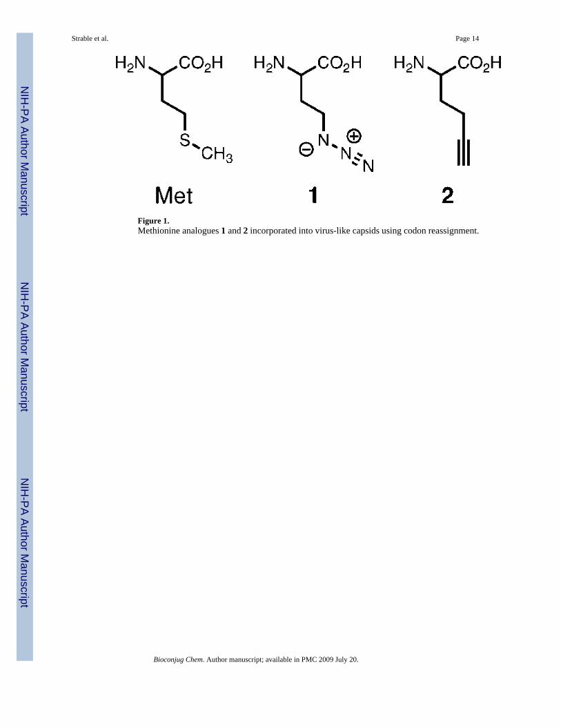
Figure 1.Methionine analogues 1 and 2 incorporated into virus-like capsids using codon reassignment.
Strable et al. Page 14
Bioconjug Chem. Author manuscript; available in PMC 2009 July 20.
NIH
-PA Author Manuscript
NIH
-PA Author Manuscript
NIH
-PA Author Manuscript
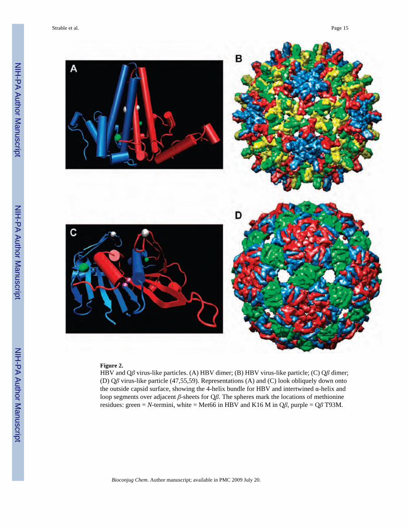
Figure 2.HBV and Qβ virus-like particles. (A) HBV dimer; (B) HBV virus-like particle; (C) Qβ dimer;(D) Qβ virus-like particle (47,55,59). Representations (A) and (C) look obliquely down ontothe outside capsid surface, showing the 4-helix bundle for HBV and intertwined α-helix andloop segments over adjacent β-sheets for Qβ. The spheres mark the locations of methionineresidues: green = N-termini, white = Met66 in HBV and K16 M in Qβ, purple = Qβ T93M.
Strable et al. Page 15
Bioconjug Chem. Author manuscript; available in PMC 2009 July 20.
NIH
-PA Author Manuscript
NIH
-PA Author Manuscript
NIH
-PA Author Manuscript
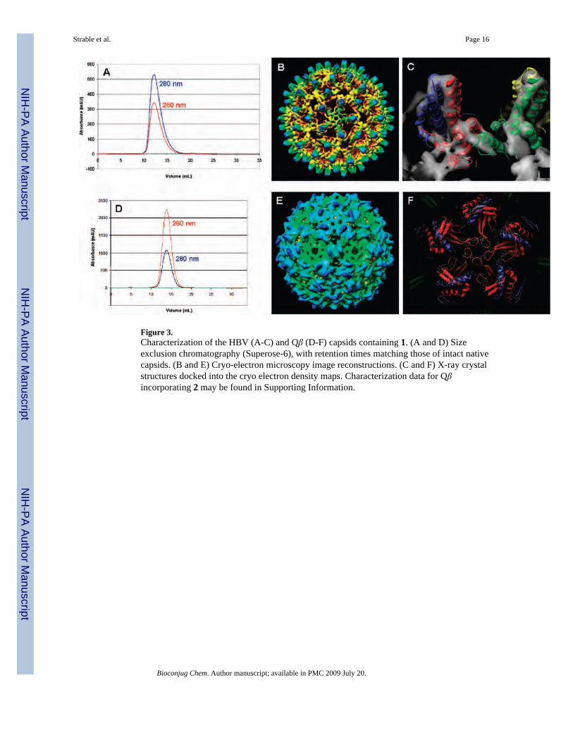
Figure 3.Characterization of the HBV (A-C) and Qβ (D-F) capsids containing 1. (A and D) Sizeexclusion chromatography (Superose-6), with retention times matching those of intact nativecapsids. (B and E) Cryo-electron microscopy image reconstructions. (C and F) X-ray crystalstructures docked into the cryo electron density maps. Characterization data for Qβincorporating 2 may be found in Supporting Information.
Strable et al. Page 16
Bioconjug Chem. Author manuscript; available in PMC 2009 July 20.
NIH
-PA Author Manuscript
NIH
-PA Author Manuscript
NIH
-PA Author Manuscript
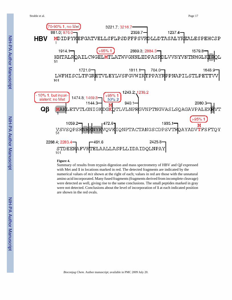
Figure 4.Summary of results from trypsin digestion and mass spectrometry of HBV and Qβ expressedwith Met and 1 in locations marked in red. The detected fragments are indicated by thenumerical values of m/z shown at the right of each; values in red are those with the unnaturalamino acid incorporated. Many fused fragments (fragments derived from incomplete cleavage)were detected as well, giving rise to the same conclusions. The small peptides marked in graywere not detected. Conclusions about the level of incorporation of 1 at each indicated positionare shown in the red ovals.
Strable et al. Page 17
Bioconjug Chem. Author manuscript; available in PMC 2009 July 20.
NIH
-PA Author Manuscript
NIH
-PA Author Manuscript
NIH
-PA Author Manuscript
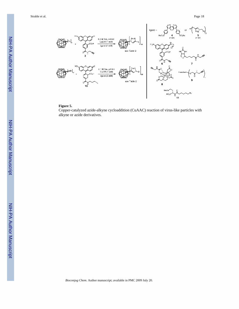
Figure 5.Copper-catalyzed azide-alkyne cycloaddition (CuAAC) reaction of virus-like particles withalkyne or azide derivatives.
Strable et al. Page 18
Bioconjug Chem. Author manuscript; available in PMC 2009 July 20.
NIH
-PA Author Manuscript
NIH
-PA Author Manuscript
NIH
-PA Author Manuscript
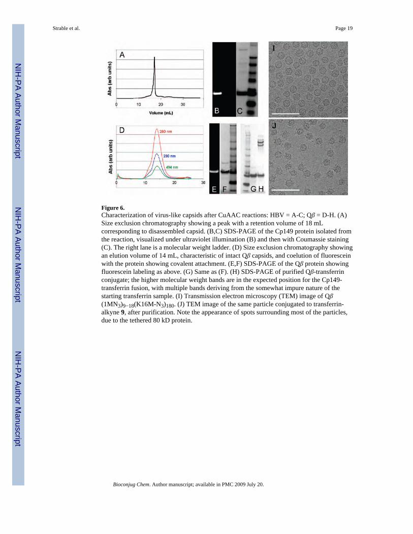
Figure 6.Characterization of virus-like capsids after CuAAC reactions: HBV = A-C; Qβ = D-H. (A)Size exclusion chromatography showing a peak with a retention volume of 18 mLcorresponding to disassembled capsid. (B,C) SDS-PAGE of the Cp149 protein isolated fromthe reaction, visualized under ultraviolet illumination (B) and then with Coumassie staining(C). The right lane is a molecular weight ladder. (D) Size exclusion chromatography showingan elution volume of 14 mL, characteristic of intact Qβ capsids, and coelution of fluoresceinwith the protein showing covalent attachment. (E,F) SDS-PAGE of the Qβ protein showingfluorescein labeling as above. (G) Same as (F). (H) SDS-PAGE of purified Qβ-transferrinconjugate; the higher molecular weight bands are in the expected position for the Cp149-transferrin fusion, with multiple bands deriving from the somewhat impure nature of thestarting transferrin sample. (I) Transmission electron microscopy (TEM) image of Qβ(1MN3)9−18(K16M-N3)180. (J) TEM image of the same particle conjugated to transferrin-alkyne 9, after purification. Note the appearance of spots surrounding most of the particles,due to the tethered 80 kD protein.
Strable et al. Page 19
Bioconjug Chem. Author manuscript; available in PMC 2009 July 20.
NIH
-PA Author Manuscript
NIH
-PA Author Manuscript
NIH
-PA Author Manuscript
NIH
-PA Author Manuscript
NIH
-PA Author Manuscript
NIH
-PA Author Manuscript
Strable et al. Page 20
Table 1Average Expression Levels of HBV and Qβ VLPs with Methionine, Azidohomoalanine (1), or Homopropargylglycine(2)
particlea amino acid yield (mg protein per gcells)
yield (mg protein per Lculture)
HBV Met 4−5 mg/g 80−100 mg/L
HBV(1M-N3)192(66M-N3)240 1 1−1.5 mg/g 20−30 mg/L
HBV(M66S) Met 4−5 mg/g 80−100 mg/L
HBV(1M-N3)102(M66S) 1 2−3 mg/g 40−60 mg/L
Qβ Met 3−4 mg/g 75−100 mg/L
Qβ(1M-N3)9−18 1 2−2.4 mg/g 50−60 mg/L
Qβ(K16M) Met 3−4 mg/g 50−75 mg/L
Qβ(1M-N3)9−18(K16M-N3)180 1 1−2 mg/g 25−35 mg/L
Qβ(K16M-CCH)90 2 2 mg/g 40 mg/L
Qβ(T93M) Met 3−4 mg/g 50−75 mg/L
Qβ(1M-N3)9−18(T93M-N3)180 1 1−2 mg/g 25−35 mg/L
aNomenclature: “HBV(1M-N3)(66M-N3)” denotes HBV particles with methionines at positions 1 and 66 (which happens to be the wild-type sequence)
expressed in the presence of 1 to allow azide incorporation at those residues. Other particles are designated in analogous fashion, with "CCH" replacing"N3" for those VLP's incorporating alkynyl amino acid 2. Subscripts refer to the average number of unnatural amino acids incorporated at each position,as described in the text.
Bioconjug Chem. Author manuscript; available in PMC 2009 July 20.
NIH
-PA Author Manuscript
NIH
-PA Author Manuscript
NIH
-PA Author Manuscript
Strable et al. Page 21
Table 2Results of CuAAC Reactions of VLP's Containing Unnatural Amino Acids, as Shown in Figure 5
entry particlea # incorporated perparticleb loadingc recoveryd
1 HBV(66M) none 0 60−70%
2 HBV(1M-N3)170−215(66M-N3)240 410−455 120 ± 15e ≤40%
3 HBV(1M-N3)170−215(M66S) 170−215 120 ± 25 60−70%
4 Qβ(K16M) none 0 75−80%
5 Qβ(1M-N3)9−18 9−18 15 ± 3 80−85%
6 Qβ(1M-N3)9−18(K16M-N3)180 189−198 190 ± 20 75−80%
7 Qβ(1M-N3)160(K16M-N3)180 340 306 ± 30f 70−80%
8 Qβ(1M-N3)9−18(T93M-N3)180 189−198 35 ± 5 75−80%
9 Qβ(K16M-CCH)90 90 50 ± 5 20%
10 Qβ(K16M-CCH)90 90 85 ± 5g 75−80%
aSee caption to Table 1 for nomenclature.
bTotal number of unnatural amino acids per particle, determined by trypsin digestion and mass spectrometry as described in the text.
cThe number of attached molecules (subscripts “m” in Figure 5). Unless otherwise indicated, the value was obtained from reaction with fluorescein
reagents 3 or 10 and quantitation by calibrated UV–visible absorbance of the dye (average of at least three independent reactions).
dAmount of purified intact capsids recovered from the reaction.
eMore than 90% of the attachments were made at position 66.
fUnder standard conditions; a loading of 330 ± 20 (75% recovery) was observed under the most forcing conditions tested.
gReaction with azide 11, loading determined by quantitation of attached Se atoms by inductively coupled plasma optical emission spectroscopy.
Bioconjug Chem. Author manuscript; available in PMC 2009 July 20.