proteins amino acids
TRANSCRIPT
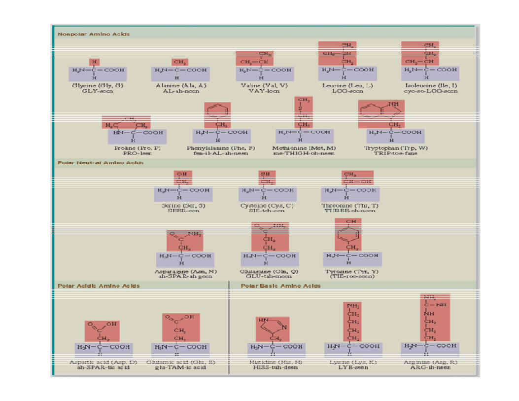
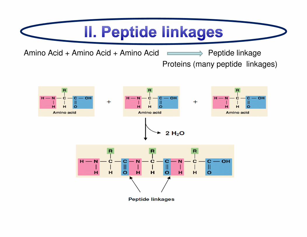
Amino Acid + Amino Acid + Amino Acid Peptide linkage
Proteins (many peptide linkages)
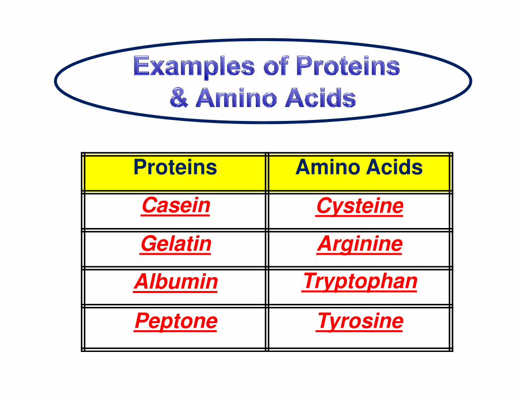
Proteins Amino Acids
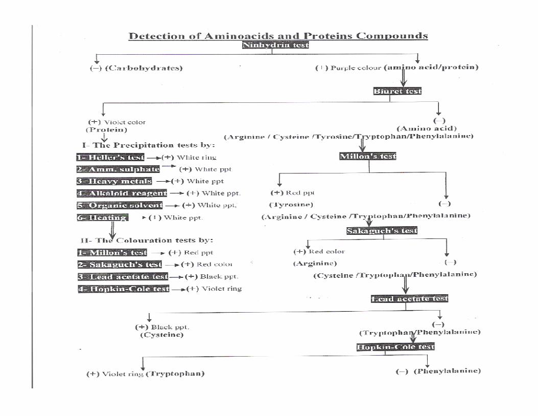
Casein CysteineCasein Cysteine
Gelatin Arginine
Albumin Tryptophan
Peptone Tyrosine
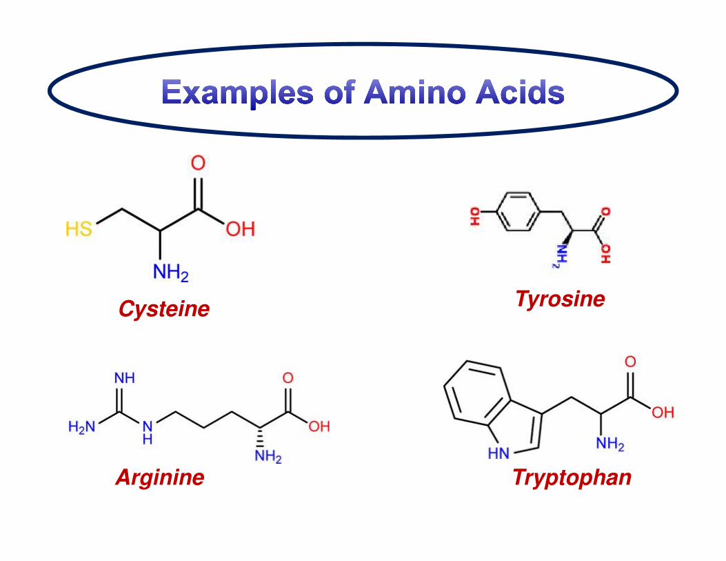
Arginine
Cysteine
Tryptophan
Tyrosine
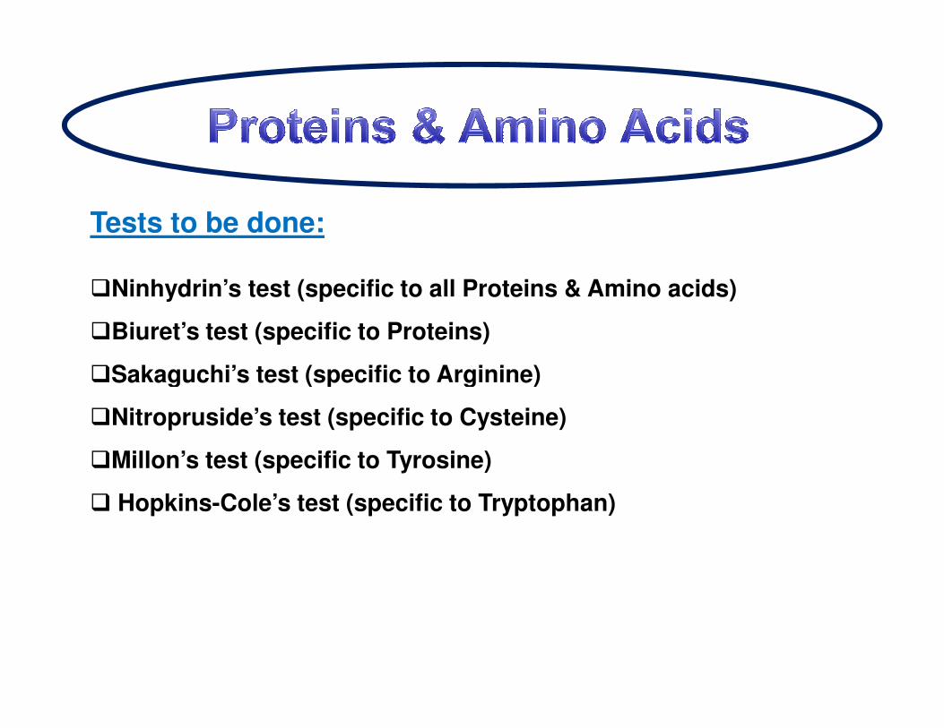
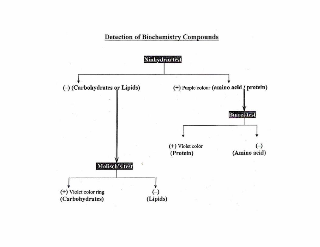
Tests to be done:
�Ninhydrin’s test (specific to all Proteins & Amino acids)
�Biuret’s test (specific to Proteins)
�Sakaguchi’s test (specific to Arginine)�Sakaguchi’s test (specific to Arginine)
�Nitropruside’s test (specific to Cysteine)
�Millon’s test (specific to Tyrosine)
� Hopkins-Cole’s test (specific to Tryptophan)
Ninhydrin reaction
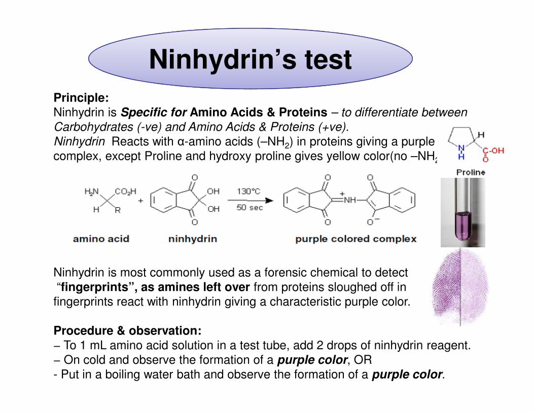
Principle:Ninhydrin is Specific for Amino Acids & Proteins – to differentiate between
Carbohydrates (-ve) and Amino Acids & Proteins (+ve).
Ninhydrin Reacts with α-amino acids (–NH2) in proteins giving a purple colored
complex, except Proline and hydroxy proline gives yellow color(no –NH2).
Ninhydrin’s test
Ninhydrin is most commonly used as a forensic chemical to detect
“fingerprints”, as amines left over from proteins sloughed off in
fingerprints react with ninhydrin giving a characteristic purple color.
Procedure & observation:− To 1 mL amino acid solution in a test tube, add 2 drops of ninhydrin reagent.
− On cold and observe the formation of a purple color, OR
- Put in a boiling water bath and observe the formation of a purple color.
Ninhydrin’stest
Ninhydrin’stest
Few drops of B
Mix Mix Heat
2-3 min
1ml of protein
solutionC
2-3 min
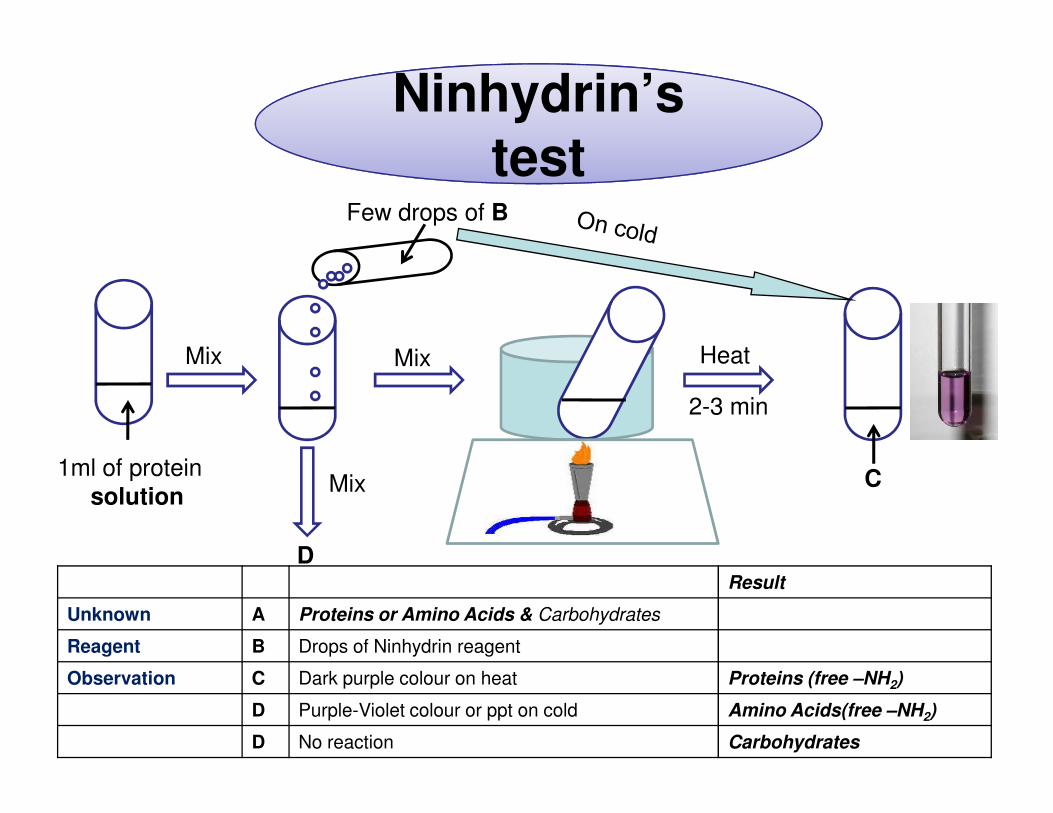
Result
Unknown A Proteins or Amino Acids & Carbohydrates
Reagent B Drops of Ninhydrin reagent
Observation C Dark purple colour on heat Proteins (free –NH2)
D Purple-Violet colour or ppt on cold Amino Acids(free –NH2)
D No reaction Carbohydrates
Mix
D
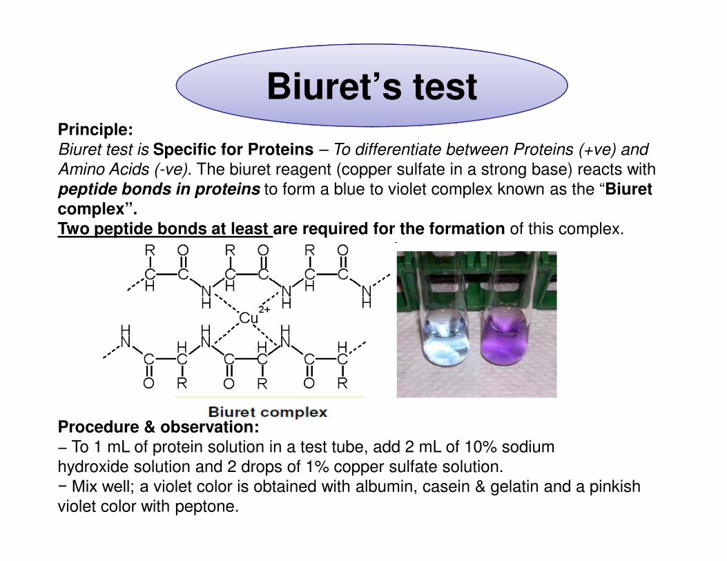
Biuret’s testPrinciple:Biuret test is Specific for Proteins – To differentiate between Proteins (+ve) and
Amino Acids (-ve). The biuret reagent (copper sulfate in a strong base) reacts with
peptide bonds in proteins to form a blue to violet complex known as the “Biuretcomplex”. Two peptide bonds at least are required for the formation of this complex.
Procedure & observation:− To 1 mL of protein solution in a test tube, add 2 mL of 10% sodium
hydroxide solution and 2 drops of 1% copper sulfate solution.
− Mix well; a violet color is obtained with albumin, casein & gelatin and a pinkish
violet color with peptone.
Biuret’s test
2 drops of B
Mix Mix
1ml of NaOH
1ml of protein
solutionC
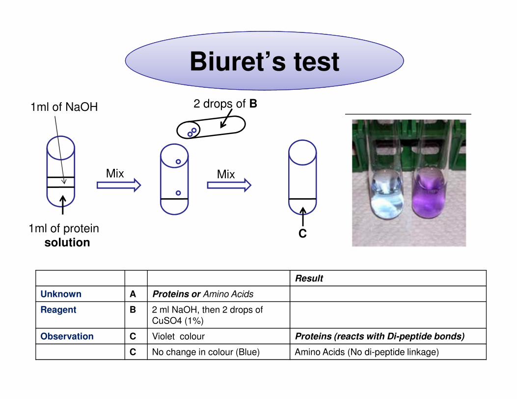
Result
Unknown A Proteins or Amino Acids
Reagent B 2 ml NaOH, then 2 drops of CuSO4 (1%)
Observation C Violet colour Proteins (reacts with Di-peptide bonds)
C No change in colour (Blue) Amino Acids (No di-peptide linkage)
Sakaguchi’s test
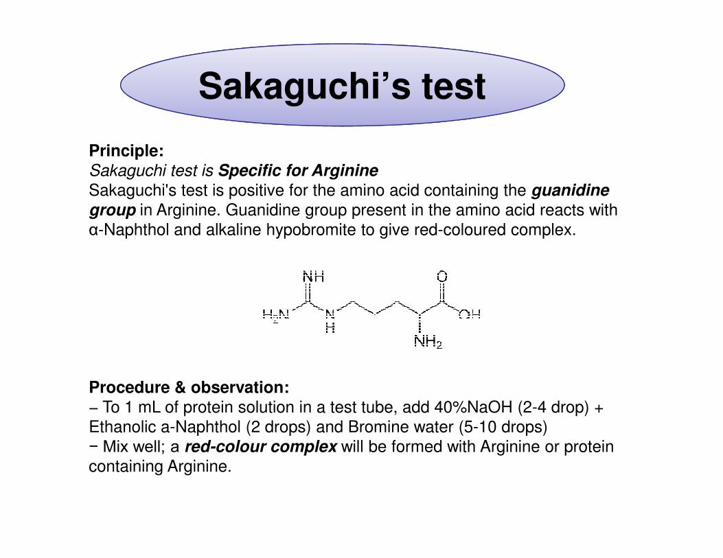
Principle:Sakaguchi test is Specific for Arginine
Sakaguchi's test is positive for the amino acid containing the guanidine
group in Arginine. Guanidine group present in the amino acid reacts with
α-Naphthol and alkaline hypobromite to give red-coloured complex.
Procedure & observation:− To 1 mL of protein solution in a test tube, add 40%NaOH (2-4 drop) +
Ethanolic a-Naphthol (2 drops) and Bromine water (5-10 drops)
− Mix well; a red-colour complex will be formed with Arginine or protein
containing Arginine.
Sakaguchi’stest
Sakaguchi’stest
Few drops of B
Mix Mix
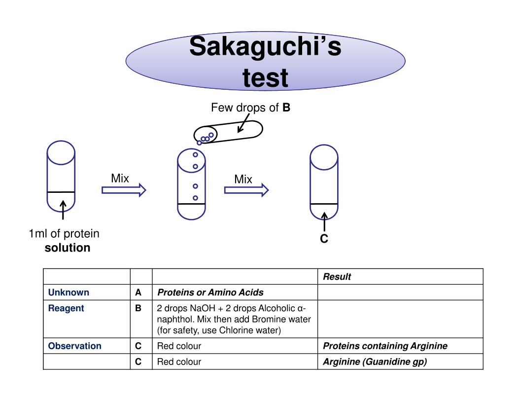
1ml of protein
solutionC
Result
Unknown A Proteins or Amino Acids
Reagent B 2 drops NaOH + 2 drops Alcoholic α-naphthol. Mix then add Bromine water (for safety, use Chlorine water)
Observation C Red colour Proteins containing Arginine
C Red colour Arginine (Guanidine gp)
Nitroprusside’s test
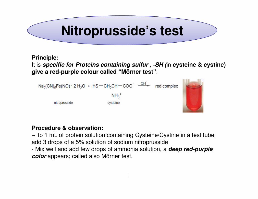
Principle:It is specific for Proteins containing sulfur , -SH (in cysteine & cystine) give a red-purple colour called “Mörner test”.
Procedure & observation:− To 1 mL of protein solution containing Cysteine/Cystine in a test tube,
add 3 drops of a 5% solution of sodium nitroprusside
- Mix well and add few drops of ammonia solution, a deep red-purple color appears; called also Mörner test.
l
Nitroprusside’s test
Nitroprusside’s test
Few drops of B
Mix Mix
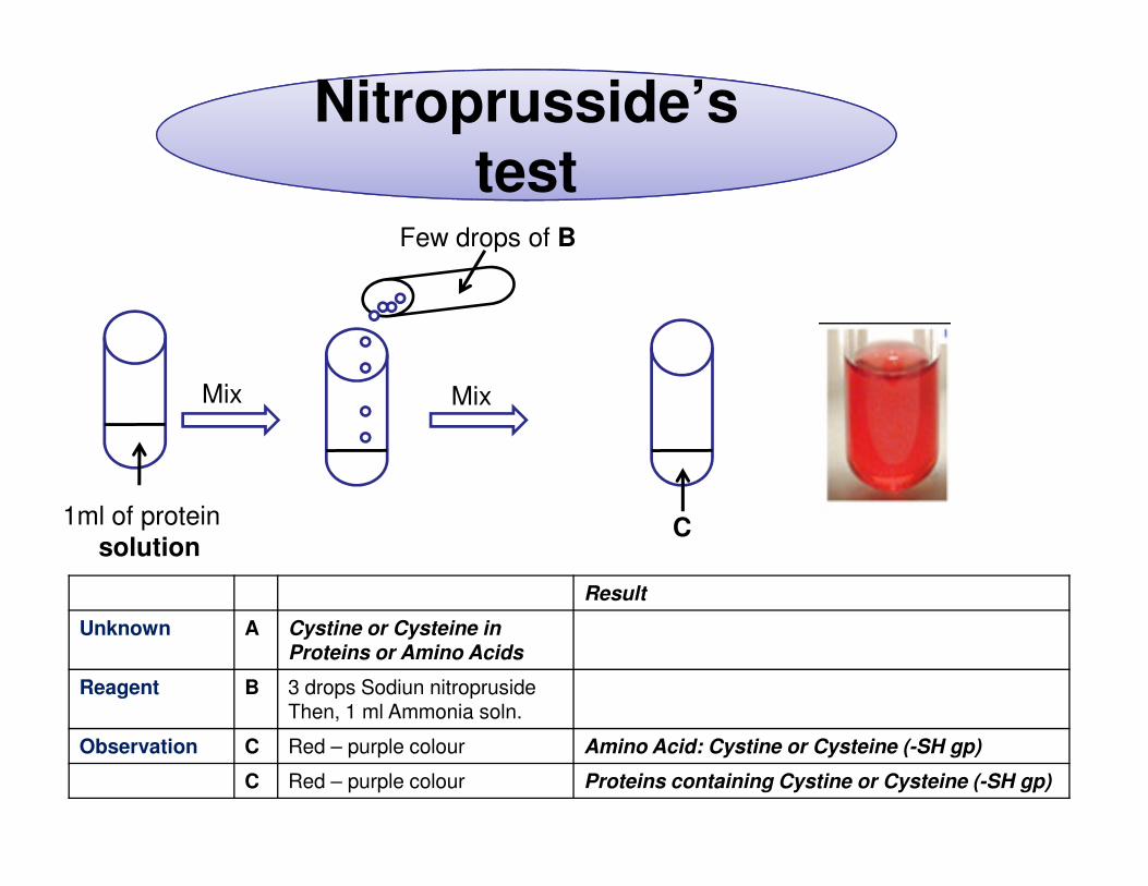
1ml of protein
solutionC
Result
Unknown A Cystine or Cysteine in Proteins or Amino Acids
Reagent B 3 drops Sodiun nitroprusideThen, 1 ml Ammonia soln.
Observation C Red – purple colour Amino Acid: Cystine or Cysteine (-SH gp)
C Red – purple colour Proteins containing Cystine or Cysteine (-SH gp)
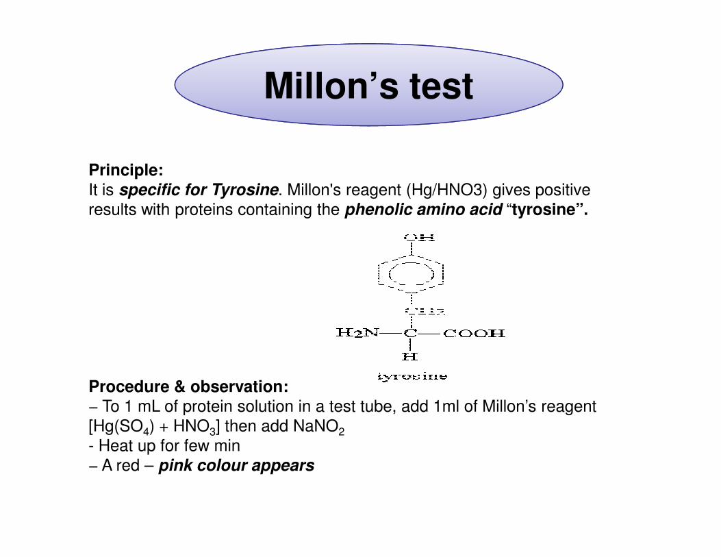
Millon’s test
Principle:It is specific for Tyrosine. Millon's reagent (Hg/HNO3) gives positive
results with proteins containing the phenolic amino acid “tyrosine”.
Procedure & observation:− To 1 mL of protein solution in a test tube, add 1ml of Millon’s reagent
[Hg(SO4) + HNO3] then add NaNO2
- Heat up for few min
− A red – pink colour appears
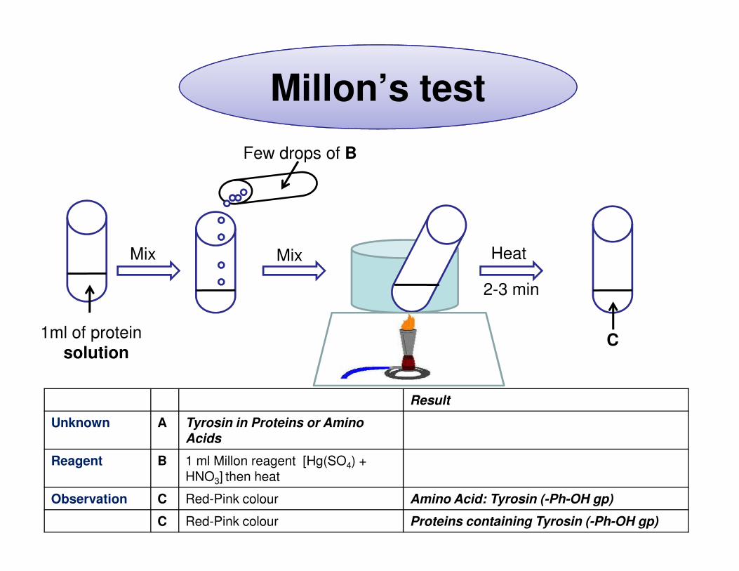
Millon’s test
Few drops of B
Mix Mix Heat
2-3 min
1ml of protein
solutionC
2-3 min
Result
Unknown A Tyrosin in Proteins or Amino Acids
Reagent B 1 ml Millon reagent [Hg(SO4) + HNO3] then heat
Observation C Red-Pink colour Amino Acid: Tyrosin (-Ph-OH gp)
C Red-Pink colour Proteins containing Tyrosin (-Ph-OH gp)
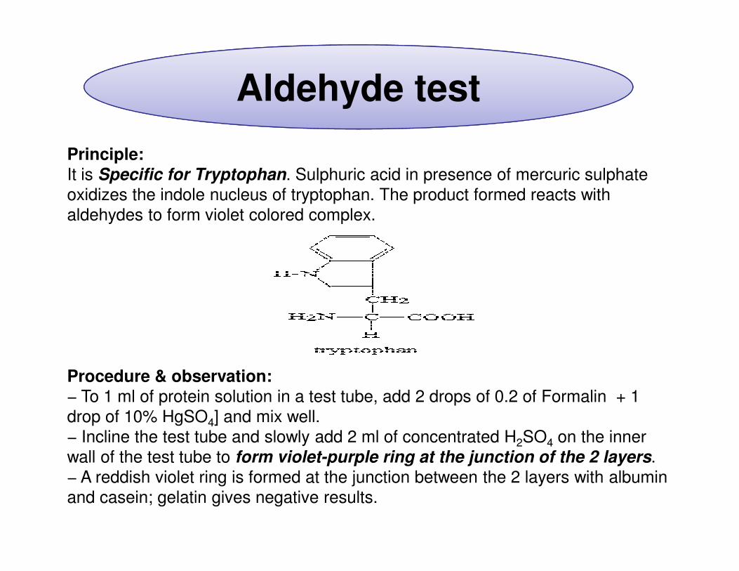
Aldehyde test
Principle:It is Specific for Tryptophan. Sulphuric acid in presence of mercuric sulphate
oxidizes the indole nucleus of tryptophan. The product formed reacts with
aldehydes to form violet colored complex.
Procedure & observation:− To 1 ml of protein solution in a test tube, add 2 drops of 0.2 of Formalin + 1
drop of 10% HgSO4] and mix well.
− Incline the test tube and slowly add 2 ml of concentrated H2SO4 on the inner
wall of the test tube to form violet-purple ring at the junction of the 2 layers.
− A reddish violet ring is formed at the junction between the 2 layers with albumin
and casein; gelatin gives negative results.
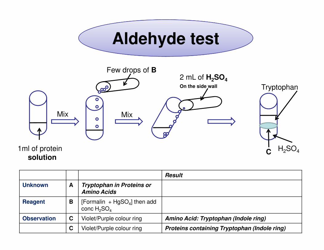
Aldehyde test
2 mL of H2SO4
On the side wall
Mix
Few drops of B
Mix
Tryptophan
Result
Unknown A Tryptophan in Proteins or Amino Acids
Reagent B [Formalin + HgSO4] then add conc H2SO4
Observation C Violet/Purple colour ring Amino Acid: Tryptophan (Indole ring)
C Violet/Purple colour ring Proteins containing Tryptophan (Indole ring)
1ml of protein
solutionC H2SO4
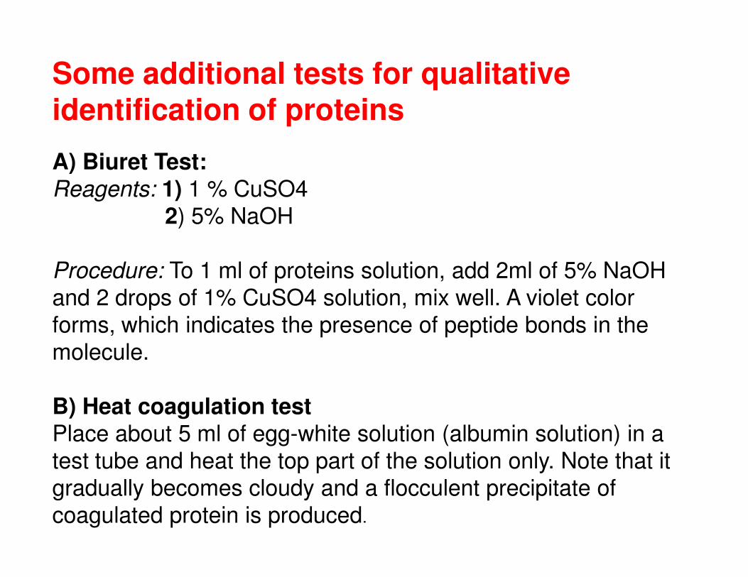
Some additional tests for qualitative identification of proteins
A) Biuret Test:Reagents: 1) 1 % CuSO4
2) 5% NaOH
Procedure: To 1 ml of proteins solution, add 2ml of 5% NaOH
and 2 drops of 1% CuSO4 solution, mix well. A violet color and 2 drops of 1% CuSO4 solution, mix well. A violet color
forms, which indicates the presence of peptide bonds in the
molecule.
B) Heat coagulation testPlace about 5 ml of egg-white solution (albumin solution) in a
test tube and heat the top part of the solution only. Note that it
gradually becomes cloudy and a flocculent precipitate of
coagulated protein is produced.
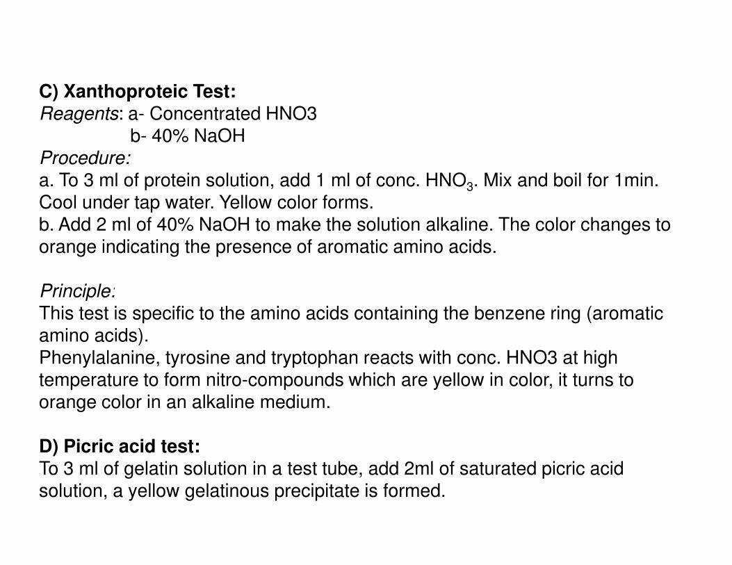
C) Xanthoproteic Test:Reagents: a- Concentrated HNO3
b- 40% NaOHProcedure:a. To 3 ml of protein solution, add 1 ml of conc. HNO3. Mix and boil for 1min. Cool under tap water. Yellow color forms.b. Add 2 ml of 40% NaOH to make the solution alkaline. The color changes to orange indicating the presence of aromatic amino acids.
Principle:Principle:This test is specific to the amino acids containing the benzene ring (aromatic amino acids).Phenylalanine, tyrosine and tryptophan reacts with conc. HNO3 at high temperature to form nitro-compounds which are yellow in color, it turns to orange color in an alkaline medium.
D) Picric acid test:To 3 ml of gelatin solution in a test tube, add 2ml of saturated picric acid solution, a yellow gelatinous precipitate is formed.
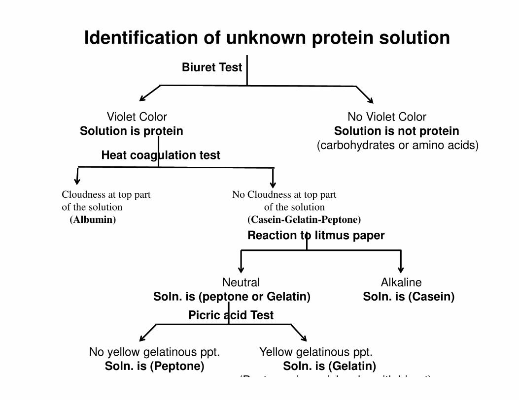
Identification of unknown protein solution
Biuret Test
Violet Color No Violet Color
Solution is protein Solution is not protein (carbohydrates or amino acids)
Heat coagulation test
Cloudness at top part No Cloudness at top part
of the solution of the solution
(Albumin) (Casein-Gelatin-Peptone)
Reaction to litmus paper
Neutral Alkaline
Soln. is (peptone or Gelatin) Soln. is (Casein)
No yellow gelatinous ppt. Yellow gelatinous ppt.
Soln. is (Peptone) Soln. is (Gelatin) (Peptone gives pink color with biuret)
Picric acid Test