university of groningen testicular cancer: diagnostic and surgical
TRANSCRIPT
University of Groningen
Testicular cancer: diagnostic and surgical strategies to improve outcomeOzturk, Cigdem
IMPORTANT NOTE: You are advised to consult the publisher's version (publisher's PDF) if you wish to cite fromit. Please check the document version below.
Document VersionPublisher's PDF, also known as Version of record
Publication date:2018
Link to publication in University of Groningen/UMCG research database
Citation for published version (APA):Ozturk, C. (2018). Testicular cancer: diagnostic and surgical strategies to improve outcome.Rijksuniversiteit Groningen.
CopyrightOther than for strictly personal use, it is not permitted to download or to forward/distribute the text or part of it without the consent of theauthor(s) and/or copyright holder(s), unless the work is under an open content license (like Creative Commons).
The publication may also be distributed here under the terms of Article 25fa of the Dutch Copyright Act, indicated by the “Taverne” license.More information can be found on the University of Groningen website: https://www.rug.nl/library/open-access/self-archiving-pure/taverne-amendment.
Take-down policyIf you believe that this document breaches copyright please contact us providing details, and we will remove access to the work immediatelyand investigate your claim.
Downloaded from the University of Groningen/UMCG research database (Pure): http://www.rug.nl/research/portal. For technical reasons thenumber of authors shown on this cover page is limited to 10 maximum.
Download date: 29-03-2022
Proefschrift
ter verkrijging van de graad van doctor aan deRijksuniversiteit Groningen
op gezag van derector magnificus prof. dr. E. Sterken
en volgens besluit van het College voor Promoties.
De openbare verdediging zal plaatsvinden op
maandag 26 november 2018 om 12.45 uur
door
Çiğdem Öztürk
geboren op 15 mei 1981te ‘s-Gravenhage
Cover Design & LayoutFleur Bominaar, FYN Werkwww.fynwerk.nl
Printed byGildeprint, Enschedewww.gildeprint.nl
ISBN: 978-94-034-0895-8 (printed version)ISBN: 978-94-034-0894-1 (electronic version)© 2018 Ç. Öztürk, The NetherlandsAll rights reserved. No part of this book may be reproduced, stored in a retreival system or transmitted in any form or by any means, without prior permission of the author.
For printing of this thesis, financial support of the following institutions and companies is gratefully acknowledged:
•• Pfizer• KARL STORZ Endoscopie Nederland B.V.• Erbe Nederland B.V. • Noord Negentig accountants en belastingadviseurs• Stichting Werkgroep Interne Oncologie• Research Institute Guide• Heelkunde Friesland
PromotoresProf. dr. H.J. HoekstraProf. dr. J.A. Gietema
CopromotorDr. R.J. van Ginkel
BeoordelingscommissieProf. dr. T.M. de ReijkeProf. dr. S. HorenblasProf. dr. A.J.H. Suurmeijer
Introduction 7General introduction 9Aims and research questions of this thesis 25
Delay in diagnosis of testicular cancer; a need for 31awareness programsPLoS One. 2015 Nov 25;10(11): e0141244
Assessment of volumetric versus manual measurement in 47disseminated testicular cancer; no difference in assessment between non-radiologists and genitourinary radiologistPLoS One. 2017 Jan 12;12(1): e0168977
Laparoscopic resection of a residual retroperitoneal tumor 65mass of nonseminomatous testicular germ cell tumorsSurgical Endoscopy and Other Interventional Techniques 2012; 26: 458-167
Laparoscopic resection of residual retroperitoneal tumor mass in 83advanced nonseminomatous testicular germ cell tumors; a feasible and safe oncological procedureScientific Reports. Submitted September 2018
Posterior retroperitoneoscopic approach in the setting of a prior 105resection of residual retroperitoneal tumor mass in advanced nonseminomatous testicular germ cell tumor: a case report of this alternative surgical approach.BMJ Case Reports. Submitted September 2018
Future perspectives 117
Summary 125Samenvatting 133
Appendix 141Historical overview of testicular cancer treatment at the UMCG
Dankwoord 152Curriculum Vitae 163Publications 165

9
Testicular germ cell tumors (TGCTs) account for only 1% of all malignancies and are the most common solid tumors in men aged between 20-35 years{1,2}. The historic pathway in the diagnostic and combined treatment of testicular cancer e.g. nonseminomatous germ cell tumors (NSTGCTs or nonseminoma), represents one of the greatest success stories in the treatment of cancer in the 20th century. This journey to cure in NSTGCTs is marked by the discovery of the effectiveness of cisplatin based combination chemotherapy in disseminated NSTGCT followed by, if indicated, adjuvant surgery. The integration of cancer care involving different medical and surgical specialties has led to significant advances in the multimodal treatment of TGCTs.Men with testicular cancer (TC) nowadays have one of the highest survival rates of any solid organ malignancy with a cure rate of 90-95 %{1,2}. The fundamental elements of the successful multimodality testicular cancer treatment include improved staging techniques, cisplatin based combination chemotherapy, aggres-sive surgical approach in case of residual disease and/or recurrent disease. Close follow-up after treatment with serum tumor marker checks and repeated CT-scan assures early detection of recurrent disease resulting in excellent second line treatment options for optimal oncologic outcome.Still in the 21th century additional progress is made in the insight of the disease, the molecular biology of TC, the diagnostic, staging and (combined) treatment of especially NSTGCTs, as well as psychosocial support.
IncidenceTesticular cancer is a rare malignancy, generally occurring in Caucasian young men. However, TGCTs are the most prevalent type of malignancy in men aged between 20 and 35 years. For NSTGCTs the incidence peak is at 25 years of age and for seminomatous germ cell tumors (or seminoma) 10 years later at 35 years. Beyond 40 years old, incidence rates decline more quickly for NSTGCTs compared to seminomas and therefore in older men more often seminomas are diagnosed. Geographic variation in testicular cancer incidence rates exists with the highest incidence in Scandinavian, Western European countries, New Zealand and North America and the lowest in Africa and Asia{3,4}. The incidence is rising in the United States and in parts of Western Europe{4-6}. In the United States in 2016 an estimated 8700 men were diagnosed with TC{2} and the incidence has been increasing from 5.7 per 100.000 in 1992 to 6.8 per 100.000 in 2009{3}. In the Netherlands, 782 men were diagnosed with testicular cancer in 2017 of which 323 men had NSTGCTs.
10 11
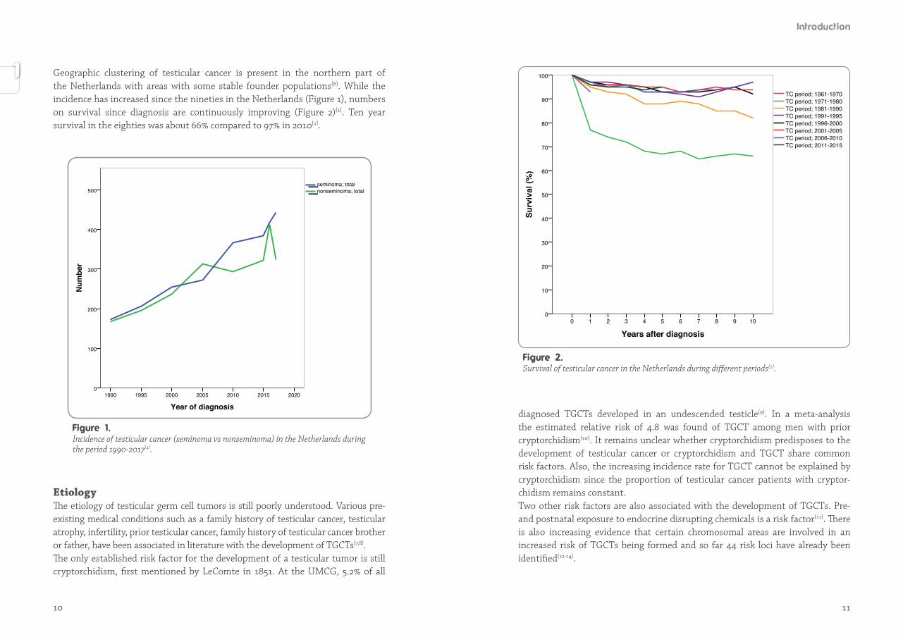
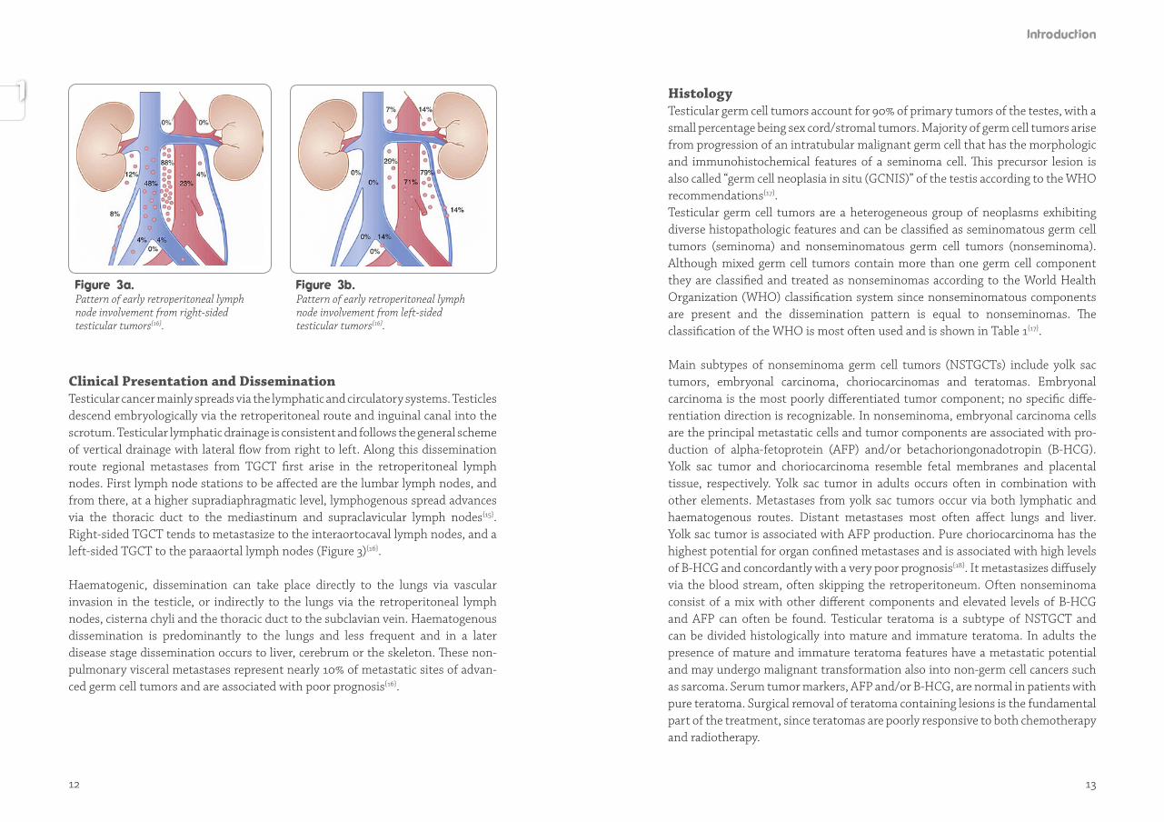
Geographic clustering of testicular cancer is present in the northern part of the Netherlands with areas with some stable founder populations{6}. While the incidence has increased since the nineties in the Netherlands (Figure 1), numbers on survival since diagnosis are continuously improving (Figure 2){1}. Ten year survival in the eighties was about 66% compared to 97% in 2010{1}.
EtiologyThe etiology of testicular germ cell tumors is still poorly understood. Various pre-existing medical conditions such as a family history of testicular cancer, testicular atrophy, infertility, prior testicular cancer, family history of testicular cancer brother or father, have been associated in literature with the development of TGCTs{7,8}. The only established risk factor for the development of a testicular tumor is still cryptorchidism, first mentioned by LeComte in 1851. At the UMCG, 5.2% of all
diagnosed TGCTs developed in an undescended testicle{9}. In a meta-analysis the estimated relative risk of 4.8 was found of TGCT among men with prior cryptorchidism{10}. It remains unclear whether cryptorchidism predisposes to the development of testicular cancer or cryptorchidism and TGCT share common risk factors. Also, the increasing incidence rate for TGCT cannot be explained by cryptorchidism since the proportion of testicular cancer patients with cryptor-chidism remains constant. Two other risk factors are also associated with the development of TGCTs. Pre- and postnatal exposure to endocrine disrupting chemicals is a risk factor{11}. There is also increasing evidence that certain chromosomal areas are involved in an increased risk of TGCTs being formed and so far 44 risk loci have already been identified{12-14}.
Surv
ival
(%)
100
90
80
70
60
50
40
30
20
10
0
Years after diagnosis10987654321
TC period; 2011-2015 TC period; 2006-2010 TC period; 2001-2005 TC period; 1996-2000 TC period; 1991-1995 TC period; 1981-1990TC period; 1971-1980 TC period; 1961-1970
0
Survival of testicular cancer in the Netherlands during different periods{1}.
Num
ber
500
400
300
200
100
0
Year of diagnosis2020201520102005200019951990
nonseminoma; totalseminoma; total
Incidence of testicular cancer (seminoma vs nonseminoma) in the Netherlands during the period 1990-2017{1}.
12 13
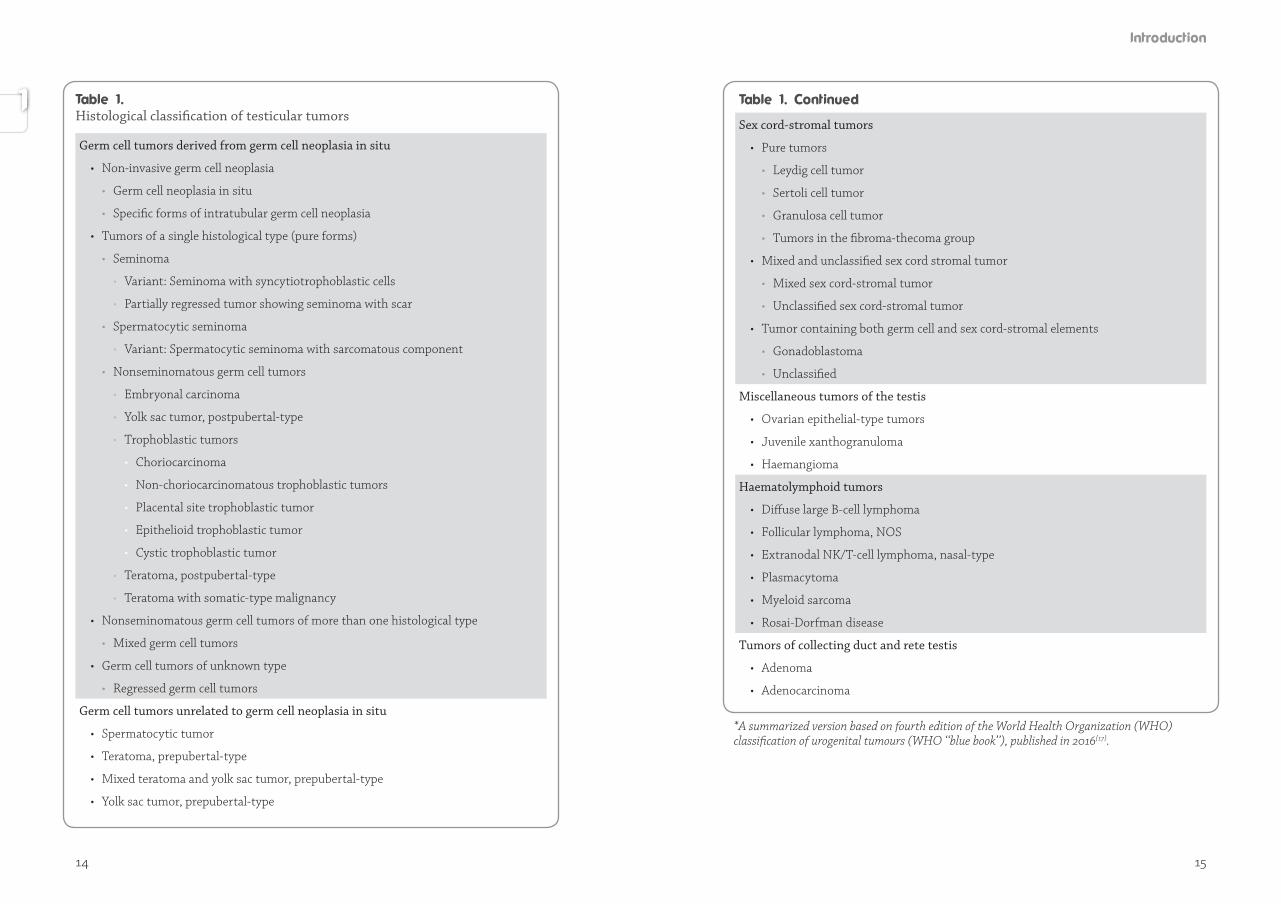
Clinical Presentation and DisseminationTesticular cancer mainly spreads via the lymphatic and circulatory systems. Testicles descend embryologically via the retroperitoneal route and inguinal canal into the scrotum. Testicular lymphatic drainage is consistent and follows the general scheme of vertical drainage with lateral flow from right to left. Along this dissemination route regional metastases from TGCT first arise in the retroperitoneal lymph nodes. First lymph node stations to be affected are the lumbar lymph nodes, and from there, at a higher supradiaphragmatic level, lymphogenous spread advances via the thoracic duct to the mediastinum and supraclavicular lymph nodes{15}. Right-sided TGCT tends to metastasize to the interaortocaval lymph nodes, and a left-sided TGCT to the paraaortal lymph nodes (Figure 3){16}.
Haematogenic, dissemination can take place directly to the lungs via vascular invasion in the testicle, or indirectly to the lungs via the retroperitoneal lymph nodes, cisterna chyli and the thoracic duct to the subclavian vein. Haematogenous dissemination is predominantly to the lungs and less frequent and in a later disease stage dissemination occurs to liver, cerebrum or the skeleton. These non-pulmonary visceral metastases represent nearly 10% of metastatic sites of advan-ced germ cell tumors and are associated with poor prognosis{16}.
Histology Testicular germ cell tumors account for 90% of primary tumors of the testes, with a small percentage being sex cord/stromal tumors. Majority of germ cell tumors arise from progression of an intratubular malignant germ cell that has the morphologicand immunohistochemical features of a seminoma cell. This precursor lesion is also called “germ cell neoplasia in situ (GCNIS)” of the testis according to the WHO recommendations{17}. Testicular germ cell tumors are a heterogeneous group of neoplasms exhibiting diver se histopathologic features and can be classified as seminomatous germ cell tumors (seminoma) and nonseminomatous germ cell tumors (nonseminoma). Although mixed germ cell tumors contain more than one germ cell component they are classified and treated as nonseminomas according to the World Health Organization (WHO) classification system since nonseminomatous components are present and the dissemination pattern is equal to nonseminomas. The classification of the WHO is most often used and is shown in Table 1{17}.
Main subtypes of nonseminoma germ cell tumors (NSTGCTs) include yolk sac tumors, embryonal carcinoma, choriocarcinomas and teratomas. Embryonal car ci noma is the most poorly differentiated tumor component; no specific diff e-rentiation direction is recognizable. In nonseminoma, embryonal carcinoma cells are the principal metastatic cells and tumor components are associated with pro-duc tion of alpha-fetoprotein (AFP) and/or betachoriongonadotropin (B-HCG). Yolk sac tumor and choriocarcinoma resemble fetal membranes and placental tissue, respectively. Yolk sac tumor in adults occurs often in combination with other elements. Metastases from yolk sac tumors occur via both lymphatic and haematogenous routes. Distant metastases most often affect lungs and liver. Yolk sac tumor is associated with AFP production. Pure choriocarcinoma has the highest potential for organ confined metastases and is associated with high levels of B-HCG and concordantly with a very poor prognosis{18}. It metastasizes diffusely via the blood stream, often skipping the retroperitoneum. Often nonseminoma consist of a mix with other different components and elevated levels of B-HCG and AFP can often be found. Testicular teratoma is a subtype of NSTGCT and can be divided histologically into mature and immature teratoma. In adults the presence of mature and immature teratoma features have a metastatic potential and may undergo malignant transformation also into non-germ cell cancers such as sarcoma. Serum tumor markers, AFP and/or B-HCG, are normal in patients with pure teratoma. Surgical removal of teratoma containing lesions is the fundamental part of the treatment, since teratomas are poorly responsive to both chemotherapy and radiotherapy.
Pattern of early retroperitoneal lymph node involvement from right-sided testicular tumors{16}.
Pattern of early retroperitoneal lymph node involvement from left-sided testicular tumors{16}.
14 15
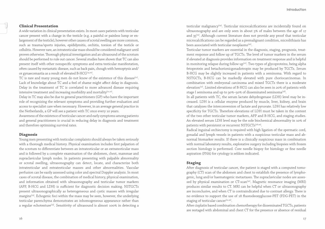
Histological classification of testicular tumors
Germ cell tumors derived from germ cell neoplasia in situ
• Non-invasive germ cell neoplasia
• Germ cell neoplasia in situ
• Specific forms of intratubular germ cell neoplasia
• Tumors of a single histological type (pure forms)
• Seminoma
• Variant: Seminoma with syncytiotrophoblastic cells
• Partially regressed tumor showing seminoma with scar
• Spermatocytic seminoma
• Variant: Spermatocytic seminoma with sarcomatous component
• Nonseminomatous germ cell tumors
• Embryonal carcinoma
• Yolk sac tumor, postpubertal-type
• Trophoblastic tumors
• Choriocarcinoma
• Non-choriocarcinomatous trophoblastic tumors
• Placental site trophoblastic tumor
• Epithelioid trophoblastic tumor
• Cystic trophoblastic tumor
• Teratoma, postpubertal-type
• Teratoma with somatic-type malignancy
• Nonseminomatous germ cell tumors of more than one histological type
• Mixed germ cell tumors
• Germ cell tumors of unknown type
• Regressed germ cell tumors
Germ cell tumors unrelated to germ cell neoplasia in situ
• Spermatocytic tumor
• Teratoma, prepubertal-type
• Mixed teratoma and yolk sac tumor, prepubertal-type
• Yolk sac tumor, prepubertal-type
Sex cord-stromal tumors
• Pure tumors
• Leydig cell tumor
• Sertoli cell tumor
• Granulosa cell tumor
• Tumors in the fibroma-thecoma group
• Mixed and unclassified sex cord stromal tumor
• Mixed sex cord-stromal tumor
• Unclassified sex cord-stromal tumor
• Tumor containing both germ cell and sex cord-stromal elements
• Gonadoblastoma
• Unclassified
Miscellaneous tumors of the testis
• Ovarian epithelial-type tumors
• Juvenile xanthogranuloma
• Haemangioma
Haematolymphoid tumors
• Diffuse large B-cell lymphoma
• Follicular lymphoma, NOS
• Extranodal NK/T-cell lymphoma, nasal-type
• Plasmacytoma
• Myeloid sarcoma
• Rosai-Dorfman disease
Tumors of collecting duct and rete testis
• Adenoma
• Adenocarcinoma
*A summarized version based on fourth edition of the World Health Organization (WHO) classification of urogenital tumours (WHO ‘‘blue book’’), published in 2016{17}.
16 17
Clinical PresentationA wide variation in clinical presentation exists. In most cases patients with testicular cancer present with a change in the testicle (e.g. a painful or painless lump or en-largement of the testicle), however other causes of scrotal swelling are more common, such as trauma/sports injuries, epididymitis, orchitis, torsion of the testicle or cellulitis. However rare, an intratesticular mass should be considered malignant until proven otherwise. Thorough physical investigation and an ultrasound of the scrotum should be performed to rule out cancer. Several studies have shown that TC can also present itself with other nonspecific symptoms and extra testicular manifestation, often caused by metastatic disease, such as back pain, chough with hemoptysis and/or gynaecomastia as a result of elevated B-HCG{19,20}. TC is rare and many young men do not know of the existence of this disease{21}. Lack of knowledge about TC and a feel of shame might affect delay in diagnosis. Delay in the treatment of TC is correlated to more advanced disease requiring intensive treatment and increasing morbidity and mortality{21,22}. Delay in TC may also be due to general practitioners (GP) who have the important role of recognizing the relevant symptoms and providing further evaluation and access to specialist care when necessary. However, in an average general practice in the Netherlands, a GP will see a patient with TC once every 10 years. Awareness of the existence of testicular cancer and early symptoms among patients and general practitioners is crucial in reducing delay in diagnosis and treatment and therefore optimising survival rates.
DiagnosisYoung men presenting with testicular complaints should always be taken seriously with a thorough medical history. Physical examination includes first palpation of the scrotum to differentiate between an intratesticular or an extratesticular mass and is followed by a complete examination of the abdomen, chest, mammae and supraclavicular lymph nodes. In patients presenting with palpable abnormality or scrotal swelling, ultrasonography can detect, locate, and characterize both intratesticular and extratesticular masses and other abnormalities. Vascular perfusion can be easily assessed using color and spectral Doppler analysis. In most cases of scrotal disease, the combination of medical history, physical examination, and information obtained with ultrasonography and testicular tumor markers (AFP, B-HCG and LDH) is sufficient for diagnostic decision making. NSTGCTs present ultrasonographically as heterogeneous and cystic masses with irregular margins{23}. Echogenic foci within the mass may be seen, however, the underlying testicular parenchyma demonstrates an inhomogeneous appearance rather than a regular echotexture{23}. Sensitivity of ultrasound is almost 100% in detecting a
testicular malignancy{24}. Testicular microcalcifications are incidentally found on ultrasonography and are only seen in about 5% of males between the age of 17 and 35{25}. Although current literature does not provide any proof that testicular microcalcifications can be regarded as a premalignant condition, microlithiasis has been associated with testicular neoplasms{26}.Testicular tumor markers are essential in the diagnosis, staging, prognosis, treat-ment response and follow up of TGCTs. The level of tumor markers in the serum if elevated at diagnosis provides information on treatment response and is helpful in monitoring relapse during follow up{27}. Two types of glycoproteins, being alpha fetoprotein and betachorionicgonadotropin may be produced by TGCTs. Serum B-HCG may be slightly increased in patients with a seminoma. With regard to NSTGCTs, B-HCG can be markedly elevated with pure choriocarcinomas. In combination with embryonal carcinoma and mixed TGCTs there is a moderate elevation{27}. Limited elevations of B-HCG can also be seen in 20% of patients with stage I seminoma and up to 30%–50% of disseminated seminoma{28}. In all patients with TC, the serum lactate dehydrogenase (LDH) level may be in-creased. LDH is a cellular enzyme produced by muscle, liver, kidney, and brain that catalyzes the interconversion of lactate and pyruvate. LDH has relatively low specificity for TGCTs. Therefore elevations of LDH must be taken in the context of the two other testicular tumor markers, AFP and B-HCG, and staging studies. An elevated serum LDH level may be the sole biochemical abnormality in 10% of patients with persistent or recurrent NSTGCTs{28,29}. Radical inguinal orchiectomy is required with high ligation of the spermatic cord, gonadal and lymph vessels in patients with a suspicious testicular mass and ab-normal biomarker results. If there is a clinically suspicious mass in combination with normal laboratory results, explorative surgery including biopsies with frozen section histology is performed. Core needle biopsy for histology or fine needle aspiration (FNA) for cytology is seldom indicated.
StagingAfter diagnosis of testicular cancer, the patient is staged with a computed to mo-graphy (CT) scan of the abdomen and chest to establish the presence of lympho-genic, lung and/or haematogenic metastases. The supraclavicular nodes are asses-sed by physical examination or CT-scan{30}. Magnetic resonance imaging (MRI) produces similar results to CT. MRI can be helpful when CT or ultrasonography are inconclusive, and when CT is contraindicated due to contrast allergy. There is no evidence to support the use of 18 fluorodeoxyglucose-PET (FDG-PET) in the sta ging of testicular cancer{31,32}.After cisplatin based combination chemotherapy for disseminated TGCTs, patients are restaged with abdominal and chest CT for the presence or absence of residual
18 19
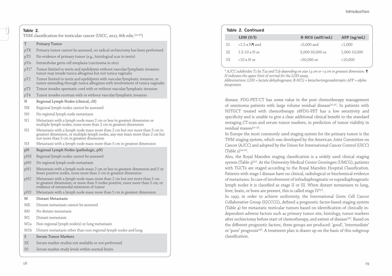
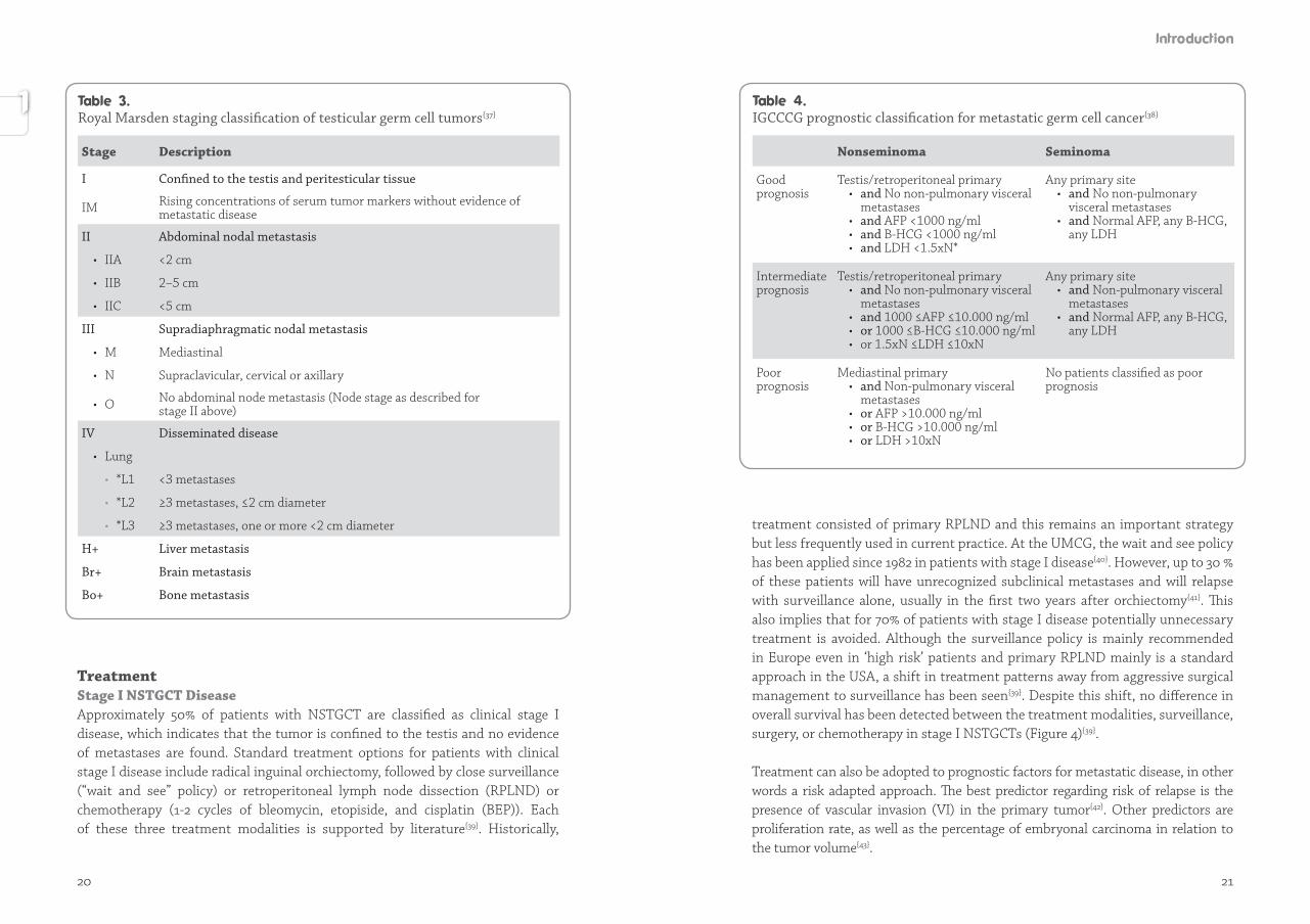
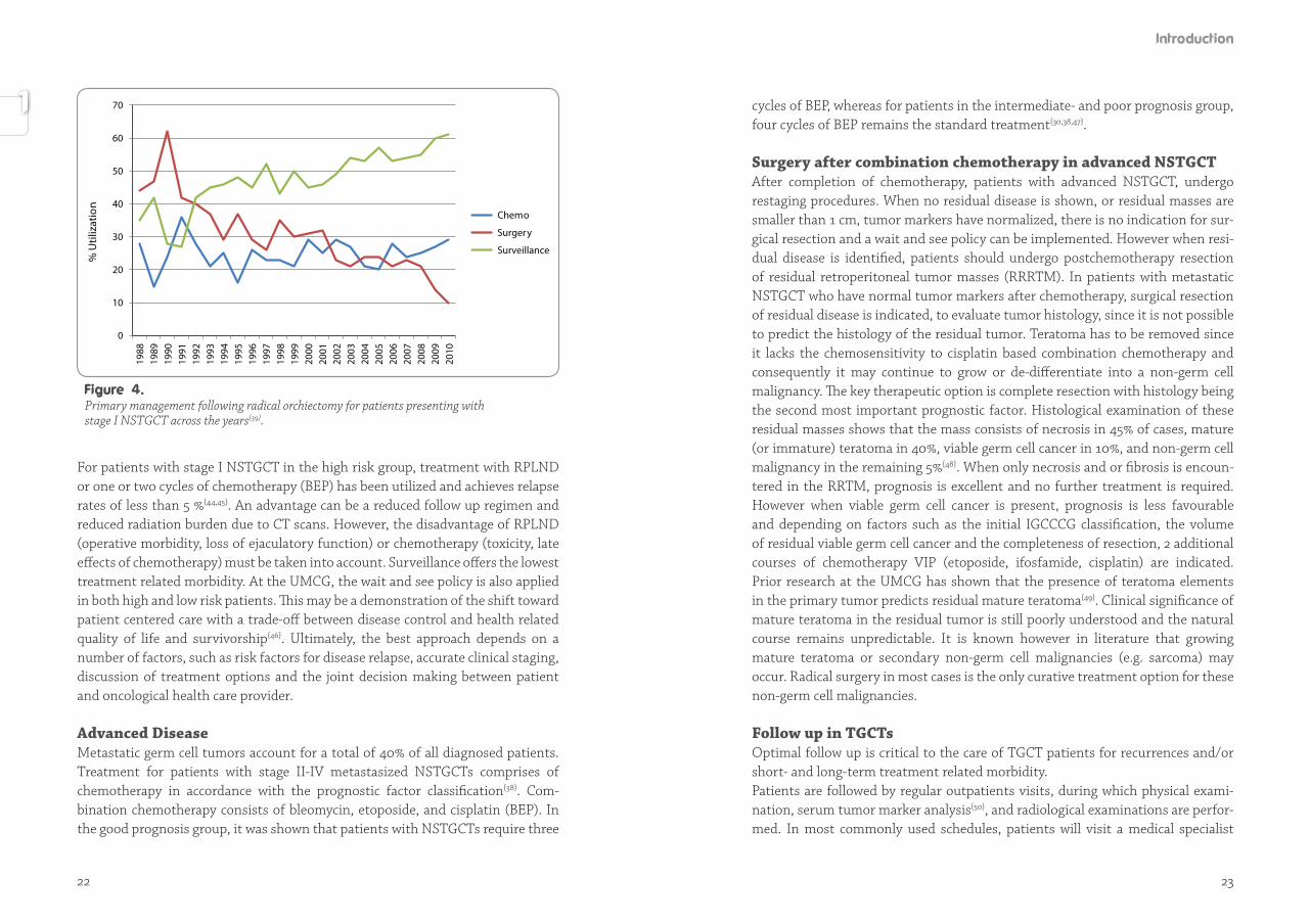
disease. FDG-PET/CT has some value in the post chemotherapy management of seminoma patients with large volume residual disease{31,32}. In patients with NSTGCT treated with chemotherapy 18FDG-PET has a low sensitivity and specificity and is unable to give a clear additional clinical benefit to the standard restaging CT-scan and serum tumor markers, in prediction of tumor viability in residual masses{31-33}. In Europe the most commonly used staging system for the primary tumor is the TNM staging system, which was developed by the American Joint Committee on Cancer (AJCC) and adopted by the Union for International Cancer Control (UICC) (Table 2){34-36}. Also, the Royal Marsden staging classification is a widely used clinical staging system (Table 3){37}. At the University Medical Center Groningen (UMCG), patients with TGCTs are staged according to the Royal Marsden Hospital Classification. Patients with stage I disease have no clinical, radiological or biochemical evidence of metastases. In case of involvement of infradiaphragmatic or supradiaphragmatic lymph nodes it is classified as stage II or III. When distant metastases to lung, liver, brain, or bone are present, this is called stage IV{37}. In 1997, in order to achieve uniformity, the International Germ Cell Cancer Collaborative Group (IGCCCG), defined a prognostic factor-based staging system (Table 4) for metastatic testicular tumors based on identification of clinically in-dependent adverse factors such as primary tumor site, histology, tumor markers after orchiectomy before start of chemotherapy, and extent of disease{38}. Based on the different prognostic factors, three groups are produced: ‘good’, ‘intermediate’ or ‘poor’ prognosis{38}. A treatment plan is drawn up on the basis of this subgroup classification.
TNM classification for testicular cancer (UICC, 2017, 8th edn.{34-36})
T Primary Tumor
pTX Primary tumor cannot be assessed, no radical orchiectomy has been performed
pT0 No evidence of primary tumor (e.g., histological scar in testis)
pTis Intratubular germ cell neoplasia (carcinoma in situ)
pT1* Tumor limited to testis and epididymis without vascular/lymphatic invasion: tumor may invade tunica albuginea but not tunica vaginalis
pT2 Tumor limited to testis and epididymis with vascular/lymphatic invasion, or tumor extending through tunica albuginea with involvement of tunica vaginalis
pT3 Tumor invades spermatic cord with or without vascular/lymphatic invasion
pT4 Tumor invades scrotum with or without vascular/lymphatic invasion
N Regional Lymph Nodes (clinical, cN)
NX Regional lymph nodes cannot be assessed
N0 No regional lymph node metastasis
N1 Metastasis with a lymph node mass 2 cm or less in greatest dimension or multiple lymph nodes, none more than 2 cm in greatest dimension
N2 Metastasis with a lymph node mass more than 2 cm but not more than 5 cm in greatest dimension, or multiple lymph nodes, any one mass more than 2 cm but not more than 5 cm in greatest dimension
N3 Metastasis with a lymph node mass more than 5 cm in greatest dimension
pN Regional Lymph Nodes (pathologic, pN)
pNX Regional lymph nodes cannot be assessed
pN0 No regional lymph node metastasis
pN1 Metastasis with a lymph node mass 2 cm or less in greatest dimension and 5 or fewer positive nodes, none more than 2 cm in greatest dimension
pN2 Metastasis with a lymph node mass more than 2 cm but not more than 5 cm in greatest dimension; or more than 5 nodes positive, none more than 5 cm; or evidence of extranodal extension of tumor
pN3 Metastasis with a lymph node mass more than 5 cm in greatest dimension
M Distant Metastasis
MX Distant metastasis cannot be assessed
M0 No distant metastasis
M1 Distant metastasis
M1a Non regional lymph node(s) or lung metastasis
M1b Distant metastasis other than non regional lymph nodes and lung
S Serum Tumor Markers
SX Serum marker studies not available or not performed
S0 Serum marker study levels within normal limits
LDH (U/l) B-HCG (mIU/mL) AFP (ng/mL)
S1 <1.5 x N¶ and <5,000 and <1,000
S2 1.5-10 x N or 5,000-50,000 or 1,000-10,000
S3 <10 x N or <50,000 or <10,000
* AJCC subdivides T1 by T1a and T1b depending on size ≤3 cm or <3 cm in greatest dimension. ¶ N indicates the upper limit of normal for the LDH assay. Abbreviations: LDH = lactate dehydrogenase; B-HCG = betachoriongonadotropin; AFP = alpha-fetoprotein.
20 21
Treatment Stage I NSTGCT DiseaseApproximately 50% of patients with NSTGCT are classified as clinical stage I disease, which indicates that the tumor is confined to the testis and no evidence of metastases are found. Standard treatment options for patients with clinical stage I disease include radical inguinal orchiectomy, followed by close surveillance (“wait and see” policy) or retroperitoneal lymph node dissection (RPLND) or chemotherapy (1-2 cycles of bleomycin, etopiside, and cisplatin (BEP)). Each of these three treatment modalities is supported by literature{39}. Historically,
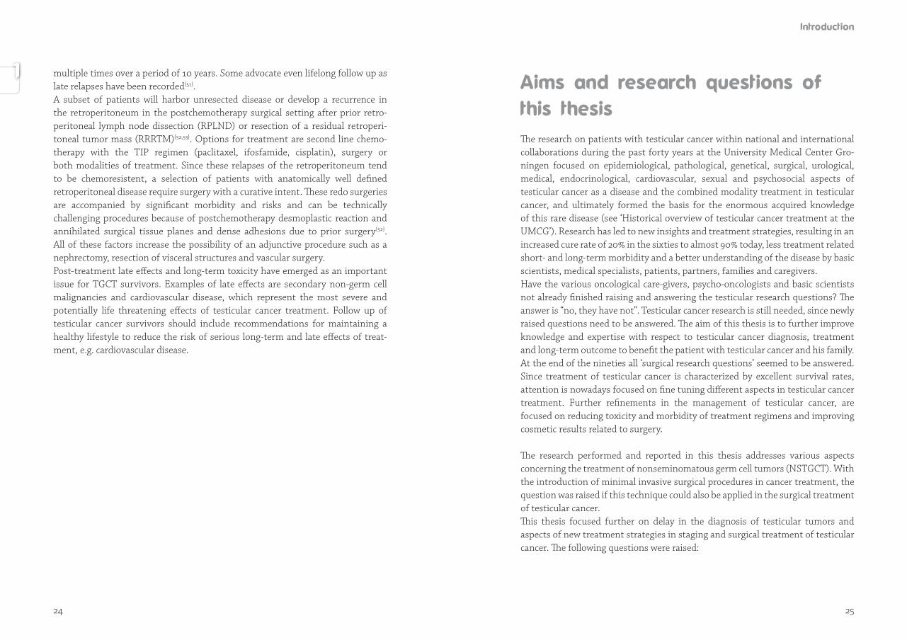
treatment consisted of primary RPLND and this remains an important strategy but less frequently used in current practice. At the UMCG, the wait and see policy has been applied since 1982 in patients with stage I disease{40}. However, up to 30 % of these patients will have unrecognized subclinical metastases and will relapse with surveillance alone, usually in the first two years after orchiectomy{41}. This also implies that for 70% of patients with stage I disease potentially unnecessary treatment is avoided. Although the surveillance policy is mainly recommended in Europe even in ‘high risk’ patients and primary RPLND mainly is a standard approach in the USA, a shift in treatment patterns away from aggressive surgical management to surveillance has been seen{39}. Despite this shift, no difference in overall survival has been detected between the treatment modalities, surveillance, surgery, or chemotherapy in stage I NSTGCTs (Figure 4){39}.
Treatment can also be adopted to prognostic factors for metastatic disease, in other words a risk adapted approach. The best predictor regarding risk of relapse is the presence of vascular invasion (VI) in the primary tumor{42}. Other predictors are proliferation rate, as well as the percentage of embryonal carcinoma in relation to the tumor volume{43}.
Royal Marsden staging classification of testicular germ cell tumors{37}
Stage Description
I Confined to the testis and peritesticular tissue
IM Rising concentrations of serum tumor markers without evidence of metastatic disease
II Abdominal nodal metastasis
• IIA <2 cm
• IIB 2–5 cm
• IIC <5 cm
III Supradiaphragmatic nodal metastasis
• M Mediastinal
• N Supraclavicular, cervical or axillary
• O No abdominal node metastasis (Node stage as described for stage II above)
IV Disseminated disease
• Lung
• *L1 <3 metastases
• *L2 ≥3 metastases, ≤2 cm diameter
• *L3 ≥3 metastases, one or more <2 cm diameter
H+ Liver metastasis
Br+ Brain metastasis
Bo+ Bone metastasis
IGCCCG prognostic classification for metastatic germ cell cancer{38}
Nonseminoma Seminoma
Good prognosis
Testis/retroperitoneal primary • and No non-pulmonary visceral
metastases • and AFP <1000 ng/ml • and B-HCG <1000 ng/ml • and LDH <1.5xN*
Any primary site • and No non-pulmonary
visceral metastases • and Normal AFP, any B-HCG,
any LDH
Intermediate prognosis
Testis/retroperitoneal primary • and No non-pulmonary visceral
metastases • and 1000 ≤AFP ≤10.000 ng/ml • or 1000 ≤B-HCG ≤10.000 ng/ml • or 1.5xN ≤LDH ≤10xN
Any primary site • and Non-pulmonary visceral
metastases • and Normal AFP, any B-HCG,
any LDH
Poor prognosis
Mediastinal primary• and Non-pulmonary visceral
metastases • or AFP >10.000 ng/ml • or B-HCG >10.000 ng/ml • or LDH >10xN
No patients classified as poor prognosis
22 23
For patients with stage I NSTGCT in the high risk group, treatment with RPLND or one or two cycles of chemotherapy (BEP) has been utilized and achieves relapse rates of less than 5 %{44,45}. An advantage can be a reduced follow up regimen and reduced radiation burden due to CT scans. However, the disadvantage of RPLND (operative morbidity, loss of ejaculatory function) or chemotherapy (toxicity, late effects of chemotherapy) must be taken into account. Surveillance offers the lowest treatment related morbidity. At the UMCG, the wait and see policy is also applied in both high and low risk patients. This may be a demonstration of the shift toward patient centered care with a trade-off between disease control and health related quality of life and survivorship{46}. Ultimately, the best approach depends on a number of factors, such as risk factors for disease relapse, accurate clinical staging, discussion of treatment options and the joint decision making between patient and oncological health care provider.
Advanced DiseaseMetastatic germ cell tumors account for a total of 40% of all diagnosed patients. Treatment for patients with stage II-IV metastasized NSTGCTs comprises of chemotherapy in accordance with the prognostic factor classification{38}. Com-bination chemotherapy consists of bleomycin, etoposide, and cisplatin (BEP). In the good prognosis group, it was shown that patients with NSTGCTs require three
cycles of BEP, whereas for patients in the intermediate- and poor prognosis group, four cycles of BEP remains the standard treatment{30,38,47}.
Surgery after combination chemotherapy in advanced NSTGCTAfter completion of chemotherapy, patients with advanced NSTGCT, undergo restaging procedures. When no residual disease is shown, or residual masses are smaller than 1 cm, tumor markers have normalized, there is no indication for sur-gic al resection and a wait and see policy can be implemented. However when resi-dual dis ease is identified, patients should undergo postchemotherapy resection of residual retroperitoneal tumor masses (RRRTM). In patients with metastatic NSTGCT who have normal tumor markers after chemotherapy, surgical resection of residual disease is indicated, to evaluate tumor histology, since it is not possible to predict the histology of the residual tumor. Teratoma has to be removed since it lacks the chemosensitivity to cisplatin based combination chemotherapy and conse quent ly it may continue to grow or de-differentiate into a non-germ cell malig nancy. The key therapeutic option is complete resection with histology being the second most important prognostic factor. Histological examination of these residual masses shows that the mass consists of necrosis in 45% of cases, mature (or imma ture) teratoma in 40%, viable germ cell cancer in 10%, and non-germ cell malig nancy in the remaining 5%{48}. When only necrosis and or fibrosis is encoun-tered in the RRTM, prognosis is excellent and no further treatment is required. However when viable germ cell cancer is present, prognosis is less favourable and depending on factors such as the initial IGCCCG classification, the volume of residual viable germ cell cancer and the completeness of resection, 2 additional courses of chemotherapy VIP (etoposide, ifosfamide, cisplatin) are indicated. Prior research at the UMCG has shown that the presence of teratoma elements in the primary tumor predicts residual mature teratoma{49}. Clinical significance of mature teratoma in the residual tumor is still poorly understood and the natural course remains unpredictable. It is known however in literature that growing mature teratoma or secondary non-germ cell malignancies (e.g. sarcoma) may occur. Radical surgery in most cases is the only curative treatment option for these non-germ cell malignancies.
Follow up in TGCTsOptimal follow up is critical to the care of TGCT patients for recurrences and/or short- and long-term treatment related morbidity. Patients are followed by regular outpatients visits, during which physical exami-nation, serum tumor marker analysis{50}, and radiological examinations are perfor-med. In most commonly used schedules, patients will visit a medical specialist
0
10
20
30
40
50
6019
8819
8919
9019
9119
9219
9319
9419
9519
9619
9719
9819
9920
0020
0120
0220
0320
0420
0520
0620
0720
0820
0920
10
Chemo
Surgery
Surveillance
% U
tiliz
atio
n70
Primary management following radical orchiectomy for patients presenting withstage I NSTGCT across the years{39}.
24 25
multiple times over a period of 10 years. Some advocate even lifelong follow up as late relapses have been recorded{51}. A subset of patients will harbor unresected disease or develop a recurrence in the retroperitoneum in the postchemotherapy surgical setting after prior retro-peritoneal lymph node dissection (RPLND) or resection of a residual retro peri-toneal tumor mass (RRRTM){52,53}. Options for treatment are second line chemo-the ra py with the TIP regimen (paclitaxel, ifosfamide, cisplatin), surgery or both modalities of treatment. Since these relapses of the retroperitoneum tend to be chemoresistent, a selection of patients with anatomically well defined retroperitoneal disease require surgery with a curative intent. These redo surgeries are accompanied by significant morbidity and risks and can be technically challenging procedures because of postchemotherapy desmoplastic reaction and annihilated surgical tissue planes and dense adhesions due to prior surgery{52}. All of these factors increase the possibility of an adjunctive procedure such as a nephrectomy, resection of visceral structures and vascular surgery. Post-treatment late effects and long-term toxicity have emerged as an important issue for TGCT survivors. Examples of late effects are secondary non-germ cell malignancies and cardiovascular disease, which represent the most severe and potentially life threatening effects of testicular cancer treatment. Follow up of testicular cancer survivors should include recommendations for maintaining a healthy lifestyle to reduce the risk of serious long-term and late effects of treat-ment, e.g. cardiovascular disease.
The research on patients with testicular cancer within national and international collaborations during the past forty years at the University Medical Center Gro-ningen focused on epidemiological, pathological, genetical, surgical, urological, medical, endocrinological, cardiovascular, sexual and psychosocial aspects of testicular cancer as a disease and the combined modality treatment in testicular cancer, and ultimately formed the basis for the enormous acquired knowledge of this rare disease (see ‘Historical overview of testicular cancer treatment at the UMCG’). Research has led to new insights and treatment strategies, resulting in an increased cure rate of 20% in the sixties to almost 90% today, less treatment related short- and long-term morbidity and a better understanding of the disease by basic scientists, medical specialists, patients, partners, families and caregivers. Have the various oncological care-givers, psycho-oncologists and basic scientists not already finished raising and answering the testicular research questions? The answer is “no, they have not”. Testicular cancer research is still needed, since newly raised questions need to be answered. The aim of this thesis is to further improve knowledge and expertise with respect to testicular cancer diagnosis, treatment and long-term outcome to benefit the patient with testicular cancer and his family. At the end of the nineties all ‘surgical research questions’ seemed to be answered. Since treatment of testicular cancer is characterized by excellent survival rates, attention is nowadays focused on fine tuning different aspects in testicular cancer treatment. Further refinements in the management of testicular cancer, are focused on reducing toxicity and morbidity of treatment regimens and improving cosmetic results related to surgery.
The research performed and reported in this thesis addresses various aspects concerning the treatment of nonseminomatous germ cell tumors (NSTGCT). With the introduction of minimal invasive surgical procedures in cancer treatment, the question was raised if this technique could also be applied in the surgical treatment of testicular cancer. This thesis focused further on delay in the diagnosis of testicular tumors and aspects of new treatment strategies in staging and surgical treatment of testicular cancer. The following questions were raised:
26 27
Surgical Research Questions• Are men and physicians aware of testicular cancer? • Is it possible to use volumetric CT analysis to measure the therapeutic response
of retroperitoneal lymph nodes in testicular cancer after chemotherapy? • What is the short-term outcome of laparoscopic resection of testicular residual
retroperitoneal tumor after systemic treatment?• What is de short- and long-term outcome of (hand-assisted) laparoscopic resec-
tion of testicular residual retroperitoneal tumor after systemic treatment? • Are there new surgical strategies to resect retroperitoneal tumor recurrence?
In the various chapters in this thesis are the previous research questions answered, followed by a summary and future perspectives.
1. Nederlandse Kankerregistratie. www.cijfersoverkanker.nl (2011-2018, accessed 20 June 2018).
2. Siegel RL, Miller KD, Jemal A. Cancer Statistics. CA Cancer J Clin 2018; 68: 7–30.
3. Nigam M, Aschebrook-Kilfoy B, Shikanov S, Eggener S. Increasing incidence of testicular cancer in the United States and Europe between 1992 and 2009. World J Urol 2015; 33: 623–631.
4. Shanmugalingam T, Soultati A, Chowdhury S, Rudman S, Van Hemelrijck M. Global incidence and outcome of testicular cancer. Clin Epidemiol. 2013; 17; 5: 417-427.
5. Adami HO, Bergström R, Möhner M, Zatoński W, Storm H, Ekbom A et al. Testicular cancer in nine Northern European countries. Int J Cancer 1994; 59: 33–38.
6. Sonneveld DJA, Schaapveld M, Sleijfer DT, Meerman GJ, van der Graaf WTA, Sijmons RH et al. Geographic clustering of testicular cancer incidence in the northern part of The Netherlands. Br J Cancer 1999; 81: 1262-1267.
7. McGlynn KA, Trabert B. Adolescent and adult risk factors for testicular cancer. Nat Rev Urol 2012; 17; 9: 339-349.
8. Sonneveld DJA, Sleijfer DT, Schraffordt Koops H, Sijmons RH, Graaf van der WT, Sluiter WJ et al. Familial testicular cancer in a single-centre population. Eur J Cancer 1999; 35: 1368-1373.
9. Wobbes Th, Schraffordt Koops H, Oldhoff JJ. The relation between testicular tumours, undescended testes, and inguinal hernias. Surg Oncol 1980; 14: 45-51.
10. Dieckmann KP, Pichlmeier U. Clinical epidemiology of testicular germ cell tumors. World J Urol 2004; 22: 2–14.
11. Bonde JP, Flachs EM, Rimborg S, Glazer CH, Giwercman A, Ramlau-Hansen CH et al. The epidemiologic evidence linking prenatal and postnatal exposure to endocrine disrupting chemicals with male reproductive disorders: a systematic review and meta-analysis. Hum Reprod Update 2016; 23: 104-125.
12. Litchfield K, Levy M, Orlando G, Loveday C, Law PJ, Migliorini G et al. Identification of 19 new risk loci and potential regulatory mechanisms influencing susceptibility to testicular germ cell tumor. Nat Genet 2017; 49: 1133- 1140.
13. Lutke Holzik MF, Sijmons RH, Hoekstra-Weebers JE, Sleijfer DT, Hoekstra HJ. Clinical and genetic aspects of testicular germ cell tumours. Hered Cancer Clin Pract 2008; 6: 3-14.
14. Lutke Holzik MF, Rapley EA, Hoekstra HJ, Sleijfer DT, Nolte IM, Sijmons RH. Genetic predisposition to testicular germ-cell tumours. Lancet Oncol. 2004; 5: 363-371.
15. Bosl GJ, Feldman DR, Bajorin DF, Sheinfeld J, Motzer RJ, Chaganti RSK. Cancer of the testis. In: DeVita VT, Hellman S, Rosenberg SA. Cancer. Principles and practice of oncology. 9th ed. Lippincott Philadelphia: Williams&Wilkins; 2011. p. 1280-1301.
16. Donohue JP. Metastatic pathways of nonseminomatous germ cell tumors. Semin Urol 1984; 2: 217-229.
17. Moch H, Cubilla AL, Humphrey PA, Reuter VE, Ulbright TM. The 2016 WHO Classification of Tumours of the Urinary System and Male Genital Organs—Part A: Renal, Penile, and Testicular Tumours. Eur Urol 2016; 70: 93-105.
18. Papiani G, Einhorn LH. Salvage chemotherapy with high-dose carboplatin plus etoposide and autologous peripheral blood stem cell transplant in male pure choriocarcinoma: a retrospective analysis of 13 cases. Bone Marrow Transplant 2007; 40: 235-237.
19. Oliver RT. Factors contributing to delay in diagnosis of testicular tumours. Br Med J 1985; 290: 356.
20. Cantwell BM. Gynaecomastia and extragonadal symptoms leading to diagnosis delay of germ cell tumours in young men. Postgrad Med J 1991; 67: 675-677.
21. Bosl GJ, Goldman A, Lange PH, Vogelzang NJ, Fraley EE, Levitt SH et al. Impact of delay in diagnosis on clinical stage of testicular cancer. Lancet 1981; 2: 970-973.
22. Chilvers CE, Saunders M, Bliss JM, Nicholls J, Horwich A. Influence of delay in diagnosis on prognosis in testicular teratoma. Br J Cancer 1989; 59: 126-128.
23. Kühn AL, Scortegagna E, Nowitzki KM, Kim YH. Ultrasonography of the scrotum in adults. Ultrasonography 2016; 35: 180-197.
24. Oyen RH, Verbist BM, Verswijvel GA. Imaging of Testicular Neoplasms. In: Petrovich Z, Baert L, Brady LW (eds) Carcinoma of the Kidney and Testis, and Rare Urologic Malignancies. Medical Radiology (Diagnostic Imaging and Radiation Oncology). Berlin, Heidelberg: Springer; 1999.
25. Peterson AC, Bauman JM, Light DE, McMann LP, Costabile RA. The prevalence of testicular microlithiasis in an asymptomatic population of men 18 to 35 years old. J Urol 2001; 166: 2061–2064.
26. Ganem JP, Workman KR, Shaban SF. Testicular microlithiasis is associated with testicular pathology. Urol 1999; 53: 209–213.
27. Milose JC, Filson CP, Weizer AZ, Hafez KS, Montgomery JS. Role of biochemical markers in testicular cancer: diagnosis, staging, and surveillance. Open Access J Urol 2011; 4: 1-8.
28 29
28. Bosl GJ, Motzer RJ. Testicular germ-cell cancer. N Eng J Med 1997; 337: 242–253.
29. Skinner DG, Scardino PT. Relevance of biochemical tumor markers and lymphadenectomy in management of nonseminomatous testis tumors: current perspective. J Urol 1980; 123: 378–382.
30. Albers P, Albrecht W, Algaba F, Bokemeyer C, Cohn-Cedermark G, Fizazi K et al. European Association of Urology. Guidelines on Testicular Cancer: 2015 Update. Eur Urol 2015; 68: 1054-1068.
31. Cook GJ, Sohaib A, Huddart RA, Dearnaley DP, Horwich A, Chua S. The role of 18F-FDG PET/CT in the management of testicular cancers. Nucl Med Commun. 2015; 36: 702-708.
32. Bouchelouche K, Choyke PL. PET/Computed Tomography in Renal, Bladder and Testicular Cancer. PET Clin 2015; 10: 361-374.
33. De Santis M, Bokemeyer C, Becherer A, Stoiber F, Oechsie K, Kletter K et al. Predictive impact of 2-18fluoro-2-deoxy-D-glucose positron emission tomography for residual post chemotherapy masses in patients with bulky seminoma. J Clin Oncol 2001; 19: 3740-3744.
34. Brierley JD, Gospodarowicz MK, Wittekind C. TNM Classification of Malignant Tumours. 8th ed. Oxford, UK: Wiley-Blackwell; 2017.
35. Paner GP, Stadler WM, Hansel DE, Montironi R, Lin DW, Amin MB. Updates in the Eighth Edition of the Tumor-Node-Metastasis Staging Classification for Urologic Cancers. Eur Urol 2018; 73: 560-569.
36. Amin MB, Edge SB, Greene FL, Byrd DR, Brookland RK, Washington MK et al. AJCC Cancer Staging Manual. 8th ed. New York: Springer; 2017.
37. Peckham MJ, McElwain TJ, Barrett A, Hendry WF. Combined management of malignant teratoma of the testis. Lancet 1979; 2: 267-270.
38. International Germ Cell Consensus Classification: a prognostic factor-based staging system for metastatic germ cell cancers. International Germ Cell Cancer Collaborative Group. J Clin Oncol 1997; 15: 594-603.
39. Yap SA, Yuh LM, Evans CP, Dall’Era MA, Wagenaar RM, Cress R et al. Evolving patterns of care in the management of stage I non-seminomatous germ cell tumors: data from the California Cancer Registry. World J Urol 2017; 35: 277–283.
40. Gels ME, Hoekstra HJ, Sleijffer DT, Marrink J, de Bruijn HW, Molenaar WM et al. Detection of recurrence in patients with clinical stage I nonseminomatous testicular germ cell tumors and consequences for further follow up: a single-center 10-year experience. J Clin Oncol 1995; 13: 1188-1194.
41. Sturgeon JF, Moore MJ, Kakiashvili DM, Duran I, Anson-Cartwright LC, Berthold DR et al. Non-risk-adapted surveillance in clinical stage I nonseminomatous germ cell tumors: the Princess Margaret Hospital’s experience. Eur Urol 2011; 59: 556–562.
42. Vergouwe Y, Steyerberg EW, Eijkemans MJ, Albers P, Habbema JD. Predictors of occult metastasis in clinical stage I nonseminoma: a systematic review. J Clin Oncol 2003; 21: 4092-4099.
43. Heidenreich A, Sesterhenn IA, Mostofi FK, Moul JW. Prognostic risk factors that identify patients with clinical stage I nonseminomatous germ cell tumors at low risk and high risk for metastasis. Cancer 1998; 83: 1002–1011.
44. Read G, Stenning SP, Cullen MH, Parkinson MC, Horwich A, Kaye SB et al. Medical Research Council prospective study of surveillance for stage I testicular teratoma. Medical Research Council Testicular Tumors Working Party. J Clin Oncol 1992; 10: 1762–1768.
45. Beck SD. Management options for stage 1 nonseminomatous germ cell tumors of the testis. Indian J Urol 2010; 26: 72-75.
46. Travis LB, Beard C, Allan JM, Dahl AA, Feldman DR, Oldenburg J et al. Testicular cancer survivorship: research strategies and recommendations. J Natl Cancer Inst 2010; 102: 1114-1130.
47. Oldenburg J, Fosså SD, Nuver J, Heidenreich A, Scmoll HJ, Bokemeyer C et al. Testicular seminoma and non-seminoma: ESMO Clinical Practice Guidelines for diagnosis, treatment, and follow up. Ann Oncol 2013; 24: 125-132.
48. Lutke Holzik MF, Hoekstra HJ, Mulder NH, Suurmeijer AJ, Sleijfer DT, Gietema JA. Non-germ cell malignancy in residual or recurrent mass after chemotherapy for nonseminomatous testicular germ cell tumor. Ann Surg Oncol 2003; 10: 131-135.
49. Oosterhuis JW, Suurmeijer AJ, Sleijfer DT, Koops HS, Oldhoff J, Fleuren G. Effects of multiple-drug chemotherapy (cis-diammine-dichloroplatinum, bleomycin, and vin-blastine) on the maturation of retroperitoneal lymph node metastases of nonsemino-matous germ cell tumors of the testis. Cancer 1983; 51: 408–416.
50. de Bruijn HW, Sleijfer DT, Schraffordt Koops H, Suurmeijer AJ, Marrink J, Ockhuizen T. Significance of human chorionic gonadotropin, alpha-fetoprotein, and pregnancy-specific beta-1-glycoprotein in the detection of tumor relapse and partial remission in 126 patients with nonseminomatous testicular germ cell tumors. Cancer 1985; 4: 829-835.
51. Gerl A, Clemm C, Schmeller N, Hentrich M, Lamerz R, Wilmanns W. Late relapse of germ cell tumors after cisplatin-based chemotherapy. Ann Oncol 1997: 8: 41-47.
52. Heidenreich A, Albers P, Hartmann M, Kliesch S, Kohrmann KU, Krege S et al. Complications of primary nerve sparing retroperitoneal lymph node dissection for clinical stage I nonseminomatous germ cell tumors of the testis: experience of the German Testicular Cancer Study Group. J Urol 2003; 169: 1710-1714.
53. Waples MJ, Messing EM. Redo retroperitoneal lymphadenectomy for germ cell tumor. Urol 1993; 42: 31-34.

33
BackgroundAim: to gain insight into patient and doctor delay in testicular cancer (TC) and factors associated with delay.
MethodsSixty of the 66 eligible men; median age 26 (range 17-45) years, diagnosed with TC at the University Medical Center Groningen completed a questionnaire on patients’ delay: interval from symptom onset to first consultation with a general practitioner (GP) and doctors’ delay: interval between GP and specialist visit.
ResultsMedian patient reported delay was 30 (range 1-365) days. Patient delay and TC tumor stage were associated (p = .01). Lower educated men and men embarrassed about their scrotal change reported longer patient delay (r = -.25, r = .79 respectively). Age, marital status, TC awareness, warning signals, nor perceived limitations were associated with patient delay. Median patient reported time from GP to specialist (doctors’ delay) was 7 (range 0-240) days. Referral time and disease stage were associated (p = .04). Six patients never reported a scrotal change. Of the 54 patients reporting a testicular change, 29 (54%) patients were initially ‘misdiagnosed’, leading to a median doctors’ delay of 14 (1-240) days, which was longer (p <.001) than in the 25 (46%) patients whose GP suspected TC (median doctors’ delay 1 (0-7 days).
ConclusionsHigh variation in patients’ and doctors’ delay was found. Most important risk variables for longer patient delay were embarrassment and lower education. Most important risk variable in GP’s was ‘misdiagnosis’. TC awareness programs for men and physicians are required to decrease delay in the diagnosis of TC and improve disease free survival.
Çiğdem Öztürk, Joke Fleer, Harald J. Hoekstra, Josette E.H.M. Hoekstra-WeebersPLoS One. 2015 Nov 25;10(11): e0141244
Current survival rates in testicular cancer (TC) are high{1}. However, delay in TC diag nosis relates to more advanced disease requiring intensive chemotherapy treat ment with increased morbidity and decreased survival{2-5}. Delay in TC can be pa tient related or doctor related. Until now, only a few, mainly qualitative studies, have explored delay in men diagnosed with TC{6-8}. These studies suggest that delay seems associated with men’s unawareness of the existence of TC and of warning signals such as a testicular lump or scrotal pain. Such signals may be appraised as a temporary annoyance and not serious enough to seek medical help. Also, TC affects an intimate organ in a group characterized by issues of masculinity, attractiveness, sexual functioning and other aspects of young adulthood{9}. Embarrassment to discuss testicular abnormalities could lead to delay in help-seeking behavior{10-12}. Additionally, TC mainly affects young men in a period of life when men generally do not perceive themselves as susceptible to serious disease and therefore are less likely to interpret symptoms as threatening{12}. Perceived susceptibility and perceived threat, which varies between individuals and is associated with engagement in health-related behaviors, are essential constructs in the Health Belief Model (HBM){13}. Besides age, in cancer literature, educational level and marital status seem to be related to patient delay{14,15}. However in TC, to our knowledge only one study included education as a possible factor and reported no effect on delay and another study included marital status and found also no effect on delay{16,17}. Delay in TC diagnosis can also be physician related, according to Andersen’s model of total patient delay{18}. In an average general practice in the Netherlands, a gene-ral practitioner (GP) will see a patient with TC once every 10 years. GPs have the important role of recognizing the relevant symptoms and providing further access to specialist care, if necessary. According to the Dutch TC guideline, patients suspected of having TC by their GP must be seen and treated by a specialist within three days{19}. In the UK exists the ‘two-week wait rules’, indicating that a patient must be seen within two weeks when urologic cancer is suspected{20}. TC diagnosis is complex because other causes than TC of scrotal swelling are more common (e.g. epididymitis, sports injuries) and patients may report complaints not associated with the testicle but caused by metastatic disease, such as fatigue, back pain and/or gynaecomastia{10,21-23}. The very low prevalence of TC, unfamiliarity with the disease, and the diversity and ambiguity of warning signals increases the chance of misdiagnosis and of delay in secondary referrals{10,24,25}.
34 35
This study aims first to gain insight into length of patients’ and doctors’ delay in TC diagnosis, and second to examine factors associated with the delay in TC diag-nosis. Knowledge thus gained may provide recommendations for a timely diagno-sis of TC.
Procedure and PatientsAll patients diagnosed with TC of all stages at the Department of Surgical Oncology, University Medical Center Groningen (UMCG), the Netherlands were approached to participate in this single-center, observational, quantitative study over a 3-year period. To be eligible, patients must have had sufficient command of the Dutch language. Patients with a psychiatric condition were excluded. All patients were staged with the biomarkers lactodehydrogenase (LDH), alpha-fetoprotein (AFP) and betachoriongonadotropin (B-HCG), and with spiral computed tomography (CT) of the chest, abdomen and pelvis according to the Royal Marsden Classification and the International Germ Cell Cancer Collaborative Group (IGCCCG). Stages range from stage 1 (no evidence of metastasis) to stage 4 (evidence of extralymfatic metastasis){26,27}. Patients with stage I disease were treated with a so called Wait and See policy. Patients with TC stages II-IV were treated with orchiectomy, cisplatin based combination chemotherapy, and if indicated adjuvant surgery, eg. resection of residual disease. The surgical oncologist informed the patients diagnosed with TC on the goal of the quality of life study and provided an envelope with a question-naire and informed consent form. Patients signed the written informed consent form, and returned the questionnaires in a prepaid return envelope. Appro val of the study was granted by the UMCG Medical Ethics Review Committee (UMCG IRB 2000/027). The study was supported by a grant from The Dutch Cancer Society (RUG 99-2130).
InstrumentsA questionnaire was developed including questions on diagnostic time path and possible predictors of delay that synthesized knowledge about TC disease-specific characteristics, Andersen’s model of total patient delay, the Health Belief Model (HBM), and the interview study of Gascoigne et al{10,13,18,28}.Diagnostic Time PathTCPs were asked to indicate the date on which they first detected symptoms and the date of the first consultation with a general practitioner (GP) (patient delay),
and the date on which they consulted a GP for the symptoms they experienced and for the first time visited a medical specialist for these symptoms (doctor delay).Factors Associated with DelayPatients completed questions on the following socio-demographic and illness characteristics: age, educational level, marital status, and stage of disease. Highest educational level completed was measured on a seven-point scale, ranging from primary school only (1), lower vocational degree (2), middle secondary degree (3), middle vocational degree (4), high secondary degree (5), high vocational degree (6), to university degree (7). Stage of disease (I through IV) was checked in the patient’s medical record.Further, patients filled in questions about TC awareness (‘heard of TC’), warning signals (i.e. change in a testicle, such as a swelling or a hard lump; scrotal pain; interpretation of testicular change as cancer), limitations in daily functioning (“did you experience limitations in daily functioning because of the symptoms?”), and embarrassment about a testicular change (range 1 = not at all, 2 = a little, 3 = quite a bit, 4 = very much). Limitations in daily functioning were seen as relevant since a perceived barrier could enhance the likelihood of health-promoting behavior, such as consulting a doctor (HBM). Also, patients were asked to indicate the diagnosis made or suspected by the GP or physician they first consulted and to what disease or cause they attributed their testicular change.
Statistical AnalysesStatistical analyses were performed with SPSS 18.0 (SPSS Inc., USA). Descriptive analyses were used to calculate means, medians, frequencies and percentages. To examine factors associated with delay, Mann-Whitney U tests and Pearson’s correlations were conducted, as appropriate. Correlations with a coefficient <0.30 were considered weak, between 0.30-0.50 moderately strong, and >0.50 strong{29}.
ParticipantsSixty-one of the 66 eligible patients returned the questionnaire (response = 91%). One patient returned an almost blank questionnaire. Therefore, analyses were performed on 60 patients. Median age was 26 (range 17-45) years. Of the patients, 3.4% completed primary school only, 8.5% completed low vocational degree, 18.6% middle secondary degree, 33.9% middle vocational degree, 16.9% high secondary degree, 15.3% high vocational degree, and 3.4% had completed university.
36 37
Fifty-two percent did not have a partner. Seventy-seven percent of the patients was diagnosed with extensive disease (stages II-IV) (Table 1).
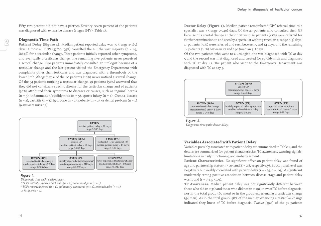
Diagnostic Time Path Patient Delay (Figure 1). Median patient reported delay was 30 (range 1-365) days. Almost all TCPs (57/60, 95%) consulted the GP, the vast majority (n = 49, (86%)) for a testicular change. Three patients initially reported other symptoms, and eventually a testicular change. The remaining five patients never perceived a scrotal change. Two patients immediately consulted an urologist because of a testicular change and the last patient visited the Emergency Department with complaints other than testicular and was diagnosed with a thrombosis of the lower limb. Altogether, 6 of the 60 patients (10%) never noticed a scrotal change. Of the 54 patients noticing a testicular change, 29 patients (54%) answered that they did not consider a specific disease for the testicular change and 16 patients (30%) attributed their symptoms to diseases or causes, such as inguinal hernia (n = 3), inflammation/epididymitis (n = 5), sports injury (n = 1), Crohn’s disease (n = 2), gastritis (n = 1), hydrocele (n = 1), puberty (n = 2), or dental problem (n = 1) (9 answers missing).
57 TCPs (95%) visited GP
median patient delay = 14 daysrange 0-252 days
3 TCPs (5%) visited ER (1) or urologist (2)
median patient delay = 14 daysrange 1-180 days
60 TCPs median patient delay = 30 days
range 1-365 days
3 TCPs (5%) initially reported other symptoms a
median patient delay = 210 daysrange 56-252 days
49 TCPs (86%) reported testicular change
median patient delay = 28 daysrange 1-365 days
5 TCPs (9%) never experienced testicular change b
median patient delay = 90 daysrange 45-240 days
Diagnostic time path: patient delay.a TCPs initially reported back pain (n = 2), abdominal pain (n = 1).b TCPs reported: stress (n = 1), pulmonary symptoms (n = 2), stomach ache (n = 1), or fatigue (n = 1).
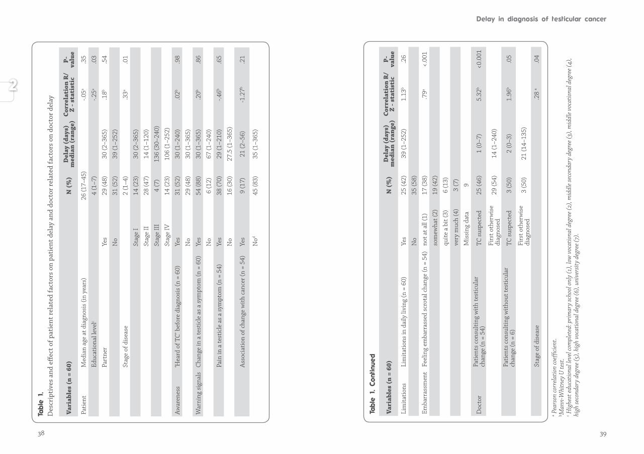
Doctor Delay (Figure 2). Median patient remembered GPs’ referral time to a specialist was 7 (range 0-240) days. Of the 49 patients who consulted their GP because of a scrotal change at their first visit, 20 patients (41%) were referred for further examination to and seen by a specialist within 3 (median 1; range 0-3) days, 15 patients (31%) were referred and seen between 5 and 14 days, and the remaining 14 patients (28%) between 17 and 240 (median 51) days. Of the two patients who went to a urologist, one was diagnosed with TC at day 5 and the second was first diagnosed and treated for epididymitis and diagnosed with TC at day 42. The patient who went to the Emergency Department was diagnosed with TC at day 3.
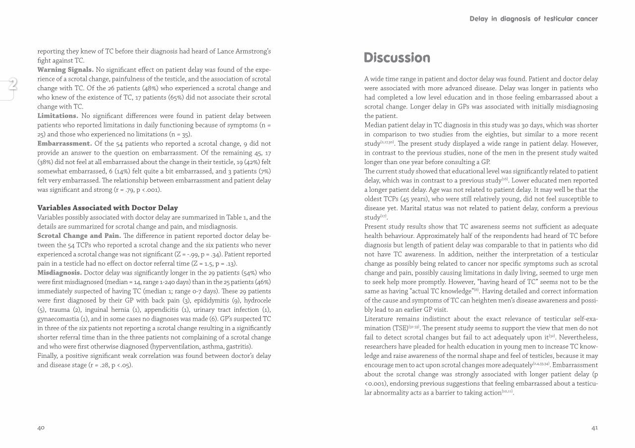
Variables Associated with Patient DelayVariables possibly associated with patient delay are summarized in Table 1, and the details are summarized for patient characteristics, TC awareness, warning signals, limitations in daily functioning and embarrassment.Patient Characteristics. No significant effect on patient delay was found of age and partnership status (r = .05 and Z = .18, respectively). Educational level was negatively but weakly correlated with patient delay (r = -.25, p = .03). A significant moderately strong positive association between disease stage and patient delay was found (r = .33, p <.01).TC Awareness. Median patient delay was not significantly different between those who did (n = 31) and those who did not (n = 29) know of TC before diagnosis, nor in the total group (60 men) or in the group experiencing a testicular change (54 men). As in the total group, 48% of the men experiencing a testicular change indicated they knew of TC before diagnosis. Twelve (39%) of the 31 patients
57 TCPs (95%) visited GP
median referral time = 7 daysrange 0-240 days
3 TCPs (5%) initially reported other symptoms
median referral time = 1 dayrange 1-5 days
49 TCPs (86%) reported testicular change
median referral time = 8 daysrange 0-240 days
5 TCPs (9%) reported other symptoms
median referral time = 1 daysrange 0-21 days
Diagnostic time path: doctor delay.
38 39
D
escr
ipti
ves
and
effec
t of p
atie
nt re
late
d fa
ctor
s on
pat
ient
del
ay a
nd d
octo
r re
late
d fa
ctor
s on
doc
tor
dela
y
Vari
able
s (n
= 6
0)N
(%
)D
elay
(da
ys)
med
ian
(ran
ge)
Corr
elat
ion
R/
Z - s
tati
stic
P-
valu
e
Pati
ent
Med
ian
age
at d
iagn
osis
(in
year
s)26
(17–
45)
-.05a
.35
Educ
atio
nal l
evel
c4
(1–7
)-.2
5a.0
3
Part
ner
Yes
29 (4
8)30
(2–3
65)
.18b
.54
No
31 (5
2)39
(1–2
52)
Stag
e of
dis
ease
2 (1
–4)
.33a
.01
Stag
e I
14 (2
3)30
(2–3
65)
Stag
e II
28 (4
7)14
(1–1
20)
Stag
e II
I4
(7)
136
(30–
240)
Stag
e IV
14 (2
3)10
6 (1
–252
)
Aw
aren
ess
‘Hea
rd o
f TC
’ bef
ore
diag
nosi
s (n
= 6
0)Ye
s31
(52)
30 (1
–240
).0
2b.9
8
No
29 (4
8)30
(1–3
65)
War
ning
sig
nals
Cha
nge
in a
test
icle
as
a sy
mpt
om (n
= 6
0)Ye
s54
(88)
30 (1
–365
).2
0b.8
6
No
6 (1
2)67
(1–2
40)
Pain
in a
test
icle
as
a sy
mpt
om (n
= 5
4)Ye
s38
(70)
29 (1
–210
)-.4
6b.6
5
No
16 (3
0)27
.5 (1
–365
)
Ass
ocia
tion
of c
hang
e w
ith
canc
er (n
= 5
4)Ye
s9
(17)
21 (2
–56)
-1.2
7b.2
1
Nod
45 (8
3)35
(1–3
65)
a Pea
rson
corr
elat
ion
coef
ficie
nt.
b Man
n-W
hitn
ey U
test
.c H
ighe
st e
duca
tiona
l lev
el co
mpl
eted
: pri
mar
y sc
hool
onl
y (1
), lo
w v
ocat
iona
l deg
ree
(2),
mid
dle
seco
ndar
y de
gree
(3),
mid
dle
voca
tiona
l deg
ree
(4),
high
seco
ndar
y de
gree
(5),
high
voc
atio
nal d
egre
e (6
), un
iver
sity
deg
ree
(7).
Vari
able
s (n
= 6
0)N
(%
)D
elay
(da
ys)
med
ian
(ran
ge)
Corr
elat
ion
R/
Z - s
tati
stic
P-
valu
e
Lim
itat
ions
Lim
itat
ions
in d
aily
livi
ng (n
= 6
0)Ye
s25
(42)
39 (1
–252
)1.
13b
.26
No
35 (5
8)
Emba
rras
smen
tFe
elin
g em
barr
asse
d sc
rota
l cha
nge
(n =
54)
not a
t all
(1)
17 (3
8).7
9a<.
001
som
ewha
t (2)
19 (4
2)
quite
a b
it (3
)6
(13)
very
muc
h (4
)3
(7)
Mis
sing
dat
a9
Doc
tor
Pati
ents
con
sulti
ng w
ith
test
icul
ar
chan
ge (n
= 5
4)TC
sus
pect
ed25
(46)
1 (0
–7)
5.32
b<0
.001
Firs
t oth
erw
ise
diag
nose
d29
(54)
14 (1
–240
)
Pati
ents
con
sulti
ng w
itho
ut te
stic
ular
ch
ange
(n =
6)
TC s
uspe
cted
3 (5
0)2
(0–3
)1.
96b
.05
Firs
t oth
erw
ise
diag
nose
d3
(50)
21 (1
4–13
5)
Stag
e of
dis
ease
.28
a.0
4
40 41
reporting they knew of TC before their diagnosis had heard of Lance Armstrong’s fight against TC.Warning Signals. No significant effect on patient delay was found of the expe-rience of a scrotal change, painfulness of the testicle, and the association of scrotal change with TC. Of the 26 patients (48%) who experienced a scrotal change and who knew of the existence of TC, 17 patients (65%) did not associate their scrotal change with TC. Limitations. No significant differences were found in patient delay between patients who reported limitations in daily functioning because of symptoms (n = 25) and those who experienced no limitations (n = 35). Embarrassment. Of the 54 patients who reported a scrotal change, 9 did not provide an answer to the question on embarrassment. Of the remaining 45, 17 (38%) did not feel at all embarrassed about the change in their testicle, 19 (42%) felt somewhat embarrassed, 6 (14%) felt quite a bit embarrassed, and 3 patients (7%) felt very embarrassed. The relationship between embarrassment and patient delay was significant and strong (r = .79, p <.001).
Variables Associated with Doctor Delay Variables possibly associated with doctor delay are summarized in Table 1, and the details are summarized for scrotal change and pain, and misdiagnosis.Scrotal Change and Pain. The difference in patient reported doctor delay be-tween the 54 TCPs who reported a scrotal change and the six patients who never experienced a scrotal change was not significant (Z = -.99, p = .34). Patient reported pain in a testicle had no effect on doctor referral time (Z = 1.5, p = .13).Misdiagnosis. Doctor delay was significantly longer in the 29 patients (54%) who were first misdiagnosed (median = 14, range 1-240 days) than in the 25 patients (46%) immediately suspected of having TC (median 1; range 0-7 days). These 29 patients were first diagnosed by their GP with back pain (3), epididymitis (9), hydrocele (5), trauma (2), inguinal hernia (1), appendicitis (1), urinary tract infection (1), gynaecomastia (1), and in some cases no diagnoses was made (6). GP’s suspected TC in three of the six patients not reporting a scrotal change resulting in a significantly shorter referral time than in the three patients not complaining of a scrotal change and who were first otherwise diagnosed (hyperventilation, asthma, gastritis).Finally, a positive significant weak correlation was found between doctor’s delay and disease stage (r = .28, p <.05).
A wide time range in patient and doctor delay was found. Patient and doctor delay were associated with more advanced disease. Delay was longer in patients who had completed a low level education and in those feeling embarrassed about a scrotal change. Longer delay in GPs was associated with initially misdiagnosing the patient.Median patient delay in TC diagnosis in this study was 30 days, which was shorter in comparison to two studies from the eighties, but similar to a more recent study{2,17,30}. The present study displayed a wide range in patient delay. However, in contrast to the previous studies, none of the men in the present study waited longer than one year before consulting a GP. The current study showed that educational level was significantly related to patient delay, which was in contrast to a previous study{16}. Lower educated men reported a longer patient delay. Age was not related to patient delay. It may well be that the oldest TCPs (45 years), who were still relatively young, did not feel susceptible to disease yet. Marital status was not related to patient delay, conform a previous study{17}.Present study results show that TC awareness seems not sufficient as adequate health behaviour. Approximately half of the respondents had heard of TC before diagnosis but length of patient delay was comparable to that in patients who did not have TC awareness. In addition, neither the interpretation of a testicular change as possibly being related to cancer nor specific symptoms such as scrotal change and pain, possibly causing limitations in daily living, seemed to urge men to seek help more promptly. However, “having heard of TC” seems not to be the same as having “actual TC knowledge”{8}. Having detailed and correct information of the cause and symptoms of TC can heighten men’s disease awareness and possi-bly lead to an earlier GP visit.Literature remains indistinct about the exact relevance of testicular self-exa-mination (TSE){31-33}. The present study seems to support the view that men do not fail to detect scrotal changes but fail to act adequately upon it{32}. Nevertheless, re sear chers have pleaded for health education in young men to increase TC know-ledge and raise awareness of the normal shape and feel of testicles, because it may encou rage men to act upon scrotal changes more adequately{2,4,33,34}. Embarrassment about the scrotal change was strongly associated with longer patient delay (p <0.001), endorsing previous suggestions that feeling embarrassed about a testicu-lar abnormality acts as a barrier to taking action{10,11}.
42 43
Median GP referral time in patients with testicular complaints in the current study was 7 days, which was shorter than in two other studies reporting 10 and 14 days respectively, and conform a third study{2,28,36}. In the present study, two-fifth of patients with a testicular change were referred within 3 days, conform the Dutch TC guideline, and almost three-quarter were referred within two weeks, conform the current UK guideline{19,20}. Although Dutch GP’s refer a large percentage of young men reporting a scrotal change adequately, misdiagnosis seems to be a risk factor for longer doctor delay in the diagnostic process. TC is a rare disease, but GPs should always bear TC in mind, in particular when adolescents and young adult men present with inguinal or scrotal complaints, or lower back pain.Findings of the present study accentuate the difficulty of the TC diagnostic process, and underlines the responsibility of GPs in this process. A recent English study showed that the positive predictive value for testicular cancer following a GP’s referral for a scrotal abnormality conform the two weeks rule is only 17%{37}. In the Netherlands, GPs refer patients with a scrotal mass to a radiologist for an ultrasonography. If the ultrasound is abnormal and TC is suspected, a patient is immediately referred to a surgical oncologist or urologist to confirm the diagnosis and if needed, for treatment. The present study showed that both longer patient delay and doctor delay were significantly associated with more advanced disease, which is in concordance with other studies{4,24}. Advanced disease requires more intensive cancer treatment and is associated with increased treatment related morbidity, decreased disease free survival and increased costs.A few limitations of the present study should be mentioned. First, methodological concerns regarding the concept of delay exist and a standardized definition is lacking. Definitions used in this study have proven operational earlier{25}. Second, patient-centered studies measuring delay are susceptible to recall bias. In particular patients reporting longer time intervals could have had difficulty remembering the exact time span. Third, to our knowledge, a validated questionnaire on this subject is not available, and thus a questionnaire incorporating information regarding TC and health behavior was developed for the present study. Fourth, the number of respondents in some subgroups is small which may affect the statistical power.
High variation in patient and doctor delay was found. Patient and doctor delay were associated with more advanced disease requiring a more intensive cancer treatment. Health care providers who aim to develop education programs to
increase TC awareness in young men should take into account that men who feel embarrassed about scrotal changes and lower educated men may benefit most from their programs. To prevent misdiagnoses, education programs for GPs should focus on increasing GPs knowledge of TC and on their awareness that TC may be the underlying illness in adolescent and young adult men who present with a scrotal change or with symptoms possibly considered vague. Further policy recommendations are continuous medical education of GPs to increase their understanding of the value of ultrasonography for scrotal abnormalities in combination with the biomarkers LDH, AFP and B-HCG in the differential diagnosis. Performing these diagnostics in time will increase the number of correct referrals for TC, and increase the chance that the ‘two-week wait rule’ will be met. Both actions, education of adolescent and young adult men and of GP’s to increase knowledge and awareness of testicular cancer, and continuous medical education of GPs with respect to scrotal pathology may decrease patient’ and doctor’ delay, thus lower the percentage of TC patients diagnosed with advanced disease, decrease costs associated with treatment of advanced disease, and improve disease free and overall survival.
1. Sonneveld DJ, Hoekstra HJ, van der Graaf WT, Sluiter WJ, Mulder NH, Willemse PH et al. Improved longterm survival of patients with metastatic nonseminomatous testicular germ cell carcinoma in relation to prognostic classification systems during the cisplatin era. Cancer 2001; 91: 1304-1315.
2. Bosl GJ, Vogelzang NJ, Goldman A, Fraley EE, Lange PH, Levitt SH et al. Impact of delay in diagnosis on clinical stage of testicular cancer. Lancet 1981; 2: 970-973.
3. Kobayashi KI, Saito T, Kitamura Y, Nobushita T, Kawasaki T, Hara N et al. Effect of the time from the presentation of symptoms to medical consultation on primary tumor size and survival in patients with testicular cancer: Shift in the last 2 decades. Urol Oncol 2014; 32: 17-22.
4. Chilvers CE, Saunders M, Bliss JM, Nicholls J, Horwich A. Influence of delay in diagnosis on prognosis in testicular teratoma. Br J Cancer 1989; 59: 126-128.
5. Aberger M, Wilson B, Holzbeiern JM, Griebling TL, Nangia AK. Testicular self-examination and testicular cancer: a cost-utility analysis. Cancer Med 2014; 3: 1629-1634.
6. Macleod U, Mitchell ED, Burgess C, Griebling TL, Nangia AK. Risk factors for delayed presentation and referral of symptomatic cancer: evidence for common cancers. Br J Cancer 2009; 101: 92-101.
7. Nooijer de J, Lechner L, Vries de H. Help-seeking behaviour for cancer symptoms: perceptions of patients and general practitioners. Psychooncol 2001; 10: 469-478.
44 45
8. Moore RA, Topping A. Young men's knowledge of testicular cancer and testicular self-examination: a lost opportunity? Eur J Cancer Care 1999; 8: 137-142.
9. Carpentier MY, Fortenberry JD. Romantic and sexual relationships, body image, and fertility in adolescent and young adult testicular cancer survivors: a review of the literature. J Adolesc Health 2010; 47: 115-125.
10. Gascoigne P, Mason MD, Roberts E. Factors affecting presentation and delay in patients with testicular cancer: results of a qualitative study. Psychooncol 1999; 8: 144-154.
11. Hubbard G, Macmillan I, Canny A, Forbat L, Neal RD, O'Carroll RE et al. Cancer symptom awareness and barriers to medical help seeking in Scottish adolescents: a cross-sectional study. BMC Public Health 2014; 14: 1117-1129.
12. Janz NK. The Health Belief Model: a decade later. Health Educ Q. 1984; 11: 1-47.
13. Carpenter CJ. A meta-analysis of the effectiveness of health belief model variables in predicting behavior. Health Commun 2010; 25: 661-669.
14. Ramirez AJ, Westcombe AM, Burgess CC, Sutton S, Littlejohns P, Richards MA et al. Factors predicting delayed presentation of symptomatic breast cancer: a systematic review. Lancet 1999; 353: 1127-1131.
15. Mitchell E, Macdonald S, Campbell NC, Weller D, Macleod U. Influences on pre-hospital delay in the diagnosis of colorectal cancer: a systematic review. Br J Cancer 2008; 98: 60-70.
16. Toklu C, Ozen H, Sahin A, Rastadoskouee M, Erdem E. Factors involved in diagnostic delay of testicular cancer. Int Urol Nephrol 1999; 31: 383-388.
17. Thornhill JA. Patients' delay in the presentation of testis cancer in Ireland. Br J Urol 1987; 59: 447-451.
18. Walter F, Webster A, Scott S, Emery J. The Andersen Model of Total Patient Delay: a systematic review of its application in cancer diagnosis. J Health Serv Res Policy 2012; 17: 110-118.
19. LWG urologische tumoren. 20Testiscarcinoom. Landelijke richtlijn. http://www.oncoline.nl/testiscarcinoom retrieved 07-09-2015.
20. Hawary AM, Warburton HE, Brough RJ, Collins GN, Brown SC, O'Reilly PH et al. The ‘2-Week Wait’ Rule for Referrals for Suspected Urological Cancers – Urgent Need for Refinement of Criteria. Ann R Coll Surg Engl 2008; 90: 517-522.
21. Winter TC. There is a mass in the scrotum-what does it mean?: Evaluation of the scrotal mass. Ultrasound Q 2009; 25: 195-205.
22. Oliver RT. Factors contributing to delay in diagnosis of testicular tumours. BMJ 1985; 290: 356.
23. Cantwell BM. Gynaecomastia and extragonadal symptoms leading to diagnosis delay of germ cell tumours in young men. Postgrad Med J 1991; 67: 675-677.
24. Fossa SD, Klepp O, Elgjo RF, Eliassen G, Melsom H, Umes T et al. The Effect of Patients Delay and Doctors Delay in Patients with Malignant Germ-Cell Tumors. Int J Androl 1981; 4: 134-144.
25. Hansen RP. Delay in the diagnosis of cancer. Thesis, Faculty of Health Sciences, University of Aarhus, 2008.
26. International Germ Cell Consensus Classification: a prognostic factor-based staging system for metastatic germ cell cancers. International Germ Cell Cancer Collaborative Group. J Clin Oncol 1997; 15: 594-603.
27. Testis. In: Edge SB, Byrd DR, Compton CC et al., eds.: AJCC Cancer Staging Manual. 7th ed. New York, NY: Springer, 2010, 469-478.
28. Andersen BL, Cacioppo JT, Roberts DC. Delay in Seeking A Cancer-Diagnosis - Delay Stages and Psychophysiological Comparison Processes. Br J Soc Psychol 1995; 34: 33-52.
29. Cohen J. Statistical power analysis for the behavioral sciences. NY: Academy Press: New York, 1988.
30. Connolly SS, Daly PJ, St J Floyd M. Terminology and details of the diagnostic process for testis cancer. J Urol 2011; 185: 876-880.
31. Rosella J. Testicular cancer health education: and integrative review. J Adv Nur 1994; 20: 666-671.
32. Hopcroft K. What benefit testicular self-examination? Br Gen Pract 2004; 54: 214-215.
33. Hopcroft K. Routine testicular self examination: it's time to stop. BMJ 2012; 28; 344.
34. Lechner L, Oenema A, de NJ. Testicular self-examination (TSE) among Dutch young men aged 15-19: determinants of the intention to practice TSE. Health Educ Res 2002; 17: 73-84.
35. Brenner JS, Hergenroeder AC, Kozinetz CA, Kelder SH. Teaching testicular self-examination: Education and practices in pediatric residents. Pediatrics 2003; 111: 239-244.
36. Vasudev NS, Joffe JK, Cooke C, Richards F, Jones WG. Delay in the diagnosis of testicular tumours - changes over the past 18 years. Br Gen Pract 2004; 54: 595-597.
37. Casey RG, Grainger R, Butler M, McDermott TE, Thornhill JA. Scrotal signs and symptoms in the general population, the value of testis self-examination and the pitfalls of a scrotal screening programme: is the two-week rule relevant? World J Urol 2011; 29: 387-391.

49
BackgroundThe aim of this study was to assess the feasibility and reproducibility of semi-automatic volumetric measurement of retroperitoneal lymph node metastases in testicular cancer (TC) patients treated with chemotherapy versus the standardized manual measurements based on RECIST criteria.
Methods 21 TC patients with retroperitoneal lymph node metastases of testicular cancer were studied with a CT scan of chest and abdomen before and after cisplatin based combination chemotherapy. Three readers, a surgical resident, a radiological technician and a radiologist, assessed tumor response independently using computerized volumetric analysis with Vitrea software® and manual measurement according to RECIST criteria (version 1.1). Intra- and inter-rater variability were evaluated with intra class correlations and Bland-Altman analysis.
ResultsAssessment of intra-observer and inter-observer variance proved non-significant in both measurement modalities. In particularly all intraclass correlation (ICC) values for the volumetric analysis were >.99 per observer and between observers. There was minimal bias in agreement for manual as well as volumetric analysis.
ConclusionIn this study volumetric measurement using Vitrea software® appears to be a reliable, reproducible method to measure initial tumor volume of retroperitoneal lymph nodes metastases of testicular cancer after chemotherapy. Both measurement methods can be performed by experienced non-radiologists as well.
Çiğdem Öztürk, Ton Velleman, Alphons H.H. Bongaerts, Lukas M. Bergman, Robert J. van Ginkel, Jourik A. Gietema, Harald J. Hoekstra PLoS One. 2017 Jan 12;12(1): e0168977
Diagnostic process for testicular cancer (TC) includes physical examina tion, laboratory tests for tumor markers alpha-fetoprotein (AFP), beta chorion go nado-tropin (B-HCG), lactate dehydrogenase (LDH), and imaging tests, such as testicular ultrasound and computed tomography (CT) of abdomen and chest to assess regional and distant metastases. CT scans and tumor marker analysis make it possible to stage the extensiveness of disease and to classify the patient according to the International Germ Cell Consensus Classification (IGCCC){1,2}. Staging results are fundamental in determining the prognosis and the optimal treatment strategy for each individual patient. Stage I nonseminomatous testicular germ cell tumor (NSTGCT) can be treated successfully with a wait-and-see policy, or unilateral nerve-sparing retroperitoneal lymph node dissection (RPLND), or one course of adjuvant cisplatin based combination chemotherapy after orchiectomy{2}. Disseminated disease is treated also with three or four courses of cisplatin based combination chemotherapy. Resection of residual retroperitoneal tumor masses (RRRTM) after chemotherapy is an essential part of the combined therapy of NSTGCT{3,4}. CT of chest and abdomen serve as the main diagnostic tool to deter-mine pre- and post-chemotherapy tumor deposit size, allowing evaluation of the response to treatment and guiding the decision to perform RRRTM, either conventional or via laparoscopic surgery{5}. An alternative approach to surgery can be observing patients after systemic chemotherapy, certainly if abdominal residual tumor masses have become undetectable (lesions <1 cm) on CT scans and in some institution if the primary tumor did not contain teratoma components{6,7}. Residual metastatic disease after completion of chemotherapy can contain tumor necrosis, teratoma or viable germ cell cancer. Optimal disease management is based on reliable and reproducible lesion measu-rement. Tumor response assessment has been standardized since the introduction of the bi-dimensional response criteria of the World Health Organization (WHO) which was more recently followed by one-dimensional (2D) response criteria based on RECIST (version 1.1), which is a set of rules defining overall tumor burden at base line and objective tumor response and disease progression after systemic treat ment{8,9}. In short, 2D measurements are obtained to assess the size of a tumor lesion. Measurements are obtained in the axial plane on a single slice, after careful selec-tion from all axial planes in which the tumor lesion is visible. Assuming such single slice manual measurements in the axial plane comprise complete change in tumor size, neglects the fact that a tumor is an irregular three-dimensional lesion which
50 51
is not a perfect sphere and therefore can be a misinterpretation of tumor burden. Additionally, in literature high inter-observer and intra-observer variability in manual tumor assessment are reported because of the difficulty to define the outer borders of the lesion to be measured and the variation in perception of the largest diameter{10,11}.An approach to improve reproducibility of lesion size measurements, and to reduce time and effort of measurements, combined with rapid technological developments, have led to the development of software allowing semi-automated lesion segmentation of tumor masses{12,13}. Semi-automatic segmentation tools are utilized to acquire 3 dimensional (3D) images and accurate determination of tumor size alterations. Opposed to manual 2D measurements, in this 3D setting, asymmetrical tumor size changes can be detected and measured. Lymph node metastases are irregularly shaped and often closely located to adjacent tissue with similar contrast density. These characteristics, in combination with anatomical location and slice thickness, influence segmentation quality and increase measurement error, especially when measuring smaller shaped lesions{14,15}. The spread of TC to lymph nodes usually follows a predictable pattern with metastases occurring in retroperitoneal lymph nodes. Lymph node metastases in this region can be quite difficult to measure even with adequate contrast administrations and regular manual measurements, because of surrounding tissues with similar contrast densities causing indistinct borders of the tumor deposits{16}. Volumetric analysis is a new promising technique to measure a therapeutic response especially for lung and liver lesions, which can also be used to measure response of retroperitoneal lymph nodes of testicular cancer after chemotherapy{17}.The purpose of this pilot study was to assess the suitability and reproducibility of semi-automated volumetric analysis when applied to retroperitoneal lymph nodes in patients with disseminated TC, compared to 2D measurements based on RECIST between non-radiologists and a genitourinary radiologist.
We consulted the institutional review board of the UMCG, and they confirmed that no formal written waiver for the need of ethics approval was required because of the retrospective design of the study. Also there was no written consent needed from the patients. One of the supervising authors has had contact with some of the study participants as the surgeon who performed the resection of the lymph node metastases and therefore was a treating physician during a short period. This author did not perform the manual and volumetric measurements. Authors
did not collect any personally-identifying information from study participants. Furthermore, all data were detached from patient information prior to analysis.
Study Population CT scans of chest and abdomen of 21 consecutive nonseminomatous testicular cancer patients (NSTGCT), treated at the University Medical Center Groningen (UMCG), with a mean age of 36 (range 27-68) years undergoing resection of residual retroperitoneal tumor mass (RRRTM) after cisplatin based combination chemotherapy were evaluated. Of the 21 patients, 16 patients (76%) had stage II disease, 2 patients (10%) had stage III disease and 3 patients (14%) had stage IV disease. According to the International Germ Cell Consensus Classification (IGCCC) the majority of patients, 15 (71%) had disease with good prognosis, 4 pa-tients (19%) intermediate prognosis and 2 (10%) poor prognosis. Lesions outside the retroperitoneal area were not considered for this analysis. All patients had pre-and post-chemotherapy CT imaging performed at the UMCG or the referring hospital.Finally 21 patients were eligible with 28 individual lesions identified in the retro-peritoneum.
Data Collection All post-chemotherapy CT scans were performed at the UMCG with a multi-slice CT scanner (Sensation S64, Siemens Healthcare, Forchheim, Germany) according to the TC protocol, which encompasses fasting 4-6 hours prior to the scan after which scanning of abdomen and thorax is performed with oral and intravenous contrast (100cc/flow 2.5). The reconstructed slice thickness for the standard protocol was 2.0 mm{18,19}.The majority of the pre-chemotherapy CT scans were performed in the referring hospitals. Depending on CT scanner quality and primary indication to perform a CT scan, reconstructed slice thickness was 2 mm for all scans.All data were transferred to a dedicated advanced visualization workstation and were analyzed with a software package of Toshiba systems (Vitrea®/Vital Images, version 4){20}.Vitrea® software is Vital Images' visualization software and commercially available for implementation. With this software 2D, 3D and 4D images are created of human anatomy from CT, MRI (magnetic resonance imaging) and PET (positron emission tomography) image data. The software provides options for cardiac, colon, vessel probe and other applications.
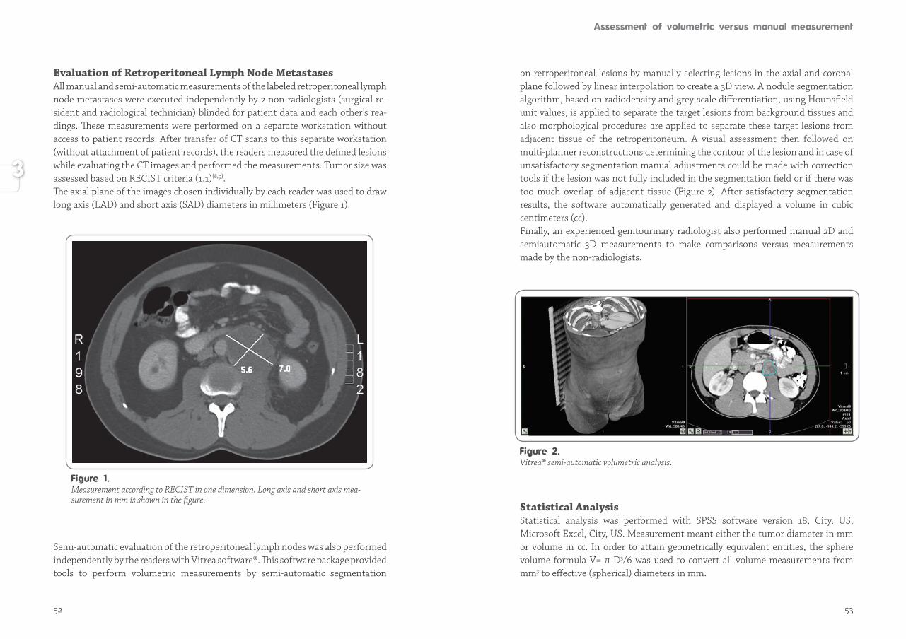
52 53
Evaluation of Retroperitoneal Lymph Node MetastasesAll manual and semi-automatic measurements of the labeled retroperitoneal lymph node metastases were executed independently by 2 non-radiologists (surgical re-si dent and radiological technician) blinded for patient data and each other’s rea-dings. These measurements were performed on a separate workstation without access to patient records. After transfer of CT scans to this separate workstation (with out attachment of patient records), the readers measured the defined lesions while evaluating the CT images and performed the measurements. Tumor size was asses sed based on RECIST criteria (1.1){8,9}. The axial plane of the images chosen individually by each reader was used to draw long axis (LAD) and short axis (SAD) diameters in millimeters (Figure 1).
Semi-automatic evaluation of the retroperitoneal lymph nodes was also performedindependently by the readers with Vitrea software®. This software package provided tools to perform volumetric measurements by semi-automatic segmentation
on retroperitoneal lesions by manually selecting lesions in the axial and coronal plane followed by linear interpolation to create a 3D view. A nodule segmentation algorithm, based on radiodensity and grey scale differentiation, using Hounsfield unit values, is applied to separate the target lesions from background tissues and also morphological procedures are applied to separate these target lesions from ad ja cent tissue of the retroperitoneum. A visual assessment then followed on multi-planner reconstructions determining the contour of the lesion and in case of un satis factory segmentation manual adjustments could be made with correction tools if the lesion was not fully included in the segmentation field or if there was too much overlap of adjacent tissue (Figure 2). After satisfactory segmentation results, the software automatically generated and displayed a volume in cubic centi meters (cc).Finally, an experienced genitourinary radiologist also performed manual 2D and semiautomatic 3D measurements to make comparisons versus measurements made by the non-radiologists.
Statistical Analysis Statistical analysis was performed with SPSS software version 18, City, US, Microsoft Excel, City, US. Measurement meant either the tumor diameter in mm or volume in cc. In order to attain geometrically equivalent entities, the sphere volume formula V= π D3/6 was used to convert all volume measurements from mm3 to effective (spherical) diameters in mm.
Measurement according to RECIST in one dimension. Long axis and short axis mea-surement in mm is shown in the figure.
Vitrea® semi-automatic volumetric analysis.
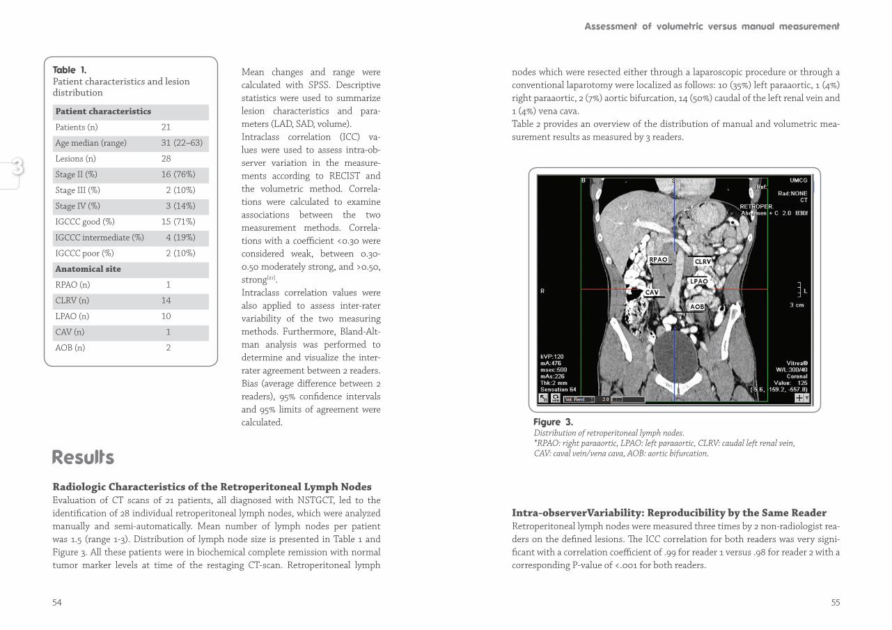
54 55
Mean changes and range were calculated with SPSS. Des crip tive statistics were used to summarize lesion cha rac te ris tics and para-meters (LAD, SAD, volume). Intraclass correlation (ICC) va-lues were used to assess intra-ob-ser ver variation in the mea su re -ments according to RECIST and the volumetric method. Cor re la-tions were calcu lated to examine asso ciations between the two measu re ment methods. Cor re la-tions with a coefficient <0.30 were considered weak, be tween 0.30-0.50 moderately strong, and >0.50, strong{21}.Intraclass correlation values were also applied to assess inter-rater variability of the two measuring methods. Fur ther more, Bland-Alt-man analysis was performed to de ter mine and visualize the inter-ra ter agree ment between 2 readers. Bias (average difference between 2 readers), 95% confidence inter vals and 95% limits of agree ment were calculated.
Radiologic Characteristics of the Retroperitoneal Lymph Nodes Evaluation of CT scans of 21 patients, all diagnosed with NSTGCT, led to the identification of 28 individual retroperitoneal lymph nodes, which were analyzed manually and semi-automatically. Mean number of lymph nodes per patient was 1.5 (range 1-3). Distribution of lymph node size is presented in Table 1 and Figure 3. All these patients were in biochemical complete remission with normal tumor marker levels at time of the restaging CT-scan. Retroperitoneal lymph
nodes which were resected either through a laparoscopic procedure or through a conventional laparotomy were localized as follows: 10 (35%) left paraaortic, 1 (4%) right paraaortic, 2 (7%) aortic bifurcation, 14 (50%) caudal of the left renal vein and 1 (4%) vena cava. Table 2 provides an overview of the distribution of manual and volumetric mea-surement results as measured by 3 readers.
Intra-observerVariability: Reproducibility by the Same ReaderRetroperitoneal lymph nodes were measured three times by 2 non-radiologist rea-ders on the defined lesions. The ICC correlation for both readers was very signi-ficant with a correlation coefficient of .99 for reader 1 versus .98 for reader 2 with a corresponding P-value of <.001 for both readers.
Distribution of retroperitoneal lymph nodes.*RPAO: right paraaortic, LPAO: left paraaortic, CLRV: caudal left renal vein, CAV: caval vein/vena cava, AOB: aortic bifurcation.
Patient characteristics and lesion distribution
Patient characteristics
Patients (n) 21
Age median (range) 31 (22–63)
Lesions (n) 28
Stage II (%) 16 (76%)
Stage III (%) 2 (10%)
Stage IV (%) 3 (14%)
IGCCC good (%) 15 (71%)
IGCCC intermediate (%) 4 (19%)
IGCCC poor (%) 2 (10%)
Anatomical site
RPAO (n) 1
CLRV (n) 14
LPAO (n) 10
CAV (n) 1
AOB (n) 2
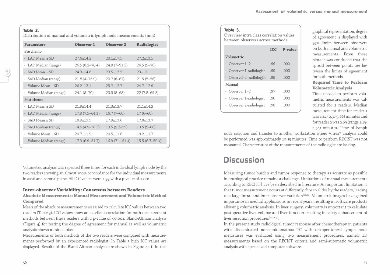
56 57
Volumetric analysis was repeated three times for each individual lymph node by the two readers showing an almost 100% concordance for the individual measurements in axial and coronal plane. All ICC values were >.99 with a p-value of <.001.
Inter-observer Variability: Consensus between Readers Absolute Measurements: Manual Measurement and Volumetric Method Compared Mean of the absolute measurements was used to calculate ICC values between two readers (Table 3). ICC values show an excellent correlation for both measurement methods between these readers with a p-value of <0.001. Bland-Altman analysis (Figure 4) for testing the degree of agreement for manual as well as volumetric analysis shows minimal bias. Measurements of both methods of the two readers were compared with mea sure-ments performed by an experienced radiologist. In Table 3 high ICC values are displa yed. Results of the Bland Altman analysis are shown in Figure 4a-f. In this
graphical representation, degree of agreement is displayed with 95% limits between observers on both manual and volumetric measure ments. From these plots it was concluded that the spread between points are be-tween the limits of agree ment for both methods. Required Time to Perform Volumetric AnalysisTime needed to perform volu-metric measurements was cal-cu lated for 2 readers. Me dian measurement time for reader 1 was 1.42 (0.37-3.66) minutes and for reader 2 was 2.69 (range 1.19-4.54) minutes. Time of lymph
node selection and transfer to another workstation where Vitrea® analysis could be performed was approximately 10-15 minutes. Time to perform RECIST was not measured. Characteristics of the measurements of the radiologist are lacking.
Measuring tumor burden and tumor response to therapy as accurate as possible in oncological practice remains a challenge. Limitations of manual measurements according to RECIST have been described in literature. An important limitation is that tumor measurement occurs at differently chosen slides by the readers, leading to a large intra- and inter-observer variation{10,11}. Volumetric images have gained importance in medical applications in recent years, resulting in software products allowing volumetric analysis. In liver surgery, volumetry is important to calculate postoperative liver volume and liver function resulting in safety enhancement of liver resection procedures{12,22,23}. In the present study radiological tumor response after chemotherapy in patients with disseminated nonseminomatous TC with retroperitoneal lymph node metastases was evaluated using two measurement procedures, namely 2D measurements based on the RECIST criteria and semi-automatic volumetric analysis with specialized computer software.
Distribution of manual and volumetric lymph node measurements (mm)
Parameters Observer 1 Observer 2 Radiologist
Pre chemo
• LAD Mean ± SD 27.6±14.2 28.1±17.5 27.2±13.5
• LAD Median (range) 26.5 (8.2–76.4) 24.8 (7–91.3) 26.5 (5–70)
• SAD Mean ± SD 24.3±14.8 23.5±13.5 23±12
• SAD Median (range) 21.8 (4–75.9) 20.7 (6–67) 21.5 (5–56)
• Volume Mean ± SD 26.2±13.1 25.7±12.7 24.7±12.9
• Volume Median (range) 24.1 (8–70) 23.5 (8–68) 22 (7.8–69.8)
Post chemo
• LAD Mean ± SD 21.9±14.4 21.3±13.7 21.1±14.3
• LAD Median (range) 17.9 (7.5–64.1) 16.7 (7–60) 17 (6–60)
• SAD Mean ± SD 18.9±13.5 17.9±13.6 17.8±13.7
• SAD Median (range) 14.6 (4.5–56.3) 13.5 (5.3–59) 13.5 (5–60)
• Volume Mean ± SD 20.7±11.9 20.5±11.6 19.2±11.7
• Volume Median (range) 17.3 (6.9–51.7) 16.9 (7.1–51.4) 15.5 (6.7–50.4)
Overview intra class correlation values between observers across methods
ICC P-value
Volumetric
• Observer 1–2 .99 .000
• Observer 1-radiologist .99 .000
• Observer 2- radiologist .99 .000
Manual
• Observer 1–2 .97 .000
• Observer 1-radiologist .96 .000
• Observer 2-radiologist .98 .000
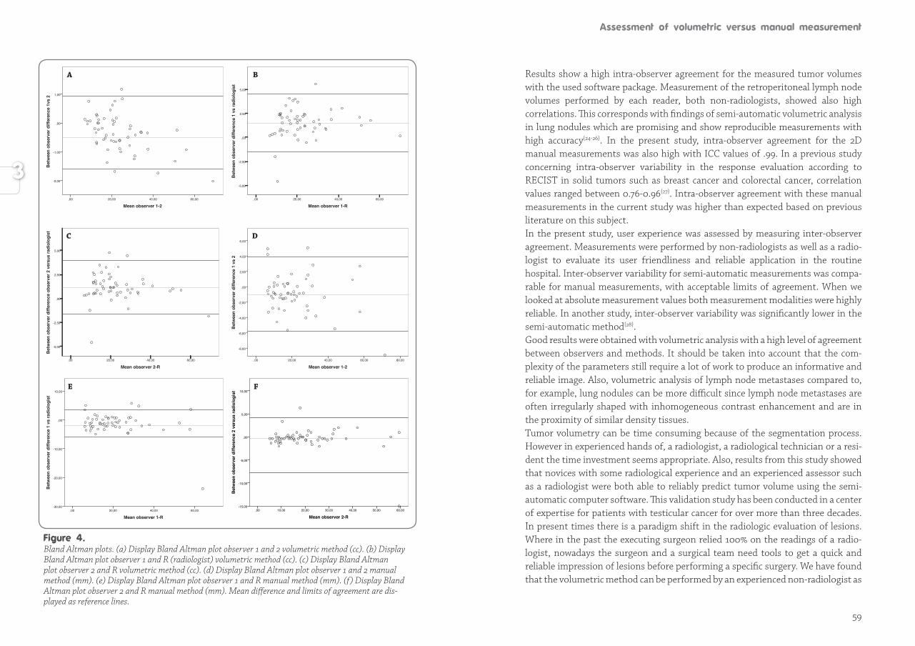
59
Results show a high intra-observer agreement for the measured tumor volumes with the used software package. Measurement of the retroperitoneal lymph node volumes performed by each reader, both non-radiologists, showed also high correlations. This corresponds with findings of semi-automatic volumetric analysis in lung nodules which are promising and show reproducible measurements with high accuracy{24-26}. In the present study, intra-observer agreement for the 2D manual measurements was also high with ICC values of .99. In a previous study concerning intra-observer variability in the response evaluation according to RECIST in solid tumors such as breast cancer and colorectal cancer, correlation values ranged between 0.76-0.96{27}. Intra-observer agreement with these manual measurements in the current study was higher than expected based on previous literature on this subject. In the present study, user experience was assessed by measuring inter-observer agreement. Measurements were performed by non-radiologists as well as a radio-logist to evaluate its user friendliness and reliable application in the routine hospital. Inter-observer variability for semi-automatic measurements was compa-rable for manual measurements, with acceptable limits of agreement. When we looked at absolute measurement values both measurement modalities were highly reliable. In another study, inter-observer variability was significantly lower in the semi-automatic method{28}. Good results were obtained with volumetric analysis with a high level of agreement between observers and methods. It should be taken into account that the com-plexity of the parameters still require a lot of work to produce an informative and reliable image. Also, volumetric analysis of lymph node metastases compared to, for example, lung nodules can be more difficult since lymph node metastases are often irregularly shaped with inhomogeneous contrast enhancement and are in the proximity of similar density tissues. Tumor volumetry can be time consuming because of the segmentation process. However in experienced hands of, a radiologist, a radiological technician or a resi-dent the time investment seems appropriate. Also, results from this study showed that novices with some radiological experience and an experienced assessor such as a radiologist were both able to reliably predict tumor volume using the semi-automatic computer software. This validation study has been conducted in a cen ter of expertise for patients with testicular cancer for over more than three deca des. In present times there is a paradigm shift in the radiologic evaluation of lesions. Where in the past the executing surgeon relied 100% on the readings of a radio-logist, nowadays the surgeon and a surgical team need tools to get a quick and reliable impression of lesions before performing a specific surgery. We have found that the volumetric method can be performed by an experienced non-radiologist as
Mean observer 1-260,0040,0020,00,00
Bet
wee
n ob
serv
er d
iffer
ence
1vs
2
1,00
,00
-1,00
-2,00
Bet
wee
n ob
serv
er d
iffer
ence
1 v
s ra
diol
ogis
t
5,00
2,50
,00
-2,50
-5,00
Mean observer 1-R60,0040,0020,00,00
A B
Mean observer 2-R60,0040,0020,00,00
Bet
wee
n ob
serv
er d
iffer
ence
obs
erve
r 2 v
ersu
s ra
diol
ogis
t
5,00
2,50
,00
-2,50
-5,00
Mean observer 1-280,0060,0040,0020,00,00
Bet
wee
n ob
serv
er d
iffer
ence
1 v
s 2
6,00
4,00
2,00
,00
-2,00
-4,00
-6,00
-8,00
Mean observer 1-R60,0040,0020,00,00
Bet
wee
n ob
serv
er d
iffer
ence
1 v
s ra
diol
ogis
t
10,00
,00
-10,00
-20,00
-30,00
Mean observer 2-R60,0050,0040,0030,0020,0010,00,00
Bet
wee
n ob
serv
er d
iffer
ence
2 v
ersu
s ra
diol
ogis
t 10,00
5,00
,00
-5,00
-10,00
-15,00
C D
E F
Bland Altman plots. (a) Display Bland Altman plot observer 1 and 2 volumetric method (cc). (b) Display Bland Altman plot observer 1 and R (radiologist) volumetric method (cc). (c) Display Bland Altman plot observer 2 and R volumetric method (cc). (d) Display Bland Altman plot observer 1 and 2 manual method (mm). (e) Display Bland Altman plot observer 1 and R manual method (mm). (f) Display Bland Altman plot observer 2 and R manual method (mm). Mean difference and limits of agreement are dis-played as reference lines.
60 61
well and that the required time to obtain and measure a reliable 3D image is under 3 minutes. Such a study for volumetric measurements is lacking in literature for retroperitoneal lymph node metastases in testicular cancer. The new technology facilitates the surgical-oncologist or the uro-oncologist in the conventional or laparoscopic surgical treatment planning of the resection of tumor masses.In conclusion, the pilot study showed that radiological technicians and (surgical/urology) residents could perform volumetric and manual measurements in a reliable manner with a high intra and inter-observer reliability. With the increase in the number of cancer patients receiving chemotherapy, automatic and semiautomatic segmentation tools will likely play an important role in the evaluation of tumor response to treatment, e.g. a cost-effective way to measure tumor response and prepare the surgical or uro-oncologist in the resection of retroperitoneal lymph node metastases. For now, clinical guidelines based on manual measurement in 2D in the axial plane according to RECIST criteria remain the standard method for assessing a radiological tumor response to anti-cancer therapy. This study has shown that the volumetric method is reproducible, user friendly and is promising as a more accurate method in the surgical field since it provides a 3D view. Especially the latter can make a clinical difference with more detailed surgical planning and can contribute to achieving better outcomes in patient care. Since tumor response criteria for volumetric measurements are not yet validated, combining the clinical outcome in a larger prospective study group could provide standardized volumetric measurements.
AcknowledgementsA.H.H. Bongaerts MD, genitourinary radiologist, deceased recently and the authors appreciate the unselfish assistance in finalizing this manuscript by his colleagues L.M. Bergman, radiologist UMCG and P.M.A. van Ooijen, MSc, PhD, CPHIT, Associate Professor of Medical Imaging Informatics UMCG.
1. International Germ Cell Consensus Classification: a prognostic factor-based staging system for metastatic germ cell cancers. International Germ Cell Cancer Collaborative Group. J Clin Oncol 1997; 15: 594-603.
2. Schmoll HJ, Jordan K, Huddart R, Pes MP, Horwich A, Fizazi K et al. Testicular non-seminoma: ESMO clinical practice guidelines for diagnosis, treatment and follow-up. Ann Oncol 2010; 5: 147-154.
3. Oldenburg J, Alfsen GC, Lien HH, Aass N, Waehre H, Fossa SD. Postchemotherapy retroperitoneal surgery remains necessary in patients with nonseminomatous testicular cancer and minimal residual tumor masses. J Clin Oncol 2003; 21: 3310-3317.
4. Kesler KA, Kruter LE, Perkins SM, Rieger KM, Sullivan KJ, Runyan ML et al. Survival after resection for metastatic testicular nonseminomatous germ cell cancer to the lung or mediastinum. Ann Thorac Surg 2011; 91: 1085-1093.
5. Oztürk C, van Ginkel RJ, Krol RM, Gietema JA, Hofker HS, Hoekstra HJ. Laparoscopic resection of a residual retroperitoneal tumor mass of nonseminomatous testicular germ cell tumors. Surg Endosc 2012; 26: 458-467.
6. Heidenreich A, Knüchel-Clarke R, Pfister D. Importance of pathology for Therapy planning of testicular germ cell tumors. Pathologe 2014; 35: 266-273.
7. Debono DJ, Heilman DK, Einhorn LH, Donahue JP. Decision analysis for avoiding postchemotherapy surgery in patients with disseminated nonseminomatous germ cell tumors. J Clin Oncol 1997; 15: 1455-1464.
8. Eisenhauer EA, Therasse P, Bogaerts J, Schwartz LH, Sargent D, Ford R et al. New response evaluation criteria in solid tumours: revised RECIST guideline (version 1.1). Eur J of Cancer 2009; 45: 228-247.
9. Tirkes T, Hollar MA, Tann M, Kohli MD, Akisik F, Sandrasegaran K. Response criteria in oncologic imaging: review of traditional and new criteria. Radiographics 2013; 33: 1323-1341.
10. Marten K, Auer F, Schmidt S, Kohl G, Rummeny EJ, Engelke C. Inadequacy of manual measurements compared to automated CT volumetry in assessment of treatment response of pulmonary metastases using RECIST criteria. European Radiology 2006; 16: 781-790.
11. Erasmus JJ, Gladish GW, Broemeling L, Sabloff BS, Truong MT, Herbst RS et al. Interobserver and intraobserver variability in measurement of non-small-cell carcinoma lung lesions: implications for assessment of tumor response. J Clin Oncol 2003; 21: 2574-2582.
12. Kalkmann J, Ladd SC, de Greiff A, Forsting M, Stattaus J. Suitability of semi-automated tumor response assessment of liver metastases using a dedicated software package. Rofo 2012; 184: 795-804.
13. Höink AJ, Weßling J, Koch R, Schülke C, Kohlhase N, Wassenaar L et al. Comparison of manual and semi-automatic measuring techniques in MSCT scans of patients with lymphoma: a multicentre study. Eur Radiol 2014; 24: 2709-2718.
14. Petrou M, Quint LE, Nan B, Baker LH. Pulmonary nodule volumetric measurement variability as a function of CT slice thickness and nodule morphology. AJR Am J Roentgenol 2007; 188: 306-312.
15. Reeves AP, Chan AB, Yankelevitz DF, Henschke CI, Kressler B, Kostis WJ. On measuring the change in size of pulmonary nodules. IEEE Trans Med Imaging 2006; 25: 435-450.
16. Hopper KD, Kasales CJ, Van Slyke MA, Schwartz TA, TenHave TR, Jozefiak JA. Analysis of interobserver and intraobserver variability in CT tumor measurements. AJR Am J Roentgenol 1996; 167: 851-854.
62
17. Basilio C, Villeda C, Culebro C, Rodríguez-Covarrubias F, Castillejos-Molina R. Volumetric assessment of lymph node metastases in patients with non-seminomatous germ cell tumours treaterd with chemotherapy. Can Urol Assoc J 2015; 9: 247-251.
18. Buerke B, Puesken M, Beyer F, Gerss J, Weckesser M, Seifarth H et al. Semiautomatic lymph node segmentation in multislice computed tomography: impact of slice thickness on segmentation quality, measurement precision, and interobserver variability. Invest Radiol 2010; 45: 82-88.
19. Fabel M, Biederer J, Jochens A, Bornemann L, Soza G, Heller M et al. Semi-automated volumetric analysis of artificial lymph nodes in a phantom study. Eur J Radiol 2011; 3: 451-457.
20. https://www.vitalimages.com/solutions/clinical-insight/vitrea-software.
21. Cohen J. Statistical power analysis for the behavioral sciences. New York: Academy Press: New York, 1988.
22. Kishi Y, Abdalla EK, Chun YS, Zorzi D, Madoff DC, Wallace MJ et al. Three hundred and one consecutive extended right hepatectomies: evaluation of outcome based on systematic liver volumetry. Ann Surg 2009; 250: 540-548.
23. Lemke AJ, Brinkmann MJ, Schott T, Niehues SM, Settmacher U, Neuhaus P et al. Living donor right liver lobes: preoperative CT volumetric measurement for calculation of intraoperative weight and volume. Radiology 2006; 240: 736-742.
24. Marten K, Auer F, Schmidt S, Kohl G, Rummeny EJ, Engelke C. Inadequacy of manual measurements compared to automated CT volumetry in assessment of treatment response of pulmonary metastases using RECIST criteria. Eur Radiol 2006; 16: 781-790.
25. Bolte H, Riedel C, Jahnke T, Inan N, Freitag S, Kohl G et al. Reproducibility of computer-aided volumetry of artificial small pulmonary nodules in ex vivo porcine lungs. Invest Radiol 2006; 41: 28-35.
26. Scholten ET, de Jong PA, Jacobs C, van Ginneken B, van Riel S, Willemink MJ et al. Interscan variation of semi-automated volumetry of subsolid pulmonary nodules. Eur Radiol 2015; 25:1040-1047.
27. Suzuki C, Torkzad MR, Jacobsson H, Aström G, Sundin A, Hatschek T et al. Interobserver and intraobserver variability in the response evaluation of cancer therapy according to RECIST and WHO-criteria. Acta Oncol. 2010; 49: 509-514.
28. Buerke B, Puesken M, Müter S, Weckesser M, Gerss J, Heindel W et al. Measurement accuracy and reproducibility of semiautomated metric and volumetric lymph node analysis in MDCT. AJR Am J Roentgenol. 2010; 195: 979-985.
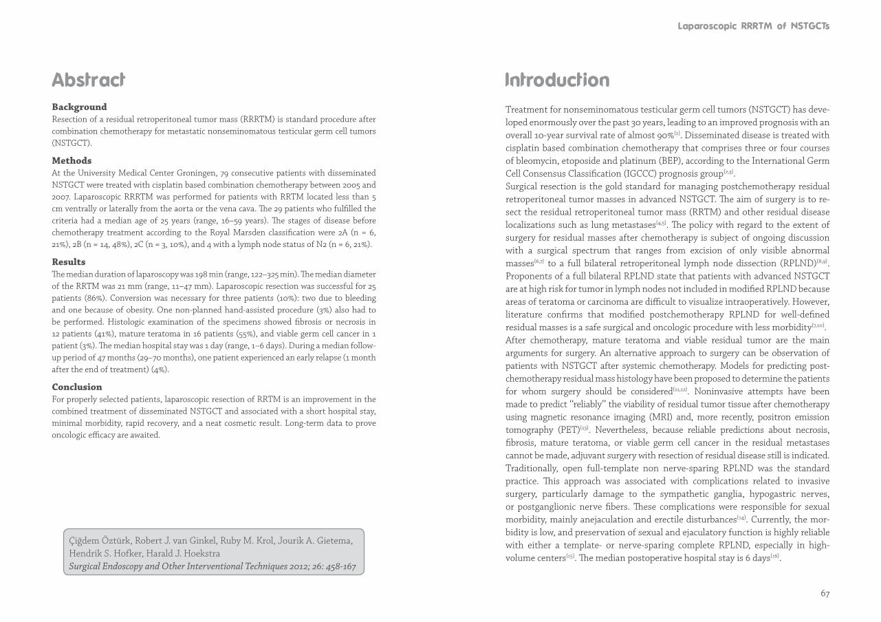
67
BackgroundResection of a residual retroperitoneal tumor mass (RRRTM) is standard procedure after combination chemotherapy for metastatic nonseminomatous testicular germ cell tumors (NSTGCT).
MethodsAt the University Medical Center Groningen, 79 consecutive patients with disseminated NSTGCT were treated with cisplatin based combination chemotherapy between 2005 and 2007. Laparoscopic RRRTM was performed for patients with RRTM located less than 5 cm ventrally or laterally from the aorta or the vena cava. The 29 patients who fulfilled the criteria had a median age of 25 years (range, 16–59 years). The stages of disease before chemotherapy treatment according to the Royal Marsden classification were 2A (n = 6, 21%), 2B (n = 14, 48%), 2C (n = 3, 10%), and 4 with a lymph node status of N2 (n = 6, 21%).
ResultsThe median duration of laparoscopy was 198 min (range, 122–325 min). The median diameter of the RRTM was 21 mm (range, 11–47 mm). Laparoscopic resection was successful for 25 patients (86%). Conversion was necessary for three patients (10%): two due to bleeding and one because of obesity. One non-planned hand-assisted procedure (3%) also had to be performed. Histologic examination of the specimens showed fibrosis or necrosis in 12 patients (41%), mature teratoma in 16 patients (55%), and viable germ cell cancer in 1 patient (3%). The median hospital stay was 1 day (range, 1–6 days). During a median follow-up period of 47 months (29–70 months), one patient experienced an early relapse (1 month after the end of treatment) (4%).
ConclusionFor properly selected patients, laparoscopic resection of RRTM is an improvement in the combined treatment of disseminated NSTGCT and associated with a short hospital stay, minimal morbidity, rapid recovery, and a neat cosmetic result. Long-term data to prove oncologic efficacy are awaited.
Çiğdem Öztürk, Robert J. van Ginkel, Ruby M. Krol, Jourik A. Gietema, Hendrik S. Hofker, Harald J. HoekstraSurgical Endoscopy and Other Interventional Techniques 2012; 26: 458-167
Treatment for nonseminomatous testicular germ cell tumors (NSTGCT) has deve-loped enormously over the past 30 years, leading to an improved prognosis with an overall 10-year survival rate of almost 90%{1}. Disseminated disease is treated with cisplatin based combination chemotherapy that comprises three or four courses of bleomycin, etoposide and platinum (BEP), according to the International Germ Cell Consensus Classification (IGCCC) prognosis group{2,3}.Surgical resection is the gold standard for managing postchemotherapy residual retroperitoneal tumor masses in advanced NSTGCT. The aim of surgery is to re-sect the residual retroperitoneal tumor mass (RRTM) and other residual disease localizations such as lung metastases{4,5}. The policy with regard to the extent of surgery for residual masses after chemotherapy is subject of ongoing discussion with a surgical spectrum that ranges from excision of only visible abnormal masses{6,7} to a full bilateral retroperitoneal lymph node dissection (RPLND){8,9}. Proponents of a full bilateral RPLND state that patients with advanced NSTGCT are at high risk for tumor in lymph nodes not included in modified RPLND because areas of teratoma or carcinoma are difficult to visualize intraoperatively. However, literature confirms that modified postchemotherapy RPLND for well-defined residual masses is a safe surgical and oncologic procedure with less morbidity{7,10}. After chemotherapy, mature teratoma and viable residual tumor are the main arguments for surgery. An alternative approach to surgery can be observation of patients with NSTGCT after systemic chemotherapy. Models for predicting post-chemotherapy residual mass histology have been proposed to determine the patients for whom surgery should be considered{11,12}. Noninvasive attempts have been made to predict ‘‘reliably’’ the viability of residual tumor tissue after chemotherapy using magnetic resonance imaging (MRI) and, more recently, positron emission tomography (PET){13}. Nevertheless, because reliable predictions about necrosis, fibrosis, mature teratoma, or viable germ cell cancer in the residual metastases cannot be made, adjuvant surgery with resection of residual disease still is indicated. Traditionally, open full-template non nerve-sparing RPLND was the standard practice. This approach was associated with complications related to invasive surgery, particularly damage to the sympathetic ganglia, hypogastric nerves, or postganglionic nerve fibers. These complications were responsible for sexual morbidity, mainly anejaculation and erectile disturbances{14}. Currently, the mor-bidity is low, and preservation of sexual and ejaculatory function is highly reliable with either a template- or nerve-sparing complete RPLND, especially in high-volume centers{15}. The median postoperative hospital stay is 6 days{16}.
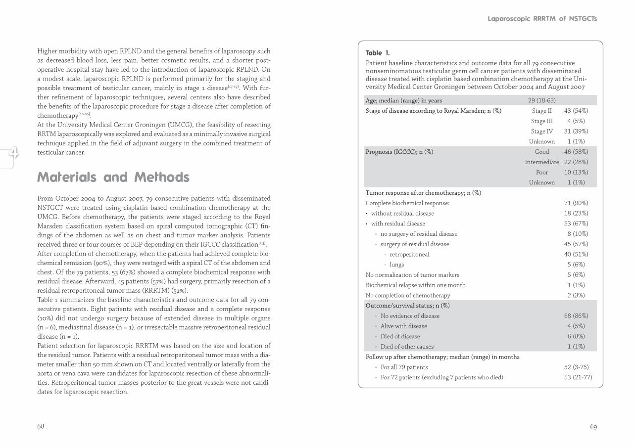
68 69
Higher morbidity with open RPLND and the general benefits of laparoscopy such as decreased blood loss, less pain, better cosmetic results, and a shorter post-operative hospital stay have led to the introduction of laparoscopic RPLND. On a modest scale, laparoscopic RPLND is performed primarily for the staging and pos sible treatment of testicular cancer, mainly in stage 1 disease{17-19}. With fur-ther refine ment of laparoscopic techniques, several centers also have described the bene fits of the laparoscopic procedure for stage 2 disease after completion of chemo therapy{20-26}.At the University Medical Center Groningen (UMCG), the feasibility of resecting RRTM laparoscopically was explored and evaluated as a minimally invasive surgical technique applied in the field of adjuvant surgery in the combined treatment of testicular cancer.
From October 2004 to August 2007, 79 consecutive patients with disseminated NSTGCT were treated using cisplatin based combination chemotherapy at the UMCG. Before chemotherapy, the patients were staged according to the Royal Marsden classification system based on spiral computed tomographic (CT) fin-dings of the abdomen as well as on chest and tumor marker analysis. Patients received three or four courses of BEP depending on their IGCCC classification{2,3}. After completion of chemotherapy, when the patients had achieved complete bio-chemical remission (90%), they were restaged with a spiral CT of the abdomen and chest. Of the 79 patients, 53 (67%) showed a complete biochemical response with residual disease. Afterward, 45 patients (57%) had surgery, primarily resection of a residual retroperitoneal tumor mass (RRRTM) (51%). Table 1 summarizes the baseline characteristics and outcome data for all 79 con-secutive patients. Eight patients with residual disease and a complete response (10%) did not undergo surgery because of extended disease in multiple organs (n = 6), mediastinal disease (n = 1), or irresectable massive retroperitoneal residual disease (n = 1).Patient selection for laparoscopic RRRTM was based on the size and location of the residual tumor. Patients with a residual retroperitoneal tumor mass with a dia -meter smaller than 50 mm shown on CT and located ventrally or laterally from the aorta or vena cava were candidates for laparoscopic resection of these abnor mali-ties. Retroperitoneal tumor masses posterior to the great vessels were not candi-dates for laparoscopic resection.
Patient baseline characteristics and outcome data for all 79 consecutive nonseminomatous testicular germ cell cancer patients with disseminated disease treated with cisplatin based combination chemotherapy at the Univer sity Medical Center Groningen between October 2004 and August 2007
Age; median (range) in years 29 (18-63)
Stage of disease according to Royal Marsden; n (%) Stage II 43 (54%)
Stage III 4 (5%)
Stage IV 31 (39%)
Unknown 1 (1%)
Prognosis (IGCCC); n (%) Good 46 (58%)
Intermediate 22 (28%)
Poor 10 (13%)
Unknown 1 (1%)
Tumor response after chemotherapy; n (%)
Complete biochemical response: 71 (90%)
• without residual disease 18 (23%)
• with residual disease 53 (67%)
• no surgery of residual disease 8 (10%)
• surgery of residual disease 45 (57%)
• retroperitoneal 40 (51%)
• lungs 5 (6%)
No normalization of tumor markers 5 (6%)
Biochemical relapse within one month 1 (1%)
No completion of chemotherapy 2 (3%)
Outcome/survival status; n (%)
• No evidence of disease 68 (86%)
• Alive with disease 4 (5%)
• Died of disease 6 (8%)
• Died of other causes 1 (1%)
Follow up after chemotherapy; median (range) in months
• For all 79 patients 52 (3-75)
• For 72 patients (excluding 7 patients who died) 53 (21-77)
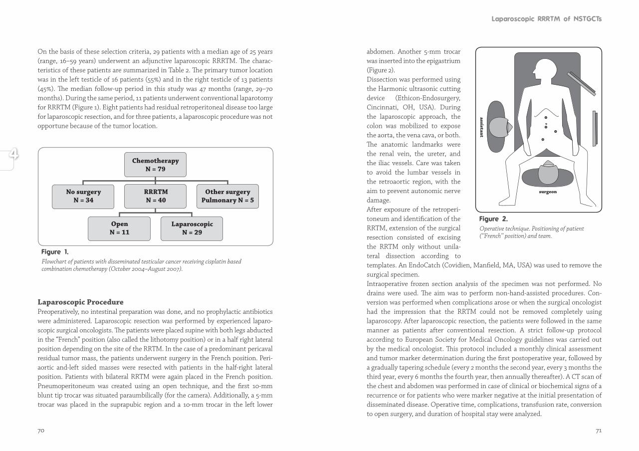
70 71
On the basis of these selection criteria, 29 patients with a median age of 25 years (range, 16–59 years) underwent an adjunctive laparoscopic RRRTM. The charac-teristics of these patients are summarized in Table 2. The primary tumor location was in the left testicle of 16 patients (55%) and in the right testicle of 13 patients (45%). The median follow-up period in this study was 47 months (range, 29–70 months). During the same period, 11 patients underwent conventional laparotomy for RRRTM (Figure 1). Eight patients had residual retroperitoneal disease too large for laparoscopic resection, and for three patients, a laparoscopic procedure was not opportune because of the tumor location.
Laparoscopic ProcedurePreoperatively, no intestinal preparation was done, and no prophylactic antibiotics were administered. Laparoscopic resection was performed by experienced laparo-scopic surgical oncologists. The patients were placed supine with both legs abducted in the “French” position (also called the lithotomy position) or in a half right lateral position depending on the site of the RRTM. In the case of a predominant pericaval residual tumor mass, the patients underwent surgery in the French position. Peri-aortic and-left sided masses were resected with patients in the half-right lateral position. Patients with bilateral RRTM were again placed in the French position. Pneumoperitoneum was created using an open technique, and the first 10-mm blunt tip trocar was situated paraumbilically (for the camera). Additionally, a 5-mm trocar was placed in the suprapubic region and a 10-mm trocar in the left lower
Flowchart of patients with disseminated testicular cancer receiving cisplatin based combination chemotherapy (October 2004–August 2007).
ChemotherapyN = 79
RRRTMN = 40
Other surgeryPulmonary N = 5
LaparoscopicN = 29
OpenN = 11
No surgeryN = 34
abdomen. Another 5-mm trocar was inserted into the epigastrium (Figure 2). Dissection was performed using the Harmonic ultra sonic cutting device (Ethicon- Endosur gery, Cin ci nnati, OH, USA). During the lapa ro s co pic approach, the co lon was mobilized to expo se the aorta, the vena cava, or both. The anatomic land marks were the renal vein, the ure ter, and the iliac vess els. Care was taken to avoid the lum bar vessels in the retro aor tic re gion, with the aim to prevent autono mic nerve da ma ge. After expo sure of the retro pe ri -to neum and identification of the RRTM, extension of the sur gical re sec tion consisted of ex ci sing the RRTM only without unila-teral dissec tion accor ding to tem plates. An EndoCatch (Covidien, Manfield, MA, USA) was used to re move the sur gi cal specimen.Intraoperative frozen section analysis of the specimen was not performed. No drains were used. The aim was to perform non-hand-assisted procedures. Con-version was performed when complications arose or when the surgical oncologist had the impression that the RRTM could not be removed completely using laparoscopy. After laparoscopic resection, the patients were followed in the same manner as patients after conventional resection. A strict follow-up protocol according to European Society for Medical Oncology guidelines was carried out by the medical oncologist. This protocol included a monthly clinical assessment and tumor marker determination during the first postoperative year, followed by a gradually tapering schedule (every 2 months the second year, every 3 months the third year, every 6 months the fourth year, then annually thereafter). A CT scan of the chest and abdomen was performed in case of clinical or biochemical signs of a recurrence or for patients who were marker negative at the initial presentation of disseminated disease. Operative time, complications, transfusion rate, conversion to open surgery, and duration of hospital stay were analyzed.
assistant
surgeon
Operative technique. Positioning of patient (‘‘French’’ position) and team.
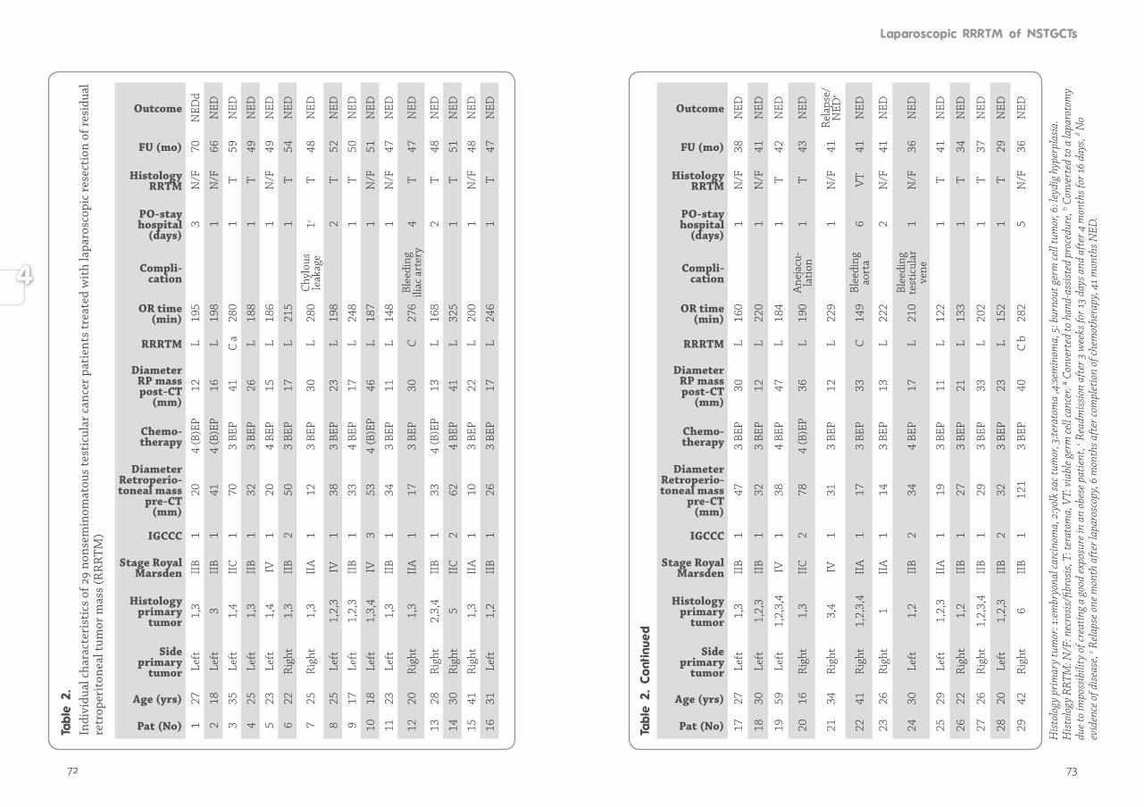
72 73
Indi
vidu
al c
hara
cter
isti
cs o
f 29
nons
emin
omat
ous
test
icul
ar c
ance
r pa
tien
ts tr
eate
d w
ith
lapa
rosc
opic
rese
ctio
n of
resi
dual
re
trop
erit
onea
l tum
or m
ass
(RR
RTM
)
Pat (No)
Age (yrs)
Side primary
tumor
Histologyprimary
tumor
Stage Royal Marsden
IGCCC
DiameterRetroperio-toneal mass
pre-CT (mm)
Chemo - therapy
Diameter RP mass post-CT
(mm)
RRRTM
OR time(min)
Compli-cation
PO-stay hos pital
(days)
Histology RRTM
FU (mo)
Outcome
127
Left
1,3
IIB
120
4 (B
)EP
12L
195
3N
/F70
NED
d
218
Left
3II
B1
414
(B)E
P16
L19
81
N/F
66N
ED
335
Left
1,4
IIC
170
3 BE
P41
C a
280
1T
59N
ED
425
Left
1,3
IIB
132
3 BE
P26
L18
81
T49
NED
523
Left
1,4
IV1
204
BEP
15L
186
1N
/F49
NED
622
Rig
ht1,
3II
B2
503
BEP
17L
215
1T
54N
ED
725
Rig
ht1,
3II
A1
123
BEP
30L
280
Chy
lous
le
akag
e1c
T48
NED
825
Left
1,2,
3IV
138
3 BE
P23
L19
82
T52
NED
917
Left
1,2,
3II
B1
334
BEP
17L
248
1T
50N
ED
1018
Left
1,3,
4IV
353
4 (B
)EP
46L
187
1N
/F51
NED
1123
Left
1,3
IIB
134
3 BE
P11
L14
81
N/F
47N
ED
1220
Rig
ht1,
3II
A1
173
BEP
30C
276
Blee
ding
ili
ac a
rter
y4
T47
NED
1328
Rig
ht2,
3,4
IIB
133
4 (B
)EP
13L
168
2T
48N
ED
1430
Rig
ht5
IIC
262
4 BE
P41
L32
51
T51
NED
1541
Rig
ht1,
3II
A1
103
BEP
22L
200
1N
/F48
NED
1631
Left
1,2
IIB
126
3 BE
P17
L24
61
T47
NED
Pat (No)
Age (yrs)
Side primary
tumor
Histologyprimary
tumor
Stage Royal Marsden
IGCCC
DiameterRetroperio-toneal mass
pre-CT (mm)
Chemo - therapy
Diameter RP mass post-CT
(mm)
RRRTM
OR time(min)
Compli-cation
PO-stay hos pital
(days)
Histology RRTM
FU (mo)
Outcome
1727
Left
1,3
IIB
147
3 BE
P30
L16
01
N/F
38N
ED
1830
Left
1,2,
3II
B1
323
BEP
12L
220
1N
/F41
NED
1959
Left
1,2,
3,4
IV1
384
BEP
47L
184
1T
42N
ED
2016
Rig
ht1,
3II
C2
784
(B)E
P36
L19
0A
neja
cu-
lati
on1
T43
NED
2134
Rig
ht3,
4IV
131
3 BE
P12
L22
91
N/F
41R
elap
se/
NED
e
2241
Rig
ht1,
2,3,
4II
A1
173
BEP
33C
149
Blee
ding
ao
rta
6V
T41
NED
2326
Rig
ht1
IIA
114
3 BE
P13
L22
22
N/F
41N
ED
2430
Left
1,2
IIB
234
4 BE
P17
L21
0Bl
eedi
ng
test
icul
ar
vene
1N
/F36
NED
2529
Left
1,2,
3II
A1
193
BEP
11L
122
1T
41N
ED
2622
Rig
ht1,
2II
B1
273
BEP
21L
133
1T
34N
ED
2726
Rig
ht1,
2,3,
4II
B1
293
BEP
33L
202
1T
37N
ED
2820
Left
1,2,
3II
B2
323
BEP
23L
152
1T
29N
ED
2942
Rig
ht6
IIB
112
13
BEP
40C
b28
25
N/F
36N
ED
His
tolo
gy p
rim
ary
tum
or: 1
:em
bryo
nal c
arcin
oma,
2:y
olk
sac t
umor
, 3:te
rato
ma
,4:se
min
oma,
5: b
urno
ut g
erm
cell
tum
or, 6
: ley
dig
hype
rpla
sia.
His
tolo
gy R
RTM
: N/F
: nec
rosi
s/fib
rosi
s, T:
tera
tom
a, V
T: v
iabl
e ge
rm ce
ll ca
ncer
. ª C
onve
rted
to h
and-
assi
sted
pro
cedu
re, b C
onve
rted
to a
lapa
roto
my
due
to im
poss
ibili
ty o
f cre
atin
g a
good
exp
osur
e in
an
obes
e pa
tient
, c Rea
dmis
sion
afte
r 3 w
eeks
for 1
3 da
ys a
nd a
fter
4 m
onth
s for
16 d
ays,
d No
evid
ence
of d
isea
se, e R
elap
se o
ne m
onth
aft
er la
paro
scop
y, 6
mon
ths a
fter
com
plet
ion
of ch
emot
hera
py, 4
1 m
onth
s NED
.
74 75
All 29 patients included in the laparoscopic treatment group had a biochemical complete remission after chemotherapy. Cisplatin based combination chemo-therapy elicited a reduction of the retroperitoneal metastases with a mean factor of 0.6, resulting in a median postchemotherapy tumor size of 21 mm (range, 11–47 mm) (Table 2). In almost two thirds of the patients, the residual tumor was located in the latero-aortic region. The median interval between the last chemotherapy course and the laparoscopic resection of RRTM was 3 months (range, 1–6 months).For 25 patients (86%), the laparoscopic procedure could be conducted as planned. For 3 patients (10%), conversion to open surgery was necessary due to a slight bleeding from the common iliac artery, a larger bleeding from the aorta, and the impossibility of creating a good exposure in an obese patient. In another patient (3%), the initial plan was changed because extreme obesity prevented the creation of a window sufficiently large for laparoscopic exploration, and a hand-assisted laparoscopic resection of the tumor was performed. The only minor perioperative complication encountered was an injury of the testicular vein at the left side, which was managed laparoscopically with clips. The median duration of surgery for the 29 patients, including positioning of the patient, was 198 min (range, 122–325 min). When the three patients who had conversion to laparotomy and the one patient who underwent a hand-assisted procedure were excluded, the median duration of a successful laparoscopic resection was 195 min (range, 122–325 min). The estimated blood loss was minimal (<50 ml), except for the converted patient with bleeding from the common iliac artery who did not need blood transfusion and the converted patient with bleeding from the aorta who lost up to 1,500 ml of blood. The median postoperative hospital stay was only 1 day (range, 1–6 days). The longest postoperative hospital stay (6 days) was experienced by the patient who had to undergo conversion due to injury of the aorta. During preparation of the two residual tumor masses from the aorta, a large bleeding occurred. Laparoscopy was quickly converted to a laparotomy, and the diathermic injury to the aorta was managed with two sutures. This procedure required a total operative time of 149 min. No postoperative infections occurred. One short-term postoperative complication (3%) included a massive chylous ascites which could not be treated conservatively. After 4 months, laparoscopic exploration showed visible leakage of a lymph vessel, which was coagulated successfully with argon diathermia and clipped. One patient had anejaculation after laparoscopic RRRTM. Histologic examination showed necrosis or fibrosis in 12 patients (41%) and teratoma in 16 patients (55%). In one patient (3%), a radically resected viable
germ cell cancer was found. Preoperatively, this patient received three courses of BEP, and after complete laparoscopic RRRTM, no additional chemo therapy courses were given. During the median follow-up period of 47 months (mean, 46 months; range, 29–70 months), one short-term local recurrence was experienced by a 34-year-old man in the good risk prognosis group who had mature, immature teratoma and seminoma elements in his right-sided primary tumor. This man received three courses of BEP. Cisplatin based combination chemo therapy resulted in a biochemical complete remission and was followed by a laparoscopic RRRTM. Residual tumor masses were located on the left side next to the aorta and between the aorta and the vena cava. Histology showed fibrosis. The man’s tumor markers were slightly elevated 1 month after laparoscopy and 6 months after completion of chemotherapy. A CT scan showed recurrence between the aorta and the vena cava (Figure 3). Additional chemotherapy (paclitaxel, ifosfamide, and cisplatin) was administered, and the patient achieved a complete response, both biochemically and radiologically. A laparotomy with a formal template dis sec tion on both sides was perfor med. Histology of the speci men showed fibrosis. No signs of recurrence were de tected 41 months after the man’s last sur ge ry.
Laparoscopic RPLND was first described in 1992 for a patient with stage I NSTGCT{17}. With further development of lapa ro-scopic techniques and expe rien-ce performing them, la pa ro-sco pic RPLND was intro du ced in several specia lized cen ters for the staging and treat ment of mainly low-stage tes ti cular cancer, with the same boun-da ries of dissection as an open approach. Table 3 summarizes the results reported by these centers{20-26}. The results show
Abdominal computed tomography (CT) scan of retroperitoneal recurrence.
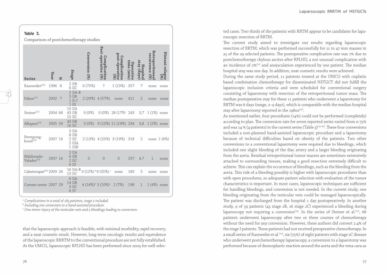
76 77
that the laparoscopic approach is feasible, with minimal morbidity, rapid recovery, and a neat cosmetic result. However, long-term oncologic results and equivalence of the laparoscopic RRRTM to the conventional procedure are not fully established. At the UMCG, laparoscopic RPLND has been performed since 2005 for well-selec-
Comparison of postchemotherapy studies
Series
Year N
Stage
Conversion (N)
Complication
Peri-operative (N)
Complication
post-operative (N
)
Operative
time (m
in)
Hospital
stay (days)
Retroperitoneal recurrence (N
)
Distant relapse
(N)
Rassweiler{20} 1996 8 2 IIB 6 IIC 6 (75%) ? 1 (13%) 357 7 none none
Palese{21} 2002 7
2 IIA-B 3 IIB 1 II C 1 III
2 (29%) 4 (57%) none 411 2 none none
Steiner{22} 2004 68 10 IIA 43 IIB 15 IIC
0 (0%) 0 (0%) 28 (17%a 243 3,7 1 (1%) none
Albqami{23} 2005 59 43 IIB 16 IIC 0 (0%) 9 (15%) 11 (19%) 234 3,8 1 (2%) none
Permpong-kosol{24} 2007 16
3 IIA 8 IIB 2 IIC 2 IIIA 1 IIIB
2 (13%) 4 (25%) 3 (19%) 318 2 none 1 (6%)
Maldonado- Valadez{25} 2007 16
2 IIA 6 IIB 2 IIC 6 III
0 0 0 237 4.7 1 none
Calestroupat{26} 2009 26 16 IIA-B 13 IIC 3 (12%) b 9 (35%) c none 183 5 none none
Current series 2007 29
6 IIA 14 IIB 3 IIC 6 IV
4 (14%)b 3 (10%)c 2 (7%) 198 1 1 (4%) none
a Complications in a total of 185 patients, stage 2 included.b Including one conversion to a hand-assisted procedure.c One minor injury of the testicular vein and 2 bleedings leading to conversion.
ted cases. Two thirds of the patients with RRTM appear to be candidates for lapa-ro scopic resection of RRTM. The current study aimed to investigate our results regarding laparoscopic resection of RRTM, which was performed successfully for 11 to 47 mm masses in 25 of the 29 selected patients. The postoperative complication rate was 7% due to postchemotherapy chylous ascites after RPLND, a not unusual complication with an incidence of 2%{27} and anejuculation experienced by one patient. The median hospital stay was one day. In addition, neat cosmetic results were achieved. During the same study period, 11 patients treated at the UMCG with cisplatin based combination chemotherapy for disseminated NSTGCT did not fulfill the laparoscopic inclusion criteria and were scheduled for conventional surgery consisting of laparotomy with resection of the retroperitoneal tumor mass. The median postoperative stay for these 11 patients who underwent a laparotomy for RRTM was 6 days (range, 2–9 days), which is comparable with the median hospital stay after laparotomy reported in the 1980s{16}.As mentioned earlier, four procedures (14%) could not be performed (completely) according to plan. The conversion rate for seven reported series varied from 0-75% and was 14 % (4 patients) in the current series (Table 3){20-26}. These four conversions included a non-planned hand-assisted laparoscopic procedure and a laparotomy because of technical difficulties based on obesity of the patients. Two other conversions to a conventional laparotomy were required due to bleedings, which included one slight bleeding of the iliac artery and a larger bleeding originating from the aorta. Residual retroperitoneal tumor masses are sometimes extensively attached to surrounding tissues, making a good resection extremely difficult to achieve. This can explain the occurrence of bleedings, such as the bleeding from the aorta. This risk of a bleeding possibly is higher with laparoscopic procedures than with open procedures, so adequate patient selection with evaluation of the tumor characteristics is important. In most cases, laparoscopic techniques are sufficient for handling bleedings, and conversion is not needed. In the current study, one bleeding originating from the testicular vein could be managed laparoscopically. The patient was discharged from the hospital 1 day postoperatively. In another study, 9 of 59 patients (43 stage 2B, 16 stage 2C) experienced a bleeding during laparoscopy not requiring a conversion{23}. In the series of Steiner et al.{22}, 68 patients underwent laparoscopy after two or three courses of chemotherapy without the need for any conversion. However, these authors did convert 2.4% of the stage I patients. These patients had not received preoperative chemotherapy. In a small series of Rassweiler et al.{20}, six (75%) of eight patients with stage 2C disease who underwent postchemotherapy laparoscopy, a conversion to a laparotomy was performed because of desmoplastic reaction around the aorta and the vena cava as
78 79
a result of chemotherapy{28,29}. Permpongkosol et al.{24} had to convert procedures due to vascular injury in 2 (12.5%) of 16 patients who had received three or four courses of chemotherapy (Table 3).
The median duration of the laparoscopic procedure in the current series was 198 minutes, compared with 216-348 minutes described in literature. Furthermore, the median postoperative hospital stay was 1 day compared with median hospital stay ranging from 2 to 8.2 days in other laparoscopic series. These other series also included patients who had conversion to a laparotomy, thus explaining the discre-pan cy with our results.Currently, very few institutions have reported laparoscopic RRRTM after chemo-therapy for disseminated NSTGCT. Our first experience with this new technique was favorable. The minimal morbidity, the short postoperative hospital stay, and the neat cosmetic results are a step forward in the combined treatment of testi-cular cancer. Although we have achieved good results over the past 30 years with conventional RRRTM, it appears that this also is possible with laparoscopic resection for patients with minimal RRTM. Approximately 70% of disseminated testicular cancer patients who require RRRTM are candidates for complete laparoscopic resection based on the current selection criteria. However, it is unknown whether the laparoscopic procedure will result in more frequent (short or late) relapses. In the current series, one patient (4%) had a short-term relapse. The recurrence was located between the vena cava and the aorta. Preoperative CT scan images showed an RRTM located on the left side of the aorta between the aorta and vena cava. Although this RRTM was resected during laparoscopy, residual tissue remained behind, causing outgrowth of viable germ cell cancer. This relapse can be calculated as a technical failure. Short- and long-term relapses after chemotherapy are mostly related to incomplete resection of residual disease and also are encountered after conventional surgery{30}. The limitation of this study is that the oncologic efficacy of the procedure remains questioned because the long-term oncologic follow-up data are not equivalent to those for the open procedure. In the future, after more patients have been treated with laparoscopic resection, data will become available with reliable relapse figures. This potential pitfall requires prolonged and very stringent follow-up assessment to monitor the oncologic safety of the laparoscopic resection of RRTM. It is important to perform thorough follow-up assessment to be certain that a minimally invasive intervention does not involve the risk of so-called extratemplate disease{31}.An overshadowing component of our study is the controversy surrounding the surgical management of patients with NSTGCT and concerns about the deve lop-ment of late relapsing abdominal teratoma{29}. Late-recurring disease is characte-
rized by slow growth, production of alphafetoprotein, chemoresistance, and a poor prognosis{28}. Our surgical management of patients with NSTGCT con forms to the European guidelines for testicular cancer{27}. Although our results and those of others are favorable, questions remain: Is lapa-roscopic RRRTM as complete oncologically as an open procedure? How long should we wait before laparoscopic RRRTM is proclaimed as the standard, or should a randomized study be performed? Particularly the rapid postoperative recovery, the low morbidity, and the neat cosmetic results contribute to the well-being of the usually young patients, most of whom still have many years ahead of them. Whatever policy is chosen for the treatment of patients with advanced NSTGCT, management should take place at a referral center with specific expertise in the treatment of testicular cancer and an oncologic team of specialists.
Laparoscopic RRRTM after chemotherapy for disseminated testicular cancer is a feasible surgical treatment option with a short hospital stay and neat cosmetic results for well-selected patients. Which patients are the right candidates for laparoscopic RRRTM remains the question. Close and long-term follow-up assess-ment of long-term results with respect to tumor recurrence is obligatory.
1. Sonneveld DJ, Hoekstra HJ, van der Graaf WT, Sluiter WJ, Mulder NH, Willemse PH et al. Improved long term survival of patients with metastatic nonseminomatous testicular germ cell carcinoma in relation to prognostic classification systems during the cisplatin era. Cancer 2001; 91: 1304-1315.
2. International Germ Cell Consensus Classification: a prognostic factor-based staging system for metastatic germ cell cancers. International Germ Cell Cancer Collaborative Group. J Clin Oncol 1997; 15: 594-603.
3. Huddart RA. Testicular seminoma: ESMO clinical recommendations for diagnosis, treatment and follow-up. Ann Oncol 2007; 18 Suppl 2: ii40-ii41.
4. Oldenburg J, Alfsen GC, Lien HH, Aass N, Waehre H, Fossa SD. Postchemotherapy retroperitoneal surgery remains necessary in patients with nonseminomatous testicular cancer and minimal residual tumor masses. J Clin Oncol 2003; 21: 3310–3317.
5. Kesler KA, Kruter LE, Perkins SM, Rieger KM, Sullivan KJ, Runyan ML et al. Survival after resection for metastatic testicular nonseminomatous germ cell cancer to the lung or mediastinum. Ann Thorac Surg 2011; 91: 1085-1093.
80
6. Ozen H, Ekici S, Sozen S, Ergen A, Tekgül S, Kendi S. Resection of residual masses alone: an alternative in surgical therapy of metastatic testicular germ cell tumors after chemotherapy. Urol 2001; 57: 323-327.
7. Rabbani F, Goldenberg SL, Gleave ME, Paterson RF, Murray N, Sullivan LD. Retroperitoneal lymphadenectomy for post-chemotherapy residual masses: is a modified dissection and resection of residual masses sufficient? Br J Urol 1998; 81: 295-300.
8. Carver BS, Cronin AM, Eggener S, Savage CJ, Motzer RJ, Bajorin D et al. The total number of retroperitoneal lymph nodes resected impacts clinical outcome after chemotherapy for metastatic testicular cancer. Sheinfeld J. Urol 2010; 75: 1431-1435.
9. Large MC, Sheinfeld J, Eggener SE. Retroperitoneal lymph node dissection: reassess-ment of modified templates. BJU Int 2009; 104: 1369-1375.
10. Heidenreich A, Pfister D, Witthuhn R, Thüer D, Albers P. Postchemotherapy retroperitoneal lymph node dissection in advanced testicular cancer: radical of modified template resection. Eur Urol 2009; 55: 217-224.
11. Steyerberg EW, Keizer HJ, Fosså SD, Sleijfer DT, Toner GC, Schraffordt Koops H et al. Prediction of residual retroperitoneal mass histology after chemotherapy for metastatic nonseminomatous germ cell tumor: multivariate analysis of individual patient data from six study groups. J Clin Oncol 1995; 13: 1177-1187.
12. Vergouwe Y, Steyerberg EW, Foster RS, Sleijfer DT, Fosså SD, Gerl A et al. Predicting retroperitoneal histology in postchemotherapy testicular germ cell cancer: a model update and multicentre validation with more than 1000 patients. Eur Urol 2007; 51: 424-432.
13. Oechsle K, Hartmann M, Brenner W, Venz S, Weissbach L, Franzius C et al. [18F] Fluorodeoxyglucose positron emission tomography in nonseminomatous germ cell tumors after chemotherapy: the German multicenter positron emission tomography study group. J Clin Oncol 2008; 26: 5930-5935.
14. van Basten JP, Jonker-Pool G, Van Driel MF, Sleijfer DT, van der Wiel HB, Hoekstra HJ. The sexual sequelae of testicular cancer. Cancer Treat Rev 1995; 21: 479-495.
15. Pettus JA, Carver BS, Masterson T, Stasi J, Sheinfeld J. Preservation of ejaculation in patients undergoing nerve-sparing postchemotherapy retroperitoneal lymph node dissection for metastatic testicular cancer. Urol 2009; 73: 328-331.
16. Gelderman WA, Schraffordt Koops H, Sleijfer DT, Oosterhuis JW, Van der Heide JN, Mulder NH et al. Results of adjuvant surgery in patients with stage III and IV nonseminomatous testicular tumors after cisplatin-vinblastine-bleomycin chemotherapy. J Surg Oncol 1988; 38: 227-232.
17. Rukstalis DB, Chodak GW. Laparoscopic retroperitoneal lymph node dissection in a patient with stage 1 testicular carcinoma. J Urol; 148: 1907-1909.
18. Bhayani SB, Ong A, Oh WK, Kantoff PW, Kavoussi LR. Laparoscopic retroperitoneal lymph node dissection for clinical stage I nonseminomatous germ cell testicular cancer: a long-term update. Urol 2003; 62: 324-327.
19. Nielsen ME, Lima G, Schaeffer EM, Porter J, Cadeddu JA, Tuerk I et al. Oncologic efficacy of laparoscopic RPLND in treatment of clinical stage I nonseminomatous germ cell testicular cancer. Urol 2007; 70: 1168-1172.
20. Rassweiler JJ, Seemann O, Henkel TO, Stock C, Frede T, Alken P. Laparoscopic retroperitoneal lymph node dissection for nonseminomatous germ cell tumors: indications and limitations. J Urol 1996; 156: 1108-1113.
21. Palese MA, Su LM, Kavoussi LR. Laparoscopic retroperitoneal lymph node dissection after chemotherapy. Urol 2002; 60: 130-134.
22. Steiner H, Peschel R, Janetschek G, Holtl L, Berger AP, Bartsch G et al. Long-term results of laparoscopic retroperitoneal lymph node dissection: a single-center 10-year experience. Urol 2004; 63: 550-555.
23. Albqami N, Janetschek G. Laparoscopic retroperitoneal lymph-node dissection in the management of clinical stage I and II testicular cancer. J Endourol 2005; 19: 683-692.
24. Permpongkosol S, Lima GC, Warlick CA, Allaf ME, Varkarakis IM, Bagga HS et al. Postchemotherapy laparoscopic retroperitoneal lymph node dissection: evaluation of complications. Urol 2007; 69: 361-365.
25. Maldonado-Valadez R, Schilling D, Anastasiadis AG, Sturm W, Stenzl A, Corvin S. Post-chemotherapy laparoscopic retroperitoneal lymph-node dissection in testis cancer patients. J Endourol 2007; 21: 1501-1504.
26. Calestroupat JP, Sanchez-Salas R, Cathelineau X, Rozet F, Galiano M, Smyth G et al. Postchemotherapy laparoscopic retroperitoneal lymph node dissection in nonseminomatous germ-cell tumor. J Endourol 2009; 23: 645-650.
27. Evans JG, Spiess PE, Kamat AM, Wood CG, Hernandez M, Pettaway CA et al. Chylous ascites after post-chemotherapy retroperitoneal lymph node dissection: review of the M. D. Anderson experience. J Urol 2006; 176: 1463-1467.
28. Shahidi M, Norman AR, Dearnaley DP, Nicholls J, Horwich A, Huddart RA. Late recurrence in 1263 men with testicular germ cell tumors. Multivariate analysis of risk factors and implications for management. Cancer 2002; 95: 520-530.
29. Baniel J, Foster RS, Gonin R, Messemer JE, Donohue JP, Einhorn LH (1995) Late relapse of testicular cancer. J Clin Oncol; 13: 1170–1176.
30. Sharp DS, Carver BS, Eggener SE, Kondagunta GV, Motzer RJ, Bosl GJ et al. Clinical outcome and predictors of survival in late relapse of germ cell tumor. J Clin Oncol 2008; 26: 5524-5529.
31. Carver BS, Shayegan B, Eggener S, Stasi J, Motzer RJ, Bosl GJ et al. Incidence of metastatic nonseminomatous germ cell tumor outside the boundaries of a modified postchemotherapy retroperitoneal lymph node dissection. J Clin Oncol 2007; 25: 4365-4369.
85
BackgroundTo describe the 10 year experience and oncological results of the University Medical Center Groningen with conventional laparotomy (C-RRRTM) and laparoscopy (L-RRRTM) to resect residual retroperitoneal tumor masses (RRTM) in a large series of patients with advanced nonseminomatous testicular germ cell tumors (NSTGCT).
MethodsFrom 2005-2015, 150 consecutive patients with disseminated NSTGCT required adjunctive surgery after completion of chemotherapy. Patients with RRTM <5 cm ventral/lateral from the aorta and/or the caval vein were candidates for L-RRRTM. Remaining patients received C-RRRTM.
ResultsL-RRRTM was scheduled in 89 patients (median age 27 [range 16-66] years) and C-RRRTM in 61 patients (median age 28 [range 16-64] years). Median residual tumor diameter was 20 (range 5-70) mm in the L-RRRTM versus 42 (range 11-220) mm in the C-RRRTM group (p <.001). Conversion in the L-RRRTM group was performed in 14 patients (15%). Perioperative complications occurred in 5 patients (6%) in the L-RRRTM and 7 (12%, NS) in the C-RRRTM group. Median duration of L-RRRTM was 146 (range 45-288) minutes vs. 221 (range 95-792) minutes for C-RRRTM (p <.001). 9/75 patients (12%) in the L-RRRTM group had postoperative complications versus 26/75 patients in the C-RRRTM group (including conversions) (35%, p <.001). Median postoperative stay in the L-RRRTM group was 1 (range 1-5) vs. 5 (3-26) days in the C-RRRTM group (p <.001). During a median follow-up of 79 (2-144) months, 27 patients had recurrences:4 (5%) in the L-RRRTM group and 23 (31%) in the C-RRRTM group (p <.001).
ConclusionsLaparoscopic resection of RRTM after cisplatin based combination chemotherapy for advanced NSTGCT is feasible and oncologically safe in selected patients, with shorter hospital stays and good oncological and excellent cosmetic outcomes.
Çiğdem Öztürk, Lukas B. Been, Robert J. van Ginkel, Jourik A. Gietema, Harald J. Hoekstra Scientific Reports, submitted 2018
The introduction of cisplatin based combination chemotherapy to treat advanced nonsemino matous germ cell tumor (NSTGCT) has impacted survival rates great-ly, with an overall 10-year survival rate of up to 90%{1,2}. Surgery plays a pivo tal role in the treatment of residual retroperitoneal tumor masses (RRTM) as well as pulmonary residual disease in NSTGCT and is aimed at resecting viable germ cell cancer tissue and/or teratoma{3-7}. The extent of surgery has remained contro ver sial for many years, with a surgical spectrum varying from a full bilateral retroperi-toneal lymph node dissection (RPLND) to a more limited approach with resection of visible abnormal retroperitoneal tumor masses{8,9}. Today’s literature supports that a modified post chemotherapy RPLND, e.g. resection of well-defined residual retroperitoneal tumor masses (RRRTM), is a safe oncological procedure, with less morbidity and it conserves sexual functioning in the majority of these patients{10-12}.
Classically, RPLND or RRRTM was executed through a midline laparotomy. Lapa-ro scopic RPLND (L-RPLND) was first performed in 1992. In the past decade, it has emerged as an alternative to reduce morbidity associated with conventional open surgery using the same boundaries of dissection. Laparoscopic surgery is mainly described in literature for stage I disease as a diagnostic procedure and for the resection of low volume disease{13-16}. In an earlier pilot study, the Groningen study group showed that the laparoscopic approach was feasible, with a low rate of retroperitoneal relapse in advanced testicular cancer in properly selected pa-tients{17}. So far, there are no large consecutive series of disseminated testicular cancer patients described with respect to the results of adjunctive surgery; e.g., conventio nal versus laparoscopic resection of RRTM. For the laparoscopic resection of RRTM to be considered a safe alternative oncologic procedure compared to conventio nal open surgery in patients with advanced NSTGCT, long-term follow up assess ments in a larger cohort are required.
The current study aimed to describe the 10-year experience of the Comprehensive Cancer Center of the University Medical Center Groningen UMCG with conventional resection of RRTM (C-RRRTM) and laparoscopic resection of RRTM (L-RRRTM) in a consecutive series of patients, with a focus on operative outcomes and oncologic results, and to define oncological and technical boundaries for laparoscopic management in the field of adjunctive surgery after cisplatin based combination chemotherapy in patients with testicular cancer.
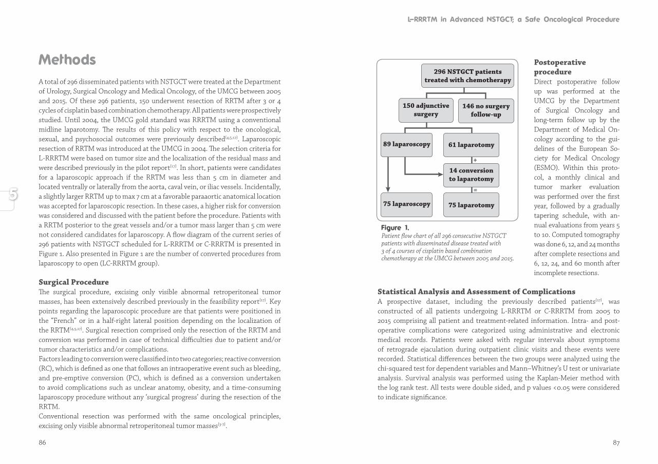
86 87
A total of 296 disseminated patients with NSTGCT were treated at the Department of Urology, Surgical Oncology and Medical Oncology, of the UMCG between 2005 and 2015. Of these 296 patients, 150 underwent resection of RRTM after 3 or 4 cycles of cisplatin based combination chemotherapy. All patients were prospectively studied. Until 2004, the UMCG gold standard was RRRTM using a conventional midline laparotomy. The results of this policy with respect to the oncological, sexual, and psychosocial outcomes were previously described{4,5,12}. Laparoscopic re sec tion of RRTM was introduced at the UMCG in 2004. The selection criteria for L-RRRTM were based on tumor size and the localization of the residual mass and were descri bed previously in the pilot report{17}. In short, patients were candidates for a laparo scopic approach if the RRTM was less than 5 cm in diameter and located ventrally or laterally from the aorta, caval vein, or iliac vessels. Incidentally, a slightly larger RRTM up to max 7 cm at a favorable paraaortic anatomical location was accepted for laparoscopic resection. In these cases, a higher risk for conversion was considered and discussed with the patient before the procedure. Patients with a RRTM posterior to the great vessels and/or a tumor mass larger than 5 cm were not considered candidates for laparoscopy. A flow diagram of the current series of 296 patients with NSTGCT scheduled for L-RRRTM or C-RRRTM is presented in Figure 1. Also presented in Figure 1 are the number of converted procedures from laparoscopy to open (LC-RRRTM group).
Surgical ProcedureThe surgical procedure, excising only visible abnormal retroperitoneal tumor masses, has been extensively described previously in the feasibility report{17}. Key points regarding the laparoscopic procedure are that patients were positioned in the “French” or in a half-right lateral position depending on the localization of the RRTM{4,5,17}. Surgical resection comprised only the resection of the RRTM and conversion was performed in case of technical difficulties due to patient and/or tumor characteristics and/or complications. Factors leading to conversion were classified into two categories; reactive conversion (RC), which is defined as one that follows an intraoperative event such as bleeding, and pre-emptive conversion (PC), which is defined as a conversion undertaken to avoid complications such as unclear anatomy, obesity, and a time-consuming lapa roscopy procedure without any ‘surgical progress’ during the resection of the RRTM.Conventional resection was performed with the same oncological principles, excising only visible abnormal retroperitoneal tumor masses{3-7}.
Postoperative procedureDirect postoperative follow up was performed at the UMCG by the Department of Surgical Oncology and long-term follow up by the Department of Medical On -co logy according to the gui -de lines of the European So -cie ty for Medical Onco lo gy (ESMO). Within this pro to-col, a monthly clini cal and tumor marker eva lu a tion was performed over the first year, followed by a gra dual ly tapering sche dule, with an-nual evalua tions from years 5 to 10. Computed to mo gra phy was done 6, 12, and 24 months after com plete resections and 6, 12, 24, and 60 month after incom ple te resections.
Statistical Analysis and Assessment of ComplicationsA prospective dataset, including the previously described patients{17}, was constructed of all patients undergoing L-RRRTM or C-RRRTM from 2005 to 2015 comprising all patient and treatment-related information. Intra- and post-operative complications were categorized using administrative and electronic medical records. Patients were asked with regular intervals about symptoms of retrograde ejaculation during outpatient clinic visits and these events were recorded. Statistical differences between the two groups were analyzed using the chi-squared test for dependent variables and Mann–Whitney’s U test or univariate analysis. Survival analysis was performed using the Kaplan-Meier method with the log rank test. All tests were double sided, and p values <0.05 were considered to indicate significance.
+
=
296 NSTGCT patientstreated with chemotherapy
150 adjunctivesurgery
146 no surgeryfollow-up
14 conversion to laparotomy
89 laparoscopy
75 laparoscopy 75 laparotomy
61 laparotomy
Patient flow chart of all 296 consecutive NSTGCT patients with disseminated disease treated with 3 of 4 courses of cisplatin based combination chemotherapy at the UMCG between 2005 and 2015.
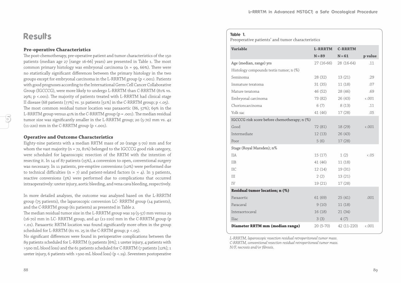
88 89
Pre-operative Characteristics The post-chemotherapy, pre-operative patient and tumor characteristics of the 150 patients (median age 27 [range 16-66] years) are presented in Table 1. The most common primary histology was embryonal carcinoma (n = 99, 66%). There were no statistically significant differences between the primary histology in the two groups except for embryonal carcinoma in the L-RRRTM group (p <.001). Patients with good prognoses according to the International Germ Cell Cancer Collaborative Group (IGCCCG), were more likely to undergo L-RRRTM than C-RRRTM (81% vs. 29%; p <.001). The majority of patients treated with L-RRRTM had clinical stage II disease (68 patients [77%] vs. 31 patients [51%] in the C-RRRTM group; p <.05). The most common residual tumor location was paraaortic (86, 57%); 69% in the L-RRRTM group versus 41% in the C-RRRTM group (p = .001). The median residual tumor size was significantly smaller in the L-RRRTM group; 20 (5-70) mm vs. 42 (11-220) mm in the C-RRRTM group (p <.001).
Operative and Outcome CharacteristicsEighty-nine patients with a median RRTM mass of 20 (range 5-70) mm and for whom the vast majority (n = 72, 81%) belonged to the IGCCCG good risk category, were scheduled for laparoscopic resection of the RRTM with the intention of resecting it. In 14 of 87 patients (15%), a conversion to open, conventional surgery was necessary. In 11 patients, pre-emptive conversions (12%) were performed due to technical difficulties (n = 7) and patient-related factors (n = 4). In 3 patients, reactive conversions (3%) were performed due to complications that occurred intraoperatively: ureter injury, aortic bleeding, and vena cava bleeding, respectively.
In more detailed analyses, the outcome was analyzed based on the L-RRRTM group (75 patients), the laparoscopic conversion LC- RRRTM group (14 patients), and the C-RRRTM group (61 patients) as presented in Table 2.The median residual tumor size in the L-RRRTM group was 19 (5-57) mm versus 29 (16-70) mm in LC- RRRTM group, and 42 (11-220) mm in the C-RRRTM group (p <.01). Paraaortic RRTM location was found significantly more often in the group scheduled for L-RRRTM (61 vs. 25 in the C-RRTM group; p <.05). No significant differences were found in perioperative complications between the 89 patients scheduled for L-RRRTM (5 patients [6%]; 1 ureter injury, 4 patients with >500 mL blood loss) and the 61 patients scheduled for C-RRRTM (7 patients [12%]; 1 ureter injury, 6 patients with >500 mL blood loss) (p <.19). Seventeen postoperative
Preoperative patients’ and tumor characteristics
Variable L-RRRTM C-RRRTM
N = 89 N = 61 p value
Age (median, range) yrs 27 (16-66) 28 (16-64) .11
Histology compounds testis tumor; n (%)
Seminoma 28 (32) 13 (21) .29
Immature teratoma 31 (35) 11 (18) .07
Mature teratoma 46 (52) 28 (46) .69
Embryonal carcinoma 73 (82) 26 (43) <.001
Chorioncarcinoma 6 (7) 8 (13) .11
Yolk sac 41 (46) 17 (28) .05
IGCCCG risk score before chemotherapy; n (%)
Good 72 (81) 18 (29) <.001
Intermediate 12 (13) 26 (43)
Poor 5 (6) 17 (28)
Stage (Royal Marsden); n%
IIA 15 (17) 1 (2) <.05
IIB 41 (46) 11 (18)
IIC 12 (14) 19 (31)
III 2 (2) 13 (21)
IV 19 (21) 17 (28)
Residual tumor location; n (%)
Paraaortic 61 (69) 25 (41) .001
Paracaval 9 (10) 11 (18)
Interaortocaval 16 (18) 21 (34)
Iliac 3 (3) 4 (7)
Diameter RRTM mm (median range) 20 (5-70) 42 (11-220) <.001
L-RRRTM, laparoscopic resection residual retroperitoneal tumor mass.C-RRRTM, conventional resection residual retroperitoneal tumor mass.N/F, necrosis and/or fibrosis.
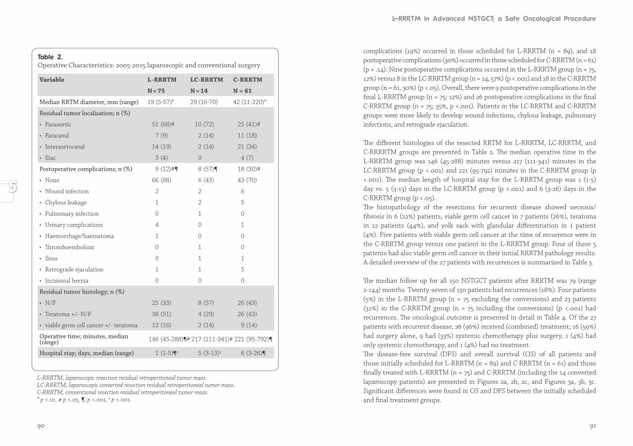
90 91
Operative Characteristics: 2005-2015 laparoscopic and conventional surgery
Variable L-RRRTM LC-RRRTM C-RRRTM
N = 75 N = 14 N = 61
Median RRTM diameter, mm (range) 19 (5-57)* 29 (16-70) 42 (11-220)*
Residual tumor localisation; n (%)
• Paraaortic 51 (68)# 10 (72) 25 (41)#
• Paracaval 7 (9) 2 (14) 11 (18)
• Interaortocaval 14 (19) 2 (14) 21 (34)
• Iliac 3 (4) 0 4 (7)
Postoperative complications; n (%) 9 (12)#¶ 8 (57)¶ 18 (30)#
• None 66 (88) 6 (43) 43 (70)
• Wound infection 2 2 6
• Chylous leakage 1 2 5
• Pulmonary infection 0 1 0
• Urinary complications 4 0 1
• Haemorrhage/haematoma 1 0 0
• Thromboembolism 0 1 0
• Ileus 0 1 1
• Retrograde ejaculation 1 1 5
• Incisional hernia 0 0 0
Residual tumor histology; n (%)
• N/F 25 (33) 8 (57) 26 (43)
• Teratoma +/- N/F 38 (51) 4 (29) 26 (43)
• viable germ cell cancer +/- teratoma 12 (16) 2 (14) 9 (14)
Operative time; minutes, median (range) 146 (45-288)¶# 217 (111-341)# 221 (95-792)¶
Hospital stay; days, median (range) 1 (1-5)¶a 5 (3-13)a 6 (3-26)¶
L-RRRTM, laparoscopic resection residual retroperitoneal tumor mass.LC-RRRTM, laparoscopic converted resection residual retroperitoneal tumor mass.C-RRRTM, conventional resection residual retroperitoneal tumor mass.* p <.01, # p <.05, ¶, p <.001, a p <.001.
complications (19%) occurred in those scheduled for L-RRRTM (n = 89), and 18 postoperative complications (30%) occurred in those scheduled for C-RRRTM (n = 61) (p = .14). Nine postoperative complications occurred in the L-RRRTM group (n = 75, 12%) versus 8 in the LC-RRRTM group (n = 14, 57%) (p <.001) and 18 in the C-RRRTM group (n = 61, 30%) (p <.05). Overall, there were 9 postoperative complications in the final L-RRRTM group (n = 75; 12%) and 26 postoperative complications in the final C-RRRTM group (n = 75; 35%, p <.001). Patients in the LC-RRRTM and C-RRRTM groups were more likely to develop wound infections, chylous leakage, pulmonary infections, and retrograde ejaculation.
The different histologies of the resected RRTM for L-RRRTM, LC-RRRTM, and C-RRRRTM groups are presented in Table 2. The median operative time in the L-RRRTM group was 146 (45-288) minutes versus 217 (111-341) minutes in the LC-RRRTM group (p <.001) and 221 (95-792) minutes in the C-RRRTM group (p <.001). The median length of hospital stay for the L-RRRTM group was 1 (1-5) day vs. 5 (3-13) days in the LC-RRRTM group (p <.001) and 6 (3-26) days in the C-RRRTM group (p <.05).The histopathology of the resections for recurrent disease showed necrosis/fibrosis in 6 (22%) patients, viable germ cell cancer in 7 patients (26%), teratoma in 12 patients (44%), and yolk sack with glandular differentiation in 1 patient (4%). Five patients with viable germ cell cancer at the time of recurrence were in the C-RRRTM group versus one patient in the L-RRRTM group. Four of these 5 patients had also viable germ cell cancer in their initial RRRTM pathology results. A detailed overview of the 27 patients with recurrences is summarized in Table 3.
The median follow up for all 150 NSTGCT patients after RRRTM was 79 (range 2-144) months. Twenty-seven of 150 patients had recurrences (18%). Four patients (5%) in the L-RRRTM group (n = 75 excluding the conversions) and 23 patients (31%) in the C-RRRTM group (n = 75 including the conversions) (p <.001) had recurrences. The oncological outcome is presented in detail in Table 4. Of the 27 patients with recurrent disease, 26 (96%) received (combined) treatment; 16 (59%) had surgery alone, 9 had (33%) systemic chemotherapy plus surgery, 1 (4%) had only systemic chemotherapy, and 1 (4%) had no treatment. The disease-free survival (DFS) and overall survival (OS) of all patients and those initially scheduled for L-RRRTM (n = 89) and C-RRRTM (n = 61) and those finally treated with L-RRRTM (n = 75) and C-RRRTM (including the 14 converted laparoscopy patients) are presented in Figures 2a, 2b, 2c, and Figures 3a, 3b, 3c. Significant differences were found in OS and DFS between the initially scheduled and final treatment groups.
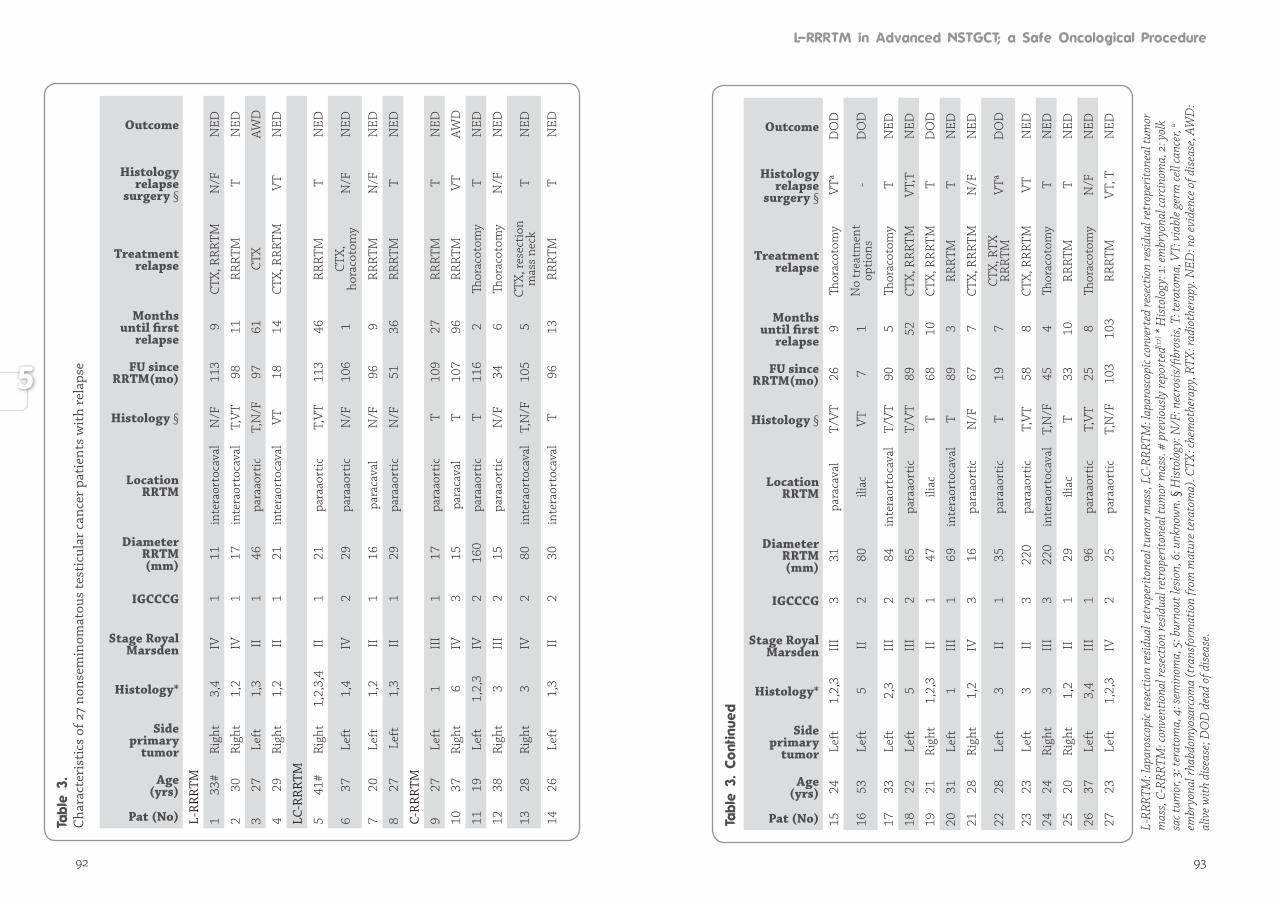
92 93
C
hara
cter
isti
cs o
f 27
nons
emin
omat
ous
test
icul
ar c
ance
r pa
tien
ts w
ith
rela
pse
Pat (No)
Age(yrs)
Side primary
tumor
Histology*
Stage Royal Marsden
IGCCCG
DiameterRRTM (mm)
LocationRRTM
Histology §
FU sinceRRTM(mo)
Months until first
relapse
Treatment relapse
Histology relapse
surgery §
Outcome
L-R
RRT
M
133
#R
ight
3,4
IV1
11in
tera
orto
cava
lN
/F11
39
CTX,
RR
RTM
N/F
NED
230
Rig
ht1,
2IV
117
inte
raor
toca
val
T,V
T98
11R
RRT
MT
NED
3 27
Left
1,3
II1
46pa
raao
rtic
T,N
/F97
61CT
XAW
D
429
Rig
ht1,
2II
121
inte
raor
toca
val
VT
1814
CTX,
RR
RTM
VT
NED
LC-R
RRT
M
541
#R
ight
1,2,
3,4
II1
21pa
raao
rtic
T,V
T11
346
RR
RTM
TN
ED
637
Left
1,4
IV2
29pa
raao
rtic
N/F
106
1CT
X,
hora
coto
my
N/F
NED
720
Left
1,2
II1
16pa
raca
val
N/F
969
RR
RTM
N/F
NED
827
Lef
t1,
3II
129
para
aort
icN
/F51
36R
RRT
MT
NED
C-R
RRT
M
927
Left
1II
I1
17pa
raao
rtic
T10
927
RR
RTM
TN
ED
1037
Rig
ht6
IV3
15pa
raca
val
T10
796
RR
RTM
VT
AWD
1119
Left
1,2,
3IV
216
0pa
raao
rtic
T11
62
Thor
acot
omy
TN
ED
1238
Rig
ht3
III
215
para
aort
ic
N/F
346
Thor
acot
omy
N/F
NED
1328
Rig
ht3
IV2
80in
tera
orto
cava
lT,
N/F
105
5CT
X, re
sect
ion
mas
s ne
ckT
NED
1426
Left
1,3
II2
30in
tera
orto
cava
lT
9613
RR
RTM
TN
ED
Pat (No)
Age(yrs)
Side primary
tumor
Histology*
Stage Royal Marsden
IGCCCG
DiameterRRTM (mm)
LocationRRTM
Histology §
FU sinceRRTM(mo)
Months until first
relapse
Treatment relapse
Histology relapse
surgery §
Outcome
1524
Left
1,2,
3II
I3
31pa
raca
val
T/V
T26
9
Thor
acot
omy
VTª
D
OD
1653
Left
5II
280
iliac
VT
71
No
trea
tmen
t op
tion
s-
DO
D
1733
Left
2,3
III
284
inte
raor
toca
val
T/V
T90
5Th
orac
otom
yT
NED
1822
Left
5 II
I2
65pa
raao
rtic
T/
VT
8952
CTX,
RR
RTM
VT,
TN
ED
1921
Rig
ht1,
2,3
II1
47ili
acT
6810
CTX,
RR
RTM
TD
OD
2031
Left
1II
I1
69in
tera
orto
cava
lT
893
RR
RTM
TN
ED
2128
Rig
ht1,
2IV
316
para
aort
icN
/F67
7CT
X, R
RRT
MN
/FN
ED
2228
Left
3II
135
para
aort
icT
197
CTX,
RTX
R
RRT
MV
TªD
OD
2323
Left
3II
322
0pa
raao
rtic
T,V
T58
8CT
X, R
RRT
MV
TN
ED
2424
Rig
ht3
III
322
0in
tera
orto
cava
lT,
N/F
454
Thor
acot
omy
TN
ED
2520
Rig
ht1,
2II
129
iliac
T33
10R
RRT
MT
NED
2637
Left
3,
4II
I1
96pa
raao
rtic
T,V
T25
8Th
orac
otom
yN
/FN
ED
2723
Left
1,2,
3IV
225
para
aort
icT,
N/F
103
103
RR
RTM
VT,
TN
ED
L-RR
RTM
: lap
aros
copi
c res
ectio
n re
sidua
l ret
rope
rito
neal
tum
or m
ass,
LC-R
RRTM
: lap
aros
copi
c con
vert
ed re
sect
ion
resid
ual r
etro
peri
tone
al tu
mor
m
ass,
C-RR
RTM
: con
vent
iona
l res
ectio
n re
sidua
l ret
rope
rito
neal
tum
or m
ass.
# pr
evio
usly
repo
rted
{17}
* H
isto
logy
: 1: e
mbr
yona
l car
cinom
a, 2
: yol
k sa
c tum
or, 3
: ter
atom
a, 4
: sem
inom
a, 5
: bur
nout
lesio
n, 6
: unk
now
n. §
His
tolo
gy: N
/F: n
ecro
sis/
fibro
sis,
T: te
rato
ma,
VT:
via
ble
germ
cell
canc
er, a
embr
yona
l rha
bdom
yosa
rcom
a (t
rans
form
atio
n fr
om m
atur
e te
rato
ma)
. CTX
: che
mot
hera
py, R
TX: r
adio
ther
apy.
NED
: no
evid
ence
of d
isea
se, A
WD
: al
ive
with
dis
ease
; DO
D d
ead
of d
isea
se.
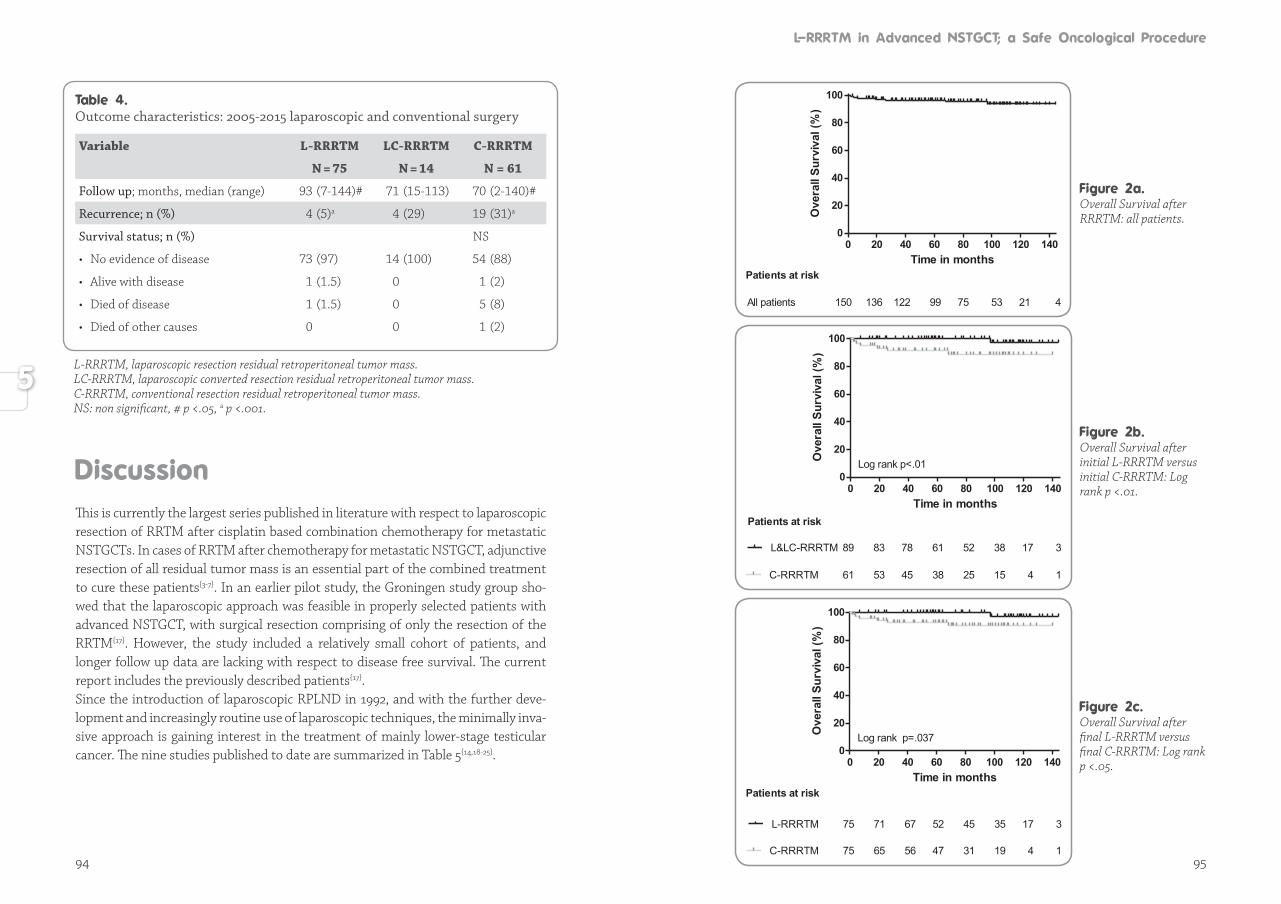
94 95
This is currently the largest series published in literature with respect to laparoscopic resection of RRTM after cisplatin based combination chemotherapy for metastatic NSTGCTs. In cases of RRTM after chemotherapy for metastatic NSTGCT, adjunctive resection of all residual tumor mass is an essential part of the combined treatment to cure these patients{3-7}. In an earlier pilot study, the Groningen study group sho-wed that the laparoscopic approach was feasible in properly selected patients with advanced NSTGCT, with surgical resection comprising of only the resection of the RRTM{17}. However, the study included a relatively small cohort of patients, and longer follow up data are lacking with respect to disease free survival. The current report includes the previously described patients{17}. Since the introduction of laparoscopic RPLND in 1992, and with the further deve-lopment and increasingly routine use of laparoscopic techniques, the minimally inva-sive approach is gaining interest in the treatment of mainly lower-stage testicular cancer. The nine studies published to date are summarized in Table 5{14,18-25}.
Outcome characteristics: 2005-2015 laparoscopic and conventional surgery
Variable L-RRRTM LC-RRRTM C-RRRTM
N = 75 N = 14 N = 61
Follow up; months, median (range) 93 (7-144)# 71 (15-113) 70 (2-140)#
Recurrence; n (%) 4 (5)a 4 (29) 19 (31)a
Survival status; n (%) NS
• No evidence of disease 73 (97) 14 (100) 54 (88)
• Alive with disease 1 (1.5) 0 1 (2)
• Died of disease 1 (1.5) 0 5 (8)
• Died of other causes 0 0 1 (2)
L-RRRTM, laparoscopic resection residual retroperitoneal tumor mass.LC-RRRTM, laparoscopic converted resection residual retroperitoneal tumor mass.C-RRRTM, conventional resection residual retroperitoneal tumor mass.NS: non significant, # p <.05, a p <.001.
Time in months
Ove
rall
Surv
ival
(%)
0 20 40 60 80 100 120 1400
20
40
60
80
100
Patients at risk
All patients 150 136 122 99 75 53 21 4
Time in months
Ove
rall
Surv
ival
(%)
0 20 40 60 80 100 120 1400
20
40
60
80
100
L&LC-RRRTM 89 83 78 61 52 38 17 3
C-RRRTM 61 53 45 38 25 15 4 1
Patients at risk
Log rank p<.01
Time in months
Ove
rall
Surv
ival
(%)
0 20 40 60 80 100 120 1400
20
40
60
80
100
L-RRRTM 75 71 67 52 45 35 17 3
C-RRRTM 75 65 56 47 31 19 4 1
Patients at risk
Log rank p=.037Overall Survival after final L-RRRTM versus final C-RRRTM: Log rank p <.05.
Overall Survival after initial L-RRRTM versus initial C-RRRTM: Log rank p <.01.
Overall Survival after RRRTM: all patients.
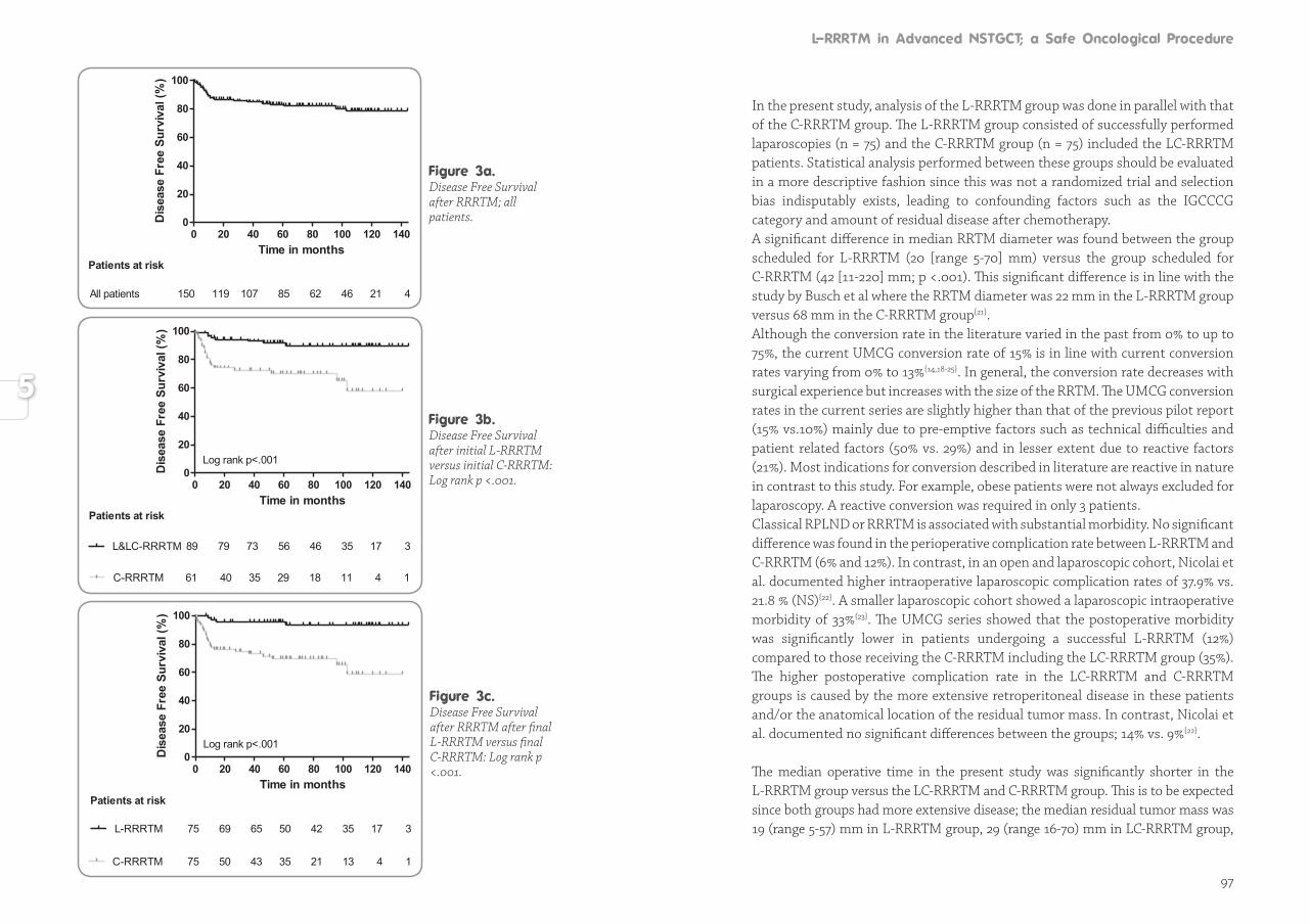
97
Time in months
Dis
ease
Fre
e Su
rviv
al (%
)
0 20 40 60 80 100 120 1400
20
40
60
80
100
All patients 150 119 107 85 62 46 21 4
Patients at risk
Time in months
Dis
ease
Fre
e Su
rviv
al (%
)
0 20 40 60 80 100 120 1400
20
40
60
80
100
L&LC-RRRTM 89 79 73 56 46 35 17 3
C-RRRTM 61 40 35 29 18 11 4 1
Patients at risk
Log rank p<.001
Time in months
Dis
ease
Fre
e Su
rviv
al (%
)
0 20 40 60 80 100 120 1400
20
40
60
80
100
L-RRRTM 75 69 65 50 42 35 17 3
C-RRRTM 75 50 43 35 21 13 4 1
Patients at risk
Log rank p<.001
Disease Free Survival after RRRTM after final L-RRRTM versus final C-RRRTM: Log rank p <.001.
Disease Free Survival after initial L-RRRTM versus initial C-RRRTM: Log rank p <.001.
Disease Free Survival after RRRTM; all patients.
In the present study, analysis of the L-RRRTM group was done in parallel with that of the C-RRRTM group. The L-RRRTM group consisted of successfully performed laparoscopies (n = 75) and the C-RRRTM group (n = 75) included the LC-RRRTM patients. Statistical analysis performed between these groups should be evaluated in a more descriptive fashion since this was not a randomized trial and selection bias indisputably exists, leading to confounding factors such as the IGCCCG category and amount of residual disease after chemotherapy.A significant difference in median RRTM diameter was found between the group scheduled for L-RRRTM (20 [range 5-70] mm) versus the group scheduled for C-RRRTM (42 [11-220] mm; p <.001). This significant difference is in line with the study by Busch et al where the RRTM diameter was 22 mm in the L-RRRTM group versus 68 mm in the C-RRRTM group{21}. Although the conversion rate in the literature varied in the past from 0% to up to 75%, the current UMCG conversion rate of 15% is in line with current conversion rates varying from 0% to 13%{14,18-25}. In general, the conversion rate decreases with surgical experience but increases with the size of the RRTM. The UMCG conversion rates in the current series are slightly higher than that of the previous pilot report (15% vs.10%) mainly due to pre-emptive factors such as technical difficulties and patient related factors (50% vs. 29%) and in lesser extent due to reactive factors (21%). Most indications for conversion described in literature are reactive in nature in contrast to this study. For example, obese patients were not always excluded for laparoscopy. A reactive conversion was required in only 3 patients.Classical RPLND or RRRTM is associated with substantial morbidity. No significant difference was found in the perioperative complication rate between L-RRRTM and C-RRRTM (6% and 12%). In contrast, in an open and laparoscopic cohort, Nicolai et al. documented higher intraoperative laparoscopic complication rates of 37.9% vs. 21.8 % (NS){22}. A smaller laparoscopic cohort showed a laparoscopic intraoperative morbidity of 33%{23}. The UMCG series showed that the postoperative morbidity was significantly lower in patients undergoing a successful L-RRRTM (12%) compared to those receiving the C-RRRTM including the LC-RRRTM group (35%). The higher postoperative complication rate in the LC-RRRTM and C-RRRTM groups is caused by the more extensive retroperitoneal disease in these patients and/or the anatomical location of the residual tumor mass. In contrast, Nicolai et al. documented no significant differences between the groups; 14% vs. 9%{22}.
The median operative time in the present study was significantly shorter in the L-RRRTM group versus the LC-RRRTM and C-RRRTM group. This is to be expected since both groups had more extensive disease; the median residual tumor mass was 19 (range 5-57) mm in L-RRRTM group, 29 (range 16-70) mm in LC-RRRTM group,
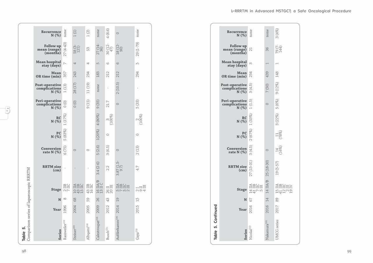
98 99
Co
mpa
riso
n se
ries
of l
apar
osco
pic
RR
RTM
Seri
es
Year
N
Stage
RRTM size (cm)
Conversion rate N (%)
PCN (%)
RCN (%)
Peri-operative complications
N (%)
Post-operative complications
N (%)
Mean OR time (min)
Mean hospital stay (days)
Follow up mean (range)
(months)
Recurrence N (%)
Ras
swei
ler{1
9}19
968
2
IIB
7
IIC
-6
(75)
5 (8
3%)
1 (1
7%)
0 (0
)1
(13)
357
727
(4-4
3)no
ne
Stei
ner{2
0}20
0468
10
IIA
43
IIB
15
IIC
-0
0 (0
)28
(17)
243
458
(3-
121)
1 (1
)
Alb
qam
i{14}
2005
59 4
3 II
B 1
6 II
C-
0 9
(15)
11 (1
9)23
44
531
(2)
Cale
stro
upat
{18}
2009
26 1
6 II
A/B
1
3 II
C
3.4
(2-6
)5
(2.6
) 1(
20%
)4
(80%
)9
(35)
no
ne18
35
27 (1
4-36
)no
ne
Busc
h{21}
2012
43
26
II 2
0 II
I2.
2 3
(6.5
)0
3 (1
00%
)21
.7
-21
2 6
30 (1
2-47
)4
(8.6
)
Aufd
erka
mm
{24}
20
1419
5
IIA
7
IIB
5
IIC
2
III
3.87
(1.5
-9.
7)0
02
(10.
5)21
26
18 (1
2-90
)0
Gay
a{23}
2015
15
2 I
9
II
4 II
I
4.7
2 (1
3)0
2 (1
00%
)5
(33)
-29
45
29 (1
-79)
none
Seri
es
Year
N
Stage
RRTM size (cm)
Conversion rate N (%)
PCN (%)
RCN (%)
Peri-operative complications
N (%)
Post-operative complications
N (%)
Mean OR time (min)
Mean hospital stay (days)
Follow up mean (range)
(months)
Recurrence N (%)
Nic
olai
{22}
2016
67 1
4 II
A
41
IIB
7
IIC
5
III
27 (1
5-31
)3
(4.5
)2
(67%
)1
(33%
)1
(1.5
)3
(4.5
)23
43
21no
ne
Nak
amur
a{25}
2016
14 1
4 II
A/B
25 (1
8-30
)0
07
(50)
439
136
none
UM
CG s
erie
s 20
1789
15
IIA
41 I
IB
12
IIC
2
III
19
IV
19 (5
-57)
14
(16%
)11
(7
8%)
3 (2
2%)
5 (6
%)
9 (1
2%)
148
179
(7-
144)
3 (4
%)
100 101
and 42 (range 11-220) mm in the C-RRRTM group. In contrast, other more recent studies reported longer operative times for laparoscopic procedures of 234, 294, and 439 minutes{23-25}. Nicolai et al reported equivalent operative times (212 vs. 232 min, p = 0.3){22}. A study originating from Japan reported not only longer operative times in the laparoscopic group, but also in the open group (439 vs. 408 minutes){25}.As expected, the L-RRRTM group required a significantly shorter hospital stay compared to the LC-RRTM and C-RRRTM groups (medians of 1 day vs. 5 and 6 days; p <.001). In contrast to the short hospital stay for the laparoscopy patients in this series, the median hospital stay in other studies was longer after L-RRRTM, ranging from 3-7 days (Table 5). Nicolai et al. also compared the L-RRRTM group versus the C-RRRTM group and documented a median hospital stay of 6 vs. 11.5 days (p <.01){22}.
Thus far, there are no long-term outcome data in advanced nonseminomatous germ cell tumors comparing laparoscopic RRRTM to conventional RRRTM. After a median follow-up of 79 months, 27 patients (18%) developed recurrent disease; 19 in the initial C-RRRTM group (31%) and 8 in the initial L-RRRTM (9%): 4 in the final L-RRRTM group (5%) and 4 in the LC-RRRTM group (28%). The overall recurrence rate in the C-RRRTM, including the laparoscopy conversion patients, was not different from the initial C-RRRTM group (31%). The recurrences in the LC-RRRTM group were not related to the reason for the conversion; aortic injury, vena cava injury, and two technical difficulties due to extensive fibrosis. Other laparoscopic surgeries for resection of residual masses also showed low recurrence rates (Table 5). However, most reports consist of only small patient series and predominantly in patients with good prognosis disease and small residual tumor masses, with a limited follow-up duration{14,18-20}.When selecting patients for L-RRRTM, factors other than merely the size of the mass are important. Selection criteria are not absolute and these criteria are dynamic; first, in relation to developing skills and second, the anatomical site of the residual mass. Large residual masses greater than 5 cm might be eligible for a laparoscopic approach based on the laparoscopic experience of the surgical oncologist and the anatomical location. Surgeons can now also perform robotic-assisted RRRTM{26,27}. Within a few years, robotic surgery might extend the indication for L-RRRTM, even for more chal len-ging RRTM. L-RRRTM meets or exceeds the results from most open conventional surgeries and should always be considered as a viable alternative in the resection of residual tumor mass after cisplatin containing chemotherapy for locally advanced testicular cancer, offering less morbidity, a favorable cosmetic outcome for the pa-tient, and a shorter hospital stay. The study showed that laparoscopic resection of
RRTM in proper selected and well-defined patients is a feasible and oncologically safe procedure. Although a randomized trial comparing both treatment modalities is the gold standard to proof the superiority of this minimally invasive approach this will probably not performed. A multidisciplinary expert testicular cancer team with an experienced (oncological) laparoscopist is essential to discuss options with the patient.
In conclusion, with a robust sample size and a median follow-up duration of more than 6 years, this study confirms that laparoscopic resection of well-defined RRTM af ter cisplatin based combination chemotherapy for metastatic nonseminomatous tes ti cular cancer is a feasible and safe surgical and oncological procedure, offering com parable oncological results as conventional surgery, with less morbidity, a shorter hos pi tal stay, and an excellent cosmetic outcome for the patient with testicular cancer.
1. International Germ Cell Consensus Classification: a prognostic factor-based staging system for metastatic germ cell cancers. International Germ Cell Cancer Collaborative Group. J Clin Oncol 1997; 15: 594-603.
2. Sonneveld DJ, Hoekstra HJ, van der Graaf WT, Sluiter WJ, Mulder NH, Willemse PH et al. Improved long term survival of patients with metastatic nonseminomatous testicular germ cell carcinoma in relation to prognostic classification systems during the cisplatin era. Cancer 2001; 91: 1304-1315.
3. Oldenburg J, Alfsen GC, Lien HH, Aass N, Waehre H, Fossa SD. Postchemotherapy retroperitoneal surgery remains necessary in patients with nonseminomatous testicular cancer and minimal residual tumor masses. J Clin Oncol 2003; 21: 3310–3317.
4. Sonneveld DJ, Sleijfer DT, Schraffordt Koops H, Keemers-Gels ME, Molenaar WM, Hoekstra HJ. Mature teratoma identified after postchemotherapy surgery in patients with disseminated nonseminomatous testicular germ cell tumors: a plea for an aggressive surgical approach. Cancer 1998; 82: 1343-1351.
5. Gels ME, Hoekstra HJ, Sleijfer DT, Nijboer AP, Molenaar WM, Ebels T et al. Thoracotomy for postchemotherapy resection of pulmonary residual tumor mass in patients with nonseminomatous testicular germ cell tumors: aggressive surgical resection is justified. Chest 1997; 112: 967-973.
6. Beyer J, Albers P, Altena R, Aparicio J, Bokemeyer C, Busch J et al. Maintaining success, reducing treatment burden, focusing on survivorship: highlights from the third European consensus conference on diagnosis and treatment of germ-cell cancer. Ann Oncol 2013; 24: 878-888.
102
7. Albers P, Albrecht W, Algaba F, Bokemeyer C, Cohn-Cedermark G, Fizazi K et al. European Association of Urology. Guidelines on testicular cancer: 2015 update. Eur Urol 2015; 6: 1054-1068.
8. van Basten JP, Schrafford Koops H, Sleijfer DT, Pras E, van Driel MF, Hoekstra HJ. Current concepts about testicular cancer. Eur J Surg Oncol 1997; 23: 354-360.
9. Gels ME, Nijboer AP, Hoekstra HJ. Complications of the post-chemotherapy resection of retroperitoneal residual tumour mass in patients with non-seminomatosous testicular germ cell tumours. Br J Urol 1997; 79: 263-268.
10. Rabbani F, Goldenberg SL, Gleave ME, Paterson RF, Murray N, Sullivan LD. Retroperitoneal lymphadenectomy for postchemotherapy residual masses: is a modified dissection and resection of residual masses sufficient? Br J Urol 1998; 81: 295–300.
11. Heidenreich A, Pfister D, Witthuhn R, Thüer D, Albers P. Postchemotherapy retroperitoneal lymph node dissection in advanced testicular cancer: radical or modified template resection. Eur Urol 2009; 55: 217-224.
12. van Basten JP, Jonker-Pool G, van Driel MF, Sleijffer DT, van der Wiel HB, Hoekstra HJ. The sexual sequelae of testicular cancer. Cancer Treat Rev 1995; 21: 479-495.
13. Rukstalis DB, Chodak GW. Laparoscopic retroperitoneal lymph node dissection in a patient with stage 1 testicular carcinoma. J Urol 1992; 148: 1907-1909.
14. Albqami N, Janetschek G. Laparoscopic retroperitoneal lymph-node dissection in the management of clinical stage I and II testicular cancer. J Endourol 2005; 19: 683-692.
15. Bhayani SB, Ong A, Oh WK, Kantoff PW, Kavoussi LR. Laparoscopic retroperitoneal lymph node dissection for clinical stage I nonseminomatous germ cell testicular cancer: a long-term update. Urology 2003; 62 :324-327.
16. Nielsen ME, Lima G, Schaeffer EM, Porter J, Cadeddu JA, Tuerk I. Oncologic efficacy of laparoscopic RPLND in treatment of clinical stage I nonseminomatous germ cell testicular cancer. Urology 2007; 70: 1168-1172.
17. Öztürk Ç, van Ginkel RJ, Krol RM, Gietema JA, Hofker HS, Hoekstra HJ. Laparoscopic resection of a residual retroperitoneal tumor mass of nonseminomatous testicular germ cell tumors. Surg endosc 2012 ; 26: 458-467.
18. Calestroupat JP, Sanchez-Salas R, Cathelineau X, Rozet F, Galiano M, Smyth G et al. Postchemotherapy laparoscopic retroperitoneal lymph node dissection in nonseminomatous germ-cell tumor. J Endourol 2009; 23: 645-650.
19. Rassweiler JJ, Seemann O, Henkel TO, Stock C, Frede T, Alken P. Laparoscopic retroperitoneal lymph node dissection for nonseminomatous germ cell tumors: indications and limitations. J Urol 1996; 156: 1108-1113.
20. Steiner H, Peschel R, Janetschek G, Höltl L, Berger AP, Bartsch G et al. Long-term results of laparoscopic retroperitoneal lymph node dissection: a single-center 10-year experience. Urology 2004; 63: 550-555.
21. Busch J, Magheli A, Erber B, Friedersdorff F, Hoffmann I, Kempkensteffen C et al. Laparoscopic and open postchemotherapy retroperitoneal lymph node dissection in patients with advanced testicular cancer-a single center analysis. BMC Urol 2012; 12: 15.
22. Nicolai N, Cattaneo F, Biasoni D, Catanzaro M, Torelli T, Zazzara M et al. Laparoscopic Postchemotherapy Retroperitoneal Lymph Node Dissection Can Be a Standard Option in Defined Nonseminomatous Germ Cell Tumor Patients. J Endourol 2016; 30: 1112-1119.
23. Gaya JM, Palou J, Peña JA, Rosales A, Maroto P, Sullivan I et al. Is there a limit for the laparoscopic approach of a retroperitoneal residual mass postchemotherapy? Actas Urol Esp 2015; 39: 264-267.
24. Aufderkamm S, Todenhöfer T, Hennenlotter J, Mischinger J, Böttge J, Rausch S et al. Postchemotherapy Laparoscopic retroperitoneal lymph node dissection for nonseminomatous germ cell tumors infiltrating the great vessels. Journal of Endourol 2014; 28: 668-674.
25. Nakamura T, Kawauchi A, Oishi M, Ueda T, Nakanishi H, Kamoi K et al. Postchemotherapy Laparoscopic retroperitoneal lymph node dissection is feasible for stage IIa/B non-seminoma germ cell tumors. Int J Clin Oncol 2016; 21: 791-795.
26. Chalvin HJ, Ludwig W, Pierorazio PM, Allaf ME. Robotic Primary RPLND for Stage I Testicular Cancer: a Review of Indications and Outcomes. Curr Urol Rep 2016; 17: 41.
27. Stepanian S, Patel M, Porter J. Robot-assisted Laparoscopic Retroperitoneal Lymph Node Dissection for Testicular Cancer: Evolution of the Technique. Eur Urol 2016: 70: 661-667.
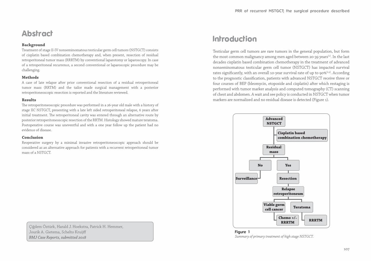
107
BackgroundTreatment of stage II-IV nonseminomatous testicular germ cell tumors (NSTGCT) consists of cisplatin based combination chemotherapy and, when present, resection of residual retroperitoneal tumor mass (RRRTM) by conventional laparotomy or laparoscopy. In case of a retroperitoneal recurrence, a second conventional or laparoscopic procedure may be challenging.
MethodsA case of late relapse after prior conventional resection of a residual retroperitoneal tumor mass (RRTM) and the tailor made surgical management with a posterior retroperitoneoscopic resection is reported and the literature reviewed.
ResultsThe retroperitoneoscopic procedure was performed in a 26-year old male with a history of stage IIC NSTGCT, presenting with a late left sided retroperitoneal relapse, 6 years after initial treatment. The retroperitoneal cavity was entered through an alternative route by posterior retroperitoneoscopic resection of the RRTM. Histology showed mature teratoma. Postoperative course was uneventful and with a one year follow up the patient had no evidence of disease.
ConclusionReoperative surgery by a minimal invasive retroperitoneoscopic approach should be considered as an alternative approach for patients with a recurrent retroperitoneal tumor mass of a NSTGCT.
Çiğdem Öztürk, Harald J. Hoekstra, Patrick H. Hemmer, Jourik A. Gietema, Schelto Kruijff BMJ Case Reports, submitted 2018
Testicular germ cell tumors are rare tumors in the general population, but form the most common malignancy among men aged between 20-39 years{1}. In the last decades cisplatin based combination chemotherapy in the treatment of advanced nonseminomatous testicular germ cell tumor (NSTGCT) has impacted survival rates significantly, with an overall 10-year survival rate of up to 90%{2,3}. According to the prognostic classification, patients with advanced NSTGCT receive three or four courses of BEP (bleomycin, etoposide and cisplatin) after which restaging is performed with tumor marker analysis and computed tomography (CT) scanning of chest and abdomen. A wait and see policy is conducted in NSTGCT when tumor markers are normalized and no residual disease is detected (Figure 1).
AdvancedNSTGCT
No Yes
Surveillance Resection
Teratoma
RRRTM
Residual mass
Relapseretroperitoneum
Viable germ cell cancer
Chemo +/-RRRTM
Cisplatin based combination chemotherapy
Summary of primary treatment of high stage NSTGCT.
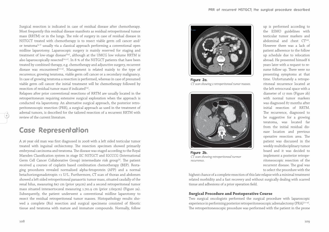
108 109
Surgical resection is indicated in case of residual disease after chemotherapy. Most frequently this residual disease manifests as residual retroperitoneal tumor mass (RRTM) or in the lungs. The role of surgery in case of residual disease in NSTGCT treated with chemotherapy is to resect viable germ cell cancer and/or teratoma{4-7} usually via a classical approach performing a conventional open midline laparotomy. Laparoscopic surgery is mainly reserved for staging and treatment of low-stage disease{8,9}, although at the UMCG low volume RRTM is also laparoscopically resected{10,11}. In 8 % of the NSTGCT patients that have been treated by combined therapy, e.g. chemotherapy and adjunctive surgery, recurrent disease was encountered{12,13}. Management is related mainly to the type of recurrence; growing teratoma, viable germ cell cancer or a secondary malignancy. In case of growing teratoma a resection is performed, whereas in case of presumed viable germ cell cancer the initial treatment will be chemotherapy followed by resection of residual tumor mass if indicated{15}. Relapses after prior conventional resections of RRTM are usually located in the retroperitoneum requiring extensive surgical exploration when the approach is conducted via laparotomy. An alternative surgical approach, the posterior retro-peritoneoscopic resection (PRR), a surgical approach as used in the treatment of adrenal tumors, is described for the tailored resection of a recurrent RRTM with review of the current literature.
A 26 year old man was first diagnosed in 2008 with a left sided testicular tumor trea ted with inguinal orchiectomy. The resection specimen showed primarily embry onal carcinoma and teratoma. The disease was staged according to the Royal Marsden Classification system in stage IIC NSTGCT and IGCCCG (International Germ Cell Cancer Collaborative Group) intermediate risk group{2}. The patient recei ved 4 courses of cisplatin based combination chemotherapy (BEP). Resta-ging procedures revealed normalised alpha-fetoprotein (AFP) and a normal betachoriongonadotropin <1 U/L. Furthermore, CT scan of thorax and abdomen showed a left sided retroperitoneal paraaortic tumor mass, situated caudally of the renal hilus, measuring 6x7 cm (prior 5x5cm) and a second retroperitoneal tumor mass situated inter aortocaval measuring 1.7x1.9 cm (prior 2.6x3cm) (Figure 2a). Subsequently, the patient underwent a conventional midline laparotomy to resect the residual retroperitoneal tumor masses. Histopathology results sho-wed a complete (R0) resection and surgical specimens consisted of fibrotic tissue and teratoma with mature and immature compounds. Normally, follow
up is performed according to the ESMO guidelines with testicular tumor markers and abdominal and chest CT{16}. However there was a lack of patient adherence to the follow up schedule due to relocation abroad. He presented himself 6 years later with a request to re-sume follow up. There were no presenting symptoms at that time. Unfortunately a retro pe-ritoneal recurrence lo ca ted at the left retrocrural space with a diameter of 17 mm (Figure 2b) with normal tumor markers was diagnosed 87 months after initial resection of RRTM. The recurrence, diagnosed to be suggestive for a growing teratoma, was located far from the initial residual dis-ease location and previous ope rative resection area. The pa tient was discussed in the week ly multidisciplinary tumor board and it was decided to imple ment a posterior retro pe-ri to neoscopic resection of the recur rent disease. The goal was to select the procedure with the
highest chance of a com plete resec tion of this late relapse with a minimal treatment related morbidity and a fast reco very and without surgically dealing with scarred tissue and adhesions of a prior operation field.
Surgical Procedure and Postoperative CourseTwo surgical oncologists performed the surgical procedure with laparoscopic experience in performing posterior retroperitoneoscopic adrenalectomy (PRA){17-19}. The retroperitoneoscopic procedure was performed with the patient in the prone
CT scan showing 2 retroperitoneal tumor masses.
CT scan showing retroperitoneal turmor recurrence.
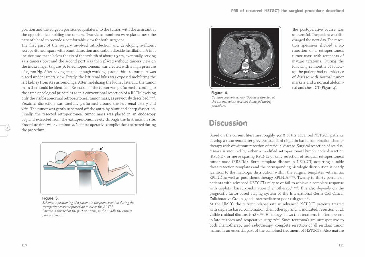
110 111
position and the surgeon positioned ipsilateral to the tumor, with the assistant at the opposite side holding the camera. Two video monitors were placed near the patient’s head to provide a comfortable view for both surgeons. The first part of the surgery involved introduction and developing sufficient retroperitoneal space with blunt dissection and carbon dioxide instillation. A first incision was made below the tip of the 12th rib of about 1.5 cm, eventually serving as a camera port and the second port was then placed without camera view on the index finger (Figure 3). Pneumoperitoneum was created with a high pressure of 25mm Hg. After having created enough working space a third 10 mm port was placed under camera view. Firstly, the left renal hilus was exposed mobilizing the left kidney from its surroundings. After mobilizing the kidney laterally, the tumor mass then could be identified. Resection of the tumor was performed according to the same oncological principles as in a conventional resection of a RRTM excising only the visible abnormal retroperitoneal tumor mass, as previously described{10,11}. Proximal dissection was carefully performed around the left renal artery and vein. The tumor was gently separated off the aorta by blunt and sharp dissection. Finally, the resected retroperitoneal tumor mass was placed in an endoscopy bag and extracted from the extraperitoneal cavity through the first incision site. Procedure time was 120 minutes. No intra operative complications occurred during the procedure.
Schematic positioning of a patient in the prone position during the retroperitoneoscopic procedure to excise the RRTM. *Arrow is directed at the port positions; in the middle the camera port is shown.
The postoperative course was un eventful. The patient was dis-charged the next day. The resec-tion specimen showed a R0 re sec tion of a retroperitoneal tu mor mass with remnants of ma ture teratoma. During the fol lo wing 12 months of follow-up the patient had no eviden ce of disease with nor mal tumor mar kers and a nor mal abdomi-nal and chest CT (Figure 4).
Based on the current literature roughly 3-23% of the advanced NSTGCT patients develop a recurrence after previous standard cisplatin based combination chemo-therapy with or without resection of residual disease. Surgical resection of residual disease is required by either a modified retroperitoneal lymph node dissection (RPLND), or nerve sparing RPLND, or only resection of residual retroperitoneal tumor mass (RRRTM). Extra template disease in NSTGCT, occurring outside these resection templates and the corresponding histologic distribution is nearly identical to the histologic distribution within the surgical templates with initial RPLND as well as post-chemotherapy RPLNDs{20,21}. Twenty to thirty percent of patients with advanced NSTGCTs relapse or fail to achieve a complete response with cisplatin based combination chemotherapy{22-24}. This also depends on the prognostic factor-based staging system of the International Germ Cell Cancer Collaborative Group: good, intermediate or poor risk group{2}.At the UMCG the current relapse rate in advanced NSTGCT patients treated with cisplatin based combination chemotherapy and, if indicated, resection of all visible residual disease, is 18 %{11}. Histology shows that teratoma is often present in late relapses and reoperative surgery{11}. Since teratoma’s are unresponsive to both chemotherapy and radiotherapy, complete resection of all residual tumor masses is an essential part of the combined treatment of NSTGCTs. Also mature
CT scan postoperatively. *Arrow is directed at the adrenal which was not damaged during procedure.
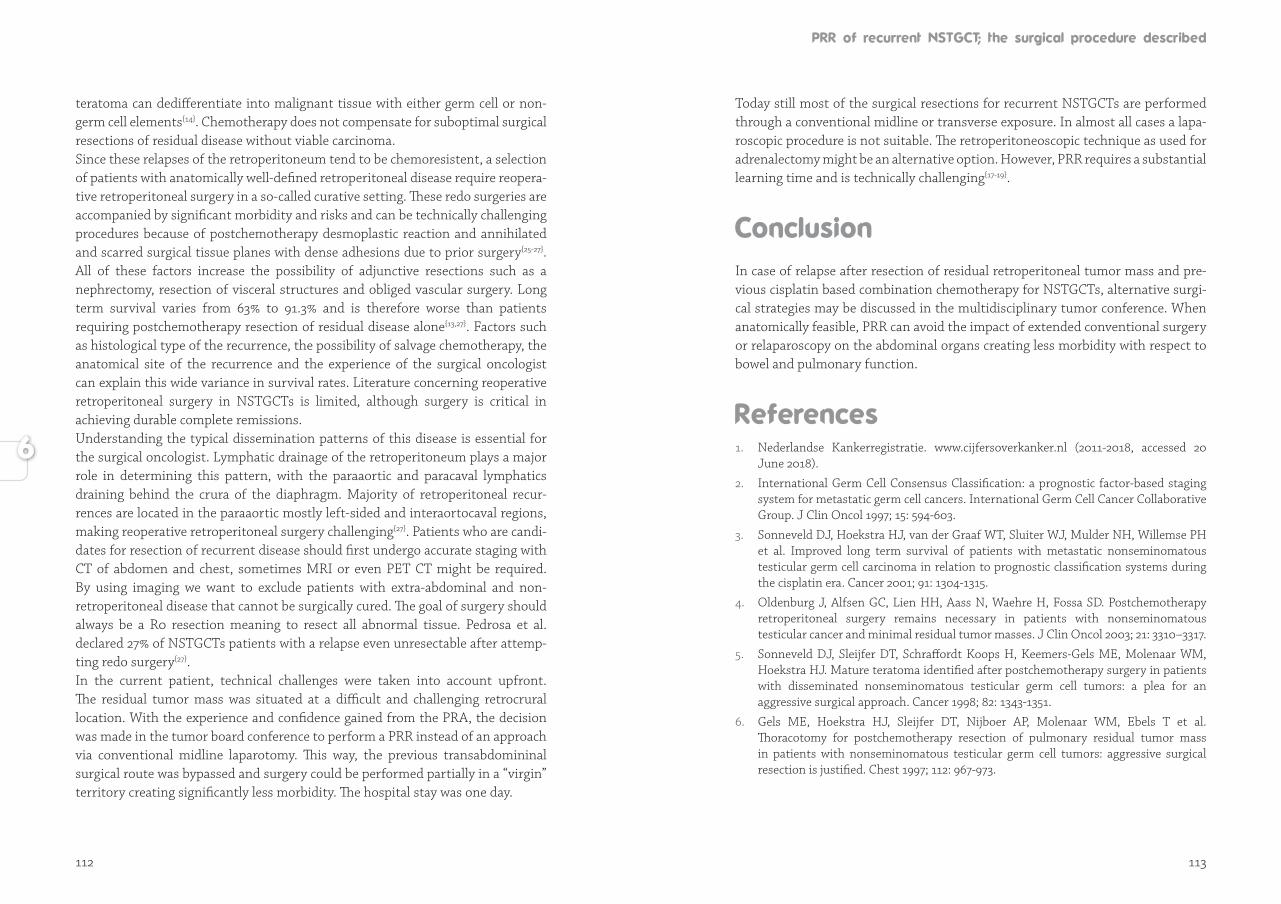
112 113
teratoma can dedifferentiate into malignant tissue with either germ cell or non-germ cell elements{14}. Chemotherapy does not compensate for suboptimal surgical resections of residual disease without viable carcinoma.Since these relapses of the retroperitoneum tend to be chemoresistent, a selec tion of patients with anatomically well-defined retroperitoneal disease require reope ra-tive retroperitoneal surgery in a so-called curative setting. These redo surgeries are accompanied by significant morbidity and risks and can be technically challenging procedures because of postchemotherapy desmoplastic reaction and annihilated and scarred surgical tissue planes with dense adhesions due to prior surgery{25-27}. All of these factors increase the possibility of adjunctive resections such as a nephrectomy, resection of visceral structures and obliged vascular surgery. Long term survival varies from 63% to 91.3% and is therefore worse than patients requiring postchemotherapy resection of residual disease alone{13,27}. Factors such as histological type of the recurrence, the possibility of salvage chemotherapy, the anatomical site of the recurrence and the experience of the surgical oncologist can explain this wide variance in survival rates. Literature concerning reoperative retroperitoneal surgery in NSTGCTs is limited, although surgery is critical in achieving durable complete remissions.Understanding the typical dissemination patterns of this disease is essential for the surgical oncologist. Lymphatic drainage of the retroperitoneum plays a major role in determining this pattern, with the paraaortic and paracaval lymphatics draining behind the crura of the diaphragm. Majority of retroperitoneal re cur-rences are loca ted in the paraaortic mostly left-sided and interaortocaval regions, making reope rative retroperitoneal surgery challenging{27}. Patients who are can di-dates for resection of recurrent disease should first undergo accurate sta ging with CT of ab domen and chest, sometimes MRI or even PET CT might be required. By using imaging we want to exclude patients with extra-abdominal and non-retro peritoneal disease that cannot be surgically cured. The goal of surgery should always be a R0 resection meaning to resect all abnormal tissue. Pedrosa et al. declared 27% of NSTGCTs patients with a relapse even unresectable after attemp-ting redo surgery{27}. In the current patient, technical challenges were taken into account upfront. The residual tumor mass was situated at a difficult and challenging retrocrural location. With the experience and confidence gained from the PRA, the decision was made in the tumor board conference to perform a PRR instead of an approach via conventional midline laparotomy. This way, the previous transabdomininal surgical route was bypassed and surgery could be performed partially in a “virgin” territory creating significantly less morbidity. The hospital stay was one day.
Today still most of the surgical resections for recurrent NSTGCTs are performed through a conventional midline or transverse exposure. In almost all cases a lapa-ro scopic procedure is not suitable. The retroperitoneoscopic technique as used for adrenalectomy might be an alternative option. However, PRR requires a substantial learning time and is technically challenging{17-19}.
In case of relapse after resection of residual retroperitoneal tumor mass and pre-vious cisplatin based combination chemotherapy for NSTGCTs, alternative surgi-cal strategies may be discussed in the multidisciplinary tumor conference. When anatomically feasible, PRR can avoid the impact of extended conventional surgery or relaparoscopy on the abdominal organs creating less morbidity with respect to bowel and pulmonary function.
1. Nederlandse Kankerregistratie. www.cijfersoverkanker.nl (2011-2018, accessed 20 June 2018).
2. International Germ Cell Consensus Classification: a prognostic factor-based staging system for metastatic germ cell cancers. International Germ Cell Cancer Collaborative Group. J Clin Oncol 1997; 15: 594-603.
3. Sonneveld DJ, Hoekstra HJ, van der Graaf WT, Sluiter WJ, Mulder NH, Willemse PH et al. Improved long term survival of patients with metastatic nonseminomatous testicular germ cell carcinoma in relation to prognostic classification systems during the cisplatin era. Cancer 2001; 91: 1304-1315.
4. Oldenburg J, Alfsen GC, Lien HH, Aass N, Waehre H, Fossa SD. Postchemotherapy retroperitoneal surgery remains necessary in patients with nonseminomatous testicular cancer and minimal residual tumor masses. J Clin Oncol 2003; 21: 3310–3317.
5. Sonneveld DJ, Sleijfer DT, Schraffordt Koops H, Keemers-Gels ME, Molenaar WM, Hoekstra HJ. Mature teratoma identified after postchemotherapy surgery in patients with disseminated nonseminomatous testicular germ cell tumors: a plea for an aggressive surgical approach. Cancer 1998; 82: 1343-1351.
6. Gels ME, Hoekstra HJ, Sleijfer DT, Nijboer AP, Molenaar WM, Ebels T et al. Thoracotomy for postchemotherapy resection of pulmonary residual tumor mass in patients with nonseminomatous testicular germ cell tumors: aggressive surgical resection is justified. Chest 1997; 112: 967-973.
114 115
7. Beyer J, Albers P, Altena R, Aparicio J, Bokemeyer C, Busch J et al. Maintaining success, reducing treatment burden, focusing on survivorship: highlights from the third European consensus conference on diagnosis and treatment of germ-cell cancer. Ann Oncol 2013; 24: 878-888.
8. Foster RS, Donohue JP. Retroperitoneal lymph node dissection for the management of clinical stage I nonseminoma. J Urol 2000; 163: 1788-1792.
9. Heidenreich A, Albers P, Hartmann M, Kliesch S, Kohrmann KU, Krege S et al. Complications of primary nerve sparing retroperitoneal lymph node dissection for clinical stage I nonseminomatous germ cell tumors of the testis: experience of the German Testicular Cancer Study Group. J Urol 2003; 169: 1710-1714.
10. Öztürk Ç, van Ginkel RJ, Krol RM, Gietema JA, Hofker HS, Hoekstra HJ. Laparoscopic resection of a residual retroperitoneal tumor mass of nonseminomatous testicular germ cell tumors. Surg Endosc 2012; 26: 458-467.
11. Öztürk Ç, Been LB, van Ginkel RJ, Gietema JA, Hoekstra HJ. Laparoscopic Resection of Residual Retroperitoneal Tumor Mass in Advanced Nonseminomatous Testicular Germ Cell Tumors; a Feasible and Safe Oncological Procedure. Manuscript submitted for publication.
12. Heindenreich, Pfister. Retroperitoneal lymphadenectomy and resection for testicular cancer: an update on best practice. Ther Adv Urol 2012; 4: 187–205.
13. Woldu SL, McKiernan JM. Reoperative retroperitoneal Surgery. Etiology and Clinical Outcome. Urol Clin North Am 2016; 42: 381-392.
14. Lutke Holzik MF, Hoekstra HJ, Mulder NH, Suurmeijer AJ, Sleijfer DT, Gietema JA. Non-germ cell malignancy in residual or recurrent mass after chemotherapy for nonseminomatous testicular germ cell tumor. Ann Surg Oncol 2003; 10: 131-135.
15. Albers P, Albrecht W, Algaba F, Bokemeyer C, Cohn-Cedermark G et al. EAU guidelines on testicular cancer: 2011 update; European Association of Urology. Eur Urol 2011; 60: 304-319.
16. Oldenburg J, Fossa SD, Nuver J, Heidenreich A, Schmoll HJ, Bokemeyer C et al. Testicular seminoma and non-seminoma: ESMO clinical practice guidelines for diagnosis, treatment, and follow up. Ann Oncol 2013; 24: 125-132.
17. Vrielink OM, Hemmer PH, Kruijff S. Considerations in minimally invasive adrenal surgery; the frontdoor or the backdoor? Minerva Chir 2018; 73: 93-99.
18. Vrielink OM, Wevers KP, Kist JW, Borel Rinkes HM, Hemmer PHJ, Vriens MR et al. Laparoscopic anterior versus endoscopic posterior approach for adrenalectomy: a shift to a new golden standard? Lang Arch Surg 2017; 402: 767-773.
19. Vrielink OM, Engelsman AF, Hemmer PHJ, de Vries J, Vorselaars WMCM, Vriens MR et al. Multicentre study evaluating the surgical learning curve for posterior retroperitoneoscopic adrenalectomy. Br J Surg 2018; 105: 544-551.
20. Eggener SE, Carver BS, Sharp DS, Motzer RJ, Bosl GJ, Sheinfeld J. Incidence of disease outside modified retroperitoneal lymph node dissection templates in clinical stage I or IIA nonseminomatous germ cell testicular cancer. J Urol 2007; 177: 937-42.
21. Carver BS, Shayegan B, Eggener S, Stasi J, Motzer RJ, Bosl GJ et al. Incidence of metastatic nonseminomatous germ cell tumor outside the boundaries of a modified postchemotherapy retroperitoneal lymph node dissection. J Clin Oncol 2007; 25: 4365-4369.
22. Horwich A. Salvage therapy of germ cell tumours. Br J Cancer 1995; 71: 901–903.
23. Loehrer PJ Sr, Gonin R, Nichols CR, Weathers T, Einhon LH. Vinblastine plus ifosfamide plus cisplatin as initial salvage therapy in recurrent germ cell tumors. J Clin Oncol 1998; 16: 2500–2504.
24. McCaffrey JA, Mazumdar M, Bajorin DF, Bosl GJ, Vlamis V, Motzer RJ. Ifosfamide and cisplatin-containing chemotherapy as first-line salvage therapy in germ cell tumors: response and survival. J Clin Oncol 1997; 15: 2559–2563.
25. Heidenreich A, Ohlmann C, Hegele A, Beyer J. Repeat retroperitoneal lymphadenectomy in advanced testicular cancer. Eur Urol 2005; 47: 64-71.
26. McKiernan JM, Motzer RJ, Bajorin DF, Bacik J, Bosl GJ, Sheinfeld J. Reoperative retroperitoneal surgery for nonseminomatous germ cell tumor: clinical presentation, patterns of recurrence, and outcome. Urol 2003; 62: 732–736.
27. Pedrosa JA, Masterson TA, Rice KR, Bihrle R, Beck SD, Foster RS. Reoperative retroperitoneal lymph node dissection for metastatic germ cell tumors: analysis of local recurrence and predictors of survival. J Urol 2014; 19: 1777-1782.

119
Today, with the high cure rates presented for disseminated testicular cancer (TC) after treatment with cisplatin based combination chemotherapy, higher than for any other metastasized cancer treated with chemotherapy, it is hard to imagine how these results can be improved beyond the current results. This success comes at the price of late- and long-term toxicity caused by the initial treatment. The impact of the burden of these late- and long-term effects becomes increasingly clear. Hence, there is a noticeable shift in the focus of testicular cancer research aiming to reduce or prevent treatment related morbidity. Reducing morbidity of treatment also starts with reducing patient and doctor delay when diagnosing testicular cancer. Today given the increasing incidence of TC, it is important to keep the focus on better education to the general public, e.g. adolescents (and their parents) and young adults, as well as physicians to increase awareness and knowledge about testicular cancer and subsequently decreasing delay. Research has shown that there is a trend in reduction of tumor size at presentation through the years and men presenting with TC are younger of age at first presentation{1}. Therefore, promotion of regular periodic testicular self-examination and public education should remain important initiatives. Health care providers who aim to develop education programs to increase TC awareness in young men should take into account that men who feel embarrassed about scrotal changes and lower educated men may benefit most from their programs.
When evaluating retroperitoneal residual tumor masses after completion of cis-platin based combination chemotherapy in patients with a biochemical complete remission, it is not possible to make the distinction between malignant and non-malignant disease. To date resection of these masses is still seen as the most accurate staging method and treatment, since 40-45% of these masses contain teratoma and 10-15% viable germ cell tumor. About 40-50% of patients who have necrotic and/or fibrotic residual masses, are unnecessarily exposed to surgery and gain no benefit from resection of these masses. Diagnostic workup of patients with residual masses remains a clinical challenge and more research is needed to determine accurate markers to predict viable residual disease. There is no diagnostic imaging tool that can help discriminate between viable germ cell cancer, mature teratoma or fibrosis. In this thesis one focus was on investigating and exploring a new modality, in specific volumetric analysis, which can be applied to monitor treatment response for advanced NSTGCT and help distinguish responders from non-responders. The future usage of volumetric analysis in retroperitoneal lymph
120 121
node masses in advanced NSTGCT can be promising, but more research is needed to define its merits in this specific patient category.
Advances made in molecular medicine, specifically in microRNA (miRNA) bio logy since the discovery in 1993{2} are promising and have markedly benefited TGCT research{3-6}. These miRNAs have gained importance because of specific charac-teristics, such as high stability in body fluids and easy detection, allowing miRNAs to act as novel biomarkers. In TGCT research the miRNA-371-3 cluster is of specific interest being overexpressed in malignant TGCTs. A first report of this important finding was published in 2011 with a decline of serum miRNA levels after treat-ment of a patient with paediatric TGCT{7}. More recently Leão and colleagues analyzed serum levels of three miRNAs (miR-371a-3p,miR-373-3p, and miR-367-3p) in patients who were treated for advanced TGCT{6}. These measurements were evaluated in relation to clinical characteristics and serum tumor markers and also tumor histology after therapy. In this study it was concluded that miR-371a-3p serum level seemed to be a useful biomarker in TGCTs predicting the presence of viable residual disease after treatment{6}. Although more research is needed, for example with regard to the specificity of miR-371a-3p as a biomarker, the potential of using miR-371a-3p as a new additive biomarker could have a positive influence on the management of advanced TGCTs. Currently such fundamental research is being performed at our center in a cohort of testicular cancer patients with recurrent or refractory disease after cisplatin based combination chemotherapy. This line of research could result in a lower frequency of routine follow up CT scans of chest and abdomen and therefore reduced radiation burden and more importantly a specific group of patients could be identified in which comfortably a wait-and-see policy can be executed and unnecessary surgery can be avoided.
In this thesis the main focus has been placed on minimally invasive procedures to resect retroperitoneal residual tumor masses. Morbidity associated with a con-ventional midline laparotomy is high and therefore even in advanced stages, lapa-roscopic procedures are continuously more performed. In the nearby future other minimally invasive procedures, such as the robot-assisted retroperitoneal lymph node dissection will also be more frequently used. With the availability of the Da Vinci Robot at the UMCG in 2018, this robotic technique to resect residual retro-peritoneal tumor masses will be explored in the treatment of disseminated testi-cular cancer. Advantages for patients are thought to be equal to laparoscopic sur-gery with less morbidity compared to a laparotomy. Also there are indications that improved visualization and dexterity over conventional laparoscopy, can lead to significantly reduced morbidity for the TC patient{8}. In times where surgeons are
facing an ergonomic crisis, where increasingly complex laparoscopic procedures are executed, another important advantage is that robotic surgery also offers optimal ergonomics for the operating surgical oncologist.
Over the last decades significant strides have been made in testicular cancer research, and small steps but equally important steps will follow. Fine tuning mana gement regarding the treatment in testicular cancer should always continue since although rare, testicular cancer overwhelmingly occurs in young men and is the most common cancer in young men. Even in the future, treatment in testi cular cancer should serve as an example for the treatment of other cancers. Another important part of the current and future research at the UMCG is to impro-ve insight in the pathogenesis of short- and long-term treatment related side effects and how to manage them. Several projects concern the identification of TC patients who are more susceptible to treatment related morbidity. This treatment related morbidity in TC survivors resemble the accelerated aging phenotype. TC patients at a high risk for late effects might benefit from intervention strategies such as lifestyle and physical activity programs. Intervention trials to address these issues are currently performed at the UMCG. Another important issue is how to take care of the increasing number of TC survivors. Just recently a shared care survivorship program for testicular cancer survivors, in which follow-up was done by the oncologist together with the patient and his primary care physician, was completed and appeared to be safe{9}. For the nearby future such developments are of importance for the increasing number of testicular cancer survivors. The ultimate goal remains to improve the short and long-term outcome of patients with testicular cancer.
1. McGuinness LA, Obeidat S, Hickerton B, Long R. Has increasing public health awareness influenced the size of testicular tumours among adult populations over the last 40 years? J Public Health (Oxf) 2017; 39: 90-94.
2. Lee RC, Feinbaum RL, Ambros V. The C. elegans heterochronic gene lin-4 encodes small RNAs with antisense complementarity to lin-14. Cell 1993; 5: 843-854.
3. van Agthoven T, Eijkenboom WMH, Looijenga LHJ. microRNA-371a-3p as informative biomarker for the follo-up of testicular germ cell cancer patients. Cell Oncol (Dordr) 2017; 4: 379-388.
4. van Agthoven T, Looijenga LHJ. Accurate primary germ cell cancer diagnosis using serum based microRNA detecton (ampTSmiR test). Oncotarget 2017; 35: 58037-58049.
122
5. Radtke A, Hennig F, Ikogho R, Hammel J, Anheuser P, Wülfing C et al. The novel biomarker of germ cell tumours, Micro-RNA-371a-3p, has a very rapid decay in patients with clinical stage 1. Urol Int 2018; 4: 470-475.
6. Leão R, van Agthoven T, Fiqueiredo A, Jewett MAS, Fadaak K, Sweet J et al. Serum miRNA predicts viable disease after chemotherapy in patients with testicular nonseminoma germ cell tumor. J Urol 2018; 1: 126-135.
7. Murray MJ, Halsall DJ, Hook CE, Williams DM, Nicholson JC, Coleman N. Identification of microRNAs from the miR-371-373 and miR-302 clusters as potential serum biomarkers of malignant germ cell tumors. Am J Clin Pathol 2011; 1: 119-125.
8. Stepanian S, Patel M, Porter J. Robot-assisted Laparoscopic Retroperitoneal Lymph Node Dissection for Testicular Cancer: Evolution of the Technique. Eur Urol 2016; 70: 661-667.
9. Boer H, Lubberts S, Bunskoek S, Nuver J, Lefrandt JD, Steursma G et al. Safety and usefulness of patient-centered shared survivorship care after chemotherapy for testicular cancer. J Clin Oncol 35, no. 15_suppl (ASCO May 20 2017): 4561-4561.

127
Treatment of testicular cancer (TC) represents a success story in oncology since it can be stated that an incurable disease transformed to a curable one. Nowadays it is hard to imagine that times existed when so many young men died of testicular cancer with an overall cure rate of 25 %. This was the case in the early seventies before the cisplatin era commenced. The results of the three drug regimen introduced by Einhorn and colleagues were astonishing, resulting in survival rates up to 80% and nowadays even up to 95%, the highest success rate of any type of cancer. After the improved results of chemotherapy, surgical advancements followed in the 1980s, describing the nerve-sparing retroperitoneal lymph node dissection (RPLND), aiming to reduce surgical associated morbidity, e.g. preserving postoperative ejaculation. In the field of surgery certainly advancement has not stopped, leading to minimally invasive procedures.Nowadays attention has shifted to further refinement of different aspects in testicular cancer treatment and reducing the “burden of treatment” for patients, such as reducing toxicity, short- and long-term morbidity of treatment regimens and improving non-cancer related & cosmetic results related to surgery. Testicular cancer research has revealed major milestones in the past five decades. Still more research is needed, since new questions are always raised in science, and answering one question creates more questions. These new scientific questions also need to be answered, to further stimulate advancement in science. This thesis focused on various aspects of new treatment strategies in the staging and surgical treatment of nonseminomatous germ cell tumors (NSTGCT). Generating new data by research and therefore improving knowledge will hopefully continue to improve current medical care in testicular cancer patients and their respective families.
In the introduction of this thesis in Chapter 1 a general introduction about tes-ticular germ cell tumors is given with an up-to-date overview on several epi de-miological, etiological and clinical aspects. Contemporary practice in the mana-gement and treatment of testicular cancer is discussed, which also contains the World Health Organization histological classification system and the Internatio nal Germ Cell Cancer Collaborative Group (IGCCCG) prognostic classification system both important in drawing up a treatment plan. Surgical treatment in advanced NSTGCT after cisplatin based combination chemotherapy and follow up after adjunc tive surgery aiming to detect disease relapse are described.
128 129
Before treatment of testicular cancer can be commenced, patients have to be referred to a surgeon or an urologist. In Chapter 2 factors influencing patient and doctor delay in the diagnosis of TC are discussed. Researchers report many men who suspect having TC that wait several weeks, months, or even years before first consulting a doctor. These men explain having feelings of embarrassment, and losing their masculinity. Delay in this group of patients is frustrating. However, treatment delay can also be caused by physicians. Delay can have an impact on survival, additional treatment may be required and morbidity is unnecessarily increased in this group of young men. In this chapter a study is presented related to delay aiming to get insight into the length of patient and doctor delay in the diagnosis of testicular cancer and to examine factors associated with delay in diagnosis of testicular cancer. Median patient reported delay was 30 days with a wide range from 1 day up to 365 days. A significant association was found as expected between patient delay and TC tumor stage. Also, lower educated men and men embarrassed about their scrotal change reported longer patient delay. Factors such as age, marital status, TC awareness, warning signals, and perceived limitations due to TC were not associated with patient delay. Median patient reported time from general practitioner until specialist visit was 7 days with a wide range of 0 to 240 days. Six patients never reported a scrotal change. Of the 54 patients reporting a testicular change, 29 (54%) patients were initially not diag-nosed as having a, for testicular cancer suspected scrotal abnormality, leading to a median doctor’s delay of 14 (1-240) days, which was longer (p <.001) than in the 25 (46%) patients whose general practitioner (GP) suspected the presence of a testicular tumor (median doctor delay 1; range 0-7 days). In conclusion, high variation in patient and doctor delay was found. Most important risk variables for longer patient delay were embarrassment and lower education and most important risk variable in doctor delay was ‘not suspecting a malignant tumor’.
Adequate staging is fundamental in determining the prognosis and the optimal treatment strategy for each individual patient. Computed Tomography (CT) of chest and abdomen serves as the main diagnostic tool to determine pre- and post-chemotherapy tumor deposit size, allowing evaluation of treatment response and guiding the decision to perform resection of residual retroperitoneal tumor mass (RRRTM). Optimal disease management is based on reliable and reproducible lesion measurement. Volumetric analysis is a new promising technique to measure a therapeutic response especially for lung and liver lesions, which can also be used to measure response of retroperitoneal lymph nodes of testicular can cer after chemotherapy. In Chapter 3 a pilot study is described which was exe cu ted at
the UMCG to assess the suitability and reproducibility of semi-auto ma ted volu-metric analysis when applied to retroperitoneal lymph nodes in patients with dis seminated NSTGCTs, compared to 2 dimensional measurements using compu-terized volumetric analysis software. Retroperitoneal lymph node metas tases of 21 patients were studied with a CT scan of chest and abdomen before and after cisplatin based combination chemotherapy. Tumor assessment was performed independently using computerized volumetric analysis by three readers; a surgical resident, a radiological technician and a radiologist, and manual measure ment according to the RECIST criteria (version 1.1). In both measurement modalities assessment of intra observer and inter observer variance proved non-signifi cant. In particularly all correlation values for the volumetric analysis were >.99 per observer and between observers. Minimal bias in agreement for manual as well as volumetric analysis was found. Therefore, it can be concluded that volumetric measurement appears to be a reliable, reproducible method to measure initial tumor volume of retroperitoneal lymph nodes metastases of testicular cancer before and after chemotherapy, even when performed by experienced non-radiologists in the clinical setting.
In Chapters 4 and 5 the feasibility of laparoscopic resection of residual masses in the retroperitoneum after cisplatin based combination chemotherapy for advanced nonseminomatous testicular cancer is described. In Chapter 4 a first report, a pilot study, is presented, on the UMCG experience with respect to the current approach in the surgical treatment of residual retroperitoneal disease after combination chemotherapy in advanced NSTGCTs. In this pilot study 29 patients out of 79 consecutive patients with disseminated NSTGCTs treated with cisplatin based combination chemotherapy were found eligible for laparoscopic resection of retroperitoneal residual tumor mass (L-RRRTM). Selection criteria for L-RRRTM first mentioned in this study were patients with a residual retroperitoneal tumor mass less than 5 cm and located ventrally or laterally from the aorta or the vena cava. In 25 patients (86%) L-RRRTM was performed successfully. Only four patients (14%) required a conversion of the laparoscopic procedure; three laparotomies (in 2 patients due to bleeding and in one patient due to obesity and therefore inadequate exposure) and one hand-assisted procedure (due to extreme obesity). After a median follow up of 47 months, one patient experienced a relapse.Laparoscopic retroperitoneal nerve preserving lymph node dissection has already been used in the treatment for testicular cancer in stage I/II disease for over a decade. The UMCG was one of the first testicular cancer centers in the world who introduced this technique as an adjunctive treatment for more advanced disease stages in testicular cancer, e.g. laparoscopic resection of residual retroperitoneal
130 131
disease after cisplatin based combination chemotherapy in NSTGCTs. In the following years the laparoscopic series expanded consisting of 75 patients, resulting in one of the largest laparoscopy series in the current literature with respect to resection of residual retroperitoneal disease in NSTGCTs after cisplatin based combination chemotherapy. In Chapter 5, a series of 150 patients with residual retroperitoneal disease after completion of chemotherapy is described; results of 75 patients receiving adjunctive laparoscopic surgery (L-RRRTM) were compared with a group of 75 retrospectively selected patients who underwent conventional midline laparotomy. L-RRRTM primarily was scheduled in 89 patients with a median residual tumor diameter of 20 (range 5-70) mm, however conversion was necessary in 14 patients (15%). Perioperative complications occurred equally in the L-RRRTM and conventional RRRTM (C-RRRTM) group. As to be expected median duration of C-RRRTM was longer than operative time in L-RRRTM (median 221 versus 146 minutes, p <.001). In the L-RRRTM group 9/75 patients (12%) had postoperative complications versus 26/75 patients in the C-RRRTM group (thus including conversions) (35%, p <.001). Median postoperative stay in the L-RRRTM group was significantly shorter in the L-RRRTM group versus the C-RRRTM group (median of 1 day vs. 5 days, p <.001). A median follow-up of 79 (2-144) months was achieved. In this period 4 (5%) patients in the L-RRRTM group had a recurrence versus 23 (31%) in the C-RRRTM group (p <.001). The study design being merely a descriptive study makes it difficult to compare the two modes of resection. It can be concluded that the laparoscopic procedure is feasible and appears also oncologically to be safe in selected patients resulting in shorter hospital stay and good oncological and excellent cosmetic results. Long-term follow up has been established in a large cohort of patients. With growing experience over the years we can state that when selecting patients for L-RRRTM, factors other than merely the size of the mass are important. These criteria are dynamic; first, in relation to developing skills of the surgical oncologists and second, the anatomical site of the residual retroperitoneal tumor mass which is evaluated per case. Performing a conversion when required should patient wise not be considered a “complication”. Considering the advantages of a minimally invasive procedure, it is best to offer a patient a laparoscopic procedure above a conventional laparotomy when evaluated as being a possibility based on solid arguments and experience. When laparoscopy is successful, the young testicular cancer patient leaves the hospital with three mini-mal visible scars! No middle scar, remaining as the surgical tattoo of previous testi-cular cancer treatment. Additional long-term outcome data needs to be awaited.
The discussion with respect to minimal invasive procedures has always been clou-ded by controversies surrounding the care of patients with disseminated non semi-nomatous testicular germ cell tumors with regard to the extent of resection. In the United States of America a strong urological surgical tradition directs treat ment into a more aggressive surgical approach whereas in Europe a more conserva-tive surgical approach is generally favored only completely resecting the ‘visible residual masses’. This discussion surrounding the optimal manage ment of post chemotherapy residual retroperitoneal tumor masses is exacerbated by a lack of comparative and randomized data, and is influenced by geographic different surgical choices made in treatment of patients with advanced NSTGCTs. Furthermore, the results of the study presented in Chapter 5 are based on the Dutch and European testicular cancer treatment guidelines in which only the residual retroperitoneal disease is resected to remove residual viable germ cell cancer and teratoma to prevent a growing teratoma syndrome and development of non-germ cell malignancies. This approach has excellent survival rates of over 80-90%. Also, the principles of resection in the laparoscopic adjunctive oncologic surgical approach in the combined treatment of advanced nonseminomatous testicular cancer are equal to that of conventional surgery for disseminated testicular cancer..
In Chapter 6 a novel alternative approach for resection of a residual mass in nonseminomatous germ cell tumors is described. This approach may be considered in a subset of patients with unresected disease or a recurrence in the retroperitoneum in the postchemotherapy surgical setting after prior retroperitoneal lymph node dissection (RPLND) or RRRTM. With the confidence and experience gained from adrenal surgery, the Posterior Retroperitoneoscopic Resection (PRR) was perfor-med on a patient with a nonseminomatous germ cell tumor relapse located at the left retrocrural space. Postoperative recovery was uneventful and hospital stay one day. Redo surgeries are accompanied by significant morbidity and risks and can be technically challenging procedures because of postchemotherapy desmoplastic reaction and annihilated surgical tissue planes and dense adhesions due to prior surgery. Therefore this novel approach, the PRR, can be considered when defining a treatment plan for an individual patient. Although it may not be required often, it should be present in the palette of surgical choices that can be made for testicular cancer patients with residual (recurrent) disease after previous combination chemotherapy.
132 133
At the UMCG, a tertiary referral center for patients with TC, considerable insti-tutional research has already been published on the subject covering many aspects of TC. This thesis is one of many in this extensive line of research tradition. Therefore it was felt necessary to provide insight into this specific academic history and an Appendix was added to this thesis. In this additional Chapter, a detailed historical overview of the testicular cancer treatment at the UMCG is presented as it was performed at the Department of Surgical Oncology from the sixties of the last century until now covering surgical and non-surgical modalities of treatment. Also the transformation in the treatment of TC over the past decades into a multi-modality treatment in which today over eight medical disciplines (surgeons, urolo-gists, medical oncologists, radiation oncologists, pathologists, radiologists, nu clear medicine physicians, geneticist, psychologists) and basic researchers are invol ved, is described.
De behandeling van testiskanker kan een succesverhaal in de oncologie worden ge noemd: de ooit ongeneeslijke ziekte is een geneeslijke ziekte geworden. Het is tegenwoordig nauwelijks nog voor te stellen dat er tijden zijn geweest waarin zo-veel jonge mannen overleden aan testiskanker, met een overall genezingsratio van 25%. Dit was het geval in de vroege jaren zeventig, voor aanvang van het cisplatine-tijdperk. De resultaten van het door Einhorn en collega’s geïntroduceerde drie-drugregime waren opzienbarend en resulteerden in overlevingsratio’s van tot wel 80% en tegenwoordig zelfs 95%, de hoogste succesratio van alle soorten kanker. Na de verbeterde resultaten van chemotherapie volgde in de jaren tachtig de chi-rurgische vooruitgang met zenuwsparende retroperitoneale lymfeklier dissec tie (RPLND), bedoeld om de chirurgie gerelateerde morbiditeit te reduceren, bijvoor-beeld door behoud van postoperatieve ejaculatie. Op chirurgisch gebied is de voor-uitgang hierna zeker niet gestopt, met als gevolg de ontwikkeling van minder inva-sieve procedures.Inmiddels is de aandacht verschoven naar verdere verfijning van verschillende as-pec ten in de behandeling van testiskanker en reductie van de ‘behandelingslast’ voor patiënten, zoals het reduceren van toxiciteit, morbiditeit van behandelings-regimes op de lange en korte termijn en verbetering van niet-kanker gerelateerde en kos metische resultaten gerelateerd aan chirurgische ingrepen.Testiskankeronderzoek heeft in de afgelopen vijftig jaar heel wat mijlpalen op-geleverd. Er is nog altijd meer onderzoek nodig, in de wetenschap komen nu een-maal altijd weer nieuwe vragen op en een beantwoorde vraag creëert weer nieuwe vragen. Ook deze nieuwe wetenschappelijke vragen moeten beantwoord worden om verdere vooruitgang in de wetenschap te stimuleren. Dit proefschrift focust op verschillende aspecten van nieuwe behandelstrategieën op het gebied van stadiëring en chirurgische behandeling van non-seminomateuze kiemceltumoren (NSTGCT’s). Door het genereren van nieuwe data uit onderzoek en de daaruit voortvloeiende kennis zal de huidige medische zorg hopelijk blijven verbeteren voor testiskankerpatiënten en hun naasten.
In de introductie van dit proefschrift in Hoofdstuk 1 wordt een algemene inlei-ding gegeven over kiemceltumoren van de testis, met een up-to-date overzicht van verschillende epidemiologische, etiologische en klinische aspecten. Er wordt ingegaan op moderne praktijken in de medische zog en de behandeling van testis -kanker, waaronder het histologische classificatiesysteem van de Wereld ge zond -heidsorganisatie (WHO) en het prognostische classificatiesysteem van de Inter-
134 135
national Germ Cell Cancer Collaborative Group (IGCCCG), beide van belang bij het opzetten van een behandelplan. Ten slotte volgt een beschrijving van de chirur-gische behandeling in NSTGCT’s in een gevorderd stadium na een cisplatine bevat-tende combinatiechemotherapie en follow-up na adjuvante behandeling om relaps van de aandoening te detecteren.
Voordat kan worden begonnen met de behandeling van testiskanker moeten patiënten worden doorverwezen naar een chirurg of een uroloog. In Hoofdstuk 2 worden factoren besproken die van invloed zijn op behandelvertraging door patiënt en arts bij de diagnose van testiskanker. Onderzoekers melden dat mannen die vermoeden testiskanker te hebben, vaak meerdere weken, maanden, soms zelfs jaren wachten voordat ze voor het eerst een arts consulteren. Als reden hiervoor noe men deze mannen gevoelens van schaamte en verlies van hun mannelijkheid. Ver traging bij deze groep patiënten is frustrerend. Behandelvertraging kan ech-ter ook worden veroorzaakt door artsen. Vertraging kan invloed hebben op de over levingskansen, kan aanvullende behandeling noodzakelijk maken en de mor-bi diteit bij deze groep jonge mannen onnodig verhogen. In dit hoofdstuk wordt een stu die gepresenteerd die zich bezighoudt met deze vertraging, met als doel in zicht te krijgen in de lengte van de door patiënt of arts veroorzaakte vertraging bij de diagnose van testiskanker en te onderzoeken welke factoren geassocieerd wor den met een vertraagde diagnose van testiskanker. De gerapporteerde gemid-del de patiëntvertraging bedroeg 30 dagen, met een groot bereik van 1 dag tot 365 dagen. Zoals verwacht, werd een significant verband gevonden tus sen patiënt-ver tra ging en tumorstadium van de testiskanker. Ook bleken mannen met een lager opleidingsniveau en mannen die zich schaamden voor de verande ring aan hun scrotum langere patiëntvertraging te vertonen. Factoren zoals leeftijd, bur-gerlijke staat, kennis van testiskanker, waarschuwingssignalen en beperkin gen die werden ervaren als gevolg van de testiskanker worden niet met patiënt vertraging geassocieerd. De gemiddelde door de patiënten gerapporteerde tijd tussen bezoek aan huisarts en bezoek aan specialist was 7 dagen, met een bereik van 0 tot 240 dagen. Zes patiënten hadden een verandering aan het scrotum helemaal niet gemeld. Van de 54 patiënten die een verandering aan de testis wel hadden gemeld, werden 29 (54%) patiënten aanvankelijk niet gediagnosticeerd met een voor testiskanker verdachte scrotale afwijking, wat leidde tot een gemiddelde artsvertraging van 14 (1-240) dagen, wat langer was (p <.001) dan bij de 25 (46%) patiënten bij wie de huisarts wel de aanwezigheid van een testistumor vermoedde (gemiddelde artsvertraging 1; bereik 0-7 dagen). Concluderend, werd er een hoge variatie in patiënt- en artsvertraging gevonden. De belangrijkste risicovariabelen voor langere patiëntvertraging waren schaamte
en een lager opleidingsniveau, de belangrijkste risicovariabele bij artsvertraging was ‘geen vermoeden van een kwaadaardige tumor’.
Adequate stadiëring is fundamenteel bij het vaststellen van de prognose en opti-male behandelstrategie voor iedere individuele patiënt. Computertomografie (CT) van borst en buik dient als belangrijkste diagnostische ‘tool’ om de pre- en post-chemotherapeutische omvang van tumordeposities te bepalen. Op grond hiervan kan een evaluatie van de behandelrespons plaatsvinden en kan al dan niet worden besloten tot het verwijderen van retroperitoneale restmassa (RRRTM). Optimale ziekte behandeling is gebaseerd op betrouwbare en reproduceerbare laesiemeting. Volumetrische analyse is een nieuwe, veelbelovende techniek voor het meten van therapeutische respons, met name bij long- en leverlaesies, maar kan ook gebruikt worden om de respons van retroperitoneale lymfeklieren bij testiskanker na chemotherapie te meten. In Hoofdstuk 3 wordt een pilotstudie beschreven die is uitgevoerd aan het UMCG om de geschiktheid en reproduceerbaarheid van semiautomatische volumetrische analyse te bepalen indien toegepast op retro-pe ritoneale lymfeklieren bij patiënten met uitgezaaide NSTGCT’s in ver ge lij-king met tweedimensionale metingen met gebruik van gecomputeriseerde soft-ware voor volumetrische analyse. Retroperitoneale lymfekliermetastases van 21 patiënten werden bestudeerd aan de hand van een CT-scan van borst en buik voor en na cisplatine bevattende combinatiechemotherapie. Tumorbeoordeling werd onafhankelijk uitgevoerd met gebruik van gecomputeriseerde volumetrische ana-lyse door drie lezers: een chirurg in opleiding, een radiologisch laborant en een ra dioloog, en handmatige meting volgens de criteria van RECIST (versie 1.1). In beide meetmodaliteiten bleek de mate van intra- en interbeoordelaarsvariatie verwaarloosbaar. Met name alle correlatiewaarden voor de volumetrische analyse waren >.99 per beoordelaar en tussen beoordelaars. Een minimale bias in zowel handmatige als volumetrische analyse werd aangetroffen. Er kan daarom wor-den geconcludeerd dat volumetrische meting een betrouwbare, repro du ceer bare methode lijkt om het initiële tumorvolume van retroperitoneale lymfe klier metas-tases bij testiskanker voor en na chemotherapie te meten, zelfs wanneer uitgevoerd door ervaren niet-radiologen in een klinische setting.
In Hoofdstuk 4 en 5 wordt de haalbaarheid van laparoscopische resectie van restmassa in het retroperitoneum na cisplatine bevattende combinatiechemo-therapie voor non-seminomateuze testiskanker in een gevorderd stadium be-schreven. In Hoofdstuk 4 wordt een eerste rapport gepresenteerd, een pilot-studie, over de ervaring van het UMCG met betrekking tot de huidige aanpak bij de chirurgische behandeling van retroperitoneale restziekte na combinatie-
136 137
chemotherapie bij NSTGCT’s in een gevorderd stadium. In deze pilotstudie bleek dat van 79 achtereenvolgende patiënten met uitgezaaide NSTGCT’s die behandeld waren met cisplatine bevattende combinatiechemotherapie, 29 patiënten in aanmerking kwamen voor laparoscopische resectie van retroperitoneale restmassa (L-RRRTM). Selectiecriteria voor een L-RRRTM die eerst genoemd worden in deze studie waren patiënten met een retroperitoneale restmassa kleiner dan 5 cm, gelokaliseerd ventraal of lateraal van de aorta of de vena cava. Bij 25 patiënten (86%) werd met succes een L-RRRTM uitgevoerd. Slechts vier patiënten (14%) hadden een conversie van de laparoscopische procedure nodig: drie laparotomieën (bij 2 patiënten vanwege bloeding en bij een patiënt vanwege onvoldoende bloot-stelling door obesitas) en één handmatig geassisteerde procedure (vanwege extre-me obesitas). Na een gemiddelde follow-up van 47 maanden vertoonde een patiënt een relaps.Zenuwsparende laparoscopische retroperitoneale lymfeklierdissectie wordt al meer dan tien jaar gebruikt bij de behandeling van testiskanker in stadium I/II. Het UMCG was een van de eerste testiskankercentra wereldwijd die deze techniek in tro du ceerde voor adjuvante behandeling van testiskanker in verder gevorderde stadia, bijvoorbeeld laparoscopische resectie van retroperitoneale restziekte na cispla tine-combinatiechemotherapie bij NSTGCT’s. In de jaren daarna groeide de laparosco pische serie uit tot 75 patiënten, een van de grootste laparoscopieseries in de huidige literatuur met betrekking tot resectie van retroperitoneale restziekte bij NSTGCT’s na cisplatine-combinatiechemotherapie. In Hoofdstuk 5 wordt een serie van 150 patiënten beschreven met retroperitoneale restziekte na afronding van chemotherapie; resultaten van 75 patiënten die adjuvante laparoscopische chirurgie (L-RRRTM) ondergingen werden vergeleken met een groep van 75 retrospectief geselecteerde patiënten die conventionele mediane laparotomie ondergingen. L-RRRTM werd primair gepland bij 89 patiënten met een gemiddelde restmassa diameter van 20 (bereik 5-70) mm; conversie was echter nodig bij 14 patiënten (15%). Perioperatieve complicaties kwamen zowel voor in de L-RRRTM-groep als in de conventionele RRRTM (C-RRRTM)-groep. Zoals verwacht kon worden was de gemiddelde duur van een C-RRRTM langer dan de operatieve tijd bij een L-RRRTM (gemiddeld 221 versus 146 minuten, p <.001). In de L-RRRTM-groep hadden 9/75 patiënten (12%) postoperatieve complicaties versus 26/75 patiënten in de C-RRRTM-groep (inclusief conversies) (35%,p <.001). Gemiddeld postoperatief verblijf in de L-RRRTM-groep was significant korter dan in de C-RRRTM-groep (gemiddeld 1 dag vs. 5 dagen, p <.001). Er werd een gemiddelde follow-up tijd bereikt van 79 (2-144) maanden. In deze periode beleefden 4 (5%) patiënten in de L-RRRTM-groep een relaps versus 23 (31%) in de C-RRRTM-groep (p <.001). Aangezien de opzet van deze studie louter descriptief is, is het moeilijk de twee soorten modaliteiten
van resectie te vergelijken. Wel kan worden geconcludeerd dat de laparoscopische procedure bruikbaar is en ook oncologisch veilig lijkt bij geselecteerde patiënten, en resulteert in korter ziekenhuisverblijf en goede oncologische en uitstekende kosmetische resultaten. Langetermijn follow-up is voorzien bij een groot cohort patiënten. Met toenemende ervaring over de jaren heen kunnen we zeggen dat bij de selectie van patiënten voor L-RRRTM andere factoren een rol spelen dan enkel de omvang van de tumor. Deze criteria zijn dynamisch; afhankelijk van enerzijds de zich ontwikkelende skills van de chirurg oncologen en anderzijds de anatomische locatie van de retroperitoneale restmassa, die per geval wordt geëvalueerd. Een eventueel benodigde conversie hoeft voor de patiënt niet als ‘complicatie’ te wor-den beschouwd. Gezien de voordelen van een minimaal invasieve procedure kan de patiënt, vooropgesteld dat er solide argumenten en ervaringen zijn voor deze mogelijkheid, beter een laparoscopische procedure worden aangeboden dan een con ven tionele laparotomie. Na een geslaagde laparoscopie verlaat de jonge testis-kanker patiënt het ziekenhuis met drie nauwelijks zichtbare littekens. Geen lit-teken in het midden dat als een permanente chirurgische tatoeage aan de onder-gane testiskankerbehandeling herinnert. Verdere uitkomstgegevens op de lange termijn moeten worden afgewacht.
De discussie over minimaal invasieve procedures wordt altijd vertroebeld door controverses rond de zorg van patiënten met uitgezaaide non-seminomateuze testiskiemceltumoren met het oog op de uitgebreidheid van resectie. In de VS bestaat in de urologie een sterke chirurgische traditie die de behandeling richting een meer agressieve chirurgische aanpak stuurt, terwijl in Europa over het alge-meen de voorkeur wordt gegeven aan een meer conservatieve chirurgische aanpak, waarbij alleen de ‘zichtbare restmassa’ wordt verwijderd. Deze discussie rond de optimale chirurgische behandeling van postchemotherapeutische retroperitoneale restmassa wordt verhevigd door het gebrek aan vergelijkende en gerandomiseerde gegevens, en wordt beïnvloed door geografisch verschillende chirurgische keuzes bij de behandeling van patiënten met NSTGCT’s in een gevorderd stadium. Bovendien zijn de resultaten van de studie die wordt gepresenteerd in Hoofdstuk 5 gebaseerd op de Nederlandse en Europese richtlijnen voor behandeling van testiskanker, waarbij enkel de retroperitoneale restziekte wordt verwijderd om de vitale rest-tumor en teratomen te verwijderen en zo een groeiend teratoomsyndroom en de ontwikkeling van kwaadaardige niet-kiemceltumoren te voorkomen. Deze aanpak heeft uitstekende overlevingsratio’s van meer dan 80-90%. Ook zijn de principes van resectie in de laparoscopische adjuvante oncologische chirurgische aanpak bij de gecombineerde behandeling van non-seminomateuze testiskanker in een gevorderd stadium gelijk aan die van conventionele chirurgie voor uitgezaaide testiskanker.
138
In Hoofdstuk 6 wordt een nieuwe aanpak voor verwijdering van restmassa in non-seminomateuze kiemceltumoren beschreven. Deze aanpak kan worden gevolgd bij een subgroep van patiënten met niet-gereseceerde ziekte of een recidief in het retroperitoneum in de postchemotherapeutische chirurgische setting na eerdere retroperitoneale lymfeklierdissectie (RPLND) of RRRTM. Met het vertrouwen en de ervaring geput uit bijnier chirurgie werd de Posterior Retro-peritoneoscopic Resection (PRR) uitgevoerd bij een patiënt met non-semino-mateuze kiemceltumorrelaps gelokaliseerd in de linker retrocrurale ruimte. Post-operatief herstel verliep voorspoedig en het ziekenhuisverblijf duurde een dag. Hersteloperaties gaan gepaard met significante morbiditeit en risico’s, en zijn vaak technisch uitdagende procedures vanwege postchemotherapeutische des-moplastische reactie, vernietigde weefsellagen en ernstige verklevingen vanwege een eerdere operatie. Deze nieuwe aanpak, de PRR, kan daarom worden overwogen bij het vaststellen van een behandelplan voor individuele patiënten. Hoewel het niet vaak nodig zal zijn, zou het zeker deel moeten uitmaken van het palet van chirurgische keuzes die kunnen worden gemaakt bij patiënten met testiskanker met restziekte (recidief) na eerdere combinatiechemotherapie behandeling.
Het UMCG, een tertiair verwijscentrum voor patiënten met testiskanker, heeft al veel institutioneel onderzoek gepubliceerd over dit onderwerp, waarin uiteen-lopende aspecten van testiskanker worden behandeld. Dit proefschrift is een van vele in deze uitvoerige onderzoekstraditie. Om inzicht te verschaffen in deze specifieke academische geschiedenis is een Appendix toegevoegd aan dit proef-schrift. In dit toegevoegde hoofdstuk wordt een gedetailleerd historisch overzicht geschetst van de behandeling van testiskanker aan het UMCG zoals dit is uitgevoerd door de afdeling Chirurgische Oncologie vanaf de jaren zestig van de vorige eeuw tot aan nu, met chirurgische en niet-chirurgische behandelmodaliteiten. Ook de transformatie in de behandeling van testiskanker in de afgelopen tientallen jaren tot een multimodaliteiten behandeling waarin tegenwoordig meer dan acht me-di sche disciplines (chirurgen, urologen, medisch oncologen, stralingsoncologen, patho logen, radiologen, nucleair geneeskundigen, genetici, psychologen) en basis-onder zoekers betrokken zijn, wordt beschreven.

143
Previously, in the introduction of this thesis, an overview was presented about today’s epidemiology, etiology, pathology, metastatic pattern, symptomatology, diagnostics, staging and (combined) treatment of testicular cancer and its prognosis. In this historical overview a short history of the treatment of testicular cancer is given as it was performed at the Department of Surgical Oncology from the sixties of the last century till now and how the testicular cancer treatment shifted to a multimodality treatment in which today over eight medical disciplines (surgeons, urologists, medical oncologists, radiation oncologists, pathologists, radiologists, nuclear medicine physicians, geneticist, psychologists) and basic researchers are involved. Testicular cancer was one of the first tumor types for which in 1971 in the Netherlands a national study group, ‘Commissie voor Testis Tumoren’ was established. The intention was to uniform the treatment and to give advice when necessary. Few years later (1978) the national guideline ‘Protocol Testistumoren’ was developed with the support of the Dutch Cancer Society (Koningin Wilhelmina Fonds, de Nederlandse Organisatie voor de Kankerbestrijding (KWF-NOK)). The goal of this guideline was to achieve uniformity in the histopathological classification of seminomatous and nonseminomatous testicular germ cell tumors, to stimulate staging according to the TNM-classification, to describe operative procedures for inguinal orchiectomy and bilateral retroperitoneal lymph node dissection, to give radiation guidelines, as well as to advice on the follow-up after treatment. Moreover the idea was to start a registration of patients with a malignant testicular tumor{1}. Since the early sixties testicular cancer is one of the clinical treatment and research topics within the Department of Surgical Oncology of the former ‘Algemeen Provinciaal, Stads- en Academisch Ziekenhuis (APSAZ)’, later called Academisch Ziekenhuis Groningen (AZG) and today University Medical Center Groningen (UMCG). Since that time all patients with a suspicious testicular tumor underwent an inguinal orchiectomy. In those days, a centralized histopathology review was performed at the Antoni van Leeuwenhoekziekenhuis in Amsterdam. After a definitive histological diagnosis, patients were staged with an intravenous pyelogram, bipedal lymphography, chest X-ray and tomography of the lungs{2}. In case of suspected retroperitoneal metastatic disease, a supraclavicular lymph node biopsy was performed. When supraclavicular nodal disease was encountered, the patient was considered to have distant lymphogenous disease and surgically ‘incurable’. Patients were initially staged according to the system of Skinner and Scardino (Table 1a), after 1979 according to the Royal Marsden classification
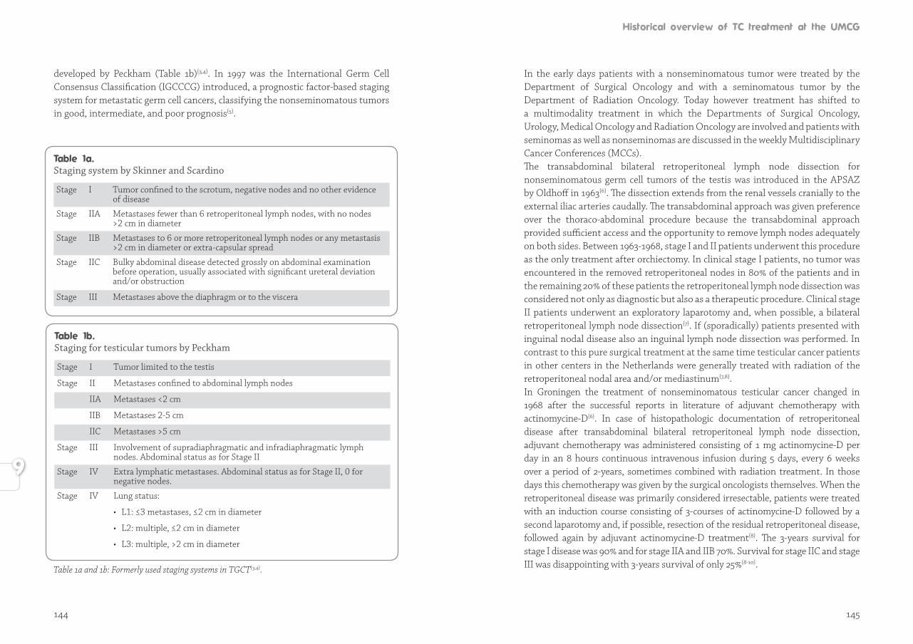
144 145
developed by Peckham (Table 1b){3,4}. In 1997 was the International Germ Cell Consensus Classification (IGCCCG) introduced, a prognostic factor-based staging system for metastatic germ cell cancers, classifying the nonseminomatous tumors in good, intermediate, and poor prognosis{5}.
Staging for testicular tumors by Peckham
Stage I Tumor limited to the testis
Stage II Metastases confined to abdominal lymph nodes
IIA Metastases <2 cm
IIB Metastases 2-5 cm
IIC Metastases >5 cm
Stage III Involvement of supradiaphragmatic and infradiaphragmatic lymph nodes. Abdominal status as for Stage II
Stage IV Extra lymphatic metastases. Abdominal status as for Stage II, 0 for negative nodes.
Stage IV Lung status:
• L1: ≤3 metastases, ≤2 cm in diameter
• L2: multiple, ≤2 cm in diameter
• L3: multiple, >2 cm in diameter
Staging system by Skinner and Scardino
Stage I Tumor confined to the scrotum, negative nodes and no other evidence of disease
Stage IIA Metastases fewer than 6 retroperitoneal lymph nodes, with no nodes >2 cm in diameter
Stage IIB Metastases to 6 or more retroperitoneal lymph nodes or any metastasis >2 cm in diameter or extra-capsular spread
Stage IIC Bulky abdominal disease detected grossly on abdominal examination before operation, usually associated with significant ureteral deviation and/or obstruction
Stage III Metastases above the diaphragm or to the viscera
Table 1a and 1b: Formerly used staging systems in TGCT{3,4}.
In the early days patients with a nonseminomatous tumor were treated by the Department of Surgical Oncology and with a seminomatous tumor by the Department of Radiation Oncology. Today however treatment has shifted to a multimodality treatment in which the Departments of Surgical Oncology, Urology, Medical Oncology and Radiation Oncology are involved and patients with seminomas as well as nonseminomas are discussed in the weekly Multidisciplinary Cancer Conferences (MCCs). The transabdominal bilateral retroperitoneal lymph node dissection for nonseminomatous germ cell tumors of the testis was introduced in the APSAZ by Oldhoff in 1963{6}. The dissection extends from the renal vessels cranially to the external iliac arteries caudally. The transabdominal approach was given preference over the thoraco-abdominal procedure because the transabdominal approach provided sufficient access and the opportunity to remove lymph nodes adequately on both sides. Between 1963-1968, stage I and II patients underwent this procedure as the only treatment after orchiectomy. In clinical stage I patients, no tumor was encountered in the removed retroperitoneal nodes in 80% of the patients and in the remaining 20% of these patients the retroperitoneal lymph node dissection was considered not only as diagnostic but also as a therapeutic procedure. Clinical stage II patients underwent an exploratory laparotomy and, when possible, a bilateral retroperitoneal lymph node dissection{7}. If (sporadically) patients presented with inguinal nodal disease also an inguinal lymph node dissection was performed. In contrast to this pure surgical treatment at the same time testicular cancer patients in other centers in the Netherlands were generally treated with radiation of the retroperitoneal nodal area and/or mediastinum{7,8}. In Groningen the treatment of nonseminomatous testicular cancer changed in 1968 after the successful reports in literature of adjuvant chemotherapy with actinomycine-D{6}. In case of histopathologic documentation of retroperitoneal disease after transabdominal bilateral retroperitoneal lymph node dissection, adjuvant chemotherapy was administered consisting of 1 mg actinomycine-D per day in an 8 hours continuous intravenous infusion during 5 days, every 6 weeks over a period of 2-years, sometimes combined with radiation treatment. In those days this chemotherapy was given by the surgical oncologists themselves. When the retroperitoneal disease was primarily considered irresectable, patients were treated with an induction course consisting of 3-courses of actinomycine-D followed by a second laparotomy and, if possible, resection of the residual retroperitoneal disease, followed again by adjuvant actinomycine-D treatment{8}. The 3-years survival for stage I disease was 90% and for stage IIA and IIB 70%. Survival for stage IIC and stage III was disappointing with 3-years survival of only 25%{8-10}.
146 147
At the end of the seventies there were three new developments in the treatment of testicular cancer: 1) the testicular tumor markers, alpha-fetoprotein (AFP), betachoriongonadotropin (B-HCG), and lactate dehydrogenase (LDH) were introduced in the diagnosis of testicular cancer and later on in the staging and evaluation of testicular cancer treatment; 2) Computed Tomography (CT) of the abdomen and chest was introduced in the staging of testicular cancer and quickly replaced the previous described staging methods, intravenous pyelogram, bipedal lymphography and tomography of the lungs; 3) discovery of cisplatin and the introduction of the cisplatin based combination chemotherapy by Einhorn with cisplatin, vinblastin and bleomycin (PVB) for disseminated testicular cancer{10}. Prolonged remissions were achieved in 70% of the disseminated testicular cancer patients treated with PVB{10}. In the Netherlands, including Groningen, shortly after the same survival figures were published{11}. With the introduction of PVB in Groningen, the treatment of testicular cancer became a team approach of pathologist, surgical oncologist, medical oncologist, radiologist and anesthesiologist. All new and treated patients with a nonseminomatous tumor were discussed within the weekly MCC. The medical oncologists had to ‘learn’ how to deliver ‘safely’ the cisplatin based combination chemotherapy{12-14}. The radiologist had to learn how to 'assess' the post chemotherapy retroperitoneal CT scans in particular with respect to tumor response. The surgical oncologists faced the resection of fibrosed and/or necrotic residual disease. The anesthesiologist had to learn how to deal with the side effects of bleomycin to the pulmonary tissue during general anesthesia{14}. The pathologist had to evaluate the resected tumor tissue, e.g. necrosis, fibrosis, mature teratoma and/or viable germ cell cancer. Two research questions were extensively studied 1) tumor maturation due to chemo-therapy and 2) immune histochemical analysis of tumor marker production by different histological components of nonseminomatous germ cell tumors{15}. The cisplatin based combination chemotherapy treatment of metastatic nonsemi-nomatous testicular cancer suddenly brought together a range of medical specialists and researchers from different medical disciplines and the testicular research line expanded successfully within the ‘Facultaire Onderzoeksprogramma Oncologie’ of the Medical Faculty of the Groningen University.Schraffordt Koops and Sleijfer successfully introduced the ‘wait and see’ policy in the APSAZ for stage I disease and exploratory laparotomies were no longer performed{16,17}. In case of a recurrence, effective chemotherapy was available{18}. Treatment of disseminated nonseminomatous tumors consisted of cisplatin, vin-blastin and bleomycin (PVB) and since the mid-eighties of bleomycin, etoposide and cisplatin (BEP). More recently the systemic treatment is based on so called ‘IGCCCG prognostic factors’ and individualized for example in patients with pul-
monary disease, administering 4 cycles EP instead of 3 cycles BEP{5}. After systemic chemotherapy there might be a biochemical complete response, with or without residual retroperitoneal and/or lung disease evaluated on abdominal/chest CT. All residual retroperitoneal/lung disease needs to be resected{19-21}. Further treatment is depending on the histopathology of the resected specimen. In case of necrosis or mature teratoma no further therapy is indicated, while second line chemotherapy might be indicated when viable germ cell cancer is encountered.Also the treatment of seminomatous tumors of the testis changed and improved during the last two decades, mainly due to CT radiation planning, less radiation doses and the introduction of systemic chemotherapy for stage III and IV disease. More recently treatment for stage I disease has changed: wait and see policy is implemented, one course of carboplatin or still radiation. Orchiectomy is the only surgical part of the treatment for patients with a seminoma of the testis. Further treatment, radiation or systemic treatment, is based on the disease stage and the advisement of the multidisciplinary cancer conference (MCC).In Groningen, the modified retroperitoneal lymph node dissection was introduced: only resection of residual retroperitoneal disease with the ultimate goal to reduce the morbidity of retrograde ejaculation{22,23}. For an adequate vascular access to continuously deliver PVB, in the early eighties patients received an AV-fistula between the radial artery and the cephalic vein{23}. The complication rate of this surgical procedure was high and therefore the Venous Access Port (VAP) was successfully introduced in the mid-eighties{25}. In Europe, there are European Society of Medical Oncology (ESMO) guidelines with regard to the treatment of disseminated nonseminomatous tumors but these do not fully correspond with the guidelines of the National Comprehensive Cancer Network (NCCN){26,27}. In contrast to the small differences in systemic treatment, differences are very large with respect to the surgical removal of residual retro-peritoneal tumor mass(es). At the UMCG only the macroscopic residual abnor-malities are removed, while in the USA complete or modified (template) retro-peritoneal lymph node dissections are still performed{19-21,26,27}. One of the ad van tages of the UMCG strategy was the less sexual disorder{28-30}. At the UMCG, resection of residual retroperitoneal disease is centralized, in contrast to other centers in the West where the referring general surgeon or urolo-gist generally performs the adjuvant surgery. The role of surgery in the treatment of disseminated testicular cancer was well defined at the end of the nineties, as well as all surgical research questions seemed to be answered. Therefore the research within the Department of Surgical Oncology, in close collaboration with the Department of Medical Oncology shifted towards the Department of Ge ne-tics and the Department of Psychosocial Oncology on subjects such as genetic

148 149
susceptibility, tumor biology, endocrine aspects, sexuality, quality of life, survivor-ship issues and long-term toxicity in testicular cancer{31-45}. The last decade Gietema of the Department of Medical Oncology has especially focused on long-term effects of testicular cancer treatment within national and international colla bo-rative groups{46-48}. With the extensive achieved experience in laparoscopic surgery, this minimal inva-sive operative technique was introduced by Hoekstra a decade ago in the treat ment of testicular cancer at the UMCG. In most centers laparoscopy is used as a minimal invasive staging procedure, e.g. nerve preserving uni- or bilateral retroperitoneal lymph node dissection (RPLND), for stage I NSTGCTs. At the UMCG the surgical staging, RPLND or exploratory laparotomy, was already abandoned in the mid-eighties when the CT staging became available. The techni que of laparoscopy was for the first time used at the UMCG in the resection of residual retroperitoneal tumor mass (RRRTM) in 2004. Since then performing laparoscopic resections of RRRTMs in testicular cancer patients has become a routine procedure at the UMCG for the surgical oncologists.Although the technique of laparoscopic resection of RRTMs is safe, this minimally invasive procedure, with a nice oncological and cosmetic outcome, is still not wide-ly accepted in the Netherlands, Europe and the United States{49,50}. Are there still new surgical treatment options in the treatment of testicular cancer? With the availability of the Da Vinci Robot at the UMCG in 2018, the technique of robotic (assisted) resection of residual retroperitoneal tumor masses will be explored in the nearby future in the treatment of disseminated testicular cancer with the goal to resect residual retroperitoneal tumor masses with the same cosmetic result but less morbidity, e.g. preserving sexual function.
1. Soebhag R. Testistumoren in Nederland. Thesis, University of Leiden, 1982.
2. Wobbes Th, Blom JM, Oldhoff J, Schraffordt Koops H. Lymphography in the diagnosis of non-seminoma tumours of the testis. J Surg Oncol 1982; 19: 1-4.
3. Skinner DG, Scardino PT. Relevance of biochemical tumor markers and lymphadenectomy in management of nonseminomatous testis tumors: current perspective. J Urol 1980; 123: 378–382.
4. Peckham MJ, McElwain TJ, Barrett A, Hendry WF. Combined management of malignant teratoma of the testis. Lancet 1979; 2: 267-270.
5. International Germ Cell Consensus Classification: a prognostic factor-based staging system for metastatic germ cell cancers. International Germ Cell Cancer Collaborative Group. J Clin Oncol 1997; 15: 594-603.
6. Wobbes Th, Eibergen R, Oldhoff J, Schraffordt Koops H. Results of retroperitoneal lymph node dissection and postoperative adjuvant chemotherapy with dactinomycin in the treatment of retroperitoneal metastases of nonseminomatous testicular germ cell tumors. Cancer 1983; 51: 1076-1079.
7. Wobbes Th. Non-seminomatous germ cell tumours of the testis. Staging and treatment. Thesis, State University Groningen, 1981.
8. Skinner DG. Non-seminomatous testis tumors: a plan of management based on 96 patients to improve survival in all stages by combined therapeutic modalities. J Urol 1976; 115: 65-69.
9. Gelderman W.A.H. Surgery in the treatment of nonseminomatous testicular tumors. Thesis, State University Groningen, 1987.
10. Einhorn LH, Donohue J. Cis-diamminedichloroplatinum, vinblastine, and bleomycin combination chemotherapy in disseminated testicular cancer. Ann Intern Med 1977; 87: 293-298.
11. Stoter G, Sleijfer DT, Vendrik CPJ, Schraffordt Koops H, Struyvenberg A, van Oosterom AT et al. Combination chemotherapy with cis-diammine-dichloro-platinum, vinblastine, and bleomycin in advanced testicular non-seminoma. Lancet 1979; 1: 941-945.
12. Meijer S. Cis-platinum and the kidney: renal function during treatment with cis-diammine-dichloro-platinum. Thesis, State University Groningen, 1982.
13. Offerman J.J.G. Cisplatin nephrotoxicity: can it be prevented? Thesis, State University Groningen, 1985.
14. Barneveld van P.W.C. Pulmonary changes induced by bleomycin. Thesis, State University Groningen, 1985.
15. Suurmeijer A.J.H. Nonseminomatous germ cell tumors of the testes. Tumor maturation due to chemotherapy, immunohistochemical analysis of tumor marker production. Thesis, State University Groningen, 1984.
16. Schraffordt Koops H, Sleijfer DT, Oosterhuis JW, Marrink J, de Bruijn HW, Oldhoff J. Wait-and-see policy in clinical stage I non-seminomatous germ cell tumors of the testis. Eur J Surg Oncol 1986; 12: 283-287.
17. Gels ME, Hoekstra HJ, Sleijfer DT, Marrink J, de Bruijn HW, Molenaar WM et al. Detection of recurrence in patients with clinical stage I nonseminomatous testicular germ cell tumors and consequences for further follow-up: a single-center 10-year experience. J Clin Oncol 1995; 13: 1188-1194.
18. Gelderman WA, Schraffordt Koops H, Sleijfer DT, Oosterhuis JW, Oldhoff J. Treatment of retroperitoneal residual tumor after PVB chemotherapy of nonseminomatous testicular tumors. Cancer 1986; 58: 1418-1421.
19. Gels ME, Nijboer AP, Hoekstra HJ, Sleijfer DT, Molenaar WM, Plukker JT et al. Complications of the post-chemotherapy resection of retroperitoneal residual tumour mass in patients with non-seminomatous testicular germ cell tumours. Br J Urol 1997; 79: 263-268.
150 151
20. Prenger K, Eysman L, Homan van der Heide JN, Oldhoff J, Sleijfer DT, Schraffordt Koops H et al. Thoracotomy as a staging procedure after chemotherapy in the treatment of Stage III nonseminomatous carcinoma of the testis. Ann Thorac Surg 1984; 38: 444-446.
21. Gels ME, Hoekstra HJ, Sleijfer DT, Nijboer AP, Molenaar WM, Ebels T et al. Thoracotomy for postchemotherapy resection of pulmonary residual tumor mass in patients with nonseminomatous testicular germ cell tumors: aggressive surgical resection is justified. Chest 1997; 112: 967-973.
22. Nijman JM, Schraffordt Koops H, Oldhoff J, Kremer J, Jager S. Sexual function after bilateral retroperitoneal lymph node dissection for nonseminomatous testicular cancer. Arch Androl 1987; 18: 255-267.
23. Gels M.E. Testicular germ cell tumors: developments in surgery and follow-up. Thesis, State University Groningen, 1997.
24. Wobbes T, Slooff MJ, Lichtendahl DH, Vriesendorp R, Mulder NH. The radiocephalic fistula as vascular access for chemotherapy. World J Surg 1983; 7: 532-535.
25. Lemmers NW, Gels ME, Sleijfer DT, et al. Complications of venous access ports in 132 patients with disseminated testicular cancer treated with polychemotherapy. J Clin Oncol 1996; 14: 2916-2922.
26. Oldenburg J, Fosså SD, Nuver J, Heidenreich A, Scmoll HJ, Bokemeyer C et al. Testicular seminoma and non-seminoma: ESMO Clinical Practice Guidelines for diagnosis, treatment, and follow up. Ann Oncol 2013; 24: 125-132.
27. Motzer RJ, Agarwal N, Beard C, Bolger GB, Boston B, Carducci MA et al. NCCN clinical practice guidelines in oncology: testicular cancer. J Natl Compr Canc Netw 2009; 7: 672-693.
28. Nijman JM, Schraffordt Koops H, Oldhoff J, Kremer J, Sleijfer DT. Sexual function after surgery and combination chemotherapy in men with disseminated nonseminomatous testicular cancer. J Surg Oncol 1988; 38: 182-186.
29. Nijman J.M. Some aspects of sexual and gonadal function in patients with a non-seminomatous germ-cell tumor of the testis. Thesis, State University Groningen, 1987.
30. Basten van J.P.A. The sexual sequelae of testicular cancer. Thesis, State University Groningen, 1999.
31. Sonneveld D.J.A. Testicular germ cell tumours: new insights in epidemiology, genetic susceptibility and outcome. Thesis, State University Groningen, 2002.
32. Jonker-Pool G. Surviving testicular cancer : sexuality & other existential issues. Thesis, State University Groningen, 2003.
33. Fleer J. Quality of life of testicular cancer survivors. Thesis, State University Groningen, 2006.
34. Lutke Holzik M.F. Genetic predisposition to testicular cancer. Thesis, State University Groningen, 2007.
35. Tuinman M.A. Surviving testicular cancer : relationship aspects. Thesis, State University Groningen, 2008.
36. Willemse PHB. Endocriene aspecten van het testiscarcinoom: invloed van HCG produktie, semicastratie en chemotherapie. Thesis, State University Groningen, 1981.
37. Castedo S.M.M.J. Pathogenesis of testicular germ cell tumors: a cytogenetical and pathological study. Thesis, State University Groningen, 1988.
38. Graaff de W.E. Tumor biological studies on adult testicular germ cell tumors. Thesis, State University Groningen, 1992.
39. Gietema J.A. New perspectives in platininum based chemotherapy. Thesis, State University Groningen, 1993.
40. Timmer A. The microenvironment as determinant of testicular germ cell tumor biology. Thesis, State University Groningen, 1995.
41. Echten-Arends van J. A cytogenetic study of male germ cell tumors. Thesis, State University Groningen, 1996.
42. Meinardi M.T. Long-term chemotherapy-related cardiovascular morbidity. Thesis, State University Groningen, 2001.
43. Nuver J. Chemotherapy-related vascular toxicity : studies in germ cell tumour patients. Thesis, State University Groningen, 2005.
44. Haas de E.C. Exploring predictors of chemotherapy efficacy and toxicity in testicular cancer. Thesis, State University Groningen, 2010.
45. Altena R. Chemotherapy-related cardiovascular morbidity in testicular cancer patients. Markers and Mechanisms. Thesis State University Groningen, 2011.
46. Boer H, Proost JH, Nuver J, Bunskoek S, Gietema JQ, Geubels BM et al. Long-term exposure to circulating platinum is associated with late effects of treatment in testicular cancer survivors. Ann Oncol 2015; 11: 2305-2310.
47. Haugnes HS, Bosl GJ, Boer H, Gietema JA, Brydøy M, Oldenburg J et al. Long-term and late effects of germ cell testicular cancer treatment and implications for follow-up. J Clin Oncol 2012; 30: 3752-3763.
48. Groot HJ, Lubberts S, de Wit R, Witjes JA, Kerst JM, de Jong IJ et al. Risk of solid cancer after treatment of testicular germ cell cancer in the platinum era. J Clin Oncol 2018; 24: 2504-2513.
49. Ozturk C, van Ginkel RJ, Krol RM, Gietema JA, Hofker HS, Hoekstra HJ. Laparoscopic resection of a residual retroperitoneal tumor mass of nonseminomatous testicular germ cell tumors. Surg endosc 2012; 26: 458-467.
50. Ozturk C, Been LB, van Ginkel RJ, Gietema JA, Hoekstra HJ. Laparoscopic Resection of Residual Retroperitoneal Tumor Mass in Advanced Nonseminomatous Testicular Germ Tumors; a Feasible and Safe Oncological Procedure. Manuscript submitted for publication.

155
Het is prettig om op het punt te zijn beland om dit dankwoord te kunnen schrijven en hiermee een voor mij belangrijke periode af te kunnen sluiten. Deze periode wordt niet alleen gekenmerkt door mijn promotieonderzoek met alle energie en inspanning die daarin is gestoken door mij en vele anderen. Ook wordt met dit proefschrift mijn opleiding heelkunde afgerond. De afgelopen negen jaar heb ik hier in het Hoge Noorden van ANIOS, tot AIOS tot chirurg veel fijne mensen ontmoet en maak ik dankbaar gebruik van dit hoofdstuk om eenieder die een rol heeft gespeeld bij de totstandkoming van dit proefschrift alsmede bij mijn vorming tot chirurg, te bedanken.
Als eerste, professor H.J. Hoekstra, beste Harald, beste prof, dank voor het in mij gestelde vertrouwen vanaf het allereerste begin toen ik, denk ik, nog vrij bleu het onderzoek in ging. Ondanks periodes van lage wetenschappelijke output vanuit mijn zijde, heeft u bewezen dat u een promovendus kunt coachen in goede en slechte tijden en dat deed u kritisch, steunend en sturend waar nodig. Bewonderenswaardig is uw bereikbaarheid van bijna 24 uur per dag, wat zich kenmerkte door een zeer snelle respons en het vermogen om meteen een nieuw plan te maken om dit proefschrift tot een wetenschappelijk gedegen geheel te construeren. Bijzondere dank voor uw bijdrage aan het historisch overzicht toegevoegd aan dit proefschrift. De uiteenzetting van dit wetenschappelijke verleden verschaft inzicht over de stand van zaken hedendaags in het onderzoek naar zaadbalkanker. Net als zovelen die mij zijn voorgegaan, heeft u ook mij naar de eindstreep begeleid en die lange lijst van promovendi is indrukwekkend. Als promovendus van u, kreeg ik altijd wel meer advies dan alleen over het proefschrift en vaak kreeg u ook wel gelijk. De Hoekstriaanse quotes zullen voor eeuwig gegrift staan in mijn hersenen. Om er één te noemen: “Een idee moet niet slapend worden gehouden. Telkens als jij een idee hebt, is er elders op de wereld nog iemand met hetzelfde idee”.
Professor J.A. Gietema, beste Jourik, als promotor heeft u sterk bijgedragen aan de wetenschappelijke inhoud van dit proefschrift. Bedankt voor alle tijd en energie die u in dit proefschrift en mijn begeleiding heeft gestoken. Dank voor uw essentiële bijdrage aan dit proefschrift en kritische blik. Ten alle tijden was u bereid om mijn vragen te beantwoorden en met mij te sparren over de koers van dit proefschrift. Het samen schrijven van manuscripten heb ik als leerzaam ervaren doordat er vragen werden gesteld die nog niet waren gesteld, waardoor het proefschrift inhoudelijk sterker en vollediger is geworden.
156 157
Dr. R.J. van Ginkel, beste Robert, dank voor jouw begeleiding als copromotor en bijdrage aan dit proefschrift door de manuscripten te beoordelen. In het bijzonder wil ik jou bedanken als opleider. Mede door jouw bijdrage kan ik terugkijken op een mooie en gedegen opleiding. En natuurlijk vooral bedankt voor die allerlaatste officiële UMCGstempel op het Cbriefje!
Dr. J.E.H.M. Hoekstra-Weebers, beste Josette, toen wij aan dit traject begonnen was jij een van de beoogde copromotoren. Uiteindelijk heeft dit proefschrift een andere wetenschappelijke lading gekregen en is het meer de chirurgische kant op gegaan en vond jij de rol van copromotor niet meer gerechtvaardigd. Wij hebben samen het ‘delay’ stuk geschreven. In dit proces heb ik ervaren hoe uitermate geduldig, secuur en professioneel jij bent, tijdens het schrijven van de zoveelste versie. De acceptatie van dit artikel bleek het keerpunt in het proces van mijn proefschrift. Tevens heb jij een belangrijke coachende rol gehad en op relevante momenten mij de juiste dosis support gegeven. Bedankt voor jouw bijdrage aan de totstandkoming van dit proefschrift. Ik voel me bevoorrecht als ik bedenk wat het team Hoekstra voor mij heeft gedaan en betekend.
Hooggeleerde heren van de leescommissie, professor dr. T.M. de Reijke, professor dr. S. Horenblas en professor dr. A.J.H. Suurmeijer, ik wil graag mijn dank uitspreken voor uw geïnvesteerde tijd om dit proefschrift in alle volledigheid te beoordelen en op wetenschappelijke waarde te toetsen.
Drs. S. Hofker, beste Sijbrand, jij stond direct open voor het idee en verzoek van Hoekstra en Van Ginkel de techniek van de laparoscopie te gaan toepassen bij de resectie van residuale tumor na chemotherapie bij gemetastaseerde nonseminoma testistumoren. Als een van de eerste groepen ter wereld voerde het Groningen Cancer Center deze techniek met succes uit bij de gecombineerde behandeling van zaadbalkanker en vormde dit uiteindelijk de basis voor het proefschrift.
Dr. J. Fleer, beste Joke, hartelijk bedankt voor jouw bijdrage en inzet aan het ‘delay’ manuscript. Jij had al veel voorwerk verricht, waardoor ik deze koers alleen maar hoefde te volgen.
Drs. T. Velleman, beste Ton, dank voor jouw inzet gedurende jouw wetenschapsstage en daarna met het manuscript over de volumetrische metingen. Zeker in de opstartfase hebben wij vele uren doorgebracht in het Triadegebouw om het onderzoek op te zetten en de software te kunnen gebruiken. Veel succes in jouw opleiding en verdere carrière als radioloog.
Drs. A.H.H. Bongaerts. Collega Bongaerts is helaas veel te vroeg overleden. Ik ben hem erkentelijk voor zijn bijdrage en vakkundigheid aan dit proefschrift.
Dr. ir. P.M.A. van Ooijen. Beste Peter, na het overlijden van collega Bongaerts heb jij Ton en mij van advies en begeleiding voorzien in de voltooiing van het manuscript omtrent de volumetrische metingen. Hartelijk dank daarvoor.
Drs. L.M. Bergman. Beste Marco, dank voor het beoordelen van het manuscript omtrent de volumetrische metingen.
Dr. S. Kruijff. Beste Schelto, dank voor jouw betrokkenheid en begeleiding bij het laatste manuscript van dit proefschrift. Door jouw snelle reacties met de nodige suggesties kon dit laatste hoofdstuk worden afgerond.
Dr. L.B. Been. Beste Lukas, dank voor jouw tijd, inbreng en oordeel bij onze grote laparoscopieserie. Tevens dank voor de begeleiding tijdens mijn opleiding heelkunde.
Drs. P.H.J. Hemmer. Beste Patrick, net als Schelto heb jij bij het sluitstuk van dit proefschrift een belangrijk aandeel in de inhoudelijke koers gehad. Bedankt hiervoor!
Dr. J.G. van Baal. Beste Sjef, mijn opleiding ben ik bij u in de kliniek begonnen. Ik heb een grote waardering ontwikkeld voor u als dokter, persoon, netwerker, wetenschapper en vooral ook als gedreven opleider. Doordat de eerste drie jaar van mijn opleiding plaatsvonden in het Ziekenhuis Groep Twente te Almelo, werd hierdoor ook mijn basis gevormd en ik zal altijd met een goed gevoel terugkijken naar die tijd. Altijd was u meer dan bereid om te kijken wat u kon betekenen voor mij en zoveel anderen. Binnenkort begint uw welverdiende pensioen waarbij u in uw carrière zo ongeveer 100 assistenten heeft opgeleid. Ik wens u en uw familie het allerbeste toe en ik ga ervan uit dat de wetenschap nog wel op u kan rekenen.
Heelkunde Friesland. Ik heb een ontzettend mooie tijd gehad in het Medisch Centrum Leeuwarden. Dank aan alle chirurgen en in het bijzonder de GE/Oncogroep voor een leerzaam jaar. Drs. M. Emous. Beste Marloes, ik vond het fijn dat er zo een ‘powervrouw’ aan het roer staat van de club Heelkunde. Als vrouw in een toppositie ben jij ook meteen een voorbeeld voor velen. Jij bent een betrokken opleider bij de AIOS en ANIOS. Bij eenieder kijk jij goed waar het carrièrepad naartoe leidt en wat jij daarin kunt betekenen. Bedankt voor jouw bijdrage aan mijn opleiding en tot ziens op jouw promotie.
158 159
Ik ben als chirurg geregistreerd, echter valt er binnen dit mooie vak nog veel te leren. Ik dank de Vakgroep Heelkunde van het Zuyderland Medisch Centrum (SittardGeleen, Heerlen) voor het in mij gestelde vertrouwen om bij jullie als fellow chirurgische oncologie mijn carrière voort te zetten.
Professor dr. E. Heineman, beste Erik, dank voor uw bijdrage aan mijn vorming tot chirurg en de mooie opleiding genoten in Regio VI. Ondanks de drukke agenda was u altijd beschikbaar wanneer nodig. U heeft altijd een brede blik gehad op de werkprocessen binnen de gezondheidszorg waarbij kwaliteit voor de patiënt bovenaan stond en tevens met oog voor de kwaliteit voor de AIOS.
Professor dr. H.J. ten Duis, dank voor uw aanmoediging en steun om te solliciteren voor de opleiding Heelkunde en dank voor uw bijdrage aan mijn opleiding in de allereerste jaren.
Dr. J.F.M. Lange, beste Johan, bedankt voor jouw coachende rol als opleider van de GEdifferentiatie. Ook buiten de formele voortgangsgesprekken was jij een zichtbare en toegankelijke opleider met de nodige humor en relativeringsvermogen. Samen met Drs. C. Krikke hadden jullie een van de gezelligste kamers om even bij langs te komen. Beste Christina, ik wil jou bedanken voor jouw bijdrage aan mijn opleiding. Ik kan me nog goed het moment herinneren bij jullie aan de keukentafel, samen met Patrick (Drs. P. Nieboer), toen ik nog in opleiding wilde komen. Voor mij ben jij een rolmodel als het gaat om iemand die haar vak beheerst en met name als iemand die je er nooit op kunt betrappen niet zichzelf te zijn.
Collega’s, chirurgen, assistenten Heelkunde regio VI, allen bedankt voor een mooie, gezellige en leerzame tijd in het Noorden; een van de leukste opleidingsregio’s. Tot in de toekomst!
Mijn paranimf, Anna Marije Leenders-Hovenkamp. Sinds ons allereerste artsen baantje zijn wij elkaar nooit uit het oog verloren en daar prijs ik mijzelf gelukkig om. Onze band is ondanks wisselende geografische locaties alleen maar sterker geworden. Dat ik de juiste persoon had gevraagd voor het paranimfschap, werd bevestigd toen jij zo blij reageerde op een dergelijk verzoek voor een verantwoordelijke taak. Bedankt en ik kijk ernaar uit om jou straks naast mijn zijde te hebben.
Lieve vriendinnen, Ayșe Uslu, Serpil Türkoğlu en Hülya Uslu. Ik ben dankbaar voor de vriendschap met deze sterke en ondernemende dames. Door onze drukke agenda’s zijn de ontmoetingen niet meer zo vaak als vroeger, maar altijd mooi, ontspannen en waardevol. Lieve Serpil (abla). Sinds dag 1 in Breda zijn we familie, dat gevoel is kostbaar. Lieve Ayșe en Hülya, wat een geluk dat ik jullie leerde kennen tijdens mijn opleidingstijd in Almelo. Een nieuwe stad werd zo een hele warme plek en jullie familie voelde als mijn familie. Ayșe, sinds kort ben jij bezig met jouw eigen tandartsenpraktijk ‘Liefde & Passie’ in Twente. Ik wens jou heel veel succes toe in deze nieuwe onderneming en weet zeker dat de praktijk een aanwinst zal zijn voor de regio.
Beste vrienden van TANNET, 'Geen toeschouwers, maar spelers in het veld' is de gemeenschappelijke deler die ons bindt. Speciale dank aan Drs. U. Pekdemir, sinds lange tijd een mentor en sparringpartner. Beste Uğur, jij bent een voorbeeld voor mij en voor velen door de positie die jij inneemt in de maatschappij als mens, waarbij de maatschappelijke winst altijd voorop gesteld wordt en dit is natuurlijk indrukwekkend als directievoorzitter van een financiële instelling. Jij bent een diversiteitsambassadeur en een veelgevraagd spreker als het gaat om thema’s als inspirerend leiderschap. Dank voor jouw steun en vriendschap door de jaren heen. Bijzonder was het dat jij voor het sluiten van mijn huwelijk nog één keer jouw ambtelijk toga aan wilde doen. Prof. dr. ir. U. Kaymak, beste Uzay, de tijd samen in het bestuur van TANNET was zeer waardevol, waarbij jij als lid van de Raad van Advies ons de nodige ondersteuning bood. Als jonge directie konden wij putten uit jouw jarenlange ervaring. Beste M. Ersoy, MA, beste Murat, dank voor jouw vriendschap door de jaren heen. Nog steeds begrijpen wij niet dat wij op dezelfde middelbare school hebben gezeten zonder elkaar te kennen. In jouw rol als voorzitter van TANNET heb jij jezelf bewezen als iemand die deze rol invult met humor, energie en een vermogen om anderen aan te spreken en in beweging te zetten. Veel gezellige avonden in Amsterdam doorgebracht met de TANNET family en hopelijk volgt nog meer gezelligheid, want ik ben natuurlijk de afgelopen tijd een beetje een afwezige factor geweest. Er zijn nog heel veel namen te noemen, maar daar ga ik bewust niet aan beginnen.
Nog een speciale dank aan Funda Müjde. Jij bent een bijzonder mens, geliefd door velen en iemand die laat zien wat levenskracht inhoudt. Met jouw theatervoorstellingen laat jij heel Nederland en daarbuiten lachen. Jouw levenspad is bijzonder en een inspiratiebron voor anderen. Dit proefschrift had voor mij zo zijn bergen en dalen, maar dan dacht ik “Als Funda naar Turkije fietst, dan moet dit ook wel lukken”. Veel succes met alle creaties die wij nog van jou gaan zien.
160 161
Lieve Fairuz en Prewesh, wij noemden onszelf altijd de drie musketiers tijdens onze studie geneeskunde en daarna. Wat hebben wij veel meegemaakt samen. Wij woonden met zijn drieën praktisch op 1 kleine studentenkamer in Leiden. En weten jullie nog in Milaan? Ook jullie hebben jezelf gevestigd in het medische vak. Ben trots op jullie en hoop dat wij elkaar nooit uit het oog verliezen.
Aan de studie in het LUMC heb ik vele mooie vrienden overgehouden. Speciale dank voor de vriendschap tijdens de studie en steun door de jaren heen aan Shirida, Hanneke, Laïla, en Yin Yin. Het blijft mooi om elkaar te zien en te spreken. Alle huwelijken zijn gevierd samen, maar gelukkig zijn er ieder jaar nog genoeg verjaardagen om op die manier bij elkaar te kunnen komen.
Lieve Priscilla Verbraeken en Percy Kewalder, één van de langstlopende vriendschappen sinds de middelbare school. Ik vind het mooi dat wij elkaar nog altijd zien en waarderen. Dank voor jullie vriendschap.
Mijn paranimf mr. Renan Anıl Öztürk. Ik ben trots dat jij naast mijn zijde staat tijdens mijn promotie. Net als wij vroeger hand in hand van school naar huis liepen, had ik nu ook niemand anders naast mij willen hebben. Jij hebt het de afgelopen jaren maar druk in Dubai, maar gelukkig spreken wij elkaar regelmatig. Ik ben vooral een hele trotse zus op wie jij als persoon bent geworden. Ik ben heel benieuwd naar jouw verdere levenspad en hoop dat jij wel weer eens in de buurt gaat wonen. En ja, ik heb deze wens hier opgeschreven.
Lieve ouders, Anneciğim ve babacığım, dankzij jullie zijn wij in een warm en hecht gezin opgegroeid, waar altijd humor was. Jullie onvoorwaardelijke steun en vertrouwen heb ik altijd gevoeld. Bedankt dat ik mij door jullie kon ontwikkelen tot wie ik nu ben. Baba, ondanks mijn zoveelste verhuizing bleef jij enthousiast helpen en ben jij er altijd geweest. Anne, jouw liefde en hulp is niet te beschrijven. Als er iets was wat jij kon doen, heb jij dat altijd gedaan. De vanzelfsprekendheid waar mee jij dat altijd hebt gedaan, legt de lat hoog voor mij als moeder. Zeker dit afgelopen jaar bij de afronding van mijn opleiding en promotie konden wij als gezin altijd op jullie rekenen. Nu ik in Limburg woon, zal ik jullie wat meer moeten missen.
Lieve Hala, Damla en Berkay, bedankt voor jullie liefde en steun. Als familie kunnen wij elkaar door de afstanden niet vaak zien, maar onze band blijft sterk. Ik ben trots op jullie en kijk uit naar een mooi moment volgend jaar.
Lieve familie Baharvand, lieve schoonfamilie, bedankt voor jullie liefde en steun. Lieve schoonouders, Maman en Baba, het is een rijkdom om in een latere levensfase nog een tweede familie erbij te krijgen. Bedankt dat jullie mij als dochter in jullie familie hebben opgenomen en voor jullie onvoorwaardelijke liefde en steun de afgelopen jaren.
Allerliefste Ehsan, wat een geluk dat ik jou als partner aan mijn zijde heb. Bedankt voor jouw steun, liefde en aanmoediging om dit promotietraject en mijn opleiding af te ronden. Jij zult ook wel verheugd zijn dat we nu wat meer tijd voor elkaar zullen hebben. Het is niet altijd gemakkelijk om de partner van een medisch specialist te zijn. Jij hebt jezelf de afgelopen jaren en nu nog steeds heel wendbaar moeten op stellen. Sinds kort hebben we dan samen ‘even’ het besluit genomen om naar Limburg te verhuizen, zodat ik daar als fellow chirurgie kan werken. Deze mooie baan in Limburg kan ik alleen beginnen, omdat ik weet dat wij samen altijd zorgen dat het werkt.
Mijn lieve zoon Kiyan, nu al ben jij een lichtpunt voor velen in hun leven en to ver jij een glimlach op ieders gezicht. Jij bent zonder twijfel het mooiste in mijn leven. Jouw gebrabbel en zangkunsten waren ook onmisbaar in de tot stand-koming van dit proefschrift. Ik ben benieuwd naar elke ontwikkeling die jij gaat doorlopen in dit leven en zal er altijd voor jou zijn.
162 163
Çiğdem Öztürk werd geboren op 15 mei 1981 te ‘s Gravenhage. Zij is de dochter van Ramazan Öztürk en Hava Soysal en de zus van Renan Anıl. In 1999 behaalde zij het Atheneumdiploma aan het Thomas More College te ’s Gravenhage. Aangezien zij werd uitgeloot voor de studie Geneeskunde, volgde zij eerst de studie Psychologie aan de Universiteit van Leiden. In 2001 jaar kon zij starten met de studie Geneeskunde in het Leids Universitair Medisch Centrum. Op 27 maart 2008 legde zij het artsexamen succesvol af. Datzelfde jaar studeerde zij ook af in de Klinische en Gezondheidspsychologie.
In 2008 startte zij als artsassistent op de afdeling Heelkunde van het Amphia Ziekenhuis in Breda. In 2009 ging zij in het Universitair Medisch Centrum in Groningen werken als artsassistent chirurgie en een jaar later als artsonderzoeker. In deze periode werd de basis voor het proefschrift gelegd, onder leiding van Prof. dr. H.J. Hoekstra. In 2011 werd zij geselecteerd voor de opleiding Heelkunde in de Regio Groningen (opleider Prof dr. H.J. ten Duis). Van 2011 tot 2014 werd de chirurgische opleiding in het Ziekenhuis Groep Twente te Almelo doorlopen (opleider Dr. J.G. van Baal), waarna in september 2014 de opleiding werd vervolgd in het Universitair Medisch Centrum in Groningen (opleider Prof dr. E. Heineman). Zij heeft gekozen voor de differentiatie gastrointestinale chirurgie. De opleiding Heelkunde en hiermee ook haar differentiatie werd afgerond in het Medisch Centrum Leeuwarden (opleider Drs. M. Emous).
Per 1 oktober 2018 is Çiğdem werkzaam in het Zuyderland Ziekenhuis in Limburg als fellow Chirurgische Oncologie. Çiğdem is getrouwd met Ehsan Baharvand en hun zoon Kiyan werd geboren op 12 oktober 2016.
164 165
Öztürk Ç, van Ginkel RJ, Krol RM, Gietema JA, Hoekstra HJ. Laparoscopic resection of a residual retroperitoneal tumor mass of nonseminomatous testicular germ cell tumors. Surg Endosc 2012; 26: 458467.
Öztürk Ç, te Slaa A, Dolmans DE, Ho GH, de Vries J, Mulder PG, van der Laan L. Quality of life in perspective to treatment of postoperative edema after peripheral bypass surgery. Ann Vasc Surg 2012; 26: 373382.
De Leur K, Öztürk Ç, van Zeeland ML, de Groot HG, Heyligers JM, Vriens PW et al. Vascular access outcome in the elderly dialysis patient in combination with the quality of life. Vasc Endovascular Surg 2013; 47: 444448.
Öztürk Ç, Fleer J, Hoekstra HJ, HoekstraWeebers JEHM. Delay in Diagnosis of Testicular Cancer; A Need for Awareness Programs. PLoS One 2015; 10: e0141244.
Öztürk Ç, Velleman T, Bongaerts AH, Bergman LM, van Ginkel RJ, Gietema JA et al. Assessment of Volumetric versus Manual Measurement in Disseminated Testicular Cancer; No Difference in Assessment between NonRadiologists and Genitourinary Radiologist. PLoS One 2017; 12: e0168977.