unclassified env/jm/mono(2016)23 - oecd
TRANSCRIPT
Unclassified ENV/JM/MONO(2016)23 Organisation de Coopération et de Développement Économiques Organisation for Economic Co-operation and Development 08-Jun-2016
___________________________________________________________________________________________
_____________ English - Or. English ENVIRONMENT DIRECTORATE
JOINT MEETING OF THE CHEMICALS COMMITTEE AND
THE WORKING PARTY ON CHEMICALS, PESTICIDES AND BIOTECHNOLOGY
SILICON DIOXIDE: SUMMARY OF THE DOSSIER
Series on the Safety of Manufactured Nanomaterials
No. 71
This document is only available in PDF format.
This document is the summary of the Silicon dioxide dossier published as:
- ENV/JM/MONO(2015)14/PART1
- ENV/JM/MONO(2015)14/PART2
- ENV/JM/MONO(2015)14/PART3
- ENV/JM/MONO(2015)14/PART4
- ENV/JM/MONO(2015)14/PART5
- ENV/JM/MONO(2015)14/PART6
JT03397644
Complete document available on OLIS in its original format
This document and any map included herein are without prejudice to the status of or sovereignty over any territory, to the delimitation of
international frontiers and boundaries and to the name of any territory, city or area.
EN
V/JM
/MO
NO
(2016)2
3
Un
classified
En
glish
- Or. E
ng
lish
ENV/JM/MONO(2016)23
2
OECD Environment, Health and Safety Publications
Series on the Safety of Manufactured Nanomaterials
No. 71
SILICON DIOXIDE:
SUMMARY OF THE DOSSIER
Environment Directorate
ORGANISATION FOR ECONOMIC CO-OPERATION AND DEVELOPMENT
Paris, 2016
ENV/JM/MONO(2016)23
3
Also published in the Series on the Safety of Manufactured Nanomaterials:
No. 1, Report of the OECD Workshop on the Safety of Manufactured Nanomaterials: Building
Co-operation, Co-ordination and Communication (2006)
No. 2, Current Developments/ Activities on the Safety of Manufactured Nanomaterials: Tour
de table at the 1st Meeting of the Working Party on Manufactured Nanomaterials
(2006)
No. 3, Current Developments/ Activities on the Safety of Manufactured Nanomaterials: Tour
de table at the 2nd Meeting of the Working Party on Manufactured Nanomaterials
(2007)
No. 4, Manufactured Nanomaterials: Programme of Work 2006-2008 (2008)
No. 5, Current Developments/ Activities on the Safety of Manufactured Nanomaterials: Tour
de table at the 3rd Meeting of the Working Party on Manufactured Nanomaterials
(2008)
No. 6, List of Manufactured Nanomaterials and List of Endpoints for Phase One of the OECD
Testing Programme (2008)
No. 7, Current Developments/ Activities on the Safety of Manufactured Nanomaterials: Tour
de table at the 4th Meeting of the Working Party on Manufactured Nanomaterials
(2008)
No. 8, Preliminary Analysis of Exposure Measurement and Exposure Mitigation in
Occupational Settings: Manufactured Nanomaterials (2009)
No. 9, EHS Research Strategies on Manufactured Nanomaterials: Compilation of Outputs
(2009)
No. 10, Identification, Compilation and Analysis of Guidance Information for Exposure
Measurement and Exposure Mitigation: Manufactured Nanomaterials (2009)
No. 11, Emission Assessment for the Identification of Sources and Release of Airborne
Manufactured Nanomaterials in the Workplace: Compilation of Existing Guidance
(2009)
No. 12, Comparison of Guidance on Selection of Skin Protective Equipment and Respirators for
Use in the Workplace: Manufactured Nanomaterials (2009)
No. 13, Report of an OECD Workshop on Exposure Assessment and Exposure Mitigation:
Manufactured Nanomaterials (2009)
No. 14, Guidance Manual for the Testing of Manufactured Nanomaterials: OECD Sponsorship
Programme (2009)
No. 15, Preliminary Review of OECD Test Guidelines for their Applicability to Manufactured
Nanomaterials (2009)
ENV/JM/MONO(2016)23
4
No. 16, Manufactured Nanomaterials: Work Programme 2009-2012 (2009)
No. 17, Current Development/ Activities on the Safety of Manufactured Nanomaterials: Tour de
table at the 5th Meeting of the Working Party on Manufactured Nanomaterials (2009)
No. 18, Manufactured Nanomaterials: Roadmap for Activities during 2009 and 2010 (2009)
No. 19, Analysis of Information Gathering Initiative on Manufactured Nanomaterials (2009)
No. 20, Current Development/ Activities on the Safety of Manufactured Nanomaterials: Tour de
table at the 6th Meeting of the Working Party on Manufactured Nanomaterials (2010)
No. 21, Report of the Workshop on Risk Assessment of Manufactured Nanomaterials in a
Regulatory Context (2010)
No. 22, OECD Programme on the Safety of Manufactured Nanomaterials 2009-2012:
Operational Plans of the Projects (2010)
No. 23, Report of the Questionnaire on Regulatory Regimes for Manufactured Nanomaterials
(2010)
No. 24, Preliminary Guidance Notes on Sample Preparation and Dosimetry for the Safety
Testing of Manufactured Nanomaterials (2010)
No. 25, Guidance Manual for the Testing of Manufactured Nanomaterials: OECD Sponsorship
Programme: First Revision (2010)
No. 26, Current Development/ Activities on the Safety of Manufactured Nanomaterials: Tour de
table at the 7th Meeting of the Working Party on Manufactured Nanomaterials (2010)
No. 27, List of Manufactured Nanomaterials and List of Endpoints for Phase One of the
Sponsorship Programme for the Testing Manufactured Nanomaterials: Revised (2010)
No. 28, Compilation and Comparison of Guidelines Related to Exposure to Nanomaterials in
Laboratories (2010)
No. 29, Current Development/ Activities on the Safety of Manufactured Nanomaterials: Tour de
table at the 8th Meeting of the Working Party on Manufactured Nanomaterials (2011)
No. 30, Regulated Nanomaterials: 2006-2009(2011)
No. 31, Information Gathering Schemes on Nanomaterials: Lessons Learned and Reported
Information (2011)
No. 32, National Activities on Life Cycle Assessment of Nanomaterials (2011)

ENV/JM/MONO(2016)23
5
No. 33, Important Issues on Risk Assessment of Manufactured Nanomaterials (2012)
No. 34, Current Development/ Activities on the Safety of Manufactured Nanomaterials: Tour de
table at the 9th Meeting of the Working Party on Manufactured Nanomaterials (2012)
No. 35, Inhalation Toxicity Testing: Expert Meeting on Potential Revisions to OECD Test
Guidelines and Guidance Document (2012)
No. 36, Guidance on Sample Preparation and Dosimetry for the Safety Testing of Manufactured
Nanomaterials (2012)
No.37, Current Developments in Delegations on the Safety of Manufactured Nanomaterials -
Tour de Table at the 10th Meeting of the WPMN (2012)
No.38, Co-Operation on Risk Assessment: Prioritisation of Important Issues on Risk Assessment
of Manufactured Nanomaterials - Final Report (2013)
No. 39, Environmentally Sustainable Use of Manufactured Nanomaterials - Workshop held on
14 September 2011 in Rome, Italy (2013)
No. 40, Ecotoxicology and Environmental Fate of Manufactured Nanomaterials:
Test Guidelines (2014)
No.41, Report of the OECD Expert meeting on the Physical Chemical Properties of
Manufactured Nanomaterials and Test Guidelines (2014)
No.42, Report of the questionnaire on regulatory regimes for manufactured nanomaterials
2010-2011 (2014)
No.43, Genotoxicity of Manufactured Nanomaterials: Report of the OECD expert meeting
(2014)
Nos. 44-54, These items are the dossiers derived from the Testing Programme on Manufactured
Nanomaterials which are located at: http://www.oecd.org/chemicalsafety/nanosafety/testing-programme-manufactured-nanomaterials.htm
No.55, Harmonized Tiered Approach to Measure and Assess the Potential Exposure to Airbone
Emissions of Engineered Nano-objects and their Agglomerates and Aggregates at
Workplaces. (2015)
No.56, Analysis of the Survey on Available Methods and Models for Assessing Exposure to
Manufactured Nanomaterials (2015)
No.57, Guidance Manual towards the integration of risk assessment into life cycle assessment
of nano-enabled applications (2015)
No.58, Preliminary guidance notes on Nanomaterials: Interspecies variability factors in human
health risk assessment (2015)
No.59, Developments on the safety of manufactured nanomaterials: 2013 (2015)
ENV/JM/MONO(2016)23
6
No.60, Current developments in delegations on the safety of manufactured nanomaterials - tour
de table (2015)
No.61, Developments in delegations on the safety of manufactured nanomaterials - tour de table
(2015)
No.62, Considerations for using dissolution as a function of surface chemistry to Evaluate
environmental behaviour of nanomaterials in risk assessments (2015)
No.63, Physical-chemical parameters: measurements and methods relevant for the regulation
of nanomaterials (2016)
No.64, Approaches on nano grouping/ equivalence/ read-across concepts based on physical-
chemical properties (GERA-PC) for regulatory regimes (2016)
No.65, Physical-chemical properties of nanomaterials: Evaluation of methods applied in the
OECD-WPMN testing programme (2016)
No.66, Categorisation of manufactured nanomaterials (2016)
No.67, Developments in delegations on the safety of manufactured nanomaterials
- tour de table(2016)
No.68, This document is the summary of the dossier Multi-walled Carbon Nanotubes (MWCNT)
derived from the Testing Programme on Manufactured Nanomaterials which are located at: http://www.oecd.org/chemicalsafety/nanosafety/testing-programme-manufactured-nanomaterials.htm
© OECD 2016
Applications for permission to reproduce or translate all or part of this material
should be made to: Head of Publications Service, [email protected],
OECD, 2 rue André-Pascal, 75775 Paris Cedex 16, France
ENV/JM/MONO(2016)23
7
ABOUT THE OECD
The Organisation for Economic Co-operation and Development (OECD) is an intergovernmental
organisation in which representatives of 34 industrialised countries in North and South America, Europe
and the Asia and Pacific region, as well as the European Commission, meet to co-ordinate and harmonise
policies, discuss issues of mutual concern, and work together to respond to international problems. Most of
the OECD’s work is carried out by more than 200 specialised committees and working groups composed
of member country delegates. Observers from several countries with special status at the OECD, and from
interested international organisations, attend many of the OECD’s workshops and other meetings.
Committees and working groups are served by the OECD Secretariat, located in Paris, France, which is
organised into directorates and divisions.
The Environment, Health and Safety Division publishes free-of-charge documents in eleven different
series: Testing and Assessment; Good Laboratory Practice and Compliance Monitoring; Pesticides;
Biocides; Risk Management; Harmonisation of Regulatory Oversight in Biotechnology; Safety of
Novel Foods and Feeds; Chemical Accidents; Pollutant Release and Transfer Registers; Emission
Scenario Documents; and Safety of Manufactured Nanomaterials. More information about the
Environment, Health and Safety Programme and EHS publications is available on the OECD’s World
Wide Web site (www.oecd.org/chemicalsafety/).
This publication was developed in the IOMC context. The contents do not necessarily reflect the
views or stated policies of individual IOMC Participating Organizations.
The Inter-Organisation Programme for the Sound Management of Chemicals (IOMC) was
established in 1995 following recommendations made by the 1992 UN Conference on
Environment and Development to strengthen co-operation and increase international co-
ordination in the field of chemical safety. The Participating Organisations are FAO, ILO, UNDP,
UNEP, UNIDO, UNITAR, WHO, World Bank and OECD. The purpose of the IOMC is to
promote co-ordination of the policies and activities pursued by the Participating Organisations,
jointly or separately, to achieve the sound management of chemicals in relation to human health
and the environment.
ENV/JM/MONO(2016)23
8
This publication is available electronically, at no charge.
For this and many other Environment,
Health and Safety publications, consult the OECD’s
World Wide Web site (www.oecd.org/chemicalsafety/)
or contact:
OECD Environment Directorate,
Environment, Health and Safety Division
2 rue André-Pascal
75775 Paris Cedex 16
France
Fax: (33-1) 44 30 61 80
E-mail: [email protected]
ENV/JM/MONO(2016)23
8
OVERVIEW
A list of abbreviations is available as appendix VII.
1. Chemical Name: Silicon Dioxide, Synthetic Amorphous
2. CAS Number: Silicon dioxide, general CAS number: 7631-86-9
Precipitated silica, CAS number: 112926-00-8
(NM-200, NM-201 and NM-204)
Pyrogenic/thermal silica, CAS number: 112945-52-5
(NM-202, NM-203)
EINECS number: 231-545-4
3. Lead Sponsor(s): France and the European Commission
4. Co-Sponsors: BIAC, Korea, Canada, Belgium and Denmark
5. Overview of Lead Sponsor(s) and Co-Sponsors:
Contact E-mail
SPONSORS
European Commission Kirsten Rasmussen [email protected]
France Nathalie Thieriet [email protected]
Myriam Saihi [email protected]
CO-SPONSORS
Belgium Juan D. Piñeros Garcet [email protected]
Paul Troisfontaines [email protected]
Korea Kyunghee Choi
Sang Hee Lee
BIAC (CEFIC ASASP
sector group)
Brett Pinker
Monika Maier
Philippe Cochet
Mario Heinemann
mario.heinemann @wacker.com
CONTRIBUTORS
Denmark Keld Alstrup Jensen [email protected]
Japan Takuya IGARASHI [email protected]
6. Date of Submission: Initial submission: 01 Sep. 2013. Date of publication: February 2016
7. Comments:
7.a End-points in the WPMN test programme:
The OECD Working Party on Manufactured Nanomaterials (WPMN) testing programme agreed on relevant
end-points, see Appendix 0, for the phase 1 of the WPMN testing programme, and this dossier on Synthetic
ENV/JM/MONO(2016)23
9
Amorphous Silicon dioxide (SAS) provides an overview of the outcomes of the testing as well as
background information and including a review of relevant literature. Detailed information on results is
presented in the appendices to this report. Information about the tests performed is presented in the
Annexes 1 to 10, which are published separately to this report.
According to definitions of nanomaterials, for example by ISO, SAS is a nanostructured material. The
SAS industry, as co-sponsor, provided several samples of SAS to the lead sponsors, to enable selection of
the most relevant sample for the Testing Programme. BIAC furthermore initiated the Cefic Long-Range
Research Initiative (LRI) N1 and Cefic LRI N3 programme, see http://cefic-lri.org/, in which NM-200
was tested. When the test results became available, their robust study summaries were provided as an
addendum to the WPMN SAS dossier.
The European Joint Action "NANOGENOTOX", which active from 2009 to 2013 and co-financed by the
European Commission's Directorate General for Health and Consumers (DG SANCO) and several EU
Member States, has contributed significantly to this document by making available all its scientific results
on characterisation and mammalian toxicology of SAS. Appendix IV lists the associated and
collaborating partners and the outcomes of the project are presented at http://nanogenotox.eu/.
7.b Previous review reports on amorphous silicon dioxide and silicates:
The properties of synthetic amorphous silica has already been reviewed and assessed in several contexts,
and the three main review reports identified are:
1) "Synthetic Amorphous Silica and Silicates" by the UK under the OECD High Production
Volume (HPV) program. The associated SAS HPV dossier was agreed at SIAM 19, 19-22
October 2004 and published by UNEP [OECD (2004a)].
2) Furthermore, the European Centre for Ecotoxicology and Toxicology of Chemicals, ECETOC,
has assessed SAS and published the outcome in JACC report number 51 of September 2006
(ECETOC (2006)).
3) The Synthetic Amorphous Silica and Silicates Industry Association (SASSI) prepared a SAS
Voluntary Submittal Package (25 July 2008) for the Nanoscale Materials Stewardship
Program (NMSP) of the U.S. Environmental Protection Agency (SASSI (2008)).
Furthermore, a Screening Information Dataset (SIDS) report on soluble silicates was identified (OECD
2004b). Parts of this report on soluble silicates are relevant for SAS, as it states about environmental
monitoring data that "Dissolved silica from commercial soluble silicates is indistinguishable from natural
dissolved silica. … Compounds of silicon and oxygen are ubiquitous in the environment; they are present
in inorganic matter, like minerals and soils as well as in organic matter, like plants, animals and man.
Silica is found in all natural waters with an average concentration of 10-20 mg SiO2/l."
In addition to these reports, the Association of Synthetic Amorphous Silica Producers (ASASP) stated at
the meeting on 30th September 2009 that SAS has been registered under REACH. The European
Chemicals Agency (ECHA) has published the registration and information is available from ECHA at
http://echa.europa.eu; the registration covers synthetic amorphous SiO2 (SAS) only.
The reports listed above give a comprehensive review of synthetic amorphous silica obtained by different
processes and as placed on the market, and contain much valuable information on the material. The
ENV/JM/MONO(2016)23
10
reports reflect that SAS is not a newly developed nanostructured material, but has been placed on the
market for decades. Nevertheless, analysing the information presented in the reports in relation to the base
data set agreed for a principal material for dossiers in the WPMN sponsorship programme as described in
the guidance manual for sponsors (OECD 2010), the data presented in the reports has some limitations.
For example, the data relate to different sources of SAS1, which differ across several physical-chemical
properties, depending on the manufacturing process and exact process parameters within each process.
The differences include for example the size of primary particles and specific surface area. Furthermore,
the reports present only limited data on the physical-chemical characterisation of the different SASs
tested and are thus not fulfilling the information requests in the Sponsorship Program. Under the WPMN
Testing Programme, validated standard test methods should be used, e.g. OECD, DIN, ISO, or adapted as
exemplified in the reports on test methods, nanomaterials and sample preparation (OECD 2009 and
OECD 20012) and the agreed data set of 59 end-points should be submitted for one source material. The
reports listed above therefore have a limited value for addressing the information needs of the
Sponsorship Programme.
In addition to those reports, several scientific articles reporting outcomes of tests using specific sources of
SAS have been identified, and this report gives an overview also of these articles grouped according to
end-points. The literature survey performed was general and included searches aimed at SAS in general,
not one specific source of SAS.
1 SAS from different producers, obtained through different chemical processes, and with different particle-sizes.
ENV/JM/MONO(2016)23
11
TABLE OF CONTENTS
OVERVIEW ................................................................................................................................................... 8
1. GENERAL INFORMATION .................................................................................................................. 12
1.1. Substance Information ........................................................................................................................ 12
1.2. Details on Chemicals Category ........................................................................................................... 12
1.3. General Substance Information ........................................................................................................... 17
1.4. Use Pattern .......................................................................................................................................... 17
2. PHYSICAL CHEMICAL DATA............................................................................................................. 19
2.1 Overview of Identification information and Physical Chemical Data for SAS ................................... 19
2.2. Characterisation data for the material from Korea .............................................................................. 34
3. ENVIRONMENTAL FATE AND PATHWAYS .................................................................................... 38
3.1. Environmental information on SAS nanoparticles tested by Korea (sewage treatment) .................... 41
4. ENVIRONMENTAL TOXICITY ............................................................................................................ 44
5. TOXICOLOGICAL INFORMATION .................................................................................................... 54
5.1 Toxicokinetics, Metabolism and Distribution ...................................................................................... 54
5.2 Acute toxicity .................................................................................................................................. 60
5.3 Irritation .......................................................................................................................................... 69
5.4 Sensitisation .................................................................................................................................... 72
5.5 Repeated dose toxicity .................................................................................................................... 72
5.6 Mutagenicity .................................................................................................................................... 90
5.7 Carcinogenicity ............................................................................................................................. 105
5.8 Toxicity for reproduction ................................................................................................................... 106
5.9 Toxicity in vitro ................................................................................................................................. 108
5.10 Summary of in vitro testing by Japan on alternate SiO2 material .................................................... 113
6. REFERENCES ....................................................................................................................................... 114
APPENDIX I: MATERIAL SELECTION ................................................................................................ 120
APPENDIX II: CONTACT DETAILS FOR INVOLVED INSTITUTIONS (2014) ................................ 123
APPENDIX III: ENDPOINTS IN THE WPMN TESTING PROGRAMME ........................................... 125
APPENDIX IV: PARTNERS OF THE JOINT ACTION NANOGENOTOX .......................................... 126
APPENDIX V: OVERVIEW OF CHARACTERISATION RESULTS FOR NM-201, NM-202, NM-203
AND NM-204 ............................................................................................................................................. 127
APPENDIX VI: PROTOCOLS FOR CHARACTERISATION OF THE NANOMATERIAL
SYNTHESISED IN KOREA ..................................................................................................................... 140
APPENDIX VII: LIST OF ABBREVIATIONS ........................................................................................ 143
ENV/JM/MONO(2016)23
12
1. GENERAL INFORMATION
1.1. Substance Information
CAS Number: Silicon dioxide, general CAS number: 7631-86-9
Precipitated silica (NM-200, NM-201 and NM-204), CAS number: 112926-
00-8
Pyrogenic silica (NM-202 and NM-203), CAS number: 112945-52-5
EINECS Number 231-545-4
OECD Name Silicon dioxide
IUPAC Name: Silicon dioxide
Molecular Formula: SiO2
Molecular Weight: 60.08 g/mol
Synonyms: Synthetic amorphous silica, SAS, silica
The principal material in the OECD WPMN test programme is NM-200 which is a precipitated synthetic
amorphous silicon dioxide (SAS). The two alternate materials NM-201 and NM-204 are also precipitated
materials, whereas the alternate materials NM-202 and NM-203 are both pyrogenic SAS. The materials in
the NM series are provided by the JRC Nanomaterials Repository, see
http://ihcp.jrc.ec.europa.eu/our_activities/nanotechnology/nanomaterials-repository, that has subsampled
representative nanomaterials (Roebben at al. 2013) initially for the OECD WPMN testing programme.
The background for material selection is given in Appendix 0 to this report.
In addition, information on silicon dioxides, which are not represented by ASASP, was submitted: Korea
submitted information for a laboratory synthesized material and manufactured in Sykgyung AT, and
furthermore Japan contributed in vitro test results of nanotek SiO2.
1.2. Details on Chemicals Category
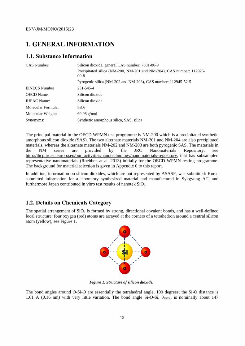
The spatial arrangement of SiO2 is formed by strong, directional covalent bonds, and has a well-defined
local structure: four oxygen (red) atoms are arrayed at the corners of a tetrahedron around a central silicon
atom (yellow), see Figure 1.
Figure 1. Structure of silicon dioxide.
The bond angles around O-Si-O are essentially the tetrahedral angle, 109 degrees; the Si-O distance is
1.61 A (0.16 nm) with very little variation. The bond angle Si-O-Si, θSiOSi, is nominally about 147
ENV/JM/MONO(2016)23
13
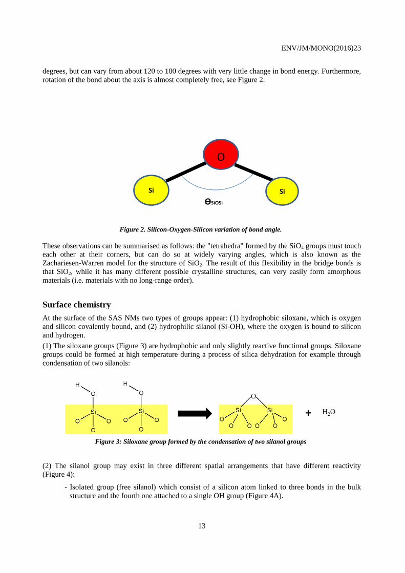
degrees, but can vary from about 120 to 180 degrees with very little change in bond energy. Furthermore,
rotation of the bond about the axis is almost completely free, see Figure 2.
Figure 2. Silicon-Oxygen-Silicon variation of bond angle.
These observations can be summarised as follows: the "tetrahedra" formed by the SiO4 groups must touch
each other at their corners, but can do so at widely varying angles, which is also known as the
Zachariesen-Warren model for the structure of SiO2. The result of this flexibility in the bridge bonds is
that SiO2, while it has many different possible crystalline structures, can very easily form amorphous
materials (i.e. materials with no long-range order).
Surface chemistry
At the surface of the SAS NMs two types of groups appear: (1) hydrophobic siloxane, which is oxygen
and silicon covalently bound, and (2) hydrophilic silanol (Si-OH), where the oxygen is bound to silicon
and hydrogen.
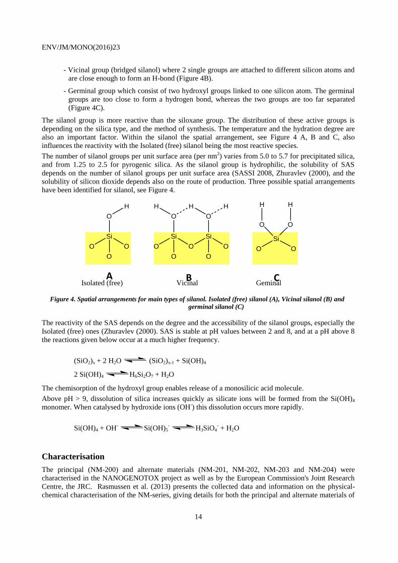
(1) The siloxane groups (Figure 3) are hydrophobic and only slightly reactive functional groups. Siloxane
groups could be formed at high temperature during a process of silica dehydration for example through
condensation of two silanols:
Figure 3: Siloxane group formed by the condensation of two silanol groups
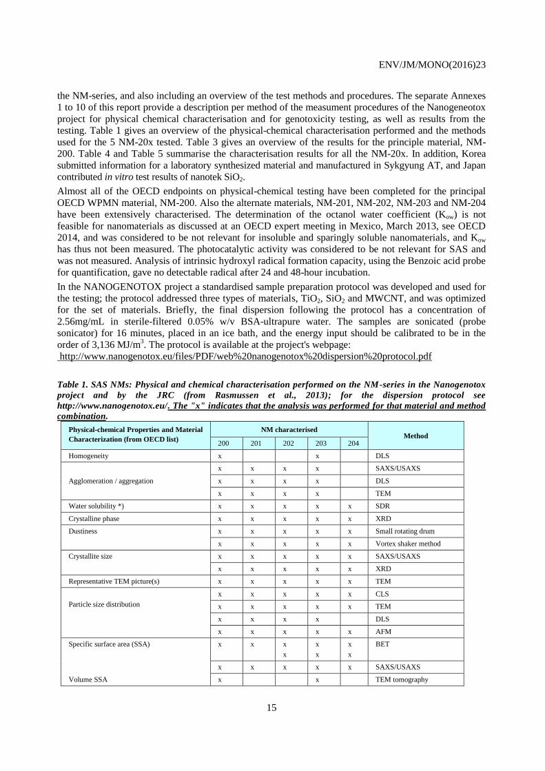
(2) The silanol group may exist in three different spatial arrangements that have different reactivity
(Figure 4):
- Isolated group (free silanol) which consist of a silicon atom linked to three bonds in the bulk
structure and the fourth one attached to a single OH group (Figure 4A).
Si
O
Si
ƟSiOSi
ENV/JM/MONO(2016)23
14
- Vicinal group (bridged silanol) where 2 single groups are attached to different silicon atoms and
are close enough to form an H-bond (Figure 4B).
- Germinal group which consist of two hydroxyl groups linked to one silicon atom. The germinal
groups are too close to form a hydrogen bond, whereas the two groups are too far separated
(Figure 4C).
The silanol group is more reactive than the siloxane group. The distribution of these active groups is
depending on the silica type, and the method of synthesis. The temperature and the hydration degree are
also an important factor. Within the silanol the spatial arrangement, see Figure 4 A, B and C, also
influences the reactivity with the Isolated (free) silanol being the most reactive species.
The number of silanol groups per unit surface area (per nm2) varies from 5.0 to 5.7 for precipitated silica,
and from 1.25 to 2.5 for pyrogenic silica. As the silanol group is hydrophilic, the solubility of SAS
depends on the number of silanol groups per unit surface area (SASSI 2008, Zhuravlev (2000), and the
solubility of silicon dioxide depends also on the route of production. Three possible spatial arrangements
have been identified for silanol, see Figure 4.
Isolated (free) Vicinal Geminal
Figure 4. Spatial arrangements for main types of silanol. Isolated (free) silanol (A), Vicinal silanol (B) and
germinal silanol (C)
The reactivity of the SAS depends on the degree and the accessibility of the silanol groups, especially the
Isolated (free) ones (Zhuravlev (2000). SAS is stable at pH values between 2 and 8, and at a pH above 8
the reactions given below occur at a much higher frequency.
(SiO2)x + 2 H2O (SiO2)x-1 + Si(OH)4
2 Si(OH)4 H6Si2O7 + H2O
The chemisorption of the hydroxyl group enables release of a monosilicic acid molecule.
Above pH > 9, dissolution of silica increases quickly as silicate ions will be formed from the Si(OH)4
monomer. When catalysed by hydroxide ions (OH-) this dissolution occurs more rapidly.
Si(OH)4 + OH- Si(OH)5
- H3SiO4
- + H2O
Characterisation
The principal (NM-200) and alternate materials (NM-201, NM-202, NM-203 and NM-204) were
characterised in the NANOGENOTOX project as well as by the European Commission's Joint Research
Centre, the JRC. Rasmussen et al. (2013) presents the collected data and information on the physical-
chemical characterisation of the NM-series, giving details for both the principal and alternate materials of
Si
O O
O O
H H
Si
O O
O
O
H
Si
O
O
O
H H
Si
O O
O
O
H
A B C
ENV/JM/MONO(2016)23
15
the NM-series, and also including an overview of the test methods and procedures. The separate Annexes
1 to 10 of this report provide a description per method of the measument procedures of the Nanogeneotox
project for physical chemical characterisation and for genotoxicity testing, as well as results from the
testing. Table 1 gives an overview of the physical-chemical characterisation performed and the methods
used for the 5 NM-20x tested. Table 3 gives an overview of the results for the principle material, NM-
200. Table 4 and Table 5 summarise the characterisation results for all the NM-20x. In addition, Korea
submitted information for a laboratory synthesized material and manufactured in Sykgyung AT, and Japan
contributed in vitro test results of nanotek SiO2.
Almost all of the OECD endpoints on physical-chemical testing have been completed for the principal
OECD WPMN material, NM-200. Also the alternate materials, NM-201, NM-202, NM-203 and NM-204
have been extensively characterised. The determination of the octanol water coefficient (Kow) is not
feasible for nanomaterials as discussed at an OECD expert meeting in Mexico, March 2013, see OECD
2014, and was considered to be not relevant for insoluble and sparingly soluble nanomaterials, and Kow
has thus not been measured. The photocatalytic activity was considered to be not relevant for SAS and
was not measured. Analysis of intrinsic hydroxyl radical formation capacity, using the Benzoic acid probe
for quantification, gave no detectable radical after 24 and 48-hour incubation.
In the NANOGENOTOX project a standardised sample preparation protocol was developed and used for
the testing; the protocol addressed three types of materials, TiO2, SiO2 and MWCNT, and was optimized
for the set of materials. Briefly, the final dispersion following the protocol has a concentration of
2.56mg/mL in sterile-filtered 0.05% w/v BSA-ultrapure water. The samples are sonicated (probe
sonicator) for 16 minutes, placed in an ice bath, and the energy input should be calibrated to be in the
order of 3,136 MJ/m3. The protocol is available at the project's webpage:
http://www.nanogenotox.eu/files/PDF/web%20nanogenotox%20dispersion%20protocol.pdf
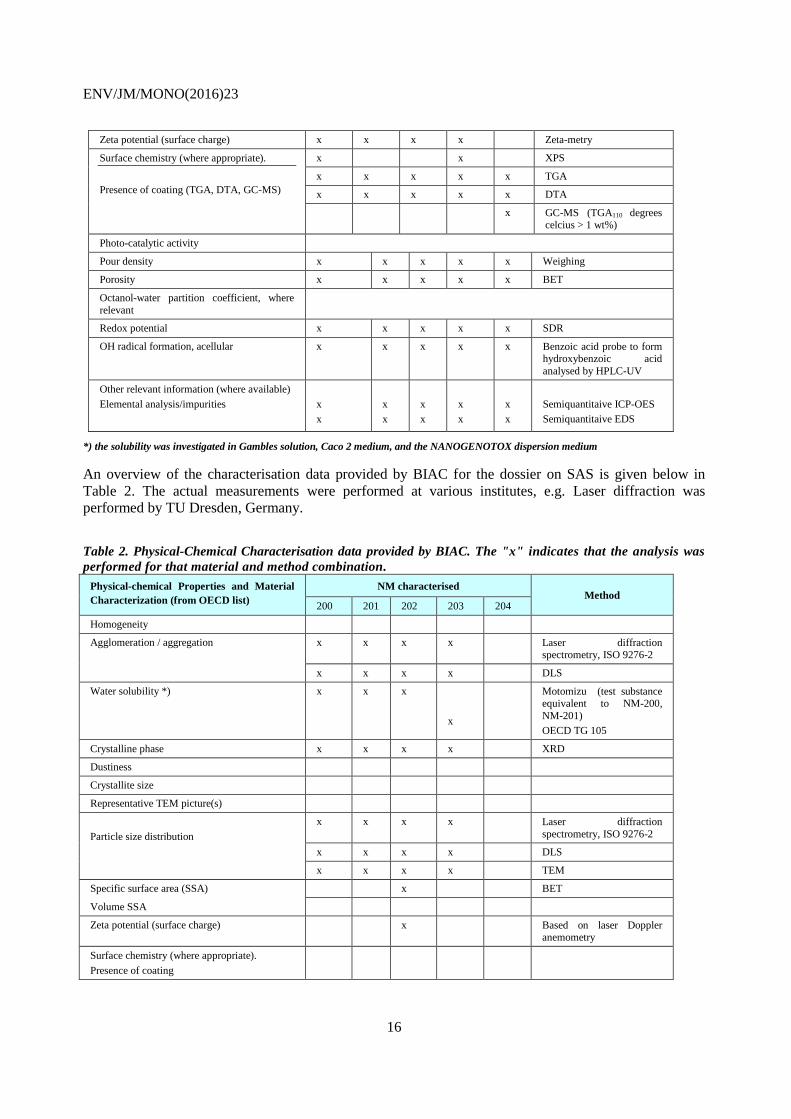
Table 1. SAS NMs: Physical and chemical characterisation performed on the NM-series in the Nanogenotox
project and by the JRC (from Rasmussen et al., 2013); for the dispersion protocol see
http://www.nanogenotox.eu/. The "x" indicates that the analysis was performed for that material and method
combination.
Physical-chemical Properties and Material
Characterization (from OECD list)
NM characterised Method
200 201 202 203 204
Homogeneity x x DLS
x x x x SAXS/USAXS
Agglomeration / aggregation x x x x DLS
x x x x TEM
Water solubility *) x x x x x SDR
Crystalline phase x x x x x XRD
Dustiness x x x x x Small rotating drum
x x x x x Vortex shaker method
Crystallite size x x x x x SAXS/USAXS
x x x x x XRD
Representative TEM picture(s) x x x x x TEM
Particle size distribution
x x x x x CLS
x x x x x TEM
x x x x DLS
x x x x x AFM
Specific surface area (SSA) x x x
x
x
x
x
x
BET
x x x x x SAXS/USAXS
Volume SSA x x TEM tomography
ENV/JM/MONO(2016)23
16
Zeta potential (surface charge) x x x x Zeta-metry
Surface chemistry (where appropriate).
Presence of coating (TGA, DTA, GC-MS)
x x XPS
x x x x x TGA
x x x x x DTA
x GC-MS (TGA110 degrees celcius > 1 wt%)
Photo-catalytic activity
Pour density x x x x x Weighing
Porosity x x x x x BET
Octanol-water partition coefficient, where
relevant
Redox potential x x x x x SDR
OH radical formation, acellular x x x x x Benzoic acid probe to form hydroxybenzoic acid
analysed by HPLC-UV
Other relevant information (where available)
Elemental analysis/impurities
x
x
x
x
x
x
x
x
x
x
Semiquantitaive ICP-OES
Semiquantitaive EDS
*) the solubility was investigated in Gambles solution, Caco 2 medium, and the NANOGENOTOX dispersion medium
An overview of the characterisation data provided by BIAC for the dossier on SAS is given below in
Table 2. The actual measurements were performed at various institutes, e.g. Laser diffraction was
performed by TU Dresden, Germany.
Table 2. Physical-Chemical Characterisation data provided by BIAC. The "x" indicates that the analysis was
performed for that material and method combination.
Physical-chemical Properties and Material
Characterization (from OECD list)
NM characterised Method
200 201 202 203 204
Homogeneity
Agglomeration / aggregation x x x x Laser diffraction spectrometry, ISO 9276-2
x x x x DLS
Water solubility *) x x x
x
Motomizu (test substance equivalent to NM-200,
NM-201)
OECD TG 105
Crystalline phase x x x x XRD
Dustiness
Crystallite size
Representative TEM picture(s)
Particle size distribution
x x x x Laser diffraction
spectrometry, ISO 9276-2
x x x x DLS
x x x x TEM
Specific surface area (SSA) x BET
Volume SSA
Zeta potential (surface charge) x Based on laser Doppler anemometry
Surface chemistry (where appropriate).
Presence of coating
ENV/JM/MONO(2016)23
17
Photo-catalytic activity
Pour density
Porosity
Octanol-water partition coefficient, where relevant
Redox potential
OH radical formation, acellular
Other relevant information (where available)
Tapped density
x
x
x
x
1.3. General Substance Information
A. Type of Substance
Element [ ]
Inorganic [X]
Natural substance [ ]
Organic [ ]
Organometallic [ ]
Petroleum product [ ]
B. Physical State (at 20°C and 1.013 hPa)
Gaseous [ ] Liquid [ ] Solid [X]
C. Purity (Indicate the percentage by weight/weight)
Different analyses were performed for identifying elemental composition and surface chemistry. Such
characterisation also gives information on the type and amount of impurities and the presence/absence of
a coating. The purity range of NM-20x was from 96 to 99 %, see Table 3 for the values for each material.
No information concerning purity was given for the materials studied by Korea and Japan.
1.4. Use Pattern
A. Uses
SAS is a High Production Volume (HPV) chemical which is widely used in many industries and in
various applications such as synthetic resins, plastics, lacquers, vinyl coatings, varnishes, adhesives,
paints, printing inks, silicone rubber, fillers in the rubber industry, tyre compounds, insulation material,
additive to coatings, as free-flow and anti-caking agents in powder materials, including food, as tooth
paste additives, pharmaceuticals, cosmetics, as liquid carriers particularly in the manufacture of
agrochemicals and animal feed, and foods, resulting in widespread exposure to these substances
(Fruijtier-Pölloth, 2012). SAS is also increasingly used in diagnostic and biomedical research such as
cancer therapy, DNA delivery, and enzyme immobilization (Barik et al., 2008); the total volumes for
these uses is though low in comparison with the industrial uses. According to SRI Consulting precipitated
SAS is one of the most abundant nanomaterial on the market in terms of quantity2.
2 http://www.ihs.com/products/chemical/planning/scup/nanoscale-chemicals.aspx?pu=1&rd=chemihs
ENV/JM/MONO(2016)23
18
The selected SASs in the NM-series are used, among others, in food applications and as reinforcement in
car tyres (rubber), e.g. car tyres.
B. Method of production (e.g., precipitation, gas phase):
The methods of production of synthetic amorphous silica are described in detail in e.g. EC, 2007. The
principles of particle formation are describy in "The Chemistry of Silica"" by Iler RK, (1979).
Thermal Route
Pyrogenic silica is a very fine particulate form of silicon dioxide and is prepared by burning silicon
tetrachloride (SiCl4) or trichlorosilane (SiHCl3) in an oxyhydrogen gas flame:
SiCl4 + 2 H2 + O2 → SiO2 + 4 HCl
SIHCl3 + H2 + O2 → SiO2 + 3 HCl
By varying e.g. the flame temperature, flame composition and feed stock, the product's physical-chemical
properties, e.g. the specific surface area and the particle size, can be controlled. NM-202 and NM-203
were synthsised via this type of process.
Wet Route
Different manufacturing methods are possible, and (1) describes the way the materials NM-200, NM-201
and NM-204 were synthetized; (2) this process is used for synthesis of monodisperse particles, with a
control of the shape and the size of the nanomaterial; process (2) is without commercial relevance.
1. Amorphous silica, silica gel, is produced by the acidification of solutions of sodium silicate to
produce a gelatinous precipitate that is then washed with water and afterwards dehydrated to
produce colourless microporous silica.
Briefly, the precipitation method reacts an alkali metal silicate dissolved in water, e.g. water glass
(Na2O . nSiO2; n = 2 – 4) with sulphuric acid, through a series of production steps that include raw
material storage, synthesis, solid-liquid-filtration, drying and packaging. The synthesis can either
be continuous or in batch.
Na2O x nSiO2 +H2SO4 →nSiO2 + Na2SO4 + H2O
2. In order to obtain monodisperse SAS nanoparticles, e.g. sol-gel methods are employed such as the
Stöber method, which is not utilized commercially, and the water-in-oil (w/o) micro-emulsion
method. The sol-gel process is based on a series of hydrolysis, condensation and polymerisation
reactions of an alkoxide. The most widely used precursors are alkoxysilanes, such as
tetramethoxysilane (TMOS) and tetraethoxysilane (TEOS).
Hydrolysis is initiated by the addition of water to the silane solution under acidic, neutral, or basic
conditions Si-(OR)4 +H2O (RO)3-Si-OH + ROH
During the condensation step a molecule, such as water or alcohol, is liberated. This leads to
polymerisation and synthesis of a network of silane (Si-O-Si) and to production of nanomaterials.
Si-(OR)4 + HO-Si-(OR)3 (RO)3-Si-O-Si -(OR)3+ ROH ( with alcohol)
or (RO)3-Si-OH+ HO-Si-(OR)3 RO)3-Si-O-Si -(OR)3+ H2O ( with water)
ENV/JM/MONO(2016)23
19
2. PHYSICAL CHEMICAL DATA
2.1 Overview of Identification information and Physical Chemical Data for
SAS
SAS is well described in the literature with regard to "classical3" physical-chemical properties and these
are reported also in standard reference works; some of the "classical" physical-chemical properties are not
part of the end-points agreed under the Sponsorship Program.
For physical-chemical properties listed in the Sponsorship Program as relevant for nanomaterials, some
appear not to be relevant for SAS (crystalline phase and size, photocatalytic activity, redox potential,
radical formation). The determination of Kow is not feasible for inorganic nanomaterials, as discussed at
an OECD expert meeting in Mexico, March 2013, see OECD 2014, and was considered to be not relevant
for insoluble and sparingly soluble nanomaterials. For the relevant parameters, a wide range of values are
presented in the background reports (OECD (2004a), ECETOC (2006), SASSI (2008)) obtained through
measurements of different sources of SAS, see for example ECETOC (2006) p. 12. These reports present
only limited physical-chemical characterisation data associated to each of the different specific SASs that
were tested; thus this data is not fulfilling the information requests in the Sponsorship Program, and the
information available from the reports would not necessarily relate to the principal material.
In the Joint Action NANOGENOTOX precipitated and pyrogenic synthetic amorphous silicon dioxide
were characterised, see Rasmussen, 2013. The Joint Action developed a dispersion protocol, found at
http://www.nanogenotox.eu/files/PDF/web%20nanogenotox%20dispersion%20protocol.pdf . For the
characterisation of the dispersed materials, it cannot be excluded that the dispersion procedure influences
the material properties.
This section describes the characterisation results of the principal material (NM-200) and alternate
materials (NM-201, NM-202, NM-203 and NM-204), and presentes the data provided for characterising
the Korean material.
The following Table 4 gives an overview of the information available for the end-point group
Nanomaterial Information / Identification. Table 3 summarises the physical chemical characterisation
data generated for the principal material (NM-200) in the Testing Programme.
The Table 5 summarises the physical chemical characterisation data generated for the WPMN Testing
Programme for the principal and alternate materials. An overview of the test methods used for obtaining
experimental data is given in Table 1. The data for NM-201, NM-202, NM-203 and NM-204, presented
in the same way as data for NM-200 in Table 3, are given in Appendix 0.
For DLS measurements, the instrument measures the Z-average (diameter of particles scattering with
higher intensity) over three runs, and this value is stated together with the minimum (-) and maximum (+)
values of the Z-average of the three runs. The polydispersity index (PdI) is a measure of the width of the
particle size distribution, and is based on assumptions of a model: A monomodal model, called the
cumulant analysis, is often used to treat the raw data correlograms (decaying as exponential). It
determines a Z-average and a polydispersity index. For polydisperse samples, more sophisticated,
multimodal analysis models, e.g the CONTIN method, can be applied to reveal size distributions.
Polydispersity indices less than 0.1 are typically referred to as "monodisperse". Details are published in
Annex 5 to the dossier (OECD WPMN, 2015).
3 Melting point, boiling point, vapour pressure, flash point, auto flammability, flammability, explosive properties,
oxidising properties, viscosity, water solubility, octanol-water partition coefficient.
ENV/JM/MONO(2016)23
20
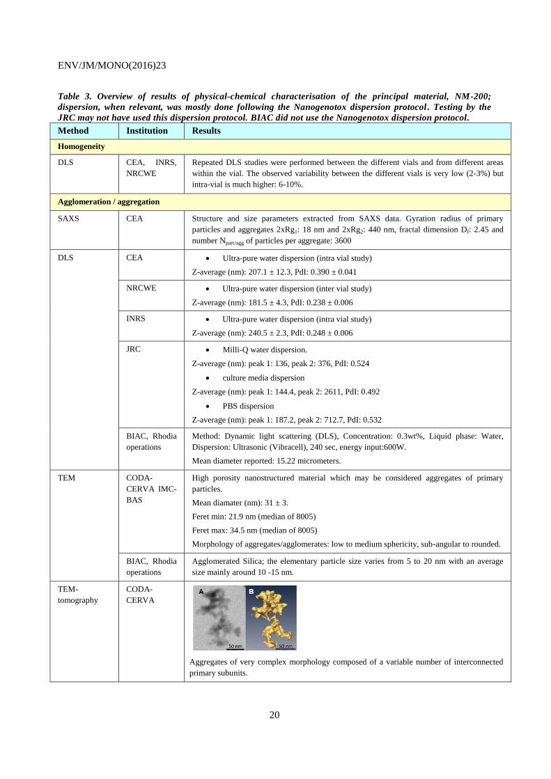
Table 3. Overview of results of physical-chemical characterisation of the principal material, NM-200;
dispersion, when relevant, was mostly done following the Nanogenotox dispersion protocol . Testing by the
JRC may not have used this dispersion protocol. BIAC did not use the Nanogenotox dispersion protocol.
Method Institution Results
Homogeneity
DLS CEA, INRS,
NRCWE
Repeated DLS studies were performed between the different vials and from different areas
within the vial. The observed variability between the different vials is very low (2-3%) but
intra-vial is much higher: 6-10%.
Agglomeration / aggregation
SAXS CEA Structure and size parameters extracted from SAXS data. Gyration radius of primary
particles and aggregates 2xRg1: 18 nm and 2xRg2: 440 nm, fractal dimension Df: 2.45 and
number Npart/agg of particles per aggregate: 3600
DLS CEA Ultra-pure water dispersion (intra vial study)
Z-average (nm): 207.1 ± 12.3, PdI: 0.390 ± 0.041
NRCWE Ultra-pure water dispersion (inter vial study)
Z-average (nm): 181.5 ± 4.3, PdI: 0.238 ± 0.006
INRS Ultra-pure water dispersion (intra vial study)
Z-average (nm): 240.5 ± 2.3, PdI: 0.248 ± 0.006
JRC Milli-Q water dispersion.
Z-average (nm): peak 1: 136, peak 2: 376, PdI: 0.524
culture media dispersion
Z-average (nm): peak 1: 144.4, peak 2: 2611, PdI: 0.492
PBS dispersion
Z-average (nm): peak 1: 187.2, peak 2: 712.7, PdI: 0.532
BIAC, Rhodia
operations
Method: Dynamic light scattering (DLS), Concentration: 0.3wt%, Liquid phase: Water,
Dispersion: Ultrasonic (Vibracell), 240 sec, energy input:600W.
Mean diameter reported: 15.22 micrometers.
TEM CODA-
CERVA IMC-
BAS
High porosity nanostructured material which may be considered aggregates of primary
particles.
Mean diamater (nm): 31 ± 3.
Feret min: 21.9 nm (median of 8005)
Feret max: 34.5 nm (median of 8005)
Morphology of aggregates/agglomerates: low to medium sphericity, sub-angular to rounded.
BIAC, Rhodia
operations
Agglomerated Silica; the elementary particle size varies from 5 to 20 nm with an average
size mainly around 10 -15 nm.
TEM-
tomography
CODA-
CERVA
Aggregates of very complex morphology composed of a variable number of interconnected
primary subunits.
ENV/JM/MONO(2016)23
21
AFM CEA Third dimension of the agglomerates/aggregates: median (of 1382): 21.9 nm
Laser
diffraction
spectrometry
BIAC,
TU Dresden,
Germnay
The test method is in accordance with EN 481, ISO 9276-2, based on the principle of light
scattering measured on the dry powder. The median particle diameter weighted by volume
amounts to 480 micrometers. (The instrument used, LAP 321, has a lower size limit of 0.3
micrometers, according to the instrument manufacturer
(http://www.exisab.com/Docs/Newsletters/ExIS_News_March_2011.pdf).)
Water Solubility
Motomizu BIAC, Rhodia
Operations
The saturation concentrations [M]tot for test substance equivalent to NM-200 has been
determined to 2.4 mmol/l. (The method is a Spectrophoto-metric Determination of Silicate in
water with Molybdate and Malachite Green)
24-hour
acellular in
vitro incubation
test in special
solutions
NRCWE The 24-hour dissolution ratio of NM-200 was measured in three different media: 0.05% BSA
in water, Gambles solution and Caco 2 media. Both NM-200 and the Al impurities are
partially soluble in all media but amounts vary considerably with medium, as does the
relative amounts of dissolved Al impurities compared with dissolved Si, suggesting that the
solubility behaviour of the Al impurity and NM-200 depends on the medium.
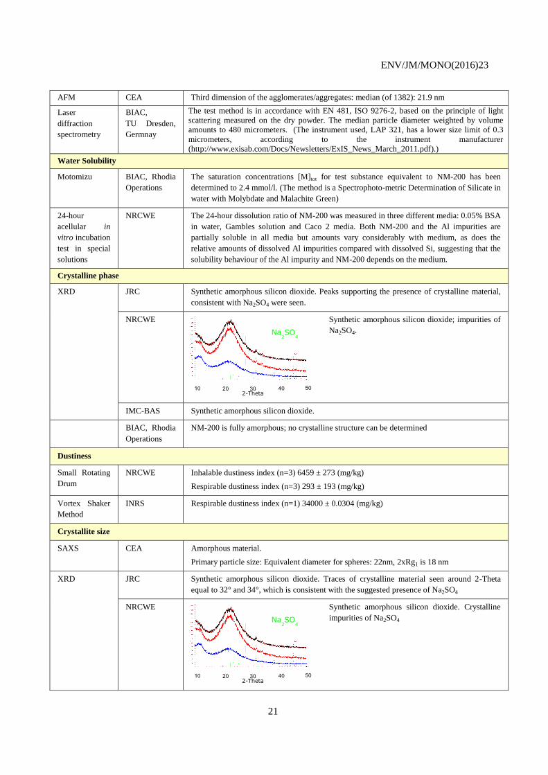
Crystalline phase
XRD JRC Synthetic amorphous silicon dioxide. Peaks supporting the presence of crystalline material,
consistent with Na2SO4 were seen.
NRCWE Synthetic amorphous silicon dioxide; impurities of
Na2SO4.
IMC-BAS Synthetic amorphous silicon dioxide.
BIAC, Rhodia
Operations
NM-200 is fully amorphous; no crystalline structure can be determined
Dustiness
Small Rotating
Drum
NRCWE Inhalable dustiness index (n=3) 6459 ± 273 (mg/kg)
Respirable dustiness index (n=3) 293 ± 193 (mg/kg)
Vortex Shaker
Method
INRS Respirable dustiness index (n=1) 34000 ± 0.0304 (mg/kg)
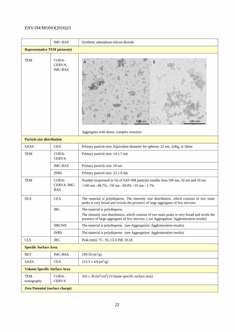
Crystallite size
SAXS CEA Amorphous material.
Primary particle size: Equivalent diameter for spheres: 22nm, 2xRg1 is 18 nm
XRD JRC Synthetic amorphous silicon dioxide. Traces of crystalline material seen around 2-Theta
equal to 32° and 34°, which is consistent with the suggested presence of Na2SO4
NRCWE Synthetic amorphous silicon dioxide. Crystalline
impurities of Na2SO4
10 20 30 40 50
Na2SO
4
2-Theta
10 20 30 40 50
Na2SO
4
2-Theta
ENV/JM/MONO(2016)23
22
IMC-BAS Synthetic amorphous silicon dioxide
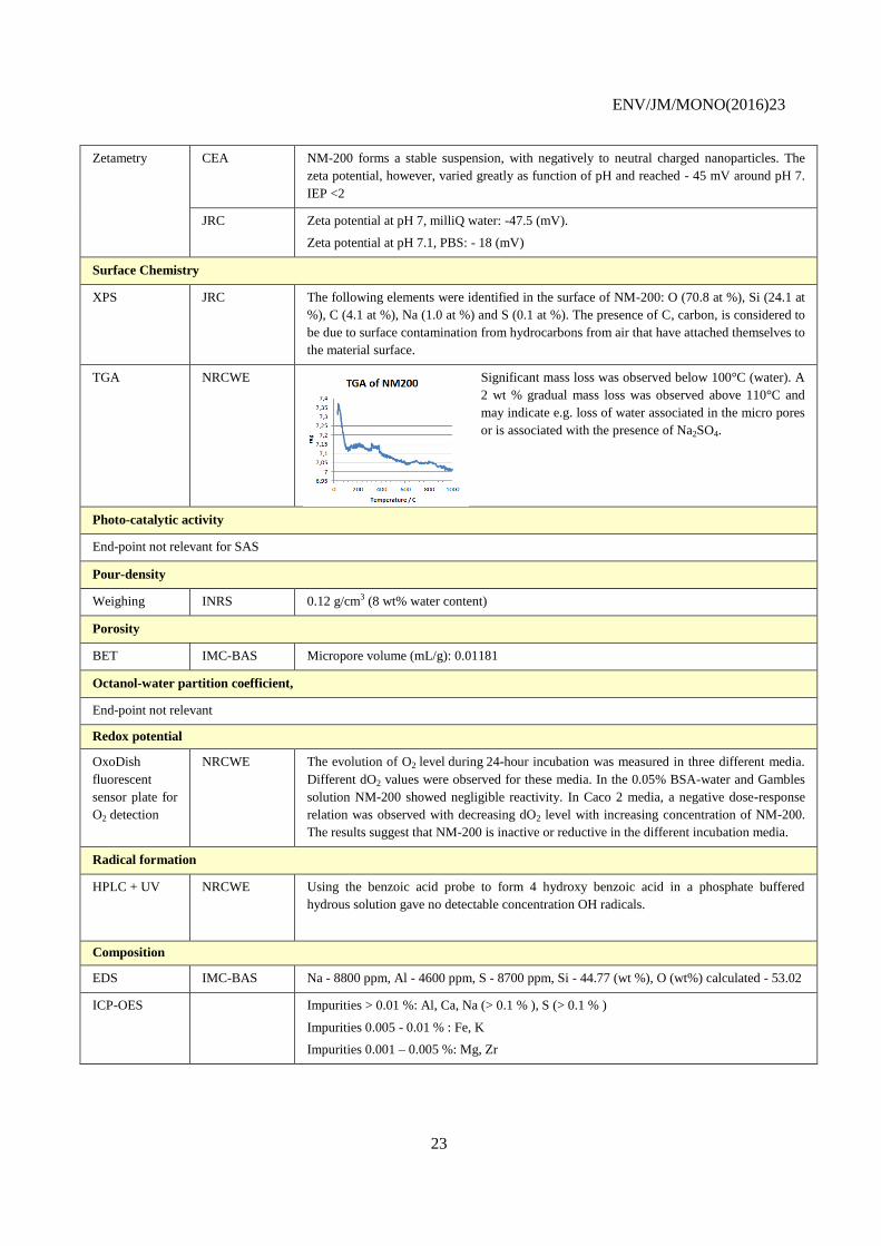
Representative TEM picture(s)
TEM CODA-
CERVA,
IMC-BAS
Aggregates with dense, complex structure
Particle size distribution
SAXS CEA Primary particle size: Equivalent diameter for spheres: 22 nm, 2xRg1 is 18nm
TEM CODA-
CERVA
Primary particle size: 14 ± 7 nm
IMC-BAS Primary particle size: 18 nm
INRS Primary particle size: 23 ± 8 nm
TEM CODA-
CERVA IMC-
BAS
Number (expressed in %) of SAS NM particles smaller than 100 nm, 50 nm and 10 nm
<100 nm - 88.7%, <50 nm - 69.8% <10 nm - 1.7%
DLS CEA The material is polydisperse. The intensity size distribution, which consists of two main
peaks is very broad and reveals the presence of large aggregates of few microns.
JRC The material is polydisperse.
The intensity size distribution, which consists of two main peaks is very broad and revels the
presence of large aggregates of few microns. ( see Aggregation/ Agglomeration results)
NRCWE The material is polydisperse. (see Aggregation/ Agglomeration results)
INRS The material is polydisperse. (see Aggregation/ Agglomeration results)
CLS JRC Peak (nm): 75 - 95, CLS Pdl: 10.18
Specific Surface Area
BET IMC-BAS 189.16 (m2/g)
SAXS CEA 123.3 ± 4.9 (m2/g)
Volume Specific Surface Area
TEM
tomography
CODA-
CERVA
342 ± 36 (m2/cm3) (Volume specific surface area)
Zeta Potential (surface charge)
ENV/JM/MONO(2016)23
23
Zetametry CEA NM-200 forms a stable suspension, with negatively to neutral charged nanoparticles. The
zeta potential, however, varied greatly as function of pH and reached - 45 mV around pH 7.
IEP <2
JRC Zeta potential at pH 7, milliQ water: -47.5 (mV).
Zeta potential at pH 7.1, PBS: - 18 (mV)
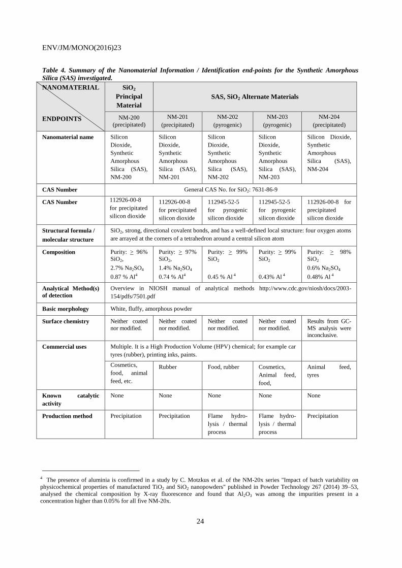
Surface Chemistry
XPS JRC The following elements were identified in the surface of NM-200: O (70.8 at %), Si (24.1 at
%), C (4.1 at %), Na (1.0 at %) and S (0.1 at %). The presence of C, carbon, is considered to
be due to surface contamination from hydrocarbons from air that have attached themselves to
the material surface.
TGA NRCWE Significant mass loss was observed below 100°C (water). A
2 wt % gradual mass loss was observed above 110°C and
may indicate e.g. loss of water associated in the micro pores
or is associated with the presence of Na2SO4.
Photo-catalytic activity
End-point not relevant for SAS
Pour-density
Weighing INRS 0.12 g/cm3 (8 wt% water content)
Porosity
BET IMC-BAS Micropore volume (mL/g): 0.01181
Octanol-water partition coefficient,
End-point not relevant
Redox potential
OxoDish
fluorescent
sensor plate for
O2 detection
NRCWE The evolution of O2 level during 24-hour incubation was measured in three different media.
Different dO2 values were observed for these media. In the 0.05% BSA-water and Gambles
solution NM-200 showed negligible reactivity. In Caco 2 media, a negative dose-response
relation was observed with decreasing dO2 level with increasing concentration of NM-200.
The results suggest that NM-200 is inactive or reductive in the different incubation media.
Radical formation
HPLC + UV NRCWE Using the benzoic acid probe to form 4 hydroxy benzoic acid in a phosphate buffered
hydrous solution gave no detectable concentration OH radicals.
Composition
EDS IMC-BAS Na - 8800 ppm, Al - 4600 ppm, S - 8700 ppm, Si - 44.77 (wt %), O (wt%) calculated - 53.02
ICP-OES Impurities > 0.01 %: Al, Ca, Na (> 0.1 % ), S (> 0.1 % )
Impurities 0.005 - 0.01 % : Fe, K
Impurities 0.001 – 0.005 %: Mg, Zr
ENV/JM/MONO(2016)23
24
Table 4. Summary of the Nanomaterial Information / Identification end-points for the Synthetic Amorphous
Silica (SAS) investigated.
NANOMATERIAL
ENDPOINTS
SiO2
Principal
Material
SAS, SiO2 Alternate Materials
NM-200
(precipitated)
NM-201
(precipitated)
NM-202
(pyrogenic)
NM-203
(pyrogenic)
NM-204
(precipitated)
Nanomaterial name Silicon
Dioxide,
Synthetic
Amorphous
Silica (SAS),
NM-200
Silicon
Dioxide,
Synthetic
Amorphous
Silica (SAS),
NM-201
Silicon
Dioxide,
Synthetic
Amorphous
Silica (SAS),
NM-202
Silicon
Dioxide,
Synthetic
Amorphous
Silica (SAS),
NM-203
Silicon Dioxide,
Synthetic
Amorphous
Silica (SAS),
NM-204
CAS Number General CAS No. for SiO2: 7631-86-9
CAS Number
112926-00-8
for precipitated
silicon dioxide
112926-00-8
for precipitated
silicon dioxide
112945-52-5
for pyrogenic
silicon dioxide
112945-52-5
for pyrogenic
silicon dioxide
112926-00-8 for
precipitated
silicon dioxide
Structural formula /
molecular structure
SiO2, strong, directional covalent bonds, and has a well-defined local structure: four oxygen atoms
are arrayed at the corners of a tetrahedron around a central silicon atom
Composition
Purity: > 96%
SiO2,
2.7% Na2SO4
0.87 % Al4
Purity: > 97%
SiO2,
1.4% Na2SO4
0.74 % Al4
Purity: > 99%
SiO2
0.45 % Al 4
Purity: > 99%
SiO2
0.43% Al 4
Purity: > 98%
SiO2
0.6% Na2SO4
0.48% Al 4
Analytical Method(s)
of detection
Overview in NIOSH manual of analytical methods http://www.cdc.gov/niosh/docs/2003-
154/pdfs/7501.pdf
Basic morphology White, fluffy, amorphous powder
Surface chemistry Neither coated
nor modified.
Neither coated
nor modified.
Neither coated
nor modified.
Neither coated
nor modified.
Results from GC-
MS analysis were
inconclusive.
Commercial uses Multiple. It is a High Production Volume (HPV) chemical; for example car
tyres (rubber), printing inks, paints.
Cosmetics,
food, animal
feed, etc.
Rubber Food, rubber Cosmetics,
Animal feed,
food,
Animal feed,
tyres
Known catalytic
activity
None None None None None
Production method Precipitation Precipitation Flame hydro-
lysis / thermal
process
Flame hydro-
lysis / thermal
process
Precipitation
4 The presence of aluminia is confirmed in a study by C. Motzkus et al. of the NM-20x series "Impact of batch variability on
physicochemical properties of manufactured TiO2 and SiO2 nanopowders" published in Powder Technology 267 (2014) 39–53,
analysed the chemical composition by X-ray fluorescence and found that Al2O3 was among the impurities present in a
concentration higher than 0.05% for all five NM-20x.
ENV/JM/MONO(2016)23
25
Table 5. Summary of the Physical-chemical Properties and Material Characterization Endpoints for the SAS-NMs from the JRC Repository generated in the Nanogenotoc
project. Dispersion, when relevant, was mostly done following the Nanogenotox dispersion protocol. Testing by the JRC may not have used this dispersion protocol. BIAC did
not use the Nanogenotox dispersion protocol.
NANOMATERIAL
ENDPOINTS
(method)
SiO2 Principal Material SiO2 (Silicon Dioxide (SAS)) Alternate Materials
Silicon Dioxide (SAS)
NM-200 (precipitated) NM-201 (precipitated) NM-202 (pyrogenic) NM-203 (pyrogenic) NM-204 (precipitated)
PHYSICAL-CHEMICAL PROPERTIES
1. Agglomeration/
Aggregation
(DLS)
Results from 3 institutions using Ultra-
pure water dispersion:
Z-average (nm): 207.1 ± 11.9, PdI:
0.390±0.041(intra vial study)
Z-average (nm): 181.5 ± 4.3, PdI: 0.238
± 0.0.006 (inter vial study)
Z-average (nm): 240.5 ± 2.3, PdI:
0.248±0.0.006(intra vial study)
BIAC reports that the diameter
measured by DLS is 15.22 micrometers
Results from 1 institution
and ultra-pure water
dispersion:
Z-average (nm): 208.1 ±
34.5, PdI: 0.352 ± 0.028
(intra vial study)
Z-average (nm): 197.0 ±
15.7, PdI: 0.337 ± 0.020
(inter vial study)
Results from 1 institution
and ultra-pure water
dispersion:
Z-average (nm): 175.9 ±
4.5, PdI: 0.355 ± 0.001
(intra vial study)
Results from 3 institutions and
Ultra-pure water dispersion:
Z-average (nm): 172.9 ± 9.2. PdI:
0.427 ± 0.025 (intra vial study)
Z-average (nm): 147.5 ± 4.5. PdI:
0.244 ± 0.017 (intra vial study)
Z-average (nm): 146.8 ± 0.6, PdI:
0.229 ± 0.015 (inter vial study)
Z-average (nm): 245.7 ± 37.2.
PdI: 0.299 ± 0.024 (intra vial
study)
-
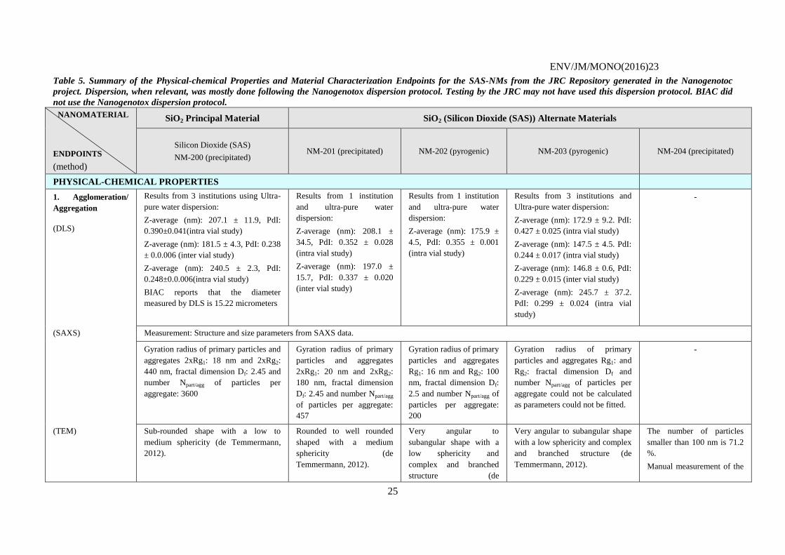
(SAXS) Measurement: Structure and size parameters from SAXS data.
Gyration radius of primary particles and
aggregates 2xRg1: 18 nm and 2xRg2:
440 nm, fractal dimension Df: 2.45 and
number Npart/agg of particles per
aggregate: 3600
Gyration radius of primary
particles and aggregates
2xRg1: 20 nm and 2xRg2:
180 nm, fractal dimension
Df: 2.45 and number Npart/agg
of particles per aggregate:
457
Gyration radius of primary
particles and aggregates
Rg1: 16 nm and Rg2: 100
nm, fractal dimension Df:
2.5 and number Npart/agg of
particles per aggregate:
200
Gyration radius of primary
particles and aggregates Rg1: and
Rg2: fractal dimension Df and
number Npart/agg of particles per
aggregate could not be calculated
as parameters could not be fitted.
-
(TEM) Sub-rounded shape with a low to
medium sphericity (de Temmermann,
2012).
Rounded to well rounded
shaped with a medium
sphericity (de
Temmermann, 2012).
Very angular to
subangular shape with a
low sphericity and
complex and branched
structure (de
Very angular to subangular shape
with a low sphericity and complex
and branched structure (de
Temmermann, 2012).
The number of particles
smaller than 100 nm is 71.2
%.
Manual measurement of the
ENV/JM/MONO(2016)23
26
NANOMATERIAL
ENDPOINTS
(method)
SiO2 Principal Material SiO2 (Silicon Dioxide (SAS)) Alternate Materials
Silicon Dioxide (SAS)
NM-200 (precipitated) NM-201 (precipitated) NM-202 (pyrogenic) NM-203 (pyrogenic) NM-204 (precipitated)
Median mean diamater (nm): 31 ± 35.
Feret min: 21.9 nm (median of 8005)
Feret max: 34.5 nm (median of 8005)
Morphology of
aggregates/agglomerates: Low to
medium sphericity, sub-angular to
rounded.
% of aggegates < 100 nm: 88.7 %
BIAC data: Agglomerated Silica; the
elementary particle size varies from 5 to
20 nm with an average size mainly
around 10-15 nm.
Median diamater (nm): 43 ±
4.
Feret min: 25.4 nm (median
of 5311)
Feret max: 38.5 nm (median
of 5311)
Morphology of
aggregates/agglomerates:
Medium sphericity, rounded
to well--rounded.
% of aggegates < 100 nm:
81.5 %
Temmermann, 2012)
Median diamater (nm): 53
± 9.
Feret min: 37.2 nm
(median of 4248)
Feret max: 58.4 nm
(median of 4248)
Morphology of
aggregates/agglomerates:
Low sphericity—very
angular to sub-angular.
% of aggregates <100 nm:
80.4 %
Median diamater (nm): 48 ± 4
Feret min: 33.5 nm (median of
4889)
Feret max: 53.2 nm (median of
4889).
% of aggregates < 100nm: 88 ± 2.
Morphology of
aggregates/agglomerates: Low
sphericity, angular.
% of aggregates <100 nm: 77.5 %
ESD gives a result in the
range of 10 nm – 15 nm
% of aggregates <100 nm:
71.2 %
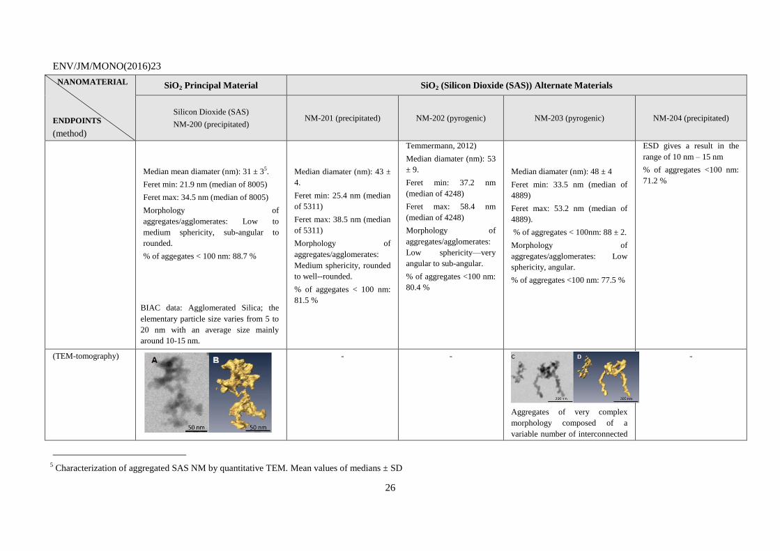
(TEM-tomography)
- -
Aggregates of very complex
morphology composed of a
variable number of interconnected
-
5 Characterization of aggregated SAS NM by quantitative TEM. Mean values of medians ± SD
ENV/JM/MONO(2016)23
27
NANOMATERIAL
ENDPOINTS
(method)
SiO2 Principal Material SiO2 (Silicon Dioxide (SAS)) Alternate Materials
Silicon Dioxide (SAS)
NM-200 (precipitated) NM-201 (precipitated) NM-202 (pyrogenic) NM-203 (pyrogenic) NM-204 (precipitated)
Aggregates of very complex
morphology composed of a variable
number of interconnected primary
subunits.
primary subunits.
AFM median (of 1382): 21.9 nm median (of 1275): 33.5 nm median (of 1103): 38.2 nm median (of 593): 24.2 nm.
2. Water Solubility/
Dispersability
(test performed: 24-
hour acellular in vitro
incubation test in
special solutions)
Measurement: The 24-hour dissolution ratio was measured in three different media: 0.05% BSA in water, Gambles solution and Caco 2 media.
Both NM-200 and the Al impurities are
partially soluble in all media but
amounts vary considerably with
medium, as does the relative amounts of
dissolved Al impurities compared with
dissolved Si, suggesting that the
solubility behaviour of the Al impurity
and NM-200 depends on the medium.
Both NM-201 and the Al
impurities are partially
soluble in Gambles Solution
and Caco2 media but
amounts vary considerably
with the medium. In 0.05%
BSA in water only the Al
impurities are partially
soluble; Si was below the
detection limit. The relative
amounts of dissolved Al
impurities and dissolved Si
are different depending on
medium, which suggests
different solubility
behaviour of Al impurities
and NM-201 depending on
the medium.
Both NM-202 and the Al
impurities are partially
soluble in all media but
amounts vary
considerably with medium
as does the relative
amounts of dissolved Al
impurities and dissolved
Si suggesting different
solubility behaviour of Al
impurities and NM-202
depending on the medium.
Both NM-203 and the Al
impurities are partially soluble in
all media but amounts vary
considerably with medium, as
does the relative amounts of
dissolved Al impurities compared
with dissolved Si, suggesting that
the solubility behaviour of the Al
impurities and NM-203 depend on
the medium.
Both NM-204 and the Al
impurities are partially
soluble in 0.05% BSA in
water and Caco2 media but
amounts vary considerably
with medium. In Gambles
solution only NM-204 is
partially soluble. The
relative amounts of
dissolved Al impurities and
dissolved Si differ
depending on medium,
which suggests different
solubility behaviour of Al
impurities and NM-204
depending on the medium.
ENV/JM/MONO(2016)23
28
NANOMATERIAL
ENDPOINTS
(method)
SiO2 Principal Material SiO2 (Silicon Dioxide (SAS)) Alternate Materials
Silicon Dioxide (SAS)
NM-200 (precipitated) NM-201 (precipitated) NM-202 (pyrogenic) NM-203 (pyrogenic) NM-204 (precipitated)
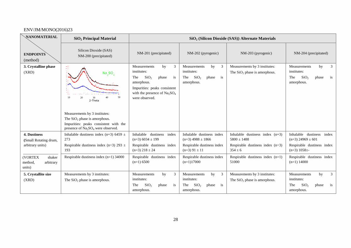
3. Crystalline phase
(XRD)
Measurements by 3 institutes:
The SiO2 phase is amorphous.
Impurities: peaks consistent with the presence of Na2SO4 were observed.
Measurements by 3
institutes:
The SiO2 phase is
amorphous.
Impurities: peaks consistent
with the presence of Na2SO4
were observed.
Measurements by 3
institutes:
The SiO2 phase is
amorphous.
Measurements by 3 institutes:
The SiO2 phase is amorphous.
Measurements by 3
institutes:
The SiO2 phase is
amorphous.
4. Dustiness
(Small Rotating drum,
arbitrary units)
(VORTEX shaker
method, arbitrary
units)
Inhalable dustiness index (n=3) 6459 ±
273
Respirable dustiness index (n=3) 293 ±
193
Inhalable dustiness index
(n=3) 6034 ± 199
Respirable dustiness index
(n=3) 218 ± 24
Inhalable dustiness index
(n=3) 4988 ± 1866
Respirable dustiness index
(n=3) 91 ± 11
Inhalable dustiness index (n=3)
5800 ± 1488
Respirable dustiness index (n=3)
354 ± 6
Inhalable dustiness index
(n=3) 24969 ± 601
Respirable dustiness index
(n=3) 1058±-
Respirable dustiness index (n=1) 34000 Respirable dustiness index
(n=1) 6500
Respirable dustiness index
(n=1)17000
Respirable dustiness index (n=1)
51000
Respirable dustiness index
(n=1) 14000
5. Crystallite size
(XRD)
Measurements by 3 institutes:
The SiO2 phase is amorphous.
Measurements by 3
institutes:
The SiO2 phase is
amorphous.
Measurements by 3
institutes:
The SiO2 phase is
amorphous.
Measurements by 3 institutes:
The SiO2 phase is amorphous.
Measurements by 3
institutes:
The SiO2 phase is
amorphous.
10 20 30 40 50
Na2SO
4
2-Theta
ENV/JM/MONO(2016)23
29
NANOMATERIAL
ENDPOINTS
(method)
SiO2 Principal Material SiO2 (Silicon Dioxide (SAS)) Alternate Materials
Silicon Dioxide (SAS)
NM-200 (precipitated) NM-201 (precipitated) NM-202 (pyrogenic) NM-203 (pyrogenic) NM-204 (precipitated)
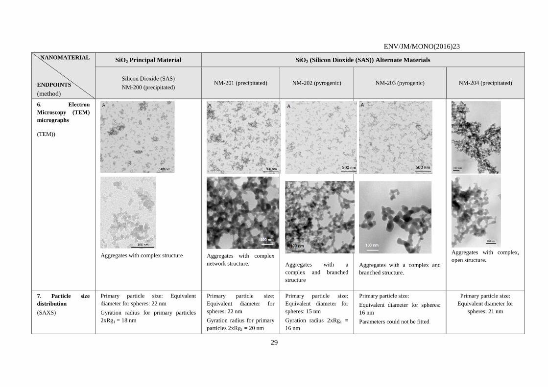
6. Electron
Microscopy (TEM)
micrographs
(TEM))
Aggregates with complex structure
Aggregates with complex
network structure.
Aggregates with a
complex and branched
structure
Aggregates with a complex and
branched structure.
Aggregates with complex,
open structure.
7. Particle size
distribution
(SAXS)
Primary particle size: Equivalent
diameter for spheres: 22 nm
Gyration radius for primary particles
2xRg1 = 18 nm
Primary particle size:
Equivalent diameter for
spheres: 22 nm
Gyration radius for primary
particles 2xRg1 = 20 nm
Primary particle size:
Equivalent diameter for
spheres: 15 nm
Gyration radius 2xRg1 =
16 nm
Primary particle size:
Equivalent diameter for spheres:
16 nm
Parameters could not be fitted
Primary particle size:
Equivalent diameter for
spheres: 21 nm
ENV/JM/MONO(2016)23
30
NANOMATERIAL
ENDPOINTS
(method)
SiO2 Principal Material SiO2 (Silicon Dioxide (SAS)) Alternate Materials
Silicon Dioxide (SAS)
NM-200 (precipitated) NM-201 (precipitated) NM-202 (pyrogenic) NM-203 (pyrogenic) NM-204 (precipitated)
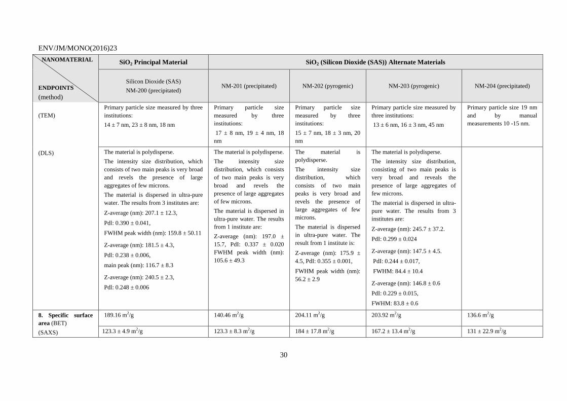
(TEM)
(DLS)
Primary particle size measured by three
institutions:
14 ± 7 nm, 23 ± 8 nm, 18 nm
Primary particle size
measured by three
institutions:
17 ± 8 nm, 19 ± 4 nm, 18
nm
Primary particle size
measured by three
institutions:
15 ± 7 nm, 18 ± 3 nm, 20
nm
Primary particle size measured by
three institutions:
13 ± 6 nm, 16 ± 3 nm, 45 nm
Primary particle size 19 nm
and by manual
measurements 10 -15 nm.
The material is polydisperse.
The intensity size distribution, which
consists of two main peaks is very broad
and revels the presence of large
aggregates of few microns.
The material is dispersed in ultra-pure
water. The results from 3 institutes are:
Z-average (nm): 207.1 ± 12.3,
Pdl: 0.390 ± 0.041,
FWHM peak width (nm): 159.8 ± 50.11
Z-average (nm): 181.5 ± 4.3,
Pdl: 0.238 ± 0.006,
main peak (nm): 116.7 ± 8.3
Z-average (nm): 240.5 ± 2.3,
Pdl: 0.248 ± 0.006
The material is polydisperse.
The intensity size
distribution, which consists
of two main peaks is very
broad and revels the
presence of large aggregates
of few microns.
The material is dispersed in
ultra-pure water. The results
from 1 institute are:
Z-average (nm): 197.0 ±
15.7, PdI: 0.337 ± 0.020
FWHM peak width (nm):
105.6 ± 49.3
The material is
polydisperse.
The intensity size
distribution, which
consists of two main
peaks is very broad and
revels the presence of
large aggregates of few
microns.
The material is dispersed
in ultra-pure water. The
result from 1 institute is:
Z-average (nm): 175.9 ±
4.5, PdI: 0.355 ± 0.001,
FWHM peak width (nm):
56.2 ± 2.9
The material is polydisperse.
The intensity size distribution,
consisting of two main peaks is
very broad and reveals the
presence of large aggregates of
few microns.
The material is dispersed in ultra-
pure water. The results from 3
institutes are:
Z-average (nm): 245.7 ± 37.2.
PdI: 0.299 ± 0.024
Z-average (nm): 147.5 ± 4.5.
PdI: 0.244 ± 0.017,
FWHM: 84.4 ± 10.4
Z-average (nm): 146.8 ± 0.6
PdI: 0.229 ± 0.015,
FWHM: 83.8 ± 0.6
8. Specific surface
area (BET)
(SAXS)
189.16 m2/g 140.46 m2/g 204.11 m2/g 203.92 m2/g 136.6 m2/g
123.3 ± 4.9 m2/g 123.3 ± 8.3 m2/g 184 ± 17.8 m2/g 167.2 ± 13.4 m2/g 131 ± 22.9 m2/g
ENV/JM/MONO(2016)23
31
NANOMATERIAL
ENDPOINTS
(method)
SiO2 Principal Material SiO2 (Silicon Dioxide (SAS)) Alternate Materials
Silicon Dioxide (SAS)
NM-200 (precipitated) NM-201 (precipitated) NM-202 (pyrogenic) NM-203 (pyrogenic) NM-204 (precipitated)
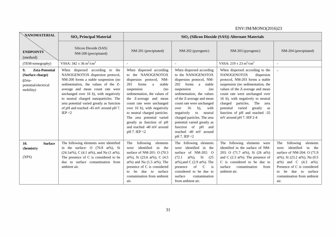
(TEM-tomography) VSSA: 342 ± 36 m2/cm3 - - VSSA: 219 ± 23 m2/cm3 -
9. Zeta-Potential
(Surface charge)
(Zeta-
potential/electrical
mobility)
When dispersed according to the
NANOGENOTOX dispersion protocol,
NM-200 forms a stable suspension (no
sedimentation, the values of the Z-
average and mean count rate were
unchanged over 16 h), with negatively
to neutral charged nanoparticles. The
zeta potential varied greatly as function
of pH and reached -45 mV around pH 7.
IEP <2
When dispersed according
to the NANOGENOTOX
dispersion protocol, NM-
201 forms a stable
suspension (no
sedimentation, the values of
the Z-average and mean
count rate were unchanged
over 16 h), with negatively
to neutral charged particles.
The zeta potential varied
greatly as function of pH
and reached -40 mV around
pH 7. IEP <2
When dispersed according
to the NANOGENOTOX
dispersion protocol, NM-
202 forms a stable
suspension (no
sedimentation, the values
of the Z-average and mean
count rate were unchanged
over 16 h), with
negatively to neutral
charged particles. The zeta
potential varied greatly as
function of pH and
reached -40 mV around
pH 7. IEP <2
When dispersed according to the
NANOGENOTOX dispersion
protocol, NM-203 forms a stable
suspension (no sedimentation, the
values of the Z-average and mean
count rate were unchanged over
16 h), with negatively to neutral
charged particles. The zeta
potential varied greatly as
function of pH and reached -35
mV around pH 7. IEP 2-4
-
10. Surface
chemistry
(XPS)
The following elements were identified
in the surface: O (70.8 at%), Si
(24.1at%), C (4.1 at%), and Na (1 at%).
The presence of C is considered to be
due to surface contamination from
ambient air.
The following elements
were identified in the
surface of NM-201: O (70.3
at%), Si (23.6 at%), C (4.5
at%) and Na (1.5 at%). The
presence of C is considered
to be due to surface
contamination from ambient
air.
The following elements
were identified in the
surface of NM-202: O
(72.1 at%), Si (25
at%),and C (2.9 at%). The
presence of C is
considered to be due to
surface contamination
from ambient air.
The following elements were
identified in the surface of NM-
203: O (71.7 at%), Si (26 at%)
and C (2.3 at%). The presence of
C is considered to be due to
surface contamination from
ambient air.
The following elements
were identified in the
surface of NM-204: O (71.9
at%), Si (23.2 at%), Na (0.5
at%) and C (4.3 at%).
Presence of C is considered
to be due to surface
contamination from ambient
air.
ENV/JM/MONO(2016)23
32
NANOMATERIAL
ENDPOINTS
(method)
SiO2 Principal Material SiO2 (Silicon Dioxide (SAS)) Alternate Materials
Silicon Dioxide (SAS)
NM-200 (precipitated) NM-201 (precipitated) NM-202 (pyrogenic) NM-203 (pyrogenic) NM-204 (precipitated)
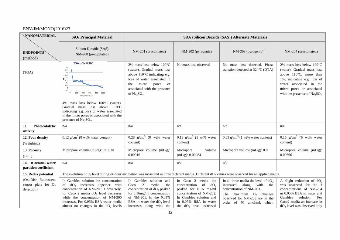
(TGA)
4% mass loss below 100°C (water).
Gradual mass loss above 110°C
indicating e.g. loss of water associated
in the micro pores or associated with the
presence of Na2SO4.
2% mass loss below 100°C
(water). Gradual mass loss
above 110°C indicating e.g.
loss of water associated in
the micro pores or
associated with the presence
of Na2SO4.
No mass loss observed No mass loss detected. Phase
transtion detected at 324°C (DTA)
2% mass loss below 100°C
(water). Gradual mass loss
above 110°C, more than
1%. indicating e.g. loss of
water associated in the
micro pores or associated
with the presence of Na2SO4
11. Photocatalytic
activity
n/a n/a n/a n/a n/a
12. Pour density
(Weighing)
0.12 g/cm3 (8 wt% water content) 0.28 g/cm3 (8 wt% water
content)
0.13 g/cm3 (1 wt% water
content)
0.03 g/cm3 (1 wt% water content) 0.16 g/cm3 (6 wt% water
content)
13. Porosity
(BET)
Micropore volume (mL/g): 0.01181 Micropore volume (mL/g):
0.00916
Micropore volume
(mL/g): 0.00084
Micropore volume (mL/g): 0.0 Micropore volume (mL/g):
0.00666
14. n-octanol-water
partition coefficient
n/a n/a n/a n/a
15. Redox potential
(OxoDish fluorescent
sensor plate for O2
detection)
The evolution of O2 level during 24-hour incubation was measured in three different media. Different dO2 values were observed for all applied media.
In Gambles solution the concentration
of dO2 increases together with
concentration of NM-200. Conversely,
for Caco 2 media dO2 level decreases
while the concentration of NM-200
increases. For 0.05% BSA water media
almost no changes in the dO2 levels
In Gambles solution and
Caco 2 media the
concentration of dO2 peaked
for 0.16mg/ml concentration
of NM-201. In the 0.05%
BSA in water the dO2 level
increases along with the
In Caco 2 media the
concentration of dO2
peaked for 0.16 mg/ml
concentration of NM-202.
In Gambles solution and
in 0.05% BSA in water
the dO2 level increased
In all three media the level of dO2
increased along with the concentration of NM-203.
The maximum O2 changes
observed for NM-203 are in the
order of 40 μmol/ml, which
A slight reduction of dO2
was observed for the 3
concentrations of NM-204
in 0.05% BSA in water and
Gambles solution. For
Caco2 media an increase in
dO2 level was observed only
ENV/JM/MONO(2016)23
33
NANOMATERIAL
ENDPOINTS
(method)
SiO2 Principal Material SiO2 (Silicon Dioxide (SAS)) Alternate Materials
Silicon Dioxide (SAS)
NM-200 (precipitated) NM-201 (precipitated) NM-202 (pyrogenic) NM-203 (pyrogenic) NM-204 (precipitated)
were observed.
The maximum O2 changes observed for
NM-200 are in the order of 40 μmol/ml,
which suggests that the particle reactivity can exceed 1μmol/mg.
concentration of NM-201.
The maximum O2 changes
observed for NM-are in the
order of 40 μmol/ml, which
suggests that the particle
reactivity can exceed
1μmol/mg.
along with the
concentration of NM-202.
The maximum O2 changes
observed for NM-202 are
in the order of 40
μmol/ml, which suggests
that the particle reactivity can exceed 1μmol/mg.
suggests that the particle reactivity
can exceed 1μmol/mg.
for the lowest concentration.
The maximum O2 changes
observed for NM-204 are in
the order of 40 μmol/ml,
which suggests that the
particle reactivity can
exceed 1μmol/mg.
16. Radical
formation potential
n/a n/a n/a n/a n/a
17. Other relevant
information
- - - - -
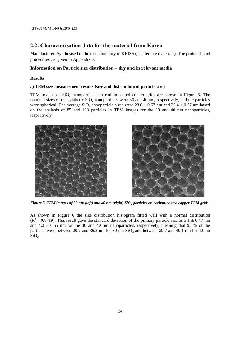
ENV/JM/MONO(2016)23
34
2.2. Characterisation data for the material from Korea
Manufacturer: Synthesised in the test laboratory in KRISS (as alternate materials). The protocols and
procedures are given in Appendix 0.
Information on Particle size distribution – dry and in relevant media
Results
a) TEM size measurement results (size and distribution of particle size)
TEM images of SiO2 nanoparticles on carbon-coated copper grids are shown in Figure 5. The
nominal sizes of the synthetic SiO2 nanoparticles were 30 and 40 nm, respectively, and the particles
were spherical. The average SiO2 nanoparticle sizes were 28.6 ± 0.67 nm and 39.4 ± 0.77 nm based
on the analysis of 85 and 103 particles in TEM images for the 30 and 40 nm nanoparticles,
respectively.
Figure 5. TEM images of 30 nm (left) and 40 nm (right) SiO2 particles on carbon-coated copper TEM grids
As shown in Figure 6 the size distribution histogram fitted well with a normal distribution
(R2 = 0.8719). This result gave the standard deviation of the primary particle size as 3.1 ± 0.47 nm
and 4.0 ± 0.55 nm for the 30 and 40 nm nanoparticles, respectively, meaning that 95 % of the
particles were between 20.9 and 36.3 nm for 30 nm SiO2 and between 29.7 and 49.1 nm for 40 nm
SiO2.
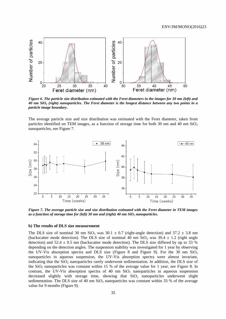
ENV/JM/MONO(2016)23
35
Figure 6. The particle size distribution estimated with the Feret diameters in the images for 30 nm (left) and
40 nm SiO2 (right) nanoparticles. The Feret diameter is the longest distance between any two points in a
particle image boundary.
The average particle size and size distribution was estimated with the Feret diameter, taken from
particles identified on TEM images, as a function of storage time for both 30 nm and 40 nm SiO2
nanoparticles, see Figure 7.
Figure 7. The average particle size and size distribution estimated with the Feret diameter in TEM images
as a function of storage time for (left) 30 nm and (right) 40 nm SiO2 nanoparticles.
b) The results of DLS size measurement
The DLS size of nominal 30 nm SiO2 was 30.1 ± 0.7 (right-angle detection) and 37.2 ± 3.8 nm
(backscatter mode detection). The DLS size of nominal 40 nm SiO2 was 39.4 ± 1.2 (right angle
detection) and 52.4 ± 0.5 nm (backscatter mode detection). The DLS size differed by up to 33 %
depending on the detection angles. The suspension stability was investigated for 1 year by observing
the UV-Vis absorption spectra and DLS size (Figure 8 and Figure 9). For the 30 nm SiO2
nanoparticles in aqueous suspension, the UV-Vis absorption spectra were almost invariant,
indicating that the SiO2 nanoparticles rarely underwent sedimentation. In addition, the DLS size of
the SiO2 nanoparticles was constant within 15 % of the average value for 1 year, see Figure 8. In
contrast, the UV-Vis absorption spectra of 40 nm SiO2 nanoparticles in aqueous suspension
decreased slightly with storage time, showing that SiO2 nanoparticles underwent slight
sedimentation. The DLS size of 40 nm SiO2 nanoparticles was constant within 33 % of the average
value for 9 months (Figure 9).
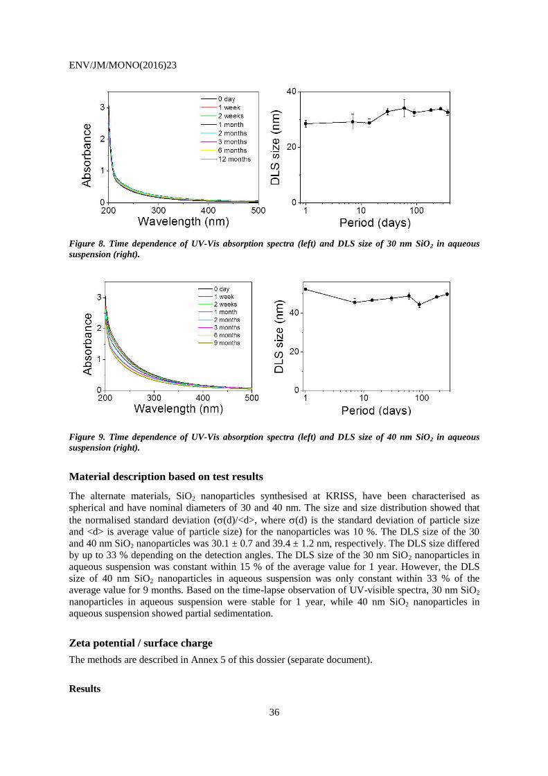
ENV/JM/MONO(2016)23
36
Figure 8. Time dependence of UV-Vis absorption spectra (left) and DLS size of 30 nm SiO2 in aqueous
suspension (right).
Figure 9. Time dependence of UV-Vis absorption spectra (left) and DLS size of 40 nm SiO2 in aqueous
suspension (right).
Material description based on test results
The alternate materials, SiO2 nanoparticles synthesised at KRISS, have been characterised as
spherical and have nominal diameters of 30 and 40 nm. The size and size distribution showed that
the normalised standard deviation ((d)/<d>, where (d) is the standard deviation of particle size
and <d> is average value of particle size) for the nanoparticles was 10 %. The DLS size of the 30
and 40 nm SiO2 nanoparticles was 30.1 ± 0.7 and 39.4 ± 1.2 nm, respectively. The DLS size differed
by up to 33 % depending on the detection angles. The DLS size of the 30 nm SiO2 nanoparticles in
aqueous suspension was constant within 15 % of the average value for 1 year. However, the DLS
size of 40 nm SiO2 nanoparticles in aqueous suspension was only constant within 33 % of the
average value for 9 months. Based on the time-lapse observation of UV-visible spectra, 30 nm SiO2
nanoparticles in aqueous suspension were stable for 1 year, while 40 nm SiO2 nanoparticles in
aqueous suspension showed partial sedimentation.
Zeta potential / surface charge
The methods are described in Annex 5 of this dossier (separate document).
Results
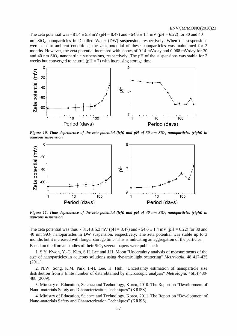
ENV/JM/MONO(2016)23
37
The zeta potential was - 81.4 ± 5.3 mV (pH = 8.47) and - 54.6 ± 1.4 mV (pH = 6.22) for 30 and 40
nm SiO2 nanoparticles in Distilled Water (DW) suspension, respectively. When the suspensions
were kept at ambient conditions, the zeta potential of these nanoparticles was maintained for 3
months. However, the zeta potential increased with slopes of 0.14 mV/day and 0.068 mV/day for 30
and 40 nm SiO2 nanoparticle suspensions, respectively. The pH of the suspensions was stable for 2
weeks but converged to neutral (pH = 7) with increasing storage time.
Figure 10. Time dependence of the zeta potential (left) and pH of 30 nm SiO2 nanoparticles (right) in
aqueous suspension
Figure 11. Time dependence of the zeta potential (left) and pH of 40 nm SiO2 nanoparticles (right) in
aqueous suspension.
The zeta potential was thus - 81.4 ± 5.3 mV (pH = 8.47) and - 54.6 ± 1.4 mV (pH = 6.22) for 30 and
40 nm SiO2 nanoparticles in DW suspension, respectively. The zeta potential was stable up to 3
months but it increased with longer storage time. This is indicating an aggregation of the particles.
Based on the Korean studies of their SiO2 several papers were published:
1. S.Y. Kwon, Y.-G. Kim, S.H. Lee and J.H. Moon "Uncertainty analysis of measurements of the
size of nanoparticles in aqueous solutions using dynamic light scattering" Metrologia, 48 417-425
(2011).
2. N.W. Song, K.M. Park, I.-H. Lee, H. Huh, "Uncertainty estimation of nanoparticle size
distribution from a finite number of data obtained by microscopic analysis" Metrologia, 46(5) 480-
488 (2009).
3. Ministry of Education, Science and Technology, Korea, 2010. The Report on “Development of
Nano-materials Safety and Characterization Techniques” (KRISS)
4. Ministry of Education, Science and Technology, Korea, 2011. The Report on “Development of
Nano-materials Safety and Characterization Techniques” (KRISS).
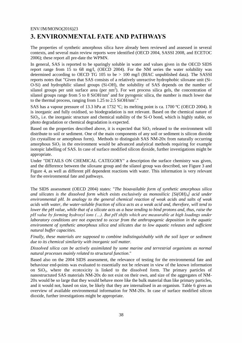
ENV/JM/MONO(2016)23
38
3. ENVIRONMENTAL FATE AND PATHWAYS
The properties of synthetic amorphous silica have already been reviewed and assessed in several
contexts, and several main review reports were identified (OECD 2004, SASSI 2008, and ECETOC
2006); these report all pre-date the WPMN.
In general, SAS is reported to be sparingly soluble in water and values given in the OECD SIDS
report range from 15 to 68 mg/l, (OECD 2004). For the NM series the water solubility was
determined according to OECD TG 105 to be > 100 mg/l (BIAC unpublished data). The SASSI
reports notes that "Given that SAS consists of a relatively unreactive hydrophobic siloxane unit (Si-
O-Si) and hydrophilic silanol groups (Si-OH), the solubility of SAS depends on the number of
silanol groups per unit surface area (per nm2). For wet process silica gels, the concentration of
silanol groups range from 5 to 8 SiOH/nm2 and for pyrogenic silica, the number is much lower due
to the thermal process, ranging from 1.25 to 2.5 SiOH/nm2."
SAS has a vapour pressure of 13.3 hPa at 1732 ºC; its melting point is ca. 1700 ºC (OECD 2004). It
is inorganic and fully oxidised, so biodegradation is not relevant. Based on the chemical nature of
SiO2, i.e. the inorganic structure and chemical stability of the Si-O bond, which is highly stable, no
photo degradation or chemical degradation is expected.
Based on the properties described above, it is expected that SiO2 released to the environment will
distribute to soil or sediment. One of the main components of any soil or sediment is silicon dioxide
(in crystalline or amorphous form). Methods to distinguish SAS NM-20x from naturally occurring
amorphous SiO2 in the environment would be advanced analytical methods requiring for example
isotopic labelling of SAS. In case of surface modified silicon dioxide, further investigations might be
appropriate.
Under "DETAILS ON CHEMICAL CATEGORY" a description the surface chemistry was given,
and the difference between the siloxane group and the silanol group was described, see Figure 3 and
Figure 4, as well as different pH dependent reactions with water. This information is very relevant
for the environmental fate and pathways.
The SIDS assessment (OECD 2004) states: "The bioavailable form of synthetic amorphous silica
and silicates is the dissolved form which exists exclusively as monosilicic [Si(OH)4] acid under
environmental pH. In analogy to the general chemical reaction of weak acids and salts of weak
acids with water, the water-soluble fraction of silica acts as a weak acid and, therefore, will tend to
lower the pH value, while that of a silicate acts as a base tending to bind protons and, thus, raise the
pH value by forming hydroxyl ions (…). But pH shifts which are measurable at high loadings under
laboratory conditions are not expected to occur from the anthropogenic deposition in the aquatic
environment of synthetic amorphous silica and silicates due to low aquatic releases and sufficient
natural buffer capacities.
Finally, these materials are supposed to combine indistinguishably with the soil layer or sediment
due to its chemical similarity with inorganic soil matter.
Dissolved silica can be actively assimilated by some marine and terrestrial organisms as normal
natural processes mainly related to structural function."
Based also on the 2004 SIDS assessment, the relevance of testing for the environmental fate and
behaviour end-points was evaluated to essentially not be relevant in view of the known information
on SiO2, where the ecotoxicity is linked to the dissolved form. The primary particles of
nanostructuted SAS materials NM-20x do not exist on their own, and size of the aggregates of NM-
20x would be so large that they would behave more like the bulk material than like primary particles,
and it would not, based on size, be likely that they are internalised in an organism. Table 6 gives an
overview of available environmental information for NM-20x. In case of surface modified silicon
dioxide, further investigations might be appropriate.
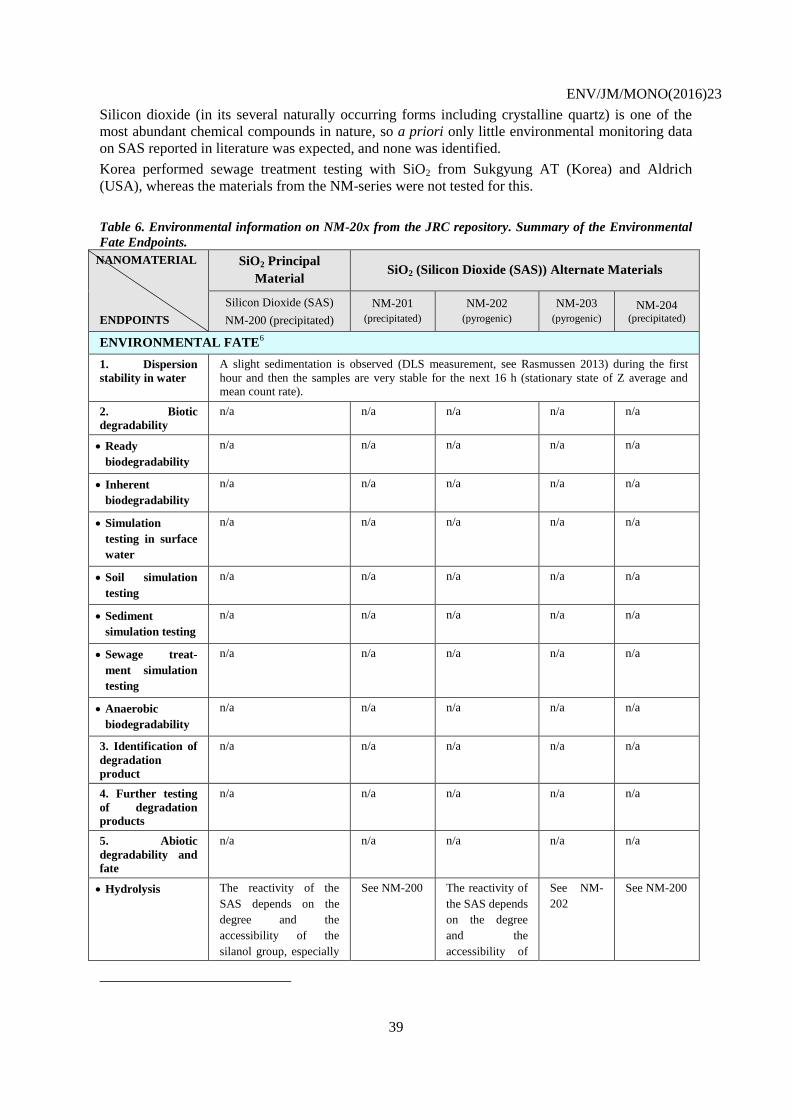
ENV/JM/MONO(2016)23
39
Silicon dioxide (in its several naturally occurring forms including crystalline quartz) is one of the
most abundant chemical compounds in nature, so a priori only little environmental monitoring data
on SAS reported in literature was expected, and none was identified.
Korea performed sewage treatment testing with SiO2 from Sukgyung AT (Korea) and Aldrich
(USA), whereas the materials from the NM-series were not tested for this.
Table 6. Environmental information on NM-20x from the JRC repository. Summary of the Environmental
Fate Endpoints.
NANOMATERIAL
ENDPOINTS
SiO2 Principal
Material SiO2 (Silicon Dioxide (SAS)) Alternate Materials
Silicon Dioxide (SAS)
NM-200 (precipitated)
NM-201
(precipitated)
NM-202
(pyrogenic)
NM-203
(pyrogenic) NM-204
(precipitated)
ENVIRONMENTAL FATE6
1. Dispersion
stability in water
A slight sedimentation is observed (DLS measurement, see Rasmussen 2013) during the first
hour and then the samples are very stable for the next 16 h (stationary state of Z average and mean count rate).
2. Biotic
degradability
n/a n/a n/a n/a n/a
Ready
biodegradability
n/a n/a n/a n/a n/a
Inherent
biodegradability
n/a n/a n/a n/a n/a
Simulation
testing in surface
water
n/a n/a n/a n/a n/a
Soil simulation
testing
n/a n/a n/a n/a n/a
Sediment
simulation testing
n/a n/a n/a n/a n/a
Sewage treat-
ment simulation
testing
n/a n/a n/a n/a n/a
Anaerobic
biodegradability
n/a n/a n/a n/a n/a
3. Identification of
degradation
product
n/a n/a n/a n/a n/a
4. Further testing
of degradation
products
n/a n/a n/a n/a n/a
5. Abiotic
degradability and
fate
n/a n/a n/a n/a n/a
Hydrolysis The reactivity of the
SAS depends on the
degree and the
accessibility of the
silanol group, especially
See NM-200 The reactivity of
the SAS depends
on the degree
and the
accessibility of
See NM-
202
See NM-200
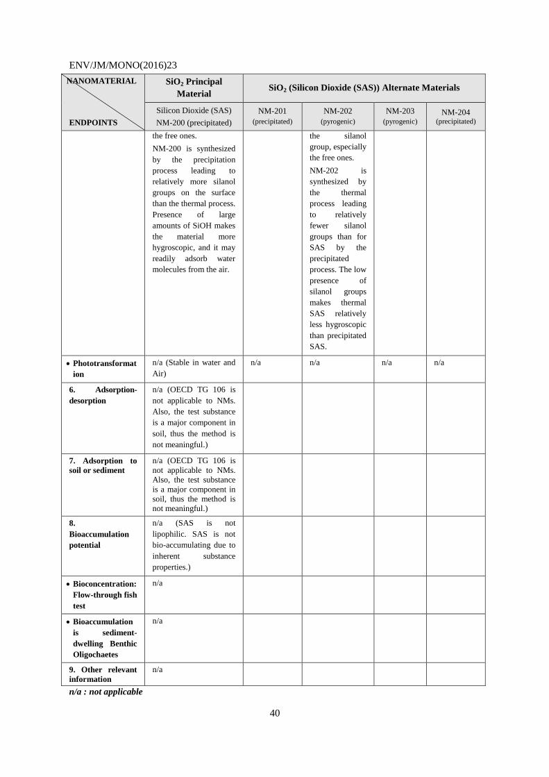
ENV/JM/MONO(2016)23
40
NANOMATERIAL
ENDPOINTS
SiO2 Principal
Material SiO2 (Silicon Dioxide (SAS)) Alternate Materials
Silicon Dioxide (SAS)
NM-200 (precipitated)
NM-201
(precipitated)
NM-202
(pyrogenic)
NM-203
(pyrogenic) NM-204
(precipitated)
the free ones.
NM-200 is synthesized
by the precipitation
process leading to
relatively more silanol
groups on the surface
than the thermal process.
Presence of large
amounts of SiOH makes
the material more
hygroscopic, and it may
readily adsorb water
molecules from the air.
the silanol
group, especially
the free ones.
NM-202 is
synthesized by
the thermal
process leading
to relatively
fewer silanol
groups than for
SAS by the
precipitated
process. The low
presence of
silanol groups
makes thermal
SAS relatively
less hygroscopic
than precipitated
SAS.
Phototransformat
ion
n/a (Stable in water and
Air)
n/a n/a n/a n/a
6. Adsorption-
desorption
n/a (OECD TG 106 is
not applicable to NMs.
Also, the test substance
is a major component in
soil, thus the method is
not meaningful.)
7. Adsorption to
soil or sediment
n/a (OECD TG 106 is
not applicable to NMs.
Also, the test substance
is a major component in
soil, thus the method is
not meaningful.)
8.
Bioaccumulation
potential
n/a (SAS is not
lipophilic. SAS is not
bio-accumulating due to
inherent substance
properties.)
Bioconcentration:
Flow-through fish
test
n/a
Bioaccumulation
is sediment-
dwelling Benthic
Oligochaetes
n/a
9. Other relevant
information
n/a
n/a : not applicable
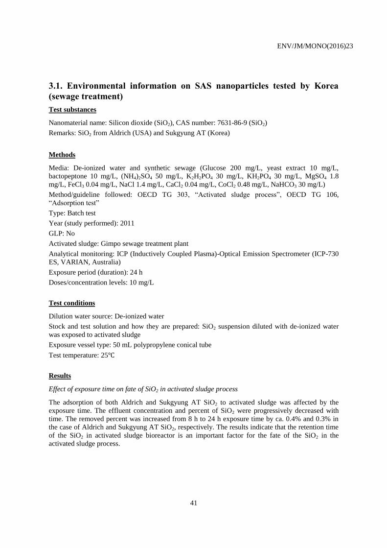
ENV/JM/MONO(2016)23
41
3.1. Environmental information on SAS nanoparticles tested by Korea
(sewage treatment)
Test substances
Nanomaterial name: Silicon dioxide (SiO2), CAS number: 7631-86-9 (SiO2)
Remarks: SiO2 from Aldrich (USA) and Sukgyung AT (Korea)
Methods
Media: De-ionized water and synthetic sewage (Glucose 200 mg/L, yeast extract 10 mg/L,
bactopeptone 10 mg/L, (NH4)2SO4 50 mg/L, K2H2PO4 30 mg/L, KH2PO4 30 mg/L, MgSO4 1.8
mg/L, FeCl3 0.04 mg/L, NaCl 1.4 mg/L, CaCl2 0.04 mg/L, CoCl2 0.48 mg/L, NaHCO3 30 mg/L)
Method/guideline followed: OECD TG 303, “Activated sludge process”, OECD TG 106,
“Adsorption test”
Type: Batch test
Year (study performed): 2011
GLP: No
Activated sludge: Gimpo sewage treatment plant
Analytical monitoring: ICP (Inductively Coupled Plasma)-Optical Emission Spectrometer (ICP-730
ES, VARIAN, Australia)
Exposure period (duration): 24 h
Doses/concentration levels: 10 mg/L
Test conditions
Dilution water source: De-ionized water
Stock and test solution and how they are prepared: SiO2 suspension diluted with de-ionized water
was exposed to activated sludge
Exposure vessel type: 50 mL polypropylene conical tube
Test temperature: 25℃
Results
Effect of exposure time on fate of SiO2 in activated sludge process
The adsorption of both Aldrich and Sukgyung AT SiO2 to activated sludge was affected by the
exposure time. The effluent concentration and percent of SiO2 were progressively decreased with
time. The removed percent was increased from 8 h to 24 h exposure time by ca. 0.4% and 0.3% in
the case of Aldrich and Sukgyung AT SiO2, respectively. The results indicate that the retention time
of the SiO2 in activated sludge bioreactor is an important factor for the fate of the SiO2 in the
activated sludge process.
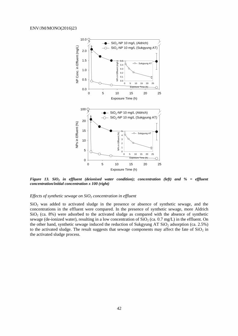
ENV/JM/MONO(2016)23
42
Exposure Time (h)
0 5 10 15 20 25
NP
Conc. in
Efflu
ent (m
g/L
)
0.0
0.5
1.0
1.5
2.0
10.0SiO2-NP 10 mg/L (Aldrich)
SiO2-NP 10 mg/L (Sukgyung AT)
Exposure Time (h)
0 5 10 15 20 25
NP
s in
Eff
lue
nt
(mg
/L)
0.0
0.1
0.2
0.3
0.4
0.5Sukgyung AT
Exposure Time (h)
0 5 10 15 20 25
NP
s in E
fflu
ent (%
)
0
5
10
15
20
100SiO2-NP 10 mg/L (Aldrich)
SiO2-NP 10 mg/L (Sukgyung AT)
Exposure Time (h)
0 5 10 15 20 25
NP
s in E
fflu
ent
(%)
0
1
2
3
4
5Sukgyung AT
Figure 13. SiO2 in effluent (deionised water condition); concentration (left) and % = effluent
concentration/initial concentration x 100 (right)
Effects of synthetic sewage on SiO2 concentration in effluent
SiO2 was added to activated sludge in the presence or absence of synthetic sewage, and the
concentrations in the effluent were compared. In the presence of synthetic sewage, more Aldrich
SiO2 (ca. 8%) were adsorbed to the activated sludge as compared with the absence of synthetic
sewage (de-ionized water), resulting in a low concentration of SiO2 (ca. 0.7 mg/L) in the effluent. On
the other hand, synthetic sewage induced the reduction of Sukgyung AT SiO2 adsorption (ca. 2.5%)
to the activated sludge. The result suggests that sewage components may affect the fate of SiO2 in
the activated sludge process.
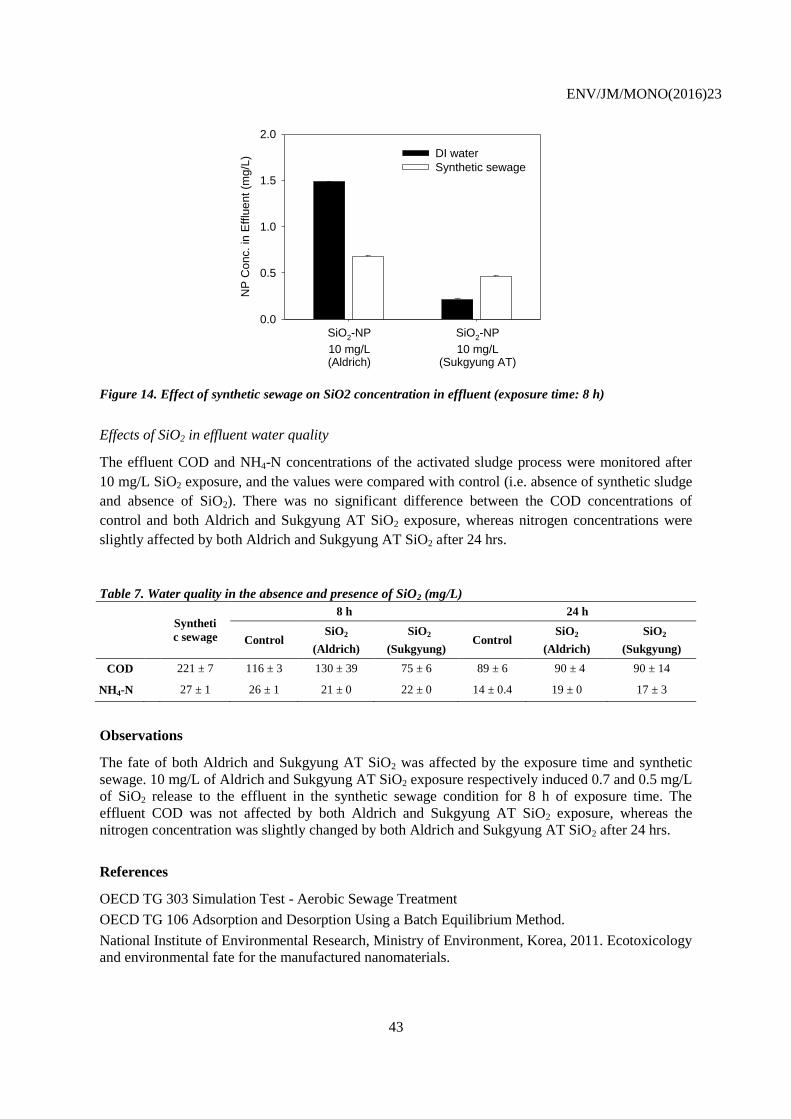
ENV/JM/MONO(2016)23
43
NP
Co
nc. in
Efflu
ent
(mg/L
)
0.0
0.5
1.0
1.5
2.0
DI water
Synthetic sewage
SiO2-NP
10 mg/L(Aldrich)
SiO2-NP
10 mg/L(Sukgyung AT)
Figure 14. Effect of synthetic sewage on SiO2 concentration in effluent (exposure time: 8 h)
Effects of SiO2 in effluent water quality
The effluent COD and NH4-N concentrations of the activated sludge process were monitored after
10 mg/L SiO2 exposure, and the values were compared with control (i.e. absence of synthetic sludge
and absence of SiO2). There was no significant difference between the COD concentrations of
control and both Aldrich and Sukgyung AT SiO2 exposure, whereas nitrogen concentrations were
slightly affected by both Aldrich and Sukgyung AT SiO2 after 24 hrs.
Table 7. Water quality in the absence and presence of SiO2 (mg/L)
Syntheti
c sewage
8 h 24 h
Control SiO2
(Aldrich)
SiO2
(Sukgyung) Control
SiO2
(Aldrich)
SiO2
(Sukgyung)
COD 221 ± 7 116 ± 3 130 ± 39 75 ± 6 89 ± 6 90 ± 4 90 ± 14
N NH4-N 27 ± 1 26 ± 1 21 ± 0 22 ± 0 14 ± 0.4 19 ± 0 17 ± 3
Observations
The fate of both Aldrich and Sukgyung AT SiO2 was affected by the exposure time and synthetic
sewage. 10 mg/L of Aldrich and Sukgyung AT SiO2 exposure respectively induced 0.7 and 0.5 mg/L
of SiO2 release to the effluent in the synthetic sewage condition for 8 h of exposure time. The
effluent COD was not affected by both Aldrich and Sukgyung AT SiO2 exposure, whereas the
nitrogen concentration was slightly changed by both Aldrich and Sukgyung AT SiO2 after 24 hrs.
References
OECD TG 303 Simulation Test - Aerobic Sewage Treatment
OECD TG 106 Adsorption and Desorption Using a Batch Equilibrium Method.
National Institute of Environmental Research, Ministry of Environment, Korea, 2011. Ecotoxicology
and environmental fate for the manufactured nanomaterials.
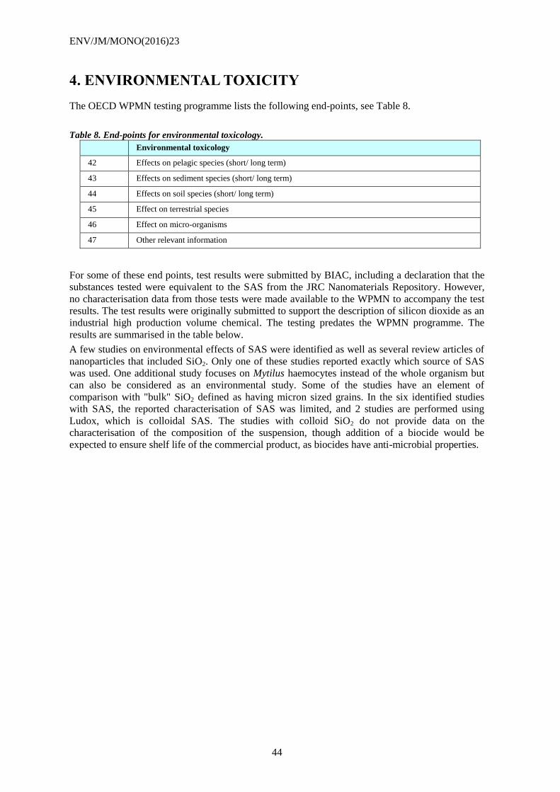
ENV/JM/MONO(2016)23
44
4. ENVIRONMENTAL TOXICITY
The OECD WPMN testing programme lists the following end-points, see Table 8.
Table 8. End-points for environmental toxicology.
Environmental toxicology
42 Effects on pelagic species (short/ long term)
43 Effects on sediment species (short/ long term)
44 Effects on soil species (short/ long term)
45 Effect on terrestrial species
46 Effect on micro-organisms
47 Other relevant information
For some of these end points, test results were submitted by BIAC, including a declaration that the
substances tested were equivalent to the SAS from the JRC Nanomaterials Repository. However,
no characterisation data from those tests were made available to the WPMN to accompany the test
results. The test results were originally submitted to support the description of silicon dioxide as an
industrial high production volume chemical. The testing predates the WPMN programme. The
results are summarised in the table below.
A few studies on environmental effects of SAS were identified as well as several review articles of
nanoparticles that included SiO2. Only one of these studies reported exactly which source of SAS
was used. One additional study focuses on Mytilus haemocytes instead of the whole organism but
can also be considered as an environmental study. Some of the studies have an element of
comparison with "bulk" SiO2 defined as having micron sized grains. In the six identified studies
with SAS, the reported characterisation of SAS was limited, and 2 studies are performed using
Ludox, which is colloidal SAS. The studies with colloid SiO2 do not provide data on the
characterisation of the composition of the suspension, though addition of a biocide would be
expected to ensure shelf life of the commercial product, as biocides have anti-microbial properties.
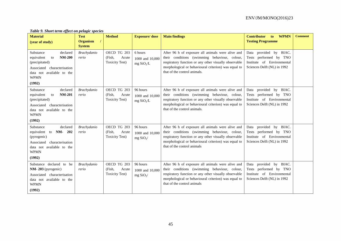
ENV/JM/MONO(2016)23
45
Table 9. Short term effect on pelagic species
Material
(year of study)
Test
Organism /
System
Method Exposure/ dose Main findings Contributor to WPMN
Testing Programme
Comment
Substance declared
equivalent to NM-200
(precipitated)
Associated characterisation
data not available to the
WPMN
(1992)
Brachydanio
rerio
OECD TG 203
(Fish, Acute
Toxicity Test)
6 hours
1000 and 10,000
mg SiO2/L
After 96 h of exposure all animals were alive and
their conditions (swimming behaviour, colour,
respiratory function or any other visually observable
morphological or behavioural criterion) was equal to
that of the control animals.
Data provided by BIAC.
Tests performed by TNO
Institute of Environmental
Sciences Delft (NL) in 1992
Substance declared
equivalent to NM-201
(precipitated)
Associated characterisation
data not available to the
WPMN
(1992)
Brachydanio
rerio
OECD TG 203
(Fish, Acute
Toxicity Test)
96 hours
1000 and 10,000
mg SiO2/L
After 96 h of exposure all animals were alive and
their conditions (swimming behaviour, colour,
respiratory function or any other visually observable
morphological or behavioural criterion) was equal to
that of the control animals.
Data provided by BIAC.
Tests performed by TNO
Institute of Environmental
Sciences Delft (NL) in 1992
Substance declared
equivalent to NM- 202
(pyrogenic)
Associated characterisation
data not available to the
WPMN
(1992)
Brachydanio
rerio
OECD TG 203
(Fish, Acute
Toxicity Test)
96 hours
1000 and 10,000
mg SiO2/
After 96 h of exposure all animals were alive and
their conditions (swimming behaviour, colour,
respiratory function or any other visually observable
morphological or behavioural criterion) was equal to
that of the control animals
Data provided by BIAC.
Tests performed by TNO
Institute of Environmental
Sciences Delft (NL) in 1992
Substance declared to be
NM- 203 (pyrogenic)
Associated characterisation
data not available to the
WPMN
(1992)
Brachydanio
rerio
OECD TG 203
(Fish, Acute
Toxicity Test)
96 hours
1000 and 10,000
mg SiO2/
After 96 h of exposure all animals were alive and
their conditions (swimming behaviour, colour,
respiratory function or any other visually observable
morphological or behavioural criterion) was equal to
that of the control animals
Data provided by BIAC.
Tests performed by TNO
Institute of Environmental
Sciences Delft (NL) in 1992
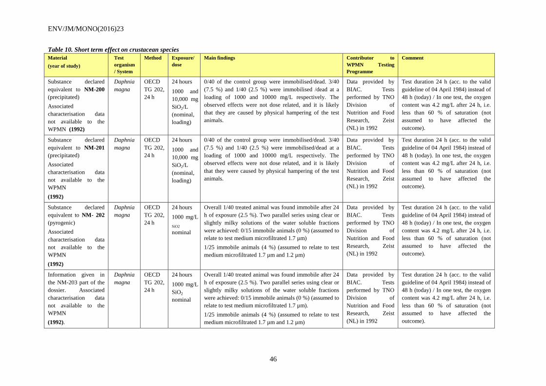
ENV/JM/MONO(2016)23
46
Table 10. Short term effect on crustacean species
Material
(year of study)
Test
organism
/ System
Method Exposure/
dose
Main findings Contributor to
WPMN Testing
Programme
Comment
Substance declared
equivalent to NM-200
(precipitated)
Associated
characterisation data
not available to the
WPMN (1992)
Daphnia
magna
OECD
TG 202,
24 h
24 hours
1000 and
10,000 mg
SiO2/L
(nominal,
loading)
0/40 of the control group were immobilised/dead. 3/40
(7.5 %) and 1/40 (2.5 %) were immobilised /dead at a
loading of 1000 and 10000 mg/L respectively. The
observed effects were not dose related, and it is likely
that they are caused by physical hampering of the test
animals.
Data provided by
BIAC. Tests
performed by TNO
Division of
Nutrition and Food
Research, Zeist
(NL) in 1992
Test duration 24 h (acc. to the valid
guideline of 04 April 1984) instead of
48 h (today) / In one test, the oxygen
content was 4.2 mg/L after 24 h, i.e.
less than 60 % of saturation (not
assumed to have affected the
outcome).
Substance declared
equivalent to NM-201
(precipitated)
Associated
characterisation data
not available to the
WPMN
(1992)
Daphnia
magna
OECD
TG 202,
24 h
24 hours
1000 and
10,000 mg
SiO2/L
(nominal,
loading)
0/40 of the control group were immobilised/dead. 3/40
(7.5 %) and 1/40 (2.5 %) were immobilised/dead at a
loading of 1000 and 10000 mg/L respectively. The
observed effects were not dose related, and it is likely
that they were caused by physical hampering of the test
animals.
Data provided by
BIAC. Tests
performed by TNO
Division of
Nutrition and Food
Research, Zeist
(NL) in 1992
Test duration 24 h (acc. to the valid
guideline of 04 April 1984) instead of
48 h (today). In one test, the oxygen
content was 4.2 mg/L after 24 h, i.e.
less than 60 % of saturation (not
assumed to have affected the
outcome).
Substance declared
equivalent to NM- 202
(pyrogenic)
Associated
characterisation data
not available to the
WPMN
(1992)
Daphnia
magna
OECD
TG 202,
24 h
24 hours
1000 mg/L
SiO2
nominal
Overall 1/40 treated animal was found immobile after 24
h of exposure (2.5 %). Two parallel series using clear or
slightly milky solutions of the water soluble fractions
were achieved: 0/15 immobile animals (0 %) (assumed to
relate to test medium microfiltrated 1.7 µm)
1/25 immobile animals (4 %) (assumed to relate to test
medium microfiltrated 1.7 µm and 1.2 µm)
Data provided by
BIAC. Tests
performed by TNO
Division of
Nutrition and Food
Research, Zeist
(NL) in 1992
Test duration 24 h (acc. to the valid
guideline of 04 April 1984) instead of
48 h (today) / In one test, the oxygen
content was 4.2 mg/L after 24 h, i.e.
less than 60 % of saturation (not
assumed to have affected the
outcome).
Information given in
the NM-203 part of the
dossier. Associated
characterisation data
not available to the
WPMN
(1992).
Daphnia
magna
OECD
TG 202,
24 h
24 hours
1000 mg/L
SiO2
nominal
Overall 1/40 treated animal was found immobile after 24
h of exposure (2.5 %). Two parallel series using clear or
slightly milky solutions of the water soluble fractions
were achieved: 0/15 immobile animals (0 %) (assumed to
relate to test medium microfiltrated 1.7 µm).
1/25 immobile animals (4 %) (assumed to relate to test
medium microfiltrated 1.7 µm and 1.2 µm)
Data provided by
BIAC. Tests
performed by TNO
Division of
Nutrition and Food
Research, Zeist
(NL) in 1992
Test duration 24 h (acc. to the valid
guideline of 04 April 1984) instead of
48 h (today) / In one test, the oxygen
content was 4.2 mg/L after 24 h, i.e.
less than 60 % of saturation (not
assumed to have affected the
outcome).
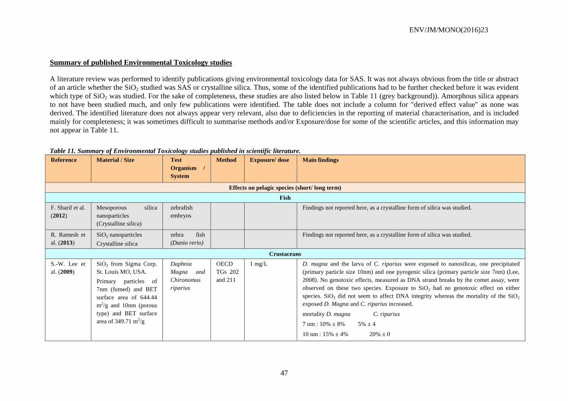
ENV/JM/MONO(2016)23
47
Summary of published Environmental Toxicology studies
A literature review was performed to identify publications giving environmental toxicology data for SAS. It was not always obvious from the title or abstract
of an article whether the SiO2 studied was SAS or crystalline silica. Thus, some of the identified publications had to be further checked before it was evident
which type of SiO2 was studied. For the sake of completeness, these studies are also listed below in Table 11 (grey background)). Amorphous silica appears
to not have been studied much, and only few publications were identified. The table does not include a column for "derived effect value" as none was
derived. The identified literature does not always appear very relevant, also due to deficiencies in the reporting of material characterisation, and is included
mainly for completeness; it was sometimes difficult to summarise methods and/or Exposure/dose for some of the scientific articles, and this information may
not appear in Table 11.
Table 11. Summary of Environmental Toxicology studies published in scientific literature.
Reference Material / Size Test
Organism /
System
Method Exposure/ dose Main findings
Effects on pelagic species (short/ long term)
Fish
F. Sharif et al.
(2012)
Mesoporous sílica
nanoparticles
(Crystalline silica)
zebrafish
embryos
Findings not reported here, as a crystalline form of silica was studied.
R. Ramesh et
al. (2013)
SiO2 nanoparticles
Crystalline silica
zebra fish
(Danio rerio)
Findings not reported here, as a crystalline form of silica was studied.
Crustaceans
S.-W. Lee et
al. (2009)
SiO2 from Sigma Corp.
St. Louis MO, USA.
Primary particles of
7nm (fumed) and BET
surface area of 644.44
m2/g and 10nm (porous
type) and BET surface
area of 349.71 m2/g
Daphnia
Magna and
Chironomus
riparius
OECD
TGs 202
and 211
1 mg/L D. magna and the larva of C. riparius were exposed to nanosilicas, one precipitated
(primary particle size 10nm) and one pyrogenic silica (primary particle size 7nm) (Lee,
2008). No genotoxic effects, measured as DNA strand breaks by the comet assay, were
observed on these two species. Exposure to SiO2 had no genotoxic effect on either
species. SiO2 did not seem to affect DNA integrity whereas the mortality of the SiO2
exposed D. Magna and C. riparius increased.
mortality D. magna C. riparius
7 nm : 10% ± 8% 5% ± 4
10 nm : 15% ± 4% 20% ± 0
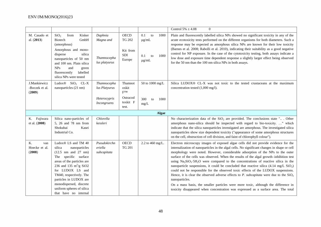
ENV/JM/MONO(2016)23
48
Control 5% ± 4.08 0
M. Casado et
al. (2013)
SiO2 from Kisker
Biotech GmbH
(amorphous)
Amorphous and mono-
disperse silica
nanoparticles of 50 nm
and 100 nm. Plain silica
NPs and green
fluorescently labelled
silica NPs were tested
Daphnia
Magna and
Thamnocepha
lus platyurus
OECD
TG 202
Kit from
SDI
Europe
0.1 to 1000
µg/mL
0.1 to 1000
µg/mL
Plain and fluorescently labelled silica NPs showed no significant toxicity in any of the
acute ecotoxicity tests performed on the different organisms for both diameters. Such a
response may be expected as amorphous silica NPs are known for their low toxicity
(Barnes et al. 2008; Rabolli et al. 2010), indicating their suitability as a good negative
control for NP exposure. In the case of the cytotoxicity testing, both assays indicate a
low dose and exposure time dependent response a slightly larger effect being observed
for the 50 nm than the 100 nm silica NPs in both assays.
J.Mankiewicz
-Boczek et al.
(2009)
Ludox® SiO2 CL-X
nanoparticles (21 nm)
Thamnocepha
lus Platyurus
Heterocypris
Incongruens
Thamnot
oxkit
F™
Ostracod
toxkit F
test.
50 to 1000 mg/L
300 to 1000
mg/L
Silica LUDOX® CL-X was not toxic to the tested crustaceans at the maximum
concentration tested (1,000 mg/l).
Algae
K. Fujiwara
et al. (2008)
Silica nano-particles of
5, 26 and 78 nm from
Shokubai Kasei
Industrial Co.
Chlorella
kessleri
No characterization data of the SiO2 are provided. The conclusions state ".. . Other
amorphous nano-silica should be inspected with regard to bio-toxicity. …" which
indicate that the silica nanoparticles investigated are amorphous. The investigated silica
nanoparticles show size dependent toxicity ("appearance of some amorphous structures
on the cell, obstruction of cell division, and faint of chlorophyll colour").
K. van
Hoecke et al.
(2008)
Ludox® LS and TM 40
silica nanoparticles
(12.5 nm and 27 nm) The specific surface
areas of the particles are
236 and 135 m2/g SiO2
for LUDOX LS and
TM40, respectively. The
particles in LUDOX are
monodispersed, discrete
uniform spheres of silica
that have no internal
Pseudokirchn
eriella
subcapitata
OECD
TG 201
2.2 to 460 mg/L. Electron microscopy images of exposed algae cells did not provide evidence for the
internalization of nanoparticles in the algal cells. No significant changes in shape or cell
morphology were noted. However, considerable adsorption of the NPs to the outer
surface of the cells was observed. When the results of the algal growth–inhibition test
using Na2SiO3·5H2O were compared to the concentrations of reactive silica in the
nanoparticle suspensions, it could be concluded that reactive silica (4.14 mg/L SiO2)
could not be responsible for the observed toxic effects of the LUDOX suspensions.
Hence, it is clear the observed adverse effects to P. subcapitata were due to the SiO2
nanoparticles.
On a mass basis, the smaller particles were more toxic, although the difference in
toxicity disappeared when concentration was expressed as a surface area. The total
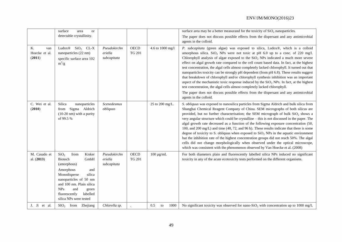
ENV/JM/MONO(2016)23
49
surface area or
detectable crystallinity.
surface area may be a better measurand for the toxicity of SiO2 nanoparticles.
The paper does not discuss possible effects from the dispersant and any antimicrobial
agents in the colloid.
K. van
Hoecke et al.
(2011)
Ludox® SiO2 CL-X
nanoparticles (22 nm)
specific surface area 102
m2/g
Pseudokirchn
eriella
subcapitata
OECD
TG 201
4.6 to 1000 mg/l P. subcapitata (green algae) was exposed to silica, Ludox®, which is a colloid
amorphous silica. SiO2 NPs were not toxic at pH 6.0 up to a conc. of 220 mg/l.
Chlorophyll analysis of algae exposed to the SiO2 NPs indicated a much more severe
effect on algal growth rate compared to the cell count based data. In fact, at the highest
test concentration, the algal cells almost completely lacked chlorophyll. It turned out that
nanoparticles toxicity can be strongly pH dependent (from pH 6.8). These results suggest
that breakdown of chlorophyll and/or chlorophyll synthesis inhibition was an important
aspect of the mechanistic toxic response induced by the SiO2 NPs. In fact, at the highest
test concentration, the algal cells almost completely lacked chlorophyll.
The paper does not discuss possible effects from the dispersant and any antimicrobial
agents in the colloid.
C. Wei et al.
(2010)
Silica nanoparticles
from Sigma Aldrich
(10-20 nm) with a purity
of 99.5 %
Scenedesmus
obliquus
25 to 200 mg/L. S. obliquus was exposed to nanosilica particles from Sigma Aldrich and bulk silica from
Shanghai Chemical Reagent Company of China. SEM micrographs of both silicas are
provided, but no further characterisation; the SEM micrograph of bulk SiO2 shows a
very angular structure which could be crystalline – this is not discussed in the paper. The
algal growth rate decreased as a function of the following exposure concentration (50,
100, and 200 mg/L) and time (48, 72, and 96 h). These results indicate that there is some
degree of toxicity to S. obliquus when exposed to SiO2 NPs in the aquatic environment
but the inhibition rate of the highest concentration groups did not reach 50%. The algal
cells did not change morphologically when observed under the optical microscope,
which was consistent with the phenomenon observed by Van Hoecke et al. (2008)
M. Casado et
al. (2013)
SiO2 from Kisker
Biotech GmbH
(amorphous)
Amorphous and
Monodisperse silica
nanoparticles of 50 nm
and 100 nm. Plain silica
NPs and green
fluorescently labelled
silica NPs were tested
Pseudokirchn
eriella
subcapitata
OECD
TG 201
100 µg/mL For both diameters plain and fluorescently labelled silica NPs induced no significant
toxicity in any of the acute ecotoxicity tests performed on the different organisms.
J. Ji et al. SIO2 from Zhejiang Chlorella sp. . 0.5 to 1000 No significant toxicity was observed for nano-SiO2 with concentration up to 1000 mg/L
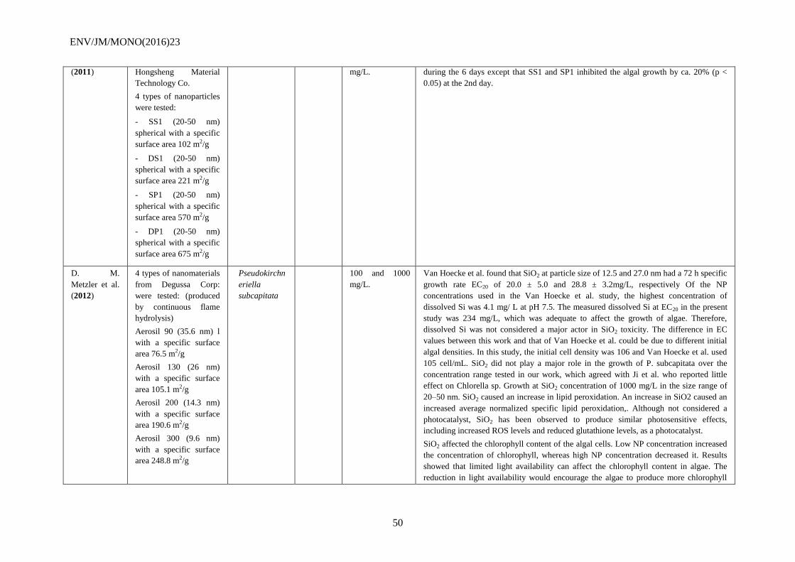
ENV/JM/MONO(2016)23
50
(2011) Hongsheng Material
Technology Co.
4 types of nanoparticles
were tested:
- SS1 (20-50 nm)
spherical with a specific
surface area 102 m2/g
- DS1 (20-50 nm)
spherical with a specific
surface area 221 m2/g
- SP1 (20-50 nm)
spherical with a specific
surface area 570 m2/g
- DP1 (20-50 nm)
spherical with a specific
surface area 675 m2/g
mg/L. during the 6 days except that SS1 and SP1 inhibited the algal growth by ca. 20% (p <
0.05) at the 2nd day.
D. M.
Metzler et al.
(2012)
4 types of nanomaterials
from Degussa Corp:
were tested: (produced
by continuous flame
hydrolysis)
Aerosil 90 (35.6 nm) l
with a specific surface
area 76.5 m2/g
Aerosil 130 (26 nm)
with a specific surface
area 105.1 m2/g
Aerosil 200 (14.3 nm)
with a specific surface
area 190.6 m2/g
Aerosil 300 (9.6 nm)
with a specific surface
area 248.8 m2/g
Pseudokirchn
eriella
subcapitata
100 and 1000
mg/L.
Van Hoecke et al. found that SiO2 at particle size of 12.5 and 27.0 nm had a 72 h specific
growth rate EC20 of 20.0 ± 5.0 and 28.8 ± 3.2mg/L, respectively Of the NP
concentrations used in the Van Hoecke et al. study, the highest concentration of
dissolved Si was 4.1 mg/ L at pH 7.5. The measured dissolved Si at EC20 in the present
study was 234 mg/L, which was adequate to affect the growth of algae. Therefore,
dissolved Si was not considered a major actor in SiO2 toxicity. The difference in EC
values between this work and that of Van Hoecke et al. could be due to different initial
algal densities. In this study, the initial cell density was 106 and Van Hoecke et al. used
105 cell/mL. SiO2 did not play a major role in the growth of P. subcapitata over the
concentration range tested in our work, which agreed with Ji et al. who reported little
effect on Chlorella sp. Growth at SiO2 concentration of 1000 mg/L in the size range of
20–50 nm. SiO2 caused an increase in lipid peroxidation. An increase in SiO2 caused an
increased average normalized specific lipid peroxidation,. Although not considered a
photocatalyst, SiO2 has been observed to produce similar photosensitive effects,
including increased ROS levels and reduced glutathione levels, as a photocatalyst.
SiO2 affected the chlorophyll content of the algal cells. Low NP concentration increased
the concentration of chlorophyll, whereas high NP concentration decreased it. Results
showed that limited light availability can affect the chlorophyll content in algae. The
reduction in light availability would encourage the algae to produce more chlorophyll
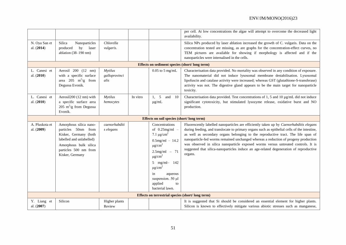
ENV/JM/MONO(2016)23
51
per cell. At low concentrations the algae will attempt to overcome the decreased light
availability.
N. Oya San et
al. (2014)
Silica Nanoparticles
produced by laser
ablation (38–190 nm)
Chlorella
vulgaris.
Silica NPs produced by laser ablation increased the growth of C. vulgaris. Data on the
concentration tested are missing, as are graphs for the concentration-effect curves, no
TEM pictures are available for showing if morphology is affected and if the
nanoparticles were internalised in the cells.
Effects on sediment species (short/ long term)
L. Canesi et
al. (2010)
Aerosil 200 (12 nm)
with a specific surface
area 205 m2/g from
Degussa Evonik.
Mytilus
galloprovinci
alis
0.05 to 5 mg/mL Characterisation data provided. No mortality was observed in any condition of exposure.
The nanomaterial did not induce lysosomal membrane destabilisation. Lysosomal
lipofuscin and catalase activity were increased; whereas GST (glutathione-S-transferase) activity was not. The digestive gland appears to be the main target for nanoparticle
toxicity.
L. Canesi et
al. (2010)
Aerosil200 (12 nm) with
a specific surface area
205 m2/g from Degussa
Evonik.
Mytilus
hemocytes
In vitro 1, 5 and 10
µg/mL
Characterisation data provided. Test concentrations of 1, 5 and 10 µg/mL did not induce
significant cytotoxicity, but stimulated lysozyme release, oxidative burst and NO
production.
Effects on soil species (short/ long term)
A. Pluskota et
al. (2009)
Amorphous silica nano-
particles 50nm from
Kisker, Germany (both
labelled and unlabelled)
Amorphous bulk silica
particles 500 nm from
Kisker, Germany
caenorhabditi
s elegans
Concentrations
of 0.25mg/ml –
7.1 μg/cm2
0.5mg/ml – 14.2
μg/cm2
2.5mg/ml – 71
μg/cm2
5 mg/ml– 142
μg/cm2
in aqueous
suspension. 50 μl
applied to
bacterial lawn.
Fluorescently labelled nanoparticles are efficiently taken up by Caenorhabditis elegans
during feeding, and translocate to primary organs such as epithelial cells of the intestine,
as well as secondary organs belonging to the reproductive tract. The life span of
nanoparticle-fed worms remained unchanged whereas a reduction of progeny production
was observed in silica nanoparticle exposed worms versus untreated controls. It is
suggested that silica-nanoparticles induce an age-related degeneration of reproductive
organs.
Effects on terrestrial species (short/ long term)
Y. Liang et
al. (2007)
Silicon Higher plants
Review
It is suggested that Si should be considered an essential element for higher plants.
Silicon is known to effectively mitigate various abiotic stresses such as manganese,
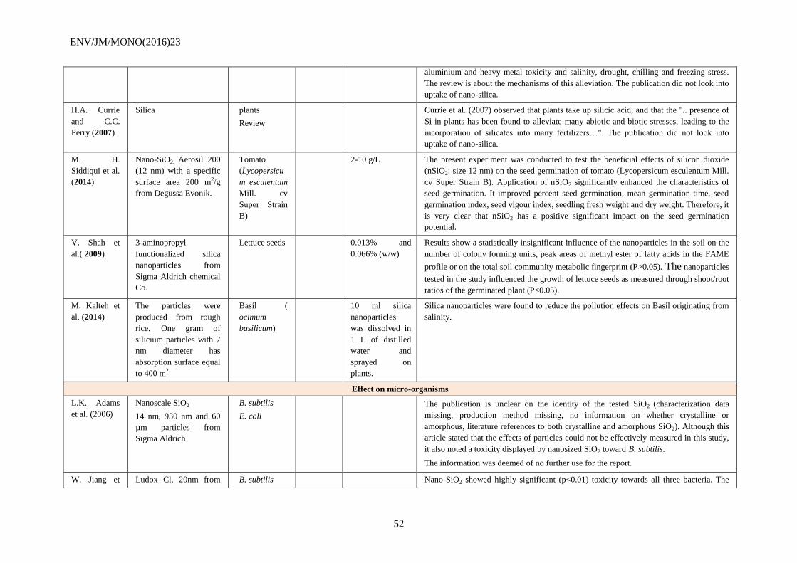
ENV/JM/MONO(2016)23
52
aluminium and heavy metal toxicity and salinity, drought, chilling and freezing stress.
The review is about the mechanisms of this alleviation. The publication did not look into
uptake of nano-silica.
H.A. Currie
and C.C.
Perry (2007)
Silica plants
Review
Currie et al. (2007) observed that plants take up silicic acid, and that the ".. presence of
Si in plants has been found to alleviate many abiotic and biotic stresses, leading to the
incorporation of silicates into many fertilizers…". The publication did not look into
uptake of nano-silica.
M. H.
Siddiqui et al.
(2014)
Nano-SiO2. Aerosil 200
(12 nm) with a specific
surface area 200 m2/g
from Degussa Evonik.
Tomato
(Lycopersicu
m esculentum
Mill. cv
Super Strain
B)
2-10 g/L The present experiment was conducted to test the beneficial effects of silicon dioxide
(nSiO2: size 12 nm) on the seed germination of tomato (Lycopersicum esculentum Mill.
cv Super Strain B). Application of nSiO2 significantly enhanced the characteristics of
seed germination. It improved percent seed germination, mean germination time, seed
germination index, seed vigour index, seedling fresh weight and dry weight. Therefore, it
is very clear that nSiO2 has a positive significant impact on the seed germination
potential.
V. Shah et
al.( 2009)
3-aminopropyl
functionalized silica
nanoparticles from
Sigma Aldrich chemical
Co.
Lettuce seeds 0.013% and
0.066% (w/w)
Results show a statistically insignificant influence of the nanoparticles in the soil on the
number of colony forming units, peak areas of methyl ester of fatty acids in the FAME
profile or on the total soil community metabolic fingerprint (P>0.05). The nanoparticles
tested in the study influenced the growth of lettuce seeds as measured through shoot/root
ratios of the germinated plant (P<0.05).
M. Kalteh et
al. (2014)
The particles were
produced from rough
rice. One gram of
silicium particles with 7
nm diameter has
absorption surface equal
to 400 m2
Basil (
ocimum
basilicum)
10 ml silica
nanoparticles
was dissolved in
1 L of distilled
water and
sprayed on
plants.
Silica nanoparticles were found to reduce the pollution effects on Basil originating from
salinity.
Effect on micro-organisms
L.K. Adams
et al. (2006)
Nanoscale SiO2
14 nm, 930 nm and 60
µm particles from
Sigma Aldrich
B. subtilis
E. coli
The publication is unclear on the identity of the tested SiO2 (characterization data
missing, production method missing, no information on whether crystalline or
amorphous, literature references to both crystalline and amorphous SiO2). Although this
article stated that the effects of particles could not be effectively measured in this study,
it also noted a toxicity displayed by nanosized SiO2 toward B. subtilis.
The information was deemed of no further use for the report.
W. Jiang et Ludox Cl, 20nm from B. subtilis Nano-SiO2 showed highly significant (p<0.01) toxicity towards all three bacteria. The
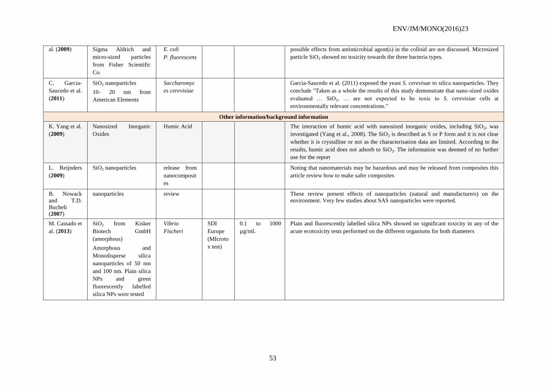
ENV/JM/MONO(2016)23
53
al. (2009) Sigma Aldrich and
micro-sized particles
from Fisher Scientific
Co.
E. coli
P. fluorescens
possible effects from antimicrobial agent(s) in the colloid are not discussed. Microsized
particle SiO2 showed no toxicity towards the three bacteria types.
C. Garcia-
Saucedo et al.
(2011)
SiO2 nanoparticles
10- 20 nm from
American Elements
Saccharomyc
es cerevisiae
Garcia-Saucedo et al. (2011) exposed the yeast S. cerevisae to silica nanoparticles. They
conclude "Taken as a whole the results of this study demonstrate that nano-sized oxides
evaluated … SiO2, … are not expected to be toxic to S. cerevisiae cells at
environmentally relevant concentrations."
Other information/background information
K. Yang et al.
(2009)
Nanosized Inorganic
Oxides
Humic Acid The interaction of humic acid with nanosized inorganic oxides, including SiO2, was
investigated (Yang et al., 2008). The SiO2 is described as S or P form and it is not clear
whether it is crystalline or not as the characterisation data are limited. According to the
results, humic acid does not adsorb to SiO2. The information was deemed of no further
use for the report
L. Reijnders
(2009)
SiO2 nanoparticles release from
nanocomposit
es
Noting that nanomaterials may be hazardous and may be released from composites this
article review how to make safer composites
B. Nowack
and T.D.
Bucheli (2007)
nanoparticles review These review present effects of nanoparticles (natural and manufacturers) on the
environment. Very few studies about SAS nanoparticles were reported.
M. Cassado et
al. (2013)
SiO2 from Kisker
Biotech GmbH
(amorphous)
Amorphous and
Monodisperse silica
nanoparticles of 50 nm
and 100 nm. Plain silica
NPs and green
fluorescently labelled
silica NPs were tested
Vibrio
Fischeri
SDI
Europe
(MIcroto
x test)
0.1 to 1000
µg/mL
Plain and fluorescently labelled silica NPs showed no significant toxicity in any of the
acute ecotoxicity tests performed on the different organisms for both diameters
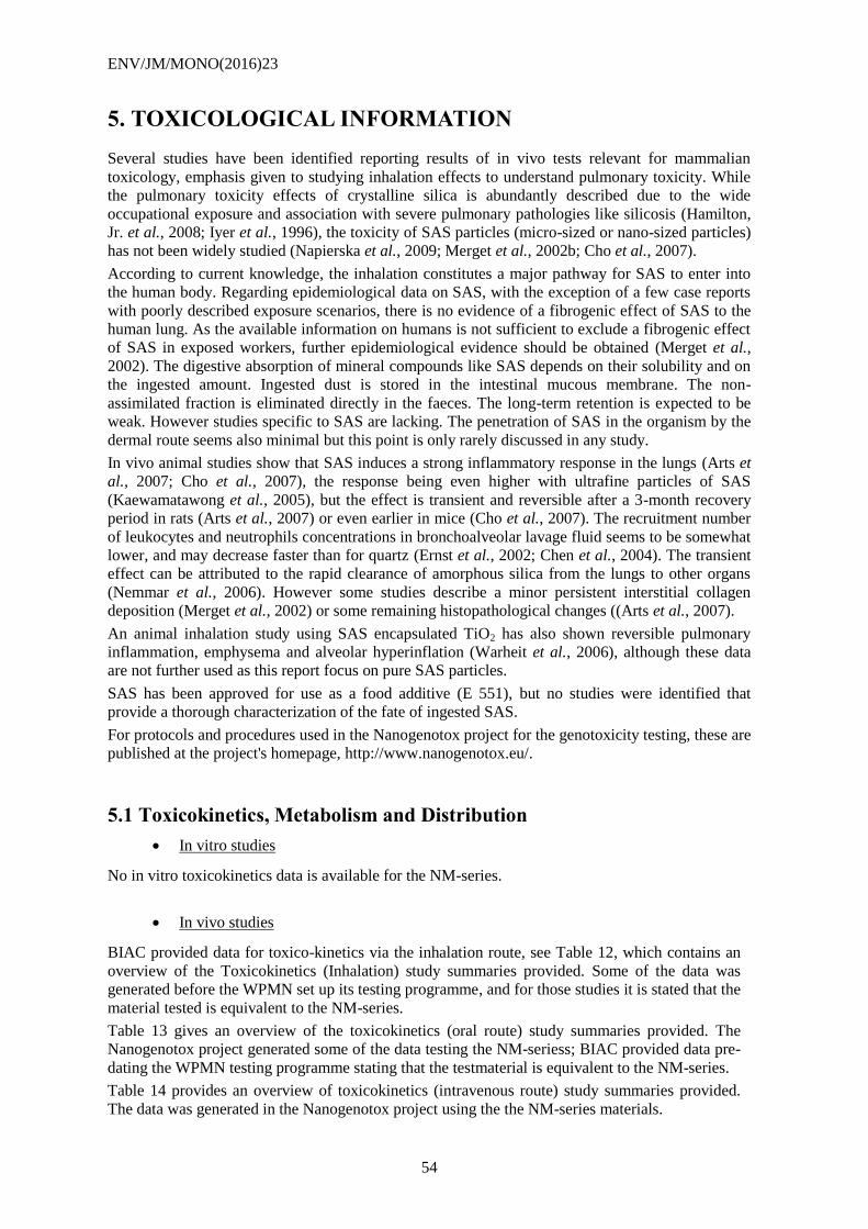
ENV/JM/MONO(2016)23
54
5. TOXICOLOGICAL INFORMATION
Several studies have been identified reporting results of in vivo tests relevant for mammalian
toxicology, emphasis given to studying inhalation effects to understand pulmonary toxicity. While
the pulmonary toxicity effects of crystalline silica is abundantly described due to the wide
occupational exposure and association with severe pulmonary pathologies like silicosis (Hamilton,
Jr. et al., 2008; Iyer et al., 1996), the toxicity of SAS particles (micro-sized or nano-sized particles)
has not been widely studied (Napierska et al., 2009; Merget et al., 2002b; Cho et al., 2007).
According to current knowledge, the inhalation constitutes a major pathway for SAS to enter into
the human body. Regarding epidemiological data on SAS, with the exception of a few case reports
with poorly described exposure scenarios, there is no evidence of a fibrogenic effect of SAS to the
human lung. As the available information on humans is not sufficient to exclude a fibrogenic effect
of SAS in exposed workers, further epidemiological evidence should be obtained (Merget et al.,
2002). The digestive absorption of mineral compounds like SAS depends on their solubility and on
the ingested amount. Ingested dust is stored in the intestinal mucous membrane. The non-
assimilated fraction is eliminated directly in the faeces. The long-term retention is expected to be
weak. However studies specific to SAS are lacking. The penetration of SAS in the organism by the
dermal route seems also minimal but this point is only rarely discussed in any study.
In vivo animal studies show that SAS induces a strong inflammatory response in the lungs (Arts et
al., 2007; Cho et al., 2007), the response being even higher with ultrafine particles of SAS
(Kaewamatawong et al., 2005), but the effect is transient and reversible after a 3-month recovery
period in rats (Arts et al., 2007) or even earlier in mice (Cho et al., 2007). The recruitment number
of leukocytes and neutrophils concentrations in bronchoalveolar lavage fluid seems to be somewhat
lower, and may decrease faster than for quartz (Ernst et al., 2002; Chen et al., 2004). The transient
effect can be attributed to the rapid clearance of amorphous silica from the lungs to other organs
(Nemmar et al., 2006). However some studies describe a minor persistent interstitial collagen
deposition (Merget et al., 2002) or some remaining histopathological changes ((Arts et al., 2007).
An animal inhalation study using SAS encapsulated TiO2 has also shown reversible pulmonary
inflammation, emphysema and alveolar hyperinflation (Warheit et al., 2006), although these data
are not further used as this report focus on pure SAS particles.
SAS has been approved for use as a food additive (E 551), but no studies were identified that
provide a thorough characterization of the fate of ingested SAS.
For protocols and procedures used in the Nanogenotox project for the genotoxicity testing, these are
published at the project's homepage, http://www.nanogenotox.eu/.
5.1 Toxicokinetics, Metabolism and Distribution
In vitro studies
No in vitro toxicokinetics data is available for the NM-series.
In vivo studies
BIAC provided data for toxico-kinetics via the inhalation route, see Table 12, which contains an
overview of the Toxicokinetics (Inhalation) study summaries provided. Some of the data was
generated before the WPMN set up its testing programme, and for those studies it is stated that the
material tested is equivalent to the NM-series.
Table 13 gives an overview of the toxicokinetics (oral route) study summaries provided. The
Nanogenotox project generated some of the data testing the NM-seriess; BIAC provided data pre-
dating the WPMN testing programme stating that the testmaterial is equivalent to the NM-series.
Table 14 provides an overview of toxicokinetics (intravenous route) study summaries provided.
The data was generated in the Nanogenotox project using the the NM-series materials.
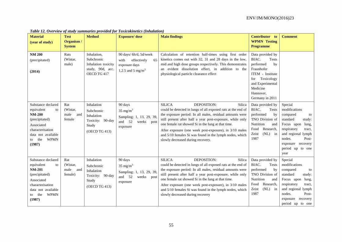
ENV/JM/MONO(2016)23
55
Table 12. Overview of study summaries provided for Toxicokinetics (Inhalation)
Material
(year of study)
Test
Organism /
System
Method Exposure/ dose Main findings Contributor to
WPMN Testing
Programme
Comment
NM 200
(precipitated)
(2014)
Rats
(Wistar,
male)
Inhalation,
Subchronic
Inhalation toxicity
study, 90d, acc.
OECD TG 417
90 days/ 6h/d, 5d/week
with effectively 65
exposure days
1,2.5 and 5 mg/m3
Calculation of retention half-times using first order
kinetics comes out with 32, 31 and 28 days in the low,
mid and high dose groups respectively. This demonstrates
an evident dissolution effect, in addition to the
physiological particle clearance effect
Data provided by
BIAC. Tests
performed by
Fraunhofer
ITEM - Institute
for Toxicology
and Experimental
Medicine
Hannover,
Germany in 2011
Substance declared
equivalent to
NM-200
(precipitated)
Associated
characterisation
data not available
to the WPMN
(1987)
Rat
(Wistar,
male and
female
Inhalation
Subchronic
Inhalation
Toxicity: 90-day
Study
(OECD TG 413)
90 days
35 mg/m3
Sampling: 1, 13, 29, 39,
and 52 weeks post
exposure
SILICA DEPOSITION: Silica
could be detected in lungs of all exposed rats at the end of
the exposure period: In all males, residual amounts were
still present after half a year post-exposure, while only
one female rat showed Si in the lung at that time.
After exposure (one week post-exposure), in 3/10 males
and 5/10 females Si was found in the lymph nodes, which
slowly decreased during recovery.
Data provided by
BIAC. Tests
performed by
TNO Division of
Nutrition and
Food Research,
Zeist (NL) in
1987
Special
modifications
compared to
standard study:
Focus upon lung,
respiratory tract,
and regional lymph
nodes. Post-
exposure recovery
period up to one
year
Substance declared
equivalent to
NM-201
(precipitated)
Associated
characterisation
data not available
to the WPMN
(1987)
Rat
(Wistar,
male and
female)
Inhalation
Subchronic
Inhalation
Toxicity: 90-day
Study
(OECD TG 413)
90 days
35 mg/m3
Sampling: 1, 13, 29, 39,
and 52 weeks post
exposure
SILICA DEPOSITION: Silica
could be detected in lungs of all exposed rats at the end of
the exposure period: In all males, residual amounts were
still present after half a year post-exposure, while only
one female rat showed Si in the lung at that time.
After exposure (one week post-exposure), in 3/10 males
and 5/10 females Si was found in the lymph nodes, which
slowly decreased during recovery
Data provided by
BIAC. Tests
performed by
TNO Division of
Nutrition and
Food Research,
Zeist (NL) in
1987
Special
modifications
compared to
standard study:
Focus upon lung,
respiratory tract,
and regional lymph
nodes. Post-
exposure recovery
period up to one
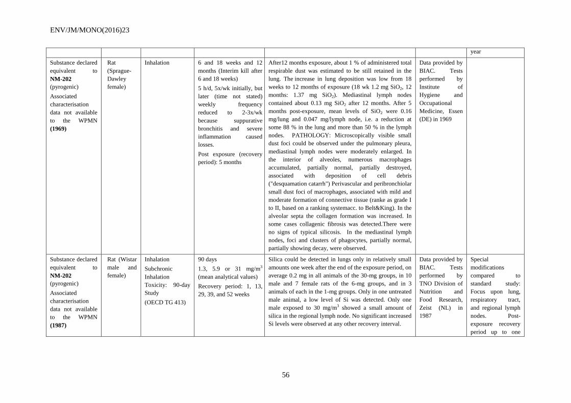
ENV/JM/MONO(2016)23
56
year
Substance declared
equivalent to
NM-202
(pyrogenic)
Associated
characterisation
data not available
to the WPMN
(1969)
Rat
(Sprague-
Dawley
female)
Inhalation
6 and 18 weeks and 12
months (Interim kill after
6 and 18 weeks)
5 h/d, 5x/wk initially, but
later (time not stated)
weekly frequency
reduced to 2-3x/wk
because suppurative
bronchitis and severe
inflammation caused
losses.
Post exposure (recovery
period): 5 months
After12 months exposure, about 1 % of administered total
respirable dust was estimated to be still retained in the
lung. The increase in lung deposition was low from 18
weeks to 12 months of exposure (18 wk 1.2 mg SiO2, 12
months: 1.37 mg SiO2). Mediastinal lymph nodes
contained about 0.13 mg SiO2 after 12 months. After 5
months post-exposure, mean levels of SiO2 were 0.16
mg/lung and 0.047 mg/lymph node, i.e. a reduction at
some 88 % in the lung and more than 50 % in the lymph
nodes. PATHOLOGY: Microscopically visible small
dust foci could be observed under the pulmonary pleura,
mediastinal lymph nodes were moderately enlarged. In
the interior of alveoles, numerous macrophages
accumulated, partially normal, partially destroyed,
associated with deposition of cell debris
("desquamation catarrh") Perivascular and peribronchiolar
small dust foci of macrophages, associated with mild and
moderate formation of connective tissue (ranke as grade I
to II, based on a ranking systemacc. to Belt&King). In the
alveolar septa the collagen formation was increased. In
some cases collagenic fibrosis was detected.There were
no signs of typical silicosis. In the mediastinal lymph
nodes, foci and clusters of phagocytes, partially normal,
partially showing decay, were observed.
Data provided by
BIAC. Tests
performed by
Institute of
Hygiene and
Occupational
Medicine, Essen
(DE) in 1969
Substance declared
equivalent to
NM-202
(pyrogenic)
Associated
characterisation
data not available
to the WPMN
(1987)
Rat (Wistar
male and
female)
Inhalation
Subchronic
Inhalation
Toxicity: 90-day
Study
(OECD TG 413)
90 days
1.3, 5.9 or 31 mg/m3
(mean analytical values)
Recovery period: 1, 13,
29, 39, and 52 weeks
Silica could be detected in lungs only in relatively small
amounts one week after the end of the exposure period, on
average 0.2 mg in all animals of the 30-mg groups, in 10
male and 7 female rats of the 6-mg groups, and in 3
animals of each in the 1-mg groups. Only in one untreated
male animal, a low level of Si was detected. Only one
male exposed to 30 mg/m3 showed a small amount of
silica in the regional lymph node. No significant increased
Si levels were observed at any other recovery interval.
Data provided by
BIAC. Tests
performed by
TNO Division of
Nutrition and
Food Research,
Zeist (NL) in
1987
Special
modifications
compared to
standard study:
Focus upon lung,
respiratory tract,
and regional lymph
nodes. Post-
exposure recovery
period up to one
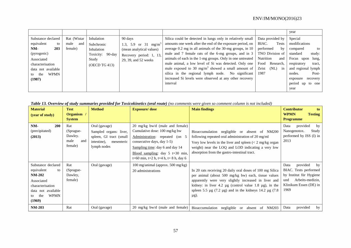
ENV/JM/MONO(2016)23
57
year
Substance declared
equivalent to
NM- 203
(pyrogenic)
Associated
characterisation
data not available
to the WPMN
(1987)
Rat (Wistar
male and
female)
Inhalation
Subchronic
Inhalation
Toxicity: 90-day
Study
(OECD TG 413)
90 days
1.3, 5.9 or 31 mg/m3
(mean analytical values)
Recovery period: 1, 13,
29, 39, and 52 weeks
Silica could be detected in lungs only in relatively small
amounts one week after the end of the exposure period, on
average 0.2 mg in all animals of the 30-mg groups, in 10
male and 7 female rats of the 6-mg groups, and in 3
animals of each in the 1-mg groups. Only in one untreated
male animal, a low level of Si was detected. Only one
male exposed to 30 mg/m3 showed a small amount of
silica in the regional lymph node. No significant
increased Si levels were observed at any other recovery
interval
Data provided by
BIAC. Tests
performed by
TNO Division of
Nutrition and
Food Research,
Zeist (NL) in
1987
Special
modifications
compared to
standard study:
Focus upon lung,
respiratory tract,
and regional lymph
nodes. Post-
exposure recovery
period up to one
year
Table 13. Overview of study summaries provided for Toxicokinetics (oral route) (no comments were given so comment column is not included)
Material
(year of study)
Test
Organism /
System
Method Exposure/ dose Main findings Contributor to
WPMN Testing
Programme
NM- 200
(precipitated)
(2013)
Rat
(Sprague-
Dawley.
male and
female)
Oral (gavage)
Sampled organs: liver,
spleen, GI tract (small
intestine), mesenteric
lymph nodes
20 mg/kg bw/d (male and female)
Cumulative dose: 100 mg/kg bw
Administration: repeated (on 5
consecutive days, day 1-5)
Sampling time: day 6 and day 14
Blood sampling: day 5 t=30 min,
t=60 min, t=2 h, t=4 h, t= 8 h, day 6
Bioaccumulation negligible or absent of NM200
following repeated oral administration of 20 mg/ml
Very low levels in the liver and spleen (< 2 mg/kg organ
weight) near the LOQ and LOD indicating a very low
absorption from the gastro-intestinal tract.
Data provided by
Nanogenotox. Study
performed by ISS (I) in
2013
Substance declared
equivalent to
NM-202
Associated
characterisation
data not available
to the WPMN
(1969)
Rat
(Sprague-
Dawley,
female)
Oral (gavage) 100 mg/animal (approx. 500 mg/kg)
20 administrations In 20 rats receiving 20 daily oral doses of 100 mg Silica
per animal (about 500 mg/kg bw) each, tissue values
apparently were very slightly increased in liver and
kidney: in liver 4.2 µg (control value 1.8 µg), in the
spleen 5.5 µg (7.2 µg) and in the kidneys 14.2 µg (7.8
µg).
Data provided by
BIAC. Tests performed
by Institut für Hygiene
und Arbeits-medizin,
Klinikum Essen (DE) in
1969
NM-203 Rat Oral (gavage) 20 mg/kg bw/d (male and female) Bioaccumulation negligible or absent of NM203 Data provided by
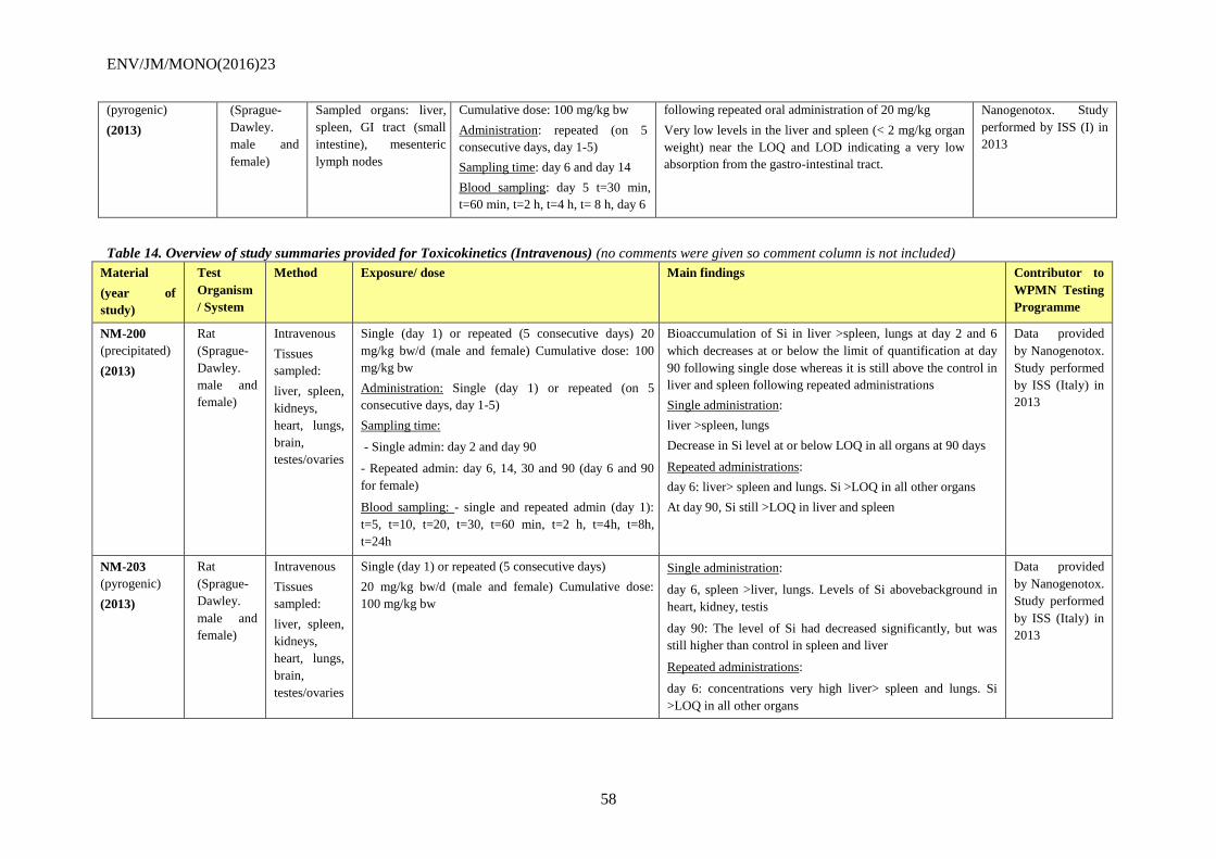
ENV/JM/MONO(2016)23
58
(pyrogenic)
(2013)
(Sprague-
Dawley.
male and
female)
Sampled organs: liver,
spleen, GI tract (small
intestine), mesenteric
lymph nodes
Cumulative dose: 100 mg/kg bw
Administration: repeated (on 5
consecutive days, day 1-5)
Sampling time: day 6 and day 14
Blood sampling: day 5 t=30 min,
t=60 min, t=2 h, t=4 h, t= 8 h, day 6
following repeated oral administration of 20 mg/kg
Very low levels in the liver and spleen (< 2 mg/kg organ
weight) near the LOQ and LOD indicating a very low
absorption from the gastro-intestinal tract.
Nanogenotox. Study
performed by ISS (I) in
2013
Table 14. Overview of study summaries provided for Toxicokinetics (Intravenous) (no comments were given so comment column is not included)
Material
(year of
study)
Test
Organism
/ System
Method Exposure/ dose Main findings Contributor to
WPMN Testing
Programme
NM-200
(precipitated)
(2013)
Rat
(Sprague-
Dawley.
male and
female)
Intravenous
Tissues
sampled:
liver, spleen,
kidneys,
heart, lungs,
brain,
testes/ovaries
Single (day 1) or repeated (5 consecutive days) 20
mg/kg bw/d (male and female) Cumulative dose: 100
mg/kg bw
Administration: Single (day 1) or repeated (on 5
consecutive days, day 1-5)
Sampling time:
- Single admin: day 2 and day 90
- Repeated admin: day 6, 14, 30 and 90 (day 6 and 90
for female)
Blood sampling: - single and repeated admin (day 1):
t=5, t=10, t=20, t=30, t=60 min, t=2 h, t=4h, t=8h,
t=24h
Bioaccumulation of Si in liver >spleen, lungs at day 2 and 6
which decreases at or below the limit of quantification at day
90 following single dose whereas it is still above the control in
liver and spleen following repeated administrations
Single administration:
liver >spleen, lungs
Decrease in Si level at or below LOQ in all organs at 90 days
Repeated administrations:
day 6: liver> spleen and lungs. Si >LOQ in all other organs
At day 90, Si still >LOQ in liver and spleen
Data provided
by Nanogenotox.
Study performed
by ISS (Italy) in
2013
NM-203
(pyrogenic)
(2013)
Rat
(Sprague-
Dawley.
male and
female)
Intravenous
Tissues
sampled:
liver, spleen,
kidneys,
heart, lungs,
brain,
testes/ovaries
Single (day 1) or repeated (5 consecutive days)
20 mg/kg bw/d (male and female) Cumulative dose:
100 mg/kg bw
Single administration:
day 6, spleen >liver, lungs. Levels of Si abovebackground in
heart, kidney, testis
day 90: The level of Si had decreased significantly, but was
still higher than control in spleen and liver
Repeated administrations:
day 6: concentrations very high liver> spleen and lungs. Si
>LOQ in all other organs
Data provided
by Nanogenotox.
Study performed
by ISS (Italy) in
2013
ENV/JM/MONO(2016)23
59
At day 90, The level of Si had decreased significantly, but was
still higher than the control in liver and spleen
ENV/JM/MONO(2016)23
60
5.2 Acute toxicity
The tables below, Table 15 and Table 16, summarise the results for acute toxicity; Table 15 gives
data for the alternate material supplied by Japan and Table 16 summarises the information from
scientific literature of toxic response to a single dose of test material. Table 17 provides an
overview of acute toxicity study summaries provided by BIAC. The data concerns the following
exposure routes: inhalation, oral and dermal. The data is generated before the WPMN set up its
testing programme, and it is stated that the material tested is equivalent to the NM-series.
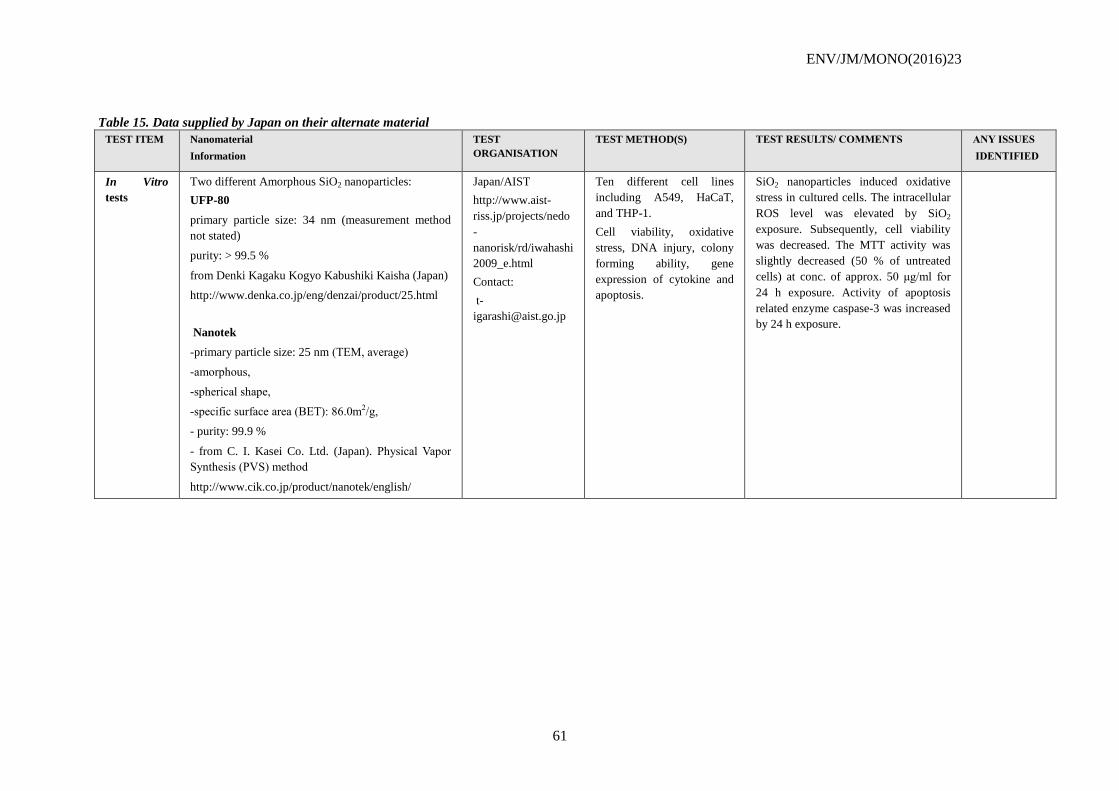
ENV/JM/MONO(2016)23
61
Table 15. Data supplied by Japan on their alternate material
TEST ITEM Nanomaterial
Information
TEST
ORGANISATION
TEST METHOD(S) TEST RESULTS/ COMMENTS ANY ISSUES
IDENTIFIED
In Vitro
tests
Two different Amorphous SiO2 nanoparticles:
UFP-80
primary particle size: 34 nm (measurement method
not stated)
purity: > 99.5 %
from Denki Kagaku Kogyo Kabushiki Kaisha (Japan)
http://www.denka.co.jp/eng/denzai/product/25.html
Nanotek
-primary particle size: 25 nm (TEM, average)
-amorphous,
-spherical shape,
-specific surface area (BET): 86.0m2/g,
- purity: 99.9 %
- from C. I. Kasei Co. Ltd. (Japan). Physical Vapor
Synthesis (PVS) method
http://www.cik.co.jp/product/nanotek/english/
Japan/AIST
http://www.aist-
riss.jp/projects/nedo
-
nanorisk/rd/iwahashi
2009_e.html
Contact:
t-
Ten different cell lines
including A549, HaCaT,
and THP-1.
Cell viability, oxidative
stress, DNA injury, colony
forming ability, gene
expression of cytokine and
apoptosis.
SiO2 nanoparticles induced oxidative
stress in cultured cells. The intracellular
ROS level was elevated by SiO2
exposure. Subsequently, cell viability
was decreased. The MTT activity was
slightly decreased (50 % of untreated
cells) at conc. of approx. 50 μg/ml for
24 h exposure. Activity of apoptosis
related enzyme caspase-3 was increased
by 24 h exposure.
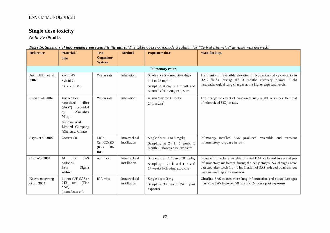
ENV/JM/MONO(2016)23
62
Single dose toxicity
A/ In vivo Studies
Table 16. Summary of information from scientific literature. (The table does not include a column for "Derived effect value" as none was derived.)
Reference Material /
Size
Test
Organism/
System
Method Exposure/ dose Main findings
Pulmonary route
Arts, JHE, et al,
2007
Zeosil 45
Syloid 74
Cal-O-Sil M5
Wistar rats Inhalation 6 h/day for 5 consecutive days
1, 5 or 25 mg/m3
Sampling at day 6, 1 month and
3 months following exposure
Transient and reversible elevation of biomarkers of cytotoxicity in
BAL fluids, during the 3 months recovery period. Slight
histopathological lung changes at the higher exposure levels.
Chen et al. 2004 Unspecified
nanosized silica
(SAS?) provided
by Zhoushan
Mingri
Nanomaterial
Limited Company
(Zhejiang, China)
Wistar rats Inhalation 40 min/day for 4 weeks
24.1 mg/m3
The fibrogenic effect of nanosized SiO2 might be milder than that
of microsized SiO2 in rats.
Sayes et al. 2007 Zeofree 80 Male
Crl :CD(SD
)IGS BR
Rats
Intratracheal
instillation
Single doses: 1 or 5 mg/kg
Sampling at 24 h; 1 week; 1
month; 3 months post exposure
Pulmonary instilled SAS produced reversible and transient
inflammatory response in rats.
Cho WS, 2007 14 nm SAS
particles
from Sigma
Aldrich
A/J mice Intratracheal
instillation
Single doses: 2, 10 and 50 mg/kg
Sampling at 24 h, and 1, 4 and
14 weeks following exposure
Increase in the lung weights, in total BAL cells and in several pro
inflammatory mediators during the early stages. No changes were
detected after week 1 or 4. Instillation of SAS induced transient, but
very severe lung inflammation.
Kaewamatawong
et al., 2005
14 nm (UF SAS) /
213 nm (Fine
SAS)
(manufacturer’s
ICR mice Intratracheal
instillation
Single dose: 3 mg
Sampling 30 min to 24 h post
exposure
Ultrafine SAS causes more lung inflammation and tissue damages
than Fine SAS Between 30 min and 24 hours post exposure
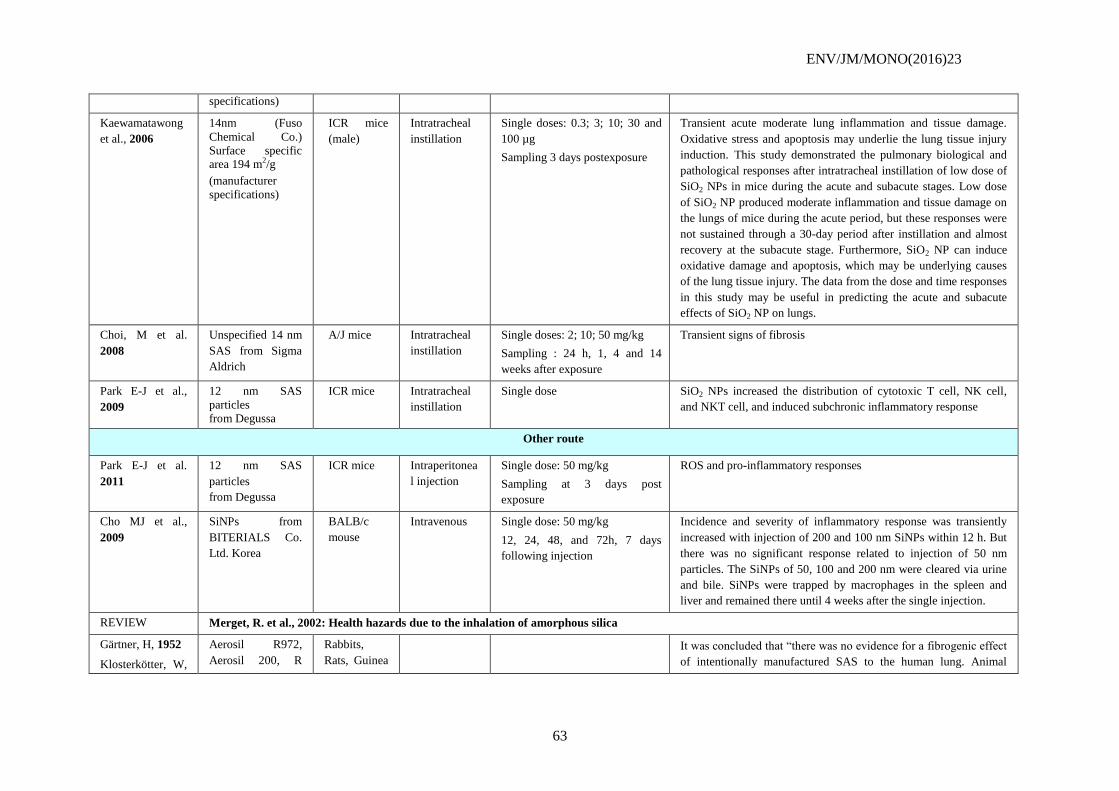
ENV/JM/MONO(2016)23
63
specifications)
Kaewamatawong
et al., 2006
14nm (Fuso
Chemical Co.)
Surface specific area 194 m2/g
(manufacturer
specifications)
ICR mice
(male)
Intratracheal
instillation
Single doses: 0.3; 3; 10; 30 and
100 µg
Sampling 3 days postexposure
Transient acute moderate lung inflammation and tissue damage.
Oxidative stress and apoptosis may underlie the lung tissue injury
induction. This study demonstrated the pulmonary biological and
pathological responses after intratracheal instillation of low dose of
SiO2 NPs in mice during the acute and subacute stages. Low dose
of SiO2 NP produced moderate inflammation and tissue damage on
the lungs of mice during the acute period, but these responses were
not sustained through a 30-day period after instillation and almost
recovery at the subacute stage. Furthermore, SiO2 NP can induce
oxidative damage and apoptosis, which may be underlying causes
of the lung tissue injury. The data from the dose and time responses
in this study may be useful in predicting the acute and subacute
effects of SiO2 NP on lungs.
Choi, M et al.
2008
Unspecified 14 nm
SAS from Sigma
Aldrich
A/J mice Intratracheal
instillation
Single doses: 2; 10; 50 mg/kg
Sampling : 24 h, 1, 4 and 14
weeks after exposure
Transient signs of fibrosis
Park E-J et al.,
2009
12 nm SAS
particles
from Degussa
ICR mice Intratracheal
instillation
Single dose SiO2 NPs increased the distribution of cytotoxic T cell, NK cell,
and NKT cell, and induced subchronic inflammatory response
Other route
Park E-J et al.
2011
12 nm SAS
particles
from Degussa
ICR mice Intraperitonea
l injection
Single dose: 50 mg/kg
Sampling at 3 days post
exposure
ROS and pro-inflammatory responses
Cho MJ et al.,
2009
SiNPs from
BITERIALS Co.
Ltd. Korea
BALB/c
mouse
Intravenous Single dose: 50 mg/kg
12, 24, 48, and 72h, 7 days
following injection
Incidence and severity of inflammatory response was transiently
increased with injection of 200 and 100 nm SiNPs within 12 h. But
there was no significant response related to injection of 50 nm
particles. The SiNPs of 50, 100 and 200 nm were cleared via urine
and bile. SiNPs were trapped by macrophages in the spleen and
liver and remained there until 4 weeks after the single injection.
REVIEW Merget, R. et al., 2002: Health hazards due to the inhalation of amorphous silica
Gärtner, H, 1952
Klosterkötter, W,
Aerosil R972,
Aerosil 200, R
Rabbits,
Rats, Guinea
It was concluded that “there was no evidence for a fibrogenic effect
of intentionally manufactured SAS to the human lung. Animal
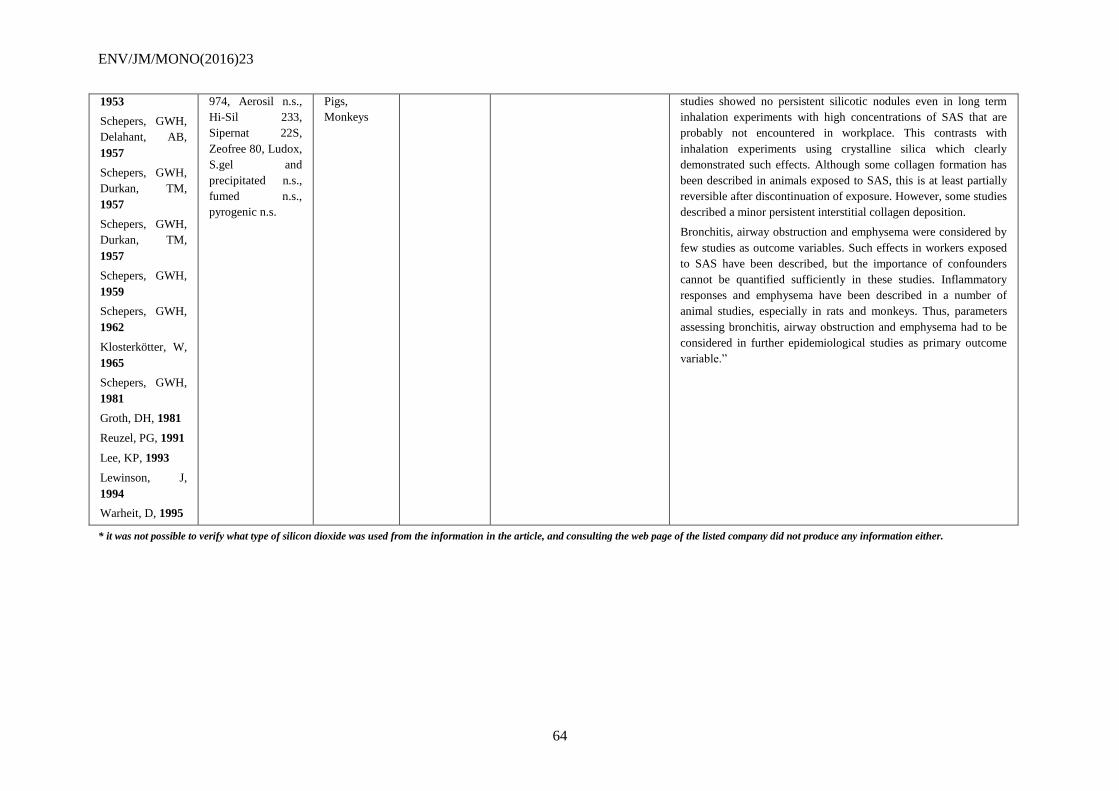
ENV/JM/MONO(2016)23
64
1953
Schepers, GWH,
Delahant, AB,
1957
Schepers, GWH,
Durkan, TM,
1957
Schepers, GWH,
Durkan, TM,
1957
Schepers, GWH,
1959
Schepers, GWH,
1962
Klosterkötter, W,
1965
Schepers, GWH,
1981
Groth, DH, 1981
Reuzel, PG, 1991
Lee, KP, 1993
Lewinson, J,
1994
Warheit, D, 1995
974, Aerosil n.s.,
Hi-Sil 233,
Sipernat 22S,
Zeofree 80, Ludox,
S.gel and
precipitated n.s.,
fumed n.s.,
pyrogenic n.s.
Pigs,
Monkeys
studies showed no persistent silicotic nodules even in long term
inhalation experiments with high concentrations of SAS that are
probably not encountered in workplace. This contrasts with
inhalation experiments using crystalline silica which clearly
demonstrated such effects. Although some collagen formation has
been described in animals exposed to SAS, this is at least partially
reversible after discontinuation of exposure. However, some studies
described a minor persistent interstitial collagen deposition.
Bronchitis, airway obstruction and emphysema were considered by
few studies as outcome variables. Such effects in workers exposed
to SAS have been described, but the importance of confounders
cannot be quantified sufficiently in these studies. Inflammatory
responses and emphysema have been described in a number of
animal studies, especially in rats and monkeys. Thus, parameters
assessing bronchitis, airway obstruction and emphysema had to be
considered in further epidemiological studies as primary outcome
variable.”
* it was not possible to verify what type of silicon dioxide was used from the information in the article, and consulting the web page of the listed company did not produce any information either.
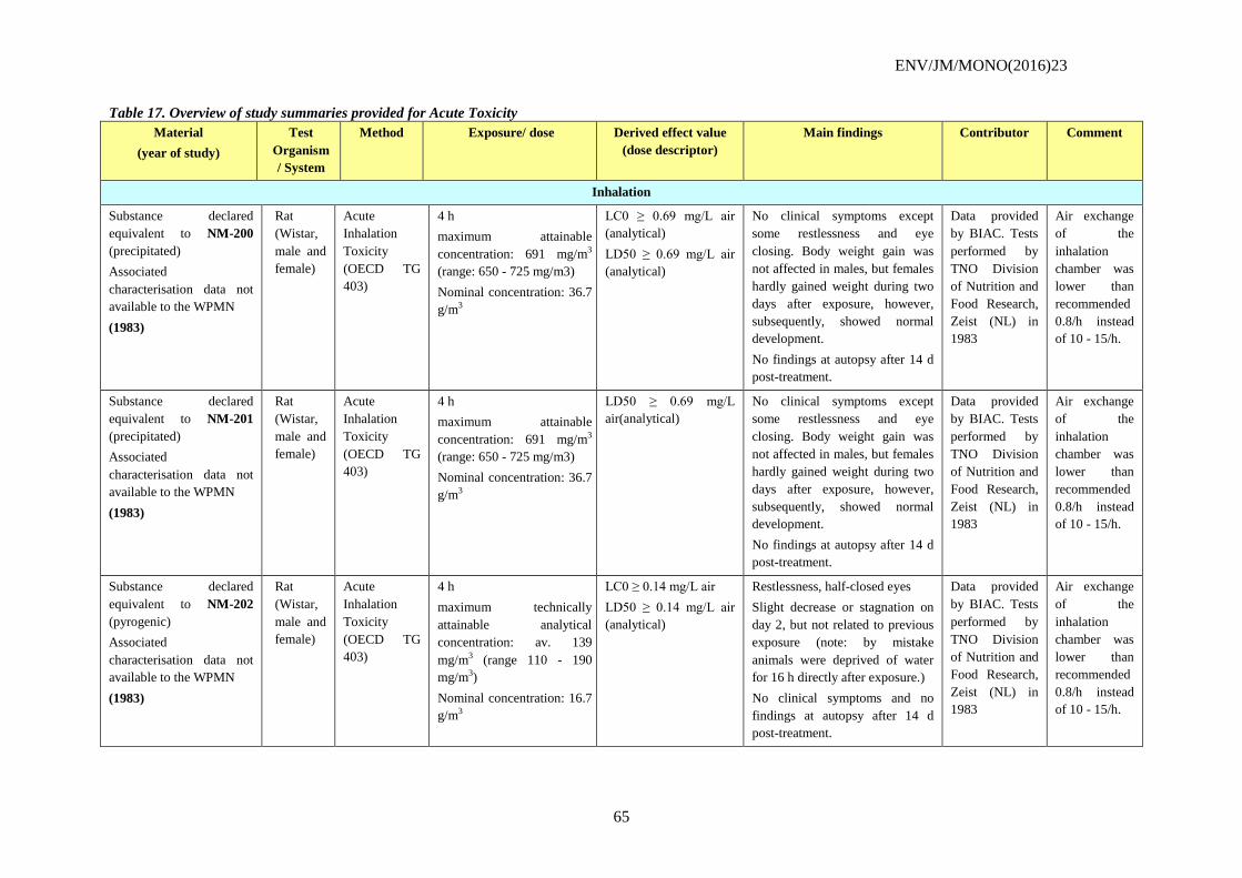
ENV/JM/MONO(2016)23
65
Table 17. Overview of study summaries provided for Acute Toxicity
Material
(year of study)
Test
Organism
/ System
Method Exposure/ dose Derived effect value
(dose descriptor)
Main findings Contributor Comment
Inhalation
Substance declared
equivalent to NM-200
(precipitated)
Associated
characterisation data not
available to the WPMN
(1983)
Rat
(Wistar,
male and
female)
Acute
Inhalation
Toxicity
(OECD TG
403)
4 h
maximum attainable
concentration: 691 mg/m3
(range: 650 - 725 mg/m3)
Nominal concentration: 36.7
g/m3
LC0 ≥ 0.69 mg/L air
(analytical)
LD50 ≥ 0.69 mg/L air
(analytical)
No clinical symptoms except
some restlessness and eye
closing. Body weight gain was
not affected in males, but females
hardly gained weight during two
days after exposure, however,
subsequently, showed normal
development.
No findings at autopsy after 14 d
post-treatment.
Data provided
by BIAC. Tests
performed by
TNO Division
of Nutrition and
Food Research,
Zeist (NL) in
1983
Air exchange
of the
inhalation
chamber was
lower than
recommended
0.8/h instead
of 10 - 15/h.
Substance declared
equivalent to NM-201
(precipitated)
Associated
characterisation data not
available to the WPMN
(1983)
Rat
(Wistar,
male and
female)
Acute
Inhalation
Toxicity
(OECD TG
403)
4 h
maximum attainable
concentration: 691 mg/m3
(range: 650 - 725 mg/m3)
Nominal concentration: 36.7
g/m3
LD50 ≥ 0.69 mg/L
air(analytical)
No clinical symptoms except
some restlessness and eye
closing. Body weight gain was
not affected in males, but females
hardly gained weight during two
days after exposure, however,
subsequently, showed normal
development.
No findings at autopsy after 14 d
post-treatment.
Data provided
by BIAC. Tests
performed by
TNO Division
of Nutrition and
Food Research,
Zeist (NL) in
1983
Air exchange
of the
inhalation
chamber was
lower than
recommended
0.8/h instead
of 10 - 15/h.
Substance declared
equivalent to NM-202
(pyrogenic)
Associated
characterisation data not
available to the WPMN
(1983)
Rat
(Wistar,
male and
female)
Acute
Inhalation
Toxicity
(OECD TG
403)
4 h
maximum technically
attainable analytical
concentration: av. 139
mg/m3 (range 110 - 190
mg/m3)
Nominal concentration: 16.7
g/m3
LC0 ≥ 0.14 mg/L air
LD50 ≥ 0.14 mg/L air
(analytical)
Restlessness, half-closed eyes
Slight decrease or stagnation on
day 2, but not related to previous
exposure (note: by mistake
animals were deprived of water
for 16 h directly after exposure.)
No clinical symptoms and no
findings at autopsy after 14 d
post-treatment.
Data provided
by BIAC. Tests
performed by
TNO Division
of Nutrition and
Food Research,
Zeist (NL) in
1983
Air exchange
of the
inhalation
chamber was
lower than
recommended
0.8/h instead
of 10 - 15/h.
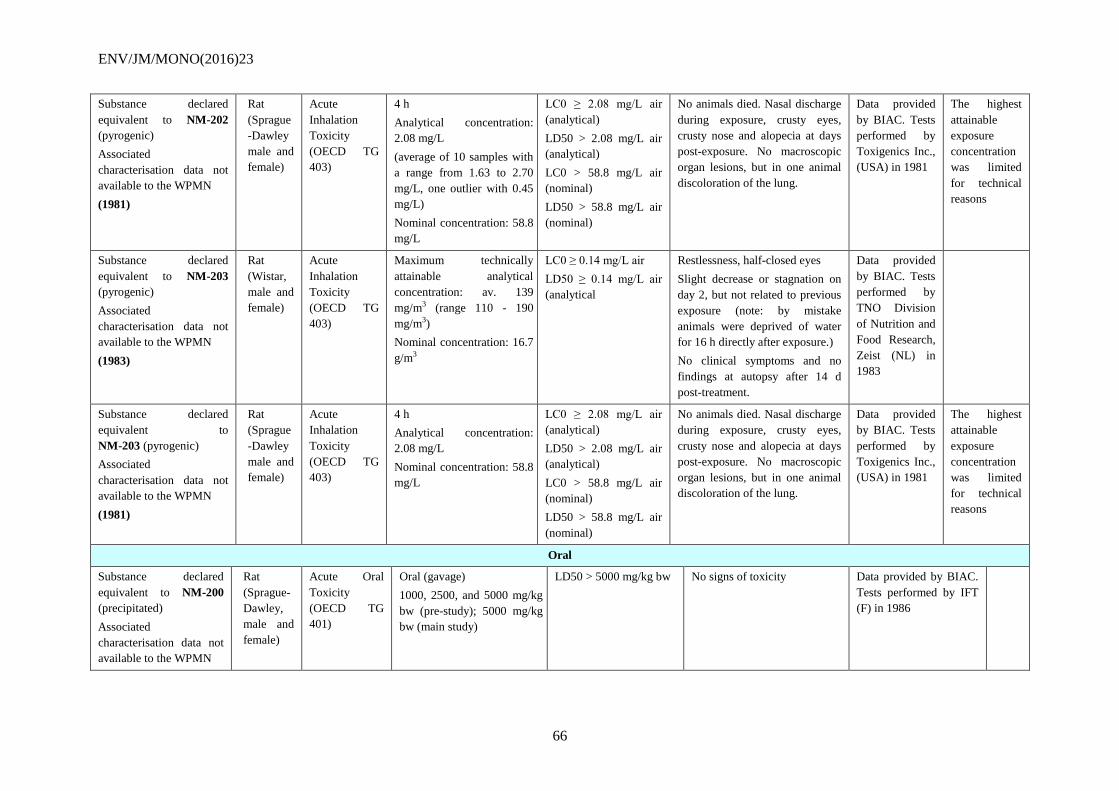
ENV/JM/MONO(2016)23
66
Substance declared
equivalent to NM-202
(pyrogenic)
Associated
characterisation data not
available to the WPMN
(1981)
Rat
(Sprague
-Dawley
male and
female)
Acute
Inhalation
Toxicity
(OECD TG
403)
4 h
Analytical concentration:
2.08 mg/L
(average of 10 samples with
a range from 1.63 to 2.70
mg/L, one outlier with 0.45
mg/L)
Nominal concentration: 58.8
mg/L
LC0 ≥ 2.08 mg/L air
(analytical)
LD50 > 2.08 mg/L air
(analytical)
LC0 > 58.8 mg/L air
(nominal)
LD50 > 58.8 mg/L air
(nominal)
No animals died. Nasal discharge
during exposure, crusty eyes,
crusty nose and alopecia at days
post-exposure. No macroscopic
organ lesions, but in one animal
discoloration of the lung.
Data provided
by BIAC. Tests
performed by
Toxigenics Inc.,
(USA) in 1981
The highest
attainable
exposure
concentration
was limited
for technical
reasons
Substance declared
equivalent to NM-203
(pyrogenic)
Associated
characterisation data not
available to the WPMN
(1983)
Rat
(Wistar,
male and
female)
Acute
Inhalation
Toxicity
(OECD TG
403)
Maximum technically
attainable analytical
concentration: av. 139
mg/m3 (range 110 - 190
mg/m3)
Nominal concentration: 16.7
g/m3
LC0 ≥ 0.14 mg/L air
LD50 ≥ 0.14 mg/L air
(analytical
Restlessness, half-closed eyes
Slight decrease or stagnation on
day 2, but not related to previous
exposure (note: by mistake
animals were deprived of water
for 16 h directly after exposure.)
No clinical symptoms and no
findings at autopsy after 14 d
post-treatment.
Data provided
by BIAC. Tests
performed by
TNO Division
of Nutrition and
Food Research,
Zeist (NL) in
1983
Substance declared
equivalent to
NM-203 (pyrogenic)
Associated
characterisation data not
available to the WPMN
(1981)
Rat
(Sprague
-Dawley
male and
female)
Acute
Inhalation
Toxicity
(OECD TG
403)
4 h
Analytical concentration:
2.08 mg/L
Nominal concentration: 58.8
mg/L
LC0 ≥ 2.08 mg/L air
(analytical)
LD50 > 2.08 mg/L air
(analytical)
LC0 > 58.8 mg/L air
(nominal)
LD50 > 58.8 mg/L air
(nominal)
No animals died. Nasal discharge
during exposure, crusty eyes,
crusty nose and alopecia at days
post-exposure. No macroscopic
organ lesions, but in one animal
discoloration of the lung.
Data provided
by BIAC. Tests
performed by
Toxigenics Inc.,
(USA) in 1981
The highest
attainable
exposure
concentration
was limited
for technical
reasons
Oral
Substance declared
equivalent to NM-200
(precipitated)
Associated
characterisation data not
available to the WPMN
Rat
(Sprague-
Dawley,
male and
female)
Acute Oral
Toxicity
(OECD TG
401)
Oral (gavage)
1000, 2500, and 5000 mg/kg
bw (pre-study); 5000 mg/kg
bw (main study)
LD50 > 5000 mg/kg bw No signs of toxicity Data provided by BIAC.
Tests performed by IFT
(F) in 1986
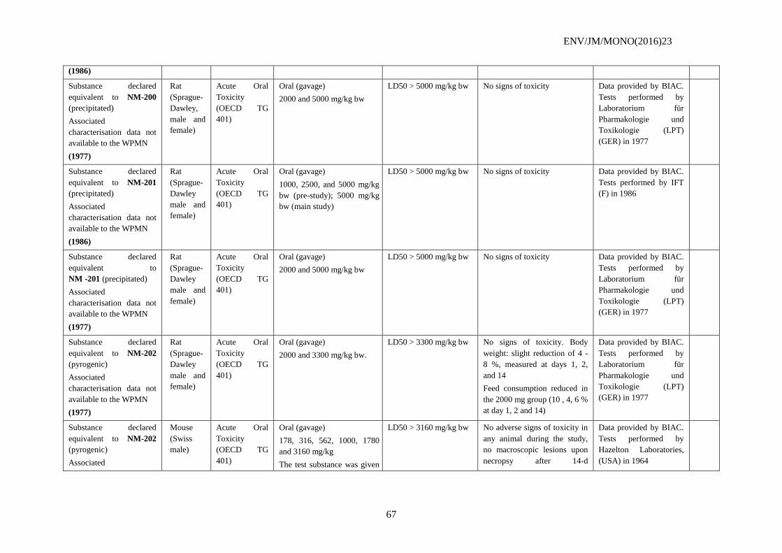
ENV/JM/MONO(2016)23
67
(1986)
Substance declared
equivalent to NM-200
(precipitated)
Associated
characterisation data not
available to the WPMN
(1977)
Rat
(Sprague-
Dawley,
male and
female)
Acute Oral
Toxicity
(OECD TG
401)
Oral (gavage)
2000 and 5000 mg/kg bw
LD50 > 5000 mg/kg bw No signs of toxicity Data provided by BIAC.
Tests performed by
Laboratorium für
Pharmakologie und
Toxikologie (LPT)
(GER) in 1977
Substance declared
equivalent to NM-201
(precipitated)
Associated
characterisation data not
available to the WPMN
(1986)
Rat
(Sprague-
Dawley
male and
female)
Acute Oral
Toxicity
(OECD TG
401)
Oral (gavage)
1000, 2500, and 5000 mg/kg
bw (pre-study); 5000 mg/kg
bw (main study)
LD50 > 5000 mg/kg bw No signs of toxicity Data provided by BIAC.
Tests performed by IFT
(F) in 1986
Substance declared
equivalent to
NM -201 (precipitated)
Associated
characterisation data not
available to the WPMN
(1977)
Rat
(Sprague-
Dawley
male and
female)
Acute Oral
Toxicity
(OECD TG
401)
Oral (gavage)
2000 and 5000 mg/kg bw
LD50 > 5000 mg/kg bw No signs of toxicity Data provided by BIAC.
Tests performed by
Laboratorium für
Pharmakologie und
Toxikologie (LPT)
(GER) in 1977
Substance declared
equivalent to NM-202
(pyrogenic)
Associated
characterisation data not
available to the WPMN
(1977)
Rat
(Sprague-
Dawley
male and
female)
Acute Oral
Toxicity
(OECD TG
401)
Oral (gavage)
2000 and 3300 mg/kg bw.
LD50 > 3300 mg/kg bw No signs of toxicity. Body
weight: slight reduction of 4 -
8 %, measured at days 1, 2,
and 14
Feed consumption reduced in
the 2000 mg group (10 , 4, 6 %
at day 1, 2 and 14)
Data provided by BIAC.
Tests performed by
Laboratorium für
Pharmakologie und
Toxikologie (LPT)
(GER) in 1977
Substance declared
equivalent to NM-202
(pyrogenic)
Associated
Mouse
(Swiss
male)
Acute Oral
Toxicity
(OECD TG
401)
Oral (gavage)
178, 316, 562, 1000, 1780
and 3160 mg/kg
The test substance was given
LD50 > 3160 mg/kg bw No adverse signs of toxicity in
any animal during the study,
no macroscopic lesions upon
necropsy after 14-d
Data provided by BIAC.
Tests performed by
Hazelton Laboratories,
(USA) in 1964
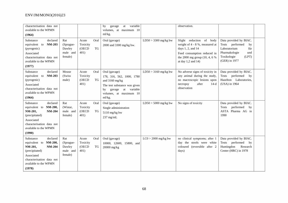
ENV/JM/MONO(2016)23
68
characterisation data not
available to the WPMN
(1964)
by gavage at variable
volumes, at maximum 10
ml/kg.
observation.
Substance declared
equivalent to NM-203
(pyrogenic)
Associated
characterisation data not
available to the WPMN
(1977)
Rat
(Sprague-
Dawley
male and
female)
Acute Oral
Toxicity
(OECD TG
401)
Oral (gavage)
2000 and 3300 mg/kg bw.
LD50 > 3300 mg/kg bw Slight reduction of body
weight of 4 - 8 %, measured at
days 1, 2, and 14
Feed consumption reduced in
the 2000 mg group (10, 4, 6 %
at day 1,2 and 14)
Data provided by BIAC.
Tests performed by
Laboratorium für
Pharmakologie und
Toxikologie (LPT)
(GER) in 1977
Substance declared
equivalent to NM-203
(pyrogenic)
Associated
characterisation data not
available to the WPMN
(1964)
Mouse
(Swiss
male)
Acute Oral
Toxicity
(OECD TG
401)
Oral (gavage)
178, 316, 562, 1000, 1780
and 3160 mg/kg
The test substance was given
by gavage at variable
volumes, at maximum 10
ml/kg.
LD50 > 3160 mg/kg bw No adverse signs of toxicity in
any animal during the study,
no macroscopic lesions upon
necropsy after 14-d
observation
Data provided by BIAC.
Tests performed by
Hazelton Laboratories,
(USA) in 1964
Substance declared
equivalent to NM-200,
NM-201, NM-204
(precipitated)
Associated
characterisation data not
available to the WPMN
(1990)
Rat
(Wistar,
male and
female)
Acute Oral
Toxicity
(OECD TG
401)
Oral (gavage)
Single administration
5110 mg/kg bw
237 mg/mL
LD50 > 5000 mg/kg bw No signs of toxicity Data provided by BIAC.
Tests performed by
ASTA Pharma AG in
1990
Substance declared
equivalent to NM-200,
NM-201, NM-204
(precipitated)
Associated
characterisation data not
available to the WPMN
(1978)
Rat
(Sprague-
Dawley
male and
female)
Acute Oral
Toxicity
(OECD TG
401)
Oral (gavage)
10000, 12600, 15800, and
20000 mg/kg
LC0 > 2000 mg/kg bw no clinical symptoms; after 1
day the stools were white
coloured (reversible after 2
days)
Data provided by BIAC.
Tests performed by
Huntingdon Research
Center (HRC) in 1978
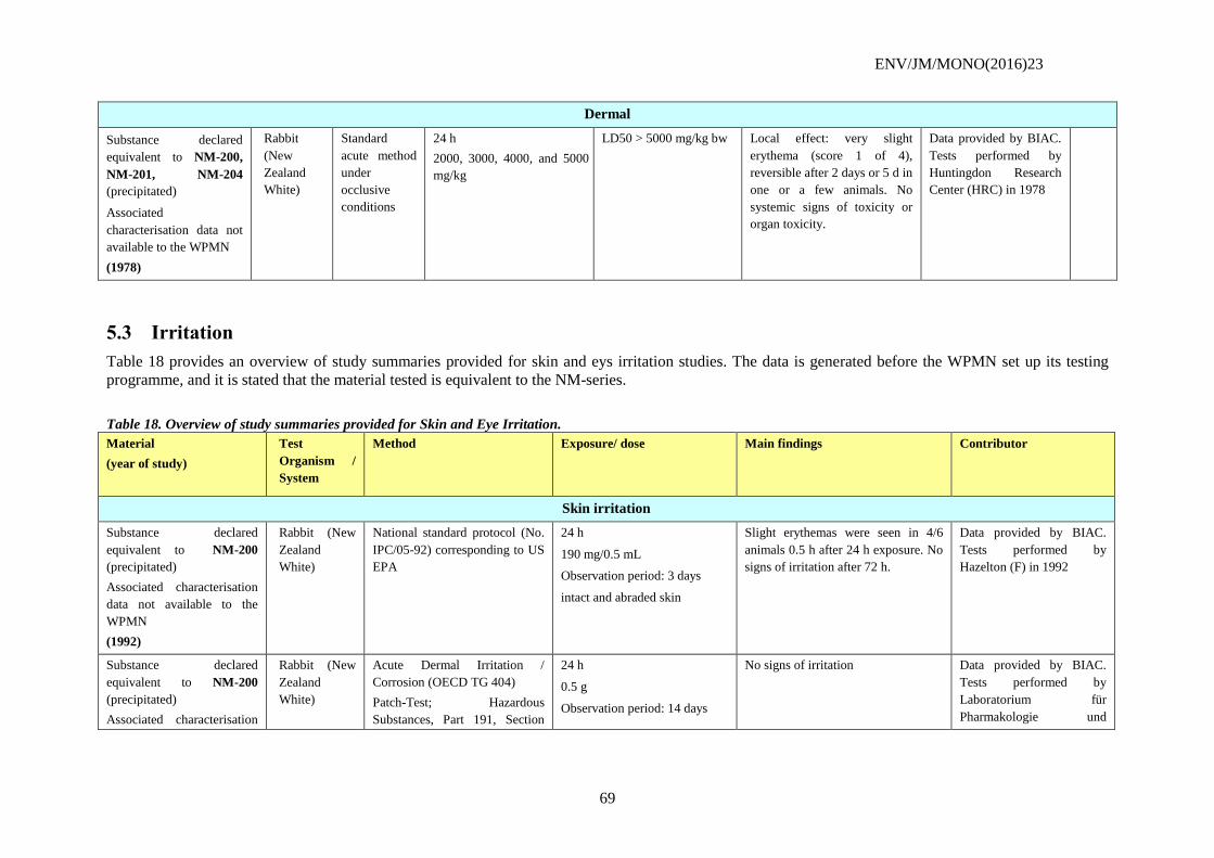
ENV/JM/MONO(2016)23
69
Dermal
Substance declared
equivalent to NM-200,
NM-201, NM-204
(precipitated)
Associated
characterisation data not
available to the WPMN
(1978)
Rabbit
(New
Zealand
White)
Standard
acute method
under
occlusive
conditions
24 h
2000, 3000, 4000, and 5000
mg/kg
LD50 > 5000 mg/kg bw Local effect: very slight
erythema (score 1 of 4),
reversible after 2 days or 5 d in
one or a few animals. No
systemic signs of toxicity or
organ toxicity.
Data provided by BIAC.
Tests performed by
Huntingdon Research
Center (HRC) in 1978
5.3 Irritation
Table 18 provides an overview of study summaries provided for skin and eys irritation studies. The data is generated before the WPMN set up its testing
programme, and it is stated that the material tested is equivalent to the NM-series.
Table 18. Overview of study summaries provided for Skin and Eye Irritation.
Material
(year of study)
Test
Organism /
System
Method Exposure/ dose Main findings Contributor
Skin irritation
Substance declared
equivalent to NM-200
(precipitated)
Associated characterisation
data not available to the
WPMN
(1992)
Rabbit (New
Zealand
White)
National standard protocol (No.
IPC/05-92) corresponding to US
EPA
24 h
190 mg/0.5 mL
Observation period: 3 days
intact and abraded skin
Slight erythemas were seen in 4/6
animals 0.5 h after 24 h exposure. No
signs of irritation after 72 h.
Data provided by BIAC.
Tests performed by
Hazelton (F) in 1992
Substance declared
equivalent to NM-200
(precipitated)
Associated characterisation
Rabbit (New
Zealand
White)
Acute Dermal Irritation /
Corrosion (OECD TG 404)
Patch-Test; Hazardous
Substances, Part 191, Section
24 h
0.5 g
Observation period: 14 days
No signs of irritation Data provided by BIAC.
Tests performed by
Laboratorium für
Pharmakologie und
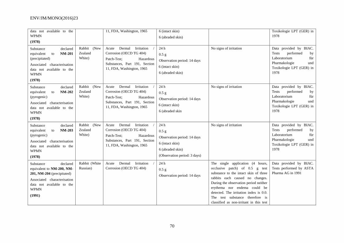
ENV/JM/MONO(2016)23
70
data not available to the
WPMN
(1978)
11, FDA, Washington, 1965 6 (intact skin)
6 (abraded skin)
Toxikologie LPT (GER) in
1978
Substance declared
equivalent to NM-201
(precipitated)
Associated characterisation
data not available to the
WPMN
(1978)
Rabbit (New
Zealand
White)
Acute Dermal Irritation /
Corrosion (OECD TG 404)
Patch-Test; Hazardous
Substances, Part 191, Section
11, FDA, Washington, 1965
24 h
0.5 g
Observation period: 14 days
6 (intact skin)
6 (abraded skin)
No signs of irritation Data provided by BIAC.
Tests performed by
Laboratorium für
Pharmakologie und
Toxikologie LPT (GER) in
1978
Substance declared
equivalent to NM-202
(pyrogenic)
Associated characterisation
data not available to the
WPMN
(1978)
Rabbit (New
Zealand
White)
Acute Dermal Irritation /
Corrosion (OECD TG 404)
Patch-Test; Hazardous
Substances, Part 191, Section
11, FDA, Washington, 1965
24 h
0.5 g
Observation period: 14 days
6 (intact skin)
6 (abraded skin
No signs of irritation Data provided by BIAC.
Tests performed by
Laboratorium für
Pharmakologie und
Toxikologie LPT (GER) in
1978
Substance declared
equivalent to NM-203
(pyrogenic)
Associated characterisation
data not available to the
WPMN
(1978)
Rabbit (New
Zealand
White)
Acute Dermal Irritation /
Corrosion (OECD TG 404)
Patch-Test; Hazardous
Substances, Part 191, Section
11, FDA, Washington, 1965
24 h
0.5 g
Observation period: 14 days
6 (intact skin)
6 (abraded skin)
(Observation period: 3 days)
No signs of irritation Data provided by BIAC.
Tests performed by
Laboratorium für
Pharmakologie und
Toxikologie LPT (GER) in
1978
Substance declared
equivalent to NM-200, NM-
201, NM-204 (precipitated)
Associated characterisation
data not available to the
WPMN
(1991)
Rabbit (White
Russian)
Acute Dermal Irritation /
Corrosion (OECD TG 404)
24 h
0.5 g
Observation period: 14 days
The single application (4 hours,
occlusive patch) of 0.5 g test
substance to the intact skin of three
rabbits each caused no changes.
During the observation period neither
erythema nor endema could be
detected. The irritation index is 0.0.
The test substance therefore is
classified as non-irritant in this test
Data provided by BIAC.
Tests performed by ASTA
Pharma AG in 1991
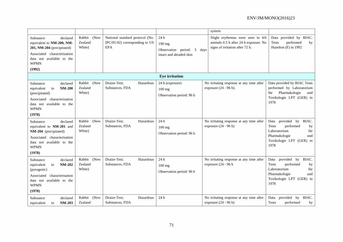
ENV/JM/MONO(2016)23
71
system
Substance declared
equivalent to NM-200, NM-
201, NM-204 (precipitated)
Associated characterisation
data not available to the
WPMN
(1992)
Rabbit (New
Zealand
White)
National standard protocol (No.
IPC/05-92) corresponding to US
EPA
24 h
190 mg
Observation period: 3 days
intact and abraded skin
Slight erythemas were seen in 4/6
animals 0.5 h after 24 h exposure. No
signs of irritation after 72 h.
Data provided by BIAC.
Tests performed by
Hazelton (F) in 1992
Eye irritation
Substance declared
equivalent to NM-200
(precipitated)
Associated characterisation
data not available to the
WPMN
(1978)
Rabbit (New
Zealand
White)
Draize-Test; Hazardous
Substances, FDA
24 h (exposure)
100 mg
Observation period: 96 h
No irritating response at any time after
exposure (24 - 96 h).
Data provided by BIAC Tests
performed by Laboratorium
für Pharmakologie und
Toxikologie LPT (GER) in
1978
Substance declared
equivalent to NM-201 and
NM-204 (precipitated)
Associated characterisation
data not available to the
WPMN
(1978)
Rabbit (New
Zealand
White)
Draize-Test; Hazardous
Substances, FDA
24 h
100 mg
Observation period: 96 h
No irritating response at any time after
exposure (24 - 96 h)
Data provided by BIAC.
Tests performed by
Laboratorium für
Pharmakologie und
Toxikologie LPT (GER) in
1978
Substance declared
equivalent to NM-202
(pyrogenic)
Associated characterisation
data not available to the
WPMN
(1978)
Rabbit (New
Zealand
White)
Draize-Test; Hazardous
Substances, FDA
24 h
100 mg
Observation period: 96 h
No irritating response at any time after
exposure (24 - 96 h
Data provided by BIAC.
Tests performed by
Laboratorium für
Pharmakologie und
Toxikologie LPT (GER) in
1978
Substance declared
equivalent to NM-203
Rabbit (New
Zealand
Draize-Test; Hazardous
Substances, FDA
24 h No irritating response at any time after
exposure (24 - 96 h)
Data provided by BIAC.
Tests performed by
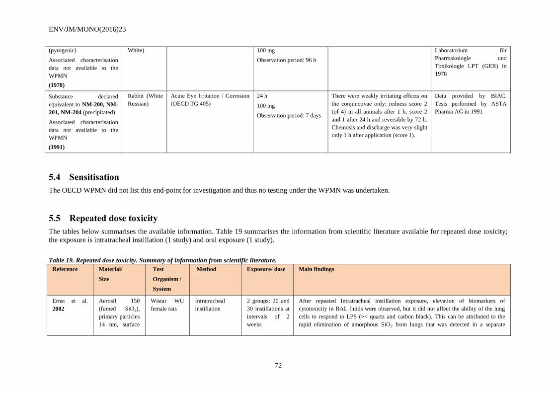
ENV/JM/MONO(2016)23
72
(pyrogenic)
Associated characterisation
data not available to the
WPMN
(1978)
White) 100 mg
Observation period: 96 h
Laboratorium für
Pharmakologie und
Toxikologie LPT (GER) in
1978
Substance declared
equivalent to NM-200, NM-
201, NM-204 (precipitated)
Associated characterisation
data not available to the
WPMN
(1991)
Rabbit (White
Russian)
Acute Eye Irritation / Corrosion
(OECD TG 405)
24 h
100 mg
Observation period: 7 days
There were weakly irritating effects on
the conjunctivae only: redness score 2
(of 4) in all animals after 1 h, score 2
and 1 after 24 h and reversible by 72 h.
Chemosis and discharge was very slight
only 1 h after application (score 1).
Data provided by BIAC.
Tests performed by ASTA
Pharma AG in 1991
5.4 Sensitisation
The OECD WPMN did not list this end-point for investigation and thus no testing under the WPMN was undertaken.
5.5 Repeated dose toxicity
The tables below summarises the available information. Table 19 summarises the information from scientific literature available for repeated dose toxicity;
the exposure is intratracheal instillation (1 study) and oral exposure (1 study).
Table 19. Repeated dose toxicity. Summary of information from scientific literature.
Reference Material/
Size
Test
Organism /
System
Method Exposure/ dose Main findings
Ernst et al.
2002
Aerosil 150
(fumed SiO2),
primary particles
14 nm, surface
Wistar WU
female rats
Intratracheal
instillation
2 groups: 20 and
30 instillations at
intervals of 2
weeks
After repeated Intratracheal instillation exposure, elevation of biomarkers of
cytotoxicity in BAL fluids were observed, but it did not affect the ability of the lung
cells to respond to LPS (>< quartz and carbon black). This can be attributed to the
rapid elimination of amorphous SiO2 from lungs that was detected in a separate
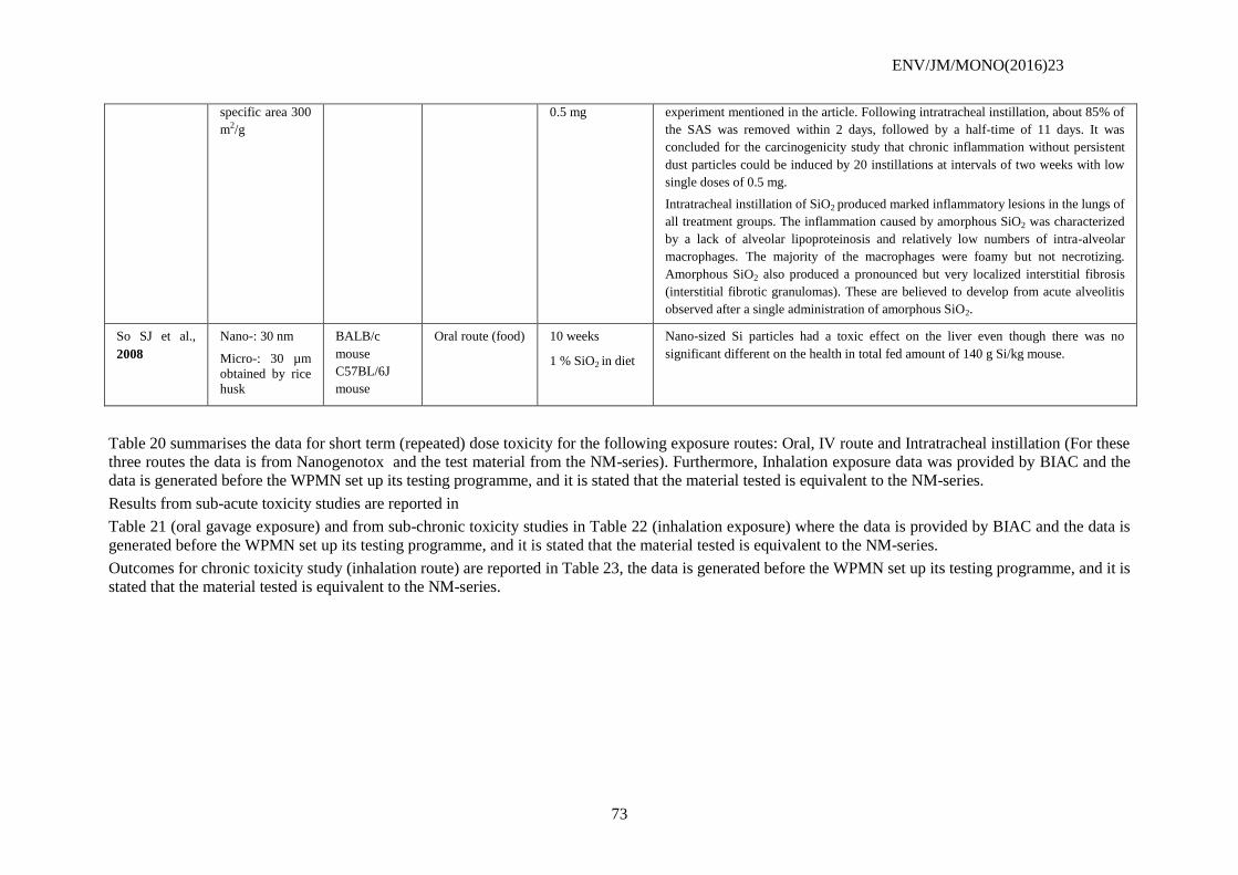
ENV/JM/MONO(2016)23
73
specific area 300
m2/g
0.5 mg experiment mentioned in the article. Following intratracheal instillation, about 85% of
the SAS was removed within 2 days, followed by a half-time of 11 days. It was
concluded for the carcinogenicity study that chronic inflammation without persistent
dust particles could be induced by 20 instillations at intervals of two weeks with low
single doses of 0.5 mg.
Intratracheal instillation of SiO2 produced marked inflammatory lesions in the lungs of
all treatment groups. The inflammation caused by amorphous SiO2 was characterized
by a lack of alveolar lipoproteinosis and relatively low numbers of intra-alveolar
macrophages. The majority of the macrophages were foamy but not necrotizing.
Amorphous SiO2 also produced a pronounced but very localized interstitial fibrosis
(interstitial fibrotic granulomas). These are believed to develop from acute alveolitis
observed after a single administration of amorphous SiO2.
So SJ et al.,
2008
Nano-: 30 nm
Micro-: 30 µm
obtained by rice
husk
BALB/c
mouse
C57BL/6J
mouse
Oral route (food) 10 weeks
1 % SiO2 in diet
Nano-sized Si particles had a toxic effect on the liver even though there was no
significant different on the health in total fed amount of 140 g Si/kg mouse.
Table 20 summarises the data for short term (repeated) dose toxicity for the following exposure routes: Oral, IV route and Intratracheal instillation (For these
three routes the data is from Nanogenotox and the test material from the NM-series). Furthermore, Inhalation exposure data was provided by BIAC and the
data is generated before the WPMN set up its testing programme, and it is stated that the material tested is equivalent to the NM-series.
Results from sub-acute toxicity studies are reported in
Table 21 (oral gavage exposure) and from sub-chronic toxicity studies in Table 22 (inhalation exposure) where the data is provided by BIAC and the data is
generated before the WPMN set up its testing programme, and it is stated that the material tested is equivalent to the NM-series.
Outcomes for chronic toxicity study (inhalation route) are reported in Table 23, the data is generated before the WPMN set up its testing programme, and it is
stated that the material tested is equivalent to the NM-series.
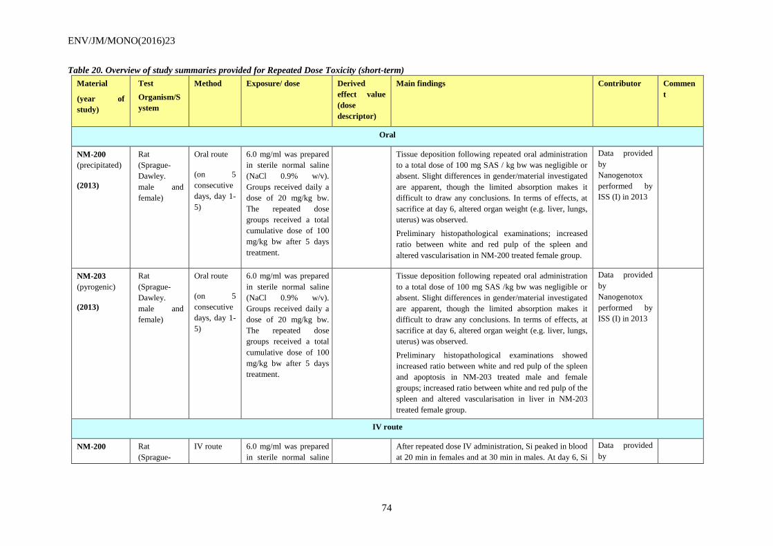
ENV/JM/MONO(2016)23
74
Table 20. Overview of study summaries provided for Repeated Dose Toxicity (short-term)
Material
(year of
study)
Test
Organism/S
ystem
Method Exposure/ dose Derived
effect value
(dose
descriptor)
Main findings Contributor Commen
t
Oral
NM-200
(precipitated)
(2013)
Rat
(Sprague-
Dawley.
male and
female)
Oral route
(on 5
consecutive
days, day 1-
5)
6.0 mg/ml was prepared
in sterile normal saline
(NaCl 0.9% w/v).
Groups received daily a
dose of 20 mg/kg bw.
The repeated dose
groups received a total
cumulative dose of 100
mg/kg bw after 5 days
treatment.
Tissue deposition following repeated oral administration
to a total dose of 100 mg SAS / kg bw was negligible or
absent. Slight differences in gender/material investigated
are apparent, though the limited absorption makes it
difficult to draw any conclusions. In terms of effects, at
sacrifice at day 6, altered organ weight (e.g. liver, lungs,
uterus) was observed.
Preliminary histopathological examinations; increased
ratio between white and red pulp of the spleen and
altered vascularisation in NM-200 treated female group.
Data provided
by
Nanogenotox
performed by
ISS (I) in 2013
NM-203
(pyrogenic)
(2013)
Rat
(Sprague-
Dawley.
male and
female)
Oral route
(on 5
consecutive
days, day 1-
5)
6.0 mg/ml was prepared
in sterile normal saline
(NaCl 0.9% w/v).
Groups received daily a
dose of 20 mg/kg bw.
The repeated dose
groups received a total
cumulative dose of 100
mg/kg bw after 5 days
treatment.
Tissue deposition following repeated oral administration
to a total dose of 100 mg SAS /kg bw was negligible or
absent. Slight differences in gender/material investigated
are apparent, though the limited absorption makes it
difficult to draw any conclusions. In terms of effects, at
sacrifice at day 6, altered organ weight (e.g. liver, lungs,
uterus) was observed.
Preliminary histopathological examinations showed
increased ratio between white and red pulp of the spleen
and apoptosis in NM-203 treated male and female
groups; increased ratio between white and red pulp of the
spleen and altered vascularisation in liver in NM-203
treated female group.
Data provided
by
Nanogenotox
performed by
ISS (I) in 2013
IV route
NM-200 Rat
(Sprague-
IV route 6.0 mg/ml was prepared
in sterile normal saline
After repeated dose IV administration, Si peaked in blood
at 20 min in females and at 30 min in males. At day 6, Si
Data provided
by
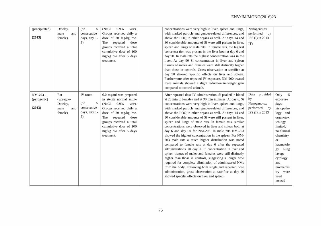
ENV/JM/MONO(2016)23
75
(precipitated)
(2013)
Dawley.
male and
female)
(on 5
consecutive
days, day 1-
5)
(NaCl 0.9% w/v).
Groups received daily a
dose of 20 mg/kg bw.
The repeated dose
groups received a total
cumulative dose of 100
mg/kg bw after 5 days
treatment.
concentrations were very high in liver, spleen and lungs,
with marked particle and gender-related differences, and
above the LOQ in other organs as well. At days 14 and
30 considerable amounts of Si were still present in liver,
spleen and lungs of male rats. In female rats, the highest
concentra-tion was present in the liver both at day 6 and
day 90. In male rats the highest concentration was in the
liver. At day 90 Si concentration in liver and spleen
tissues of males and females were still distinctly higher
than those in controls. Gross observation at sacrifice at
day 90 showed specific effects on liver and spleen.
Furthermore after repeated IV exposure, NM-200 treated
male animals showed a slight reduction in weight gain
compared to control animals.
Nanogenotox
performed by
ISS (I) in 2013
IT)
NM-203
(pyrogenic)
(2013)
Rat
(Sprague-
Dawley,
male and
female)
IV route
(on 5
consecutive
days, day 1-
5)
6.0 mg/ml was prepared
in sterile normal saline
(NaCl 0.9% w/v).
Groups received daily a
dose of 20 mg/kg bw.
The repeated dose
groups received a total
cumulative dose of 100
mg/kg bw after 5 days
treatment.
After repeated dose IV administration, Si peaked in blood
at 20 min in females and at 30 min in males. At day 6, Si
concentrations were very high in liver, spleen and lungs,
with marked particle and gender-related differences, and
above the LOQ in other organs as well. At days 14 and
30 considerable amounts of Si were still present in liver,
spleen and lungs of male rats. In female rats, similar
concentrations were observed in liver and spleen both at
day 6 and day 90 for NM-203. In male rats NM-203
showed the highest concentration in the spleen. For NM-
203 male rats a much higher distribution was noted
compared to female rats at day 6 after the repeated
administrations. At day 90 Si concentration in liver and
spleen tissues of males and females were still distinctly
higher than those in controls, suggesting a longer time
required for complete elimination of administered NMs
from the body. Following both single and repeated dose
administration, gross observation at sacrifice at day 90
showed specific effects on liver and spleen.
Data provided
by
Nanogenotox
performed by
ISS (I) in 2013
Only 5
exposure
days;
histopatho
logy and
organotox
icology
limited;
no clinical
chemistry
or
haematolo
gy. Lung
lavage
cytology
and
biochemis
try were
used
instead
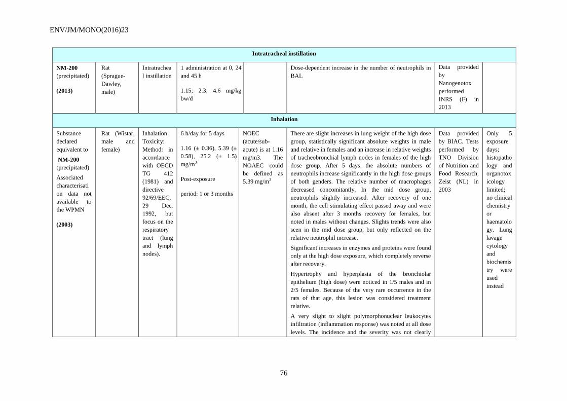
ENV/JM/MONO(2016)23
76
Intratracheal instillation
NM-200
(precipitated)
(2013)
Rat
(Sprague-
Dawley,
male)
Intratrachea
l instillation
1 administration at 0, 24
and 45 h
1.15; 2.3; 4.6 mg/kg
bw/d
Dose-dependent increase in the number of neutrophils in
BAL
Data provided
by
Nanogenotox
performed
INRS (F) in
2013
Inhalation
Substance
declared
equivalent to
NM-200
(precipitated)
Associated
characterisati
on data not
available to
the WPMN
(2003)
Rat (Wistar,
male and
female)
Inhalation
Toxicity:
Method: in
accordance
with OECD
TG 412
(1981) and
directive
92/69/EEC,
29 Dec.
1992, but
focus on the
respiratory
tract (lung
and lymph
nodes).
6 h/day for 5 days
1.16 (± 0.36), 5.39 (±
0.58), 25.2 (± 1.5)
mg/m3
Post-exposure
period: 1 or 3 months
NOEC
(acute/sub-
acute) is at 1.16
mg/m3. The
NOAEC could
be defined as
5.39 mg/m3
There are slight increases in lung weight of the high dose
group, statistically significant absolute weights in male
and relative in females and an increase in relative weights
of tracheobronchial lymph nodes in females of the high
dose group. After 5 days, the absolute numbers of
neutrophils increase significantly in the high dose groups
of both genders. The relative number of macrophages
decreased concomitantly. In the mid dose group,
neutrophils slightly increased. After recovery of one
month, the cell stimulating effect passed away and were
also absent after 3 months recovery for females, but
noted in males without changes. Slights trends were also
seen in the mid dose group, but only reflected on the
relative neutrophil increase.
Significant increases in enzymes and proteins were found
only at the high dose exposure, which completely reverse
after recovery.
Hypertrophy and hyperplasia of the bronchiolar
epithelium (high dose) were noticed in 1/5 males and in
2/5 females. Because of the very rare occurrence in the
rats of that age, this lesion was considered treatment
relative.
A very slight to slight polymorphonuclear leukocytes
infiltration (inflammation response) was noted at all dose
levels. The incidence and the severity was not clearly
Data provided
by BIAC. Tests
performed by
TNO Division
of Nutrition and
Food Research,
Zeist (NL) in
2003
Only 5
exposure
days;
histopatho
logy and
organotox
icology
limited;
no clinical
chemistry
or
haematolo
gy. Lung
lavage
cytology
and
biochemis
try were
used
instead
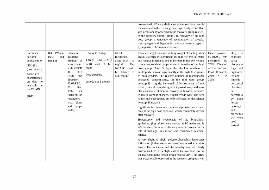
ENV/JM/MONO(2016)23
77
dose-related, 1/5 very slight case at the low dose level in
the male and in the female group respectively. This effect
was occasionally observed in the recovery group (as well
in the recovery control group). In recovery of the high
dose group, a tendency of accumulation of alveolar
macrophages and hyperemic capillary unusual type II
hyperplasia in 1/5 males were noted.
Substance
declared
equivalent to
NM-201
(precipitated)
Associated
characterisati
on data not
available to
the WPMN
(2003)
Rat (Wistar
male and
female)
Inhalation
Toxicity:
Method: in
accordance
with OECD
TG 412
(1981) and
directive
92/69/EEC,
29 Dec.
1992, but
focus on the
respiratory
tract (lung
and lymph
nodes).
6 h/day for 5 days
1.16 (± 0.36), 5.39 (±
0.58), 25.2 (± 1.5)
mg/m3
Post-exposure
period: 1 or 3 months
NOEC
(acute/sub-
acute) is at 1.16
mg/m3. The
NOAEC could
be defined as
5.39 mg/m3
There are slight increases in lung weight of the high dose
group, statistically significant absolute weights in males
and relative in females and an increase in relative weights
of tracheobronchial lymph nodes in females of the high
dose group. After 5 days, the absolute numbers of
neutrophils increase significantly in the high dose groups
of both genders. The relative number of macrophages
decreased concomitantly. In the mid dose group,
neutrophils slightly increased. After recovery of one
month, the cell stimulating effect passed away and were
also absent after 3 months recovery on females, but noted
in males without changes. Slights trends were also seen
in the mid dose group, but only reflected on the relative
neutrophil increase.
Significant increases in enzymes and proteins were found
only at the high dose exposure, which completely reverse
after recovery.
Hypertrophy and hyperplasia of the bronchiolar
epithelium (high dose) were noticed in 1/5 males and in
2/5 females. Because of the very rare occurrence in the
rats of that age, this lesion was considered treatment
relative.
A very slight to slight polymorphonuclear leukocytes
infiltration (inflammation response) was noted at all dose
levels. The incidence and the severity was not clearly
dose-related, 1/5 very slight case at the low dose level in
the male and in the female group respectively. This effect
was occasionally observed in the recovery group (as well
Data provided
by BIAC. Tests
performed by
TNO Division
of Nutrition and
Food Research,
Zeist (NL) in
2003
Only 5
exposure
days;
histopatho
logy and
organotox
icology
limited;
no clinical
chemistry
or
haematolo
gy. Lung
lavage
cytology
and
biochemis
try were
used
instead
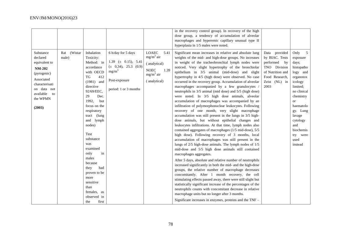
ENV/JM/MONO(2016)23
78
in the recovery control group). In recovery of the high
dose group, a tendency of accumulation of alveolar
macrophages and hyperemic capillary unusual type II
hyperplasia in 1/5 males were noted.
Substance
declared
equivalent to
NM-202
(pyrogenic)
Associated
characterisati
on data not
available to
the WPMN
(2003)
Rat (Wistar
male)
Inhalation
Toxicity:
Method: in
accordance
with OECD
TG 412
(1981) and
directive
92/69/EEC,
29 Dec.
1992, but
focus on the
respiratory
tract (lung
and lymph
nodes)
Test
substance
was
examined
only in
males
because
they had
proven to be
more
sensitive
than
females, as
observed in
the first
6 h/day for 5 days
1.39 (± 0.15), 5.41
(± 0,34), 25.3 (0.9)
mg/m3
Post-exposure
period: 1 or 3 months
LOAEC 5.41
mg/m3 air
( analytical)
NOEC 1.39
mg/m3 air
( analytical)
Significant mean increases in relative and absolute lung
weights of the mid- and high-dose groups. No increases
in weight of the tracheobronchial lymph nodes were
noticed. Very slight hypertrophy of the bronchiolar
epithelium in 3/5 animal (mid-dose) and slight
hypertrophy in 4/5 (high dose) were observed. No case
occurred in the recovery group. Accumulation of alveolar
macrophages accompanied by a few granulocytes /
neutrophils in 3/5 animal (mid dose) and 5/5 (high dose)
were noted. In 3/5 high dose animals, alveolar
accumulation of macrophages was accompanied by an
infiltration of polymorphonuclear leukocytes. Following
recovery of one month, very slight macrophage
accumulation was still present in the lungs in 3/5 high-
dose animals, but without epithelial changes and
leukocytes infiltrations. At that time, lymph nodes also
contained aggregates of macrophages (1/5 mid-dose), 5/5
high dose). Following recovery of 3 months, local
accumulation of macrophages was still present in the
lungs of 2/5 high-dose animals. The lymph nodes of 1/5
mid-dose and 5/5 high dose animals still contained
macrophages aggregates.
After 5 days, absolute and relative number of neutrophils
increased significantly in both the mid- and the high-dose
groups, the relative number of macrophage decreases
concomitantly. After 1 month recovery, the cell
stimulating effects passed away, there were still slight but
statistically significant increase of the percentages of the
neutrophils counts with concomitant decrease in relative
macrophage units but no longer after 3 months.
Significant increases in enzymes, proteins and the TNF –
Data provided
by BIAC. Tests
performed by
TNO Division
of Nutrition and
Food Research,
Zeist (NL) in
2003
Only 5
exposure
days;
histopatho
logy and
organotox
icology
limited;
no clinical
chemistry
or
haematolo
gy. Lung
lavage
cytology
and
biochemis
try were
used
instead
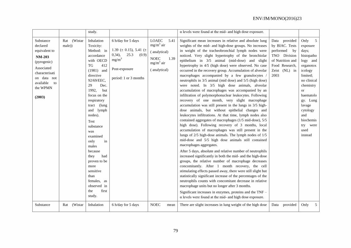
ENV/JM/MONO(2016)23
79
study. α levels were found at the mid- and high dose exposure.
Substance
declared
equivalent to
NM-203
(pyrogenic)
Associated
characterisati
on data not
available to
the WPMN
(2003)
Rat (Wistar
male))
Inhalation
Toxicity:
Method: in
accordance
with OECD
TG 412
(1981) and
directive
92/69/EEC,
29 Dec.
1992, but
focus on the
respiratory
tract (lung
and lymph
nodes).
Test
substance
was
examined
only in
males
because
they had
proven to be
more
sensitive
than
females, as
observed in
the first
study.
6 h/day for 5 days
1.39 (± 0.15), 5.41 (±
0,34), 25.3 (0.9)
mg/m3
Post-exposure
period: 1 or 3 months
LOAEC 5.41
mg/m3 air
( analytical)
NOEC 1.39
mg/m3 air
( analytical)
Significant mean increases in relative and absolute lung
weights of the mid- and high-dose groups. No increases
in weight of the tracheobronchial lymph nodes were
noticed. Very slight hypertrophy of the bronchiolar
epithelium in 3/5 animal (mid-dose) and slight
hypertrophy in 4/5 (high dose) were observed. No case
occurred in the recovery group. Accumulation of alveolar
macrophages accompanied by a few granulocytes /
neutrophils in 3/5 animal (mid dose) and 5/5 (high dose)
were noted. In 3/5 high dose animals, alveolar
accumulation of macrophages was accompanied by an
infiltration of polymorphonuclear leukocytes. Following
recovery of one month, very slight macrophage
accumulation was still present in the lungs in 3/5 high-
dose animals, but without epithelial changes and
leukocytes infiltrations. At that time, lymph nodes also
contained aggregates of macrophages (1/5 mid-dose), 5/5
high dose). Following recovery of 3 months, local
accumulation of macrophages was still present in the
lungs of 2/5 high-dose animals. The lymph nodes of 1/5
mid-dose and 5/5 high dose animals still contained
macrophages aggregates.
After 5 days, absolute and relative number of neutrophils
increased significantly in both the mid- and the high-dose
groups, the relative number of macrophage decreases
concomitantly. After 1 month recovery, the cell
stimulating effects passed away, there were still slight but
statistically significant increase of the percentages of the
neutrophils counts with concomitant decrease in relative
macrophage units but no longer after 3 months.
Significant increases in enzymes, proteins and the TNF –
α levels were found at the mid- and high dose exposure.
Data provided
by BIAC. Tests
performed by
TNO Division
of Nutrition and
Food Research,
Zeist (NL) in
2003
Only 5
exposure
days;
histopatho
logy and
organotox
icology
limited;
no clinical
chemistry
or
haematolo
gy. Lung
lavage
cytology
and
biochemis
try were
used
instead
Substance Rat (Wistar Inhalation 6 h/day for 5 days NOEC mean There are slight increases in lung weight of the high dose Data provided Only 5
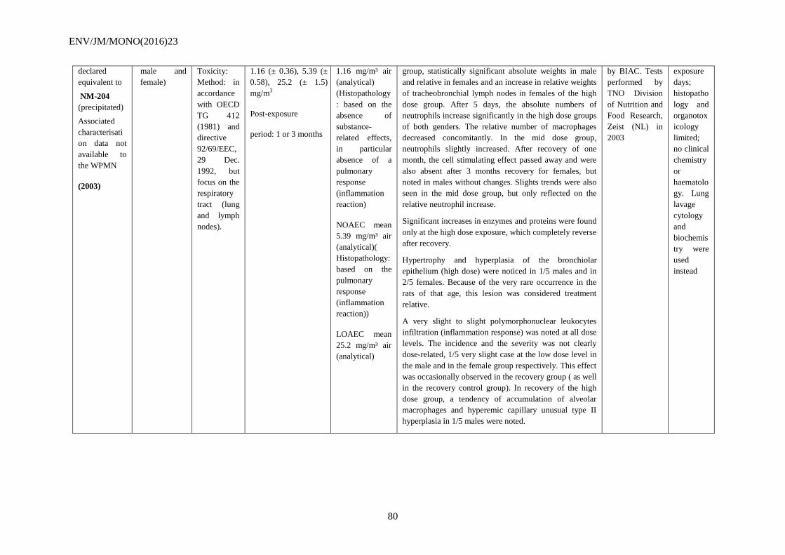
ENV/JM/MONO(2016)23
80
declared
equivalent to
NM-204
(precipitated)
Associated
characterisati
on data not
available to
the WPMN
(2003)
male and
female)
Toxicity:
Method: in
accordance
with OECD
TG 412
(1981) and
directive
92/69/EEC,
29 Dec.
1992, but
focus on the
respiratory
tract (lung
and lymph
nodes).
1.16 (± 0.36), 5.39 (±
0.58), 25.2 (± 1.5)
mg/m3
Post-exposure
period: 1 or 3 months
1.16 mg/m³ air
(analytical)
(Histopathology
: based on the
absence of
substance-
related effects,
in particular
absence of a
pulmonary
response
(inflammation
reaction)
NOAEC mean
5.39 mg/m³ air
(analytical)(
Histopathology:
based on the
pulmonary
response
(inflammation
reaction))
LOAEC mean
25.2 mg/m³ air
(analytical)
group, statistically significant absolute weights in male
and relative in females and an increase in relative weights
of tracheobronchial lymph nodes in females of the high
dose group. After 5 days, the absolute numbers of
neutrophils increase significantly in the high dose groups
of both genders. The relative number of macrophages
decreased concomitantly. In the mid dose group,
neutrophils slightly increased. After recovery of one
month, the cell stimulating effect passed away and were
also absent after 3 months recovery for females, but
noted in males without changes. Slights trends were also
seen in the mid dose group, but only reflected on the
relative neutrophil increase.
Significant increases in enzymes and proteins were found
only at the high dose exposure, which completely reverse
after recovery.
Hypertrophy and hyperplasia of the bronchiolar
epithelium (high dose) were noticed in 1/5 males and in
2/5 females. Because of the very rare occurrence in the
rats of that age, this lesion was considered treatment
relative.
A very slight to slight polymorphonuclear leukocytes
infiltration (inflammation response) was noted at all dose
levels. The incidence and the severity was not clearly
dose-related, 1/5 very slight case at the low dose level in
the male and in the female group respectively. This effect
was occasionally observed in the recovery group ( as well
in the recovery control group). In recovery of the high
dose group, a tendency of accumulation of alveolar
macrophages and hyperemic capillary unusual type II
hyperplasia in 1/5 males were noted.
by BIAC. Tests
performed by
TNO Division
of Nutrition and
Food Research,
Zeist (NL) in
2003
exposure
days;
histopatho
logy and
organotox
icology
limited;
no clinical
chemistry
or
haematolo
gy. Lung
lavage
cytology
and
biochemis
try were
used
instead
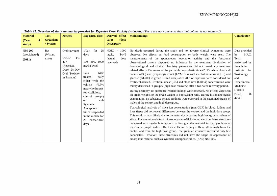
ENV/JM/MONO(2016)23
81
Table 21. Overview of study summaries provided for Repeated Dose Toxicity (subacute) (There are not comments thus that column is not included)
Material
(Year of
study)
Test
Organism
/ System
Method Exposure/ dose Derived effect
value (dose
descriptor)
Main findings Contributor
NM-200
(precipitated)
(2011)
Rat
(Wistar,
male)
Oral (gavage)
OECD TG
407
(Repeated
Dose 28-Day
Oral Toxicity
in Rodents)
1/day for 28
days
100, 300, 1000
mg/kg bw/d
Rats were
treated daily
either with the
vehicle (0.5%
methylhydroxyp
ropylcellulose,
control groups)
or with
Synthetic
Amorphous
Silica suspended
in the vehicle for
28 consecutive
days.
NOEL = 1000
mg/kg bw/d
(actual dose
received)
No death occurred during the study and no adverse clinical symptoms were
observed. No effects on food consumption or body weight were seen. The
measurements of the spontaneous locomotor activity and the functional
observational battery displayed no influence by the treatment. Evaluation of
haematological and clinical chemistry parameters did not reveal any treatment
related effects. Decreases of the partial thromboplastin time (PTT), white blood cell
count (WBC) and lymphocyte count (LYMC) as well as cholinesterase (CHE) and
glucose (GLUC) in group 3 (mid dose) after 28 d of exposure were considered not
treatment-related. Creatinin kinase (CK) and blood urea (UREA) concentration were
mildly decreased in group 6 (high dose recovery) after a two week recovery period.
During necropsy, no substance-related findings were observed. No effects were seen
on organ weights or the organ weight to bodyweight ratio. During histopathological
examination, no substance-related findings were observed in the examined organs of
males of the control and high dose group.
Toxicological analysis of silica ion concentration (non-GLP) in blood, kidney and
liver tissue did not reveal differences between the control and the high dose group.
This result is most likely due to the naturally occurring high background values of
silica. Transmission electron microscopy (non-GLP) found electron dense structures
composed of irregular homogenous to fine granular material in the cytoplasm of
mesenteric lymph nodes cells, liver cells and kidney cells of all animals from the
control and from the high dose group. The granular structures measured only few
nanometers. However, these structures did not have the shape or appearance of
amorphous material such as synthetic amorphous silica, (SAS) NM-200.
Data provided
by BIAC.
Tests
performed by
Fraunhofer
Institute for
Toxicology
and
Experimental
Medicine
(ITEM)
(GER) in
2011
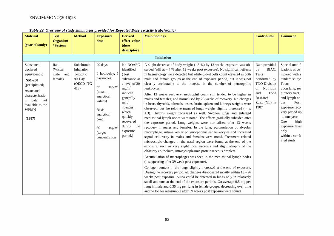
ENV/JM/MONO(2016)23
82
Table 22. Overview of study summaries provided for Repeated Dose Toxicity (subchronic)
Material
(year of study)
Test
Organism
/ System
Method Exposure/
dose
Derived
effect value
(dose
descriptor)
Main findings Contributor Comment
Inhalation
Substance
declared
equivalent to
NM-200
(precipitated)
Associated
characterisatio
n data not
available to the
WPMN
(1987)
Rat
(Wistar,
male and
female)
Subchronic
Inhalation
Toxicity:
90-Day
(OECD TG
413)
90 days
6 hours/day, 5
days/week
35 mg/m3
(mean
analytical
values)
Basis
analytical
conc.
30 mg/m3
(target
concentration
No NOAEC
identified
(Test
substance at
a level of 30
mg/m3
induced
generally
mild
changes,
which
quickly
recovered
during the
exposure
period.)
A slight decrease of body weight (- 5 %) by 13 weeks exposure was ob-
served (still at – 4 % after 52 weeks post exposure). No significant effects
in haematology were detected but white blood cells count elevated in both
male and female groups at the end of exposure period, but it was not
clear-ly attributable to the increase in the number of neutrophilic
leukocytes.
After 13 weeks recovery, neutrophil count still tended to be higher in
males and females, and normalised by 28 weeks of recovery. No changes
in heart, thyroids, adrenals, testes, brain, spleen and kidneys weights were
observed, but the relative mean of lungs weighs slightly increased ( ≈ x
1.3). Thymus weight increased as well. Swollen lungs and enlarged
mediastinal lymph nodes were noted. The effects gradually subsided after
the exposure period. Lung weights were normalised after 13 weeks
recovery in males and females. In the lung, accumulation of alveolar
macrophage, intra-alveolar polymorphonuclear leukocytes and increased
septal cellurarity in males and females were noted. Treatment related
microscopic changes in the nasal region were found at the end of the
exposure, such as very slight local necrosis and slight atrophy of the
olfactory epithelium, intracystoplasmic proteinaeceous droplets.
Accumulation of macrophages was seen in the mediastinal lymph nodes
(disappearing after 39 week post exposure).
Collagen content in the lungs slightly increased at the end of exposure.
During the recovery period, all changes disappeared mostly within 13 - 26
weeks post exposure. Silica could be detected in lungs only in relatively
small amounts at the end of the exposure periods. On average 0.5 mg per
lung in male and 0.35 mg per lung in female groups, decreasing over time
and no longer measurable after 39 weeks post exposure were found.
Data provided
by BIAC.
Tests
performed by
TNO Division
of Nutrition
and Food
Research,
Zeist (NL) in
1987
Special modif
ications as co
mpared with s
tandard study:
Focus
upon lung, res
piratory tract,
and lymph no
des. Post-
exposure reco
very period up
to one year.
One high
exposure level
only
within a comb
ined study
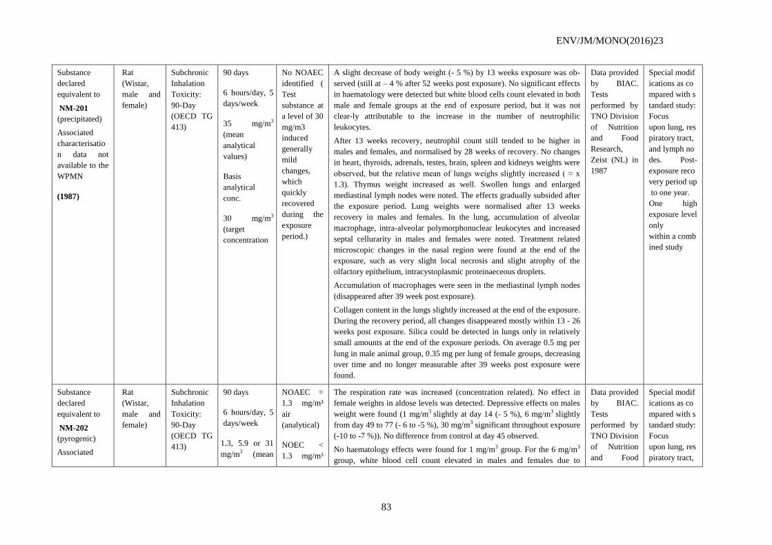
ENV/JM/MONO(2016)23
83
Substance
declared
equivalent to
NM-201
(precipitated)
Associated
characterisatio
n data not
available to the
WPMN
(1987)
Rat
(Wistar,
male and
female)
Subchronic
Inhalation
Toxicity:
90-Day
(OECD TG
413)
90 days
6 hours/day, 5
days/week
35 mg/m3
(mean
analytical
values)
Basis
analytical
conc.
30 mg/m3
(target
concentration
No NOAEC
identified (
Test
substance at
a level of 30
mg/m3
induced
generally
mild
changes,
which
quickly
recovered
during the
exposure
period.)
A slight decrease of body weight (- 5 %) by 13 weeks exposure was ob-
served (still at – 4 % after 52 weeks post exposure). No significant effects
in haematology were detected but white blood cells count elevated in both
male and female groups at the end of exposure period, but it was not
clear-ly attributable to the increase in the number of neutrophilic
leukocytes.
After 13 weeks recovery, neutrophil count still tended to be higher in
males and females, and normalised by 28 weeks of recovery. No changes
in heart, thyroids, adrenals, testes, brain, spleen and kidneys weights were
observed, but the relative mean of lungs weighs slightly increased ( ≈ x
1.3). Thymus weight increased as well. Swollen lungs and enlarged
mediastinal lymph nodes were noted. The effects gradually subsided after
the exposure period. Lung weights were normalised after 13 weeks
recovery in males and females. In the lung, accumulation of alveolar
macrophage, intra-alveolar polymorphonuclear leukocytes and increased
septal cellurarity in males and females were noted. Treatment related
microscopic changes in the nasal region were found at the end of the
exposure, such as very slight local necrosis and slight atrophy of the
olfactory epithelium, intracystoplasmic proteinaeceous droplets.
Accumulation of macrophages were seen in the mediastinal lymph nodes
(disappeared after 39 week post exposure).
Collagen content in the lungs slightly increased at the end of the exposure.
During the recovery period, all changes disappeared mostly within 13 - 26
weeks post exposure. Silica could be detected in lungs only in relatively
small amounts at the end of the exposure periods. On average 0.5 mg per
lung in male animal group, 0.35 mg per lung of female groups, decreasing
over time and no longer measurable after 39 weeks post exposure were
found.
Data provided
by BIAC.
Tests
performed by
TNO Division
of Nutrition
and Food
Research,
Zeist (NL) in
1987
Special modif
ications as co
mpared with s
tandard study:
Focus
upon lung, res
piratory tract,
and lymph no
des. Post-
exposure reco
very period up
to one year.
One high
exposure level
only
within a comb
ined study
Substance
declared
equivalent to
NM-202
(pyrogenic)
Associated
Rat
(Wistar,
male and
female)
Subchronic
Inhalation
Toxicity:
90-Day
(OECD TG
413)
90 days
6 hours/day, 5
days/week
1.3, 5.9 or 31
mg/m3 (mean
NOAEC =
1.3 mg/m³
air
(analytical)
NOEC <
1.3 mg/m³
The respiration rate was increased (concentration related). No effect in
female weights in aldose levels was detected. Depressive effects on males
weight were found (1 mg/m3 slightly at day 14 (- 5 %), 6 mg/m3 slightly
from day 49 to 77 (- 6 to -5 %), 30 mg/m3 significant throughout exposure
(-10 to -7 %)). No difference from control at day 45 observed.
No haematology effects were found for 1 mg/m3 group. For the 6 mg/m3
group, white blood cell count elevated in males and females due to
Data provided
by BIAC.
Tests
performed by
TNO Division
of Nutrition
and Food
Special modif
ications as co
mpared with s
tandard study:
Focus
upon lung, res
piratory tract,
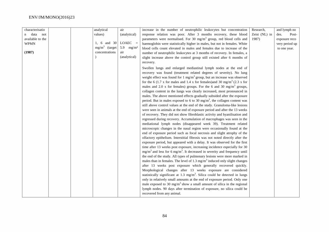
ENV/JM/MONO(2016)23
84
characterisatio
n data not
available to the
WPMN
(1987)
analytical
values)
1, 6 and 30
mg/m3 (target
concentrations
)
air
(analytical)
LOAEC =
5.9 mg/m³
air
(analytical)
increase in the number of neutrophilic leukocytes but concentration
response relation was poor. After 3 months recovery, these blood
parameters were normalised. For 30 mg/m3 group, red blood cells and
haemoglobin were statistically higher in males, but not in females. White
blood cells count elevated in males and females due to increase of the
number of neutrophilic leukocytes at 3 months of recovery. In females, a
slight increase above the control group still existed after 6 months of
recovery.
Swollen lungs and enlarged mediastinal lymph nodes at the end of
recovery was found (treatment related degrees of severity). No lung
weight effect was found for 1 mg/m3 group, but an increase was observed
for the 6 (1.7 x for males and 1.4 x for females)and 30 mg/m3 (2.3 x for
males and 2.0 x for females) groups. For the 6 and 30 mg/m3 groups,
collagen content in the lungs was clearly increased, most pronounced in
males. The above mentioned effects gradually subsided after the exposure
period. But in males exposed to 6 to 30 mg/m3, the collagen content was
still above control values at the end of the study. Granuloma-like lesions
were seen in animals at the end of exposure period and after the 13 weeks
of recovery. They did not show fibroblastic activity and hyanilisation and
regressed during recovery. Accumulation of macrophages was seen in the
mediational lymph nodes (disappeared week 39). Treatment related
microscopic changes in the nasal region were occasionally found at the
end of exposure period such as focal necrosis and slight atrophy of the
olfactory epithelium. Interstitial fibrosis was not noted directly after the
exposure period, but appeared with a delay. It was observed for the first
time after 13 weeks post exposure, increasing incidence especially for 30
mg/m3 and less for 6 mg/m3. It decreased in severity and frequency until
the end of the study. All types of pulmonary lesions were more marked in
males than in females. The level of 1.3 mg/m3 induced only slight changes
after 13 weeks post exposure which generally recovered quickly.
Morphological changes after 13 weeks exposure are considered
statistically significant at 1.3 mg/m3. Silica could be detected in lungs
only in relatively small amounts at the end of exposure period. Only one
male exposed to 30 mg/m3 show a small amount of silica in the regional
lymph nodes. 90 days after termination of exposure, no silica could be
recovered from any animal.
Research,
Zeist (NL) in
1987)
and lymph no
des. Post-
exposure reco
very period up
to one year.
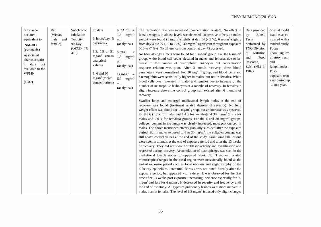
ENV/JM/MONO(2016)23
85
Substance
declared
equivalent to
NM-203
(pyrogenic)
Associated
characterisatio
n data not
available to the
WPMN
(1987)
Rat
(Wistar,
male and
female)
Subchronic
Inhalation
Toxicity:
90-Day
(OECD TG
413)
90 days
6 hours/day, 5
days/week
1.3, 5.9 or 31
mg/m3 (mean
analytical
values)
1, 6 and 30
mg/m3 (target
concentrations)
NOAEC =
1.3 mg/m³
air
(analytical)
NOEC <
1.3 mg/m³
air
(analytical)
LOAEC =
5.9 mg/m³
air
(analytical)
The respiration rate was increased (concentration related). No effect in
female weights in aldose levels was detected. Depressive effects on males
weight were found (1 mg/m3 slightly at day 14 (- 5 %), 6 mg/m3 slightly
from day 49 to 77 (- 6 to -5 %), 30 mg/m3 significant throughout exposure
(-10 to -7 %)). No difference from control at day 45 observed.
No haematology effects were found for 1 mg/m3 group. For the 6 mg/m3
group, white blood cell count elevated in males and females due to in-
crease in the number of neutrophilic leukocytes but concentration
response relation was poor. After 3 month recovery, these blood
parameters were normalised. For 30 mg/m3 group, red blood cells and
haemoglobin were statistically higher in males, but not in females. White
blood cells count elevated in males and females due to increase of the
number of neutrophilic leukocytes at 3 months of recovery. In females, a
slight increase above the control group still existed after 6 months of
recovery.
Swollen lungs and enlarged mediastinal lymph nodes at the end of
recovery was found (treatment related degrees of severity). No lung
weight effect was found for 1 mg/m3 group, but an increase was observed
for the 6 (1.7 x for males and 1.4 x for females)and 30 mg/m3 (2.3 x for
males and 2.0 x for females) groups. For the 6 and 30 mg/m3 groups,
collagen content in the lungs was clearly increased, most pronounced in
males. The above mentioned effects gradually subsided after the exposure
period. But in males exposed to 6 to 30 mg/m3, the collagen content was
still above control values at the end of the study. Granuloma like lesions
were seen in animals at the end of exposure period and after the 13 weeks
of recovery. They did not show fibroblastic activity and hyanilisation and
regressed during recovery. Accumulation of macrophages was seen in the
mediational lymph nodes (disappeared week 39). Treatment related
microscopic changes in the nasal region were occasionally found at the
end of exposure period such as focal necrosis and slight atrophy of the
olfactory epithelium. Interstitial fibrosis was not noted directly after the
exposure period, but appeared with a delay. It was observed for the first
time after 13 weeks post exposure, increasing incidence especially for 30
mg/m3 and less for 6 mg/m3. It decreased in severity and frequency until
the end of the study. All types of pulmonary lesions were more marked in
males than in females. The level of 1.3 mg/m3 induced only slight changes
Data provided
by BIAC.
Tests
performed by
TNO Division
of Nutrition
and Food
Research,
Zeist (NL) in
1987)
Special modif
ications as co
mpared with s
tandard study:
Focus
upon lung, res
piratory tract,
and
lymph nodes.
Post-
exposure reco
very period up
to one year.
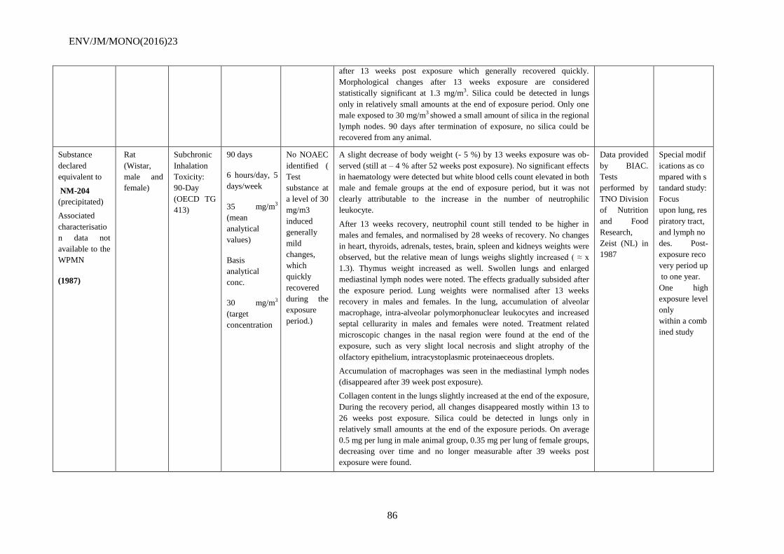
ENV/JM/MONO(2016)23
86
after 13 weeks post exposure which generally recovered quickly.
Morphological changes after 13 weeks exposure are considered
statistically significant at 1.3 mg/m3. Silica could be detected in lungs
only in relatively small amounts at the end of exposure period. Only one
male exposed to 30 mg/m3 showed a small amount of silica in the regional
lymph nodes. 90 days after termination of exposure, no silica could be
recovered from any animal.
Substance
declared
equivalent to
NM-204
(precipitated)
Associated
characterisatio
n data not
available to the
WPMN
(1987)
Rat
(Wistar,
male and
female)
Subchronic
Inhalation
Toxicity:
90-Day
(OECD TG
413)
90 days
6 hours/day, 5
days/week
35 mg/m3
(mean
analytical
values)
Basis
analytical
conc.
30 mg/m3
(target
concentration
No NOAEC
identified (
Test
substance at
a level of 30
mg/m3
induced
generally
mild
changes,
which
quickly
recovered
during the
exposure
period.)
A slight decrease of body weight (- 5 %) by 13 weeks exposure was ob-
served (still at – 4 % after 52 weeks post exposure). No significant effects
in haematology were detected but white blood cells count elevated in both
male and female groups at the end of exposure period, but it was not
clearly attributable to the increase in the number of neutrophilic
leukocyte.
After 13 weeks recovery, neutrophil count still tended to be higher in
males and females, and normalised by 28 weeks of recovery. No changes
in heart, thyroids, adrenals, testes, brain, spleen and kidneys weights were
observed, but the relative mean of lungs weighs slightly increased ( ≈ x
1.3). Thymus weight increased as well. Swollen lungs and enlarged
mediastinal lymph nodes were noted. The effects gradually subsided after
the exposure period. Lung weights were normalised after 13 weeks
recovery in males and females. In the lung, accumulation of alveolar
macrophage, intra-alveolar polymorphonuclear leukocytes and increased
septal cellurarity in males and females were noted. Treatment related
microscopic changes in the nasal region were found at the end of the
exposure, such as very slight local necrosis and slight atrophy of the
olfactory epithelium, intracystoplasmic proteinaeceous droplets.
Accumulation of macrophages was seen in the mediastinal lymph nodes
(disappeared after 39 week post exposure).
Collagen content in the lungs slightly increased at the end of the exposure,
During the recovery period, all changes disappeared mostly within 13 to
26 weeks post exposure. Silica could be detected in lungs only in
relatively small amounts at the end of the exposure periods. On average
0.5 mg per lung in male animal group, 0.35 mg per lung of female groups,
decreasing over time and no longer measurable after 39 weeks post
exposure were found.
Data provided
by BIAC.
Tests
performed by
TNO Division
of Nutrition
and Food
Research,
Zeist (NL) in
1987
Special modif
ications as co
mpared with s
tandard study:
Focus
upon lung, res
piratory tract,
and lymph no
des. Post-
exposure reco
very period up
to one year.
One high
exposure level
only
within a comb
ined study
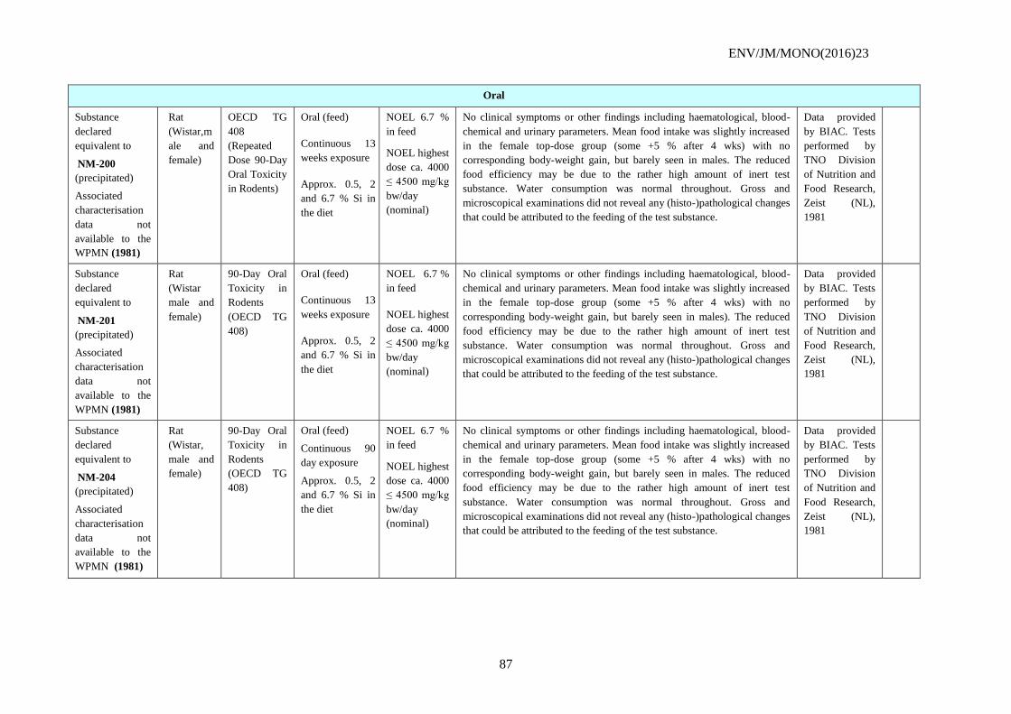
ENV/JM/MONO(2016)23
87
Oral
Substance
declared
equivalent to
NM-200
(precipitated)
Associated
characterisation
data not
available to the
WPMN (1981)
Rat
(Wistar,m
ale and
female)
OECD TG
408
(Repeated
Dose 90-Day
Oral Toxicity
in Rodents)
Oral (feed)
Continuous 13
weeks exposure
Approx. 0.5, 2
and 6.7 % Si in
the diet
NOEL 6.7 %
in feed
NOEL highest
dose ca. 4000
≤ 4500 mg/kg
bw/day
(nominal)
No clinical symptoms or other findings including haematological, blood-
chemical and urinary parameters. Mean food intake was slightly increased
in the female top-dose group (some +5 % after 4 wks) with no
corresponding body-weight gain, but barely seen in males. The reduced
food efficiency may be due to the rather high amount of inert test
substance. Water consumption was normal throughout. Gross and
microscopical examinations did not reveal any (histo-)pathological changes
that could be attributed to the feeding of the test substance.
Data provided
by BIAC. Tests
performed by
TNO Division
of Nutrition and
Food Research,
Zeist (NL),
1981
Substance
declared
equivalent to
NM-201
(precipitated)
Associated
characterisation
data not
available to the
WPMN (1981)
Rat
(Wistar
male and
female)
90-Day Oral
Toxicity in
Rodents
(OECD TG
408)
Oral (feed)
Continuous 13
weeks exposure
Approx. 0.5, 2
and 6.7 % Si in
the diet
NOEL 6.7 %
in feed
NOEL highest
dose ca. 4000
≤ 4500 mg/kg
bw/day
(nominal)
No clinical symptoms or other findings including haematological, blood-
chemical and urinary parameters. Mean food intake was slightly increased
in the female top-dose group (some +5 % after 4 wks) with no
corresponding body-weight gain, but barely seen in males). The reduced
food efficiency may be due to the rather high amount of inert test
substance. Water consumption was normal throughout. Gross and
microscopical examinations did not reveal any (histo-)pathological changes
that could be attributed to the feeding of the test substance.
Data provided
by BIAC. Tests
performed by
TNO Division
of Nutrition and
Food Research,
Zeist (NL),
1981
Substance
declared
equivalent to
NM-204
(precipitated)
Associated
characterisation
data not
available to the
WPMN (1981)
Rat
(Wistar,
male and
female)
90-Day Oral
Toxicity in
Rodents
(OECD TG
408)
Oral (feed)
Continuous 90
day exposure
Approx. 0.5, 2
and 6.7 % Si in
the diet
NOEL 6.7 %
in feed
NOEL highest
dose ca. 4000
≤ 4500 mg/kg
bw/day
(nominal)
No clinical symptoms or other findings including haematological, blood-
chemical and urinary parameters. Mean food intake was slightly increased
in the female top-dose group (some +5 % after 4 wks) with no
corresponding body-weight gain, but barely seen in males. The reduced
food efficiency may be due to the rather high amount of inert test
substance. Water consumption was normal throughout. Gross and
microscopical examinations did not reveal any (histo-)pathological changes
that could be attributed to the feeding of the test substance.
Data provided
by BIAC. Tests
performed by
TNO Division
of Nutrition and
Food Research,
Zeist (NL),
1981
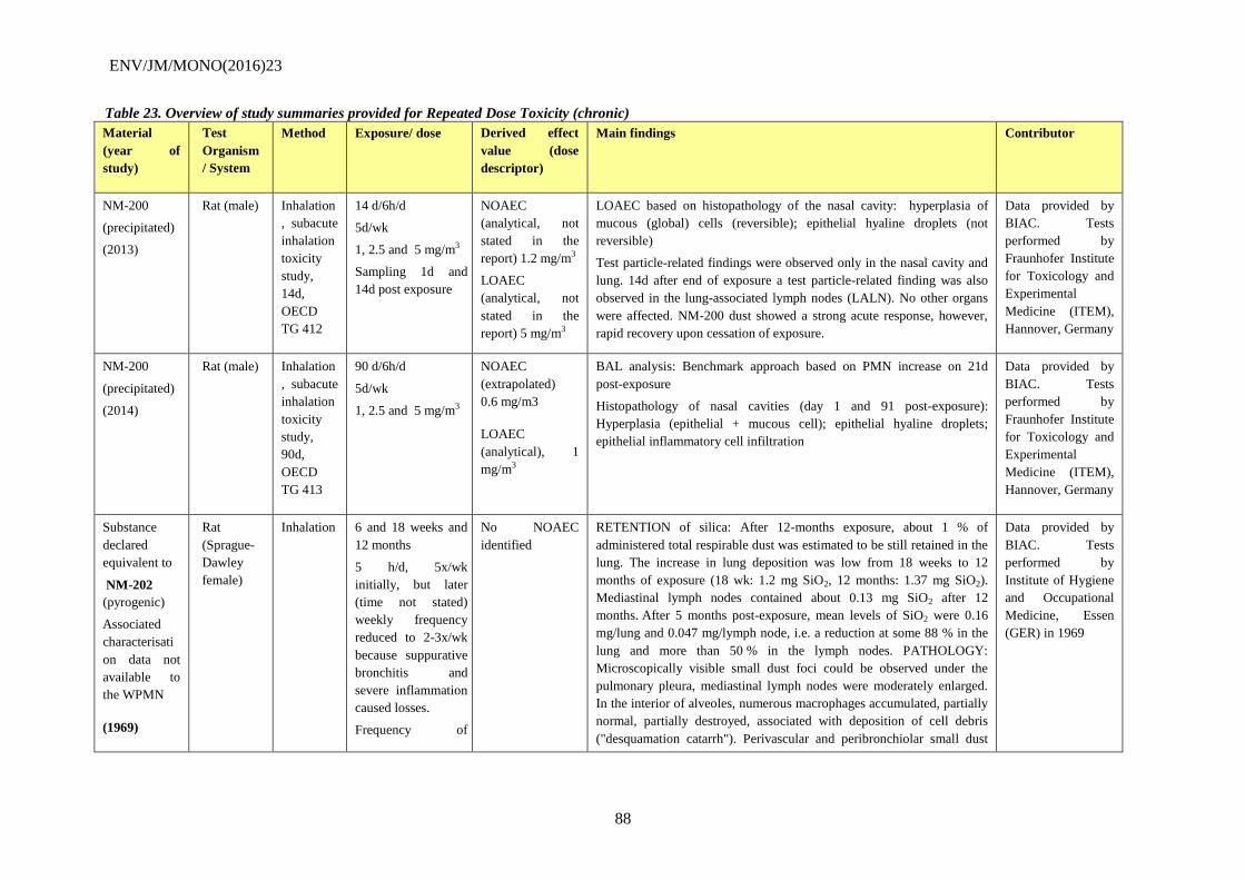
ENV/JM/MONO(2016)23
88
Table 23. Overview of study summaries provided for Repeated Dose Toxicity (chronic)
Material
(year of
study)
Test
Organism
/ System
Method Exposure/ dose Derived effect
value (dose
descriptor)
Main findings Contributor
NM-200
(precipitated)
(2013)
Rat (male) Inhalation
, subacute
inhalation
toxicity
study,
14d,
OECD
TG 412
14 d/6h/d
5d/wk
1, 2.5 and 5 mg/m3
Sampling 1d and
14d post exposure
NOAEC
(analytical, not
stated in the
report) 1.2 mg/m3
LOAEC
(analytical, not
stated in the
report) 5 mg/m3
LOAEC based on histopathology of the nasal cavity: hyperplasia of
mucous (global) cells (reversible); epithelial hyaline droplets (not
reversible)
Test particle-related findings were observed only in the nasal cavity and
lung. 14d after end of exposure a test particle-related finding was also
observed in the lung-associated lymph nodes (LALN). No other organs
were affected. NM-200 dust showed a strong acute response, however,
rapid recovery upon cessation of exposure.
Data provided by
BIAC. Tests
performed by
Fraunhofer Institute
for Toxicology and
Experimental
Medicine (ITEM),
Hannover, Germany
NM-200
(precipitated)
(2014)
Rat (male) Inhalation
, subacute
inhalation
toxicity
study,
90d,
OECD
TG 413
90 d/6h/d
5d/wk
1, 2.5 and 5 mg/m3
NOAEC
(extrapolated)
0.6 mg/m3
LOAEC
(analytical), 1
mg/m3
BAL analysis: Benchmark approach based on PMN increase on 21d
post-exposure
Histopathology of nasal cavities (day 1 and 91 post-exposure):
Hyperplasia (epithelial + mucous cell); epithelial hyaline droplets;
epithelial inflammatory cell infiltration
Data provided by
BIAC. Tests
performed by
Fraunhofer Institute
for Toxicology and
Experimental
Medicine (ITEM),
Hannover, Germany
Substance
declared
equivalent to
NM-202
(pyrogenic)
Associated
characterisati
on data not
available to
the WPMN
(1969)
Rat
(Sprague-
Dawley
female)
Inhalation 6 and 18 weeks and
12 months
5 h/d, 5x/wk
initially, but later
(time not stated)
weekly frequency
reduced to 2-3x/wk
because suppurative
bronchitis and
severe inflammation
caused losses.
Frequency of
No NOAEC
identified
RETENTION of silica: After 12-months exposure, about 1 % of
administered total respirable dust was estimated to be still retained in the
lung. The increase in lung deposition was low from 18 weeks to 12
months of exposure (18 wk: 1.2 mg SiO2, 12 months: 1.37 mg SiO2).
Mediastinal lymph nodes contained about 0.13 mg SiO2 after 12
months. After 5 months post-exposure, mean levels of SiO2 were 0.16
mg/lung and 0.047 mg/lymph node, i.e. a reduction at some 88 % in the
lung and more than 50 % in the lymph nodes. PATHOLOGY:
Microscopically visible small dust foci could be observed under the
pulmonary pleura, mediastinal lymph nodes were moderately enlarged.
In the interior of alveoles, numerous macrophages accumulated, partially
normal, partially destroyed, associated with deposition of cell debris
("desquamation catarrh"). Perivascular and peribronchiolar small dust
Data provided by
BIAC. Tests
performed by
Institute of Hygiene
and Occupational
Medicine, Essen
(GER) in 1969
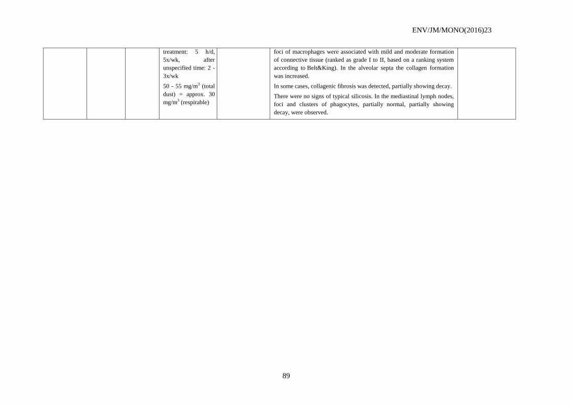
ENV/JM/MONO(2016)23
89
treatment: 5 h/d,
5x/wk, after
unspecified time: 2 -
3x/wk
50 - 55 mg/m3 (total
dust) = approx. 30
mg/m3 (respirable)
foci of macrophages were associated with mild and moderate formation
of connective tissue (ranked as grade I to II, based on a ranking system
according to Belt&King). In the alveolar septa the collagen formation
was increased.
In some cases, collagenic fibrosis was detected, partially showing decay.
There were no signs of typical silicosis. In the mediastinal lymph nodes,
foci and clusters of phagocytes, partially normal, partially showing
decay, were observed.
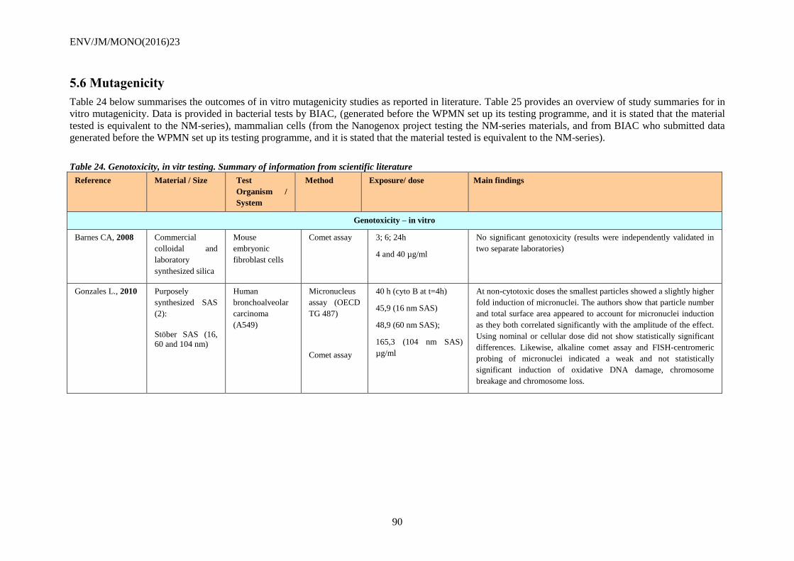
ENV/JM/MONO(2016)23
90
5.6 Mutagenicity
Table 24 below summarises the outcomes of in vitro mutagenicity studies as reported in literature. Table 25 provides an overview of study summaries for in
vitro mutagenicity. Data is provided in bacterial tests by BIAC, (generated before the WPMN set up its testing programme, and it is stated that the material
tested is equivalent to the NM-series), mammalian cells (from the Nanogenox project testing the NM-series materials, and from BIAC who submitted data
generated before the WPMN set up its testing programme, and it is stated that the material tested is equivalent to the NM-series).
Table 24. Genotoxicity, in vitr testing. Summary of information from scientific literature
Reference Material / Size Test
Organism /
System
Method Exposure/ dose Main findings
Genotoxicity – in vitro
Barnes CA, 2008 Commercial
colloidal and
laboratory
synthesized silica
Mouse
embryonic
fibroblast cells
Comet assay 3; 6; 24h
4 and 40 µg/ml
No significant genotoxicity (results were independently validated in
two separate laboratories)
Gonzales L., 2010
Purposely
synthesized SAS
(2):
Stöber SAS (16,
60 and 104 nm)
Human
bronchoalveolar
carcinoma
(A549)
Micronucleus
assay (OECD
TG 487)
Comet assay
40 h (cyto B at t=4h)
45,9 (16 nm SAS)
48,9 (60 nm SAS);
165,3 (104 nm SAS)
µg/ml
At non-cytotoxic doses the smallest particles showed a slightly higher
fold induction of micronuclei. The authors show that particle number
and total surface area appeared to account for micronuclei induction
as they both correlated significantly with the amplitude of the effect.
Using nominal or cellular dose did not show statistically significant
differences. Likewise, alkaline comet assay and FISH-centromeric
probing of micronuclei indicated a weak and not statistically
significant induction of oxidative DNA damage, chromosome
breakage and chromosome loss.
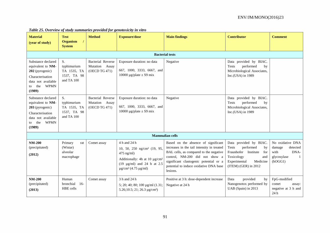
ENV/JM/MONO(2016)23
91
Table 25. Overview of study summaries provided for genotoxicity in vitro
Material
(year of study)
Test
Organism /
System
Method Exposure/dose Main findings Contributor Comment
Bacterial tests
Substance declared
equivalent to NM-
202 (pyrogenic)
Characterisation
data not available
to the WPMN
(1989)
S.
typhimurium
TA 1535, TA
1537, TA 98
and TA 100
Bacterial Reverse
Mutation Assay
(OECD TG 471)
Exposure duration: no data
667, 1000, 3333, 6667, and
10000 µg/plate ± S9 mix
Negative Data provided by BIAC.
Tests performed by
Microbiological Associates,
Inc.(USA) in 1989
Substance declared
equivalent to NM-
203 (pyrogenic)
Characterisation
data not available
to the WPMN
(1989)
S.
typhimurium
TA 1535, TA
1537, TA 98
and TA 100
Bacterial Reverse
Mutation Assay
(OECD TG 471)
Exposure duration: no data
667, 1000, 3333, 6667, and
10000 µg/plate ± S9 mix
Negative Data provided by BIAC.
Tests performed by
Microbiological Associates,
Inc.(USA) in 1989
Mammalian cells
NM-200
(precipitated)
(2012)
Primary rat
(Wistar)
alveolar
macrophage
Comet assay 4 h and 24 h
10, 50, 250 ng/cm² (19, 95,
475 ng/ml)
Additionally: 4h at 10 µg/cm²
(19 µg/ml) and 24 h at 2.5
µg/cm² (4.75 µg/ml)
Based on the absence of significant
increases in the tail intensity in treated
BAL cells, as compared to the negative
control, NM-200 did not show a
significant clastogenic potential or a
potential to induce oxidative DNA base
lesions.
Data provided by BIAC.
Tests performed by
Fraunhofer Institute for
Toxicology and
Experimental Medicine
(ITEM) (GER) in 2012
No oxidative DNA
damage detected
with DNA-
glycosylase 1
(hOGG1)
NM-200
(precipitated)
(2013)
Human
bronchial 16-
HBE cells
Comet assay 3 h and 24 h
5; 20; 40; 80; 100 µg/ml (1.31;
5.26;10.5; 21; 26.3 µg/cm²)
Positive at 3 h: dose-dependent increase
Negative at 24 h
Data provided by
Nanogenotox performed by
UAB (Spain) in 2013
FpG-modified
comet assay:
negative at 3 h and
24 h
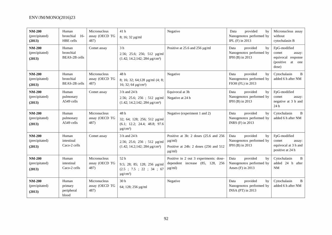
ENV/JM/MONO(2016)23
92
NM-200
(precipitated)
(2013)
Human
bronchial 16-
HBE cells
Micronucleus
assay (OECD TG
487)
41 h
8; 16; 32 µg/ml
Negative Data provided by
Nanogenotox performed by
IPL (F) in 2013
Micronucleus assay
without
cytochalasin B
NM-200
(precipitated)
(2013)
Human
bronchial
BEAS-2B cells
Comet assay 3 h
2.56; 25.6; 256; 512 µg/ml
(1.42; 14.2;142; 284 µg/cm²)
Positive at 25.6 and 256 µg/ml Data provided by
Nanogenotox performed by
IPH (B) in 2013
FpG-modified
comet assay:
equivocal response
(positive at one
dose)
NM-200
(precipitated)
(2013)
Human
bronchial
BEAS-2B cells
Micronucleus
assay (OECD TG
487)
48 h
8; 16; 32; 64;128 µg/ml (4; 8;
16; 32; 64 µg/cm²)
Negative Data provided by
Nanogenotox performed by
FIOH (FL) in 2013
Cytochalasin B
added 6 h after NM
NM-200
(precipitated)
(2013)
Human
pulmonary
A549 cells
Comet assay 3 h and 24 h
2.56; 25.6; 256 ; 512 µg/ml
(1.42; 14.2;142; 284 µg/cm²)
Equivocal at 3h
Negative at 24 h
Data provided by
Nanogenotox performed by
IPH (B) in 2013
FpG-modified
comet assay:
negative at 3 h and
24 h
NM-200
(precipitated)
(2013)
Human
pulmonary
A549 cells
Micronucleus
assay (OECD TG
487)
48 h
32; 64; 128; 256; 512 µg/ml
(6.1; 12.2; 24.4; 48.8; 97.6
µg/cm²)
Negative (experiment 1 and 2) Data provided by
Nanogenotox performed by
INRS (F) in 2013
Cytochalasin B
added 6 h after NM
NM-200
(precipitated)
(2013)
Human
intestinal
Caco-2 cells
Comet assay 3 h and 24 h
2.56; 25.6; 256 ; 512 µg/ml
(1.42; 14.2;142; 284 µg/cm²)
Positive at 3h: 2 doses (25.6 and 256
µg/ml)
Positive at 24h: 2 doses (256 and 512
µg/ml)
Data provided by
Nanogenotox performed by
IPH (B) in 2013
FpG-modified
comet assay:
equivocal at 3 h and
positive at 24 h
NM-200
(precipitated)
(2013)
Human
intestinal
Caco-2 cells
Micronucleus
assay (OECD TG
487)
52 h
9.5; 28; 85; 128; 256 µg/ml
(2.5 ; 7.5 ; 22 ; 34 ; 67
µg/cm²)
Positive in 2 out 3 experiments: dose-
dependent increase (85, 128, 256
µg/ml)
Data provided by
Nanogenotox performed by
Anses (F) in 2013
Cytochalasin B
added 24 h after
NM
NM-200
(precipitated)
(2013)
Human
primary
peripheral
blood
Micronucleus
assay (OECD TG
487)
30 h
64; 128; 256 µg/ml
Negative Data provided by
Nanogenotox performed by
INSA (PT) in 2013
Cytochalasin B
added 6 h after NM
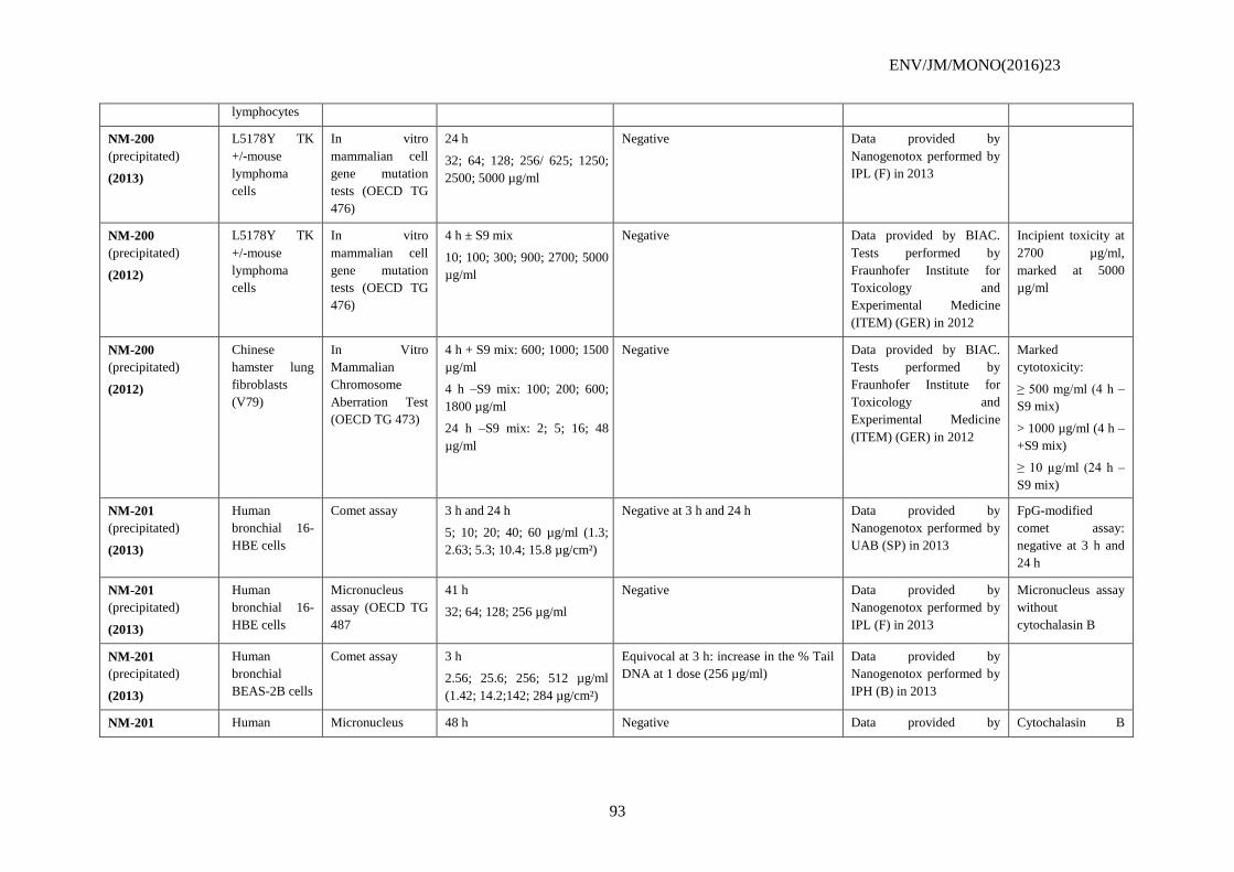
ENV/JM/MONO(2016)23
93
lymphocytes
NM-200
(precipitated)
(2013)
L5178Y TK
+/-mouse
lymphoma
cells
In vitro
mammalian cell
gene mutation
tests (OECD TG
476)
24 h
32; 64; 128; 256/ 625; 1250;
2500; 5000 µg/ml
Negative Data provided by
Nanogenotox performed by
IPL (F) in 2013
NM-200
(precipitated)
(2012)
L5178Y TK
+/-mouse
lymphoma
cells
In vitro
mammalian cell
gene mutation
tests (OECD TG
476)
4 h ± S9 mix
10; 100; 300; 900; 2700; 5000
µg/ml
Negative Data provided by BIAC.
Tests performed by
Fraunhofer Institute for
Toxicology and
Experimental Medicine
(ITEM) (GER) in 2012
Incipient toxicity at
2700 µg/ml,
marked at 5000
µg/ml
NM-200
(precipitated)
(2012)
Chinese
hamster lung
fibroblasts
(V79)
In Vitro
Mammalian
Chromosome
Aberration Test
(OECD TG 473)
4 h + S9 mix: 600; 1000; 1500
µg/ml
4 h –S9 mix: 100; 200; 600;
1800 µg/ml
24 h –S9 mix: 2; 5; 16; 48
µg/ml
Negative Data provided by BIAC.
Tests performed by
Fraunhofer Institute for
Toxicology and
Experimental Medicine
(ITEM) (GER) in 2012
Marked
cytotoxicity:
≥ 500 mg/ml (4 h –
S9 mix)
> 1000 µg/ml (4 h –
+S9 mix)
≥ 10 µg/ml (24 h –
S9 mix)
NM-201
(precipitated)
(2013)
Human
bronchial 16-
HBE cells
Comet assay 3 h and 24 h
5; 10; 20; 40; 60 µg/ml (1.3;
2.63; 5.3; 10.4; 15.8 µg/cm²)
Negative at 3 h and 24 h Data provided by
Nanogenotox performed by
UAB (SP) in 2013
FpG-modified
comet assay:
negative at 3 h and
24 h
NM-201
(precipitated)
(2013)
Human
bronchial 16-
HBE cells
Micronucleus
assay (OECD TG
487
41 h
32; 64; 128; 256 µg/ml
Negative Data provided by
Nanogenotox performed by
IPL (F) in 2013
Micronucleus assay
without
cytochalasin B
NM-201
(precipitated)
(2013)
Human
bronchial
BEAS-2B cells
Comet assay 3 h
2.56; 25.6; 256; 512 µg/ml
(1.42; 14.2;142; 284 µg/cm²)
Equivocal at 3 h: increase in the % Tail
DNA at 1 dose (256 µg/ml)
Data provided by
Nanogenotox performed by
IPH (B) in 2013
NM-201 Human Micronucleus 48 h Negative Data provided by Cytochalasin B
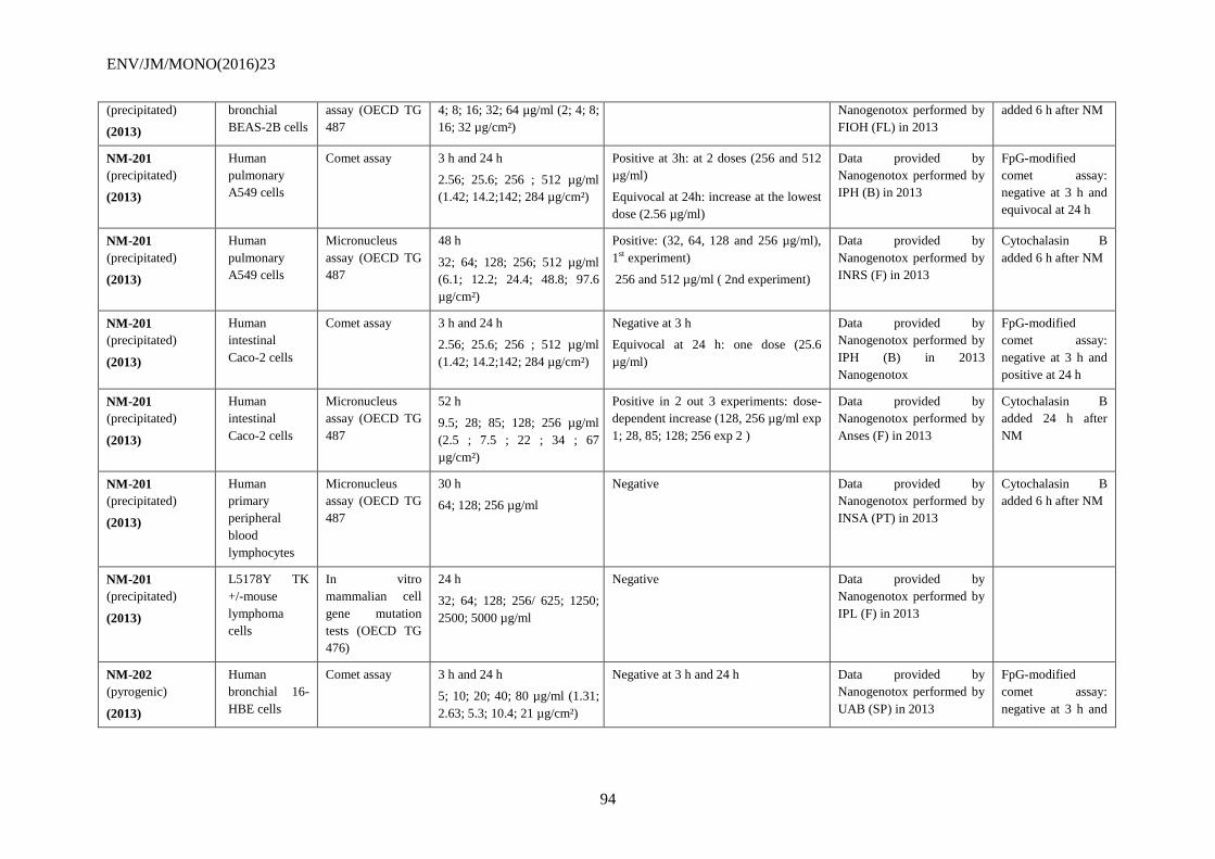
ENV/JM/MONO(2016)23
94
(precipitated)
(2013)
bronchial
BEAS-2B cells
assay (OECD TG
487
4; 8; 16; 32; 64 µg/ml (2; 4; 8;
16; 32 µg/cm²)
Nanogenotox performed by
FIOH (FL) in 2013
added 6 h after NM
NM-201
(precipitated)
(2013)
Human
pulmonary
A549 cells
Comet assay 3 h and 24 h
2.56; 25.6; 256 ; 512 µg/ml
(1.42; 14.2;142; 284 µg/cm²)
Positive at 3h: at 2 doses (256 and 512
µg/ml)
Equivocal at 24h: increase at the lowest
dose (2.56 µg/ml)
Data provided by
Nanogenotox performed by
IPH (B) in 2013
FpG-modified
comet assay:
negative at 3 h and
equivocal at 24 h
NM-201
(precipitated)
(2013)
Human
pulmonary
A549 cells
Micronucleus
assay (OECD TG
487
48 h
32; 64; 128; 256; 512 µg/ml
(6.1; 12.2; 24.4; 48.8; 97.6
µg/cm²)
Positive: (32, 64, 128 and 256 µg/ml),
1st experiment)
256 and 512 µg/ml ( 2nd experiment)
Data provided by
Nanogenotox performed by
INRS (F) in 2013
Cytochalasin B
added 6 h after NM
NM-201
(precipitated)
(2013)
Human
intestinal
Caco-2 cells
Comet assay 3 h and 24 h
2.56; 25.6; 256 ; 512 µg/ml
(1.42; 14.2;142; 284 µg/cm²)
Negative at 3 h
Equivocal at 24 h: one dose (25.6
µg/ml)
Data provided by
Nanogenotox performed by
IPH (B) in 2013
Nanogenotox
FpG-modified
comet assay:
negative at 3 h and
positive at 24 h
NM-201
(precipitated)
(2013)
Human
intestinal
Caco-2 cells
Micronucleus
assay (OECD TG
487
52 h
9.5; 28; 85; 128; 256 µg/ml
(2.5 ; 7.5 ; 22 ; 34 ; 67
µg/cm²)
Positive in 2 out 3 experiments: dose-
dependent increase (128, 256 µg/ml exp
1; 28, 85; 128; 256 exp 2 )
Data provided by
Nanogenotox performed by
Anses (F) in 2013
Cytochalasin B
added 24 h after
NM
NM-201
(precipitated)
(2013)
Human
primary
peripheral
blood
lymphocytes
Micronucleus
assay (OECD TG
487
30 h
64; 128; 256 µg/ml
Negative Data provided by
Nanogenotox performed by
INSA (PT) in 2013
Cytochalasin B
added 6 h after NM
NM-201
(precipitated)
(2013)
L5178Y TK
+/-mouse
lymphoma
cells
In vitro
mammalian cell
gene mutation
tests (OECD TG
476)
24 h
32; 64; 128; 256/ 625; 1250;
2500; 5000 µg/ml
Negative Data provided by
Nanogenotox performed by
IPL (F) in 2013
NM-202
(pyrogenic)
(2013)
Human
bronchial 16-
HBE cells
Comet assay 3 h and 24 h
5; 10; 20; 40; 80 µg/ml (1.31;
2.63; 5.3; 10.4; 21 µg/cm²)
Negative at 3 h and 24 h Data provided by
Nanogenotox performed by
UAB (SP) in 2013
FpG-modified
comet assay:
negative at 3 h and
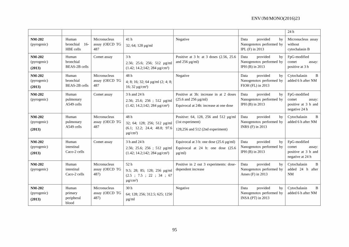
ENV/JM/MONO(2016)23
95
24 h
NM-202
(pyrogenic)
Human
bronchial 16-
HBE cells
Micronucleus
assay (OECD TG
487
41 h
32; 64; 128 µg/ml
Negative Data provided by
Nanogenotox performed by
IPL (F) in 2013
Micronucleus assay
without
cytochalasin B
NM-202
(pyrogenic)
(2013)
Human
bronchial
BEAS-2B cells
Comet assay 3 h
2.56; 25.6; 256; 512 µg/ml
(1.42; 14.2;142; 284 µg/cm²)
Positive at 3 h: at 3 doses (2.56, 25.6
and 256 µg/ml)
Data provided by
Nanogenotox performed by
IPH (B) in 2013
FpG-modified
comet assay:
positive at 3 h
NM-202
(pyrogenic)
(2013)
Human
bronchial
BEAS-2B cells
Micronucleus
assay (OECD TG
487
48 h
4; 8; 16; 32; 64 µg/ml (2; 4; 8;
16; 32 µg/cm²)
Negative Data provided by
Nanogenotox performed by
FIOH (FL) in 2013
Cytochalasin B
added 6 h after NM
NM-202
(pyrogenic)
Human
pulmonary
A549 cells
Comet assay 3 h and 24 h
2.56; 25.6; 256 ; 512 µg/ml
(1.42; 14.2;142; 284 µg/cm²)
Positive at 3h: increase in at 2 doses
(25.6 and 256 µg/ml)
Equivocal at 24h: increase at one dose
Data provided by
Nanogenotox performed by
IPH (B) in 2013
FpG-modified
comet assay:
positive at 3 h and
negative 24 h
NM-202
(pyrogenic)
(2013)
Human
pulmonary
A549 cells
Micronucleus
assay (OECD TG
487
48 h
32; 64; 128; 256; 512 µg/ml
(6.1; 12.2; 24.4; 48.8; 97.6
µg/cm²)
Positive: 64, 128, 256 and 512 µg/ml
(1st experiment)
128,256 and 512 (2nd experiment)
Data provided by
Nanogenotox performed by
INRS (F) in 2013
Cytochalasin B
added 6 h after NM
NM-202
(pyrogenic)
(2013)
Human
intestinal
Caco-2 cells
Comet assay 3 h and 24 h
2.56; 25.6; 256 ; 512 µg/ml
(1.42; 14.2;142; 284 µg/cm²)
Equivocal at 3 h: one dose (25.6 µg/ml)
Equivocal at 24 h: one dose (25.6
µg/ml)
Data provided by
Nanogenotox performed by
IPH (B) in 2013
FpG-modified
comet assay:
positive at 3 h and
negative at 24 h
NM-202
(pyrogenic)
Human
intestinal
Caco-2 cells
Micronucleus
assay (OECD TG
487)
52 h
9.5; 28; 85; 128; 256 µg/ml
(2.5 ; 7.5 ; 22 ; 34 ; 67
µg/cm²)
Positive in 2 out 3 experiments: dose-
dependent increase
Data provided by
Nanogenotox performed by
Anses (F) in 2013
Cytochalasin B
added 24 h after
NM
NM-202
(pyrogenic)
(2013)
Human
primary
peripheral
blood
Micronucleus
assay (OECD TG
487)
30 h
64; 128; 256; 312.5; 625; 1250
µg/ml
Negative Data provided by
Nanogenotox performed by
INSA (PT) in 2013
Cytochalasin B
added 6 h after NM
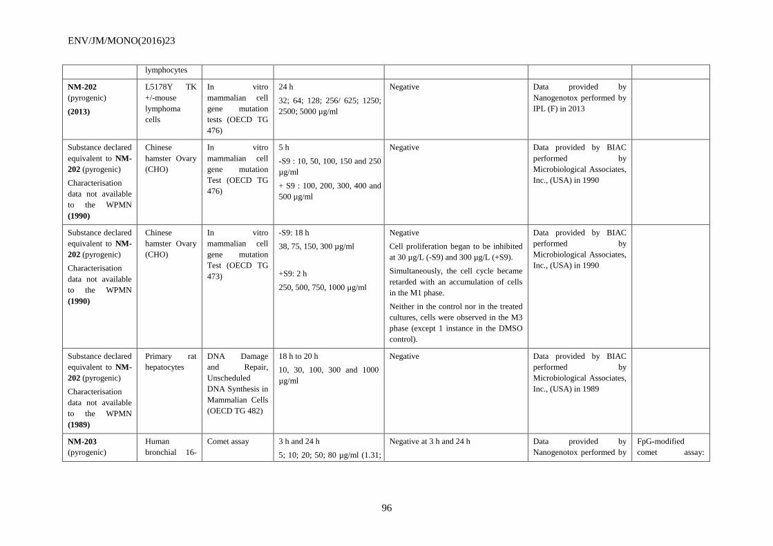
ENV/JM/MONO(2016)23
96
lymphocytes
NM-202
(pyrogenic)
(2013)
L5178Y TK
+/-mouse
lymphoma
cells
In vitro
mammalian cell
gene mutation
tests (OECD TG
476)
24 h
32; 64; 128; 256/ 625; 1250;
2500; 5000 µg/ml
Negative Data provided by
Nanogenotox performed by
IPL (F) in 2013
Substance declared
equivalent to NM-
202 (pyrogenic)
Characterisation
data not available
to the WPMN
(1990)
Chinese
hamster Ovary
(CHO)
In vitro
mammalian cell
gene mutation
Test (OECD TG
476)
5 h
-S9 : 10, 50, 100, 150 and 250
µg/ml
+ S9 : 100, 200, 300, 400 and
500 µg/ml
Negative Data provided by BIAC
performed by
Microbiological Associates,
Inc., (USA) in 1990
Substance declared
equivalent to NM-
202 (pyrogenic)
Characterisation
data not available
to the WPMN
(1990)
Chinese
hamster Ovary
(CHO)
In vitro
mammalian cell
gene mutation
Test (OECD TG
473)
-S9: 18 h
38, 75, 150, 300 µg/ml
+S9: 2 h
250, 500, 750, 1000 µg/ml
Negative
Cell proliferation began to be inhibited
at 30 µg/L (-S9) and 300 µg/L (+S9).
Simultaneously, the cell cycle became
retarded with an accumulation of cells
in the M1 phase.
Neither in the control nor in the treated
cultures, cells were observed in the M3
phase (except 1 instance in the DMSO
control).
Data provided by BIAC
performed by
Microbiological Associates,
Inc., (USA) in 1990
Substance declared
equivalent to NM-
202 (pyrogenic)
Characterisation
data not available
to the WPMN
(1989)
Primary rat
hepatocytes
DNA Damage
and Repair,
Unscheduled
DNA Synthesis in
Mammalian Cells
(OECD TG 482)
18 h to 20 h
10, 30, 100, 300 and 1000
µg/ml
Negative Data provided by BIAC
performed by
Microbiological Associates,
Inc., (USA) in 1989
NM-203
(pyrogenic)
Human
bronchial 16-
Comet assay 3 h and 24 h
5; 10; 20; 50; 80 µg/ml (1.31;
Negative at 3 h and 24 h Data provided by
Nanogenotox performed by
FpG-modified
comet assay:
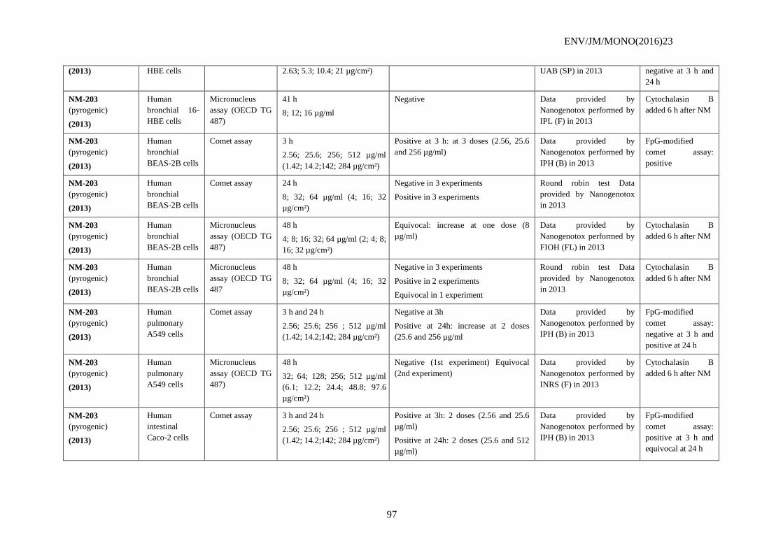
ENV/JM/MONO(2016)23
97
(2013) HBE cells 2.63; 5.3; 10.4; 21 µg/cm²) UAB (SP) in 2013 negative at 3 h and
24 h
NM-203
(pyrogenic)
(2013)
Human
bronchial 16-
HBE cells
Micronucleus
assay (OECD TG
487)
41 h
8; 12; 16 µg/ml
Negative Data provided by
Nanogenotox performed by
IPL (F) in 2013
Cytochalasin B
added 6 h after NM
NM-203
(pyrogenic)
(2013)
Human
bronchial
BEAS-2B cells
Comet assay 3 h
2.56; 25.6; 256; 512 µg/ml
(1.42; 14.2;142; 284 µg/cm²)
Positive at 3 h: at 3 doses (2.56, 25.6
and 256 µg/ml)
Data provided by
Nanogenotox performed by
IPH (B) in 2013
FpG-modified
comet assay:
positive
NM-203
(pyrogenic)
(2013)
Human
bronchial
BEAS-2B cells
Comet assay 24 h
8; 32; 64 µg/ml (4; 16; 32
µg/cm²)
Negative in 3 experiments
Positive in 3 experiments
Round robin test Data
provided by Nanogenotox
in 2013
NM-203
(pyrogenic)
(2013)
Human
bronchial
BEAS-2B cells
Micronucleus
assay (OECD TG
487)
48 h
4; 8; 16; 32; 64 µg/ml (2; 4; 8;
16; 32 µg/cm²)
Equivocal: increase at one dose (8
µg/ml)
Data provided by
Nanogenotox performed by
FIOH (FL) in 2013
Cytochalasin B
added 6 h after NM
NM-203
(pyrogenic)
(2013)
Human
bronchial
BEAS-2B cells
Micronucleus
assay (OECD TG
487
48 h
8; 32; 64 µg/ml (4; 16; 32
µg/cm²)
Negative in 3 experiments
Positive in 2 experiments
Equivocal in 1 experiment
Round robin test Data
provided by Nanogenotox
in 2013
Cytochalasin B
added 6 h after NM
NM-203
(pyrogenic)
(2013)
Human
pulmonary
A549 cells
Comet assay 3 h and 24 h
2.56; 25.6; 256 ; 512 µg/ml
(1.42; 14.2;142; 284 µg/cm²)
Negative at 3h
Positive at 24h: increase at 2 doses
(25.6 and 256 µg/ml
Data provided by
Nanogenotox performed by
IPH (B) in 2013
FpG-modified
comet assay:
negative at 3 h and
positive at 24 h
NM-203
(pyrogenic)
(2013)
Human
pulmonary
A549 cells
Micronucleus
assay (OECD TG
487)
48 h
32; 64; 128; 256; 512 µg/ml
(6.1; 12.2; 24.4; 48.8; 97.6
µg/cm²)
Negative (1st experiment) Equivocal
(2nd experiment)
Data provided by
Nanogenotox performed by
INRS (F) in 2013
Cytochalasin B
added 6 h after NM
NM-203
(pyrogenic)
(2013)
Human
intestinal
Caco-2 cells
Comet assay 3 h and 24 h
2.56; 25.6; 256 ; 512 µg/ml
(1.42; 14.2;142; 284 µg/cm²)
Positive at 3h: 2 doses (2.56 and 25.6
µg/ml)
Positive at 24h: 2 doses (25.6 and 512
µg/ml)
Data provided by
Nanogenotox performed by
IPH (B) in 2013
FpG-modified
comet assay:
positive at 3 h and
equivocal at 24 h
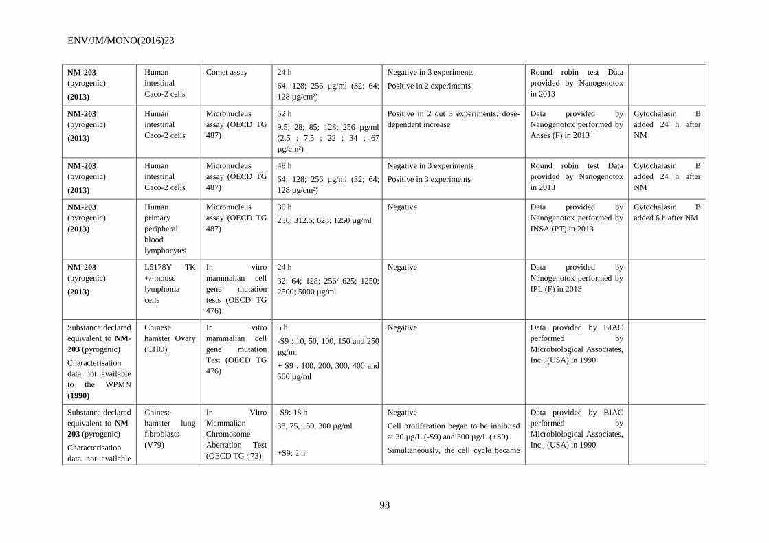
ENV/JM/MONO(2016)23
98
NM-203
(pyrogenic)
(2013)
Human
intestinal
Caco-2 cells
Comet assay 24 h
64; 128; 256 µg/ml (32; 64;
128 µg/cm²)
Negative in 3 experiments
Positive in 2 experiments
Round robin test Data
provided by Nanogenotox
in 2013
NM-203
(pyrogenic)
(2013)
Human
intestinal
Caco-2 cells
Micronucleus
assay (OECD TG
487)
52 h
9.5; 28; 85; 128; 256 µg/ml
(2.5 ; 7.5 ; 22 ; 34 ; 67
µg/cm²)
Positive in 2 out 3 experiments: dose-
dependent increase
Data provided by
Nanogenotox performed by
Anses (F) in 2013
Cytochalasin B
added 24 h after
NM
NM-203
(pyrogenic)
(2013)
Human
intestinal
Caco-2 cells
Micronucleus
assay (OECD TG
487)
48 h
64; 128; 256 µg/ml (32; 64;
128 µg/cm²)
Negative in 3 experiments
Positive in 3 experiments
Round robin test Data
provided by Nanogenotox
in 2013
Cytochalasin B
added 24 h after
NM
NM-203
(pyrogenic)
(2013)
Human
primary
peripheral
blood
lymphocytes
Micronucleus
assay (OECD TG
487)
30 h
256; 312.5; 625; 1250 µg/ml
Negative Data provided by
Nanogenotox performed by
INSA (PT) in 2013
Cytochalasin B
added 6 h after NM
NM-203
(pyrogenic)
(2013)
L5178Y TK
+/-mouse
lymphoma
cells
In vitro
mammalian cell
gene mutation
tests (OECD TG
476)
24 h
32; 64; 128; 256/ 625; 1250;
2500; 5000 µg/ml
Negative Data provided by
Nanogenotox performed by
IPL (F) in 2013
Substance declared
equivalent to NM-
203 (pyrogenic)
Characterisation
data not available
to the WPMN
(1990)
Chinese
hamster Ovary
(CHO)
In vitro
mammalian cell
gene mutation
Test (OECD TG
476)
5 h
-S9 : 10, 50, 100, 150 and 250
µg/ml
+ S9 : 100, 200, 300, 400 and
500 µg/ml
Negative Data provided by BIAC
performed by
Microbiological Associates,
Inc., (USA) in 1990
Substance declared
equivalent to NM-
203 (pyrogenic)
Characterisation
data not available
Chinese
hamster lung
fibroblasts
(V79)
In Vitro
Mammalian
Chromosome
Aberration Test
(OECD TG 473)
-S9: 18 h
38, 75, 150, 300 µg/ml
+S9: 2 h
Negative
Cell proliferation began to be inhibited
at 30 µg/L (-S9) and 300 µg/L (+S9).
Simultaneously, the cell cycle became
Data provided by BIAC
performed by
Microbiological Associates,
Inc., (USA) in 1990
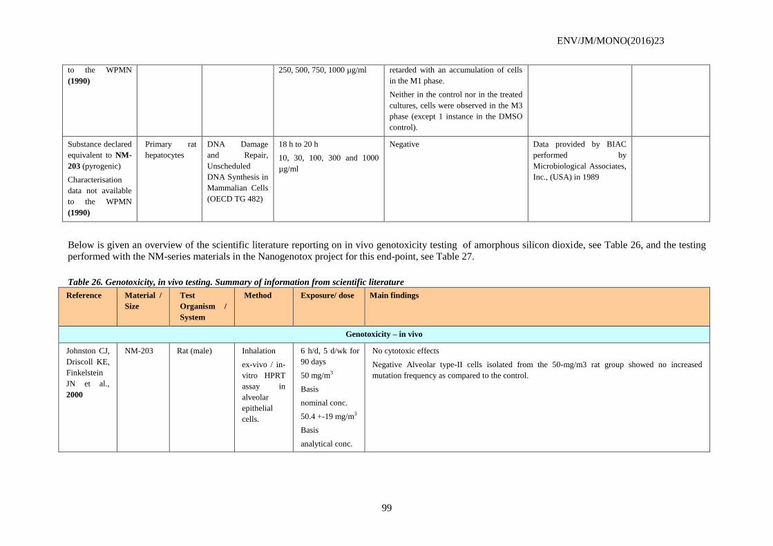
ENV/JM/MONO(2016)23
99
to the WPMN
(1990)
250, 500, 750, 1000 µg/ml retarded with an accumulation of cells
in the M1 phase.
Neither in the control nor in the treated
cultures, cells were observed in the M3
phase (except 1 instance in the DMSO
control).
Substance declared
equivalent to NM-
203 (pyrogenic)
Characterisation
data not available
to the WPMN
(1990)
Primary rat
hepatocytes
DNA Damage
and Repair,
Unscheduled
DNA Synthesis in
Mammalian Cells
(OECD TG 482)
18 h to 20 h
10, 30, 100, 300 and 1000
µg/ml
Negative Data provided by BIAC
performed by
Microbiological Associates,
Inc., (USA) in 1989
Below is given an overview of the scientific literature reporting on in vivo genotoxicity testing of amorphous silicon dioxide, see Table 26, and the testing
performed with the NM-series materials in the Nanogenotox project for this end-point, see Table 27.
Table 26. Genotoxicity, in vivo testing. Summary of information from scientific literature
Reference Material /
Size
Test
Organism /
System
Method Exposure/ dose Main findings
Genotoxicity – in vivo
Johnston CJ,
Driscoll KE,
Finkelstein
JN et al.,
2000
NM-203 Rat (male) Inhalation
ex-vivo / in-
vitro HPRT
assay in
alveolar
epithelial
cells.
6 h/d, 5 d/wk for
90 days
50 mg/m3
Basis
nominal conc.
50.4 +-19 mg/m3
Basis
analytical conc.
No cytotoxic effects
Negative Alveolar type-II cells isolated from the 50-mg/m3 rat group showed no increased
mutation frequency as compared to the control.
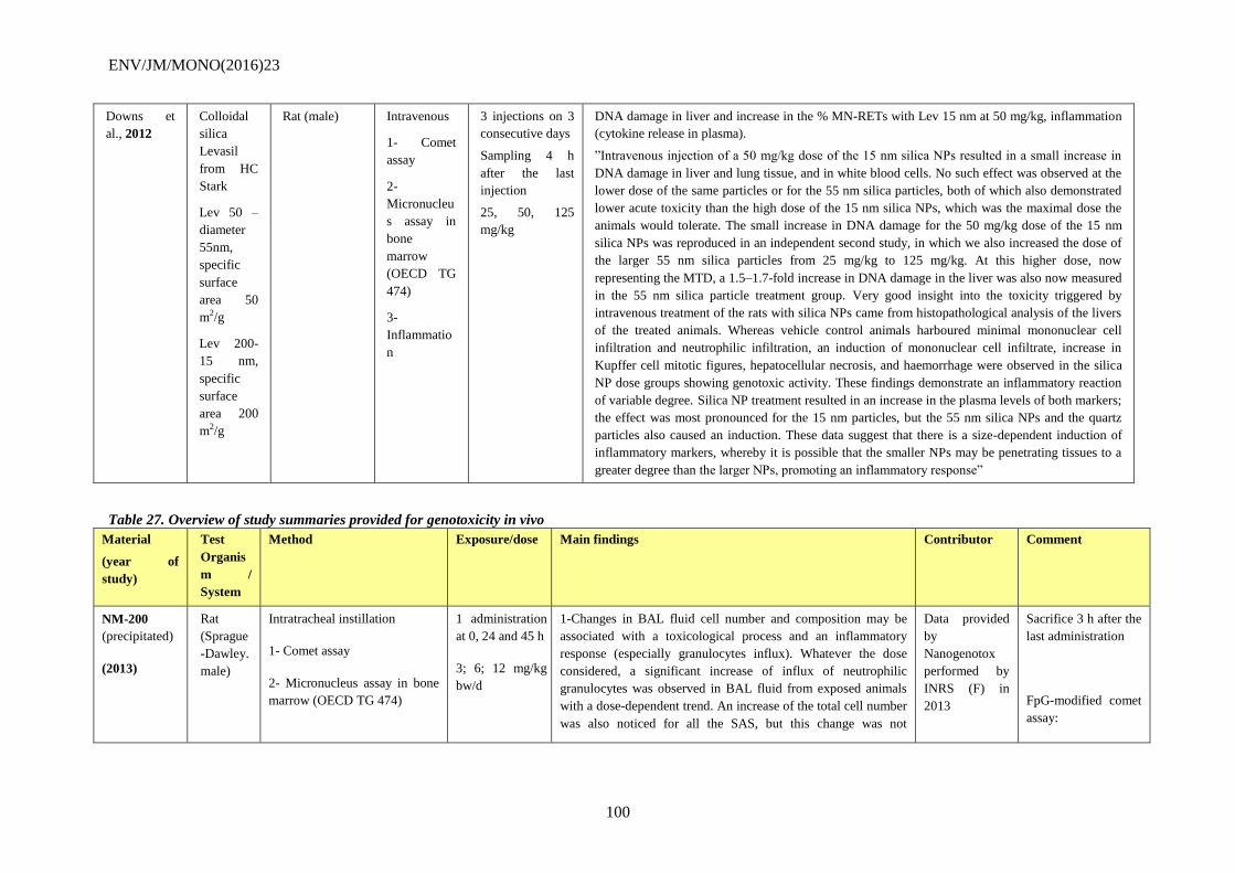
ENV/JM/MONO(2016)23
100
Downs et
al., 2012
Colloidal
silica
Levasil
from HC
Stark
Lev 50 –
diameter
55nm,
specific
surface
area 50
m2/g
Lev 200-
15 nm,
specific
surface
area 200
m2/g
Rat (male) Intravenous
1- Comet
assay
2-
Micronucleu
s assay in
bone
marrow
(OECD TG
474)
3-
Inflammatio
n
3 injections on 3
consecutive days
Sampling 4 h
after the last
injection
25, 50, 125
mg/kg
DNA damage in liver and increase in the % MN-RETs with Lev 15 nm at 50 mg/kg, inflammation
(cytokine release in plasma).
”Intravenous injection of a 50 mg/kg dose of the 15 nm silica NPs resulted in a small increase in
DNA damage in liver and lung tissue, and in white blood cells. No such effect was observed at the
lower dose of the same particles or for the 55 nm silica particles, both of which also demonstrated
lower acute toxicity than the high dose of the 15 nm silica NPs, which was the maximal dose the
animals would tolerate. The small increase in DNA damage for the 50 mg/kg dose of the 15 nm
silica NPs was reproduced in an independent second study, in which we also increased the dose of
the larger 55 nm silica particles from 25 mg/kg to 125 mg/kg. At this higher dose, now
representing the MTD, a 1.5–1.7-fold increase in DNA damage in the liver was also now measured
in the 55 nm silica particle treatment group. Very good insight into the toxicity triggered by
intravenous treatment of the rats with silica NPs came from histopathological analysis of the livers
of the treated animals. Whereas vehicle control animals harboured minimal mononuclear cell
infiltration and neutrophilic infiltration, an induction of mononuclear cell infiltrate, increase in
Kupffer cell mitotic figures, hepatocellular necrosis, and haemorrhage were observed in the silica
NP dose groups showing genotoxic activity. These findings demonstrate an inflammatory reaction
of variable degree. Silica NP treatment resulted in an increase in the plasma levels of both markers;
the effect was most pronounced for the 15 nm particles, but the 55 nm silica NPs and the quartz
particles also caused an induction. These data suggest that there is a size-dependent induction of
inflammatory markers, whereby it is possible that the smaller NPs may be penetrating tissues to a
greater degree than the larger NPs, promoting an inflammatory response”
Table 27. Overview of study summaries provided for genotoxicity in vivo
Material
(year of
study)
Test
Organis
m /
System
Method Exposure/dose Main findings Contributor Comment
NM-200
(precipitated)
(2013)
Rat
(Sprague
-Dawley.
male)
Intratracheal instillation
1- Comet assay
2- Micronucleus assay in bone
marrow (OECD TG 474)
1 administration
at 0, 24 and 45 h
3; 6; 12 mg/kg
bw/d
1-Changes in BAL fluid cell number and composition may be
associated with a toxicological process and an inflammatory
response (especially granulocytes influx). Whatever the dose
considered, a significant increase of influx of neutrophilic
granulocytes was observed in BAL fluid from exposed animals
with a dose-dependent trend. An increase of the total cell number
was also noticed for all the SAS, but this change was not
Data provided
by
Nanogenotox
performed by
INRS (F) in
2013
Sacrifice 3 h after the
last administration
FpG-modified comet
assay:
ENV/JM/MONO(2016)23
101
significant for the lowest dose of NM-200. Main cell types
observed in BAL fluid were macrophages and neutrophilic
granulocytes, some lymphocytes were also observed but their
frequency and number in SAS exposed animals were not
significantly different from the control group. In addition, in this
study, the percentage of neutrophilic granulocytes in control
(vehicle) group appears higher than that is usually obtained
following a single intratracheal instillation and may be
considered as an experimental artefact due to repeated animal
exposure. Increase in lactate dehydrogenase (LDH), alkaline
phosphatase (ALP) and N-acetyl-β-Dglucosaminidase (NAG)
activities and protein content are often signs of pulmonary
toxicity following pulmonary exposure to particulate matter. A
significant increase in the broncho-alveolar fluid of LDH and
NAG activities was observed. No change in alkaline phosphatase
(ALP) was however observed (Figure 17).
2-Thus, intratracheal instillation did not induce any significant
increase of DNA damages in all the organs tested. In all the
experiments performed, positive controls (MMS Or ENU)
always induced significant DNA strand breaks in the tissues
analysed.
Negative in all the
tested organs except
from spleen
(equivocal result)
NM-200
(precipitated)
(2013)
Rat
(Sprague
-Dawley.
male)
Oral
1- Comet assay
2- Micronucleus assay in bone
marrow (OECD TG 474)
3- Micronucleus assay in colon
1 administration
at 0, 24 and 45 h
5, 10, 20 mg/kg
bw/d
1-Negative in duodenum, colon, blood, bone marrow, liver,
kidney, spleen
2-Negative
3-Negative
Data provided
by
Nanogenotox
performed by
Anses (F) in
2013
Sacrifice 3 h after the
last administration
FpG-modified comet
assay:
negative
NM-201
(precipitated)
(2013)
Rat
(Sprague
-Dawley.
male)
Intratracheal instillation
1- Comet assay
2- Micronucleus assay in bone
marrow (OECD TG 474)
1 administration
at 0, 24 and 45 h
3; 6; 12 mg/kg
bw/d
1- Changes in BAL fluid cell number and composition may be
associated with a toxicological process and an inflammatory
response (especially granulocytes influx). Whatever the dose
considered, a significant increase of influx of neutrophilic
granulocytes was observed in BAL fluid from exposed animals
with a dose-dependent trend. An increase of the total cell number
was also noticed. Main cell types observed in BAL fluid were
macrophages and neutrophilic granulocytes, some lymphocytes
Data provided
by
Nanogenotox
performed by
INRS (F) in
2013
Sacrifice 3 h after the
last administration
FpG-modified comet
assay:
negative
ENV/JM/MONO(2016)23
102
were also observed but their frequency and number in SAS
exposed animals were not significantly different from the control
group. In addition, in this study, the percentage of neutrophilic
granulocytes in control (vehicle) group appears higher than that
is usually obtained following a single intratracheal instillation
and may be considered as an experimental artefact due to
repeated animal exposure. Increase in lactate dehydrogenase
(LDH), alkaline phosphatase (ALP) and N-acetyl-β-
Dglucosaminidase (NAG) activities and protein content are often
signs of pulmonary toxicity following pulmonary exposure to
particulate matter. For all SAS, a significant increase in the
broncho-alveolar fluid of LDH and NAG activities was observed.
No change in alkaline phosphatase (ALP) was however
observed.
2- Intratracheal instillation did not induce any significant
increase of DNA damages in all the organs tested. In all the
experiments performed, positive controls (MMS or ENU) always
induced significant DNA strand breaks in the tissues analysed.
NM-201
(precipitated)
(2013)
Rat
(Sprague
-Dawley.
male)
Oral
1- Comet assay
2- Micronucleus assay in bone
marrow (OECD TG 474)
3- Micronucleus assay in colon
1 administration
at 0, 24 and 45 h
5, 10, 20 mg/kg
bw/d
1-Negative in duodenum, colon, blood, bone marrow, liver,
kidney, spleen
2-Negative
3-Negative
Data provided
by
Nanogenotox
performed by
Anses (F) in
2013
Sacrifice 3 h after the
last administration
FpG-modified comet
assay:
negative
NM-202
(pyrogenic)
(2013)
Rat
(Sprague
-Dawley.
male)
Intratracheal instillation
1- Comet assay
2- Micronucleus assay in bone
marrow (OECD TG 474)
1 administration
at 0, 24 and 45 h
3; 6; 12 mg/kg
bw/d
1- Changes in BAL fluid cell number and composition may be
associated with a toxicological process and an inflammatory
response (especially granulocytes influx). Whatever the dose
considered, a significant increase of influx of neutrophilic
granulocytes was observed in BAL fluid from exposed animals
with a dose-dependent trend. An increase of the total cell number
was also noticed, but this change was not significant for the
lowest dose of NM- 202. Main cell types observed in BAL fluid
were macrophages and neutrophilic granulocytes, some
lymphocytes were also observed but their frequency and number
in SAS exposed animals were not significantly different from the
Data provided
by
Nanogenotox
performed by
INRS (F) in
2013
Sacrifice 3 h after the
last administration
FpG-modified comet
assay:
negative
ENV/JM/MONO(2016)23
103
control group. In addition, in this study, the percentage of
neutrophilic granulocytes in control (vehicle) group appears
higher than that is usually obtained following a single
intratracheal instillation and may be considered as an
experimental artefact due to repeated animal exposure. Increase
in lactate dehydrogenase (LDH), alkaline phosphatase (ALP) and
N-acetyl-β-Dglucosaminidase (NAG) activities and protein
content are often signs of pulmonary toxicity following
pulmonary exposure to particulate matter. A significant increase
in the broncho-alveolar fluid of LDH and NAG activities was
observed. A significant increase in the BAL fluid of protein
content was only observed for NM-202. No change in alkaline
phosphatase (ALP) was however observed.
2- Intratracheal instillation did not induce any significant
increase of DNA damages in all the organs tested. In all the
experiments performed, positive controls (MMS or ENU) always
induced significant DNA strand breaks in the tissues analysed.
NM-202
(pyrogenic)
(2013)
Rat
(Sprague
-Dawley.
male)
Oral
1-Comet assay
2-Micronucleus assay in bone
marrow (OECD TG 474)
3-Micronucleus assay in colon
1 administration
at 0, 24 and 45 h
5, 10, 20 mg/kg
bw/d
1-Negative in duodenum, colon, blood, bone marrow, liver,
kidney, spleen
2-Negative
3-Equivocal: increase at 1 dose (5 µg/ml)
Data provided
by
Nanogenotox
performed by
Anses (F) in
2013
Sacrifice 3 h after the
last administration
FpG-modified comet
assay:
negative
NM-203
(pyrogenic)
(2013)
Rat
(Sprague
-Dawley.
male)
Intratracheal instillation
1- Comet assay
2- Micronucleus assay in bone
marrow (OECD TG 474)
1 administration
at 0, 24 and 45 h
3; 6; 12 mg/kg
bw/d
1- Changes in BAL fluid cell number and composition may be
associated with a toxicological process and an inflammatory
response (especially granulocytes influx). whatever the dose
considered, a significant increase of influx of neutrophilic
granulocytes was observed in BAL fluid from exposed animals
with a dose-dependent trend. An increase of the total cell number
was also noticed. Main cell types observed in BAL fluid were
macrophages and neutrophilic granulocytes, some lymphocytes
were also observed but their frequency and number in SAS
exposed animals were not significantly different from the control
group. In addition, in this study, the percentage of neutrophilic
granulocytes in control (vehicle) group appears higher than that
Data provided
by
Nanogenotox
performed by
INRS (F) in
2013
Sacrifice 3 h after the
last administration
FpG-modified comet
assay:
negative
ENV/JM/MONO(2016)23
104
is usually obtained following a single intratracheal instillation
and may be considered as an experimental artefact due to
repeated animal exposure. Increase in lactate dehydrogenase
(LDH), alkaline phosphatase (ALP) and N-acetyl-β-
Dglucosaminidase (NAG) activities and protein content are often
signs of pulmonary toxicity following pulmonary exposure to
particulate matter. A significant increase in the broncho-alveolar
fluid of LDH and NAG activities was observed. A significant
increase in the BAL fluid of protein content was only observed
for NM-203 (highest dose). No change in alkaline phosphatase
(ALP) was however observed.
2- Intratracheal instillation did not induce any significant
increase of DNA damages in all the organs tested. In all the
experiments performed, positive controls (MMS or ENU) always
induced significant DNA strand breaks in the tissues analysed.
NM-203
(pyrogenic)
(2013)
Rat
(Sprague
-Dawley.
male)
Oral
1- Comet assay
2- Micronucleus assay in bone
marrow (OECD TG 474)
3- Micronucleus assay in colon
1 administration
at 0, 24 and 45 h
5, 10, 20 mg/kg
bw/d
1-Negative in duodenum, colon, blood, bone marrow, liver,
kidney, spleen
2-Negative
3- Equivocal: increase at 1 dose (5 µg/ml)
Data provided
by
Nanogenotox
performed by
Anses (F) in
2013
Sacrifice 3 h after the
last administration
FpG-modified comet
assay:
negative
NM-203
(pyrogenic)
(2013)
Rat
(Sprague
-Dawley.
male)
Intravenous
1- Comet assay
2- Micronucleus assay in bone
marrow (OECD TG 474)
1 administration
at 0, 24 and 45 h
5, 10, 20 mg/kg
bw/d
The highest dose of intravenous (20 mg/kg) induced animal
death (3 out of 6). NM-203 induced a dose dependent: increase
of spleen weight, increase of liver damage as determined by liver
enzymes (glutamic pyruvic transaminase (GPT) and glutamic
oxaloacetic transaminase (GOT)) released into plasma,
thrombocytopenia.
In both regular and FpG-modified comet assays, no increase of
the percentage of tail DNA intensity was noticed in all the organs
tested following intravenous NM-203 treatment.
Only a significant induction of micronucleus was obtained at the
highest dose (20 mg/kg) However, the induction is weak and the
results were obtained only from 3 animals due to the lethality
induced at this concentration.
Data provided
by
Nanogenotox
performed by
INRS (F) in
2013
Sacrifice 3 h after the
last administration
FpG-modified comet
assay:
negative
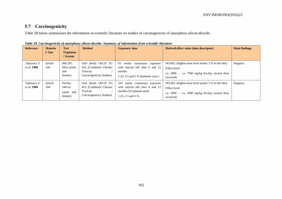
ENV/JM/MONO(2016)23
105
5.7 Carcinogenicity
Table 28 below summarises the information in scientific literature on studies of carcinogenicity of amorphous silicon dioxide.
Table 28. Carcinogenicity of amorphous silicon dioxide: Summary of information from scientific literature
Reference Materia
l/ Size
Test
Organism
/ System
Method Exposure/ dose Derived effect value (dose descriptor)
Main findings
Takizawa Y
et al, 1988
Syloid
244
B6C3F1
Mice (male
and
female)
Oral (feed) OECD TG
453 (Combined Chronic
Toxicity /
Carcinogenicity Studies)
93 weeks continuous exposure
with interim kill after 6 and 12
months
1.25, 2.5 and 5 % (nominal conc.)
NOAEL (highest dose level tested: 5 % in the diet)
Effect level
ca. 5000 — ca. 7000 mg/kg bw/day (actual dose
received)
Negative
Takizawa Y
et al, 1988
Syloid
244
Fischer
344 rat
(male and
female)
Oral (feed) OECD TG
453 (Combined Chronic
Toxicity /
Carcinogenicity Studies)
103 weeks continuous exposure
with interim kill after 6 and 12
months (10 animals each)
1.25, 2.5 and 5 %
NOAEL (highest dose level tested: 5 % in the diet)
Effect level
ca. 1800 — ca. 3000 mg/kg bw/day (actual dose
received)
Negative
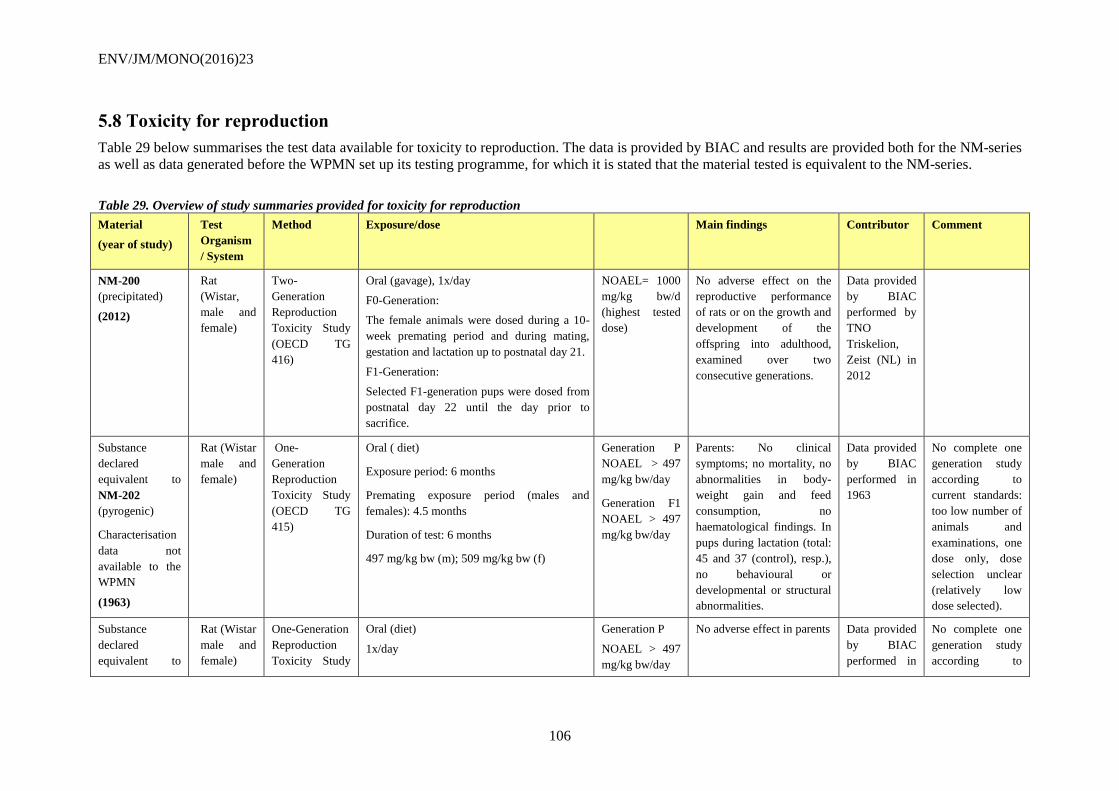
ENV/JM/MONO(2016)23
106
5.8 Toxicity for reproduction
Table 29 below summarises the test data available for toxicity to reproduction. The data is provided by BIAC and results are provided both for the NM-series
as well as data generated before the WPMN set up its testing programme, for which it is stated that the material tested is equivalent to the NM-series.
Table 29. Overview of study summaries provided for toxicity for reproduction
Material
(year of study)
Test
Organism
/ System
Method Exposure/dose Main findings Contributor Comment
NM-200
(precipitated)
(2012)
Rat
(Wistar,
male and
female)
Two-
Generation
Reproduction
Toxicity Study
(OECD TG
416)
Oral (gavage), 1x/day
F0-Generation:
The female animals were dosed during a 10-
week premating period and during mating,
gestation and lactation up to postnatal day 21.
F1-Generation:
Selected F1-generation pups were dosed from
postnatal day 22 until the day prior to
sacrifice.
NOAEL= 1000
mg/kg bw/d
(highest tested
dose)
No adverse effect on the
reproductive performance
of rats or on the growth and
development of the
offspring into adulthood,
examined over two
consecutive generations.
Data provided
by BIAC
performed by
TNO
Triskelion,
Zeist (NL) in
2012
Substance
declared
equivalent to
NM-202
(pyrogenic)
Characterisation
data not
available to the
WPMN
(1963)
Rat (Wistar
male and
female)
One-
Generation
Reproduction
Toxicity Study
(OECD TG
415)
Oral ( diet)
Exposure period: 6 months
Premating exposure period (males and
females): 4.5 months
Duration of test: 6 months
497 mg/kg bw (m); 509 mg/kg bw (f)
Generation P
NOAEL > 497
mg/kg bw/day
Generation F1
NOAEL > 497
mg/kg bw/day
Parents: No clinical
symptoms; no mortality, no
abnormalities in body-
weight gain and feed
consumption, no
haematological findings. In
pups during lactation (total:
45 and 37 (control), resp.),
no behavioural or
developmental or structural
abnormalities.
Data provided
by BIAC
performed in
1963
No complete one
generation study
according to
current standards:
too low number of
animals and
examinations, one
dose only, dose
selection unclear
(relatively low
dose selected).
Substance
declared
equivalent to
Rat (Wistar
male and
female)
One-Generation
Reproduction
Toxicity Study
Oral (diet)
1x/day
Generation P
NOAEL > 497
mg/kg bw/day
No adverse effect in parents Data provided
by BIAC
performed in
No complete one
generation study
according to
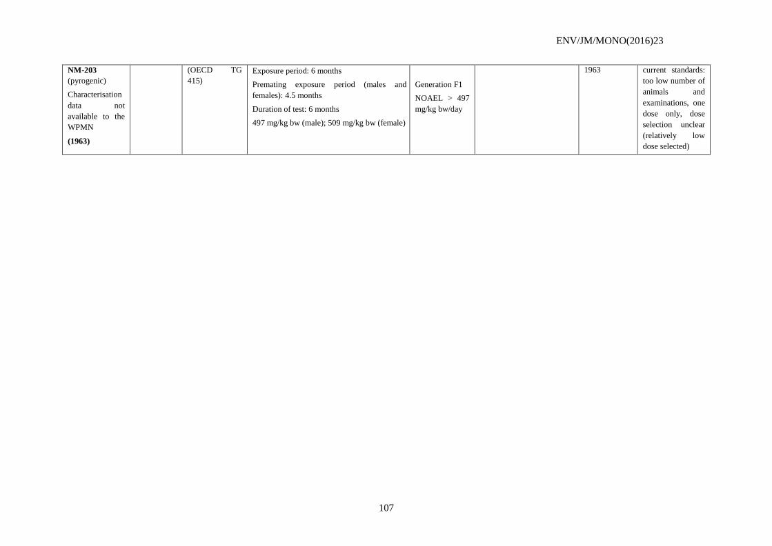
ENV/JM/MONO(2016)23
107
NM-203
(pyrogenic)
Characterisation
data not
available to the
WPMN
(1963)
(OECD TG
415)
Exposure period: 6 months
Premating exposure period (males and
females): 4.5 months
Duration of test: 6 months
497 mg/kg bw (male); 509 mg/kg bw (female)
Generation F1
NOAEL > 497
mg/kg bw/day
1963 current standards:
too low number of
animals and
examinations, one
dose only, dose
selection unclear
(relatively low
dose selected)
ENV/JM/MONO(2016)23
108
5.9 Toxicity in vitro
In vitro Mammalian Toxicology
The in vitro cytotoxicity of Synthetic Amorphous Silica (SAS) is strongly influenced by the
chemistry of the surface and in particular by the presence of two surface groups: the silanols (Si-
OH) and siloxanes (Si-O-Si). A number of in vitro studies studies on different SAS are available
from published literature. The larger the number of silanols and siloxanes groups on the surface of
SAS, the greater is the SAS reactivity and thus cytotoxicity. The covering of these active sites by
ions will modify the properties of the surface of silicas and can thus lead to a change, and in
particular to a reduction, of biological activity. The specific surface of SAS also influences their
cellular toxicity. (Chang et al., 2007; Jin et al., 2007; Lin et al., 2006; Lison et al., 2008;
Napierska et al., 2009; Sayes et al., 2007). Importantly, one study question the correlation of the
results obtained in vivo and in vitro (Sayes et al., 2007). Although, looking at all the studies
collected in Table30, some themes common to what is seen in vivo are emerging: even if SAS is
not always shown to be cytotoxic it is quite potent to induce an inflammatory response, most
probably mediated via oxidative stress (Lin, W., 2006; Park, EJ, 2009; Choi, SJ, 2009). In one
reference various epithelial cells are exposed to dye doped SAS without significant genotoxic
effects (Jin et al., 2007). In another study the Comet assay was shown to be reproducibly used to
study SAS nanoparticles and no significant genotoxicity on fibroblasts was detected (Barnes et
al., 2008), but one study shows also that silica nanoparticles can enter the cell and impair nuclear
functions (Chen, 2005).
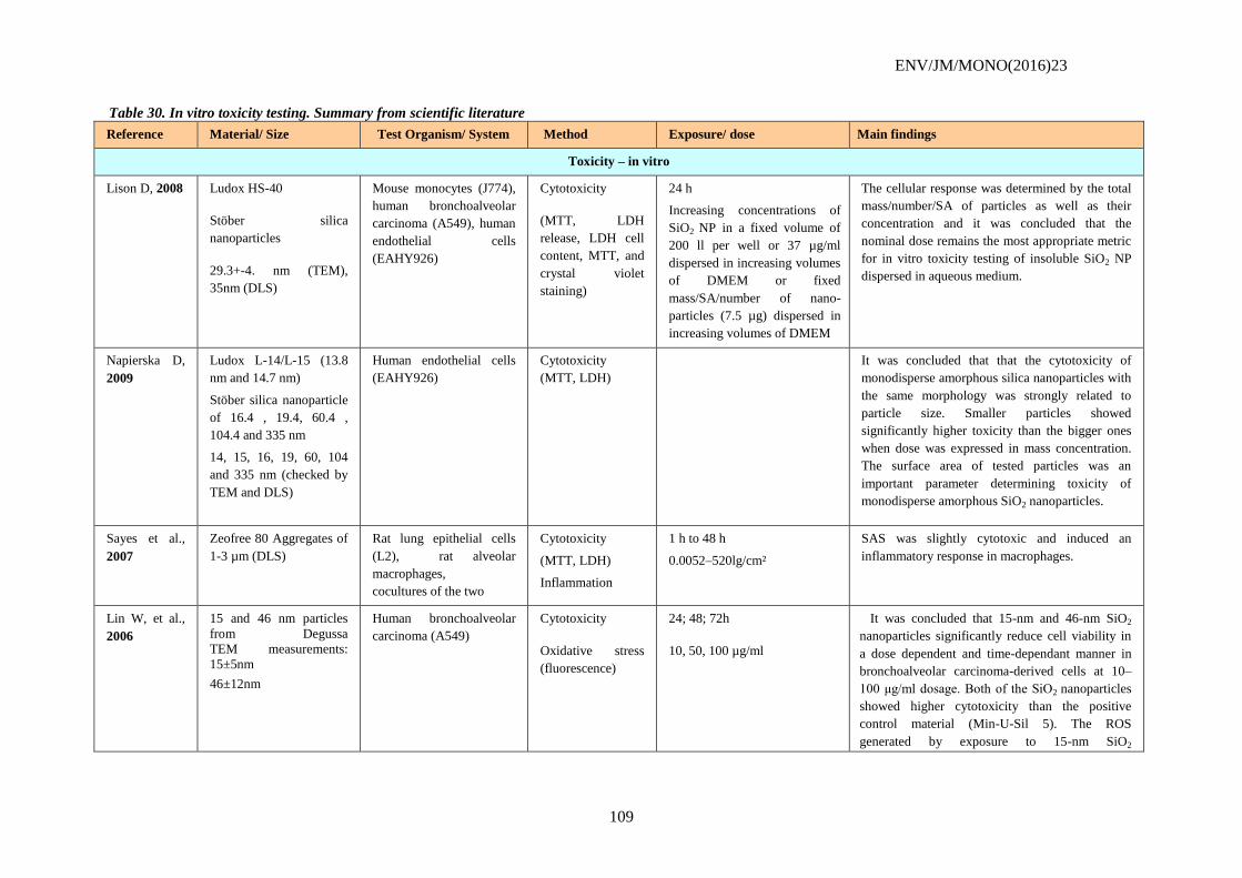
ENV/JM/MONO(2016)23
109
Table 30. In vitro toxicity testing. Summary from scientific literature
Reference Material/ Size Test Organism/ System Method Exposure/ dose Main findings
Toxicity – in vitro
Lison D, 2008 Ludox HS-40
Stöber silica
nanoparticles
29.3+-4. nm (TEM),
35nm (DLS)
Mouse monocytes (J774),
human bronchoalveolar
carcinoma (A549), human
endothelial cells
(EAHY926)
Cytotoxicity
(MTT, LDH
release, LDH cell
content, MTT, and
crystal violet
staining)
24 h
Increasing concentrations of
SiO2 NP in a fixed volume of
200 ll per well or 37 µg/ml
dispersed in increasing volumes
of DMEM or fixed
mass/SA/number of nano-
particles (7.5 µg) dispersed in
increasing volumes of DMEM
The cellular response was determined by the total
mass/number/SA of particles as well as their
concentration and it was concluded that the
nominal dose remains the most appropriate metric
for in vitro toxicity testing of insoluble SiO2 NP
dispersed in aqueous medium.
Napierska D,
2009
Ludox L-14/L-15 (13.8
nm and 14.7 nm)
Stöber silica nanoparticle
of 16.4 , 19.4, 60.4 ,
104.4 and 335 nm
14, 15, 16, 19, 60, 104
and 335 nm (checked by
TEM and DLS)
Human endothelial cells
(EAHY926)
Cytotoxicity
(MTT, LDH)
It was concluded that that the cytotoxicity of
monodisperse amorphous silica nanoparticles with
the same morphology was strongly related to
particle size. Smaller particles showed
significantly higher toxicity than the bigger ones
when dose was expressed in mass concentration.
The surface area of tested particles was an
important parameter determining toxicity of
monodisperse amorphous SiO2 nanoparticles.
Sayes et al.,
2007
Zeofree 80 Aggregates of
1-3 µm (DLS)
Rat lung epithelial cells
(L2), rat alveolar
macrophages,
cocultures of the two
Cytotoxicity
(MTT, LDH)
Inflammation
1 h to 48 h
0.0052–520lg/cm²
SAS was slightly cytotoxic and induced an
inflammatory response in macrophages.
Lin W, et al.,
2006
15 and 46 nm particles
from Degussa
TEM measurements:
15±5nm
46±12nm
Human bronchoalveolar
carcinoma (A549)
Cytotoxicity
Oxidative stress
(fluorescence)
24; 48; 72h
10, 50, 100 µg/ml
It was It was concluded that 15-nm and 46-nm SiO2
nanoparticles significantly reduce cell viability in
a dose dependent and time-dependant manner in
bronchoalveolar carcinoma-derived cells at 10–
100 μg/ml dosage. Both of the SiO2 nanoparticles
showed higher cytotoxicity than the positive
control material (Min-U-Sil 5). The ROS
generated by exposure to 15-nm SiO2
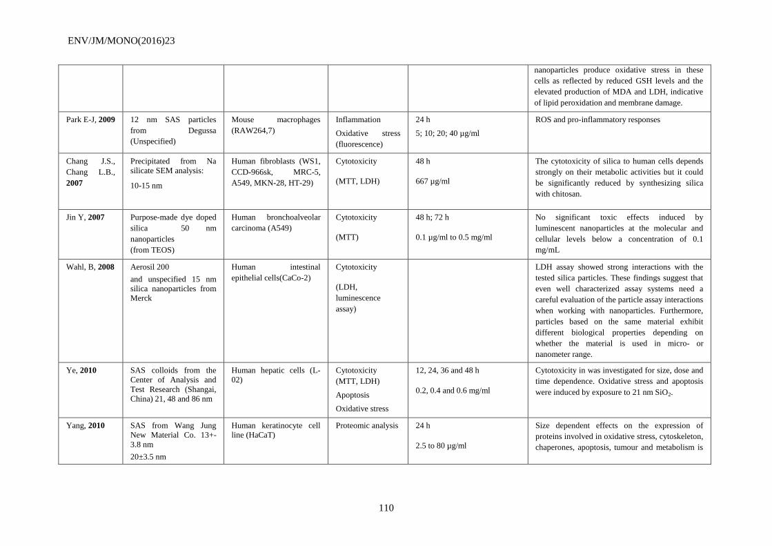
ENV/JM/MONO(2016)23
110
nanoparticles produce oxidative stress in these
cells as reflected by reduced GSH levels and the
elevated production of MDA and LDH, indicative
of lipid peroxidation and membrane damage.
Park E-J, 2009 12 nm SAS particles
from Degussa
(Unspecified)
Mouse macrophages
(RAW264,7)
Inflammation
Oxidative stress
(fluorescence)
24 h
5; 10; 20; 40 µg/ml
ROS and pro-inflammatory responses
Chang J.S.,
Chang L.B.,
2007
Precipitated from Na
silicate SEM analysis:
10-15 nm
Human fibroblasts (WS1,
CCD-966sk, MRC-5,
A549, MKN-28, HT-29)
Cytotoxicity
(MTT, LDH)
48 h
667 µg/ml
The cytotoxicity of silica to human cells depends
strongly on their metabolic activities but it could
be significantly reduced by synthesizing silica
with chitosan.
Jin Y, 2007 Purpose-made dye doped
silica 50 nm
nanoparticles
(from TEOS)
Human bronchoalveolar
carcinoma (A549)
Cytotoxicity
(MTT)
48 h; 72 h
0.1 µg/ml to 0.5 mg/ml
No significant toxic effects induced by
luminescent nanoparticles at the molecular and
cellular levels below a concentration of 0.1
mg/mL
Wahl, B, 2008 Aerosil 200
and unspecified 15 nm
silica nanoparticles from
Merck
Human intestinal
epithelial cells(CaCo-2)
Cytotoxicity
(LDH,
luminescence
assay)
LDH assay showed strong interactions with the
tested silica particles. These findings suggest that
even well characterized assay systems need a
careful evaluation of the particle assay interactions
when working with nanoparticles. Furthermore,
particles based on the same material exhibit
different biological properties depending on
whether the material is used in micro- or
nanometer range.
Ye, 2010 SAS colloids from the
Center of Analysis and
Test Research (Shangai,
China) 21, 48 and 86 nm
Human hepatic cells (L-
02)
Cytotoxicity
(MTT, LDH)
Apoptosis
Oxidative stress
12, 24, 36 and 48 h
0.2, 0.4 and 0.6 mg/ml
Cytotoxicity in was investigated for size, dose and
time dependence. Oxidative stress and apoptosis
were induced by exposure to 21 nm SiO2.
Yang, 2010 SAS from Wang Jung
New Material Co. 13+-
3.8 nm
20±3.5 nm
Human keratinocyte cell
line (HaCaT)
Proteomic analysis 24 h
2.5 to 80 µg/ml
Size dependent effects on the expression of
proteins involved in oxidative stress, cytoskeleton,
chaperones, apoptosis, tumour and metabolism is
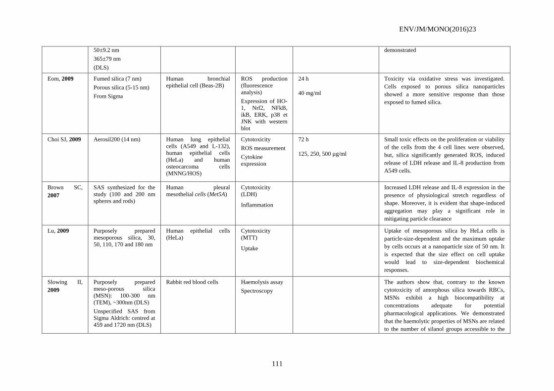
ENV/JM/MONO(2016)23
111
50±9.2 nm
365±79 nm
(DLS)
demonstrated
Eom, 2009 Fumed silica (7 nm)
Porous silica (5-15 nm)
From Sigma
Human bronchial
epithelial cell (Beas-2B)
ROS production
(fluorescence
analysis)
Expression of HO-
1, Nrf2, NFkB,
ikB, ERK, p38 et
JNK with western
blot
24 h
40 mg/ml
Toxicity via oxidative stress was investigated.
Cells exposed to porous silica nanoparticles
showed a more sensitive response than those
exposed to fumed silica.
Choi SJ, 2009 Aerosil200 (14 nm) Human lung epithelial
cells (A549 and L-132),
human epithelial cells
(HeLa) and human
osteocarcoma cells
(MNNG/HOS)
Cytotoxicity
ROS measurement
Cytokine
expression
72 h
125, 250, 500 µg/ml
Small toxic effects on the proliferation or viability
of the cells from the 4 cell lines were observed,
but, silica significantly generated ROS, induced
release of LDH release and IL-8 production from
A549 cells.
Brown SC,
2007
SAS synthesized for the
study (100 and 200 nm
spheres and rods)
Human pleural
mesothelial cells (Met5A)
Cytotoxicity
(LDH)
Inflammation
Increased LDH release and IL-8 expression in the
presence of physiological stretch regardless of
shape. Moreover, it is evident that shape-induced
aggregation may play a significant role in
mitigating particle clearance
Lu, 2009 Purposely prepared
mesoporous silica, 30,
50, 110, 170 and 180 nm
Human epithelial cells
(HeLa)
Cytotoxicity
(MTT)
Uptake
Uptake of mesoporous silica by HeLa cells is
particle-size-dependent and the maximum uptake
by cells occurs at a nanoparticle size of 50 nm. It
is expected that the size effect on cell uptake
would lead to size-dependent biochemical
responses.
Slowing II,
2009
Purposely prepared
meso-porous silica
(MSN): 100-300 nm
(TEM), ~300nm (DLS)
Unspecified SAS from
Sigma Aldrich: centred at
459 and 1720 nm (DLS)
Rabbit red blood cells Haemolysis assay
Spectroscopy
The authors show that, contrary to the known
cytotoxicity of amorphous silica towards RBCs,
MSNs exhibit a high biocompatibility at
concentrations adequate for potential
pharmacological applications. We demonstrated
that the haemolytic properties of MSNs are related
to the number of silanol groups accessible to the
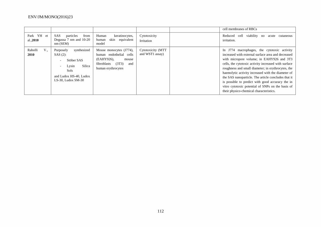
ENV/JM/MONO(2016)23
112
cell membranes of RBCs
Park YH et
al.,2010
SAS particles from
Degussa 7 nm and 10-20
nm (SEM)
Human keratinocytes,
human skin equivalent
model
Cytotoxicity
Irritation
Reduced cell viability no acute cutaneous
irritation.
Rabolli V.,
2010
Purposely synthesized
SAS (2):
- Stöber SAS
- Lysin Silica
Sols
and Ludox HS-40, Ludox
LS-30, Ludox SM-30
Mouse monocytes (J774),
human endothelial cells
(EAHY926), mouse
fibroblasts (3T3) and
human erythrocytes
Cytotoxicity (MTT
and WST1 assay)
In J774 macrophages, the cytotoxic activity
increased with external surface area and decreased
with micropore volume; in EAHY926 and 3T3
cells, the cytotoxic activity increased with surface
roughness and small diameter; in erythrocytes, the
haemolytic activity increased with the diameter of
the SAS nanoparticle. The article concludes that it
is possible to predict with good accuracy the in
vitro cytotoxic potential of SNPs on the basis of
their physico-chemical characteristics.
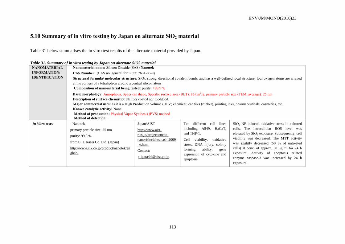
ENV/JM/MONO(2016)23
113
5.10 Summary of in vitro testing by Japan on alternate SiO2 material
Table 31 below summarises the in vitro test results of the alternate material provided by Japan.
Table 31. Summary of in vitro testing by Japan on alternate SiO2 material
NANOMATERIAL
INFORMATION/
IDENTIFICATION
Nanomaterial name: Silicon Dioxide (SAS) Nanotek
CAS Number: (CAS no. general for SiO2: 7631-86-9)
Structural formula/ molecular structure: SiO2, strong, directional covalent bonds, and has a well-defined local structure: four oxygen atoms are arrayed
at the corners of a tetrahedron around a central silicon atom
Composition of nanomaterial being tested: purity: >99.9 %
Basic morphology: Amorphous, Spherical shape, Specific surface area (BET): 86.0m2/g, primary particle size (TEM, average): 25 nm
Description of surface chemistry: Neither coated nor modified.
Major commercial uses: as it is a High Production Volume (HPV) chemical; car tires (rubber), printing inks, pharmaceuticals, cosmetics, etc.
Known catalytic activity: None
Method of production: Physical Vapor Synthesis (PVS) method
Method of detection:
In Vitro tests - Nanotek
primary particle size: 25 nm
purity: 99.9 %
from C. I. Kasei Co. Ltd. (Japan)
http://www.cik.co.jp/product/nanotek/en
glish/
Japan/AIST
http://www.aist-
riss.jp/projects/nedo-
nanorisk/rd/iwahashi2009
_e.html
Contact:
Ten different cell lines
including A549, HaCaT,
and THP-1.
Cell viability, oxidative
stress, DNA injury, colony
forming ability, gene
expression of cytokine and
apoptosis.
SiO2 NP induced oxidative stress in cultured
cells. The intracellular ROS level was
elevated by SiO2 exposure. Subsequently, cell
viability was decreased. The MTT activity
was slightly decreased (50 % of untreated
cells) at conc. of approx. 50 μg/ml for 24 h
exposure. Activity of apoptosis related
enzyme caspase-3 was increased by 24 h
exposure.
ENV/JM/MONO(2016)23
114
6. REFERENCES
[] http://timedomaincvd.com/CVD_Fundamentals/films/SiO2_properties.html
[] http://www.inchem.org/documents/sids/sids/SolubleSilicates.pdf
[] http://www.cdc.gov/niosh/npg/npgd0552.html, consulted 24 Nov. 2009
Introduction, Silica in the Environment and Appendices
Adams L. (2006). Adams, L.K., Lyon, D.Y. and Alvarez, P.J.J. "Comparative eco-toxicity of nanoscale TiO2,
SiO2 and ZnO water suspensions". Water Research, vol. 40, (2006), pp. 3527-3532.
Borm P. (2006). Borm, P., Klaessig, F.C., Landry, T.D., Moudgil, B., Pauluhn, J., Thomas, K., Trottier, R. and
Wood, S. "Research Strategies for Safety Evaluation of Nanomaterials, Part V: Role of Dissolution in
Biological Fate and Effects of Nanoscale Particles". Tox. Sci., vol.90, iss. 1 (2006), pp. 23-32.
Canesi L. (2010a). Canesi, L., Ciacci, C., Vallotto, D., Gallo, G., Marcomini, A. and Pojana, G. (2010) In vitro
effects of suspensions of selected nanoparticles (C60 fullerene, TiO2, SiO2) on Mytilus hemocytes.
Aquat Toxicol 96: 151-158.
Canesi L. (2010b). Canesi, L., Fabbri, R., Gallo, G., Vallotto, D. and Marcomini, A. (2010) Biomarkers in
Mytilus galloprovincialis exposed to suspensions of selected nanoparticles (C60 fullerene, Nano-TiO2,
Nano-SiO2). Aquat Toxicol 100: 168-177.
Casado (2013). Casado M., Macken A. and Byrne H.J. "Ecotoxicological Assessment of Silica and Polystyrene
Nanoparticles Assessed by a Multitrophic Test Battery". Environmental International, Vol. 51 (2013)
pp. 97-105
Currie (2007). Currie, H.A. and Perry, C.C. (2007) Silica in plants: Biological, biochemical and chemical
studies. Annals of Botany 100: 1383-1389
De Temmerman (2012). De Temmerman P.-J., Van Doren E., Verleysen E., Van der Stede Y., Abi Daoud
Francisco M. and Jan Mast J. Quantitative characterization of agglomerates and aggregates of
pyrogenic and precipitated amorphous silica nanomaterials by transmission electron microscopy
Journal of Nanobiotechnology 2012, 10:24
EC (EC, 2006). "Regulation (EC) No 1907/2006 of the European Parliament and of the Council of 18 December
2006 concerning the Registration, Evaluation, Authorisation and Restriction of Chemicals (REACH),
establishing a European Chemicals Agency, amending Directive 1999/45/EC and repealing Council
Regulation (EEC) No 793/93 and Commission Regulation (EC) No 1488/94 as well as Council
Directive 76/769/EEC and Commission Directives 91/155/EEC, 93/67/EEC, 93/105/EC and
2000/21/EC." Official Journal of the European Union (L396/1): 1-849.
EC (EC, 2007) Integrated Pollution Prevention and Control. Reference Document on Best Available Techniques
for the Manufacture of Large Volume Inorganic Chemicals - Solids and Others industry, chapter 5.
August 2007. http://eippcb.jrc.es.
ECETOC (2006). "Synthetic Amorphous Silica (CAS No. 7631-86-9)". JACC No. 51. ISSN-0773-6339-51
Farré M. (2009). Farré, M., Gajda-Schrants, K., Kantiani, L. and Barceló, D. "Ecotoxicity and analysis of
nanomaterials in the aquatic environment". Anal. Bioanal. Chem., vol. 393, (2009), pp. 81-95.
Fent (2010). Fent K., Weisbrod C.J., Wirth-Heller A. and Pieles U. "Assessment of uptake and toxicity of
fluorescent silica nanoparticles in zebrafish (Danio rerio) early life stages." Aquatic Toxicology 100
(2010) pp. 218-228
ENV/JM/MONO(2016)23
115
Fruijtier-Pölloth (2012). Fruijtier-Pölloth C. "The toxicological mode of action and the safety of synthetic
amorphous silica—A nanostructured material." Toxicology 294 (2012) 61– 79
Fujiwara K. (2008). Fujiwara, K., Suematsu, H., Kiyomiya, E., Aoki, M., Sata M.and Moritoki N. "Size
dependent toxicity of silica nano-particles to Chlorella kessleri". J. Environ. Sci. and Health, Part A,
vol. 43, iss. 10, (2008), pp. 1167-1173.
Garcia-Saucedo C. (2011). Garcia-Saucedo, C., Field, J.A., Otero-Gonzalez, L. and Sierra-Alvarez, R. (2011)
Low Toxicity of HfO2, SiO2, Al2O3 and CeO2 nanoparticles to the yeast, saccharomyces cerevisiae. J.
Haz. Mat. 192: 1572-1579
Handy R.D. (2008). Handy R.D., Owen, R. and Valsami-Jones, E. "The ecotoxicology of nanoparticles and
nanomaterials: current status, knowledge gaps, challenges and future needs". Ecotoxicology, vol. 17,
(2008), pp. 315-325.
Iler (1979). Iler, R. K. "The Chemistry of Silica". John Wiley & Sons: NewYork, 1979.
Ji J. (2011). Ji j., Long Z. and Lin D. "Toxicity of oxide nanoparticles to the green algae Chlorella sp."
Chemical Engeneering Journal 170 (2011) 525-530
W. (2009 Jiang). Jiang, W., Mashayekhi, H. and Xing, B. "Bacterial toxicity comparison between nano- and
micro-sized oxide particles". Environmnetal Pollution, vol. 157, (2009), pp. 1619-1625.
Kalteh (2014). Kalteh M., Alipour Z.T., Ashraf S., Aliabadi M.M. and Nosratabadi A.F. "Effect of silica
Nanoparticles on Basil (Ocimum basilicum) Under Saline Stress". Journal of Chemical Health Risks
(2014) 4(3), 49-55
Klaine S.J. (2008). Klaine, S.J., Alvarez, P.J.J., Batley, G.E., Fernandes, T.F., Handy, R. D., Lyon, D.Y.,
Mahendra, S., McLaughlin M.J. and Lead J.R. "Nanomaterials in the Environment: Behaviour, fate
bioavailability and effects". Environ. Tox. Chem., vol. 27, iss. 9, (2008), pp. 1825-1851.
Lee S.-W. (2009). Lee, S.-W., Kim, S.-M. and Choi, J. "Genotoxicity and ecotoxicity assays using the
freshwater crustacean Daphnia Magna and the larva of the aquatic midge Chironomus riparius to
screen the ecological risks of nanoparticle exposure". Environ. Tox. Pharm, vol. 28, (2009), pp. 86-91.
Liang (2007). Liang, Y., Sun, W., Zhu, Y.-G. and Christie, P. (2007) Mechanisms of silicon-mediated
alleviation of abiotic stresses in higher plants: A review. Env. Poll. 147: 422-428.
Lovestam G. (2010) Lovestam, G., Rauscher, H., Roebben, G. Sokull-Klüttgen, B. Gibson, N., Putuad, J.-P. and
Stamm,H. "Considerations on a Definition of Nanomaterial for Regulatory Purposes". European
Commission, Joint Research Centre, EUR-report 24403 (2010)
Mankiewicz-Boczek (2009). Mankiewicz-Boczek j., Osiecki R. and Rydzynski K. "Effect of Ceria and Silica
Nanoparticles on Pelagic (Thamnocephalus platyurus) and Benthic (Heterocypris incongruens)
Crustaceans " Poster presented at the 14th International Symposium on Toxicity Assessment, Metz,
France, August 30 – September 4, 2009
Metzler (2012). Metzler D.M., Erdem A., Tseng Y.H. and Huang C.P. "Response of Algal Cells to Engineered
Nanoparticles Measured as Algal Cell Population, Chlorophyll a, and Lipid Peroxidation: Effect of
Particle Size and Type". Journal of Nanotechnology, Vol. 2012.
Moore (2006). Moore, M.N. "Do nanoparticles present ecotoxicological risks for the health of the aquatic
environment?". Env. Int., vol. 32, (2006), pp. 967-976.
NANOGENOTOX dispersion protocol (2011). Available at URL
http://www.nanogenotox.eu/files/PDF/Deliverables/nanogenotox%20deliverable%203_wp4_%20dispe
rsion%20protocol.pdf
Nowack B. (2007). Nowack B. and Bucheli T.D. "Occurrence, behaviour and effects of nanoparticles in the
environment". Environmental Pollution.150, (2007) pp. 5-22.
ENV/JM/MONO(2016)23
116
OECD (2004a). OECD SIDS report "Synthetic Amorphous Silica and Silicates". UNEP publication. 2004.
Available from http://www.chem.unep.ch/irptc/sids/oecdsids/sidspub.html at
http://www.chem.unep.ch/irptc/sids/OECDSIDS/silicates.pdf .
OECD (2004b). OECD SIDS report "Soluble Silicates". UNEP publication. 2004. Available from
http://www.chem.unep.ch/irptc/sids/oecdsids/sidspub.html at
http://www.chem.unep.ch/irptc/sids/OECDSIDS/silicates.pdf .
OECD (2009). Preliminary Review of OECD Test Guidelines for their Applicability to Manufactured
Nanomaterials (2009). ENV/JM/MONO(2009)21.
OECD (2010). "Guidance Manual for the Testing of Manufactured Nanomaterials: OECD's Sponsorship
Programme". ENV/JM/MONO(2009)20-REV-ENG. OECD, Paris.
OECD (2012). ENV/JM/MONO(2012)40. Guidance on sample preparation and dosimetry for the safety testing of
manufactured nanomaterials, OECD, Paris. .
OECD (2014). Report of the OECD Expert Meeting on the Physical Chemical Properties of Manufactured
Nanomaterials and Test Guidelines. ENV/JM/MONO(2014)15. Available at http://www.oecd.org/
officialdocuments/publicdisplaydocumentpdf/?cote=ENV/JM/MONO(2014)15&docLanguage=En
OECD WPMN (2015). Annex 5 to the silicon dioxide dossier. Available at
http://www.oecd.org/officialdocuments/publicdisplaydocumentpdf/?cote=env/jm/mono%282015%2914/ann5
&doclanguage=en
Oya San (2014). Oya San N., Kursungoz C., Tumtas Y. Yasa O, Ortac B. and Tekinay T. "Novel one-step synthesis of
silica nanoparticles from sugarbeet bagasse by laser ablation and their effects on the growth of freshwater
algae culture" Particuology. (2014) in press.
Pluskota (2009). Pluskota, A., Horzowski, E., Bossinger, O. and von Mikecz, A. (2009) In caenorhabditis
elegans nanoparticle-bio-interactions become transparent: Silica nano-particles induce reproductive
senescence. PLoS ONE 4(8): e6622.
Rasmussen K.(2013). Rasmussen K., Mech A., Mast J., De Temmerman P.-J, Waegeneers N., Van Steen F.,
Pizzolon J.C., De Temmerman L., Van Doren E., Jensen K. A., Birkedal R., Levin M., Nielsen S.H.,
Koponen I.K., Clausen P.A., Kembouche Y., Thieriet N., Spalla O., Giuot C., Rousset D., Witschger O.,
Bau S., Bianchi B., Shivachev B., Gilliland D., Pianella F., Ceccone G., Cotogno G., Rauscher H.,
Gibson N. and Stamm H. "Synthetic Amorphous Silicon Dioxide (NM-200, NM-201, NM-202, NM-
203, NM-204). Characterisation and Physico-Chemical Properties" European Commission, Joint
Research Centre, EUR 26046. (2013)
Ramesh (2013). Ramesh, R., Kavitha, P., Kanipandian, N., Arun, S., Thirumurugan, R. and Subra, P. (2013)
Alteration of antioxidant enzymes and impairment of DNA in the SiO2 nanoparticles exposed zebra
fish (Danio rerio). Environ. Monit. Assess. 185: 5873-5881.
Reijnders L. (2009). L. Reijnders "The release of TiO2 and SiO2 nanoparticles from nanocomposites". Polymer
Degradation and Stability, 94 (2009) pp. 873-876.
SASSI (2008). "Nanoscale Materials Stewardship programme (NMSP) Voluntary Submittal Package for
Synthetic Amorphous Silica (CAS No. 7631-86-9)" prepared by The Synthetic Amorphous Silica and
Silicates Industry Association, SASSI. http://www.epa.gov/oppt/nano/sassia.pdf
Sharif (2012). Sharif, F., Porta, F., Meijer, A.H., Kros, A. and Richardson, M.K. (2012) Mesoporous sílica
nanoparticles as a compound delivery system in zebrafish embryos. Int. J. Nanomed. 2012:7, 1875-
1890.
Siddiqui (2014). Siddiqui M.H. and Al-Whaibi M.H. "Role of nano-SiO2 in germination of tomato
(Lycopersicum esculentum seeds Mill.)" Saudi Journal of Biological Sciences (2014) 21, pp. 13-17
ENV/JM/MONO(2016)23
117
Sneh (1995). Sneh O. and George S."Thermal Stability of Hydroxyl Groups on a Well-Defined Silica Surface",
J Phys Chem vol 99, p. 4639
Stone V. (2010). Stone, V., Nowack, B., Baun, A., van den Brink, N., Von der Kammer, F., Dusinska, M.,
Handy, R., Hankin, S., Hassellöv, M., Joner E.,and Fernandes T.F. "Nanomaterials for environmental
studies: Classification, reference materials issues and strategies for physico-chemical characterisation".
Sci.Total Environ (2010) Mar 1;408(7):1745-54.
Yang K. (2009). Yang, K., Lin, D. and Xing, B. "Interactions of Humic Acid with Nanosized Inorganic
Oxides". Langmuir, 25 (2009), pp. 3571-3576.
van Hoecke K. (2008). van Hoecke, K., de Schamphelaere, K.A.C., van der Meeren, P., Lucas, S. and Janssen,
C.R. "Ecotoxicity of silica nanoparticles to the green alga pseudokirchneriella subcapitata: importance
of surface area". Environ. Tox. Chem. vol. 27, iss. 9, (2008), pp. 1948-1957.
van Hoecke K. (2011). van Hoecke, K., de Schamphelaere, K.A.C., Ramirez-Garcia, S., Smagghe, G. and
Janssen, C.R. "Influence of alumina coating on characteristics and effects of SiO2 nanoparticles in algal
growth inhibition assays at various pH and organic matter contents. Environ. Int., 2011, 3: 1118-1125.
Wei (2010). Wei, C., Zhang, Y., Guo, J., Han, B., Yang, X. and Yuan, J. (2010) Effects of silica nanoparticles
on growth and photo synthetic pigment contents of Scenedesmus obliquus. J. Env. Sci. 22(1): 155-160.
Zhuravlev (2000). Zhuravlev L.T. (2000) The surface chemistry of amorphous silica. Zhuravlev model. Colloids
and Surfaces, A: Physicochemical and Engineering Aspects 173 (2000) 1–38
Silica and Mammalian Toxicology
In vivo
Arts J.H. (2007) Arts, J.H., Muijser, H., Duistermaat, E., Junker, K., and Kuper, C.F. (2007) Five-day inhalation
toxicity study of three types of synthetic amorphous silicas in Wistar rats and post-exposure
evaluations for up to 3 months. Food Chem Toxicol 2007 Oct; 45(10):1856-67.
Chen Y. (2004). Chen,Y., Chen,J., Dong,J. and Jin,Y. (2004) Comparing study of the effect of nanosized silicon
dioxide and microsized silicon dioxide on fibrogenesis in rats. Toxicol Ind Health 20: 21-27.
Cho W.S. (2007). Cho, W.S., Choi, M., Han, B.S., Cho, M., Oh, J., Park, K. Kim, S.J., Kim, S.H. and Jeong, J.
(2007) Inflammatory mediators induced by intratracheal instillation of ultrafine amorphous silica
particles. Toxicol. Lett. Dec 10; 175(1-3):24-33.
Cho M. (2009). Cho, M., Cho, W.S., Choi, M., Kim, S.J., Han, B.S., Kim, S.H., Kim, H.O., Sheen, Y.Y., and
Jeong, J. (2009) The impact of size on tissue distribution and elimination by single intravenous
injection of silica nanoparticles. Toxicol Lett. 189: 177-183.
Downs T.R. (2012). Downs, T. R., Crosby, M. E., Hu, T., Kumar, S., Sullivan, A., Sarlo, K., Reeder, B., Lynch,
M., Wagner, M., Mills, T. and Pfuhler, S. (2012). Silica nanoparticles administered at the maximum
tolerated dose induce genotoxic effects through an inflammatory reaction while gold nanoparticles do
not. Mutat Res 745: 38-50
Ernst H. (2002). Ernst, H., Rittinghausen, S., Bartsch, W., Creutzenberg, O., Dasenbrock, C., Gorlitz,B.D.,
Hecht, M., Kairies, U., Muhle, H., Müller, M., Heinrich, U. and Pott, F. (2002) Pulmonary
inflammation in rats after intratracheal instillation of quartz, amorphous SiO2, carbon black, and coal
dust and the influence of poly-2-vinylpyridine-N-oxide (PVNO). Exp Toxicol Pathol 54.
Hamilton R.F. (2008). Hamilton, R.F., Jr., Thakur, S.A. and Holian, A. (2008) Silica binding and toxicity in
alveolar macrophages. Free Radic Biol Med 44: 1246-1258.
ENV/JM/MONO(2016)23
118
Iyer R. (1996). Iyer, R., Hamilton, R.F., Li, L., and Holian, A. (1996) Silica-induced apoptosis mediated via
scavenger receptor in human alveolar macrophages. Toxicol Appl Pharmacol 141: 84-92.
Kaewamatawong T. (2005). Kaewamatawong,T., Kawamura,N., Okajima,M., Sawada, M., Morita,T. and
Shimada,A. (2005) Acute pulmonary toxicity caused by exposure to colloidal silica: particle size
dependent pathological changes in mice. Toxicol Pathol 33 (7):743-9
Merget R. (2002). Merget, R., Bauer, T., Kupper, H.U., Philippou, S., Bauer, H.D., Breitstadt, R., and Bruening,
T. (2002) Health hazards due to the inhalation of amorphous silica. Arch Toxicol 75: 625-634.
Napierska D. (2009). Napierska, D., Thomassen, L.C., Rabolli, V., Lison, D., Gonzalez, L., Kirsch-Volders, M.,
Martens, J.A. and Hoet, P.H. (2009) Size-dependent cytotoxicity of monodisperse silica nanoparticles
in human endothelial cells. Small 5. Apr;5(7):846-53.
Nemmar A. (2006). Nemmar, A., Hoet, P.H., and Nemery, B. (2006) Translocation of ultrafine particles.
Environ Health Perspect 114: A211-A212.
Park E.J. (2009). Park, E.J., and Park, K. (2009) Oxidative stress and pro-inflammatory responses induced by
silica nanoparticles in vivo and in vitro. Toxicol Lett 184(1): 18-25.
Park E.J. (2011). Park, E.J., Roh, J.K., Kim, Y., and Choi, K. (2011) A single instillation of amorphous silica
nanoparticles into mouse lungs induced subchronic inflammatory response. Environ Res. J. Health Sci.
57(1) 60-71.
Reuzel P.G. (1991). Reuzel, P.G., Bruijntjes, J.P., Feron, V.J., and Woutersen, R.A. (1991) Subchronic
inhalation toxicity of amorphous silicas and quartz dust in rats. Food Chem Toxicol 29: 341-354.
So S.J. (2008). So, S.J., Jang, I.S. and Han, C.S. (2008) Effect of micro/nano silica particle feeding for mice. J
Nanosci Nanotechnol. 8(10): 5367-5371.
TakizawaY. (1988). Takizawa, Y., Hirasawa, F., Noritomi, E., Aida, M., Tsunoda, H. and Uesugi, S. (1988)
Oral ingestion of syloid to mice and rats and its chronic toxicity and carcinogenicity. Acta Medica et
Biologica, 36, 27-56
Warheit D. (2006). Warheit, D.B., Webb, T.R., and Reed, K.L. (2006) Pulmonary toxicity screening studies in
male rats with TiO2 particulates substantially encapsulated with pyrogenically deposited, amorphous
silica. Part Fibre Toxicol 3.
Warheit D. (1995). Warheit, D.B., McHugh, T.A., and Hartsky, M.A. (1995) Differential pulmonary responses
in rats inhaling crystalline, colloidal or amorphous silica dusts. Scand J Work Environ Health 21 Suppl
2: 19-21.
In vitro
Barik T.K. (2008). Barik, T. K., Sahu, B. and Swain V. "Nanosilica-from medicine to pest control."
Parasitol.Res. 103.2 (2008): 253-58.
Barnes C.A. (2008). Barnes, C.A., Elsaesser, A., Arkusz, J., Smok, A., Palus, J., Lesniak, A. et al. (2008)
Reproducible comet assay of amorphous silica nanoparticles detects no genotoxicity. Nano Lett 8:
3069-3074.
Brown S.C. (2007). Brown, S.C., Kamal, M., Nasreen, N., Baumuratov, A., Sharma, P., Antony, V.b. and
Moudgil, B.M. (2007) Influence of shape, adhesion and simulated lung mechanics on amorphous silica
nanoparticle toxicity. Advanced Powder Technol 18: 69-79.
Chang J.S. (2007). Chang, J.S., Chang,K.L., Hwang,D.F., and Kong,Z.L. (2007) In vitro cytotoxicitiy of silica
nanoparticles at high concentrations strongly depends on the metabolic activity type of the cell line.
Environ Sci Technol 41.
Choi S.J. (2009). Choi, S.J., Oh, J.M. and Choy, J.H. (2009) Toxicological effects of inorganic nanoparticles on
human lung cancer A549 cells. J Inorg Biochem 103: 463-471.
ENV/JM/MONO(2016)23
119
Eom H.J. (2009). Eom,H.J., and Choi,J. (2009) Oxidative stress of silica nanoparticles in human bronchial
epithelial cell, Beas-2B. Toxicol In Vitro 23: 1326-1332.
Gonzalez L. (2010). Gonzalez, L., Thomassen, L.C., Plas, G., Rabolli, V., Napierska, D. and Decordier, I. et al.
Exploring the aneugenic and clastogenic potential in the nanosize range: A549 human lung carcinoma
cells and amorphous monodisperse silica nanoparticles as models. Nanotoxicology 2010 Dec;4:382-95.
Jin Y. (2007). Jin,Y., Kannan,S., Wu,M., and Zhao,J.X. (2007) Toxicity of luminescent silica nanoparticles to
living cells. Chem Res Toxicol 20.
Lin W. (2006). Lin,W., Huang,Y.W., Zhou,X.D., and Ma,Y. (2006) In vitro toxicity of silica nanoparticles in
human lung cancer cells. Toxicol Appl Pharmacol 217.
Lison D. (2008). Lison,D., Thomassen,L.C., Rabolli,V., Gonzalez,L., Napierska,D., Seo,J.W. et al. (2008)
Nominal and effective dosimetry of silica nanoparticles in cytotoxicity assays. Toxicol Sci 104.
Lu F. (2009). Lu,F., Wu,S.H., Hung,Y., and Mou,C.Y. (2009) Size effect on cell uptake in well-suspended,
uniform mesoporous silica nanoparticles. Small 5: 1408-1413.
Napierska D. (2009). Napierska,D., Thomassen,L.C., Rabolli,V., Lison,D., Gonzalez,L., Kirsch-Volders,M. et
al. (2009) Size-dependent cytotoxicity of monodisperse silica nanoparticles in human endothelial cells.
Small 5.
Park E.J.(2009). Park,E.J., and Park,K. (2009) Oxidative stress and pro-inflammatory responses induced by
silica nanoparticles in vivo and in vitro. Toxicol Lett 184.
Sayes C.M. (2007). Sayes,C.M., Reed,K.L., and Warheit,D.B. (2007) Assessing toxicity of fine and
nanoparticles: comparing in vitro measurements to in vivo pulmonary toxicity profiles. Toxicol Sci 97.
Slowing (2009). Slowing, II, Wu,C.W., Vivero-Escoto,J.L. and Lin,V.S. (2009) Mesoporous silica nanoparticles
for reducing hemolytic activity towards mammalian red blood cells. Small 5.
Wahl B. (2008). Wahl,B., Daum,N., Ohrem,H.L., and Lehr,C.M. (2008) Novel luminescence assay offers new
possibilities for the risk assessment of silica nanoparticles. Nanotoxicology 2: 243-251.
Yang X. (2010). Yang,X., Liu,J., He,H., Zhou,L., Gong,C., Wang,X. et al. (2010) SiO2 nanoparticles induce
cytotoxicity and protein expression alteration in HaCaT cells. Part Fibre Toxicol 7: 1.
Ye Y. (2010). Ye,Y., Liu,J., Xu,J., Sun,L., Chen,M., and Lan,M. (2010) Nano-SiO2 induces apoptosis via
activation of p53 and Bax mediated by oxidative stress in human hepatic cell line. Toxicol In Vitro 24:
751-758.
O'Farrell N. (2006). N. O'Farrell, A. Houlton and B.R. Horrocks. "Silicon nanoparticles: applications in cell
biology and medicine". Int. J. Nanomedicine, I(4), (2006) pp. 451-472.
Park Y.H. (2010). Park Y.H., Kim, J.N., Jeong, S.H., Choi, J.E., Lee, S.H., Choi, B.H., Lee, J.P., Sohn, K.H.,
Park, K.L., Kim, M.K., and Son, S.W. (2010) Assessment of dermal toxicity of nanosilica using
cultured keratinocytes, a human skin equivalent model and an in vivo model. Toxicology. 267: 178-
181.
Rabolli (2010). Rabolli V, Thomassen LC, Princen C, Napierska D, Gonzalez L, Kirsch-Volders M, et al.
Influence of size, surface area and microporosity on the in vitro cytotoxic activity of amorphous silica
nanoparticles in different cell types. Nanotoxicology 2010 Sep;4(3):307-18.
Thomassen L.C.(2010). Thomassen, L.C., Aerts, A., Rabolli, V., Lison, D., Gonzalez, L., Kirsch-Volders M, et
al. Synthesis and characterization of stable monodisperse silica nanoparticle sols for in vitro
cytotoxicity testing. Langmuir 2010 Jan 5;26(1):328-35.
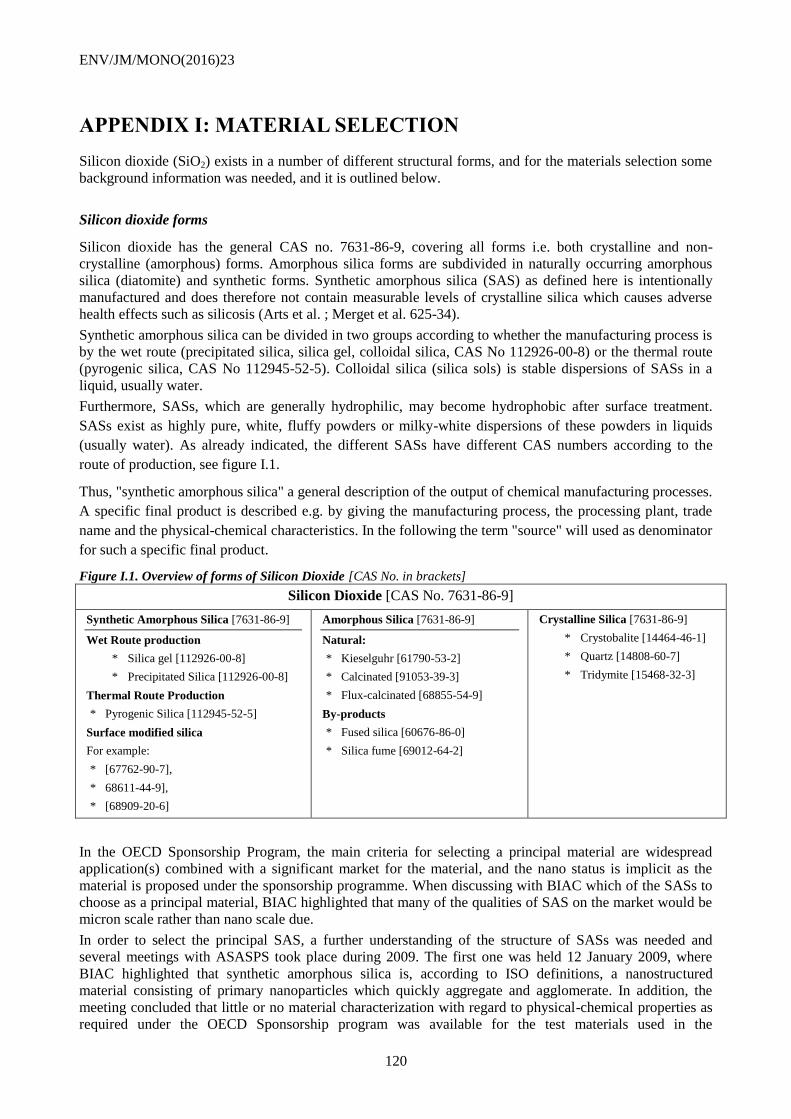
ENV/JM/MONO(2016)23
120
APPENDIX I: MATERIAL SELECTION
Silicon dioxide (SiO2) exists in a number of different structural forms, and for the materials selection some
background information was needed, and it is outlined below.
Silicon dioxide forms
Silicon dioxide has the general CAS no. 7631-86-9, covering all forms i.e. both crystalline and non-
crystalline (amorphous) forms. Amorphous silica forms are subdivided in naturally occurring amorphous
silica (diatomite) and synthetic forms. Synthetic amorphous silica (SAS) as defined here is intentionally
manufactured and does therefore not contain measurable levels of crystalline silica which causes adverse
health effects such as silicosis (Arts et al. ; Merget et al. 625-34).
Synthetic amorphous silica can be divided in two groups according to whether the manufacturing process is
by the wet route (precipitated silica, silica gel, colloidal silica, CAS No 112926-00-8) or the thermal route
(pyrogenic silica, CAS No 112945-52-5). Colloidal silica (silica sols) is stable dispersions of SASs in a
liquid, usually water.
Furthermore, SASs, which are generally hydrophilic, may become hydrophobic after surface treatment.
SASs exist as highly pure, white, fluffy powders or milky-white dispersions of these powders in liquids
(usually water). As already indicated, the different SASs have different CAS numbers according to the
route of production, see figure I.1.
Thus, "synthetic amorphous silica" a general description of the output of chemical manufacturing processes.
A specific final product is described e.g. by giving the manufacturing process, the processing plant, trade
name and the physical-chemical characteristics. In the following the term "source" will used as denominator
for such a specific final product.
Figure I.1. Overview of forms of Silicon Dioxide [CAS No. in brackets]
Silicon Dioxide [CAS No. 7631-86-9]
Synthetic Amorphous Silica [7631-86-9]
Wet Route production
* Silica gel [112926-00-8]
* Precipitated Silica [112926-00-8]
Thermal Route Production
* Pyrogenic Silica [112945-52-5]
Surface modified silica
For example:
* [67762-90-7],
* 68611-44-9],
* [68909-20-6]
Amorphous Silica [7631-86-9]
Natural:
* Kieselguhr [61790-53-2]
* Calcinated [91053-39-3]
* Flux-calcinated [68855-54-9]
By-products
* Fused silica [60676-86-0]
* Silica fume [69012-64-2]
Crystalline Silica [7631-86-9]
* Crystobalite [14464-46-1]
* Quartz [14808-60-7]
* Tridymite [15468-32-3]
In the OECD Sponsorship Program, the main criteria for selecting a principal material are widespread
application(s) combined with a significant market for the material, and the nano status is implicit as the
material is proposed under the sponsorship programme. When discussing with BIAC which of the SASs to
choose as a principal material, BIAC highlighted that many of the qualities of SAS on the market would be
micron scale rather than nano scale due.
In order to select the principal SAS, a further understanding of the structure of SASs was needed and
several meetings with ASASPS took place during 2009. The first one was held 12 January 2009, where
BIAC highlighted that synthetic amorphous silica is, according to ISO definitions, a nanostructured
material consisting of primary nanoparticles which quickly aggregate and agglomerate. In addition, the
meeting concluded that little or no material characterization with regard to physical-chemical properties as
required under the OECD Sponsorship program was available for the test materials used in the
ENV/JM/MONO(2016)23
121
toxicological and ecotoxicological studies7 presented on this occasion. Since SAS consists of aggregates
with a mean size above the nanoscale, which build up agglomerates with a size range above 100µm, it was
not straight forward to decide which SAS material(s) to select as a principal material for the Sponsorship
Program and quite some discussion took place, arriving at the following conclusions:
Based on current information available, the aggregates and agglomerates of SASs used in the current main
applications are larger than the nano-scale. In addition, while the SAS aggregates and agglomerates are
known to be nanostructured, the number of nano-sized aggregates/agglomerates and discrete nanoparticles,
which they may contain, is not clear as characterization data are lacking; when measuring the size of SAS
as supplied from a manufacturer the measurement would reflect the particle size distribution of the
aggregates/agglomerates, with a tail of particles with smaller sizes. Furthermore, the measured size of SAS
in dispersion strongly depends on the applied shear forces during sample preparation.
Analysis of some background information
Three main reference reports on silicon dioxide have been identified: the aforementioned reports ECETOC
JACC report No. 51 and the OECD HPV report and the SASSI report.
Examining the available information relating to the physical-chemical data presented in these reports, and
especially the characterization of size of particles used in the studies shows that none of the reports contain
any detailed characterization data with regard to physical-chemical properties as required under the OECD
Sponsorship program.
The OECD High Production Volume (HPV) Chemicals report on synthetic amorphous silica and silicates
gives a good general description of SAS under the HPV program. An underlying assumption in this report,
which was agreed to by OECD under the HPV program, is that the different SASs are sufficiently similar
that their data obtained by using OECD test guidelines (TG) for chemicals can be combined into one HPV
dataset. As a consequence of this combination of data, the base data set for one source of SAS, as required
under the OECD WPMN program, cannot be extracted from the summary information presented. The
report contains limited detailed characterization data with regard to the physical-chemical properties as
required under the OECD Sponsorship program. In order to possibly use the data for the OECD
sponsorship program for nanomaterials it would be necessary to extract and evaluate the substance
characterization information, if available, from each original study.
The JACC report, p.12, cites information that the primary particle size is in the range of 0.001 to 0.1
micron, i.e. the primary particles are nano-sized, adding the footnote that "Primary particles do not
normally exist as individual units". The report analyses in more detail the size of particles concluding that
(p. 38) "Under conditions of normal technical handling and use, agglomerates8 are the relevant particles,
both for pyrogenic and precipitated SAS." Appendix B of the JACC report lists the SAS types mentioned in
the report and the list contains 49 hydrophilic SAS, which are the ones of interest for the OECD
sponsorship program. The report does not give a detailed material characterization with regard to the
physical-chemical properties as required under the OECD Sponsorship program. The JACC report indicates
[p. 25] that the different sources of SAS have different particle structures depending on the route of
manufacture. In order to possibly use the data for the OECD Sponsorship Program it would be necessary to
extract and evaluate the substance characterization information, if available, from each original study.
The SASSI report is essentially based on data presented in the two reports mentioned above and it
demonstrates that SAS is a nanostructured material, but does not provide in-depth characterization data for
individual sources of SAS with regard to the physical-chemical properties as required under the OECD
Sponsorship program. In addition, the SASSI report indicates [page 16] that depending on the
manufacturing process, the different sources of SAS differ across several physical-chemical properties,
including size and surface area.
It is important, in this context, to note that the OECD WPMN and the OECD HPV programme are two
programmes addressing different issues. The OECD HPV programme addresses industrial chemicals
7 The outcome of those studies are part of the information found the document published by ECETOC, Synthetic
amorphous silica, JACC n° 51, ISSN-0773-6339-51, 2006.
8 In the JACC report, p. 24, the convention is described: "Agglomerates are assemblies of aggregates"
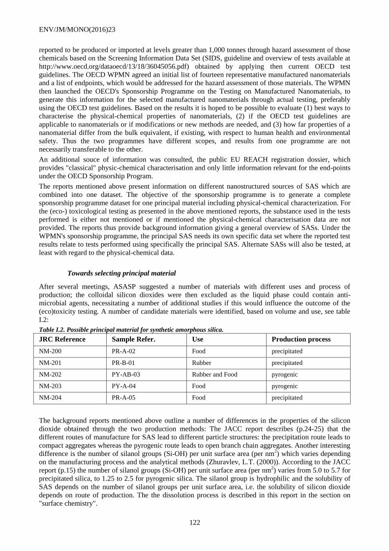
ENV/JM/MONO(2016)23
122
reported to be produced or imported at levels greater than 1,000 tonnes through hazard assessment of those
chemicals based on the Screening Information Data Set (SIDS, guideline and overview of tests available at
http://www.oecd.org/dataoecd/13/18/36045056.pdf) obtained by applying then current OECD test
guidelines. The OECD WPMN agreed an initial list of fourteen representative manufactured nanomaterials
and a list of endpoints, which would be addressed for the hazard assessment of those materials. The WPMN
then launched the OECD's Sponsorship Programme on the Testing on Manufactured Nanomaterials, to
generate this information for the selected manufactured nanomaterials through actual testing, preferably
using the OECD test guidelines. Based on the results it is hoped to be possible to evaluate (1) best ways to
characterise the physical-chemical properties of nanomaterials, (2) if the OECD test guidelines are
applicable to nanomaterials or if modifications or new methods are needed, and (3) how far properties of a
nanomaterial differ from the bulk equivalent, if existing, with respect to human health and environmental
safety. Thus the two programmes have different scopes, and results from one programme are not
necessarily transferable to the other.
An additional souce of information was consulted, the public EU REACH registration dossier, which
provides "classical" physic-chemical characterisation and only little information relevant for the end-points
under the OECD Sponsorship Program.
The reports mentioned above present information on different nanostructured sources of SAS which are
combined into one dataset. The objective of the sponsorship programme is to generate a complete
sponsorship programme dataset for one principal material including physical-chemical characterization. For
the (eco-) toxicological testing as presented in the above mentioned reports, the substance used in the tests
performed is either not mentioned or if mentioned the physical-chemical characterisation data are not
provided. The reports thus provide background information giving a general overview of SASs. Under the
WPMN's sponsorship programme, the principal SAS needs its own specific data set where the reported test
results relate to tests performed using specifically the principal SAS. Alternate SASs will also be tested, at
least with regard to the physical-chemical data.
Towards selecting principal material
After several meetings, ASASP suggested a number of materials with different uses and process of
production; the colloidal silicon dioxides were then excluded as the liquid phase could contain anti-
microbial agents, necessitating a number of additional studies if this would influence the outcome of the
(eco)toxicity testing. A number of candidate materials were identified, based on volume and use, see table
I.2:
Table I.2. Possible principal material for synthetic amorphous silica.
JRC Reference Sample Refer. Use Production process
NM-200 PR-A-02 Food precipitated
NM-201 PR-B-01 Rubber precipitated
NM-202 PY-AB-03 Rubber and Food pyrogenic
NM-203 PY-A-04 Food pyrogenic
NM-204 PR-A-05 Food precipitated
The background reports mentioned above outline a number of differences in the properties of the silicon
dioxide obtained through the two production methods: The JACC report describes (p.24-25) that the
different routes of manufacture for SAS lead to different particle structures: the precipitation route leads to
compact aggregates whereas the pyrogenic route leads to open branch chain aggregates. Another interesting
difference is the number of silanol groups (Si-OH) per unit surface area (per nm2) which varies depending
on the manufacturing process and the analytical methods (Zhuravlev, L.T. (2000)). According to the JACC
report (p.15) the number of silanol groups (Si-OH) per unit surface area (per nm2) varies from 5.0 to 5.7 for
precipitated silica, to 1.25 to 2.5 for pyrogenic silica. The silanol group is hydrophilic and the solubility of
SAS depends on the number of silanol groups per unit surface area, i.e. the solubility of silicon dioxide
depends on route of production. The the dissolution process is described in this report in the section on
"surface chemistry".
ENV/JM/MONO(2016)23
123
APPENDIX II: CONTACT DETAILS FOR INVOLVED
INSTITUTIONS (2014)
A. European Commission (EC)
Joint Research Centre, Institute for Health and Consumer Protection (JRC, IHCP)
Kirsten Rasmussen
TP 125, Via E Fermi 2749
21027 Ispra (VA), Italy
Tel.: +(39) 0332 78 5344
E-mail: [email protected]
B. France
Ministère de la Santé, de la Jeunesse et des Sports
Direction Générale de la Santé
Myriam Saihi
14 avenue Duquesne
75350 Paris 07 SP
Tel: +33 1 4056 7929
Fax: +33 1 4056 5056
ANSES (French Agency for Food, Environmental and Occupational Health & Safety)
Nathalie Thieriet
253, Av. Du General Leclerc
F-94701 Maisons-Alfort Cedex
Tel: +33 1 5629 5199
Fax: +33 1 4396 3767
E-mail: [email protected]
C. Belgium
SPF Sante publique, Sécurité de la
chaine alimentaire et
environnement
Juan David Piñeros Garcet
Direction générale Environnement
Service Maîtrise des risques
Place Victor Horta 40, bte 10
B-1060 Bruxelles.
Tel: +32 (0)2 524 96 72
Scientific Institute of Public Health
Paul Troisfontaines
Juliette Wytsmanstraat 14
B-1050 Brussels
Tel: +(32) 2 642 53 86
Fax: +(32) 2 642 52 24
Email:
ENV/JM/MONO(2016)23
124
D. Korea
National Institute of Environmental Research (NIER)
Ministry of Environment
Kyunghee Choi
Environmental Research Complex
Kyungseo-dong,
Seo-gu,
404-708 Incheon
Korea
Tel.: +(82) 32 560 7206
Fax: +(82) 32 568 2041
E-mail: [email protected]
Sang Hee Lee
Environmental Exposure Assessment
Division
National Institute of Environmental
Research
Gyeongseo-dong, Seo-gu
404-708 Incheon,
Korea
Tel.: +(82) 32 560 7216
Fax: +(82) 32 568 2041
E-mail: [email protected]
E. BIAC
On behalf of CEFIC ASASP sector group
Monika Maier, PhD
Evonik Resource Efficiency GmbH
Rodenbacher Chaussee 4
63457 Hanau-Wolfgang
Germany
Tel: 0049-6181-59-4964
Fax:0049-6181-59-4205
E-mail: [email protected]
Philippe Cochet
Solvay
170 Avenue Thiers
F-69006 Lyon
Tel: 0033 4 37 24 88 88
Fax: 0033 4 37 24 88 89
E-Mail: [email protected]
Mario Heienmann, PhD
Wacker Chemie AG
Johannes-Hess-Straße24
D-84489 Burghausen
Tel: 00498677 83-4022
Fax: 00498677 83-6814
E-Mail: [email protected]
F. Denmark
National Research Centre for the Working Environment (NRCWE)
Keld Alstrup Jensen
Nanotoxicology and Occupational Hygiene Group
Lersø Parkallé 105
DK-2100 Copenhagen, Denmark
Tel. direct: +45 3916 5302
E-mail [email protected]
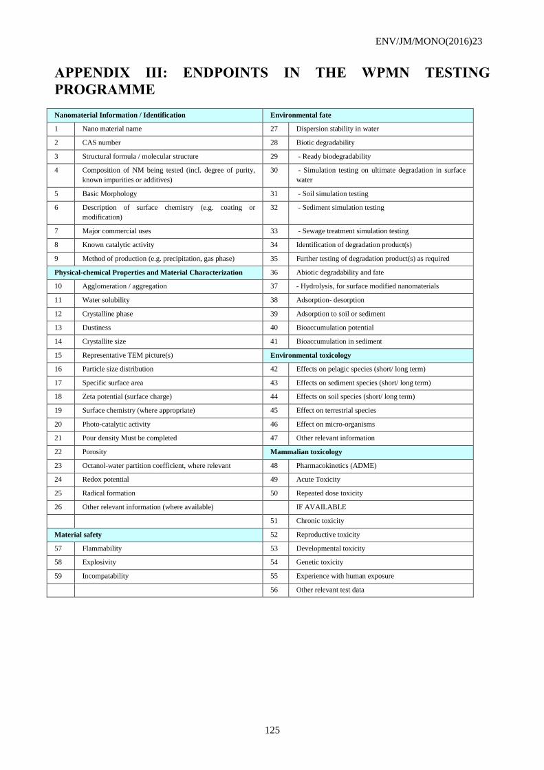
ENV/JM/MONO(2016)23
125
APPENDIX III: ENDPOINTS IN THE WPMN TESTING
PROGRAMME
Nanomaterial Information / Identification Environmental fate
1 Nano material name 27 Dispersion stability in water
2 CAS number 28 Biotic degradability
3 Structural formula / molecular structure 29 - Ready biodegradability
4 Composition of NM being tested (incl. degree of purity,
known impurities or additives)
30 - Simulation testing on ultimate degradation in surface
water
5 Basic Morphology 31 - Soil simulation testing
6 Description of surface chemistry (e.g. coating or
modification)
32 - Sediment simulation testing
7 Major commercial uses 33 - Sewage treatment simulation testing
8 Known catalytic activity 34 Identification of degradation product(s)
9 Method of production (e.g. precipitation, gas phase) 35 Further testing of degradation product(s) as required
Physical-chemical Properties and Material Characterization 36 Abiotic degradability and fate
10 Agglomeration / aggregation 37 - Hydrolysis, for surface modified nanomaterials
11 Water solubility 38 Adsorption- desorption
12 Crystalline phase 39 Adsorption to soil or sediment
13 Dustiness 40 Bioaccumulation potential
14 Crystallite size 41 Bioaccumulation in sediment
15 Representative TEM picture(s) Environmental toxicology
16 Particle size distribution 42 Effects on pelagic species (short/ long term)
17 Specific surface area 43 Effects on sediment species (short/ long term)
18 Zeta potential (surface charge) 44 Effects on soil species (short/ long term)
19 Surface chemistry (where appropriate) 45 Effect on terrestrial species
20 Photo-catalytic activity 46 Effect on micro-organisms
21 Pour density Must be completed 47 Other relevant information
22 Porosity Mammalian toxicology
23 Octanol-water partition coefficient, where relevant 48 Pharmacokinetics (ADME)
24 Redox potential 49 Acute Toxicity
25 Radical formation 50 Repeated dose toxicity
26 Other relevant information (where available) IF AVAILABLE
51 Chronic toxicity
Material safety 52 Reproductive toxicity
57 Flammability 53 Developmental toxicity
58 Explosivity 54 Genetic toxicity
59 Incompatability 55 Experience with human exposure
56 Other relevant test data
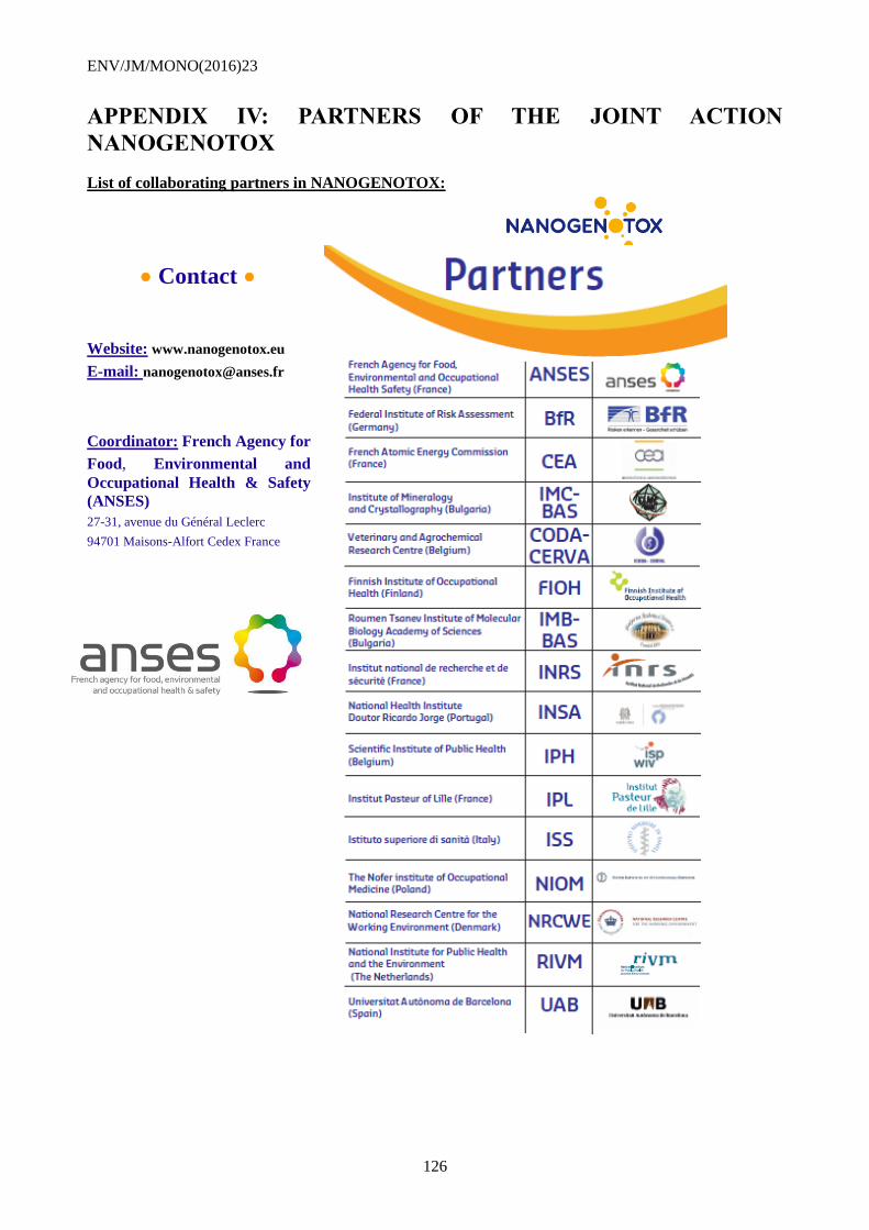
ENV/JM/MONO(2016)23
126
APPENDIX IV: PARTNERS OF THE JOINT ACTION
NANOGENOTOX
List of collaborating partners in NANOGENOTOX:
Contact
Website: www.nanogenotox.eu
E-mail: [email protected]
Coordinator: French Agency for
Food, Environmental and
Occupational Health & Safety
(ANSES)
27-31, avenue du Général Leclerc
94701 Maisons-Alfort Cedex France
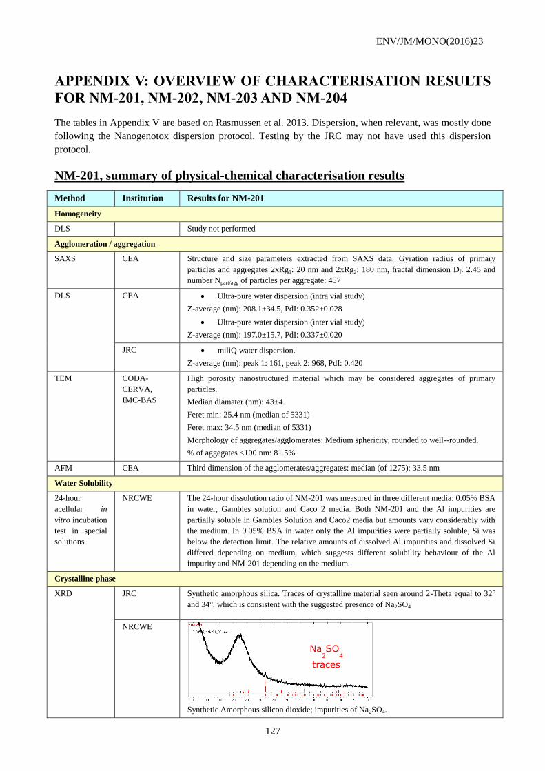
ENV/JM/MONO(2016)23
127
APPENDIX V: OVERVIEW OF CHARACTERISATION RESULTS
FOR NM-201, NM-202, NM-203 AND NM-204
The tables in Appendix V are based on Rasmussen et al. 2013. Dispersion, when relevant, was mostly done
following the Nanogenotox dispersion protocol. Testing by the JRC may not have used this dispersion
protocol.
NM-201, summary of physical-chemical characterisation results
Method Institution Results for NM-201
Homogeneity
DLS Study not performed
Agglomeration / aggregation
SAXS CEA Structure and size parameters extracted from SAXS data. Gyration radius of primary
particles and aggregates 2xRg1: 20 nm and 2xRg2: 180 nm, fractal dimension Df: 2.45 and
number Npart/agg of particles per aggregate: 457
DLS CEA Ultra-pure water dispersion (intra vial study)
Z-average (nm): 208.1±34.5, PdI: 0.352±0.028
Ultra-pure water dispersion (inter vial study)
Z-average (nm): 197.0±15.7, PdI: 0.337±0.020
JRC miliQ water dispersion.
Z-average (nm): peak 1: 161, peak 2: 968, PdI: 0.420
TEM CODA-
CERVA,
IMC-BAS
High porosity nanostructured material which may be considered aggregates of primary
particles.
Median diamater (nm): 43±4.
Feret min: 25.4 nm (median of 5331)
Feret max: 34.5 nm (median of 5331)
Morphology of aggregates/agglomerates: Medium sphericity, rounded to well--rounded.
% of aggegates <100 nm: 81.5%
AFM CEA Third dimension of the agglomerates/aggregates: median (of 1275): 33.5 nm
Water Solubility
24-hour
acellular in
vitro incubation
test in special
solutions
NRCWE The 24-hour dissolution ratio of NM-201 was measured in three different media: 0.05% BSA
in water, Gambles solution and Caco 2 media. Both NM-201 and the Al impurities are
partially soluble in Gambles Solution and Caco2 media but amounts vary considerably with
the medium. In 0.05% BSA in water only the Al impurities were partially soluble, Si was
below the detection limit. The relative amounts of dissolved Al impurities and dissolved Si
differed depending on medium, which suggests different solubility behaviour of the Al
impurity and NM-201 depending on the medium.
Crystalline phase
XRD JRC Synthetic amorphous silica. Traces of crystalline material seen around 2-Theta equal to 32°
and 34°, which is consistent with the suggested presence of Na2SO4
NRCWE
Synthetic Amorphous silicon dioxide; impurities of Na2SO4.
Na2SO
4
traces
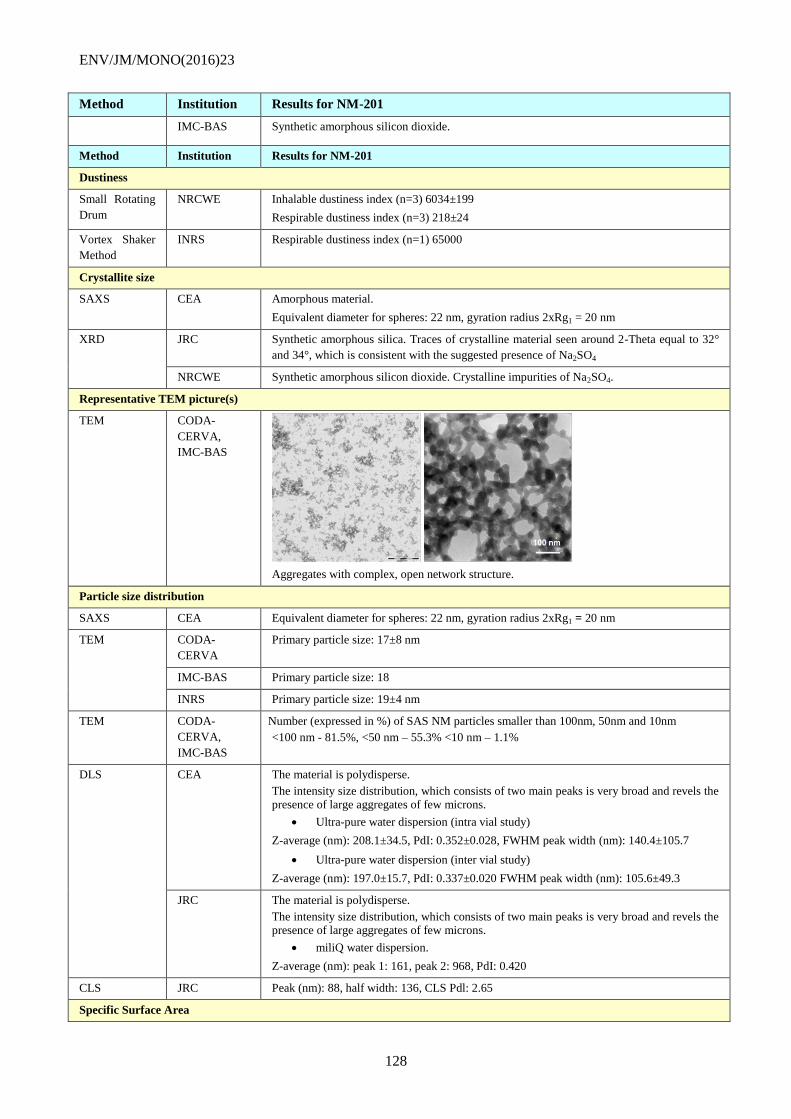
ENV/JM/MONO(2016)23
128
Method Institution Results for NM-201
IMC-BAS Synthetic amorphous silicon dioxide.
Method Institution Results for NM-201
Dustiness
Small Rotating
Drum
NRCWE Inhalable dustiness index (n=3) 6034±199
Respirable dustiness index (n=3) 218±24
Vortex Shaker
Method
INRS Respirable dustiness index (n=1) 65000
Crystallite size
SAXS CEA Amorphous material.
Equivalent diameter for spheres: 22 nm, gyration radius 2xRg1 = 20 nm
XRD JRC Synthetic amorphous silica. Traces of crystalline material seen around 2-Theta equal to 32°
and 34°, which is consistent with the suggested presence of Na2SO4
NRCWE Synthetic amorphous silicon dioxide. Crystalline impurities of Na2SO4.
Representative TEM picture(s)
TEM CODA-
CERVA,
IMC-BAS
Aggregates with complex, open network structure.
Particle size distribution
SAXS CEA Equivalent diameter for spheres: 22 nm, gyration radius 2xRg1 = 20 nm
TEM CODA-
CERVA
Primary particle size: 17±8 nm
IMC-BAS Primary particle size: 18
INRS Primary particle size: 19±4 nm
TEM CODA-
CERVA,
IMC-BAS
Number (expressed in %) of SAS NM particles smaller than 100nm, 50nm and 10nm
<100 nm - 81.5%, <50 nm – 55.3% <10 nm – 1.1%
DLS CEA The material is polydisperse.
The intensity size distribution, which consists of two main peaks is very broad and revels the presence of large aggregates of few microns.
Ultra-pure water dispersion (intra vial study)
Z-average (nm): 208.1±34.5, PdI: 0.352±0.028, FWHM peak width (nm): 140.4±105.7
Ultra-pure water dispersion (inter vial study)
Z-average (nm): 197.0±15.7, PdI: 0.337±0.020 FWHM peak width (nm): 105.6±49.3
JRC The material is polydisperse.
The intensity size distribution, which consists of two main peaks is very broad and revels the presence of large aggregates of few microns.
miliQ water dispersion.
Z-average (nm): peak 1: 161, peak 2: 968, PdI: 0.420
CLS JRC Peak (nm): 88, half width: 136, CLS Pdl: 2.65
Specific Surface Area

ENV/JM/MONO(2016)23
129
Method Institution Results for NM-201
BET IMC-BAS 140.46 (m2/g)
SAXS CEA 123.3±8.3 (m2/g)
Method Institution Results for NM-201
Zeta Potential (surface charge)
Zetametry CEA NM-201 forms a stable suspension, with negatively to neutral charged particles. The zeta
potential varied greatly as function of pH and reached -40 mV around pH 7. IEP <2
JRC Zeta potential at pH 6.9, milliQ water: -51.7 (mV).
Surface Chemistry
XPS JRC The following elements were identified in the surface of NM-201: O (70.3 at%), Si (23.6
at%), C (4.5 at%), Na (1.5 at%), Ce (0.25 at%) and S (0.01 at%). The presence of C is
considered to be due to surface contamination from hydrocarbons from air that have attached
themselves to the material surface.
TGA NRCWE A significant mass loss is observed below 100°C
(water). Gradual 3 wt% mass loss was observed
above 110°C indicating e.g. loss of water
associated in the micro pores or associated with
the presence of Na2SO4.
Photo-catalytic activity
End-point not relevant for SAS
Pour-density
Weighing INRS 0.28 g/cm3 (8 wt.% water content)
Porosity
BET IMC-BAS Micropore volume (mL/g): 0.00916
Octanol-water partition coefficient
End-point not relevant
Redox potential
OxoDish
fluorescent
sensor plate for
O2 detection
NRCWE The evolution of O2 level during 24-hour incubation was measured in three different media.
Different dO2 values were observed for all applied media. In the Gambles solution and Caco
2 media the concentration of dO2 was the highest (increased ca. 40 µmol/l) for 0.16mg/ml
concentration of NM-201. In the 0.05% BSA in water the dO2 level increases along with the
concentration of NM-201 The results suggest that NM-201 has oxidative behaviour in these
incubation media.
Radical formation
HPLC + UV NRCWE Using the benzoic acid probe to form 4 hydroxy benzoic acid in a phosphate buffered
hydrous solution, gave no detectable concentration OH radicals.
Composition
ICP-OES CODA-
CERVA
>0.01%: Al(>0.1%), Ca, Na(>0.1%), S;
00.5-0.01%: Zr;
0.001-0.005%: Fe, K, Mg
EDS IMC-BAS Na-4400ppm, Al- 7400ppm, S- 4600ppm, Si -45.27 (wt %), O (wt%) calculated- 53.08
ENV/JM/MONO(2016)23
130
NM-202, summary of physical-chemical characterisation results
Method Institution Results for NM-202
Homogeneity
DLS Study not performed.
Agglomeration / aggregation
SAXS CEA Structure and size parameters extracted from SAXS data. Gyration radius of primary
particles and aggregates Rg1: 16 nm and Rg2: 100 nm, fractal dimension Df: 2.5 and number
Npart/agg of particles per aggregate: 200
DLS CEA Ultra-pure water dispersion (intra vial study)
Z-average (nm): 175.9±4.5, PdI: 0.355±0.001
JRC miliQ water dispersion.
Z-average (nm): peak 1: 156, peak 2: 200, PdI: 0.160
TEM CODA-
CERVA,
IMC-BAS
High porosity nanostructured material which may be considered aggregates of primary
silicon dioxide particles.
Median diamater (nm): 53±9.
Feret min: 37.2 nm (median of 4248)
Feret max: 58.4 nm (median of 4248)
Morphology of aggregates/agglomerates: Low sphericity—very angular to sub-angular.
% of aggregates <100 nm: 80.4 %
AFM CEA Third dimension of the agglomerates/aggregates: median (of 1103): 38.2 nm
Water Solubility
24-hour
acellular in
vitro incubation
test in special
solutions
NRCWE The 24-hour dissolution ratio of NM-202 was measured in three different media: 0.05% BSA
in water, Gambles solution and Caco 2 media. Both NM-202 and the Al impurities are
partially soluble in all media but amounts vary considerably with medium as does the
relative amounts of dissolved Al impurities compared with dissolved Si suggesting different
solubility behaviour of the Al impurity and NM-202 depending on the medium.
Crystalline phase
XRD JRC Synthetic amorphous silicon dioxide.
NRCWE
Synthetic amorphous silicon dioxide.
IMC-BAS Synthetic amorphous silicon dioxide.
Dustiness
Small Rotating
Drum
NRCWE Inhalable dustiness index (n=3) 4988±1866
Respirable dustiness index (n=3) 91±11
Vortex Shaker
Method
INRS Respirable dustiness index (n=1) 510000
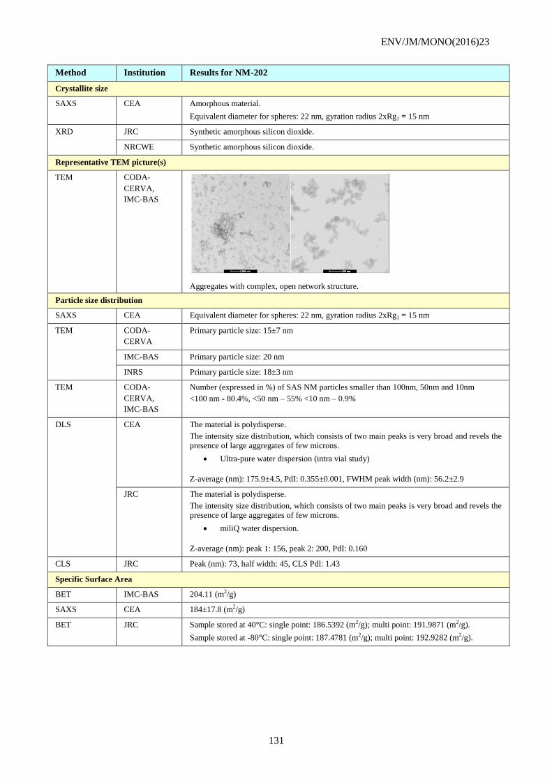
ENV/JM/MONO(2016)23
131
Method Institution Results for NM-202
Crystallite size
SAXS CEA Amorphous material.
Equivalent diameter for spheres: 22 nm, gyration radius 2xRg1 = 15 nm
XRD JRC Synthetic amorphous silicon dioxide.
NRCWE Synthetic amorphous silicon dioxide.
Representative TEM picture(s)
TEM CODA-
CERVA,
IMC-BAS
Aggregates with complex, open network structure.
Particle size distribution
SAXS CEA Equivalent diameter for spheres: 22 nm, gyration radius 2xRg1 = 15 nm
TEM CODA-
CERVA
Primary particle size: 15±7 nm
IMC-BAS Primary particle size: 20 nm
INRS Primary particle size: 18±3 nm
TEM CODA-
CERVA,
IMC-BAS
Number (expressed in %) of SAS NM particles smaller than 100nm, 50nm and 10nm
<100 nm - 80.4%, <50 nm – 55% <10 nm – 0.9%
DLS CEA The material is polydisperse.
The intensity size distribution, which consists of two main peaks is very broad and revels the presence of large aggregates of few microns.
Ultra-pure water dispersion (intra vial study)
Z-average (nm): 175.9±4.5, PdI: 0.355±0.001, FWHM peak width (nm): 56.2±2.9
JRC The material is polydisperse.
The intensity size distribution, which consists of two main peaks is very broad and revels the presence of large aggregates of few microns.
miliQ water dispersion.
Z-average (nm): peak 1: 156, peak 2: 200, PdI: 0.160
CLS JRC Peak (nm): 73, half width: 45, CLS Pdl: 1.43
Specific Surface Area
BET IMC-BAS 204.11 (m2/g)
SAXS CEA 184±17.8 (m2/g)
BET JRC Sample stored at 40°C: single point: 186.5392 (m2/g); multi point: 191.9871 (m2/g).
Sample stored at -80°C: single point: 187.4781 (m2/g); multi point: 192.9282 (m2/g).
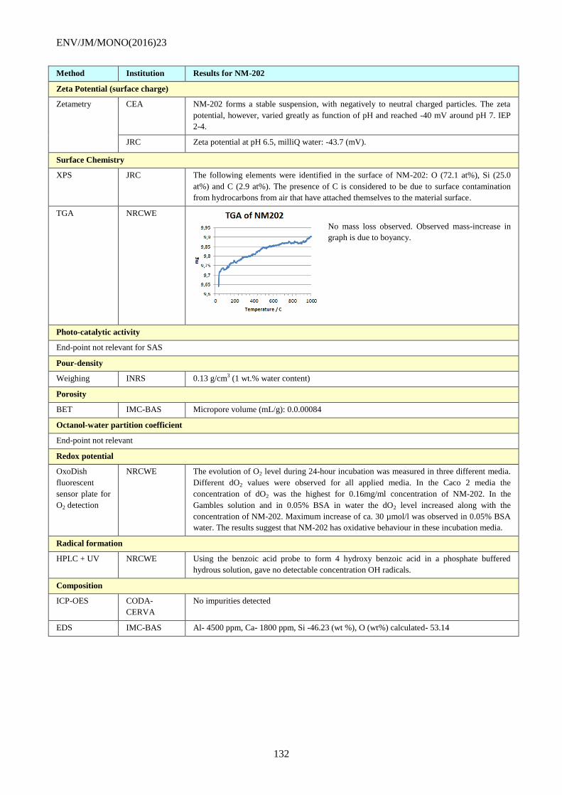
ENV/JM/MONO(2016)23
132
Method Institution Results for NM-202
Zeta Potential (surface charge)
Zetametry CEA NM-202 forms a stable suspension, with negatively to neutral charged particles. The zeta
potential, however, varied greatly as function of pH and reached -40 mV around pH 7. IEP
2-4.
JRC Zeta potential at pH 6.5, milliQ water: -43.7 (mV).
Surface Chemistry
XPS JRC The following elements were identified in the surface of NM-202: O (72.1 at%), Si (25.0
at%) and C (2.9 at%). The presence of C is considered to be due to surface contamination
from hydrocarbons from air that have attached themselves to the material surface.
TGA NRCWE
No mass loss observed. Observed mass-increase in
graph is due to boyancy.
Photo-catalytic activity
End-point not relevant for SAS
Pour-density
Weighing INRS 0.13 g/cm3 (1 wt.% water content)
Porosity
BET IMC-BAS Micropore volume (mL/g): 0.0.00084
Octanol-water partition coefficient
End-point not relevant
Redox potential
OxoDish
fluorescent
sensor plate for
O2 detection
NRCWE The evolution of O2 level during 24-hour incubation was measured in three different media.
Different dO2 values were observed for all applied media. In the Caco 2 media the
concentration of dO2 was the highest for 0.16mg/ml concentration of NM-202. In the
Gambles solution and in 0.05% BSA in water the dO2 level increased along with the
concentration of NM-202. Maximum increase of ca. 30 µmol/l was observed in 0.05% BSA
water. The results suggest that NM-202 has oxidative behaviour in these incubation media.
Radical formation
HPLC + UV NRCWE Using the benzoic acid probe to form 4 hydroxy benzoic acid in a phosphate buffered
hydrous solution, gave no detectable concentration OH radicals.
Composition
ICP-OES CODA-
CERVA
No impurities detected
EDS IMC-BAS Al- 4500 ppm, Ca- 1800 ppm, Si -46.23 (wt %), O (wt%) calculated- 53.14
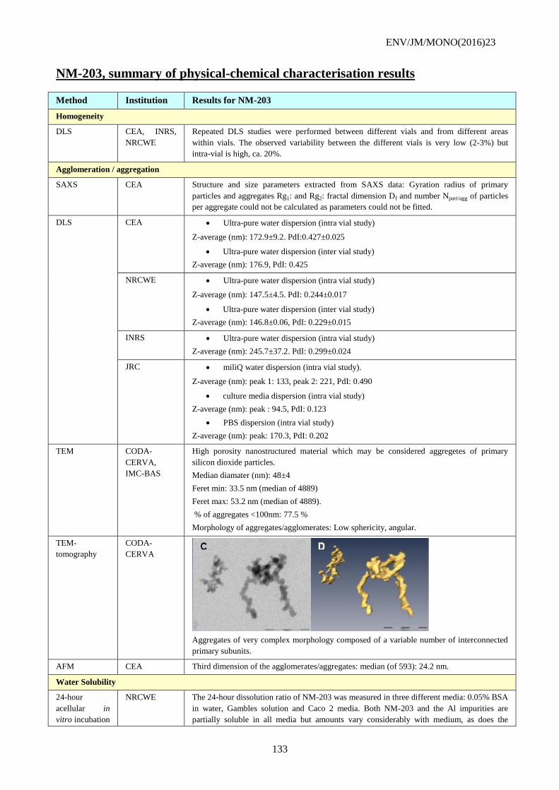
ENV/JM/MONO(2016)23
133
NM-203, summary of physical-chemical characterisation results
Method Institution Results for NM-203
Homogeneity
DLS CEA, INRS,
NRCWE
Repeated DLS studies were performed between different vials and from different areas
within vials. The observed variability between the different vials is very low (2-3%) but
intra-vial is high, ca. 20%.
Agglomeration / aggregation
SAXS CEA Structure and size parameters extracted from SAXS data: Gyration radius of primary
particles and aggregates Rg1: and Rg2: fractal dimension Df and number Npart/agg of particles
per aggregate could not be calculated as parameters could not be fitted.
DLS CEA Ultra-pure water dispersion (intra vial study)
Z-average (nm): 172.9±9.2. PdI:0.427±0.025
Ultra-pure water dispersion (inter vial study)
Z-average (nm): 176.9, PdI: 0.425
NRCWE Ultra-pure water dispersion (intra vial study)
Z-average (nm): 147.5±4.5. PdI: 0.244±0.017
Ultra-pure water dispersion (inter vial study)
Z-average (nm): 146.8±0.06, PdI: 0.229±0.015
INRS Ultra-pure water dispersion (intra vial study)
Z-average (nm): 245.7±37.2. PdI: 0.299±0.024
JRC miliQ water dispersion (intra vial study).
Z-average (nm): peak 1: 133, peak 2: 221, PdI: 0.490
culture media dispersion (intra vial study)
Z-average (nm): peak : 94.5, PdI: 0.123
PBS dispersion (intra vial study)
Z-average (nm): peak: 170.3, PdI: 0.202
TEM CODA-
CERVA,
IMC-BAS
High porosity nanostructured material which may be considered aggregetes of primary
silicon dioxide particles.
Median diamater (nm): 48±4
Feret min: 33.5 nm (median of 4889)
Feret max: 53.2 nm (median of 4889).
% of aggregates <100nm: 77.5 %
Morphology of aggregates/agglomerates: Low sphericity, angular.
TEM-
tomography
CODA-
CERVA
Aggregates of very complex morphology composed of a variable number of interconnected
primary subunits.
AFM CEA Third dimension of the agglomerates/aggregates: median (of 593): 24.2 nm.
Water Solubility
24-hour
acellular in
vitro incubation
NRCWE The 24-hour dissolution ratio of NM-203 was measured in three different media: 0.05% BSA
in water, Gambles solution and Caco 2 media. Both NM-203 and the Al impurities are
partially soluble in all media but amounts vary considerably with medium, as does the
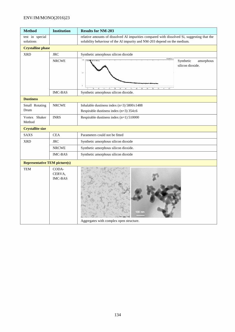
ENV/JM/MONO(2016)23
134
Method Institution Results for NM-203
test in special
solutions
relative amounts of dissolved Al impurities compared with dissolved Si, suggesting that the
solubility behaviour of the Al impurity and NM-203 depend on the medium.
Crystalline phase
XRD JRC Synthetic amorphous silicon dioxide
NRCWE Synthetic amorphous
silicon dioxide.
IMC-BAS Synthetic amorphous silicon dioxide.
Dustiness
Small Rotating
Drum
NRCWE Inhalable dustiness index (n=3) 5800±1488
Respirable dustiness index (n=3) 354±6
Vortex Shaker
Method
INRS Respirable dustiness index (n=1) 510000
Crystallite size
SAXS CEA Parameters could not be fitted
XRD JRC Synthetic amorphous silicon dioxide
NRCWE Synthetic amorphous silicon dioxide.
IMC-BAS Synthetic amorphous silicon dioxide
Representative TEM picture(s)
TEM CODA-
CERVA,
IMC-BAS
Aggregates with complex open structure.
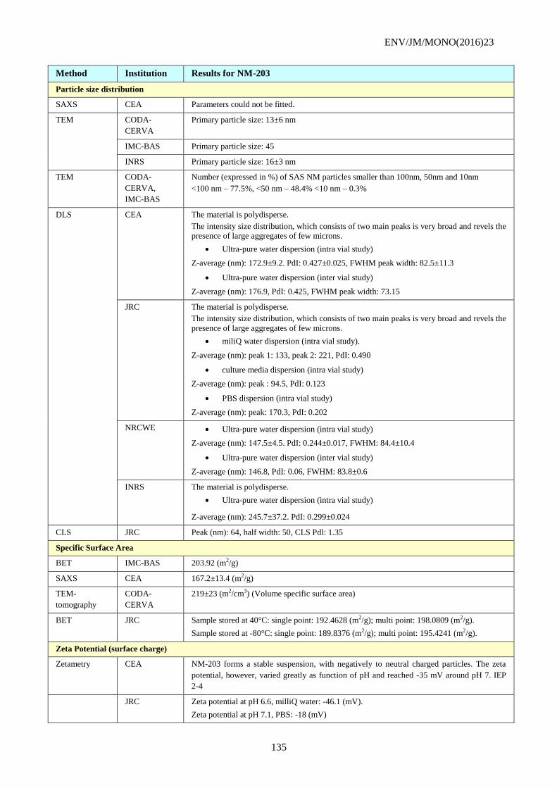
ENV/JM/MONO(2016)23
135
Method Institution Results for NM-203
Particle size distribution
SAXS CEA Parameters could not be fitted.
TEM CODA-
CERVA
Primary particle size: 13±6 nm
IMC-BAS Primary particle size: 45
INRS Primary particle size: 16±3 nm
TEM CODA-
CERVA,
IMC-BAS
Number (expressed in %) of SAS NM particles smaller than 100nm, 50nm and 10nm
<100 nm – 77.5%, <50 nm – 48.4% <10 nm – 0.3%
DLS CEA The material is polydisperse.
The intensity size distribution, which consists of two main peaks is very broad and revels the presence of large aggregates of few microns.
Ultra-pure water dispersion (intra vial study)
Z-average (nm): 172.9±9.2. PdI: 0.427±0.025, FWHM peak width: 82.5±11.3
Ultra-pure water dispersion (inter vial study)
Z-average (nm): 176.9, PdI: 0.425, FWHM peak width: 73.15
JRC The material is polydisperse.
The intensity size distribution, which consists of two main peaks is very broad and revels the
presence of large aggregates of few microns.
miliQ water dispersion (intra vial study).
Z-average (nm): peak 1: 133, peak 2: 221, PdI: 0.490
culture media dispersion (intra vial study)
Z-average (nm): peak : 94.5, PdI: 0.123
PBS dispersion (intra vial study)
Z-average (nm): peak: 170.3, PdI: 0.202
NRCWE Ultra-pure water dispersion (intra vial study)
Z-average (nm): 147.5±4.5. PdI: 0.244±0.017, FWHM: 84.4±10.4
Ultra-pure water dispersion (inter vial study)
Z-average (nm): 146.8, PdI: 0.06, FWHM: 83.8±0.6
INRS The material is polydisperse.
Ultra-pure water dispersion (intra vial study)
Z-average (nm): 245.7±37.2. PdI: 0.299±0.024
CLS JRC Peak (nm): 64, half width: 50, CLS Pdl: 1.35
Specific Surface Area
BET IMC-BAS 203.92 (m2/g)
SAXS CEA 167.2±13.4 (m2/g)
TEM-
tomography
CODA-
CERVA
219±23 (m2/cm3) (Volume specific surface area)
BET JRC Sample stored at 40°C: single point: 192.4628 (m2/g); multi point: 198.0809 (m2/g).
Sample stored at -80°C: single point: 189.8376 (m2/g); multi point: 195.4241 (m2/g).
Zeta Potential (surface charge)
Zetametry CEA NM-203 forms a stable suspension, with negatively to neutral charged particles. The zeta
potential, however, varied greatly as function of pH and reached -35 mV around pH 7. IEP
2-4
JRC Zeta potential at pH 6.6, milliQ water: -46.1 (mV).
Zeta potential at pH 7.1, PBS: -18 (mV)
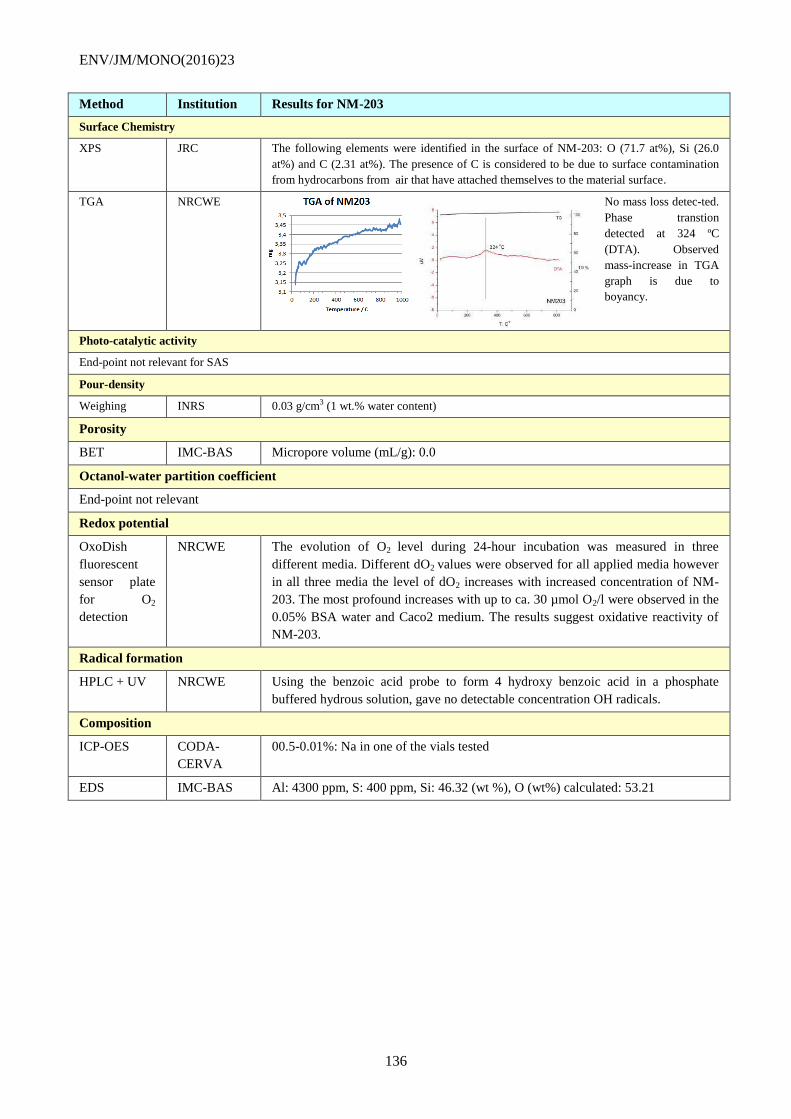
ENV/JM/MONO(2016)23
136
Method Institution Results for NM-203
Surface Chemistry
XPS JRC The following elements were identified in the surface of NM-203: O (71.7 at%), Si (26.0
at%) and C (2.31 at%). The presence of C is considered to be due to surface contamination
from hydrocarbons from air that have attached themselves to the material surface.
TGA NRCWE No mass loss detec-ted.
Phase transtion
detected at 324 ºC
(DTA). Observed
mass-increase in TGA
graph is due to
boyancy.
Photo-catalytic activity
End-point not relevant for SAS
Pour-density
Weighing INRS 0.03 g/cm3 (1 wt.% water content)
Porosity
BET IMC-BAS Micropore volume (mL/g): 0.0
Octanol-water partition coefficient
End-point not relevant
Redox potential
OxoDish
fluorescent
sensor plate
for O2
detection
NRCWE The evolution of O2 level during 24-hour incubation was measured in three
different media. Different dO2 values were observed for all applied media however
in all three media the level of dO2 increases with increased concentration of NM-
203. The most profound increases with up to ca. 30 µmol O2/l were observed in the
0.05% BSA water and Caco2 medium. The results suggest oxidative reactivity of
NM-203.
Radical formation
HPLC + UV NRCWE Using the benzoic acid probe to form 4 hydroxy benzoic acid in a phosphate
buffered hydrous solution, gave no detectable concentration OH radicals.
Composition
ICP-OES CODA-
CERVA
00.5-0.01%: Na in one of the vials tested
EDS IMC-BAS Al: 4300 ppm, S: 400 ppm, Si: 46.32 (wt %), O (wt%) calculated: 53.21
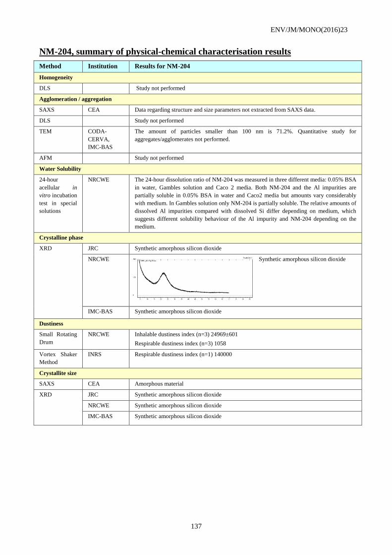
ENV/JM/MONO(2016)23
137
NM-204, summary of physical-chemical characterisation results
Method Institution Results for NM-204
Homogeneity
DLS Study not performed
Agglomeration / aggregation
SAXS CEA Data regarding structure and size parameters not extracted from SAXS data.
DLS Study not performed
TEM CODA-
CERVA,
IMC-BAS
The amount of particles smaller than 100 nm is 71.2%. Quantitative study for
aggregates/agglomerates not performed.
AFM Study not performed
Water Solubility
24-hour
acellular in
vitro incubation
test in special
solutions
NRCWE The 24-hour dissolution ratio of NM-204 was measured in three different media: 0.05% BSA
in water, Gambles solution and Caco 2 media. Both NM-204 and the Al impurities are
partially soluble in 0.05% BSA in water and Caco2 media but amounts vary considerably
with medium. In Gambles solution only NM-204 is partially soluble. The relative amounts of
dissolved Al impurities compared with dissolved Si differ depending on medium, which
suggests different solubility behaviour of the Al impurity and NM-204 depending on the
medium.
Crystalline phase
XRD JRC Synthetic amorphous silicon dioxide
NRCWE Synthetic amorphous silicon dioxide
IMC-BAS Synthetic amorphous silicon dioxide
Dustiness
Small Rotating
Drum
NRCWE Inhalable dustiness index (n=3) 24969±601
Respirable dustiness index (n=3) 1058
Vortex Shaker
Method
INRS Respirable dustiness index (n=1) 140000
Crystallite size
SAXS CEA Amorphous material
XRD JRC Synthetic amorphous silicon dioxide
NRCWE Synthetic amorphous silicon dioxide
IMC-BAS Synthetic amorphous silicon dioxide
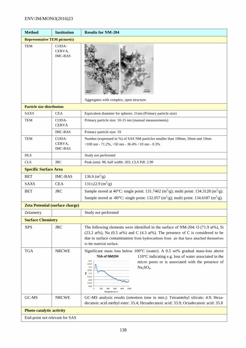
ENV/JM/MONO(2016)23
138
Method Institution Results for NM-204
Representative TEM picture(s)
TEM CODA-
CERVA,
IMC-BAS
Aggregates with complex, open structure.
Particle size distribution
SAXS CEA Equivalent diameter for spheres: 21nm (Primary particle size)
TEM CODA-
CERVA
Primary particle size: 10-15 nm (manual measurements)
IMC-BAS Primary particle size: 19
TEM CODA-
CERVA,
IMC-BAS
Number (expressed in %) of SAS NM particles smaller than 100nm, 50nm and 10nm
<100 nm - 71.2%, <50 nm - 36.4% <10 nm - 0.3%
DLS Study not performed
CLS JRC Peak (nm): 98, half width: 203, CLS Pdl: 2.99
Specific Surface Area
BET IMC-BAS 136.6 (m2/g)
SAXS CEA 131±22.9 (m2/g)
BET JRC Sample stored at 40°C: single point: 131.7462 (m2/g); multi point: 134.3128 (m
2/g).
Sample stored at -80°C: single point: 132.057 (m2/g); multi point: 134.6187 (m
2/g).
Zeta Potential (surface charge)
Zetametry Study not performed
Surface Chemistry
XPS JRC The following elements were identified in the surface of NM-204: O (71.9 at%), Si
(23.2 at%), Na (0.5 at%) and C (4.3 at%). The presence of C is considered to be
due to surface contamination from hydrocarbons from air that have attached themselves
to the material surface.
TGA NRCWE Significant mass loss below 100°C (water). A 0.5 wt% gradual mass-loss above
110°C indicating e.g. loss of water associated in the
micro pores or is associated with the presence of
Na2SO4.
GC-MS NRCWE GC-MS analysis results (retention time in min.): Tetramtehyl silicate: 4.9; Hexa-
decanoic acid methyl ester: 33.4; Hexadecanoic acid: 33.9; Octadecanoic acid: 35.8
Photo-catalytic activity
End-point not relevant for SAS
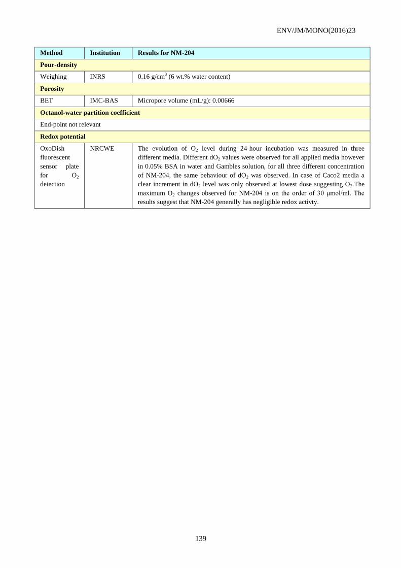
ENV/JM/MONO(2016)23
139
Method Institution Results for NM-204
Pour-density
Weighing INRS 0.16 g/cm3 (6 wt.% water content)
Porosity
BET IMC-BAS Micropore volume (mL/g): 0.00666
Octanol-water partition coefficient
End-point not relevant
Redox potential
OxoDish
fluorescent
sensor plate
for O2
detection
NRCWE The evolution of O2 level during 24-hour incubation was measured in three
different media. Different dO2 values were observed for all applied media however
in 0.05% BSA in water and Gambles solution, for all three different concentration
of NM-204, the same behaviour of dO2 was observed. In case of Caco2 media a
clear increment in dO2 level was only observed at lowest dose suggesting O2.The
maximum O2 changes observed for NM-204 is on the order of 30 μmol/ml. The
results suggest that NM-204 generally has negligible redox activty.
ENV/JM/MONO(2016)23
140
APPENDIX VI: PROTOCOLS FOR CHARACTERISATION OF
THE NANOMATERIAL SYNTHESISED IN KOREA
Information on Particle size distribution – dry and in relevant media
Methods
Media: Distilled water
Method/guideline followed: TEM and DLS size measurement procedures are as follows:
a) Protocol for TEM measurements
ⅰ) Sample preparation
- Holey carbon-coated copper TEM grids were chosen for the preparation of TEM nanoparticle
specimens.
- One or two droplets of SiO2 nanoparticle suspension were dropped onto the shiny side of the grids.
- The grids were dried in a desiccator.
ⅱ) Observation and analysis by TEM
- A 300 kV accelerating voltage was used (FEI Tecnai G2 F30 field emission gun (FEG) TEM).
- The microscope magnification was calibrated by imaging silicon {111} lattice fringes at the
eucentric specimen position and assuming a 0.31355 nm spacing.
- Coarse focusing was accomplished by raising and lowering the specimen along the z-axis (optic
axis) and fine focusing was accomplished by adjusting the objective lens current.
- As the actual magnification is sensitive to the objective lens current, the current was precisely
controlled during experiments.
- Bright field images were collected using a CCD camera (UIltraScanTM
) and DigitalMicrograph
software (Gatan, Inc.).
- The SiO2 nanoparticle sizes were measured manually from the TEM images using
DigitalMicrograph software (Gatan, Inc.).
b) Protocol for DLS measurement of a SiO2 suspension using ELS-Z
- Measure the UV/Vis absorption spectrum of a 150 L aliquot of the suspension between 200 and 800 nm
against solvents (DW) as a baseline using a spectrophotometer (UV-1700, Shimadzu).
- Measure the DLS size on a 1 mL aliquot of the suspension using a particle analyzer (ELS-Z, Otzuka
Electronics Co. LTD). Perform 15 runs/measurement 5 measurements and average of 5 data points.
- Dilute the suspension in DW to 1/10 of the original concentration.
- Measure the DLS size of a 1 mL aliquot of the suspension.
- Repeat the dilution and DLS size measurement until the DLS size is constant within 20 %.
- Obtain the DLS size of SiO2 nanoparticles from the most dilute suspension in the valid concentration
range9.
- The instrument performance was qualified using a nanoparticle size reference (Gold Nanoparticle, RM
1980 – NIST).
c) The protocol for the DLS measurement of a SiO2 suspension using a Brookhaven particle size
analyzer is described in “Development of Nano-materials Safety and Characterization Techniques”
published by the Ministry of Education, Science and Technology, Korea, 2010. (KRISS).
9 Valid concentration range means that the DLS size is constant within 20 %.
ENV/JM/MONO(2016)23
141
Year (study performed): 2010 - 2011
GLP: No
Analytical monitoring: TEM (FEI Tecnai G2 F30) and two Particle size analyzers (Brookhaven Instrument
Co., ELS-Z, Otzuka Electronics Co. LTD. Japan)
Exposure period (duration): 1 year
Doses/concentration levels: 20 mg/mL for 30 nm SiO2 nanoparticle suspension and
44 mg/mL for 40 nm SiO2 nanoparticle suspension
Test conditions
Dilution water source: Distilled water
Stock and test solutions and their preparation: A stock suspension (> 150 mL) was prepared by mixing
three batches (>50 mL) of synthesised SiO2 nanoparticle suspensions. The suspension was stirred for 24 h
using a magnetic bar. Three 1 mL aliquots of the suspension were sampled to obtain nanoparticle
concentrations by drying-and-weighing. Next, 5 mL aliquots of the suspension were transferred to Teflon-
capped vials and labelled with the time of characterization (0, 7, 14, 30, 60, 90, 180, 270 and 360 days after
sampling). Each bottle was analyzed at the predetermined storage times.
Stability of the test chemical solutions: Suspensions were stable up to 12 months
Exposure vessel type: not described
Test temperature range: 26 - 28 C (ambient temperature).
ZETA POTENTIAL/SURFACE CHARGE
Methods
Media: Distilled water
Method/guideline followed: The procedure for zeta potential measurement was as follows (3 batches of
SiO2 nanoparticle suspensions were measured independently):
- Measure the UV/Vis absorption spectrum of a 150 L aliquot of the suspension between 200 and 800 nm
against solvents (DW or pH-buffered solution) as a baseline using a spectrophotometer (UV-1700,
Shimadzu).
- Measure the DLS size of a 1 mL aliquot of the suspension using a particle analyzer (ELS-Z, Otzuka
Electronics Co. LTD.). Perform 15 runs/measurement 5 measurements and average 5 independent data
points.
- The instrument performance was qualified using a nanoparticle size reference (Gold Nanoparticle, RM
1980 – NIST).
- Measure the zeta potential of a 1 mL aliquot of the suspension using an electrophoretic mobility analyzer
(Zetasizer Nano Z, Malvern). Perform 20 runs/measurement 5 measurements and average 5
independent data points.
- The instrument performance was qualified using a vendor-supplied -50 mV transfer standard referred to
SRM 1980.
The measurement procedure for suspension pH was as follows:
- Divide a 6 mL aliquot of the suspension into 4 aliquots of 1.5 mL each and transfer them into 2 mL
microcentrifuge tubes. Centrifuge the solutions at 10,000 rpm for 20 min (HM-150IV, Hanil Science
Ind.).
ENV/JM/MONO(2016)23
142
- Add 1.3 mL aliquots of the supernatant from the 4 microcentrifuge tubes to a 50 mL conical tube (Cat.
No. 50050, SPL Lifesciences).
- Measure the pH of the DW and the supernatant using a pH meter (Orion 3 star pH Benchtop, Thermo
Electron Co.) according to the following procedure. After each measurement, wash the pH electrode with
DW.
• Measure the solution pH (DW or supernatant) in a 50 mL conical tube (3 times).
• Measure the pH of a pH 4.01 standard buffer solution (Orion 910104) (3 times).
• Measure the solution pH (DW or supernatant) in a 50 mL conical tube (3 times).
• Measure the pH of a pH 10.01 standard buffer solution (Orion 910110) (3 times).
• Measure the solution pH (DW or supernatant) in a 50 mL conical tube (3 times).
- The instrument performance was qualified using standard pH buffers of pH 4.01 (Orion 910104), pH
10.01(Orion 910110) and pH 7.00 (Orion 910107).
Year (study performed): 2010-2011
GLP: No
Analytical monitoring: Zetasizer (Nano Z, Malvern, UK)
Exposure period (duration): 1 year
Doses/concentration levels: 20 mg/mL for 30 nm SiO2 nanoparticle suspension and
44 mg/mL for 40 nm SiO2 nanoparticle suspension
Test conditions
Dilution water source: Distilled water
Stock and test solutions and how they are prepared: A stock suspension (> 150 mL) was prepared by
mixing three batches (>50 mL) of synthesised SiO2 nanoparticle suspensions. The suspension was stirred
for 24 h using a magnetic bar. Three 1 mL aliquots of the suspension were sampled to obtain nanoparticle
concentrations by drying-and-weighing. Next, 5 mL aliquots of the suspension were transferred to Teflon-
capped vials and labelled with the time of characterization (0, 7, 14, 30, 60, 90, 180, 270 and 360 days after
sampling). Each bottle was analyzed at the predetermined storage times.
Stability of the test chemical solutions: Zeta potential was stable up to 3 months
Exposure vessel type: not described
Test temperature range: not described
ENV/JM/MONO(2016)23
143
APPENDIX VII: LIST OF ABBREVIATIONS
2D Two Dimensional
3D Three Dimensional
ANOVA Analysis of Variance
APS Aerodynamic Particle Sizer
ASASP Association of Synthetic Amorphous Silica Producers
BET Brunauer, Emmet and Teller
BSA Bovine Serum Albumin
CEA Commissariat à l'énergie atomique et aux énergies alternatives
CEN Comité Européen de Normalisation
CLS Centrifugal Liquid Sedimentation
CODA-CERVA Veterinary and Agrochemical Research Centre (Belgium)
CPC Condensation Particle Counter
DLS Dynamic Light Scattering
ELPI Electrical Low Pressure Impactor
EM Electron microscopy
EDX Energy-Dispersive X-ray spectroscopy
Fh-IME Fraunhofer Institute for Molecular Biology and Applied Ecology, Germany
FMPS Fast Mobility Particle Sizer
FWHM Full-Width Half-Maximum
GLP Good Laboratory Practice
h hours
HEPA filter High-Efficiency Particulate Air filter
ICP-OES Inductively Coupled Plasma – Optical Emission Spectrometry
IEP Iso-Electric Point
IHCP Institute for Health and Consumer Protection (JRC)
IMC-BAS Institute of Mineralogy and Crystallography, Bulgaria
INRS Institut National de Recherche et de Sécurite
ISO International Organisation for Standardization
ISO/TC 229 ISO/Technical Committee on Nanotechnologies
IUPAC International Union of Pure and Applied Chemistry
JRC Joint Research Centre, European Commission
L or l Litre
LNE Laboratoire national de métrologie et d'essais, France
lpm Litre per minute
ml / mL Milli litre
MWCNT Multi Walled Carbon Nano Tube
NIST USA National Institute of Standards and Technology
NM Nanomaterial
NRCWE National Research Centre for the Working Environment
ENV/JM/MONO(2016)23
144
OECD Organisation for Economic Co-operation and Development
PSD Particle Size Distribution
PBS Phosphate Buffered Saline
PCS Photon Correlation Spectroscopy
PdI Poly Dispersion Index
pH Acidity value
REACH EU Legislation on Registration, Evaluation, Authorisation and restriction of Chemicals
RH Relative Humidity
RMN Representative Manufactured Nanomaterial
rpm Rounds Per Minute
RSD Relative Standard Deviation
RTM Representative Test Material
s second
SAS Synthetic Amorphous Silicon Dioxide
SAS NMs All of NM-200, NM-201, NM-202, NM-203 and NM-204
SAXS Small Angle X-ray Scattering
SD Standard Deviation
SD Small Rotating Drum
SDR Sensor Disk Reader
SEM Scanning Electron Microscopy
SEM-EDS Scanning Electron Microscopy-Energy Dispersive Spectroscopy
SIRT Simultaneous Iterative Reconstruction Technique
SOP Standard Operating Procedure
SCENIHR Scientific Committee for Emerging and Newly Identified Health Risks
TEM Transmission Electron Microscopy
USA United States of America
USA-EPA USA Environmental Protection Agency
USAXS Ultra Small Angle X-ray Scattering
VS Vortex Shaker
WPMN Working Party on Manufactured Nanomaterials
w/w% weight percent
XPS X-ray Photoelectron Spectrometry
XRD X-ray Diffraction