ultrastructural examination of the host cellular response in the gills of atlantic salmon, salmo...
TRANSCRIPT

Ultrastructural Examination of the Host Cellular Response in theGills of Atlantic Salmon, Salmo salar, with Amoebic Gill Disease
J. LOVY, J. A. BECKER, D. J. SPEARE, D. W. WADOWSKA, G. M. WRIGHT, AND M. D. POWELL
Department of Pathology and Microbiology (JL, DJS); Electron Microscopy Laboratory (DWW); andDepartment of Biomedical Sciences (GMW); Atlantic Veterinary College, University of Prince Edward
Island, Charlottetown, Prince Edward Island, Canada; and Aquafin Cooperative Research Centre,School of Aquaculture, Tasmanian Aquaculture and Fisheries Institute, University of Tasmania,
Launceston, Tasmania, Australia (JAB, MDP)
Abstract. Gills from Atlantic salmon with experimentally induced amoebic gill disease (Neopar-amoeba spp.) were examined with transmission electron microscopy to assess pathology and host-cellresponses. Amoebae were found either on the surface epithelium or with pseudopodia extending deeplyinto invaginations of epithelial cells. The amoebae had various densities along the plasma membrane andcontained electron-dense deposits within their cytoplasm. Surface epithelial cells sloughed from the gillsand had features consistent with apoptosis, including rounded shape, loss of surface microridges, andhypercondensation of nuclear chromatin. Affected areas of gills had fusion of secondary lamellae withinterlamellar spaces occupied by mitotic epithelial cells and eosinophils. Eosinophils contained abundantfusiform-shaped granules that measured approximately 1 mm long and 360 nm wide. The granuleconsisted of an electron-dense matrix with a central inclusion that was less electron-dense, consisting ofparticulate and fibrillar material. In many instances, the central inclusion appeared empty and 90% ofthe eosinophils had morphology suggestive of piecemeal degranulation. Also observed within affectedareas were a few neutrophils, mucous cells releasing mucus, and a small number of dendritic-like cells.
Key words: Amoebic gill disease; electron microscopy; eosinophils; fish; gills.
Amoebic gill disease (AGD) is a commonproblem for the culture of Atlantic salmon, Salmosalar, within seawater net pens in Tasmania,Australia. Disease is associated with the coloniza-tion of the amphizoic amoeba Neoparamoeba spp.(comprising at least 2 species: Neoparamoebapemaquidensis and Neoparamoeba branchiphila) onthe surface of the gills.10,16 A distinguishing featureof amoebae in the Neoparamoeba genus is thepresence of an endosymbiotic Perkinsiella amoe-bae–like organism, also referred to as the para-some.10 AGD has been described as affectingAtlantic salmon and rainbow trout, Oncorhynchusmykiss, from around the world, including Austra-lia, North America, France, Spain, Ireland, Chile,and New Zealand.24 The disease has also beendescribed in nonsalmonids including turbot,Scophthalmus maximus; sea bream, Sparus aurata;and sea bass, Dicentrarchus labrax.9 AGD is ofeconomic significance to Australia, whereby thecost of the disease (which, although potentiallyfatal to fish, is treated with freshwater baths) incursan estimated cost to the salmonid aquacultureindustry in the range between 10 and 20% of the
annual gross production value estimated to beequivalent to approximately $17–19 million Aus-tralian dollars annually.2,23 Clinical signs attributedto AGD include raised white mucoid patches ongill filaments, lethargy, and flared opercula, andpathophysiologic changes include respiratory andacid-base disturbances and systemic hyperten-sion.18,19,22,27,28 There is a significant metabolic costassociated with the disease, which translates toreductions in growth if the disease is not controlledby freshwater bathing (the only effective treatmentfor AGD). Mortalities attributed to AGD inuntreated net pens have been reported to reachup to 2% per day and 50% total.22
Infection results in a host-cell response leading tothe fusion of secondary lamellae, focal hyperplasiaof the filament epithelium, and a leucocytic in-filtration of the lesion.1,23 Clinical outbreaks ofAGD appear to be associated with water tempera-tures between 12–20uC, although other risk factorshave been identified including algae, site character-istics, salinity, and water chemistry.4,7,8 Amoebicgill infections have also been suggested to bea secondary complication of bacterial infections,
Vet Pathol 44:663–671 (2007)
663

as seen in salmonids with nodular gill disease(NGD); In Ireland, AGD can occur concurrentlywith ciliate infections.4,35
There have been several reports on the host-cellresponses associated with AGD.1,33 The mostprominent change in infected gills was degenerationof the surface epithelium with hyperplasia of theunderlying epithelial cells expressing proliferatingcell nuclear antigen histologically, as well as down-regulation of the tumor-suppressor protein p 53 inaffected gills.1,21 It has been reported that duringdisease outbreaks, gills are infiltrated with neutro-phils, while gills recovered from disease are infil-trated with macrophages and lymphocytes.33 A lightmicroscopic study of the pathogenesis of AGDshowed that early attachment of amoebae causedhypertrophy and desquamation of the epithelium,followed by hyperplasia leading to the fusion of thesecondary lamellae. These areas were infiltrated withpredominantly neutrophils, which emigrated fromthe central venous sinus. Large numbers of eosino-philic granule cells (EGC) were reported surround-ing the primary filament cartilage of infected fish,although this is likely to reflect the inherentpopulation of these cell types in the gill.1
Understanding host cellular responses is impor-tant in understanding the pathogenesis of disease.Examination of cell types in fish gills responding topathogens provides information about defensemechanisms in the gills. The associated inflamma-tory response with AGD may be more detrimentalto the host rather than curative, and treating theinflammation may be a viable approach to treatingfish during disease outbreaks. With an understand-ing of the nature of the inflammatory responseassociated with AGD, one can propose therapystrategies aimed at host responses. This studydescribes the host-gill cellular responses andultrastructural pathology of AGD-affected Atlan-tic salmon.
Materials and Methods
Source of AGD-affected gill tissue
At the University of Tasmania, seawater-acclimatedAtlantic salmon (approximately 100 g each) were in-troduced to an active infection tank containing about 20AGD-affected conspecifics for at least 7 days to allow forinfection by cohabitation. The infection tank consisted ofan 1,800-L fiberglass Rathburn tank, biologic filter, andrecirculating pump (total system volume of ,3,000 L).Water was maintained at a salinity of 35 parts perthousand, temperature of 15–17uC, dissolved oxygen inexcess of 90% saturation, and total ammonia nitrogenless than 0.25 mg L21. Moribund salmon with clinicalsigns of AGD were removed from the tank andeuthanized with an overdose of clove oil (0.03 mL L21).
Rapidly, individual gill arches were excised, placed into 3consecutive washes of 20 ml of chilled 2.5% glutaralde-hyde in sodium cacodylate buffer (0.1 M), and finallyplaced into 25-ml chilled fresh fixative and refrigeratedovernight (4uC). Under a dissecting microscope, macro-scopic white raised patches, characteristic of AGDlesions, were excised from the gill arches in 5–10 lamellaesections and placed in chilled fixative. The fixative wasreplaced daily, and the samples were shipped 48 hoursafter initial collection to the Atlantic Veterinary College,Prince Edward Island, Canada.
Transmission electron microscopy
Upon arrival, samples were washed in 2 changes ofcacodylate buffer for 10 minutes each and postfixed in1% osmium tetroxide in cacodylate buffer for 1 hour atroom temperature. Tissue was then dehydrated througha series of ascending concentrations of ethanol includingtwo 10 minute changes of 50%, 75%, and 95% and,lastly, 2 changes of 100% ethanol for 15 minutes each.Tissue was then cleared in propylene oxide (PO) (23,10 min each) and infiltrated with Epon (Canemco-Marivec, Quebec, Canada). Infiltration included 1change in a 1 : 1 ratio of Epon to PO, then to 1 changein a 3 : 1 ratio of Epon to PO for 1 hour each, and,lastly, to 100% Epon overnight in a desiccator undervacuum. For each fish, 5 pieces of gill tissue wereembedded in Epon. Semithin sections (0.5 mm) were cutfrom each piece of gill tissue and stained with toluidineblue to examine with light microscopy. Areas wherelesions were observed were trimmed, and ultrathinsections were cut (90 nm) for transmission electronmicroscopy. Ultrathin sections were stained with uranylacetate and lead citrate and examined and photo-graphed with a Hitachi-7500 transmission electronmicroscope (Hitachi High-Technologies Canada Inc.)operated at 80 kV. Measurements of cell organelles weremade directly from electron micrographs and reportedas the mean 6 SE.
Results
Gills infected with Neoparamoeba spp. displayedcharacteristic pathology, which included hyperpla-sia of the epithelium leading to the fusion ofsecondary lamellae, around which amoebae con-taining clearly identifiable parasomes (a diagnosticfeature of the genus Paramoeba) could be seen onthe surface of the epithelium. Bacteria were presentin areas that were heavily infected with theamoebae.
The interface between the amoebae and the hosttissue was examined, and various forms of in-teraction were observed. In many instances, amoe-bae were observed without direct contact with theepithelial membrane, but the shape of the amoebaeappeared to conform to the shape of the surfaceepithelium, suggesting the amoebae had been lyingon the surface but lifted off during fixation or tissue
664 Lovy, Becker, Speare, Wadowska, Wright, and Powell Vet Pathol 44:5, 2007

processing. Some amoebae, with intimate contactwith the host epithelium, extended their pseudopo-dia into invaginations of the epithelial layer(Fig. 1). The plasma membrane of amoebae hada heterogeneous density where the membraneappeared increasingly electron-dense in some areas.In one instance, an area of increased membranedensity was accompanied by increased cytoplasmicdensity (Fig. 2). The surface epithelial cells adja-cent to areas of the host-parasite interface weresloughed away from the underlying tissue, much oftheir microridge surface patterning was lost, andthe cells assumed a rounded shape (Fig. 3). Thenuclei of these cells appeared rounded and hadhypercondensation of chromatin (Fig. 3). Many ofthe epithelial cells below the surface layer were invarious stages of mitosis.
Within the hyperplastic epithelium of the second-ary lamellae was a heavy infiltrate of inflammatorycells. The infiltrate predominantly consisted of cellsresembling eosinophils (Fig. 4). These cells measuredbetween 6 and 15 mm along their long axis, depend-ing on their plane of section. The nuclei appearedrounded in most cells, and occasionally an in-dentation was seen on 1 side (Fig. 4). The cellscontained an abundance of unique granules; up to 50could be seen within a cell in a single section plane.When cut in longitudinal sections, the granules
appeared fusiform in shape (Fig. 5). Frequentlyrunning along the long axis of the granules wasa density composed of particulate and fibrillarmaterial, which was less electron-dense than the restof the granules (Fig. 5). Similar granules thatcontained electron-lucent inclusions were observed(Fig. 6). When cut in cross-section, the granules werespheric, and the electron-lucent inclusion was seen inthe center or off-center (Fig. 6). The granulesmeasured 1.00 6 0.09 mm long (range of 0.64–1.6 mm) and 0.36 6 0.03 mm wide (range of 0.22–0.51 mm), based on measurements of 10 granulesappearing in longitudinal section. Measurements ofthe diameter of 12 granules cut in cross-section hada mean of 0.42 6 0.02 mm (range of 0.32–0.56 mm).Granules of 30 cells were examined, and 90% of thecells contained granules with completely electron-lucent inclusions. From a total of 666 granules, 32%appeared with an electron-lucent inclusion and theremaining 68% contained the particulate and fibrillarmaterial. All lesions that were associated with theamoebae were significantly infiltrated with this celltype; the cells were observed within areas of fusionand at the surface epithelium, where the amoebaewere also present. Neutrophils were present, andgoblet cells were commonly observed to be releasingtheir contents in areas of the lesions (Fig. 7).
Fig. 1. Gill; Atlantic salmon. Amoeba (A) haspseudopodia within the surface epithelial layer (E),where the epithelial cells affected by the amoeba havea distorted shape and loss of surface microridges.Uranyl acetate and lead citrate. Bar 5 3 mm.
Fig. 2. Gill; Atlantic salmon. Amoeba (A) arelocated near the surface epithelium (E) of the gills. Bar5 2 mm. Inset: An area of the amoeba near theepithelium where the amoeba plasma membrane isincreasingly electron-dense (arrow) and electron-densedeposits are seen within the amoeba (arrowheads).Uranyl acetate and lead citrate. Bar 5 700 nm.
Vet Pathol 44:5, 2007 Amoebic Gill Disease in Salmon 665

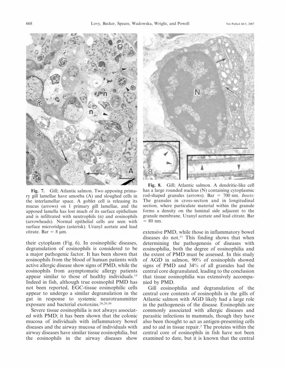
Examination of all the material revealed 2 cellswith large rounded nuclei and large rod-shapedgranules within their cytoplasm (Fig. 8). Thegranules contained particulate material forminga density adjacent to the granule membraneforming an electron-dense lining along the innersurface of the granule membrane (Fig. 8, inset).Spheric granules were observed with a similardensity on the luminal side adjacent to the granulemembrane, forming a concentric ring (Fig. 8,inset). The lumen of the granules often containeda square-lattice structured material (Fig. 9). Thiscell type is identical to previously describeddendritic-like cells. These cells were observed in 2instances and not consistently encountered in thelesions.
Discussion
An important finding in this study was theobservation of cells that resembled eosinophils inall the lesions examined resulting from this acute
experimental infection. These cells represented themajority of the infiltrated cells and were charac-teristic in the AGD lesions. The eosinophils weredistinct from previously described EGCs, which arecommonly observed in the gills, skin, and gut ofsalmonids.13 EGCs, which are thought to be similarto mammalian mast cells, contain large sphericmembrane-bound granules containing a dense ho-mogenous matrix.12,31,32 The morphology of theeosinophil granules in this study very closelyresembled that of mammalian eosinophil granules,which are elliptic and contain a crystalline inclusionwithin the center of the granule, confirming thatthese cells are indeed not EGCs but actualeosinophils of perhaps a different lineage to EGC/mast cells.25,34 The crystalline cores of mammalianeosinophil granules can be electron-dense or lucent.Eosinophils have not been previously described forsalmonids, although they have been described forother fish species. Previously identified granules of
Fig. 4. Gill; Atlantic salmon. Fusion of gill lamellaeare seen adjacent to areas infected with amoebae. Threesecondary lamellae are identified by pillar cells (arrows)with intermittent blood channels often containingerythrocytes (e). The interlamellar areas contain epithe-lial cells (E) and cells that resemble eosinophils (arrow-heads). Uranyl acetate and lead citrate. Bar 5 10 mm.
Fig. 3. Gill; Atlantic salmon. Two amoebae (A)near the epithelium (E) have several epithelial cells(arrows) that have sloughed from the tissue. Thesloughed cells have a rounded shape, loss of mostsurface microridges, and hypercondensation of thechromatin within the nucleus. Uranyl acetate and leadcitrate. Bar 5 5 mm.
666 Lovy, Becker, Speare, Wadowska, Wright, and Powell Vet Pathol 44:5, 2007

eosinophils in carp, Cyprinus carpio, have a centralzone called the ‘‘internum,’’ which is surrounded bya dark wrapper, often elongated at 1 end.17 Adifferent description given for carp eosinophilgranules was oval to elongate in shape, containing1 or more, dense bar-shaped crystalloids.36 Thesalmonid eosinophil granules (SEG) described inthis study showed similarities and differences whencompared with the carp eosinophil granules (CEG).The inclusions within the SEGs contained materialthat was less electron-dense than the rest of thegranule. The density of the inclusion was similar tothe internum described for the CEG, but the shapewas different; in carp, internums are localized on 1side of the granule and are more robust than thecores seen in the SEG. The core of the SEG waslocated centrally within the granule and sometimeshad a crystalloid appearance. None of the cores ofthe SEGs were electron-dense, as described forsome mammalian and carp eosinophils.
Many of the granules seen in this studycontained electron-lucent cores, while the remain-ing parts of the granule remained unchanged. Thisfeature has also been observed in carp eosino-phils.17 The empty cores could be a result ofdegranulation of the core portion of the granule. Atype of degranulation, called piecemeal degranula-tion (PMD), has been reported in eosinophils,which involves selective release of granule pro-teins.11,15 The process of PMD is characterized byan increase in vesicles within the cytoplasm of thecell and loss of material from granules withoutgranule-granule or granule-plasma membrane fu-sion.6 The eosinophilic granules in this study fit themorphologic criteria for PMD, and the granuleswith fibrillar cores are likely intact granules, whilethe granules with empty cores have released theircontents by PMD. Cells that have granules withempty cores also have abundant vesicles within
Fig. 6. Gill; Atlantic salmon. The cytoplasm ofeosinophil-containing granules has electron-lucent crys-talloid inclusions (arrows) and an abundance ofcytoplasmic vesicles (arrowheads). Uranyl acetate andlead citrate. Bar 5 500 nm.
Fig. 5. Gill; Atlantic salmon. Eosinophil-contain-ing electron-dense fusiform granules (arrow) havefibrillar inclusions that are less electron-dense. Arrow-heads indicate plasma membrane. N 5 nucleus. Uranylacetate and lead citrate. Bar 5 1 mm.
Vet Pathol 44:5, 2007 Amoebic Gill Disease in Salmon 667
their cytoplasm (Fig. 6). In eosinophilic diseases,degranulation of eosinophils is considered to bea major pathogenic factor. It has been shown thateosinophils from the blood of human patients withactive allergic disease show signs of PMD, while theeosinophils from asymptomatic allergy patientsappear similar to those of healthy individuals.15
Indeed in fish, although true eosinophil PMD hasnot been reported, EGC-tissue eosinophilic cellsappear to undergo a similar degranulation in thegut in response to systemic neurotransmitterexposure and bacterial exotoxins.26,29,30
Severe tissue eosinophilia is not always associat-ed with PMD; it has been shown that the colonicmucosa of individuals with inflammatory boweldiseases and the airway mucosa of individuals withairway diseases have similar tissue eosinophilia, butthe eosinophils in the airway diseases show
extensive PMD, while those in inflammatory boweldiseases do not.11 This finding shows that whendetermining the pathogenesis of diseases witheosinophilia, both the degree of eosinophilia andthe extent of PMD must be assessed. In this studyof AGD in salmon, 90% of eosinophils showedsigns of PMD and 34% of all granules had thecentral core degranulated, leading to the conclusionthat tissue eosinophilia was extensively accompa-nied by PMD.
Gill eosinophilia and degranulation of thecentral core contents of eosinophils in the gills ofAtlantic salmon with AGD likely had a large rolein the pathogenesis of the disease. Eosinophils arecommonly associated with allergic diseases andparasitic infections in mammals, though they havealso been thought to act as antigen-presenting cellsand to aid in tissue repair.2 The proteins within thecentral core of eosinophils in fish have not beenexamined to date, but it is known that the central
Fig. 8. Gill; Atlantic salmon. A dendritic-like cellhas a large rounded nucleus (N) containing cytoplasmicrod-shaped granules (arrows). Bar 5 700 nm. Insets:The granules in cross-section and in longitudinalsection, where particulate material within the granuleforms a density on the luminal side adjacent to thegranule membrane. Uranyl acetate and lead citrate. Bar5 80 nm.
Fig. 7. Gill; Atlantic salmon. Two apposing prima-ry gill lamellae have amoeba (A) and sloughed cells inthe interlamellar space. A goblet cell is releasing itsmucus (arrows) on 1 primary gill lamellae, and theapposed lamella has lost much of its surface epitheliumand is infiltrated with neutrophils (n) and eosinophils(arrowheads). Normal epithelial cells are seen withsurface microridges (asterisk). Uranyl acetate and leadcitrate. Bar 5 8 mm.
668 Lovy, Becker, Speare, Wadowska, Wright, and Powell Vet Pathol 44:5, 2007

core of mammalian eosinophils is composed mainlyof major basic protein.25 Major basic protein isknown to efficiently destroy parasites and also hasbeen shown to damage the resident population ofcells exposed to the protein around the tissueeosinophilia.14 There are known differences in thecentral core of eosinophils in fish and mammalsthough. The central core internum of carp eosino-phils contains activity of both acid phosphataseand peroxidase, while neither of these has beendemonstrated in the central core of mammalianeosinophils.17
A small number of dendritic-like cells with rod-shaped granules containing square-latticed struc-tured material, which were occasionally vacuolatedon 1 end, were encountered in this study (Figs. 8, 9).These cells very closely resembled previously de-
scribed dendritic-like cells from the gills of chinooksalmon, Oncorhynchus tshawytscha, suffering frommicrosporidial gill disease caused by the inflamma-tion-inducing parasite, Loma salmonae.20 These cellswere not typical in the lesions caused by amoebaebut were encountered in 2 lesions that wereexamined. The role of this cell type may not bemajor in the pathogenesis of AGD, but this is thefirst finding of this cell type in Atlantic salmon gills.Morphologically, this cell type contains granulesvery similar to Birbeck granules found in mamma-lian Langerhans cells. Birbeck granules fromLangerhans cells and the granules in the salmoniddendritic-like cells are both rod-shaped, containingsquare-lattice structured material and commonlyvacuolated at 1 end.20 The granules in the salmoniddendritic-like cells also bear close resemblance tothose found in mammalian monocyte-derived den-dritic cells, which contain granules with a ring-likeshape and comprise 2 concentric membranes.20 Thefinding of this cell type in a different species witha different gill disease further suggests that it is animportant cell type responding to pathogens in thegills. These cells were located in connective tissue offused secondary lamellae, which also containedeosinophils, few neutrophils, mitotic epithelial cells,and mucous cells.
There has been interest in examining the in-teraction of amoebae and host tissue in order toelucidate some mechanisms of attachment andpathogenesis leading to disease. The interactionsbetween amoebae and the host epithelium, whichresult in disease, remain a mystery. Previous workhas shown that gill-attached amoebae were distinctfrom in vitro cultured amoebae in that the gill-attached amoebae had more pseudopodia exten-sions and contained electron-dense deposits withinvesicles in the cytoplasm.16,33 The composition andorigin of the electron-dense deposits are unclear,but similar deposits were observed in this study.Also in this study, portions of the amoebaemembrane appeared more electron-dense andcontained cytoplasmic electron-dense deposits.Although many amoebae were observed withoutdirect contact to the host epithelium in this study, itwas common to observe amoebae with pseudopo-dia inserted within invaginations of epithelial cells.This may be a mechanism to better anchor itselfinto the tissue or a way to increase the surface areaof the amoebae contacting host tissue, perhaps toincrease efficiency of factors the amoebae mayrelease to affect the host tissue.
In this investigation, it is clear that the hostsurface epithelial cells are severely affected by thepresence of the amoebae. Normal surface epithelial
Fig. 9. Gill; Atlantic salmon. Rod-shaped granuleswithin dendritic-like cells that have a vacuolated portion(arrowheads), which contains particulate material witha square-lattice structure (arrows). Uranyl acetate andlead citrate. Bar 5 100 nm.
Vet Pathol 44:5, 2007 Amoebic Gill Disease in Salmon 669

cells contain elaborate fingerprint-like microridgesalong their surface, which aid in increasing therespiratory surface area and aid in the attachmentof mucus.13 An example of normal surface micro-ridges on gill epithelial cells can be seen in Fig. 7.The affected epithelial cells in the present studyhave lost much of their surface microridges, andadditionally, the cells have sloughed from thesurface and have a rounded shape and hypercon-densed nuclear chromatin. These morphologicfeatures are consistent with those of apoptosis.Cells undergoing apoptosis lose surface differentia-tions and round up, and chromatin becomeshypercondensed due to DNA fragmentation.25 Ithas been shown that culture of N. pemaquidensiswith an Atlantic salmon gill epithelium cell lineresulted in complete cytolysis of the cell monolayerwithin 5 days.5 The cytopathic effect that amoebaehave on the epithelial cells or the presence ofinflammatory cells in vivo may initiate apoptosis ofepithelial cells.
Acknowledgements
This research formed part of a project of the AquafinCooperative Research Centre (CRC), the AustralianGovernments CRC Programme, and received fundsfrom Fisheries Research & Development Corporationand other CRC participants (MDP). Additional fundingwas received by Contributing to Australian Scholarshipand Science foundation (JAB).
References
1 Adams MB, Nowak BF: Amoebic gill disease:sequential pathology in cultured Atlantic salmon,Salmo salar L. J Fish Dis 26:601–614, 2003
2 Adams MB, Nowak B: Distribution and structure oflesions in the gills of Atlantic salmon, Salmo salar L.,affected with amoebic gill disease. J Fish Dis24:535–542, 2001
3 Behm CA, Ovington KS: The role of eosinophils inparasitic helminth infections: insights from geneti-cally modified mice. Parasitol Today 16:202–209,2000
4 Bermingham ML, Mulcahy MF: Environmental riskfactors associated with amoebic gill disease incultured salmon, Salmo salar L. smolts in Ireland.J Fish Dis 27:555–571, 2004
5 Butler R, Nowak BF: In vitro interactions betweenNeoparamoeba sp. and Atlantic salmon epithelialcells. J Fish Dis 27:343–349, 2004
6 Crivellato E, Nico B, Mallardi F, Beltrami CA,Ribatti D: Piecemeal degranulation as a generalsecretory mechanism? Anat Rec A Discov Mol CellEvol Biol 274:778–784, 2003
7 Douglas-Helders GM, Saksida S, Nowak BF:Questionnaire-based risk assessment for amoebic gilldisease (AGD) and evaluation of freshwater bathing
efficacy of reared Atlantic salmon Salmo salar. DisAquat Organ 63:175–184, 2005
8 Douglas-Helders M, Nowak B, Butler R: The effectof environmental factors on the distribution ofNeoparamoeba pemaquidensis in Tasmania. J FishDis 28:583–592, 2005
9 Dykova I, Figueras A, Peric Z: Neoparamoeba Page1987: light and electron microscopic observations onsix strains of different origin. Dis Aquat Organ43:217–223, 2000
10 Dykova I, Nowak BF, Crosbie PB, Fiala I, PeckovaH, Adams MB, Machackova B, Dvorakova H:Neoparamoeba branchiphila n. sp. and related speciesof the genus Neoparamoeba Page, 1987: morpholog-ical and molecular characterization of selectedstrains. J Fish Dis 28:49–64, 2005
11 Erjefalt JS, Greiff L, Andersson M, Adelroth E,Jeffery PK, Persson CGA: Degranulation patterns ofeosinophil granulocytes as determinants of eosino-phil driven disease. Thorax 56:341–344, 2001
12 Ezeasor DN, Stokoe WM: A cytochemical, light andelectron microscopic study of the eosinophilicgranule cells in the gut of the rainbow trout. J FishBiol 17:619–634, 1980
13 Ferguson HW: Systemic pathology of fish, 1st ed,Iowa State University Press, Ames, IA, 1989
14 Gleich GJ, Frigas E, Loegering DA, Wassom DL,Steinmuller D: Cytotoxic properties of the eosinophilmajor basic protein. J Immunol 123:2925–2927, 1979
15 Karawajczyk M, Seveus L, Barcia R, Bjornsson E,Peterson CGB, Roomans GM, Venge P: Piecemealdegranulation of peripheral blood eosinophils.Am J Respir Cell Mol Biol 23:521–529, 2000
16 Kent ML, Sawyer TK, Hedrick RP: Paramoebapemaquidensis (Sarcomastigophora: Paramoebidae)infestation of the gills of coho salmon Oncorhynchuskisutch reared in seawater. Dis Aquat Organ 5:163–169, 1988
17 Kralj-Klobucar N: Differentiation of eosinophilicgranulocytes of carp (Cyprinus carpio L.). Int J DevBiol 35:341–344, 1991
18 Leef MJ, Harris JO, Hill J, Powell MD: Cardiovas-cular responses of three salmonid species affectedwith amoebic gill disease (AGD). J Comp Physiol [B]175:523–532, 2005
19 Leef MJ, Harris JO, Powell MD: Respiratorypathogenesis of amoebic gill disease (AGD) inexperimentally infected Atlantic salmon Salmo salar.Dis Aquat Organ 66:205–213, 2005
20 Lovy J, Wright GM, Speare DJ: Morphologicalpresentation of a dendritic-like cell within the gills ofchinook salmon infected with Loma salmonae. DevComp Immunol 30:259–263, 2006
21 Morrison RN, Cooper GA, Koop BF, Rise ML,Bridle AR, Adams MB, Nowak BF: Transcriptomeprofiling of amoebic gill disease (AGD)-affectedAtlantic salmon (Salmo salar L): a role for tumorsuppressor p53 in AGD pathogenesis? PhysiolGenomics 26:15–34, 2006
670 Lovy, Becker, Speare, Wadowska, Wright, and Powell Vet Pathol 44:5, 2007

22 Munday BL, Foster CK, Roubal FR, Lester RGL:Paramoebic gill infection and associated pathologyof Atlantic salmon, Salmo salar, and rainbow trout,Salmo gairdneri, in Tasmania. In: Pathology inMarine Science, ed. Perkins FO and Cheng TC,pp. 215–222. Academic Press, San Diego, CA, 1990
23 Munday BL, Zilberg D, Findlay V: Gill disease ofmarine fish caused by infection with Neoparamoebapemaquidensis. J Fish Dis 24:497–507, 2001
24 Nowak BF, Carson J, Powell MD, Dykova I:Amoebic gill disease in the marine environment.Bull Europe Assoc Fish Pathol 22:144–147, 2002
25 Pavelka M, Roth J: Functional Ultrastructure.Springer Wien, New York, NY, 2005
26 Powell MD, Briand HA, Wright GM, Burka JF:Rainbow trout (Oncorhynchus mykiss Walbaum)intestinal eosinophilic granule cell (EGC) responseto Aeromonas salmonicida, and Vibrio anguillarumextracellular products (ECPs). Fish Shellfish Immu-nol 3:279–289, 1993
27 Powell MD, Fisk D, Nowak BF: Effects of gradedhypoxia on Atlantic salmon infected with amoebicgill disease. J Fish Biol 57:1047–1057, 2000
28 Powell MD, Nowak B, Adams MB: Cardiacmorphology in relation to amoebic gill diseasehistory in Atlantic salmon, Salmo salar L. J FishDis 25:209–215, 2002
29 Powell MD, Wright GM, Burka JF: Degranulationof eosinophilic granule cells (EGC) induced bycapsaicin and substance P in the intestine of therainbow trout (Oncorhynchus mykiss Walbaum). CellTissue Res 266:469–474, 1991
30 Powell MD, Wright GM, Burka JF: Morphologicaland distributional changes in the eosinophilicgranule cell (EGC) population of the rainbow trout(Oncorhynchus mykiss) intestine following systemicadministration of capsaicin and substance P. J ExpZool 266:19–30, 1993
31 Reite O, Evensen O: Inflammatory cells of teleosteanfish: a review focusing on mast cell/eosinophilicgranule cells and rodlet cells. Fish Shellfish Immunol20:192–208, 2006
32 Reite O: Mast cells/eosinophilic granule cells ofteleostean fishes: a review focusing on the stainingproperties and their functional responses. FishShellfish Immunol 8:489–513, 1998
33 Roubal FR, Lester RJG, Foster CK: Studies oncultured and gill-attached Paramoeba sp. (Gymna-moeba: Paramoebidae) and the cytopathology ofparamoebic gill disease in Atlantic salmon, Salmosalar L., from Tasmania. J Fish Dis 12:481–492,1989
34 Sire MF, Vernier JM: Partial characterization ofeosinophilic granule cells (EGCs) and identificationof mast cells of the intestinal lamina propria inrainbow trout (Oncorhynchus mykiss): biochemicaland cytochemical study. Biol Cell 85:35–41, 1995
35 Speare DJ: Nodular gill disease (amoebic gill in-festation) in Arctic char, Salvelinus alpinus. J CompPath 121:277–282, 1999
36 Tripathi NK, Latimer KS, Burnley VV: Hematolog-ic reference intervals for koi (Cyprinus carpio),including blood cell morphology, cytochemistry,and ultrastructure. Vet Clin Pathol 33:74–83, 2004
Request reprints from Jan Lovy, Department of Pathology and Microbiology, Atlantic Veterinary College,University of Prince Edward Island, Charlottetown, PEI C1A 4P3, (Canada). E-mail: [email protected].
Vet Pathol 44:5, 2007 Amoebic Gill Disease in Salmon 671