ultrasonographic characterization of ovarian events and fetal gestational parameters in two southern...
TRANSCRIPT

ELSEVIER
ULTRASONOGRAPHIC CHARACTERIZATION OF OVARIAN EVENTS AND FETAL GESTATIONAL PARAMETERS IN TWO SOUTHERN BLACK RHINOCEROS (Diceros
bicornis minor) AND CORRELATION TO FECAL PROGESTERONE
R. W. Radcliffe, la A. I. Eyres, 1 M. L. Patton 2, N. M. Czekala 2, and R. H. Emslie 3 1 Fossil Rim Wildlife Center, 2155 County Road 2008, Glen Rose, Texas 76043 USA
2Center for Reproduction of Endangered Species, San Diego, California 92101 USA
3 IUCN SSC African Rhino Specialist Group, Cascades, 3202 South Africa
Received for publication: May 16, 2000 Accepted: August 2] , 2000
ABSTRACT A tremendous potential exists for the application of transrectal ultrasonography as a tool
to enhance the captive management of endangered species. Reproductive study of two southern black rhinoceros (Diceros bicornis minor) females was performed daily to every other day for a -60 day period to document ovarian changes, and three times weekly in early pregnancy to once monthly in late pregnancy in order to characterize changes in fetal parameters throughout gestation. All ovarian and fetal anatomical structures were measured in millimeters. The mean (+/- SD) length of the estrous cycle or interovulatory period was 26 +/- 1.4 days (n=2 cycles). Follicular growth rate of a dominant follicle was ~3 mm/day once the follicle reached 35 mm in diameter. Ovulation was observed to occur at a mean (+/- SD) follicular diameter of 49.5 +/- 2.6 mm (n=4) and within 48 to 72 h after observed estrus (n=2). Large ovarian structures [mean (+/- SD) diameter of 71.7 +/- 2.9 ram; n=3], considered analogous to equine anovulatory hemorrhagic follicles, were observed to form in the winter months and suggest seasonal periods of reduced fertility. Fecal progesterone assays confirmed ultrasonographic events. Although preliminary, the results of fetal sexing are presented and compared to the horse. Our data indicate that fetal eye or fetal foot diameter measurements can be used to accurately predict gestational age from about 2 months to term, providing useful information to managers of both captive and wild rhino populations. The ability to identify and quickly release animals in late term pregnancy in the wild and thereby reduce abortions and neonatal mortalities in holding bomas is one potential practical conservation benefit of the fetal age predictwe models. © 2001 by Elsevier Science Inc
Key words: black rhinoceros, Diceros bicornis, reproduction, estrous cycle, pregnancy
Acknowledgment The authors would like to thank the Fossil Rim Wildlife Center and all of our rhino research interns for making this work possible. The International Rhino Foundation and ABC/Kane Productions provided invaluable support of our rhino research program. We thank Chuck Boland of Aloka tbr donating ultrasound equipment, Dr. Matt Read for assisting with data collection, Dr. Ron Swaisgood for help with data analysis, and Drs. Rolfe Radcliffe and Chris Foggin for support. Finally, we thank Dr. Steve Osofsky for helping to develop the rhino research program at Fossil Rim during his tenure as Director of Animal Health Services.
aCorrespondence and reprint requests (E-mail: [email protected] / Ph: (254) 897-2960).
Thenogenology 55:1033-1049, 2001 0093-691 X/O 1/S-see front matter © 2001 Elsevier Science Inc. PII SOO93-691X(01)O0464-2
1034 Theriogenology
INTRODUCTION
Knowledge of basic reproductive biology in many large, nondomestic species is incomplete. Although numerous factors affect captive rhinoceros conservation efforts, our insufficient knowledge of reproductive events may limit the long-term sustainability of captive rhino populations. The reproductive biology of both species of African rhinoceros has been previously documented through serum, urinary, fecal and salivary hormone analysis (4,5,11,13,21,22). Although commonly used in large domestic species, ultrasound technology has seen only limited application in nondomestic ungulates because of subject intractability.
Ultrasound is invaluable because this noninvasive technique immediately gives a detailed morphological study of otherwise little-known reproductive events. Transrectal ultrasonography was used successfully for serial examination of the African rhinoceros without sedation, allowing for enhanced characterization of reproductive events compared to hormonal assay alone (1,17,18). The technique for the black rhinoceros involves transrectal examination of the subject in a "free-stall" chute, a design that allows the animal to enter or leave the chute at will (17,18). Improved captive management of rhinoceros species will require a more detailed understanding of reproductive function. Our research findings corroborate previous reports and add a morphological understanding that will facilitate more precise and informed management decisions. Here we report data from the long-term ultrasonographic study of two female southern black rhinoceros (Diceros bicornis minor). This information will provide a scientific basis for decision-making for black rhino captive breeding efforts and important implications for managers of wild rhino populations. Due to the inherent difficulty in visual identification of late term pregnancy in wild rhinos, ultrasonography would give game capture teams a field tool to identify and quickly release these late term animals and prevent abortions and neonatal mortalities in holding bomas (17).
MATERIALS AND METHODS
Animals and Ultrasound Technique
Ultrasonographic evaluation of two wild-caught female southern black rhinoceros was conducted in a serial manner without sedation in a "'free-stall" chute (17,18; Figure I) from December 11, 1995 to May 27, 1999. Ovarian activity of female 1 ("Sinampande," Studbook No. 0466, est. date of birth: 1977) was examined transrectally by ultrasound and fecal material for progesterone assay was simultaneously collected three times per week (September 11, 1996 to November 27, 1996) to characterize estrous. Female 2 ("Coco," Studbook No. 0462, date of birth: December 1, 1990) was monttored in a similar fashion to female 1. In addition, behavioral estrus observations were recorded for two estrous cycles (July 25, 1997 to September 27, 1997) and ultrasound monitoring of two pregnancies was conducted only for female 2. Serial gestational monitoring in Female 2 encompassed most of the fetal stage of one pregnancy (Gestation I = 475 days; initial exam on Day 56 through last exam on Day 475 [day of parturition] for a total of 59 exams); and the embryo and fetal stages of a second pregnancy (Gestation 2 = 465 days; initial exam on Day 16 through last exam on Day 452 for a total of 28 exams). Ultrasound exams were conducted daily for 3 days after observed signs of estrus (exams

Theriogenology 1035
Figure 1. Illustration of the transrectal ultrasonography technique developed for application to managed captive breeding efforts for the black rhinoceros (Diceros bicornis minor). The free-stall chute allows the rhinoceros freedom to enter or leave the chute, but the subject normally chooses to stand readily for examination in return for positive food rewards.

1036 Theriogenology
performed at 24, 48 and 72 h post estrus) in order to correlate the precise timing of ovulation with "'standing estrus.'" Day 0 was defined as the day of ovulation and was confirmed by documentation of follicular collapse followed by formation of a luteal structure visualized on ultrasound.
The transrectal examination technique was evaluated for the rhinoceros and the risks of rectal perforation were considered minimal (1). Ultrasonographic exams were of short duration (2 to 10 minutes). Although ultrasonic waves have been described to cause cellular damage via thermal and cavitation mechanisms, these effects are inconsequential at the exposure intensity and duration typical of diagnostic ultrasound (7).
Both black rhino females were housed separately in an indoor facility and had free-choice access to a 3-acre outdoor enclosure at the Fossil Rim Wildlife Center in Glen Rose, Texas, USA (Latitude 32 ° 14' 11" N: Longitude 97 ° 45' 17" W). The climate was temperate with a definite photoperiod effect (longest daylight period, 16 h; shortest, 10 h). The females were housed next to an adult male southern black rhinoceros. Females were separated from the male, except for the purpose of breeding. Both rhinos were observed closely for a minimum of 2h daily to document any behavioral changes during the estrous cycle. Behavioral data was not collectable on Female 1, as this female did not demonstrate appreciable signs of estrus. Nutritional management of the black rhinoceros consisted of 11 to 13 kg herbivore pellets (9 to 11 kg
Mazuri ADF-16 No. 5648 and 2 kg Mazuri ADF-25 No. 5649), b 1 kg Equine Athlete, b 14 to 20 kg alfalfa hay, and occasional sliced apples and yams as treats. Local browse species [prairie sumac (Rhus lanceolata) and black willow (Salix nigra)] were fed daily during the summer months (approximately 5 kg per rhino; May through October).
Transrectal ultrasonography was performed with a portable scanner (Aloka Model 500
V) c equipped with a 5 MHz linear-array transducer (Aloka Model UST-588U-5). c Although the left ovary was more difficult to visualize, both ovaries were imaged without an extensor. The size of ovarian and fetal structures was determined by measuring the longest cross-sectional diameter in ram. All ultrasound examinations were recorded on a Hi-8 professional
videocassette d for later evaluation. All data were summarized as the mean plus or minus one sample standard deviation (SD). A Wilcoxon signed rank test for related samples was used to determine when pregnane values increased significantly above nonpregnant luteal levels post- copulation. Pregnane values were pooled from the two pregnancies. Regression models were derived to estimate gestational age of the fetus from measures of fetal eye and fetal foot. As both measurements were not made in all cases separate models were built for each measure. All fetal
age regressions were undertaken using StatSoft Inc.'s Statistica for Windows Version 5.0. e
bpurina Mills, St. Louis, Missouri 63166, USA.
CAloka Inc., Wallingford, Connecticut 06492, USA.
dSony Recording Media of America, Montvale, New Jersey 07645, USA.
estatSoft Inc., Tulsa, Oklahoma 74104 USA.

Theriogenology 1037
Fecal samples were collected directly from the rectum before each ultrasonographic evaluation. Approximately 50 g of feces were placed in a plastic bag and frozen at -20 ° C until analysis. Fecal pregnanes were extracted and measured as previously described in the southern white rhinoceros (16).
Assay Methodology
Processing and extraction. Briefly, fecal samples were lyophilized to reduce variability in water content. Dried samples were sifted to remove vegetation debris. Fecal samples (0.02 g) were wetted with distilled water (2.0 mL) and diethyl ether (anhydrous) extracted. Ether was dried and the sample was reconstituted in 1.0 mL of absolute ethanol.
Radioimmunoassay. The RIA for pregnanes used a monoclonal antibody to 4-pregnen- 1 l-ol-3,20-dione hemisuccinate:BSA (10). Tritiated progesterone (10,000 cpm/0.1 mL, Dupont NEN Boston MA) was used to compete against standard progesterone. Depending on the status of reproduction, 1.0 uL to 5.0 uL of reconstituted sample was taken to the RIA and diluted with phosphate buffered saline (0.1 ml, pH=7.0). After overnight incubation at 4 °C, the reaction was ended by adding 0.25 mL dextran-coated charcoal solution. A further 30 min incubation (4 °C) was followed by centrifugation (15 min). Supernatant was decanted into scintillation vials with scintillation fluid (5.0 mL Ultima Gold, Packard Instrument, Meriden, CT) and counted for 2 min in a scintillation counter.
The standard progesterone curve was compared to the serially diluted extracts to assess the validity of this assay for evaluation of black rhinoceros progestins. The standard curve and the extracted fecal sample showed parallel displacement curves (R = 0.9982, sample range 0.0002 to 0.003 mL of a sample from a nonpregnant female). Buffer blanks were below the assay sensitivity. All samples reported were analyzed in a total of 4 assays. Inter-specific coefficients of variation for control samples measured in 4 assays was 7% for the high control (mean = 211.8 pg/tube) and 15% for the low control (mean = 39.5 pg/tube). Intra-assay variation estimates (10 replicates of the same pools in a single assay) were 4.1% and 4.4% for a high and low pool respectively. Results are presented as ng/g of dry fecal weight.
Estrous Cycle RESULTS
Mean (+/- SD) estrous cycle length (one complete interovulatory interval) in the two black rhinoceros subjects was 26 ÷/- 1.4 days (n=2 cycles; Figure 2). Follicular growth of a dominant follicle occurred at a rate o f - 3 mm/day once the follicle reached 35 mm in diameter. Follicles less than 35 mm in diameter did not predictably become a dominant follicle. Preovulatory follicles (n=4) were observed to reach a mean (+/- SD) follicular diameter of 49.5 -4-/- 2.6 mm changing from spherical to pear-shaped before ovulation. Ultrasonographic documentation of ovulation was based on observed disappearance of the preovulatory follicle followed by formation of a visible luteal structure (n=4, mean (+/- SD) luteal structure diameter = 38.3 +/- 13.4 mm; Figure 2). Ovulation was later confirmed by a rapid increase in fecal progestins - 6 days after ultrasonographic identification of ovulation. Behavioral observation in

1038 Theriogenology
2- 1td
Figure 2.
12- 2Z- 1- 11- 21- 31- 10- 20- 30- 10- 20- 30- 9- 19- 29- 9- Jul Jul Aug Aug Aug Aug Sep Sep Sep Oct Oct Oct Nov Nov Nov Dec
Changes in the diameter of the largest follicle and corpus luteum (graph above', Ovl and Ov2 represents ovulation in Female 1 and Female 2, respectively) in comparison to fecal progesterone (graph below) during the estrous cycle in two female black rhinoceros (.Diceros bicornis minor). Behavioral data for one female is marked on the lower graph (E represents day of standing estrous, D represents diestrous or period of nonreceptivity to male). Image axes are marked in 1 cm increments, and letter labels represent the corresponding data point in the chart.

Theriogenology 1039
Female 2 was correlated with daily ultrasonographic events (Figure 2); ovulation was documented to occur within 24 to 48 h and 24 to 72 h of estrus (n--2; rhino uncooperative at 48 h exam of latter estrus period so no data acquired). Signs of estrus in Female 2 (urine squirting, "'standing" behavior, and male rhino climbing fence into female's enclosure) were prominent for a 12 h period with a follicular diameter of 50 mm (n-2). Overt signs of behavioral estrus were very limited in Female 1, and therefore no comparisons to the stage of the cycle were made. Receptivity and nonreceptivity of each female was objectively determined by observation of male and female interaction. For example, Female 1 would not tolerate approaches by the male housed in an adjacent pen at any time, whereas this same male would attempt to climb the fence when Female 2 demonstrated signs of estrus.
Several large, apparently anovulatory structures (n=3, mean diameter = 71.7 +/- 2.9 mm) were observed in both rhinos towards the end of the examination period (October/November) and on one occasion was associated with erratic progesterone concentrations (Figure 2). These structures formed at the time of expected ovulation (i.e. after observed development of a preovulatory-size follicle), but were not associated with signs of follicular collapse or formation of a corpus luteum. Instead, a large fluid-filled structure persisted for up to 30 days, and differed from an ovulatory corpus hemorrhagicum in reaching a larger size and developing numerous gelatinous fibrous bands (Figure 2).
Gestational Parameters
Fetal measurements. Serlal measurements of various fetal parts were recorded during two nearly complete gestational periods in Female 2 (Gestation l = 475 days, and Gestation 2 = 465 days). All measurements were made at the longest cross-sectional diameter or length in mm. Fetal parameters monitored included fetal eye diameter, foot diameter, trunk (fetal body cross- section) diameter, rib diameter, skull length, and heart rate (Figures 3 and 4). During both pregnancies monitored, the fetus became increasingly difficult to visualize between 6 and 9 months of gestation presumably due to movement of the fetus over the pelvis. A dirt ramp was incorporated into the front of the chute to facilitate improved fetal visualization by elevation of the rhino's forequarters; as the fetus enlarged beyond 9 months, the use of the ramp was no longer necessary. Changes in the size of various fetal parts were compared to known gestational age over time. Usefulness of each parameter as an estimate of gestational age in the black rhino was assessed by ease of structure visualization on ultrasound and the duration each structure was visible and measurable throughout the gestational period. Ultrasonographic measurement of fetal eye or fetal foot diameter provided a readily available method for estimation of gestational age in most stages of pregnancy (Figures 3 and 4).
Predicting gestauonal age from fetal eye measurements. In the first gestation period, 12 fetal eye diameter measurements covered an approximately four month period from 56 to 182 days; while 11 measurements in the second period covered almost the entire gestation period (days 44-452 of a 465 day gestation). Data from the second gestation period were initially modeled due to the better coverage (allowing predictive extrapolation over a longer period).
The simple regression of gestational age on fetal eye width (Gestation 2) was highly significant (F [df2,8] = 3163.96 p<0.000001) with a corrected R 2 of 0.9862 and a Standard Error

1040 Thefiogenology
Figure 3. Images of the fetal eye (above; arrows mark eye and lines show measurement orientation) and gestational age chart (below) summarizing data from serial ultrasonographic examination during two gestational periods in a southern black rhinoceros (Diceros bicomis minor). Image axes are marked in lcm increments, and letter labels represent the corresponding data point in the chart.

Theriogenology 1041
Figure 4. Images of the fetal foot (above; arrows mark foot and lines show measurement orientation) and gestational age chart (below) summarizing data from serial ultrasound of two gestational periods in a southern black rhinoceros (.Diceros bicornis minor). Image axes are marked in 1 cm increments, and letter labels represent the corresponding data point in the chart.

1042 Theriogenology
of the estimate of 17.7. However. the plot of residuals against observed gestational age had a U shape violating the regression assumption of linearity (23), thus indicating that a curvilinear relationship should be considered. This was successfully achieved by adding a single polynomial term, which not only linearized the residuals but also increased estimate precision. The coefficients for both the linear (fetal eye mm) and curvilinear (fetal eye mm 2) terms were both significant (in both cases p<0.0001), and predictive accuracy of the model increased (F [df2,8] = 3164.0 p<0.000001) with the Standard Error of the estimate dropping (from 17.7 in the linear) to 6.0 days in the polynomial model. This polynomial regression was then validated by using it to predict gestational age based on eye diameters measured in the first gestation (i.e. testing the model using independent data not used to build it). The mean prediction error was only 2.8 days (n =12 error range 0.8 to 15.7 days), with a residual standard deviation of 7.0 days (coefficient of variation of 5.35%). This predictive accuracy was almost as good as using the model to predict the data points used to build it (standard deviation of the residual = 5.8 days). This observation, and the fact that the separate polynomial regressions for each gestation period were very similar, suggested that it may be possible to build a joint model using data from both gestation periods, The methodology outlined in Zar (23) and statistical output from Statistica were then used to determine whether both polynomial regressions (for Gestation 1 and 2) were estimating the same regression function. The null hypothesis that there was a single population underlying both fetal eye polynomial regressions was supported (F [df 3,17] = 1.2679 n.s. p>0.5). Thus data from both gestation periods could justifiably be analyzed together in a single pooled regression giving a final model of:
Predicted fetal age, x (days) = 46.6078 + 6.0445y + 0.2535 y2 (where y --- fetal eye diameter in mm).
This model was highly significant (F [dr2,20] = 3386.5 p<0.000001) with an adjusted R 2 of 0.9984 and a Standard Error of the estimate of 6.5 days. This degree of predictive accuracy is remarkable considering errors introduced into the model by measurement resolution (eye diameter rounded to the nearest mm). A check of the residuals in the pooled model revealed there were no outliers (all +/- 2 standard deviations). Statistical output from Statistica and the methodology outlined in Neter et. al. (14) was used to calculate 95% confidence intervals around the regression estimates, which varied from +/- 3.4 days to +/-9.5 days (depending upon gestational age), with a mean of+/- 4.7 days.
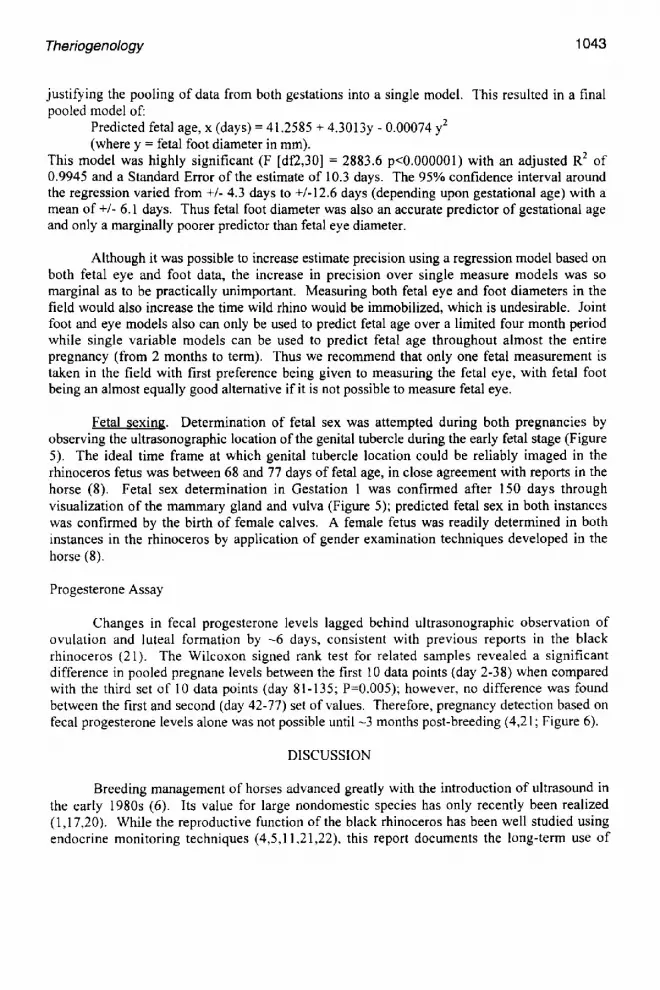
Predicting gestational age from fetal foot measurements. Similar analyses were undertaken using the fetal foot data obtained during the same two gestation periods. In the first gestation period, 24 fetal foot diameter measurements covered almost the entire gestation from 56 to 475 days: while in the second period only 9 observations were recorded partially covering the gestation (days 50-211). Fetal eye and fetal foot diameters were measured concurrently on only 12 occasions from 56-182 days. After 182 days it was only possible to measure either the foot or the eye (but not both), depending upon the position of the fetus. Fetal foot was measurable with a caudal presentation (Gestation 1) while fetal eye could be measured with a cranial presentation (Gestation 2). As with the fetal eye analyses, straight regressions of gestational age on fetal foot diameter violated the assumption of linearity. Once again a simple polynomial regression successfully dealt with the slight curvilinearity in the residuals and improved estimate precision. Independent polynomial regressions for gestation 1 and 2 were also found to be estimating the same regression function (F [df 3,27] = 0.5828 n.s. p>0.5),
Theriogenology 1043
justifying the pooling of data from both gestations into a single model. This resulted in a final pooled model of:
Predicted fetal age, x (days) = 41.2585 + 4.3013y - 0.00074 y2 (where y = fetal foot diameter in mm).
This model was highly significant (F [dr2,30] = 2883.6 p<0.000001) with an adjusted R 2 of 0.9945 and a Standard Error of the estimate of 10.3 days. The 95% confidence interval around the regression varied from +/- 4.3 days to +/-12.6 days (depending upon gestational age) with a mean of +/- 6.1 days. Thus fetal foot diameter was also an accurate predictor of gestational age and only a marginally poorer predictor than fetal eye diameter.
Although it was possible to increase estimate precision using a regression model based on both fetal eye and foot data, the increase in precision over single measure models was so marginal as to be practically unimportant. Measuring both fetal eye and foot diameters in the field would also increase the time wild rhino would be immobilized, which is undesirable. Joint foot and eye models also can only be used to predict fetal age over a limited four month period while single variable models can be used to predict fetal age throughout almost the entire pregnancy (from 2 months to term). Thus we recommend that only one fetal measurement is taken in the field with first preference being given to measuring the fetal eye, with fetal foot being an almost equally good alternative if it is not possible to measure fetal eye.
Fetal sexing. Determination of fetal sex was attempted during both pregnancies by observing the ultrasonographic location of the genital tubercle during the early fetal stage (Figure 5). The ideal time frame at which genital tubercle location could be reliably imaged in the rhinoceros fetus was between 68 and 77 days of fetal age, in close agreement with reports in the horse (8). Fetal sex determination in Gestation 1 was confirmed after 150 days through visualization of the mammary gland and vulva (Figure 5); predicted fetal sex in both instances was confirmed by the birth of female calves. A female fetus was readily determined in both instances in the rhinoceros by application of gender examination techniques developed in the horse (8).
Progesterone Assay
Changes in fecal progesterone levels lagged behind ultrasonographic observation of ovulation and luteal formation by -6 days, consistent with previous reports in the black rhinoceros (21). The Wilcoxon signed rank test for related samples revealed a significant difference in pooled pregnane levels between the first 10 data points (day 2-38) when compared with the third set of 10 data points (day 81-135; P=0.005); however, no difference was found between the first and second (day 42-77) set of values. Therefore, pregnancy detection based on fecal progesterone levels alone was not possible until -3 months post-breeding (4,21; Figure 6).
DISCUSSION
Breeding management of horses advanced greatly with the introduction of ultrasound in the early 1980s (6). Its value for large nondomestic species has only recently been realized (1,17,20). While the reproductive function of the black rhinoceros has been well studied using endocrine monitoring techniques (4,5,11,21,22), this report documents the long-term use of

1044 Thedogeno~gy
Figure 5. Image of the genital tubercle located caudal to the rear legs (A) of a 68 day-old black rhinoceros (Diceros bicomis minor) fetus, and absence of any hyperechoic structure caudal to the umbilicus (B) indicate female sex. Gender was confirmed at 291 days by visualization of the mammary gland (C) and female genitalia (D). Image axes are marked in l cm increments.

Thedogenology 1045
Z
op-I
o
¢.J
35000
C) Gestation I (G1 --- 475 days)
1." Gestation 2 (G2 = 465 days)
Day of Parturition
• Day of first ultrasound detection of pregnancy
30000
25000
20000
I 0 0 0 0
5000
0 i i i i i i i i i i r i i ~
G2 G1 Day From Mating G2 G1
Figure 6. Graph of fecal progestin levels in ng/g dry weight during two gestational periods in a southern black rhinoceros (Diceros bicomis minor). Gestation 1 (G1) measured 475 days and Gestation 2 (G2) measured 465 days. First ultrasonographic diagnosis of pregnancy for GI was 56 days as conditioning to the procedure was required before application of the technique, whereas G2 was detected at 16 days.

1046 Theriogenology
ultrasonography to characterize reproductive events in this species. This work provides the first look into the morphologic changes that occur during the estrous cycle and pregnancy in the black rhinoceros, information critical for improving managed breeding efforts.
The estrous cycle of the black rhino has previously been reported to be approximately 25 days in length (11,21). Our study confirms these findings, but adds important morphological information. Initial findings suggest that signs of estrus and ovulation in this species occur at follicular diameters approaching 50 mm, similar to reports for large draft horse breeds (8). Impending ovulation also was indicated by a change in follicular shape from spherical to pear- shaped (Figure 2) as reported for horses (6,8). Ovulation has been observed 48 to 72 h following estrus. This is important basic information that will allow improved management decisions in captive rhino breeding programs. For example, the authors have already used such information to predict estrus and breeding, to enhance timed-breeding of otherwise incompatible animals, and to document ovulation and ovulation failure.
Evidence suggests that fertility may decline during the fall and winter months in these animals housed at a North American facility; if substantiated, this could have a profound impact on captive breeding programs for the black rhino. A bimodal distribution of births has been described in the wild for white rhinos (15) and black rhinos (12) in Hluhluwe-Umfolozi Park. This seasonal pattern was closely associated with grass quality in the white rhino suggesting a "'flushing effect"; a similar association between browse quality and black rhino births was not apparent (12). However, highly seasonal unimodal calving distributions were recently documented in several populations of black rhino in South Africa (2). In a population in the summer rainfall region (Pilanesberg NP) a clear relationship was found between rainfall and timing of conception (2). However, the timing of peak conceptions and thus births in two areas outside the summer rainfall region (Addo Elephant NP and Great Fish River Game Reserve Complex) differed from Pilanesberg and was not correlated with rainfall levels, suggesting a relationship to plant phenological patterns and succulence of the vegetation (2). Although not considered a truly seasonal breeder because of the production of offspring in all times of the year in both captivi~' and the wild (9,12), there appeared to be a reduction in fertile ovulations during the fall and winter months (October to December) for our study animals. However, study periods encompassing all times of the year have not been conducted, making meaningful comparisons difficult. This pattern of"seasonal infertility" also was observed in a captive black rhino breeding program in the southern hemisphere where the infertile period occurred during
their winter (April to September). f In our stud,,,', this period was characterized by the formation of large apparently anovulatory ovarian structures considered analogous to equine hemorrhagic follicles (Figure 2). For horses, hemorrhagic follicles (or "autumn follicles") are commonly reported during the "'transitional phase," that part of the year when the mare is approaching anestrus (6). Hemorrhagic follicles also were documented during an infertile period in a white rhinoceros (I 8). Perhaps seasonal and or management-related activities (photoperiod effect in temperate location, cold temperatures, increased stress from confinement indoors in bad weather, impaired nutrition due to lack of browse in many facilities, etc.) adversely affect fertility m the captive black rhino.
fAustralian continent; Thorne A, personal communication, 1999.

Theriogenology 1047
Data on embryo and fetal changes throughout gestation in the mare were used to develop accurate gestational age charts for horses (8). Similar data for rhinos in captive breeding programs would facilitate management changes needed in late gestation and before parturition (17,19). Gestation 2 embryonic morphology correlated well with the only previously published ultrasonographic report of an early embryo in the black rhinoceros (1). The observed changes in black rhino embryonic development were also remarkably similar to those reported for the equine conceptus (8). Serial monitoring of various embryo/fetal structures throughout gestation in the black rhino included measurement of embryonic vesicle diameter, embryo length, skull length, fetal eye diameter, foot diameter, and heart rate. Of these, only fetal eye diameter and foot diameter provided a consistent and easily repeatable measurement throughout most stages of pregnancy in this study. Because of changes in fetal positioning (i.e., caudal [Gestation 1] and cranial presentations [Gestation 2]) the complementary use of fetal eye and fetal foot measurement may improve reliability of such estimates. Fetal sexing in the rhinoceros appears feasible if future studies confirm our preliminary findings of a close analogy with the horse (6.8). Although primarily a research tool, fetal sexing may hold future applications in the management of captive rhino populations. Currently rhino managers are experiencing a demographic problem with a marked skew in the sex ratio of the eastern black rhinoceros (Diceros bicomis michaeli) in favor of male offspring (3).
In agreement with previous studies in the black rhinoceros (4,21), fecal progesterone levels of pregnancy did not rise above nonpregnant luteal phase levels until -90 days post- breeding and therefore fecal progesterone assay would not be useful for early pregnancy detection in this species. Ultrasonographic documentation of early pregnancy and estimation of gestational age (and thus parturition date) has proven beneficial in numerous instances at our facility (17). Confirmation of conception alone has allowed us to separate pregnant animals or confirm embryo loss and infertility (18), thus allowing concentration of breeding efforts and limited resources on other animals. Routine ultrasound evaluation has allowed close monitoring of fetal health during medical problems including a snake envenomation and allergic reaction in a pregnant cow (19). Furthermore, data accumulated in captive rhino studies provided the basis
for applied reproductive evaluation of increasingly managed wild black rhino populations (17). g Knowledge of pregnancy status may improve capture and translocation decision-making for rhinos in the field; the potential to quickly release animals in late term and hopefully reduce abortion-related mortalities in holding boreas is one potential practical conservation benefit of the fetal age predictive models.
The long gestational length and interbirth interval of the rhinoceros mandates that we make intelligent management decisions to enhance reproductive success in captive breeding programs. Ultrasound allows real-time evaluation of reproductive function and thus provides a valuable clinical modality for improved management of captive rhinoceros. In addition, this technique provides a useful research tool to help us understand and interpret the heretofore enigmatic reproductive biology of these endangered Perissodactylids. Much work remains. These preliminary data present numerous questions surrounding ovulation failure, potential seasonal patterns in fertility, and embryo and fetal development.
g Radcliffe et al., unpublished data, 1998, 1999.

1048 Theriogenology
REFERENCES
1. Adams GP, Plotka ED, Asa CS, Ginther OJ. Feasibility of characterizing reproductive events in large nondomestic species by transrectal ultrasonic imaging. Zoo Biol 1991; 10: 247-259.
2. Adcock K. [compiler]. Status and Management of Black Rhino in South Africa. January 1997-December 1998, Rhino Management Group of Southern Africa (RMG) confidential report. 2000; 74pp.
3. Atkinson SJ. Possible determinants of skewed natal sex ratios in captive black (Diceros bicornis) and Indian (Rhinoceros unicornis) rhinoceros in North America. Report prepared for the International Rhino Foundation. 1997.
4. Berkeley EV, Kirkpatrick JF, Schaffer NE, Bryant WM, Threlfall WR. Serum and fecal steroid analysis of ovulation, pregnancy and parturition in the black rhinoceros (Diceros bicornis). Zoo Biol 1997;16:121-132.
5. Czekala NM, Callison L. Pregnancy diagnosis in the black rhinoceros (Diceros bicornis) by salivary hormone analysis. Zoo Biol 1996; 15: 37-44.
6. Gmther OJ. Reproductive Biology of the Mare, Basic and Applied Aspects, 2nd ed. Equiservices, Cross Plains, Wisconsin. 1992.
7. Ginther OJ. Ultrasomc Imaging and Animal Reproduction: Fundamentals, Book I. Equiservlces, Cross Plains, Wisconsin. 1995a.
8. Ginther OJ. Ultrasonic Imaging and Animal Reproduction: Horses, Book 2. Equiservices, Cross Plains, Wisconsin. 1995b.
9. Goltenboth R. International Studbook for African Rhinoceroses. Published by Zoologischer Garten Berlin AG, 01.01.99. 1999.
10. Grieger DM, Scarborough R, de Avila DM, Johnson HE, Reeves JJ. Active immunization of beef heifers against luteinizing hormone: III. Evaluation of dose and longevity. J Anim Sci 1990; 68: 3755-3764.
11. Hindle JE, Mostl E, Hodges JK. Measurement of urinary oestrogens and 20 - dihydroprogesterone during the ovarian cycle of black (Diceros bicornis) and white (Ceratotherium simum.) rhinoceroses. J Reprod Fertil 1992; 94: 237-249.
12. Hitchins PM, Anderson JL. Reproduction, population characteristics and management of the black rhinoceros Diceros bicornis minor in the Hluhluwe/Corridor/Umfolozi Game Reserve Complex. South African J Wildl Res 1983; 13(3): 78-85.
13. Kock N, Morton D, Kock M. Reproductive parameters in free-ranging female black rhinoceroses (Diceros bicornis) in Zimbabwe. Onderstepoort J Vet Res 1991; 58:55-57.
14. Neter J, Wasserman W, Whitmore GA. Applied Statistics (Fifth printing). Allyn and Bacon Inc. 1979; 743 pp.
15. Owen-Smith RN. The behavioural ecology of the white rhinoceros. Ph D. thesis, University of Wisconsin. 1973.
16. Patton ML, Swaisgood RR, Czekala NM, White AM, Fetter GA, Montagne JP, Rieches RG, Lance VA. Reproductive cycle length and pregnancy in the southern white rhinoceros (Ceratotherium simum simum) as determined by fecal pregnane analysis and observations of mating behavior. Zoo Biol 1999; 18. 111-127.
17. Radcliffe RW, Bommarito MP, Osofsky SA. Ultrasonography as a tool in the conservation of the African rhinoceros: ex situ and in situ applications. Pachyderm 1996: 21: 55-59.

Theriogenology 1049
18. Radcliffe RW, Czekala NM, Osofsky SA. Combined serial ultrasonography and fecal progestin analysis for reproductive evaluation of the female white rhinoceros (Ceratotherium simum simum): preliminary results. Zoo Biol 1997; 16: 445-456.
19. Radcliffe RW, Eyres AI, Miller CA. Reproductive management of the captive rhinoceros: a collaborative approach to maximize rhino health and reproduction. Proc. First Ann. Rhino Keepers Workshop, Orlando Florida. May 1999.
20. Schaffer N, ZainaI-Zahari Z, Suri MSM, Jainudeen MR, Jeyendran RS. Ultrasonography of the reproductive anatomy in the sumatran rhinoceros (Dicerorhinus sumatrensis). J Zoo Wildl Med 1994; 25: 337-348.
21. Schwarzenberger F, Francke R, Goltenboth R. Concentrations of faecal immunoreactive progestagen metabolites during the oestrous cycle and pregnancy in the black rhinoceros (Diceros bicornis michaeli). J Reprod Fertil 1993; 98: 285-291.
22. Schwarzenberger F, Tomasova K, Holeckova D, Matern B, Mostl E. Measurement of fecal steroids in the black rhinoceros (Diceros bicornis) using group-specific enzyme immunoassays for 20-oxo-pregnanes. Zoo Biol 1996: 15: 159-171.
23. Zar JH. Biostaustical Analysis (second edition). Prenuce Hall International. 1984; 718pp.