ugzy-102 - uprtou
TRANSCRIPT
UGZY-102 Diversity of Animal Life
Block-1 Comparative Forms and Functions-I 03-30 Unit-1 General characters & Classification of Protozoa Unit-2 Body Organization & Characteristics of Metazoa
Block-2 Comparative Forms and Functions-II31-124
Unit-3 General characters and classification of Porfera, Cnidaria, Ctenophora, Platyhelminthes, Nematoda
Unit-4 General characters and classification of Phylum Annelida, Arthropoda, Mollusca and Larval forms of Echinodermata
Unit-5 Comparative form and Functions : Locomotion, Digestion, Excretion
Unit-6 Respiratory, Circulatory and Nervous system Unit-7 Reproductive system
Block-3 Adaption and Behavioral Pattern 125-164
Unit-8 Adaptive Radition Unit-9 Behavioural patterns Unit-10 Harmful and beneficial Non-Chordates
Uttar Pradesh Rajarshi Tandon Open University
UGZY-102/1

UGZY-102/2

UGZY-102 Diversity of Animal Life
Block
1 Comparative Forms and Functions-I Unit 1 07-20 General characters & Classification of Protozoa Unit 2 21-30 Body Organization & Characteristics of Metazoa
Uttar Pradesh Rajarshi Tandon Open University
UGZY-102/3
Course Design Committee Chairman Prof. Ashutosh Gupta
Director, School of Science UPRTOU, Prayagraj.
Prof. Paramhand Pathak Member Department of Zoology Deendayal Upadhyay University, Gorakhpur.
Prof. Ajay Singh Member Department of Zoology Deendayal Upadhyay University, Gorakhpur.
Prof. Rajendra Singh Special Invitees Department of Zoology Deendayal Upadhyay University, Gorakhpur.
Member/Secretary Dr. Deepa Chaubey Acadmic Consultant, School of Science UPRTOU, Prayagraj.
Course Preparation Committee Dr. V.C. Srivastava Author Ex. Associate Prof. C.M.P. Degree College, Prayagraj
Dr. A.K. Srivastava EditorEx. Head Of Zoology B.B.C, Jhansi.
Coordinator Dr. Deepa Chaubey Acadmic Consultant, School of Science UPRTOU, Prayagraj.
© UPRTOU, Prayagraj. 2020 ISBN : All Rights are reserved. No part of this work may be reproduced in any form, by mimeograph or any other means, without permission in writing from the Uttar Pradesh Rajarshi Tondon Open University, Prayagraj. Printed and Published by Dr. Arun Kumar Gupta Registrar, Uttar Pradesh Rajarshi Tandon Open University, 2020. Printed By: Chandrakala Universal Pvt. Ltd. 42/7 Jawahar Lal Neharu Road, Prayagraj. UGZY-102/4
DUKE-005]
Course Introduction
Block-1
Comparative Forms and Functions-I
This block will explore how different invertebrate organisms without a backbone are classified into different categories. Protozoa is an informal term for single celled, microscopic, either free living or parasitic forms which feed on organic matter such as micro-organisms or organic tissues and debris.
Block-I :- Comparative Forms and Functions-I, consist of two units.
Unit-I :- Begins with the distinction between prokaryotes and eukaryotes; Acellular and cellular organisms followed by general characters, classification, locomotory organelle and locomotion in Protozoa. It also covers the biology of amoeboid, flagellate, ciliate parasitic and spore forming Protozoa.
Unit-2 :- Describes the characteristic features and body organization of Metazoa. It covers the concept of symmetry and body cavity present in the metazoans. The different developmental patterns of Metazoa have also been incorporated. More so over the theories regarding the origin and evolution of Metazoa have been discussed.
Objective:
After studying this block you should be able to:
• discuss the classification and locomotion of Protozoa.
• discuss the Prokaryotes and Eukaryotes.
• discuss the biology off flagellated, Amoeboid and ciliatedProtozoans.
• discuss the symmetry, origin and evolution of Metazoa.
UGZY-102/5

UGZY-102/6
UNIT-1
General characters & Classification of Protozoa
Structure 1.1 Introduction and Objectives
1.2 Prokaryotes and Eukaryotes
1.3 Acellular and cellular organisms
1.4 General characters of Phylum Protozoa
1.5 Classification of Phylum Protozoa
1.6 Locomotary organelle in Protozoa
1.7 Locomotion in Protozoa
1.8 Biology of amoeboid, flagellate, ciliate, parasitic and spore forming Protozoa,
1.9 Summary
1.10 Terminal Questions
1.11 Answers
1.1 Introduction
In Unit 1 you will study the pattern of division of living beings on the surface of earth. The basic difference between Prokaryotes and Eukaryotes and acellular and cellular forms will be studied by you. Apart from that you shall be studying the general characters and classification of Protozoa along with their locomotary organelle and the process of locomotion. General biology of the various kinds of Protozoa will also be studied by you. It includes the different important examples, their life style, mode of reproduction and economic importance.
Objective: After studying this unit you should be able to
• distinguish between Prokaryotes and Eukaryotes and acellular andcellular organisms.
UGZY-102/7

• describe the characters and classification of Protozoa .
• know about the locomotary organelle and locomotion in Protozoa.
• know about the biology of different Protozoa.
1.2 Prokaryotes and Eukaryotes
Living beings are divided in two distinctive groups as Prokaryotes and Eukaryotes. Prokaryotes are more primitive than Eukaryotes. The Prokaryotes are represented by viruses, bacteria and blue green algae while the Eukaryotes represent the plants and animals. The basic difference between the two groups lies in the structure of their genome. A prokaryotic cell contains a single circular molecule of double stranded DNA known as nucleoid or genophore or chromatin body. It lacks a nuclear envelope and nucleolus. The DNA lacks histone proteins but contains acidic proteins. In contrast a eukayotic cell contains an organized nucleus enclosed in a nuclear membrane. It shows the presence of a few to many paired linear chromosomes. The DNA contains both histones and acidic proteins. The eukayotic nucleus contains one or more nucleoli, membrane bound organelle like mitochondria, golgi bodies, endoplasmic reticulum. Plastids are absent in a prokaryotic cell but present in the eukaryotic cell. The flagella of a prokaryote, if present possesses a single fibril of flagellin protein in contrast to the flagellum of a eukaryote which shows 9+2 fibrillar structure. Genes present on a prokaryotic nucleoid are unpaired while those present in eukaryotic chromosomes are paired. Repetitive genes are common in eukaryotic genome but absent in prokaryotic genome.
1.3 Acellular and Cellular organisms
Animal kingdom is divided in two major sub kingdoms, Protozoa and Metazoa. The sub kingdom Protozoa is represented by a single phylum known as Protozoa. The Protozoa are microscopic single celled organisms possessing typical cellular structure. A protozoan body is a specialized mass of protoplasm enclosed by a membrane but is not divided in cellular units. It is an independent entity. In contrast, a metazoan body is made up of many cells which are interdependent. Thus the Protozoa are referred to as acellular while Metazoa as cellular animals. According to some workers Protozoa are considered as unicellular and Metazoa as multicellular animals. Thus a debate comes in existence as to whether the Protozoa are acellular or unicellular. If we consider the cell as a unit of animal body the Protozoa are unicellular in contrast to multicellular Metazoa. Thus the body of a Protozoan becomes homologous to a single cell of the Metazoan body which does not appear justified. If we consider Protozoa as acellular animals they become functionally homologous to the whole body of a Metazoan which is made up of many interdependent cells. Thus it can be concluded that a Protozoan is a complete and independent animal performing all vital life activities such as locomotion, UGZY-102/8
nutrition, excretion, respiration and reproduction etc. In Metazoa the body is made up of many cells which have gone specialized differently. They become grouped together to form different tissues which get united to form different body organs performing varied functions.
1.4 General Characters of Phylum Protozoa
Protozoa were discovered by Leeuwenhock in 1764. They represent the most primitive form of life. They show following general characters.
(1) They are very small microscopic animalcules.
(2) They are found in fresh water, marine water, and moist soil. Some of them may be parasitic or commensal on animals and plants.
(3) Mostly they lead a solitary life but some of them may form a loose colony.
(4) Cell shape is usually constant. In some cases if may change with environment or age.
(5) Body spherical, radial, bilateral or sometimes asymmetrical.
(6) They perform all essential activities of an ideal animal as under:-
(a) Locomotion is performed by pseudopodia, flagella, cilia or myonemes.
(b) Nutrition may be plant like (holophytic) in which they undergo photosynthesis or animal like (holozoic) in which they ingest the food and digest it in intracellular manner within a food vacuole or it may be parasitic or saprophytic.
(c) Respiration and excretion occurs through general body surface.
(d) Osmoregulation and excretion occurs through contractile vacuoles.
(e) Reproduction is generally asexual occurring by binary fission, multiple fission or budding. Rarely sexual reproduction occurs by conjugation of adults or by fusion of gametes.
(f) Resistance from unfavourable conditions and dispersal occurs through encystment.
SAQ 1-
(a) Protozoa were discovered by …………..
(b) In protozoa respiration and excretion occurs through …………….
UGZY-102/9
1.5 Classification of Phylum Protozoa
Phylum Protozoa is represented by about 50,000 known species. According to Hyman (1940) it is divided in two subphyla on the basis of presence, absence and nature of locomotary organelle as under:-
Subphylum Plasmodroma Subphylum Ciliophora
1. Body organization simple. ...... Body organization complex.
2. Locomotion by pseudopodia or flagella .
...... Locomotion by cilia or sucking tentacles.
3. Nucleus single or many, ofonly one kind.
...... Nuclei of two kinds, meganucleus controlling vegetative activities and micronucleus controlling reproduction.
4. Asexual reproduction bybinary fission or multiplefission.
...... Asexual reproduction by binary fission and budding.
5. Sexual reproduction mayoccur by syngamy.
...... Sexual reproduction by conjugation.
6. Alternation of generation may be seen in life cycle.
...... Alternation of generation not seen in life cycle.
Subphylum Plasmodroma
It is divided in 4 classes as under:-
Sarcoding Mastigophora Opalinata Sporozoa
• Body nakedor withinternalshell orexternaltest.
Body covered with thin pellicle, or test of cellulose, chitin or silica.
Body covered with thick pellicle.
• Pseudopodia performlocomotionand foodcapture.
Flagella perform locomotion; Pseudopodia may be present.
Body covered with cilia like flagella in oblique rows.
No locomotary organelle.
UGZY-102/10
• Nutritionholozoic.
Nutrition autotropic, heterotropic or mixiotropic.
Nutrition saprozoic.
Nutrition parasitic.
• Nucleussingle.
Nucleus single. Nucleus two to many, monomorphic.
Nucleus single.
• Asexualreproduction by binaryfission andmultiplefissionthroughencystment.
Asexual reproduction by longitudinal binary fission and encystment.
Asexual reproduction by binary fission.
reproduction by multiple fission. Sexual reproduction by spore formation.
• Free livingor parasitic.
Free living or parasitic.
Parasitic in cold blooded vertebrates.
Parsitic.
eg. Amoeba
Fig.1.1(a)
eg.Euglina Trypanosoma Leishmania
Fig.1.1(b)
eg.Opalina
Fig.1.1(c)
eg.Monocystis Plasmodium
Fig.1.1(d)
Fig. 1.1(a) Structure of Amoeba
UGZY-102/11

Fig.1.1(b) Structure of Euglena
Fig.1.1(c) Structure of OpalinaUGZY-102/12
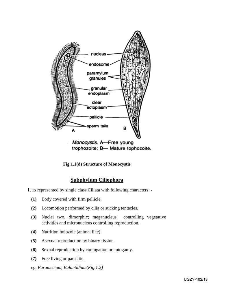
Fig.1.1(d) Structure of Monocystis
Subphylum Ciliophora
It is represented by single class Ciliata with following characters :-
(1) Body covered with firm pellicle.
(2) Locomotion performed by cilia or sucking tentacles.
(3) Nuclei two, dimorphic; meganucleus controlling vegetative activities and micronucleus controlling reproduction.
(4) Nutrition holozoic (animal like).
(5) Asexual reproduction by binary fission.
(6) Sexual reproduction by conjugation or autogamy.
(7) Free living or parasitic.
eg. Paramecium, Balantidium(Fig.1.2)
UGZY-102/13

.Fig.1.2 Balantedium
SAQ 2-
(a) Amoeba is the example of which class?
(b) Euglena is the example of which class?
(c) Monocystic is the example of which class?
1.6 Locomotary Organelle in Protozoa
Movement of an animal is locomotion. It is performed for searching food and mate and for protection against predators and abnormal climatic conditions. The organelle responsible for locomotion in Protozoa are pseudopodia, flagella, cilia and myonemes.
(1) Pseudopodia
They are seen in Sarcodina, many flagellates and some Sporozoa. They are temporary extensions of body protoplasm. They may be withdrawn or formed a new when needed. A pseudopodium is an extension of ectoplasm enclosing the endoplasm. They are of four kinds as lobopodium, filopodium, reticulopodium and axopodium. Lobopodium is broad finger like extension while a filopodium is slender and thread like. Reticulopodium is also slender and thread like but the threads get fused forming a network. An axopodium is stiff and spine like enclosing an axial rod. UGZY-102/14
(2) Flagella
They are found in Flagellata, some sarcodines and Sporozoa. Apart from locomotion they create water currents, perform attachment and may act as sensory organelle. A flagellum is thread like showing an elastic axial filament known as axoneme enclosed by outer sheath. A flagellum shows two central fibrils surrounded by nine equidistant peripheral fibrils which remain embedded in fluid matrix. Their number varies from one to many.
(3) Cilia
They are present in Ciliata and Suctoria. They are short hair like processes of ectoplasm arranged in longitudinal, diagonal or spiral rows all over the body surface or may be restricted to some specific areas of the body . A cilium is structurally similar to a flagellum having nine paired peripheral fibrils and two central fibrils enclosed in a fluid matrix. At the base each cilium shows a basal granule known as blepheroplast. In some cases the cilia get fused forming an undulating membrane, membranelle and cirri. A cirrus is a bristle like organ formed by a tuft of cilia which get fused with each other.
(4) Myonemes
They are present in Flagellata and Ciliata but remain absent in Sarcodina. They are highly contractile thickenings of pellicle or ectoplasm. They may be in the form of ridges and grooves or microtubules or myofibrils. They are responsible for changing the shape of the animal as required in different conditions. They may serve as hydrostatic organelle also, causing variation in volume of the body for rising or sinking in water.
1.7 Locomotion in Protozoa
Protozoa perform three kinds of locomotion as amoeboid (creeping), swimming and gliding.
1. Amoeboid movement:
It is performed by the formation of pseudopodia in Sarcodina (eg.Amoeba) and many Sporozoa. The body of Amoeba shows plasmalemma enclosing ectoplasm and endoplasm. The endoplasm is made up of outer plasmagel and inner plasmasol. During the formation of a pseudopodium, the plasmagel is converted into plasmasol which flows forwards to form the lobed pseudopodium. At its anterior end the plasmasol is converted into plasmagel which flows backwards. Actinomycin and other ATP sensitive proteins play significant role. This theory is known as Sol-Gel theory.
UGZY-102/15
2. Swimming:
It is performed by flagella in flagellates and cilia in ciliates. Inflagellates the flagellum may undergo spiral rotation tracing a cone which develops sufficient current to move the animal forwards. Sometimes the flagellum gives a sidewise lash undergoing an effective stroke followed by a recovery stroke. During this process the flagellum beats obliquely so that the body rotates on its longitudinal axis during forward movement. Sometimes the flagellum performs sinuous undulations from tip to base during forward movement and from base to tip during backward movement of the animal.
In ciliates the cilia are innumerable arranged in longitudinal, diagonal and spiral rows. During the swimming process each cilium bends and straightens showing effective and recovery strokes. Thus the water moves in the direction of the beat and the animal moves in opposite direction. All the body cilia do not pulsate simultaneously. The cilia of the longitudinal row undergo metachronous movement while those of diagonal rows undergo synchronous movements. This movement of cilia is similar to the movement of plants in a corn field during a windy day.
3. Gliding
Such movement is performed by the contraction and relaxation ofmyonemes in some flagellates (eg. Euglina and Gregarina). Thus the animal changes its shape as the need be.
1.8 Biology of Amoeboid Protozoa, Flagellata, Ciliata, parasitic and Spore forming Protozoa Most of the amoeboid Protozoa are free living in water and soil but
some of them are parasitic. Amoeba represents a typical free living form. It feeds on bacteria, diatoms, minute algae, dead organic matter, flagellates and ciliates. The food is captured in a food vacuole in which it is digested. During absorption the food vacuole undergoes cyclosis in endoplasm for homogenous distribution. The undigested food is egested through a temporary opening in ectoplasm. Respiration and excretion of nitrogenous waste occurs through general body surface. Osmoregulation is performed by contractile vacuole.
Normal method of reproduction is binary fission which occurs under normal conditions of food and temperature. During this process the nucleus divides mitotically in two daughter nuclei followed by cytoplasmic division. Under unfavourable conditions it may undergo encystment which is followed by multiple fission on the return of favorable conditions. It shows great power of regeneration. Typical parasitic amoeba is Entamoeba histolytica. It lives in the intestine of human beings feeding upon mucous and blood. Normal method of reproduction is binary fission. It also undergoes mutliple fission by proudcing tetranucleate cyst which gives rise to eight daughter forms on excystment. It causes amoebic dysentry in humans. UGZY-102/16
Biology of Flagellate Protozoa
Most of the flagellates are free living in water but some of them have gone parasitic. A typical free living flagellate is Euglena found in fresh water ponds and ditches. It undergoes two kinds of nutrition, holophytic and saprophytic. Holophytic nutrition is conducted by photosynthesis in which water, carbon dioxide and inorganic salts are utilized in the manufacture of carbohydrates with the help of chlorophyll present in its chloroplast in sunlight. It is a typical plant like method. In the absence of sunlight it absorbs products of decaying organic matter through its general body surface. It is known as saprozoic method. Respiration and excretion of nitrogenous waste is performed through general body surface. Osmoregulation is performed by contractile vacuole which performs excretion also. The stigma and photoreceptor spot work together to register the direction of incoming light. Normal method of reproduction is longitudinal binary fission occuring in normal condition of food and temperature in which the nucleus divides mitotically in two equal daughter nuclei, which is followed by longitudinal fission of the parent body producing two equal daughter forms. During unfavourable conditions it undergoes encystment producing a Palmella stage which undergoes multiple fission on the return of favourable conditions. Typical parasitic flagellate is Trypanosoma which is a parasite of circulatory system of different mammals including human beings. It causes sleeping sickness.
Biology of Ciliate Protozoa
Most ciliates are found in fresh water, ponds, pools and ditches rich in decaying organic matter. Some of them are parasitic also. Common fresh water ciliate is Paramecium. It feeds upon bacteria, diatoms, algae, small protozoa and yeast. The movement of cilia mainly around the oral groove make the micro organisms enter the oral groove and cytopharynx which are received in a food vacuole. Digestion of food occurs in the food vacuole which undergoes cyclosis during absorption of the food. The undigested food is released at a point known as cytopyge. Respiration and excretion occurs through general body surface. The two contractile vacuoles perform osmoregulation and excretion. Normal method of reproduction is transverse binary fission which occurs in favourable conditions of food and temperature. As the result of repeated binary fission the animal becomes exhausted. It undergoes senile decay. At this stage two individuals from two different mating types come in contact ventrally to undergo conjugation in which the existing meganucleus disappears. The micronuclens divides to form pronuclei which unite to form zygote nucleus. Conjugation leads to nuclear reorganization and heriditary variations thus providing rejuvenation to the animal. Some parasitic ciliates like Balantidium and Nyctotherus live in the intestine of cold blooded vertebrates.
UGZY-102/17
Biology of Parasite Protozoa
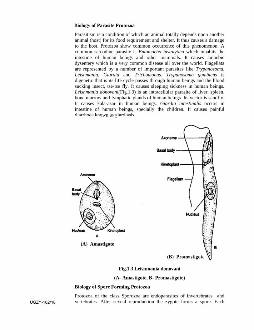
Parasitism is a condition of which an animal totally depends upon another animal (host) for its food requirement and shelter. It thus causes a damage to the host. Protozoa show common occurrence of this phenomenon. A common sarcodine parasite is Entamoeba histolytica which inhabits the intestine of human beings and other mammals. It causes amoebic dysentery which is a very common disease all over the world. Flagellata are represented by a number of important parasites like Trypanosoma, Leishmania, Giardia and Trichomonas. Trypanosoma gambiens is digenetic that is its life cycle passes through human beings and the blood sucking insect, tse-tse fly. It causes sleeping sickness in human beings. Leishmania donovani(Fig.1.3) is an intracellular parasite of liver, spleen, bone marrow and lymphatic glands of human beings. Its vector is sandfly. It causes kala-azar in human beings. Giardia intestinalis occurs in intestine of human beings, specially the children. It causes painful diarrhoea known as giardiasis.
Fig.1.3 Leishmania donovani
(A- Amastigote, B- Promastigote)
Biology of Spore Forming Protozoa
Protozoa of the class Sporozoa are endoparasites of invertebrates and vertebrates. After sexual reproduction the zygote forms a spore. Each
(A) Amastigote
(B) Promastigote
UGZY-102/18

spore is enclosed in a spore case having one or many thick coverings. Spore membrane may show two to three valves and one to six polar capsules, each with a polar filament .The valves, polar capsuls and polar filaments may be absent in some cases. The zygote in each spore undergoes repeated nuclear divisions followed by cytoplasmic divisions to form sporozoites which perform transmission of the parasite to a new host. The life cycle shows alternation of sexual and asexual phases in different hosts. Sometimes the life cycle passes through only single host without any alternation of generation. Common examples of such Protozoa are Monocystis inhabiting the seminal vesicle of earthworms and Plasmodium which passes its asexual phase in liver cells and R.B.C. of human beings and sexual phase and sporogony in the stomach cavity and stomach wall of female Anopheles mosquito. The Plasmodium causes malaria in human beings.
1.9 Summary Prokaryotes are represented by viruses, bacteria and blue green
algae while Eukaryotes by plants and animals. The basic differencelies in the pattern of their genetic material. The Prokaryotic cellcontains single, circular DNA molecule while the Eukaryotic cellshows a few to many chromosomes enclosed in a definite nuclearmembrane.
Protozoa is made up of a single cell and Metazoa of many cells,hence we can say Protozoa as unicelluar and Metazoa andmulticelluar. On the basis of function it is seen that a Protozanperforms all vital functions which a metatozan performs hence wecan consider Protozoa as an individual without cell (acellular) andMetazoa as individuals made up of many cells (cellular).
Protozoa are microscopic animals performing locomotion bypseudopodia, flagella, cilia and myonemes. The are generallyholozoic but some may be holophytic. They perform reproductionby binary fission in favourable conditions and multiple fission inunfavourable conditions.
Protozoa have been basically divided in two groups, Plasmodromaand Ciliophora mainly on the basis of locomotary organealle andnature of nuclei.
The locomotary organelle of Protozoa are pseudopodia, flagella,cilia and myonemes. A psuedopodium is a temporary extension ofcytoplasm, a flagellum and a cilium is a thread like structureshowing 9+2 fibril pattern. A myoneme is a contractile thickeningof cytoplasm. With the help of these organelle the Protozoaperform amoeboid movements, swimming and gliding.
UGZY-102/19
The biology of different groups of Protozoa deals with their habit,habitat, life style, feeding and reproduction. Some of them havegone parasitic even on human being causing significant diseases.
1.10 Terminal Questions
Q.1 Differentiate between Prokaryote and Eukaryote.
Q.2 Give 5 important characters of Protozoa.
Q.3 Give an account of amoeboid movement in Protozoa.
Q.4 Give an account of binary fission in Protozoa.
Q.5 Give and account of spore forming Protozoa.
Q.6 Match the two -
Column-A Column-B
1. Euglina (a) Colonial
2. Plasmodium (b) Conjujation
3. Volvox (c) Amoebic dysentry
4. Paramecium (d) Holophytic
5. Entamoeba (e) Parasite
ANSWERS
SAQ 1- (a) Leeuwenhock (b) general body surface
SAQ 2- (a) Sarcodina (b) Mastigophora (c) Sporozoa
UGZY-102/20
UNIT-2
Body Organization & Characteristic of Metazoa
Structure 2.1 Introduction and Objectives
2.2 Characteristics of Metazoa
2.3 Body Organization Metazoa
2.4 Symmetry of Metazoan body
2.5 Development pattern of Metazoa
2.6 Body cavity
2.7 Origin and evolution of Metazoa
2.8 Summary
2.9 Terminal questions
2.10 Answers
2.1 Introduction
In unit I you studied all about the important characters of Protozoa including their origin and evolution and their developmental pattern. Metazoan animals are multicellular, their cells are arranged in two or three layers. They are divided in three branches (i) Mesozoa which are sessile or parasitic showing the absence of tissues and organs, digestive tract and body cavity. They show intracellular digestion (eg. Rhopalura). (ii) Parazoa in which tissues are poorly defined with no mouth and digestive cavity. They show the presence of choanocytes for maintaining water current and digestion (eg. Sycon) and (iii) Eumetazoa in which different organs have been formed. They include all phyla from Coelenterata upto Chordata.
Objectives: After studying this unit you will be able to -
know all about the general organization of Metazoa including theirbody symmetry and body cavity.
UGZY-102/21
know all about their developmental patterns.
know all about their origin and evolutionary theories.
2.2 Characteristics of Metazoa
During the process of organic evolution with the increase in complexity of life, animals developed multicellularity, a phenomenon in which the body shows the presence of innumerable cells. Such animals are termed as Metazoa. They are represented by the chief phyla as Porifera, Coelenterata, Ctenophora, Platyhelminthes, Aschelminthes, Annelida, Arthropoda, Mollusca, Echinodermata and Chordata.
The metazoa show following important characteristics
1. The body may be unisexual or bisexual. It may be sessile orlocomotary. Locomotion may by performed by cilia, parapodia,paired jointed limbs, foot or podia.
2. Fertilization may be external or internal.
3. Zygotes may be alecithal, microlecithal, mesolecithalmacrolecithal, telolcithal or discoidal.
4. Cleavage may be holoblastic (complete) or meroblastic(incomplete). It may be radial or spiral. The cleavage may bedeterminate or indeterminate.
5. At the gastrula stage two or three germ layers appear. The twogerm layer condition is known as diploblastic while the three germlayer condition is known as triploblastic. A diploblastic conditionshows the presence of ectoderm and endoderm while a triploblasticcondition shows the presence of ectoderm, endoderm andmesoderm. The fate of these layers is very well defined that is aparticular layer invariably gives rise to a particular kind of tissueand organ system.
6. A larva may or may not be present in the life cycle. The larva ifpresent, may be locomotary or of feeding type.
7. The body shows cellular level, tissue level or organ system level oforganization.
8. The body shows spherical, radial, biradial or bilateral symmetry.
9. Metamerism or true segmentation is repetition of organs whichmay or may not be seen.
10. A body cavity may be absent or present. If present it lies betweenectoderm and endoderm. When it gets internally lined by
UGZY-102/22
endoderm it is known as coelom. If not lined by mesoderm it is known is pseudocoel.
11. Skin may be single layered or many layered (stratified).
12. Exoskeleton represented by cuticle, chitin, calcareous shells, scale,feather or hair etc.
13. Endoskeleton of spicules, notochord, cartilage or bone.
14. An alimentary canal may or may not be present.
15. The digestion may be intrcellular only or extracellular only orboth. Intracellular digestion occurs in a food vacuole whileextracellular digestion occurs in the cavity of alimentary canal.
16. Respiration may be performed through general body surface, gillsor lungs. Sometimes it is performed by special structures liketrachea, book lungs, ctenidia or papulae.
17. A circulatory system may be absent or present. If present it may beclosed or open.
18. Excretion of nitrogenous waste is performed through general bodysurface or specialized structures like flame cells, nephridia orkidneys.
2.3 Body Organization of Metazoa
All Metazoa are multicellular eukaryotes. They possess different types of cells. Each type of cells occur in groups performing a specific function. Such a group of cells in called as tissue. The body shows four types of tissues as epithelial, connective, muscular and nervous. In most animals some of these tissues get joined to from an organ performing a definite function. In more complex animals groups of organs work together for a common purpose as an organ system. The different organ systems together form an organism. Sometimes a particular system may be absent in a group of organisms. Thus the body shows three levels of structural organization as under -
(1) Ceullar Level: Body contains many cells but the cells do not form a tissue. It is seen in phylum Porifera.
(2) Tissue level : The multicellular body contains many specialized cells which form two distinct tissues as epidermis and gastrodermis. It is seen in phylum Coelenterata.
(3) Organ system level: The multicellular body contains many specialized tissues which form different organs which join together
UGZY-102/23
as different organ systems. It is seen in Platyhelminthes, Aschelminthes, Annelida, Arthropoda, Mollusca, Echinodermata and Chordata.
The Metazoan body shows three basic plans as under:
(1) Cell aggregate plan: The body contains many cells which do not form tissues and organs. The different cells function independently. It is seen in Phylum Porifera.
(2) Blind sac plan: The body is sac like with a single cavity showing single mouth opening performing both ingestion of food and release of undigested waste matter. It shows good cell differentiation. It is seen in phylum Coelenterata and Platyhelminthes.
(3) Tube within tube plan: The body shows inner tube performing as digestive tract which is enclosed by an outer tube of body wall. The inner tube shows two openings, the mouth for the ingestion of food and anus at the opposite end for ejection of faecal matter. Between the outer and inner tube is present a fluid filled cavity. It is seen is Aschelminthes, Annelida, Arthropoda and Chordata.
2.4 Symmetry of Metazoan body:
The patterns of body symmetry may be of 2 kinds
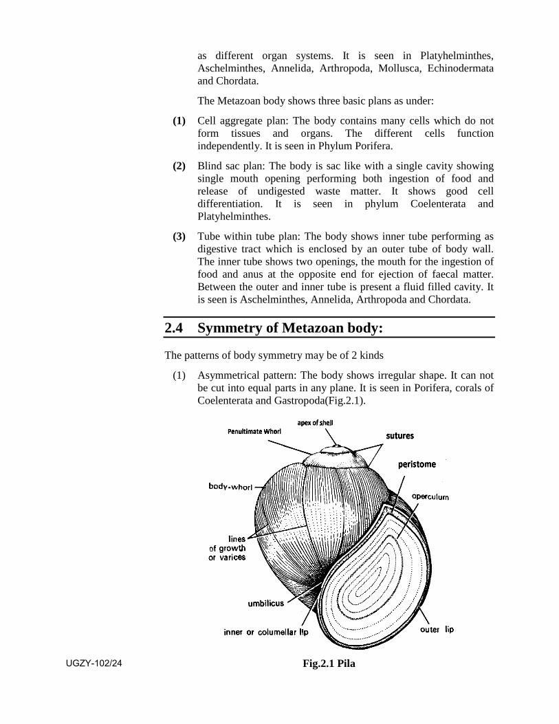
(1) Asymmetrical pattern: The body shows irregular shape. It can not be cut into equal parts in any plane. It is seen in Porifera, corals of Coelenterata and Gastropoda(Fig.2.1).
Fig.2.1 Pila UGZY-102/24
(2) Symmetrical pattern: The body shows similarity in the arrangement of parts on opposite sides. Thus it becomes possible to cut the body into two exactly similar halves in one or more planes. Such pattern shows three kinds of symmetries as under.
(a) Spherical Symmetry: The body shows the shape of a sphere. It can be divided in two similar halves in all plains passing through the centre. Such animals show floating on rolling movements. It is seen in eggs and early embryos of some animals.
(b) Radial Symmetry: The body of such animals shows a number of equivalent parts arranged in a radiating manner around a central axis. Thus the body can be divided in two similar halves through any plane passing through the centre from top to bottom. Such animals lead a sedentary life. It is seen in some sponges and coelenterates as Hydra and Jellyfish.
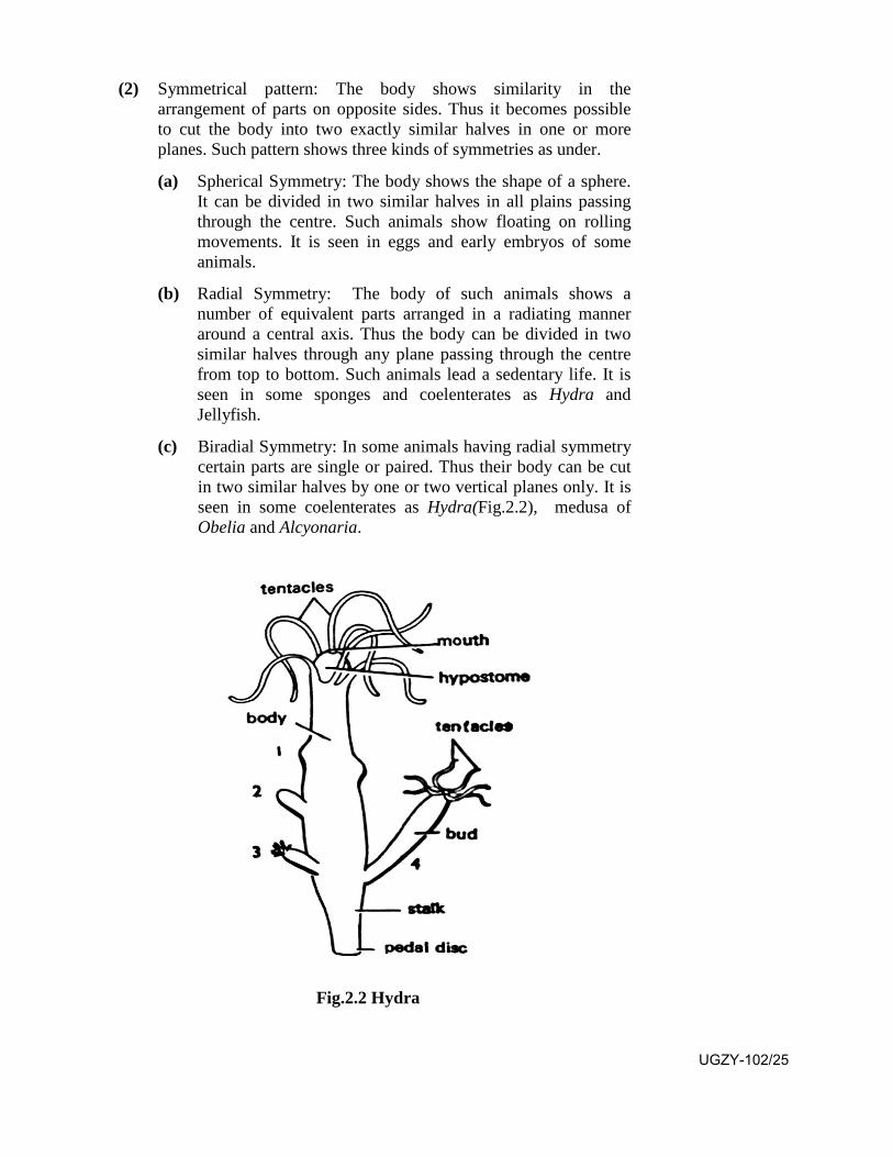
(c) Biradial Symmetry: In some animals having radial symmetry certain parts are single or paired. Thus their body can be cut in two similar halves by one or two vertical planes only. It is seen in some coelenterates as Hydra(Fig.2.2), medusa of Obelia and Alcyonaria.
Fig.2.2 Hydra
UGZY-102/25

(d) Bilateral Symmetry: The body of such animals shows paired organs which are arranged on the sides of a central axis from head to tail. Thus the body can be divided in two similar halves by a single plane passing through the median longitudinal line. The right and left halves are mirror opposite. It is seen in many invertebrates and all vertebrates.
SAQ 1-
(a) What are the symmetry of metazoan body?
(b) What are the different kinds of symmetrical pattern?
2.5 Developmental Patterns of Metazoa
The ammount and distribution of yolk in the zygote is variable. It may be absent or may be present in very small quantity or in a large quantity. Yolk provides energy for cleavage but remains inert, that is its density checks the occurrence of cleavage.
Cleavage - Cleavage refers to a series of mitotic divisions in the zygote producing blastomeres. It is of two kinds holoblastic (complete) and meroblastic (in complete). A holoblastic cleavage always divides the complete zygote while the meroblastic cleavage is restricted to only that portion of the zygote which lacks yolk. Holoblastic cleavage is of two kind.
(a) Radial cleavage: First cleavage appears in longitudinal axis from animal pole to vegetal pole dividing the zygote in two equal blastomeres, second cleavage also appears in the same axis but at right angle to the first, thus producing four equal blastomers. The third cleavage may be longitudinal or transverse. It forms eight blastomeres. If longitudinal it forms eight equal blastomers in one plane or if it is in transverse axis it forms 8 equal blastomers arranged in two tiers (4+4), one above the other. It is followed by successive longitudinal and transverse divisions thus doubling the number of blastomers to sixteen, thirty two and sixty four onwards, This pattern is seen in Porifera, Coelenterata, Echinodermata and Chordata.
(b) Spiral cleavage: After first two cleavages are formed four equal blastomers. The third cleavage appears in transverse axis slightly above the equator. Thus are formed 8 blastomers in 2 tiers in which the 4 upper blastomers are smaller (micromere) and the 4 lower blastomers are larger (megamere). It is followed by successive longitutional and transverse divisions. Before the occurance of a cleavage the cells of upper tier are displaced to right or left over the lower tiers. Thus the daughter micromere cells lie above and alternating with the megameres. This pattern continues through many successive cleavages producing a spiralling effect in the arrangement of the blastomers. This pattern is seen in Platyhelminthes, Annelida and Mollusca.
UGZY-102/26
According to potentiality of blastomeres the cleavage is of two kinds, determinate and indeterminate.
(a) Determinate cleavage: In animals showing spiral cleavage the fate of blastomers is fixed from the very first division of the zygote. It means that a blastomere is destined to form a particular part of the future body. Thus a complete embryo will be formed only if all the blastomeres remain together. If any blastomere is removed the embryo will lack that part destined to develop from that blastomere.
(b) Indeterminate cleavage: Sometimes the fate of blastomeres is not fixed upto second cleavage. Thus if the two blastomeres formed after first cleavage are separated, each of them forms a new individual (monozygotic or identical twins).
Formation and Fate of Blastopore:
As the result of continued cleavage a solid ball of cells is formed known as morula. It develops a cavity by the disintegration of central cells thus forming a single celled thick hollow ball of cells known as blastula with the cavity known as blastocoel. The single celled thick lining represents micromere in the animal half and megamere in the vegetal half. At this stage the megamere layer undergoes invagination (in pushing) gradually reducing the size of blastocoel, ultimately the megamere layer comes below the micromere layer. Thus is formed a double layered elongated ball of cells in which blastocoel gets completely lost but a new cavity comes in existence known as archenteron with an opening known as blastopore. In Proterostomes (Nematoda, Annelida, Arthropoda and Mollusca) the blastopore forms the future mouth opening but in Deuterostomes (Echinodermata, Hemichordata and Chordata) it forms the anal opening. In Proterostomes the anus develops at the opposite pole of mouth while in Deuterostomes the mouth develops at the opposite pole of anus.
Formation and Fate of Germ Layers:
In diploblastic animals (Porifera and Coelenterata) the space between ectoderm and endoderm gets filled up with a cell less gelatinous matrix known as mesoglea. In Platyhelminthes it gets filled up with mesenchyme cells. In Annelida onwards a third layer develops between the ectoderm and endoderm as mesoderm. It develops basically from the endoderm either as isolated cells or a portion of endoderm is cut off on each side which forms round segmented sacs knows as mesodermel pouches. Gradually the two sacs on each side of each segment grow to occupy the whole space between ectoderm and endoerm. They join midventrally. The space within the mesoderm represents the coelom. The fate of the three germ layers (ectoderm, endoderm and mesoderm) is well defined in all Metazoa.
UGZY-102/27

Ectoderm gives rise to skin and its derivatives, foregut and hindgut, nervous system and sense organs. Endoderm gives rise to midgut and associated gland (liver, pancreas etc.) Mesoderm gives rise to heart, kidneys, and connective tissue etc.
2.6 Body Cavity (Pseudocoel and Coelom):
The space between ectoderm and endoderm gets filled up with cell less gelatinous mesoglea in Porifera and Coelenterata. In Platyhelminthes it gets filled up with mesenchyme cells. In Aschelminthes the blastocoel persists in between the ectoderm and endoderm as pseudocoel, which is considered as primary coelom but in Annelida onwards the segmented mesodermal pouches on each side grow to their maximum to occupy all the space between ectoderm and endoderm, thus the cavity within the mesodermal pouches becomes large enough to represent the true coelom. It is considered as secondary coelom. It remains filled up with coelomic fluid having coelomic corpuscles.
2.7 Origin and Evolution of Metazoa:
In the process of evolution the first cell which appeared was a prokaryotic cell without a nucleus. Later on appeared the eukaryotic cell with a definite nucleus. With the increase in complexity multicellularity appeared which gave rise to Metazoa. It is evident from the fact that the earliest cell which appeared in the history of all Metazoa is the zygote which undergoes cleavage to form multicellular body as morula. There are three principal theories which explain the evolution of Metazoa as under.
1. Colonial Theory : The profounders of this concept are Butschli,Lankaster and Haeckel. It propagates that hollow colonial flagellates like Volvox were probable ancestors of Metazoa. With the increase in number of such flagellated cells in the colony they became more and more specialized structurally and functionally. Later on the individuality of the cells was lost and they started functioning as single multicellular body. It is supported by the fact that a unicellular zygote develops as a multicellular body, a metazoan tailed spermatozoon resembles a modified flagellate and flagellated cells appear in the organization of Porifera, Coelenterata and higher Metazoa also.
2. Syncytial theory : The profounders of this concept are Sedgwick,Hanson and Hatzi. It propagates that a primitive multinucleate syncitial ciliate was the probable ancestor of Metazoa. Its syncitial condition showed the presence of many nuclei in the protoplasm but no cell walls. In the process of evolution a cell wall developed around each nucleus making the body multinucleate.
UGZY-102/28
According to this theory the Turbellaria (eg. Convoluta) are regarded as the most primitive metazoa which are similar to some multinucleate ciliates as both show an antero-posterior axis, bilateral symmetry and absence of a digestive cavity. Moreover the trichocyst of ciliates is similar to sagittocyst of Turbellaria and nematocyst of Cnidaria and central parenchyma of Convoluta looks syncitial due to imperfect cellularization.
3. Polyphyletic theory : It is profounded by Greenberg and Preston.According to this concept different Metazoa have evolved from different ancestors, that is Porifera developed from colonial flagellates and other Metazoa originated by the cellularization of syncitial protociliates.
SAQ 2-
What are the different theories which explain the explanation of metazoan?
2.8 Summary As the result of fertilization the Metazoan zygotes showing
variable amount of yolk undergo hololastic or meroblastic cleavageresulting in the formation of a diploblastic or triploblastic gastrulawhich may form a larva which later on forms the adult showingmetamerism and a body cavity.
A Metazoan body may show cellular level, tissue level or organsystem level of organization with cell aggregate plan or blind sacplan or tube within tube plan.
A Metazoan body may be asymmetrical or symmetrical showingspherical, radial, biradial or bilateral symmetry.
During its development a Metazoan body shows radial or spiralcleavage which may be determinate or indeterminate. It is followedby blastulation and gastrulation with an opening known asblastopore which may form mouth or anal opening of the adult.The diploblastic or triploblastic gastrula undergoes organogenesisto form the adult. During organogenesis the fate of the germ layersin well defined.
The body cavity may appear as pseudocoel or true coelom. A truecoelom is the cavity between body wall and gut wall and it remainsinternally lined with mesoderm.
The colonial theory, syncitial theory and polyphyletic theory arethe three theories which explain the origin and evolution ofMetazoa.
UGZY-102/29

2.9 Terminal Questions:
Q.1 Write five important characters of Metazoa.
Q.2 Differentiate between pseudocoel and coelom.
Q.3 Differentiate between cellular level, tissue level and organ system level of organization. Give examples.
Q.4 Describe the different types of cleavage patterns of zygotes in Metazoa.
Q.5 Describe colonial theory of the origin of Metazoa
Q.6 Match the following:
Column-I Column-II
(i) Flame cell 1. Annelida
(ii) Tissue level of organization 2. Archenteron
(iii) Radial symmetry 3. Excretion
(iv) Blastopore 4. Coelenterata
(v) Tube within tube plan 5. Jelly fish
Answers
SAQ 1- (a) Asymmetrical pattern and symmetrical pattern
(b) Spherical pattern, radial pattern, biradial pattern and bilateral
pattern
SAQ 2- (1) Colonial theory (2) Syncytial theory (3) Polyphyletic theory
UGZY-102/30
UGZY-102 Diversity of Animal Life
Block
2 Comparative Forms and Functions-II Unit 3 35-58 General characters and classification of Porifera, Cnidaria, Ctenophora, Platyhelminthes, Nematoda Unit 4 59-74 General characters and classification of Phylum Annelida, Arthropoda, Mollusca and Larval forms of Echinodermata
Unit 5 75-94 Comparative form and Functions : Locomotion, Digestion, Excretion Unit 6 95-108 Respiratory, Circulatory and Nervous system
Unit 7 109-124 Reproductive system
Uttar Pradesh Rajarshi Tandon Open University
UGZY-102/31
Course Design Committee Chairman Prof. Ashutosh Gupta
Director, School of Science UPRTOU, Prayagraj.
Prof. Paramhand Pathak Member Department of Zoology Deendayal Upadhyay University, Gorakhpur.
Prof. Ajay Singh Member Department of Zoology Deendayal Upadhyay University, Gorakhpur.
Prof. Rajendra Singh Special Invitees Department of Zoology Deendayal Upadhyay University, Gorakhpur.
Member/Secretary Dr. Deepa Chaubey Acadmic Consultant, School of Science UPRTOU, Prayagraj.
Course Preparation Committee Dr. V.C. Srivastava Author Ex. Associate Prof. C.M.P. Degree College, Prayagraj
Dr. A.K. Srivastava EditorEx. Head Of Zoology B.B.C, Jhansi.
Coordinator Dr. Deepa Chaubey Acadmic Consultant, School of Science UPRTOU, Prayagraj.
© UPRTOU, Prayagraj. 2020 ISBN : All Rights are reserved. No part of this work may be reproduced in any form, by mimeograph or any other means, without permission in writing from the Uttar Pradesh Rajarshi Tondon Open University, Prayagraj. Printed and Published by Dr. Arun Kumar Gupta Registrar, Uttar Pradesh Rajarshi Tandon Open University, 2020. Printed By: Chandrakala Universal Pvt. Ltd. 42/7 Jawahar Lal Neharu Road, Prayagraj. UGZY-102/32
Course Introduction
Block-II
Comparative Forms and Functions-II
This block of the study material incorporates 5 units as under:
Units-3 :- It deals with the general characters and classification of Porifera, Ciridria, ctenophore, Platyhelminthes and Nematoda. The Porifera are characterized by the possession of choanocytes; the cnidaria by the possession of nematoblsts; ctenophore by the possession of combplates; Platyhelminthes by the possession of flame cells and Nematoda by the possession of psuedocoel. This unit also deals with the formation of coral reefs which form the sea gardens and the occurance of polymorphism in coelenterata.
Unit 4 :- It deals with the general characters and classification of Phylum Annelida, Arthropoda and Mollusca. Annelida are characterized by the possession of nephridia, Arthropoda by the possession of paired jointed legs and Mollusca by the possession of a sell of calcium carbonate. This unit also incorporates the Peculiar phenomenon of torsion and detorsion occurring in same molluscs and the kinds of different larval forms of echinoderms.
Unit -5 :- This unit deals with the locomotory structures and locomotion n members of phylum coelenterate, Platyhelminthes, Nematoda, Arnelida, Arthropoda, Mollusca and Echinodermata. It covers the phenomenon of feeding and digestion in sponges and coelenterates. It also incorporates the important excretory structures like Protonephridia, Metanephridia, Malpighion tubules and coelomoducts.
Unit-6 :- This unit deals with the structure and function of respiratory organs, open and closed plans of circulatory systems, general organization and nervous systems in Platyhelminthes, Annelida, Arthropoda and Mollusca.
Unit 7 :- In this unit the phenomenon of reproduction has been discussed. It includes the methods of asexual reproduction including different kinds of fission, budding, and gemmulation. It discusses the significance of asexual reproduction. It deals with the phenomenon of regeneration also. It incorporates the informations regarding pattern, reproductive organs, mating and fertilization also. It also gives the concept ovipary, vivipary, ovovivipary, parthenogenesis and metagenesis.
UGZY-102/33

Objectives:-
After studying this block you will be able to:
• discuss the general characters and classification of invertebrates.
• discuss the different types of locomotion in invertebrates.
• discuss the respiratory, circulatory, nervous and reproductivesystems.
UGZY-102/34

UNIT-3
General characters and classification of Porifera, Cnidaria, Ctenophora,
Platyhelminthes, Nematoda
Structure 3.1 Introduction and Objectives
3.2 General characters and classification of Porifera
3.3 General characters and classification of Cnidaria
3.4 General characters and classification of Ctenophora
3.5 General Characters and classification of Platyhelminthes
3.6 General characters and classification of Nematoda
3.7 Coral reefs
3.8 Polymorphism in Coelenterata
3.9 Summary
3.10 Terminal Questions
3.11 Answers
3.1 Introduction
In unit 2 you have read all about metazoan animals. In this unit you shall study the characteristic features of the lower invertebrate phyla. Porifera are represented by sponges. They possess the peculiar cells known as choanocytes which maintain a water current and perform feeding. The cnidarians are the Coelenterata which possess the peculiar cells known as nematoblast which perform defence. The Ctenophora possess 8 vertical rows of fused cilia known as comb plates for locomotion. Platyhelminthes are flatworms possessing the peculiar cells known as flame cells for excretion. The Nematoda are round worms possessing the pseudocoel. You have to read the classification of these phyla in this unit. Coral reefs are known as sea gardens formed by some coelenterate animals. Coelenterata show the beginning of colonial life in which many kinds of zooids live together showing division of labour.
UGZY-102/35
Objectives: After studying this unit you should be able to-
know the characteristic features of the lower invertebrate phyla.
know the classification of these phyla.
known about coral reefs and polymorphism in coelenterata.
3.2 General characters and classification of Phylum Porifera General Characters
1. They occupy aquatic habitat, mostly found in sea but some arefound in fresh water.
2. Body remains attached to a substratum. It may be solitary orcolonial.
3. Body shape cylindrical, tubular or branched.
4. Body shows radial symmetry, sometimes it is asymmetrical.
5. Body made up of many cells but each cell shows its ownindividuality hence body organization shows cellular grade.
6. Body wall shows two layers, outer dermal epithelium(Pinacoderm) and inner gastral epithelium (Choanoderm).
7. Body contains a canal system for the passage of water. It has manyostia for entry of water and one to many oscula for exist of water.This system shows one to many flagellated chambers.
8. Flagellated chambers are lined with choanocytes which areflagellated and collared cells maintaining a water current. Thesecells perform food capture by pseudopodia.
9. Digestion occurs in intracellular manner in food vacuoles withinchoanocytes.
10. Skeleton is represented by calcareous and siliceous spicules andproteinaceous spongin fibres.
11. Respiration and excretion of nitrogenous substances occursthrough outer and inner lining cells.
12. Nervous system primitive showing a network of bipolar andmultipolar neurons.
13. Asexual reproduction occurs by budding which may be external orUGZY-102/36
internal by gemmule formation.
14. Body bisexual producing both male and female gametes.
15. Fertilization cross and internal.
16. Cleavage holoblastic (complete).
17. A free swimming ciliated larva occurs in the life cycle whichperforms dispersal of the species.
18. Sponges show extreme power of regeneration that is a small cutpiece is able to form a new individual. If all the cells of a livingsponge are separated and kept in a favourable medium, they getrearranged to form the body.
SAQ 1-
Porifera shows ………
Classes
Calcarea Hexactinillida. Demospongiae.
1. Endoskeletonof Calcareousspicules.
1. Endoskeleton of silecious spicules.
1. Endoskeleton ofspongin fibres orsilecious spicules.
2. Canal systemascanoid, syconoid or leuconoid type.
2. Canal system simplesyconoid type.
2. Canal systemleuconoid type.
3. Marine only. 3. Marine only. 3. Mostly marine somefound in fresh water.
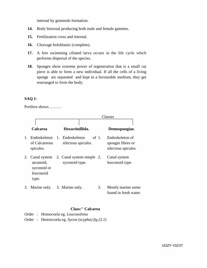
Class:" Calcarea Order : Homocoela eg. Leucosolinia Order : Heterocoela eg. Sycon (scypha) fig (3.1)
UGZY-102/37
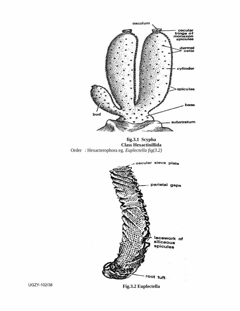
fig.3.1 Scypha Class Hexactinillida
Order : Hexacterophora eg. Euplectella fig(3.2)
Fig.3.2 Euplectella UGZY-102/38

Order : Amphidiscophora eg. Hyalonema
Class: Demospongiae Subclass : Tetractinellida
Order : Myxospongida eg. Oscarella
Order : Carnosa eg. Chondrilla
Order : Chondristidia eg. Geodia
Sub Class Monoaxonida
Order : Hadromerina eg. Cliona
Order : Halichondrina eg. Helichondria
Order : Poecilosclerina eg. Microciona
Order : Haplosclerina eg. Spongilla
Sub Class Keratosa
Order : Keratosa eg. Euspongia fig (3.3)
SAQ 2-
What are the different kinds of porifera?
Fig.3.3 Euspongia
3.3 General Characters and Classification of Cnidaria
General Characters:
1. They occupy aqatic habitat, mostly found in sea water but some are
UGZY-102/39
found in fresh water.
2. Body remains attached to a substratum (sessile) or free swimming.It may be solitary or colonial.
3. Body shape cylindrical, tubular or branched.
4. Body shows radial or biradial symmetry along oral-aboral axis.
5. Body made up of many specialized cells which show tissue gradeof organization.
6. Bodywall represented by two layers, outer epidermis and innergastrodermis. In between the two is present a gelatinous matrixknown as mesoglea. Thus they represent diploblastic organization.
7. Life cycle generally shows two kinds of individuals, representedby sessile polyp and free swimming medusa which alternate witheach other. Thus they show dimorphism but some of them showpolymorphism in which more than two kinds of functional zooidsare present.
8. Mouth of a polyp and bell margin of the medusa are provided withmovable and slender tentacles.
9. Epidermis is provided with many specialized stinging cells knowsas nematoblasts which are characteristic of this phylum. They areused for offence and defence, adhesion and food capture.
10. Body shows a single internal cavity known as coelenteron which islined with gastrodermis. It shows single mouth but no anus.
11. Digestion of food occurs in the coelenteron (extracellular) andwithin the food vacuoles in nutritive muscular cells ofgastrodermis (intracellular)
12. Muscular system is represented by longitudinal and circular fibresformed by epithelio-muscular cells of epidermis and nutritivemuscular cells of gastrodermis respectively.
13. Nervous system shows two seperate nerve nets of bipolar andmultipolar neurons, one outer and the other on inner border ofmesoglea. It shows absence of a ganglion hence is unpolarized.
14. Sensory structures are represented by light perceiving ocelli andstatocyst which maintains the balancing mechanism duringswimming movements of medusa.
15. Respiration and excretion of nitrogenous substances occursthrough outer and inner lining cells.
16. Asexual reproduction occurs through budding and fission.
17. Sexual reproduction occurs through ova and sperms.
18. The individuals may be unisexual or bisexual.UGZY-102/40

19. Life cycle passes through a free swimming blastula larva.
20. Ability of regeneration is seen.
SAQ 3-
The life cycle of cnidaria show two kinds of individuals ……………………...
Classification of Phylum Cnidaria Phylum Cnidaria
Classes
Hydrozoa Scyphozoa Anthozoa
1. Habitat fresh waterand marine.
Marine. Marine.
2. Solitary or colonial. Solitary. Solitary or colonial.
3. Life cycle showsonly polyps orboth polyp andmedusa.
Medusa is predominan
t, Polyp reduced or
absent.
Polyps present Medusa absent.
4. Medusa with truevelum.
Medusa without velum.
Medusa absent.
5. Mesogleanoncellular.
Mesoglea with cells and fibres.
Mesoglea cellular.
6. Gonads epidermal. Gonads gastroderma
l.
Gonads gastrodermal
.
7. Mesentries absent. Mesentries absent.
Mesentries present.
Class Hydrozoa Order : Hydroida eg. Hydra, Obelia
Order : Trachylina eg. Cunina
Order : Hydrocorallina eg. Millipora
Order : Siphonophora eg. Physalia Order : Chondrophora eg. Porpita, Vellela fig(3.4)
UGZY-102/41
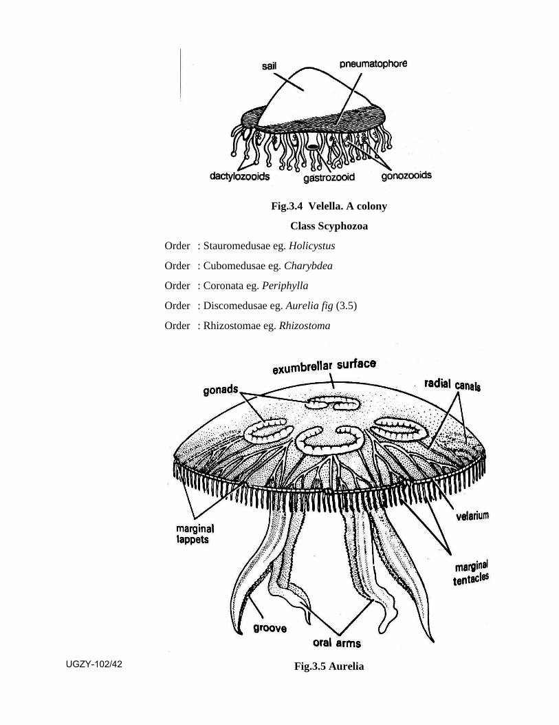
Fig.3.4 Velella. A colony
Class Scyphozoa
Order : Stauromedusae eg. Holicystus
Order : Cubomedusae eg. Charybdea
Order : Coronata eg. Periphylla
Order : Discomedusae eg. Aurelia fig (3.5)
Order : Rhizostomae eg. Rhizostoma
Fig.3.5 Aurelia UGZY-102/42
Class Anthozoa
Sub Class Alcyonaria
Order : Stolonifera eg. Tubipora
Order : Testacea eg. Telesto
Order : Alcyonacea eg. Alcyomium
Order : Coenothecalia eg. Haliopora
Order : Gorgonacea eg. Ranilla
Order : Pinnatulacea eg. Pinnatula
Sub Class Zoantheria
Order : Zoanthidia eg. Zoanthus
Order : Actiniaria eg. Adamsia
Order : Ceriantharia eg. Carianthus
Order : Antipatharia eg. Antipathes
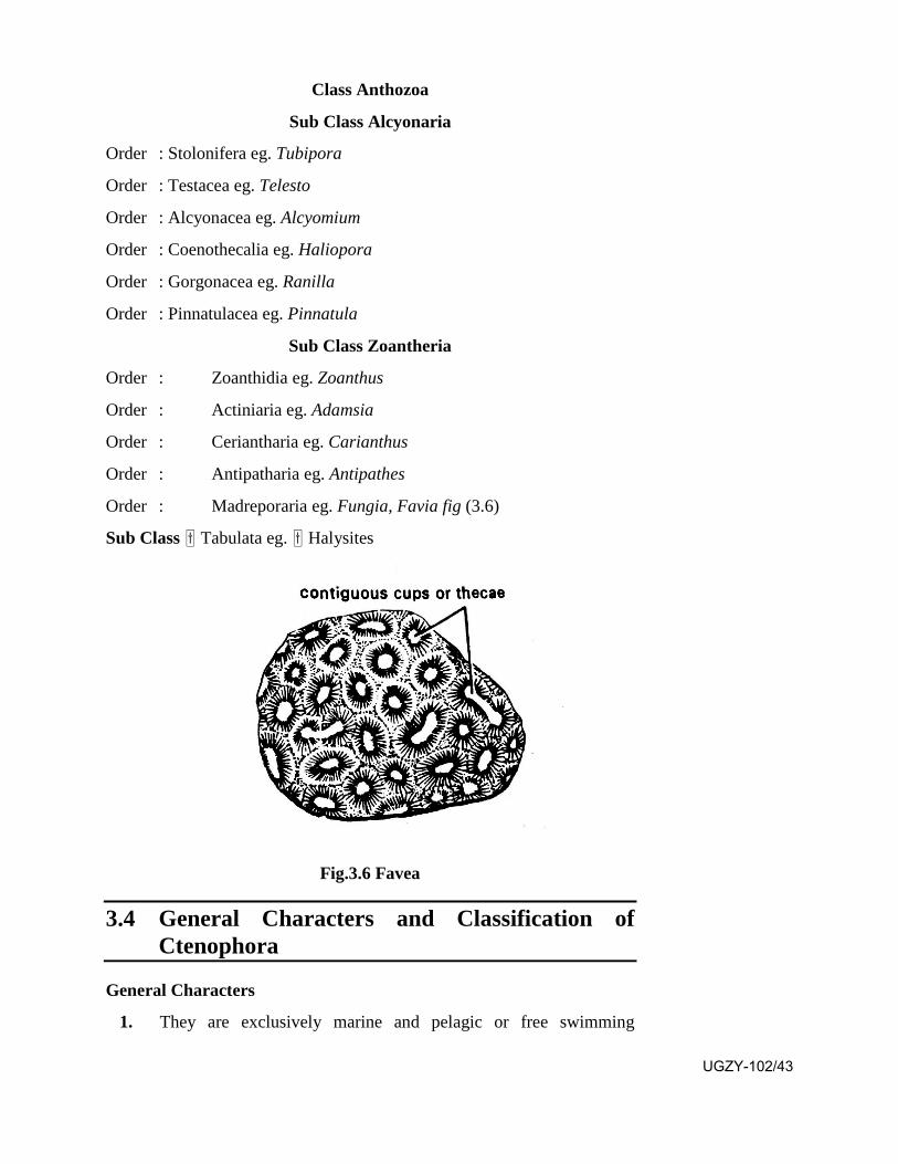
Order : Madreporaria eg. Fungia, Favia fig (3.6)
Sub Class Tabulata eg. Halysites
Fig.3.6 Favea
3.4 General Characters and Classification of Ctenophora
General Characters
1. They are exclusively marine and pelagic or free swimming
UGZY-102/43
animals.
2. Body shape rounded, oval, conical or flattened.
3. Body shows biradial symmetry which is a combination of radialand bilateral symmetries.
4. Body made up of many cells which show tissue grade oforganization.
5. Bodywall represented by ectoderm and endoderm. A jelly likemesoglea with scattered cells and muscle fibres is present betweenthe two. It is diploblastic showing a tendency to becometripoblastic.
6. Polymorphism, colony formation and alternation of generationabsent.
7. Body shows two long unsheathed and retractile tentacles. They arepresent on opposite per radii.
8. Nematoblasts are absent. Special adhesive cells known ascolloblasts are present in epidermis of tentacles.
9. Digestive tract shows mouth, stomodaeum, stomach complex,gastrovascular canals and two aboral anal pores.
10. Digestion is both extracellular and intracellular.
11. Skeletal, circulatory, respiratory and excretory structures areabsent.
12. Nervous system forms a subepidermal plexus.
13. Aboral end shows a statocyst for maintaining balance.
14. Asexual reproduction absent.
15. Sexual reproduction occurs through sperms and ova. Bodyhermaphrodite. Life cycle shows cydippid larva.
16. Regeneration is seen.
17. Most peculiar feature of this group is the presence of eight verticalrows of combplates of fused cilia for locomotion. Hence they arecommonly known as comb jellies.
Classification of Phylum Ctenophora
Phylum Ctenophor
Classes
Tentaculata Nuda
(1) Tentacles present 1. Tentacles absentUGZY-102/44
Class : Tentaculata
Order : Cydippida eg. Horniphora
Order : Lobata eg. Bolinopsis
Order : Cestida eg. Cestum
Order : Platyctenida eg. Coeloplana
Class: Nuda
Order : Beroida eg. Beroe
3.5 General Characters and Classification of Phylum Platyhelminthes
General Characters:
1. They may be free-living (terrestrial, fresh water or marine) orcommensal or parasitic.
2. Their body is soft and dorso-ventrally flattened, may be leaf like orribbon like.
3. Body shows bilateral symmetry. It shows marked anterior andposterior ends and dorsal and ventral surfaces.
4. Primary germ layers are ectoderm and endoderm. In between thetwo is present the mesoderm. Thus they show triploblasticorganization.
5. Body shows organ-system level of organization in which the cellsform different tissues, which join to form different organs, whichunite to form different organ systems.
6. They show acoelomate organization in which the body cavity getsfilled up with mesenchymal cells.
7. Alimentary canal with anterior or ventral mouth, pharynx andsimple or branched intestine. Anus is generally absent. In someforms as in tapeworms alimentary canal is totally absent.
8. In free living forms aerobic respiration occurs through generalbody surface. In parasitic forms respiration is anaerobic.
9. Circulatory system is absent.
10. Excretory system is represented by flame cells leading into finetubules which join together to form excretory duct opening byexcretory pore.
11. Nervous system is primitive. It shows a pair of anterior ganglia
UGZY-102/45
with two longitudinal nerve cords connected by transverse commissures. Thus it is a ladder like system.
12. Free living aquatic forms show locomotion by epidermal cilia.Parasitic forms show absence of locomotion. Instead they showorgans of attachment in the form of suckers, hooks and spines.
13. Body is mostly hermaphroditic with complex reproductive system;female reproductive system shows peculiar vitelline glands whichproduce yolk.
14. Fertilization is generally cross and internal.
15. Development may be direct in free living forms or indirectoccurring in endoparasites showing complex life cycles involvingmany larvae and hosts.
Classfication of Phylum Platyhelminthes
Phylum Platyhelminthes
Classes
Turbellaria Trematoda. Cestoda.
1. Free living in moist soil, fresh water andsea. Some may becommensal orparasitic.
Ecto or endo-parasites of vertebrates.
Endoparasites in intestine of vertebrates.
2. Digestive system shows a mouth, protrusible pharynx and simple or branched intestine ending in anus.
Digestive system shows a mouth,
muscular pharynx, short oesophagus and simple or branched
intestine. Anus absent.
Digestive system absent.
3. Suckers absent. Suckers present for attachment. Hooks
and spines may also be present.
Scolex with suckers present at anterior end for attachment. Hooks and spines may also be
present.
SAQ 4-
Platyhelminthes are …………. Showing mesenchyme in between …… and ………..
Class Turbellaria
Order : Acoela eg. Convoluta UGZY-102/46
Order : Rhabdocoela eg. Microstomum
Order : Alloecoela eg. Plagiostomum
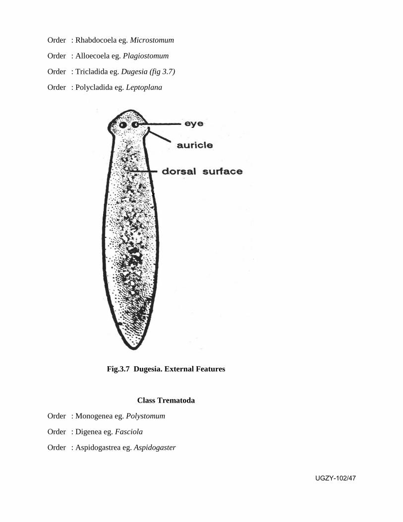
Order : Tricladida eg. Dugesia (fig 3.7)
Order : Polycladida eg. Leptoplana
Fig.3.7 Dugesia. External Features
Class Trematoda
Order : Monogenea eg. Polystomum
Order : Digenea eg. Fasciola
Order : Aspidogastrea eg. Aspidogaster
UGZY-102/47

Fig.3.8 Fasciola
Class Cestoda
Subclass Cestodaria
Order : Amphilinida eg. Amphilina
Order : Gyrocotylidea eg. Gyrocotyle
Subclass Eucestoda
Order : Proteocephalidea eg. Proteocephalus
Order : Tetraphyllidea eg. Phyllobothrium
Order : Disculicepitidea eg. Disculiceps
Order : Lecanicephaloidea eg. Lecanicephalum
Order : Pseudophyllidea eg. Dibothriocephalus
Order : Trypanorhyncha eg. Trypanorhynchus
Order : Cyclophyllidea eg. Taenia
Order : Aporidea eg. Nematoparataenia
Order : Nippotaeniidea eg. Nippotaenia UGZY-102/48
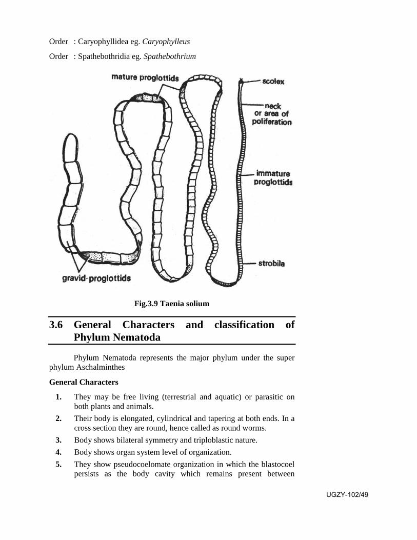
Order : Caryophyllidea eg. Caryophylleus
Order : Spathebothridia eg. Spathebothrium
Fig.3.9 Taenia solium
3.6 General Characters and classification of Phylum Nematoda
Phylum Nematoda represents the major phylum under the super phylum Aschalminthes
General Characters
1. They may be free living (terrestrial and aquatic) or parasitic onboth plants and animals.
2. Their body is elongated, cylindrical and tapering at both ends. In across section they are round, hence called as round worms.
3. Body shows bilateral symmetry and triploblastic nature.4. Body shows organ system level of organization.5. They show pseudocoelomate organization in which the blastocoel
persists as the body cavity which remains present between
UGZY-102/49
ectoderm and endoderm but is not lined internally with mesoderm. 6. Bodywall shows outer cuticle, middle layer as syncitial epidermis
and inner layer of longitudinal muscles arranged in 4 sectors.7. Alimentary canal complete with muscular and suctorial pharynx. It
shows anterior mouth and posterior anus.8. Excretory system appears as 'H' shaped canals known as 'H'
system.9. Circulatory and respiratory systems absent.10. Nervous system shows a circumentric ring and anterior and
posterior longitudinal nerves.11. Sexes separate with sexual dimorphism. The male body is smaller
than female. It shows a ventrally curved posterior end.12. Males show the presence of a cloaca with 2 retractile copulatory
spicules which may protrude out of the cloacal opening. Anusopens with in cloaca.
13. Fertilization cross and internal, spermatozoa amoeboid.14. Asexual reproduction and regeneration not seen.
SAQ 5- Phylum Nematoda represents the major phylum under the super
phylum ……………… Classification of Phylum Nematoda
Classes
Aphasmidia Phasmidia
1. Caudal Sensory organs(Phasmids) absent.
1. Caudal sensory organs(Phasmids) present.
2. Anterior sensory organs(Amphids) without pore.
2. Anterior sensory organ(Amphids) pore like.
3. Excretory system absent. 3. Excretory system welldeveloped.
4. Caudal glands present. 4. Caudal glands absent.
Class Aphasmidia
Order : Desmoscolecoidea eg. Desmoscolex
Order : Enoploidea eg. Enoplus
Order : Dorylaimoidea eg. Dorylaimus
Order : Mermithoidea eg. Mermis
Order : Chromadoroidea eg. Halichoanolaimus
Order : Monohysteroidea eg. Plectus UGZY-102/50

Class Phasmidia Order : Trichuroidea eg. Trichurus
Order : Dioctophymoidea eg. Dioctophyma
Order : Rhabditoidea eg. Rhabditis
Order : Rhabdiasoidea eg. Rhabdias
Order : Oxyroidea eg. Oxyurus
Order : Ascaroidea eg. Ascaris
Order : Strongyloidea eg. Strongylus
Order : Spiruroidea eg. Spiroxys
Order : Dracunculoidea eg. Dracunculus
Order : Filarioidea eg. Wuchereria
A B
Fig.3.10 Ascaris (A-male, B-Female)
UGZY-102/51
3.7 Coral Reefs
A coral reef is a mound of calcium carbonate within the sea. It has a number of hydrozoan and anthozoan coelenterates along with a variety of sponges, sea anemons, sea urchins, star fishes, holothurians, crabs, annelids, snails and bivalves. The coelenterate polyps and tentacles show varied shapes and brilliant colours like red, yellow brown, green, blue and violet. From the top it looks like a beautiful garden having brilliantly coloured flowers. It is commonly known as sea garden.
Such coral reefs are common in a belt along the equator from 30° north latitude to 30° south latitude. The suitable temperature ranges from 25°C-29°C. They flourish well at a depth upto which the sunlight can reach.
The bodies of polyps contain algae and zooxanthellae which undergo photosynthesis in sunlight to manufacture starch which is shared by the coelenterate which grows faster and larger increasing the size of coral reef. The polyps extract CaCo3 from the sea water for forming the coral. When polyps die the next generation builds new coral houses above the empty ones. The process continues for thousands of years to form very large porus rocks in the sea as a coral reef.
Some important coelenterate genera forming different types of corals are Tubipora (organ pipe coral), Haliopora (blue coral), Gorgonia (sea fan), Antipathes (black coral), Fungia (Mushroom coral) and Meandra (Brain coral).
Some important coral reefs are Great Barrier reef on north east coast of Australia and the other on eastern coast of Bahama islands.
3.8 Polymorphism in Coelenterata
The anthozoan and many hydrozoan coelenterates have gone colonial. A colony is a group of many individuals which live together for better survival of the species. A generalized colony is represented by many zooids living together. In such a colony each zooid performs feeding defence and reproduction. Such a colony is seen in Millipora and Gorgonia.
In a typical hydrozoan colony the zooids living together show variations in their structure and function. In some cases the zooids are only of two kinds, the polyps and the medusa. The polyps perform feeding and defence and medusa perform feeding, dispersal and reproduction. Such a condition is that of a dimorphic colony (eg. Bougainvillea of Hydrozoa UGZY-102/52
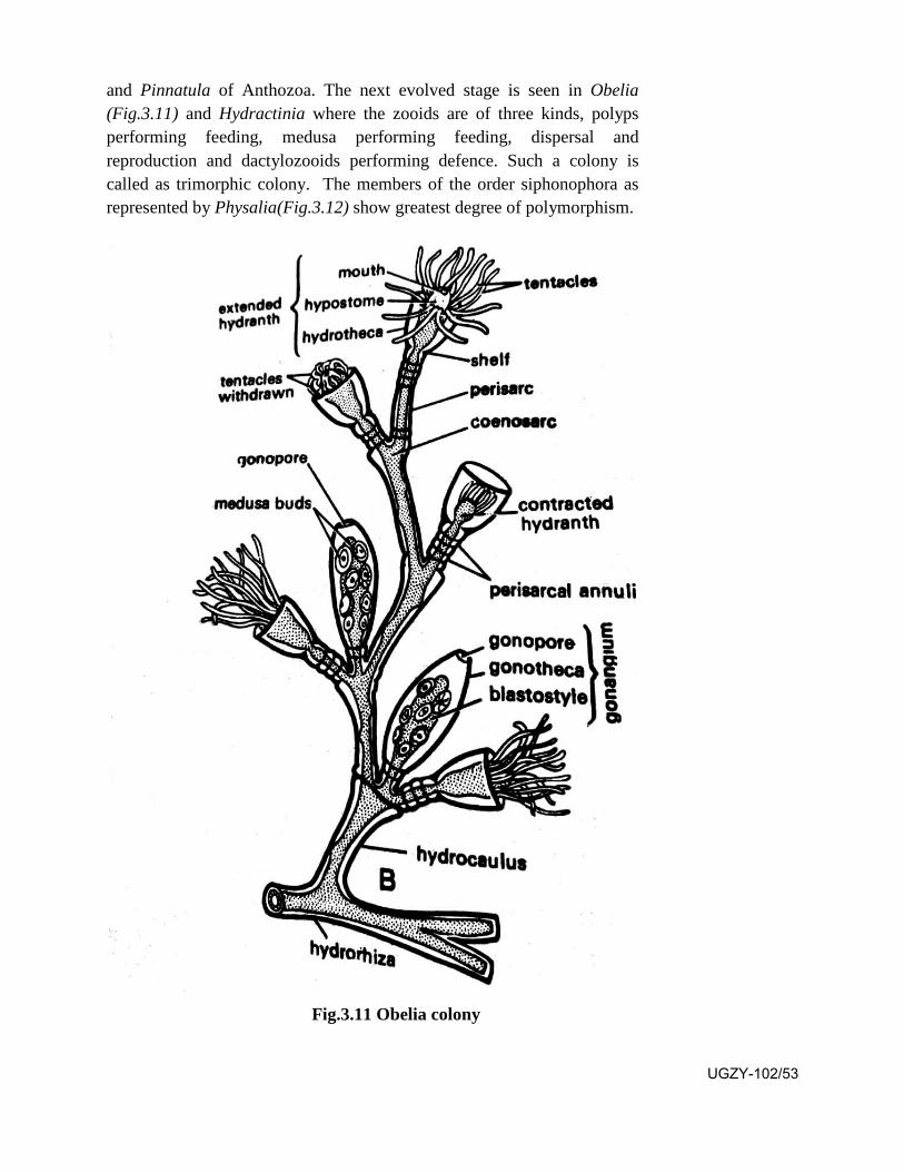
and Pinnatula of Anthozoa. The next evolved stage is seen in Obelia (Fig.3.11) and Hydractinia where the zooids are of three kinds, polyps performing feeding, medusa performing feeding, dispersal and reproduction and dactylozooids performing defence. Such a colony is called as trimorphic colony. The members of the order siphonophora as represented by Physalia(Fig.3.12) show greatest degree of polymorphism.
Fig.3.11 Obelia colony
UGZY-102/53
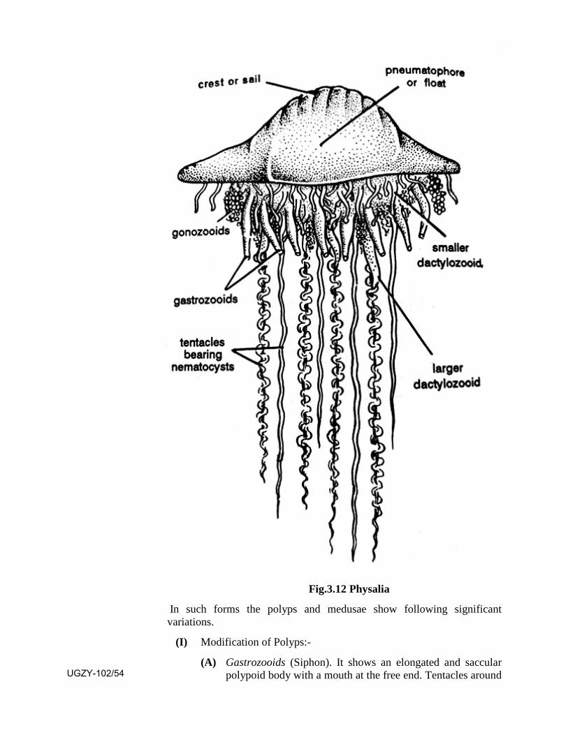
Fig.3.12 Physalia
In such forms the polyps and medusae show following significant variations.
(I) Modification of Polyps:-
(A) Gastrozooids (Siphon). It shows an elongated and saccular polypoid body with a mouth at the free end. Tentacles around UGZY-102/54
the mouth are absent but a long hollow tentacle arises from its base which shows many branches called as tentilla having batteries of nematoblasts. It performs feeding.
(B) Dactylozooid (Palpon, feeler, taster)
It is an elongate polypoid body which shows the absence of mouth and tentacles but a long tentacle arises from its base which is provided with batteries of nemtoblasts. Sometimes the tentacles become extremely long and filamentous. It is specilized for the defence of the colony.
(C) Blastostyle (Gonozooid)
It is a sac like polypoid body which may get branched. It may or may not show mouth but the tentacles are absent. It produces many medusa buds from its surface.
II. Modifications of Medusa: The medusae show following forms.
(a) Pneumatophore (Float) - It shows absence of mouth,manubrium, velum, tentacles and mesoglea. Its muscular wall shows two layers, outer layer is called as pneumatocodon while the inner layer as pneumatosaccus. Sometimes it shows a dorsal ridge and a posterior opening. Sometimes it may show concentric chambers. The inner lining possesses gas glands which generate oxygen and nitrogen. The main function of this zooid is to float or sink the colony as needed.
(b) Nectocalces (Nectophores, Nectozooids, Swimming bells). They are medusoid bodies showing absence of mouth, manubrium, tentacles and sense organs. They show the presence of a velum, 4 radial and a circular canal and well developed muscles. These zooids make the colony swim in a particular direction by alternate contraction and relaxation.
(c) Bracts (Hydrophyllia, Phyllozooids) - They form a leaf like, shield like, prism like or helmet like shape with a solid core of endoderm covered with ectoderm, mesoglea being absent. They show the absence of mouth, manubrium, velum and tentacles but show the presence of simple or branched gastro vascular cavity. These zooids provide protection against enemies.
(d) Gonophores - They are medusa like, with a velum, radial canals and manubrium. They show the absence of mouth, tentacles and sense organs. They show sexual dimorphism. Male gonophores are sac like while female gonophores are medusa like. They undergo gamete production after which they die.
UGZY-102/55
In forms like Helistema and Physalia groups of different kinds of zooids arising from the float constitute a cormidium. A generalized cormidium shows a gastrozooid, a large dactylozooid, a small dactylozooid, a bract and a gonangium. From the ventral surface of the float arise many such cormidia.
The zooids in such a colony show marked division of labour and functional interdependence. Thus they work together for the facilitation of a unified life. Thus the tightly integrated colony becomes a self sufficient unit which displays all attributes of a society showing cooperative behaviour between members of the colony.
3.9 Summary
In this unit you have learnt that
Porifera show cellular grade of organization. They are diploblasticshowing a mesoglea in between the pinacoderm and choanoderm.They possess peculiar cells knwon as choanocytes and a canalsystem. Their endoskeleton is in the form of spicules.
Porifera are of three kinds (i) those possessing caleareous spicules(ii) those possessing siliceous spicules and (iii) those possessingspongin fibres.
Cnidaria show tissue grade of organization. They are diploblasticshowing a mesoglea in betwen epidermis and gastrodermis. Theypossess a coelenteron which shows a single mouth opening. Theyshow both extracellular and intracellular digestion. They possesspeculiar nematoblast cells which are used for defence. They showdimorphism, trimorphism or polymorphism.
Cnidaria are of three kinds (i) those showing only polyps or bothpolyp and medusa (ii) those in whcih medusa is predominant,polyp is reduced or absent and (iii) those showing polyps only,medusa being absent.
Ctenophora show tissue grade of organization. Being diploblasticthey show a tendency to become triploblastic. They show 8vertical rows of comb plates formed by fusion of cilia.
Ctenophora are of two kids (i) those provided with tentacles and(ii) those lacking the tentacles.
Plalyhelminthes are triploblastic showing mesenchyme in betweenectoderm and endoderm. They show absence of a body cavity. Theare darsoventrally flattened possessing the peculiar flame cells forexcretion.
Platyhelminthes are of three kinds (i) free living aquatic formUGZY-102/56
(Turbellaria) (ii) ectoparsitic or endoparsitic forms (Trematoda) and (iii) only endoparasitic forms possessing a scolex and a strobila (Cestoda).
Nematoda are the round worms showing triploblastic organizationand a pseudocoel as the body cavity. Their 'H system' acts as theexcretory structure.
Nematoda are of two kinds (i) those possessing phasmids and (ii)those lacking phasmids.
Coral reefs are present at the bottom of shallow and clean seabetween 30° north and 30° south of equator. They are formed bythe anthozoan coelenterata which secrete a exoskeleton of CaCO3.It is a complete ecosystem in itself. It forms a sea garden.
A typical hydrozoan colony shows varied kinds of zooids whichare modified polyps or medusae. They perform different functionsin the benefit of the colony that is they show division of labour.This phenomenon is known as polymorphism.
3.10 Terminal Questions Q.1 Give important characteristic features of Phylum Porifera.
Q.2 Write a brief account of the coral reefs.
Q.3 What is polymorphism? Describe a polymorphic colony.
Q.4 Give five important characters of Phylum Platyhelminthes. Classify them upto classes giving their examples.
Q.5 Write short notes on
1. Nematoblast cells
2. Flame cells
3. Canal system
4. Gastrozooid
5. Choanocyte
Q.6 Match the two:
Column I Column II (1) H-System 1. Ctenophora
(2) Euspongia 2. Zooid
(3) Beroe 3. Organ pipe coral
(4) Gonophore 4. Nematoda
(5) Tubipora 5. Porifera
UGZY-102/57

ANSWERS:-
SAQ 1 - Cellular grade of organization
SAQ2 - Porifera are of three kinds
(i) those possessing caleareous spicules (ii) those possessing siliceous spicules and (iii) those possessing spongin fibres.
SAQ 3 - Polyp and medusa
SAQ 4 - triploblastic,ectoderm,endoderm
SAQ 5 - Aschalminthes
UGZY-102/58

UNIT-4
General characters and classification of Phylum Annelida, Arthropoda, Mollusca and
Larval forms of Echinodermata
Structure 4.1 Introduction and objectives
4.2 General characters and classification of Phylum Annelida
4.3 General characters and classification of Phylum Arthropoda
4.4 General characters and classification of Phylum Mollusca
4.5 Torsion and Detorsion in Mollusca
4.6 Larval forms of Echinodermata
4.7 Summary
4.8 Terminal Questions
4.9 Answers
4.1 Introduction
In unit 3 you have studied the general characters and classification of lower invertebrate phyla. In this unit you will study the general characters and classification of higher invertebrate phyla. Annelida represents the first metazoan phylum showing true segmentation (metamerism) and the presence of true coelom which is the cavity between body wall and gut wall, lined internally with mesoderm. The Arthropoda also show metamerism but here the true coelom gets filled up with blood forming haemocoel. They possess paired jointed legs and an exoskeleton in the form of chitin. Mollusca are the shelled invertebrates represented by snails, bivalves and squids. Some of the snails undergo a phenomenon of torsion in which their body gets twisted. This unit will give you the idea of this phenomenon along with a related phenomenon of detorsion. A larva is a transitory form between the gastrula and the adult. It may be of two kinds locomotary performing dispersal of species and feeding type. You will study in this unit about the different larvae found in phylum Echinodermata.
UGZY-102/59
Objectives: After studying this unit you should be able to -
know the characteristic features of the three higher invertebratephyla.
know the broad classification of the three phyla with the importantexamples.
know about the phenomenon of torsion and detorsion in somegastropod mollusca.
know about the different kinds of larvae seen in echinoderms.
4.2 General Characters and Classification of Phylum Annelida.
General Characters
1. They may be free living (terristrial or aquatic), parasitic orcommensal.
2. Their body is elongated showing true segmentation (metamerism).
3. Body shows bilateral symmetry and triploblastic nature.
4. Body shows organ system level of organization.
5. Body shows the presence of true coelom which is a cavity betweenbody wall and gut wall and internally lined with mesoderm.
6. Body wall shows single celled thick epidermis covered externallyby thin cuticle. Internally it shows outer circular muscle fibres andinner longitudinal muscle fibres.
7. Alimentary canal complete with anterior mouth and posterior analopening. It undergoes extra cellular digestion.
8. Excretory system represented by segmentally arranged nephridia.
9. Circulatory system closed. Blood plasma contains haemoglobin orerythrocruorin.
10. Respiration by moist skin, gills or parapodia.
11. Nervous system shows paired cerebral ganglia and double ventralganglionated nerve cord.
12. Sensory organs represented by tactile receptors, taste buds,statocyst and photoreceptor cells.
13. Body bisexual or unisexual.UGZY-102/60
14. Cleavage spiral and determinate.
15. Life cycle passes through a free swimming trochophore larva.
16. Regeneration may be seen.
SAQ 1-
Phylum annelida show …….
Classification of Phylum Annelida
Phylum Annelida
Classes
Polychaeta Oligochaeta Hirudinea Archiannelida
1. Head distinct witheyes, plalps andtentacles.
Head indistinct.
Head indistinct.
Head indistinct.
2. Parapodia present. Parapodiaabsent.
Parapodia absent.
Parapodia absent.
3. Setae present inparapodia.
Setae present in body wall.
Setae absent. Setae absent.
4. Clitellum absent. Clitellumpresent.
Clitellum appears during breeding season.
Clitellum absent.
eg. Polygordius
Class Polyehaeta
Sub class
Errantia Sedentaria
1. Free swimming, crawling, tubedwelling.
1. Sedentary, burrowing.
2. Prostomium distinct. 3. Prostomium small.
3. Parapodia with acicula andcompound setae.
3. Parapodia without aciculaand compound setae.
4. Pharynx protrusible with jawsand teeth.
eg. Nereis (Fig.4.1)
4. Phynynx without jaws andsetae.
eg. Arenicola (Fig.4.2)
UGZY-102/61
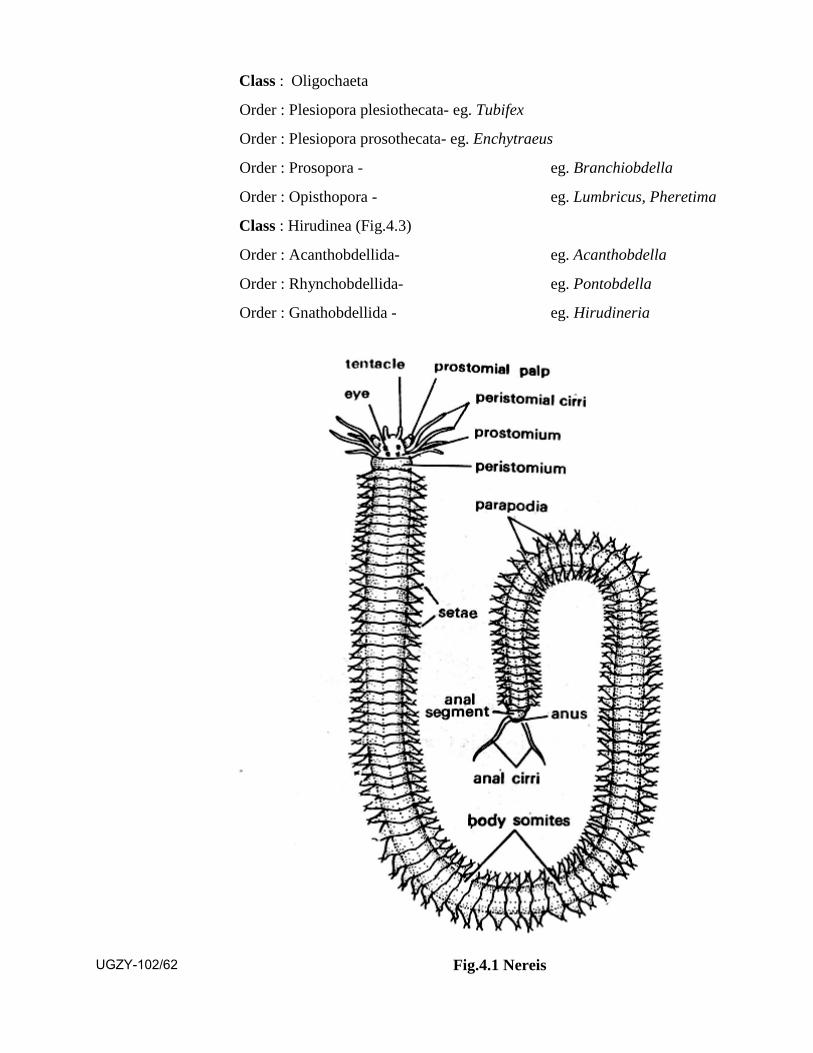
Class : Oligochaeta
Order : Plesiopora plesiothecata- eg. Tubifex
Order : Plesiopora prosothecata- eg. Enchytraeus
Order : Prosopora - eg. Branchiobdella
Order : Opisthopora - eg. Lumbricus, Pheretima
Class : Hirudinea (Fig.4.3)
Order : Acanthobdellida- eg. Acanthobdella
Order : Rhynchobdellida- eg. Pontobdella
Order : Gnathobdellida - eg. Hirudineria
Fig.4.1 Nereis UGZY-102/62
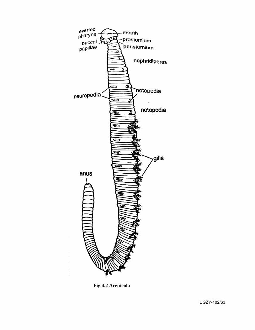
Fig.4.2 Arenicola
UGZY-102/63
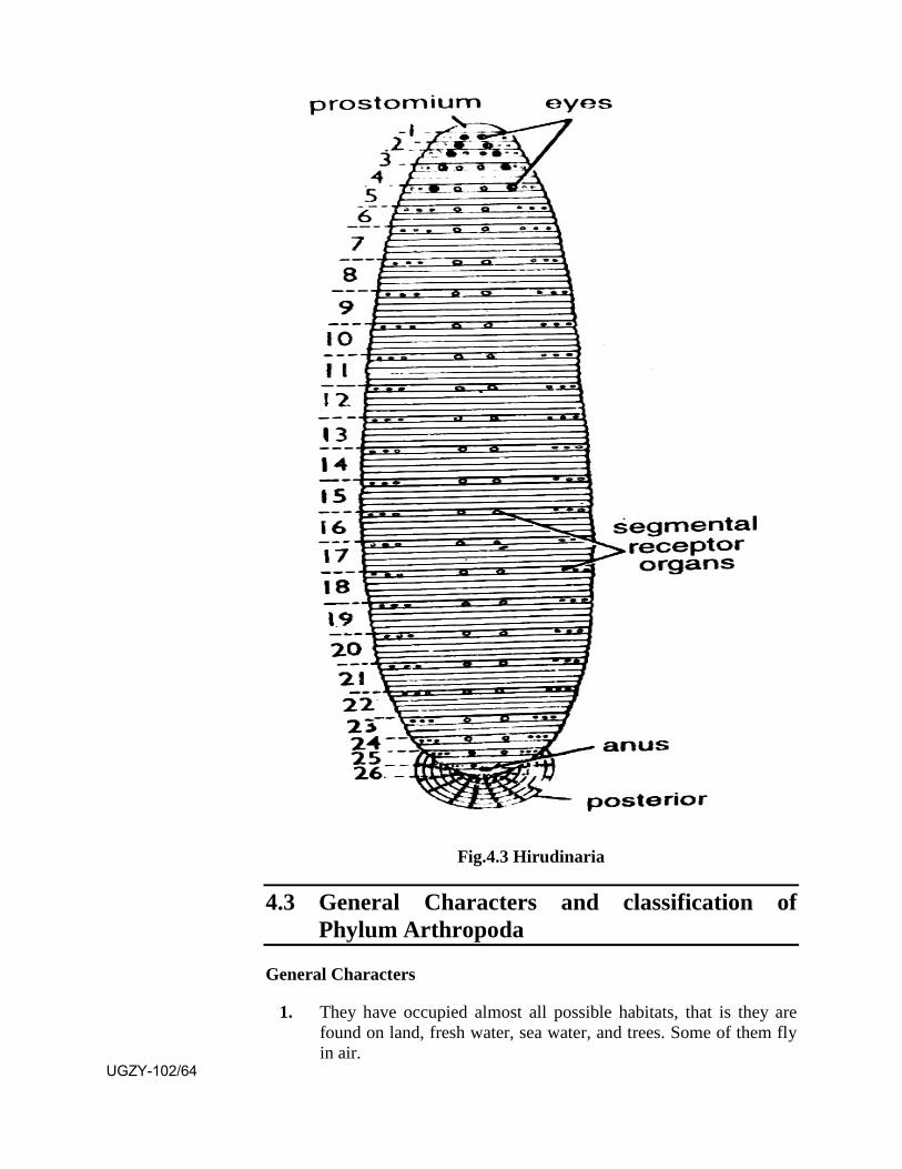
Fig.4.3 Hirudinaria
4.3 General Characters and classification of Phylum Arthropoda
General Characters
1. They have occupied almost all possible habitats, that is they arefound on land, fresh water, sea water, and trees. Some of them flyin air.
UGZY-102/64

2. Body remains bilaterally symmetrical, triploblastic andmetamerically segmented.
3. Body shows head, thorax and abdomen, sometimes head andthorax get fused forming cephalothorax.
4. They possess paired jointed appendages.
5. Exoskeleton made up of chitin which is light and protective . Itundergoes moulting for growth and development.
6. True coelom replaced by blood filled haemocoel.
7. Alimentary cand complete with mouth and anus. Mouth partsadapted for varied modes of feeding.
8. Respiration by general body surface, gills, trachae and book lungs.
9. Circulatory system open with a dorsal many chambered heart andblood sinuses.
10. Excretion is performed by green glands or malpighian tubules.
11. Nervous system shows a dorsal brain, a nerve ring and doubleventral ganglionated nerve cord.
12. Sensory organs are represented by simple or compound eyes,tactile receptors, chemoreceptors, balancing receptors and auditoryreceptors.
13. Body unisexual. Fertilization internal, development direct orindirect.
14. Parental care seen.
15. They are represented by maximum number of species and theyoccupy maximum amount of living protoplasm.
SAQ 2-
Paired and jointed appendages are the example of which phylum?
Classification of Phylum Arthropoda
Phylum Arthropoda
Subphylum : Onychophora ..... eg Peripatus
Subphylum : Tardigrada eg. Microbiolus
Subphylum : Pentastomida eg. Linguatula
Subphylum : Pycnogonida eg. Pycnogonum
Subphylum : Chelicerata :
UGZY-102/65
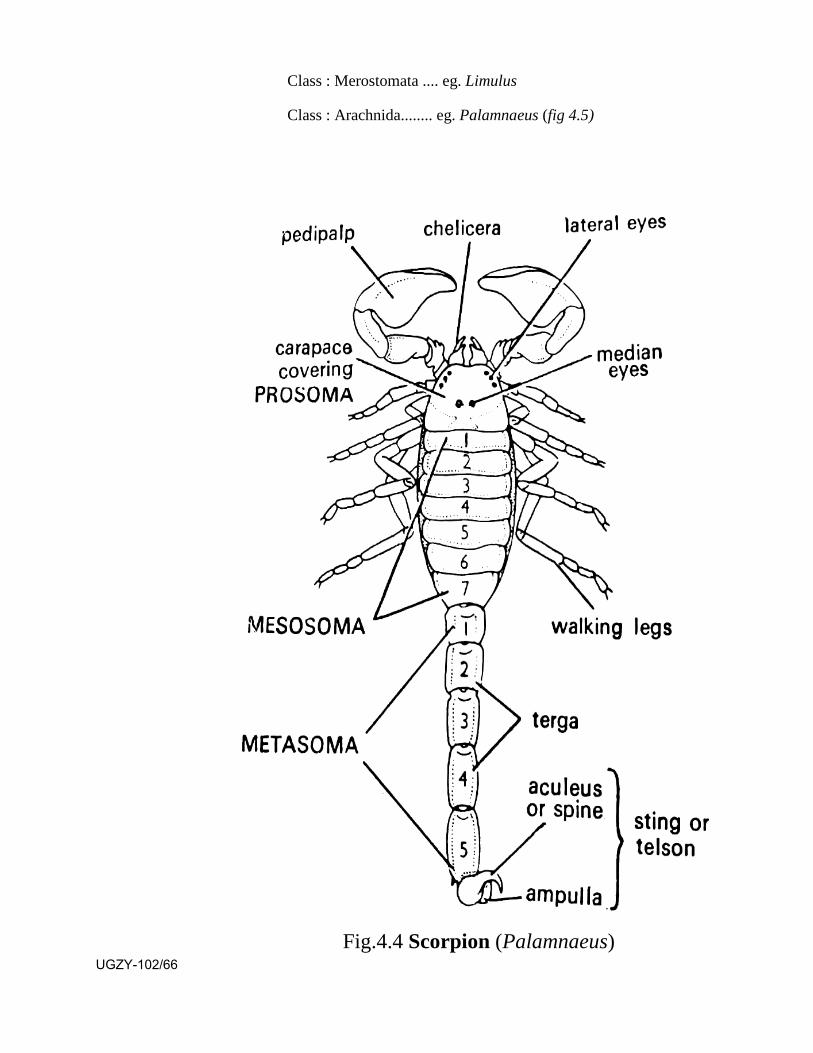
Class : Merostomata .... eg. Limulus
Class : Arachnida........ eg. Palamnaeus (fig 4.5)
Fig.4.4 Scorpion (Palamnaeus) UGZY-102/66

Subphylum : Mandibulata
Class : Crustacea ...... eg. Palaemon
Fig.4.5 Palaemon Class : Myriapoda ...... eg. Scolopendra
Class Insecta .......
Order : Orthoptera . eg. Periplaneta
Fig.4.6 Periplaneta
UGZY-102/67
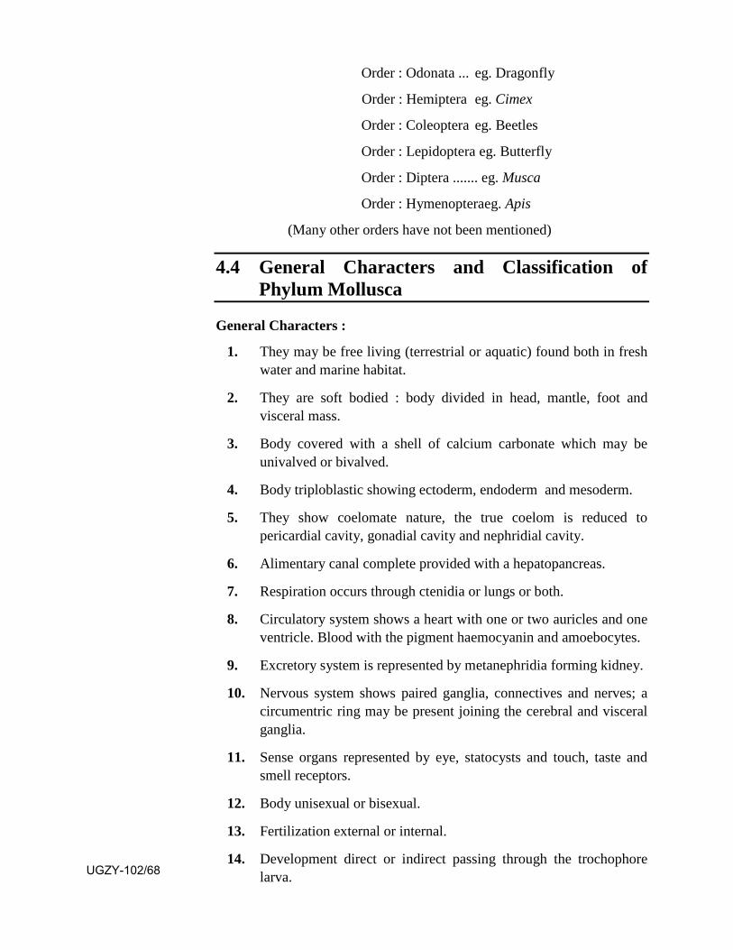
Order : Odonata ... eg. Dragonfly
Order : Hemiptera eg. Cimex
Order : Coleoptera eg. Beetles
Order : Lepidoptera eg. Butterfly
Order : Diptera ....... eg. Musca
Order : Hymenopteraeg. Apis
(Many other orders have not been mentioned)
4.4 General Characters and Classification of Phylum Mollusca
General Characters :
1. They may be free living (terrestrial or aquatic) found both in freshwater and marine habitat.
2. They are soft bodied : body divided in head, mantle, foot andvisceral mass.
3. Body covered with a shell of calcium carbonate which may beunivalved or bivalved.
4. Body triploblastic showing ectoderm, endoderm and mesoderm.
5. They show coelomate nature, the true coelom is reduced topericardial cavity, gonadial cavity and nephridial cavity.
6. Alimentary canal complete provided with a hepatopancreas.
7. Respiration occurs through ctenidia or lungs or both.
8. Circulatory system shows a heart with one or two auricles and oneventricle. Blood with the pigment haemocyanin and amoebocytes.
9. Excretory system is represented by metanephridia forming kidney.
10. Nervous system shows paired ganglia, connectives and nerves; acircumentric ring may be present joining the cerebral and visceralganglia.
11. Sense organs represented by eye, statocysts and touch, taste andsmell receptors.
12. Body unisexual or bisexual.
13. Fertilization external or internal.
14. Development direct or indirect passing through the trochophorelarva.UGZY-102/68

Classification of Phylum Mollusca Phylum Mollusca
Classes
Monoplacophora Amphineura Scaphopoda Gastropoda Bivalvia Cephalopoda
1. Shell as a singledorsal plate.
Shell as 8 dorsal plate.
Shell tusk like.
Shell usually coiled.
Shell of 2 valves.
Shell external, internal or absent.
2. Foot broad andflat.
Foot, broad and flat.
Foot conical.
Foot large, flat.
Foot hatchet shaped.
Foot as tentacles.
eg. Neopilina eg. Chiton eg. Dentalium
Class Gastropoda Subclass
Prosobranchia Opisthobranchia Pulmonata Shell closed by operculum present on foot.
Shell small, without operculum.
Shell may or may not be present.
eg. Patella eg. Aplysia eg. Helix
Class Bivalvia Orders
Protobranchia Filibranchia Eulamellibranchia Septibranchia
Gill filaments not folded.
Gill filaments
in completely fused.
Gill filaments completely fused.
Gills absent.
eg. Nucula eg. Mytilus eg. Unio eg. Poromya
Class Cephadopoda Sub Class
Nautiloidea Coeloidea Shell external, coiled or straight. Tentacles suckerless.
eg. Nautilus
Shell internal or absent. Tentacles with suckers.
eg. Loligo, Octopus
4.5 Torsion and Detorsion in Mollusca
The phenomenon of torsion occurs in the veliger larva of subclass Prosobranchiata of class Gastropoda of phylum Mollusca. Torsion is the
UGZY-102/69

process of rotation of visceropallium through 1800 from the original position in anticlockwise direction. It occurs in the neck region of the larva.
Important changes occurring during torsion are as under-
(i) The digestive gland grows enormously resulting in the formation of the visceral hump near the head.
(ii) The visceral hump rotates by 1800 anticlockwise within limits of the shell.
(iii) The rotation of the hump is caused by active growth on left side and retarded growth and right side.
(iv) Muscular contractions also play important role during this process.
The process lasts for about 3 minutes or even less. Following important events occur at the end of torsion.
(i) The posterior mantle cavity comes to lie on anterior side.
(ii) The visceral organs of right side come on the left side and those on left side come on right side.
(iii) Auricles present behind ventricle come on its anterior side.
(iv) Ctenidia which were directed backwards are pointed forwards.
(v) Backwardly directed excretory pore comes forward.
(vi) Straight alimentary canal forms a loop.
(vii) Backwardly directed anus present on left side comes on right side and becomes forwardly directed.
(viii) Dorsal visceral sac and shell become ventral.
(ix) Bilatral symmetry gets disturbed and the body becomes asymmetrical.
(x) Pleurovisceral connectives of nervous system which were parallel before torsion get twisted in the form of 8. Right connective forms supra intestinal nerve while left forms infraintestinal nerve. This process in known as chiastoneury.
Torsion is advantages for both the larva and the adult. As the result of torsion the mantle cavity comes anteriorly above the head. In order to avoid the predator the larva would now retract the head and anterior ciliary band in mantle cavity, thus stops its movements and falls on the botton. Because of the positioning of mantle cavity in anterior region the respiratory and locomotary currents come in the same direction in the adult. It increases ventilation of mantle cavity. The main disadvantage of torsion is the anterior positioning of excretory opening and anus in the adult. It pollutes the respiratory current by faecal matter and nitrogenous wastes. UGZY-102/70
The phenomenon of detorsion is the reversion of torsion. It occurs in Opisthobranchia (eg. Aplysia). During this process the pallial complex again comes back towards the posterior end along the right side after the occurance of torsion. Thus the visceral loop becomes untwisted and symmetrical, auricles come to lie behind ventricle, ctenidia become backwardly pointed, secondary bilateral symmetry appears and shell becomes lost. Thus the disadvantages of torsion in adult get minimized.
SAQ 3-
The phenomenon of torsion occurs in the ………
4.6 Larval formd in Echinodermata
In the development of echinoderms the zypote as the result of cleavage and blastulation forms a gastrula which gives rise to the first larva khown as Dipleurula larva which shows a peri oral band of cilia around the mouth on ventral side and an adoral band of cilia lining the mouth. It is a free swimming larva which lateron forms the different kinds of larvae.
In Asteroidea (eg. starfish) the Dipleurula larva forms the Bipinneria larva. In Holothuroidea it forms Auricularia larva. In Echinoidea and Ophiuroidea it forms Pleuteus larva (Echinopleutens and Ophiopleuteus) while in Crinoidea it form Doliolaria larva.
All such larvae possess complete functional alimentary canal showing ciliated stomodaeum, oesophagus, stomach, intestine and anus, coelomic sacs and varying number of ciliary bands. They show bilateral symmetry and undergo free swimming planktonic life. These larvae differ in their shape, size, number and arrangement of arms and number of ciliary bands.
SAQ 4-
The different larval forms occurring in phylum ……
Arrangement of Arms
In Bipinnaria preoral lobe is large showing three arms, one ventromedial and two lateral. The sides bear 4 pairs of long slender arms. They are anterodorsal, posterodorsal, posterolateral and post oral. Apart from them one dorsomedial arm extends forwards. In Auricularia also the preoral lobe is large and the sides bear short paired arms supported by calcareous particles. In Pleuteus the preoral lobe is small and the slender. Paired arms are directed forwards and supported by calcareous rods. They are in 4 pairs in Ophiopleuteus as pre oral, post oral, antero lateral and postero dorsal but in Echinopleuteus they are in 4, 5 or 6 pairs as pre oral, post oral, antero lateral, posterodorsal while the antero dorsal and postero lateral arms may or may not be present. In Doliolaria arms are absent.
UGZY-102/71
Arrangement of ciliary Bands
In Bipinnaria the peri oral band of cilia shows a preoral loop bordering preoral lobe and its arm and a post oral lobe bordering remaining arms. It Auricularia also it forms preoral and post oral loops showing a wavy path along lateral and ventral sides. In Pleuteus the ciliary band passes all over the arms. In Doliolaria ciliary bands form 4-5 transverse rings.
Presence of Apical plate
An apical plate with ciliary band remains absent in Bripinnaria and Pleuteus. It remains present in Doliolaria while in Auricularia it may or may not be present.
Presence of Adhesive pit
An adhesive pit is present only in Doliolaria near the anterior end.
Fate of Larvae (Metamorphosis)
The Bipinnaria larva at later stage develops three fixing processes on ventral side in front of pre oral loop known as brachiolar arms. They show adhesive suckers between their bases. This stage of larva is known as Brachiolaria. It gets attached on a substratum by tips of brachiolar arms and adhesive suckers and undergoes metamorphosis to form the adult.
The Auricularia larva gets converted into a barrel shaped free swimming Doliolaria and undergoes metamorphosis to form the adult. The Pleuteus also undergoes metamorphosis without showing any attachment. In Crinoids the free swimming barrel shaped Doliolaria gets attached to a substratum by its adhesive pit and undergoes metamorphosis to form a pentacrinoid larva showing a stalk and crown which lateron forms the adult.
4.7 Summary
In this unit you have learnt
that the members of phylum Annelida show true segmentation(metamerism) in which the body shows repetition of variousinternal organs. These animals for the first time in evolution showthe appearance of true coelom. They show ventral longitudinalnerve cord.
all about the broad classification of the phylum Annelida.
that the members of phylum Arthropoda are most successful asthey are represented by maximum number of species and they havecaptured the maximum amount of living protoplasm. They possesspaired and jointed appendages. Their coelom is filled with bloodforming haemocoel.
all about the broad classification of the phylum Arthropoda.
that the phylum Mollusca represents the shelled invertebratesUGZY-102/72
which undergo locomotion by foot and respiration by ctenidia or lungs or both.
all about the broad classification of phylum Mollusca.
all about the phenomenon of torsion occurring in the veliger larvaof members of subclass prosobranchiata of the class gastropoda inwhich the visceropallium gets rotated through 1800 from itsoriginal position in anticlockwise direction. It gives definiteadvantage to the larva and adult both.
all about the different larval forms occurring in phylumEchinodermata and the phenomenon of metamorphosis undergoneby then.
4.8 Terminal questions
Q.1 Give the significance of important characters of phylum Arthropoda in making them most successful lot.
Q.2 Classify phylum Mollusca upto subclass level giving the differentiating features and examples.
Q.3 What do you understand by true coelom? How does it become haemocoel.
Q.4 Write short notes on-
(i) Hirudinea (ii) Gastropoda
(iii) Chiastoneury (iv) Bipinnaria larva
Q.5 Match the two-
Column I Column II 1. Nephridia (a) Arthropoda
2. Trochophore (b) Oligochaeta
3. Green gland (c) Cephalopoda
4. Lumbricus (d) Annelida
5. Nautilus (e) Larva
ANSWERS
SAQ 1 - True segmentation
SAQ 2 - Arthropoda
SAQ 3 - veliger larva
SAQ 4 - Echinodermata
UGZY-102/73

UGZY-102/74

UNIT-5
Comparative form,s and Functions : Locomotion, Digestion, Excretion
Structure 5.1 Introduction and Objectives
5.2 Locomotion in Metazoa
5.2.1 Significance of Hydraulic pressure in locomotion
5.2.2 Locomotion in Coelenterata
5.2.3 Locomotion in Flatworms
5.2.4 Locomotion in Nematoda
5.2.5 Locomotion in Annelida
5.2.6 Locomotion in Arthropoda
5.3 Locomotion in Mollusca
5.3.1 Foot as creeping and crawling organ
5.3.2 Foot as burrowing organ
5.3.3 Foot as leaping organ
5.3.4 Foot as swimming organ
5.4 Locomotion in Echinodermata
5.5 Feeding and Digestion
5.5.1 Feeding and digestion in sponges
5.5.2 Feeding and digestion in Coelenterata
5.6 Excretion in Metazoa
5.6.1 Protonephridia
5.6.2 Metanephridia
5.6.3 Malpighian tubules
5.6.4 Coelomoducts in Mollusca
5.7 Summary
5.8 Terminal Questions
UGZY-102/75
5.9 Answers
5.1 Introduction
In this unit you will study the activity of locomotion, feeding and excretion of waste matter from the body.
Locomotion is the activity of movement of the body from one place to the other. It is important for the survival of the species because it helps the animal to perform food capture and to defend oneself from the predator. It also helps in finding a mate for reproduction.
For the various activities the body requires material and energy for which food is needed. Herbivore species depend on vegetation and carnivores feed on other animals. The animals have developed various ways to capture their food. The food has to be digested in which larger molecules of proteins, carbohydrates and fats are broken down in simple molecules like amino acids, glucose and fatty acids which are absorbed in the body. Glucose provides quick energy, aminoacids are needed for growth and fatty acids provide reserve energy.
Excretion deals with the removal of metabolic wastes which are generated by the oxidation of energy rich compounds and metabolism of proteins. This activity is carried out by nephridia, coelomoducts and malpighian tubules in nonchordate Metazoa.
Objectives After studying this unit you should be able to-
learn about the different modes of locomotion in non- chordates.
learn about the locomotary organs and their functioning.
learn about the feeding and digestion in sponges and coelenterata.
learn about the protonophridia and metanephridia in Annelida,malpighian tubules in Insecta and coelomo ducts in Mollusca.
5.2 Locomotion in Metazoa
5.2.1 Significance of Hydraulic pressure in locomotion In Nematoda and Annelida a cavity is present between bodywall
and gutwall. It is known as pseudocoel in Nematoda and coelom in Annelida. Both the cavities remain filled up with a fluid with some corpuscles. It gives turgidity to the body. Along with the related muscles this fluid filled cavity makes the hydrostatic skeleton. In Nematoda are present longitudinal muscles below the syncitial ectoderm of the bodywall. These muscles are present in 2 dorso-lateral and 2 ventro lateral sectors. In Annelida the muscles are present in association with the bodywall and gutwall both. In the bodywall they are represented by outer band of UGZY-102/76
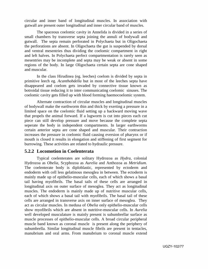
circular and inner band of longitudinal muscles. In association with gutwall are present outer longitudinal and inner circular band of muscles.
The spaceous coelomic cavity in Annelida is divided in a series of small chambers by transverse septa joining the annuli of bodywall and gutwall. The septa remain perforated in Polychaeta but in Oligochaeta the perforations are absent. In Oligochaeta the gut is suspended by dorsal and ventral mesentries thus dividing the coelomic compartment in right and left halves. In Polychaeta perfect compartmentation is rarely seen as mesentries may be incomplete and septa may be weak or absent in some regions of the body. In large Oligochaeta certain septa are cone shaped and muscular.
In the class Hirudinea (eg. leeches) coelom is divided by septa in primitive leech eg. Acanthobdella but in most of the leeches septa have disappeared and coelom gets invaded by connective tissue known as botroidal tissue reducing it to inter communicating coelomic sinuses. The coelomic cavity gets filled up with blood forming haemocoelomic system.
Alternate contraction of circular muscles and longitudinal muscles of bodywall make the earthworm thin and thick by exerting a pressure in a limited space on the coelomic fluid setting up a backward moving wave that propels the animal forward. If a lugworm is cut into pieces each cut piece can still develop pressure and move because the complete septa seperate the body in independent compartments. In larger earthworms certain anterior septa are cone shaped and muscular. Their contraction increases the pressure in coelomic fluid causing eversion of pharynx or if mouth is closed it results in elongation and stiffening of first segment for burrowing. These activities are related to hydraulic pressure.
5.2.2 Locomotion in Coelenterata Typical coelenterates are solitary Hydrozoa as Hydra, colonial
Hydrozoa as Obelia, Scyphozoa as Aurelia and Anthozoa as Metridium. The coelenterate body is diploblastic, represented by ectoderm and endoderm with cell less gelatinous mesoglea in between. The ectoderm is mainly made up of epithelio-muscular cells, each of which shows a basal tail having myofibrils. The basal tails of these cells are arranged in longitudinal axis on outer surface of mesoglea. They act as longitudinal muscles. The endoderm is mainly made up of nutritive muscular cells, each of which shows a basal tail with myofibrils. The basal tail of these cells are arranged in transverse axis on inner surface of mesoglea. They act as circular muscles. In medusa of Obelia only epithelio-muscular cells show myofibrils which are absent in nutritive-muscular cells. In Aurelia well developed musculature is mainly present is subumbrellar surface as muscle processes of epithelio-muscular cells. A broad circular peripheral muscle band known as coronal muscle is present along the periphery of subumbrella. Similar longitudinal muscle fibrils are present in tentacles, manubrium and oral arms. From manubrium to coronal muscle extend
UGZY-102/77
radial muscles along the main radii. In Metridium epidermal musculature is limited to tentacles and oral disc but absent in column of the body. Gastrodermal musculature forming circular muscles is present in tentacles, oral disc, pedal disc, gullet and column. A mesentry shows thick longitutional muscles as retractor muscle.
With the help of such muscle patterns the animals show varied movements. The Hydra shows elongation and contraction at intervals(Fig.5.1). Apart from this it shows looping, somersaulting, gliding, walking, climbing, floating, surfacing and swimming. Hydroid Obelia colony does not show body movements. Only the hydrozooids can contract and expand and bend their tentacles. The medusa generally floats passively in water. It may move forwards in a series of jerks by rhythmic contraction and expansion of the bell by hydropropulsion.
SAQ 1- (a) The coelenterate body is …………, represented by ……… and
………… with cell less gelatinous ………. in between. (b) The coelenterate body is ……….
In Aurelia well developed musculature is mainly present is ………… surface
Fig.5.1. Hydra UGZY-102/78
Fig5.2.3. Locomotion in Flat worms Platyhelminthes are represented by 3 classes, Turbellaria,
Trematoda and Cestoda. Turbellaria are free living in water or commensalic while Trematoda and Cestoda are parasitic, but the trematode larvae miracidium and cercaria are free living in water. The Turbellaria and miracidium larva show marginal cilia while cercaria larva possesses a tail. The typical turbellarian, Dugesia does not swim in water. It moves in contact with the substratum. It performs gliding by secreting a thin layer of mucous on substratum and moving on it by ciliary beating, raising its head during the process. A turbellarian shows outer circular muscles, middle diagonal muscles and inner longitudinal muscles below the basement membrance of bodywall. It performs crawling movements with the help of these muscles. Crawling movement shows following actions (i) contraction of circular muscles elongates the body (ii) secretion of adhesive glands makes the head attached to the substractum (iii) Waves of contraction of longitudinal muscles pass from anterior to posterior end alternately on right and left side (iv) the body moves forwards in wavy manner. The miracidium larva of Trematoda swims in water with the help of its marginal cilia and the cercaria larva swims by the lashing movements of its tail.
5.2.4 Locomotion in Nematoda A typical nematode (eg. Ascaris) shows a bodywall with thin
cuticle, syncitial epidermis and inner most layer of longitudinal muscles. Between the bodywall and alimentary canal is present a cavity known as pseudocoel filled up with a fluid having 93% water, different minerals and some corpuscles. The fluid filled pseudocoel acts as hydrostatic skeleton providing rigidity to the body. Undulating movements can be performed by alternate contraction of darsolateral and ventrolateral muscles.
5.2.5 Locomotion in Annelida In Polychaeta (eg. Nereis) locomotion is performed by parapodia,
body musculature and coelomic fluid. These animals perform slow and rapid crawling, swimming and burrowing. Crawling is performed on a substratum by metachronal rhythm of parapodia. Parapodia of one side are turned downwards with setae protracted while the parapodia of the other side are raised upwards with setae withdrawn. Every 5th or 6th parapodia of one side remain at the same stage of the cycle. During swimming the contracentral waves of contraction of longitudinal muscles are fewer but more frequent. During burrowing protrusion of proboscis is done. In Oligochaeta (eg. earthworm) the action of circular and longitudinal muscles on coelomic fluid creates peristaltic waves while setae help in providing grip to substratum. During burrowing anterior segments are extended forwards creating a space in moist soil. Widening of the space is
UGZY-102/79
done by increasing the hydrostatic pressure.
In Hirudinea (eg. leeches) crawling is performed by alternate attachement of anterior and posterior suckers. During this process circular and longitudinal muscles show antagonistic activity. A leech gets upright posture on posterior sucker by the increase in hydrostatic pressure. During swimming the darsoventral muscles get contracted making it flattened and waves of contraction of longitudinal muscles cause serpentine movements.
5.2.6 Locomotion in Arthropoda Arthropoda shows maximum adaptive radiation. They have
occupied almost all possible habitats. They undergo varied ways of locomotion like crawling, walking, jumping, burrowing and swimming. Some insects even show flying. For such activities they possess limbs and wings. The body shows segmentation. It may show a head, thorax and abdomen. Sometimes the head and thorax get fused forming cephalothorax.
Limbs : The paired jointed limbs are lateral or ventrolateral extensions of the body segments. Their position and number is highly variable in different groups of the phylum as under.
(1) Subphylum Onychophora (eg. Peripatus) shows many pairs of stumpy limbs.
(2) Subphylum Tardigrada (eg. Water bears) shows 4 pairs of unjointed limbs.
(3) Suphylum Chelicerata (eg. Scorpion) shows 6 pairs of cephalo-thoracic appendages (chelicerae- 1 pair; pedipalpi-1 pair; walking legs 4 pairs)
(4) Class Crustacea of subphylum Mandibulata (eg. Prawn) shows 19 pairs of appendages (5 pairs of cephalic, 3 pairs of thoracic, 5 pairs of walking legs and 6 pairs of abdominal appendages).
(5) Class Myriapoda of subphylum Mahdibulata, (eg. centipede and millipede) shows many body segments, each segment showing 1 or 2 pairs of appendages.
(6) Class Insecta of subphylum Mandibulata (eg. cockroach) shows only 3 pairs of jointed thoracic appendages.
Wings : They are present in Pterygote insects (eg. Cockroach(Fig.5.2), grasshopper, housefly, mosquito, butterfly).
(c) They are in 2 pairs, one associated with mesothorax and the other with metathorax. In some insects (eg. cockroach) the first pair is an opaque covering while the thin, second pair is transleuscent and flying, In others (eg. mosquito) the first pair is used for flight and the second pair is rudimentary.
UGZY-102/80

Fig.5.2 Wings of cockroach
SAQ 2- Wings are present in ……….. insects
Structure of limbs: The limbs may be uniramous or biramous. A uniramous limb is
represented by thoracic leg of a prawn(Fig.5.3) which shows 7 joints and the insect leg which shows 5 joints ending in movable podomers terminating in 2 claws. A biramaous limb has a common base with 2 rami, inner endopodite and outer exopodite. It is represented by maxillepedes and abdominal appendages of prawn.
Fig.5.3 Thoracic legs of prawn
UGZY-102/81
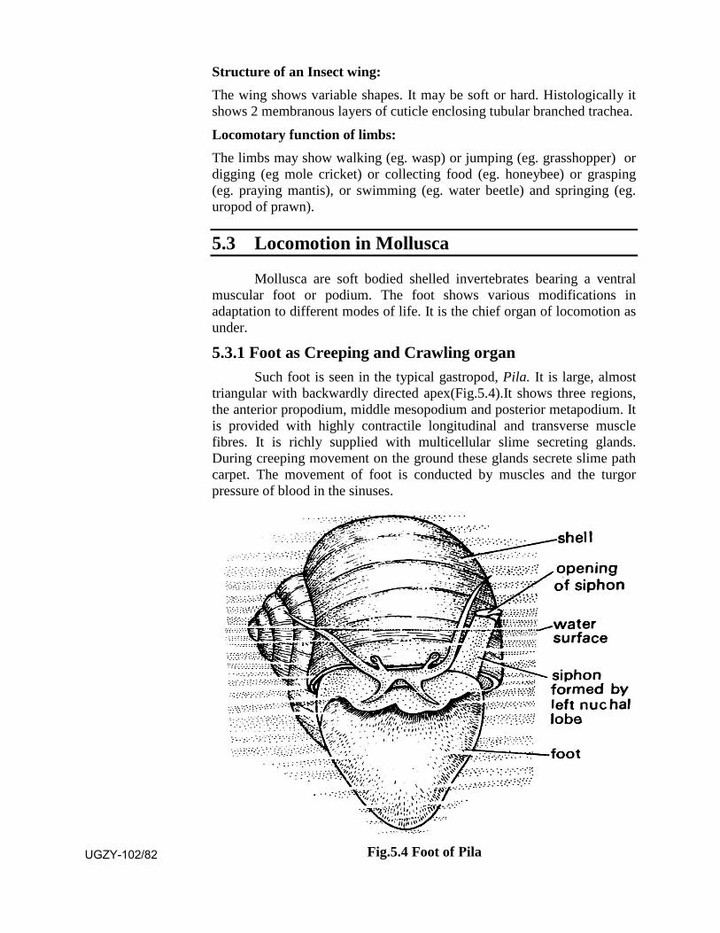
Structure of an Insect wing: The wing shows variable shapes. It may be soft or hard. Histologically it shows 2 membranous layers of cuticle enclosing tubular branched trachea.
Locomotary function of limbs: The limbs may show walking (eg. wasp) or jumping (eg. grasshopper) or digging (eg mole cricket) or collecting food (eg. honeybee) or grasping (eg. praying mantis), or swimming (eg. water beetle) and springing (eg. uropod of prawn).
5.3 Locomotion in Mollusca
Mollusca are soft bodied shelled invertebrates bearing a ventral muscular foot or podium. The foot shows various modifications in adaptation to different modes of life. It is the chief organ of locomotion as under.
5.3.1 Foot as Creeping and Crawling organ Such foot is seen in the typical gastropod, Pila. It is large, almost
triangular with backwardly directed apex(Fig.5.4).It shows three regions, the anterior propodium, middle mesopodium and posterior metapodium. It is provided with highly contractile longitudinal and transverse muscle fibres. It is richly supplied with multicellular slime secreting glands. During creeping movement on the ground these glands secrete slime path carpet. The movement of foot is conducted by muscles and the turgor pressure of blood in the sinuses.
Fig.5.4 Foot of Pila UGZY-102/82
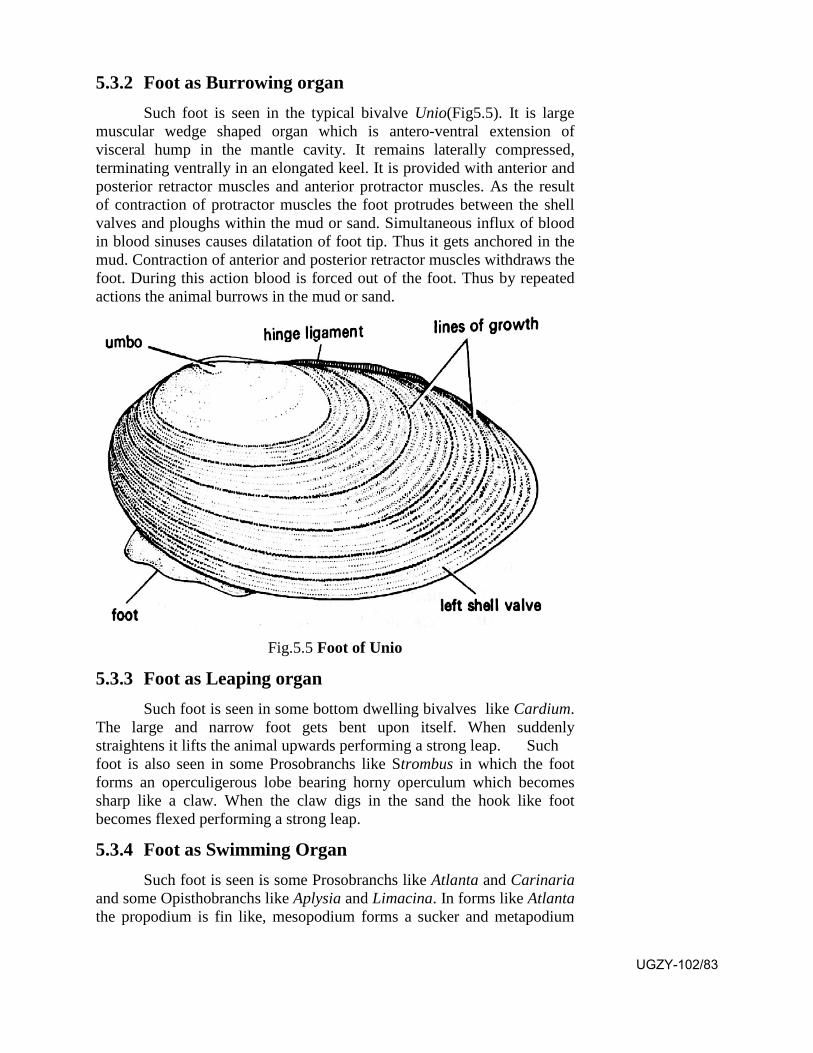
5.3.2 Foot as Burrowing organ Such foot is seen in the typical bivalve Unio(Fig5.5). It is large
muscular wedge shaped organ which is antero-ventral extension of visceral hump in the mantle cavity. It remains laterally compressed, terminating ventrally in an elongated keel. It is provided with anterior and posterior retractor muscles and anterior protractor muscles. As the result of contraction of protractor muscles the foot protrudes between the shell valves and ploughs within the mud or sand. Simultaneous influx of blood in blood sinuses causes dilatation of foot tip. Thus it gets anchored in the mud. Contraction of anterior and posterior retractor muscles withdraws the foot. During this action blood is forced out of the foot. Thus by repeated actions the animal burrows in the mud or sand.
Fig.5.5 Foot of Unio
5.3.3 Foot as Leaping organ Such foot is seen in some bottom dwelling bivalves like Cardium.
The large and narrow foot gets bent upon itself. When suddenly straightens it lifts the animal upwards performing a strong leap. Such foot is also seen in some Prosobranchs like Strombus in which the foot forms an operculigerous lobe bearing horny operculum which becomes sharp like a claw. When the claw digs in the sand the hook like foot becomes flexed performing a strong leap.
5.3.4 Foot as Swimming Organ Such foot is seen is some Prosobranchs like Atlanta and Carinaria
and some Opisthobranchs like Aplysia and Limacina. In forms like Atlanta the propodium is fin like, mesopodium forms a sucker and metapodium
UGZY-102/83
bears operculum. In Carinaria the propodium forms a large fin while mesopodium and metapodium are reduced. The fin performs undulating movements in water and acts as a swimming organ. In Opisthobranchs like Aplysia the foot shows two lateral projections as broadfins known as parapadia. They can be folded and united over the head forming a sac through which water can be expelled like a jet during swimming. In Limacina the anterior region of foot forms broad wing like parapodia. It uses parapodia to perform spiral swimming. For dropping down the animal makes its parapodia motionless.
5.4 Locmotion in Echinodermata
In Echinoderms locomotion is performed by water vascular system.
Water vascular System in Echinodermata
Typical water vascular system is seen in starfish. It shows a round or oval calcareous plate on the aboral surface of central disc called as madreporite. It contains many openings leading internally in a madreporic vesicle which extends downwards in the form of a sigmoid canal called as stone canal. It opens in a ring canal around the mouth(Fig.5.6). From the ring canal 5 radial canals arise, each located in one arm over the ambulacral groove. From each radial canal arise many lateral podial canals. Each lateral canal ends in an ampulla at the base of the tube foot, its opening being guarded by a valve. The ampullary cavity continues within the podium of tube foot as podial canal. The system shows 5 sac like polian vesicles from the inter radii of ring canal for storing the sea water. Five pairs of racemose vesicles called as tiedman's vesicles arise from the inter radii of ring canal on inner side. The system shows following variations in different classes. Madreporite remains absent in Crinoidea. It is internal in Holothuroidea and external in others. It is more than one in Ophiuroidea and single in Asteroidea and Echinoidea. The stone canals are a few in Holothuroidea, many in Crinoidea and single in rest of the classes. Ring canal is present around mouth in Asteroidea and Erinoidea, in the Crinoidea and Holothuroidea it is deeper, in Ophiuoroidea it is on aboral side. Single radial canal is present in the medial line of each arm in Asteroidea and Ophiuroidea and on each radial line of the body in Echinoidea and Holothuroidea. In Crinoidea it is branched. Polian vesicles are absent in Echinoidea and Crinoidea. They are 5 in Asteroidea and Holothuroidea but 4 in Ophiuroidea. Tiedman's bodies are present in Asteroidea but absent in rest of them. The fluid filled tube feet with their mucular walls and ampulla play important role in locomotion. With the valve closed the contraction of ampullary muscles causes elongation of tube foot. When the tube toot touches the stratum the sucker produces vacuum resulting in its attachment. Now the longitudinal muscles contract. It shortens the tube feet driving the fluid back in the ampulla. Thus the tube foot moves in a particular direction.
UGZY-102/84
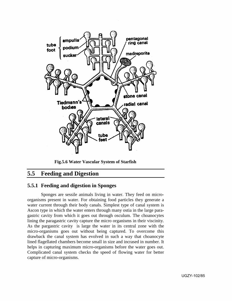
Fig.5.6 Water Vascular System of Starfish
5.5 Feeding and Digestion
5.5.1 Feeding and digestion in Sponges Sponges are sessile animals living in water. They feed on micro-
organisms present in water. For obtaining food particles they generate a water current through their body canals. Simplest type of canal system is Ascon type in which the water enters through many ostia in the large para-gastric cavity from which it goes out through osculum. The choanocytes lining the paragastric cavity capture the micro organisms in their viscinity. As the pargastric cavity is large the water in its central zone with the micro-organisms goes out without being captured. To overcome this drawback the canal system has evolved in such a way that choanocyte lined flagellated chambers become small in size and incrased in number. It helps in capturing maximum micro-organisms before the water goes out. Complicated canal system checks the speed of flowing water for better capture of micro-organisms.
UGZY-102/85
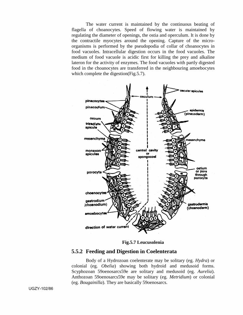
The water current is maintained by the continuous beating of flagella of choanocytes. Speed of flowing water is maintained by regulating the diameter of openings, the ostia and operculum. It is done by the contractile myocytes around the opening. Capture of the micro-organisms is performed by the pseudopodia of collar of choanocytes in food vacuoles. Intracellular digestion occurs in the food vacuoles. The medium of food vacuole is acidic first for killing the prey and alkaline lateron for the activity of enzymes. The food vacuoles with partly digested food in the choanocytes are transferred in the neighbouring amoebocytes which complete the digestion(Fig.5.7).
Fig.5.7 Leucusolenia
5.5.2 Feeding and Digestion in Coelenterata Body of a Hydrozoan coelenterate may be solitary (eg. Hydra) or
colonial (eg. Obelia) showing both hydroid and medusoid forms. Scyphozoan 59oenosarcs59e are solitary and medusoid (eg. Aurelia). Anthozoan 59oenosarcs59e may be solitary (eg. Metridium) or colonial (eg. Bougainilla). They are basically 59oenosarcs.
UGZY-102/86
The body of a solitary hydrozoan (eg. Hydra) is cylindrical showing an oral-aboral axis. Distal end shows an elevation known as hypostome showing a circular mouth at the top. Hypostome at its base bears 6-10 contractile tentacles beset with nematoblasts. Mouth leads internally to a gastrovascular cavity lined with gastrodermis having nutritive muscular cells, gland cells, interstitial cells, sensory cell and nerve cells. The body of a colonial hydrozoan (eg. Obelia) shows horizontal sessile hydrorhiza from which arise many vertical stems called as hydrocauli which may branch and rebranch. It shows two kinds of zooids, gastrozooids (polyps) and gonozooids (blastostyles). Branches and zooids of the colony show inner living tubular 60oenosarcs and outer dead chitinous perisarc. The blastostyles produce 60oenos which are free swimming reproductive bodies. Both gastrozooid and 60oenos possess a digestive apparatus showing mouth and gastrovascular cavity. A typical scyphozoan (eg. Aurelia) also shows a similar digestive apparatus with mouth and gastrovascular cavity and with radial and circular canals. They all bear batteries of nematoblasts in some part or the other in association with mouth. A typical solitary anthozoan (eg. Metridium) also shows a similar digestive apperatus with mouth and a gastrovascular cavity which shows more complex organization.
The food of different coelenterates is variable. Solitary hydroid forms (eg. Hydra) and colonial hydroid forms (eg. Obelia)
Obelia feed on insect larvae, crustaceans (Cyclops, Daphnia), nematodes and annelid worms while scyphozoans (eg. Aurelia) feed on planktonic organisms, crustacean worms, eggs and larvae. The anthozoans feed upon 60oenosar, crustaceans, worms, sea urchins and fishes.
In all of these Coelenterata the moving prey is captured and paralyzed by the different kinds of nematoblasts present in the tentacles of Hydrozoa, lobules of arms and gastric filaments of the gastric pouches of a jelly fish and mesentric filaments and acontia of Anthozoa.
The captured prey in ingested in the gastrovascular cavity. It undergoes both extracellular and intracellular digestion. Extracellular digestion is performed in the gastrovascular cavity by the enzymes released from the gland cells of gastrodermis, leading to incomplete digestion. The incompletely digested food is captured by the nutritive muscular cells by pseudopodia in food vacuoles. The medium of food vacuole is acidic first for killing the pray and alkaline later on for enzyme activity. The digested food is transported to various parts of the body by diffusion from cell to cell. In colonial hydroids (eg. Obelia) it is passed to the whole colony through continuous 60oenosarcs. In Scyphozoa it travels through radial canals and circular canals to reach the various parts of body. Reserve food is stored in thescocytes. The excretion of waste matter occurs through the mouth as no anus in present.
UGZY-102/87
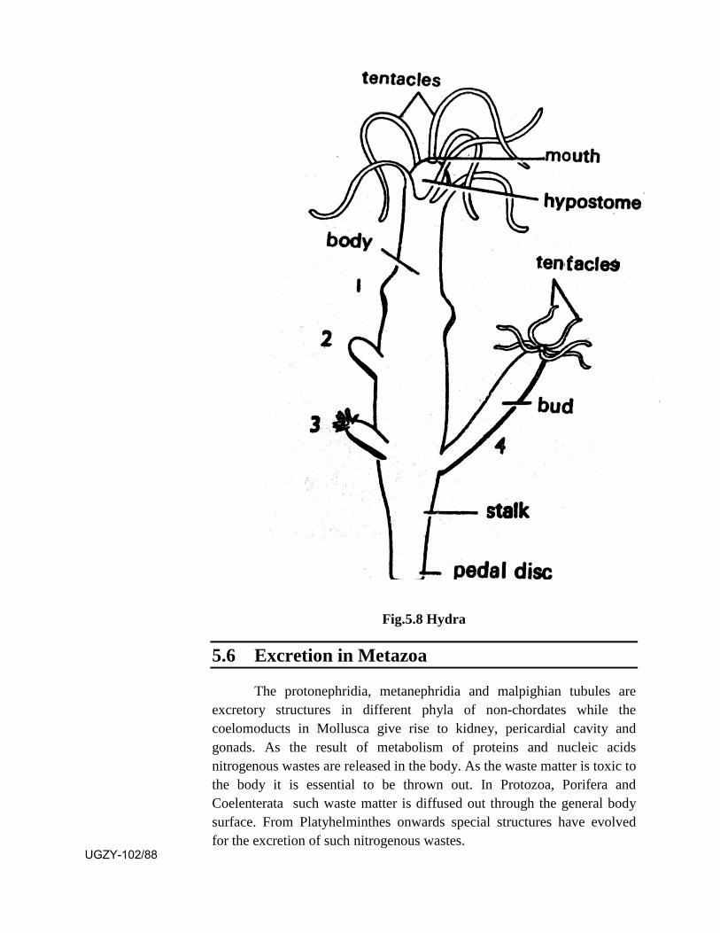
Fig.5.8 Hydra
5.6 Excretion in Metazoa
The protonephridia, metanephridia and malpighian tubules are excretory structures in different phyla of non-chordates while the coelomoducts in Mollusca give rise to kidney, pericardial cavity and gonads. As the result of metabolism of proteins and nucleic acids nitrogenous wastes are released in the body. As the waste matter is toxic to the body it is essential to be thrown out. In Protozoa, Porifera and Coelenterata such waste matter is diffused out through the general body surface. From Platyhelminthes onwards special structures have evolved for the excretion of such nitrogenous wastes.
UGZY-102/88
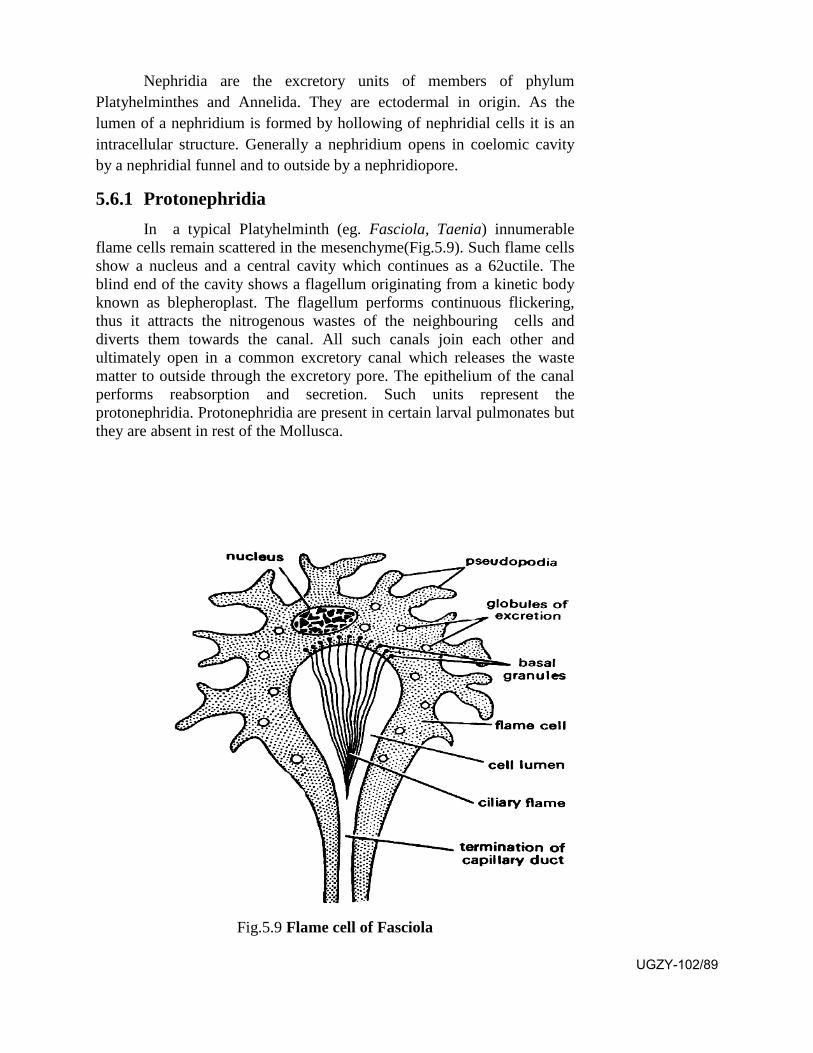
Nephridia are the excretory units of members of phylum Platyhelminthes and Annelida. They are ectodermal in origin. As the lumen of a nephridium is formed by hollowing of nephridial cells it is an intracellular structure. Generally a nephridium opens in coelomic cavity by a nephridial funnel and to outside by a nephridiopore.
5.6.1 Protonephridia In a typical Platyhelminth (eg. Fasciola, Taenia) innumerable
flame cells remain scattered in the mesenchyme(Fig.5.9). Such flame cells show a nucleus and a central cavity which continues as a 62uctile. The blind end of the cavity shows a flagellum originating from a kinetic body known as blepheroplast. The flagellum performs continuous flickering, thus it attracts the nitrogenous wastes of the neighbouring cells and diverts them towards the canal. All such canals join each other and ultimately open in a common excretory canal which releases the waste matter to outside through the excretory pore. The epithelium of the canal performs reabsorption and secretion. Such units represent the protonephridia. Protonephridia are present in certain larval pulmonates but they are absent in rest of the Mollusca.
Fig.5.9 Flame cell of Fasciola
UGZY-102/89

In phylum Annelida both protonephridia and meta-nephridia may be present. A protonephridium is seen in the annelid Venadis in which it remains present in the posterior part of a segment. It shows a blind sac which continues as a 63uctile to enter the next posterior segment through the septum, to open to outside by nephridiopore. In the blind sac open many solenocytes which are blindly ending tubes with flickering cilia each originating from a kinetic body known as blepheroplast. The flicking cilium attracts the nitrogenous wastes from the coelom in the cavity of solenocyte which is diverted through the cavity of protonephridium to outside. In the annelid Phyllodoce the protonephridium joins the neighbouring coelomoduct which opens to outside. Such a structure is known as protonephromixium.
Fig.5.10 Diagramatic representation of types of nephridia
5.6.2 Metanephridia
The metanephridium is seen in the annelid Notomastus. It is a ciliated funnel which opens in the coelom of a segment and continues as a duct, passing through the posterior septum to open ventrally in the next posterior segment. In the annelid Hesione the metanephridial duct joins the respective coelomoduct to share the common opening. Such a structive is known as metanephromixium. In the annelid Arenicola the metanephridium and coelomoduct of each segement get so intimately fused that they are represented by a single funnel, opening to outside through its duct. Such a structure is known as mixionephridium.
UGZY-102/90

5.6.3 Malpighian tubules Malpighian tubules are characterstic excretory structures of the
class Insecta of phylum Arthropoda (eg. cockroach). They are delicate, yellowish blind tubes which open in the alimentary canal at the junction of midgut and hindgut. They bathe in haemolymph of the perivisceral cavity. Their number is variable. Generally they appear in 6 to 8 bunches with 10 tubules in each bunch. Histologically they show 3 to 8 epithelial cells surrounding a lumen. Their inner surface shows striated border showing brush like filaments. A malpighian tubule shows two functional regions, distal secretory part extracts nitrogenous wastes as salt of uric acid and water from the haemolymph in the form of urine. The proximal absorptive part reabsorbs certain salts and water thus leaving uric acid in the lumen. The uric acid moves towards alimentary canal as the result of peristaltic waves. Colon and rectum region of the insect also absorbs most of the water, thus uric acid is liberated along with faecal matter.
5.6.4 Coelomoducts of Mollusca In Mollusca the two coelomic cavities meet dorsally to enclose the
heart. Their walls proliferate to form germ cells. The cavities by further differentiation give rise to gonad anteriorly, pericardial canal centrally and gonoduct posteriorly. The last segment shows excretory function. In Polyplacophora the excretory region remains connected to pericardial region. In Gastropoda excretory organ is present only on left side. In Lamellibranchiata the genital and renal ducts get separated. In Protobranchs the entire coelomoduct shows excretory function. In Filibranchs the coelomoduct is 'U' shaped. Its lower limb is glandular and upper limb forms the bladder. In Cephalopods the coelomic complex shows separation of genital and excretory complexes.
5.7 Summary
• The body cavity in Nematoda and Annelida remains filled up witha fluid which gives turgidity to the body. Along with the musclesof bodywall it serves as hydraulic skeleton.
• In Coelenterata the muscle tails of epithelio-muscular cells andnutritive-muscular cells contain contractile myofibrils. The muscletails of the two layers are antagonistic giving the ability oflocomotion.
• Amongst Platyhelminthes the free living Turbellaria show mucousglands and cilia in the skin and outer circular, middle diagonal andinner longitudinal muscles in bodywall. They undergo gliding withthe help of mucous and cilia and undergo crawling with the help ofthe muscles.
UGZY-102/91
• The Nematoda perform undulating movements by alternatecontraction of dorsolateral and ventrolateral longitudinal musclesbelow the epidermis.
• The Polychaete annelids undergo locomotion with the help ofparapodia, body muscles and coelomic fluid. In Oligochaeta thesetae also help in the movement on the ground. The leechesperform crawling by alternate attachment of anterior and posteriorsuckers which is helped by the circular and longitudinal muscles.
• Arthropods possess paired jointed legs which perform varied kindof movements. The insects show the ability of flying in air with thehelp of their wings.
• Mollusca possess foot as the locomotary organ. With its help theyperform creeping and crawling, burrowing, leaping and swimmingfor which they shows various modifications.
• The members of phylum Echinodermata utilize the hydrostaticpressure generated in their water vascular system and the podiaalong with their musculature for locomotion.
• Sponges maintain a water current in their canal system by themovement of flagella of their choanocytes. It brings microorganisms within the radial canals. The choanocytes capture themicro-organisms with the help of pseudopodia from their collarregion in food vacuoles in which the digestion occurs inintracellular manner.
• Coelenterates capture their prey with the help of their tentacleswhich are provided with stinging cells known as nematoblasts. Theingested prey undergoes mixed digestion. The extracellulardigestion occurs in coelenteron. The partly digested food iscaptured by nutritive muscular cells in food vacuoles where it isfurther digested in intracellular manner.
• For the release of nitrogenous wastes the flat worms show thepresence of flame cells. They represent protonephridia. In someannelids (eg. Venadis) and larval pulmonate molluscs theprotonephridia appear as blind sacs in which many solenocytesopen. Each solenocyte is a blindly ending tube with a flickeringcilium arising from a blepheroplast. The flame cells andsolenocytes both capture nitrogenous wastes and release them tooutside . In some annelids (eg. Notomastus) for the excretion ofnitrogenous waste a ciliated funnel opens in the coelom of onesegment. It continues as a duct to open to outside in the nextposterior segment. Such structures represent metanephridia.
• In Insecta the excretory structures appear as malpighian tubuleswhich are delicate tubules which bathe in the haemolymph ofperivisceral cavity. They open in the alimentary canal.
UGZY-102/92

• In Mollusca the excretory structures are represented by coelomicducts which show variable nature in different groups.
5.8 Terminal Questions
Q.1 Give an account of locomotion in Annelida.
Q.2 Describe the water vascular system of star fish.
Q.3 Describe the role of foot of mollusca in performing creeping and crawling.
Q.4 Describe the method of food capture and mixed digestion in Coelenterata.
Q.5 Describe a typical metanephridium and its association with coelomoduct in Annelida.
Q.6 Write short notes on
(i) Flame cell (ii) Malpighian tubules
(iii) Choanocyte (iv) Parapodium
Q.7 Match the two
Column -A Column-B
1. Coelom a. tail
2. Myofibrils b. Insecta
3. Cercaria larva c. Locomotion
4. Three paris of limbs d. Annelida
5. Polian vesicles e. Starfish
ANSWERS
SAQ 1- (a) diploblastic, ectoderm, endoderm, mesoglea
(b) diploblastic
(c) subumbrellar
SAQ 2- Pterygote
UGZY-102/93

UGZY-102/94
UNIT-6
Respiratory , Circulatory and Nervous system
Structure 6.1 Introduction and Objectives
6.2 Respiratory System
6.2.1 Respiratory Organs
6.2.2 Respiratory pigments
6.2.3 Process of respiration
6.3 Circulatory System
6.3.1 Open and closed type of circulatory systems
6.4 Organization of Nervous system
6.4.1 Nerve cell
6.4.2 Neuroglia
6.4.3 Ganglia
6.4.4 Nerve tract
6.5 Nervous systems
6.5.1 Nervous system in Platyhelminthes
6.5.2 Nervous system in Annelida
6.5.3 Nervous system in Arthropoda
6.5.4 Nervous system in Mollusca
6.6 Summary
6.7 Terminal Questions
6.8 Answers
6.1 Introduction
A living body requires energy for various body activities for which it needs food. The food has to be broken down in smaller and smaller fractions which could be absorbed in the body cells. Within the cell oxygen is needed for final break down of food and release of energy. As
UGZY-102/95
the result of this activity CO2 is released which has got to be released out of the body. Respiratory system is responsible for this activity. It obtains O2 from air or water through organs like skin, trachea, book lungs, lungs or gills. Respiratory activity involves inspiration, gaseous exchange and expiration.
The transport of the O2 and CO2 within the body is done through circulatory system in higher invertebrates and chordates which involves a fluid like blood, many vessels and capillaries for its flow and a pumping organ as heart.
A living body has to perform a number of activities such as locomotion, food capture, mating and defence against predators. They are performed by different organ systems. The animal has to change its activities in relation to the changing environmental conditions for which coordination becomes essential between the activities of different organs. The animal registers the changes in the environment, computes them and translates them into such actions which are profitable for the animal. This activity is performed by the nervous system which includes different sense organs which act as receptors.
The animal registers the changes and sends the informations through a definite tract to a modulator centre which analyses them and releases the necessary order for any specific muscle or gland which acts accordingly. The nervous system in any animal is made up of specific cells known as neurons and neuroglia. The neurons join together to form a nerve tract. Sometimes they get accumulated at one place to form a ganglion or a brain which performes modulation.
Objectives: After studying this unit you should be able to-
describe the respiratory organs in invertebrates.
know all about the respiratory pigments.
know the difference between open and closed type of circulatorysystem.
know the formation and pattern of nervous systems ininvertebrates.
6.2 Respiratory System
6.2.1 Respiratory Organs
Animals need oxygen for the breakdown of food in the body. During this process at the celullar level CO2 is released. As CO2 is toxic it should be released outside the body. The intake of Oxygen and release of CO2 is done from the surrounding environment which may be water or air. Animals have evolved varied organs for respiration which are as under: UGZY-102/96
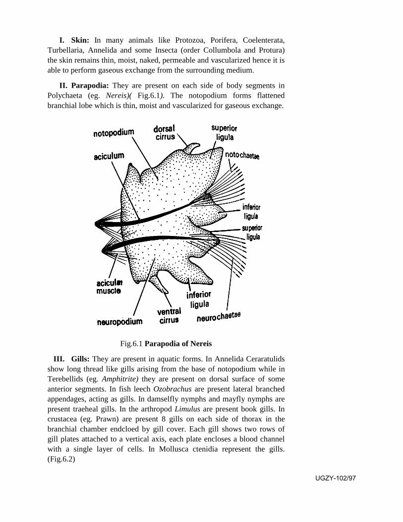
I. Skin: In many animals like Protozoa, Porifera, Coelenterata, Turbellaria, Annelida and some Insecta (order Collumbola and Protura) the skin remains thin, moist, naked, permeable and vascularized hence it is able to perform gaseous exchange from the surrounding medium.
II. Parapodia: They are present on each side of body segments inPolychaeta (eg. Nereis)( Fig.6.1). The notopodium forms flattened branchial lobe which is thin, moist and vascularized for gaseous exchange.
Fig.6.1 Parapodia of Nereis
III. Gills: They are present in aquatic forms. In Annelida Ceraratulidsshow long thread like gills arising from the base of notopodium while in Terebellids (eg. Amphitrite) they are present on dorsal surface of some anterior segments. In fish leech Ozobrachus are present lateral branched appendages, acting as gills. In damselfly nymphs and mayfly nymphs are present traeheal gills. In the arthropod Limulus are present book gills. In crustacea (eg. Prawn) are present 8 gills on each side of thorax in the branchial chamber endcloed by gill cover. Each gill shows two rows of gill plates attached to a vertical axis, each plate encloses a blood channel with a single layer of cells. In Mollusca ctenidia represent the gills. (Fig.6.2)
UGZY-102/97
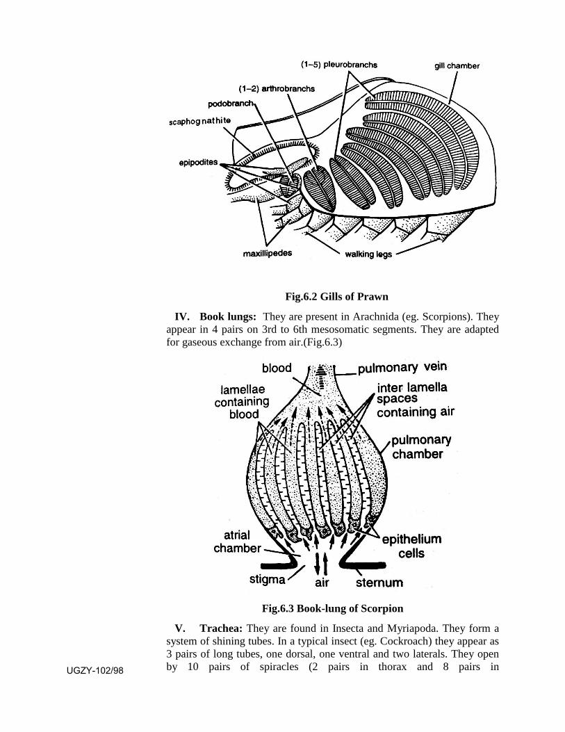
Fig.6.2 Gills of Prawn
IV. Book lungs: They are present in Arachnida (eg. Scorpions). Theyappear in 4 pairs on 3rd to 6th mesosomatic segments. They are adapted for gaseous exchange from air.(Fig.6.3)
Fig.6.3 Book-lung of Scorpion
V. Trachea: They are found in Insecta and Myriapoda. They form a system of shining tubes. In a typical insect (eg. Cockroach) they appear as 3 pairs of long tubes, one dorsal, one ventral and two laterals. They open by 10 pairs of spiracles (2 pairs in thorax and 8 pairs in UGZY-102/98
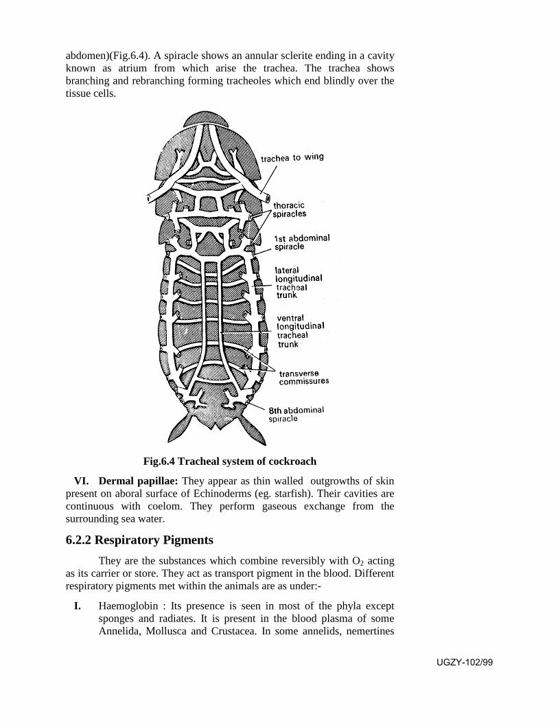
abdomen)(Fig.6.4). A spiracle shows an annular sclerite ending in a cavity known as atrium from which arise the trachea. The trachea shows branching and rebranching forming tracheoles which end blindly over the tissue cells.
Fig.6.4 Tracheal system of cockroach
VI. Dermal papillae: They appear as thin walled outgrowths of skinpresent on aboral surface of Echinoderms (eg. starfish). Their cavities are continuous with coelom. They perform gaseous exchange from the surrounding sea water.
6.2.2 Respiratory Pigments They are the substances which combine reversibly with O2 acting
as its carrier or store. They act as transport pigment in the blood. Different respiratory pigments met within the animals are as under:-
I. Haemoglobin : Its presence is seen in most of the phyla except sponges and radiates. It is present in the blood plasma of some Annelida, Mollusca and Crustacea. In some annelids, nemertines
UGZY-102/99
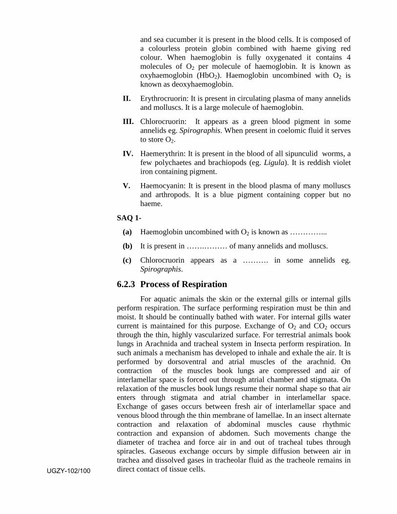
and sea cucumber it is present in the blood cells. It is composed of a colourless protein globin combined with haeme giving red colour. When haemoglobin is fully oxygenated it contains 4 molecules of O2 per molecule of haemoglobin. It is known as oxyhaemoglobin (HbO2). Haemoglobin uncombined with O2 is known as deoxyhaemoglobin.
II. Erythrocruorin: It is present in circulating plasma of many annelidsand molluscs. It is a large molecule of haemoglobin.
III. Chlorocruorin: It appears as a green blood pigment in someannelids eg. Spirographis. When present in coelomic fluid it servesto store O2.
IV. Haemerythrin: It is present in the blood of all sipunculid worms, afew polychaetes and brachiopods (eg. Ligula). It is reddish violetiron containing pigment.
V. Haemocyanin: It is present in the blood plasma of many molluscs and arthropods. It is a blue pigment containing copper but no haeme.
SAQ 1-
(a) Haemoglobin uncombined with O2 is known as …………...
(b) It is present in …….……… of many annelids and molluscs.
(c) Chlorocruorin appears as a ………. in some annelids eg. Spirographis.
6.2.3 Process of Respiration For aquatic animals the skin or the external gills or internal gills
perform respiration. The surface performing respiration must be thin and moist. It should be continually bathed with water. For internal gills water current is maintained for this purpose. Exchange of O2 and CO2 occurs through the thin, highly vascularized surface. For terrestrial animals book lungs in Arachnida and tracheal system in Insecta perform respiration. In such animals a mechanism has developed to inhale and exhale the air. It is performed by dorsoventral and atrial muscles of the arachnid. On contraction of the muscles book lungs are compressed and air of interlamellar space is forced out through atrial chamber and stigmata. On relaxation of the muscles book lungs resume their normal shape so that air enters through stigmata and atrial chamber in interlamellar space. Exchange of gases occurs between fresh air of interlamellar space and venous blood through the thin membrane of lamellae. In an insect alternate contraction and relaxation of abdominal muscles cause rhythmic contraction and expansion of abdomen. Such movements change the diameter of trachea and force air in and out of tracheal tubes through spiracles. Gaseous exchange occurs by simple diffusion between air in trachea and dissolved gases in tracheolar fluid as the tracheole remains in direct contact of tissue cells. UGZY-102/100

6.3 Circulatory System
6.3.1 Open and Closed type of Circulatory Systems
Circulatory system provides oxygen and nutrients to the body tissues and removes their waste products such as carbon dioxide, ammonia and urea to outside. In lower invertbrates as the transport of Oxygen, CO2, nutrients and other waste products is mainly done through diffusion hence the circulatory system is not needed but in higher invertebrates and vertebrates as the body bulk has increased the circulation of these substances through blood becomes essential hence a circulatory system has come in existence. The circulatory system is of two kinds, open type and closed type which can be differenti,ated as under:-
Open Circulatory System Closed Circulatory System
1. Blood flows in open tissuespaces known as sinuseswhich do not have properwalls.
1. Blood flows in closed tubes,the blood vessels posessingdefinite walls.
2. Blood comes in direct contactwith the tissue cells.
2. Blood does not come indirect contact with tissuecells.
3. Exchange of materials occursdirectly between blood andtissue cells.
3. Exchange of materialsoccurs between blood andtissue cells through tissuefluid.
4. Blood flows slowly under lowpressure.
4. Blood flows fast under highblood pressure.
5. Respiratory pigment if presentis dissolved in the plasma, asred corpuscles are absent.
5. Respiratory pigment ispresent mainly in the redblood corpuscles.
6. The system is less efficient asblood flow is not regulated.
6. The system is more efficientas the blood flow can beregulated.
7. It is seen in phylumArthropoda and Mollusca.
7. It is seen is phylumAnnelida and Chordata.
UGZY-102/101

SAQ 2-
(a) Open type of circulatory system is present in ……… and …………
(b) Closed type is present in ……… and ………...
6.4 Organization of Nervous System
6.4.1. Nerve cell A neuron shows a cell body and cell processes. The cell body
contains a nucleus, golgi apparatus, mitochondria, nissl granules and neurofibrils. The cell processes are of two kinds, the dendrite through which the nerve impulse travels towards the cell and the axon through which the nerve impulse travels away from the cell. A cell process arises from a pole of the neuron.
On the basis of number of poles present in a neuron, the latter may be apolar (without a pole), unipolar (with one pole from which arises the axon), bipolar (with two poles, from one arises the dendrite and from the other the axon) and multipolar (with many poles, from one pole arises the axon, from the rest arise dendrites). On the basis of function the neurons are of 3 kinds, sensory, motor and interneuron. Through a sensory neuron the nerve impulse travels towards the modulator, through a motor neuron the impulse travels away from the modulator. The interneurons remain present within the modulator. They receive a sensory impulse, analyse it and release an order impluse for the effector which may be a muscle or gland.
6.4.2 Neurolgia: A typical nervous system also contains another kind of cells known
as glia cells or neuroglia. They mainly constitute the packing material of nervous system. They are of three kinds, (i) Astrocytes (ii) Oligodendroglia and (iii) Microglia. Astrocytes are star shaped cells, oligodendroglia are amoeboid and microglia are small cells bodies.
6.4.3 Ganglia Whensoever there is an accumulation of neurons a ganglion is
formed. A ganglion serves as the modulator. It receives sensory impulse, modulates it and releases an order impulse which travels upto the effector which may be a muscle or a gland. According to the order impulse the muscle contracts or relaxes or a gland releases its secretion or stops it. This passage of nerve impulse which begins from a receptor and ends in an effector is known as reflex arc.
6.4.4 Nerve tract : A nerve tract is the path through which a nerve impulse travels. It
is made up of many neurons in a row with many gaps known as synapse. UGZY-102/102
A synapse is the point at which one neuron ends and the other begins. At each synapse the axon divides in many axon terminals, each of which ends in a round sac. The round sac remains filled up with many synaptic vesicles containing the neurohormone acetylcholine. In between the various synaptic knobs remain present the dendrite terminals which join to form a dendrite which joins the cyton of the next neuron. The nerve impulse at the synaptic knob makes some of the synaptic vesicles burst releasing the neurohormone which comes in contact with the nearby dendrite terminal. Thus the electrical nerve impulse passes through each synapse in the form of a chemical wave.
6.5 Nervous System
6.5.1 Nervous System in Platyhelminthes Phylum Platyhelminthes is represented by free living Turbellaria
and parasitic Trematoda and Cestoda. In all of them are present cerebral ganglia in anterior region. In Turbellaria this ganglion is a bilobed mass behind eyes. In Trematoda there are 3 such ganglia, one on each lateral side of pharynx and one below it. In Cestoda two such ganglia are present in scolex, one on each side. In Trematoda and Cestoda a nerve ring is present around the pharynx joining the main ganglia. In all the three groups longtudinal nerve cords arise from the nerve ring. In Turbellaria arising from the cerebral ganglia they are two in number, one on each side extending upto the postrior end of body. In Trematoda they are 3 pairs in number, one pair in dorsal half arising from nerve ring, one pair in ventral half arising from nerve ring and the third pair on lateral lines arising from respective lateral ganglia. In Cestoda 3 pairs of longitudinal nerve cords arise from nerve ring, one pair being dorsal, one pair ventral and the third pair as lateral. In Turbellaria and Trematoda the two lateral nerve cords are joined by many transverse connections. In Cestoda at the posterior end of each proglottid all the 6 nerve cords are joined by a transverse nerve. Thus in all the three groups the nervous system shows a ladder like appearence.
6.5.2 Nervous System in Annelida The annelid body shows true segmentation (metamerism). All the
three classes, Polychaeta, Oligochaeta and Hirudinea show the appearance of 3 kinds of ganglia. They are cerebral ganglia, subpharyngeal ganglia and segmental ganglia. In Polychaeta the cerebral ganglion is a bilobed mass present in dorsal half of prostomium. In Oligochaeta the cerebral ganglia are two in number present in dorsal half of 3rd segment. In Hirudinea also they are 2 in number, present above the pharynx in 5th segment. The subpharyngeal ganglion in Polychaeta appears as a bilobed mass below the pharynx in first trunk segment. In Oligochaeta also it is a bilobed mass below the pharynx in 4th segment. In Hirudinea it is a
UGZY-102/103
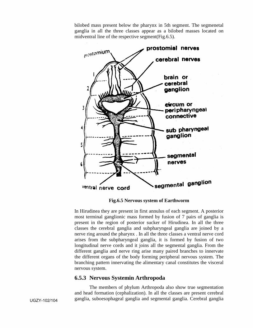
bilobed mass present below the pharynx in 5th segment. The segmenetal ganglia in all the three classes appear as a bilobed masses located on midventral line of the respective segment(Fig.6.5).
Fig.6.5 Nervous system of Earthworm
In Hirudinea they are present in first annulus of each segment. A posterior most terminal ganglionic mass formed by fusion of 7 pairs of ganglia is present in the region of posterior sucker of Hirudinea. In all the three classes the cerebral ganglia and subpharyngeal ganglia are joined by a nerve ring around the pharynx . In all the three classes a ventral nerve cord arises from the subpharyngeal ganglia, it is formed by fusion of two longitudinal nerve cords and it joins all the segmental ganglia. From the different ganglia and nerve ring arise many paired branches to innervate the different organs of the body forming peripheral nervous system. The branching pattern innervating the alimentary canal constitutes the visceral nervous system.
6.5.3 Nervous Systemin Arthropoda The members of phylum Arthropoda also show true segmentation
and head formation (cephalization). In all the classes are present cerebral ganglia, suboesophageal ganglia and segmental ganglia. Cerebral ganglia UGZY-102/104
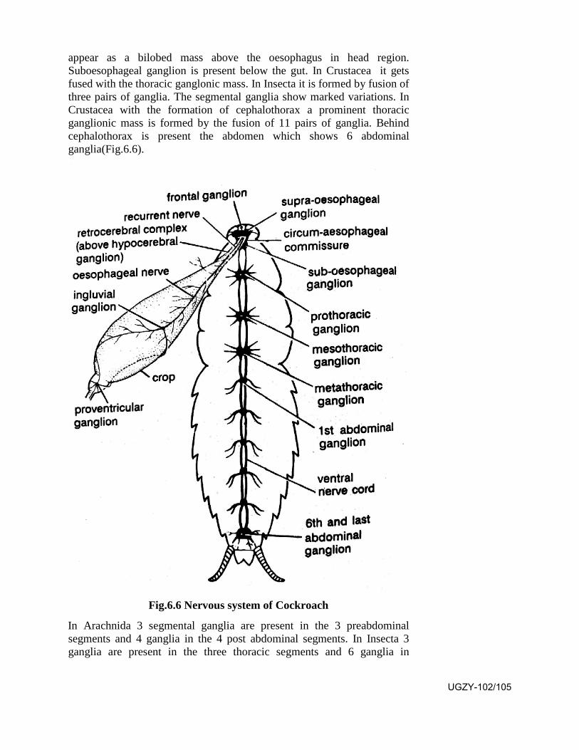
appear as a bilobed mass above the oesophagus in head region. Suboesophageal ganglion is present below the gut. In Crustacea it gets fused with the thoracic ganglonic mass. In Insecta it is formed by fusion of three pairs of ganglia. The segmental ganglia show marked variations. In Crustacea with the formation of cephalothorax a prominent thoracic ganglionic mass is formed by the fusion of 11 pairs of ganglia. Behind cephalothorax is present the abdomen which shows 6 abdominal ganglia(Fig.6.6).
Fig.6.6 Nervous system of Cockroach
In Arachnida 3 segmental ganglia are present in the 3 preabdominal segments and 4 ganglia in the 4 post abdominal segments. In Insecta 3 ganglia are present in the three thoracic segments and 6 ganglia in
UGZY-102/105

abdominal segments. In all the classes a nerve ring is present around the gut joining the cerebral and suboesophageal ganglia. In Crustacea the ventral nerve cord originates from the posterior end of fused thoracic mass in 11th segment running upto the posterior end joining the various segmental ganglia. It is formed by fusion of two parallel nerve cords. In Arachnida and Insecta two seperate and parallel nerve cords run on each side of midventral line joining the various segmental ganglia. The branches arising from the cerebral ganglia, suboesophageal ganglia, nerve ring and segmental ganglia innervating the different organs form the peripheral nervous system. The innervation to the alimentary canal forms visceral nervous system.
6.5.4 Nervous System in Mollusca In Mollusca the nervous system shows variable degrees of development. In primitive groups like Monoplacophora and Amphineura the occurrence of ganglia is rare. Only a nerve ring is present from which two parallel nerve cords, pedal cord to foot and pallial cord to mantle and visceral mass are present. These cords show many transverse connections forming the ladder like system. But in the higher classes the formation of ganglia is seen. In Gastropoda are present one pair of cerebral ganglia above the buccal mass, one pair of pleuropedal ganglia below the buccal mass, one pair of buccal ganglia on either side of buccal mass, single supra intestinal ganglia and single visceral ganglia(Fig.6.7).
Fig.6.7 Nervous system of Pila
In Bivalvia are present one pair of cerebropleural ganglia on each side of mouth, one pair of pedal ganglia in foot and one pair of fused visceral
UGZY-102/106
ganglia on posterior adductor muscle. The class Cephalopoda shows high degree of cephalization. Their nervous system shows a number of ganglia as supraoesophageal, suboesophageal, pleurovisceral, superior buccal, inferior buccal, optic, olfactory, stelate and gastric. In all the groups commissures and connectives are seen. A number of branches arise from different ganglia which innervate the different body parts. In Cephalopoda are present giant nerve fibres which are functionally associated with escape reaction.
6.6 Summary
• Respiration is the process of obtaining O2 from the environmentfor the breakdown of food at cellular level in which the CO2 isreleased which is made to go out of body during this process.
• The organs which perform respiration are skin, in lowerinvertebrates, parapodia in polychaeta, gills in aquatic Annelida,Arthropoda and Mollusca, book lungs in spiders and scorpions,trachea in Insecta and dermal papillae in Echinoderms.
• Respiratory pigments combine reversibly with O2. They transportthe oxygen from the respiratory organ upto the tissue.They remainpresent in the blood, either in the plasma or in the corpuscles.Important pigments are haemoglobin, erythrocruorin,chlorocruorin, haemoerythrin and haemocyanin.
• The activity of respiration involves inspiration in which O2 isobtained from the environment by a respiratory organ. It is nowmanaged to reach the body tissues. Within the cells of a tissuegaseous exchange occurs in which O2 is taken in and CO2 isreleased. It is known as respiration. The process of releasing outthe CO2 is known as expiration.
• The transport of O2 from the respiratory organ upto the tissue isperformed through circulatory system. This system performs thetransport of nutrients also. The circulatory system is of two types,open type and closed type. In open type blood flows slowly insinuses, coming in direct contact of tissue cells. In closed typeblood flows faster in blood vessels not coming in direct contactwith tissue cells.
• Open type of circulatory system is present in Arthropoda andMollusca while closed type is present in Annelida and Chordata.
• The nervous system performs the responsibility of conduction ofinformations for coordination. It is made up of neurons which maybe apolar, unipolar, bipolar or multipolar. They get assembled atone place to form a ganglion which performs modulation. The
UGZY-102/107
neurons join together to form sensory and motor nerves. Sensory nerve transports informations from a receptor upto the modulator and the motor nerves transport the order impulse from modulator upto the effector.
• In Platyhelminthes the cerebral ganglia may be two or three, inAnnelida and Arthropoda they appear as a bilobed mass.Segmental ganglia are absent in Platyhelminthes but present inAnnelida and Arthropoda. A nerve ring is present in Trematoda,Cestoda, Annelida and Arthropoda. Longitudinal nerve cords aretwo in Turbellaria, 3 pairs in Trematoda and Cestoda and two,close to each other, on each side of midventral line in Annelidaand Arthropoda in which they may fuse with each other to formsingle ventral nerve cord.
6.7 Terminal Questions
Q.1 Give an account of the gills in higher invertebrates.
Q.2 Give an account of the various respiratory pigments.
Q.3 Differentiate between open and closed type of circulatory systems.
Q.4 Give an account of the cells which constitute the nervous system.
Q.5 Describe the nervous system of an arthropod.
Q.6 Write short notes on
(i) Book lungs (ii) Dermal papilla
(iii) Bipolar neuron (iv) Apolar neuron
Q.7 Match the two
Column A Column B
1. Parapodia a. Spider
2. Book gills b. Annelida
3. Closed circulatory system c. Spirographis
4. Book lungs d. Limulus
5. Chlorocruorin e. Polychaeta
ANSWERS
SAQ 1 - (a)deaoxyhaemoglobin, (b) circulating plasma, (c) green blood pigment SAQ 2 - (a)Arthropoda, Mollusca, (b) Annelida, Chordata UGZY-102/108
UNIT-7
Reproductive system
Structure 7.1 Introduction and Objectives
7.2 Asexual Reproduction
7.2.1 Fission
7.2.1.1 Binary Fission
7.2.1.2 Multiple Fission
7.2.1.3 Unequal Fission
7.2.2 Budding
7.2.3 Special Reproductive Units-Gemmules
7.3 Regeneration
7.3.1 Autotomy
7.3.2 Eitoky
7.3.3 Polarity
7.4 Prevalence and Significance of Asexual Reproduction
7.5 Sexual Reproduction
7.5.1 Patterns
7.5.2 Sexual Dimorphism and Hermaphroditism
7.5.3 Reproductive Organs
7.5.4 Mating and Fertilization
7.5.5 Ovipary, Vivipary, Ovovivipary
7.6 Parthenogenesis
7.7 Metagenesis
7.8 Summary
7.9 Terminal Questions
7.10 Answers
UGZY-102/109
7.1 Introduction
Reproduction is the process of giving birth to new members of ones own kind by the existing members of a species. Thus the species continues to live generations after generations. Animals show two kinds of reproduction as asexual and sexual. Sexual reproduction is more advanced as the progeny shows a combination of characters of both the parents leading to infinite variability which is helpful in the survival. In asexual reproduction a part of the body of the parent gives rise to a new individual hence there is no variability of characters. Such reproduction is seen in lower invertebrates. It is performed by fission, budding and gemmule formation.
Regeneration is the ability to replace the lost or damaged part of the body. It is very much marked in lower invertebrates as Porifera, Coelenterata and Turbellaria in which a cut piece of the body can give rise to the new individual leading to regenerative reproduction. In higher animals this ability is reduced to only healing of a wound.
Sexual reproduction is performed by the joint effort of a male and a female parent by the production of sperms and ova respectively. Thus the offspring shows a combination of characters of both the parents. In this process the sperm has to fertilize the ovum to form a zygote. If due to some reasons fertilization is checked, nature has managed reproduction through parthenogenesis in which the unfertilized ovum forms the new individual. Nature wants that reproduction must go on.
Objectives: After reading this unit you should be able to
(i) know the various ways of asexual reproduction.
(ii) know the steps involved in the sexual reproduction.
(iii) know all about regeneration.
(iv) know all about parthenogenesis.
7.2 Asexual Reproduction
In non-chordates asexual method of reproduction is common in which only a small body part of the parent gives rise to a new individual. The part of the parent which produces a new individual is known as blastema, the process of formation is called as blastogenesis and the daughter forms produced are known as blastozooids. It is of four kinds as under.
7.2.1 Fission In this process the adult individual divides into 2 or more equal or
unequal parts. The fragment thus formed forms a complete animal. It is of UGZY-102/110
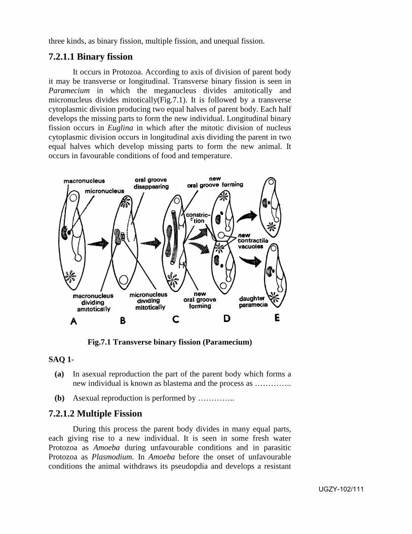
three kinds, as binary fission, multiple fission, and unequal fission.
7.2.1.1 Binary fission It occurs in Protozoa. According to axis of division of parent body
it may be transverse or longitudinal. Transverse binary fission is seen in Paramecium in which the meganucleus divides amitotically and micronucleus divides mitotically(Fig.7.1). It is followed by a transverse cytoplasmic division producing two equal halves of parent body. Each half develops the missing parts to form the new individual. Longitudinal binary fission occurs in Euglina in which after the mitotic division of nucleus cytoplasmic division occurs in longitudinal axis dividing the parent in two equal halves which develop missing parts to form the new animal. It occurs in favourable conditions of food and temperature.
Fig.7.1 Transverse binary fission (Paramecium)
SAQ 1-
(a) In asexual reproduction the part of the parent body which forms a new individual is known as blastema and the process as …………..
(b) Asexual reproduction is performed by …………..
7.2.1.2 Multiple Fission During this process the parent body divides in many equal parts,
each giving rise to a new individual. It is seen in some fresh water Protozoa as Amoeba during unfavourable conditions and in parasitic Protozoa as Plasmodium. In Amoeba before the onset of unfavourable conditions the animal withdraws its pseudopdia and develops a resistant
UGZY-102/111

cyst wall to protect itself. On the return of favourable conditions the nucleus divides mitotically many times followed by cytoplasmic division to form many uninucleate bodies. On the bursting of the cyst each such body forms a new Amoeba. In Plasmodium it occurs as schizogony in the liver and RBC of human beings and as sporogony in the stomach wall of female Anopheles(Fig.7.2).
Fig.7.2 Life cycle of plasmodium (vivax)
7.2.1.3 Unequal Fission During this process the parent divides in 2 or more unequal parts which develop the missing structures to form the adult. It is seen in the protozoan Vorticella in which the meganucleus divides amitotically and the micronucleus divides mitotically. It is followed by longitudinal fission. The larger half retains the peristome and stalk while the smaller part develops a ring of aboral cilia, gets detatched and swims freely, lateron settling at aboral end it develops the missing parts to form the adult.
7.2.2 Budding It is seen in Porifera and Coelenterata. During this process a bud appears as a small protruberance of the body by the accumulation of archaeocytes in sponges and somatic and interstitial cells in Coelenterata. In sponges external buds arise from horizontal branches, they grow and develop ostia UGZY-102/112
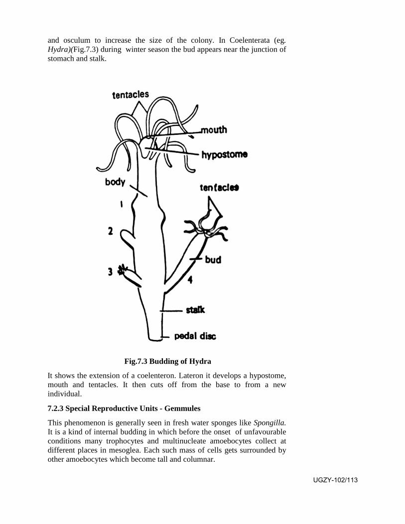
and osculum to increase the size of the colony. In Coelenterata (eg. Hydra)(Fig.7.3) during winter season the bud appears near the junction of stomach and stalk.
Fig.7.3 Budding of Hydra
It shows the extension of a coelenteron. Lateron it develops a hypostome, mouth and tentacles. It then cuts off from the base to from a new individual.
7.2.3 Special Reproductive Units - Gemmules
This phenomenon is generally seen in fresh water sponges like Spongilla. It is a kind of internal budding in which before the onset of unfavourable conditions many trophocytes and multinucleate amoebocytes collect at different places in mesoglea. Each such mass of cells gets surrounded by other amoebocytes which become tall and columnar.
UGZY-102/113
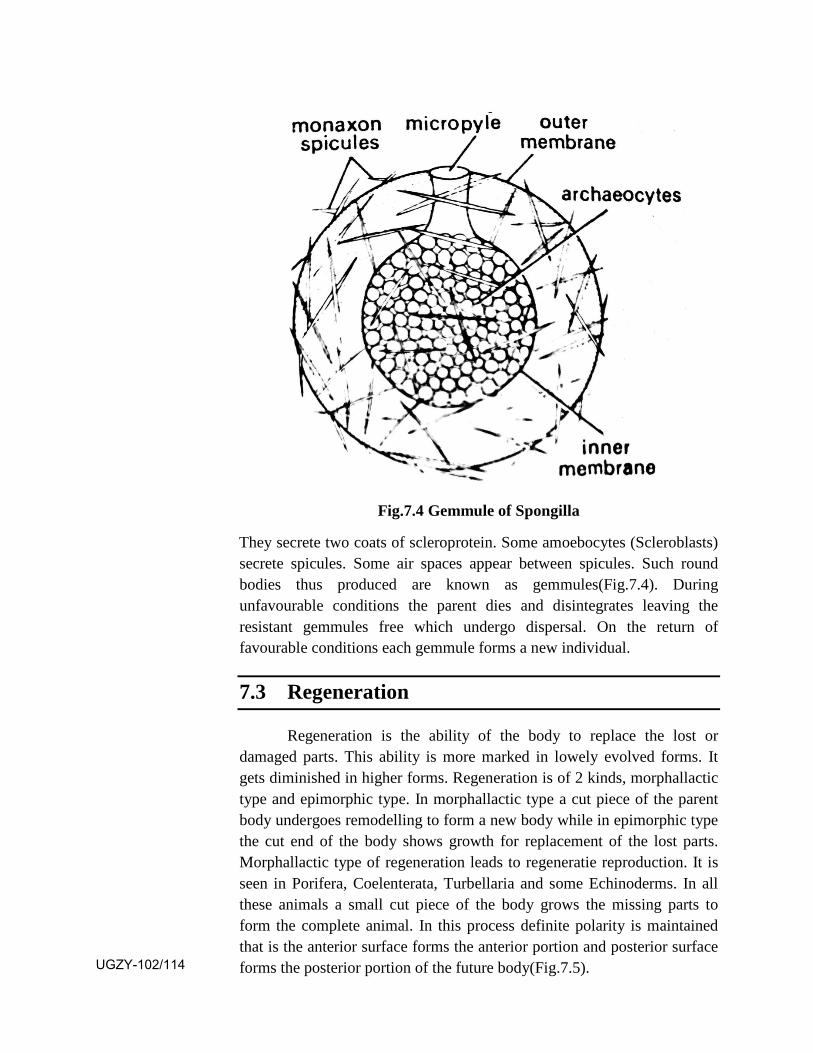
Fig.7.4 Gemmule of Spongilla
They secrete two coats of scleroprotein. Some amoebocytes (Scleroblasts) secrete spicules. Some air spaces appear between spicules. Such round bodies thus produced are known as gemmules(Fig.7.4). During unfavourable conditions the parent dies and disintegrates leaving the resistant gemmules free which undergo dispersal. On the return of favourable conditions each gemmule forms a new individual.
7.3 Regeneration
Regeneration is the ability of the body to replace the lost or damaged parts. This ability is more marked in lowely evolved forms. It gets diminished in higher forms. Regeneration is of 2 kinds, morphallactic type and epimorphic type. In morphallactic type a cut piece of the parent body undergoes remodelling to form a new body while in epimorphic type the cut end of the body shows growth for replacement of the lost parts. Morphallactic type of regeneration leads to regeneratie reproduction. It is seen in Porifera, Coelenterata, Turbellaria and some Echinoderms. In all these animals a small cut piece of the body grows the missing parts to form the complete animal. In this process definite polarity is maintained that is the anterior surface forms the anterior portion and posterior surface forms the posterior portion of the future body(Fig.7.5). UGZY-102/114
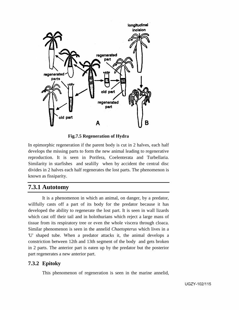
Fig.7.5 Regeneration of Hydra
In epimorphic regeneration if the parent body is cut in 2 halves, each half develops the missing parts to form the new animal leading to regenerative reproduction. It is seen in Porifera, Coelenterata and Turbellaria. Similarity in starfishes and sealilly when by accident the central disc divides in 2 halves each half regenerates the lost parts. The phenomenon is known as fissiparity.
7.3.1 Autotomy
It is a phenomenon in which an animal, on danger, by a predator, willfully casts off a part of its body for the predator because it has developed the ability to regenerate the lost part. It is seen in wall lizards which cast off their tail and in holothurians which reject a large mass of tissue from its respiratory tree or even the whole viscera through cloaca. Similar phenomenon is seen in the annelid Chaetopterus which lives in a 'U' shaped tube. When a predator attacks it, the animal develops a constriction between 12th and 13th segment of the body and gets broken in 2 parts. The anterior part is eaten up by the predator but the posterior part regenerates a new anterior part.
7.3.2 Epitoky
This phenomenon of regeneration is seen in the marine annelid,
UGZY-102/115

Nereis irrorata. The animal during breeding season converts its posterior region of the body in sexual phase. It is known as heteronereis(Fig.7.6).
Fig.7.6 Heteronereis
The segments of this portion develop broad fan like parapodia and setae externally and male or female gonads internally. The anterior portion of heteronereis is known as atoke and posterior sexual portion as epitoke. The male and female expitoke portions get detached from heteronereis and form a swarm in the sea. They burst and release sperms and ova which undergo fertilization. The atoke portion regenerates the posterior part and repeats the process in following breeding season.
7.3.3 Polarity
During morphallactic regeneration in Coelenterata (Hydra) and planaria a cut piece of the body regenerates the lost parts. During this process definite polarity is maintained in which the anterior cut surface forms the anterior region and posterior cut surface forms posterior region. Child has given gradient theory to explain this phenomenon. According to this theory the physiological activity in the body of such an animal shows a gradient from one pole to the other. Such activities may be protein synthesis, rate of respiration and sensitivity to poison. The pole with higher gradient shows greater morphogenesis hence polarity is maintained during regeneration.
UGZY-102/116

7.4 Prevalence and significance of Asexual Reproduction
Asexual reproduction is more prevalent in lower phyla. In such animals it is associated with the regenerative ability which leads to regenerative reproduction. Asexual reproduction is surer method in comparison to sexual reproduction because in sexual reproduction the availability of male and female parents is essential and fertilization of ovum is a chance factor. Asexual reproduction mainly by fission and budding leads to very fast growth of the population. Gemmule formation provides resistance against unfavourable conditions and is helpful in dispersal of the species.
7.5 Sexual Reproduction
Sexual reproduction is a very common phenomenon in both non-chordates and chordates. The species showing this process is represented by male and female individuals. Male individuals possess testes and females possess ovary for the production of sperms and ova respectively. During the process of gamete (sperms and ova) formation the germ mother cell undergoes reduction division hence the gametes show haploid number of chromosomes. During fertilization the sperm fuses with the ovum leading to the formation of diploid zygote. Thus the zygote carries the characters of both the parents leading to infinite variability which is helpful in the better survival of the progeny.
SAQ 2-
(a) The male and female sexes are separate called ………..
(b) The two reproductive systems are present in the same individual called ……………
7.5.1 Patterns of Sexual Reproduction
1. Conjugation is seen in ciliate protozoan Paramecium where thetwo individuals undergo temporary union. Their vegetative meganucleus disintergrates. The reproductive nuclei (micronuclei) of both undergo repeated nuclear divisions including one reduction division, thus each individual shows one male and the other female nucleus. The male nucleus of one fuses with the female nucleus of the other to form synkaryon in both individuals. The synkaryon further divides to from a new meganucleus and a micronucleus. The conjugant individuals now separate
UGZY-102/117
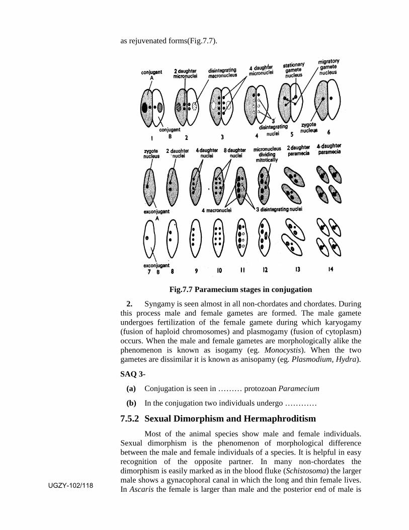
as rejuvenated forms(Fig.7.7).
Fig.7.7 Paramecium stages in conjugation
2. Syngamy is seen almost in all non-chordates and chordates. Duringthis process male and female gametes are formed. The male gamete undergoes fertilization of the female gamete during which karyogamy (fusion of haploid chromosomes) and plasmogamy (fusion of cytoplasm) occurs. When the male and female gametes are morphologically alike the phenomenon is known as isogamy (eg. Monocystis). When the two gametes are dissimilar it is known as anisopamy (eg. Plasmodium, Hydra).
SAQ 3-
(a) Conjugation is seen in ……… protozoan Paramecium
(b) In the conjugation two individuals undergo …………
7.5.2 Sexual Dimorphism and Hermaphroditism Most of the animal species show male and female individuals.
Sexual dimorphism is the phenomenon of morphological difference between the male and female individuals of a species. It is helpful in easy recognition of the opposite partner. In many non-chordates the dimorphism is easily marked as in the blood fluke (Schistosoma) the larger male shows a gynacophoral canal in which the long and thin female lives. In Ascaris the female is larger than male and the posterior end of male is UGZY-102/118
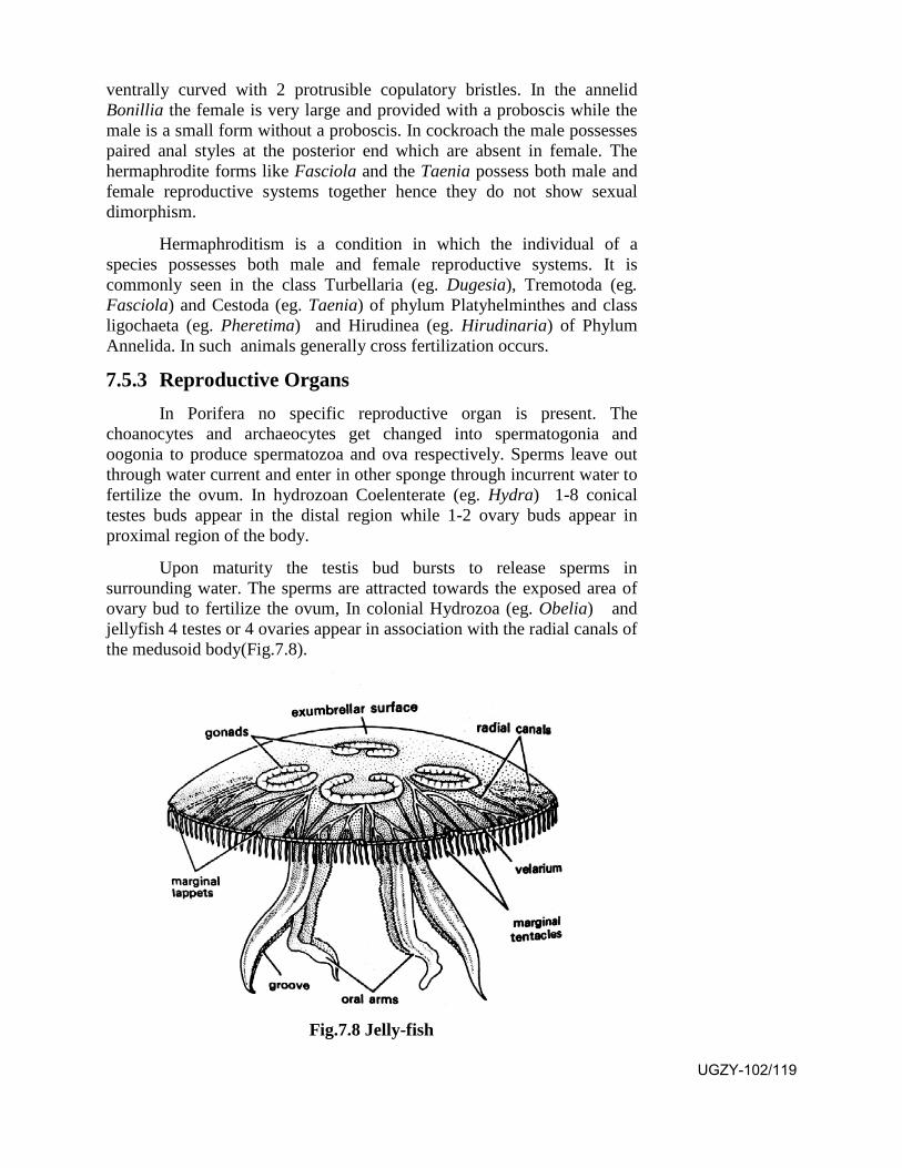
ventrally curved with 2 protrusible copulatory bristles. In the annelid Bonillia the female is very large and provided with a proboscis while the male is a small form without a proboscis. In cockroach the male possesses paired anal styles at the posterior end which are absent in female. The hermaphrodite forms like Fasciola and the Taenia possess both male and female reproductive systems together hence they do not show sexual dimorphism.
Hermaphroditism is a condition in which the individual of a species possesses both male and female reproductive systems. It is commonly seen in the class Turbellaria (eg. Dugesia), Tremotoda (eg. Fasciola) and Cestoda (eg. Taenia) of phylum Platyhelminthes and class ligochaeta (eg. Pheretima) and Hirudinea (eg. Hirudinaria) of Phylum Annelida. In such animals generally cross fertilization occurs.
7.5.3 Reproductive Organs In Porifera no specific reproductive organ is present. The
choanocytes and archaeocytes get changed into spermatogonia and oogonia to produce spermatozoa and ova respectively. Sperms leave out through water current and enter in other sponge through incurrent water to fertilize the ovum. In hydrozoan Coelenterate (eg. Hydra) 1-8 conical testes buds appear in the distal region while 1-2 ovary buds appear in proximal region of the body.
Upon maturity the testis bud bursts to release sperms in surrounding water. The sperms are attracted towards the exposed area of ovary bud to fertilize the ovum, In colonial Hydrozoa (eg. Obelia) and jellyfish 4 testes or 4 ovaries appear in association with the radial canals of the medusoid body(Fig.7.8).
Fig.7.8 Jelly-fish
UGZY-102/119
Upon maturity they release sperms and ova. Fertilization is external. In higher non-chordates the reproductive system is represented by gonads (testes or ovary), gonoducts, copulatory organs and associated glands. Gonads possess germ mother cells (spermatogonia or oogonia) which undergo reduction division to produce the gametes (sperms or ova). Gonoducts perform the transport of the sperms or ova. Male gonoducts are vasa efferentia and vas deferens while the female gonoducts are oviducts. Copulatory organs of male and female individuals help in the transport of sperms upto the ovum in female body.
Testes number may be single (eg. Ascaris, Pila) or two (Fasciola, cockroach, prawn) or two pairs (earthworm) or 5 pairs (starfish) or many (tapeworms). Their shape is variable. It may be oval, round, branched, elongated, thread like or horse shoe like. In leeches the testes are replaced by testes sacs.
Ovaries number one (Fasicola, Taenia) or two (Ascaris, earthworm, prawn, cockroach, Unio) or 5 pairs (starfish). Their shape varies. They may be thread like, oval, elongated, digitate or bilobed.
7.5.4 Mating and fertilization Mating is the temporary contact of the male and female
individuals during which the male transfers its sperms or spermatophores (bundles of sperms) in the female genital tract. In many cases it occurs during a specific season. It is followed by internal fertilization of the ovum. Generally mating does not occur in cases where fertilization occurs in water.
Fertilization is the process in which the sperm enters the ovum to undergo karyogamy and plasmogamy to form the diploid zygote. It has been well studied in the echinoderm sea urchin. The ovum remains surrouned by a gelationous coat containing a glycoprotein known as fertilizin. Sperm contains an acidic protein antifertilizin. Antifertilizin combines with fertilizin and sperm gets trapped in it. Lysins present in acrosome of the sperm dissolve a path for entrance of sperm through egg membrane. The sperm nueleus now moves towards egg nucleus. The meeting of the two haploid nuclei along with fusion of cytoplasm of both completes fertilization.
7.5.5. Ovipary, Vivipary and Ovovivipary Ovipary is the phenomenon in which the zygote formed within the
female reproductive system is laid out as egg in water, soil or elsewhere. Thus the development of the zygote occurs outside the mother's body. It is seen in the fresh water prawn and starfish.
Vivipary is the phenomenon in which the zygote is retained in a specific part of mother's reproductive system where it undergoes further development. In such cases a fully formed baby is born. It is seen in some scorpions in which small eggs are poor in yolk. Such mothers give birth to 6-90 young ones at a time. Ovovivipary is the phenomenon in which the UGZY-102/120
zygote is retained in some specific part of female reproductive system without any direct connection with maternal tissue for nourishment. The egg develops and hatches within mother's body. Thus the mother gives birth to young ones. It is seen in the scorpions producing large telolecithal eggs, rich in yolk.
7.6 Parthenogenesis
It is the phenomenon in which an unfertilized ovum develops into the adult. The daughter individual thus formed are known as parthenote. When it occurs in nature it is called as natural parthenogenesis. In contrast when it is experimentally induced it is called as artificial parthenogenesis. Natural parthenogenesis may be complete or incomplete Species showing complete parthenogenesis do not undergo sexual reproduction. They breed exclusively by parthenogenesis. In such species ony the female individuals exist, males are unknown. Complete parthenogenesis is of two kinds, Arrhenotoky (haploid parthenogenesis) and Thelytoky (diploid parthenogenesis). In arrhenotoky the haploid males develop from unfertilized haploid eggs. It is seen in the formation of drones (males) of honeybee. In thelytoky the young individuals develop from unfertilized diploid eggs. It is seen in weevils and long horned grasshoppers. Parthenogenesis is seen in some Rotifera and the insect aphids. During artificial parthenogenesis a ripe haploid ovum, when artificially stimulated by shaking, temperature shock, ultra violet radiation and treatment of chlorides of sodium, calcium and magnesium, dilute organic acids and pricking, starts further development and forms the adult. Such experiments have been successfully conducted on ripe ova of sea urchin and frogs.
7.7 Metagenesis
The life cycle of a colonial hydroid Coelenterate (eg Obelia) shows a diploid polypoid form which buds off diploid medusoid forms from its blastostyles. The medusoid form develops gonads which produce haploid sperms and ova. As the result of fertilization the zygote forms the polypoid form. Thus it appears that the polypoid and medusoid forms show an alternation of generation. It is not a true alternation of generation as the latter exists between a diploid and haploid generations (eg. fern). Such a phenomenon as seen in Obelia is called as metagenesis.
7.8 Summary
• In asexual reproduction the part of the parent body which forms anew individual is known as blastema and the process asblastogenesis.
UGZY-102/121
• Asexual reproduction is performed by binary fission, occurringduring favourable conditions of food and temperature, in which thebody of the parent divides in two equal or unequal parts, each partforming a new individual. The other method is multiple fission inwhich the parent body divides in many parts, each forming a newbody. It occurs during unfavourable conditions, preceeded by cystformation, thus it is associated with protection of the body againstunfavourable conditions.
• The asexual reproduction may occure by the formation of externaland internal budding (gemmule formation). Each bud so formedgives rise to a new individual. A gemmule provides protectionagainst unfavourable conditions.
• In normal life due to some reason or the other a part of the bodymay get lost or damaged. Nature has managed this odd situation byregeneration of lost part. It is inversely proportional to the extentof evolution.
• Sexual reproduction occurs by the formation of a diploid zygote asthe result of fertilization of haploid ovum by the haploid sperm.The zygote develops into the new individual having thecombination of characters of both the parents, which is helpful insurvival. Generally the male and female sexes are separate(unisexualism) but sometimes the two reproductive systems arepresent in the same individual (hermaphroditism). It ensures surityof production and availability of male and female gametes andfertilization.
• After fertilization the ovum forms the zygote which may developas an egg in the body of female. Such an egg is laid in water orland. This phenomenon is called as ovipary. Such egg is alwaysexposed to the attack of predators. To over come this situation insome species the zygote is retained in the female body in its uteruswhere it completes the developmental process leading to the birthof an infant. This phenomenon is known as vivipary.
• Sometimes when the sexual reproduction fails, the unfertilizedovum forms a new individual. It is known as parthenogenesis.
7.9 Terminal Questions
Q.1 Describe the various methods of fission.
Q.2 Give an account of gemmule formation. UGZY-102/122

Q.3 Write a short essay on regeneration.
Q.4 Describe the basic process of sexual reproduction. Why it is superior to asexual reproduction?
Q.5 Write short notes on -
(i) Blastema (ii) Epitoky
(iii) Polarity (iv) Vivipary
(v) Parthenogeneis
ANSWERS
SAQ 1 - (a) blastogenesis, (b) binary fission
SAQ 2 - (a) unisexualism, (b) hermaphroditism
SAQ 3 - (a) ciliate, (b) temporary union
UGZY-102/123

UGZY-102/124
UGZY-102 Diversity of Animal Life
Block
3 Adaption and behavioral Patterns Unit 8 129-138 Adaptive Radition Unit 9 139-148 Behavioural patterns
Unit 10 149-164
Harmful and beneficial Non-Chordates
Uttar Pradesh Rajarshi Tandon Open University
UGZY-102/125

Course Design Committee Chairman Prof. Ashutosh Gupta
Director, School of Science UPRTOU, Prayagraj.
Prof. Paramhand Pathak Member Department of Zoology Deendayal Upadhyay University, Gorakhpur.
Prof. Ajay Singh Member Department of Zoology Deendayal Upadhyay University, Gorakhpur.
Prof. Rajendra Singh Special Invitees Department of Zoology Deendayal Upadhyay University, Gorakhpur.
Member/Secretary Dr. Deepa Chaubey Acadmic Consultant, School of Science UPRTOU, Prayagraj.
Course Preparation Committee Dr. V.C. Srivastava Author Ex. Associate Prof. C.M.P. Degree College, Prayagraj
Dr. A.K. Srivastava EditorEx. Head Of Zoology B.B.C, Jhansi.
Coordinator Dr. Deepa Chaubey Acadmic Consultant, School of Science UPRTOU, Prayagraj.
© UPRTOU, Prayagraj. 2020 ISBN : All Rights are reserved. No part of this work may be reproduced in any form, by mimeograph or any other means, without permission in writing from the Uttar Pradesh Rajarshi Tondon Open University, Prayagraj. Printed and Published by Dr. Arun Kumar Gupta Registrar, Uttar Pradesh Rajarshi Tandon Open University, 2020. Printed By: Chandrakala Universal Pvt. Ltd. 42/7 Jawahar Lal Neharu Road, Prayagraj. UGZY-102/126
Course Introduction
Block-3
Adaption and behavioral Pattern
In the previous blocks your have studied about the comparative forms and functions of nonchordates. You have noted how form a simple type, complex and highly developed systems of respiration and circulation have evolved. In the previous block you have also noted that nonchordates have evolved diverse methods and reproduction ranging from simple splitting into two to very complicated methods of sexual reproduction and parthenogenesis etc. This block deals with the Adaptation and behavioral patterns of invertebrates. Behavior of invertebrates is diverse and complex. We have discussed the basic concepts and explained the different approaches to study the animal behavior.
(Each unit in the course contains the learning objective, text summary and self assessment question).
Unit-8 :- In this unit, the concept regarding the appearance of colony formation in coelenterate which has lead to the formation of a true society in some insects has been given. More so over the phenomenon of adaptive radiation has been discussed. Apart from that the flight adaptations and mechanism of flight along with the ability of migration in insects have been covered.
Unit-9 :- This unit has presented the advantages of social behavior and the different kinds of honey bees. It incorporates informations regarding production of honey, silk, lac, bees wax pearl, spongin, dyes and pigments.
Unit-10 :- This unit describes the parasitic forms of Platyhelminthes and Nematoda. It elaborates the significance of Arthropoda in agriculture, soil fertility, pollination and as scavenger. It also deals with the management of pests.
After studying this block you will be able to:
• list the adaptive radiations in Annelids, Arthropoda and Mollusca.
• explain the behavioral patterns and their adaptive valuesinvertebrates
• explain about the flying ability and migratory behavior of insects.
• become familiar with the variety of ways in which nonchordatesbenefit humans directly or indirectly.
UGZY-102/127

UGZY-102/128
UNIT-8
Adaptive Radition
Structure 8.1 Introduction and Objectives
8.2 Colonial forms among Protozoa and Metazoa
8.3 Adaptive Radition
8.3.1 In Annelida
8.3.2 In Arthropoda
8.3.3 In Mollusca
8.4 Flight in Insects
8.5 Migration in Insects
8.6 Summary
8.7 Terminal Questions
8.8 Answers
8.1 Introduction
A protozoan body is made up of a single cell which performs all life activities. In the process of evolution some of them start living together. In some cases the number of such cells is very few while in others it is large. In such circumstances different kinds of cells have gone specialized to perform a specific function in the interest of the colony. Thus they show a division of labour. A good example of such a colony is that of Volvox.
Similar phenomenon is seen in metazoa also. Members of some species of Coelenterata and Insecta show the tendency to live in a colony showing division of labour. Such Coelenterata show different kinds of individuals known as zooids. Such Coelenterate colonies may be dimorphic showing gastrozooids for feeding and blastostyles producing medusae for sexual reproduction. Some of the colonies have gone trimorphic. Apart from gastrozooids and blastostyles they possess dactylozooids which perform defence of the colony. Some coelenterates
UGZY-102/129
have gone polymorphic in which the zooids are of seven kinds performing different functions. As the result of division of labour a zooid gets specialized for a specific function. It thus saves the energy of different zooids in performing varied functions. Insect colonies as those of honey bees, ants and termites have gone very much specialized forming a society.
Adaptive radiation is an universal phenomenon occurring in the process of evolution. An ancestral group shows a tendency to occupy different habitats. It is helpful in the survival of the progency. The descendants gradually get more and more adapted to the varied conditions of a specific habitat by changing their morphology and behaviour accordingly, thus leading to formation of new species. Such a phenomenon is markedly seen in Annelida, Arthropoda and Mollusca which you will study in this unit.
Insects represent the most successful group on earth. They have occupied maximum amount of living protoplasm and are represented by maximum number of species. They are the first group which has shown the ability of flying in air. As the result of this ability they cover long distances for their survival. Such a behaviour is known as migration. In this unit you will study in details about the flying ability and migration in insects.
Objectives: After studying this unit you should be able to.
(i) know a lot about the Protozoan and Coelenterate colonies.
(ii) know a lot about adaptive radiation in Annelida, Arthropoda and Mollusca.
(iii) know about the flying ability of insects.
(iv) know about the migratory behaviour of insects.
8.2 Colonial forms among Protozoa and Metazoa
In general protozoa are represented by single celled individuals which perform all vital life activities. In the process of evolution some of them show an organization in which a few to many cells live together. In such a colony the different cells show functional variability but each cell functions in the interest of the colony as a whole. Such a condition is seen in some genera of order Phytomonadina and Proterospongia of order Choanoflagellida. In Phytomonadina the genus Gonium shows a colony of 4-16 cells, genus Pandorina shows 16 cells, genus Eudorina shows 32 cells, genus Pleodorina shows 128 cells and Volvox showing numerous UGZY-102/130
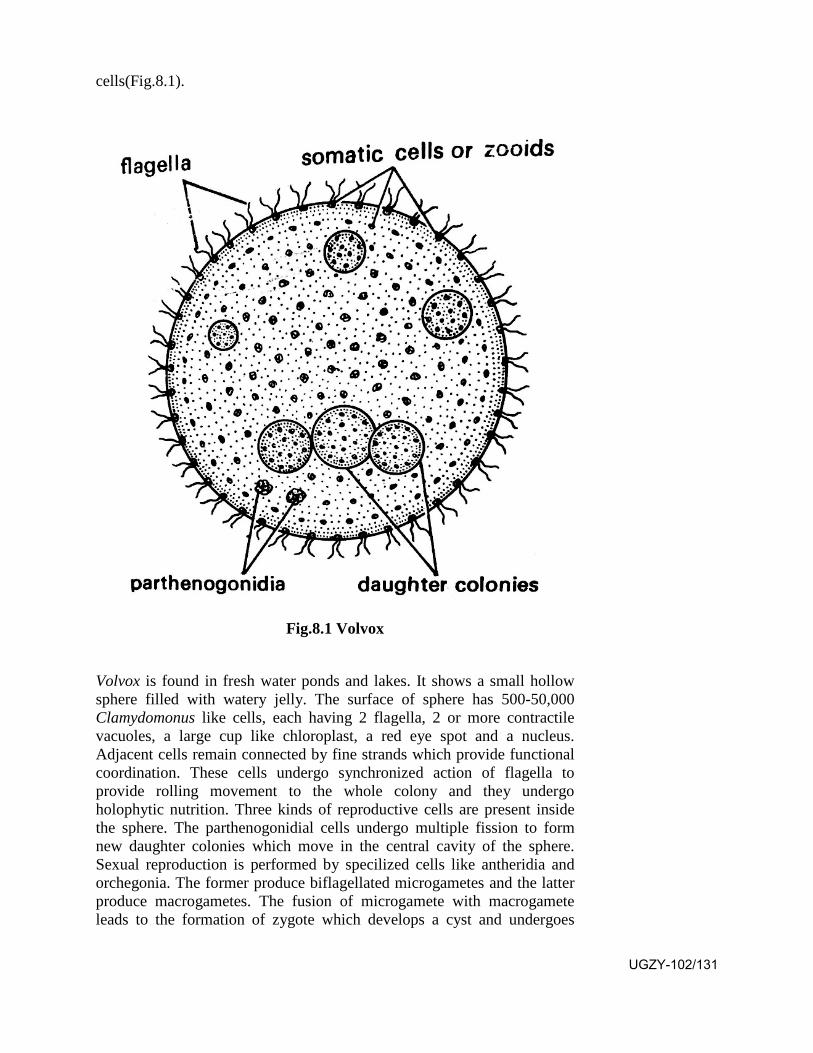
cells(Fig.8.1).
Fig.8.1 Volvox
Volvox is found in fresh water ponds and lakes. It shows a small hollow sphere filled with watery jelly. The surface of sphere has 500-50,000 Clamydomonus like cells, each having 2 flagella, 2 or more contractile vacuoles, a large cup like chloroplast, a red eye spot and a nucleus. Adjacent cells remain connected by fine strands which provide functional coordination. These cells undergo synchronized action of flagella to provide rolling movement to the whole colony and they undergo holophytic nutrition. Three kinds of reproductive cells are present inside the sphere. The parthenogonidial cells undergo multiple fission to form new daughter colonies which move in the central cavity of the sphere. Sexual reproduction is performed by specilized cells like antheridia and orchegonia. The former produce biflagellated microgametes and the latter produce macrogametes. The fusion of microgamete with macrogamete leads to the formation of zygote which develops a cyst and undergoes
UGZY-102/131
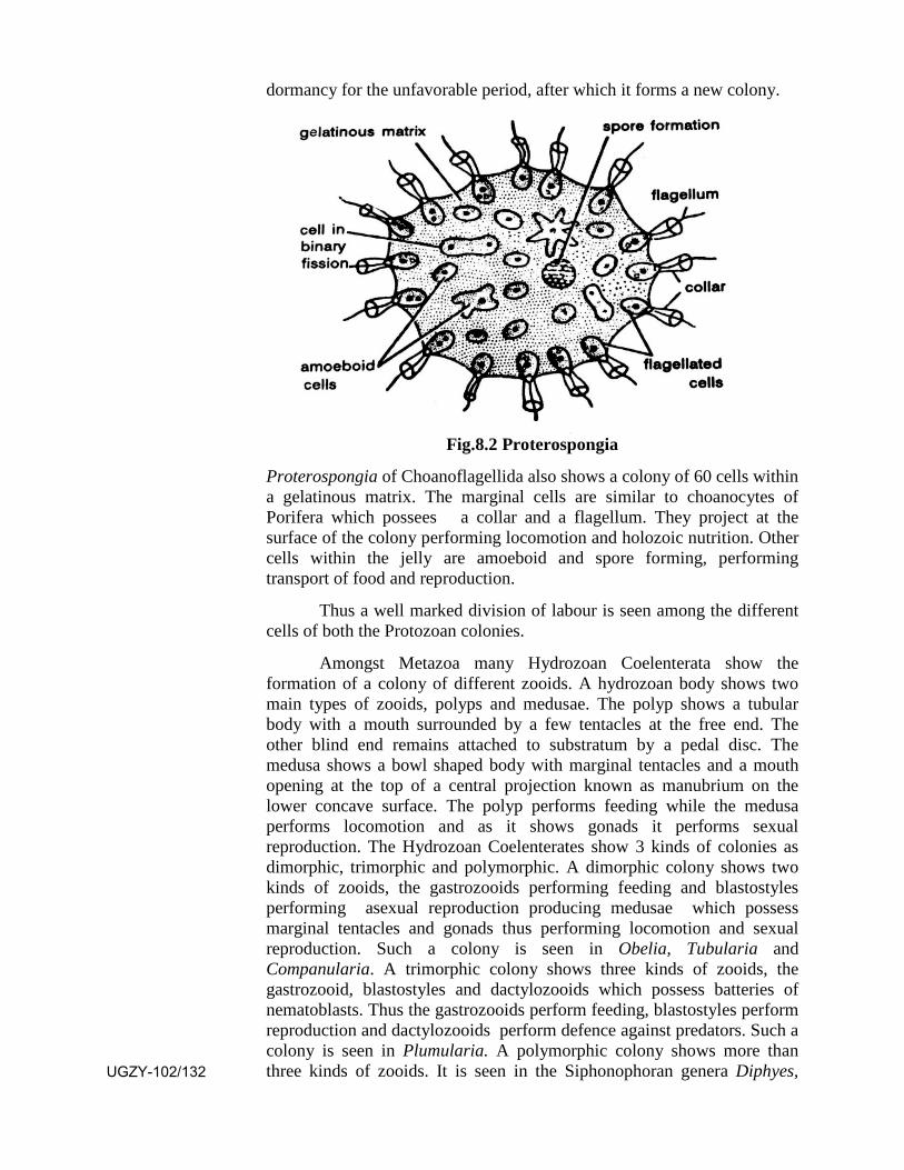
dormancy for the unfavorable period, after which it forms a new colony.
Fig.8.2 Proterospongia
Proterospongia of Choanoflagellida also shows a colony of 60 cells within a gelatinous matrix. The marginal cells are similar to choanocytes of Porifera which possees a collar and a flagellum. They project at the surface of the colony performing locomotion and holozoic nutrition. Other cells within the jelly are amoeboid and spore forming, performing transport of food and reproduction.
Thus a well marked division of labour is seen among the different cells of both the Protozoan colonies.
Amongst Metazoa many Hydrozoan Coelenterata show the formation of a colony of different zooids. A hydrozoan body shows two main types of zooids, polyps and medusae. The polyp shows a tubular body with a mouth surrounded by a few tentacles at the free end. The other blind end remains attached to substratum by a pedal disc. The medusa shows a bowl shaped body with marginal tentacles and a mouth opening at the top of a central projection known as manubrium on the lower concave surface. The polyp performs feeding while the medusa performs locomotion and as it shows gonads it performs sexual reproduction. The Hydrozoan Coelenterates show 3 kinds of colonies as dimorphic, trimorphic and polymorphic. A dimorphic colony shows two kinds of zooids, the gastrozooids performing feeding and blastostyles performing asexual reproduction producing medusae which possess marginal tentacles and gonads thus performing locomotion and sexual reproduction. Such a colony is seen in Obelia, Tubularia and Companularia. A trimorphic colony shows three kinds of zooids, the gastrozooid, blastostyles and dactylozooids which possess batteries of nematoblasts. Thus the gastrozooids perform feeding, blastostyles perform reproduction and dactylozooids perform defence against predators. Such a colony is seen in Plumularia. A polymorphic colony shows more than three kinds of zooids. It is seen in the Siphonophoran genera Diphyes, UGZY-102/132

Halistemmia, Stepholia and Physalia(Fig.8.3) and Chondrophoran genera, Velella and Porpita.
Fig.8.3 Physalia colony
Such colonies show seven kinds of zooids which are (i) Gastrozooid with a mouth and a long tentacle. It performs feeding (ii) Dactylozooid without mouth but with a long tentacle having batteries of nematoblasts performing defence (iii) Gonozooids which produce medusae performing locomotion for dispersal and sexual reproduction (iv) Nectophore which appear as a muscular bell without manubrium and tentacles. It performs swimming (v) Pneumatophore which is bladder like filled with gas. It performs the floating activity of the colony (vi) Phyllozooid which is leaf like provided with batteries of nematoblast for defence and (vii) Gonophore provided with gonads, the testes or ovary for sexual reproduction. The Chondrophoran colony of Velella and Porpita are highly organized. They show a single gastrozooid with mouth for feeding. Around it are present gonozooids in concentric rows for reproduction. Around the gonozooids are present a few rows of dactylozooids for defence. Thus the colony looks like a single individual.
In still higher Metazoa as represented by Insects an advancement over the colony formation is seen in the establishment of a society which
UGZY-102/133
shows a perfection in honeybees, termites and ants.
SAQ 1-
…………. are the first group which has shown the ability to fly in air.
8.3 Adaptive Radiation
Adaptive radiation is a process in which organisms diversify rapidly from an ancestral group into a multitude of new forms particularly when a change in environment makes new resources available, opening new environmental niches. This process leads to speciation and phenotypic adaptations.
8.3.1 In Annelida It is believed that the ancestral annelid was marine, living in bottom sand or mud of shallow coastal water. Out of the present annelid groups Polychaeta are believed to be most primitive. In the process of evolution they have shown marked adaptive radiation as some of them live in the muddy bottom of sea (eg. Aphrodite), some live a intertidal zone under stones (eg. Polynoe), some inhabit cracks and crevices of coral rocks (eg. Eunice). A number of polychaetes have gone tubicolous (eg. Chaetopterus, Arenicola, Amphitrite) while some others live in the pelagic zone (eg. Tomopteris). Living in the different habitats these worms have adapted themselves perfectly feeding on the food available in the specific zone. In the process of evolution the Polychaeta has given rise to Oligochaeta which are represented by earthworms. Different species of Oligochaeta have successfully occupied varied habitats as some of them live in fresh water (eg. Chaetogaster) while others live in a tube in the muddy bottom (eg. Tubifex) while some burrow in moist soil (eg. Lumbricus)
The Oligochaeta have given rise to Hirudinea represented by leeches. They also show successful living in different habitats as many of them have parasitized varied kinds of fresh water and marine vertebrates (eg. Pontobdella being parasitic on sharks, rays and skates, Ozobranchus on turtles and crocodiles and Placobdella on molluscs). Still some leaches have gone swamp dwellers (eg. Haemedipsa)
8.3.2 In Arthropoda At present phylum Arthropoda is largest and most successful group
of animals. They show maximum number of species and maximum amount of living protoplasm. They have occupied varieties of habitats feeding on varied kinds of food. They inhabit fresh water lakes, streams, ponds, salt water, sulpher springs, hot springs, petroleum pools, sea beaches, surface of sea to the depth of 5 miles inside the ocean. They also live in desert, underground, as parasites in and on the body of other animals and as plant pests. The insects have invaded the air. They can fly, swim, hop and crawl. Some of them have gone social. Living in different UGZY-102/134

habitats they have successfully utilized the available food. Thus it can be concluded that they have shown maximum adaptive radiation occupying different niches on the earth showing their evolutionary success.
8.3.3 In Mollusca Mollusca are shelled invertebrates. They have occupied all
possible aquatic and terrestrial habitats except aerial. Most of them are marine, living along sea shores in intertidal zone (eg. Mytilus). Some of them are pelagic (eg. Cliopsis) while some are found in the deep sea (eg. Nautilus). Many snails and bivalves inhabit fresh water lakes, rivers and ponds while many live in brakish water. Some snails are terristrial. One species of snail is found in desert of California. They are mostly free living, creeping slowly on wet land (eg. Limax), others cling to rocks (eg. Chiton). Some snails and bivalves burrow, others float. The Cephalopods perform active swimming in the sea. Most of the gastropods with radula feed on vegetation while the bivalvia feed mainly on micro organisms suspended in water by ciliary action. The Cephalopods are predaceous. It can thus be concluded that the Mollusca have also shown very successful adaptive radiation.
8.4 Flight in Insects
Insects are very successful animals. They possess maximum amount of living protoplasm and are represented by maximum number of species. The Pterygota group of insects show the ability of flight. They are the earliest fliers on earth. They show following important adaptations for flight.
(1) Presence of chitin as exoskeleton: Chitin is a tough covering of the body but is very light hence facilitates the insect for fligh(Fig.8.4).
Fig.8.4 Cockroach wings
UGZY-102/135
(2) Presence of wings: Wings appear as fold of skin from the region between tergum and pleuron of the thorax. They appear in two pairs, first pair arising from mesothorax and the second from metathorax dorsally. In Orthopera (eg. cockroach) forewings are leathery and hind wings are membranous showing ability to fly. In Diptera (eg. housefly) forewings are large and membranous showing the ability to fly while the hindwings are reduced as halters. In Hymenoptera (eg. honey bee) both wings are membranous showing ability to fly. A typical wing is longer than wide showing straight anterior margin and round posterior margin. Wing membrane is reinforced by veins which provide nerve supply and blood supply to the wing. During flight they counterbalance the wind pressure and prevent its collapse. Surface of wing may be hairy, scaly or provided with pits.
(3) Presence of thoracic muscles: These muscles are basically attached to legs. They stretch from base of leg to upper armoured plate of thorax. Apart from them longitudinal muscles passing from one thoracic segment to the next also play important role.
(4) Presence of air sacs: In many insects the trachea get swollen forming air sacs. They vary in number and position in different species. In honey bee two large sacs occupy a large area of abdominal cavity. In many beetles hundreds of small sacs are present. These sacs help to lighten the body weight for easier flight.
Wing action during flight:
Wing moves rapidly, upwards, forwards, downwards and backwards in a definite rhythmic sequence. Downward stroke of wing is accompanied by simultaneous forward movement so that front margin is deflected and hind margin turned upwards. Upward stroke is associated with backward movement and deflection of hind margin of wing. The tip of wing describes a series of loops. The wing beats provide lifting force and propelling power by creating a low pressure above the body in front and high pressure below and behind the body. In fast flying insects front wings create a turbulance behind them. Wing strokes are brought about indirectly by muscles of legs. In Diptera the frequency of wing beat is about 1000 times per second.
Speed of insect flight is variable, some fly in leasurly fashion, others with great speed. Hawk moth is the fastest flier showing a speed of fifteen meters per second. During flight they can arrest their motion, remain stationary, reverse their direction and can dart swiftly sideways. They can feed, mate or deposit their eggs during flight. Thus it can be concluded that they are marvellous fliers.
8.5 Insect Migration
Migration is mass movement of the members of an insect species from one place to the other. Such movements may be performed in search of hibernating places before the onset of unfavouable conditions or in UGZY-102/136

search of new and better feeding ground for the survival of the species. Certain factors compel an insect population to move for a considerable distance for example when a pond dries up the aquatic insects living in it move to find a new suitable location or when the hive of a bee colony becomes over crowded the existing queen along with some members moves away and establishes a new hive or a change in wind direction forces a swarm of Chironomidea to find a more sheltered and protected spot. Insects like army worms, clinch bugs and grasshoppers undergo migration as a seasonal affair in which they migrate to new fields when the existing crop is cut and they face food scarcity. Some insect species show a definite and regular annual migration in which they perform pendulum swing movements as seen in fish and bird migration. A common example of such an insect is the milk weed butterfly, Danaus menippe. These insects gather at one place in thousands during fall and travel southwards where they pass winter in a suitable location. In spring the whole population returns to north. During fall period, under the influence of unusual storms the cotton leaf worm, Alabama argillacea moves northwards in enormous number. Brown tailed butterfly, Timetes chiron fly to southwest in large numbers. In some seasons migratory swarms of these butterflies are seen moving south east. African locust, Locusta migratoria shows two kinds of forms. The solitary form appears as a large grasshopper which under favourable conditions breeds rapidly. In bad season when the food becomes scarce they congregate in a few areas with favourable conditions and undergo fast reproduction leading to overcrowding of gregarious forms which are smaller in size. Under crowded condition a specific hormone is released from a gland in the head region in large quantity. It speeds up reproduction and effects morphological changes in the progeny. They migrate to long distances is swarms. The phenomenon is known as locust invasion or plaque. Such swarms are commonly seen in Africa, Middle East, Pakistan, North West India and South America. An average swarm occupies 130 to 260 square kilometer area having 500 million individuals. They blot out the sun and darken the sky. The locust swarms can cover 1000 to 5000 kilometer area and can fly non-stop for seventeen hours. Upon settling on the ground they eat up green vegetation and grains. A swarm covering one square kilometer area eats up 14 tons of grains per day. Locust invasion in an area may lead to famine.
SAQ 2-
For their survival the insects show the ability of long distance flight which is known as …………..
8.6 Summary
• A colony is an assemblage of many individuals of a species livingtogether. Such individuals show varied morphology. They are
UGZY-102/137
specialized to perform a specific function in the interest of the colony, thus a colony shows division of labour.
• Such colony formation is seen in Protozoa in which different cellsliving together get specialized for a specific function. A goodexample is Volvox.
• Similar colony formation is seen in Hydrozoan Coelenterata inwhich the colony may be dimorphic, trimorphic or polymorphic.
• Adaptive radiation is the ability of the descendents of an ancestralgroup to change their morphology and behaviour in accordance tothe varied conditions of a specific habitat in the process ofevolution. It leads to the formation of new species. Thisphenomenon is well marked in Annelida, Arthropoda andMollusca.
• Insects are the first group which has shown the ability to fly in air.They show different adaptations as presence of chitin, wings,thoracic muscles and air sacs which have made them a successfulflying machine.
• For their survival the insects show the ability of long distanceflight which is known as migration. It is helpful in the survival ofspecies.
8.7 Terminal Questions
Q.1 Describe the colony of Volvox showing division of labour.
Q.2 Give an account of a polymorphic Coelenterate colony.
Q.3 Describe the adaptive radiation in Arthropoda.
Q.4 Give an account of flight adaptations of an insect.
Q.5 Write an essay of migration in insects
Q.6 Match the two
Column I Column II 1. Tomopteris a. Obelia2. Gonium colony b. Deep sea Mollusc3. Dimorphic colony c Gregarious insect 4. Nautilus d. 4-16 cells5. Locust e. Pelagic annelid
ANSWERS:-
SAQ 1- insects
SAQ 2- migration UGZY-102/138
UNIT-9
Behavioural patterns
Structure
9.1 Introduction and Objectives
9.2 Social organization in insects
9.2.1 Advantage and disadvantage of social behaviour
9.2.2 Kinds of honey bees
9.3 Industrial Products
9.3.1 Production of honey
9.3.2 Honey production in India
9.3.3 Composition of honey
9.3.4 Silk production
9.3.5 Lac production
9.3.6 Bees Wax production
9.3.7 Pearl production
9.3.8 Sponge production
9.3.9 Dyes and pigment production
9.4 Summary
9.5 Terminal Questions
9.6 Answers
9.1 Introduction
In unit 8 you have learnt that some lower invertebrates show a tendency of colony formation. In a colony the individual cells or individual zooids perform a specific function in the interest of the colony as a whole. This phenomenon is called as division of labour. The colony formation in the process of evolution has lead to the formation of a society in higher invertebrates like some insect species in which the society shows different ranks, each performing its own specific function in the interest of the society. It saves time and energy of individuals. A very good example
UGZY-102/139

of an ideal society is that of honey bee. All bees do not lead a social life. Some of them make varied kinds of burrows in the soil for their living. A perfect social organization is seen in Apis dorsata, Apis indica, Apis florea and Apis mellifica.
The honey bee prepares honey from the nectar which it collects from flowers of different plant species. It stores honey for its own use. The use of honey as a nutrient and as a medicine is very well known since times immemorial. With the advancement of knowledge human beings have tamed honeybees for the production of honey by developing artificial hive. It has evolved as a significant industry in a number of countries including India. Apart from honey, bees wax is also obtained through this industry.
Human beings have tamed other insects also like silk worm for the production of silk threads, lac insect for the production of lac, different sponge species for sopongin and different insects and molluscs for dyes and pigments.
Objectives: After studying this unit you should be able to
know about the social life of insects.
know about the kinds of bees and their honey production.
know about the composition of honey.
know about the different industries based on rearing of differentanimal species.
9.2. Social Organization in Insects
Insects are one of the most successful groups of animals existing on the surface of earth. Behind their evolutionary success are some of their body characters like occurrence of chitin, compound eye and tracheal respiration. Some insect groups have developed the art of living in the form of a society of their own. A society is a genuine community which exists by virtue of some social instincts. A true society involves more than a mated pair including adults, subadults and juveniles of different age, sex and class. A true society gives facility for exchange of food, water, body care and sexual favours amongst its members. A true society shows some important properties like cohesion, division of labour, communication and impermeability. Cohesion is the tendency of the members to remain in close proximity. Division of labour is a phenomenon in which the members of different status, sexes and age groups perform different functions for the benefit of the society as a whole. Communication between the members of a society is significant for their progeneation and survival. Impermeability leads to a condition in which the existing members do not allow the outsiders to immigrate in the society. Formation of a well organized society is seen in honey bee, termites and ants. Such UGZY-102/140
insect societies are basically a society of females, run by females for the females, the males are virtually ignored, ill treated denigrated and eliminated. In such societies rigid caste system exists which represents a queen, a few males and thousands of sterile female workers. The queen performs egg laying, males copulate with the queen when need be and workers perform food collection, nest construction, storage and distribution of food, caring of eggs, larvae and queen, cleaning the nest and defending the hive against predators.
9.2.1 Advantage and Disadvantage of Social Behaviour Most of the species are unisexual. The members may be male or
female. They are bound to interact and cooperate for the purpose of reproduction and progeneation. A number of species do not show social behaviour. The individuals of such species lead a solitary life. They have to manage their own feeding, defence and progeneation for which each of them has to spend a lot of time and energy. In contrast when the species shows social behaviour, different casts appear in the society, each cast performing a particular function in the interest of the society, thus the time and energy of the members are saved. Social behaviour does not give any marked disadvantage to the species.
9.2.2 Kinds of honey bees In the process of evolution many kinds of bees have appeared
which differ mainly in the formation of nest. They are as under:
(1) Obtuse tongue bee: It makes nest in the cavities of the ground and stems of pithy plants. They swallow pollen grains along with nectar in their crops and transport it to the nest. They lack pollen collecting bristles on their legs.
(2) Plasterer bees: They burrow in soil to form long cylindrical tunnel. They posses pollen collecting brushes on the hind legs of females.
(3) Mining bees: Many females work together to dig a cylindrical burrow in the ground, close to each other. They maintain a common entrance tunnel which gets branched, each branch terminates in the nest of a female. Thus they form an apartment house. Its main opening is always guarded by a scutinal guard. The females possess pollen brushes for collecting pollen.
(4) Leaf cutting bees: They cut circular and oval pieces of rose leaves and make thimble shaped cells placed in a burrow of soil. They place an egg in each cell along with some pollen, nectar and honey.
(5) Carpenter bees: They make a tunnel in the solid wood. The tunnel is about a foot deep. It is divided in a linear series of cells made up of saw dust and saliva.
UGZY-102/141
(6) Cuckoo bee: They do not make their own nest but lay eggs in the nests prepared by other bees. They depend on the food collected by other bees.
(7) Bumble bees: They make nest in cavities of soil having irregularly round waxen cells. Queen collects pollen grains from pollen baskets of hind legs. It moistens the pollen grains with nectar to form pollen paste. It encircles the mass of pollen paste with a circular wall of wax and undergoes egg laying on it. Between the egg nest and the opening she makes an open jar filled with honey. It settles on it to feed and protect the progeny.
(8) Honeybees: They lead a perfect social life. They make a multichambered nest of wax in which three ranks of individuals live. The single fertile queen (female) which undergoes egg laying, a few hundred drones (males) which fertilize the queen when it is essential and 20,000 to 90,000 workers which are sterile females performing nest building, food collection, defence, food distribution, cleaning of queen and nest, taking care of egg and larvae and generating a new queen when required. The common species is Apis mellifica.
9.3 Industrial Products
9.3.1 Production of Honey
During food capture the worker bee gathers nectar from a wide range of flowers. It is ingested in the crop of the worker bee. Within the crop the nectar is exposed to the enzymes which convert the cane sugar into dextrose and levulose. On returning to the hive it regurgates the partly digested nectar in a cell of the hive and fans its wings with a great speed over the mouth of the cell. The fanning action converts the digested nectar in honey. The cell with honey is sealed by wax for future use.
9.3.2 Honey Production in India
Honey has been in use by human beings since prehistoric times. People have been very much dependent on honey for medicines and essential nutritive elements of the diet. In the recent past people have gone more health conscious hence use of honey has markedly increased. Per capita consumption of honey in India is 8-10 g per day. People of Pune consume it 80 times more than average Indian. Previously the method of extraction of honey from honey comb was very much crude but after the invention of artificial hive, it became more scientific and commercial. In 2018 the honey production in our country was worth INR 15579=00 million. The industry is fast growing. It is estimated that by 2024 the production will reach worth INR 28057=00 million. Out of may commercial brands the most popular ones are Dabur, Patanjali, Hitkari and Bees. The industry has a bright future. UGZY-102/142

9.3.3 Composition of Honey Honey shows following chemical composition:
Levulose ..... 38.19%
Dextrose ..... 21.28%
Maltose ..... 8.81%
and other sugar Enzymes ..... 2.21%
and pigments Ash ..... 1.0%
Water ..... 17.20%
Colour ..... White to black
Smell ..... Variable
Taste ..... Very sweet
SAQ 1-
(a) Honey contains …….. , ………. and maltose
(b) Honey is ………… and ………...
9.3.4 Silk production Silk thread is used in the manufacture of garments, parachutes,
parachute cords, fishing lines, sieve for flour mills, insulation coils for telephones and tyres of racing cars. Thus it is of great economic importance. The industry is known as sericulture. Silk is a pasty secretion of the silk worm produced by its silk gland which are modified salivary glands. When this secretion comes in contact with air it becomes hard and forms strong and pliable silk threads. Such silk thread is produced by the last instar larva when it converts itself in the form of a cocoon around the pupa. The length of the continuous thread secreted by the caterpillar in the formation of a cocoon is about 1000-1200 meters. One pound of silk is obtained from 2500 cocoons. Before the emergence of the imago such cocoons are killed by dropping them in hot water or subjecting to steam or exposure to sunlight. Killing in boiling water helps in softening the adhesion of silk threads facilitating the unbinding of the thread. Four or five free ends of the threads of a cocoon are passed through eyelets and guided to twist into one thread which is wound round a large wheel from which it is transferred to spools. The raw silk is further boiled, stretched and purified by acid and carefully washed many times. The silk thread thus obtained is used for commercial purpose. The common insect species which is reared on mulberry plantations for obtaining cocoons is Bombyx mori. Some other insect species are also reared for this purpose. They are Antheraea paphia, Antheraea assama and Attacus ricinii. The production of mulberry silk is confined to five states of our country. They are Andhra
UGZY-102/143
Pradesh, Karnataka, Tamil Nadu, West Bengal and Jammu and Kashmir. These states produce about 99% of total mulberry raw silk in our country.
9.3.5 Lac production Lac is used for making polishes, paints and varnishes for finishing
wooden and metal furnitures and doors. It is used in the preparation of toys, buttons, pottery, artificial leather, lithographic ink, insulating material for electrical goods and sealing wax. Thus it is of great commercial importance. It contains resins (68% - 90%), dye (2% - 10%), wax (6%), Albuminous matter (5%-10%), mineral matter (3%-7%) and water (3%). It is not soluble in water but easily soluble in alcohol. On heating it is easily fusible. It is a bad conductor showing adhesive and binding properties. The insect which produces lac is known as Tachardia lacca. It is a minute crawling scale insect which inserts its beak in plant tissue for sucking the plant juice. From its hind end it secretes lac for its protection. Thus it gets covered in a cell of lac. The insect lives on a number of trees like babool, palas, peepal, gular, mango, sheesham and anjeer. Quality of lac depends on the kind of tree on which the insect lives. Palas and Ber trees produce Kusumi lac. India produces 50-60% of the total lac output in the world. About 50% of total lac produced in our country is obtained from Chhota Nagpur area. The average yearly yield in India is about 1500 metric tons. Out of total lac produced in India about 85% to 90% is exported to Britain, U.S.A., Russia and Germany.
9.3.6 Bee's Wax production Bees wax is used in the manufacture of cosmetics, face creams,
paints, ointments, insulators, polishes and carbon paper. It is also used in laboratory in microtomy for making wax blocks of various tissues. This wax is yellowish to grayish brown in colour, insoluble in water but soluble in ether. It is a natural secretion of worker bees which is released as thin scales or flakes. It contains even numbered alcohols (from C24 to C36), even numbered fatty acids (C24
to C34) and odd numbered normal parffnis (C23 to C37). In different bee waxes the proportion of these ingredients varies. The bee species Apis dorsata is significant for its production.
9.3.7 Pearl production Pearl is a highly shining globular concretion found within the shell
of a bivalve mollusc, the oyster. It is called 'Moti' or 'Muktaphal'. Since antiquity it is considered as a gem used as ornaments. Pearls are obtained in variable shapes and sizes. They may be white, cream red or pink red in colour. Rarely they possess rainbow colour. Best quality of pearl is known as 'Lingha pearl'. Largest pearl so far obtained is "Pearl of Allah". Fresh water bivalves also produce pearls but they are not as valuable. Chemical composition of a pearl is represented by water (2-4%), organic matter (3.5-5.9%), calcium carbonate (90%) and residue (0.1%-0.8%). In nature pearls are formed in the mantle cavity of a few species of a marine bivalve genus, Pinctada. Pinctada vulgaris in commonly found in gulf of Kutch, gulf of Mannar and Pak Bay. Earlier divers used to dive in the sea UGZY-102/144
to collect pearls but presently this practice is not followed. Instead good quality pearl oysters are reared in shallow sea water. Their eggs are incubated artificially to solve the problem of obtaining oysters for pearl culture. The oysters are reared in cages. Before insertion of a nucleus oysters are dipped alternatively in cold and warm water current so that they eject their sperms or ova. Now they are kept under stress of suffocation hence they open their shells. A bamboo piece is inserted between the two shells so that they remain open for insertion of a nucleus. A piece of mantle of a living oyster is cut off and inserted with a suitable nucleus inside the mantle cavity of another oyster. Best nucleus is formed by the shell of a mollusc. About 3000 nucleated oysters are kept in cages suspended in sea water at 2-3 meters depth for 3 to 6 years after which the oysters are harvested and pearls taken out. Pearls so obtained are cleaned and marketed.
9.3.8 Sponge production Sponges are of significant economic value. Those belonging to the
class Demospongiae possess spongin in their mesoglea. Dried up spongin is of commercial value. It is used in washing, polishing and for taking sponge bath, wiping and cleaning hot glasses. Inferior quality of spongin is used for stuffing the sound absorbing walls, garments, saddles, cushions, furnitures and as packing material. Sponging is a horny elastic substance made up of seleroprotein combining sulphur and collagen. It is insoluble and highly elastic in nature. It is made up of branching and anastomosing fibres showing the soft and granular axial core surrounded by concentric layers of spongin. Valuable commercial sponges occur in warm and shallow waters of Mediterranean sea, Gulf of Maxico, West Indies and Australia. Sponge fishing is a big industry. Collection of live sponges of Demospongiae is done by divers, trawlers and drag hooks. In shallow waters they are gathered by long handled two pronged hooks. Living sponge is black smooth and slimy mass. It is cleaned to remove dust. Its protoplasm is decayed and dried. It is beaten with woody mallet to break up any solid material as shells, thoroughly washed and dried in sunlight. The mass is cut and trimmed for marketing. Sponges show great power of regeneration which is used for their cultivation. Good quality of sponges are cut in small pieces. Each piece is plated on a cement block and placed in a favourable site of the sea. The cut piece grows to a full sponge which is processed for commercial use. Dried up Hexactinillid sponges (eg. Euplectella) are of ornamental value hence are significant for commercial use.
9.3.9 Dyes and Pigment production
Some insects and molluscs are important for the production of dyes and pigments. The mealy bug, Dactylopius coccus lives on the plant prickly pear, Opuntia species. From the dried and pulverized bodies of these insects a carmine red dye known as Cochineal is obtained. It is used
UGZY-102/145
as a cosmetic for decorating fancy cakes and colouring beverages and medicines. The cultivation of these insects on commercial basis is done on Handuras and Canary islands, Maxico, Peru, Algires and Spain. On the broken branches of prickly pear the insects are kept indoors over winter. In next spring the female insects are kept in little straw nets fastened to cacti. Young bugs settle on cactus. In 3 months time the adults are fully ready to be harvested. Such insects are brushed off in bags, killed with hot water or steam and dried in sunlight. After removing the impurities the insects are crushed in the form of a powder for marketing. For obtaining one pound of cochineal about 70,000 insects are used. It contains 10% pure carminic acid. As synthetic aniline dyes are being produced now a days, cochineal is no longer of commercial use.
Different dyes are also prepared from insect galls. From reddish gall on oak a scarlet colour in obtained. From mad apple gall the dye turkey red is obtained. Aleppo galls were used by ancient Greeks for colouring wool, hair, skin, and leather as they contained 30%-70% tannic acid. Some galls are used for preparation of durable and permanent ink. Certain scale insects provide tannins and crimson lake dye.
Ink sac of the cephalopod mollusc, cuttlefish gives a rich brown pigment called as sepia. It is used in the production of ink.
9.4 Summary
• Society is a genuine community which exists by virtue of somesocial instincts. A true society shows cohesion, communication,division of labour and impermeability.
• Formation of a well organized society is seen in honey bees,termites and ants. These societies are basically female societies,run by females for the females. Males are virtually ignored.
• In a society members are divided in different castes, eachperforming a specific function for the whole society. Thus timeand energy of the members is saved and they get better protection.
• Different species of bees show varied kinds of nests. They appearas small cavity in the ground or plant stem. Sometimes a longcylindrical burrow is made, which may be branched. Some make atunnel in dry wood. The honey bee makes a hive of wax.
• The honey bee gathers nectar and pollen grains from flowers.Nectar is ingested in crop where it is party digested as dextrose andlevulose. It is then regurgated in a cell of the hive. By their fanningaction the bees convert it into honey.
• Honey is nutritive and medicinal. The use of honey has markedlyincreased. Honey industry is becoming more scientific andcommercial hence it is growing fast. It has bright future in India.UGZY-102/146
• Honey contains levulose, dextrose and maltose apart from someenzymes, pigments, water and ash.
• Silk is a pasty secretion of silk glands of last instar of silk insect. Itis secreted during cocoon formation. Silk thread is obtained fromcocoons. The industry is known as sericulture. Silk thread is usedin manufacture of garments, parachute cords, insulation coils andtyres of racing cars. The industry is well established in India.
• The female lac insect sucks plant juice and secretes lac around itsbody for protection. Lac contains resins, wax and dye. It isinsoluble in water but soluble in alcohol. It is a bad conductorshowing adhesive properties. Its use is multifarious hence is ofcommercial importance. Lac industry is well established in India.
• Worker bees secrete thin flakes of very soft wax known as bee'swax. It is used in the manufacture of face creams, ointments, paintsand polishes hence is of significant commercial importance.
• Pearl oysters are marine bivalve molluscs. They secrete pearlaround of a foreign body which reaches their mantle cavity fortheir protection. As pearls are of great ornamental value, the pearlindustry has been established for culturing the pearl oysters andmaking them to produce pearl around an innoculated foreign body.This industry is well established in India.
• Poriferans of the group Demospongiae produce spongin fibres astheir endoskeleton. Dried up spongin is used in washing, polishing,taking sponge bath and wiping glasses, hence is of commercialvalue. Sponge fishing is a big industry.
• Some insects and their galls are used for the manufacture of dyes.Cochineal is a red dye obtained from dried and pulverized bodiesof the mealy bug. Certain scale insects provide tannins andcrimson lake dye. From the ink present in ink sac of the mollusccuttle fish, a rich brown pigment called as sepia is obtained. It isused in manufacture of ink.
9.5 Terminal Questions
Q.1 Give a brief account of a typical insect society.
Q.2 Write an essay on Silk production.
Q.3 Give an account of nature, uses and production of lac.
UGZY-102/147
Q.4 Write a brief essay on Pearl industry.
Q.5 Write short notes on
(i) Spongin (ii) Cochineal
(iii) Bumble bees (iv) Composition of honey
(v) Bee's wax
Q.6 Match the two-
Column I Column II
1. Apis dorsata a. Lac production
2. Bombyx mori b. honey
3. Nectar c. Silk production
4. Queen bee d. Bee's wax
5. Tacchardia lacca 4. Egg production
ANSWERS
SAQ 1 - (a) levulose, dextrose (b) medicinal
UGZY-102/148
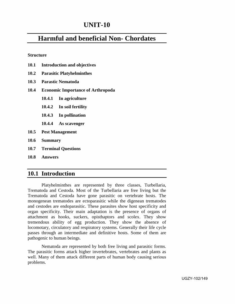
UNIT-10
Harmful and beneficial Non- Chordates
Structure
10.1 Introduction and objectives
10.2 Parasitic Platyhelminthes
10.3 Parastic Nematoda
10.4 Economic Importance of Arthropoda
10.4.1 In agriculture
10.4.2 In soil fertility
10.4.3 In pollination
10.4.4 As scavenger
10.5 Pest Management
10.6 Summary
10.7 Terminal Questions
10.8 Answers
10.1 Introduction
Platyhelminthes are represented by three classes, Turbellaria, Trematoda and Cestoda. Most of the Turbellaria are free living but the Trematoda and Cestoda have gone parasitic on vertebrate hosts. The monogenean trematodes are ectoparasitic while the digenean trematodes and cestodes are endoparasitic. These parasites show host specificity and organ specificity. Their main adaptation is the presence of organs of attachment as hooks, suckers, opisthaptors and scolex. They show tremendous ability of egg production. They show the absence of locomotary, circulatory and respiratory systems. Generally their life cycle passes through an intermediate and definitive hosts. Some of them are pathogenic to human beings.
Nematoda are represented by both free living and parasitic forms. The parasitic forms attack higher invertebrates, vertebrates and plants as well. Many of them attack different parts of human body causing serious problems.
UGZY-102/149
The plant parasitic nematodes attack the crop plants, fruit yielding plants and vegetables.
Out of all arthropods the insects act as pests of different crops. They show organ specificity and host specificity for example Cane Sugar plant is attacked by root borer, shoot borer, leaf eater and juice sucker insects. Paddy is attacked by Rice Gandhi bug, Tana Bedhak, Kharif grass hopper, Rice gall midge and army worm. Pulse Crops are attacked by Grampod borer and Gram cutworm. Oil seed crops are affected by Painted bugs and Mustard Sawfly. Cotton Crop is affected by Spotted Boll worm, Pink Boll worm and Red cotton bug. The insect pests cause huge damage to the economy.
A number of subterranean insects bore tunnels in the soil. During this process they bring the soil of interior of the field to the surface thus increasing the fertility.
Insects like bees, wasps, butterflies, moths, flies and beetles visit flowers to obtain nectar and pollen grains. During this process they perform the pollination. Butterflies and moths are worst pollinators as they undergo egg laying on the same plant and their larvae eat the leaves of that plant. Bees are said to be the best pollinators as they do not damage the plant.
The scavenger insects feed upon the waste matter as debris of decomposed plant or animal body and excreta of animals. The common insects of this category belong to the order Thysanura, Collembola and Dicellura. Some insects like carpet beetles and cloth moths feed upon dried animal products.
About one thousand species of insect pests damage the different crops, out of them seventy species are most significant. The different control measures of the insect pests are mechanical, physical, cultural, chemical and biological. Chemical control involves the use of insecticides which are effective as stomach poison, contact poison, systemic poison or fumigant. Biological control involves the use of predaceous or parasitic species of the insect pest. Such predators may be some insect or a vertebrate. The parasites may be insect caterpillars and adults and nematodes.
Objectives: After studying this unit you should be able to-
• know the basic facts related to Platyhelminth parasites.
• know the basic facts related to Nematode parasites.
• know the basic facts related to the role of insect pests inagriculture.
• know the basic facts related to role of insects in increasing the soilfertility, performing large scale plant pollination and acting as aUGZY-102/150
scavanger.
• know the different ways to manage and control the insects pets.
10.2 Parasitic Platyhelminthes
Members of phylum Platyhelminthes, commonly known as flat worms show triploblastic body organization. They do not possess a coelom. The dorsoventrally flattened body shows mesenchyme between ectoderm and endoderm. The phylum is divided in three classes, Turbellaria, Trematoda and Cestoda. The Turbellaria are free living but Trematoda and Cestoda are parasitic on vertebrate hosts. A parasitic animal totally depends on its host for its food and shelter. The platyhelminth parasites may be of two kinds, ectoparasite living on the body surface of the host and the endoparsite living within the body of the host. Although the Turbellaria are free living but some of them show a tendency of becoming ectoparasitic. The class Tematoda is divided in three orders as Monogenea, Digenea and Aspidogastraea. Out of them the Monogenea lead the life of an ectoparsite living on the body surface of fishes and amphibia. Some of them show a tendency to become endoparsite as Polystomum which is normally present on the body surface of frog but sometimes remains present within its cloaca or rectum. The Digenea are generally endoparasitic in the body of all vertebrates. They have occupied the different possible habitats within the vertebrate body as genus Paramphistomum occupies stomach cavity, Trimiorchis occupies intestinal cavity, Fasciola(Fig.10.1) remains present in bile capillaries, Schistosoma remains in the circulatory system and Paragonimus occupies the lung cavity.
Fig.10.1 Fasciola
UGZY-102/151
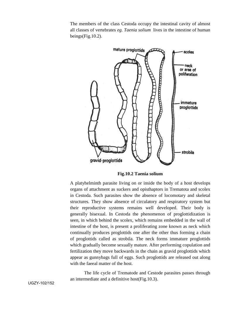
The members of the class Cestoda occupy the intestinal cavity of almost all classes of vertebrates eg. Taenia solium lives in the intestine of human beings(Fig.10.2).
Fig.10.2 Taenia solium
A platyhelminth parasite living on or inside the body of a host develops organs of attachment as suckers and opisthaptors in Trematota and scolex in Cestoda. Such parasites show the absence of locomotary and skeletal structures. They show absence of circulatory and respiratory system but their reproductive systems remains well developed. Their body is generally bisexual. In Cestoda the phenomenon of proglottidization is seen, in which behind the scolex, which remains embedded in the wall of intestine of the host, is present a proliferating zone known as neck which continually produces proglottids one after the other thus forming a chain of proglottids called as strobila. The neck forms immature proglottids which gradually become sexually mature. After performing copulation and fertilization they move backwards in the chain as gravid proglottids which appear as gunnybags full of eggs. Such proglottids are released out along with the faeeal matter of the host.
The life cycle of Trematode and Cestode parasites passes through an intermediate and a definitive host(Fig.10.3).
UGZY-102/152
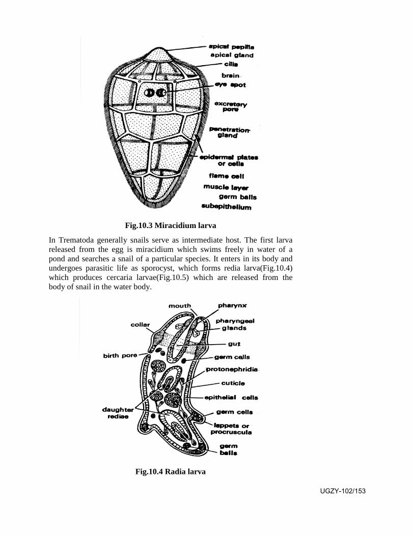
Fig.10.3 Miracidium larva
In Trematoda generally snails serve as intermediate host. The first larva released from the egg is miracidium which swims freely in water of a pond and searches a snail of a particular species. It enters in its body and undergoes parasitic life as sporocyst, which forms redia larva(Fig.10.4) which produces cercaria larvae(Fig.10.5) which are released from the body of snail in the water body.
Fig.10.4 Radia larva
UGZY-102/153
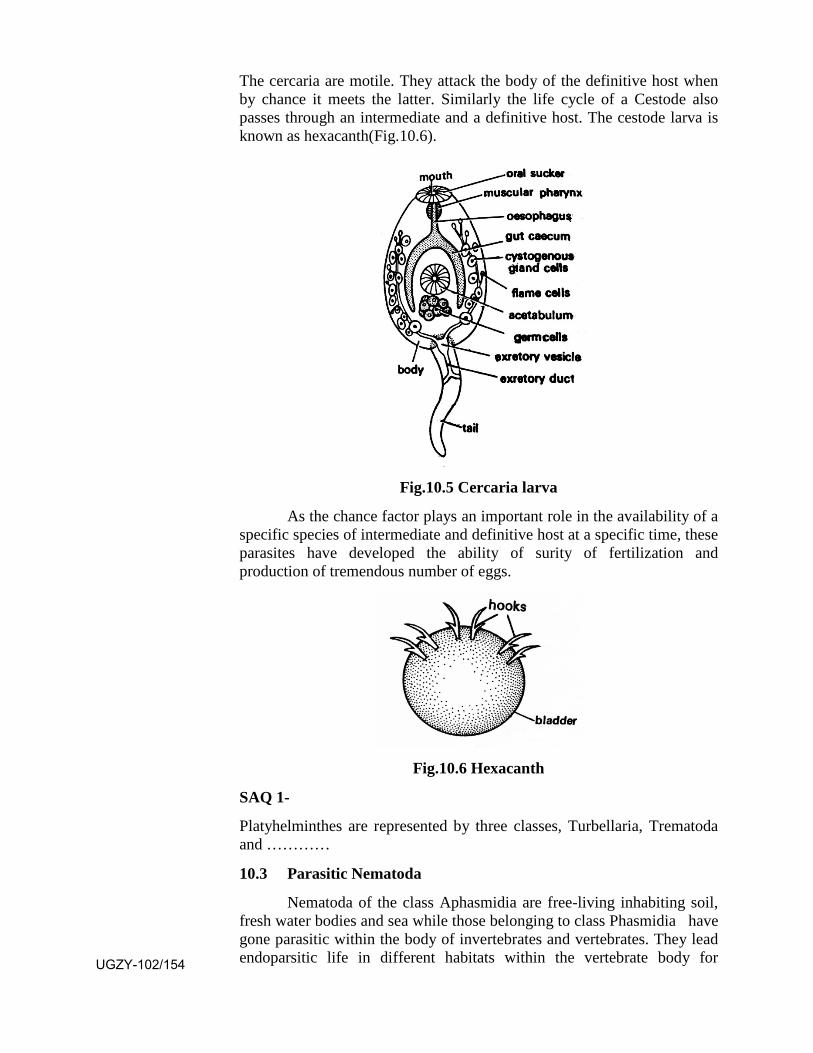
The cercaria are motile. They attack the body of the definitive host when by chance it meets the latter. Similarly the life cycle of a Cestode also passes through an intermediate and a definitive host. The cestode larva is known as hexacanth(Fig.10.6).
Fig.10.5 Cercaria larva
As the chance factor plays an important role in the availability of a specific species of intermediate and definitive host at a specific time, these parasites have developed the ability of surity of fertilization and production of tremendous number of eggs.
Fig.10.6 Hexacanth
SAQ 1-
Platyhelminthes are represented by three classes, Turbellaria, Trematoda and …………
10.3 Parasitic Nematoda
Nematoda of the class Aphasmidia are free-living inhabiting soil, fresh water bodies and sea while those belonging to class Phasmidia have gone parasitic within the body of invertebrates and vertebrates. They lead endoparsitic life in different habitats within the vertebrate body for UGZY-102/154
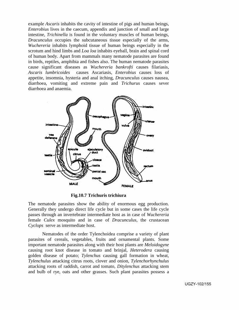
example Ascaris inhabits the cavity of intestine of pigs and human beings, Enterobius lives in the caecum, appendix and junction of small and large intestine, Trichinella is found in the voluntary muscles of human beings, Dracunculus occupies the subcutaneous tissue especially of the arms, Wuchereria inhabits lymphoid tissue of human beings especially in the scrotum and hind limbs and Loa loa inhabits eyeball, brain and spinal cord of human body. Apart from mammals many nematode parasites are found in birds, reptiles, amphibia and fishes also. The human nematode parasites cause significant diseases as Wuchereria bankrofti causes filariasis, Ascaris lumbricoides causes Ascariasis, Enterobius causes loss of appetite, insomnia, hysteria and anal itching, Dracunculus causes nausea, diarrhoea, vomiting and extreme pain and Trichurus causes sever diarrhoea and anaemia.
Fig.10.7 Trichuris trichiura
The nematode parasites show the ability of enormous egg production. Generally they undergo direct life cycle but in some cases the life cycle passes through an invertebrate intermediate host as in case of Wuchereria female Culex mosquito and in case of Dracunculus, the crustacean Cyclops serve as intermediate host.
Nematodes of the order Tylenchoidea comprise a variety of plant parasites of cereals, vegetables, fruits and ornamental plants. Some important nematode parasites along with their host plants are Meloidogyne causing root knot disease in tomato and brinjal, Heterodera causing golden disease of potato; Tylenchus causing gall formation in wheat, Tylenchulus attacking citrus roots, clover and onion, Tylenchorhynchulus attacking roots of raddish, carrot and tomato, Ditylenchus attacking stem and bulb of rye, oats and other grasses. Such plant parasites possess a
UGZY-102/155
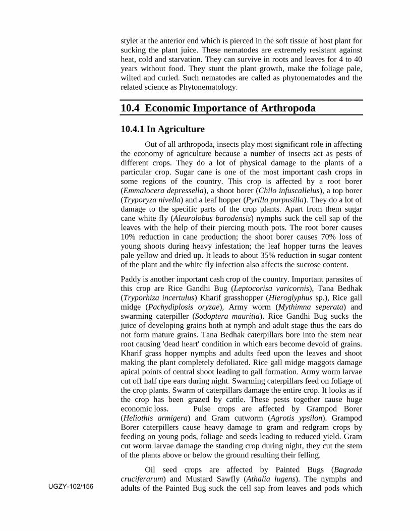
stylet at the anterior end which is pierced in the soft tissue of host plant for sucking the plant juice. These nematodes are extremely resistant against heat, cold and starvation. They can survive in roots and leaves for 4 to 40 years without food. They stunt the plant growth, make the foliage pale, wilted and curled. Such nematodes are called as phytonematodes and the related science as Phytonematology.
10.4 Economic Importance of Arthropoda
10.4.1 In Agriculture Out of all arthropoda, insects play most significant role in affecting
the economy of agriculture because a number of insects act as pests of different crops. They do a lot of physical damage to the plants of a particular crop. Sugar cane is one of the most important cash crops in some regions of the country. This crop is affected by a root borer (Emmalocera depressella), a shoot borer (Chilo infuscallelus), a top borer (Tryporyza nivella) and a leaf hopper (Pyrilla purpusilla). They do a lot of damage to the specific parts of the crop plants. Apart from them sugar cane white fly (Aleurolobus barodensis) nymphs suck the cell sap of the leaves with the help of their piercing mouth pots. The root borer causes 10% reduction in cane production; the shoot borer causes 70% loss of young shoots during heavy infestation; the leaf hopper turns the leaves pale yellow and dried up. It leads to about 35% reduction in sugar content of the plant and the white fly infection also affects the sucrose content.
Paddy is another important cash crop of the country. Important parasites of this crop are Rice Gandhi Bug (Leptocorisa varicornis), Tana Bedhak (Tryporhiza incertulus) Kharif grasshopper (Hieroglyphus sp.), Rice gall midge (Pachydiplosis oryzae), Army worm (Mythimna seperata) and swarming caterpiller (Sodoptera mauritia). Rice Gandhi Bug sucks the juice of developing grains both at nymph and adult stage thus the ears do not form mature grains. Tana Bedhak caterpillars bore into the stem near root causing 'dead heart' condition in which ears become devoid of grains. Kharif grass hopper nymphs and adults feed upon the leaves and shoot making the plant completely defoliated. Rice gall midge maggots damage apical points of central shoot leading to gall formation. Army worm larvae cut off half ripe ears during night. Swarming caterpillars feed on foliage of the crop plants. Swarm of caterpillars damage the entire crop. It looks as if the crop has been grazed by cattle. These pests together cause huge economic loss. Pulse crops are affected by Grampod Borer (Heliothis armigera) and Gram cutworm (Agrotis ypsilon). Grampod Borer caterpillers cause heavy damage to gram and redgram crops by feeding on young pods, foliage and seeds leading to reduced yield. Gram cut worm larvae damage the standing crop during night, they cut the stem of the plants above or below the ground resulting their felling.
Oil seed crops are affected by Painted Bugs (Bagrada cruciferarum) and Mustard Sawfly (Athalia lugens). The nymphs and adults of the Painted Bug suck the cell sap from leaves and pods which UGZY-102/156
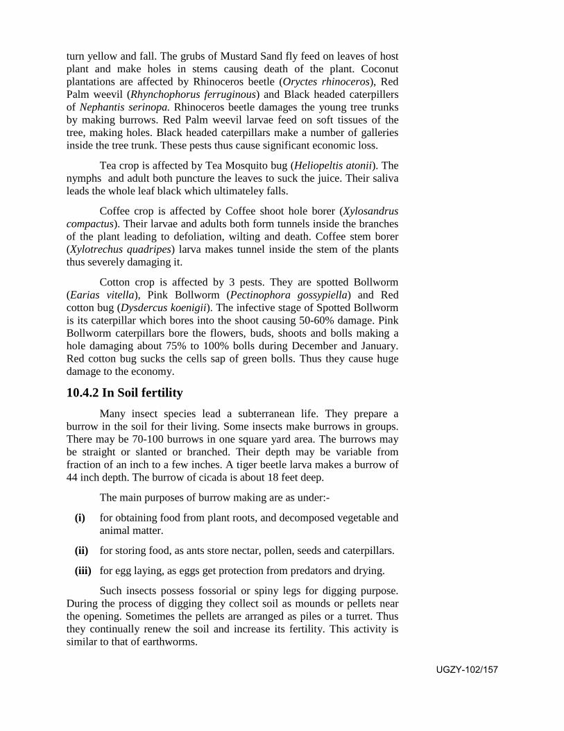
turn yellow and fall. The grubs of Mustard Sand fly feed on leaves of host plant and make holes in stems causing death of the plant. Coconut plantations are affected by Rhinoceros beetle (Oryctes rhinoceros), Red Palm weevil (Rhynchophorus ferruginous) and Black headed caterpillers of Nephantis serinopa. Rhinoceros beetle damages the young tree trunks by making burrows. Red Palm weevil larvae feed on soft tissues of the tree, making holes. Black headed caterpillars make a number of galleries inside the tree trunk. These pests thus cause significant economic loss.
Tea crop is affected by Tea Mosquito bug (Heliopeltis atonii). The nymphs and adult both puncture the leaves to suck the juice. Their saliva leads the whole leaf black which ultimateley falls.
Coffee crop is affected by Coffee shoot hole borer (Xylosandrus compactus). Their larvae and adults both form tunnels inside the branches of the plant leading to defoliation, wilting and death. Coffee stem borer (Xylotrechus quadripes) larva makes tunnel inside the stem of the plants thus severely damaging it.
Cotton crop is affected by 3 pests. They are spotted Bollworm (Earias vitella), Pink Bollworm (Pectinophora gossypiella) and Red cotton bug (Dysdercus koenigii). The infective stage of Spotted Bollworm is its caterpillar which bores into the shoot causing 50-60% damage. Pink Bollworm caterpillars bore the flowers, buds, shoots and bolls making a hole damaging about 75% to 100% bolls during December and January. Red cotton bug sucks the cells sap of green bolls. Thus they cause huge damage to the economy.
10.4.2 In Soil fertility Many insect species lead a subterranean life. They prepare a
burrow in the soil for their living. Some insects make burrows in groups. There may be 70-100 burrows in one square yard area. The burrows may be straight or slanted or branched. Their depth may be variable from fraction of an inch to a few inches. A tiger beetle larva makes a burrow of 44 inch depth. The burrow of cicada is about 18 feet deep.
The main purposes of burrow making are as under:-
(i) for obtaining food from plant roots, and decomposed vegetable and animal matter.
(ii) for storing food, as ants store nectar, pollen, seeds and caterpillars.
(iii) for egg laying, as eggs get protection from predators and drying.
Such insects possess fossorial or spiny legs for digging purpose. During the process of digging they collect soil as mounds or pellets near the opening. Sometimes the pellets are arranged as piles or a turret. Thus they continually renew the soil and increase its fertility. This activity is similar to that of earthworms.
UGZY-102/157
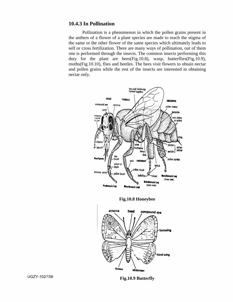
10.4.3 In Pollination Pollination is a phenomenon in which the pollen grains present in
the anthers of a flower of a plant species are made to reach the stigma of the same or the other flower of the same species which ultimately leads to self or cross fertilization. There are many ways of pollination, out of them one is performed through the insects. The common insects performing this duty for the plant are bees(Fig.10.8), wasp, butterflies(Fig.10.9), moths(Fig.10.10), flies and beetles. The bees visit flowers to obtain nectar and pollen grains while the rest of the insects are interested in obtaining nectar only.
Fig.10.8 Honeybee
Fig.10.9 Butterfly UGZY-102/158
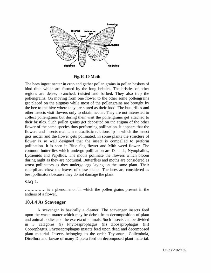
Fig.10.10 Moth
The bees ingest nectar in crop and gather pollen grains in pollen baskets of hind tibia which are formed by the long bristles. The bristles of other regions are dense, branched, twisted and barbed. They also trap the pollengrains. On moving from one flower to the other some pollengrains get placed on the sitgmas while most of the pollengrains are brought by the bee to the hive where they are stored as their food. The butterflies and other insects visit flowers only to obtain nectar. They are not interested to collect pollengrains but during their visit the pollengrains get attached to their bristles. Such pollen grains get deposited on the stigma of the other flower of the same species thus performing pollination. It appears that the flowers and insects maintain mutualistic relationship in which the insect gets nectar and the flower gets pollinated. In some plants the structure of flower is so well designed that the insect is compelled to perform pollination. It is seen in Blue flag flower and Mith weed flower. The common butterflies which undergo pollination are Danaids, Nymphalids, Lycaenids and Papillios. The moths pollinate the flowers which bloom during night as they are nocturnal. Butterflies and moths are considered as worst pollinators as they undergo egg laying on the same plant. Their caterpillars chew the leaves of these plants. The bees are considered as best pollinators because they do not damage the plant.
SAQ 2-
…………… is a phenomenon in which the pollen grains present in the anthers of a flower.
10.4.4 As Scavenger A scavanger is basically a cleaner. The scavenger insects feed
upon the waste matter which may be debris from decomposition of plant and animal bodies and the excreta of animals. Such insects can be divided in 3 catagores (i) Phytosaprophagus (ii) Zoosaprophagus (iii) Coprophagus. Phytosaprophagus insects feed upon dead and decomposed plant material. Insects belonging to the order Thysanura, Collembola, Dicellura and larvae of many Diptera feed on decomposed plant material.
UGZY-102/159
Decomposing wood may be of 4 kinds (i) Dying tree (ii) Dead tree (iii) Decayed tree (iv) Dried wood. Dead and decayed tree trunks are attacked by many beetles and termites. The dried wood is devoured by termites, ants and powder port beetles. The zoosaprophagus insects feed upon dead and decayed animals. They can be divided in 2 categories (i) Those feeding on fresh recently killed and decaying animals eg. Aglossa suprealis feeds during early stage of fermentation while Phorid and Anthomyiid flies feed during ammonical fermentation and Carrion beetles feed during liquidation stage (ii) Those feeding on dried animal products are Dermestids, skin beetles, cheese skipper, carpet beetle and cloth moths. They feed mainly upon skiny hair, feather and fur, including woolen garments, fur coats and carpets. The coprophagus insects feed on the excreta of animals. Common coprophogus insects are Brown scavenger beetle feeding on waste products of insects. Scatophagus species feed on dung of higher animals eg. Scarabaeid beetles and Histerid beetless. Volucella inhabits the nest of wasp and bumble bees and feeds upon their waste matter. Many insect species are omnivorous which feed on any and every kind of material eg. cockroaches and ants.
SAQ 3-
The zoosaprophagus insects feed upon …………and……..
10.5 Pest Management
A pest is an animal species which affects human economy in someway or the other. Many insect species attacking various plants for feeding do a lot of damage to the crops. Such insect pests show host specificity and organ specificity that is if a particular pest species attacks, the root system of a particular plant it will not attack any other part of that plant for example Emmalcera depressella attacks the root system while Chilo infuscatellus attacks the stem of sugar cane. In many cases some part of the life cycle of a pest passes in the soil. The number of known insect pests doing damage to crops is about 1000, out of which about 70 species are responsible for maximum loss. In 1994-95 out of 210 million tones of agriculture produce about 15-20 million tones losses were due to insect pests alone. The target of a farmer is to minimuze the damage of the crop caused by the attack of the pest for which various control measures are practiced which are as under.
I. Mechanical control: In this practice the pests are killed manually with or without any specific equipment. The methods include hand picking, use of hand nets and bag nets, use of light traps or electric traps. Such methods are costly because of manual labour and are not effective in large scale eradication.
II. Physical Control: It involves the manipulation of temperature,moisture, light, electricity and atomic energy for checking the pest population.
UGZY-102/160

III. Cultural control: It involves regular farm operations by thefarmers to reduce pest population. It involves removal of weeds and residues of crops, decaying fruits and dead grasses and leaves. The other methods are crop rotation, tilling of soil, use of resistant varieties, pruning and thinning, use of clean seeds, regulating irrigation and mixed cropping.
IV. Chemical Control: It involves the use of insecticides which killthe insects due to their chemical action. The insecticides are of 2 kinds: (i) Inorganic substances like compounds of antimony, arsenic, barium, boron, copper, mercury, zinc, phosphorus, sulphur and thallium (ii) organic substances like hydrocarbon oils, organic compounds of plant origin and the synthetic ones.
The insecticides are formulated in the form of dust (fine powder), granules (aggregation of dust particles) solutions (in water or organic solvents), emulsified concentrates, concentrated liquids, aerosols (minute particles suspended in air as smoke), fumigants (gaseous form) and insecticide-fertilizer mixture.
The insecticide is effective in the form of stomach poison, contact poison, systemic poison or fumigant. A stomach poison is effective when it is ingested by the pest species. A contact poison is effective on contact with the pest body. Systemic poison is used to control pests with piercing and sucking type of mouth parts. It is applied to the seeds, root, stem or leaves through which it is absorbed in the body of the plant, making its juice toxic and thus it becomes fatal. A fumigant is in vapour form. It affects the spiracles and tracheal system of the insect pest thus killing it.
V. Biological Control: It involves the use of predaceous and parasitic species of the pest species. It requires the collection of predators and parasites from different places where they occur naturally in a large number and releasing them at the places where they are needed. Further it requires rearing of predators and parasites in large number under favourable conditions. It also requires the collection storage and handling of pest species for killing them. The predators may be insect predators or vertebrate predators. The parasites may be in the form of insect caterpillars and adults and nematodes. The use of fungi, bacteria, virus and protozoa is made to cause a particular disease in the pest species for its eradication.
10.6 Summary
• Platyhelminthes are represented by three classes, Turbellaria,Trematoda and Cestoda. Out of them Trematoda and Cestoda havegone parasitic. Order Monogenea of Trematoda are ectoparasiticon fish and amphibia while digenea are endoparasitic in allvertebrates. Cestoda are also endoparasitic on all vertebrates.
• The Trematoda and Cestoda show organs of attachment and
UGZY-102/161

tremendous ability of egg production. They show the absence of locomotary organs, respiratory organs and circulatory system.
• Nematodes of the class Phasmidia have gone parasitic on bothinvertebrates and vertebrates. Nematodes of the orderTylenchoidea have gone parasitic on plants. Many nematodesinfesting human beings cause serious diseases.
• Many species of the class Insecta have gone parasitic on plantsserving as pests. they cause serious loss to agriculture.
• Many insect species lead subterranean life for which they burrowin the soil. They transfer the inner soil on the surface. Thus theyhelp in increasing the soil fertility.
• Many insect species visit flowers for obtaining nectar and pollenon which they feed. During this process they perform croppollination. Bees are the best pollinators.
• Some insect species habitually devour the decomposed plant andanimal bodies. Some of them feed on the faecal matter also. Thusthey serve as scavengers.
• As the insect pest cause huge damage to the varied crops itbecomes essential to control and eradicate them. The managementof the pest in done by mechanical, physical, and cultural methods.Apart from these methods chemical control by insecticides is verypopular. In the recent past, biological control by the use ofpredatory and parasitic species of the pest has been introduced.
10.7 Terminal Questions
Q.1 Write a brief essay on Platyhelminth parasites.
Q.2 Give an account of some human nematode parasites and the diseases they cause.
Q.3 Describe insect pollination.
Q.4 What are the various ways for pest control. Add a note of the role of insecticides.
Q.5 Write short notes on.
(i) Cestoda
(ii) Pest of paddy
(iii) Biological Control
(iv) Plant parasitic nematodes. UGZY-102/162

Q.6 Match the two-
Column I Column II
1. Heterodera a. Cotton pest
2. Schistosoma b. Cestoda
3. Wuchereria c. Potato
4. Dysdercus d. blood parasite
5. Taenia 4. Culex
ANSWERS
SAQ 1 - cestoda
SAQ 2 - pollination
SAQ 3 - dead, decay
BIBLIOGRAPHY 1. Textbook of zoology invertebrates by Parker and Haswell
2. Modern textbook of zoology invertebrates by R.L. Kotpal
3. Invertebrate zoology by Jordan and P.S. Verma
UGZY-102/163
Rough Work
UGZY-102/164