ubiquitous microbial disorders of medicinal plants and
TRANSCRIPT
12
www.jobpcr.com tAvailable online a
ISSN-2394:3076
CODEN(USA) : JBPCBK
Journal of Biological Pharmaceutical And Chemical Research , 2022, 9(1): 12-24
)ttp://www.jobpcr.com/arhcive.phph(
Ubiquitous Microbial Disorders of Medicinal Plants and Potential
Management of such Phytopathogens
3, and Sneha Chaklader2, Anirudh Modak1*Shamayita Basu
.est Bengal, IndiaUniversity of Kalyani, Kalyani, Nadia, W*1
Gangtok, Sikkim, India.Sikkim Manipal University, Tadong, 2,3
@gmail.combasu95shamayita :Authororresponding C
REVIEW ARTICLE
ABSTRACT
Medicinal plants are known as traditional healers, since ancient times. India has been bestowed
with many diversified groups of medicinal plants accounting for approximately 8000 species being
used in over 10,000 herbal products. Commercial gardening began as a single way of assessing for
the conservation of these valuable plant species, which unknowingly introduced the complexity of
many pests and diseases, resulting in crop forfeiture of varying magnitudes. The gravity of
medicinal plants has been escalating day by day. Traditional medicine, in the recent years, has
made a resurgence for many different reasons together with toxicity and side effects of
contemporary synthetic drugs, expansion of multiple drug resistant microbes, and also the
inaptitude of modern medicine for locating effective healing procedures for various diseases. Over
70% of the population of developing world currently relies on the traditional medicinal structure,
also known as alternative or complementary system of medicine. Medicinal plants are being more
widely employed in healthcare organizations, whereas chemical pest management and disease
management strategies are becoming obsolete. Among the various microbes, viruses are immensely
responsible for causing diseases in medicinal plants. This chapter imparts comprehensive
information regarding the family, morphology, pharmacological profile, distribution, active
constituents and symptoms of the viral infections among the medicinal plants reported in various
parts of India. The analysis of these viral diseases will authorize developing effective mechanisms
for safeguarding medicinal plants and maintaining the standard of the fresh materials for
pharmaceuticals.
Key words: pests, infection, medicine, microbes, traditional healers, toxicity, pest management.
Shamayita Basu et al J. of Bio.Pharm. And Chemical Research, 2022,9(1):12:24
13
INTRODUCTION
Plants have been used for medicinal reasons since prehistoric times. Ancient Unani writings,
Egyptian papyrus, and Chinese literature all contain descriptions of herbs. Plants have been utilized
as medicine by Unani Hakims, Indian Vaids, European and Mediterranean cultures for over 4000
years, according to evidence. Indigenous societies such as Rome, Egypt, Iran, Africa, and America
employed herbs in healing rituals, while traditional medical systems such as Unani, Ayurveda, and
Chinese Medicine were created by others, in which herbal remedies were used systematically.
Indian traditional healers have long been aware of medicinal herbs. The plants were mostly
harvested in the wild and employed in a variety of health product formulations. India is home to a
broad range of medicinal plants, totaling about 8000 species that are employed in more than 10,000
herbal treatments. Ninety percent of the raw materials required by the herbal business come from
the natural ecosystem - forests – leading in brutal exploitation and destruction of the ecosystem's
natural habitats [1]. Commercial cultivation began as one of the strategies to protect the valuable
species, but it unwittingly brought with it the problem of pests and diseases, resulting in crop losses
of varying magnitudes. Glory lily (Gloriosa superba), Noni (Morinda citrifolia L.), medicinal
coleus (Coleus forskohlii), makoi (Solanum nigrum), senna (Cassia angustifolia), and ashwagandha
are some of the most significant medicinal plants discussed in this chapter (Withania somnifera).
Medicinal plants are being phased out of the health-care system, as are chemical pest and disease
management practices. Non-chemical, environmentally friendly, and safer management solutions,
which are also covered in this chapter, are required. [1]
Markets and research on medicinal plants are growing at an exponential rate in the pharmaceutical
and associated professions across the world. In reality, about fifty to seventy thousand plant species
are known to have therapeutic characteristics across the world, with around four to ten thousand of
them being endangered [2,3]. Over four lakh tones of medicinal plants are traded globally for over
3000 species. India is one of the major producers and exporters of such medicinal plants [3].
Natural medicines derived from medicinal plants have fewer negative effects on human systems
than their synthetic equivalents.
Important Medicinal Plants
India has long been known as a rich source of medicinal herbs among ancient civilizations. The
Indian Forest is home to a diverse range of medicinal and fragrant plants, which are usually
exploited as raw materials for medicines and perfumery products. Approximately 8,000 herbal
therapies have been defined in India's AYUSH systems. Unani, Ayurveda, Folk (tribal), Siddha,
medicine are the main stems of indigenous medicine. In India, Ayurveda and Unani Medicine are
the most advanced and widely practiced of these systems.[4]
As per the World Health Organization (WHO), herbal medicines are used by 80% of people
throughout the world for some aspect of their primary health care. According to the WHO, there are
around 21,000 plant species that have the potential to be implemented as medicinal plants.
Thus, as per to published data, approximately three-quarters of the world's population gets their
health care mostly from plants and plant extracts. At one point or another, more than 30% of all
plant species have been used for medicinal purposes. Plant treatments are predicted to account for
up to 25% of total drugs in developed countries such as the United States, but up to 80% in fast-
developing countries such as India and China. As a result, the economic relevance of medicinal
plants is significantly greater in India than in the rest of the globe. These nations offer two-thirds of
Shamayita Basu et al J. of Bio.Pharm. And Chemical Research, 2022,9(1):12:24
14
the plants used in modern medicine, while the rural population's health care system is based on
indigenous medicine. [5,6]
The usage of medicinal plants is seen to be relatively risk-free, as there are no or only minor side
effects. The most significant benefit is that these treatments are in sync with nature. Herbal
medicines may be utilized by individuals of all ages and genders, according to the golden truth.[7]
Herbs are only answers to a number of health-related concerns and diseases, according to ancient
doctors. They conducted significant investigation and testing in order to arrive at trustworthy
conclusions about the efficacy of numerous medicinal herbs. The vast majority of drugs developed
this way have no significant side effects or reactions. This is why herbal medicine is gaining
popularity all over the world. These medicinal plants provide a practical method to treating a wide
range of internal problems that are traditionally thought to be difficult to cure. [6, 7]
As per Floral Statistics of India 2017, which was held by the Botanical Survey of India in Kolkata,
West Bengal, there were a total of 2,68,600 blooming plants globally, with 18,386 (6.84%) of them
being in India. In India, over 3000 plant species are known to have therapeutic characteristics [8],
and according to another study, 2500 plants are employed in traditional medicine, with 100 plants
being used on a daily basis [9]. Traditional plant medicinal knowledge is helpful not just for
biodiversity conservation, but also for healthcare and medicine development. The Himalaya is a
global biodiversity hotspot with a diverse variety of topographical, ecological, and evolutionary
traits that support 18,440 plant species, 25.3 percent of which are indigenous to the region. [10,11].
Indian state of Uttarakhand is situated in the Himalayan hotspot, and it covers 17.3 percent of
India's total land area, with 92.57 percent of it covered by hills and 7.43 percent by plains. It is
situated between the latitudes of 28˚43'–31˚27'N and the longitudes of 77˚34'–81˚02'E. River Tons
divides it from the state of Himachal Pradesh, river Kali from the country of Nepal, and the broader
Himalaya forms the state's northern boundary as well as with China (international border). In the
Himalaya, there are around 1748 commercially significant plants. The Tharus, Bokshas, Bhotias,
Marchchas, Van-gujjars, Jaunsaris, Tolchas, Koltas, Banw-rauat, Gangwal, and other people
communities rely on wild flora for their traditional medicine [11]. Joshi et al. found 102 plant
species from 48 families that have ethno-medicinal applications in the state's four districts: Almora,
Champawat, Bageshwar, and Pithoragarh [9,12]. Adhikari et al. At the Wildlife Institute of
Dehradun in Uttarakhand, researchers looked at the condition and distribution pattern of medicinal
plants, discovering 605 species from 94 families. This essay is based on earlier research on
Uttarakhand medicinal herbs [13,14]. Authors concentrated on several key features of local
medicinal plants that require protection and cultivation since they are naturally abundant and
rapidly disappearing, and they may assist indigenous make a living to some extent. This work will
be useful to pharmacologists, phytochemists, and researchers in this subject in the future. As a
result, the purpose of this study is to write a manuscript that emphasizes the value of traditional
knowledge in the Himalayan state of Uttarakhand, India, for the treatment of various ailments.
Table 1: A list of selected medicinal plants with their ethno-medicinal uses from Uttarakhand, India
Botanical
Name
(Family)
Local
name
Parts
used Ethno-medicinal Uses
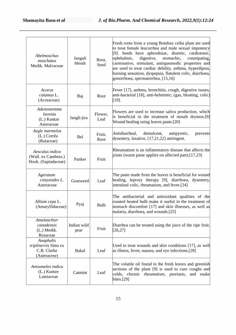
Shamayita Basu et al J. of Bio.Pharm. And Chemical Research, 2022,9(1):12:24
15
Abelmoschus
moschatus
Medik. Malvaceae
Jangali
bhindi Root,
Seed
Fresh roots from a young Bombax ceiba plant are used
to treat female leucorrhea and male sexual impotency
[9]. Seeds have aphrodisiac, diuretic, cardiotonic,
ophthalmic, digestive, stomachic, constipating,
carminative, stimulant, antispasmodic properties and
are used to treat cardiac debility, asthma, hyperdipsia,
burning sensation, dyspepsia, flatulent colic, diarrhoea,
gonorrhoea, spermatorrhea, [15,16]
Acorus
calamus L.
(Acoraceae) Baj Root
Fever [17], asthma, bronchitis, cough, digestive issues;
anti-bacterial [18], anti-helmintic; (gas, bloating, colic)
[19].
Adenostemma
lavenia
(L.) Kuntze
Asteraceae
Jangli-jira Flower,
Leaf
Flowers are used to increase saliva production, which
is beneficial in the treatment of mouth dryness.[9]
Wound healing using leaves paste.[20]
Aegle marmelos
(L.) Corrêa
(Rutaceae) Bel
Fruit,
Root
Antidiarrheal, demulcent, antipyretic, prevents
dysentery, laxative, [17,21,22] astringent.
Aesculus indica
(Wall. ex Cambess.)
Hook. (Sapindaceae)
Panker
Fruit
Rheumatism is an inflammatory disease that affects the
joints (warm paste applies on affected part).[17,23]
Ageratum
conyzoides L.
Asteraceae Goatweed Leaf
The paste made from the leaves is beneficial for wound
healing, leprosy therapy [9], diarrhoea, dysentery,
intestinal colic, rheumatism, and fever.[24]
Allium cepa L.
(Amaryllidaceae) Pyaj Bulb
The antibacterial and antioxidant qualities of the
roasted heated bulb make it useful in the treatment of
stomach discomfort [17] and skin illnesses, as well as
malaria, diarrhoea, and wounds.[25]
Amelanchier
canadensis
(L.) Medik.
Rosaceae
Indian wild
pear Fruit Diarrhea can be treated using the juice of the ripe fruit.
[26,27]
Anaphalis
triplinervis Sims ex
C.B. Clarke
(Asteracese)
Bakal
Leaf
Used to treat wounds and skin conditions [17], as well
as illness, fever, nausea, and eye infections.[28]
Anisomeles indica
(L.) Kuntze
Lamiaceae Catmint Leaf
The volatile oil found in the fresh leaves and greenish
sections of the plant [9] is used to cure coughs and
colds, chronic rheumatism, psoriasis, and snake
bites.[29]
Shamayita Basu et al J. of Bio.Pharm. And Chemical Research, 2022,9(1):12:24
16
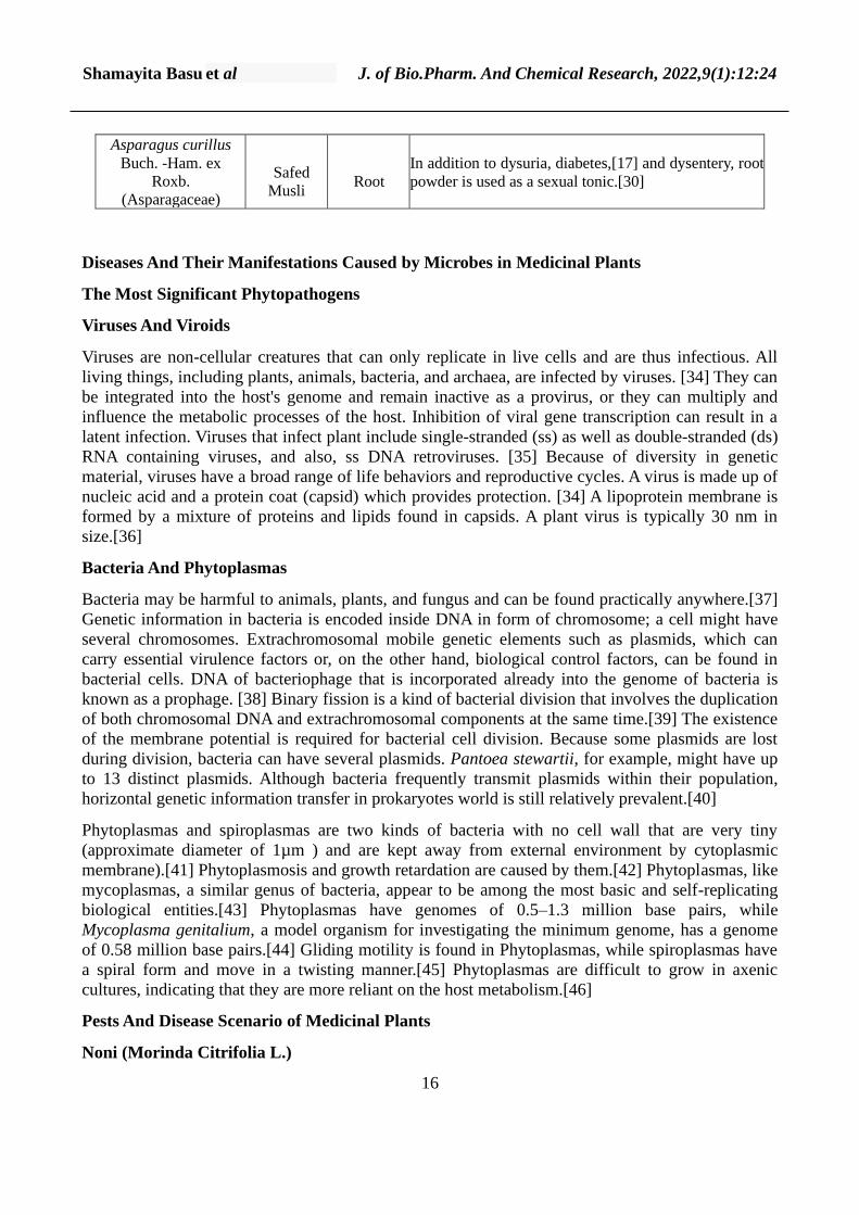
Asparagus curillus
Buch. -Ham. ex
Roxb.
(Asparagaceae)
Safed
Musli
Root
In addition to dysuria, diabetes,[17] and dysentery, root
powder is used as a sexual tonic.[30]
Diseases And Their Manifestations Caused by Microbes in Medicinal Plants
The Most Significant Phytopathogens
Viruses And Viroids
Viruses are non-cellular creatures that can only replicate in live cells and are thus infectious. All
living things, including plants, animals, bacteria, and archaea, are infected by viruses. [34] They can
be integrated into the host's genome and remain inactive as a provirus, or they can multiply and
influence the metabolic processes of the host. Inhibition of viral gene transcription can result in a
latent infection. Viruses that infect plant include single-stranded (ss) as well as double-stranded (ds)
RNA containing viruses, and also, ss DNA retroviruses. [35] Because of diversity in genetic
material, viruses have a broad range of life behaviors and reproductive cycles. A virus is made up of
nucleic acid and a protein coat (capsid) which provides protection. [34] A lipoprotein membrane is
formed by a mixture of proteins and lipids found in capsids. A plant virus is typically 30 nm in
size.[36]
Bacteria And Phytoplasmas
Bacteria may be harmful to animals, plants, and fungus and can be found practically anywhere.[37]
Genetic information in bacteria is encoded inside DNA in form of chromosome; a cell might have
several chromosomes. Extrachromosomal mobile genetic elements such as plasmids, which can
carry essential virulence factors or, on the other hand, biological control factors, can be found in
bacterial cells. DNA of bacteriophage that is incorporated already into the genome of bacteria is
known as a prophage. [38] Binary fission is a kind of bacterial division that involves the duplication
of both chromosomal DNA and extrachromosomal components at the same time.[39] The existence
of the membrane potential is required for bacterial cell division. Because some plasmids are lost
during division, bacteria can have several plasmids. Pantoea stewartii, for example, might have up
to 13 distinct plasmids. Although bacteria frequently transmit plasmids within their population,
horizontal genetic information transfer in prokaryotes world is still relatively prevalent.[40]
Phytoplasmas and spiroplasmas are two kinds of bacteria with no cell wall that are very tiny
(approximate diameter of 1µm ) and are kept away from external environment by cytoplasmic
membrane).[41] Phytoplasmosis and growth retardation are caused by them.[42] Phytoplasmas, like
mycoplasmas, a similar genus of bacteria, appear to be among the most basic and self-replicating
biological entities.[43] Phytoplasmas have genomes of 0.5–1.3 million base pairs, while
Mycoplasma genitalium, a model organism for investigating the minimum genome, has a genome
of 0.58 million base pairs.[44] Gliding motility is found in Phytoplasmas, while spiroplasmas have
a spiral form and move in a twisting manner.[45] Phytoplasmas are difficult to grow in axenic
cultures, indicating that they are more reliant on the host metabolism.[46]
Pests And Disease Scenario of Medicinal Plants
Noni (Morinda Citrifolia L.)
Shamayita Basu et al J. of Bio.Pharm. And Chemical Research, 2022,9(1):12:24
17
Noni, Morinda citrifolia L., is a highly prized medicinal tree whose therapeutic virtues have been
known to Indians since ancient times. The therapeutic benefits of all parts of the plants — leaves,
flowers, fruits, roots, and stems – are being explored [47]. Various insect and non-insect pests are
said to be wreaking havoc on the crop [48,49,50].
Pests of Noni: -
Dulinius conchatus- a lacewing bug (Tingidae, Hemiptera)
The lacewing insect is a significant noni pest, especially in nurseries. Lacewing insect nymphs and
adults feed on the sap, causing brown stains on the top surface of the leaves. Spots turn dark, and
the leaves shrink and dry out in extreme cases. Adults have a heavily reticulate body and 5–8 mm
long wings that they use to live on the underside of leaves.
Black Fly: Aleurocanthus terminaliae (Aleyrodidae, Hemiptera)
Due to the desapping of leaves from the lower surface of the leaves, they turn blackish and often
coiled upside down. On the sticky material released by the insect pest, sooty mould grows. In a
spiral pattern, the adult female lays 15–20 eggs. In her lifetime, a female can lay one to three
spirals.
Coccus viridis (Green) (Coccidae, Hemiptera)
The adult green coffee scale has an oval to elongate body that is dorsoventrally flattened. Insects eat
the plant's phloem and can be found on the stems, leaves, fruits, and young twigs. The sap feeding
by green scales causes yellowing of the leaves, which can lead to defoliation and a reduction in fruit
set, as well as a general decrease in plant growth and vitality. [49,51]
Diseases of Noni
Anthracnose: Colletotrichum gloeosporioides
On the leaves, little brown shaped patches of varied sizes (0.5–2.5 cm) emerge, expand, and
combine. The centre of the consolidated lesion becomes greyish white, causing a shot hole
symptom. Acervuli with pink masses of spores appeared on the lesions under humid settings.
Anthracnose has been reported for the first time in India [52,53].
Dry Fruit Rot: Colletotrichum gloeosporioides
Colletotrichum gloeosporioides infects all areas of the noni plant, regardless of crop stage. On
twigs, flowers, and fruits, the symptoms were noticed. The infection induced by C. gloeosporioides
on flowers manifests itself as dull brown lesions. Within 48 hours of infection, the diseased
blossoms were dried. Brown necrotic patches on the corolla tube sticking to the noni fruit were
discovered when the blooms were examined with a stereo zoom microscope. The occurrence of
necrotic brown lesions with a yellow halo was characteristic of C. gloeosporioides-infected twigs.
The lesions grew in size over time and had a grey centre. The number of minute pinhead-shaped
acervuli fructifications was counted on the grey centre. The necrotic lesions spread slowly along the
peduncle to the fruits, infecting the flowers and fruits. Later, the infected fruits shrink, dry off and
got mummified. Post mummification, saprophytic moulds like Aspergillus and Penicillium
colonized the fruits that got infected. [53].
Leaf Blight and Dry Fruit Rot: Alternaria alternata
Shamayita Basu et al J. of Bio.Pharm. And Chemical Research, 2022,9(1):12:24
18
Tamil Nadu and Karnataka states witnessed a severe outbreak of leaf blight, for the first time during
2008–2009. The causal agent was identified as Alternaria alternate [54]. The same disease causes
dry fruit rot, which is marked by the formation of a black necrotic sunken region on green unripe
fruits with a diameter of 2–3 mm. The spot becomes a dark black lesion that coalesces when the
environment is favorable. The centre of the lesions is black in color with alternate concentric
regions. Drying and splitting of fruits along with saprophytic infection of some other moulds,
occurs, as the fruit expands [53].
Diseases of Glory Lily
Root Rot: Macrophomina phaseolina
Symptoms of M. phaseolina-caused root rot include yellowing of leaves, the formation of dark
lesions on the stems with black sclerotial bodies, and the rotting of roots. The pathogen might live
for several years in the soil. According to Meena and Rajamani, the yield loss ranged from 50 to 60
percent [56].
Leaf Blight: Alternaria alternata
Leaf blight begins as little reddish spots on leaves that expand into concentric rings; many of these
spot’s merge, causing the entire leaf to be blighted [57].
Conventional Methods of Plant Disease Control
Generations of farmers have been developing techniques to battle the different pests that afflict our
crops since the dawn of agriculture. Since our discovery of the causes of plant diseases in the early
1800s, we have been able to devise a wide range of strategies for the management of specific plant
diseases as our understanding of the interplay between pathogen and host has improved.
We can derive some fundamental principles of plant disease control from this cumulative
knowledge base to aid in the treatment of emerging issues on every crop in any environment. H. H.
Whetzel's concepts, initially defined in 1929 and somewhat amended by other writers over the
years, have been extensively embraced and taught to generations of plant pathology students all
over the world. A committee of the National Academy of Sciences in the United States articulated
these "traditional principles" in 1968.
Traditional Principles of Plant Disease Control
1. Avoidance—avoid illness by going at a time of year or visiting a location where there is no
inoculum or the environment is not conducive to infection.
2. Exclusion—prevent inoculum from being introduced.
3. Eradication—the inoculum must be eliminated, destroyed, or rendered inactive.
4. Protection—use a toxin or another barrier to infection to avoid infection.
5. Tolerance or resistance to infection—Use cultivars that are resistant or tolerable to infection.
6. Treatment—Plants that have been infected can be healed.
While these beliefs are still true now as they were in 1929, they have a slew of issues when it comes
Shamayita Basu et al J. of Bio.Pharm. And Chemical Research, 2022,9(1):12:24
19
to plant disease prevention. To begin with, these principles are stated in absolute terms (e.g.,
"exclude," "prevent," and "eliminate"), implying that the objective is to remove all sickness. "Plant
disease management" in this sense is usually impractical, if not impossible. Indeed, we do not need
to eradicate a disease; rather, we just need to slow down its spread and protect the condition from
progressing too far. We should think about plant disease management rather than plant disease
control. Traditional plant disease control ideas, on the other hand, ignore the dynamics of plant
disease, that is, fluctuations in the frequency and severity of sickness across time and space.
Furthermore, because different diseases have different dynamics, it is impossible to assess the
relative efficacy of different disease-control techniques. They also don't show how different disease
control measures interact with one another in terms of disease dynamics. We need a technique to
quantify the effects of various control measures on disease development, both individually and in
combination. Finally, traditional plant disease control strategies place a greater emphasis on tactics
than on integrating them into a workable overall strategy. [58]
Nanoparticles In Detection and Control of Phytopathogens
It is critical to get an assessment of the incidence and severity of illness on the plants in order to
regulate phytopathogens. Because illnesses are linked to yield losses, this knowledge is critical for
making decisions that lead to successful disease control methods. Previously, visual indicators such
as leaf yellowing, curling, and wilting were employed to determine the severity of disease/infection,
followed by quick testing to establish the pathogen's identification. Disease diagnoses based on
symptom observations are very subjective and reliant on the observer's competence. Furthermore, to
identify unknown infections, Koch’s postulates have to be confirmed. Making the difference
between pathogen and saprophytic bacteria in a traditional microbiological investigation of diseased
tissue is particularly important since the latter are common in the environment.[59]
Rapid detection and identification of the pathogen has been aided by the development of molecular
methods and immunodiagnostic techniques for characterization of microorganisms. PCR for
specific detection of Clavibacter michiganesis subsp. michiganesis in tomato [60], Xanthomonas
axonopodis pv. punicae in pomegranate [61], and enzyme-linked immunosorbent assay (ELISA) for
detection of Xylella fastidiosa in landscape trees [62] are just for pathogen confirmation, advanced
technologies such as real-time PCR (qPCR) are also useful.
Silica nanoparticles
Fluorescent dye-doped silica nanoparticles have shown great promise in enhanced bioanalysis,
allowing for a more detailed understanding of biology and medicine at the molecular level[63]. The
fluorescent dye is protected from photobleaching and leaking by being incorporated into the silica
nanoparticles. Using antibody-conjugated Rubpy-doped silica nanoparticles as a detection
probe,[64] effectively identified Xanthomonas axonopodis pv. vesicatoria, a plant pathogen
responsible for bacterial spot disease in Solanaceae. The findings show that fluorescent silica
nanoprobes may be used to diagnose plant diseases quickly.
Gold nanoparticles
In the detection of harmful bacteria, DNA-gold nanoparticle probes offer promise as a new
generation of biosensors. Hereby, probes of gold nanoparticle-oligonucleotides are getting
hybridized with complementary DNA, which there by stabilizes these gold nanoparticles against
aggregation by pertaining to the native pink hue of gold) (retaining the native pink colour of the
colloidal gold). The solution becomes purple in the absence of complementary DNA because the
Shamayita Basu et al J. of Bio.Pharm. And Chemical Research, 2022,9(1):12:24
20
agglomeration of gold nanoparticles causes the absorbance peak to shift toward a longer wavelength
[65,66].
Gold nanoparticle-bound probes were utilized in a recent work to colorimetrically detect P.
syringae pathovars. PCR was used to amplify the hrcV gene from pathogen DNA, and thiol-
functionalized DNA coupled to gold nanoparticles was used to detect it. The test was able to
identify genomic template concentrations as low as 15 ng/L. As a result, the test was sensitive and
proven to be highly specific, having the ability to detect P. syringae early. [67].
CONCLUSION
The study of the causes and effects of phytopathogens on plants has developed to a
multidisciplinary level with the introduction of modern diagnostic procedures, genome sequencing
and editing tools, as well as technologies for microbiome and proteome investigation. In this article,
we have put in an attempt to present a comprehensive image of the current scenario of pest
management. However, many other aspects of the interaction between plants and phytopathogens
need to be considered, like damage by the ice nucleation proteins, which leads to ice crystal
formation in the plant cells [68] or the sequences of effector molecules having conserved nature in
bacteria: the pathogens of animals, humans and plants [69]. Control and management of plant
diseases needs agents which can reduce the suspected pathogen to minimal levels, means of early-
stage pathogen detection, and chemical compounds that effectively trigger immune response among
the host plants. Nanomaterials have been assessed by scientists and researchers for their
considerable utility in all such approaches for control of disease. This present review article also
focuses on nanomaterials for control of the known phytopathogens, role of nanomaterials as
mediators of the plant immune response. Hence, nanomaterials can be referred as novel weapons in
this war against these phytopathogens and subsequently, can be a potential component in agro-
ecosystem.
CONFLICT OF INTEREST
None Declare
ACKNOWLEDGEMENT
None Declare
REFERENCE
1. Mathivanan N, Sithanantham S, Marimuthu T, Peter KV, Rethinam P, Brahma S, Peter PI, Kirti S
(2016) Therapeutic and commercial potential of medicinal plants with special focus on Morinda
citrifolia L. (Noni). Souvenir cum Abstracts, Second World Noni Congress, March, 2016, SRM
University, Kattankulathur, Tamil Nadu.
2. Canter, P. H., Thomas, H., & Ernst, E. (2005). Bringing medicinal plants into cultivation:
opportunities and challenges for biotechnology. Trends in Biotechnology, 23(4), 180–185.
3. Taylor, D. A. (2008). Standards: New yardstick for medicinal plant harvests. Environmental
Health Perspectives, 116(1), A21–A21.
4. www.nofa.org/tnf/Summer2012B.pdf
5. http://www.who.int/medicines/areas/traditional/SelectMonoVol4.pdf
Shamayita Basu et al J. of Bio.Pharm. And Chemical Research, 2022,9(1):12:24
21
6. http://www.cals.ncsu.edu/plantbiology/Faculty/dxie/Chapter1-1.pdf
7. www.tee.org/fileadmin/downloads/Botanische%20Bestandsaufnahme%20indischer%20Heilpflanzen.pdf
8. Prakasha HM, Krishnappa M, Krishnamurthy YL, Poornima SV. Folk medicine of NR PuraTaluk
in Chikamaglur district of Karnataka. Indian Journal of Traditional Knowledge. 2010; 9(1):55-60.
9. Joshi B, Pant SC. Ethnobotanical study of some common plants used among the tribal
communities of Kashipur, Uttarakhand. Indian Journal of Natural Products and Resources. 2012;
3(2):262-266.
10. Singh DK, Hajra PK. Floristic diversity. In: Gujral GS, Sharma V (Eds), Changing Perspective
of Biodiversity Status in the Himalaya. British Council Division, British High Commission Publ
Wildlife Youth Services, New Delhi, 1996.
11. Samant SS, Dhar U, Palni LMS. Medicinal Plants of Indian Himalaya: Diversity Distribution
Potential Value Almora: G.B Pant Institute of Himalayan Environment and Development, 1998.
12. Gaur RD. Traditional dye yielding plants of Uttarakhand, India. Natural Product Radiance.
2008; 7(2):154-165.
13. Joshi Y, Joshi AK, Prasad N, Juyal D. A review on Ficus palmate (Wild Himalayan Fig). Journal
of Psychopharmacology. 2014; 3(5):374-377.
14. Adhikari BS, Babu MM, Saklani PL, Rawat GS. Medicinal Plants Diversity and their
Conservation Status in Wildlife Institute of India (WII) Campus. Ethnobotanical Leaflets. 2010;
14(1):46-83.
15. Harborne JB. Indian Medicinal Plants, A compendium of 500 species Journal of Pharmacy and
Pharmacology. 1994; 46(11):635.
16. Pawar AT, Vyawahare NS. Protective effect of ethyl acetate fraction of Biophytum sensitivum
extract against sodium oxalate-induced urolithiasis in rats. Journal of Traditional and
Complementary Medicine. 2017; 7(4):476-486.
17. CCRS. An appraisal of Tribal- folk medicines. Vijay nagar, New Delhi, 1999.
18. McGaw LJ, Jäger AK, van Staden J, Houghton PJ. Antibacterial effects of fatty acids and
related compounds from plants. South African Journal of Botany. 2002; 68(4):417-423.
19. Balakumbahan R, Rajamani K, Kumanan K. Acorus calamus: An overview. Journal of
Medicinal Plants Research. 2010; 4(25):2740-2745.
20. Prasad AGD, Shyma TB, Raghavendra MP. Plants used by the tribes for the treatment of
digestive system disorders in Wayanad district, Kerala. Journal of Applied Pharmaceutical Science.
2013; 3(8):171-175.
21. Kesari AN, Gupta RK, Singh SK, Diwakar S, Wata G. Hypoglycemic and antihyperglycemic
activity of Aegle marmelos seed extract in normal and diabetic rats. Journal of Ethnopharmacology.
2006; 107(3):374-379.
22. Kala CP. Ethnobotany and ethno conservation of Aegle marmelos (L), Correa. Indian Journal of
Traditional Knowledge. 2006; 5(4):537-540.
Shamayita Basu et al J. of Bio.Pharm. And Chemical Research, 2022,9(1):12:24
22
23. Rajasekaran A, Singh J. Ethnobotany of Indian horse chestnut (Aesculus indica) in Mandi
district, Himachal Pradesh. Indian Journal of Traditional Knowledge. 2009; 8(2):285-286.
24. Kamboj A, Saluja AK. Ageratum conyzoides L.: A review on its phytochemical and
pharmacological profile. International Journal of Green Pharmacy. 2008; 2:59-68.
25. Okusa PN, Penge O, Devleeschouwer M, Duez P. Direct and indirect antimicrobial effects and
antioxidant activity of Cordia gilletii De Wild (Boraginaceae). Journal of Ethnopharmacology.
2007; 112(3):476-481.
26. Manandhar NP. Plants and People of Nepal. Timber Press Oregon. 2002; 7(12):599. 20. Sharma
IP, Kanta C, Semwal SC, Goswami N. Wild Fruits of Uttarakhand (India): Ethnobotanical and
Medicinal Uses. International Journal of Complementary & Alternative Medicine. 2017; 8(3):1-8.
27. Khan B, Abdukadir A, Qureshi R, Mustafa G. Medicinal uses of plants by the inhabitants of
Khunjerab National Park, Gilgit, Pakistan. Pakistan Journal of Botany. 2011; 43(5):2301-2310.
28. Batish DR, Kaur M, Harminder P, Singh R, Kohli K. Phytotoxicity of a medicinal plant,
Anisomeles indica, against Phalaris minor and its potential use as natural herbicide in wheat fields.
Crop Protection. 2007; 26(7):948-952.
29. Negi JS, Singh P, Joshi GP, Rawat MS, Bisht VK. Chemical constituents of Asparagus.
Pharmacognosy Reviews. 2010; 4(8):215-220.
30. Sajem AL, Gosai K. Traditional use of medicinal plants by the Jaintia tribes in North Cachar
Hills district of Assam. Journal of Ethnobiology and Ethnomedicine. 2006; 2:2-7.
31. Nazarov, P. A., Baleev, D. N., Ivanova, M. I., Sokolova, L. M., & Karakozova, M. V. (2020).
Infectious Plant Diseases:Etiology, Current Status, Problems and Prospects in Plant Protection. Acta
naturae, 12(3), 46–59.
32. Horst R.K., Plant In: Westcott’s Plant Disease Handbook. Boston, MA: Springer, 2001:65–530.
33. Shkalikov V.A., Beloshapkina O.O., Bukreev D.D., Gorbachev I.V., Dzhalilov F.S.U., Korsak
I.V., Minaev V. Yu., Stroykov Yu. M. Plant protection from disease. M.: Kolos, 2010. 404 p. 2010.
34. Koonin E.V., Senkevich T.G., Dolja V.V. Biol. Direct. 2006; 1:29.:10.1186/1745-6150-1-29.
35. Richert-Pöggeler K.R., Minarovits J., Plant virus-host interaction: Molecular aroachesand viral
evolution / Eds Gaur R.K., Hohn T., Pradeep Sharma P. Elsevier Inc., 2014:263–275.
36. Gergerich R.C., Dolja V.V., Plant Health Instructor. 2006:10.1094/PHI-I-2006-0414-01.
37. Whitman W.B., Coleman D.C., Wiebe W.J. Proc. Natl. Acad. Sci. USA. 1998;95(12):6578–
6583.
38. Strahl H., Hamoen L.W. Proc. Natl. Acad. Sci. USA. 2010;107(27):12281–12286.
39. Coplin D.L., Rowan R.G., Chisholm D.A., Whitmoyer R.E. Appl. Environ.
Microbiol. 1981;42(4):599–604.
40. Dimitriu T., Marchant L., Buckling A., Raymond B., Proc. Biol. Sci. 2019. V. 286. №
1905. 2019;286 (1905)
Shamayita Basu et al J. of Bio.Pharm. And Chemical Research, 2022,9(1):12:24
23
41. Maniloff J.. Proc. Natl. Acad. Sci. USA. 1996;93(19):10004–10006.
42. Kakizawa S., Oshima K., Namba S.. Trends Microbiol. 2006;14(6):254–256.
43. Fraser C.M., Gocayne J.D., White O., Adams M.D., Clayton R.A., Fleischmann R.D., Bult C.J.,
Kerlavage A.R., Sutton G., Kelley J.M.. Science. 1995;270(5235):397–403.
44. Uenoyama A., Miyata M., Proc.Natl.Acad. Sci. USA. 2005; 102(36):12754–12758.:10.1073/pnas.0506114102.
45. Shaevitz J.W., Lee J.Y., Fletcher D.A.. Cell. 2005;122(6):941–945.
46. Kube M., Schneider B., Kuhl H., Dandekar T., Heitmann K., Migdoll A.M., Reinhardt R.,
Seemüller E.. BMC Genomics. 2008; 9:306.:10.3389/fmicb.2019.01349.
47. Peter KV (2009) Compendium of Noni research. World Noni Research Foundation, Chennai, p
884
48. Jayakumar M (2010) Pests and their natural enemies of Morinda in South India. WNRF
Technical Bulletin-04, World Noni Research Foundation and Sun Agro Biotech Research Centre,
Chennai, India, p 29
49. Malarvannan S (2010). Surveillance of insect pests of Morinda citrifolia L. and Morinda
pubescencs J.E. Sm. In West Coast of Kerala and Karnataka. WNRF Technical Bulletin-02, World
Noni Research Foundation and M.S. Swaminathan Research Foundation, Chennai, p 28
50. Sithananthaam S, Mathivanan N, Marimuthu T, Kannaiyan J, Jayakumar M, Nakkeeran S
(2010) Identification of pests and diseases of Noni – a hand book. WNRF Technical Bulletin-06,
World Noni Research Foundation, Chennai, p 95
51. Singh, Brahma; Peter, K.V. (2018). New Age Herbals || Common Pests and Diseases of
Medicinal Plants and Strategies to Manage Them. , 10.1007/978-981-10-8291-7(Chapter 14), 289–
312. doi:10.1007/978-981-10-8291-7_14
52. Hubballi M, Nakkeeran S, Raguchander T (2012) First report of anthracnose on noni caused by
Colletotrichum gloeosporioides in India. Arch Phytopathol Plant Protect 45(3):276–279
53. Nakkeeran S, Marimuthu T, Raguchander T (2013) Exploring DAPG and phenazine producing
PGPR strains and fungal antagonists for the management of diseases of Noni (Morinda citrifolia
L.), WNRF Technical Bulletin-11, World Noni Research Foundation, p 329
54. Hubballi M, Nakkeeran S, Raguchander T, Rajendran L, Renukadevi P, Samiyappan R (2010)
First report of leaf blight of noni caused by Alternaria alternata (Fr.) Keissler. J Gen Plant Pathol
76:284–286
55. Suganthy M, Sakthivel P (2012) Field efficacy of biopesticides against Plusia signata
(Fabricius) on Gloriosa superba. Madras Agri J 99(4–6):368–370
56. Meena B, Rajamani K (2016) Biological management of root-rot disease in Gloriosa superba.
Int J Noni Res 11(1 & 2):82–85
57. Maiti CK, Sen S, Paul AK, Acharya K (2007) First report of leaf blight disease of Gloriosa
superba L., caused by Alternaria alternata (Fr.) Keissler in India. J Gen Plant Pathol 73(5):377–378
Shamayita Basu et al J. of Bio.Pharm. And Chemical Research, 2022,9(1):12:24
24
58. https://www.apsnet.org/edcenter/disimpactmngmnt/topc/EpidemiologyTemporal/Pages/ManagementStrategies.aspx
59. Rajwade, Jyutika M.; Chikte, R. G.; Paknikar, K. M. (2020). Nanomaterials: new weapons in a
crusade against phytopathogens. Applied Microbiology and Biotechnology,
https://doi.org/10.1007/s00253-019-10334-y
60. Dreier J, Bermpohl A, Eichenlaub R (1995) Southern hybridization and PCR for specific
detection of phytopathogenic Clavibacter michiganensis subsp. michiganensis. Phytopathology
85:462–468
61. Mondal KK, Mani C (2012) Investigation of the antibacterial properties of nanocopper against
Xanthomonas axonopodis pv. punicae, the incitant of pomegranate bacterial blight. Ann Microbiol
62:889–893
62. Sherald JL, Lei JD (1991) Evaluation of a rapid ELISA test kit for detection of Xylella
fastidiosa in landscape trees. Plant Dis 75:200–203
63. Bae SW, Tan W, Hong JI (2012) Fluorescent dye-doped silica nanoparticles: new tools for
bioapplications. Chem Commun 48:2270–2282
64. Yao KS, Li SJ, Tzeng KC, Cheng TC, Chang CY, Chiu CY, Liao CY, Hsu JJ, Lin ZP (2009)
Fluorescence silica nanoprobe as a biomarker for rapid detection of plant pathogens. Adv Mater Res
79:513–516
65. Bakthavathsalam P, Rajendran VK, Mohammed JAB (2012) A direct detection of Escherichia
coli genomic DNA using gold nanoprobes. J Nanobiotechnol 10:8
66. Baptista PV, Koziol-Montewka M, Paluch-Oles J, Doria G, Franco R (2006) Gold-nanoparticle-
probe–based assay for rapid and direct detection of Mycobacterium tuberculosis DNA in clinical
samples. Clin Chem 52:1433–1434
67. Vaseghi A, Safaie N, Bakhshinejad B, Mohsenifar A, Sadeghizadeh M (2013) Detection of
Pseudomonas syringae pathovars by thiollinked DNA–gold nanoparticle probes. Sensors Actuators
B Chem 181:644–651
68. Gurian-Sherman D., Lindow S.E. FASEB J. 1993;7(14):1338–1343.
69. Ghosh P. Microbiol. Mol. Biol. Rev. 2004;68(4):771–795.