two examples of exploiting bacteriophages to defeat
TRANSCRIPT
HAL Id: tel-03435147https://tel.archives-ouvertes.fr/tel-03435147
Submitted on 18 Nov 2021
HAL is a multi-disciplinary open accessarchive for the deposit and dissemination of sci-entific research documents, whether they are pub-lished or not. The documents may come fromteaching and research institutions in France orabroad, or from public or private research centers.
L’archive ouverte pluridisciplinaire HAL, estdestinée au dépôt et à la diffusion de documentsscientifiques de niveau recherche, publiés ou non,émanant des établissements d’enseignement et derecherche français ou étrangers, des laboratoirespublics ou privés.
Two examples of exploiting bacteriophages to defeatPseudomonas aeruginosa : study of the viral protein
Gp92 and in vivo evaluation of the efficacy of abacteriophage cocktail to treat pneumonia in
immunocompromised animalsMathieu De Jode
To cite this version:Mathieu De Jode. Two examples of exploiting bacteriophages to defeat Pseudomonas aeruginosa :study of the viral protein Gp92 and in vivo evaluation of the efficacy of a bacteriophage cocktail totreat pneumonia in immunocompromised animals. Bacteriology. Sorbonne Université, 2021. English.�NNT : 2021SORUS030�. �tel-03435147�
Sorbonne Université
Ecole doctorale 515 Complexité du vivant
Laboratoire Bactériophage Bactérie Hôte
Two examples of exploiting bacteriophages to defeat
Pseudomonas aeruginosa: study of the viral protein Gp92
and in vivo evaluation of the efficacy of a bacteriophage
cocktail to treat pneumonia in immunocompromised animals.
Par Mathieu DE JODE
Thèse de doctorat de Microbiologie
Dirigée par Laurent DEBARBIEUX
Présentée et soutenue publiquement le 30/06/2021
Devant un jury composé de :
Mr SEZONOV Guennadi ‒ Professeur ‒ Président du jury
Mme ATTREE-DELIC Ina ‒ Directrice de Recherches ‒ Rapporteuse
Mr BRIERS Yves ‒ Professeur ‒ Rapporteur
Mme VALLET Isabelle ‒ Directrice de Recherches ‒ Examinatrice
Mr LEBEAUX David ‒ Maître de Conférences-Praticien Hospitalier ‒ Examinateur
Mr DEBARBIEUX Laurent ‒ Directeur de Recherches ‒ Directeur de thèse

DE JODE Mathieu – Thèse de doctorat - 2021
1
A Rokhaya
DE JODE Mathieu – Thèse de doctorat - 2021
2
Acknowledgments/Remerciements
First, I would like to thank all the members of the jury for their time and consideration.
Je tiens à remercier particulièrement mon directeur de thèse, Laurent Debarbieux, pour m’avoir
offert la possibilité de travailler sur les phages, un sujet qui me passionnait déjà des années
avant ma thèse. Merci pour cette occasion, merci pour ta disponibilité, et pour la confiance et
la liberté que tu m’as accordé.
Je souhaite bien sûr remercier tous les membres de notre équipe. Merci à Raphaëlle, Quentin,
Baptiste, Lorenzo, Marta et Rokhaya pour toutes nos discussions scientifiques mais aussi, et
surtout, pour des moments de détente bien mérité. Merci à Luisa pour tes conseils précieux que
je garde en mémoire. Merci à Thierry, tu m’as beaucoup appris, et à Sophia pour ton aide sur
mon projet.
J’adresse aussi mes remerciements à mes collaborateurs de l’Institut Pasteur pour leur aide
précieuse : à Marc Monot pour m’avoir appris les ficelles de la qPCR, à Quentin Giai-Gianetto
pour sa patience lors de l’analyse statistique des données de spectrométrie de masse, et enfin à
Ioanna Theodorou pour son assistance à l’IVIS.
Merci à tous mes amis qui m’ont soutenu, ceux de Pasteur (Emy, Flo, PhDnD, StaPa) qui
partageaient mon quotidien, mais aussi ceux de la fac (team corsica), de la P1 (Gergy !) et du
lycée, qui me supportent pour les plus anciens depuis plus de 10 ans…
Je tiens à remercier mes parents qui m’ont donné l’envie et les moyens de poursuivre la
recherche scientifique.
Enfin, je tiens à remercier ma copine pour son incroyable patience et son soutien à tout épreuve
qui furent d’un grand secours lors de cette dernière année de thèse pour le moins éprouvante.
DE JODE Mathieu – Thèse de doctorat - 2021
3
Contents
Acknowledgments/Remerciements ................................................................................ 2
Contents .......................................................................................................................... 3
Introduction .................................................................................................................... 7
Chapter I: Pseudomonas aeruginosa infections, a growing health problem. ................. 8
1- A Versatile Pathogen ............................................................................................. 8
2- The Main Weapons of P. aeruginosa .................................................................... 9
A) First Contacts......................................................................................................... 9
B) Type III Secretion System: Shooting Toxins in Cells ......................................... 10
C) Virulence Without a T3SS .................................................................................. 10
D) Type II Secretion System: Shooting Toxins Next to Cells ................................. 11
E) Next Target: Lipids .............................................................................................. 11
F) Iron Is All I Need ................................................................................................. 12
G) LPS: Smooth or Rough ....................................................................................... 12
H) Hacking the Immune System .............................................................................. 13
3- Current antibiotic treatments and associated resistance mechanisms .................. 15
A) Antibiotics Against P. aeruginosa: Choose Carefully ........................................ 15
B) All the Roads Lead to Resistance ........................................................................ 15
C) Biofilms: United We Stand ................................................................................. 17
DE JODE Mathieu – Thèse de doctorat - 2021
4
4- P. aeruginosa in Cystic Fibrosis: From Acute to Chronic Infections. ................. 18
A) Cystic Fibrosis Lungs: a Bacterial Paradise ........................................................ 18
B) Mucoidy Origins .................................................................................................. 18
C) Evolution of P. aeruginosa in CF: Less Virulence, More Resistance ................. 19
Chapter II: Bacteriophages, the most abundant bacterial predators. ............................ 22
1- The Three Faces of Phage Research. ................................................................... 22
A) An Ubiquitous Predator. ...................................................................................... 22
B) Phage Therapy Part 1: Chaotic Beginnings. ........................................................ 22
C) Phages and the Birth of Molecular Biology. ....................................................... 23
2- Introduction to phage biology. ............................................................................. 24
A) Phage Diversity and their Classification. ............................................................ 24
B) Phage Life Cycles. ............................................................................................... 26
C) Adsorption: First Step Toward Infection. ............................................................ 28
D) Preventing Phage Adsorption: First Line of Defense.......................................... 29
E) Genome Injection or Ejection? ............................................................................ 30
F) Superinfection Exclusion: A Prophage Protects Against Phages ........................ 31
G) Restriction-Modification Systems: Cutting Genome of Phages ......................... 32
H) CRISPR-Cas: Adaptive Immunity against Phages ............................................. 32
I) From Bacterial Cell to Virocell ............................................................................ 34
J) Viral Genome Replication .................................................................................... 35
DE JODE Mathieu – Thèse de doctorat - 2021
5
K) DNA Packaging and Phage Assembly ................................................................ 36
L) Host Cell Lysis: Three Steps and Two Choices to Release Phages .................... 36
M) Abortive Infection: Bacterial Altruistic Suicide ................................................. 38
3- Phage Therapy Part II: A New Hope ................................................................... 40
A) Clinical Experience of Phage Therapy: Does It Work? ...................................... 40
B) Compassionate Phage Therapy around the World: Sporadic Successes ............. 43
C) The Importance of Dosage and Timing ............................................................... 46
D) Choosing the Right Phage ................................................................................... 48
E) Phage Product Formulation ................................................................................. 50
F) Phage Cocktail Design Strategies ........................................................................ 51
G) Interactions Between Phage Therapy and the Immune System .......................... 53
Results .......................................................................................................................... 56
1- A bacteriophage protein favors bacterial host takeover by impairing stress
response. ............................................................................................................................... 57
2- The combination of bacteriophages prevents resistant outgrowth in vitro but does
not increase pulmonary phage therapy efficacy in immunocompromised animals. ............ 89
Perspectives ................................................................................................................ 113
1- A bacteriophage protein favors bacterial host takeover by impairing stress
response. ............................................................................................................................. 114
A) Hypotheses and Complementary Work............................................................. 114
B) From a Protein of Unknown Function to a Potent New Drug ........................... 118
DE JODE Mathieu – Thèse de doctorat - 2021
6
2- The combination of bacteriophages prevents resistant outgrowth in vitro but does
not increase pulmonary phage therapy efficacy in immunocompromised animals. .......... 121
A) Hypotheses and Complementary Work............................................................. 121
B) Phage Receptor Cocktail Design ....................................................................... 124
C) Exploiting Phage Resistance to Steer Bacterial Evolution ................................ 127
D) Phages/Antibiotics Combinations: Everything Can Happen ............................ 129
References .................................................................................................................. 131

DE JODE Mathieu – Thèse de doctorat - 2021
7
Introduction
DE JODE Mathieu – Thèse de doctorat - 2021
8
Chapter I: Pseudomonas aeruginosa infections, a
growing health problem.
1- A Versatile Pathogen
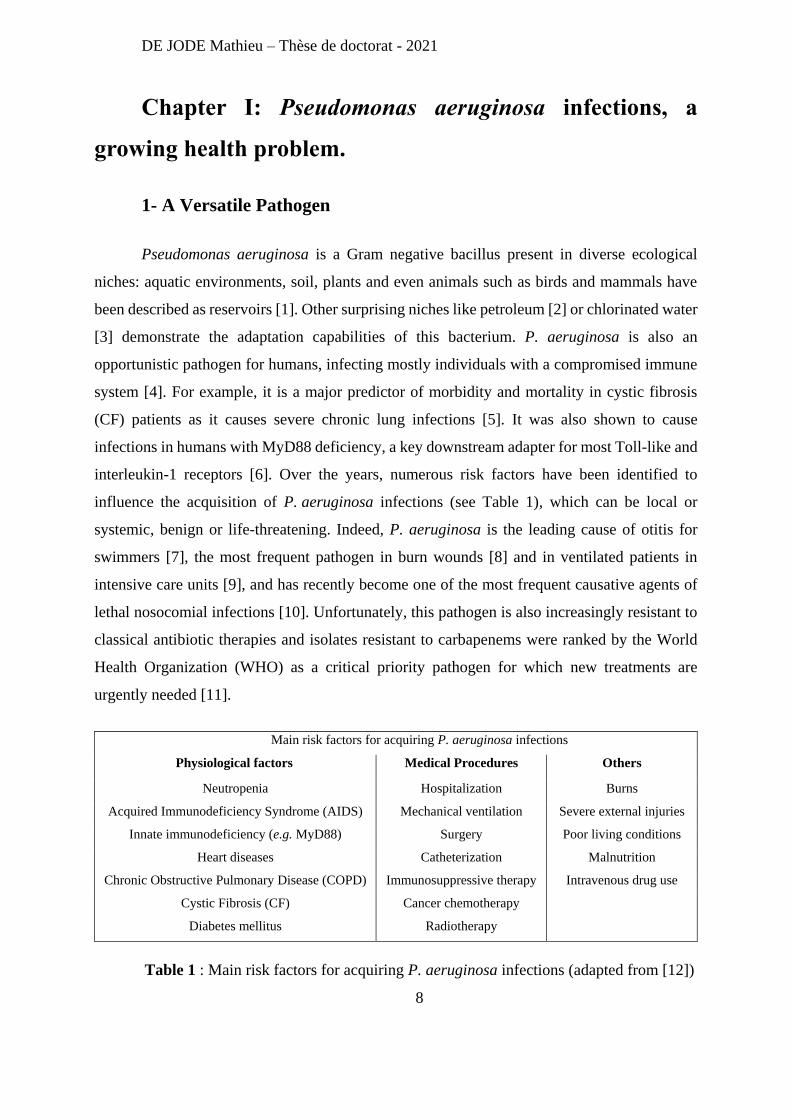
Pseudomonas aeruginosa is a Gram negative bacillus present in diverse ecological
niches: aquatic environments, soil, plants and even animals such as birds and mammals have
been described as reservoirs [1]. Other surprising niches like petroleum [2] or chlorinated water
[3] demonstrate the adaptation capabilities of this bacterium. P. aeruginosa is also an
opportunistic pathogen for humans, infecting mostly individuals with a compromised immune
system [4]. For example, it is a major predictor of morbidity and mortality in cystic fibrosis
(CF) patients as it causes severe chronic lung infections [5]. It was also shown to cause
infections in humans with MyD88 deficiency, a key downstream adapter for most Toll-like and
interleukin-1 receptors [6]. Over the years, numerous risk factors have been identified to
influence the acquisition of P. aeruginosa infections (see Table 1), which can be local or
systemic, benign or life-threatening. Indeed, P. aeruginosa is the leading cause of otitis for
swimmers [7], the most frequent pathogen in burn wounds [8] and in ventilated patients in
intensive care units [9], and has recently become one of the most frequent causative agents of
lethal nosocomial infections [10]. Unfortunately, this pathogen is also increasingly resistant to
classical antibiotic therapies and isolates resistant to carbapenems were ranked by the World
Health Organization (WHO) as a critical priority pathogen for which new treatments are
urgently needed [11].
Main risk factors for acquiring P. aeruginosa infections
Physiological factors Medical Procedures Others
Neutropenia Hospitalization Burns
Acquired Immunodeficiency Syndrome (AIDS) Mechanical ventilation Severe external injuries
Innate immunodeficiency (e.g. MyD88) Surgery Poor living conditions
Heart diseases Catheterization Malnutrition
Chronic Obstructive Pulmonary Disease (COPD) Immunosuppressive therapy Intravenous drug use
Cystic Fibrosis (CF) Cancer chemotherapy
Diabetes mellitus Radiotherapy
Table 1 : Main risk factors for acquiring P. aeruginosa infections (adapted from [12])
DE JODE Mathieu – Thèse de doctorat - 2021
9
P. aeruginosa can colonize a large range of environments and cause a wide array of
infections. This versatility stands from its large genome (5.5 to 7Mbp) which includes more
than 500 regulatory genes, an arsenal of virulence factors and a multitude of drug-resistance
mechanisms [13].
2- The Main Weapons of P. aeruginosa
A) First Contacts
Motility and adhesion are amongst the first properties that P. aeruginosa exploit to
initiate an infection. To these ends, the pathogen relies on two complex extracellular apparatus
the flagellum and the type IV pili. P. aeruginosa possesses a single polar flagellum responsible
for "swimming" and also involved in "swarming" motilities [14]. It also participates in adhesion
to epithelial cells [15]. The flagellum is highly immunogenic and induces strong inflammatory
response as it is recognized by Toll-like receptor (TLR) TLR5 [16]. P. aeruginosa’s motility
also relies on the type IV pili required for "twitching" [17] and "swarming" motilities [14]. This
type IV pili is also P. aeruginosa’s main adhesine, involved in binding to epithelial cells [18].
Adhesion was also reported to be carried by adhesins such as the lectins LecA and LecB. Both
are important virulence factors as, in vitro, lecA and lecB mutants were associated with
decreased cytotoxicity and adhesion on A549 cells compared to the ancestral strain. Moreover,
in vivo (murine acute lung injury model), the increase in alveolar barrier permeability was
reduced with both mutants, as were bacterial burden and dissemination, compared with the
parental strain. Finally, coadministration of specific lectin inhibitors (α-methyl-galactoside and
α-methyl-fucoside) markedly reduced lung injury and mortality [19]. However, these effects
might not be due to adhesive properties of lectins as a group fund that neither lectin was
involved in adhesion to human tracheobronchial mucin [20]. They proposed that the
contributions of lectins to P. aeruginosa’s virulence reported in the literature might be due to
secondary effects on other systems rather than effects of the lectins themselves (e.g. LecB
proteolytic activity and role in pilus biogenesis).
DE JODE Mathieu – Thèse de doctorat - 2021
10
B) Type III Secretion System: Shooting Toxins in Cells
Once P. aeruginosa is in contact with host cells it injects toxins directly in their cytosol.
This process relies mainly on the Type III Secretion System (T3SS), a major virulence factor
[21]. A study looking at 108 P. aeruginosa clinical isolates (from respiratory tract infections)
found that the relative risk of mortality was 6-fold higher when T3SS toxins were produced
[22]. This T3SS allows P. aeruginosa to secrete 4 toxins: ExoS, ExoT, ExoU and ExoY. Both
ExoS and ExoT are Rho GTPase-activating proteins (RhoGAP) and ADP-ribosyltransferases
(ADPRT). These toxins are able to disrupt actin filaments [23] and block the reactive oxygen
species burst in neutrophils [24]. Moreover, ExoT was shown to elicit inhibition of
internalization of P. aeruginosa by epithelial cells and macrophages [25] and promotes the
apoptosis of host cells [26]. Expression of the phospholipase A2 ExoU is associated with a
cytotoxic effect on epithelial cells in vitro, and increases virulence in mice [27, 28]. Its toxicity
relies on host cell membrane disruption [29]. Note that ExoS and ExoU are almost mutually
exclusive in P. aeruginosa, with most strains being ExoS-positive, few strains being ExoU-
positive, and barely any strains having both [30]. Finally, ExoY is an extra-cellular nucleotidyl
cyclase, homologous to other virulence factors (CyaA from Bortedella pertussis or EF from
Bacillus anthracis) [31] and was shown to disrupt the actin cytoskeleton of epithelial cells in
vitro [32]. Recently, secretion of ExoY was shown to be associated with production of cUMP,
higher prevalence of cell apoptosis, and a break of lung barrier integrity in mice [33], but its
precise function in P. aeruginosa pathogenesis still needs investigation.
C) Virulence Without a T3SS
Some P. aeruginosa strains lack the T3SS but are still causing infections. For example,
the clinical strain CLJ1 was able to cause fatal hemorrhage in a mouse model of acute
pneumonia. This phenotype was associated with the expression of ExlA and ExlB. ExlA is a
pore-forming toxin, secreted with the porin ExlB, via a Two‐Partner Secretion System (TPSS).
These two proteins are required and sufficient to provoke membrane permeabilization of human
endothelial cells and macrophages (in vitro) and fatal hemorrhage in mice [34]. Furthermore, a
strain lacking the T3SS is still able to inject toxins directly in the cytosol of cells via the Type
VI Secretion System (T6SS). Indeed, the T6SS, which structurally resembles a bacteriophage
tail [35], is not only able to inject toxins in other bacterial cells, but also into eukaryotic cells,
DE JODE Mathieu – Thèse de doctorat - 2021
11
delivering what is known as “Trans-Kingdoms Toxins”: PldA, PldB and TplE. Both PldA and
PldB are phospholipases D exhibiting antibacterial properties and facilitating intracellular
invasion of eukaryotic cells (in vitro) by activation of the PI3K/Akt pathway [36]. TplE, a
phospholipase A1, is able to disrupt the endoplasmic reticulum, thus promoting autophagy by
the activation of the unfolded protein response [37].
D) Type II Secretion System: Shooting Toxins Next to Cells
Another toxin of interest is ToxA, the most toxic protein secreted by P. aeruginosa with
a Lethal Dose 50 (LD50) of 0.2 mg in mice [38]. While being secreted in the extracellular
milieu by the type II secretion system (T2SS), ToxA has an intracellular target. Indeed, this
toxin with ADPRT activity mediates its entry into target host cells through its cell-binding
domain, then ADP-ribosylates host elongation factor 2 to block protein synthesis [39, 40]. The
T2SS is also known to secrete diverse proteases involve in tissue invasion including LasA,
LasB, and Prpl. Both LasA and LasB are metalloproteases. LasA has low elastolytic and high
staphylolytic activities [41], it also highly increases the elastolytic activity of LasB [42]. LasB
has a strong elastolytic activity and allows for tissue invasion by increasing membrane
permeability of epithelial cells. LasB also inactivates immunoglobulin A (IgA), IgG and
complement components [43-46]. The loss of either LasB or LasA significantly decreases the
ability of P. aeruginosa to invade epithelial cells in vitro and the loss of both proteases leads to
an even lower invasion phenotype [47]. Prpl is another T2SS secreted protease (a lysine
endoproteinase) that degrades proteins such as complement components, immunoglobulins,
elastin, lactoferrin, and transferrin [48].
E) Next Target: Lipids
A different virulence strategy of P. aeruginosa relies on its ability to degrade host cell
phospholipids. In addition to the previously mentioned phospholipases (PldA, PldB and Prpl),
P. aeruginosa uses the hemolytic phospholipase C PlcH (secreted by the T2SS). PlcH can
degrade phospholipids (phosphatidylcholine and sphingomyelin) found in eukaryotic
membranes and in lung surfactant [49] and suppresses bacterium-induced neutrophil respiratory
burst [50]. Furthermore, to increase the efficacy of its phospholipases, P. aeruginosa produces
extracellular glycolipids called rhamnolipids. Indeed, these rhamnolipids act as detergents on
DE JODE Mathieu – Thèse de doctorat - 2021
12
the phospholipids of the lung surfactant [51], disrupt mucociliary transport and ciliary beating
[52], and inhibit phagocytosis [53].
F) Iron Is All I Need
Once host cells have been destroyed, P. aeruginosa has the ability to retrieve important
nutriments, molecules and cofactors necessary to its growth, including iron. Iron is an essential
cofactor of many proteins (iron–sulfur cluster‐containing proteins, enzymes in mitochondrial
respiration…) and is critical to P. aeruginosa physiology [54]. To capture this essential metal,
P. aeruginosa uses iron chelating molecules named siderophores: pyoverdin and pyochelin.
Both siderophores have been shown to be essential virulence factors in diverse mice infection
models [55-57]. P. aeruginosa can also get iron via the uptake of siderophores produced by
other bacteria (xenosiderophores), or via the uptake of heme molecules from the host
hemoproteins, or by using phenazine compounds. Phenazine-1-carboxylic acid (PCA) is the
precursor of pyocyanin, the blue-green compound typical of P. aeruginosa (this bacterium used
to be nicknamed “pyocyanic bacilli”). PCA is able to reduce Fe3+ bound to host proteins into
Fe2+, allowing the uptake of iron via the Feo system [58]. If pyocyanin is less potent than PCA
for iron-uptake, it nevertheless plays other roles during the infection. Indeed, in vitro studies
have shown that pyocyanin inhibits cell respiration, ciliary function, epidermal cell growth, and
also induces neutrophils apoptosis [59-61]. Moreover, it was shown to be a major virulence
factor in vivo [62].
G) LPS: Smooth or Rough
Overall, P. aeruginosa infections are characterized by severe tissue damages and a
strong inflammatory response. The immune system recognizes different components of the
bacterial envelope via Toll like Receptors (TLRs). More specifically, the lipopolysaccharide
(LPS), peptidoglycan and flagellin are recognized by TLR4, TLR2 and TLR5, respectively,
driving the induction of pro‐inflammatory cytokines and type I interferon (IFN) responses [63].
The LPS is composed of 3 parts: the lipid A, which can overstimulate the immune system and
lead to a fatal “septic shock” [64]; the oligosaccharide core; and the O-antigen side chain, a
variable part that can be present (smooth LPS) or absent (rough LPS). A P. aeruginosa strain
with smooth LPS was shown to be significantly more virulent in mice than its rough LPS mutant
DE JODE Mathieu – Thèse de doctorat - 2021
13
[65]. This lower virulence might be related to the involvement of LPS in P. aeruginosa motility.
Indeed, both “swimming” and “swarming” motilities were impaired in mutants that lacked
smooth LPS [14, 66]. Furthermore, P. aeruginosa strains with rough LPS are sensitive to human
serum whereas strains with smooth LPS are not [67].
H) Hacking the Immune System
Infections by P. aeruginosa are characterized by a highly inflammatory immune
response. Unfortunately, P. aeruginosa can efficiently disrupt this immune response. Indeed,
numerous virulence factors previously mentioned (Table 2) degrade proteins of the immune
system (e.g. immunoglobulins, complement components…), inhibit antibacterial cell functions
(e.g. oxidative burst, phagocytosis…) but also disturb cytokine signaling. For example, the
alkaline protease ArpA (secreted by the Type I Secretion System) and the elastase LasB, can
inactivate human interferon (IFN)-γ, tumor necrosis factor (TNF)-α, and degrade two key pro-
inflammatory cytokines, IL-6 and IL-8 as well [68-70]. These two proteases also inhibit
neutrophils function by interfering with their chemotaxis [71], reduce phagocytic activities of
leukocytes [72], inhibit natural killers [73] and disrupt lymphocytes via degradation of
interleukin 2 [74].
DE JODE Mathieu – Thèse de doctorat - 2021
14
Name Product type Secretion type Role
ArpA Metalloprotease I immune system inhibition
ExlA Pore-forming toxin TPSS antiphagocytosis, cytotoxic
ExoS RhoGAP and ADPRT III antiphagocytosis
ExoT RhoGAP and ADPRT III antiphagocytosis, cytotoxic
ExoU Phospholipase A2 III cytotoxic
ExoY Nucleotidyl cyclase III cytotoxic
Flagellum Motility apparatus - motility, adhesion, tissue invasion
LasA Metalloprotease II tissue invasion, immune system inhibition
LasB Metalloprotease II tissue invasion, immune system inhibition
LecA Lectin (binds to galactose) - adhesion, tissue invasion
LecB Lectin (binds to mannose) - adhesion, tissue invasion
LPS Exopolysaccharide - motility, inflammation
PCA Phenazine II iron scavenging
PlcH Phospholipase C II tissue invasion, immune system inhibition
PldA Phospholipase D VI tissue invasion
PldB Phospholipase D VI tissue invasion
PrpL Lysine endoproteinase II tissue invasion, immune system inhibition
Pyochelin Siderophore - iron scavenging
Pyocyanin Phenazine II cytotoxic, immune system inhibition
Pyoverdin Siderophore - iron scavenging
Rhamnolipids Extracellular glycolipids - cytotoxic, antiphagocytosis
Tox A ADPRT II cytotoxic
TplE Phospholipase A1 VI cytotoxic
Type IV pilus Motility apparatus - motility, adhesion
Alginate Exopolysaccharide - resistance to immune cells killing
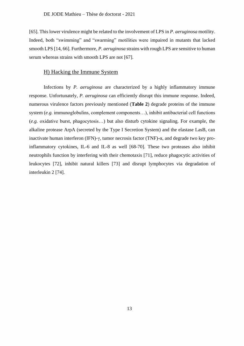
Table 2 : List of the main virulence factors of P. aeruginosa
To summarize, P. aeruginosa possesses a large arsenal of virulence factors allowing it
to destroy cells, invade tissues, and protect itself against immune defenses. Additionally, P.
aeruginosa primarily targets individuals with altered immunity, making self-healing unlikely.
Treatment options for P. aeruginosa infections are relatively limited as this pathogen is
intrinsically resistant to multiple drugs and was found to acquire resistance against others,
strongly reducing the number highly efficient drugs available.
DE JODE Mathieu – Thèse de doctorat - 2021
15
3- Current antibiotic treatments and associated resistance mechanisms
A) Antibiotics Against P. aeruginosa: Choose Carefully
P. aeruginosa is intrinsically resistant to oxazolidinones, macrolides, lincosamides,
streptogramins, daptomycin, glycopeptides, rifampin, trimethoprim-sulfamethoxazole,
tetracycline, some β-lactams (aminopenicillins with or without β-lactamase-inhibitors, as well
as 1st and 2nd generation cephalosporins) [12]. This impressive drug-resistance is due to a
particularly low membrane permeability (12 to 100 fold lower than Escherichia coli
permeability) associated with several efficient efflux systems and antibiotic inactivating
enzymes [75]. Luckily, some antibiotics can still affect most P. aeruginosa isolates:
fluoroquinolones, aminoglycosides, polymyxins, and some β-lactams (3rd and 4th generation
cephalosporins, monobactams, carbapenems, and novel β-lactam/β-lactamase inhibitor
combinations) [12]. Unfortunately, the overuse of these drugs can lead to the selection of
mutations increasing P. aeruginosa antibiotic resistance. Moreover, this pathogen can acquire
additional resistance mechanisms through acquisition of mobile genetic elements like plasmids.
B) All the Roads Lead to Resistance
The membrane of P. aeruginosa is the first line of defense against antibiotics. As the
efficient diffusion/uptake of antibiotics in P. aeruginosa require specific porin or transporters,
resistance can by acquired via mutations in these components: e.g. β-lactam antibiotics and
fluoroquinolones enter bacterial cells through porin channels, and the loss of the OprD porin
leads to carbapenem resistance [76, 77]. Likewise, aminoglycosides uptake involves oxygen-
or nitrogen-dependent electron transport chains, and it was shown that absence of oxygen (e.g.
inside biofilms) or functional deficiency of ATPases contribute to resistance to these antibiotics
[78, 79]; Finally, colistin (polymyxin) promotes its own uptake by interacting with the LPS,
and modification of the lipid A part contributes to polymyxin resistance [80, 81].
Even when a drug successfully reaches the cytoplasm of P. aeruginosa cells, it can be
rapidly expelled by efflux systems. P. aeruginosa possesses multiple efflux systems that
undeniably play a critical role in its intrinsic resistance to different antibiotics including β-
lactams, tetracycline, chloramphenicol, and fluoroquinolones [82, 83]. Overexpression of some
DE JODE Mathieu – Thèse de doctorat - 2021
16
of these efflux systems (by mutations in their regulators) further contribute to the multi-drug-
resistance (MDR) phenotype of P. aeruginosa: e.g. overexpression of MexAB-OprM leads to
increased resistance to β-lactams and quinolones [84, 85].
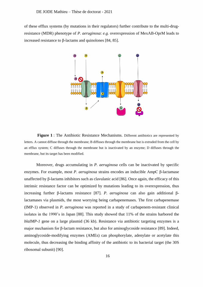
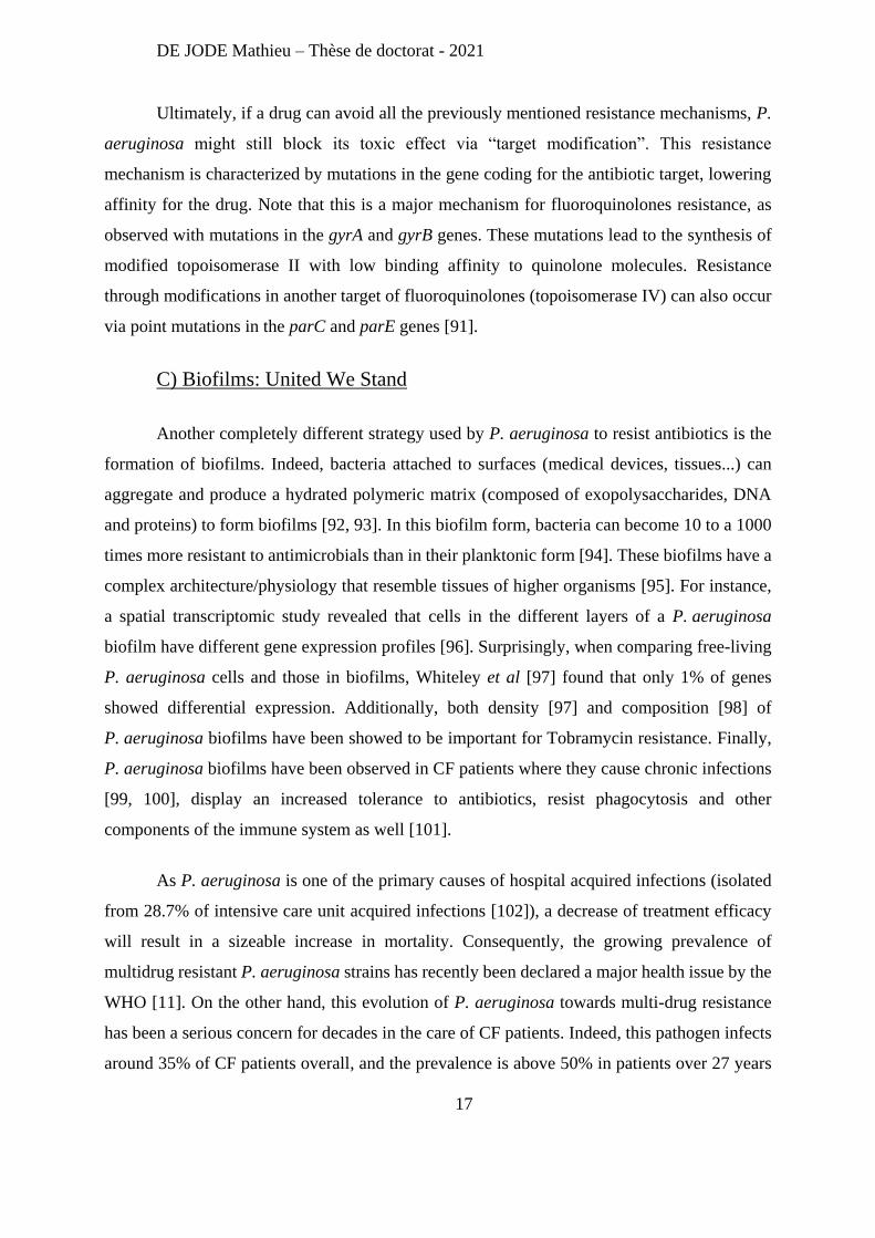
Figure 1 : The Antibiotic Resistance Mechanisms. Different antibiotics are represented by
letters. A cannot diffuse through the membrane; B diffuses through the membrane but is extruded from the cell by
an efflux system; C diffuses through the membrane but is inactivated by an enzyme; D diffuses through the
membrane, but its target has been modified.
Moreover, drugs accumulating in P. aeruginosa cells can be inactivated by specific
enzymes. For example, most P. aeruginosa strains encodes an inducible AmpC β-lactamase
unaffected by β-lactams inhibitors such as clavulanic acid [86]. Once again, the efficacy of this
intrinsic resistance factor can be optimized by mutations leading to its overexpression, thus
increasing further β-lactams resistance [87]. P. aeruginosa can also gain additional β-
lactamases via plasmids, the most worrying being carbapenemases. The first carbapenemase
(IMP-1) observed in P. aeruginosa was reported in a study of carbapenem-resistant clinical
isolates in the 1990’s in Japan [88]. This study showed that 11% of the strains harbored the
blaIMP-1 gene on a large plasmid (36 kb). Resistance via antibiotic targeting enzymes is a
major mechanism for β-lactam resistance, but also for aminoglycoside resistance [89]. Indeed,
aminoglycoside-modifying enzymes (AMEs) can phosphorylate, adenylate or acetylate this
molecule, thus decreasing the binding affinity of the antibiotic to its bacterial target (the 30S
ribosomal subunit) [90].
DE JODE Mathieu – Thèse de doctorat - 2021
17
Ultimately, if a drug can avoid all the previously mentioned resistance mechanisms, P.
aeruginosa might still block its toxic effect via “target modification”. This resistance
mechanism is characterized by mutations in the gene coding for the antibiotic target, lowering
affinity for the drug. Note that this is a major mechanism for fluoroquinolones resistance, as
observed with mutations in the gyrA and gyrB genes. These mutations lead to the synthesis of
modified topoisomerase II with low binding affinity to quinolone molecules. Resistance
through modifications in another target of fluoroquinolones (topoisomerase IV) can also occur
via point mutations in the parC and parE genes [91].
C) Biofilms: United We Stand
Another completely different strategy used by P. aeruginosa to resist antibiotics is the
formation of biofilms. Indeed, bacteria attached to surfaces (medical devices, tissues...) can
aggregate and produce a hydrated polymeric matrix (composed of exopolysaccharides, DNA
and proteins) to form biofilms [92, 93]. In this biofilm form, bacteria can become 10 to a 1000
times more resistant to antimicrobials than in their planktonic form [94]. These biofilms have a
complex architecture/physiology that resemble tissues of higher organisms [95]. For instance,
a spatial transcriptomic study revealed that cells in the different layers of a P. aeruginosa
biofilm have different gene expression profiles [96]. Surprisingly, when comparing free-living
P. aeruginosa cells and those in biofilms, Whiteley et al [97] found that only 1% of genes
showed differential expression. Additionally, both density [97] and composition [98] of
P. aeruginosa biofilms have been showed to be important for Tobramycin resistance. Finally,
P. aeruginosa biofilms have been observed in CF patients where they cause chronic infections
[99, 100], display an increased tolerance to antibiotics, resist phagocytosis and other
components of the immune system as well [101].
As P. aeruginosa is one of the primary causes of hospital acquired infections (isolated
from 28.7% of intensive care unit acquired infections [102]), a decrease of treatment efficacy
will result in a sizeable increase in mortality. Consequently, the growing prevalence of
multidrug resistant P. aeruginosa strains has recently been declared a major health issue by the
WHO [11]. On the other hand, this evolution of P. aeruginosa towards multi-drug resistance
has been a serious concern for decades in the care of CF patients. Indeed, this pathogen infects
around 35% of CF patients overall, and the prevalence is above 50% in patients over 27 years
DE JODE Mathieu – Thèse de doctorat - 2021
18
old [103]. Since chronic airway infection is the most important cause of morbidity and mortality
in CF patients, P. aeruginosa has been extensively studied in this environment.
4- P. aeruginosa in Cystic Fibrosis: From Acute to Chronic Infections.
A) Cystic Fibrosis Lungs: a Bacterial Paradise
CF is the most common life-shortening autosomal recessive disorder for Caucasian
(affecting 1 in 2500 newborns), and is caused by mutations in the CF-transmembrane
conductance regulator (CFTR) [104]. These mutations (over 300 have been documented by the
CF Genetic Analysis Consortium) lead to a defective chloride (Cl−) channel, which disrupts
electrolyte secretion, thus increasing the viscosity of the mucus secreted at the surface of
epithelial cells and affecting osmolarity [104]. The main lung defenses against bacterial
colonization (mucociliary clearance, polymorphonuclear neutrophil phagocytosis, and local
production of antibacterial cationic peptides) are not performing efficiently in the CF
environment, resulting in chronic bacterial infections [105, 106]. Nonetheless, in this specific
environment, bacteria must survive the limitations in growth factors, dehydration, the host’s
immune defenses, and the yearlong antibiotic therapies. Most CF patients become permanently
colonized by P. aeruginosa, thus the same bacterial lineage can persist in the lungs for years or
even decades [107]. With the advent of cheaper genome sequencing technologies, evolution of
P. aeruginosa in the lungs of CF patients has been extensively studied and genetic basis of
several phenotypes have now been identified.
B) Mucoidy Origins
The most characterized phenotype of P. aeruginosa isolates from CF patients is
mucoidy. In the 1960’s Doggett et al reported “An atypical Pseudomonas aeruginosa
associated with cystic fibrosis” [108, 109]. This “atypical” P. aeruginosa was defined by an
uncommon aspect on agar plates and liquid cultures [108]. This mucoid phenotype corresponds
to an overproduction of alginate (an exopolysaccharide). It was then reported that this mucoid
phenotype was absent from recently P. aeruginosa-infected CF patients, whereas it was present
in patients with chronic infection [110]. Later, the mechanism of mucoidy conversion was
identified by Martin et al [111]. They discovered that a mutation in the mucA gene leads to a
DE JODE Mathieu – Thèse de doctorat - 2021
19
loss of function of this inhibitor of the sigma factor AlgU, the latter regulating the transcription
of genes involved in the alginate biosynthetic pathway. The couple MucA/AlgU, localized at
the cytoplasmic membrane, controls the expression of more than 200 genes related to membrane
homeostasis (membrane proteins, LPS, peptidoglycan synthesis…). Its activation upon
membrane stress, starts with the proteolysis of MucA catalyzed by AlgW. Once free from its
anti-sigma factor MucA, AlgU binds to the RNA polymerase and direct the transcription of its
regulon [112]. Recently a portion of mucA was shown to be essential in P. aeruginosa [113].
Indeed, overexpression of algU in the absence of MucA prevents cell growth, and this
phenotype can be rescued by the overproduction of RpoD (the housekeeping sigma factor).
In CF strains, mutations of mucA are commonly found, which implies an important role
for alginate overproduction in P. aeruginosa’s adaptation to the CF lung environment. This role
might be to protect P. aeruginosa from the constant inflammation in the lungs as it increases
resistance to antibody-independent opsonic killing [114], confers resistance to phagocyte-
generated hypochlorite [115], decreases phagocytosis of planktonic mucoid bacteria by
neutrophils and macrophages [116], and reduces polymorphonuclear chemotaxis while
inhibiting activation of the complement system [117]. Additionally, alginate plays a role in
antibiotic resistance since biofilms formed by an alginate-overproducing strain were shown to
be more resistant to tobramycin than biofilms formed by an isogenic non-mucoid strain [118].
Conversely, the use of alginate lyase proved to increase antibiotic efficacy against mucoid P.
aeruginosa biofilms [119].
C) Evolution of P. aeruginosa in CF: Less Virulence, More Resistance
Longitudinal studies of P. aeruginosa’s genome throughout years of lung infection in
CF patients, shined a light on its evolution in this environment. Such a study of the phenotypic
and genotypic changes in P. aeruginosa isolates, was performed in a cohort of 40 CF children
during the first 3 years of life [120]. A high degree of genotypic variability was found as each
patient had unique genotypes. Surprisingly, the early isolates were generally non-mucoid and
antibiotic susceptible, which is unusual for CF isolates. These results support the hypothesis of
an initial infection by “classical” non-mucoid P. aeruginosa strains which then evolve during
the chronic infection to become the mucoid “CF” strain.
DE JODE Mathieu – Thèse de doctorat - 2021
20
In another study, Smith et al. [121] used whole-genome sequencing to investigate from
the same patient genomic adaptations of P. aeruginosa isolates 7.5 years apart (early vs
chronic). The analysis revealed 68 mutations in the late isolate, mostly single-base pair changes,
and half of them predicted to result in change or loss of protein function. Remarkably, 13 out
of the 68 mutated genes were coding virulence factors and regulators, including genes related
to O-antigen biosynthesis, T3SS, twitching motility, regulation of exotoxin A, phenazine
biosynthesis, quorum sensing, and iron acquisition. These mutations were then confirmed
phenotypically, as loss of serotype-specific antigenicity, loss of twitching motility, loss of
pyoverdin production, loss of secreted protease activities, and reduced biofilm formation were
observed. In addition, numerous efflux systems were mutated in the late isolate, which might
explain the higher level of resistance of this strain to aminoglycosides (compare to the early
strain). Moreover, by analyzing multiple samples from the same patient, at each time point, the
authors found that multiple related lineages coexisted over the course of this patient’s infection.
For example, mucA mutations arose independently in three different lineages. Isolates from a
single time point also had heterogeneous genotypes. Finally, they investigated whether the
identified mutations were common in CF patients by genotyping paired (early and late) P.
aeruginosa isolates from 29 other patients and found that mexZ (repressor of MexXY-OprM)
and lasR (a quorum sensing regulator associated with the regulation of many virulence factors)
were mutated in most CF patients. In a following study [122] analyzing the same set of strains,
the authors found that the observed mutations were significantly concentrated in “mutator
lineages” (defined as strain deficient for DNA mismatch repair system). Furthermore, they
estimated that non-mutator lineages acquired a median of only 0.25 mutation per year of
infection, while this rate was over 3 mutations per year in mutator lineages. These hyper-
mutable P. aeruginosa were found to be prevalent in CF as another study reported 36% of the
patients were colonized with a mutator strain [123]. This study also noted that mutator CF
strains were significantly more resistant to antibiotics than their non-mutator counterpart.
To summarize, in CF patients, its seems that the initial P. aeruginosa infection involves
a “classical/environmental” strain that will colonize the lungs thanks to its invasive virulence
factors. During the following years, P. aeruginosa will be confronted to many challenges
including the immune defenses and year-long antibiotic treatments. To survive in this stressful
environment, P. aeruginosa will adapt by losing major virulence factors (e.g. loss of secreted
toxins) and deploying defense systems (e.g. mucoid conversion and overexpression of efflux
DE JODE Mathieu – Thèse de doctorat - 2021
21
pumps). Moreover, this adaptation seems catalyzed by the selection of mutator strains which
accumulate mutations at increased rates. The adapted “CF” strains become so difficult to
eradicate that current therapeutic strategies are focused on avoiding the primary infection
(alcohol-based antiseptic hand rubs, use of chirurgical mask…) [124], and aggressive antibiotic
therapies against the early infections [125]. A study found that these early antibiotics treatments
can lead to a P. aeruginosa free-period of 18 months in average, and delays the decline of lung
function, but new acquisition with different P. aeruginosa genotypes occurred in 73% of the
studied patients [125]. Unfortunately, failure rate of treatments of exacerbation in CF patients
increases when the number of active drugs decreases (three active drugs: 0% of treatment
failure; two: 24%; one: 27%; zero: 43%) [126]. To summarize, if the increased prevalence of
MDR P. aeruginosa strains is a serious threat to the general public health, it is especially
worrisome for CF patients.
In conclusion, P. aeruginosa is a widespread bacterium, able to colonize and survive
harsh environments, from chlorinated water to chronically inflamed CF lungs. This versatile
pathogen possesses an arsenal of virulence factors allowing it to efficiently damage tissues and
resist immune defenses. Its numerous antibiotic resistance mechanisms provide protection
against most (sometimes all) current treatment options. To fight against this highly adaptable
pathogen, our laboratory investigates an antimicrobial weapon that has been co-evolving with
P. aeruginosa for billions of years: its natural viral predators, the bacteriophages.
DE JODE Mathieu – Thèse de doctorat - 2021
22
Chapter II: Bacteriophages, the most abundant
bacterial predators.
1- The Three Faces of Phage Research.
A) An Ubiquitous Predator.
Bacteriophages, literally “bacteria eaters”, are viruses infecting bacteria. This name was
given upon their discovery by Félix d’Hérelle in 1917, while he was working at Institut Pasteur
in Paris [127]. Bacteriophages (or phages), are ubiquitous: every place where bacteria can grow,
associated phages are found, including in the human body. Indeed, phages are particularly
highly abundant in the human gut microbiome and are involved in shaping this microbiome, in
both healthy and diseases conditions [128]. More unexpected is the total number of phages on
Earth that is estimated to be around 1031, making phages the most abundant biological entity on
our planet [129]. Ecological functions of these abundant phages have been investigated,
particularly in marine ecosystems. There, phages play critical roles in the structure and function
of aquatic food webs, and are estimated to kill almost 20% of the bacterial community daily
[130, 131]. These killing rates make phages the primary predator of bacteria, which leads to a
never-ending selective pressure, driving the evolution of both phages and bacteria. This
relationship is a well-characterized example of the Red Queen hypothesis: predator and prey
species must constantly evolve [132].
Investigations on the abundance and ecological roles of phages are actually quite recent
and powered by the advances in bioinformatics and sequencing technologies (especially
metagenomics). However, historically, phages were first studied for their potent bactericidal
activities.
B) Phage Therapy Part 1: Chaotic Beginnings.
As soon as in its first report on phages, Félix d’Hérelle investigated their use as
antimicrobials. In 1919, he successfully treated chickens infected with Salmonella gallinarum
[133, 134], and in 1921, five humans with dysentery were cured using a phage that infects
Shigella dysenteriae [134]. At this time, antibiotics were not yet discovered (Fleming will report
DE JODE Mathieu – Thèse de doctorat - 2021
23
the discovery and proprieties of penicillin in 1929 [135]) and the antibacterial arsenal was fairly
limited. In this context, d’Hérelle successes with phages allowed him to travel the world to
conduct numerous phage therapy studies. In 1925, in Egypt, d’Hérelle reported rapid healing
from bubonic plague following only one injection in each of the two buboes of the patient [136].
Furthermore, in 1927, despite reluctant medical professionals, d’Hérelle conducted a study of
phage treatment for cholera in India that yielded excellent results: while the mortality rate of
the untreated group (124 patients) was 63%, it was only 8% in the phage-treated group [137].
Unfortunately, these encouraging results were overshadowed by contradicting results regarding
the efficacy of phage therapy (probably due to the use of phages with limited host range) [138]
and problems related to clinical study designs making results hard to interpret [139].
Additionally, it is now believed that phage preparations at this time were not optimized with
highly variable phage titers and heavily contaminated with LPS and other bacterial debris,
which could both lead to treatment failure. Moreover, the nature of phages themselves was
debated for a long time as most of the scientific community (on the impulse of Nobel Prize
winner Jules Bordet, involved in a personal feud with d’Hérelle) discarded d’Hérelle’s theory
on phages being parasites in favor of the theory of a “self-perpetuating lytic enzyme” [140]. It
is only in the 1940’s that this debate was put to an end as electron microscopy micrograph of
phages revealed without a doubt their viral nature [141]. Overall, the lack of understanding of
basic phage biology, a general distrust from part of the scientific and medical communities, and
the increased use of a novel very efficient antimicrobial (the first antibiotic, penicillin)
precipitated the decline of phage therapy, until its reappraisal about 100 years later.
C) Phages and the Birth of Molecular Biology.
The halt in phage therapy research around the 1940’s, did not however mark the end of
basic research on phages. Indeed, by the 1950’s significant advances were made on the
biological processes supporting the parasitic lifestyle of phages in bacteria and paved the way
for the emergence of the field of molecular biology. Phages were instrumental in discovering
that DNA molecules hold the genetic information [142] and that messenger RNAs are
intermediate molecules carrying this genetic information to ribosomes for protein synthesis
[143]. Moreover, the study of phage-encoded DNA-manipulating enzymes (DNA and RNA
polymerases, ligases and endo- and exonucleases), and the discovery of bacterial restriction
enzymes (used to protect bacteria against phages), provided the first molecular tools for genetic
DE JODE Mathieu – Thèse de doctorat - 2021
24
engineering that are still frequently used today [144]. Nowadays, phage research continues to
fuel molecular biology advances as witnessed by the revolution in genome editing initiated
from the characterization of a bacterial phage resistance mechanism: the CRISPR–Cas system
[145, 146].
The three faces of phage research (ecology, medicine and biotechnology) have emerged
during the 20th century and are now fully integrated into a renewed worldwide interest on these
viruses. Moreover, the lack of understanding of basic phage biology was a major downfall of
phage therapy, but we have since learned much about the basic processes governing phages life
cycles, their host ranges and mechanisms by which bacteria might become phage-resistant.
2- Introduction to phage biology.
A) Phage Diversity and their Classification.
Phage classification, organized by the International Committee on Taxonomy of Viruses
(ICTV), relies on both genomic and morphological information [147]. Note that the ICTV
continues to work on this classification and update it periodically [148] (for an up to date phage
classification please consult the latest ICTV publications). This classification helps appreciate
the immense diversity of phages. Indeed, their genetic information can consist of double-
stranded (ds), single-stranded (ss) DNA, or RNA. Moreover, phage genomes size can vary a
lot, from 3.5 kb of ssRNA for phage MS2 [149] to 670 kb of dsDNA for Bacillus megaterium
phage G [150]. Regarding morphology, if 96% of isolated phages are tailed, other morphologies
have been described (polyhedral, filamentous, or pleomorphic) [151] (Figure 2). Like some
eukaryotic viruses, phages particles can be enveloped by a layer of lipids or lipoproteins [151].
DE JODE Mathieu – Thèse de doctorat - 2021
25
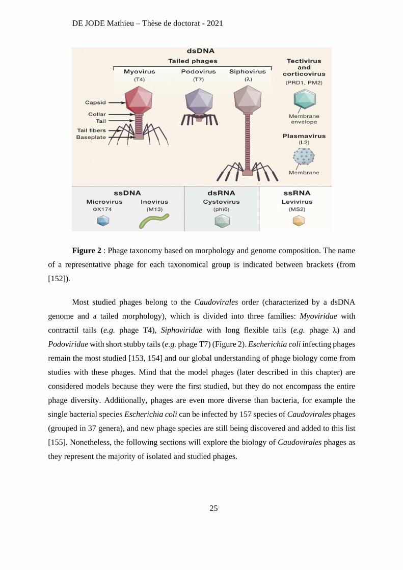
Figure 2 : Phage taxonomy based on morphology and genome composition. The name
of a representative phage for each taxonomical group is indicated between brackets (from
[152]).
Most studied phages belong to the Caudovirales order (characterized by a dsDNA
genome and a tailed morphology), which is divided into three families: Myoviridae with
contractil tails (e.g. phage T4), Siphoviridae with long flexible tails (e.g. phage λ) and
Podoviridae with short stubby tails (e.g. phage T7) (Figure 2). Escherichia coli infecting phages
remain the most studied [153, 154] and our global understanding of phage biology come from
studies with these phages. Mind that the model phages (later described in this chapter) are
considered models because they were the first studied, but they do not encompass the entire
phage diversity. Additionally, phages are even more diverse than bacteria, for example the
single bacterial species Escherichia coli can be infected by 157 species of Caudovirales phages
(grouped in 37 genera), and new phage species are still being discovered and added to this list
[155]. Nonetheless, the following sections will explore the biology of Caudovirales phages as
they represent the majority of isolated and studied phages.
DE JODE Mathieu – Thèse de doctorat - 2021
26
Overlapping with the ICTV classification, phages can also be classified according to
their lifestyle: virulent phages only able to replicate through a lytic cycle, or temperate phages
able to replicate via a lytic or a lysogenic cycle.
B) Phage Life Cycles.
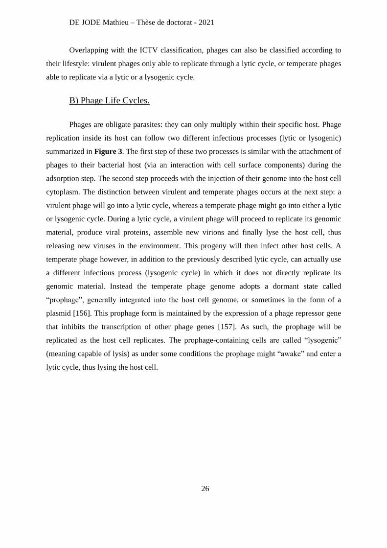
Phages are obligate parasites: they can only multiply within their specific host. Phage
replication inside its host can follow two different infectious processes (lytic or lysogenic)
summarized in Figure 3. The first step of these two processes is similar with the attachment of
phages to their bacterial host (via an interaction with cell surface components) during the
adsorption step. The second step proceeds with the injection of their genome into the host cell
cytoplasm. The distinction between virulent and temperate phages occurs at the next step: a
virulent phage will go into a lytic cycle, whereas a temperate phage might go into either a lytic
or lysogenic cycle. During a lytic cycle, a virulent phage will proceed to replicate its genomic
material, produce viral proteins, assemble new virions and finally lyse the host cell, thus
releasing new viruses in the environment. This progeny will then infect other host cells. A
temperate phage however, in addition to the previously described lytic cycle, can actually use
a different infectious process (lysogenic cycle) in which it does not directly replicate its
genomic material. Instead the temperate phage genome adopts a dormant state called
“prophage”, generally integrated into the host cell genome, or sometimes in the form of a
plasmid [156]. This prophage form is maintained by the expression of a phage repressor gene
that inhibits the transcription of other phage genes [157]. As such, the prophage will be
replicated as the host cell replicates. The prophage-containing cells are called “lysogenic”
(meaning capable of lysis) as under some conditions the prophage might “awake” and enter a
lytic cycle, thus lysing the host cell.
DE JODE Mathieu – Thèse de doctorat - 2021
27
Figure 3 : Schematic representation of Lytic and Lysogenic phage life cycles (from
[144]).
Temperate phages, in the prophage state, provide additional genetic material to their
bacterial host. They have actually been shown to be a major contributor to genetic diversity in
pathogen like Escherichia coli [158], and can provide virulence factors, like the cholera toxin
acquired by Vibrio cholerae through the filamentous phage CTXΦ [159]. Temperate phages
also play a role in the dissemination of antibiotic resistance genes via transduction [160, 161],
a mechanism in which bacterial host DNA is encapsided in newly formed virions and delivered
to another cell (the phage acting like a DNA delivery system between two bacteria). All of these
properties make temperate phages ill-suited for phage therapy, and therapeutic phage genomes
are routinely screened for gene coding integrases, toxins or antibiotic resistance determinants
[162]. Note that this exclusion of temperate phages for phage therapy is now being questioned
as recently, genetically modified temperate phages were successful in treating a Mycobacterium
abscessus infection in a 15-year-old CF patient [163]. These temperate phages were engineered
by removing the repressor genes, essential to the lysogenic pathway, making them strictly lytic.
DE JODE Mathieu – Thèse de doctorat - 2021
28
Phage therapy relies on the antimicrobial proprieties of phages, namely their ability to
lyse bacteria. Thus, knowledge on how phages perform each step of the lytic pathway is useful
to guide the choice of therapeutic phages. Moreover, the bacterial mechanisms to disrupt this
infectious process and gain resistance to phages also need to be known to exploit phage therapy
to its maximum potential.
C) Adsorption: First Step Toward Infection.
The infection process starts with the adsorption of phage to its host. This process
consists in a series of interactions between the phage binding proteins and the bacterial cell
surface receptors. Doing so allows the virus to recognize a suitable host, and then positions
itself to injects its DNA. The adsorption can be subdivided in three steps: the initial contact, the
reversible binding and finally, the irreversible attachment.
The initial contact is a game of chance as it relies on random collisions between phage
and host caused by diffusion, dispersion, Brownian motion…[164]. Subsequently, during the
reversible step, phage binding to the bacterial surface components is not definitive, as phage
can detach from the host [165]. This reversible interaction may help the phage in its search for
a specific receptor, by keeping it close to the host cell surface [164]. Once the phage-binding
domains and the specific bacterial receptor connect, conformational changes in the phage allow
its genome to be ejected into the host [166].
Phage can use a wide array of cell surface receptors. Moreover, the membrane
architecture of Gram positive and Gram negative bacteria being very distinctive, their
associated phages use different receptors. On the one hand, phages infecting Gram positive
bacteria have been shown to use element of the cell wall, including peptidoglycan and teichoic
acids as receptors [167]. On the other hand, phages infecting Gram negative bacteria can use
numerous receptors including proteins (like OmpC) [168], different part of the LPS [169] or
capsular polysaccharides [170]. Interestingly, phages using specific bacterial appendages, such
as the flagella and pili as receptors have been described for both Gram positive [171, 172] and
Gram negative bacteria [173, 174].
DE JODE Mathieu – Thèse de doctorat - 2021
29
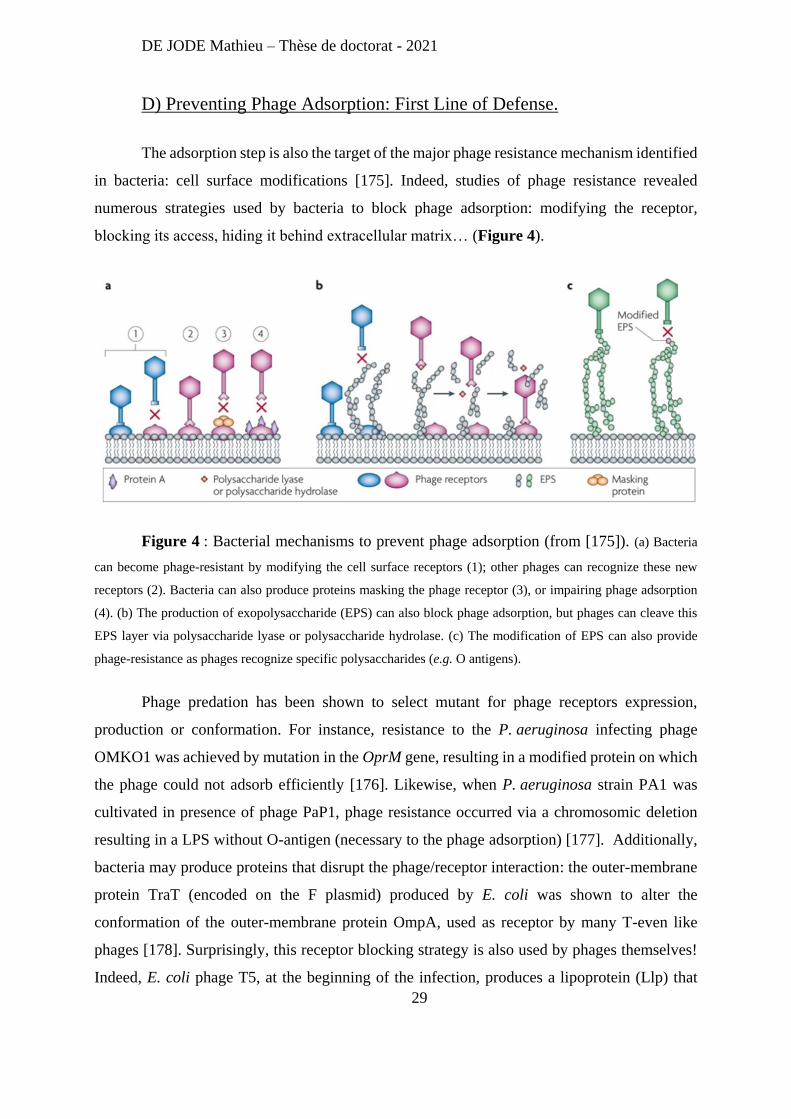
D) Preventing Phage Adsorption: First Line of Defense.
The adsorption step is also the target of the major phage resistance mechanism identified
in bacteria: cell surface modifications [175]. Indeed, studies of phage resistance revealed
numerous strategies used by bacteria to block phage adsorption: modifying the receptor,
blocking its access, hiding it behind extracellular matrix… (Figure 4).
Figure 4 : Bacterial mechanisms to prevent phage adsorption (from [175]). (a) Bacteria
can become phage-resistant by modifying the cell surface receptors (1); other phages can recognize these new
receptors (2). Bacteria can also produce proteins masking the phage receptor (3), or impairing phage adsorption
(4). (b) The production of exopolysaccharide (EPS) can also block phage adsorption, but phages can cleave this
EPS layer via polysaccharide lyase or polysaccharide hydrolase. (c) The modification of EPS can also provide
phage-resistance as phages recognize specific polysaccharides (e.g. O antigens).
Phage predation has been shown to select mutant for phage receptors expression,
production or conformation. For instance, resistance to the P. aeruginosa infecting phage
OMKO1 was achieved by mutation in the OprM gene, resulting in a modified protein on which
the phage could not adsorb efficiently [176]. Likewise, when P. aeruginosa strain PA1 was
cultivated in presence of phage PaP1, phage resistance occurred via a chromosomic deletion
resulting in a LPS without O-antigen (necessary to the phage adsorption) [177]. Additionally,
bacteria may produce proteins that disrupt the phage/receptor interaction: the outer-membrane
protein TraT (encoded on the F plasmid) produced by E. coli was shown to alter the
conformation of the outer-membrane protein OmpA, used as receptor by many T-even like
phages [178]. Surprisingly, this receptor blocking strategy is also used by phages themselves!
Indeed, E. coli phage T5, at the beginning of the infection, produces a lipoprotein (Llp) that
DE JODE Mathieu – Thèse de doctorat - 2021
30
blocks its own receptor, the ferrichrome-iron receptor (FhuA). This, not only prevents
superinfection, but also limit the binding of newly released virions to cells debris, which would
inactivate them. [179]. The inactivation of phages through their adsorption on a “decoy
receptor” present on outer membrane vehicles (OMVs) has also been described as a bacterial
mechanism to prevent infection [180, 181]. OMVs are nanostructures composed of outer
membrane lipids and proteins, and periplasmic components. They are secreted by Gram
negative bacteria and play a role in diverse biological functions (stress response, nutriments
acquisition…) [182]. As their surface present the same elements than the bacterial surface,
phages might adsorb and get stuck on these OMVs.
Additionally, bacteria can prevent phage adsorption by masking the phage receptor
behind a layer of extracellular polysaccharides. For example, bacteria might produce a capsule,
thus hiding the phage-receptor and granting phage resistance. This strategy was demonstrated
for different bacteria/phage couples including Staphylococcus simulans/phage U16 [183],
Staphylococcus aureus/phage 84 [184] or E. coli/phage T7 [185]. Note that this approach can
efficiently protect bacteria from multiple distinct phages as a coat of exopolysaccharide can
hide different phage receptors at once. For instance, Betts et al [186] showed that when
P. aeruginosa strain PA01 grew in presence of a single phage, resistant clones had mutations
in genes related to the phage receptor (LPS, or pili, depending on the phage), but when the same
strain was submitted to 5 different phages at once, one of the resistant clone had a single
mutation in mucA (resulting in a mucoid phenotype). Interestingly, the selection of mucoid
P. aeruginosa strains following phage predation has been recorded multiple times [187, 188]
hinting that it is an advantageous trait in these conditions. However, mucoidy does not provide
complete phage resistance [186] and some phages infecting P. aeruginosa encode alginate lyase
[189].
E) Genome Injection or Ejection?
The first theories on phage DNA injection postulated that the virus act like an
hypodermic syringe [190]. However, this representation is challenged by new insights in this
phenomenon. Indeed, it is now believed that osmotic pressures and the entry of water inside of
the phage particle are the main driver of the phage genome transfer in the bacterial cell. As
such, many researchers refer this phenomenon as “genome ejection” instead of “genome
DE JODE Mathieu – Thèse de doctorat - 2021
31
injection”. Briefly, when the phage genome is packaged in the capsid, most of the water that
normally hydrates the DNA is removed. Moreover, most tailed phages contained within the
capsid ∼500 mg/mL of DNA, creating an osmotic imbalance between the capsid and the
bacterial cell. Once the phage recognized its specific receptor, the tip of its tail might penetrate
the peptidoglycan layer and touch or penetrate the inner membrane via enzymatic activity.
According to the hydrodynamic model of phage genome ejection, once the exit channel is open,
water diffuses through the capsid shell and neutralize the osmotic imbalance. It is the
hydrostatic pressure gradient across the tail that pushes the phage DNA out, into the bacterial
cell [166]. This general mechanism of phage genome ejection is still being investigated, and it
is already known that smaller phages may eject their genome using different mechanisms. As
the tail of Podoviruses (e.g. T7) is too short to span across the cell envelope, phage proteins
(e.g. Gp14, Gp15 and Gp16) are needed to form a channel allowing phage DNA ejection into
the cell [191]. Moreover, phage T7 genome ejection is a three-step process: After phage
adsorption and formation of the protein channel, approximately 10% of the genome (40kb) is
internalized by the cell, the transport of the next 50% is coupled to the transcription process by
E. coli RNA polymerase, and the final 40% internalization is dependent on transcription by the
T7 RNA polymerase [192, 193].
As previously mentioned for the adsorption step, this genome ejection step can be
disrupted by bacteria. Interestingly these defense mechanisms are mostly conferred via
integrated prophages that block genome ejection of infecting phages.
F) Superinfection Exclusion: A Prophage Protects Against Phages
Some temperate phages encode superinfection exclusion systems designed to protect
the bacterial host against other phages. For instance, E. coli phage P1 encodes the sim gene
conferring resistance to other P1 phages [194]. Likewise, Salmonella phage P22 encodes sieA
protecting its host against phages L, MG178 and MG40 [195]. The underlying molecular
mechanism of these immunity proteins is not yet fully understood, but the fact that phage
adsorption is not affected by the presence of these proteins and that a bacterium can be
successfully transformed with the phage genome hint that they must prevent the ejection step
of the infection.
DE JODE Mathieu – Thèse de doctorat - 2021
32
When the phage genome ejection is not prevented, bacteria might still manage to disrupt
the infection process by degrading the phage genetic material.
G) Restriction-Modification Systems: Cutting Genome of Phages
Most studied bacteria encode Restriction-Modification systems (RMs). RMs main
function is to protect the bacterial cell against foreign DNA, including phage DNA. Indeed,
when unmethylated phage DNA is ejected in the cytoplasm, it can be rapidly degraded by
restriction enzymes. Being methylated, the host DNA is protected from these restriction
enzymes. Against this simple, yet effective system, phages have developed a wide array of
countermeasures: use of a phage-encoded methylase [196]; absence of endonuclease
recognition sites in their genomes [197]; use of modified bases, not recognized by restriction
enzymes (e.g. phage T4 DNA has hydroxymethylcytosine instead of cytosine) [198].
Another bacterial defense mechanism based on phage DNA degradation is the now
famous CRISPR-Cas system.
H) CRISPR-Cas: Adaptive Immunity against Phages
The CRISPRs (clustered regularly interspaced short palindromic repeats) Cas (CRISPR-
associated proteins) system is another restriction based defense mechanism. Different bacterial
species encode different CRISPR systems [199] but a general overview can be provided:
CRISPR–cas loci are composed of direct repeats (21–48 bp) interspaced by non-repetitive
spacers (26–72 bp) and, flanked by cas genes (Figure 5).
DE JODE Mathieu – Thèse de doctorat - 2021
33
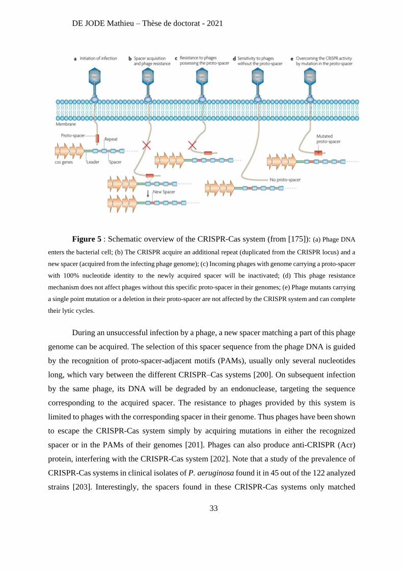
Figure 5 : Schematic overview of the CRISPR-Cas system (from [175]): (a) Phage DNA
enters the bacterial cell; (b) The CRISPR acquire an additional repeat (duplicated from the CRISPR locus) and a
new spacer (acquired from the infecting phage genome); (c) Incoming phages with genome carrying a proto-spacer
with 100% nucleotide identity to the newly acquired spacer will be inactivated; (d) This phage resistance
mechanism does not affect phages without this specific proto-spacer in their genomes; (e) Phage mutants carrying
a single point mutation or a deletion in their proto-spacer are not affected by the CRISPR system and can complete
their lytic cycles.
During an unsuccessful infection by a phage, a new spacer matching a part of this phage
genome can be acquired. The selection of this spacer sequence from the phage DNA is guided
by the recognition of proto-spacer-adjacent motifs (PAMs), usually only several nucleotides
long, which vary between the different CRISPR–Cas systems [200]. On subsequent infection
by the same phage, its DNA will be degraded by an endonuclease, targeting the sequence
corresponding to the acquired spacer. The resistance to phages provided by this system is
limited to phages with the corresponding spacer in their genome. Thus phages have been shown
to escape the CRISPR-Cas system simply by acquiring mutations in either the recognized
spacer or in the PAMs of their genomes [201]. Phages can also produce anti-CRISPR (Acr)
protein, interfering with the CRISPR-Cas system [202]. Note that a study of the prevalence of
CRISPR-Cas systems in clinical isolates of P. aeruginosa found it in 45 out of the 122 analyzed
strains [203]. Interestingly, the spacers found in these CRISPR-Cas systems only matched
DE JODE Mathieu – Thèse de doctorat - 2021
34
temperate pseudomonas phages. Finally, these CRISPR-Cas systems did not provide resistance
against the tested phages, even for CRISPR with spacers 100 % identical to a region of the tested
phage. Despite this surprising results, other studies (using the PA14 lab strain) have
demonstrated that P. aeruginosa can readily acquire phage resistance via CRISPR-Cas systems
[204]. However, this mechanism of resistance seems more prevalent following mono-phage
treatment as cell surface modifications were more prevalent after multi-phage treatment [205].
I) From Bacterial Cell to Virocell
To achieve a successful infection of their host cell and produce new virions, phages
hijack several cell resources and shift the “bacterial cell physiology” toward a “virocell
physiology” [206]. This transformation is due to efficient hijacking of different bacterial
machineries, catalyzed by the expression of “early” expressed phage genes. A target of choice
in this endeavor is the bacterial RNA polymerase, as disrupting the host transcription will have
tremendous effects on its physiology. The E. coli phage T4 actually possess 11 genes dedicated
to its interaction with the host RNA polymerase [207-213], which redirect its activity during
the infectious process towards the transcription of specific phage genes. Other strategies exist
as phage T7 inhibits the host RNA polymerase and uses its own [214]. Phages can also regulate
RNA degradation as the degradosome interacting protein (Dip) of P. aeruginosa phage phiKZ
prevents viral RNA degradation in infected cells [215]. Additionally, phages PAK_P3 and
PAK_P4 (isolated in our laboratory), also infecting P. aeruginosa, affect RNA turnover by
eliciting rapid host transcripts degradation via an unknown mechanism [216, 217]. Note that
the underlying mechanism might be different for these two phages as the kinetic of host RNA
degradation during PAK_P4 infection is much faster than during PAK_P3 infection [217].
Phages have also been shown to shut off ‘non-essential’ host processes presumably to
preserve energy for the infectious process. For instance, E. coli phage Rac uses the Kil protein,
interacting with the bacterial tubulin homologue FtsZ to impair cell division [218]. Another
E. coli phage, N4, uses Gp8 to shut off host DNA replication [219].
Phages can also tune the host cell metabolism to favor viral replication. To this end,
phages use acquire host-derived metabolic genes called “auxiliary metabolic genes” (AMGs).
These AMGs have been found to interfere with a wide array of metabolic processes including
DE JODE Mathieu – Thèse de doctorat - 2021
35
nucleic acid synthesis [220], phosphate [221] and nitrogen metabolism [222], the pentose
phosphate pathway [223], and even photosynthesis [224]. PAK_P3 phage was shown to
particularly alter the pyrimidine metabolism [216], whereas PAK_P4 manipulates the
expression of iron-related host genes [217].
One of the two projects of my PhD was dedicated to decipher the molecular mechanism
of an early-expressed phage gene conserved in both PAK_P3 and PAK_P4 phages that
profoundly affects P. aeruginosa physiology.
If the first phage genes expressed (referred as “early”) are mostly dedicated to hijacking
host cell machinery, the genes expressed afterwards (referred as “middle”) focus on phage
genome replication.
J) Viral Genome Replication
Most phage genes involved in DNA replication are grouped in clusters (e.g. DNA
polymerase, helicase, primase…) and expressed during the “middle” phase of the infection. The
DNA replication mechanism of phage genomes does not necessarily match the one of its host.
If the E. coli phage λ uses the same mechanism as its host (theta-type replication), the E. coli
phage P2 uses the rolling circle-type, and the E. coli phage T7 uses a transcription-initiated
mechanism [225].
Note that if phages encode most of the proteins they need to perform their genome
replication, they still might depend on host factors to successfully achieve this process. This
dependency can be exploited by bacteria as a resistance mechanism as long as the mutation of
the host proteins is not detrimental. This is the case for E. coli phage T7 that uses the host
protein thioredoxin (TrxA) as a processivity factor for its DNA polymerase [226]. A thioredoxin
mutant strain was shown to be resistant to phage T7 [227], but unsurprisingly, phage T7 can
escape this resistance mechanism via mutations within its DNA polymerase gene (gene 5)
[227].
After the replication process, comes the “late” phase during which the newly replicated
genomes are processed and packaged in freshly made capsids.
DE JODE Mathieu – Thèse de doctorat - 2021
36
K) DNA Packaging and Phage Assembly
Generally, phage DNA replication mechanisms lead to the accumulation of concatemers
in which each phage DNA genomes are joined together in a head-to-tail manner through
terminal repetitions. These concatemers are processed into single genomes during the
packaging process (also known as encapsidation). Phage DNA packaging relies on a packaging
enzyme composed of two subunits: the large subunit has ATP‐binding, prohead binding, and
DNA cleavage activities, whereas the small subunit is a DNA binding protein. The packaging
enzyme, thanks to ATP hydrolysis, catalyzes DNA translocation into a viral protein shell (the
prohead) [228]. The DNA is then translocated through a channel formed by the portal protein.
At the end of the DNA packaging, the portal channel closes and the phage tail (preassembled
separately) binds to the capsid to form the mature phage particle [229].
The phage assembly process is mainly driven by phage proteins, but can also require
host proteins, notably chaperones. For instance, multiple E. coli phages rely on the host
chaperone GroEL and its cofactor GroES for their assembly. Moreover, groE mutants prohibit
the growth of multiple phages as they inhibit the head assembly of phages λ [230] and T4 [231],
prevent the tail assembly of phage T5 [232], and block the assembly of both the head and the
tail of phage Mu [233].
These newly formed virions are still trapped in the host cell. They will be released from
the cell after the final step of the infectious process: the host cell lysis.
L) Host Cell Lysis: Three Steps and Two Choices to Release Phages
The lysis induced by phages infecting Gram negative bacteria has been described as a
three steps process. Moreover, two different pathways have been uncovered: the classical λ-
like lysis pathway (involving holins, endolysins and spanins) and the pinholin-SAR endolysin
lysis pathway (involving pinholins, SAR endolysins and spanins) [234]. In both pathways,
while new phages are being assembled, the phage proteins involve in lysis are produced and
directed to their respective compartments (Fig. 1A and 1E): both holins and pinholins
accumulate in the inner membrane (IM); endolysins accumulate in the cytosol, whereas
pinholins accumulates in the IM; spanins form a protein bridge that connects both IM and outer
membrane (OM). These accumulations last for a genetically programmed time, depending on
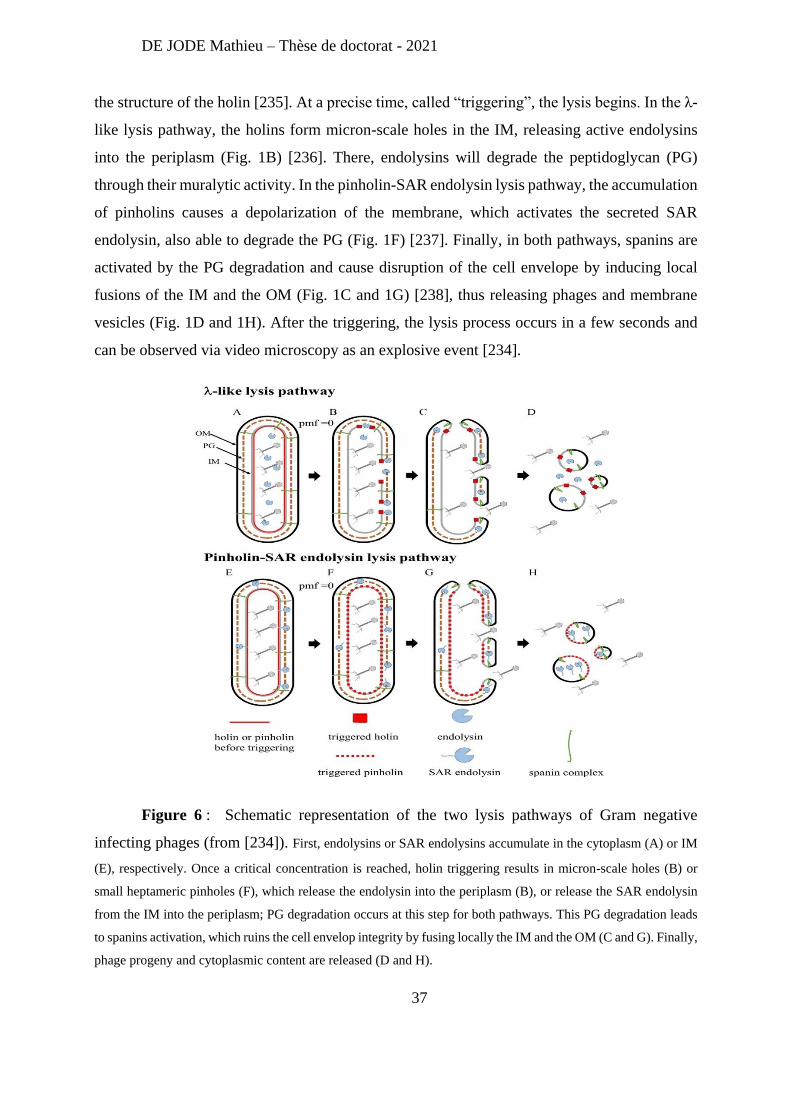
DE JODE Mathieu – Thèse de doctorat - 2021
37
the structure of the holin [235]. At a precise time, called “triggering”, the lysis begins. In the λ-
like lysis pathway, the holins form micron-scale holes in the IM, releasing active endolysins
into the periplasm (Fig. 1B) [236]. There, endolysins will degrade the peptidoglycan (PG)
through their muralytic activity. In the pinholin-SAR endolysin lysis pathway, the accumulation
of pinholins causes a depolarization of the membrane, which activates the secreted SAR
endolysin, also able to degrade the PG (Fig. 1F) [237]. Finally, in both pathways, spanins are
activated by the PG degradation and cause disruption of the cell envelope by inducing local
fusions of the IM and the OM (Fig. 1C and 1G) [238], thus releasing phages and membrane
vesicles (Fig. 1D and 1H). After the triggering, the lysis process occurs in a few seconds and
can be observed via video microscopy as an explosive event [234].
Figure 6 : Schematic representation of the two lysis pathways of Gram negative
infecting phages (from [234]). First, endolysins or SAR endolysins accumulate in the cytoplasm (A) or IM
(E), respectively. Once a critical concentration is reached, holin triggering results in micron-scale holes (B) or
small heptameric pinholes (F), which release the endolysin into the periplasm (B), or release the SAR endolysin
from the IM into the periplasm; PG degradation occurs at this step for both pathways. This PG degradation leads
to spanins activation, which ruins the cell envelop integrity by fusing locally the IM and the OM (C and G). Finally,
phage progeny and cytoplasmic content are released (D and H).
DE JODE Mathieu – Thèse de doctorat - 2021
38
No mechanism of lysis resistance per se has been described yet, but biological pathways
affecting the lysis timing have been reported. First, lysis delay, sometimes referred as lysis
inhibition, has been observed numerous times (starting in the 1940’s [239]), and linked to
superinfection (the infection by a second phage) [240]. This delay of the lysis in presence of
other phages is believed to have ecological importance: the release of new virions is delayed to
a time with a more advantageous ratio of uninfected/infected host cells [241]. Recently, a
molecular mechanism for lysis delay in T4 was proposed: the DNA of the second phage would
interact with holins, thus delaying the triggering of the lysis [242].
The opposite phenomenon can also occur as other mechanisms will induce a quicker
lysis. For example, the expression of the abiZ gene, caused phage φ31-infected Lactococcus
lactis to lyse 15 min earlier, thus reducing a 100-fold the burst size of phage φ31 [243]. Results
suggest that this premature lysis might be caused through interactions between AbiZ proteins
and holins.
This induced quicker lysis is actually part of a specific class of bacterial resistance
mechanism called Abortive Infection, in which an infected bacterial cell will commit an
altruistic “suicide” to protect the rest of the bacterial community.
M) Abortive Infection: Bacterial Altruistic Suicide
The term Abortive Infection (or Abi) does not refer to a precise resistance mechanism,
but to a general strategy aiming at limiting phage propagation to the whole bacterial community.
The main principle of this strategy is that a phage infected bacterial cell commits suicide before
the phage can complete its replication cycle [244]. This will limit the production of virions and
the spread of the phage epidemic to the neighboring cells, thus protecting the rest of the colony
(Figure 7). Abi can be characterized as an altruistic mechanism, as one cell sacrifices itself to
protect the other. However, considering phages specificity (only infecting one bacterial species
or a few strains of a bacterial species) this altruism is restrained and primarily protects only
closely related bacteria. Moreover, this protection is only efficient in a structured environment:
if phages and bacteria are dispersed (e.g. in liquid culture), community protection is less likely
to be achieved, whereas in a locally structured environment (e.g. a biofilm or a colony on a
plate) suicide of infected cells results in an overall community protection [245].
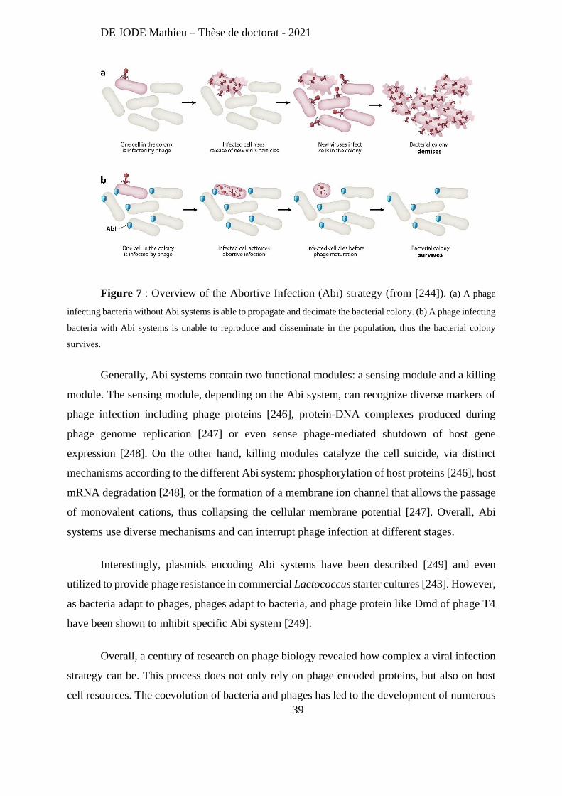
DE JODE Mathieu – Thèse de doctorat - 2021
39
Figure 7 : Overview of the Abortive Infection (Abi) strategy (from [244]). (a) A phage
infecting bacteria without Abi systems is able to propagate and decimate the bacterial colony. (b) A phage infecting
bacteria with Abi systems is unable to reproduce and disseminate in the population, thus the bacterial colony
survives.
Generally, Abi systems contain two functional modules: a sensing module and a killing
module. The sensing module, depending on the Abi system, can recognize diverse markers of
phage infection including phage proteins [246], protein-DNA complexes produced during
phage genome replication [247] or even sense phage-mediated shutdown of host gene
expression [248]. On the other hand, killing modules catalyze the cell suicide, via distinct
mechanisms according to the different Abi system: phosphorylation of host proteins [246], host
mRNA degradation [248], or the formation of a membrane ion channel that allows the passage
of monovalent cations, thus collapsing the cellular membrane potential [247]. Overall, Abi
systems use diverse mechanisms and can interrupt phage infection at different stages.
Interestingly, plasmids encoding Abi systems have been described [249] and even
utilized to provide phage resistance in commercial Lactococcus starter cultures [243]. However,
as bacteria adapt to phages, phages adapt to bacteria, and phage protein like Dmd of phage T4
have been shown to inhibit specific Abi system [249].
Overall, a century of research on phage biology revealed how complex a viral infection
strategy can be. This process does not only rely on phage encoded proteins, but also on host
cell resources. The coevolution of bacteria and phages has led to the development of numerous
DE JODE Mathieu – Thèse de doctorat - 2021
40
bacterial resistance mechanisms, targeting every step of the phage infection process.
Conversely, phages have evolved to counteract these resistance strategies. This never ending
arms race is a demonstration of the plasticity of the genomic information that serves the
impressive adaptation abilities of bacteria to different forms of threats.
Another example of bacterial adaptation is the development of drug resistance. Soon
after the discovery of penicillin bacterial resistance was observed (in 1928) [250]. Nevertheless,
penicillin and other antibiotics became widely used, and precipitated the decline of phage
therapy. However, in the former Soviet Union, a limited access to western developed antibiotics
actually strengthen phage therapy research and usage. The Eliava Institute, founded by Georgyi
Eliava in Tbilisi (Georgia), was the main center from which phage therapy spread in former
USSR countries [251]. Up to now, phage therapy has been continuously practiced in few
countries like Georgia (through The Eliava Institute) or Russia, where phages are available in
pharmacy over the counter [252]. In the rest of the world, the increased prevalence of antibiotic
resistant bacteria and the threat of a post-antibiotic era has revived interest in phage therapy
during the last two decades [253].
3- Phage Therapy Part II: A New Hope
A) Clinical Experience of Phage Therapy: Does It Work?
Phage therapy was originally dismissed partially because of inconsistent reports of
efficacy. This phenomenon was attributed (post factum) to the use of ill-suited phages (e.g. not
infecting the patient’s strain) or use of poorly prepared phage products (low titer and/or
contaminated with bacterial endotoxin). Since that time, microbiological diagnosis (cultured-
based [254] or molecular-based [255]) and phage preparation techniques have been improved,
allowing a precise diagnosis (identification of the bacterial species) and the production of high
titer, endotoxin-free phage products [256, 257]. Unfortunately, this is not enough to secure a
positive output during a clinical trial. For instance, in 2015, a study on the efficacy of a phage
cocktail targeting the E. coli species was evaluated for the treatment of acute bacterial diarrhea
in children, but failed to prove an improvement of the health condition [258]. It was found that
the phages had a limited replication in the gut, probably because of a limited host availability,
as E. coli represented less than 5% of the fecal bacteria in these patients. Moreover, a microbiota
DE JODE Mathieu – Thèse de doctorat - 2021
41
analysis revealed that disease progression (increased diarrhea) was associated with the
overgrowth of other bacterial species, notably Streptococcus gallolyticus and Streptococcus
salivarius, that were obviously unaffected by the phage treatment. This study reasserted the
importance of knowing the etiology of a disease. Indeed, phage therapy is probably one of the
best example of precision medicine. In contrast to antibiotic treatments that often target multiple
bacterial species, a precise identification of the bacterial species and even the strain causing the
disease is a critical step for phage therapy.
Another lesson that needed to be taught again was the importance of the phage product
quality. The PhagoBurn study led by the pharmaceutical company Pherecydes Pharma,
partnered with military and civilian hospitals in France, Switzerland, and Belgium, consisted in
a 3-year phage therapy trial on burn patients with infected wounds [259]. Due to multiple issues,
notably regulatory hurdles, the trial was not only delayed but its scope was also reduced: the
initial project aimed to investigate the efficacy of phage treatment in 220 patients with burn
wounds infection by either P. aeruginosa or E. coli, but it finally included only 27 patients with
P. aeruginosa infections. The phage treatment (daily topical application of phage dressing) was
found to be less effective than the standard care (daily application of 1% sulfadiazine silver
emulsion cream). This inefficacy was attributed to a very low phage titer, which was four orders
of magnitude lower than expected (100 instead of 106 Plaque Forming Units (pfu)/ml per
application). It seems that the phage product was unstable, as it was prepared almost 2-years
ahead of its application in patients.
The two disappointing examples reported above are counter-balanced by two studies
performed few years before. In 2006 Marza et al [260] reported successful treatments of two P.
aeruginosa infections: a human burn wound infection and a dog otitis infection. In both cases,
clinical improvements and phage replication (from 42 fold to over a million fold) were
observed. No adverse effects were reported following the phage treatments and no toxicity to
human keratinocytes was observed when they were cultured with the purified phage suspension.
Following the above success, Biophage Limited engaged in 2009 a small clinical trial
(23 patients) for the treatment of chronic otitis caused by MDR P. aeruginosa.[261]. This
randomized, double‐blind, placebo‐controlled Phase I/II clinical trial was approved by UK
Medicines and Healthcare products Regulatory Agency (MHRA), First, the inclusion of
DE JODE Mathieu – Thèse de doctorat - 2021
42
patients was well performed as four patients were excluded of the study due to the lack of
susceptibility of their swab-sampled P. aeruginosa to the tested phage product (a cocktail of
six phages). This exclusion rate was in agreement with prior in vitro studies where 86% of 57
isolates (from human ear infections), were susceptible to at least one of the six phages. This
result highlights the role of the host range of a phage product. Presumably, a phage product
including a small number of phages will likely have a narrower host range than a product with
a higher number of phages, thus limiting its clinical applications. The results of this study were
very promising: significant clinical improvements from baseline were observed in the phage
treated group but not in the placebo group and P. aeruginosa counts were significantly reduced
in the phage treated group whereas they were unchanged in the placebo group. Moreover, phage
replication was observed in treated patients and no side effects nor evidence of local or systemic
toxicity reported. Replication of the six phages was observed in patients from the phage treated
group (note that up to five phages could replicate in one patient). The mean recovery of phages
from swabs taken from the ears of the phage-treated group over all three post-treatment visits
was 1.27x10⁸ pfu. Compared to the treatment dose of 6x10⁵ pfu, this suggests an average
amplification in the treated ear of 200 fold. Moreover, the median duration of phage replication
in the phage-treated group was 21 days, and clearance of the phages was observed after all the
resolved infections. Finally, it is interesting to note that in this study (the only successful phase
II clinical trial to date), a single phage application had several weeks lasting effects and manage
to successfully treat three previously untreatable cases of chronic otitis. Indeed, this three cases
were irresponsive to classical antibiotic therapies that required multi doses given at regular
intervals over several weeks.
Overall, these three studies showed that for a phage therapy trial to be successful the
inclusion of patients is critical. Only those for which the phage product show lysis activity on
the patient’s bacteria should be included, which per se introduces a bias in the analysis of the
overall treatment efficacy of the trial. It also represents a hurdle for regulatory agencies that
often request a new treatment to be compared to an existing treatment for which the selection
of patients is not needed. Also, the production of a high titer, stable phage product has to be
insured. Moreover, the lack of approved phage products on the market is slowing down the re-
development of phage therapy. Facing all of these issues, a number of clinicians have
nevertheless initiated tailored “compassionate” treatment for patients, which has propelled the
publication of several case reports
DE JODE Mathieu – Thèse de doctorat - 2021
43
B) Compassionate Phage Therapy around the World: Sporadic Successes
In France, a total of 15 patients with osteoarticular infections received a compassionate
treatment with phages between 2006 and 2018 at the hospital of Villeneuve Saint Georges
[262]. Causative agents included mostly Staphylococcus aureus (11/15) and P. aeruginosa
(3/15), and 73% (11/15) of the treatments concluded with complete cure of the patient’s
infection. The other outcomes included clearance of the targeted pathogen but appearance of
another one (2/15) or only partial disinfection nevertheless associated with clinical
improvements (2/15). In addition, the local application of phages was shown to be safe. Note
that phage treatments were always supplemented with antibiotics, as these patients had
worrying clinical conditions, the aim was not to experiment phage therapy per se, but to treat
these patients with all available therapeutics.
Recently, Lebaux et al [263] reported the first case of phage therapy for Achromobacter
xylosoxidans lung infection in a lung-transplanted CF patient. The patient received two rounds
of phage treatment (5 months apart due to regulatory and production delays). No adverse effects
were reported following phage administration, but subsequent bronchoalveolar lavage (BAL)
still contained A. xylosoxidans, including phage resistant bacteria. The patient clinical status
improved slowly despite low level airway colonization by A. xylosoxidans that persisted for
months, before samples turned negative. No re-colonisation was detected more than two years
after the phage treatment, and the imipenem treatment was then stopped. This study revealed a
limited short-term bactericidal effect of the phage treatment in the lungs, as it did not manage
to eliminate every A. xylosoxidans, but was followed nonetheless by a clinical improvement of
the patient.
In 2007, the Queen Astrid military hospital in Brussels was the first Belgian hospital to
revive phage therapy and its use, using the article 37 (regarding unproven interventions in
clinical practice) of the Declaration of Helsinki [264]. This allowed the treatment of 15 patients,
including one with acute kidney injury and developing a septicemia caused by a colistin-only-
sensitive P. aeruginosa strain [265]. Phage cocktail BFC1 [266], containing two phages with
in vitro activity against the patient’s P. aeruginosa isolates, was administered (50µL) as a 6-h
intravenous infusion for 10 days to treat the septicemia in absence of any antibiotic treatment.
Moreover, the patient infected wounds were irrigated with 50 ml of BFC1 every 8 h for 10 days.
DE JODE Mathieu – Thèse de doctorat - 2021
44
Rapidly, blood cultures turned negative, the fever disappeared and kidney function recovered
after a few days. However, the patient’s wounds remained infected with several bacterial
species, including P. aeruginosa, and caused multiple episodes of sepsis, eventually treated
with antibiotics. This patient died 4 months later from a cardiac arrest due to a Klebsiella
pneumoniae sepsis. This case report, while demonstrating a clear therapeutic effect of the phage
treatment rapidly after its administration, also points out the limitation of a specific phage
product in a case of a multi-species infection.
In the US, in 2017, a successful case of intravenous phage therapy to treat a disseminated
MDR Acinetobacter baumannii infection, made the headlines [267]. Indeed, the absence of
effective antibiotics pushed two laboratories to identify nine different phages with lytic activity
against a bacterial isolate from the patient. An authorization to administer these phages as an
emergency investigational new drug (eIND) was obtained from the Food and Drug
Administration (FDA). These phages were given both intravenously and percutaneously, into
the abscess cavities. In vitro studies of patient isolates during the treatment showed that bacteria
became resistant to all phages (8 days after treatment started), leading to the design of a new
phage cocktail. This treatment was finally associated with clearance of A. baumannii and
clinical improvement of the patient. As usual for compassionate treatments, antibiotics
(meropenem, colistin, minocycline, fluconazole) were administered with phages and might
have play a role in the patient’s recovery. Nevertheless, this case report had a significant impact
on how phage therapy is perceived in the US, and this success was actually followed by the
creation of the Center for Innovative Phage Applications and Therapeutics (IPATH) at the
University of California, San Diego, in June 2018.
The University of California has reported 10 cases of phage therapy treatment (including
6 before IPATH) in more details. Globally, intravenous phage therapy was safe and associated
with a successful outcome in 7 out of the 10 cases, and phages alone were demonstrated as a
good preventive approach as P. aeruginosa infections were prevented in a CF patient during
phage treatment and for the subsequent 3 months. Moreover, phage resistance occurred during
the treatment for 3 patients, but was overcome by a personalized treatment strategy (addition
of new phages active on resistant bacterial isolates). Phage treatment was also reported to fail
despite in vitro phage susceptibility. Indeed, two cases of P. aeruginosa chronic and biofilm-
based infections (ventricular assist device infections) were associated with clinical failure of
DE JODE Mathieu – Thèse de doctorat - 2021
45
adjunctive phage therapy. The authors hypothesized that biofilm structure and composition
might have hindered phages diffusion and efficacy. Overall, IPATH experience highlights the
potential of phage therapy for several clinical indications, but also reveals the lack of reliable
predictors to ensure successful phage therapy.
A recent systematic review on the safety and efficacy of phage therapy to treat
superficial bacterial infections collected 6801 article since 1930’s, amongst which only 27 were
considered as eligible for in-depth analysis [268]. These studies showed clinical resolution or
improvement in 77.5% (n = 111) of burn wound infections, in 86.1% (n = 310) of chronic
wound/ulcer infections and in 94.14% (n = 734) of dermatological infections. Interestingly, on
the 15 reports addressing phage therapy safety, all the reports published after 2001 expressed
no safety concern, whereas 7 early reports (from 1929 to 1987), described adverse effects,
which is consistent with the use of phage preparations that were not highly purified.
In conclusion, despite a lot of positive reports regarding phage therapy, an undisputable
demonstration of its efficacy is still lacking. Indeed, in most of the previously described clinical
cases, antibiotic treatments were not stopped (even when the bacteria were shown to be
resistant), thus the phage treatment may not be declared as solely responsible for the patient
recovery. Moreover, the sporadic nature of the reported cases and the lack of proper control
(without phage treatment or phage treatment without antibiotics) limits our ability to conclude
on phage therapy efficacy.
To design and perform a clinical study demonstrating the efficacy of phage therapy,
there is a crucial need of understanding the factors influencing phage therapy success. These
factors can be revealed through research focusing not only on phages and bacteria interactions
but also taking into account the host. Indeed, for phage therapy to succeed we need to identify
the characteristics that make one phage a better therapeutic candidate than another one, and
how to formulate phage cocktails to address host range or resistance issues. We also need to
understand how bacteria evolve in response to phage treatment and how the host, and in
particular its immune system, reacts to both phages and bacteria during the treatment.
Since my PhD projects are related to the use of phages infecting P. aeruginosa, the
following sections, addressing some of the challenges mentioned above, are focused on reports
from studies involving phages targeting this bacterium.
DE JODE Mathieu – Thèse de doctorat - 2021
46
C) The Importance of Dosage and Timing
First, to investigate the important factors for phage therapy success, one should
establish a model in which phage therapy efficacy is well demonstrated. An early report by
Vinodkumar et al [269] demonstrated the efficacy of phage therapy to treat MDR P. aeruginosa
septicemia in mice. Septicemia was induced in BALB/c mice via intraperitoneal (i.p) injection
of 107 colony forming units (cfu) of a P. aeruginosa clinical strain and the treatment was a
single i.p. injection of phage CSV-31. The control mice died within 48h whereas, 100% of mice
treated with the highest dose of phage (4x10⁹ plaque forming units) 45min after infection
survived. Interestingly, only 50% of mice treated with a lower dose of phage (3x10⁴ pfu)
survived, demonstrating a dose dependent effect of the phage treatment. This is particularly
interesting as the replicative nature of phages suggests that few phages could be sufficient for
therapeutic success. Phage therapy success was dependent on active phage particles as heat
inactivated phages had no effect on mice survival. Moreover, a 5h delayed treatment (at the
highest dose) was still efficient (100% of mice rescued) but a treatment given 24h after the
infection only rescued 50% of the mice. Overall, this report not only demonstrate phage therapy
efficacy, but also points out the importance of phage dosage and timing of treatment for phage
therapy success.
Similar conclusions were obtained in our lab as Debarbieux et al [270] reported efficient
treatment and prevention of P. aeruginosa acute lung infection in mice, using the phage
PAK_P1. Indeed, this report highlighted again the importance of the phage dosage as the use
of increased Multiplicity of Infection (MOI, the phage/bacteria ratio), MOI 1 or MOI 0.1, was
associated with increased survival of mice 5-day post infection (0% and 100% respectively).
They also demonstrated the importance of the delay between infection and treatment as mice
treated 2h, 4h or 6h post infection showed decreased survival with 100%, 80%, and 30%,
respectively. Using a bioluminescent strain of P. aeruginosa (PAK-lumi) these authors provide
the first dynamic data allowing the follow-up of each animal over time (Figure 8). Finally, they
observed that PAK_P1 particles were stable enough in the lung environment (1x10⁸ pfu given,
3x10⁶ pfu recovered in bronchoalveolar lavages 22h later) to protect 100% of infected mice
when administered 24h before the lethal dose of the pathogen.
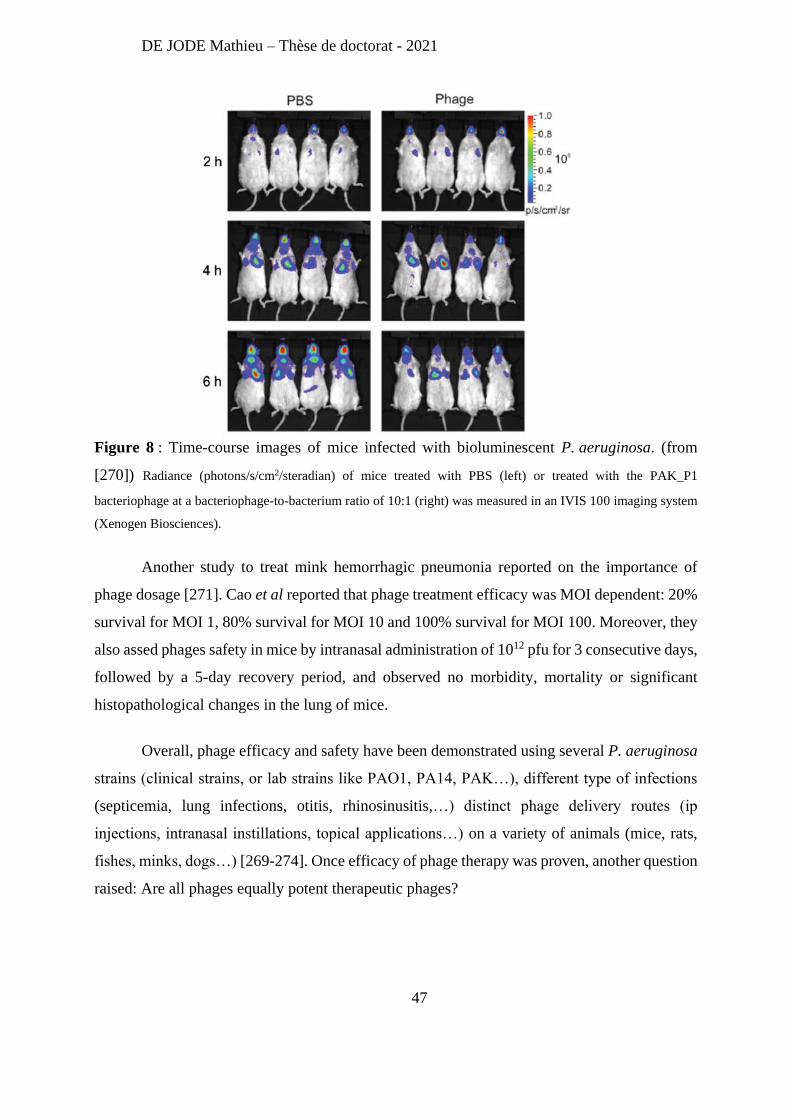
DE JODE Mathieu – Thèse de doctorat - 2021
47
Figure 8 : Time-course images of mice infected with bioluminescent P. aeruginosa. (from
[270]) Radiance (photons/s/cm2/steradian) of mice treated with PBS (left) or treated with the PAK_P1
bacteriophage at a bacteriophage-to-bacterium ratio of 10:1 (right) was measured in an IVIS 100 imaging system
(Xenogen Biosciences).
Another study to treat mink hemorrhagic pneumonia reported on the importance of
phage dosage [271]. Cao et al reported that phage treatment efficacy was MOI dependent: 20%
survival for MOI 1, 80% survival for MOI 10 and 100% survival for MOI 100. Moreover, they
also assed phages safety in mice by intranasal administration of 1012 pfu for 3 consecutive days,
followed by a 5-day recovery period, and observed no morbidity, mortality or significant
histopathological changes in the lung of mice.
Overall, phage efficacy and safety have been demonstrated using several P. aeruginosa
strains (clinical strains, or lab strains like PAO1, PA14, PAK…), different type of infections
(septicemia, lung infections, otitis, rhinosinusitis,…) distinct phage delivery routes (ip
injections, intranasal instillations, topical applications…) on a variety of animals (mice, rats,
fishes, minks, dogs…) [269-274]. Once efficacy of phage therapy was proven, another question
raised: Are all phages equally potent therapeutic phages?
DE JODE Mathieu – Thèse de doctorat - 2021
48
D) Choosing the Right Phage
All virulent phages can kill bacteria, but does that mean that every phage will be a good
therapeutic candidate? Are there good and bad phages regarding phage therapy? Can in vitro
testing predict in vivo efficacy? These questions have been addressed (more or less directly)
throughout different studies.
In 2009, Heo et al [275] reported antibacterial efficacy of phages against P. aeruginosa
infections in mice and in flies. This study focused on two phages isolated from sewage using
PA01 as host, MPK1 (a Myoviridae) MPK6 (a Podoviridae). In vitro experiments revealed that
both phages use the LPS as bacterial receptor and that MPK1 lyses faster than MPK6.
Moreover, these two phages showed different pharmacodynamics as MPK1 displayed better
persistence than MPK6 in blood, liver and lungs of mice, and in tissues of flies. To test the in
vivo efficacy of these phages, they were administered in mice by either the intramuscular (i.m.)
or intraperitoneal (i.p.) route with different dosage (2x10⁶ pfu or 2x10⁷ pfu) 6 h post peritonitis
infection with PAO1. While the 2x10⁷ pfu dose of each phage significantly protected the
infected mice, compared to the untreated mice, the MPK6 dose of 2x10⁶ pfu administered by
the i.m. route failed to protect mice. Presumably, the i.p. administration displayed better
efficacy than the i.m. administration for both phages because i.p. route delivers phages more
directly to the infection site (in this peritonitis model). Additionally, phage efficacy was also
investigated in flies, where despite large differences in the pharmacokinetics of these two
phages in fly tissues, they showed similar efficacy on fly survival and no toxicity after 72h
phage feeding. Overall this report clearly demonstrates that different phages might present
distinct pharmacokinetics profiles, potentially impacting their efficacy (depending on the
administration route and animal model).
The different efficacy of distinct phages, and the ability to extrapolate in vivo success
from in vitro tests has been thoroughly investigated by our lab. Henry et al [276] studied 9
different phages, through in vitro experiments including EOPs (Efficiency Of Plating) and lysis
kinetics, and in vivo with an acute lung infection mice model (intranasal phage treatment
applied 2h post infection, at MOI 0.1). Overall, for 7 of the phages, a good correlation between
in vitro and in vivo efficacy was observed. However, this was not the case for two phages:
PhiKZ and CHA_P1. Despite having an EOP of 1.2 on the PAK-lumi strain and a reasonably
DE JODE Mathieu – Thèse de doctorat - 2021
49
good lysis kinetics (in vitro), survival rates following treatment with phage PhiKZ (MOI 0.1)
were as low as those treated with PBS (mock treatment). Interestingly, an increase of the phage
dose to reach a MOI to 20 provided enough phage to obtain 100% survival. Concerning
CHA_P1, despite similar in vitro lysis kinetics and genetic closeness to some “effective
phages”, this phage was unable to cure animals infected with the PAK-lumi strain. Nonetheless,
CHA_P1, isolated using strain CHA a host, was able to rescue CHA-infected mice, proving its
therapeutic potential. An attempt to adapt CHA_P1 to the PAK strain by serial passages failed
to increase its EOP and its in vivo therapeutic efficacy. Finally, this study demonstrates that in
vitro activity (e.g. the ability to form plaques on an overlay of a bacterial strain) is not sufficient
to ensure in vivo efficacy. Nevertheless, a correlation was found between in vitro data (high
EOP, quick lysis kinetics) and potent therapeutic effects in vivo. It also demonstrated that in
vitro phage adaptation is not always possible and that phages isolated using the bacterial strain
of interest seem more efficient, which is a strong argument for personalized or “sur mesure”
phage therapy compare to the fixed or "prêt à porter" product [277].
Another study from our laboratory included the in vitro efficacy of the above 9 phages
on the few isolates from the sputum of CF patients [278]. Interestingly, the most active phage,
PAK_P5, was genetically closely related to the least active phage tested CHA_P1. As these two
phages share protein sequences with around 90% identity, it showed that phage genomic data
is insufficient for predicting infectivity of clinical strains.
Overall, it seems that all phages are not equal regarding their therapeutic efficacies.
Indeed, all phages do not possess the same pharmacodynamics profile or the same ability to
infect a specific strain. Moreover, today, we are unable to predict phage characteristics from
genomic data alone, as phages sharing high sequence identity might still present very different
infection abilities. Luckily, some of these critical parameters can be assessed in vitro and the
results might predict in vivo efficacy, thus helping us choosing the right phage. Unfortunately,
some phage characteristics (e.g. EOP or lysis kinetics) are host dependent and will need to be
reassessed for every clinical strain on which phage use is intended.
Once potent phages have been identified, the question of formulation rises. A
therapeutic phage product with a fixed formulation will need to infect many strains, whereas a
DE JODE Mathieu – Thèse de doctorat - 2021
50
more personal therapeutic approach might consider the synergy of different phages against a
specific bacterial strain.
E) Phage Product Formulation
Phages ability to infect only a limited number of strains of a bacterial species has led to
the formulation of phage combination (also known as phage cocktail). For instance, a phage
product available in Russia, recommended for the treatment of E. coli infections, contains 18
distinct phages, including the three main families of Caudovirales (Podoviridae, Myoviridae,
and Siphoviridae) [279]. The Russian strategy for formulating cocktail relies on regular update,
with new phages added to adapt to the evolving epidemiology of the targeted pathogen. As the
previous phages are kept and new phages are added, this historical strategy leads to very
complex formulation. This formulation strategy was, and still is, also employed at the Eliava
Institute in Georgia [251].
In western countries, the situation is different as current regulation regarding drugs
requires to demonstrate the safety and efficacy of a defined, fixed product (no update possible).
This has led phage companies to focus on host range driven strategy, as the final phage product
has to be able to infect most of the clinically relevant strains of a specific pathogen. This is
sometimes referred as the "prêt à porter" strategy, as patients with the same disease, but different
infecting strains, will receive the same treatment [277].
However, in academic labs, focusing on specific phage/bacteria systems, another phage
cocktail design strategy emerged: tailored-made or “sur mesure” [277]. This cocktail design
strategy does not focus on the host range parameter, but rather on the efficacy of the cocktail
on a specific strain. This strategy was motivated by observations of phage resistant bacteria
emerging following phage application in vitro. Indeed, selection of phage-resistant bacteria was
already described in details by Luria and Delbrück almost 80 years ago [153]. They observed
that bacterial cultures infected by phages initially become clear (due to phage-induced lysis),
but after variable amounts of time (from hours to days) the cultures became turbid because of
the growth of phage-resistant bacteria. Moreover, they found that the selected phage-resistant
bacteria were already present, in low abundance, in the initial bacterial culture.
DE JODE Mathieu – Thèse de doctorat - 2021
51
Interestingly, the simple addition of a second phage can delay the growth of phage-
resistant clones of P. aeruginosa, from 8-9 h for each single phage to more than 20 h for the
combination of the two [280]. In another in vitro study, Hall et al [281] described a variability
of effectiveness of single-phage treatments with frequent resistance and cross-resistance.
Additionally, they showed that multi-phage treatment is more efficient than any phage alone,
and that the simultaneous application of different phages was better or equal to the sequential
application, in reducing bacterial population density. However, these two methods were similar
(on average) regarding decreasing resistance rates. Note that phage-resistant bacteria emerged
in all conditions tested and were generally characterized by a reduced growth rate in absence
of phages, suggesting a fitness defect. Moreover, when investigating the therapeutic potential
of these phages (in wax moth), the authors found that a phage cocktail was the most effective
short-term treatment as it cured otherwise lethal infections. On the other hand, a study focusing
on three P. aeruginosa phages from three different genera (SL1, a PB1-like virus, SL2, a
phiKZ-like virus and SL3, a LUZ24-like virus), found that a cocktail with these three phages
had similar effect than the best phage alone in vivo (Galleria melonella) [282]. Therefore, in
this model, no synergy between the phages was detected. Finally, O’Flynn et al described a
similar frequency (10-6 CFU) of resistant bacteria to either a 3 phages cocktail or each individual
phages [283], demonstrating once again that a multiple phage treatment is not always more
efficient than monophage treatment.
As the random combination of different phages does not seem to be the most successful
strategy to design a potent phage cocktail, research groups have designed strategies relying on
different rationales to build phage cocktails.
F) Phage Cocktail Design Strategies
One strategy proposed to efficiently prevent, or at least delay phage resistance was to
incubate a phage and a bacterial strain to isolate phage-resistant clones, and then select
additional phages for their capacity to infect the phage-resistant strain. This process could be
repeated multiple times to insure that phage resistant selected against one phage would be lysed
by another phage of the cocktail. Once combined this way, the cocktail should prevent the
growth of at least some of the phage-resistant population. For example, this approach allowed
Gu et al [284] to design a phage cocktail against Klebsiella pneumoniae that significantly delays
DE JODE Mathieu – Thèse de doctorat - 2021
52
the growth of phage-resistant bacteria in vitro (26 h compared to 6 h for monophage treatment),
and also reduces the frequency of phage-resistant bacteria (10-7 CFU, compare to 10-4 for
monophage treatment). Moreover, the phage cocktail had a higher efficacy than monophage
treatment in vivo as the minimal protective dose of the cocktail to protect 80% of mice from
lethal infection was 3 x 10⁴ PFU compared to 3 x 10⁵ to 3 x 10⁷ PFU for individual phages.
This strategy is straightforward but is exceedingly time-consuming for deployment in clinics.
Another strategy to formulate phage cocktail relies on combining phages that use
different receptors on the bacterial cell surface. If, the idea of combining different phages to
limit cross-resistance is quite old (first report of this strategy by Smith and Huggins in 1983
[285]), the emphasis on the receptor used by the phage is more recent, as works on phage
resistance mechanisms has increased by the lower cost of genome sequencing [286-288]. For
example, Wright et al [288] investigated cross-resistance among P. aeruginosa resistant clones
evolved against 27 different phages. The authors discovered that cross-resistance between
phages using the same receptor was conferred by mutations targeting either LPS or type IV
pilus biosynthesis. On the other hand, mutations affecting a general regulator (RpoN) provided
cross-resistance between phages using different receptors, but were characterized by a higher
fitness costs than phage receptor mutations. Overall this study supports the idea that combining
phages according to their bacterial receptors could be a very potent cocktail design strategy,
when the aim is to limit bacterial resistance.
However, this strategy has not yet been used as a guiding strategy by most phage
laboratories or companies, probably because the identification of these receptors is not easily
done: compared to quite simple in vitro testing such as EOPs or lysis kinetics, performed in 24
to 48h at almost no costs, the determination of a phage receptors might take a much longer time
from days to weeks. Therefore, besides model phages studied by academic groups, most of
phages isolated with the aim to treat patients do not have their receptors characterized [289].
Overall, different strategies have been imagined to formulate potent phage cocktails,
and our growing understanding of bacterial phage resistance mechanism will surely inspire new
ones. Nonetheless, despite their inability to prevent phage resistance in vitro, various phage
treatments still manage to be efficient in vivo. For instance, when testing a 6 phages cocktail
(including PAK_P1 and PAK_P4 from our laboratory), Forti et al [290] observed that this
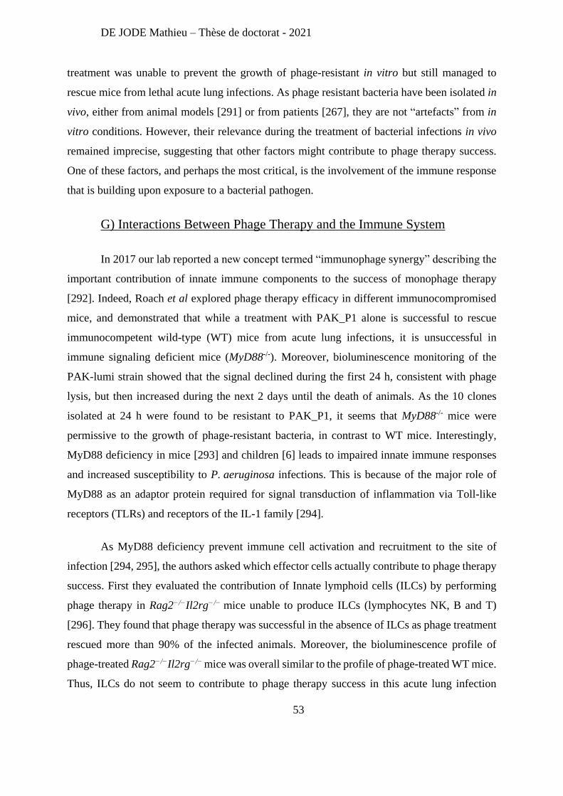
DE JODE Mathieu – Thèse de doctorat - 2021
53
treatment was unable to prevent the growth of phage-resistant in vitro but still managed to
rescue mice from lethal acute lung infections. As phage resistant bacteria have been isolated in
vivo, either from animal models [291] or from patients [267], they are not “artefacts” from in
vitro conditions. However, their relevance during the treatment of bacterial infections in vivo
remained imprecise, suggesting that other factors might contribute to phage therapy success.
One of these factors, and perhaps the most critical, is the involvement of the immune response
that is building upon exposure to a bacterial pathogen.
G) Interactions Between Phage Therapy and the Immune System
In 2017 our lab reported a new concept termed “immunophage synergy” describing the
important contribution of innate immune components to the success of monophage therapy
[292]. Indeed, Roach et al explored phage therapy efficacy in different immunocompromised
mice, and demonstrated that while a treatment with PAK_P1 alone is successful to rescue
immunocompetent wild-type (WT) mice from acute lung infections, it is unsuccessful in
immune signaling deficient mice (MyD88-/-). Moreover, bioluminescence monitoring of the
PAK-lumi strain showed that the signal declined during the first 24 h, consistent with phage
lysis, but then increased during the next 2 days until the death of animals. As the 10 clones
isolated at 24 h were found to be resistant to PAK_P1, it seems that MyD88-/- mice were
permissive to the growth of phage-resistant bacteria, in contrast to WT mice. Interestingly,
MyD88 deficiency in mice [293] and children [6] leads to impaired innate immune responses
and increased susceptibility to P. aeruginosa infections. This is because of the major role of
MyD88 as an adaptor protein required for signal transduction of inflammation via Toll-like
receptors (TLRs) and receptors of the IL-1 family [294].
As MyD88 deficiency prevent immune cell activation and recruitment to the site of
infection [294, 295], the authors asked which effector cells actually contribute to phage therapy
success. First they evaluated the contribution of Innate lymphoid cells (ILCs) by performing
phage therapy in Rag2−/−Il2rg−/− mice unable to produce ILCs (lymphocytes NK, B and T)
[296]. They found that phage therapy was successful in the absence of ILCs as phage treatment
rescued more than 90% of the infected animals. Moreover, the bioluminescence profile of
phage-treated Rag2−/−Il2rg−/− mice was overall similar to the profile of phage-treated WT mice.
Thus, ILCs do not seem to contribute to phage therapy success in this acute lung infection
DE JODE Mathieu – Thèse de doctorat - 2021
54
model. Next, the authors investigated the potential role of neutrophils in phage therapy efficacy,
using an antibody to deplete these cells in mice. In these neutropenic mice, phage treatment did
not improve their survival, thus demonstrating the importance of neutrophils in phage therapy
success (in this experimental model).
Additionally, the authors also established that the half-life of phage PAK_P1 in the
lungs of an immunocompromised mice (MyD88-/- mice) is longer than in a WT mice, pointing
to a clearing of the phages by immune components (a phenomenon already described in other
studies [297, 298]).
These results allowed a better understanding of the interactions between phages,
bacteria, and the immune system, represented in the Figure 9.
Figure 9 : Schematic representation of the interactions between bacteria, phages and the
immune system (from [292]): Phage can inhibit the growth of phage-sensitive bacteria through phage lysis,
whereas the immune cells can kill both phage-sensitive and phage resistant bacteria. This immune killing can be
activated by the recognition of pathogen components, but also inhibited by bacteria through immune evasions
mechanisms. Additionally, immune regulation can also limit the inflammation process (to limit tissue damages)
and immune cells contribute to phage elimination.
To summarize, in an immunocompetent mouse, a bacterial infection might be
uncontrolled by the immune system, thus causing the death of the animal. A phage treatment
given to this animal can efficiently reduce the bacterial population by lysing the phage
DE JODE Mathieu – Thèse de doctorat - 2021
55
susceptible bacteria, while the immune system (especially neutrophils) can eliminate both
phage susceptible and phage-resistant bacteria: the synergistic action of phages and neutrophils
allows a successful treatment. However, in the absence of neutrophils (e.g. in MyD88-/- mice)
phages can lyse the phage-susceptible bacteria, but phage-resistant clones will grow
uncontrolled leading to the animal death.
The second project of my PhD was directly linked to the above results with the objective
to restore phage therapy efficacy in immunocompromised mice (MyD88-/-). The strategy that I
tested aimed to limit phage resistance through the design of a tailored phage cocktail.
DE JODE Mathieu – Thèse de doctorat - 2021
56
Results
Overview
During my PhD I worked on two different project both related to the study of phages
infecting P. aeruginosa. Hereafter, I briefly summarized the main results obtained for each of
these two projects, which are then developed under the form of two drafted papers. For the first
project on a phage protein, we are currently looking for a journal that will send it out for review.
For the second project, on phage therapy and phage resistance in immunocompromised animals,
a last piece of data from genome sequencing will be obtained in the next few weeks and the
draft will be submitted soon after.
DE JODE Mathieu – Thèse de doctorat - 2021
57
1- A bacteriophage protein favors bacterial host takeover by impairing
stress response.
Our laboratory has isolated several phages infecting the P. aeruginosa strain PAK and
initiated the characterization of the original molecular mechanisms they deploy to hijack
bacterial resources. More precisely, two publications focused on phages PAK_P3 [216] and
PAK_P4 [217].
The transcriptomic analysis of strain PAK infected by either phage revealed rapid
degradation of the host mRNA, although with different kinetics as PAK_P4 was faster than
PAK_P3. Moreover, these two phages showed different pattern of expression of their core
genes. To better understand the underlying molecular mechanisms of the infection by these
phages, we followed these studies by focusing on the only early-expressed gene conserved
between these two phages: gp92 in PAK_P3 and gp109 in PAK_P4.
Since the function of the proteins coded by these two genes could not be predicted and
since they do not share sequence homology with any other proteins except few homologs
present in closely related P. aeruginosa phages, we decided to combined both global and focal
approaches to identify the role of these proteins during phage infection. For this we cloned these
phage genes and inserted them on the bacterial chromosome, under an IPTG inducible
promoter. Overall, we found that expression of gp92 alters bacterial motility, morphology and
proteome, without affecting bacterial growth. Surprisingly, despite sequence homology and
similar localization (in the cytoplasmic membrane), expression of gp109 had none of these
effects. Moreover, we linked the morphological changes induced by Gp92 with its interaction
with the anti-sigma factor MucA, a major regulator of membrane-associated stress response in
P. aeruginosa. Finally, we found that Gp92 impairs this stress response, which results in an
increased susceptibility of P. aeruginosa to antibiotics, including Imipenem.
This work demonstrates that the study of phage proteins of unknown functions can
reveal new bacterial weaknesses and could guide the development of new drugs.
DE JODE Mathieu – Thèse de doctorat - 2021
58
Title
A bacteriophage protein favors bacterial host takeover by impairing stress response.
Authors
Mathieu De Jode1,2, Elisa Brambilla3, Gouzel Karimova4, Marc Monot5, Rob Lavigne6, Laurent
Debarbieux1*$, Anne Chevallereau1,2,7*,
Affiliations
1 Bacteriophage, Bacterium, Host Laboratory, Department of Microbiology, Institut Pasteur,
Paris F-75015 France
2 Sorbonne université, collège doctoral, F-75005 Paris, France
3 Groupe Croissance et Morphogénèse Microbienne, Institut Pasteur, Institut Pasteur, Paris F-
75015 France
4 Unité de Biochimie des Interactions Macromoléculaires, CNRS UMR 3528, Département de
Biologie Structurale et Chimie, Institut Pasteur, Paris F-75015 France
5 Pathogenesis of anaerobic bacteria laboratory, Department of Microbiology, Institut Pasteur,
Paris F-75015 France
6 Laboratory of Gene Technology, Department of Biosystems, KU Leuven, Belgium
7 Present address: Department of Infection, Immunity, and Inflammation, Institut Cochin,
INSERM U1016, CNRS UMR8104, Université de Paris, F-75014 Paris, France
* Co-Corresponding authors
DE JODE Mathieu – Thèse de doctorat - 2021
59
Author contributions
Conceptualization, MDJ, AC and LD; Methodology, MDJ, AC, GK, EB, MM, RL and LD;
Investigation, MDJ, AC, EB, GK; Data Analyses, MDJ, AC and LD; Writing – Original Draft,
MDJ, AC and LD; Writing – Review & Editing, MDJ, AC and LD with contributions from RL
and GK; Supervision, AC and LD; Funding Acquisition, LD and RL
Competing interest statement
The authors declared no competing interest.
Abstract
Bacteriophages have developed unique and diverse strategies to hijack bacterial functions.
However, the underlying molecular mechanisms remain largely understudied. We characterized
gp92, an early-expressed gene from the virulent bacteriophage PAK_P3 infecting the
opportunistic pathogen Pseudomonas aeruginosa. The ectopic expression of gp92 alters
bacterial morphology and motility and provokes a major shift of the cell proteome, including
several sigma factors, without affecting bacterial growth. Through its interaction with the anti-
sigma factor MucA, Gp92 impairs the membrane-associated stress response mediated by the
sigma factor AlgU. This in turn revealed an unexpected link between AlgU-mediated stress
response and phage lysis timing. In addition, expression of gp92 increased the susceptibility of
P. aeruginosa to antibiotics, including Imipenem, attesting that targeting the membrane stress
response can increase bacterial susceptibility to carbapenems. Investigations of unknown
bacteriophage functions not only identify original molecular virus-host interactions but also
provide opportunities to uncover new bacterial targets for drugs development.
DE JODE Mathieu – Thèse de doctorat - 2021
60
Introduction
Bacteriophages (phages) are the viral enemies of bacteria. Through evolution they have built
an arsenal of mechanisms to disrupt the bacterial physiology, including genes that are involved
in the subversion of the host metabolism, a process which occurs at the early stages of infection
and consists in the implementation of specific hijacking tasks (1). Some of these early tasks
have been well characterized, such as the redirection of the bacterial RNA polymerase towards
phage genes (2) or blocking host defense systems (3, 4), but could as well highlight putative
targets for developing new antibacterial drugs (5, 6). This is particularly needed in the current
context of antimicrobial resistance. Metagenomics studies of environmental and clinical
samples provide a large resource to be exploited (7, 8). However, most of the viral genetic
information has not yet been associated with defined functions and is referred to as viral “dark
matter” (9).
To identify the functions of early-expressed phage genes, several approaches have been
proposed, ranging from targeted searches for a given phenotype (e.g. growth inhibition) to more
general strategies relying on mass spectrometry (5, 10-12). Despite the potential for discovery
of novel biological functions and biotechnological development (5), systematic studies of phage
early gene products remain scarce.
We previously uncovered two related genera of virulent phages infecting the opportunistic
pathogen Pseudomonas aeruginosa, namely Nankokuvirus and Pakpunavirus, from which one
representative (PAK_P3 and PAK_P4, respectively) was studied in depth (13-15). Despite the
overall genetic divergence between PAK_P3 and PAK_P4, we found that the transcriptional
landscapes of cells infected by these phages displayed strong similarities, notably with the
activation of a common host response, suggesting that the host subversion strategies of PAK_P3
and PAK_P4 may rely on conserved functions that we aim to uncover in the present study.
We identified and functionally characterized the only early-expressed gene family conserved
across Nankokuvirus and Pakpunavirus genera (15). The representative genes from PAK_P3
and PAK_P4 genomes, gp92 and gp109, respectively, both encode small proteins that localize
at the cytoplasmic membrane. Surprisingly, only the ectopic expression of gp92 altered the
morphology and swimming motility of P. aeruginosa cells. By combining global and targeted
approaches, we showed that expression of gp92 reduced the ability of the host cell to respond
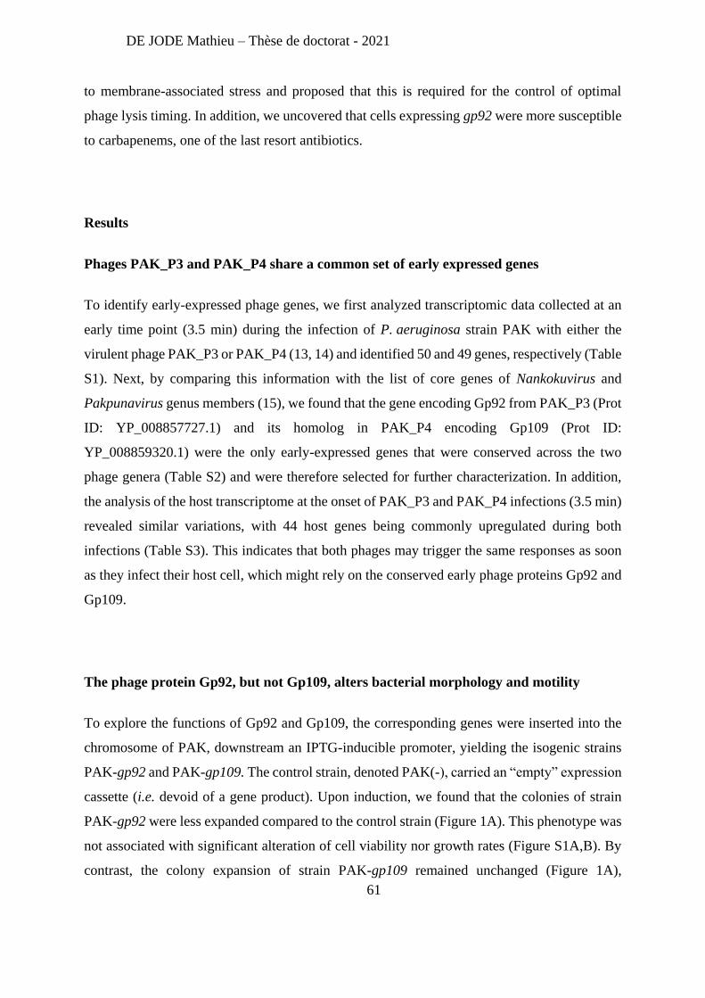
DE JODE Mathieu – Thèse de doctorat - 2021
61
to membrane-associated stress and proposed that this is required for the control of optimal
phage lysis timing. In addition, we uncovered that cells expressing gp92 were more susceptible
to carbapenems, one of the last resort antibiotics.
Results
Phages PAK_P3 and PAK_P4 share a common set of early expressed genes
To identify early-expressed phage genes, we first analyzed transcriptomic data collected at an
early time point (3.5 min) during the infection of P. aeruginosa strain PAK with either the
virulent phage PAK_P3 or PAK_P4 (13, 14) and identified 50 and 49 genes, respectively (Table
S1). Next, by comparing this information with the list of core genes of Nankokuvirus and
Pakpunavirus genus members (15), we found that the gene encoding Gp92 from PAK_P3 (Prot
ID: YP_008857727.1) and its homolog in PAK_P4 encoding Gp109 (Prot ID:
YP_008859320.1) were the only early-expressed genes that were conserved across the two
phage genera (Table S2) and were therefore selected for further characterization. In addition,
the analysis of the host transcriptome at the onset of PAK_P3 and PAK_P4 infections (3.5 min)
revealed similar variations, with 44 host genes being commonly upregulated during both
infections (Table S3). This indicates that both phages may trigger the same responses as soon
as they infect their host cell, which might rely on the conserved early phage proteins Gp92 and
Gp109.
The phage protein Gp92, but not Gp109, alters bacterial morphology and motility
To explore the functions of Gp92 and Gp109, the corresponding genes were inserted into the
chromosome of PAK, downstream an IPTG-inducible promoter, yielding the isogenic strains
PAK-gp92 and PAK-gp109. The control strain, denoted PAK(-), carried an “empty” expression
cassette (i.e. devoid of a gene product). Upon induction, we found that the colonies of strain
PAK-gp92 were less expanded compared to the control strain (Figure 1A). This phenotype was
not associated with significant alteration of cell viability nor growth rates (Figure S1A,B). By
contrast, the colony expansion of strain PAK-gp109 remained unchanged (Figure 1A),
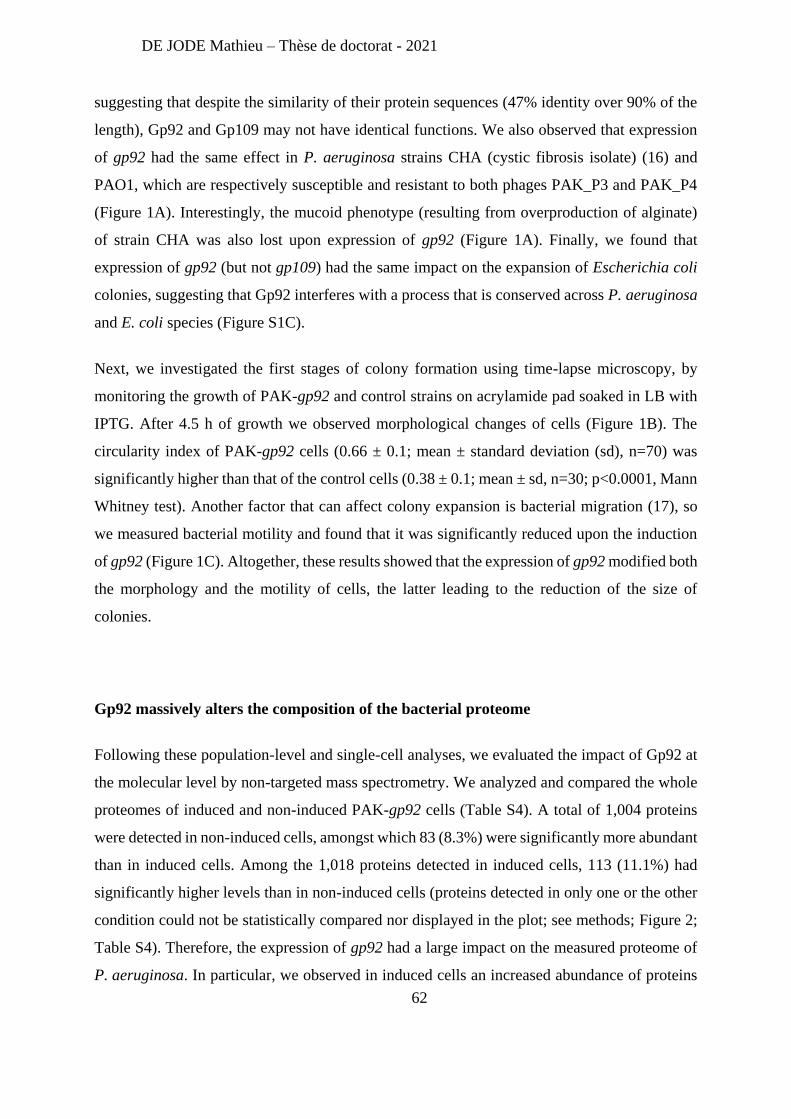
DE JODE Mathieu – Thèse de doctorat - 2021
62
suggesting that despite the similarity of their protein sequences (47% identity over 90% of the
length), Gp92 and Gp109 may not have identical functions. We also observed that expression
of gp92 had the same effect in P. aeruginosa strains CHA (cystic fibrosis isolate) (16) and
PAO1, which are respectively susceptible and resistant to both phages PAK_P3 and PAK_P4
(Figure 1A). Interestingly, the mucoid phenotype (resulting from overproduction of alginate)
of strain CHA was also lost upon expression of gp92 (Figure 1A). Finally, we found that
expression of gp92 (but not gp109) had the same impact on the expansion of Escherichia coli
colonies, suggesting that Gp92 interferes with a process that is conserved across P. aeruginosa
and E. coli species (Figure S1C).
Next, we investigated the first stages of colony formation using time-lapse microscopy, by
monitoring the growth of PAK-gp92 and control strains on acrylamide pad soaked in LB with
IPTG. After 4.5 h of growth we observed morphological changes of cells (Figure 1B). The
circularity index of PAK-gp92 cells (0.66 ± 0.1; mean ± standard deviation (sd), n=70) was
significantly higher than that of the control cells (0.38 ± 0.1; mean ± sd, n=30; p<0.0001, Mann
Whitney test). Another factor that can affect colony expansion is bacterial migration (17), so
we measured bacterial motility and found that it was significantly reduced upon the induction
of gp92 (Figure 1C). Altogether, these results showed that the expression of gp92 modified both
the morphology and the motility of cells, the latter leading to the reduction of the size of
colonies.
Gp92 massively alters the composition of the bacterial proteome
Following these population-level and single-cell analyses, we evaluated the impact of Gp92 at
the molecular level by non-targeted mass spectrometry. We analyzed and compared the whole
proteomes of induced and non-induced PAK-gp92 cells (Table S4). A total of 1,004 proteins
were detected in non-induced cells, amongst which 83 (8.3%) were significantly more abundant
than in induced cells. Among the 1,018 proteins detected in induced cells, 113 (11.1%) had
significantly higher levels than in non-induced cells (proteins detected in only one or the other
condition could not be statistically compared nor displayed in the plot; see methods; Figure 2;
Table S4). Therefore, the expression of gp92 had a large impact on the measured proteome of
P. aeruginosa. In particular, we observed in induced cells an increased abundance of proteins
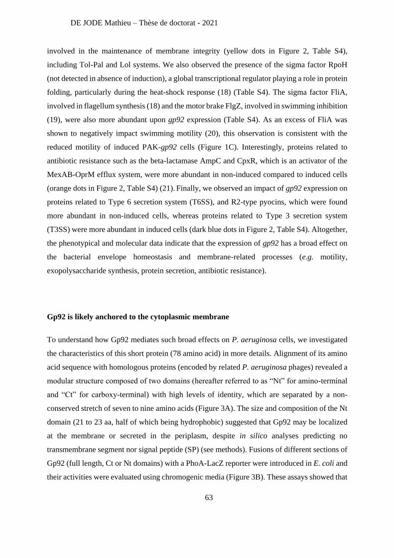
DE JODE Mathieu – Thèse de doctorat - 2021
63
involved in the maintenance of membrane integrity (yellow dots in Figure 2, Table S4),
including Tol-Pal and Lol systems. We also observed the presence of the sigma factor RpoH
(not detected in absence of induction), a global transcriptional regulator playing a role in protein
folding, particularly during the heat-shock response (18) (Table S4). The sigma factor FliA,
involved in flagellum synthesis (18) and the motor brake FlgZ, involved in swimming inhibition
(19), were also more abundant upon gp92 expression (Table S4). As an excess of FliA was
shown to negatively impact swimming motility (20), this observation is consistent with the
reduced motility of induced PAK-gp92 cells (Figure 1C). Interestingly, proteins related to
antibiotic resistance such as the beta-lactamase AmpC and CpxR, which is an activator of the
MexAB-OprM efflux system, were more abundant in non-induced compared to induced cells
(orange dots in Figure 2, Table S4) (21). Finally, we observed an impact of gp92 expression on
proteins related to Type 6 secretion system (T6SS), and R2-type pyocins, which were found
more abundant in non-induced cells, whereas proteins related to Type 3 secretion system
(T3SS) were more abundant in induced cells (dark blue dots in Figure 2, Table S4). Altogether,
the phenotypical and molecular data indicate that the expression of gp92 has a broad effect on
the bacterial envelope homeostasis and membrane-related processes (e.g. motility,
exopolysaccharide synthesis, protein secretion, antibiotic resistance).
Gp92 is likely anchored to the cytoplasmic membrane
To understand how Gp92 mediates such broad effects on P. aeruginosa cells, we investigated
the characteristics of this short protein (78 amino acid) in more details. Alignment of its amino
acid sequence with homologous proteins (encoded by related P. aeruginosa phages) revealed a
modular structure composed of two domains (hereafter referred to as “Nt” for amino-terminal
and “Ct” for carboxy-terminal) with high levels of identity, which are separated by a non-
conserved stretch of seven to nine amino acids (Figure 3A). The size and composition of the Nt
domain (21 to 23 aa, half of which being hydrophobic) suggested that Gp92 may be localized
at the membrane or secreted in the periplasm, despite in silico analyses predicting no
transmembrane segment nor signal peptide (SP) (see methods). Fusions of different sections of
Gp92 (full length, Ct or Nt domains) with a PhoA-LacZ reporter were introduced in E. coli and
their activities were evaluated using chromogenic media (Figure 3B). These assays showed that
DE JODE Mathieu – Thèse de doctorat - 2021
64
the Nt domain supports the periplasmic localization of the reporter, while the Ct domain
remains in the cytoplasm.
Next, we evaluated if and how the localization of Gp92 would affect colony expansion. We
observed a reduction of the size of colonies only with the full-length Gp92, but not with the Ct
or the Nt domains on their own, nor when the Ct domain was artificially exported into the
periplasm using PhoA SP (Figure 3C). Therefore, we concluded that Gp92 is likely anchored
to the membrane and this localization is crucial to mediate its function. Importantly, since both
Gp92 and Gp109 displayed the same localization (Figure S2) but did not cause the same effects
on colony expansion, we concluded that the production of such a likely-anchored membrane
protein per se was not responsible for the observed phenotypes. It also suggested that the
differences between Gp92 and Gp109 activities were likely caused by the divergence in the
sequences of their Ct domains. Finally, homology searches (Phyre2) (22) with the full length
or with the Ct domain of Gp92 did not reveal further predictions about the function of this
protein.
The membrane stress response regulator MucA is targeted by Gp92
To unravel the function of Gp92, we searched for putative bacterial protein partners by
performing two independent bacterial adenylate cyclase two-hybrid (BACTH) screens (23)
using either an E. coli or a Salmonella typhi peptide library. The choice of these libraries was
guided by the fact that the effect on colony expansion was observed in both P. aeruginosa and
E. coli (Figure 1 and S1), therefore we aimed at identifying an interacting partner that is
conserved across diverse bacterial species. The screens identified 13 inner-membrane protein
candidates (Tables S5), amongst which the anti-sigma factor RseA was most frequently
identified and the only one present in the two independent screens (Table S5). Anti-sigma
factors are regulatory proteins that sequester sigma factors thereby preventing their binding to
the RNA polymerase core and the transcriptional activation of genes they control (known as
regulon). The homolog of RseA in P. aeruginosa is the transmembrane protein MucA that
sequesters the sigma factor AlgU, which is involved in mediating the response to membrane-
associated stresses (24). Using pairwise interaction tests, we confirmed interactions between
Gp92 and both RseA and MucA (Figure 4A). To confirm the involvement of MucA, we used
DE JODE Mathieu – Thèse de doctorat - 2021
65
an isogenic P. aeruginosa strain carrying the mucA22 allele, which encodes a truncated non-
functional version of MucA. We found no reduction of the size of colonies upon gp92
expression in this strain (Figure 4B). Importantly, in the mucA22 background, AlgU is no longer
sequestered and consequently, the membrane-associated stress response is constitutively
activated (25). Therefore, the loss of the phenotype may be the direct consequence of the
absence of MucA or indirectly due to the constitutively activated membrane-associated stress
response.
Gp92 impairs cell response to membrane-associated stress
Because Gp92 has a broad effect on the bacterial proteome related to envelope homeostasis
(Figure 2) and directly interacts with MucA (Figure 4), we next investigated whether Gp92
impacted the ability of bacteria to respond to membrane-associated stress. We tested the
transcriptional activation of AlgU regulon by quantifying the transcripts level of algD,
commonly used as a reporter of the activation of AlgU-response (25). In absence of stress, the
expression of gp92 by itself did not induce the transcription of algD (Figure S3A). By contrast,
in presence of D-cycloserine, an analogue of D-alanine interfering with cell-wall synthesis, we
observed a strong increase of algD transcript levels, as previously reported (Figure S3A) (25).
Under this stress, the induction of gp92 expression significantly reduced the level of algD
transcripts compared to cells that do not express gp92 (Figure S3A), indicating that Gp92 limits
the transcriptional activation of the AlgU-stress response.
Next, we evaluated with strain PAK-gp92 how the expression of gp92 affected the proteome
during a membrane-associated stress. A total of 1,005 proteins were detected in non-induced
stressed cells, amongst which 99 (9.9%) were significantly more abundant than in gp92-induced
stressed cells, suggesting that gp92 expression lowers the abundance of these proteins (Figure
S3B and Table S6). Among the 1,007 proteins detected in gp92-induced stressed cells, 95
(9.4%) had significantly higher levels than in non-induced stressed cells (Table S6). These
results indicated that the induction of gp92 had a broad impact on the proteome of stressed cells
with variations detected across several pathways, including 20 transcriptional regulators.
Nevertheless, proteins related to stress and/or adaptation (black dots in Figure S3B and Table
S6) and proteins regulated by AlgU (purple dots in Figure S3B and Table S6) were detected in
DE JODE Mathieu – Thèse de doctorat - 2021
66
both conditions, but with different abundance, hinting that the AlgU stress response is altered
in gp92 expressing cells. Notably, we found that AlgW, the protease that cleaves MucA to
release AlgU and activate the stress response (26), was more abundant in gp92-induced stressed
cells, which suggests that Gp92/MucA interaction may affect AlgW/MucA interaction.
Consistent with previous results (Figure 2), we found that RpoH was over-represented in gp92-
induced stressed cells, as well as the proteins it regulates (green dots in Figure S3B and Table
S6). The impact of Gp92 on protein secretion systems (TSS3 and TSS6) previously observed
(Figure 2, Table S4) was maintained upon membrane-associated stress (dark blue dots in Figure
S3B and Table S6). We also noticed the significant increased abundance of several proteins
related to antibiotic resistance (a beta-lactamase in non-induced stressed cells and two
components of efflux systems, OprM and MexV, in induced stressed cells) (orange dots in
Figure S3B and Table S6).
Altogether, these data suggest that Gp92 affects the capacity of bacteria to activate a stress
response, both at the transcriptional and protein levels. Strengthening this hypothesis, we found
that bacteria expressing gp92 became more susceptible to sub-lethal concentrations of D-
cycloserine as their growth rate decreased by 20% compared to wild type bacteria (Figure
S3C,D).
Expression of gp92 is lethal in cells lacking algU
Two observations: (i) the lack of Gp92-associated phenotype in the mucA22 strain and (ii) the
alteration of the AlgU-mediated stress response by Gp92, prompted us to investigate the effect
of gp92 expression in a strain lacking algU (∆algU strain). Surprisingly, we observed that the
viability of these bacteria was dramatically reduced (Figure 5A), with an effect on the growth
of the bacterial population detectable as soon as 4 h after induction of gp92 expression (Figure
5B). A trans-complementation with a plasmid encoding AlgU restored cell viability (2.6 106
+/- 1.8 106 CFU/mL without IPTG compared to 8.3 105 +/- 7.2 105 CFU/mL with 1 mM IPTG;
n=3). Strikingly, time lapse microscopy observations revealed that ∆algU cells expressing gp92
underwent extensive morphological changes and eventually burst (Figure 5C). Interestingly,
the severity of Gp92 effects on the physiology of ∆algU, WT and mucA22 strains (respectively,
lethal, mild, none) was inversely correlated with the level of free AlgU available in the cell
DE JODE Mathieu – Thèse de doctorat - 2021
67
(null, intermediate, high), strengthening the link between the activity of Gp92 and the AlgU-
mediated stress response. Therefore, we speculated that cells with a functional AlgU-response
may be protected from a lethal effect caused by Gp92.
Alginate biosynthesis rescues ∆algU cells from the lethal expression of gp92
Within the large AlgU regulon (293 genes),(27) the alg operon is the most well-known and is
responsible for the biosynthesis of alginate, an exopolysaccharide that protects P. aeruginosa
cells against adverse conditions (including oxidative stress, antibiotics and phages) (28). The
promoter of the alg operon (PalgD, located upstream of the first gene, algD) is dependent of
AlgU, therefore a strain lacking AlgU does not produce alginate. To test if alginate biosynthesis
could protect cells against Gp92 activity, we studied a strain with an in-frame deletion of algD.
We found that the expression of gp92 reduced the viability of ∆algD cells by approximately
two orders of magnitude (Figure 5D). The optical density of a liquid culture of the ∆algD strain
remained unaffected upon gp92 induction, which may be explained by the filamentous
morphology that these cells adopted upon gp92 induction (Figure 5E,F). To demonstrate further
the protective role of alginate biosynthesis, we restored the transcription of the alg operon in a
∆algU strain, by replacing the native PalgD promoter by an arabinose-inducible promoter. The
viability of these cells was largely recovered upon arabinose induction (Figure 5G)
demonstrating that the biosynthesis of alginate plays a protective role against Gp92.
Nevertheless, the mechanisms by which Gp92 exerts its toxicity and by which alginate rescue
cells will require further investigations. Next, we sought to understand the role of Gp92 during
phage infection.
Gp92 reveals a novel link between phage lysis timing and membrane stress response
Given that Gp92 primarily interacts with MucA, we tested the performance of phage PAK_P3
on the mucA22 strain. First, we found that the phage efficiency of plaquing (EOP) was
significantly increased in the mucA22 strain compared to the PAK(-) strain (Figure 6A). This
effect was not caused by differences in phage adsorption (Figure S4A). Second, in conditions
DE JODE Mathieu – Thèse de doctorat - 2021
68
of synchronized infection, we determined the time necessary to observe the first sign of lysis in
the bacterial population (measured as the time needed to observe a 20% reduction from the
maximum optical density). We found that lysis of mucA22 cells was significantly delayed
compared to PAK(-) cells (Figure S4B,C). Altogether, these data showed that the constitutive
activity of the AlgU-mediated stress response delayed phage lysis and increased the EOP. We
propose that, through its interaction with MucA, Gp92 participates to the control of optimal
phage lysis timing, which is a critical determinant of phage fitness (29). Several mechanisms
have been reported for the control of the lysis timing, but to our knowledge, none relied on the
manipulation of a host sigma/anti-sigma system.
Gp92 increases the susceptibility of P. aeruginosa to carbapenems
Interestingly, the AlgU membrane-associated stress response is induced by several antibiotics
(30), although it remains unclear to which extent this response contributes to the antibiotic-
resistance of P. aeruginosa. We first defined the resistome of strain PAK using CARD (31) and
ResFinder (32) and identified 52 genes predicted to be involved in antibiotic resistance,
amongst which 43 (83.7%) are involved in efflux systems (Table S7). We also found that 15.4%
of these genes are regulated by AlgU, including mexAB-oprM and mexCD-oprJ, two major
efflux systems (33, 34). The mass spectrometry analyses reported above (Figure 2 and S3) have
highlighted that the abundance of several proteins involved in antibiotic resistance were
affected by the expression of gp92, leading us to measure the susceptibility of several strains of
P. aeruginosa to antibiotics.
We found that the level of resistance to drugs targeting peptidoglycan synthesis (Penicillin,
Cefoxitin, Ceftriaxone) was not affected in PAK cells expressing gp92, with the exception of
Imipenem, one of the last resort antibiotics (Table 1). The synergy between Gp92 and Imipenem
was also observed in the cystic fibrosis isolate CHA, with a tenfold reduction of the minimum
inhibitory concentration (MIC). Moreover, we found that the MIC of Imipenem for strain
PAK∆algU was reduced while it was increased for strain PAKmucA22 (Table 1). This suggests
that the susceptibility to Imipenem can be modulated by the AlgU-stress response.
Consequently, this stress response is a candidate target to raise the susceptibility of P.
aeruginosa to carbapenems. We hypothesize that the increased susceptibility to Imipenem but
DE JODE Mathieu – Thèse de doctorat - 2021
69
not to other β-lactams is due to the presence of two β-lactamases (ampC and OXA-50), which
are not AlgU-dependent in their expression (35).
On the other hand, the susceptibility of gp92-expressing cells to drugs targeting protein
synthesis gave mixed results. Indeed, MIC values of Kanamycin, Tetracycline and
Chloramphenicol were reduced (making the resistant strain sensitive again) while it was
unchanged for Erythromycin (Table 1). The presence of 18 AlgU-independent macrolide
resistance genes (Table S7) provide an explanation for the unchanged resistance to
Erythromycin. Interestingly, the susceptibility to Chloramphenicol and Tetracycline was
restored, but a strict link to AlgU regulon was not confirmed when testing PAKmucA22 and
PAK∆algU strains (Table 1). Finally, the lower MIC value of Kanamycin was unrelated to
AlgU regulon as both PAKmucA22 and PAK∆algU strains also displayed a reduced MIC value.
To conclude, this study of gene gp92 revealed that phage PAK_P3 targets the membrane-
associated stress response of P. aeruginosa to control phage lysis timing, but also unveiled that
impairing this stress response provide a novel strategy to lower P. aeruginosa resistance to
carbapenems.
Discussion
Metagenomics sequences have revealed a huge diversity of phage coding sequences. Aside
from a handful of conserved functions (e.g. polymerase, major capsid protein and helicase) that
are sufficient to re-organize viral classification (36), the function of most phage genes remain
elusive. While the identification of bacterial phage resistance systems is racing ahead (37), the
identification of phage genes that interfere with bacterial physiology is still lagging behind.
Notably, the lack of genetic tools allowing an easy manipulation of virulent phage genomes is
slowing down the identification of phage protein functions. Here we studied gp92, an early-
expressed phage gene, which has a subtle effect on colony expansion. Our investigations
demonstrated that Gp92 has a broad impact on bacterial physiology and notably impairs the
AlgU-mediated stress response, which is involved in the susceptibility to Imipenem. We
hypothesize that Gp92 disrupts this stress response to control the phage lysis timing, as
activation of this response was associated with delayed lysis.

DE JODE Mathieu – Thèse de doctorat - 2021
70
Following the initial observation of the modest effect that Gp92 has on the expansion of
bacterial colonies, we uncovered unexpectedly broad consequences linked to the expression of
gp92. First, the composition of the cell proteome was largely affected, with important variations
in the levels of proteins involved in membrane homeostasis, which was in agreement with the
drastic modifications of cell morphology that we observed. Strikingly, a large number of
transcriptional regulators were affected by the expression of gp92, including two sigma factors
(RpoH and FliA). A role of RpoH in phage infection has been reported previously, as it
regulates chaperon proteins necessary for the assembly of phage capsids (38-40). We noticed
that out of the 53-upregulated genes during early infection by phage PAK_P3, eleven (20.8%)
were upregulated by RpoH (in bold in Table S3). This suggests that Gp92, might contribute to
the massive transcriptomic alteration elicited by phage PAK_P3 described previously (14).
Looking for specific targets of Gp92, we identified the anti-sigma factor MucA. The
Gp92/MucA interaction impaired the AlgU membrane-associated stress response. Replacing
this finding in the context of phage lifecycle revealed a novel link between this stress response
and lysis timing.
Another surprising result came from the lethality of gp92 expression in the absence of AlgU or
alginate biosynthesis as it is unclear how the interaction of Gp92 with MucA would lead to cell
death. Based on BACTH and mass spectrometry results, we hypothesize that the affinity of
Gp92 for multiple membrane proteins may make cells more fragile. Further studies are now
required to identify the molecular mechanism(s), underlying the pleiotropic effects of this small
protein, as well as how it controls phage lysis timing.
Finally, our study uncovered a link between the AlgU regulon and the susceptibility to
Imipenem. We hypothesize that peptides or drugs mimicking the activity that Gp92 has on the
membrane stress response could be proposed as adjuvants to carbapenems, one of the last resort
class of antibiotics.
DE JODE Mathieu – Thèse de doctorat - 2021
71
Materials and methods
Bacterial strains and plasmids
Bacterial strains and plasmids are listed in Table S8. Strains were grown in LB medium at 37°C
supplemented with appropriate antibiotics when required (kanamycin 50 µg.mL-1, carbenicillin
100 µg.mL-1 for E. coli, gentamycin 30 µg.mL-1 for P. aeruginosa).
Genetic manipulation of bacteria and phage genes
All primers used for this study are listed in Table S8. Insertion of gp92 and gp109 coding
sequences (CDS) on the chromosome of P. aeruginosa were performed as previously described
(10). Briefly, phage CDS were PCR amplified from phage lysates and cloned in E. coli –
P. aeruginosa shuttle expression vector pUC18-mini-Tn7T-Lac and verified by DNA
sequencing. Expression vector was co-transformed with pTNS2 in P. aeruginosa cells allowing
the integration of the expression cassette (inducible by IPTG) at the unique transposon site Tn7
on the bacterial chromosome.
The PAK∆algU-gp92-Para(algD) strain was generated by replacing a 386bp-region containing
the native PalgD promoter by a 1236bp-cassette including the araC regulator and the arabinose-
inducible araBAD promoter, following a method previously described (41). Briefly, a 429bp-
region upstream and a 446bp-region downstream PalgD were amplified from strain
PAK∆algU-gp92. The 1236bp cassette was amplified from the pJN105 plasmid. These three
PCR amplicons were assembled together and flanked with attB1 and attB2 sites to form a
recombination cassette, which was then inserted into the donor allelic exchange vector
pDONRpEX18Gm using BP Clonase II (Invitrogen). The resulting vector was introduced into
the strain PAK∆algU-gp92 through conjugation with E. coli S17.1pir. Recombinant clones
were isolated upon sucrose-mediated counter-selection and verified by amplifying and
sequencing the recombinant region using primers AC_82F, AC_82R, AC_83F and AC_84R
(Table S1).
DE JODE Mathieu – Thèse de doctorat - 2021
72
Growth, motility and survival assays
Overnight cultures at 37°C in LB medium under agitation of strains carrying phage ORFs and
their corresponding controls were diluted 100-fold in fresh LB medium and incubated 2 h until
OD600nm reached 0.15-0.4. To monitor growth in presence or absence of IPTG (1 mM), 100
µL of bacteria were introduced in 96-well plates and OD600nm was recorded overtime (OD
measured every 15 min after 30 sec of shaking, Infinite M200 Pro, Tecan). To evaluate the
survival of strains upon induction of phage genes, ten-fold serial dilutions of refreshed
overnight cultures were plated on LB agar supplemented with appropriate antibiotics with or
without 1 mM IPTG. Plates were incubated at 37°C for 24 h to 48 h and scanned with Epson
perfection V700 Pro scanner. Colonies areas were measured using ImageJ software. Motility
assays were performed by spotting 3 µL of overnight cultures at the centre of a LB soft agar
(0.3%) plate containing 1 mM IPTG and the swimming diameters were measured after 20 h of
incubation at 37°C. To monitor the growth of bacteria in presence or absence of D-cycloserine
(10 µM), refreshed overnight cultures were first cultivated in presence or absence of IPTG (1
mM) during 1 h before the addition of D-cycloserine or buffer and OD recording in the
microplate reader. Growth rates correspond to the slopes (OD.h-1) of linear regressions
calculated between 1 and 3 h. For each strain, growth rate ratios between induced and non-
induced conditions were calculated.
Time Lapse Microscopy
Exponentially growing bacteria were diluted to reach the OD600nm value of 0.1 and then 2 µL
were spotted on an acrylamide pad (previously soaked in LB + 1 mM IPTG for 4 h). Samples
were eventually sealed under a coverslip with solvent-free sealant “Valap” (1:1:1, w/w/w
vaseline, lanolin, paraffin) and placed under the microscope equipped with an incubation
chamber set at 37°C. Successive divisions of single cells were monitored by taking pictures
every 20 min during 8 h. Images were compiled and analysed using NIS-Elements Viewer 4.2
(Nikon) software. The circularity index is defined as Circ=4π × [Area]/[Perimeter]² and ranges
from 0 (infinitely elongated polygon) to 1 (perfect circle) and was assessed with ImageJ
software.
DE JODE Mathieu – Thèse de doctorat - 2021
73
Sequence analysis tools
Homologs of gp92 and gp109 belonging to family of core ORFs #21 (15) from the
corresponding phage genomes available from Genbank were aligned with ClustalO. Prediction
of protein localization were performed with the online tools Topcons (http://topcons.cbr.su.se/)
and TMHMM (http://www.cbs.dtu.dk/services/TMHMM/). The search for structural homologs
using Phyre 2 returned for Gp92 a stretch of 17 amino acids with 11% identity to TGF-β with
a confident score of 52% and a stretch of 32 amino acids with 25% identity to LuxS/MPP-like
metallohydrolase for Gp109 with a confident score of 48%
Topological analysis of Gp92 and Gp109
Both gp92 and gp109 ORFs were PCR-amplified from phage lysates and cloned in frame in the
pKTop plasmid encoding the dual PhoA-LacZ reporter (42). Specific primers (Table S8) were
used to amplify both Nt (from nucleotide 1 to 78) and Ct (from nucleotide 69 to 234) parts of
Gp92. Plasmids were verified by DNA sequencing and E. coli DH5α strains carrying the
resulting plasmids were plated on the following indicator medium: LB agar supplemented with
5-Bromo-4-chloro-3-indolyl phosphate (80 µg.mL-1), 5-Bromo-6-chloro-3-indolyl β-D-
galactopyranoside (100 µg.mL-1), carbenicillin (100 µg.mL-1), kanamycin (50 µg.mL-1), IPTG
(1 mM) and phosphate buffer (50 mM, pH 7.0). Plates were incubated at 30°C during 24 to
48 h.
RT-qPCR on D-cycloserine stressed cells
Strains PAK-gp92 was grown in LB supplemented or not with IPTG (1 mM) during 1 h prior
the eventual addition of D-cycloserine (10 mM). One hour later total RNAs were extracted by
using RNeasy Plus Mini® kit (Qiagen). After DNaseI treatment and purification, RNAs were
reverse transcribed following manufacturer’s instructions (Random Hexamers, SuperScript™
III, Invitrogen) and quantitative PCR was performed (Applied biosystem 7300) using the
primers indicated in Table S8. Relative expression was assessed by the ∆∆Ct method and
normalized against gyrA expression.
DE JODE Mathieu – Thèse de doctorat - 2021
74
Samples preparation and analysis of peptides by mass spectrometry
Strain PAK-gp92 was cultivated in the same conditions as described for the RT-qPCR
experiments. One hour after the eventual addition of D-cycloserine samples were centrifuged
at 8.000g for 5 min and supernatants were discarded. Pellets were resuspended in 8 M urea, 100
mM Tris HCl pH 8.5, and disulfide bonds were reduced with 5 mM Tris(2-carboxyethyl)
phosphine (TCEP) for 30 minutes and alkylated with 10 mM iodoacetamide for 30 min at room
temperature in the dark. Protein samples were then incubated with 500 ng rLys-C Mass Spec
Grade (Promega, Madison, WI, USA) for 5 h at 30°C for the first digestion. Then samples were
diluted below 2 M urea with 100 mM Tris HCl pH 8.5 and 1 ug Sequencing Grade Modified
Trypsin (Promega, Madison, WI, USA) was added for the second digestion overnight at 37°C.
A second incubation with the same amount of trypsin (5 h at 37°C) was performed to ensure a
complete digestion. Digestion was stopped by adding formic acid and peptides were desalted
and concentrated on Sep-Pak C18 SPE cartridge (Waters, Milford, MA, USA) according to
manufactures instructions.
Tryptic peptides were analyzed on a Q Exactive Plus instrument (Thermo Fisher Scientific,
Bremen) coupled with an EASY nLC 1000 chromatography system (Thermo Fisher Scientific).
Each sample was loaded on an in-house packed 50 cm nano-HPLC column (75 μm inner
diameter) with C18 resin (1.9 μm particles, 100 Å pore size, Reprosil-Pur Basic C18-HD resin,
Dr. Maisch GmbH, Ammerbuch-Entringen, Germany) and equilibrated in 98 % solvent A
(H2O, 0.1 % FA) and 2 % solvent B (ACN, 0.1 % FA). Peptides were first eluted using a 6 to
22 % gradient of solvent B during 150 min and then a 22 to 50 % gradient of solvent B during
60 min all at 250 nL.min-1 flow rate. The instrument method for the Q Exactive Plus was set up
in the data dependent acquisition mode. After a survey scan in the Orbitrap (resolution 70 000),
the 10 most intense precursor ions were selected for HCD fragmentation with a normalized
collision energy set up to 28. Charge state screening was enabled, and precursors with unknown
charge state or a charge state of 1 and ≥ 7 were excluded. Dynamic exclusion was enabled for
45 s.
All data were searched using Andromeda (43) with MaxQuant software (44, 45) version 1.5.3.8
against the P. aeruginosa strain PAK database (2989 entries) concatenated with the phage
PAK_P3 proteome (142 entries), as well as usual known mass spectrometry contaminants and
DE JODE Mathieu – Thèse de doctorat - 2021
75
reversed sequences of all entries. Andromeda searches were performed choosing trypsin as
specific enzyme with a maximum number of two missed cleavages. Possible modifications
included carbamidomethylation (Cys, fixed), oxidation (Met, variable) and Nter acetylation
(variable). The mass tolerance in MS was set to 20 ppm for the first search then 6 ppm for the
main search and 10 ppm for the MS/MS. Maximum peptide charge was set to seven. Five amino
acids were required as minimum peptide length. The “match between runs” feature was applied
for samples having the same experimental condition with a maximal retention time window of
0.7 min. One unique peptide to the protein group was required for the protein identification.
For the statistical analysis of one condition versus another, proteins identified in the reverse and
contaminant databases and proteins only identified by site were first discarded from the list.
Then, proteins exhibiting fewer than two summed intensities in at least one condition were
discarded from the list to avoid misidentified proteins. After log2 transformation of the leftover
proteins, summed intensities were normalised by median centering within conditions
(normalizeD function of the R package DAPAR (46). Remaining proteins without any summed
intensities in one of both conditions have been considered as proteins present in a condition and
absent in another. They have therefore been set aside and considered as differentially abundant
proteins. Next, missing values were imputed using the imp.norm function of the R package
norm (47). Proteins with a log2 (fold-change) inferior to 1 have been considered as proteins
which are not significantly differentially abundant. Statistical testing of the remaining proteins
(having a log2 (fold-change) superior to 1) was conducted using a limma t-test (48) with the R
package limma (49). An adaptive Benjamini-Hochberg procedure was applied on the resulting
p-values using the function adjust.p of R package cp4p (50) with the robust method of Pounds
and Cheng (51) to estimate the proportion of true null hypotheses among the set of statistical
tests. The proteins associated to an adjusted p-value inferior to a FDR level of 1% have been
considered as significantly differentially abundant proteins. Finally, the proteins of interest
were therefore those which emerged from this statistical analysis supplemented by those which
were considered to be absent from one condition and present in another.
Bacterial two hybrid assays
Bacterial two hybrid screening against E. coli and S. typhimurium libraries as well as BACTH
and dimerization assays were performed as previously described (23). Briefly, plasmid
DE JODE Mathieu – Thèse de doctorat - 2021
76
pUT18C-gp92 was introduced into E. coli cya strains BTH101, DHM1 or DHT1. The resultant
strains were then transformed with the indicated plasmids (Table S8) and plated either on M63
agar supplemented with 40 µg.mL-1 X-Gal or MacConkey agar supplemented with 0.2% or 1%
maltose, 0.5 mM IPTG, 25 or 50 µg.mL-1 kanamycin and 50 or 100 µg.mL-1 carbenicillin,
respectively. Plates were incubated at 30°C for 2 to 7 days until appearance of colored colonies.
Screened colonies were isolated twice and further characterized by DNA sequencing of the
inserts.
Adsorption assays
PAK_P3 adsorption constants were calculated as reported previously (14). Briefly,
exponentially growing bacteria were infected at a MOI of 0.001 and immediately after PAK_P3
addition (t=0) and then every 30 s, samples of 50 μL were collected, mixed with CHCl3 and
placed on ice until phage titration on strain PAK. To calculate the adsorption constant k
(mL/min), we first plotted the neperian logarithm of the free phage count against time. Then,
the slope of a linear regression was divided by the bacterial initial concentration (CFU/mL)
used in the experiment. Experiment was performed in triplicate.
Phage lysis kinetics and timing
Exponentially growing cultures of bacteria (until OD600nm reached 0.4) were infected at MOI
15 (to ensure synchronized infection (14)) before introduction in the 96-well microplate reader
for 2 h. OD was recorded every 97 s after 30 s of shaking. To compare lysis timing we calculated
the time interval between the maximum OD value and the reduction of this OD value by 20%
to capture the early kinetics of lysis. Means of this time interval (n=3) for each strain were
compared to the control strain.
DE JODE Mathieu – Thèse de doctorat - 2021
77
Acknowledgments
We thank Drs Nicolas Dufour, Luisa De Sordi, Marta Lourenço and Dwayne Roach for
discussions and suggestions along this work. We warmly thank Dr. Jeroen Wagemans and Dr.
Anne-Sophie Delattre for providing guidance on cloning phage genes. We thank Sven van
Teffelen for access to time-lapse microscope. We are grateful to Mariette Matondo and Quentin
Giai-Gianetto from the proteomic platform of Institut Pasteur.
Funding
AC and MDJ were supported by Sorbonne université, collège doctoral, Paris, France. RL and
LD are members of the « PhageBiotics research community », supported by the FWO
Vlaanderen.
This article is part of a project that has received funding from the European Research Council
(ERC) under the European Union’s ERC consolidator grant awarded to RL (Grant agreement
No. [819800]).
DE JODE Mathieu – Thèse de doctorat - 2021
78
References
1. De Smet J, Hendrix H, Blasdel BG, Danis-Wlodarczyk K, & Lavigne R (2017)
Pseudomonas predators: understanding and exploiting phage-host interactions. Nat Rev
Microbiol 15(9):517-530.
2. Nechaev S & Severinov K (2003) Bacteriophage-induced modifications of host RNA
polymerase. Annu Rev Microbiol 57:301-322.
3. Bondy-Denomy J, Pawluk A, Maxwell KL, & Davidson AR (2013) Bacteriophage
genes that inactivate the CRISPR/Cas bacterial immune system. Nature 493(7432):429-432.
4. Stanley SY, et al. (2019) Anti-CRISPR-Associated Proteins Are Crucial Repressors of
Anti-CRISPR Transcription. Cell 178(6):1452-1464 e1413.
5. Liu J, et al. (2004) Antimicrobial drug discovery through bacteriophage genomics. Nat
Biotechnol 22(2):185-191.
6. Wagemans J, Lavigne, R. (2012) Phages and their hosts: a web of interactions -
applications to drug design. Bacteriophages in health and disease., ed Hyman P, Abedon, S.
T.), pp 119-133.
7. Bin Jang H, et al. (2019) Taxonomic assignment of uncultivated prokaryotic virus
genomes is enabled by gene-sharing networks. Nat Biotechnol 37(6):632-639.
8. Shkoporov AN & Hill C (2019) Bacteriophages of the Human Gut: The "Known
Unknown" of the Microbiome. Cell Host Microbe 25(2):195-209.
9. Filee J, Tetart F, Suttle CA, & Krisch HM (2005) Marine T4-type bacteriophages, a
ubiquitous component of the dark matter of the biosphere. Proceedings of the National Academy
of Sciences of the United States of America 102(35):12471-12476.
10. Wagemans J, et al. (2014) Functional elucidation of antibacterial phage ORFans
targeting Pseudomonas aeruginosa. Cellular microbiology 16(12):1822-1835.
11. Molshanski-Mor S, et al. (2014) Revealing bacterial targets of growth inhibitors
encoded by bacteriophage T7. Proceedings of the National Academy of Sciences of the United
States of America 111(52):18715-18720.
12. Tabib-Salazar A, et al. (2017) Full shut-off of Escherichia coli RNA-polymerase by T7
phage requires a small phage-encoded DNA-binding protein. Nucleic Acids Res 45(13):7697-
7707.
13. Blasdel BG, Chevallereau A, Monot M, Lavigne R, & Debarbieux L (2017)
Comparative transcriptomics analyses reveal the conservation of an ancestral infectious strategy
in two bacteriophage genera. ISME J 11(9):1988-1996.
14. Chevallereau A, et al. (2016) Next-Generation "-omics" Approaches Reveal a Massive
Alteration of Host RNA Metabolism during Bacteriophage Infection of Pseudomonas
aeruginosa. PLoS genetics 12(7):e1006134.
DE JODE Mathieu – Thèse de doctorat - 2021
79
15. Henry M, et al. (2015) The search for therapeutic bacteriophages uncovers one new
subfamily and two new genera of Pseudomonas-infecting Myoviridae. PloS one
10(1):e0117163.
16. Dacheux D, Attree I, Schneider C, & Toussaint B (1999) Cell death of human
polymorphonuclear neutrophils induced by a Pseudomonas aeruginosa cystic fibrosis isolate
requires a functional type III secretion system. Infect Immun 67(11):6164-6167.
17. Harshey RM (2003) Bacterial motility on a surface: many ways to a common goal. Annu
Rev Microbiol 57:249-273.
18. Potvin E, Sanschagrin F, & Levesque RC (2008) Sigma factors in Pseudomonas
aeruginosa. FEMS Microbiol Rev 32(1):38-55.
19. Bense S, et al. (2019) Spatiotemporal control of FlgZ activity impacts Pseudomonas
aeruginosa flagellar motility. Molecular microbiology 111(6):1544-1557.
20. Lo YL, et al. (2018) Characterization of the role of global regulator FliA in the
pathophysiology of Pseudomonas aeruginosa infection. Res Microbiol 169(3):135-144.
21. Tian Z-X, Yi X-X, Cho A, O’Gara F, & Wang Y-P (2016) CpxR activates MexAB-
OprM efflux pump expression and enhances antibiotic resistance in both laboratory and clinical
nalB-type isolates of Pseudomonas aeruginosa. PLoS pathogens 12(10).
22. Kelley LA, Mezulis S, Yates CM, Wass MN, & Sternberg MJ (2015) The Phyre2 web
portal for protein modeling, prediction and analysis. Nat Protoc 10(6):845-858.
23. Karimova G, Pidoux J, Ullmann A, & Ladant D (1998) A bacterial two-hybrid system
based on a reconstituted signal transduction pathway. Proceedings of the National Academy of
Sciences of the United States of America 95(10):5752-5756.
24. Jones AK, et al. (2010) Activation of the Pseudomonas aeruginosa AlgU regulon
through mucA mutation inhibits cyclic AMP/Vfr signaling. Journal of bacteriology
192(21):5709-5717.
25. Wood LF & Ohman DE (2009) Use of cell wall stress to characterize sigma 22 (AlgT/U)
activation by regulated proteolysis and its regulon in Pseudomonas aeruginosa. Molecular
microbiology 72(1):183-201.
26. Cezairliyan BO & Sauer RT (2009) Control of Pseudomonas aeruginosa AlgW protease
cleavage of MucA by peptide signals and MucB. Molecular microbiology 72(2):368-379.
27. Wood LF & Ohman DE (2012) Identification of genes in the σ22 regulon of
Pseudomonas aeruginosa required for cell envelope homeostasis in either the planktonic or the
sessile mode of growth. mBio 3(3):e00094-00012.
28. Llamas MA, Imperi F, Visca P, & Lamont IL (2014) Cell-surface signaling in
Pseudomonas: stress responses, iron transport, and pathogenicity. FEMS microbiology reviews
38(4):569-597.
29. Wang IN (2006) Lysis timing and bacteriophage fitness. Genetics 172(1):17-26.
DE JODE Mathieu – Thèse de doctorat - 2021
80
30. Wood LF, Leech AJ, & Ohman DE (2006) Cell wall-inhibitory antibiotics activate the
alginate biosynthesis operon in Pseudomonas aeruginosa: Roles of sigma (AlgT) and the AlgW
and Prc proteases. Molecular microbiology 62(2):412-426.
31. Jia B, et al. (2017) CARD 2017: expansion and model-centric curation of the
comprehensive antibiotic resistance database. Nucleic Acids Res 45(D1):D566-D573.
32. Zankari E, et al. (2012) Identification of acquired antimicrobial resistance genes. J
Antimicrob Chemother 67(11):2640-2644.
33. Fraud S, Campigotto AJ, Chen Z, & Poole K (2008) MexCD-OprJ multidrug efflux
system of Pseudomonas aeruginosa: involvement in chlorhexidine resistance and induction by
membrane-damaging agents dependent upon the AlgU stress response sigma factor.
Antimicrobial agents and chemotherapy 52(12):4478-4482.
34. Schulz S, et al. (2015) Elucidation of sigma factor-associated networks in Pseudomonas
aeruginosa reveals a modular architecture with limited and function-specific crosstalk. PLoS
Pathog 11(3):e1004744.
35. Lister PD, Wolter DJ, & Hanson ND (2009) Antibacterial-resistant Pseudomonas
aeruginosa: clinical impact and complex regulation of chromosomally encoded resistance
mechanisms. Clinical microbiology reviews 22(4):582-610.
36. Koonin EV, et al. (2020) Global Organization and Proposed Megataxonomy of the
Virus World. Microbiol Mol Biol Rev 84(2).
37. Doron S, et al. (2018) Systematic discovery of antiphage defense systems in the
microbial pangenome. Science 359(6379).
38. Hanninen AL, Bamford DH, & Bamford JK (1997) Assembly of membrane-containing
bacteriophage PRD1 is dependent on GroEL and GroES. Virology 227(1):207-210.
39. Grimaud R & Toussaint A (1998) Assembly of both the head and tail of bacteriophage
Mu is blocked in Escherichia coli groEL and groES mutants. Journal of bacteriology
180(5):1148-1153.
40. Rousset F, et al. (2018) Genome-wide CRISPR-dCas9 screens in E. coli identify
essential genes and phage host factors. PLoS genetics 14(11):e1007749.
41. Hmelo LR, et al. (2015) Precision-engineering the Pseudomonas aeruginosa genome
with two-step allelic exchange. Nat Protoc 10(11):1820-1841.
42. Karimova G, Robichon C, & Ladant D (2009) Characterization of YmgF, a 72-residue
inner membrane protein that associates with the Escherichia coli cell division machinery.
Journal of bacteriology 191(1):333-346.
43. Cox J, et al. (2011) Andromeda: a peptide search engine integrated into the MaxQuant
environment. Journal of proteome research 10(4):1794-1805.
DE JODE Mathieu – Thèse de doctorat - 2021
81
44. Cox J & Mann M (2008) MaxQuant enables high peptide identification rates,
individualized ppb-range mass accuracies and proteome-wide protein quantification. Nature
biotechnology 26(12):1367-1372.
45. Tyanova S, Temu T, & Cox J (2016) The MaxQuant computational platform for mass
spectrometry-based shotgun proteomics. Nature protocols 11(12):2301.
46. Wieczorek S, et al. (2017) DAPAR & ProStaR: software to perform statistical analyses
in quantitative discovery proteomics. Bioinformatics 33(1):135-136.
47. Novo A & Schafer J (2013) Package ‘norm’: Analysis of multivariate normal datasets
with missing values. R Package Version 1.0-9.5.
48. Smyth GK, Ritchie M, Thorne N, & Wettenhall J (2005) LIMMA: linear models for
microarray data. In Bioinformatics and Computational Biology Solutions Using R and
Bioconductor. Statistics for Biology and Health.
49. Ritchie ME, et al. (2015) limma powers differential expression analyses for RNA-
sequencing and microarray studies. Nucleic acids research 43(7):e47-e47.
50. Giai Gianetto Q, et al. (2016) Calibration plot for proteomics: A graphical tool to
visually check the assumptions underlying FDR control in quantitative experiments. Proteomics
16(1):29-32.
51. Pounds S & Cheng C (2006) Robust estimation of the false discovery rate.
Bioinformatics 22(16):1979-1987.
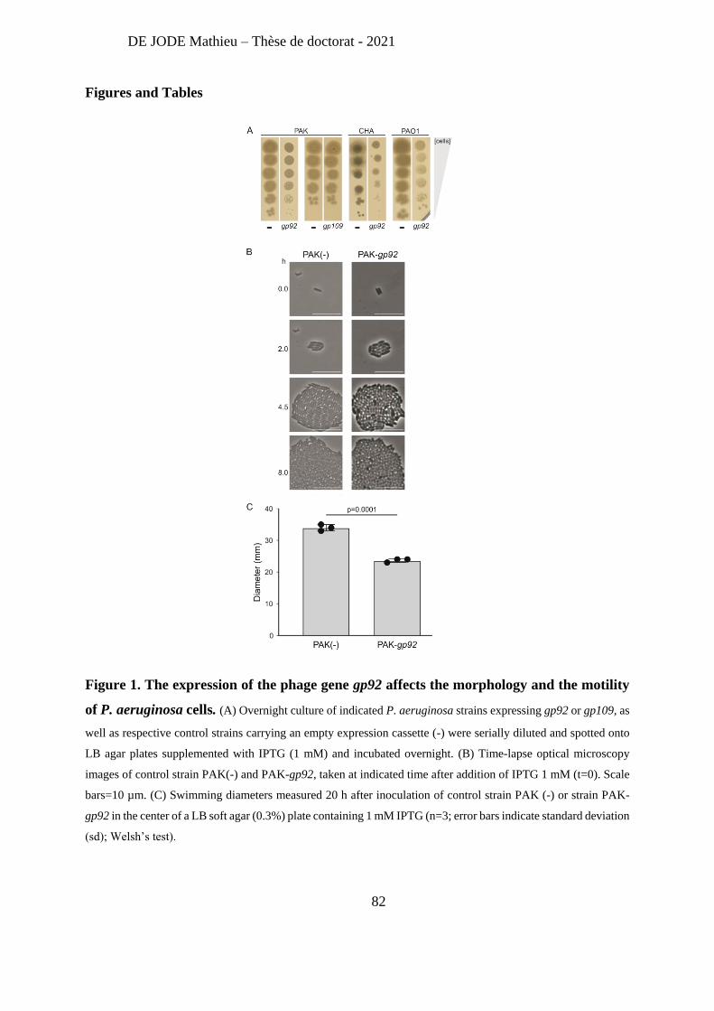
DE JODE Mathieu – Thèse de doctorat - 2021
82
Figures and Tables
Figure 1. The expression of the phage gene gp92 affects the morphology and the motility
of P. aeruginosa cells. (A) Overnight culture of indicated P. aeruginosa strains expressing gp92 or gp109, as
well as respective control strains carrying an empty expression cassette (-) were serially diluted and spotted onto
LB agar plates supplemented with IPTG (1 mM) and incubated overnight. (B) Time-lapse optical microscopy
images of control strain PAK(-) and PAK-gp92, taken at indicated time after addition of IPTG 1 mM (t=0). Scale
bars=10 µm. (C) Swimming diameters measured 20 h after inoculation of control strain PAK (-) or strain PAK-
gp92 in the center of a LB soft agar (0.3%) plate containing 1 mM IPTG (n=3; error bars indicate standard deviation
(sd); Welsh’s test).
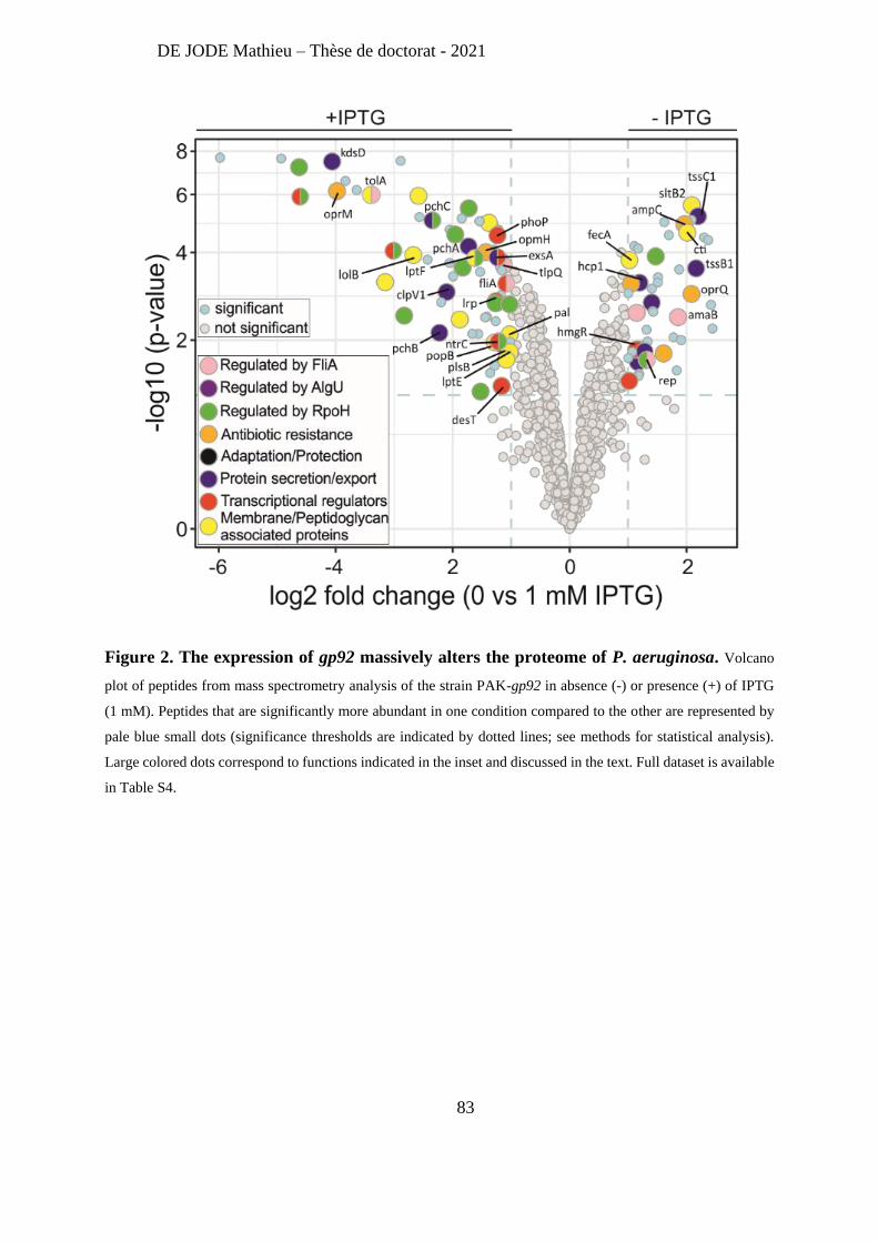
DE JODE Mathieu – Thèse de doctorat - 2021
83
Figure 2. The expression of gp92 massively alters the proteome of P. aeruginosa. Volcano
plot of peptides from mass spectrometry analysis of the strain PAK-gp92 in absence (-) or presence (+) of IPTG
(1 mM). Peptides that are significantly more abundant in one condition compared to the other are represented by
pale blue small dots (significance thresholds are indicated by dotted lines; see methods for statistical analysis).
Large colored dots correspond to functions indicated in the inset and discussed in the text. Full dataset is available
in Table S4.
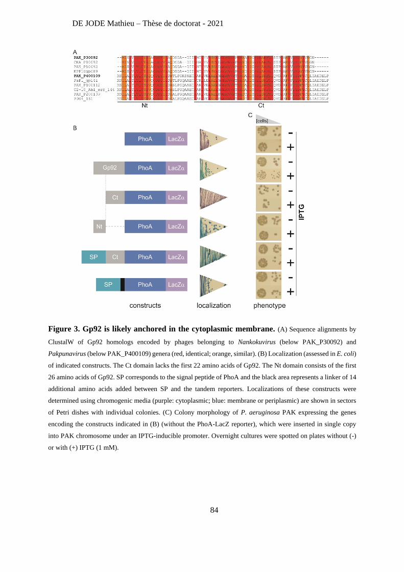
DE JODE Mathieu – Thèse de doctorat - 2021
84
Figure 3. Gp92 is likely anchored in the cytoplasmic membrane. (A) Sequence alignments by
ClustalW of Gp92 homologs encoded by phages belonging to Nankokuvirus (below PAK_P30092) and
Pakpunavirus (below PAK_P400109) genera (red, identical; orange, similar). (B) Localization (assessed in E. coli)
of indicated constructs. The Ct domain lacks the first 22 amino acids of Gp92. The Nt domain consists of the first
26 amino acids of Gp92. SP corresponds to the signal peptide of PhoA and the black area represents a linker of 14
additional amino acids added between SP and the tandem reporters. Localizations of these constructs were
determined using chromogenic media (purple: cytoplasmic; blue: membrane or periplasmic) are shown in sectors
of Petri dishes with individual colonies. (C) Colony morphology of P. aeruginosa PAK expressing the genes
encoding the constructs indicated in (B) (without the PhoA-LacZ reporter), which were inserted in single copy
into PAK chromosome under an IPTG-inducible promoter. Overnight cultures were spotted on plates without (-)
or with (+) IPTG (1 mM).
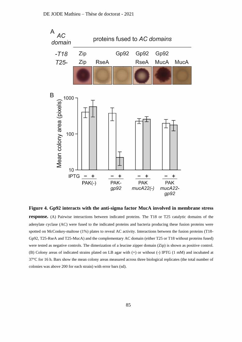
DE JODE Mathieu – Thèse de doctorat - 2021
85
Figure 4. Gp92 interacts with the anti-sigma factor MucA involved in membrane stress
response. (A) Pairwise interactions between indicated proteins. The T18 or T25 catalytic domains of the
adenylate cyclase (AC) were fused to the indicated proteins and bacteria producing these fusion proteins were
spotted on McConkey-maltose (1%) plates to reveal AC activity. Interactions between the fusion proteins (T18-
Gp92, T25-RseA and T25-MucA) and the complementary AC domain (either T25 or T18 without proteins fused)
were tested as negative controls. The dimerization of a leucine zipper domain (Zip) is shown as positive control.
(B) Colony areas of indicated strains plated on LB agar with (+) or without (-) IPTG (1 mM) and incubated at
37°C for 16 h. Bars show the mean colony areas measured across three biological replicates (the total number of
colonies was above 200 for each strain) with error bars (sd).
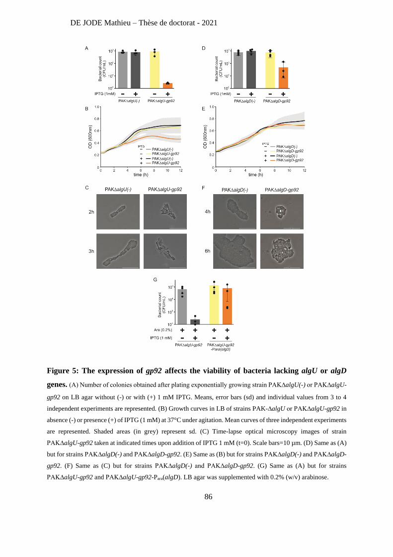
DE JODE Mathieu – Thèse de doctorat - 2021
86
Figure 5: The expression of gp92 affects the viability of bacteria lacking algU or algD
genes. (A) Number of colonies obtained after plating exponentially growing strain PAK∆algU(-) or PAK∆algU-
gp92 on LB agar without (-) or with (+) 1 mM IPTG. Means, error bars (sd) and individual values from 3 to 4
independent experiments are represented. (B) Growth curves in LB of strains PAK-∆algU or PAK∆algU-gp92 in
absence (-) or presence (+) of IPTG (1 mM) at 37°C under agitation. Mean curves of three independent experiments
are represented. Shaded areas (in grey) represent sd. (C) Time-lapse optical microscopy images of strain
PAK∆algU-gp92 taken at indicated times upon addition of IPTG 1 mM (t=0). Scale bars=10 µm. (D) Same as (A)
but for strains PAK∆algD(-) and PAK∆algD-gp92. (E) Same as (B) but for strains PAK∆algD(-) and PAK∆algD-
gp92. (F) Same as (C) but for strains PAK∆algD(-) and PAK∆algD-gp92. (G) Same as (A) but for strains
PAK∆algU-gp92 and PAK∆algU-gp92-Para(algD). LB agar was supplemented with 0.2% (w/v) arabinose.
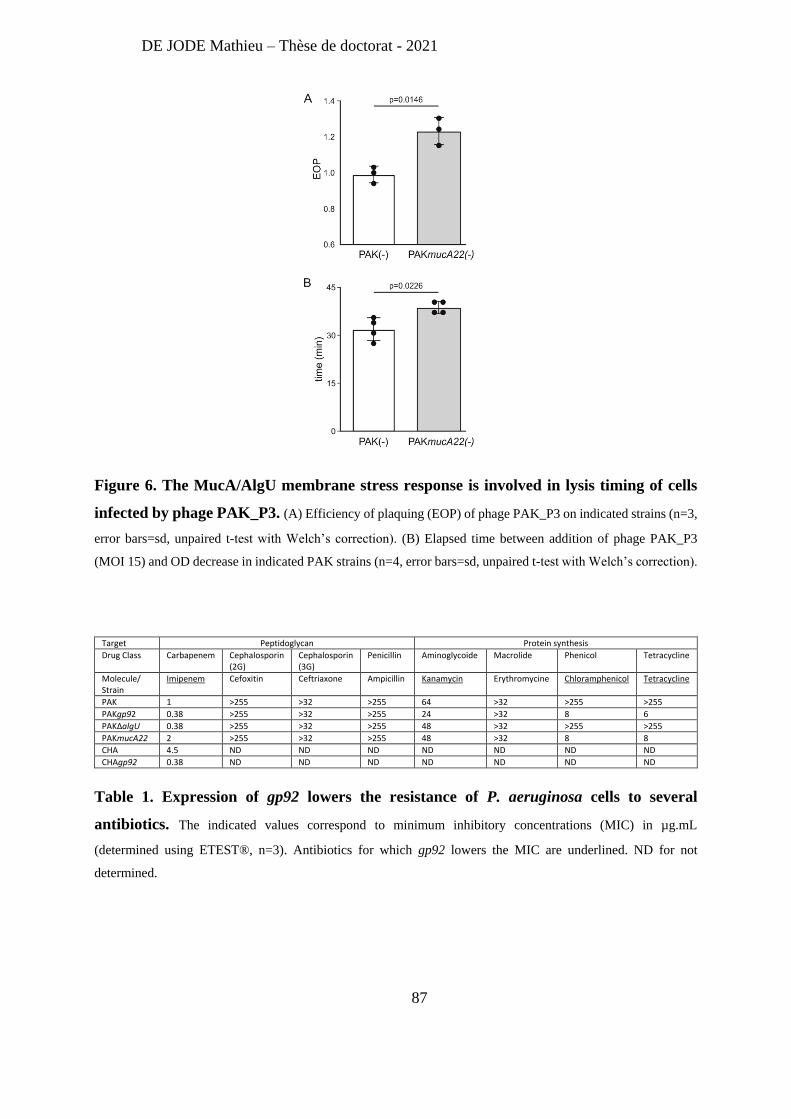
DE JODE Mathieu – Thèse de doctorat - 2021
87
Figure 6. The MucA/AlgU membrane stress response is involved in lysis timing of cells
infected by phage PAK_P3. (A) Efficiency of plaquing (EOP) of phage PAK_P3 on indicated strains (n=3,
error bars=sd, unpaired t-test with Welch’s correction). (B) Elapsed time between addition of phage PAK_P3
(MOI 15) and OD decrease in indicated PAK strains (n=4, error bars=sd, unpaired t-test with Welch’s correction).
Target Peptidoglycan Protein synthesis
Drug Class Carbapenem Cephalosporin (2G)
Cephalosporin (3G)
Penicillin Aminoglycoide Macrolide Phenicol Tetracycline
Molecule/ Strain
Imipenem Cefoxitin Ceftriaxone Ampicillin Kanamycin Erythromycine Chloramphenicol Tetracycline
PAK 1 >255 >32 >255 64 >32 >255 >255
PAKgp92 0.38 >255 >32 >255 24 >32 8 6
PAKΔalgU 0.38 >255 >32 >255 48 >32 >255 >255
PAKmucA22 2 >255 >32 >255 48 >32 8 8
CHA 4.5 ND ND ND ND ND ND ND
CHAgp92 0.38 ND ND ND ND ND ND ND
Table 1. Expression of gp92 lowers the resistance of P. aeruginosa cells to several
antibiotics. The indicated values correspond to minimum inhibitory concentrations (MIC) in µg.mL
(determined using ETEST®, n=3). Antibiotics for which gp92 lowers the MIC are underlined. ND for not
determined.
DE JODE Mathieu – Thèse de doctorat - 2021
88
Supplementary information includes
Figure S1. Phenotypic characterization of bacteria expressing the phage gene gp92.
Figure S2. Gp109 displays the same localisation as Gp92.
Figure S3. Expression of gp92 modifies the physiology of membrane stressed cells.
Figure S4. Adsorption and lysis timing of phage PAK_P3
The legends for Datasets S1 to S8 (large tables)
Table S1. List of early expressed genes of phages PAK_P3 and PAK_P4
Table S2. List of homologous proteins between phages PAK_P3 and PAK_P4
Table S3. Up- and down-regulated host genes 3.5 min after infection by either phage PAK_P3
or PAK_P4
Table S4. List of the significantly more abundant proteins from the IPTG-induced strain PAK-
gp92 compared to the non-induced strain PAK-gp92
Table S5. List of Gp92-binding proteins identified by bacterial two-hybrid screens
Table S6. List of the significantly more abundant proteins from the IPTG-induced strain PAK-
gp92 compared to the non-induced strain PAK-gp92 both in presence of D-cycloserin
TableS7. Resistome of strain PAK crossed with AlgU-regulated genes
Table S8. List of strains, plasmids and primers
DE JODE Mathieu – Thèse de doctorat - 2021
89
2- The combination of bacteriophages prevents resistant outgrowth in
vitro but does not increase pulmonary phage therapy efficacy in
immunocompromised animals.
Our laboratory has previously demonstrated that monophage therapy can cure and
prevent acute lung infection caused by P. aeruginosa in immunocompetent mice [270]. More
recently, we have shown that this therapeutic success relies on antibacterial activities of both
the phage and the immune system, a concept we named ‘immunophage synergy’ [292]. Indeed,
monophage therapy with phage PAK_P1 failed to cure both immune signaling deficient
MyD88- /- mice and neutropenic mice, as the lack of neutrophils allows the growth of phage-
resistant bacteria, not detected in immunocompetent animals.
My second PhD project aimed at investigating bacterial phage-resistance in vitro and in
vivo in immunocompromised mice (MyD88-/-), and assess the therapeutic efficacy of a cocktail
composed of two phages using different bacterial receptors to prevent the growth of phage-
resistant bacteria.
We found that the immunophage synergy concept is not restricted to phage PAK_P1 as
monophage treatment with phage LUZ19 also failed to cure acute lung infection in MyD88- /-
mice. In vitro, both monophage treatments failed to prevent phage-resistant growth, but the
combination of these two phages did. However, this combined treatment did not prevent phage-
resistance to take place in vivo and was not more efficient than monophage treatments. Analysis
of the phage resistance phenotypes of isolated clones from every conditions (both in vitro and
in vivo) suggests that bacterial phage resistance is highly phage-dependent as PAK_P1 selects
for bacteria resistant to PAK_P1 and not LUZ19. Our results also suggest a role of the
experimental environment (in vitro or in vivo) on phage-resistance outcomes. A future genomic
analysis of these clones should reveal to which extent phage resistance mechanisms are
different between each phage treatment (PAK_P1 or LUZ19, or both) and each environment
(in vivo or in vitro).
Understanding phage resistance taking place in vivo could help guiding the design of
efficient phage combination, and overall support the development of phage therapy.
DE JODE Mathieu – Thèse de doctorat - 2021
90
Title
The combination of two bacteriophages targeting different receptors fails to prevent the
emergence of bacteriophage resistant bacteria in immunocompromised animals.
Authors
Mathieu De Jode1,2, Sophia Zborowsky1, Laurent Debarbieux1
Affiliations
1 Bacteriophage, Bacterium, Host Laboratory, Department of Microbiology, Institut Pasteur,
Paris F-75015 France.
2 Sorbonne université, collège doctoral, F-75005 Paris, France
Summary
Pulmonary infections are a major cause of death worldwide exacerbated by the increasing
antibiotic resistance of bacteria. Treatment using bacteriophages, phage therapy, have been
shown to improve the condition of patients but still lack mechanistic support. The success of
monophage therapy in treating acute Pseudomonas aeruginosa lung infection in wild type mice
was shown to rely on the synergistic action of phages and the immune system. In
immunocompromised MyD88-/- mice, monophage therapy failed due to the uncontrolled growth
of phage-resistant bacteria. Here, to jugulate the growth of phage-resistant bacteria we
evaluated two phages recognizing different bacterial receptors and their combination in both in
vitro and in vivo (MyD88-/- mice) conditions. We found that in vitro monophage treatments
failed to prevent phage-resistance growth while the combination succeeded. In contrast, the
combination was not associated with a significant higher efficacy in vivo compared to
monophage treatments. A better understanding of phage resistance mechanisms taking place in
vivo is required to optimize the formulation of phage cocktails and ultimately improve phage
therapy efficacy in clinical settings.
DE JODE Mathieu – Thèse de doctorat - 2021
91
Introduction
The worldwide increase of the prevalence of antibiotic resistant bacteria causing difficult to
treat infections in humans led the World Health Organization to highlight a list of priority
pathogens, for which new treatments are urgently needed [1]. Amongst them, the carbapenem
resistant strains of Pseudomonas aeruginosa are worrisome as this opportunistic pathogen is
ubiquitous in the environment and is a leading cause of nosocomial infections [2, 3].
Furthermore, P. aeruginosa is a major cause of death for immunocompromised individuals
such as cystic fibrosis patients, for which antibiotics treatments are no longer sufficient to
prolong lifetime [4].
Amongst solutions to defeat multidrug resistant bacteria, the use of bacteriophages (phages) to
kill bacterial pathogens, phage therapy, is gaining increasing weight with the recent publication
of several successful compassionate treatments in both Europe and the USA [5-10]. Despite the
uninterrupted clinical experience for nearly a century, mainly from Georgia, phage therapy
remains under evaluated through double blind clinical trials [11, 12]. To fill the gap between
compassionate treatments and broader medical applications molecular mechanisms underlying
treatments failure and success must be provided.
Decades of research performed in vitro identified an array of phage resistance mechanisms,
from single point mutations on bacterial receptors to more sophisticated machineries (e.g.
CRISPR-Cas systems) [13]. Interestingly, the relative abundance of these resistance
mechanisms did not prevent the success of many proofs of concept phage therapy studies
performed with animal models [14, 15]. On few occasions, phage resistant bacteria were
isolated from experimentally infected animals and characterized as less virulent, which did not
jeopardized the treatment efficacy [16, 17]. Recently, during the course of compassionate
treatments with multiple phages on two patients with an altered immune response, phage
resistant clones were isolated, showing that the growth of phage resistant clones could be of
higher relevance in immunocompromised patients [8, 10].
Previously, we found that the monophage treatment of a P. aeruginosa pneumonia in mice was
successful in wild type (WT) but failed in immunocompromised (MyD88-/-) animals [15, 18].
The isolation of few clones from infected and treated MyD88-/- mice revealed that the growth
DE JODE Mathieu – Thèse de doctorat - 2021
92
of phage resistant bacteria was the main cause for the failure of this monophage treatment in
immunocompromised these animals. We then introduced the concept of ‘immunophage
synergy’ taking place during phage therapy in WT animals: both phages and immune cells act
synergistically to obtain a successful treatment in conditions where each of the two was
insufficient [18].
Here, we investigated in vitro and in vivo the consequences of adding a second phage to limit
the growth of phage resistant clones to the first phage. We found that the combination of the
two phages prevented the growth of phage resistant clones in vitro. The survival of MyD88-/-
infected mice increased with the treatment of each phage individually and also in combination,
compared to untreated controls. However, the combination did not improve the clinical status
of animals compared to individual phages. The phenotypic analysis of P. aeruginosa clones
isolated at end points, revealed that the combination limited but not prevented the growth of
phage resistant bacteria compared to monophage treatments.
Results
Establishing conditions for in vitro and in vivo parallel studies of phage resistance
Phage resistance is inherent to any phage-bacteria interaction, but its nature and frequency are
affected by environmental conditions. Having shown that the treatment of P. aeruginosa (strain
PAK-lumi) infected MyD88-/- mice by phage PAK_P1 (virulent, Myoviridae) failed because of
the growth of phage resistant clones, we wished to investigate the molecular mechanisms
involved and compared them to those taking place in laboratory (in vitro) conditions. To
perform parallel experiments (same doses of phages and bacteria) in vitro and in vivo, we
refined our in vivo procedure (see methods) and used a bacterial inoculum of 2 x 10⁶ colony
forming units (CFU) and a phage dose of 2 x 10⁷ plaque forming units (PFU) administered 2 h
post infection (p.i.). During preliminary experiments, WT animals controlled the infection and
survived, while 100% of MyD88-/- infected mice experienced an acute and fatal pneumonia
within 24 h p.i., leading us to set an end point at 22 h p.i. for subsequent experiments. Compared
to our previous conditions, the survival of MyD88-/- mice decreased from 27% at day 3 to 0%
at 24 h, thus allowing a clearer assessment of phage therapy effect on mice survival.
DE JODE Mathieu – Thèse de doctorat - 2021
93
In these conditions, we observed that the intensity of light emitted from the pulmonary area of
infected MyD88-/- mice increased continuously up to 22 h, at which point the median pulmonary
bacterial load reached 5.85 x 10⁷ [3.80 x 10⁷ -3.39 x 10⁸] CFU/lungs (Fig. 1A,1B and Fig. S1).
In WT animals the median pulmonary bacterial load recovered at 22 h p.i. was much lower with
6.17 x 10² [1.75 x 10² - 3.31 x 10³] CFU/lungs, testifying that in these animals’ immune cells
control bacterial growth while in MyD88-/- mice cannot (Fig. 1A,1B and Fig. S1). When
infected MyD88-/- mice were treated by a single dose of phage PAK_P1 the emitted light
increased less rapidly compared to untreated animals (Fig. 1A). The median pulmonary
bacterial load at 22 h p.i. reached 3.40 x 10⁵ [1.33 x 10⁴ – 4.70 x 10⁵] CFU/lungs and the median
phage load was 3.33 x 10⁹ [1.25 x 10⁹ – 1.00 x 10¹⁰] plaque forming unit (PFU) (Fig 1B and
Fig. S1), which represents a 167-fold increase compared to the administered dose of 2 x 10⁷
PFU demonstrating that phage PAK_P1 amplified at the expenses of strain PAK-lumi.
These data are consistent with our previous observations that phage PAK_P1 treatment
jugulates the infection during the first hours. In addition, out of 257 clones collected from the
lungs of PAK_P1 treated mice 89.11% are resistant to phage PAK_P1, confirming that in these
immunodeficient animals the growth of phage resistant clones is largely taking place within 20
h (Table S1). As a comparison, only 0.83% of 360 clones from the lungs of untreated animals
at 22 h p.i. are resistant to phage PAK_P1, which is similar to the proportion found in the PAK-
lumi inoculum (0.55% out of 182 clones) (Table S1).
For the parallel in vitro experiments we recorded the OD600 nm over time. While the growth of
strain PAK-lumi without phage reached the stationary phase within 10 h, the presence of phage
PAK_P1 rapidly stopped the growth from 5 h until 18 h at which time point the OD600 nm started
to increase exponentially (Fig. 2). As for in vivo experiments, samples were collected at 22 h,
CFUs determined and susceptibility to phage PAK_P1 tested. From samples not exposed to
phage PAK_P1, none of the 188 clones was resistant to PAK_P1, which is consistent with the
low frequency of such clones in the inoculum (0.55%) (Table S1). By contrast, the frequency
of PAK_P1 resistant clones from samples exposed to this phage reached 90.96 % (177 clones)
(Tables S1). Therefore, in both in vitro and in vivo conditions, the addition of phage PAK_P1
led to the growth of phage resistant clones. When comparing over time the intensity of emitted
light from animals and the OD values from in vitro plates, we observed that the kinetics of the
bacterial decay was faster in vitro, which is expected because of the lack of activated immune
DE JODE Mathieu – Thèse de doctorat - 2021
94
cells and the difference between the in vitro homogenous liquid environment compared to the
heterogeneous semi-solid environment in the animal lungs.
Phage LUZ19v infects PAK_P1 phage-resistant clones
One solution to lower the growth of phage resistant clones consist in the addition of a second
phage that recognizes a different bacterial receptor. Indeed, mutations affecting phage receptors
were shown to be the main mechanism of cross-resistance in P.aeruginosa exposed to 27
different phages [19]. Previous work on a highly similar phage to PAK_P1, as well as our
unpublished observations, suggested that PAK_P1 uses the LPS as receptor [20]. Phage LUZ19,
which we previously assessed for its efficacy to infect strain PAK-lumi both in vitro and in the
murine pneumonia model [21], is known to use type IV pilus as a receptor [22]. However, our
data indicated that both in vitro and in WT mice, phage LUZ19 was less efficient than phage
PAK_P1 [21]. As this could be a cofounding factor in this study, we performed serial in vitro
passages in liquid culture and isolated a LUZ19 variant (LUZ19v) that displayed an efficiency
of plaquing (EOP) on strain PAK-lumi of 1 instead of 0.2 for the original phage LUZ19 [21].
Next, we tested the ability of phage LUZ19v towards PAK_P1-resistant clones collected above
from both in vitro and in vivo samples. We found that 100% of PAK_P1-resistant clones were
susceptible to LUZ19v, which showed a lack of cross-resistance (Table S1).
Phage LUZ19v controls more rapidly pneumonia than phage PAK_P1
Using the same in vitro condition as for phage PAK_P1, we observed that LUZ19v rapidly
lysed and prevented the growth of strain PAK-lumi from 4 h until 12 h at which time point the
OD600 nm started to increase exponentially to almost reached the OD of the control without phage
(Fig. 2). The clones collected at 22 h were tested for their susceptibility to phage LUZ19v and
we found that 98.90% of them (182 clones tested) were resistant (Table S1). Therefore, in vitro
phage LUZ19v treatment resulted in the same outcome than in vitro phage PAK_P1 treatment,
but with faster kinetics. Interestingly, 5.49% of these clones were resistant to PAK_P1 and
overall, 4.95% were resistant to both phages. These results contrast with those obtain with
DE JODE Mathieu – Thèse de doctorat - 2021
95
PAK_P1 where no cross-resistance was observed. Next, administered to infected MyD88-/-
mice, we observed that phage LUZ19v controlled the infection more quickly than phage
PAK_P1 (Fig. 3A). Indeed, the light emitted barely increased within 22 h p.i. at which point
the median pulmonary bacterial load in lungs reached 1.13 x 10² [1.04 x 10² - 2.12 x 10²]
CFU/lungs (Fig. 3B). Unfortunately, we were unable to recover enough clones from these
animals to assess their phenotypic characterization. We also observed a lower phage replication
compared to phage PAK_P1 as the median amount of phage LUZ19v recovered at 22 h p.i.
(1,17 x 10⁸ [9,57 x 10⁷ - 2,17 x 10⁸]) was only 5.85-fold higher than the amount administered
(Fig. 3B). This may be due to the rapid action of LUZ19v that quickly limits the number of host
available for its replication but could also be caused by a higher rate of elimination by the
animals as LUZ19 is a Podoviridae, smaller than Myoviridae PAK_P1. Overall, we found that
the reduction of PAK-lumi growth in both in vitro and in vivo conditions was faster with
LUZ19v compared to PAK_P1. These observations are in agreement with the link between in
vitro and in vivo phage efficacies that we previously established from experiments performed
with WT mice [21].
The combination of PAK_P1 with LUZ19v is more efficient than either phage in vitro but
not in vivo
Next, we assessed the efficacy of the combination of phage PAK_P1 with phage LUZ19v
(hereafter named cocktail) by mixing an equal amount of each of them to obtain a dose of 2 x
107 PFU identical to the dose of either phage used during experiments reported above. When
incubated in liquid broth with strain PAK-lumi, the cocktail stopped the growth from 4 h until
22 h (Fig. 2). This is in sharp contrast with either phage alone for which the growth of bacteria
resumed after some delays and nearly reached the density of the control without phage (Fig. 2).
The amount of bacteria collected at 22 h was significantly lower from the cocktail condition
compared to any other conditions (Fig. S1). The phenotypic analysis of the few clones (n=45)
recovered following exposition to the cocktail revealed that 97.78% of them are resistant to
PAK_P1 but only 64.44% are resistant to LUZ19v. Interestingly, 100% of clones resistant to
LUZ19v are also resistant to PAK_P1. These results demonstrate that cross-resistance is largely
taking place but does not, yet within the 22 h time window, favor the growth of these clones,
DE JODE Mathieu – Thèse de doctorat - 2021
96
which supports the conclusion that at this time point the cocktail is more efficient than either
phage.
When the cocktail was administered to infected MyD88-/- mice, we observed at 22 h p.i. that
the amount of emitted light reached a level intermediate between PAK_P1- and LUZ19v-
treated mice (Fig. 1A and 3A). This could reflect the 2-fold lower abundance of each phage in
the cocktail. The median pulmonary bacterial load at 22 h p.i. confirmed the low intensity of
bioluminescent data (1.33 x 10³ [3.50 x 10² – 6.77 x 10⁴] CFU/lungs) (Fig. 3B). Likewise, the
median pulmonary phage load (7.50 x 10⁸ [1.59 x 10⁸ - 1.46 x 10⁹] PFU/lungs) also reached an
intermediate level compared to those observed with PAK_P1- and LUZ19v-treated mice (Fig.
3B). The clones from these mice were further tested for their phenotypic resistance to either
phages. From the 90 clones recovered, we found that 70.00% of them are resistant to PAK_P1,
while 98.89% are resistant to LUZ19v and consequently 70.00% of these 90 clones are resistant
to both phages. Overall, within the time window of the above experiments the results obtained
in vivo did not allow to confirm the benefit of using two phages instead of one compared to in
vitro conditions. However, we noticed that clones exposed to the cocktail from the in vitro
condition were mainly resistant to phage PAK_P1, while those from the in vivo condition were
mainly resistant to LUZ19v.
The phage cocktail controls pneumonia but does not cure immunodeficient mice
Next, we questioned whether the treatments of infected MyD88-/- mice by single phages or the
cocktail could have a prolonged impact on preventing bacterial growth. To this end we
performed additional experiments using the same settings as above but extended our
observations up to 44 h p.i. For most of the phage-treated animals, either by PAK_P1 or
LUZ19v or the cocktail, the emitted light remained very close to the infected and untreated WT
animals (as mentioned earlier infected and untreated MyD88-/- mice do not survive over 24 h)
(Fig. 4A,B,C). However, we observed for at least for one animal in each group a continuous
increase of emitted light between 24 and 44 h p.i. suggesting that the growth of phage resistant
clones was not fully controlled (Fig. 4A,B,C). This observation was confirmed by the median
pulmonary bacterial load observed at 44 h p.i. with 1.25 x 10³ [9.17 x 101 - 3.58 x 10³]
CFU/lungs for PAK_P1-treated animals, 1.37 x 10³ [1.34 x 10² - 2.32 x 10⁷] CFU/lungs for
DE JODE Mathieu – Thèse de doctorat - 2021
97
LUZ19v-treated animals and 1.84 x 10² [5.00 x 101 - 1.22 x 10⁴] CFU/lungs for cocktail-treated
animals (Fig. S1). These low values are more than 1-log below the bacterial inoculum (1 x 10⁵
CFU) that is lethal for 50% of MyD88-/- mice 48 h p.i. [18]. Nevertheless, between 24 and 44 h
the clinical status of most of the animals worsened and reached end points without being
associated to an increase of the bacterial load except for few animals in each group. It is
therefore most likely that damages to the pulmonary tissues caused by P. aeruginosa during the
first hours could not be efficiently repaired in MyD88-/- mice. Indeed, Skerett et al found that at
24 h p.i. the lungs of MyD88-/- mice infected by strain PAK displayed widespread peribronchial
and bronchiolar neutrophilic infiltrates, necrosis of bronchial epithelium, extensive neutrophilic
alveolitis, and alveolar hemorrhage [23]. We also previously reported that strain PAK-lumi
administered to WT mice damaged epithelial cells as soon as 6 h p.i., including in mice treated
with phage PAK_P1 2 h p.i. [15]. Finally, MyD88 deficiency has been linked with impaired
epithelial repair following tracheal injury [24]. Therefore, despite the efficacy of all phage
treatments to control bacterial growth in vivo, the virulence of strain PAK coupled to the MyD88
deficiency led to fatal outcomes.
From the lungs of the above phage-treated animals we recovered bacterial clones that were
tested for their susceptibility to each of the two phages. From PAK_P1-treated animals we
found that 63.03% of the 119 clones tested are resistant to this phage while they all remain
susceptible to LUZ19v. From LUZ19v-treated animals 81.82% of the 121 clones tested are
resistant to this phage while 0.83% of are resistant to PAK_P1, and none of them are resistant
to both phages. Finally, from cocktail-treated animals 83.19% of the 119 clones tested are
resistant to LUZ19v, 58.82% are resistant to PAK_P1 and 51.26% are resistant to both phages.
All of these values are similar to those obtained at 22 h p.i from infected and treated animals,
suggesting that phage bacteria interactions were not evolving between 22 and 44 h.
Genomic analysis of population and individual clones revealed resistance mechanisms
taking place in vitro and in vivo.
The genome sequencing of some individuals as well as populations of phage-resistant clones is
planned in the near future. DNA will be extracted in May and sequencing will be performed in
June and analyzed by the bioinformatics hub of Institut Pasteur. These data will inform on the
DE JODE Mathieu – Thèse de doctorat - 2021
98
resistance mechanisms taking place both in vivo and in vitro and may guide the next steps to
control the growth of phage resistant clones during phage therapy in immunodeficient animals.
Discussion
The success of monophage therapy to treat P. aeruginosa pneumonia in WT mice was found to
rely on the synergistic action of the phage and the immune system. While the phage treatment
rapidly decreases the bacterial load at the expense of the growth of phage resistant clones, the
immune system targets both phage susceptible and phage resistant bacteria. These data led us
to propose the concept of immunophage synergy. This concept explains the failure of phage
therapy observed in MyD88-/- infected mice and treated with phage PAK_P1, a Myovirus using
LPS as a receptor. Here we showed that this failure is not restricted to this particular phage as
similar results were obtained with phage LUZ19v, a Podovirus using the type IV pili as a
receptor, which broadens the immunophage synergy concept. In vitro tests performed with the
same initial bacteria and phage densities than during in vivo experiments showed similar results:
monophage treatments led to an initial reduction of the bacterial density followed, after a few
hours, by the growth of phage resistant clones.
To evaluate the strategy of designing a phage cocktail combining phages using different
receptors we selected phage LUZ19v, which infected all PAK_P1 resistant clones, isolated
following in vivo and in vitro treatments. The lack of cross-resistance following monophage
treatments indicates that phage resistance emerges in a phage-specific manner. Moreover, when
tested in vitro the cocktail successfully lysed bacteria and prevented the growth of phage
resistant clones during at least 22 h. However, analysis of the few viable clones isolated at this
time point revealed that the majority (64%) were resistant to both phages, showing that, in these
conditions, the apparent success of the cocktail may not last very long.
All the phage treatments administered to infected MyD88-/- mice had a significant impact on
the lung bacterial density compared to PBS-treated animals (Fig. S1) (ANOVA followed by
Tukey’s multiple comparison test, p-values < 0.005). However, there was no significant
differences between the lung bacterial density between the phage treatments. Thus, despite the
synergic effect of phages PAK_P1 and LUZ19v observed in vitro, their combination was not
DE JODE Mathieu – Thèse de doctorat - 2021
99
more efficient than either monophage treatment in vivo. Moreover, the bacterial load in the
lungs of phage-treated animals did not significantly increased between 22 and 44h p.i., which
suggests a growth defect of the phage-resistant populations in the lungs. Finally, phage resistant
clones recovered following exposition to the cocktail are, in vitro, mainly resistant to PAK_P1
while in vivo they are mainly resistant to LUZ19v. Altogether, these observations are in
agreement with a role of the environment (in vitro vs. in vivo) in the selection of phage
resistance.
Several studies have reported the influence of environmental factors on bacterial phage
resistance mechanisms. For example, P. aeruginosa strain PA14 was shown to favor cell
surface modifications to acquire resistance to phage DMS3vir in nutrient-rich medium, whereas
it relied on CRISPR–Cas to acquire phage resistance in nutrient-limited conditions [25] or in
presence of other bacteria [26]. Additionally, Meaden et al demonstrated that Pseudomonas
syringae mutants that acquired phage resistance via single mutation paid a substantial cost for
this resistance when grown on their tomato plant hosts but not in nutrient‐rich media [27]. In
addition to physiological conditions and fitness costs, physical constraints can also influence
phage bacteria interactions. We previously showed that taking into account the spatial
heterogeneity of phage and bacterial populations in the lungs was pivotal for the accurate
modelling of phage lysis kinetics during pulmonary phage therapy [18]. The faster kinetics of
phage mediated lysis in vitro compared to in vivo conditions further support this model.
Against the initial hypothesis and in vitro data, the combination of the two phages did not
provide a significant advantage over single treatment in vivo and was not sufficient to cure
infected MyD88-/- mice. This may be due to the broad consequence of the lack of MyD88
protein that has been linked to higher susceptibility to P. aeruginosa infections (in both mice
and humans [23, 28]). Indeed, the central role of MyD88 in the inflammation process [29]
leaves only alveolar macrophages as antibacterial immune cells in the lungs of MyD88-/-
animals. Interestingly, these cells are still able to phagocyte bacteria and provide good
protection against Staphylococcus aureus, but not P. aeruginosa [23]. We previously
demonstrated that neutrophils are the key immune cells involved in the immunophage synergy
[18], but these cells are significantly less abundant in the lungs of MyD88-/- animals infected
with P. aeruginosa [23]. Neutropenia is also associated with the inefficacy of classical
antibiotics (e.g. Aminoglycosides) [30], and systematic reviews have concluded that antibiotic
DE JODE Mathieu – Thèse de doctorat - 2021
100
prophylaxis (treatment before an infection is detected) should be used in neutropenic patients
to reduce the incidence of bacterial infections [31, 32]. Overall, MyD88-/- mice are a suitable
model to study the growth of phage resistance in vivo (as these resistant bacteria are usually
rapidly cleared by the immune system) but these animals might be too susceptible to P.
aeruginosa pulmonary infections to study the efficacy of phage treatments.
DE JODE Mathieu – Thèse de doctorat - 2021
101
Methods
Ethics statement
Wild-type C57Bl6/J (B6) (n=7), and myeloid differentiation primary response gene 88 (MyD88-
/-) (n=31) [33] male and female mice were used between the ages of 10 to 12 weeks.
Corresponding groups of mice were age and gender matched and housed under pathogen-free
conditions with ad libitum access to food and water. Animal experiments were conducted in
accordance with European directives on animal protection and welfare. Protocols were
approved by the veterinary staff of Institut Pasteur animal facility (Ref.#20.173) and the
National Ethics Committee (APAFIS#26874-2020081309052574 v1).
Microorganisms
The bioluminescent P. aeruginosa strain PAK-lumi constitutively expresses the luxCDABE
operon from Photorhabdus luminescens [34] and was cultured in lysogeny broth (LB) at 37°C.
Phages were amplified and purified as described in [21]. Briefly, early-log growing PAK-lumi
strain was infected by phages at a multiplicity of infection (MOI) of 0.001. Cell lysates were
filtered and then concentrated by ultrafiltration (Vivaflow 200™, Sartorius) in PBS before to
be purified by cesium chloride density gradients and finally dialyzed in Tris buffer (10 mM
Tris, 150 mM NaCl, pH 7.5). Endotoxin was removed by three passages through an EndoTrap
HD™ column (Hyglos, Germany). Endotoxin levels (31,8EU/mL for PAK_P1 and 13 EU/mL
for LUZ19v) were measured using the Endonext™ kit (Biomerieux, France). Purified phage
stocks were stored at 4°C until use.
Bacteria and phages quantifications
Samples were serially diluted in PBS and 100µL of each dilution were spread on LB agar plates
to count bacteria. Conversely, 4µL of each dilution were spotted on LB agar plates covered by
a layer of strain PAK-lumi to count phages. The PAK-lumi layer was obtained by damping
1mL of a mid-log growing culture in order to cover the entire surface of the agar. Then the plate
was inclined to remove the excess of liquid and put under a safety cabinet for at least 20 min
before use. To quantify the phage content from homogenized lung samples, an aliquot was
taken and centrifuged at 5.000g for 10 min before serial dilution.
DE JODE Mathieu – Thèse de doctorat - 2021
102
Adaptation of phage LUZ19 to strain PAK-lumi
Serial passages of the original phage LUZ19 on strain PAK-lumi were performed daily during
10 days. Briefly, early-log growing PAK-lumi strain in LB was infected at a MOI of 0.001 and
incubated for 5 h. Cells were centrifuged (5 min. at 5.000g) and 10-fold dilutions of the
supernatants were spotted on both PAO1 and PAK-lumi lawns. Each supernatant was used the
next day to infect a new culture of PAK-lumi at a MOI of 0.001. At the end of this process
several individual plaques were randomly picked and their EOP was measured on both PAO1
and PAK-lumi strains. As all plaques had the same EOP on both strains, one was chosen,
labelled LUZ19v and further amplified using strain PAK-lumi. Note that the EOP of LUZ19v
on strain PAO1 is identical to the original phage LUZ19.
Quantification of the light emitted from animals
Before measurements with the IVIS™ platform (PerkinElmer) mice were anesthetized by
isoflurane inhalation. The luminescent signal was recorded by means of a charge-coupled
device camera managed by the LivingImage software (version 4.7; Xenogen). Photons were
counted within a defined area corresponding to the whole lung region. The average radiance of
regions of interest was recorded and expressed as photons/second/cm2/steradian.
Murine pneumonia model
Compared to our original procedure [18], we increased the inoculum of strain PAK-lumi from
1 x 10⁵ to 2 x 10⁶ colony forming units (CFU) and proceeded with an intra-tracheal (i.t.)
administration instead of intranasal (i.n.), but kept the same phage:bacterium ratio (10:1) and
administration (i.n.) applied 2 h post-infection (p.i.). These modifications were intended to
increase the reproducibility of the lung infection procedure as i.t. has been shown to provide
more reproducible delivery in the lungs [35] and allowed us to perform parallel experiments
(same doses of phages and bacteria) in vitro while reading OD600nm over time with a plate reader
(limit of detection around 106 CFU). Mice were infected via non-chirurgical i.t. instillation
using a BioLITE™ (BioSeb) intubation illumination system [35]. First, mice were anesthetized
by intraperitoneal injection of a ketamine (100mg/kg) xylazine (10mg/kg) mix. Then, mice
DE JODE Mathieu – Thèse de doctorat - 2021
103
were placed on a custom built mice intubation stand and were infected using the BioLITE™
system using 20 G, 1.5cm intra-venous (i.v.) catheters (BD Insyte). The bacterial dose was
prepared from a mid-log growing PAK-lumi strain in LB that was washed twice in PBS and
adjusted to obtain 2 x 10⁶ CFU in 20 μL for each mouse. The phage or PBS treatments (20 µL)
were administrated i.n. 2 h p.i., following a light anesthesia via isoflurane inhalation. The phage
dose was 2 x 10⁷ PFU. At specified end points (22 or 44 h p.i.) lungs were aseptically collected
and homogenized (gentleMACS™ Octo Dissociator, Milteny Biotec) in PBS.
In vitro phage infection dynamics
Bacteria were prepared following the exact same procedure as for an animal experiment (see
above). Overall, 20 µL of PAK-lumi (2 x 10⁶ CFU) were added in a flat bottom 96 wells plate
(Falcon) filled with 140 µL of LB. The plate was then incubated for 2 h at 37°C with shaking.
Next, 20 µL of PBS (control), or 20 µL of phage PAK_P1 (2 x 10⁷ PFU), or 20 µL of phage
LUZ19 (2 x 10⁷ PFU), or 20 µL of a PAK_P1 + LUZ19 phage cocktail (1 x 10⁷ PFU of each
phages) were added before the plate was sealed with a transparent cover and placed in an
automatic plate reader (GlomaX, Promega) at 37°C for 20 h with OD at 600nm recorded every
30 min after 30 s of shaking. Next, a sample of every well was 10-fold diluted and serial
dilutions spotted on LB agar plates to count CFU, and on LB agar plates covered with lawns of
mid-log growing PAK-lumi strain in order to count PFU.
Phage resistance assay
Phage resistance of isolated bacterial clones was assessed through double spot assay. Mid-log
growing culture of a clone was spotted (10 µL) on LB agar plate, allowed to dry for 10 min
under a safety cabinet. Then 4 µL of phage solution (107 PFU/mL of phage PAK_P1 or
LUZ19v) was spotted over the previous bacterial spot and dry once again. A control plate with
only bacterial spots was also prepared. Plates were then incubated overnight at 37°C. A clone
was considered as phage resistant if no difference was observed between the two plates (with
and without phage spots).
DE JODE Mathieu – Thèse de doctorat - 2021
104
References
1. Tacconelli, E., et al., Global priority list of antibiotic-resistant bacteria to guide
research, discovery, and development of new antibiotics. World Health Organization, 2017. 27:
p. 318-327.
2. Buhl, M., S. Peter, and M. Willmann, Prevalence and risk factors associated with
colonization and infection of extensively drug-resistant Pseudomonas aeruginosa: a systematic
review. Expert review of anti-infective therapy, 2015. 13(9): p. 1159-1170.
3. Zilberberg, M.D. and A.F. Shorr, Prevalence of multidrug‐resistant pseudomonas
aeruginosa and carbapenem‐resistant enterobacteriaceae among specimens from hospitalized
patients with pneumonia and bloodstream infections in the United States from 2000 to 2009.
Journal of hospital medicine, 2013. 8(10): p. 559-563.
4. Emerson, J., et al., Pseudomonas aeruginosa and other predictors of mortality and
morbidity in young children with cystic fibrosis. Pediatric pulmonology, 2002. 34(2): p. 91-100.
5. Aslam, S., et al. Lessons learned from the first 10 consecutive cases of intravenous
bacteriophage therapy to treat multidrug-resistant bacterial infections at a single center in the
United States. in Open Forum Infectious Diseases. 2020. Oxford University Press US.
6. Djebara, S., et al., Processing phage therapy requests in a Brussels military hospital:
Lessons identified. Viruses, 2019. 11(3): p. 265.
7. Patey, O., et al., Clinical indications and compassionate use of phage therapy: personal
experience and literature review with a focus on osteoarticular infections. Viruses, 2019. 11(1):
p. 18.
8. Lebeaux, D., et al., A Case of Phage Therapy against Pandrug-Resistant
Achromobacter xylosoxidans in a 12-Year-Old Lung-Transplanted Cystic Fibrosis Patient.
Viruses, 2021. 13(1): p. 60.
9. Jennes, S., et al., Use of bacteriophages in the treatment of colistin-only-sensitive
Pseudomonas aeruginosa septicaemia in a patient with acute kidney injury—a case report.
Critical Care, 2017. 21(1): p. 1-3.
10. Schooley, R.T., et al., Development and use of personalized bacteriophage-based
therapeutic cocktails to treat a patient with a disseminated resistant Acinetobacter baumannii
infection. Antimicrobial agents and chemotherapy, 2017. 61(10).
11. Kutateladze, á. and R. Adamia, Phage therapy experience at the Eliava Institute.
Medecine et maladies infectieuses, 2008. 38(8): p. 426-430.
12. McCallin, S., et al., Metagenome analysis of Russian and Georgian Pyophage cocktails
and a placebo‐controlled safety trial of single phage versus phage cocktail in healthy
Staphylococcus aureus carriers. Environmental microbiology, 2018. 20(9): p. 3278-3293.
13. Labrie, S.J., J.E. Samson, and S. Moineau, Bacteriophage resistance mechanisms.
Nature Reviews Microbiology, 2010. 8(5): p. 317-327.
DE JODE Mathieu – Thèse de doctorat - 2021
105
14. Heo, Y.-J., et al., Antibacterial efficacy of phages against Pseudomonas aeruginosa
infections in mice and Drosophila melanogaster. Antimicrobial agents and chemotherapy, 2009.
53(6): p. 2469-2474.
15. Debarbieux, L., et al., Bacteriophages can treat and prevent Pseudomonas aeruginosa
lung infections. The Journal of infectious diseases, 2010. 201(7): p. 1096-1104.
16. Oechslin, F., et al., Synergistic interaction between phage therapy and antibiotics clears
Pseudomonas aeruginosa infection in endocarditis and reduces virulence. The Journal of
infectious diseases, 2017. 215(5): p. 703-712.
17. Smith, H.W. and M.B. Huggins, Successful Treatment of Experimental Escherichia coli
Infections in Mice Using Phage: its General Superiority over Antibiotics. Microbiology, 1982.
128(2): p. 307-318.
18. Roach, D.R., et al., Synergy between the host immune system and bacteriophage is
essential for successful phage therapy against an acute respiratory pathogen. Cell host &
microbe, 2017. 22(1): p. 38-47. e4.
19. Wright, R.C., et al., Cross-resistance is modular in bacteria–phage interactions. PLoS
biology, 2018. 16(10): p. e2006057.
20. Le, S., et al., Chromosomal DNA deletion confers phage resistance to Pseudomonas
aeruginosa. Sci Rep, 2014. 4: p. 4738.
21. Henry, M., R. Lavigne, and L. Debarbieux, Predicting in vivo efficacy of therapeutic
bacteriophages used to treat pulmonary infections. Antimicrobial agents and chemotherapy,
2013. 57(12): p. 5961-5968.
22. Pires, D.P., et al., A genotypic analysis of five P. aeruginosa strains after biofilm
infection by phages targeting different cell surface receptors. Frontiers in microbiology, 2017.
8: p. 1229.
23. Skerrett, S.J., et al., Cutting edge: myeloid differentiation factor 88 is essential for
pulmonary host defense against Pseudomonas aeruginosa but not Staphylococcus aureus. The
Journal of Immunology, 2004. 172(6): p. 3377-3381.
24. Giangreco, A., et al., Myd88 deficiency influences murine tracheal epithelial metaplasia
and submucosal gland abundance. The Journal of pathology, 2011. 224(2): p. 190-202.
25. Westra, E.R., et al., Parasite exposure drives selective evolution of constitutive versus
inducible defense. Current biology, 2015. 25(8): p. 1043-1049.
26. Alseth, E.O., et al., Bacterial biodiversity drives the evolution of CRISPR-based phage
resistance. Nature, 2019. 574(7779): p. 549-552.
27. Meaden, S., K. Paszkiewicz, and B. Koskella, The cost of phage resistance in a plant
pathogenic bacterium is context‐dependent. Evolution, 2015. 69(5): p. 1321-1328.
28. Von Bernuth, H., et al., Pyogenic bacterial infections in humans with MyD88 deficiency.
Science, 2008. 321(5889): p. 691-696.
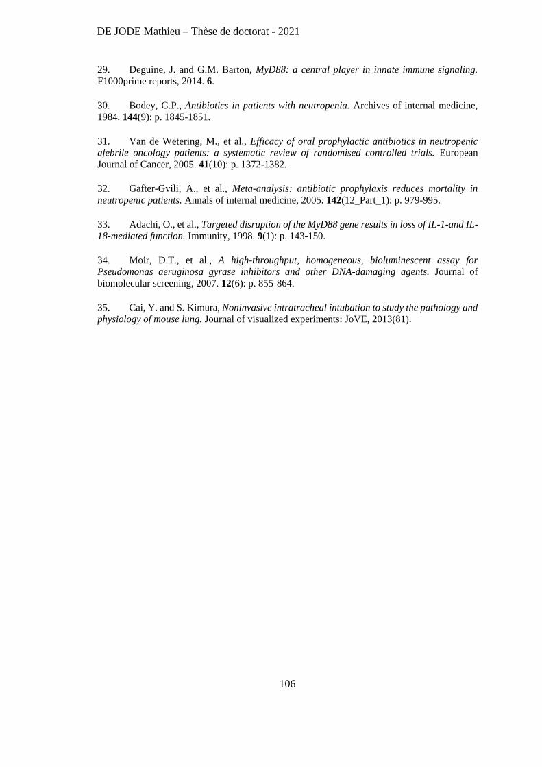
DE JODE Mathieu – Thèse de doctorat - 2021
106
29. Deguine, J. and G.M. Barton, MyD88: a central player in innate immune signaling.
F1000prime reports, 2014. 6.
30. Bodey, G.P., Antibiotics in patients with neutropenia. Archives of internal medicine,
1984. 144(9): p. 1845-1851.
31. Van de Wetering, M., et al., Efficacy of oral prophylactic antibiotics in neutropenic
afebrile oncology patients: a systematic review of randomised controlled trials. European
Journal of Cancer, 2005. 41(10): p. 1372-1382.
32. Gafter-Gvili, A., et al., Meta-analysis: antibiotic prophylaxis reduces mortality in
neutropenic patients. Annals of internal medicine, 2005. 142(12_Part_1): p. 979-995.
33. Adachi, O., et al., Targeted disruption of the MyD88 gene results in loss of IL-1-and IL-
18-mediated function. Immunity, 1998. 9(1): p. 143-150.
34. Moir, D.T., et al., A high-throughput, homogeneous, bioluminescent assay for
Pseudomonas aeruginosa gyrase inhibitors and other DNA-damaging agents. Journal of
biomolecular screening, 2007. 12(6): p. 855-864.
35. Cai, Y. and S. Kimura, Noninvasive intratracheal intubation to study the pathology and
physiology of mouse lung. Journal of visualized experiments: JoVE, 2013(81).
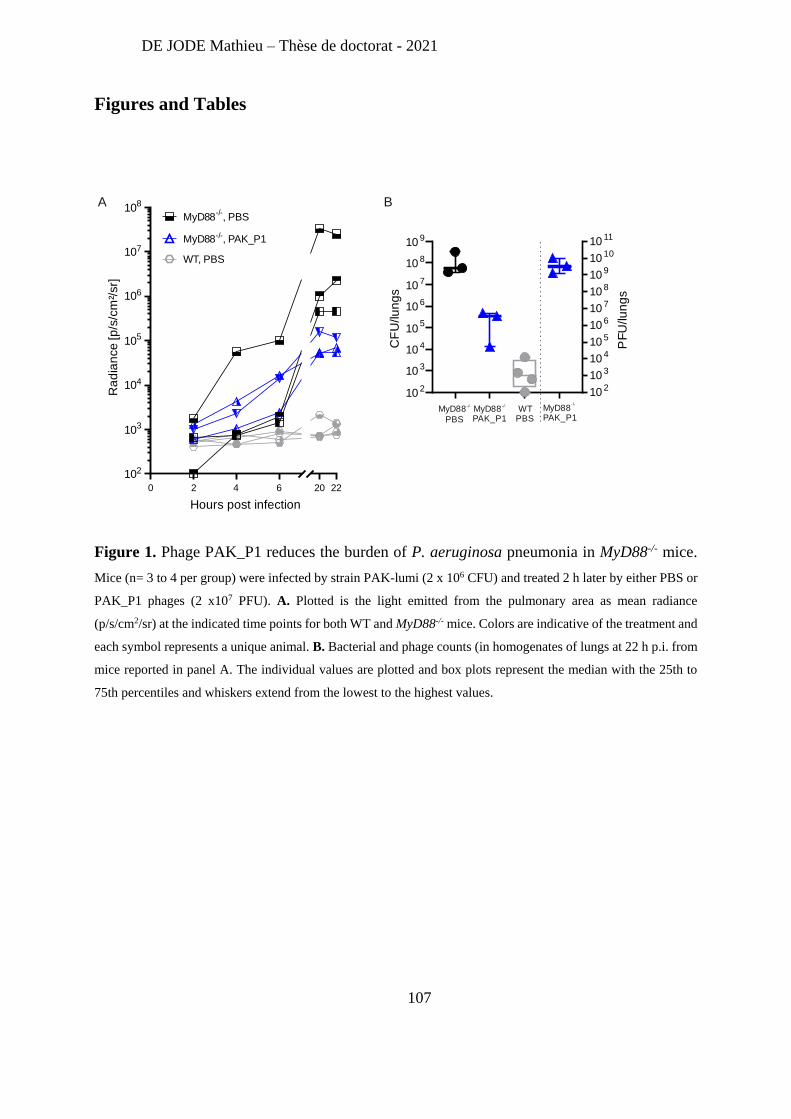
DE JODE Mathieu – Thèse de doctorat - 2021
107
Figures and Tables
Figure 1. Phage PAK_P1 reduces the burden of P. aeruginosa pneumonia in MyD88-/- mice.
Mice (n= 3 to 4 per group) were infected by strain PAK-lumi (2 x 10⁶ CFU) and treated 2 h later by either PBS or
PAK_P1 phages (2 x10⁷ PFU). A. Plotted is the light emitted from the pulmonary area as mean radiance
(p/s/cm2/sr) at the indicated time points for both WT and MyD88-/- mice. Colors are indicative of the treatment and
each symbol represents a unique animal. B. Bacterial and phage counts (in homogenates of lungs at 22 h p.i. from
mice reported in panel A. The individual values are plotted and box plots represent the median with the 25th to
75th percentiles and whiskers extend from the lowest to the highest values.
A B
0 2 4 6
102
103
104
105
106
107
108
20 22
Hours post infection
Ra
dia
nce
[p
/s/c
m²/
sr]
WT, PBS
MyD88-/-, PAK_P1
MyD88-/-
, PBS
10 2
10 3
10 4
105
10 6
10 7
10 8
10 9
10 2
10 3
10 4
10 5
10 6
10 7
10 8
10 9
10 10
10 11
CF
U/lu
ng
s
PF
U/lu
ng
s
MyD88-/-
PBS
MyD88-/-
PAK_P1
MyD88-/-
PAK_P1WT
PBS
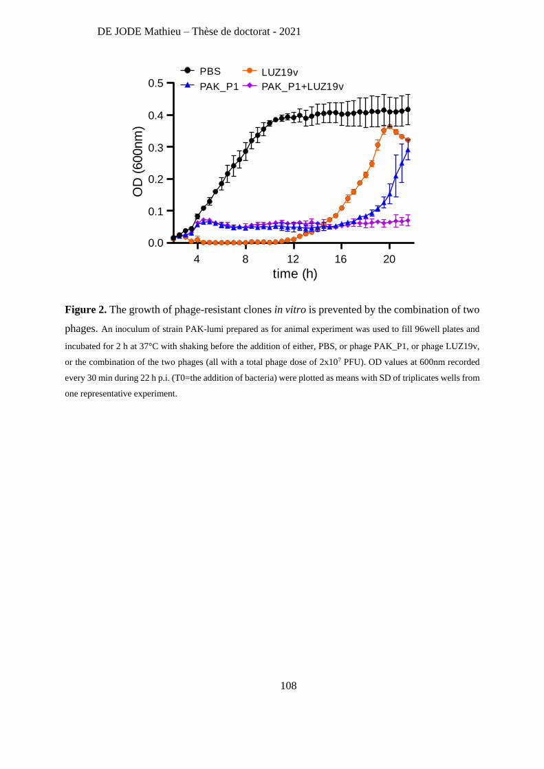
DE JODE Mathieu – Thèse de doctorat - 2021
108
Figure 2. The growth of phage-resistant clones in vitro is prevented by the combination of two
phages. An inoculum of strain PAK-lumi prepared as for animal experiment was used to fill 96well plates and
incubated for 2 h at 37°C with shaking before the addition of either, PBS, or phage PAK_P1, or phage LUZ19v,
or the combination of the two phages (all with a total phage dose of 2x10⁷ PFU). OD values at 600nm recorded
every 30 min during 22 h p.i. (T0=the addition of bacteria) were plotted as means with SD of triplicates wells from
one representative experiment.
4 8 12 16 20
0.0
0.1
0.2
0.3
0.4
0.5
time (h)
OD
(600nm
)
PBS
PAK_P1
LUZ19v
PAK_P1+LUZ19v
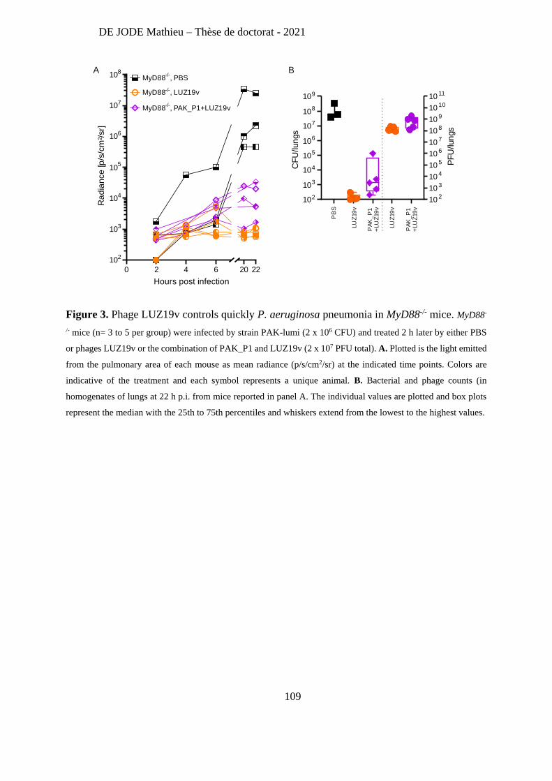
DE JODE Mathieu – Thèse de doctorat - 2021
109
Figure 3. Phage LUZ19v controls quickly P. aeruginosa pneumonia in MyD88-/- mice. MyD88-
/- mice (n= 3 to 5 per group) were infected by strain PAK-lumi (2 x 10⁶ CFU) and treated 2 h later by either PBS
or phages LUZ19v or the combination of PAK_P1 and LUZ19v (2 x 10⁷ PFU total). A. Plotted is the light emitted
from the pulmonary area of each mouse as mean radiance (p/s/cm2/sr) at the indicated time points. Colors are
indicative of the treatment and each symbol represents a unique animal. B. Bacterial and phage counts (in
homogenates of lungs at 22 h p.i. from mice reported in panel A. The individual values are plotted and box plots
represent the median with the 25th to 75th percentiles and whiskers extend from the lowest to the highest values.
0 2 4 6
102
103
104
105
106
107
108
20 22
Hours post infection
Ra
dia
nce
[p
/s/c
m²/
sr]
MyD88-/-
, PBS
MyD88-/-, LUZ19v
MyD88-/-
, PAK_P1+LUZ19v
102
103
104
105
106
107
108
109
10 2
10 3
10 4
10 5
10 6
10 7
10 8
10 9
10 10
10 11
CF
U/lun
gs
PF
U/lun
gs
A B
PB
S
LU
Z19
v
LU
Z19
v
PA
K_
P1
+LU
Z19
v
PA
K_
P1
+LU
Z19
v
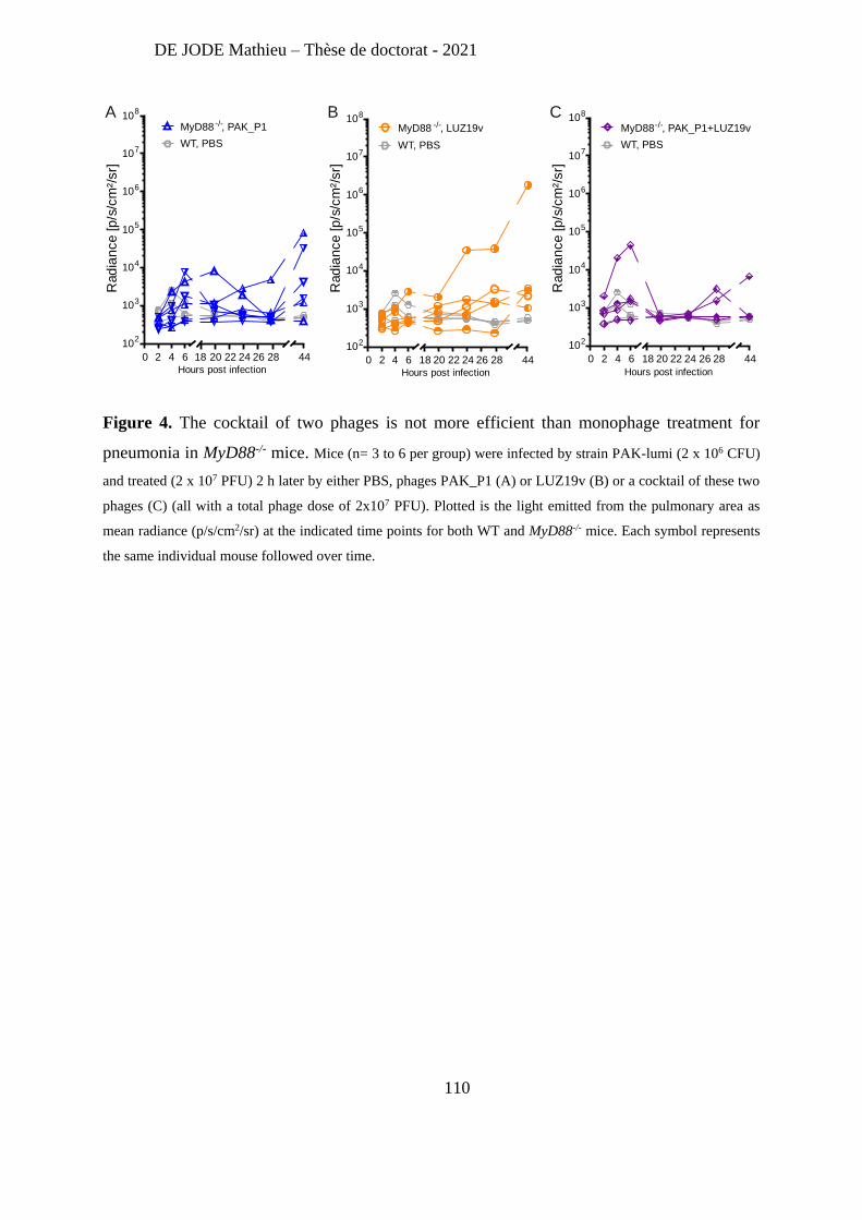
DE JODE Mathieu – Thèse de doctorat - 2021
110
Figure 4. The cocktail of two phages is not more efficient than monophage treatment for
pneumonia in MyD88-/- mice. Mice (n= 3 to 6 per group) were infected by strain PAK-lumi (2 x 10⁶ CFU)
and treated (2 x 10⁷ PFU) 2 h later by either PBS, phages PAK_P1 (A) or LUZ19v (B) or a cocktail of these two
phages (C) (all with a total phage dose of 2x10⁷ PFU). Plotted is the light emitted from the pulmonary area as
mean radiance (p/s/cm2/sr) at the indicated time points for both WT and MyD88-/- mice. Each symbol represents
the same individual mouse followed over time.
A B C
0 2 4 6
102
103
104
105
106
107
108
2018 22 24 26 28 44Hours post infection
Rad
ian
ce [
p/s
/cm
²/sr]
WT, PBS
MyD88 -/-, PAK_P1
0 2 4 6
102
103
104
105
106
107
108
18 20 22 24 26 28 44Hours post infection
Rad
ian
ce [
p/s
/cm
²/sr]
WT, PBS
MyD88 -/-, LUZ19v
0 2 4 6
102
103
104
105
106
107
108
18 20 22 24 26 28 44
Hours post infection
Rad
ian
ce [
p/s
/cm
²/sr]
WT, PBS
MyD88-/-, PAK_P1+LUZ19v
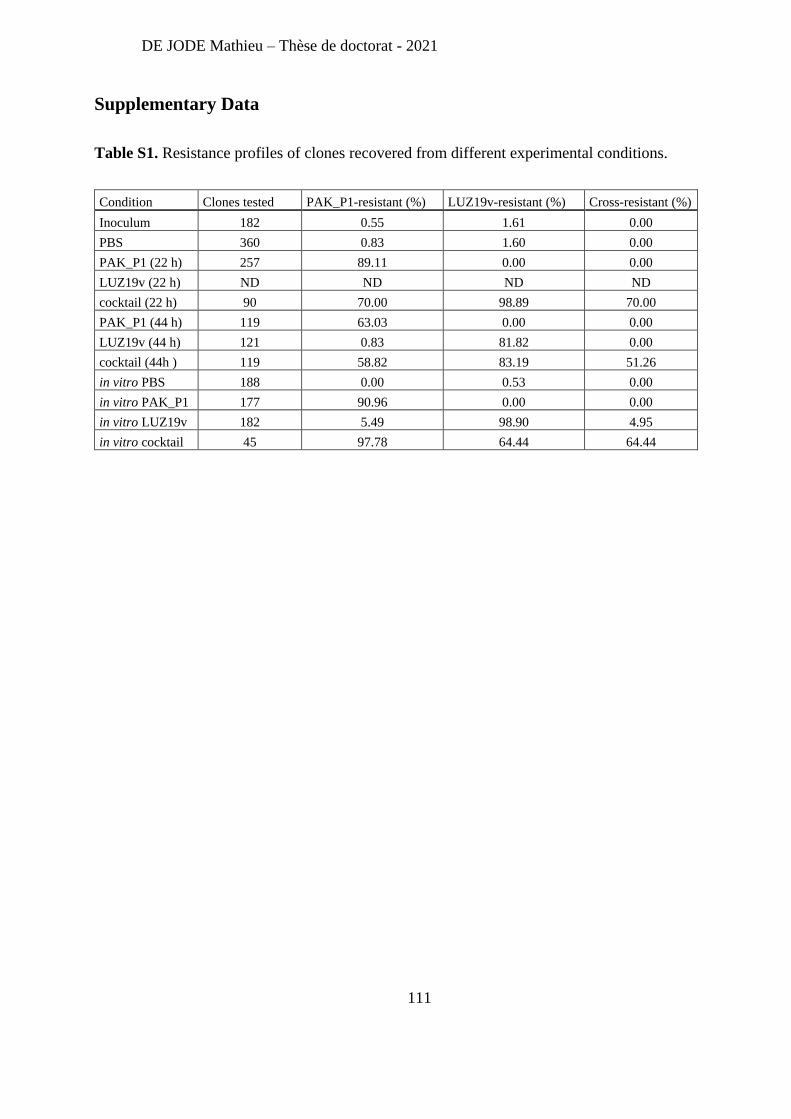
DE JODE Mathieu – Thèse de doctorat - 2021
111
Supplementary Data
Table S1. Resistance profiles of clones recovered from different experimental conditions.
Condition Clones tested PAK_P1-resistant (%) LUZ19v-resistant (%) Cross-resistant (%)
Inoculum 182 0.55 1.61 0.00
PBS 360 0.83 1.60 0.00
PAK_P1 (22 h) 257 89.11 0.00 0.00
LUZ19v (22 h) ND ND ND ND
cocktail (22 h) 90 70.00 98.89 70.00
PAK_P1 (44 h) 119 63.03 0.00 0.00
LUZ19v (44 h) 121 0.83 81.82 0.00
cocktail (44h ) 119 58.82 83.19 51.26
in vitro PBS 188 0.00 0.53 0.00
in vitro PAK_P1 177 90.96 0.00 0.00
in vitro LUZ19v 182 5.49 98.90 4.95
in vitro cocktail 45 97.78 64.44 64.44
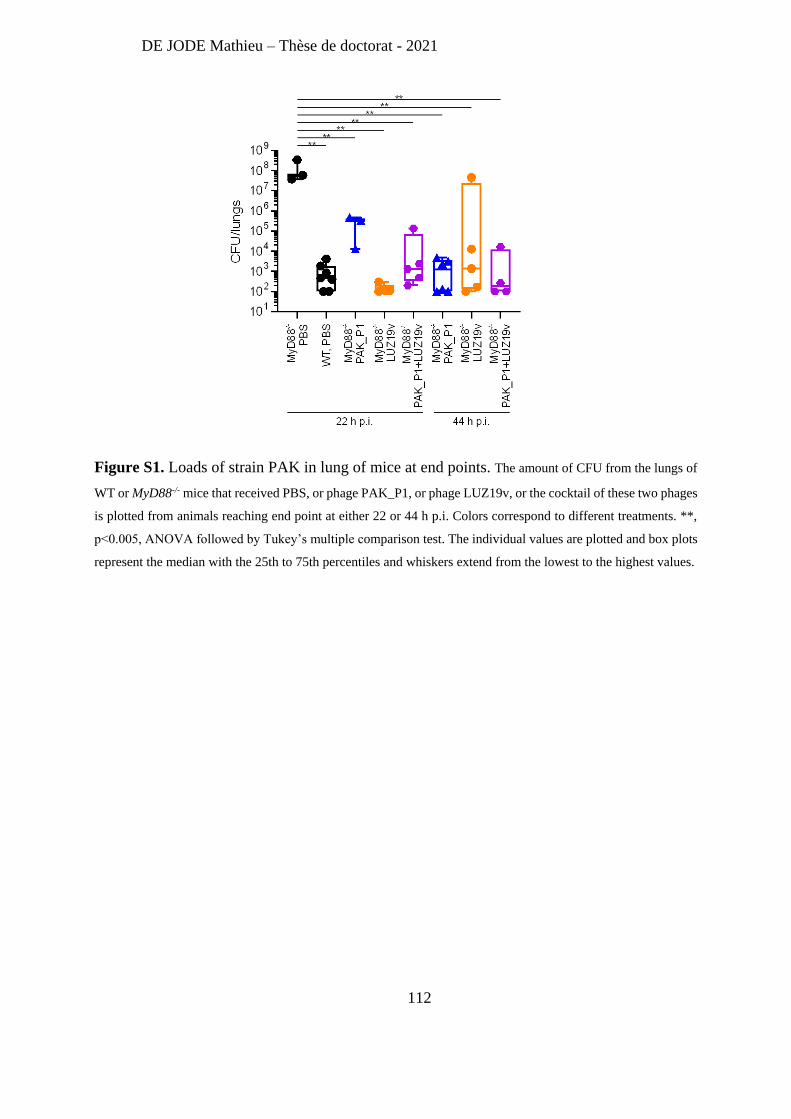
DE JODE Mathieu – Thèse de doctorat - 2021
112
Figure S1. Loads of strain PAK in lung of mice at end points. The amount of CFU from the lungs of
WT or MyD88-/- mice that received PBS, or phage PAK_P1, or phage LUZ19v, or the cocktail of these two phages
is plotted from animals reaching end point at either 22 or 44 h p.i. Colors correspond to different treatments. **,
p<0.005, ANOVA followed by Tukey’s multiple comparison test. The individual values are plotted and box plots
represent the median with the 25th to 75th percentiles and whiskers extend from the lowest to the highest values.
DE JODE Mathieu – Thèse de doctorat - 2021
113
Perspectives
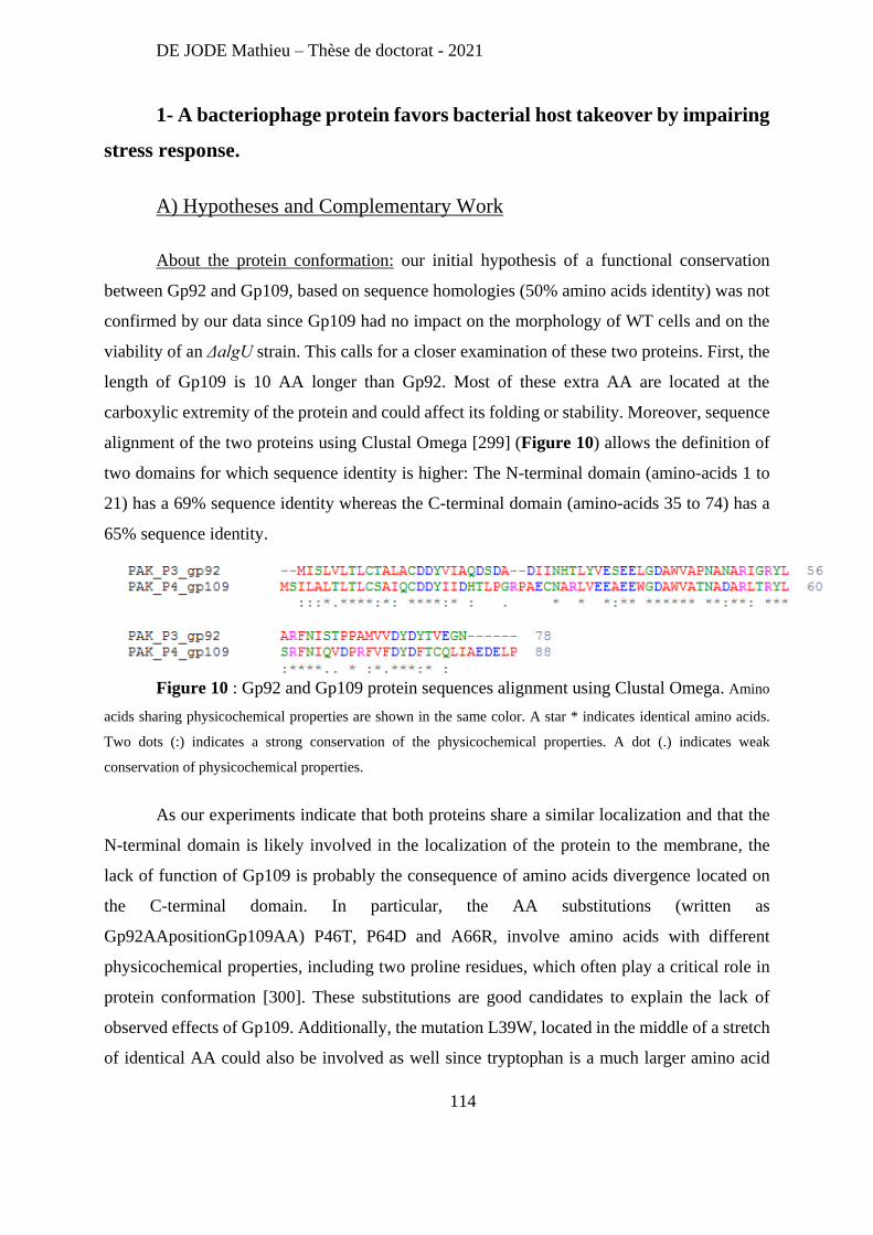
DE JODE Mathieu – Thèse de doctorat - 2021
114
1- A bacteriophage protein favors bacterial host takeover by impairing
stress response.
A) Hypotheses and Complementary Work
About the protein conformation: our initial hypothesis of a functional conservation
between Gp92 and Gp109, based on sequence homologies (50% amino acids identity) was not
confirmed by our data since Gp109 had no impact on the morphology of WT cells and on the
viability of an ΔalgU strain. This calls for a closer examination of these two proteins. First, the
length of Gp109 is 10 AA longer than Gp92. Most of these extra AA are located at the
carboxylic extremity of the protein and could affect its folding or stability. Moreover, sequence
alignment of the two proteins using Clustal Omega [299] (Figure 10) allows the definition of
two domains for which sequence identity is higher: The N-terminal domain (amino-acids 1 to
21) has a 69% sequence identity whereas the C-terminal domain (amino-acids 35 to 74) has a
65% sequence identity.
Figure 10 : Gp92 and Gp109 protein sequences alignment using Clustal Omega. Amino
acids sharing physicochemical properties are shown in the same color. A star * indicates identical amino acids.
Two dots (:) indicates a strong conservation of the physicochemical properties. A dot (.) indicates weak
conservation of physicochemical properties.
As our experiments indicate that both proteins share a similar localization and that the
N-terminal domain is likely involved in the localization of the protein to the membrane, the
lack of function of Gp109 is probably the consequence of amino acids divergence located on
the C-terminal domain. In particular, the AA substitutions (written as
Gp92AApositionGp109AA) P46T, P64D and A66R, involve amino acids with different
physicochemical properties, including two proline residues, which often play a critical role in
protein conformation [300]. These substitutions are good candidates to explain the lack of
observed effects of Gp109. Additionally, the mutation L39W, located in the middle of a stretch
of identical AA could also be involved as well since tryptophan is a much larger amino acid
DE JODE Mathieu – Thèse de doctorat - 2021
115
than leucine. To test these hypotheses, site directed mutations on the gp92 gene could be made
and proteins expressed in the WT strain PAK. Assessment of the morphology of colonies could
then revealed which AA are necessary for this phenotype. This targeted approach could also be
complemented by a more systematic approach (e.g. alanine scanning), where one by one, every
single amino acid is replaced by an alanine residue, to identify the residues needed for Gp92
activity.
As phages PAK_P3 and PAK_P4 showed different kinetics of host take over, it is also
possible that despite their sequence homology, Gp92 and Gp109 serves different functions for
each phage. If we concur that gene reduction is linked to optimized viral infection, we can
hypothesize that Gp92 diverged from Gp109 and gain a function for controlling the host
membrane stress response. Alternatively, Gp109 might require other phage proteins to support
the same function. Note that both gp92 in PAK_P3 and gp109 in PAK_P4 are part of operons.
Unfortunately, attempt to clone these operons was unsuccessful.
About the impact of Gp92 on cell morphology: our data have shown that the synthesis
of Gp92 changes cell morphology of WT strain PAK from rod-shape to spherical bacteria. As
this effect was abolished in a mucA22 background (characterized by constitutive expression of
the AlgU membrane stress response) and led to cell bursting in a ΔalgU background, a link
between Gp92 and this stress response was established. However, the precise mechanism
remains to be deciphered. Indeed, despite the involvement of AlgU in the regulation of the
biosynthesis of the peptidoglycan and some components of the bacterial cytoskeleton (e.g.
mreB), both crucial for cell shape determination and maintenance [301], ΔalgU cells still
display a rod-shape. Therefore, the disruption of the AlgU stress response by Gp92 cannot
solely explain the impact on cell morphology. This points out that Gp92 has a broader impact
than disrupting the AlgU stress response. Our results suggest that Gp92 is both causing a
membrane stress and disrupting the stress response responsible for mitigating this effect. One
hypothesis could be that the overexpression of an inner-membrane protein (e.g. Gp92) might
disrupt the cell morphology, but the absence of a similar effect when expressing Gp109 does
not support this hypothesis. A direct assessment of Gp92 localization in the cell (by
fluorescence microscopy) could help defining its localization and help drawing hypothesis
regarding its mechanism of action. Unfortunately, fusions of Gp92 with GFP led to the abolition
of the colony phenotype on plates, probably due to a loss of function of these fused proteins.
DE JODE Mathieu – Thèse de doctorat - 2021
116
About the role of Gp92 in phage infection: the phenotypes observed in different bacterial
backgrounds (WT, mucA22 and ΔalgU) pushed us to study the phage infection process in these
cells. These studies revealed that EOP and lysis timing were affected, with mucA22 cells
showing an increased EOP and a slightly delayed lysis. We hypothesize that during phage
infection Gp92 disrupts the AlgU stress response in order to keep the control on the timing of
cell lysis. It could be interesting to explore this phenomenon in more details, perhaps through
one-step growth experiments or video microscopy (to time precisely the first cell lysis). We
also checked if the activation of the AlgU stress response might be relevant for other
P. aeruginosa phages. We found that variations of the EOP between the different genetic
backgrounds were similar for phage PAK_P3 and PAK_P4, whereas they were different for
both phages LUZ19 and PhiKZ. Indeed, the EOP of LUZ19 was reduced on both mucA22 and
ΔalgU strains compared to WT, while the EOP of PhiKZ was reduced on mucA22 but increased
on ΔalgU strains. Due to time constrains these experiments were not pursued but these initial
results showed that the AlgU stress response impacts the replication process of these phages in
different ways. Finally, the role of Gp92 during phage infection could also be more easily
assessed if we had access to a PAK_P3Δgp92 phage. Unfortunately, genetic tools, using
CRISPR/Cas enzymes, have not yet been implemented for P. aeruginosa.
About the link between phage replication and bacterial stress: we have found that
activation of AlgU stress response can impact phage lysis. Interestingly, other studies have
described impact of cellular stresses (provoked by antibiotics) on phage replications. First,
Comeau et al [302] described the Phage Antibiotic Synergy (PAS) phenomenon in which the
use of sub-lethal concentrations of some antibiotics can significantly increase the production of
a specific virulent phage in Gram-negative bacteria. They observed this PAS with diverse
host/phage pairs, including T4-like phages, with β-lactams and quinolone antibiotics. As these
antibiotics inhibit cell division and trigger the SOS system, the authors investigated the PAS
effect in these contexts. They found that PAS effect was SOS-independent and was primarily a
consequence of cellular filamentation and suggested that this elongated cells provided a
favorable environment for increasing phage production. As Gp92 also affects cellular
morphology, we hypothesize that the effect of stressor on the bacterial membrane could impact
phage lysis.
DE JODE Mathieu – Thèse de doctorat - 2021
117
Later, Kim et al [303] demonstrated a similar PAS in Gram-positive bacteria and
showed that cell filamentation did not significantly increased phage adsorption, but might
actually affect the lysis timing. Indeed, they observed a delayed lysis and an increased burst
size in presence of specific antibiotics. They proposed that the increased of cell surface during
filamentation was not matched by an increased holin production, thus delaying the lysis. This
interesting hypothesis does not however explain their observation of PAS in absence of
filamentation, nor the increased phage production when bacteria where stressed with reactive
oxygen species (H202). In our Gp92 study, we observed an effect on the cell morphology (from
rod-shape to spherical), however we did not evaluate the effect on the cell volume, which could
affect lysis timing by “dilution” of the lysis effectors. Moreover, the authors observed an
increased phage production following stress with reactive oxygen species, which supports
further the hypothesis of a link between bacterial stress responses and phage infectious process.
Overall, the underlying molecular mechanisms of the PAS phenomenon are still not
clear, but its use for increased phage production has been patented (patent number:
US8178087B2) as well as the production of phage in presence of H202 (patent number:
KR101909831B1).
About the broad impact on cellular proteome: the major shift of the proteome following
Gp92 induction is puzzling regarding its underlying mechanism: how a single phage protein
could have such a broad impact? It is unlikely that Gp92 is interacting directly with the hundreds
affected proteins. The direct interactions between Gp92 and a sigma/anti-sigma regulatory
system points towards the origin of a broad impact via interactions with several regulatory
proteins. We indeed found that numerous regulators were affected by Gp92 expression,
including sigma factor like FliA (responsible of flagellum regulation) and RpoH (responsible
of the heat shock stress response). Since sigma factors show high structural conservation [304]
it is possible that Gp92 recognizes a conserved structural motif in such regulators, although
double hybrid assays detected only direct interaction with MucA and AlgU. However, the
original screening was performed using banks made in E. coli and S. typhi. A screen performed
with a P. aeruginosa bank might yield a higher number of hits in regulators but could also
identify potential new targets of Gp92. Interactions with regulators is a very potent way to
profoundly affect bacterial physiology as most of them do not simply regulate a specific
function, but present cross-talks with other regulators. For example, regarding the networks of
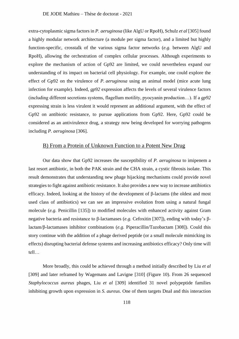
DE JODE Mathieu – Thèse de doctorat - 2021
118
extra-cytoplasmic sigma factors in P. aeruginosa (like AlgU or RpoH), Schulz et al [305] found
a highly modular network architecture (a module per sigma factor), and a limited but highly
function-specific, crosstalk of the various sigma factor networks (e.g. between AlgU and
RpoH), allowing the orchestration of complex cellular processes. Although experiments to
explore the mechanism of action of Gp92 are limited, we could nevertheless expand our
understanding of its impact on bacterial cell physiology. For example, one could explore the
effect of Gp92 on the virulence of P. aeruginosa using an animal model (mice acute lung
infection for example). Indeed, gp92 expression affects the levels of several virulence factors
(including different secretions systems, flagellum motility, pyocyanin production…). If a gp92
expressing strain is less virulent it would represent an additional argument, with the effect of
Gp92 on antibiotic resistance, to pursue applications from Gp92. Here, Gp92 could be
considered as an antivirulence drug, a strategy now being developed for worrying pathogens
including P. aeruginosa [306].
B) From a Protein of Unknown Function to a Potent New Drug
Our data show that Gp92 increases the susceptibility of P. aeruginosa to imipenem a
last resort antibiotic, in both the PAK strain and the CHA strain, a cystic fibrosis isolate. This
result demonstrates that understanding new phage hijacking mechanisms could provide novel
strategies to fight against antibiotic resistance. It also provides a new way to increase antibiotics
efficacy. Indeed, looking at the history of the development of β-lactams (the oldest and most
used class of antibiotics) we can see an impressive evolution from using a natural fungal
molecule (e.g. Penicillin [135]) to modified molecules with enhanced activity against Gram
negative bacteria and resistance to β-lactamases (e.g. Cefoxitin [307]), ending with today’s β-
lactam/β-lactamases inhibitor combinations (e.g. Piperacillin/Tazobactam [308]). Could this
story continue with the addition of a phage derived peptide (or a small molecule mimicking its
effects) disrupting bacterial defense systems and increasing antibiotics efficacy? Only time will
tell…
More broadly, this could be achieved through a method initially described by Liu et al
[309] and later reframed by Wagemans and Lavigne [310] (Figure 10). From 26 sequenced
Staphylococcus aureus phages, Liu et al [309] identified 31 novel polypeptide families
inhibiting growth upon expression in S. aureus. One of them targets DnaI and this interaction
DE JODE Mathieu – Thèse de doctorat - 2021
119
was used to screen for small inhibitors to finally identify 11 compounds that inhibit both
bacterial growth and DNA synthesis with a MIC ≤ 16 μg/ml. After this pioneer work,
Wagemans and Lavigne [310] described a general method to go from an uncharacterized phage
gene to an antimicrobial compound (Figure 11).
Figure 11 : Bacteriophage–host interaction-based development of new antibacterial
compounds (from [310]): After sequencing of phage genomes, uncharacterized genes are cloned under an
inducible promoter to assess their toxicity. Then, the identification of the bacterial target can be achieved via
different techniques, including double-hybrid assays. Once the phage/host interaction has been characterized, a
screening for inhibitors of this interaction can be carried. Among these inhibitors, molecules mimicking the phage
protein antibacterial effects might be found.
During the study of Gp92, we already performed most of the described steps of this
strategy (genome screening, cloning, toxicity assessment, and identification of the bacterial
target), but the most challenging, i.e. devising a robust screen to search for inhibitors of the
Gp92/MucA interaction, has not yet been achieved.
The idea of not using the whole phage but only some of its parts (e.g. phage proteins) is
actually not new, and a lot of research has been particularly dedicated to the enzymes catalyzing
the cell lysis: the phage endolysins.
Indeed, the use of endolysin as a new therapeutic strategy seems promising as numerous
successes both in vitro and in vivo have been reported [311-314], and a recent review on the
global preclinical antibacterial pipeline [315] found that out of the 407 preclinical projects
DE JODE Mathieu – Thèse de doctorat - 2021
120
identified, 33 projects involved phages or phage-derived peptides, including 11 natural phage
cocktails projects, 11 engineered phage cocktails projects, and 7 engineered endolysin projects
(the remaining 4 projects utilize phages as cargo for drug delivery). Note that the interest in
endolysins has not only been fueled by successful laboratory results, but also by an “easier fit”
into the medicinal products regulatory framework (still a big hurdle for classical phage therapy)
[316]. As the first lysins targeting Staphylococcus aureus (CF-301, SAL200, P128, and
Staphefekt™) are currently being evaluated in clinical trials, phage-derived enzymes might end
up being widely used while phages may remain restricted to punctual compassionate use.
DE JODE Mathieu – Thèse de doctorat - 2021
121
2- The combination of bacteriophages prevents resistant outgrowth in
vitro but does not increase pulmonary phage therapy efficacy in
immunocompromised animals.
A) Hypotheses and Complementary Work
About the differences between in vitro and in vivo results: the in vitro environment (a
well of 96-well plate filled with LB) is a liquid homogenous environment, characterized by a
rather small volume (180 µL) compared to the lungs of mice (total lung capacity of C57BL/6J
= 1400 µL [317]), which is a semi-solid heterogeneous environment. These differences of
physical properties between in vitro and in vivo environment could be responsible of the
differences of kinetics observed (quicker in vitro than in vivo) as they impact the frequency of
phage/bacteria interactions. Our previous work showed that in silico modelling of phage
therapy needed to incorporate the heterogeneity of the lung environment to correctly depict
phage lysis kinetics in our experimental settings compared to well-mixed environments (in
vitro) [292]. To explore the contributions of these physical parameters, we can design in vitro
experiments in which either the volume (larger wells), or the viscosity (addition of mucins) of
the medium is increased.
Additionally, we observed that the phage cocktail treatment was superior to monophage
treatment in vitro, but not in vivo. This indicates that bacteria in vitro and in vivo can have
different biological response to the same event (phage predation) according to their
environment. As already discussed in the article, different groups have reported that
environmental conditions (e.g. nutrient concentrations, or presence of competitor bacteria) can
influence phage-resistance in P. aeruginosa, by tipping the balance towards cell surface
modifications or CRISPR-Cas based resistance [318, 319]. Since our strain of interest (PAK)
does not possess a functional CRISPR-Cas system, we are left with the hypothesis of the
involvement of a different bacterial physiology between in vitro and in vivo environments.
Indeed, the in vitro experiments were conducted in rich medium, whereas the mice lungs are
certainly not as rich in term of nutriments. To explore the impact of nutriment concentrations
on phage resistance, we could perform in vitro experiments with increasingly diluted medium
and measure the rate at which phage resistant clones grow. We could also perform an ex-vivo
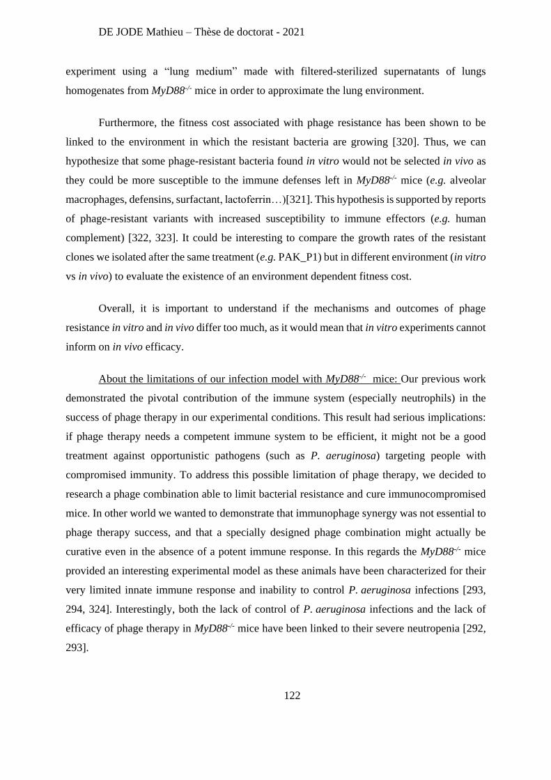
DE JODE Mathieu – Thèse de doctorat - 2021
122
experiment using a “lung medium” made with filtered-sterilized supernatants of lungs
homogenates from MyD88-/- mice in order to approximate the lung environment.
Furthermore, the fitness cost associated with phage resistance has been shown to be
linked to the environment in which the resistant bacteria are growing [320]. Thus, we can
hypothesize that some phage-resistant bacteria found in vitro would not be selected in vivo as
they could be more susceptible to the immune defenses left in MyD88-/- mice (e.g. alveolar
macrophages, defensins, surfactant, lactoferrin…)[321]. This hypothesis is supported by reports
of phage-resistant variants with increased susceptibility to immune effectors (e.g. human
complement) [322, 323]. It could be interesting to compare the growth rates of the resistant
clones we isolated after the same treatment (e.g. PAK_P1) but in different environment (in vitro
vs in vivo) to evaluate the existence of an environment dependent fitness cost.
Overall, it is important to understand if the mechanisms and outcomes of phage
resistance in vitro and in vivo differ too much, as it would mean that in vitro experiments cannot
inform on in vivo efficacy.
About the limitations of our infection model with MyD88-/- mice: Our previous work
demonstrated the pivotal contribution of the immune system (especially neutrophils) in the
success of phage therapy in our experimental conditions. This result had serious implications:
if phage therapy needs a competent immune system to be efficient, it might not be a good
treatment against opportunistic pathogens (such as P. aeruginosa) targeting people with
compromised immunity. To address this possible limitation of phage therapy, we decided to
research a phage combination able to limit bacterial resistance and cure immunocompromised
mice. In other world we wanted to demonstrate that immunophage synergy was not essential to
phage therapy success, and that a specially designed phage combination might actually be
curative even in the absence of a potent immune response. In this regards the MyD88-/- mice
provided an interesting experimental model as these animals have been characterized for their
very limited innate immune response and inability to control P. aeruginosa infections [293,
294, 324]. Interestingly, both the lack of control of P. aeruginosa infections and the lack of
efficacy of phage therapy in MyD88-/- mice have been linked to their severe neutropenia [292,
293].
DE JODE Mathieu – Thèse de doctorat - 2021
123
In the end, we were unsuccessful in creating a phage cocktail able to cure MyD88-/- mice, as
phage treatment only delay the death of the animals. However, this failure needs to be
contextualized. Indeed, as neutropenia has also been linked to a limited efficacy of antibiotics
in both neutropenic rats [325] and neutropenic human patients [326], the observed limitation of
phage therapy efficacy might not be as critical as initially thought. For example, Lumish et al
found that when treating neutropenic rats with Carbenicillin, Gentamicin or both, animals death
was only shifted by a day, and overall the survival of treated-mice at 5 days post infection (i.p.
of 1.8 x 10⁸ CFU of P. aeruginosa), only increased from 5 to 15% (the antibiotic combination
being the most efficient treatment) [325] (
Figure 12).
Figure 12: Limited efficacy of antibiotic treatments in neutropenic rats (adapted from [325]): Cumulative mortality rate (percentage) of neutropenic rats challenged with 1.8 x 10⁸ CFU of P. aeruginosa on day
5 and treated for 72 hr at the intervals (hr) indicated by the arrows on the abscissa. The dose of carbenicillin (CB)
was 400 mg/kg (27 rats) and that of gentamicin (G) was 10 mg/kg (27 rats); 400 mg of carbenicillin/kg and 10 mg
of gentamicin/kg were given for combined therapy (CB + G) (27 rats). As control 10 rats received saline im.
An interesting observation coming from systematic reviews on antibiotics use in
neutropenic patients is the efficacy of prophylaxis (e.g. the administration of antibiotics before
an infection is detected) to reduce bacterial infection incidence [327, 328]. We have already
demonstrated the efficacy of phage prophylaxis in our acute lung infection in WT mice [270],
so it is tempting to predict that such treatment in MyD88-/- mice could be successful.
It is important to note that MyD88-/- mice are not just neutropenic, but lack a proper
activation of the inflammation process, which limits the recruitment and activations of
numerous immune cells [294]. This does not only impact antibacterial activity, but could also
DE JODE Mathieu – Thèse de doctorat - 2021
124
disrupt lung tissue repair mechanisms, as cells first recruited during the inflammatory process
are also responsible for tissue repairs [329, 330]. Notably, MyD88 was found to be essential for
tracheal epithelial remodeling following acute epithelial injury [331]. This is very relevant for
our experiments, as the clinical signs of phage treated-animals did not improved despite the
significant reduction of the bacterial loads in their lungs. We hypothesize that this could be due
to the accumulation of tissue damages that might be exacerbated in MyD88-/- mice.
Overall, the “extreme phenotypes” of MyD88-/- mice might be suitable to study phage
resistance in vivo (as it is not observed in WT mice), but might not be the best model to
investigate phage therapy efficacy. To address these limitations of the MyD88-/- mice model,
future experiments will be carried in WT mice with various degree of neutropenia using
different doses of Anti-granulocyte receptor-1 (Gr1) monoclonal antibodies. This strategy will
help defining the levels of neutrophils actually needed for phage therapy success. These results
could guide phage therapy clinical applications by defining an immune status suitable for phage
therapy.
Finally, our results demonstrate that formulating a phage cocktail able to limit phage-
resistance both in vitro and in vivo is a difficult task. In the following sections we will discuss
other ways to formulate potent therapeutic cocktails. Hopefully, this information will help us
design a phage cocktail with superior efficacy notably in MyD88-/- mice.
B) Phage Receptor Cocktail Design
Our strategy to design a potent phage cocktail was based on phage receptor, meaning
that we combined two phages (PAK_P1 and LUZ19v) using two different receptors on the
bacterial surface (LPS and Type IV pili). As cell surface modification is utilized by bacteria to
gain phage resistance, using phages with different receptors allows to limit the cross-resistance.
In our study we have isolated bacteria resistant to both phages in vitro and in vivo pointing out
the limit of our two phages cocktail. If this cross-resistance is catalyzed by two independent
mutations (one for each phage receptor), we can hypothesize that adding new phages using
other receptors (e.g. porins) would decrease the probability of resistance and increase the
efficacy of the treatment. However, in order to achieve such a goal, the identification of the
phage receptors is mandatory. The classical method for phage-receptor determination relies on
DE JODE Mathieu – Thèse de doctorat - 2021
125
the isolation of phage-resistant bacteria (following phage and bacteria incubation), whole-
genome sequencing of these clones, and deduction of the phage receptor from the identified
mutations. Once a candidate receptor is find, its gene is cloned into a plasmid and its expression
in phage-resistant clones should restore their susceptibility to the phage (Figure 13b).
However, this approach is time consuming and become unmanageable as the number of
phages to be tested increases, especially if we want to create a phage library using a wide array
of receptors. A more high-throughput method utilizes library of random bacterial variant
(generated for example via transposon mutagenesis [332]) with genomic tags, allowing a more
targeted sequencing (cheaper and easier to analyzed). Incubation of the phage with this library
will lead to the selection of phage-resistant variants, and once again genomic analysis of the
mutations will reveal the potent phage receptors (Figure 13c).
In the future, once sufficient knowledge regarding phage receptors is acquired, we may
design genetic mutations to change the phage host range by the modification of base-plate or
tail proteins, or even generate semi-random sequences to target different bacterial structures
(Figure 13e). Moreover, once a high number of phage receptors has been characterized, we
might even be able to predict a phage’s receptor from its genome (Figure 13f), which would
incredibly speed up the process of phage characterization, thus helping a fast design and
production of efficient phage cocktails.
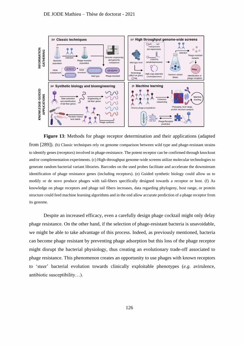
DE JODE Mathieu – Thèse de doctorat - 2021
126
Figure 13: Methods for phage receptor determination and their applications (adapted
from [289]). (b) Classic techniques rely on genome comparison between wild type and phage-resistant strains
to identify genes (receptors) involved in phage-resistance. The potent receptor can be confirmed through knockout
and/or complementation experiments. (c) High-throughput genome-wide screens utilize molecular technologies to
generate random bacterial variant libraries. Barcodes on the used probes facilitate and accelerate the downstream
identification of phage resistance genes (including receptors). (e) Guided synthetic biology could allow us to
modify or de novo produce phages with tail-fibers specifically designed towards a receptor or host. (f) As
knowledge on phage receptors and phage tail fibers increases, data regarding phylogeny, host range, or protein
structure could feed machine learning algorithms and in the end allow accurate prediction of a phage receptor from
its genome.
Despite an increased efficacy, even a carefully design phage cocktail might only delay
phage resistance. On the other hand, if the selection of phage-resistant bacteria is unavoidable,
we might be able to take advantage of this process. Indeed, as previously mentioned, bacteria
can become phage resistant by preventing phage adsorption but this loss of the phage receptor
might disrupt the bacterial physiology, thus creating an evolutionary trade-off associated to
phage resistance. This phenomenon creates an opportunity to use phages with known receptors
to ‘steer’ bacterial evolution towards clinically exploitable phenotypes (e.g. avirulence,
antibiotic susceptibility…).
DE JODE Mathieu – Thèse de doctorat - 2021
127
C) Exploiting Phage Resistance to Steer Bacterial Evolution
In contrast to antibiotic resistance that is mostly acquired via plasmids carrying
resistance genes or by the overexpression of resistance determinants, phage resistance mainly
occurs via the loss or dysregulation of cell surface components. This resistance mechanism,
although very efficient, is sometimes costly for the bacteria. Indeed, bacterial surface
components can play critical roles in infection (e.g. motility, attachment, secretion…) as they
often contribute to virulence, or to antibiotics resistance (e.g. efflux pumps). For example, a list
of 306 virulence factors has been reported for P. aeruginosa, from which 45% are predicted in
silico (using PSORTb) to be localized to the outer-membrane [333].
Numerous studies have reported phage resistant clones with reduced growth rates, lower
virulence or increased susceptibility to antimicrobials. The first report of this phenomenon was
published in the 1980’s by Smith and Huggins in an article entitled “Successful Treatment of
Experimental Escherichia coli Infections in Mice Using Phage: its General Superiority over
Antibiotics” [334]. Despite the emergence of phage-resistant bacteria, the efficacy of phage
treatments was not jeopardized because these phage-resistant clones had loss the K1 capsule
and were then avirulent in mice. Almost 40 years later, Gordillo Altamirano et al [323] reported
similar findings when they used A. baumannii phages recognizing the capsule as receptor.
Phage-resistant clones that lost the capsule were unable to form biofilms, avirulent in a
bacteremia murine model, and re-sensitized to multiple antimicrobials they were previously
resistant to, including the complement system, other phages, and antibiotics. Besides capsules,
the LPS is another frequent phage receptor determinant that bacteria can modify to resist to
phage predation. Filippov et al [286] identified a direct correlation between truncation of LPS
molecules and reduction of virulence using eight phages infecting Yersinia pestis.
Moreover, the acquired vulnerability to antibiotics following phage treatment, is not
constrained to Gram negative bacteria as a report by Chatterjee et al [335] described how phage-
resistant mutants lacking enterococcal polysaccharide antigen became susceptible to cell wall
targeting antibiotics (e.g. vancomycin). Interestingly, these phage-resistant mutants were also
deficient in a murine model of intestinal colonization.
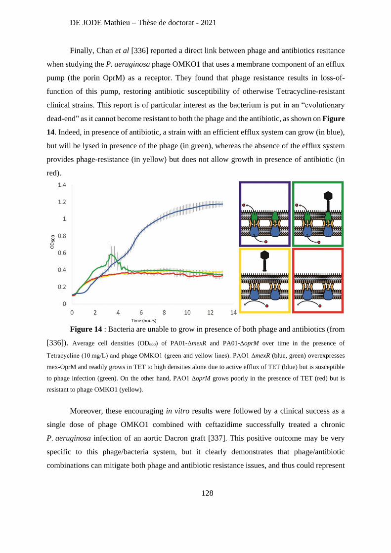
DE JODE Mathieu – Thèse de doctorat - 2021
128
Finally, Chan et al [336] reported a direct link between phage and antibiotics resitance
when studying the P. aeruginosa phage OMKO1 that uses a membrane component of an efflux
pump (the porin OprM) as a receptor. They found that phage resistance results in loss-of-
function of this pump, restoring antibiotic susceptibility of otherwise Tetracycline-resistant
clinical strains. This report is of particular interest as the bacterium is put in an “evolutionary
dead-end” as it cannot become resistant to both the phage and the antibiotic, as shown on Figure
14. Indeed, in presence of antibiotic, a strain with an efficient efflux system can grow (in blue),
but will be lysed in presence of the phage (in green), whereas the absence of the efflux system
provides phage-resistance (in yellow) but does not allow growth in presence of antibiotic (in
red).
Figure 14 : Bacteria are unable to grow in presence of both phage and antibiotics (from
[336]). Average cell densities (OD600) of PA01-ΔmexR and PA01-ΔoprM over time in the presence of
Tetracycline (10 mg/L) and phage OMKO1 (green and yellow lines). PAO1 ∆mexR (blue, green) overexpresses
mex-OprM and readily grows in TET to high densities alone due to active efflux of TET (blue) but is susceptible
to phage infection (green). On the other hand, PAO1 ∆oprM grows poorly in the presence of TET (red) but is
resistant to phage OMKO1 (yellow).
Moreover, these encouraging in vitro results were followed by a clinical success as a
single dose of phage OMKO1 combined with ceftazidime successfully treated a chronic
P. aeruginosa infection of an aortic Dacron graft [337]. This positive outcome may be very
specific to this phage/bacteria system, but it clearly demonstrates that phage/antibiotic
combinations can mitigate both phage and antibiotic resistance issues, and thus could represent
DE JODE Mathieu – Thèse de doctorat - 2021
129
a potent therapeutic option to successfully treat infections in our immunocompromised mice
model.
D) Phages/Antibiotics Combinations: Everything Can Happen
Currently, clinical applications of phages are almost always paired with antibiotic
treatments as clinicians use everything at their disposal to treat patients in life-threatening
conditions. Thus, it is of the upmost importance to better understand the interactions between
phages and antibiotics. Moreover, as mentioned in the previous section, phage/antibiotic
combination may be a very potent solution against both phage and antibiotic resistance [336,
337], and could be efficient even in the absence of a functional immune system. Unfortunately,
a limited number of studies have investigated phage/antibiotic combinations, and their
conclusions are as diverse as the phage/bacteria/antibiotic systems evaluated. Hence, no general
rule can yet be drawn, but an overview of described effects will be presented.
Synergic activity of phages and antibiotics exploiting their differential efficacies on
sessile bacteria and biofilm was described by multiple groups. Indeed, Kirby et al [338] reported
that effective gentamycin treatments on S. aureus planktonic cultures promotes aggregation and
biofilm formation, leading to increase antibiotic resistance. On the other hand, the phage alone
had limited effect on the planktonic cultures, but were efficient on biofilms. When antibiotics
and phages were combined an efficacy on both planktonic bacteria and biofilms was reported.
Similar results were observed in other studies on E. coli biofilms using a Myoviridae associated
with cefotaxime [339], or on S. aureus biofilms, but this time using a Siphoviridae combined
with rifampicin [340], demonstrating that these synergistic effects are not limited to a specific
phage type or antibiotic class.
However, the combination of both phage and antibiotics does not always has positive
outcomes. Indeed, Moulton et al [341] studied the combination of a phage and gentamycin in
long term evolutionary experiments and they found that P. aeruginosa developed a resistance
against both gentamycin and the phage, nine days after the beginning of the treatment. The
combination performed worse than the antibiotic alone from the ninth day, demonstrating that
phages can impede the action of antibiotics. Likewise, phage replication was consistently lower
in the combination treatment compare to phage alone, pointing out a negative effect of the
DE JODE Mathieu – Thèse de doctorat - 2021
130
antibiotic on phage replication. The most striking result of this study is that exposure to phage
had a significant positive effect on bacterial growth in the presence of antibiotic, meaning that
phage selection could lead to lower antibiotic susceptibility. Conversely, Allen et al [342]
discovered through a wide screening of phage and antibiotic combinations (94 E. coli strains,
10 antibiotics and 7 phages) that the resistance against gentamycin in E. coli isolates was
correlated with the resistance against phage T6 (a Myoviridae). This association was then
confirmed by the deletion of tsx (encoding a protein channel used as a receptor by phage T6),
which conferred resistance to both T6 and gentamicin. However, the authors did not find an
association between overall phage resistance and antibiotic resistance profiles across their
isolates, indicating that despite rare associations among individual pairs of phage and antibiotic
resistance phenotypes, susceptibility to antibiotics and phages are not generally correlated.
Overall, our current knowledge of phage/antibiotic interactions is still hazy as both
positive and negative interactions have been described regarding antibiotic resistance and
overall treatment efficacy. In absence of general rules for potent combinations, more research
is needed to identify solid phages/antibiotics combinations and in particular which phage
functions are involved.
To conclude, the logical follow up of my work would be to exploit the upcoming
genomic sequences from phage resistant clones I isolated in order to design the next generation
of cocktails with improved therapeutic efficacy in our immunocompromised mice model. In
addition, to find the best combination of phages, we could utilize the knowledge of their
receptors, and select phage cocktails driving phage-resistance towards lower bacterial virulence
or increased antibiotic susceptibility. Finally, this newly designed phage cocktail could be
tested in combination with different antibiotics to further increase its therapeutic efficacy.
Hopefully, such a treatment will be able to successfully treat acute lung infection in
immunocompromised animals.
DE JODE Mathieu – Thèse de doctorat - 2021
131
References
1. Klockgether, J. and B. Tümmler, Recent advances in understanding Pseudomonas aeruginosa
as a pathogen. F1000Research, 2017. 6.
2. Zhang, Z., et al., Degradation of n-alkanes and polycyclic aromatic hydrocarbons in petroleum
by a newly isolated Pseudomonas aeruginosa DQ8. Bioresource Technology, 2011. 102(5): p.
4111-4116.
3. Grobe, S., J. Wingender, and H.-C. Flemming, Capability of mucoid Pseudomonas aeruginosa
to survive in chlorinated water. International journal of hygiene and environmental health, 2001.
204(2-3): p. 139-142.
4. Falkinham III, J.O., et al., Epidemiology and ecology of opportunistic premise plumbing
pathogens: Legionella pneumophila, Mycobacterium avium, and Pseudomonas aeruginosa.
Environmental Health Perspectives, 2015. 123(8): p. 749-758.
5. Emerson, J., et al., Pseudomonas aeruginosa and other predictors of mortality and morbidity in
young children with cystic fibrosis. Pediatric pulmonology, 2002. 34(2): p. 91-100.
6. Von Bernuth, H., et al., Pyogenic bacterial infections in humans with MyD88 deficiency.
Science, 2008. 321(5889): p. 691-696.
7. Wang, M.-C., et al., Ear problems in swimmers. Journal of the chinese medical association,
2005. 68(8): p. 347-352.
8. Estahbanati, H.K., P.P. Kashani, and F. Ghanaatpisheh, Frequency of Pseudomonas aeruginosa
serotypes in burn wound infections and their resistance to antibiotics. Burns, 2002. 28(4): p.
340-348.
9. Zilberberg, M.D. and A.F. Shorr, Prevalence of multidrug‐resistant pseudomonas aeruginosa
and carbapenem‐resistant enterobacteriaceae among specimens from hospitalized patients with
pneumonia and bloodstream infections in the United States from 2000 to 2009. Journal of
hospital medicine, 2013. 8(10): p. 559-563.
10. Buhl, M., S. Peter, and M. Willmann, Prevalence and risk factors associated with colonization
and infection of extensively drug-resistant Pseudomonas aeruginosa: a systematic review.
Expert review of anti-infective therapy, 2015. 13(9): p. 1159-1170.
11. Tacconelli, E., et al., Global priority list of antibiotic-resistant bacteria to guide research,
discovery, and development of new antibiotics. World Health Organization, 2017. 27: p. 318-
327.
12. Behzadi, P., Z. Baráth, and M. Gajdács, It’s Not Easy Being Green: A Narrative Review on the
Microbiology, Virulence and Therapeutic Prospects of Multidrug-Resistant Pseudomonas
aeruginosa. Antibiotics, 2021. 10(1): p. 42.
13. Klockgether, J., et al., Pseudomonas aeruginosa genomic structure and diversity. Frontiers in
microbiology, 2011. 2: p. 150.
DE JODE Mathieu – Thèse de doctorat - 2021
132
14. Köhler, T., et al., Swarming of Pseudomonas aeruginosa is dependent on cell-to-cell signaling
and requires flagella and pili. Journal of bacteriology, 2000. 182(21): p. 5990-5996.
15. Feldman, M., et al., Role of flagella in pathogenesis ofpseudomonas aeruginosa pulmonary
infection. Infection and immunity, 1998. 66(1): p. 43-51.
16. Adamo, R., et al., Pseudomonas aeruginosa flagella activate airway epithelial cells through
asialoGM1 and toll-like receptor 2 as well as toll-like receptor 5. American journal of
respiratory cell and molecular biology, 2004. 30(5): p. 627-634.
17. Wall, D. and D. Kaiser, Type IV pili and cell motility. Molecular microbiology, 1999. 32(1): p.
01-10.
18. Hahn, H.P., The type-4 pilus is the major virulence-associated adhesin of Pseudomonas
aeruginosa–a review. Gene, 1997. 192(1): p. 99-108.
19. Chemani, C., et al., Role of LecA and LecB lectins in Pseudomonas aeruginosa-induced lung
injury and effect of carbohydrate ligands. Infection and immunity, 2009. 77(5): p. 2065-2075.
20. Sonawane, A., J. Jyot, and R. Ramphal, Pseudomonas aeruginosa LecB is involved in pilus
biogenesis and protease IV activity but not in adhesion to respiratory mucins. Infection and
immunity, 2006. 74(12): p. 7035-7039.
21. Galán, J.E. and A. Collmer, Type III secretion machines: bacterial devices for protein delivery
into host cells. Science, 1999. 284(5418): p. 1322-1328.
22. Roy-Burman, A., et al., Type III protein secretion is associated with death in lower respiratory
and systemic Pseudomonas aeruginosa infections. The Journal of infectious diseases, 2001.
183(12): p. 1767-1774.
23. Pederson, K.J., et al., The amino‐terminal domain of Pseudomonas aeruginosa ExoS disrupts
actin filaments via small‐molecular‐weight GTP‐binding proteins. Molecular microbiology,
1999. 32(2): p. 393-401.
24. Vareechon, C., et al., Pseudomonas aeruginosa effector ExoS inhibits ROS production in human
neutrophils. Cell host & microbe, 2017. 21(5): p. 611-618. e5.
25. Garrity-Ryan, L., et al., The arginine finger domain of ExoT contributes to actin cytoskeleton
disruption and inhibition of internalization ofPseudomonas aeruginosa by epithelial cells and
macrophages. Infection and immunity, 2000. 68(12): p. 7100-7113.
26. Wood, S.J., et al., Pseudomonas aeruginosa ExoT induces mitochondrial apoptosis in target
host cells in a manner that depends on its GTPase-activating protein (GAP) domain activity.
Journal of Biological Chemistry, 2015. 290(48): p. 29063-29073.
27. Allewelt, M., et al., Acquisition of expression of the Pseudomonas aeruginosa ExoU cytotoxin
leads to increased bacterial virulence in a murine model of acute pneumonia and systemic
spread. Infection and immunity, 2000. 68(7): p. 3998-4004.
28. Finck‐Barbançon, V., et al., ExoU expression by Pseudomonas aeruginosa correlates with acute
cytotoxicity and epithelial injury. Molecular microbiology, 1997. 25(3): p. 547-557.
DE JODE Mathieu – Thèse de doctorat - 2021
133
29. Sato, H., et al., The mechanism of action of the Pseudomonas aeruginosa‐encoded type III
cytotoxin, ExoU. The EMBO journal, 2003. 22(12): p. 2959-2969.
30. Feltman, H., et al., Prevalence of type III secretion genes in clinical and environmental isolates
of Pseudomonas aeruginosa. Microbiology, 2001. 147(10): p. 2659-2669.
31. Yahr, T.L., et al., ExoY, an adenylate cyclase secreted by the Pseudomonas aeruginosa type III
system. Proceedings of the National Academy of Sciences, 1998. 95(23): p. 13899-13904.
32. Cowell, B.A., D.J. Evans, and S.M. Fleiszig, Actin cytoskeleton disruption by ExoY and its
effects on Pseudomonas aeruginosa invasion. FEMS microbiology letters, 2005. 250(1): p. 71-
76.
33. Kloth, C., et al., The role of Pseudomonas aeruginosa ExoY in an acute mouse lung infection
model. Toxins, 2018. 10(5): p. 185.
34. Elsen, S., et al., A type III secretion negative clinical strain of Pseudomonas aeruginosa employs
a two-partner secreted exolysin to induce hemorrhagic pneumonia. Cell host & microbe, 2014.
15(2): p. 164-176.
35. Leiman, P.G., et al., Type VI secretion apparatus and phage tail-associated protein complexes
share a common evolutionary origin. Proceedings of the National Academy of Sciences, 2009.
106(11): p. 4154-4159.
36. Jiang, F., et al., A Pseudomonas aeruginosa type VI secretion phospholipase D effector targets
both prokaryotic and eukaryotic cells. Cell host & microbe, 2014. 15(5): p. 600-610.
37. Jiang, F., et al., The Pseudomonas aeruginosa type VI secretion PGAP1-like effector induces
host autophagy by activating endoplasmic reticulum stress. Cell reports, 2016. 16(6): p. 1502-
1509.
38. Iglewski, B.H. and J.C. Sadoff, [69] Toxin Inhibitors of protein synthesis: production,
purification, and assay of Pseudomonas aeruginosa toxin A, in Methods in enzymology. 1979,
Elsevier. p. 780-793.
39. Wick, M.J., et al., Structure, function, and regulation of Pseudomonas aeruginosa exotoxin A.
Annual review of microbiology, 1990. 44(1): p. 335-363.
40. Foley, B.T., J.M. Moehring, and T.J. Moehring, Mutations in the Elongation Factor 2 Gene
Which Confer Resistance to Diphtheria Toxin and Pseudomonas Exotoxin A GENETIC AND
BIOCHEMICAL ANALYSES. Journal of Biological Chemistry, 1995. 270(39): p. 23218-23225.
41. Kessler, E., et al., Secreted LasA of Pseudomonas aeruginosa is a staphylolytic protease.
Journal of Biological Chemistry, 1993. 268(10): p. 7503-7508.
42. Kessler, E., et al., Inhibitors and specificity of Pseudomonas aeruginosa LasA. Journal of
Biological Chemistry, 1997. 272(15): p. 9884-9889.
43. Azghani, A., E. Miller, and B.T. Peterson, Virulence factors from Pseudomonas aeruginosa
increase lung epithelial permeability. Lung, 2000. 178(5): p. 261-269.
DE JODE Mathieu – Thèse de doctorat - 2021
134
44. Heck, L., et al., Degradation of IgA proteins by Pseudomonas aeruginosa elastase. The Journal
of Immunology, 1990. 144(6): p. 2253-2257.
45. Schultz, D.R. and K.D. Miller, Elastase of Pseudomonas aeruginosa: inactivation of
complement components and complement-derived chemotactic and phagocytic factors.
Infection and Immunity, 1974. 10(1): p. 128-135.
46. Bainbridge, T. and R.B. Fick, Functional importance of cystic fibrosis immunoglobulin G
fragments generated by Pseudomonas aeruginosa elastase. The Journal of laboratory and
clinical medicine, 1989. 114(6): p. 728-733.
47. Cowell, B.A., et al., Mutation of lasA and lasB reduces Pseudomonas aeruginosa invasion of
epithelial cells. Microbiology, 2003. 149(8): p. 2291-2299.
48. Engel, L.S., et al., Protease IV, a unique extracellular protease and virulence factor from
Pseudomonas aeruginosa. Journal of Biological Chemistry, 1998. 273(27): p. 16792-16797.
49. Berka, R.M. and M.L. Vasil, Phospholipase C (heat-labile hemolysin) of Pseudomonas
aeruginosa: purification and preliminary characterization. Journal of bacteriology, 1982.
152(1): p. 239-245.
50. Terada, L.S., et al., Pseudomonas aeruginosa hemolytic phospholipase C suppresses neutrophil
respiratory burst activity. Infection and immunity, 1999. 67(5): p. 2371-2376.
51. Liu, P.V., Extracellular toxins of Pseudomonas aeruginosa. Journal of Infectious Diseases,
1974. 130(Supplement): p. S94-S99.
52. Read, R.C., et al., Effect of Pseudomonas aeruginosa rhamnolipids on mucociliary transport
and ciliary beating. Journal of Applied Physiology, 1992. 72(6): p. 2271-2277.
53. McClure, C.D. and N.L. Schiller, Inhibition of macrophage phagocytosis by Pseudomonas
aeruginosa rhamnolipids in vitro and in vivo. Current microbiology, 1996. 33(2): p. 109-117.
54. Nairz, M., et al., The struggle for iron–a metal at the host–pathogen interface. Cellular
microbiology, 2010. 12(12): p. 1691-1702.
55. Meyer, J.-M., et al., Pyoverdin is essential for virulence of Pseudomonas aeruginosa. Infection
and immunity, 1996. 64(2): p. 518-523.
56. Takase, H., et al., Impact of Siderophore Production onPseudomonas aeruginosa Infections in
Immunosuppressed Mice. Infection and immunity, 2000. 68(4): p. 1834-1839.
57. Minandri, F., et al., Role of iron uptake systems in Pseudomonas aeruginosa virulence and
airway infection. Infection and immunity, 2016. 84(8): p. 2324-2335.
58. Wang, Y., et al., Phenazine-1-carboxylic acid promotes bacterial biofilm development via
ferrous iron acquisition. Journal of bacteriology, 2011. 193(14): p. 3606-3617.
59. Sorensen, R. and J. Klinger, Biological effects of Pseudomonas aeruginosa phenazine pigments.
Antibiotics and chemotherapy, 1987. 39: p. 113.
DE JODE Mathieu – Thèse de doctorat - 2021
135
60. Wilson, R., et al., Measurement of Pseudomonas aeruginosa phenazine pigments in sputum and
assessment of their contribution to sputum sol toxicity for respiratory epithelium. Infection and
immunity, 1988. 56(9): p. 2515-2517.
61. Usher, L.R., et al., Induction of neutrophil apoptosis by the Pseudomonas aeruginosa exotoxin
pyocyanin: a potential mechanism of persistent infection. The Journal of Immunology, 2002.
168(4): p. 1861-1868.
62. Lau, G.W., et al., Pseudomonas aeruginosa pyocyanin is critical for lung infection in mice.
Infection and immunity, 2004. 72(7): p. 4275-4278.
63. Raoust, E., et al., Pseudomonas aeruginosa LPS or flagellin are sufficient to activate TLR-
dependent signaling in murine alveolar macrophages and airway epithelial cells. PloS one,
2009. 4(10): p. e7259.
64. Lynn, W.A. and D.T. Golenbock, Lipopolysaccharide antagonists. Immunology today, 1992.
13(7): p. 271-276.
65. Cryz, S., et al., Role of lipopolysaccharide in virulence of Pseudomonas aeruginosa. Infection
and immunity, 1984. 44(2): p. 508-513.
66. Lindhout, T., et al., Truncation in the core oligosaccharide of lipopolysaccharide affects
flagella-mediated motility in Pseudomonas aeruginosa PAO1 via modulation of cell surface
attachment. Microbiology, 2009. 155(10): p. 3449-3460.
67. Dasgupta, T., et al., Characterization of lipopolysaccharide-deficient mutants of Pseudomonas
aeruginosa derived from serotypes O3, O5, and O6. Infection and immunity, 1994. 62(3): p.
809-817.
68. Parmely, M., et al., Proteolytic inactivation of cytokines by Pseudomonas aeruginosa. Infection
and immunity, 1990. 58(9): p. 3009-3014.
69. Horvat, R.T. and M.J. Parmely, Pseudomonas aeruginosa alkaline protease degrades human
gamma interferon and inhibits its bioactivity. Infection and immunity, 1988. 56(11): p. 2925-
2932.
70. Matheson, N.R., J. Potempa, and J. Travis, Interaction of a novel form of Pseudomonas
aeruginosa alkaline protease (aeruginolysin) with interleukin-6 and interleukin-8. Biological
chemistry, 2006. 387(7): p. 911-915.
71. Kharazmi, A., et al., Pseudomonas aeruginosa exoproteases inhibit human neutrophil
chemiluminescence. Infection and immunity, 1984. 44(3): p. 587-591.
72. Kharazmi, A., et al., Effect of Pseudomonas aeruginosa proteases on human leukocyte
phagocytosis and bactericidal activity. Acta Pathologica Microbiologica Scandinavica Series
C: Immunology, 1986. 94(1‐6): p. 175-179.
73. Pedersen, B. and A. Kharazmi, Inhibition of human natural killer cell activity by Pseudomonas
aeruginosa alkaline protease and elastase. Infection and immunity, 1987. 55(4): p. 986-989.
DE JODE Mathieu – Thèse de doctorat - 2021
136
74. Theander, T., et al., Inhibition of human lymphocyte proliferation and cleavage of interleukin-
2 by Pseudomonas aeruginosa proteases. Infection and immunity, 1988. 56(7): p. 1673-1677.
75. Nikaido, H., R. Hancock, and J. Sokatch, Outer membrane permeability of Pseudomonas
aeruginosa. Bacteria., 2012. 10: p. 145-193.
76. Richardot, C., et al., Carbapenem resistance in cystic fibrosis strains of Pseudomonas
aeruginosa as a result of amino acid substitutions in porin OprD. International journal of
antimicrobial agents, 2015. 45(5): p. 529-532.
77. Shariati, A., et al., Insertional inactivation of oprD in carbapenem-resistant Pseudomonas
aeruginosa strains isolated from burn patients in Tehran, Iran. New microbes and new
infections, 2018. 21: p. 75-80.
78. Schlessinger, D., Failure of aminoglycoside antibiotics to kill anaerobic, low-pH, and resistant
cultures. Clinical Microbiology Reviews, 1988. 1(1): p. 54-59.
79. Karlowsky, J., et al., Altered denA and anr gene expression in aminoglycoside adaptive
resistance in Pseudomonas aeruginosa. The Journal of antimicrobial chemotherapy, 1997.
40(3): p. 371-376.
80. Moskowitz, S.M., R.K. Ernst, and S.I. Miller, PmrAB, a two-component regulatory system of
Pseudomonas aeruginosa that modulates resistance to cationic antimicrobial peptides and
addition of aminoarabinose to lipid A. Journal of bacteriology, 2004. 186(2): p. 575-579.
81. Conrad, R.S. and C. Galanos, Fatty acid alterations and polymyxin B binding by
lipopolysaccharides from Pseudomonas aeruginosa adapted to polymyxin B resistance.
Antimicrobial agents and chemotherapy, 1989. 33(10): p. 1724-1728.
82. Li, X.-Z., D.M. Livermore, and H. Nikaido, Role of efflux pump (s) in intrinsic resistance of
Pseudomonas aeruginosa: resistance to tetracycline, chloramphenicol, and norfloxacin.
Antimicrobial agents and chemotherapy, 1994. 38(8): p. 1732-1741.
83. Li, X.-Z., et al., Role of efflux pump (s) in intrinsic resistance of Pseudomonas aeruginosa:
active efflux as a contributing factor to beta-lactam resistance. Antimicrobial agents and
chemotherapy, 1994. 38(8): p. 1742-1752.
84. Cao, L., R. Srikumar, and K. Poole, MexAB‐OprM hyperexpression in NalC‐type multidrug‐
resistant Pseudomonas aeruginosa: identification and characterization of the nalC gene
encoding a repressor of PA3720‐PA3719. Molecular microbiology, 2004. 53(5): p. 1423-1436.
85. Saito, K., H. Yoneyama, and T. Nakae, nalB-type mutations causing the overexpression of the
MexAB-OprM efflux pump are located in the mexR gene of the Pseudomonas aeruginosa
chromosome. FEMS microbiology letters, 1999. 179(1): p. 67-72.
86. Lister, P.D., V.M. Gardner, and C.C. Sanders, Clavulanate induces expression of the
Pseudomonas aeruginosa AmpC cephalosporinase at physiologically relevant concentrations
and antagonizes the antibacterial activity of ticarcillin. Antimicrobial agents and
chemotherapy, 1999. 43(4): p. 882-889.
DE JODE Mathieu – Thèse de doctorat - 2021
137
87. Tsutsumi, Y., H. Tomita, and K. Tanimoto, Identification of novel genes responsible for
overexpression of ampC in Pseudomonas aeruginosa PAO1. Antimicrobial agents and
chemotherapy, 2013. 57(12): p. 5987-5993.
88. Senda, K., et al., Multifocal outbreaks of metallo-beta-lactamase-producing Pseudomonas
aeruginosa resistant to broad-spectrum beta-lactams, including carbapenems. Antimicrobial
agents and chemotherapy, 1996. 40(2): p. 349-353.
89. Vakulenko, S.B. and S. Mobashery, Versatility of aminoglycosides and prospects for their
future. Clinical microbiology reviews, 2003. 16(3): p. 430-450.
90. Llano-Sotelo, B., et al., Aminoglycosides modified by resistance enzymes display diminished
binding to the bacterial ribosomal aminoacyl-tRNA site. Chemistry & biology, 2002. 9(4): p.
455-463.
91. Hooper, D.C., Emerging mechanisms of fluoroquinolone resistance. Emerging infectious
diseases, 2001. 7(2): p. 337.
92. Costerton, J.W., P.S. Stewart, and E.P. Greenberg, Bacterial biofilms: a common cause of
persistent infections. Science, 1999. 284(5418): p. 1318-1322.
93. Whitchurch, C.B., et al., Extracellular DNA required for bacterial biofilm formation. Science,
2002. 295(5559): p. 1487-1487.
94. Hoyle, B.D. and J.W. Costerton, Bacterial resistance to antibiotics: the role of biofilms.
Progress in Drug Research/Fortschritte der Arzneimittelforschung/Progrès des recherches
pharmaceutiques, 1991: p. 91-105.
95. Nikolaev, Y.A. and V. Plakunov, Biofilm—“City of microbes” or an analogue of multicellular
organisms? Microbiology, 2007. 76(2): p. 125-138.
96. Heacock‐Kang, Y., et al., Spatial transcriptomes within the Pseudomonas aeruginosa biofilm
architecture. Molecular microbiology, 2017. 106(6): p. 976-985.
97. Whiteley, M., et al., Gene expression in Pseudomonas aeruginosa biofilms. Nature, 2001.
413(6858): p. 860-864.
98. Mah, T.-F., et al., A genetic basis for Pseudomonas aeruginosa biofilm antibiotic resistance.
Nature, 2003. 426(6964): p. 306-310.
99. Singh, P.K., et al., Quorum-sensing signals indicate that cystic fibrosis lungs are infected with
bacterial biofilms. Nature, 2000. 407(6805): p. 762-764.
100. Baltimore, R.S., C.D. Christie, and G.W. Smith, Immunohistopathologic localization of
Pseudomonas aeruginosa in lungs from patients with cystic fibrosis. Am Rev Respir Dis, 1989.
140(6): p. 1650-61.
101. Høiby, N., O. Ciofu, and T. Bjarnsholt, Pseudomonas aeruginosa biofilms in cystic fibrosis.
Future microbiology, 2010. 5(11): p. 1663-1674.
DE JODE Mathieu – Thèse de doctorat - 2021
138
102. Vincent, J.-L., et al., The prevalence of nosocomial infection in intensive care units in Europe:
results of the European Prevalence of Infection in Intensive Care (EPIC) Study. Jama, 1995.
274(8): p. 639-644.
103. Hatziagorou, E., et al., Changing epidemiology of the respiratory bacteriology of patients with
cystic fibrosis–data from the European cystic fibrosis society patient registry. Journal of Cystic
Fibrosis, 2020. 19(3): p. 376-383.
104. Tsui, L.-C., The cystic fibrosis transmembrane conductance regulator gene. American journal
of respiratory and critical care medicine, 1995. 151(3): p. S47.
105. Smith, J.J., et al., Cystic fibrosis airway epithelia fail to kill bacteria because of abnormal
airway surface fluid. Cell, 1996. 85(2): p. 229-236.
106. Goldman, M.J., et al., Human β-defensin-1 is a salt-sensitive antibiotic in lung that is inactivated
in cystic fibrosis. Cell, 1997. 88(4): p. 553-560.
107. Nguyen, D. and P.K. Singh, Evolving stealth: genetic adaptation of Pseudomonas aeruginosa
during cystic fibrosis infections. Proceedings of the National Academy of Sciences, 2006.
103(22): p. 8305-8306.
108. Doggett, R.G., et al., An atypical Pseudomonas aeruginosa associated with cystic fibrosis of the
pancreas. The Journal of Pediatrics, 1966. 68(2): p. 215-221.
109. Doggett, R.G., G.M. Harrison, and E.S. Wallis, Comparison of some properties of Pseudomonas
aeruginosa isolated from infections in persons with and without cystic fibrosis. Journal of
bacteriology, 1964. 87(2): p. 427-431.
110. Penketh, A., et al., The relationship of phenotype changes in Pseudomonas aeruginosa to the
clinical condition of patients with cystic fibrosis. American Review of Respiratory Disease,
1983. 127(5): p. 605-608.
111. Martin, D., et al., Mechanism of conversion to mucoidy in Pseudomonas aeruginosa infecting
cystic fibrosis patients. Proceedings of the National Academy of Sciences, 1993. 90(18): p.
8377-8381.
112. Wood, L.F. and D.E. Ohman, Use of cell wall stress to characterize sigma 22 (AlgT/U)
activation by regulated proteolysis and its regulon in Pseudomonas aeruginosa. Mol Microbiol,
2009. 72(1): p. 183-201.
113. Schofield, M.C., et al., The anti‐sigma factor MucA is required for viability in Pseudomonas
aeruginosa. Molecular Microbiology, 2021.
114. Pier, G.B., et al., Role of alginate O acetylation in resistance of mucoid Pseudomonas
aeruginosa to opsonic phagocytosis. Infection and immunity, 2001. 69(3): p. 1895-1901.
115. Learn, D., E. Brestel, and S. Seetharama, Hypochlorite scavenging by Pseudomonas aeruginosa
alginate. Infection and immunity, 1987. 55(8): p. 1813-1818.
DE JODE Mathieu – Thèse de doctorat - 2021
139
116. Cabral, D.A., B.A. Loh, and D.P. Speert, Mucoid Pseudomonas aeruginosa resists nonopsonic
phagocytosis by human neutrophils and macrophages. Pediatric research, 1987. 22(4): p. 429-
431.
117. Pedersen, S.S., et al., Pseudomonas aeruginosa alginate in cystic fibrosis sputum and the
inflammatory response. Infection and immunity, 1990. 58(10): p. 3363-3368.
118. Hentzer, M., et al., Alginate overproduction affects Pseudomonas aeruginosa biofilm structure
and function. Journal of bacteriology, 2001. 183(18): p. 5395-5401.
119. Alkawash, M.A., J.S. Soothill, and N.L. Schiller, Alginate lyase enhances antibiotic killing of
mucoid Pseudomonas aeruginosa in biofilms. Apmis, 2006. 114(2): p. 131-138.
120. Burns, J.L., et al., Longitudinal assessment of Pseudomonas aeruginosa in young children with
cystic fibrosis. The Journal of infectious diseases, 2001. 183(3): p. 444-452.
121. Smith, E.E., et al., Genetic adaptation by Pseudomonas aeruginosa to the airways of cystic
fibrosis patients. Proceedings of the National Academy of Sciences, 2006. 103(22): p. 8487-
8492.
122. Mena, A., et al., Genetic adaptation of Pseudomonas aeruginosa to the airways of cystic fibrosis
patients is catalyzed by hypermutation. Journal of bacteriology, 2008. 190(24): p. 7910-7917.
123. Oliver, A., et al., High frequency of hypermutable Pseudomonas aeruginosa in cystic fibrosis
lung infection. Science, 2000. 288(5469): p. 1251-1253.
124. Saiman, L. and J. Siegel, Infection control recommendations for patients with cystic fibrosis:
microbiology, important pathogens, and infection control practices to prevent patient-to-patient
transmission. Infection Control & Hospital Epidemiology, 2003. 24(S5): p. S6-S52.
125. Taccetti, G., et al., Early eradication therapy against Pseudomonas aeruginosa in cystic fibrosis
patients. European Respiratory Journal, 2005. 26(3): p. 458-461.
126. Parkins, M.D., J.C. Rendall, and J.S. Elborn, Incidence and risk factors for pulmonary
exacerbation treatment failures in patients with cystic fibrosis chronically infected with
Pseudomonas aeruginosa. Chest, 2012. 141(2): p. 485-493.
127. d'Hérelle, F., On an invisible microbe antagonistic toward dysenteric bacilli: brief note by Mr.
F. D'Herelle, presented by Mr. Roux. 1917. Research in microbiology, 2007. 158(7): p. 553-
554.
128. Mirzaei, M.K. and C.F. Maurice, Ménage à trois in the human gut: interactions between host,
bacteria and phages. Nature Reviews Microbiology, 2017. 15(7): p. 397.
129. Wommack, K.E. and R.R. Colwell, Virioplankton: viruses in aquatic ecosystems. Microbiology
and molecular biology reviews, 2000. 64(1): p. 69-114.
130. Suttle, C.A., The significance of viruses to mortality in aquatic microbial communities.
Microbial ecology, 1994. 28(2): p. 237-243.
DE JODE Mathieu – Thèse de doctorat - 2021
140
131. Wilhelm, S.W. and C.A. Suttle, Viruses and nutrient cycles in the sea: viruses play critical roles
in the structure and function of aquatic food webs. Bioscience, 1999. 49(10): p. 781-788.
132. Stern, A. and R. Sorek, The phage‐host arms race: shaping the evolution of microbes.
Bioessays, 2011. 33(1): p. 43-51.
133. d'Herelle, F. and G.H. Smith, The bacteriophage and its behavior. 1926: Am Assoc Immnol.
134. Ho, K., Bacteriophage therapy for bacterial infections: rekindling a memory from the pre-
antibiotics era. Perspectives in Biology and Medicine, 2001. 44(1): p. 1-16.
135. Fleming, A., On the antibacterial action of cultures of a penicillium, with special reference to
their use in the isolation of B. influenzae. British journal of experimental pathology, 1929. 10(3):
p. 226.
136. d'Herelle, F., Essai de traîtement de la peste bubonique par le bactériophage. Presse Med, 1925.
33: p. 1393-1394.
137. d'Herelle, F. and R.H. Malone, A preliminary report of work carried out by the cholera
bacteriophage enquiry. The Indian medical gazette, 1927. 62(11): p. 614.
138. Smith, J., The bacteriophage in the treatment of typhoid fever. British medical journal, 1924.
2(3315): p. 47.
139. Beckerich, A. and P. Hauduroy, Le bacteriophage de d'Herelle: Ses applications
therapeutiques. Journal of bacteriology, 1923. 8(2): p. 163.
140. Summers, W.C., The strange history of phage therapy. Bacteriophage, 2012. 2(2): p. 130-133.
141. Peankuch, E. and G. Kausche, Isolierung und, übermikroskopische Abbildung eines
Bakteriophagen. Naturwissenschaften, 1940. 28(3): p. 46-46.
142. Hershey, A.D. and M. Chase, Independent functions of viral protein and nucleic acid in growth
of bacteriophage. Journal of general physiology, 1952. 36(1): p. 39-56.
143. Brenner, S., F. Jacob, and M. Meselson, An unstable intermediate carrying information from
genes to ribosomes for protein synthesis. Nature, 1961. 190(4776): p. 576-581.
144. Salmond, G.P. and P.C. Fineran, A century of the phage: past, present and future. Nature
Reviews Microbiology, 2015. 13(12): p. 777-786.
145. Barrangou, R., et al., CRISPR provides acquired resistance against viruses in prokaryotes.
Science, 2007. 315(5819): p. 1709-1712.
146. Jinek, M., et al., A programmable dual-RNA–guided DNA endonuclease in adaptive bacterial
immunity. science, 2012. 337(6096): p. 816-821.
147. King, A.M., et al., Virus taxonomy. Ninth report of the International Committee on Taxonomy
of Viruses, 2012: p. 486-487.
DE JODE Mathieu – Thèse de doctorat - 2021
141
148. Walker, P.J., et al., Changes to virus taxonomy and the Statutes ratified by the International
Committee on Taxonomy of Viruses (2020). 2020, Springer.
149. Fiers, W., et al., Complete nucleotide sequence of bacteriophage MS2 RNA: primary and
secondary structure of the replicase gene. Nature, 1976. 260(5551): p. 500-507.
150. Donelli, G., et al., Structure and physico-chemical properties of bacteriophage G: III. A
homogeneous DNA of molecular weight 5× 10⁸. Journal of molecular biology, 1975. 94(4): p.
555-565.
151. Ackermann, H.-W., Phage classification and characterization, in Bacteriophages. 2009,
Springer. p. 127-140.
152. Ofir, G. and R. Sorek, Contemporary phage biology: from classic models to new insights. Cell,
2018. 172(6): p. 1260-1270.
153. Luria, S.E. and M. Delbrück, Mutations of bacteria from virus sensitivity to virus resistance.
Genetics, 1943. 28(6): p. 491.
154. Demerec, M. and U. Fano, Bacteriophage-resistant mutants in Escherichia coli. Genetics, 1945.
30(2): p. 119.
155. Korf, I.H., et al., Still something to discover: Novel insights into Escherichia coli phage diversity
and taxonomy. Viruses, 2019. 11(5): p. 454.
156. Ravin, N.V., N15: The linear phage–plasmid. Plasmid, 2011. 65(2): p. 102-109.
157. Johnson, A.D., et al., λ repressor and cro—components of an efficient molecular switch. Nature,
1981. 294(5838): p. 217-223.
158. Ohnishi, M., K. Kurokawa, and T. Hayashi, Diversification of Escherichia coli genomes: are
bacteriophages the major contributors? Trends in microbiology, 2001. 9(10): p. 481-485.
159. Waldor, M.K. and J.J. Mekalanos, Lysogenic conversion by a filamentous phage encoding
cholera toxin. Science, 1996. 272(5270): p. 1910-1914.
160. Zhang, Y. and J.T. LeJeune, Transduction of blaCMY-2, tet (A), and tet (B) from Salmonella
enterica subspecies enterica serovar Heidelberg to S. Typhimurium. Veterinary microbiology,
2008. 129(3-4): p. 418-425.
161. Ross, J. and E. Topp, Abundance of antibiotic resistance genes in bacteriophage following soil
fertilization with dairy manure or municipal biosolids, and evidence for potential transduction.
Applied and environmental microbiology, 2015. 81(22): p. 7905-7913.
162. Hyman, P., Phages for phage therapy: isolation, characterization, and host range breadth.
Pharmaceuticals, 2019. 12(1): p. 35.
163. Dedrick, R.M., et al., Engineered bacteriophages for treatment of a patient with a disseminated
drug-resistant Mycobacterium abscessus. Nature medicine, 2019. 25(5): p. 730-733.
DE JODE Mathieu – Thèse de doctorat - 2021
142
164. Fry, J.C., et al., Release of genetically engineered and other microorganisms. Vol. 2. 1992:
Cambridge University Press.
165. Garen, A. and T.T. Puck, The first two steps of the invasion of host cells by bacterial viruses. II.
The Journal of experimental medicine, 1951. 94(3): p. 177.
166. Molineux, I.J. and D. Panja, Popping the cork: mechanisms of phage genome ejection. Nature
Reviews Microbiology, 2013. 11(3): p. 194-204.
167. Dunne, M., et al., Molecular basis of bacterial host interactions by Gram-positive targeting
bacteriophages. Viruses, 2018. 10(8): p. 397.
168. Marti, R., et al., Long tail fibres of the novel broad‐host‐range T‐even bacteriophage S 16
specifically recognize S almonella OmpC. Molecular microbiology, 2013. 87(4): p. 818-834.
169. North, O.I. and A.R. Davidson, Phage proteins required for tail fiber assembly also bind
specifically to the surface of host bacterial strains. Journal of Bacteriology, 2021. 203(3).
170. Fehmel, F., et al., Escherichia coli capsule bacteriophages. VII. Bacteriophage 29-host
capsular polysaccharide interactions. Journal of virology, 1975. 16(3): p. 591-601.
171. Raimondo, L.M., N.P. Lundh, and R.J. Martinez, Primary adsorption site of phage PBS1: the
flagellum of Bacillus subtilis. Journal of virology, 1968. 2(3): p. 256-264.
172. Joys, T.M., Correlation between susceptibility to bacteriophage PBS1 and motility in Bacillus
subtilis. Journal of bacteriology, 1965. 90(6): p. 1575-1577.
173. Choi, Y., et al., Identification and characterization of a novel flagellum-dependent Salmonella-
infecting bacteriophage, iEPS5. Applied and environmental microbiology, 2013. 79(16): p.
4829-4837.
174. Guerrero-Ferreira, R.C., et al., Alternative mechanism for bacteriophage adsorption to the
motile bacterium Caulobacter crescentus. Proceedings of the National Academy of Sciences,
2011. 108(24): p. 9963-9968.
175. Labrie, S.J., J.E. Samson, and S. Moineau, Bacteriophage resistance mechanisms. Nature
Reviews Microbiology, 2010. 8(5): p. 317-327.
176. Chan, B.K., et al., Phage selection restores antibiotic sensitivity in MDR Pseudomonas
aeruginosa. Scientific reports, 2016. 6(1): p. 1-8.
177. Le, S., et al., Chromosomal DNA deletion confers phage resistance to Pseudomonas
aeruginosa. Scientific reports, 2014. 4(1): p. 1-8.
178. Riede, I. and M.-L. Eschbach, Evidence that TraT interacts with OmpA of Escherichia coli.
FEBS letters, 1986. 205(2): p. 241-245.
179. Pedruzzi, I., J.P. Rosenbusch, and K.P. Locher, Inactivation in vitro of the Escherichia coli
outer membrane protein FhuA by a phage T5-encoded lipoprotein. FEMS microbiology letters,
1998. 168(1): p. 119-125.
DE JODE Mathieu – Thèse de doctorat - 2021
143
180. Manning, A.J. and M.J. Kuehn, Contribution of bacterial outer membrane vesicles to innate
bacterial defense. BMC microbiology, 2011. 11(1): p. 1-15.
181. Reyes-Robles, T., et al., Vibrio cholerae outer membrane vesicles inhibit bacteriophage
infection. Journal of bacteriology, 2018. 200(15).
182. Kulp, A. and M.J. Kuehn, Biological functions and biogenesis of secreted bacterial outer
membrane vesicles. Annual review of microbiology, 2010. 64: p. 163-184.
183. Ohshima, Y., et al., The role of capsule as a barrier to bacteriophage adsorption in an
encapsulated Staphylococcus simulans strain. Medical microbiology and immunology, 1988.
177(4): p. 229-233.
184. Wilkinson, B.J. and K.M. Holmes, Staphylococcus aureus cell surface: capsule as a barrier to
bacteriophage adsorption. Infection and immunity, 1979. 23(2): p. 549-552.
185. Scholl, D., S. Adhya, and C. Merril, Escherichia coli K1's capsule is a barrier to bacteriophage
T7. Applied and environmental microbiology, 2005. 71(8): p. 4872-4874.
186. Betts, A., et al., Parasite diversity drives rapid host dynamics and evolution of resistance in a
bacteria‐phage system. Evolution, 2016. 70(5): p. 969-978.
187. Martin, D.R., Mucoid Variation in Pseudomonas AeruginosaInduced by the Action of Phage.
Journal of medical microbiology, 1973. 6(1): p. 111-118.
188. Govan, J. and J.A. Fyfe, Mucoid Pseudomonas aeruginosa and cystic fibrosis: resistance of the
mucoid form to carbenicillin, flucloxacillin and tobramycin and the isolation of mucoid variants
in vitro. Journal of Antimicrobial Chemotherapy, 1978. 4(3): p. 233-240.
189. Hanlon, G.W., et al., Reduction in exopolysaccharide viscosity as an aid to bacteriophage
penetration through Pseudomonas aeruginosa biofilms. Applied and environmental
microbiology, 2001. 67(6): p. 2746-2753.
190. Hayes, W., The genetics of bacteria and their viruses. Studies in basic genetics and molecular
biology. The genetics of bacteria and their viruses. Studies in basic genetics and molecular
biology., 1964.
191. Chang, C.-Y., P. Kemp, and I.J. Molineux, Gp15 and gp16 cooperate in translocating
bacteriophage T7 DNA into the infected cell. Virology, 2010. 398(2): p. 176-186.
192. Zavriev, S. and M. Shemyakin, RNA polymerase-dependent mechanism for the stepwise T7
phage DNA transport from the virion into E. coli. Nucleic acids research, 1982. 10(5): p. 1635-
1652.
193. Garcia, L.R. and I.J. Molineux, Rate of translocation of bacteriophage T7 DNA across the
membranes of Escherichia coli. Journal of bacteriology, 1995. 177(14): p. 4066-4076.
194. Kliem, M. and B. Dreiseikelmann, The superimmunity gene sim of bacteriophage P1 causes
superinfection exclusion. Virology, 1989. 171(2): p. 350-355.
DE JODE Mathieu – Thèse de doctorat - 2021
144
195. Hofer, B., M. Ruge, and B. Dreiseikelmann, The superinfection exclusion gene (sieA) of
bacteriophage P22: identification and overexpression of the gene and localization of the gene
product. Journal of bacteriology, 1995. 177(11): p. 3080-3086.
196. McGrath, S., et al., Molecular characterization of a phage-encoded resistance system in
Lactococcus lactis. Applied and environmental microbiology, 1999. 65(5): p. 1891-1899.
197. Tock, M.R. and D.T. Dryden, The biology of restriction and anti-restriction. Current opinion in
microbiology, 2005. 8(4): p. 466-472.
198. Lehman, I. and E. Pratt, On the structure of the glucosylated hydroxymethylcytosine nucleotides
of coliphages T2, T4, and T6. Journal of Biological Chemistry, 1960. 235(11): p. 3254-3259.
199. Shmakov, S., et al., Diversity and evolution of class 2 CRISPR–Cas systems. Nature Reviews
Microbiology, 2017. 15(3): p. 169-182.
200. Mojica, F.J., et al., Short motif sequences determine the targets of the prokaryotic CRISPR
defence system. Microbiology, 2009. 155(3): p. 733-740.
201. Deveau, H., et al., Phage response to CRISPR-encoded resistance in Streptococcus
thermophilus. Journal of bacteriology, 2008. 190(4): p. 1390-1400.
202. Bondy-Denomy, J., et al., Bacteriophage genes that inactivate the CRISPR/Cas bacterial
immune system. Nature, 2013. 493(7432): p. 429-432.
203. Cady, K., et al., Prevalence, conservation and functional analysis of Yersinia and Escherichia
CRISPR regions in clinical Pseudomonas aeruginosa isolates. Microbiology, 2011. 157(Pt 2):
p. 430.
204. Cady, K.C., et al., The CRISPR/Cas adaptive immune system of Pseudomonas aeruginosa
mediates resistance to naturally occurring and engineered phages. Journal of bacteriology,
2012. 194(21): p. 5728-5738.
205. Broniewski, J.M., et al., The effect of phage genetic diversity on bacterial resistance evolution.
The ISME journal, 2020. 14(3): p. 828-836.
206. Forterre, P., The virocell concept and environmental microbiology. The ISME journal, 2013.
7(2): p. 233-236.
207. Sommer, N., et al., T4 early promoter strength probed in vivo with unribosylated and ADP-
ribosylated Escherichia coli RNA polymerase: a mutation analysis. Microbiology, 2000.
146(10): p. 2643-2653.
208. Kashlev, M., et al., Bacteriophage T4 Alc protein: a transcription termination factor sensing
local modification of DNA. Cell, 1993. 75(1): p. 147-154.
209. SKÓRKO, R., et al., Purification and Properties of the NAD+: Protein ADP‐ribosyltransferase
Responsible for the T4‐Phage‐Induced Modification of the œ Subunit of DNA‐Dependent RNA
Polymerase of Escherichia coli. European journal of biochemistry, 1977. 79(1): p. 55-66.
DE JODE Mathieu – Thèse de doctorat - 2021
145
210. Orsini, G., et al., The asiA gene of bacteriophage T4 codes for the anti-sigma 70 protein. Journal
of bacteriology, 1993. 175(1): p. 85-93.
211. Kassavetis, G.A., et al., Initiation of transcription at phage T4 late promoters with purified RNA
polymerase. Cell, 1983. 33(3): p. 887-897.
212. Kolesky, S., et al., Sigma competition: the contest between bacteriophage T4 middle and late
transcription. Journal of molecular biology, 1999. 291(2): p. 267-281.
213. Mosig, G., N.E. Colowick, and B.C. Pietz, Several new bacteriophage T4 genes, mapped by
sequencing deletion endpoints between genes 56 (dCTPase) and dda (a DNA-dependent
ATPase-helicase) modulate transcription. Gene, 1998. 223(1-2): p. 143-155.
214. Nechaev, S. and K. Severinov, Inhibition of Escherichia coli RNA polymerase by bacteriophage
T7 gene 2 protein. Journal of molecular biology, 1999. 289(4): p. 815-826.
215. Van den Bossche, A., et al., Structural elucidation of a novel mechanism for the bacteriophage-
based inhibition of the RNA degradosome. Elife, 2016. 5: p. e16413.
216. Chevallereau, A., et al., Next-generation “-omics” approaches reveal a massive alteration of
host RNA metabolism during bacteriophage infection of Pseudomonas aeruginosa. PLoS
genetics, 2016. 12(7): p. e1006134.
217. Blasdel, B.G., et al., Comparative transcriptomics analyses reveal the conservation of an
ancestral infectious strategy in two bacteriophage genera. The ISME journal, 2017. 11(9): p.
1988-1996.
218. Conter, A., J.-P. Bouche, and M. Dassain, Identification of a new inhibitor of essential division
gene ftsZ as the kil gene of defective prophage Rac. Journal of bacteriology, 1996. 178(17): p.
5100-5104.
219. Yano, S.T. and L.B. Rothman‐Denes, A phage‐encoded inhibitor of Escherichia coli DNA
replication targets the DNA polymerase clamp loader. Molecular microbiology, 2011. 79(5): p.
1325-1338.
220. Miller, E.S., et al., Bacteriophage T4 genome. Microbiology and molecular biology reviews,
2003. 67(1): p. 86-156.
221. Martiny, A.C., Y. Huang, and W. Li, Occurrence of phosphate acquisition genes in
Prochlorococcus cells from different ocean regions. Environmental Microbiology, 2009. 11(6):
p. 1340-1347.
222. Sullivan, M.B., et al., Genomic analysis of oceanic cyanobacterial myoviruses compared with
T4‐like myoviruses from diverse hosts and environments. Environmental microbiology, 2010.
12(11): p. 3035-3056.
223. Thompson, L.R., et al., Phage auxiliary metabolic genes and the redirection of cyanobacterial
host carbon metabolism. Proceedings of the National Academy of Sciences, 2011. 108(39): p.
E757-E764.
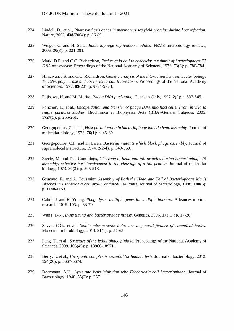
DE JODE Mathieu – Thèse de doctorat - 2021
146
224. Lindell, D., et al., Photosynthesis genes in marine viruses yield proteins during host infection.
Nature, 2005. 438(7064): p. 86-89.
225. Weigel, C. and H. Seitz, Bacteriophage replication modules. FEMS microbiology reviews,
2006. 30(3): p. 321-381.
226. Mark, D.F. and C.C. Richardson, Escherichia coli thioredoxin: a subunit of bacteriophage T7
DNA polymerase. Proceedings of the National Academy of Sciences, 1976. 73(3): p. 780-784.
227. Himawan, J.S. and C.C. Richardson, Genetic analysis of the interaction between bacteriophage
T7 DNA polymerase and Escherichia coli thioredoxin. Proceedings of the National Academy
of Sciences, 1992. 89(20): p. 9774-9778.
228. Fujisawa, H. and M. Morita, Phage DNA packaging. Genes to Cells, 1997. 2(9): p. 537-545.
229. Ponchon, L., et al., Encapsidation and transfer of phage DNA into host cells: From in vivo to
single particles studies. Biochimica et Biophysica Acta (BBA)-General Subjects, 2005.
1724(3): p. 255-261.
230. Georgopoulos, C., et al., Host participation in bacteriophage lambda head assembly. Journal of
molecular biology, 1973. 76(1): p. 45-60.
231. Georgopoulos, C.P. and H. Eisen, Bacterial mutants which block phage assembly. Journal of
supramolecular structure, 1974. 2(2‐4): p. 349-359.
232. Zweig, M. and D.J. Cummings, Cleavage of head and tail proteins during bacteriophage T5
assembly: selective host involvement in the cleavage of a tail protein. Journal of molecular
biology, 1973. 80(3): p. 505-518.
233. Grimaud, R. and A. Toussaint, Assembly of Both the Head and Tail of Bacteriophage Mu Is
Blocked in Escherichia coli groEL andgroES Mutants. Journal of bacteriology, 1998. 180(5):
p. 1148-1153.
234. Cahill, J. and R. Young, Phage lysis: multiple genes for multiple barriers. Advances in virus
research, 2019. 103: p. 33-70.
235. Wang, I.-N., Lysis timing and bacteriophage fitness. Genetics, 2006. 172(1): p. 17-26.
236. Savva, C.G., et al., Stable micron‐scale holes are a general feature of canonical holins.
Molecular microbiology, 2014. 91(1): p. 57-65.
237. Pang, T., et al., Structure of the lethal phage pinhole. Proceedings of the National Academy of
Sciences, 2009. 106(45): p. 18966-18971.
238. Berry, J., et al., The spanin complex is essential for lambda lysis. Journal of bacteriology, 2012.
194(20): p. 5667-5674.
239. Doermann, A.H., Lysis and lysis inhibition with Escherichia coli bacteriophage. Journal of
Bacteriology, 1948. 55(2): p. 257.
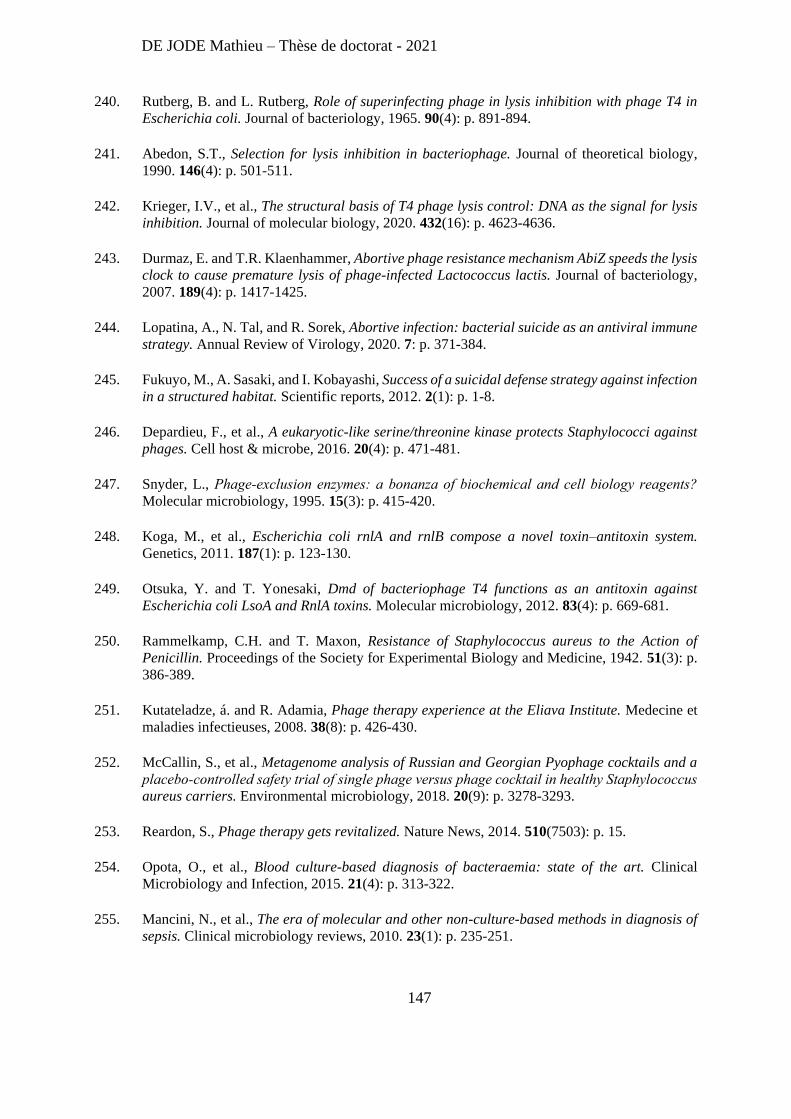
DE JODE Mathieu – Thèse de doctorat - 2021
147
240. Rutberg, B. and L. Rutberg, Role of superinfecting phage in lysis inhibition with phage T4 in
Escherichia coli. Journal of bacteriology, 1965. 90(4): p. 891-894.
241. Abedon, S.T., Selection for lysis inhibition in bacteriophage. Journal of theoretical biology,
1990. 146(4): p. 501-511.
242. Krieger, I.V., et al., The structural basis of T4 phage lysis control: DNA as the signal for lysis
inhibition. Journal of molecular biology, 2020. 432(16): p. 4623-4636.
243. Durmaz, E. and T.R. Klaenhammer, Abortive phage resistance mechanism AbiZ speeds the lysis
clock to cause premature lysis of phage-infected Lactococcus lactis. Journal of bacteriology,
2007. 189(4): p. 1417-1425.
244. Lopatina, A., N. Tal, and R. Sorek, Abortive infection: bacterial suicide as an antiviral immune
strategy. Annual Review of Virology, 2020. 7: p. 371-384.
245. Fukuyo, M., A. Sasaki, and I. Kobayashi, Success of a suicidal defense strategy against infection
in a structured habitat. Scientific reports, 2012. 2(1): p. 1-8.
246. Depardieu, F., et al., A eukaryotic-like serine/threonine kinase protects Staphylococci against
phages. Cell host & microbe, 2016. 20(4): p. 471-481.
247. Snyder, L., Phage‐exclusion enzymes: a bonanza of biochemical and cell biology reagents?
Molecular microbiology, 1995. 15(3): p. 415-420.
248. Koga, M., et al., Escherichia coli rnlA and rnlB compose a novel toxin–antitoxin system.
Genetics, 2011. 187(1): p. 123-130.
249. Otsuka, Y. and T. Yonesaki, Dmd of bacteriophage T4 functions as an antitoxin against
Escherichia coli LsoA and RnlA toxins. Molecular microbiology, 2012. 83(4): p. 669-681.
250. Rammelkamp, C.H. and T. Maxon, Resistance of Staphylococcus aureus to the Action of
Penicillin. Proceedings of the Society for Experimental Biology and Medicine, 1942. 51(3): p.
386-389.
251. Kutateladze, á. and R. Adamia, Phage therapy experience at the Eliava Institute. Medecine et
maladies infectieuses, 2008. 38(8): p. 426-430.
252. McCallin, S., et al., Metagenome analysis of Russian and Georgian Pyophage cocktails and a
placebo‐controlled safety trial of single phage versus phage cocktail in healthy Staphylococcus
aureus carriers. Environmental microbiology, 2018. 20(9): p. 3278-3293.
253. Reardon, S., Phage therapy gets revitalized. Nature News, 2014. 510(7503): p. 15.
254. Opota, O., et al., Blood culture-based diagnosis of bacteraemia: state of the art. Clinical
Microbiology and Infection, 2015. 21(4): p. 313-322.
255. Mancini, N., et al., The era of molecular and other non-culture-based methods in diagnosis of
sepsis. Clinical microbiology reviews, 2010. 23(1): p. 235-251.
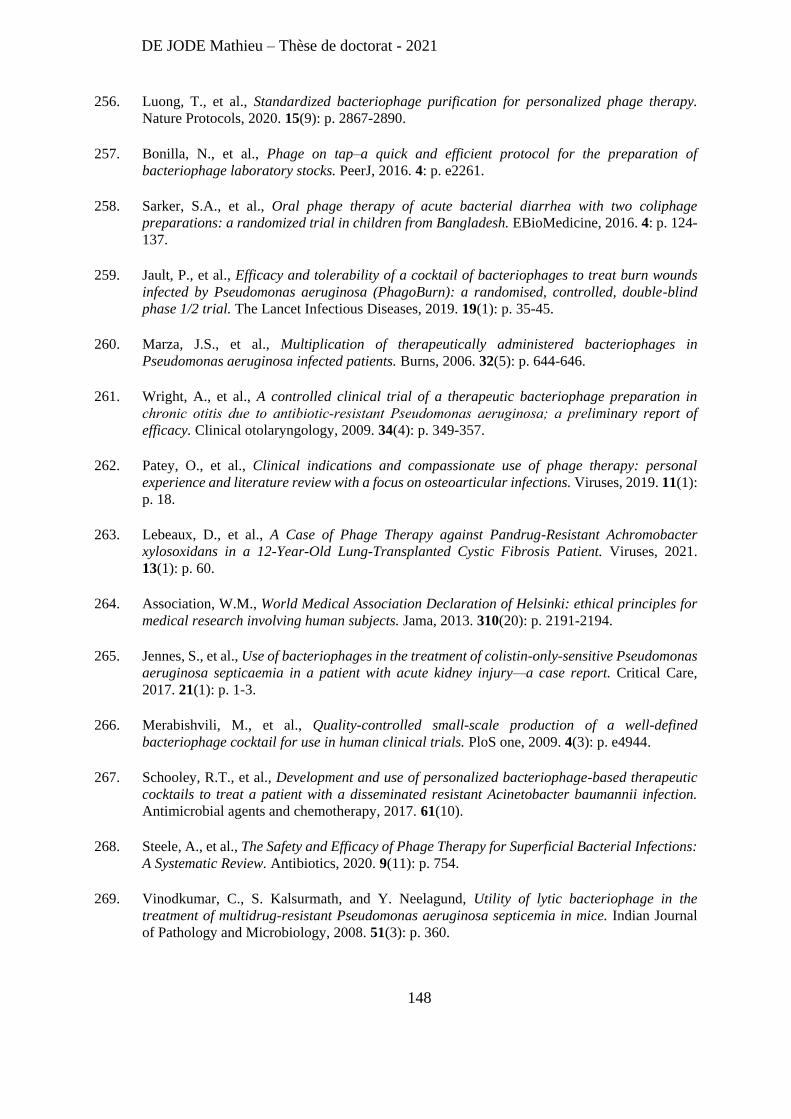
DE JODE Mathieu – Thèse de doctorat - 2021
148
256. Luong, T., et al., Standardized bacteriophage purification for personalized phage therapy.
Nature Protocols, 2020. 15(9): p. 2867-2890.
257. Bonilla, N., et al., Phage on tap–a quick and efficient protocol for the preparation of
bacteriophage laboratory stocks. PeerJ, 2016. 4: p. e2261.
258. Sarker, S.A., et al., Oral phage therapy of acute bacterial diarrhea with two coliphage
preparations: a randomized trial in children from Bangladesh. EBioMedicine, 2016. 4: p. 124-
137.
259. Jault, P., et al., Efficacy and tolerability of a cocktail of bacteriophages to treat burn wounds
infected by Pseudomonas aeruginosa (PhagoBurn): a randomised, controlled, double-blind
phase 1/2 trial. The Lancet Infectious Diseases, 2019. 19(1): p. 35-45.
260. Marza, J.S., et al., Multiplication of therapeutically administered bacteriophages in
Pseudomonas aeruginosa infected patients. Burns, 2006. 32(5): p. 644-646.
261. Wright, A., et al., A controlled clinical trial of a therapeutic bacteriophage preparation in
chronic otitis due to antibiotic‐resistant Pseudomonas aeruginosa; a preliminary report of
efficacy. Clinical otolaryngology, 2009. 34(4): p. 349-357.
262. Patey, O., et al., Clinical indications and compassionate use of phage therapy: personal
experience and literature review with a focus on osteoarticular infections. Viruses, 2019. 11(1):
p. 18.
263. Lebeaux, D., et al., A Case of Phage Therapy against Pandrug-Resistant Achromobacter
xylosoxidans in a 12-Year-Old Lung-Transplanted Cystic Fibrosis Patient. Viruses, 2021.
13(1): p. 60.
264. Association, W.M., World Medical Association Declaration of Helsinki: ethical principles for
medical research involving human subjects. Jama, 2013. 310(20): p. 2191-2194.
265. Jennes, S., et al., Use of bacteriophages in the treatment of colistin-only-sensitive Pseudomonas
aeruginosa septicaemia in a patient with acute kidney injury—a case report. Critical Care,
2017. 21(1): p. 1-3.
266. Merabishvili, M., et al., Quality-controlled small-scale production of a well-defined
bacteriophage cocktail for use in human clinical trials. PloS one, 2009. 4(3): p. e4944.
267. Schooley, R.T., et al., Development and use of personalized bacteriophage-based therapeutic
cocktails to treat a patient with a disseminated resistant Acinetobacter baumannii infection.
Antimicrobial agents and chemotherapy, 2017. 61(10).
268. Steele, A., et al., The Safety and Efficacy of Phage Therapy for Superficial Bacterial Infections:
A Systematic Review. Antibiotics, 2020. 9(11): p. 754.
269. Vinodkumar, C., S. Kalsurmath, and Y. Neelagund, Utility of lytic bacteriophage in the
treatment of multidrug-resistant Pseudomonas aeruginosa septicemia in mice. Indian Journal
of Pathology and Microbiology, 2008. 51(3): p. 360.
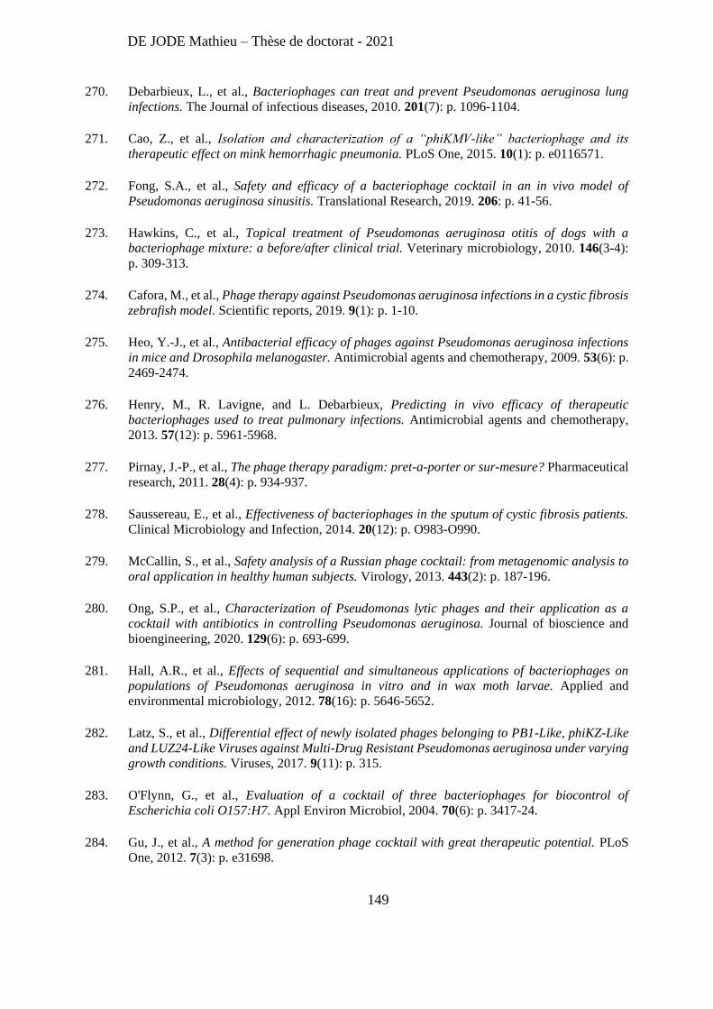
DE JODE Mathieu – Thèse de doctorat - 2021
149
270. Debarbieux, L., et al., Bacteriophages can treat and prevent Pseudomonas aeruginosa lung
infections. The Journal of infectious diseases, 2010. 201(7): p. 1096-1104.
271. Cao, Z., et al., Isolation and characterization of a “phiKMV-like” bacteriophage and its
therapeutic effect on mink hemorrhagic pneumonia. PLoS One, 2015. 10(1): p. e0116571.
272. Fong, S.A., et al., Safety and efficacy of a bacteriophage cocktail in an in vivo model of
Pseudomonas aeruginosa sinusitis. Translational Research, 2019. 206: p. 41-56.
273. Hawkins, C., et al., Topical treatment of Pseudomonas aeruginosa otitis of dogs with a
bacteriophage mixture: a before/after clinical trial. Veterinary microbiology, 2010. 146(3-4):
p. 309-313.
274. Cafora, M., et al., Phage therapy against Pseudomonas aeruginosa infections in a cystic fibrosis
zebrafish model. Scientific reports, 2019. 9(1): p. 1-10.
275. Heo, Y.-J., et al., Antibacterial efficacy of phages against Pseudomonas aeruginosa infections
in mice and Drosophila melanogaster. Antimicrobial agents and chemotherapy, 2009. 53(6): p.
2469-2474.
276. Henry, M., R. Lavigne, and L. Debarbieux, Predicting in vivo efficacy of therapeutic
bacteriophages used to treat pulmonary infections. Antimicrobial agents and chemotherapy,
2013. 57(12): p. 5961-5968.
277. Pirnay, J.-P., et al., The phage therapy paradigm: pret-a-porter or sur-mesure? Pharmaceutical
research, 2011. 28(4): p. 934-937.
278. Saussereau, E., et al., Effectiveness of bacteriophages in the sputum of cystic fibrosis patients.
Clinical Microbiology and Infection, 2014. 20(12): p. O983-O990.
279. McCallin, S., et al., Safety analysis of a Russian phage cocktail: from metagenomic analysis to
oral application in healthy human subjects. Virology, 2013. 443(2): p. 187-196.
280. Ong, S.P., et al., Characterization of Pseudomonas lytic phages and their application as a
cocktail with antibiotics in controlling Pseudomonas aeruginosa. Journal of bioscience and
bioengineering, 2020. 129(6): p. 693-699.
281. Hall, A.R., et al., Effects of sequential and simultaneous applications of bacteriophages on
populations of Pseudomonas aeruginosa in vitro and in wax moth larvae. Applied and
environmental microbiology, 2012. 78(16): p. 5646-5652.
282. Latz, S., et al., Differential effect of newly isolated phages belonging to PB1-Like, phiKZ-Like
and LUZ24-Like Viruses against Multi-Drug Resistant Pseudomonas aeruginosa under varying
growth conditions. Viruses, 2017. 9(11): p. 315.
283. O'Flynn, G., et al., Evaluation of a cocktail of three bacteriophages for biocontrol of
Escherichia coli O157:H7. Appl Environ Microbiol, 2004. 70(6): p. 3417-24.
284. Gu, J., et al., A method for generation phage cocktail with great therapeutic potential. PLoS
One, 2012. 7(3): p. e31698.
DE JODE Mathieu – Thèse de doctorat - 2021
150
285. Smith, H.W. and M.B. Huggins, Effectiveness of Phages in Treating Experimental Escherichia
coli Diarrhoea in Calves, Piglets and Lambs. Microbiology, 1983. 129(8): p. 2659-2675.
286. Filippov, A.A., et al., Bacteriophage-Resistant Mutants in Yersinia pestis: Identification of
Phage Receptors and Attenuation for Mice. PLOS ONE, 2011. 6(9): p. e25486.
287. Pires, D.P., et al., A genotypic analysis of five P. aeruginosa strains after biofilm infection by
phages targeting different cell surface receptors. Frontiers in microbiology, 2017. 8: p. 1229.
288. Wright, R.C., et al., Cross-resistance is modular in bacteria–phage interactions. PLoS biology,
2018. 16(10): p. e2006057.
289. Gordillo Altamirano, F.L. and J.J. Barr, Unlocking the next generation of phage therapy: the
key is in the receptors. Curr Opin Biotechnol, 2020. 68: p. 115-123.
290. Forti, F., et al., Design of a broad-range bacteriophage cocktail that reduces Pseudomonas
aeruginosa biofilms and treats acute infections in two animal models. Antimicrobial agents and
chemotherapy, 2018. 62(6).
291. Oechslin, F., et al., Synergistic interaction between phage therapy and antibiotics clears
Pseudomonas aeruginosa infection in endocarditis and reduces virulence. The Journal of
infectious diseases, 2017. 215(5): p. 703-712.
292. Roach, D.R., et al., Synergy between the host immune system and bacteriophage is essential for
successful phage therapy against an acute respiratory pathogen. Cell host & microbe, 2017.
22(1): p. 38-47. e4.
293. Skerrett, S.J., et al., Cutting edge: myeloid differentiation factor 88 is essential for pulmonary
host defense against Pseudomonas aeruginosa but not Staphylococcus aureus. The Journal of
Immunology, 2004. 172(6): p. 3377-3381.
294. Deguine, J. and G.M. Barton, MyD88: a central player in innate immune signaling. F1000prime
reports, 2014. 6.
295. Kawai, T., et al., Unresponsiveness of MyD88-deficient mice to endotoxin. Immunity, 1999.
11(1): p. 115-122.
296. Colucci, F., et al., Dissecting NK cell development using a novel alymphoid mouse model:
investigating the role of the c-abl proto-oncogene in murine NK cell differentiation. The Journal
of Immunology, 1999. 162(5): p. 2761-2765.
297. Merril, C.R., et al., Long-circulating bacteriophage as antibacterial agents. Proceedings of the
National Academy of Sciences, 1996. 93(8): p. 3188-3192.
298. Hodyra-Stefaniak, K., et al., Mammalian Host-Versus-Phage immune response determines
phage fate in vivo. Scientific reports, 2015. 5(1): p. 1-13.
299. Sievers, F., et al., Fast, scalable generation of high‐quality protein multiple sequence
alignments using Clustal Omega. Molecular systems biology, 2011. 7(1): p. 539.
DE JODE Mathieu – Thèse de doctorat - 2021
151
300. MacArthur, M.W. and J.M. Thornton, Influence of proline residues on protein conformation.
Journal of molecular biology, 1991. 218(2): p. 397-412.
301. Divakaruni, A.V., et al., The cell shape proteins MreB and MreC control cell morphogenesis
by positioning cell wall synthetic complexes. Molecular microbiology, 2007. 66(1): p. 174-188.
302. Comeau, A.M., et al., Phage-antibiotic synergy (PAS): β-lactam and quinolone antibiotics
stimulate virulent phage growth. Plos one, 2007. 2(8): p. e799.
303. Kim, M., et al., Phage-Antibiotic Synergy via Delayed Lysis. Appl Environ Microbiol, 2018.
84(22).
304. Paget, M.S., Bacterial sigma factors and anti-sigma factors: structure, function and
distribution. Biomolecules, 2015. 5(3): p. 1245-1265.
305. Schulz, S., et al., Elucidation of sigma factor-associated networks in Pseudomonas aeruginosa
reveals a modular architecture with limited and function-specific crosstalk. PLoS Pathog, 2015.
11(3): p. e1004744.
306. Dickey, S.W., G.Y.C. Cheung, and M. Otto, Different drugs for bad bugs: antivirulence
strategies in the age of antibiotic resistance. Nat Rev Drug Discov, 2017. 16(7): p. 457-471.
307. Stapley, E.O., et al., Cefoxitin and cephamycins: microbiological studies. Reviews of infectious
diseases, 1979. 1(1): p. 73-87.
308. Gin, A., et al., Piperacillin–tazobactam: a β-lactam/β-lactamase inhibitor combination. Expert
review of anti-infective therapy, 2007. 5(3): p. 365-383.
309. Liu, J., et al., Antimicrobial drug discovery through bacteriophage genomics. Nature
biotechnology, 2004. 22(2): p. 185-191.
310. Wagemans, J. and R. Lavigne, Phages and their hosts, a web of interactions–applications to
drug design. 2012.
311. Loeffler, J.M., D. Nelson, and V.A. Fischetti, Rapid killing of Streptococcus pneumoniae with
a bacteriophage cell wall hydrolase. Science, 2001. 294(5549): p. 2170-2172.
312. Nelson, D., L. Loomis, and V.A. Fischetti, Prevention and elimination of upper respiratory
colonization of mice by group A streptococci by using a bacteriophage lytic enzyme.
Proceedings of the National Academy of Sciences, 2001. 98(7): p. 4107-4112.
313. Briers, Y., et al., Art-175 is a highly efficient antibacterial against multidrug-resistant strains
and persisters of Pseudomonas aeruginosa. Antimicrobial agents and chemotherapy, 2014.
58(7): p. 3774-3784.
314. Defraine, V., et al., Efficacy of artilysin Art-175 against resistant and persistent Acinetobacter
baumannii. Antimicrobial agents and chemotherapy, 2016. 60(6): p. 3480-3488.
315. Theuretzbacher, U., et al., The global preclinical antibacterial pipeline. Nature Reviews
Microbiology, 2020. 18(5): p. 275-285.
DE JODE Mathieu – Thèse de doctorat - 2021
152
316. Abdelkader, K., et al., The preclinical and clinical progress of bacteriophages and their lytic
enzymes: the parts are easier than the whole. Viruses, 2019. 11(2): p. 96.
317. Reinhard, C., et al., Genomewide linkage analysis identifies novel genetic Loci for lung function
in mice. American journal of respiratory and critical care medicine, 2005. 171(8): p. 880-888.
318. Alseth, E.O., et al., Bacterial biodiversity drives the evolution of CRISPR-based phage
resistance. Nature, 2019. 574(7779): p. 549-552.
319. Westra, E.R., et al., Parasite exposure drives selective evolution of constitutive versus inducible
defense. Current biology, 2015. 25(8): p. 1043-1049.
320. Meaden, S., K. Paszkiewicz, and B. Koskella, The cost of phage resistance in a plant pathogenic
bacterium is context‐dependent. Evolution, 2015. 69(5): p. 1321-1328.
321. Martin, T.R. and C.W. Frevert, Innate immunity in the lungs. Proceedings of the American
Thoracic Society, 2005. 2(5): p. 403-411.
322. Flyg, C., K. Kenne, and H.G. Boman, Insect pathogenic properties of Serratia marcescens:
phage-resistant mutants with a decreased resistance to Cecropia immunity and a decreased
virulence to Drosophila. Microbiology, 1980. 120(1): p. 173-181.
323. Gordillo Altamirano, F., et al., Bacteriophage-resistant Acinetobacter baumannii are
resensitized to antimicrobials. Nature Microbiology, 2021. 6(2): p. 157-161.
324. Adachi, O., et al., Targeted disruption of the MyD88 gene results in loss of IL-1-and IL-18-
mediated function. Immunity, 1998. 9(1): p. 143-150.
325. Lumish, R.M. and C.W. Norden, Therapy of neutropenic rats infected with Pseudomonas
aeruginosa. Journal of Infectious Diseases, 1976. 133(5): p. 538-547.
326. Bodey, G.P. and V. Rodriguez, Advances in the management of Pseudomonas aeruginosa
infections in cancer patients. European Journal of Cancer (1965), 1973. 9(6): p. 435-441.
327. Van de Wetering, M., et al., Efficacy of oral prophylactic antibiotics in neutropenic afebrile
oncology patients: a systematic review of randomised controlled trials. European Journal of
Cancer, 2005. 41(10): p. 1372-1382.
328. Gafter-Gvili, A., et al., Meta-analysis: antibiotic prophylaxis reduces mortality in neutropenic
patients. Annals of internal medicine, 2005. 142(12_Part_1): p. 979-995.
329. Herold, S., K. Mayer, and J. Lohmeyer, Acute lung injury: how macrophages orchestrate
resolution of inflammation and tissue repair. Frontiers in immunology, 2011. 2: p. 65.
330. Johnston, L.K., et al., Pulmonary macrophage subpopulations in the induction and resolution
of acute lung injury. American journal of respiratory cell and molecular biology, 2012. 47(4):
p. 417-426.
331. Giangreco, A., et al., Myd88 deficiency influences murine tracheal epithelial metaplasia and
submucosal gland abundance. The Journal of pathology, 2011. 224(2): p. 190-202.
DE JODE Mathieu – Thèse de doctorat - 2021
153
332. Reeve, W.G., et al., Constructs for insertional mutagenesis, transcriptional signal localization
and gene regulation studies in root nodule and other bacteria. Microbiology (Reading), 1999.
145 ( Pt 6): p. 1307-1316.
333. Yu, N.Y., et al., PSORTb 3.0: improved protein subcellular localization prediction with refined
localization subcategories and predictive capabilities for all prokaryotes. Bioinformatics, 2010.
26(13): p. 1608-1615.
334. Smith, H.W. and M.B. Huggins, Successful Treatment of Experimental Escherichia coli
Infections in Mice Using Phage: its General Superiority over Antibiotics. Microbiology, 1982.
128(2): p. 307-318.
335. Chatterjee, A., et al., Bacteriophage Resistance Alters Antibiotic-Mediated Intestinal Expansion
of Enterococci. Infection and Immunity, 2019. 87(6): p. e00085-19.
336. Chan, B.K., et al., Phage selection restores antibiotic sensitivity in MDR Pseudomonas
aeruginosa. Scientific Reports, 2016. 6(1): p. 26717.
337. Chan, B.K., et al., Phage treatment of an aortic graft infected with Pseudomonas aeruginosa.
Evolution, Medicine, and Public Health, 2018. 2018(1): p. 60-66.
338. Kirby, A.E., Synergistic action of gentamicin and bacteriophage in a continuous culture
population of Staphylococcus aureus. Plos one, 2012. 7(11): p. e51017.
339. Ryan, E.M., et al., Synergistic phage-antibiotic combinations for the control of Escherichia coli
biofilms in vitro. FEMS Immunology & Medical Microbiology, 2012. 65(2): p. 395-398.
340. Rahman, M., et al., Characterization of induced Staphylococcus aureus bacteriophage SAP-26
and its anti-biofilm activity with rifampicin. Biofouling, 2011. 27(10): p. 1087-1093.
341. Moulton‐Brown, C.E. and V.P. Friman, Rapid evolution of generalized resistance mechanisms
can constrain the efficacy of phage–antibiotic treatments. Evolutionary applications, 2018.
11(9): p. 1630-1641.
342. Allen, R.C., et al., Associations among antibiotic and phage resistance phenotypes in natural
and clinical Escherichia coli isolates. MBio, 2017. 8(5).
DE JODE Mathieu – Thèse de doctorat - 2021
154
Abstract (English)
Pseudomonas aeruginosa is an increasingly antibiotic resistant pathogenic bacterium. Here, we studied
bacteriophages infecting this bacterium to develop antibacterial strategies. We focused on two objectives: (1) the
characterization of an early-expressed phage protein that increases the susceptibility of P. aeruginosa to antibiotics
and (2) the development of a bacteriophage cocktail to optimize phage therapy efficacy in immunocompromised
MyD88-/- mice.
The first objective aimed at understanding the molecular mechanisms underlying viral strategies to hijack bacterial
functions. We characterized gp92, an early-expressed gene of phage PAK_P3 during its infection of P. aeruginosa.
The ectopic expression of gp92 alters bacterial morphology, motility and provokes a major shift of the cell
proteome, without affecting growth. Via its interaction with the anti-sigma factor MucA, Gp92 impairs the
membrane stress response mediated by the sigma factor AlgU. We hypothesize that Gp92 disrupts this stress
response to control phage lysis timing. Additionally, expression of gp92 increased the susceptibility of P.
aeruginosa to antibiotics, including Imipenem, thus revealing a new target to increase bacterial susceptibility to
Carbapenems.
The second objective focused on phage therapy efficacy and bacterial resistance to monophage treatments in
immunocompromised MyD88-/- mice. In these mice monophage therapy fails due to the uncontrolled growth of
phage-resistant bacteria. To limit this growth, we evaluated two phages recognizing different bacterial receptors
and their combination, both in vitro and in vivo. In vitro, monophage treatments failed to limit phage-resistance
growth while the combination succeeded. In contrast, the combination was not associated with higher efficacy in
treating MyD88-/- mice, compared to monophage treatments. Analysis of the resistance pattern of bacteria isolated
in each experimental conditions suggests that phage resistance is phage- and environment-dependent. We found
that bacteria will not resist to each phage the same way and that in vitro experiments are not predictive of phage
therapy success in vivo.
Résumé (Français)
Pseudomonas aeruginosa est une bactérie pathogène de plus en plus résistante aux antibiotiques. Nous avons
étudié les bactériophages infectant cette bactérie afin de développer des stratégies antibactériennes. Nous nous
sommes concentrés sur deux objectifs : (1) la caractérisation d'une protéine de phage exprimée précocement qui
augmente la sensibilité de P. aeruginosa aux antibiotiques et (2) le développement d'un cocktail de bactériophages
pour optimiser l'efficacité de la phagothérapie chez les souris immunodéprimées MyD88-/-.
Le premier objectif visait à comprendre les mécanismes moléculaires sous-jacent aux stratégies virales de
détournement des fonctions bactériennes. Nous avons caractérisé gp92, un gène exprimé par le phage PAK_P3 au
début de son infection de P. aeruginosa. L'expression ectopique de gp92 modifie la morphologie et la motilité
bactérienne. Elle provoque aussi une modification majeure du protéome cellulaire, mais n’affecte pas la croissance
bactérienne. Par son interaction avec le facteur anti-sigma MucA, Gp92 altère la réponse au stress membranaire
médiée par le facteur sigma AlgU. Nous émettons l'hypothèse que Gp92 perturbe cette réponse au stress pour
contrôler le moment de déclenchement de la lyse par le phage. De plus, l'expression de gp92 augmente la sensibilité
de P. aeruginosa aux antibiotiques, y compris l'Imipénem, révélant ainsi une nouvelle cible pour accroitre la
sensibilité bactérienne aux Carbapénèmes.
Le deuxième objectif est centré sur l'efficacité de la phagothérapie et la résistance bactérienne aux phages chez
des souris MyD88-/- immunodéprimées. Chez ces souris, la thérapie via un seul phage échoue à cause de la
croissance de bactéries résistantes aux phages. Pour limiter cette croissance, nous avons évalué in vitro et in vivo,
deux phages reconnaissant différents récepteurs bactériens et leur combinaison. In vitro, les traitements par un seul
phage n'ont pas réussi à limiter la croissance de clones résistants aux phages, tandis que la combinaison a réussi.
En revanche, cette combinaison n’avait pas une plus grande efficacité que les traitements avec un seul phage chez
les souris MyD88-/-. L'analyse du profil de résistance des bactéries isolées dans toutes les conditions expérimentales
suggère que la résistance aux phages dépend du phage utilisé et de l’environnement. Nous avons observé que les
bactéries ne résistent pas à tous les phages de la même manière et que les expériences in vitro ne sont pas
prédictives du succès de la phagothérapie in vivo.