tumor invasion as dysregulated cell motility
TRANSCRIPT

/r
AD_ _ _ _
Award Number: DAIMD17-01-1-0093
TITLE: Cell Motility in Tumor Invasion
PRINCIPAL INVESTIGATOR: Alan Wells, M.D.Douglas A. Lauffenburger, Ph.D.Timothy Turner, Ph.D.
CONTRACTING ORGANIZATION: University of PittsburghPittsburgh, Pennsylvania 15260
REPORT DATE: July 2004
TYPE OF REPORT: Final
PREPARED FOR: U.S. Army Medical Research and Materiel CommandFort Detrick, Maryland 21702-5012
DISTRIBUTION STATEMENT: Approved for Public Release;Distribution Unlimited
The views, opinions and/or findings contained in this report arethose of the author(s) and should not be construed as an officialDepartment of the Army position, policy or decision unless sodesignated by other documentation.
20050105 081


Table of Contents
Cover..........................................................................
SF 298.........................................................................
Table of Contents.............................................................
Introduction ................................................................... 3
Body........................................................................... 3
Key Research Accomplishments............................................. 7
Reportable Outcomes ........................................................ 8
Conclusions .................................................................. 9
References................................................................... 10
Appendices................................................................... 11

Wells, AlanDAMD1 7-01-1-0093
CELL MOTILITY IN TUMOR INVASION
Alan Wells, Douglas Lauffenburger, Timothy Turner
INTRODUCTIONOur overall objective is to understand how dysregulation of cell migration contributes to
tumor cell invasiveness in prostate cancer. A combination of correlative epidemiological studiesand basic experimental investigations demonstrate a role for upregulated EGF receptor (EGFR)and other receptor signaling of motility in tumor progression. Especially in prostate tumor cells,EGFR-mediated cell motility has been demonstrated to be critical for tumor invasion (2, 5). Sincesignals from extracellular matrix through integrins and from cell-cell contacts also stronglyinfluence cell motility, the underlying common biophysical processes and biochemical controls ofmotility offer an attractive target for limiting tumor progression.
Our central premise is that prostate tumor cell invasiveness can be inhibited by interferingwith the specific motility-associated calpain activation that governs the critical underlyingbiophysical process of de-adhesion. Prior work by ourselves and others has shown thatintegrin/matrix binding and growth factor stimulation jointly regulate cell locomotion (1, 3).These studies have identified cell/substratum adhesiveness, especially the ability of a cell todetach at its trailing edge, as a primary governor of cell locomotion. We have recently foundthat this tail detachment is regulated by calpain activation. We will employ a set of modelprostate tumor cell lines including the moderately invasive androgen-independent PC3 cell andits highly metastatic variant PC3M cell, along with a panel of syngeneic androgen-independentDU-145 cells that vary in invasiveness. We will determine whether targeted disruption of calpainactivation and de-adhesion can block tumor invasiveness.
BODYThe original Statement of Work (Table 1) described a series of tasks to accomplish the two
Objectives proposed and the additional training Objective. We have tackled these Tasks in theorder of greatest yield so that work in areas can progress as systems are being optimized inothers. The total three years of effort have led to the accomplishment of these task
Table 1. Original Statement of Work
Work to be performed at University of Pittsburgh (A. Wells Laboratory):1. determine whether calpain is activated by growth factors and integrins in prostate cancer
cells2. determine whether calpain is limiting for prostate tumor cell motility on complex surfaces3. determine whether prostate tumor cell transmigration of extracellular matrices is
dependent on calpain activity4. determine whether inhibition of calpain limits tumor invasiveness and metastasis in
murine models of progressive prostate cancer
Work to be performed at MIT (D.A. Lauffenburger Laboratory):1. determine optimal adhesiveness and high and low adhesiveness surfaces for fibroblast
motility2. test prostate tumor cell motility on defined adhesiveness surfaces3. determine whether calpain activation is required for prostate cell motility
Work to be performed in partnership with Tuskegee (T. Turner Laboratory):
3
Wells, AlanDAMD1 7-01-1-0093
1. trainees will perform prostate cell growth and motility assays at Tuskegee and UPitt2. trainees will perform in vivo mouse assays at UPitt
The reader of the Year 2 progress report deemed task completion as the following:
Task 1 No time given In progressTask 2 No time given In progressTask 3 No time given CompletedTask 4 No time given CompletedTask 5 No time given CompletedTask 6 No time given In progressTask 7 No time given CompletedTask 8 No time given In progressTask 9 No time given In progress
Work to be performed at University of Pittsburgh:
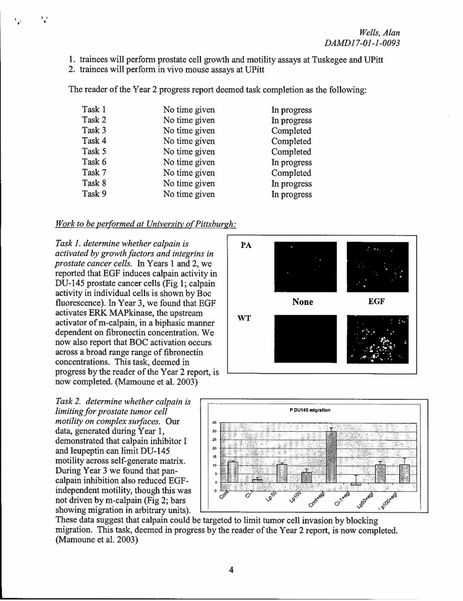
Task 1. determine whether calpain is PAactivated by growth factors and integrins inprostate cancer cells. In Years 1 and 2, wereported that EGF induces calpain activity inDU-145 prostate cancer cells (Fig 1; calpainactivity in individual cells is shown by Bocfluorescence). In Year 3, we found that EGF None EGFactivates ERK MAPkinase, the upstream WTactivator of m-calpain, in a biphasic mannerdependent on fibronectin concentration. Wenow also report that BOC activation occursacross a broad range range of fibronectinconcentrations. This task, deemed inprogress by the reader of the Year 2 report, isnow completed. (Mamoune et al. 2003)
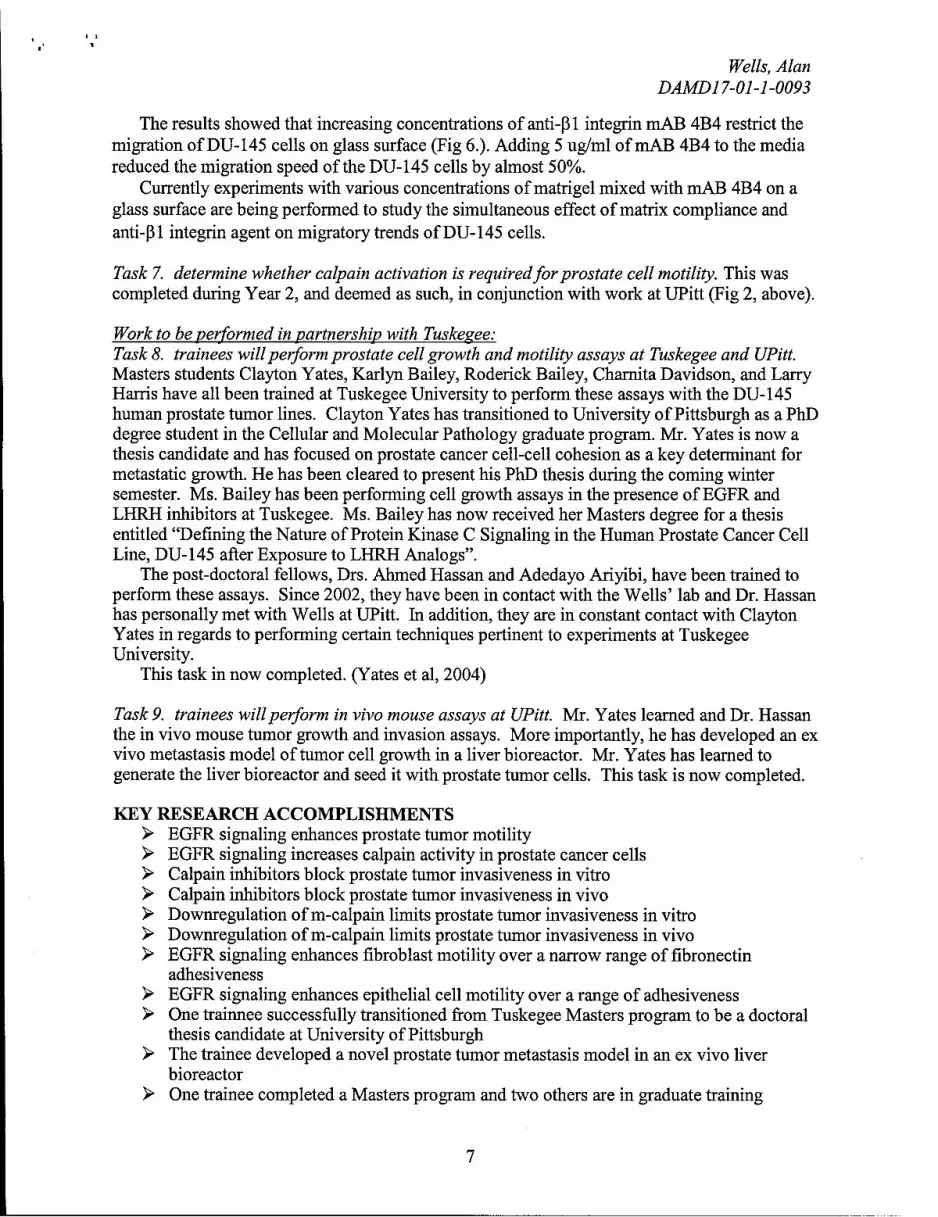
Task 2. determine whether calpain islimiting for prostate tumor cell P DU145 migration
motility on complex surfaces. Our 35data, generated during Year 1, 30 --demonstrated that calpain inhibitor [ 25
and leupeptin can limit DU- 145 20
motility across self-generate matrix. 10
During Year 3 we found that pan- '-S
calpain inhibition also reduced EGF- 0 L 1 - 1independent motility, though this was -5 -, ----Knot driven by m-calpain (Fig 2; barsshowing migration in arbitrary units).These data suggest that calpain could be targeted to limit tumor cell invasion by blockingmigration. This task, deemed in progress by the reader of the Year 2 report, is now completed.(Mamoune et al. 2003)
4

Wells, AlanDAMD17-01-1-0093
Task 3. determine whether prostate tumor cell M None
transmigration of extracellular matrices is dependent 120% - 11 CHon calpain activity. This task was completed during i LpYear 2. In vitro transmigration of a Matrigel matrix 100%by both Parental and WT EGFR-expressing DU-145 80%
cells is blocked by inhibitors of calpain, CM-I and "0leupeptin (Fig 3; ** P < 0.01 compare to diluent , 60% **
alone/none). Furthermore, antisense downregulation 40%
of M-calpain limits this transmigration, providing 4-specificity (Fig 4, below). 20% -
0%
C PA WT120%
100%These data are presented in Mamoune
Q 80% et al, 2003.60% **
~- 40%
20%
0%
None Scr MAS
Task 4. determine whether inhibition of calpain limits tumor invasiveness and metastasis inmurine models ofprogressive prostate cancer. This tasked was deemed completed after Year 2.In brief, we challenged mice with DU-145 prostate carcinoma tumor xenografts with inhibitorsof calpain. Tumor invasiveness was reduced in the presence of daily injections of the inhibitorleupeptin (Table 1). The differences invasiveness between treated and mock treated weresignificant (P < 0.05) by T-test and ANOVA analyses.
PA+HBSS PA+Leupeptin WT+HBSS WT+leupeptinDiaphragm tumors 14/14 13/14 14/14 13/14Diaphragm invasivei 1.71 0.7** 2.35 1.25*
Antisense constructs to m-calpain were expressed stably in DU-145 cells. These also limitedtumor invasion into the diaphragm of mice (Table 2).
V PA DU145 C2AS PA DU145 V WT DU145 C2AS WT DU145Diaphragm tumors 9/10 8/10 4/5 3/5Diaphragm invasivel 2.33 1.13* 3.50 1.67 A
These data are presented in Mamoune et al, 2003.
5

Wells, AlanDAMD1 7-01-1-0093
Work to be performed at MIT:Task 5. determine optimal adhesiveness andhigh and low adhesiveness surfaces for -. .fibroblast motility. This task was completed .. - GFin Year 1, and was deemed completed by the 40
Year 2 reviewer.In brief, for EGF-induced motility, we found • 30
that optimal fibronectin coating occurs ataround lug/ml, with 0.3 ug/ml and 3 ug/ml _------------.---being low and high adhesiveness, "respectively. At these extremes, motility is 10
reduced to levels on par with no EGFstimulation (Fig 5). The optimal adhesive 0.1 010
strength of the fibroblasts to the surface is Fibronectin Coating Conc. (ig/nil)
approximately 0.8 nN, with a movement of0.2 nN either direction wiping out growth factor induced movement.
Task 6. test prostate tumor cell motility on defined adhesiveness surfaces. This task is inprogress and should be completed by September 2004. Recent high throughput analyses hasexamined the ability of the DU-145 cells to migrate in 2 and 3 dimensions on a matrigel-coatedsurface. Briefly, DU-145 parental cells were plated on glass bottom 96-well Packard plates. Thecells were plated in high serum media (final concentration = 7% FBS in DMEM media) andvarying concentrations of anti-13 1 integrin mAB 4B4 were added to study the effect of anti- f31integrin on cell speed. The cells were imaged using high throughput Cellomics kinetic scan for 6hrs, at controlled temperature (370 C, controlled CO 2 and humidity conditions) with images takenat ten minute intervals.
Cell Migration speed as a function of10/ varying concentrations of anti-P1
integrin mAB 4B4 on a 2D- glass" 9- surface. Addition of 5ug/ml of mAB
4B4 inhibits the migration of DU 145by as much as 50%. The error barsrepresent standard deviation in the
*, mean cell speed for each concentration7- of mAB 4B4. The cell speed was
.0_ obtained by built-in computerized
.0 mobject tracking algorithm in Imaris2 6- 4.06 (Bitplane AG, Inc.).
5-41 2 3 4 5
Conc. of 4B4 mAB (uglml)
6
Wells, AlanDAMD1 7-01-1-0093
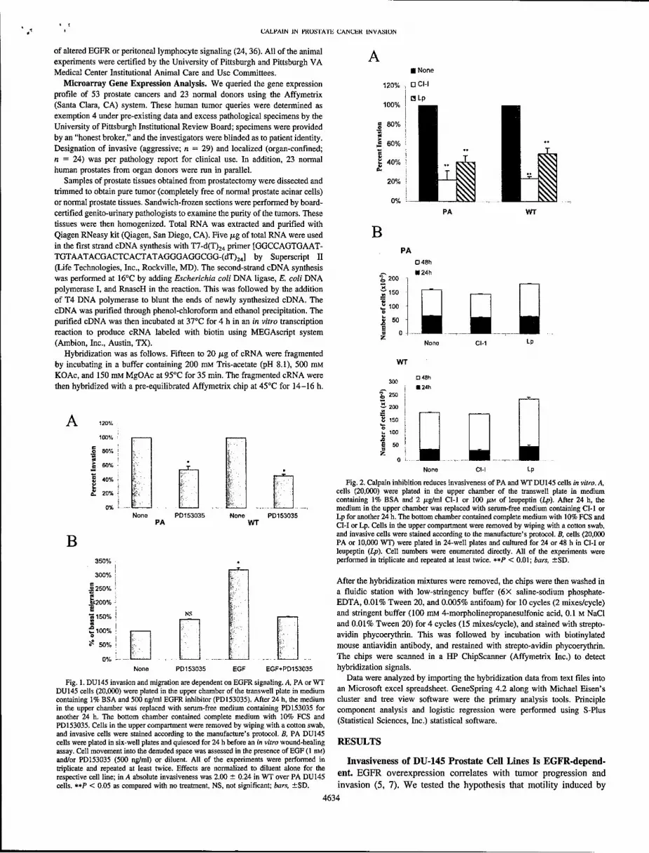
The results showed that increasing concentrations of anti-3 1 integrin mAB 4B4 restrict themigration of DU-145 cells on glass surface (Fig 6.). Adding 5 ug/ml of mAB 4B4 to the mediareduced the migration speed of the DU-145 cells by almost 50%.
Currently experiments with various concentrations of matrigel mixed with mAB 4B4 on aglass surface are being performed to study the simultaneous effect of matrix compliance andanti-3 1 integrin agent on migratory trends of DU- 145 cells.
Task 7. determine whether calpain activation is required for prostate cell motility. This wascompleted during Year 2, and deemed as such, in conjunction with work at UPitt (Fig 2, above).
Work to be performed in partnership with Tuskegee:Task 8. trainees will perform prostate cell growth and motility assays at Tuskegee and UPitt.Masters students Clayton Yates, Karlyn Bailey, Roderick Bailey, Charnita Davidson, and LarryHarris have all been trained at Tuskegee University to perform these assays with the DU-145human prostate tumor lines. Clayton Yates has transitioned to University of Pittsburgh as a PhDdegree student in the Cellular and Molecular Pathology graduate program. Mr. Yates is now athesis candidate and has focused on prostate cancer cell-cell cohesion as a key determinant formetastatic growth. He has been cleared to present his PhD thesis during the coming wintersemester. Ms. Bailey has been performing cell growth assays in the presence of EGFR andLHRH inhibitors at Tuskegee. Ms. Bailey has now received her Masters degree for a thesisentitled "Defining the Nature of Protein Kinase C Signaling in the Human Prostate Cancer CellLine, DU-145 after Exposure to LHRH Analogs".
The post-doctoral fellows, Drs. Ahmed Hassan and Adedayo Ariyibi, have been trained toperform these assays. Since 2002, they have been in contact with the Wells' lab and Dr. Hassanhas personally met with Wells at UPitt. In addition, they are in constant contact with ClaytonYates in regards to performing certain techniques pertinent to experiments at TuskegeeUniversity.
This task in now completed. (Yates et al, 2004)
Task 9. trainees will perform in vivo mouse assays at UPitt. Mr. Yates learned and Dr. Hassanthe in vivo mouse tumor growth and invasion assays. More importantly, he has developed an exvivo metastasis model of tumor cell growth in a liver bioreactor. Mr. Yates has learned togenerate the liver bioreactor and seed it with prostate tumor cells. This task is now completed.
KEY RESEARCH ACCOMPLISHMENTS> EGFR signaling enhances prostate tumor motility> EGFR signaling increases calpain activity in prostate cancer cells> Calpain inhibitors block prostate tumor invasiveness in vitro> Calpain inhibitors block prostate tumor invasiveness in vivo> Downregulation of m-calpain limits prostate tumor invasiveness in vitro> Downregulation of m-calpain limits prostate tumor invasiveness in vivo> EGFR signaling enhances fibroblast motility over a narrow range of fibronectin
adhesiveness> EGFR signaling enhances epithelial cell motility over a range of adhesiveness> One trainnee successfully transitioned from Tuskegee Masters program to be a doctoral
thesis candidate at University of Pittsburgh> The trainee developed a novel prostate tumor metastasis model in an ex vivo liver
bioreactor> One trainee completed a Masters program and two others are in graduate training
7

Wells, AlanDAMD1 7-01-1-0093
lookiing to transition to a doctoral programSTwo trainees are in post-graduate training at Tuskegee in conjunction with the laboratory
at University of Pittsburgh
PAID PERSONNEL INVOLVED IN THE GRANTUniversity of Pittsburgh:Alan WellsKathleen SullivanAshiro AwabuLatha SatishShanmuga Kulasekaran
Tuskegee UniversityTimothy TurnerAdedayo AriyibiLarry HarrisRoderick BaileyCharnita DavidsonAhmed Hassan
Massachusetts Institute of TechnologyDoug LauufenburgerChristina LewisKirsty SmithBart HendriksClayton Yates
REPORTABLE OUTCOMESArticles:A Mamoune, JH Luo, DA Lauffenburger, A Wells (2003). m-Calpain as a target for limitingprostate cancer invasion. Cancer Research 63, 4632-4640.
A Wells, J Grandis (2003). PLCy- 1 in tumor progression. Clinical & Experimental Metastasis20, 285-290.
C Yates, A Wells, T Turner (2004). Luteinizing hormone releasing hormone (LHRH) antagonistreverses the cell adhesion profile of DU-145 human prostate carcinoma, submitted
Abstracts.:A Mamoune, J Kassis, D Lauffenburger, A Wells (2002) Calpain inhibition reduces prostatetumor invasion. American Association for Cancer Research (AACR) Annual Meeting, SanFrancisco, CA
C C Yates, K J Bailey, A Wells, T Turner (2001). The Effects of the Luteinizing HormoneReleasing Hormone Antagonist, Cetrorelix on the Cell Adhesion Profile of an Invasive DU-145Human Prostate Cell Line. Selected Abstract-5th Joint Conference of the American Associationfor Cancer Research and the Japanese Cancer Association, Maui, HI
8

Wells, AlanDAMD1 7-01-1-0093
C C Yates, K J Bailey, A Wells, T Turner (2001). Cetrorelix, a Luteinizing Hormone ReleasingHormone Antagonist, Influences the Cell Adhesion Profile of an Invasive DU-145 HumanProstate Cell Line. Selected Abstract-Keystone Symposium, Tahoe City, CA
C Yates, D B Stolz, L Griffith, A Wells (2003) An organotypic model for prostate tumormetastasis. 94th Annual Meeting of the American Association for Cancer Research, Washington,DC
K J Bailey, A Hassan, A Wells, T Turner (2002) Protein kinase C signaling in the humanprostate cancer cell line DU-145 after exposure to an LHRH analog. Research Centers inMinority Institutions (RCMI) 2002 Spring Symposium, Jackson, MS
A M Hassan, K J Bailey, A Wells and T Turner (2003) Activation of Protein Kinase C/Asignaling in the human prostate cancer cell line DU- 145 after exposure to a LHRH analog,Cetrorelix. AACR symposium in Phoenix, AZ
C Yates, D B Stolz, L Griffith, A Wells (2004) An organotypic model for prostate tumormetastasis. 9 5 th Annual Meeting of the American Association for Cancer Research, Orlando, FL
Thesis:K J Bailey (2002) Defining the nature of Protein Kinase C Signaling in the human prostatecancer cell line DU-145 after exposure to a LHRH analog. M.S. Thesis. Tuskegee University.
Training:C Yates has been accepted as from Tuskegee University with a MA in Biology to a PhDcandidate in the program in Cellular and Molecular Pathology at University of Pittsburgh.
K Bailey has received her Masters degree from Tuskegee University and served as programmanager of the Tuskegee University/University of Alabama at Birmingham ComprehensiveCancer Center Partnership for three years. She recently took a position at the EnvironmentalProtection Agency as a toxicology specialist.
R Bailey and L Harris are still working towards the completion of their Masters degree atTuskegee University. Both are keenly interested in following in the steps of Clayton Yates atUniversity of Pittsburgh.
Drs. Hassan and Ariyibi are currently completing experiments in collaboration with Ms. Baileyand Mr. Yates on protein kinase C and A activation in DU-145 human prostate cell line afterexposure to the LHRH analogs. Dr. Hassan is currently being strongly considered for a tenure-track faculty position in the Department of Biology at Tuskegee University.
CONCLUSIONSThe third and last year of this multiyear award has seen the attainment of all the original tasks ofthe award. This has resulted in 3 Articles published or submitted and many abstracts publishedor presented at national and international meetings. It is has also highlighted new directions forfurther research.
9

Wells, AlanDAMD1 7-01-1-0093
Importance/Implications: The Key Accomplishments above firmly demonstrate the validity ofthe model of the tumor biology that calpain-mediated deadhesion is a rate-limiting step in tumorcell motility and invasion. This provide the 'proof a concept' that targeting calpain is a rationaletherapeutic option (6, 7). The implications are clear that calpain inhibitors, currently beingdeveloped for muscle-wasting conditions, may have a role as adjuvant cancer therapy to limit thespread of prostate carcinoma.
Furthermore, initial data suggest that cell-cell cohesion may be a rate-limiting process inprostate tumor metastasis. Data generated during this award and now being submitted (Yates, etal. 2004) demonstrate this. Work from others suggest that calpain may cleave cadherins inprostate cancer cells (4). Thus, we are now pursuing our postulate that cell-cell cohesion iscontrolled by calpain cleavage of cadherins (Yates et al. 2004).
Recommended changes: The results have completed the key tasks. The findings on cell-cellcohesion have major implications for the regulation of tumor invasion and thus led us tointroduce this project along side the original tasks in Year 3.
REFERENCES
1. DiMilla, P. A., J. A. Stone, J. A. Quinn, S. M. Albelda, and D. A. Lauffenburger. 1993.Maximal migration of human smooth muscle cells on fibronectin and type IV collagenoccurs at an intermediate attachment strength. Journal of Cell Biology. 122:729-737.
2. Kim, H., T. Turner, J. Kassis, J. Souto, and A. Wells. 1999. EGF receptor signaling inprostate development. Histology and Histopathology. 14:1175-1182.
3. Maheshwari, G., G. Brown, D. A. Lauffenburger, A. Wells, and L. G. Griffith. 2000.Cell adhesion and motility depend on nanoscale RGD clustering. Journal of Cell Science.113:1677-1686.
4. Rios-Doria, J., K. C. Day, R. Kuefer, M. G. Rashid, A. M. Chinnaiyan, M. A. Rubin,and M. L. Day. 2002. The role of calpain in the proteolytic cleavage of E-cadherin inprostate and mammary epithelial cells. Journal of Biological Chemistry. 278:1372-1379.
5. Turner, T., M. VanEpps-Fung, J. Kassis, and A. Wells. 1997. Molecular inhibition ofPLCy signaling abrogates DU- 145 prostate tumor cell invasion. Clinical Cancer Research.3:2275-2282.
6. Wells, A. 2000. Tumor invasion: role of growth factor-induced cell motility. Advances inCancer Research. 78:31-101.
7. Wells, A., J. Kassis, J. Solava, T. Turner, and D. A. Lauffenburger. 2002. Growth factor-induced cell motility in tumor invasion. Acta Oncologica. 41:124-130.
10

Appendices
Articles:1. A Mamoune, JH Luo, DA Lauffenburger, A Wells (2003). m-Calpain as a target forlimiting prostate cancer invasion. Cancer Research 63, 4632-4640.
2. A Wells, J Grandis (2003). PLCy-1 in tumor progression. Clinical & ExperimentalMetastasis 20, 285-290.
3. C Yates, A Wells, T Turner (2004). Luteinizing hormone releasing hormone (LHRH)antagonist reverses the cell adhesion profile of DU-145 human prostate carcinoma.submitted
Abstracts:4. A Mamoune, J Kassis, D Lauffenburger, A Wells (2002) Calpain inhibition reducesprostate tumor invasion. American Association for Cancer Research (AACR) AnnualMeeting, San Francisco, CA
5. C C Yates, K J Bailey, A Wells, T Turner (2001). The Effects of the LuteinizingHormone Releasing Hormone Antagonist, Cetrorelix on the Cell Adhesion Profile of anInvasive DU-145 Human Prostate Cell Line. Selected Abstract-5'h Joint Conference ofthe American Association for Cancer Research and the Japanese Cancer Association,Maui, HI
6. C C Yates, K J Bailey, A Wells, T Turner (2001). Cetrorelix, a Luteinizing HormoneReleasing Hormone Antagonist, Influences the Cell Adhesion Profile of an Invasive DU-145 Human Prostate Cell Line. Selected Abstract-Keystone Symposium, Tahoe City, CA
7. C Yates, D B Stolz, L Griffith, A Wells (2003) An organotypic model for prostatetumor metastasis. 94' Annual Meeting of the American Association for CancerResearch, Washington, DC
8. K J Bailey, A Hassan, A Wells, T Turner (2002) Protein kinase C signaling in thehuman prostate cancer cell line DU-145 after exposure to an LHRH analog. ResearchCenters in Minority Institutions (RCMI) 2002 Spring Symposium, Jackson, MS
9. A M Hassan, K J Bailey, A Wells and T Turner (2003) Activation of Protein KinaseC/A signaling in the human prostate cancer cell line DU-145 after exposure to a LHRHanalog, Cetrorelix. AACR symposium in Phoenix, AZ
10. C Yates, D B Stolz, L Griffith, A Wells (2004) An organotypic model for prostatetumor metastasis. 95' Annual Meeting of the American Association for CancerResearch, Orlando, FL

"I[ýAý,CER RESEARCH 63, 4632-4640, August 1, 20031
Calpain-2 as a Target for Limiting Prostate Cancer Invasion' Appendix 1
Asmaa Mamoune, Jian-Hua Luo, Douglas A. Lauffenburger, and Alan Wells 2
Department of Pathology, University of Pittsburgh, Pittsburgh, Pennsylvania 15261 [A. M., J-H. L., A. W.1; Pittsburgh VAMC, Pittsburgh, Pennsylvania 15261 [A. W.]; andBiological Engineering Division, Department of Biology and Center for Cancer Research, Massachusetts Institute of Technology, Cambridge, Massachusetts 02139 [D. A. L]
ABSTRACT signaling chains may vary dependent on the initiating signal (12). Therear detachment step appears to be regulated by convergent signalingMortality and morbidity of prostate cancer result from extracapsular from growth factors and integrin (14, 15). Calpains are required for
invasion and metastasis. This tumor progression depends on active cell d ho n ofth tail during both hpoie is and cemoinesis
motility. Previous studies have shown that calpain-regulated rear detach-
ment enabling forward locomotion is required for cell migration initiated (17, 18), at least on moderately to highly adhesive surfaces (19).
by growth factor and adhesion receptors. Therefore, we asked whether However, it appears that integrins activate the calpain-1 (A.-calpain)calpain would be a target for limiting tumor progression, using as our isoform, whereas growth factor receptors trigger calpain-2 (m-calpain).model the PA DU-145 human prostate carcinoma cell line and a highly As these two ubiquitously coexpressed proteins are highly homolo-invasive subline, wild-type DU-145, derived from it. In vitro, the calpain- gous and appear to cleave the same targets, this convergence is likelyspecific inhibitor CI-I (ALLN) and the preferential-but-less-specific in- because of differential regulation of the calpain isoforms (14, 20).hibitor leupeptin decreased transmigration of both cell lines across a Inhibition of calpain does block the motility of fibroblasts and myo-Matrigel barrier. These calpain inhibitors limited epidermal growth fibroblasts (16, 17), as well as keratinocytes (21). In the one study tofactor-induced motility but did not alter the growth rate of the tumor cells, date examining calpain-dependency of motility in carcinoma cells,as expected. Antisense down-regulation of the growth factor-activatedcalpain-2 (m-calpain) isoform also reduced transmigration and cell mo- inhibition of calpain in bladder carcinoma cells limited both motilitytility. These in vitro findings were then buttressed by in vivo studies, in and transmigration of a Matrigel barrier in vitro (22). The effects ofwhich i.p. DU-145 tumor xenografts were treated with leupeptin. Tumor inhibiting calpain were similar to when other motility-related signalsinvasion into the diaphragm was reduced by leupeptin treatment for both are blocked, such as peritoneal lymphocyte y-mediated cytoskeletonthe PA and wild-type DU-145 cells (from 1.7 to 0.78 for the parental line reorganization (22-24). Thus, there is promise that calpain may be aand 2.3 to 1.2 for the invasive derivative, respectively). Tumor cells of both target for limited tumor invasiveness. However, this has yet to betypes engineered to express calpain-2 antisense constructs also demon- determined in animal models.strated a similar 50% reduced invasiveness in vivo. Finally, we found by Calpains are a family of >12 known mammalian intracellulargene expression survey of 53 human prostate tumors and 23 normalgeneexpesson srve of53 hmanprotatetumrs nd 2 nomal limited proteases that share a similar catalytic structure (25). The twoprostates that calpain was not up-regulated in relationship to invasiveness limited protese tatsare a simi ar tat structure ) e twoor metastatic activity, consistent with expectation from the biological role ubiquitous isoforms, calpain-1 and -2, are the best characterized andof this effector. Taken together, these results strongly suggest that epige- defined by their calcium requirements for in vitro activation. Whereasnetic activation of calpain plays an important role in the invasion of the biochemistry and structural biology of the ubiquitous calpains ishuman prostate cancer and that it can be targeted to reduce tumor highly advanced (25-28), the cell biology of these enzymes is laggingprogression. because of questions of mode of activation in vivo (14, 15). Calpains
contribute not only to cell motility, as noted above, but also are likely
INTRODUCTION involved in cell proliferation and apoptosis (15, 20, 29). Still less isknown about the role of calpains in carcinogenesis and tumor pro-
Prostate cancer is among the most frequent tumors in men (1), with gression. There is a report in a subset of 21 clear cell renal carcinomasthe vast majority of morbidity and mortality resulting from tumor of calpain-1, being up-regulated at the mRNA level in metastaticspread beyond the prostate (2, 3). Thus, work has focused on molec- tumors compared with node-negative tumors (30). The gastric-ular changes that invasive and metastic tumors acquire to enable them specific calpain-9 is down-regulated in carcinomas from that tissue,to breach the barrier matrices and extend beyond the prostate capsule. although whether it is related to differentiation status or tumorigenesisWhereas there are a number of cell properties and their controlling is still open to question (31, 32). On the other hand, the decrease ofsignaling pathways, we have focused on cell migration as a critical muscle-specific calpain-3, and reciprocal increase in calpain-2 andrate-limiting step in tumor invasion (4-7). Extravasating and meta- ubiquitin-dependent proteolysis in muscles during cancer cachexia isstatic cells have been observed as displaying active motility during almost assuredly a secondary organismal effect unrelated to tumorthese actions (8-10). Therefore, inhibition of tumor cell motility growth and progression (33). However, because calpain is regulated inshould provide a novel therapeutic approach. an epigenetic manner and detection of changes in calpains are not
Cell motility is a highly orchestrated process that requires cell expected, either calpain activity has to be determined directly orprotrusion of leading lamellipodia with subsequent new adhesions, challenged in experimental systems to substantiated potential roles incontraction through the cell body, and release from the substratum at tumor biology.the trailing edge (11). Each of these biophysical processes is con- To investigate the role of calpain in prostate cancer invasion, wetrolled coordinately by biochemical signaling cascades (12). Such used the androgen-independent cell line DU 145 (PA; Ref. 34) and itscascades can be initiated by adhesion receptors, notably integrins (13), derivative, WT,3 which overexpresses the full length of EGFR andor by growth factor receptors, although the specific elements in which has been shown to be more invasive (35, 36). Because the
signature of activated calpain within cells is not known, we could notReceived 10/23/02; accepted 5/23/03. survey de novo tumors for activation status. Rather, we used anThe costs of publication of this article were defrayed in part by the payment of page
charges. This article must therefore be hereby marked advertisement in accordance with interventional strategy to establish proof of concept that calpains18 U.S.C. Section 1734 solely to indicate this fact. contribute to tumor invasion. Both ubiquitous calpains were inhibited
' Supported by grants from the VA Merit Award program, the United States ArmyCMRP in Prostate Cancer, and National Cancer Institute.
2 To whom requests for reprints should be addressed, at Department of Pathology, 713 3 The abbreviations used are: WT, wild-type; EGFR, epidermal growth factor receptor;Scaife, University of Pittsburgh, Pittsburgh, PA 15261. Phone: (412) 647-7813; Fax: EGF, epidermal growth factor; CI, calpain inhibitor; AS, antisense; MAP, microtubule-(412) 647-8567; E-mail: [email protected]. associated protein; V, vector; ECM, extracellular matrix.
4632

CALPAIN IN PROSTIATE CANCER INVASION
pharmacologically by the calpain-specific inhibitor CI-I (ALLN) or (model BX40 with an Olympus M-NUA filter), and representative images
the calpain-preferential but broad-spectrum cysteine-serine protease were captured using a spot CDD camera. The exposure and time settings were
inhibitor, leupeptin. This latter agent was chosen because it has been fixed within each experimental series.
used in mice and even, on the basis of compassionate release, in To determine calpain activity in cell lysates, MAP2 (Cytoskeleton, Denver,
humans with little toxicity evident (37, 38). To confirm calpain CO) was labeled with DTAF by incubation of MAP2 and dichlorotriazi-
targeting and identify the key isoform, AS down-regulation of nylaminofluorescein in (pH 8.5) PIPES buffer for 30 min at 4VC. LabeledMAP2 was then isolated by size exclusion column chromatography and
calpain-2 was performed in these cells. Our findings indicate that dilzdaint(H75HPE bufroeig.Clswrerwnodialyzed against (pH 7.5) HEPES buffer overnight. Cells were grown to
calpain may represent a key molecular switch that regulates a rate- confluence in six-well plates, quiesced for 24 h, and treated or not with 1 nm
limiting step in tumor invasion. EGF. Cells were washed twice with ice-cold PBS and lysed with cell lysis
buffer [20 nM HEPES (pH 7.4), 10% glycerol, 0.1% Triton X-100, 500 mm
MATERIALS AND METHODS sodium chloride, and 1 mm sodium vanadate). After removing the cell debrisby centrifugation, 0.9 /g of DTAF-labeled MAP2 was added to the samples
Cell Lines and Reagents. Human DU 145 prostate carcinoma cell line and with 20 A.M free Ca2 ' concentration. Fluorescence was immediately measured
its derivative WT DU145 (35, 36) were maintained in DMEM supplemented by an Aminco-Bowman Series II spectrofluorimeter (Spectronic Instruments
with 10% heat-inactivated fetal bovine serum supplemented with L-glutamine Inc., Rochester, NY) at excitation and emission wavelength of 470 and 520
(2 mM), nonessential amino acids (0.1 mM), sodium pyruvate (1 mM), and nm, respectively, for 3 min at room temperature
antibiotics; 350 mg/ml of G418 was added to the medium for the WT cells. To detect the total potential calpain activity in a cell, we used casein
Medium was purchased from Life Technologies, Inc. (Gaithersburg, MD). The zymography. Twenty jig of cell lysate were resolved under nonreducing
parental DU145 cells are referred to as PA DU145, whereas those cells conditions by PAGE in HEPES-imidazole buffer with 5 mM EDTA that
overexpressing full length, WT EGFR are referred to as WT DU145. Human separates calpain-1 and -2 isoforms. After washing, gels were incubated for
recombinant EGF was purchased from BD Biosciences, Cl-I (ALLN) from 20 h in a calpain activation buffer (20 mm 4-morpholinepropanesulfonic acid
Biomol (Plymouth Meeting, PA), and leupetin and all of the other reagents 2 plus 5 mM beta-mercaptoethanol) containing 5 mm CaCI2 or in 4-morpho-
were purchased from Sigma (St. Louis, MO). linepropanesulfonic acid buffer without CaCI2 and with EDTA as a control.
Plasmids and DNA Constructs. To generate a minigene complementary The gels were stained for protein content with transparent bands identified by
to human calpain-2, we chose a sequence that spanned the translation initiating comparison to calpain standards. The density of the bands was measured using
ATG, as AS to this sequence was productive (17). Human cDNA coding for 80 NIH image.
pb (C2AS) minigene was generated by RT-PCR using the following primers: Immunoblotting. Protein expression was determined as described previ-
5' oligo sequence 5'ACCGCAGCATGGCGGGCA; and 3' reverse oligo se- ously (17). Briefly, cells were washed in PBS and lysed in SDS lysis buffer
quence 5'TGGCCCTCTCGTGGGAGC. The cDNA was cloned into pBlue- before analysis by reducing SDS-PAGE. Primary antibodies included antical-
script II KS vector, digested with XhoI and BamHl, and inserted into the XhoI pain-2 (clone N-19 and C-19; Santa Cruz Biologics, Santa Cruz, CA), anti-
and BamHI sites of the mammalian pCEP4 expression vector. cDNA was calpain-1 (Biomol), and antiactin (Sigma). Bands were visualized using alka-
sequenced to verify correct orientation and sequence. Expression was obtained line-phosphatase-coupled secondary antibody (Promega, Madison, WI).
by electroporation into DU145 cells. Stable transfectant cells were selected by Cell Proliferation Assay. Mitogenic stimulus was determined by direct
supplementing the medium with 100 /Ag/ml hygmomycin. These cells are cell counting. Cells were plated in 24-well plates and cultured for 24 or 48 h
referred to as C2AS WT or PA DU145, whereas the vector only controls are in their regular medium, with or without leupeptin or CI-I. The number of cells
named V WT or PA DU145. was determined using a Coulter Counter model Z2 (Miami, FL).
AS Oligonucleotides. Phosphorothioate AS oligodeoxynucleotides were Invasion Assays. Invasive potential was determined in vitro by transmi-
synthesized by DNA synthesis facility (University of Pittsburgh). The se- gration of an ECM (5). Matrigel invasion chamber plates were obtained from
quences of calpain-2 AS have been described previously, 5'CGCGATGC- Becton Dickinson/Biocoat (Bedford, MA). The upper surface of the matrix
CCGCCCGCCATGCT (39). A scrambled (SCR: 5'TCGTACCGCCCGC- was challenged with 20,000 cells, a number derived from empirical experi-
CCGTAGCGC) phosphothiorated oligonucleotide was used as a control. mentation (22, 23, 35). Cells were kept in serum-free medium containing 1%
These sequences and their complementary sequences presented no similarity BSA for the first 24 h and then replaced with only serum-free medium for the
with other target mRNA, as best we could determine using the BLASTN remaining 24 h; the lower chamber contained medium containing 10% serum
program. for the entire assay. Enumeration of the cells that invaded through the matrix
Quiescent cells were transfected using the superfectin reagent according to over a 48-h period was accomplished by visually counting cells on the bottom
the manufactured protocol. Briefly, cells plated in 12-well plates were incu- of the filter, as per routine procedures, after any uninvaded cells were removed
bated with 20 A.M of oligonucleotide with 7.5 At of superfect in a final volume from the top of the filter with a cotton swab. In all of the cases, individual
500 Al for 3 h, then washed twice with PBS and incubated with or without 1 experiments were performed in duplicate chambers.
nM EGF for 24 h. For invasion assay, cells were counted and transferred into True invasiveness of the cells was determined in vivo using the diaphragm
the transwell chambers. Otherwise, cells were kept in the same plate and used invasion model (5, 24, 36). For the first experimental series, 14 male 6-week-
for MAP2 assay or wounded (0 h) for the migration assay. old Balb/c nu/nu athymic mice (day 0) were inoculated i.p. with 2 X 10⁶ PA
Migration Assay. An in vitro "wound healing" assay was used to assess or WT DU145 cells and randomly separated into two groups at day 10. After
cell motility in two dimensions (40). Cells (105) were plated on a six-well plate 10 days, the mice received daily i.p. injections of 12 mg/kg of leupeptin or
and grown to confluence in their regular medium. To minimize the autocrine diluent only for 30 days. In the second experimental series, mice were inoc-
signaling, confluent cells were kept in 1% dialyzed FBS, then wounded using ulated with either PA or WT DU145 expressing C2AS minigene or V alone to
a rubber policeman (0 h). Cells were washed twice with PBS and treated with assess AS down-regulation of calpain-2 on tumor invasion after 60 days. In all
or without specific effectors for 24 h. Photographs were taken at 0 and 24 h, of the cases, invasion was determined as follows. Mice were sacrificed, and the
and the distance traveled was determined by subtracting the values obtained at diaphragm and any tumors were removed, fixed in 10% paraformaldehyde, and
0 from 24 h. Mitomycin C (0.5 /Ag/ml) was used to limit proliferation (41). stained with H&E. Invasiveness was scored semiquantitatively on a four point
Calpain Activity Assays. Calpain activity was detected in living cells or in scale measuring the greatest extent of invasion into the diaphragm muscle, with
the whole cell lysates using BOC or MAP2 assays, respectively, as described 0 being no invasion and 4 being complete transmigration of the diaphragm.
previously (17). Briefly, for BOC, cells were plated on glass coverslips at Mice without evident diaphragmatic tumors were not included in the invasion
between 50 and 70% confluence in their regular media. Quiescent cells were scoring. Each experiment was repeated and the data collated for the two
treated with or without 1 nM EGF, CI-I, or leupetin for 24 h. t-butoxycarbonyl- experiments. The number of mice challenged was determined a priori for a
Leu-Met-chloromethylaminocoumarin (0.5 Am; Molecular Probes, Eugene, 95% confidence level of determining a difference (P < 0.05) using the
OR) is added to the cells for 20 min followed by 1 nM EGF for 10 min. The assumptions of 80% diaphragmatic tumors with a 30% difference in invasive-
activity of calpain was detected by the increase of fluorescence noted on the ness between the comparison groups; this yielded a minimum mouse number
cleavage of the substrate BOC using an Olympus fluorescent microscope of 12 mice per test set. These assumptions were based on prior experimentation
4633
CALPAIN IN PROSTATE CANCER INVASION
of altered EGFR or peritoneal lymphocyte signaling (24, 36). All of the animal Aexperiments were certified by the University of Pittsburgh and Pittsburgh VA
Medical Center Institutional Animal Care and Use Committees. U None
Microarray Gene Expression Analysis. We queried the gene expression 120% H cl-Iprofile of 53 prostate cancers and 23 normal donors using the Affymetrix p(Santa Clara, CA) system. These human tumor queries were determined as 100%exemption 4 under pre-existing data and excess pathological specimens by theUniversity of Pittsburgh Institutional Review Board; specimens were provided 0 80%
by an "honest broker," and the investigators were blinded as to patient identity. S60%"Designation of invasive (aggressive; n = 29) and localized (organ-confined; rn = 24) was per pathology report for clinical use. In addition, 23 normal 40%human prostates from organ donors were run in parallel.
Samples of prostate tissues obtained from prostatectomy were dissected and 20%trimmed to obtain pure tumor (completely free of normal prostate acinar cells)or normal prostate tissues. Sandwich-frozen sections were performed by board- 0% .....
certified genito-urinary pathologists to examine the purity of the tumors. These PA WTtissues were then homogenized. Total RNA was extracted and purified withQiagen RNeasy kit (Qiagen, San Diego, CA). Five tkg of total RNA were used Bin the first strand cDNA synthesis with T7-d(T)24 primer [GGCCAGTGAAT- PATGTAATACGACTCACTATAGGGAGGCGG-(dT) 2 4 ] by Superscript II 0A048h(Life Technologies, Inc., Rockville, MD). The second-strand cDNA synthesis * 24h
was performed at 16'C by adding Escherichia coli DNA ligase, E. coli DNA 200
polymerase I, and RnaseH in the reaction. This was followed by the addition 150
of T4 DNA polymerase to blunt the ends of newly synthesized cDNA. ThecDNA was purified through phenol-chloroform and ethanol precipitation. The 100
purified cDNA was then incubated at 37°C for 4 h in an in vitro transcription .9 5-
reaction to produce cRNA labeled with biotin using MEGAscript system 1 0 .(Ambion, Inc., Austin, TX). Z None CIA Lp
Hybridization was as follows. Fifteen to 20 tig of cRNA were fragmentedby incubating in a buffer containing 200 mm Tris-acetate (pH 8.1), 500 mm wr
KOAc, and 150 mm MgOAc at 95°C for 35 min. The fragmented cRNA were 0 48h300
then hybridized with a pre-equilibrated Affymetrix chip at 45°C for 14-16 h. " s i250
200
A 120% 150
100%50 o.
60% None C-I1 Lp
40% Fig. 2. Calpain inhibition reduces invasiveness of PA and WT DU145 cells in vitro. A,20% k:,l I cells (20,000) were plated in the upper chamber of the transwell plate in medium0% Lj-.Jcontaining 1% BSA and 2 usg/ml Cl-1 or 100 gLM of leupeptin (Lp). After 24 h, the
u L. .... .. _medium in the upper chamber was replaced with serum-free medium containing CI-I orNone PD153035 None PD153035 Lp for another 24 h. The bottom chamber contained complete medium with 10% FCS and
PA WT CM-I or Lp. Cells in the upper compartment were removed by wiping with a cotton swab,and invasive cells were stained according to the manufacture's protocol. B, cells (20,000
B PA or 10,000 WT') were plated in 24-well plates and cultured for 24 or 48 h in CH-I orleupeptin (Lp). Cell numbers were enumerated directly. All of the experiments were
350% performed in triplicate and repeated at least twice. **P < 0.01; bars, ±SD.
300%After the hybridization mixtures were removed, the chips were then washed in
.250% a fluidic station with low-stringency buffer (6X saline-sodium phosphate-
_200% • EDTA, 0.01% Tween 20, and 0.005% antifoam) for 10 cycles (2 mixes/cycle)
150%1 N and stringent buffer (100 mm 4-morpholinepropanesulfonic acid, 0.1 m NaCI&I00% [7 [•and 0.01% Tween 20) for 4 cycles (15 mixes/cycle), and stained with strepto-
0 avidin phycoerythrin. This was followed by incubation with biotinylated50 so% - mouse antiavidin antibody, and restained with strepto-avidin phycoerythrin.
0% .. The chips were scanned in a HP ChipScanner (Affymetrix Inc.) to detectNone PD153035 EGF EGF+PD153035 hybridization signals.
Fig. 1. DU145 invasion and migration are dependent on EGFR signaling. A, PA or WT a were analyzed by importing the hybridization data from text files intoDU145 cells (20,000) were plated in the upper chamber of the transwell plate in medium an Microsoft excel spreadsheet. GeneSpring 4.2 along with Michael Eisen'scontaining 1% BSA and 500 ng/ml EGFR inhibitor (PD153035). After 24 h, the medium cluster and tree view software were the primary analysis tools. Principlein the upper chamber was replaced with serum-free medium containing PD153035 for component analysis and logistic regression were performed using S-Plusanother 24 h. The bottom chamber contained complete medium with 10% FCS and (Statistical Sciences, Inc.) statistical software.PD153035. Cells in the upper compartment were removed by wiping with a cotton swab,and invasive cells were stained according to the manufacture's protocol. B, PA DU145cells were plated in six-well plates and quiesced for 24 h before an in vitro wound-healing RESULTSassay. Cell movement into the denuded space was assessed in the presence of EGF (1 rM)and/or PD153035 (500 ng/ml) or diluent. All of the experiments were performed in Invasiveness of DU-145 Prostate Cell Lines Is EGFR-depend-triplicate and repeated at least twice. Effects are normalized to diluent alone for therespective cell line; in A absolute invasiveness was 2.00 ± 0.24 in WT over PA DU145 ent. EGFR overexpression correlates with tumor progression andcells. **P < 0.05 as compared with no treatment, NS, not significant; bars, ±SD. invasion (5, 7). We tested the hypothesis that motility induced by
4634

CALPAIN IN PROSTATE CANCER INVASION
APA
None EGF CI-I+EGF Lp+EGFFig. 3. Leupeptin and CH' block EGF'induced VNTffmm mmcalpain activation. A, quiescent WT and PA DU145
cells were treated with CI-I (ALLN; 2 jkg/ml) orleupeptin (Lp; 100 AM) for 24 h before loading withBOC-Leu-Met-CMAC for 20 min. Cells were thenstimulated with EGF (I niM) for 10 min beforevisualizing with a preset CCD camera. The shownexposures are set so only cells with activatedcalpain are seen; in all of the frames similar num-bers of cells were present as determined by phase Bcontrast performed in parallel. B, cells were qui-
esced for 24 h before exposure to EGF (1 nM). Cells 1. Pwere treated with Cl-I, leupeptin (as in A), or j PAPD153035 (500 nM). Cells were lysed and cleared 1A4cytosolic lysates evaluated for their ability to .cleave DTAF-labeled MAP2 as described. C, cells iPwere grown in complete medium in six-well plates, S I C PA WTwashed, lysed, and proteins separated by SDS- • 0.8PAGE. Equal protein loads were immunoblotted 0.6for calpain-2, calpain-1, or actin as a loading con- o 0.4 alpain-2trol. All of the experiments were repeated at least 2twice with the calpain assays performed in dupli- 0cate. **P < 0.01, as compared with diluent alone N EGF CE Lfor PD153035 and compared with EGF treatment Noe EGF Cl-I+EG t4p+EGFfor CL-I and leupeptin. 1.4 WT calpain-1
1.2
S0.8
0.06
0 0.4O h0 .2
Nwe PD153035 EGF CI-I EGF Lp-EGF
autocrine EGFR signaling is a rate-limiting step in the invasion using concentrations of Cl-i and leupeptin used in this assay did not blockour model of variously invasive syngeneic DU145 prostate cancer cell cell proliferation (Fig. 2B).lines. Exposure of the moderately invasive PA DU145 or the highly Calpain activation was inhibited by both CI-1 and leupeptin (Fig.invasive WT DU145 cells to the EGFR kinase inhibitor PD153035 3). First, we ascertained calpain activity in vivo by visualizing thedecreased significantly the invasiveness through Matrigel even in the bright blue fluorescence after the proteolysis of BOC-LM-CMAC, aabsence of exogenously added EGFR ligand (Fig 1A). The data are calpain-selective substrate (42). Induced calpain activity was inhibitednormalized to the respective mock-treated controls; WNT DU145 cells by both CI-1 and leupeptin in both PA and WT cells lines (Fig. 3A).are 1.7 (35) to 2.0 (data herein; P < 0.05) times more invasive than In addition, we quantitated calpain activation using cleavage of thePA DU145 cells. EGFR-signaled cell motility was examined under prefluorescence substrate DTAF from MAP2; again, both inhibitorsconditions that minimize autocrine EGFR signaling (Fig. 1B). As limited or eliminated EGF-induced activation of calpain (Fig. 3B).shown in PA DU145 cells, EGF increased motility, which was abro- WT DU145 demonstrated a somewhat higher basal activity as ex-gated by PD153035. These data support the previous literature (22, pected because of increased autocrine EGFR signaling (35), as it was35, 36) and demonstrate that the invasiveness of these cells is driven inhibited by the pharmacologic agent PD153035 (Fig. 3B). Inducedby EGFR signaling. calpain activity was inhibited by both CI-1 and leupeptin to a level
CIs Reduce DU 145 Cell Invasiveness in Vitro. The initial ques- similar to that seen in the presence of PD153035 (Fig. 3B). The highertion we asked was whether calpain signaling was required for trans- basal activity observed in WT compared with PA cells is not attrib-migration of an ECM. Transmigration of Matrigel by the moderately utable to a higher amount of calpain-1 or -2 expression (Fig. 2Q.invasive PA DU145 and highly invasive WT DU145 lines was deter- Thus, we had evidence for calpain inhibition limiting tumor cellmined in the presence of Cl-I (ALLN; 2 tkg/ml) or leupeptin (100 jiM; invasion.Fig. 2A). The number of cells that reached the lower chamber within Down-Regulation of Calpain-2 Limits PA DU145 Invasion.48 h was significantly decreased by both inhibitors in both cell lines; Molecular targeting of calpain was required, as leupeptin, in particu-the absolute invasiveness of WT DU-145 cells was 2.0-fold that of PA lar, and possibly even CI-1 inhibit proteases in addition to calpain. WeDU-145 cells. This agent-related decrement in cells transmigrated was used AS approaches to calpain-2 to abrogate signaling through thisnot secondary to decreased proliferation (15, 20, 29, 39), as the molecule (17). Oligonucleotides against calpain-2 in PA DU145 cells
4635

CALPAIN IN PROSTATE CANCER INVASION
A BCalpaln-2 300% -aT
Scr+EGF MAS+EGF • 200% EU-100%
Scr-EGF MAS-EGF Scr+EGF MAS+EGF
1.8
1.62 120%Z 1.4 100%
*..S40% . . ..
0.6IE 0.4 [VS~20%
= 0.2 : ,•!
Scr-EGF MAS-EGF Scr+EGF MAS+EGF None Scr MAS
Fig. 4. AS oligonucleotides to calpain-2 decrease PA DU145 cell invasiveness and migration. Phosphothiorated oligonucleotides (20 /j') specific for calpain-2 (MAS; Ref. 17) wereadded to cells; a scrambled oligonucleotide (Scr) of similar composition served as a control. A, calpain activation was assessed by zymography (top panel), BOC-Leu-Met-CMAC(middle panel), or MAP2 (bottom panel) as described. B, the effect of MAS or Scr oligonucleotides on PA DU145 cell migration across a two-dimensional surface were calculatedas a percentage of the values obtained with the Scr oligonucleotide alone. C, the invasion through Matrigel was evaluated as previously described and the number of cells thattransmigrated through the Matrigel were normalized to no treated cells. All of the experiments were repeated at least twice, with the assays performed in duplicate. **P < 0.01; NS,not significant; bars, ±-SD.
limited EGF-induced calpain activation cell migration and transmi- with PA DU145 tumors), similarly to the increment in vitro transmi-gration of the Matrigel barrier (Fig. 4). A control scrambled oligonu- gration of Matrigel; this finding is consistent with our previous reportscleotide did not effect these parameters. (24, 36). For both cell lines, leupeptin treatment significantly reduced
We generated a stable PA DU145 derivative in which an 80-bp the extent of invasion into and through the diaphragm (Table 1).minigene around the calpain-2 translation initiation site was expressed Invasion into other soft organs was not scored because of difficulty inin the AS direction from the cytomegalovirus promoter. In these cells, quantitation but qualitatively reflected this difference. The reductioncalpain-2 levels were reduced by >30% as quantified using an NIH of invasiveness seen with leupeptin was not attributable to decreasedprogram (Fig. 5); such partial down-regulation was expected because tumor growth, because tumors in the diaphragm with the same sizecalpain-2 is required for cell viability and growth (15, 20, 29, 39); from treated or not treated mice showed different level of invasivenessimportantly, a similar level of calpain-2 down-regulation eliminates (Fig. 6). This is expected, because leupeptin did not affect cellEGF-induced calpain activity and motility in fibroblasts (17). These proliferation (Fig. 2B).cells were significantly less invasive than a PA DU145 derivative Verifying that this invasiveness was because of calpain inhibitionexpressing the vector alone as a control. This decrement in invasive- required a second approach because leupeptin inhibits other proteases,ness was not because of decreased cell numbers, whether reduced both intracellularly as well as extracellularly. We repeated the dia-proliferation or survival, because the two derivative polyclonal lines phragm invasion assay using the PA and WT DU145 cells expressinggrew at the same rate. In sum, these data strongly suggest that the calpain-2 AS minigene or vector alone (Table 2). Mice inoculatedcalpain-2 activation is required for increased tumor cell motility and with the calpain-2 AS showed 50% less invasiveness compared withsubsequent invasiveness in vitro, the mice carrying the vector alone (Fig. 7). The PA DU145 cells
Leupeptin and Down-Regulation of Calpain-2 Decreases exhibited high significance in themselves, whereas the WT DU145DU145 Invasiveness in Vivo. Our data in vitro show that calpain were marginally inhibited; this affect is likely because of the few miceactivity is required for cell transmigration throughout a "defined" challenged in this second series, which was curtailed because of thelayer of ECM. To investigate the role of calpain in an in vivo outcome of the P DU145 cells. Again the tumor take rates and size ofenvironment where complex and various interactions occur, we used the diaphragm tumors were indistinguishable between the sublinesthe murine tumor xenograft model of diaphragm invasion (24, 36). expressing C2AS or V constructs. This degree of inhibition of inva-This assay was used because it is more easily quantitated than inva- siveness by slightly more than half was in line with the extent ofsiveness of orthotopic tumor growth for both technical and biological inhibition shown by leupeptin.reasons; however, the semiquantitative scores of diaphragm invasion Calpain Levels Are Not Altered in Human Prostate Tumors.correlate well with the qualitative assessment of invasiveness of The above data strongly suggest an epigenetic role for calpain inorthotopic tumors (24, 36). The pharmacological agent chosen was enabling tumor cell motility and subsequent invasion. To addressleupeptin because this has been used in both mice and humans with whether this is also altered gene expression levels of calpain-2 inminimal toxicity (37, 38). Either PA or WT DU145 cells were prostate tumors, we analyzed 29 aggressive/invasive or metastaticinoculated i.p. into athymic mice and allowed to establish for 10 days tumors and 24 organ-confined tumors (Table 3). In addition, 23before treatment with leupeptin or diluent alone. The WT DU145 normal prostates were queried on the same chip set. We also examinedtumors demonstrated increased invasion in vivo (P < 0.05 compared the expression of calpain-1 and calpastatin, because these might alter
4636

CALPAIN IN PROS''A•E CANCER INVASION
A Herein, we report that pharmacological and molecular inhibitors ofcalpain-2 significantly reduce the motility and invasiveness of DU145
Scalpain-2 human prostate carcinoma cells both in vitro and in vivo. These datasuggest that calpain may be rationally targeted to limit prostate cancerspread.
actin Our data strongly implicate calpain-2 control of cell motility as theoperative target. However, this assignment is compromised by the
V C2l. lack of selectivity of the pharmacological agents for the calpain-2isoform; this is especially true for the broad spectrum inhibitor leu-peptin. Despite this uncertainty of inhibition, leupeptin was chosen,because it has been used in both animals and humans with minimal
B. reported toxicity (37, 38). Still, a strong case for calpain-2 being the120'% critical element is made by the fact that AS approaches to calpain-2og 100][mimic the findings with leupeptin and CI-I. Whereas leupeptin inhib-
">= 80% its both intracellular and extracellular proteases, and ECM remodeling60% might be hindered (43), the expression of the AS calpain-2 minigene40% should not alter the myriad of extracellular proteases. Thus, a conflu-
20% ence of data support targeting calpain-2.
0_ _ _ ___ A second point of contention may rest on which cell behavior isv C2AS limited by calpain inhibition. In many settings calpain activity is
required for cell proliferation or apoptosis in addition to motility (20).Our in vitro data suggest that in this setting our level of calpain
C inhibition does not affect cell proliferation (Fig. 2B; Fig. 5C). How-S350 396h ever, the in vivo experiments are not readily amenable to such anal-
"300 48h yses; although the fact that the tumor take rate (Tables 1 and 2) and250 size of the tumors were indistinguishable between the calpain-inhib-
10 ited and control tumors is reassuring that overall cell number is not the1001
* LSALPAIN IN PROSTATE CANCER INVASION
Untreated Leupeptin
Fig. 6. Leupeptin reduces diaphragmatic invasion in vivo. J k P.,Six-week-old male BALBIc nulnu mice were injected i.p.with 2 million PA DU-145 cells. Ten days later, mice were 'o"
separated into two groups, one was daily given i.m. injection V• .,.. .
of 12 mg/kg of leupeptin for 30 days, and the other control ;: .• •..,, :? .,••,,?
group was injected with a similar volume of HBSS. Dia- -phragms were isolated and evaluated by histopathology. 0 , i .. .. .*.. 4Shown are representative invasion values of 2+ (diluent) and S- 0,.-0 (Leupeptin).
Vector C2AS
0-a., I, ,, I 'I I
S. Z rl • .... Fig. 7. AS down-regulation of calpain-2 reduces diaphrag-M . .,,, matic invasion in viva. Six-week-old male BALBIc nu/nu mice
: 5. ,, . .were injected i.p. with 2 million PA DU-145 cells expressing3 o the minigene or vector alone. Sixty days later the diaphragms
i iA 7 • J6 were isolated and evaluated by histopathology. Shown are't* . : fr, .,,%, representative invasion values of 3+ (vector) and 1 (C2AS).
cells (47). The autocrine EGFR-mediated signaling would activate quired, making specific intervention ineffectual. Second, it is likelycalpain-2 preferentially via an ERK mitogen-activated protein kinase that the end target of calpain is a structural component and, thus, notpathway at the inner face of the plasma membrane (17, 45). Thus, readily "inhibitable," although the activation of the rho-GTPase maythere appears to be a convergent signaling through the two ubiquitous suggest sensitive points for intervention (48). Third, the ability tocalpain isoforms to regulate cell deadhesion (14). Whereas this might inhibit only one isoform may limit toxicity, because homeostaticsuggest that the best target for intervention is the end target, there are mechanisms that require low level motility, such as colonic or skinreasons to focus on calpains. First, the presumably common end epithelial replacement, would use one of the isoforms and not thetarget(s) might be individually sufficient, but none are actually re- other in the absence of injury repair needs (49). Unfortunately, the
commonly available inhibitors such as leupeptin and Cl-I do notdistinguish between the isoforms, making molecular approaches the
Table 2 Antisense down-regulation of calpain-2 decreases prostate tumor invas.veness
in mice only viable option at present to determine whether inhibition of a
Mice injected with PA or WT DU-145 cells expressing calpain-2 minigene (C2AS) single isoform can accomplish blockade of tumor invasiveness. Ob-were compared with mice receiving cells transfected with the vector alone (V). Results are viously, new, isoform-specific small molecule inhibitors wouldthe average of diaphragm score of 9 mice versus 8 for PA cells and 4 versus 3 for greatly advance our understanding of the physiology of calpain acti-WT cells. vation.
V PA C2AS V WT C2AS WTDU145 PA DU145 DU145 DU145 The question remains of whether these findings in model systems
Diaphragm tumors 9/10 8/10 4/5 3/5 translate to the human clinical situation. We surveyed 53 specimensDiaphragm invasiveness 2.33 1.13' 3.50 1.67 b from human prostate tumors and normal prostate tissue. Segregated bySP <0.05. tumor stage, invasiveness, and metastases, we found no significantbP <0.10. differences in mRNA levels of these tissues. This is in contradistinc-
Table 3 Calpain expression does not differ in human prostate cancers based on tumor invasiveness
Probe set Description Average of AC' Average of OCC Average of ND AC/ND P AC/OCC P
33384 Calpastatin 64.228 68.457 67.957 0.9451 0.6445 0.9382 0.800233385_g Calpastatin 288.345 298.095 324.826 0.8877 0.1824 0.9673 0.420233908_ Calpain-1 2,485.08 2,451.91 2,356.19 1.0547 0.785 1.0135 0.996237001 Calpain-2 1,560.52 1,507.79 1,751.10 0.8912 0.1428 1.035 0.737347510r CAST Calpastatin 293.507 288.238 256.426 1.1446 0.3642 1.0183 0.9132
AC, aggressive prostate cancer (n = 29); OCC, organ-confined prostate cancer (n = 24); ND, normal prostate donor (n = 23).
4638

a CALPAIN IN PROSTATE CANCER INVASION
tion to a recent report in which calpain-2 mRNA was found to be 7. Wells, A., Kassis, J., Solava, J., Turner, T., and Lauffenburger, D. A. Growthmildly (1.4 times) up-regulated in prostate carcinomas in conjunction factor-induced cell motility in tumor invasion. Acta Oncologica, 41: 124-130, 2002.
8. Wyckoff, J. B., Jones, J. G., Condeelis, J. S., and Segall, J. E. A critical step inwith cadherin cleavage (50). We did not note this increased transcript metastasis: in vivo analysis of intravasation at the primary tumor. Cancer Res., 60:level in our series of tumors, although the reasons for this discrepancy 2504-2511, 2000.
are not evident at present. However, in a different tissue, calpain-2 9. Condeelis, J. S., Wyckoff, J., and Segall, J. E. Imaging of cancer invasion andmetastasis using green fluorescent protein, Eur. J. Cancer, 36: 1671-1680, 2000.
levels were not increased over that in normal skin in either squamous 10. Farina, K. L., Wyckoff, J. B., Rivera, J., Lee, H., Segall, J. E., Condeelis, J. S., and
or basal cell carcinomas (51). Another calpain isoform reported al- Jones, J. G. Cell motility of living cells visualized in living intact primary tumorstered in tumors, calpain-9 (3 1) is not reliably detectable in our prostate ~using green fluorescent protein. Cancer Res., 58: 2528-2532, 1998.
11. Lauffenburger, D. A., and Horwitz, A. F. Cell migration: a physically integratedtissues: neither normal donor nor tumor (data not shown). According molecular process. Cell, 84: 359-369, 1996.
to accepted models of calpain activation (14, 15, 25), the lack of 12. Wells, A., Gupta, K., Chang, P., Swindle, S., Glading, A., and Shiraha, H. Epidermaltranscriptional change is not unexpected. Calpains appear to be acti- growth factor receptor-mediated motility in fibroblasts. Microsc. Res. Tech., 43:
395-411, 1998.vated at a post-translational level with calcium or other mechanisms, 13. Stupack, D. G., and Cheresh, D. A. Get a ligand, get a life: integrins, signalling andsuch as coactivators or phosphorylation (52-54),4 being the operative cell survival. J. Cell Sci., 115: 3729-3738, 2002.event. In fact, in studies that attempt to exogenously express calpains, 14. Glading, A., Lauffenburger, D. A., and Wells, A. Cutting to the chase: calpain
proteases in cell migration. Trends Cell Biol., 12: 46-54, 2002.one usually fails to attaien a doubling of calpatn levels, as higher 15. Perrin, B. J., and Huttenlocher, A. Calpain. Int. J. Biochem. Cell Biol., 34: 722-725,activity leads to apoptosis (55). Thus, to demonstrate increased cal- 2002.pain activation in invasive tumors would require a way to assess in 16. Huttenlocher, A., Palecek, S. P., Lu, Q., Zhang, W., Mellgren, R. L., Lauffenburger,
situ activation. For live cells, this can be accomplished by fluorescent D. A., Ginsburg, M. H., and Horwitz, A. F. Regulation of cell migration by thecalcium-dependent protease calpain. J. Biol. Chem., 272: 32719-32722, 1997.
substrates (Fig. 2). However, in nonliving cells we need to develop 17. Glading, A., Chang, P., Lauffenburger, D. A., and Wells, A. Epidermal growth factor
reagents to detect either the post-translational modifications that mark receptor activation of calpain is required for fibroblast motility and occurs via an
activation or colocalization of the activator cofactors. ERK/MAP kinase signaling pathway. J. Biol. Chem., 275: 2390-2398, 2000.18. Shiraha, H., Gupta, K., Glading, A., and Wells, A. IP-10 inhibits epidermal growth
In summary, we found that targeting calpain can limit prostate factor-induced motility by decreasing epidermal growth factor receptor-mediatedcancer cell invasiveness both in vitro and in vivo. This was likely calpain activity. J. Cell Biol., 146: 243-253, 1999.because of the inhibition of rear deadhesion during growth factor- 19. Palecek, S., Huttenlocher, A., Horwitz, A. F., and Lauffenburger, D. A. Physical and
biochemical regulation of integrin release during rear detachment of migrating cells.induced motility. In fact, CI-I limits EGFR-mediated deadhesion of J. Cell Sci., 111: 929-940, 1998.
DU145 cells (data not shown) similar to the calpain-dependent de- 20. Sorimachi, H., Ishura, S., and Suzuki, K. Structure and physiological function of
tachment of fibroblasts (17) and epithelial keratinocytes (21). Our calpains. Biochem. J., 328: 721-732, 1997.21. Satish, L., Yater, D., Wells, A. Glu-Leu-Arg-negative CXC chemokine interferon v
operative model of calpain function during tumor invasion posits an inducible protein-9 as a mediator of epidermal-dermal communication during wound
epigenetic or post-translational activation of calpain-2 rather than repair. J. Investig. Dermatol., 120: 1110-1117, 2003.
significant changes in protein levels. A survey of mRNA profiles of 22. Kassis, J., Radinsky, R., and Wells, A. Motility is rate-limiting for invasion of bladdercarcinoma cell lines. Int. J. Biochem. Cell Biol., 34: 262-275, 2002.
human prostate carcinoma specimens supports this by failing to dem- 23. Kassis, J., Moellinger, J., Lo, H., Greenberg, N., Kim, H-G., and Wells, A. A role for
onstrate calpain gene expression differences between invasive and phospholipase C-y-mediated signaling in tumor cell invasion. Clin. Cancer Res., 5:
noninvasive carcinomas. However, to fully demonstrate the validity of 2251-2260, 1999.24. Turner, T., VanEpps-Fung, M., Kassis, J., and Wells, A. Molecular inhibition of
this model will require a knowledge of how calpain-2 is activated and PLCy signaling abrogates DU-145 prostate tumor cell invasion. Clin. Cancer Res., 3:
development of tools to detect such changes in activation. Addition- 2275-2282, 1997.
ally, the targeting of calpain-2 as a rational therapeutic intervention 25. Sorimachi, H., and Suzuki, K. The structure of calpain. J. Biochem., 129: 653-664,2001.strategy will also necessitate new reagents, isoform-specific inhibi- 26. Hosfield, C. M., Elce, J. S., Davies, 0. K., and Jia, Z. Crystal structure of calpain
tors. Because of the high degree of homology at the amino acid and reveals the structural basis for Ca2
+-dependent protease activity and a novel model ofstructure levels (25) molecular agents offer the greatest hope of enzyme activation. EMBO J., 18: 6880-6889, 1999.
27. Strobl, S., Femandez-Catalan, C., Braun, M., Huber, R., Masumoto, H., Nakagawa,discriminatory agents. Thus, the full exposition of this potential novel K., trie, A., Sorimachi, H., Bourenkow, G., Bartunik, H., Suzuki, K., and Bode, W.
target to limit tumor progression will rely as much on technical The crystal structure of calcium-free human m-calpain suggests an electrostatic
developments as on biological insights, switch mechanism for activation by calcium. Proc. Natl. Acad. Sci. USA, 97:588-592, 2000.
28. Moldoveanu, T., Hosfield, C. M., Lim, D., Elce, L. S., Jia, Z., and Davies, P. L. A
ACKNOWLEDGMENTS Ca(2+) switch aligns the active site of calpain. Cell, 108: 649-660, 2002.29. Wang, K. K. Calpain and caspase: can you tell the difference? Trends Neurosci., 23:
20-26, 2000.Diana Whaley was invaluable in the successful completion of the in vivo 30. Braun, C., Engel, M., Seifert, M., Theisinger, B., Seitz, G., Zang, K. D., and Welter,
studies. We thank Hidenori Shiraha, Angela Glading, and John Gilbertson for C. Expression of calpain I messenger RNA in human renal cell carcinoma: correlation
important insights and suggestions. with lymph node metastasis and histological type. Int. J. Cancer, 84: 6-9, 1999.31. Yoshikawa, Y., Mukal, H., Hino, F., Asada, K., and Kato, I. Isolation of two novel
genes, down-regulated in gastric cancer. Jpn. J. Cancer Res., 91: 459-463, 2000.
REFERENCES 32. Liu, K., Li, L., and Cohen, S. N. Antisense RNA-mediated deficiency of the calpainprotease, nCL-4, in NIH3T3 cells is associated with neoplastic transformation and
1. Jemal, A., Thomas, A., Murray, T., and Thun, M. Cancer Statistics, 2002. CA Cancer tumorigenesis. J. Biol. Chem., 275: 31093-31098, 2000.J. Clin., 52: 23-47, 2002. 33. Busquets, S., Garcia-Martinez, C., Alvarez, B., Carbo, N., Lopez-Soriano, F. J., and
2. Morton, R. A. Management of clinically localized prostate cancer, U. S. Medicine, Argiles, J. M. Calpain-3 gene expression is decreased during experimental cancer36: S4-7, 2000. cachexia. Biochim. Biophys. Acta, 1475: 5-9, 2000.
3. Chay, C., and Smith, D. C. Adjuvant and neoadjuvant therapy in prostate cancer. 34. Stone, K., Mickey, D. D., Wunderli, H., Mickey, G. H., and Paulson, D. F. IsolationSemin. Oncol., 28: 3-12, 2001. of a human prostate carcinoma cell line (DU145). Int. J. Cancer, 21: 274-281, 1978.
4. Coffey, D. S. Prostate cancer metastasis: talking the walk, Nat. Med., 2: 1305-1306, 35. Xie, H., Turner, T., Wang, M-H., Singh, R. K., Siegal, G. P., and Wells, A. In vitro1996..factor-induced cell motility. Adv. Cancer invasiveness of DU-145 human prostate carcinoma cells is modulated by EGF
5. Wells, A. Tumor invasion: role of growth receptor-mediated signals. Clin. Exp. Metastasis, 13: 407-419, 1995.Rca., 78: 3 1-101. 2000.
6. Levine, M. D., Liotta, L. A., and Stracke, M. L. Stimulation and regulation of tumor 36. Turner, T., Chen, P., Goodly, L. J., and Wells, A. EGF receptor signaling enhances
cell motility in invasion and metastasis. In: I. D. Goldberg and E. M. Rosen (eds.), in vivo invasiveness of DU-145 human prostate carcinoma cells. Clin. Exp. Metas-
Epithelial-Mesenchymal Interactions in Cancer, pp. 157-179. Basel, Switzerland: tasis, 14: 409-418, 1996.
Bikhauser Verlag, 1995. 37. Stracher, A. Calpain inhibitors as therapeutic agents in nerve and muscle degenera-tion. Ann. N. Y. Acad. Sci., 884: 52-59, 1999.
38. Badalamente, M. A., and Stracher, A. Delay of muscle degeneration and necrosis in4 Unpublished observations. mdx mice by calpain inhibition. Muscle Nerve, 23: 106-111, 2000.
4639

CALPAIN IN PROSTATE CANCER INVASION
39. Ariyoshi, H., Okahara, K., Sakon, M., Kambayashi, J., Kawashima, S., Kawasaki, T., 48. Kulkarni, S., Goll, D. E., and Fox, J. E. Calpain cleaves RhoA generating a dominant-and Monden, M. Possible involvement of m-calpain in vascular smooth muscle cell negative form that inhibits integrin-induced actin filament assembly and cell spread-proliferation. Arterioscler. Thromb. Vasc. Biol., 18: 493-498, 1998. ing, J. Biol. Chem., 277: 24435-24441, 2002.
40. Chen, P., Gupta, K., and Wells, A. Cell movement elicited by epidermal growth factor 49. Babu, M., and Wells, A. Dermal-epidermal communication in wound healing.receptor requires kinase and autophosphorylation but is separable from mitogenesis. Wounds, 13: 183-189, 2001.J. Cell Biol., 124: 547-555, 1994. 50. Rios-Doria, J., Day, K. C., Kuefer, R., Rashid, M. G., Chinnaiyan, A. M., Rubin,
41. Chen, J., and Iyengar, R. Suppression of ras-induced transformation of NIH 3T3 cells M. A., and Day, M. L. The role of calpain in the proteolytic cleavage of E-cadherinby activated Ga.. Science (Wash. DC), 263: 1278-1281, 1994. in prostate and mammary epithelial cells. J. Biol. Chem., 278: 1372-1379, 2003.
42. Crawford, C., Mason, R. W., Wikstrom, P., and Shaw, E. The design of peptidyldia- 51. Reichrath, J., Welter, C., Mitschele, T., Classen, U., Meineke, V., Tilgen, W., andzomethane inhibitors to distinguish between the cysteine proteinases calpain II, Seifert, M. Different expression patterns of calpaln isozymes 1 and 2 (CAPN1 and 2)cathepsin L and cathepsin B. Biochem. J., 253: 751-758, 1988. . in squamous cell carcinomas (SCC) and basal cell carcinomas (BCC) of human skin.
43. Umezawa, H. Structures and activities of protease inhibitors of microbial origin. J. Pathol., 199: 509-516, 2003.Methods Enzymol., 45: 678-695, 1976. 52 Avema, M., deTullio, R., Passalacqus, M., Salamino, F., Pontremoli, S., and Melloni,
44. McCawley, L. J., O'Brien, P., and Hudson, L. G. Epidermal growth factor (EGF)- andscatter factor/hepatocyte growth factor (SF/HGF)-mediated keratinocyte migration is E. Changes in intracellular calpastatin localization are mediated by reversible phos-
coincident with induction of matrix metalloproteinase (MMP)-9. J. Cell. Physiol., phorylation. Biochem. J., 354: 25-30, 2001.
176: 255-265, 1998. 53. Melloni, E., Michetti, M., Salamino, F., Minafra, R., and Pontremoli, S. Modulation
45. Glading, A., Uberall, F., Keyse, S. M., Lauffenburger, D. A., and Wells, A. Mem- of the calpain autoproteolysis by calpastatin and phospholipids. Biochem. Biophys.brane proximal ERK signaling is required for M-calpain activation downstream of Res. Commun., 229: 193-197, 1996.EGF receptor signaling. J. Biol. Chem., 276: 23341-23348, 2001. 54. Melloni, E., Averna, M., Salamino, F., Sparatore, B., Minafra, R., and Pontermoli, S.
46. Kim, H., Turner, T., Kassis, J., Souto, J., and Wells, A. EGF receptor signaling in Acyl-CoA-binding protein is a potent m-calpain activator. J. Biol. Chem., 275:prostate development. Histol. Histopathol., 14: 1175-1182, 1999. 82-86, 2000.
47. Hirose, K., Kadowald, S., Tanabe, M., Takeshima, T., and Iino, M. Spatiotemporal 55. Shiraha, H., Glading, A., Chou, J., Jia, Z., and Wells, A. Activation of m-calpaindynamics of inositol 1, 4, 5-trisphosphate that underlies complex Ca
2" mobilization (calpain II) by epidermal growth factor is limited by PKA phosphorylation of
patterns. Science (Wash. DC), 284: 1527-1530, 1999. m-calpain. Mol. Cell. Biol., 22: 2716-2727, 2002.
4640

Appendix 2
SClinical & Experimental Metastasis 20: 285-290, 2003. 285© 2003 Kluwer Academic Publishers. Printed in the Netherlands.
Review
Phospholipase C-y 1 in tumor progression
Alan Wells 1' 2 & Jennifer Rubin Grandis 3
'Pathology and Laboratory Service, Pittsburgh VAMC and Departments of 2pathology and 3 Otolaryngology, University ofPittsburgh, Pittsburgh, Pennsylvania, USA
Received 2 November 2002; accepted in revised form 23 December 2002
Key words: cytoskeleton reorganization, EGF receptor, growth factor receptors, migration, phosphoinositide hydrolysis,signal transduction, transcriptional regulation, tumor invasion
Abstract
The vast majority of cancer morbidity and mortality arises from tumor progression beyond the primary tumor site. Unfortu-nately, most therapies are not effective for advanced stage disease with regional extension or distant metastases. Thus, newtreatments are needed to target rate limiting steps in tumor progression. The ability of cancers to invade and metastasizerequires the acquisition of specific cell behaviors that enable the cell to escape from the localized site, breach the definedboundaries, reach a hospitable ectopic site and grow in this new locale. Recently, dysregulation of cell motility as stimulatedby various extracellular factors has gained credence as a rate-limiting alteration in tumor progression in carcinomas and someother solid tumors. This has focused attention on initiators of signaling cascades that regulate tumor migration. In this effort,one molecule, phospholipase C-y 1 (PLCy), has been shown to function as a key molecular switch.
Phospholipase C-y allows for juxtapositioning of the split phospholipase do-mains to hydrolyse phosphatidylinositol (4,5) bisphosphate
There are 11 known mammalian gene members of (PIP2) to its components inositol-trisphosphate (IP3) andphosphoinositide-specific phospholipases, termed phospho- diacylglycerol (DAG). Interestingly, while PLCy is boundlipase C (for review of the structure/function of the PLC and phosphorylated by growth factor receptors both at thefamily see [1]). One family, PLCy, is activated by receptor inner face of the cell surface membrane and and remains soand non-receptor tyrosine kinases. PLCy 1 is ubiquitously after internalization of the receptor in endosomes, hydrol-expressed, whereas the other member, PLCy2, is largely ysis of PIP2 occurs only from the surface receptors, likelyrestricted to cells of hematopoietic lineages [2]; therefore due to lack of available substrate in the endosomes [4-6].the following discussion will focus on PLCy 1. These two This intracellular restriction limits key PLCy events to theisoforms are distinguished from the other PLCs by pre- perimembrane locale.senting a region between the split phospholipase domain The role of PLCy in organismal functioning is incom-(Figure 1) that contains two phosphotyrosine-binding SH2 pletely understood. PLCy 1-deficient mice die in early em-domains and a polyproline binding SH3 domain. This spe- bryogenesis at day 9 [7], whereas mice lacking PLCy2 sur-cific array or Z-domain, residing within a second split PH vive postnatally and display deficits in cells of the hemopoi-domain, also contains an autoinhibitory sequence [3]. These etic lineage, particularly in B cells. Interestingly, cellsdomains serve to target PLCy 1 to receptors with intrinsic ty- from the PLCy 1-deficient animals display normal mitogenicrosine kinase activity (via binding to phosphotyrosines) and responses to various growth factors suggesting that prolifer-to the inner surface of the plasma membrane (via binding to ation is not dependent on this signaling cascade. The lack ofPIP3). a role for PLCy in mediating cell growth is supported by var-
PLCy is activated by both binding to and being phospho- ious in vitro and in vivo studies in which pharmacologic andrylated by tyrosine kinases; with the most relevant kinases molecular inhibitors of PLCy did not limit cell proliferationbeing members of the growth factor receptor superfamily [8, 9]. Although others have implicated PLCy in mitogenic[1]. PLCy is strongly activated by all families of growth signaling [3, 10, 11], these studies did not directly abrogatefactor receptors including EGF, PDGF, NGF, FGFs, VEGF, only PLC signaling and unintended side events might be re-HGF and IGF-1. This phosphorylation and complexing, sponsible for the phenomena observed. While it is possiblealong with PH-domain binding to plasma-membrane PIP3 that PLCy is involved in mitogenic signaling under certain
circumstances, most studies to date have demonstrated that,Correspondence to: Alan Wells, MD DMS, Department of Pathology, 713 in carcinoma cells, the studies mitogenesis can occur despiteScaife, University of Pittsburgh, Pittsburgh, PA 15261, USA. Tel: +1-412-647-7813; Fax: +1-412-647-8567; E-mail: [email protected] blockade of PLCy signaling [12, 13].

A
286 A. Wells & J.R. Grandis
PH EF Hands X X-Y linker C2PLC-8
S• 'PLC-f3
RasGEF RA RA
PLC-s
SH2 SH2 PCI SH3
PLC-y
PH Pleckstrin homology domain
X-Y Catalytic regionC2 PKC homology domainRasGEF: Ras quanine nucleotide exchange factor-like domainRA Ras binding domainSH2 • Src homology 2 domainPCI PLC inhibitor regionSH3 Src homology 3 domain
Figure 1. Structure of phospholipase C isoforms. The schematic shows the general domain organization of the four types of PLC isoforms. The domainsizes are not to scale. The split PH domain that brackets the SH2-SH2-SH3 casette in PLCy is shown in partial shape. For more details on these structuressee [1]
PLCy is required for cell motility induced by many ing in breast cancer progression [22] and the EGF receptorgrowth factors, including EGF [8], PDGF [14], IGF-1 [15], in a host of cancers including glioblastoma [23], bladdercar-and HGF [16], but not FGF [17]. This has been shown in cinoma [24], gastric carcinoma [25], breast cancer [26] anda variety of cell types including fibroblasts, keratinocytes, head and neck cancer [27]. Other growth factors have beensoft-organ epithelium, glial cells and, most importantly found to be upregulated or dysregulated in various otherfor this discussion, carcinoma cells [12, 13]. Interestingly, cancers, usually correlating with progression to invasion andcell motility driven by adhesion receptors such as integrins metastasis.appears not to require active PLC signaling [18], despite The upregulated activity of these receptors likely in-complexing together in focal adhesions. This dichotomy of dicates subsequent upregulated PLCy signaling. However,motility signaling may be critical during tumor progression there has not been published evidence linking PLCy to tu-since upregulated autocrine signaling through growth factor mor progression in a large survey of de novo human cancers.receptors is a hallmark of the invasive state [12, 13]. An earlier report [28], detected increased levels of PLCy in
the majority of breast cancers compared to normal breasttissue, but they did not link this to disease stage or out-
Growth factor receptors and PLCy in tumor come. Interestingly, they found some of the PLCy wasprogression phosphorylated in those cases in which there appeared to be
upregulation of EGF receptor or ErbB2. While intriguing,Cumulative data has implicated growth factor receptors in the lack of further reports could reflect technical challengescarcinogenesis, starting from their identification as cellu- due to the fact that upregulation of PLCy activity is an epi-lar proto-oncogenes [19, 20]. Also, most epithelial tissues genetic phenomenon that would not be detected by geneproduce both growth factors and their cognate ligands, nor- expression studies and may be missed by proteomic ap-mally separated by cellular polarity into apical and basolat- proaches due to rapid dephosphorylation; such studies mayeral aspects; autocrine signaling loops are established upon require fresh, living tumor cells in culture for positive ver-cancer-related dedifferentiation and loss of cell polarity [21]. ification of PLCy functioning. This challenging situationInterestingly, growth factor receptor activities are associated would not be unprecedented, as increased EGFR signalingprimarily with tumor progression rather than carcinogenesis, can be noted in the absence of increased receptor mRNA ordespite the early studies with derivative oncogenes [ 13, 20]. protein levels in prostate and hepatocellular carcinoma linesThe best described examples of these involve ErbB2 signal- [21, 29, 30]. This epigenetic increased signaling is noted

PLCy and tumor progression 287
in a large fraction of the archetypal tumor with upregulated therapeutic target to prevent invasion in a wide spectrumEGFR, glioblastoma multiformes [23]. of tumors. PLCy is required for motility signaling from
The evidence for PLCy promoting tumor invasion arises PDGFR [14], EGFR [8], IGF-1R [15, 48], HGFR/c-metfrom experimental approaches with human cancer lines [16], and HER2/erbB2 [33] (although seemingly not FGFRsin vivo, ex vivo and in vitro [13]. Inhibiting PLCy signaling [17]). These receptors have been implicated in promotingblocked invasion into normal brain tissue of human glioblas- tumor progression in a wide variety of carcinomas and eventoma multiformes explants [31]. Similar pharmacologic and mesenchymal tumors; often more than one receptor has beenmolecular inhibition of PLCy prevented prostate carcinoma shown to be upregulated in invasive/metastatic subsets of thecell line invasion in animal models [9, 32]. In vitro inva- same tumor type. Thus, a point of convergence would besion through matrix barriers of bladder, breast, and head and the prefered target. Two tumors are particularly illustrative.neck squamous cell carcinomas was similarly diminished In breast cancers, tumors with poor prognosis have beenby PLCy inhibitors [33-35]. Thus, experimental data have shown to upregulate expression of HER2 in a majority butpreceded clinical confirmation of this linkage, not all of the cases [49]. However, some of the aggressive
The two putative downstream biological events that de- tumors display upregulation of EGFR [26], IGF-lR [50] orrive from PLCy signaling, cell motility and proliferation, other growth factor receptors [51]. Despite this, PLCy in-are key to tumor progression. Dysregulated cell migration is hibition blocks invasiveness of breast carcinoma lines withcentral to both tumor invasion through surrounding matrix upregulation of either HER2 or EGFR [33].barriers [12, 13] and during tumor metastasis [36, 37]. Cell In glioblastoma multiformes, EGFR upregulation hasproliferation is the sine qua non of tumorigenesis and pro- classically been related to CNS dissemination [23], thoughgression. It is reasonable to explore PLCy signaling further others have implicated PDGFR, NGF or IGF-1 [31, 52].to unravel these relationships. Again, regardless of whether driven by PDGF, EGF or
IGF-1, disruption of PLCy signaling prevents invasion ofglioblastoma cells into normal brain tissue [31]. This sug-
PLCy signaling during induced cell motility gests that even if there are multiple receptors upregulatedin a tumor, targeting a downstream point of convergence
The mechanisms by which PLCy actuates cell motility in could be beneficial. Furthermore, clinical trials of com-response to growth factors are being defined. In this cellular pounds such as tyrosine kinase inhibitors that target a growthresponse, PLC catalytic activity is required in that pharma- factor receptor alone (e.g., ZD1839/Iressa), have shown lim-cologic inhibitors, which do not prevent coupling to targets, ited anti-tumor efficacy when administered as monotherapy.abrogate cell motility. PIP2 levels, which are controlled by Although combining the receptor targeting agent with cy-both kinases and phosphatases in addition to PLCy, appear totoxic chemotherapy or radiation therapy appears to becritical in two membrane events during motility, stabilization more efficacious, these treatments remain relatively toxic.and maintenance of the adhesions [38, 39] and cytoskeletal A potentially attractive strategy may include dual mole-reorganization behind the leading edge. The cytoskeletal epi- cular therapy with one compound targeting the upstreamgenetic events are better understood (Figure 2). Upon growth growth factor receptor and the second a downstream sig-factor binding to the cognate receptor, PLCy is phospho- naling molecule that regulates tumor cell invasion such asrylated on two specific tyrosines, at amino acids 783 and PLCy.1253, concomitant with binding to the autophosphorylatedreceptor [1]. When the receptor-PLCy complex is residentat the plasma membrane [4], PIP2 is hydrolyzed to DAG PLCy in gene expression changes during progressionand IP3. This action releases actin binding proteins, such asgelsolin [40, 41], that are sequestered by docking to PIP2 A new area for exploration is altered gene expression in re-[42, 43]. The release of these proteins is regulated by PLCy sponse to PLCy signaling. This has come to the fore withphosphorylation in that profilin prevents PIP2 hydrolysis the availability of broad profiling of gene expression. Weby non-phosphorylated PLCy but tyrosyl-phosphorylated have catalogued gene expression changes over -- 5000 genePLCy overcomes this steric inhibition [44]. Gelsolin severs sequences for DU145 human prostate cancer lines that areestablished actin filaments [40,41], while cofilin and profilin isogenic for EGFR variants and vary in their invasivenessserve to nucleate new filament growth [45, 46]. These actin [53]. One major signaling effect of the EGFR mutations isbinding proteins appear to regulate Arp2/3 branching at the to provide for graded PLCy signaling. We found some 11leading edge [47]. In addition the downstream effectors such genes to change in relationship to PLCy signaling; sevenas PKC and calcium (activated secondarily to the release were increased in parallel with PLCy signaling, four wereof DAG and IP3, respectively), have a multitude of effects decreased (Table 1). For some of the genes, we validatedon adhesion turnover and acto-myosin contractility, both of that the changes in expression were due to PLCy signal-which contribute to motility. The result of this decade of ing by modulating expression levels by direct inhibition ofinvestigation is a relatively linear pathway from receptors PLCy. Interestingly, many of these genes had already beento actin cytoskeletal alterations at the leading edge to enable implicated in prostate cancer progression by independentprotrusion. analyses. For instance, increased density of the urokinase
What is attractive about this model is the central role receptor (uPAR) [54] and plasma levels of IGF-1 [50, 55]played by PLCy thus supporting it's potential role as a

288 A. Wells & J.R. Grandis
T yp
Y YPY YP A
Y YP
G-actin
SIncreased Actin
Cytoskeletal Rearrangement Nucleation Sites Regulation ofCytoskeleton and Adhesions
EGFR : Epidermal growth factor receptor IP3 Inositol trisphosphatePLCy : Phospholipase C-y DAG Diacyl glycerolPIP2 : Phosphoinositide (4,5) bisphosphate PKC Protein kinase C
Figure 2. Phospholipase C-y activation leads to actin cytoskeleton alteration. A simplified schematic of the initial epigenetic events from growth factoractivation of PLCy to alterations in the cytoskeleton that enables lamellipodial protrusion. Not shown are other enzymes that regulate PIP2 levels, such asphospho-inositide specific kinases and phosphatases; also not shown are the putative roles of PIP2 in generating and stabilizing adhesions.
EGFR ~Pr°'HG*%uPA uPAR Itrn
M iMotility &
ectopic growth?
Proliferation' HG F
Altered transcription trans ription
EGFR Epidermal growth factor receptorPLCy Phospholipase C-y uPA/ Urokinase-type plasminogen activator/rceptorHGF Hcpatocyte growth factor/c-met ligand DPM3 :Dolichol-phosphate-mannose synthase subunit 3/Prostin-I
Figure 3. Schematic of reinforcing signals from PLCy-induced genes. This protrays one hypothesized gene expression cycle that reinforces cell motilityupon PLCy activation. The expressed genes, uPAR and HGF, interact to increase signaling through the c-met receptor with intrinsic tyrosine kinaseactivity, which in turn further activates PLCy. This cycle is proposed based on the gene array profiles in [53].

PLCy and tumor progression 289
Table 1. PLCy-regulated genes in DU145 human prostate carcinoma PLCy activities in the right time and place in a tumor cellsublines.
will we be able to determine its suitability as a rationale
Invasion promoter Invasion suppressor target to limit tumor progression.
Urokinase receptor (uPAR) DPM3/prostinv-1Insulin-like growth factor I (IGF-1) 70kD heat shock protein Acknowledgements
Hepatocyte growth factor (HGF) MHC class IPlatelet-derived growth factor-A (PDGF-A) EST AA548375 These studies were supported by grants from the VA MeritCyclin I
EST AA207900 Award program, the US Army cmRP in Prostate Cancer, and
EST 548375 National Institutes of Health. We thank John Wong, SufiThomas, David Jones, Tim Turner, Jareer Kassis, and Jeff
Adapted from [53]. Chou for invaluable suggestions and comments.
has been proposed as markers of prostate cancer invasive- Referencesness, and stromally-derived HGF is implicated in invasionin experimental prostate cancer systems [56]. Since IGF-1, 1. Rhee SG. Regulation of phosphoinositide-specific phospholipase C.
PDGF and HGF further activate PLCy, there is evidence that Ann Rev Biochem 2001; 70: 281-312.
PLCy-induced gene expression reinforces the aggressive 2. Liao H-J, Kume T, McKay C et al. Absence of erythrogenesis andvasculogenesis in Plcg I-deficient mice. J Biol Chem 2002; 277:
phenotype (Figure 3). 9335-41.One gene product that was suppressed by PLCy sig- 3. Homma MK, Yamasaki M, Ohmi Set al. Inhibition of phosphoinosi-
naling provides an interesting conundrum. The regulatory tide hydrolysis and cell growth of Swiss 3T3 cells by myristoylatedphospholipase C inhibitor peptides. I Biochem 1997; 122: 738-42.subunit of dolichol-phosphate-mannose synthase complex 4. Haugh JM, Schooler K, Wells A et al. Effect of epidermal growth
(DPM3) [57] was found to be downregulated by PLCy sig- factor receptor internalization on regulation of the phospholipase C-ynaling [53]. This machinery is thought to generate the GPI signaling pathway. J Biol Chem 1999; 274: 8958-65.
anchors for a variety of cell surface proteins including uPAR 5. Haugh JM, Meyer T. Active EGF receptors have limited access toPtdlns(4,5)P-2 in endosomes: Implications for phospholipase C and
[57]. Thus, the downregulation of this gene would seem PI 3-kinase signaling. J Cell Sci 2002; 115: 303-10.contrary to upregulation of uPAR. However, when DPM3 6. Matsuda M, Paterson HF, Rodriguez P et al. Real time fluorescencewas overexpressed in COS-1 cells, apoptosis ensued [53], imaging of PLCy translocation and its interaction with the epidermal
suggesting either a separate functional role for DPM3 or that growth factor receptor. J Cell Biol 2001; 153: 599-612.7. Ji Q-S, Ermini S, Baulida I et al. Epidermal growth factor signaling
increased GPI-anchored proteins may lead to anoikis [58]. and mitogenesis in Plegl null mouse embryonic fibroblasts. Mol BiolRather, high levels of secreted uPAR may activate HGF over Cell 1998; 9: 749-57.a wide area and thus promote tumor cell dissemination even 8. Chen P, Xie H, Sekar MC et al. Epidermal growth factor receptor-
in cells lacking the initiating upregulation of growth factor mediated cell motility: Phospholipase C activity is required, but MAPkinase activity is not sufficient for induced cell movement. J Cell Biol
receptor/PLCy signaling. 1994; 127: 847-57.9. Turner T, VanEpps-Fung M, Kassis J, et al. Molecular inhibition of
PLCy signaling abrogates DU-145 prostate tumor cell invasion. ClinCancer Res 1997; 3: 2275-82.
Future developments 10. Valius M, Kazlauskas A. Phospholipase C-y I and phosphatidylinos-
itol 3 kinase are the downstream mediators of the PDGF receptor'sOur present understanding of PLCy functioning in tumor mitogenic signal. Cell 1993; 73: 321-34.
biology remains incomplete. While the cytoskeletal reor- 11. Wang Z, Gluck S, Zhang L et al. Requirement for phospholipase Cy-1 enzymatic activity in growth factor-induced mitogenesis. Mol Cell
ganization is likely to be increasingly detailed in the near Biol 1998; 18: 590-7.future, the gene expression changes downstream of PLCy 12. Wells A, Kassis J, Solava J et al. Growth factor-induced cell motility
are only beginning to be characterized. The secondary ef- in tumor invasion. Acta Oncol 2002; 41: 124-30.fectors from PLCy that alter gene expression have not been 13. Wells A. Tumor invasion: Role of growth factor-induced cell motility.identified - are they the classically described calcium flux 1 Adv Cancer Res 2000; 78: 31-101.
14. Kundra V, Escobedo JA, Kazlauskas A et al. Regulation of chemotaxisand DAG acting via protein kinase C (PKC)? An intriguing by the platelet-derived growth factor receptor-P. Nature 1994; 367:
report describes the SH3 domain of PLCy as a guanine nu- 474-6.cleotide exchange factor (GEF) for a nuclear phosphatidyli- 15. Bornfeldt KE, Raines EW, Nakano T et al. Insulin-like growth factor-
1 and platelet-derived growth factor-BB induce directed migration ofnositol kinase, PIKE; with this GEF activity, but not lipase human arterial smooth muscle cells via signalling pathways that areactivity, required for PLCy-mediated mitogenesis [59]. Or distinct from those of proliferation. J Clin Invest 1994; 93: 1266-74.
is that a non-enzymatic, scaffolding function [60] of PLCy 16. Derman MP, Chen JY, Spokes KC et al. An 11-amino acid sequence
contribute to such signaling? Such findings need to be de- from c-met initiates epithelial chemotaxis via phosphatidylinositol 3-kinase and phospholipase C. I Biol Chem 1996; 271: 4251-5.
ciphered at a finer level and integrated with the other data 17. Clyman RI, Peters KG, Chen YQ et al. Phospholipase C-y activation,
concerning PLCy signaling. phosphotidylinositol hydrolysis, and calcium mobilization are not re-
Applying the new proteomic tools under development to quired for FGF receptor-mediated chemotaxis. Cell Adhes Commun
PLCy signaling should also yield insightful surprises. This 1994; 1: 333-42.18. Carloni V, Romanelli RG, Pinzani M et al. Focal adhesion kinase and
is critical as much of PLCy regualtion is epigenetic and de- phospholipase Cy involvement in adhesion and migration of humanpendent on specific subcellular localization. Only by placing hepatic stellate cells. Gastroenterology 1997; 112: 522-31.

290 A. Wells & J.R. Grandis
19. Wells A. The epidermal growth factor receptor and its ligands. In Benz 40. Chen P, Murphy-Ullrich J, Wells A. A role for gelsolin in actuatingS, Lui E (eds): Oncogenes. Boston: Kluwer Academic Publishers EGF receptor-mediated cell motility. J Cell Biol 1996; 134: 689-98.1989; 143-68. 41. Chou J, Beer-Stolz D, Burke Net al. Distribution of gelsolin and phos-
20. Aaronson SA. Growth factors and cancer. Science 1991; 254: 1146- phoinositol 4,5-bisphosphate in lamellipodia during EGF-induced53. motility. Intern J Biochem Cell Biol 2002; 34: 776-90.
21. Kim H, Turner T, Kassis J et al. EGF receptor signaling in prostate 42. Yu FX, Sun H-Q, Janmey PA et al. Identification of adevelopment. Histol Histopathol 1999; 14:1175-82. polyphosphoinositide-binding sequence in an actin monomer-binding
22. Harris AL, Fox S, Bicknell R et al. Gene therapy through signal trans- domain of gelsolin. J Biol Chem 1992; 267: 14616-21.duction pathways and angiogenic growth factors as therapeutic targets 43. Aderem A. Signal transduction and the actin cytoskeleton: The rolesin breast cancer. Cancer 1994; 74: 1021-5. of MARCKS and profilin. Trends Biochem Sci 1992; 17: 438-43.
23. Libermann TA, Nusbaum HR, Razon N et al. Amplification, en- 44. Goldschmidt-Clermont PJ, Kim JW, Machesky LM et al. Regula-hanced expression and possible rearrangement of EGF receptor gene tion of phospholipase C-y 1 by profilin and tyrosine phosphorylation.in primary human brain tumours of glial origin. Nature 1985; 313: Science 1991; 251: 1231-3.144-7. 45. Chan AY, Bailly M, Zebda Net al. Role of cofilin in epidermal growth
24. Neal DE, Marsh C, Bennett MK et al. Epidermal-growth-factor recep- factor-stimulated actin polymerization and lamellipod protrusion. Jtors in human bladder cancer: comparison of invasive and superficial Cell Biol 2000; 148: 531-42.tumours. Lancet 1985; i: 366-8. 46. Chan AY, Raft S, Bailly M, et al. EGF stimulates an increase in actin
25. Yasui W, Sumiyoshi H, Hata J et al. Expression of epidermal growth nucleation and filament number at the leading edge of the lamellipodfactor receptor in human gastric and colon carcinomas. Cancer Res in mammary adenocarcinoma cells. J Cell Sci 1998; 111: 199-211.1988; 48: 137-41. 47. Condeelis J. How is actin polymerization nucleated in vivo? Trends
26. Klijn JG, Berns PM, Schmitz PI et al. The clinical significance of Cell Biol 2001; 11: 288-93.epidermal growth factor receptor (EGF-R) in human breast cancer: A 48. Abedi H, Zachary I. Vascular endothelial growth factor stimulatesreview on 5232 patients. Endocrine Rev 1992; 13: 3-17. tyrosine phosphorylation and recruitment to new focal adhesions of
27. Grandis JR, Tweardy DJ. Elevated levels of transforming growth fac- focal adhesion kinase and paxillin in endothelial cells. J Biol Chemtor alpha and epidermal growth factor receptor messenger RNA are 1997; 272: 15442-51.early markers of carcinogenesis in head and neck cancer. Cancer Res 49. Slamon DJ, Clark GM, Wong SG et al. Human breast cancer: Cor-1993; 53: 3579-84. relation of relapse and survival with amplification of the HER-21neu
28. Arteaga CL, Johnson MD, Todderud G et al. Elevated content of oncogene. Science 1987; 235: 177-82.the tyrosine kinase substrate phospholipase C-y 1 in primary breast 50. Pollak M, Beamer W, Zhang JC. Insulin-like growth factors andcarcinomas. Proc Natl Acad Sci USA 1991; 88: 10435-9. prostate cancer. Cancer Metastasis Rev 1998; 17: 383-90.
29. Liu D, Aguirre-Ghiso JA, Estrada Y et al. EGFR is a transducer of the 51. Goustin AS, Leof EB, Shipley GD et al. Growth factors and cancer.urokinase receptor initiated signal that is required for in vivo growth Cancer Res 1986; 46: 1015-29.of a human carcinoma. Cancer Cell 2002; 1: 445-57. 52. Engebraaten 0, Bjerkvig R, Pedersen PH et al. Effects of EGF bFGF,
30. Ching KZ, Ramsey E, Pettigrew N et al. Expression of mRNA for NGF and PDGF(bb) on cell proliferative, migratory and invasiveepidermal growth factor, transforming growth factor-alpha and their capacities of human brain-tumour biopsies in vitro. Intern J Cancerreceptor in human prostate tissue and cell lines. Mol Cell Biochem 1993; 53: 209-14.1993; 126: 151-8. 53. Manos EJ, Kim M, Kassis Jet al. Prostin-1, a novel phospholipase C-
31. Khoshyomn S, Penar PL, Rossi J et al. Inhibition of PLC gamma-1 y regulated gene negatively associated with prostate tumor invasion.activation blocks glioma cell motility and invasion of fetal rat brain Oncogene 2001; 20: 2781-90.aggregates. Neurosurgery 1999; 44: 568-77. 54. Miyake H, Hara I, Yamanaka K et al. Elevation of urokinase-type
32. Turner T, Chen P, Goodly LJ et al. EGF receptor signaling enhances plasminogen activator and its receptor densities as new predictors ofin vivo invasiveness of DU-145 human prostate carcinoma cells. Clin disease progression and prognosis in men with prostate cancer. InternExp Metastasis 1996; 14: 409-18. J Oncol 1999; 14: 535-41.
33. Kassis J, Moellinger J, Lo H et al. A role for phospholipase C-y- 55. Chan JM, Stampfer MJ, Giovannucci E et al. Plasma insulin-likemediated signaling in tumor cell invasion. Clin Cancer Res 1999; 5: growth factor-I and prostate cancer risk: A prospective study. Science2251-60. 1998; 279: 563-6.
34. Kassis J, Radinsky R, Wells A. Motility is rate-limiting for invasion 56. Nishimura K, Kitamura M, Miura H et al. Prostate stromal cell-of bladder carcinoma cell lines. Intern J Biochem Cell Biol 2002; 34: derived hepatocyte growth factor induces invasion of prostate cancer262-75. cell line DU145 through tumor-stromal interaction. Prostate 1999; 41:
35. Thomas SM, Coppelli FM, Song JI et al. Epidermal growth factor 145-53.receptor-stimulated activation of phospholipase Cy-1 and its role in 57. Maeda Y, Tanaka S, Hino Jet al. Human dolichol-phosphate-mannoseinvasion of head and neck squamous cell carcinoma in vitro. Proc Am synthase consists of three subunits, DPM1, DPM2 and DPM3. EMBOAssoc Cancer Res 2002; 43: 542-3. J 2000; 19: 2475-82.
36. Luzzi KJ, MacDonald IC, Schmidt EE et al. Multistep nature of 58. Murphy-Ullrich J. The de-adhesive activity of matricellular proteins:metastatic inefficiency: Dormancy of solitary cells after successful ex- Is intermediate cell adhesion an adaptive state. J Clin Invest 2001;travasation and limited survival of early micrometastases. Am J Pathol 107: 785-90.1998; 153: 865-73. 59. Ye K, Aghasi B, Luo HR et al. Phospholipase Cy I is a physiologi-
37. Wyckoff JB, Jones JG, Condeelis JS et al. A critical step in metastasis: cal guanine nucleotide exchange factor for the nuclear GTPase PIKE.in vivo analysis of intravasation at the primary tumor. Cancer Res Nature 2002; 415: 541-4.2000; 60: 2504-11. 60. Patterson RL, van Rossum DB, Ford DL et al. Phospholipase C-y is
38. Di Paolo G, Pellegrini L, Letinic K et al. Recruitment and regulation required for agonist-induced Ca2+ entry. Cell 2002; 111: 529-41.
of phosphatidylinositol phosphate kinase type I y by FERM domain 61. Ling K, Doughman RL, Firestone AJ et al. Type I-gamma phos-of talin. Nature 2002; 420: 85-9. phatidylinositol phosphate kinase targets and regulates focal adhe-
39. Gilmore AP, Burridge K. Regulation of vinculin binding to talin and sions. Nature 2002, 420: 89-93.actin by phosphatidyl-inositol-4-5-bisphoshate. Nature 1996; 381:531-5.

Appendix 3
Luteinizing Hormone Releasing Hormone (LHRH) Analog Reverses theCell Adhesion Profile of DU-145 Human Prostate Carcinoma
Clayton Yates*A, Alan Wells*, Timothy TurnerA
*Department of Pathology, University of Pittsburgh,
and Pittsburgh VAMC, Pittsburgh, PA 15261
andA Department of Biology and Center for Cancer Research, Tuskegee University,
Tuskegee, AL 36088
Running title: Signaling Mechanism of Hormonal Therapy in Prostate Cancer
Corresponding author
Timothy Turner, PhD
Department of Biology and Center for Cancer Research
Carver Research Foundation
Tuskegee University
Tuskegee, AL 36088
334-727-8787
334-727-8124 (fax)
Email: [email protected]
Keywords: Tumor Progression, EGF receptor, LHRH receptor, Prostate Cancer

AbstractCetrorelix, a luteinizing hormone releasing hormone (LHRH) analog, has been
shown to limit growth of the human androgen-independent prostate cell line DU-145,though other inhibitory actions may also be effected. Both growth and invasion of DU-145 cells are linked to autocrine epidermal growth factor receptor (EGFR) signaling.Invasiveness requires not only cells to migrate to conduits, but also reduced adhesivenessbetween tumor cells to enable separation from the tumor mass. Thus, we investigatedwhether Cetrorelix alters the DU-145 cell-cell adhesion and if this occurs via alteredEGFR signaling. Pharmacologic levels of Cetrorelix limited the invasiveness of a highlyinvasive DU-145 subline overexpressing full-length EGFR (DU-145 WT). Extendedexposure of the cells to Cetrorelix resulted in increased levels of the cell-cell adhesioncomplex molecules E-cadherin, cx- and 13-catenin, and p120. Puromycin blocked theincreases in E-cadherin and [3-catenin levels, suggesting that de novo protein synthesis isrequired. The Cetrorelix effect appears to occur via transmodulation of EGFR by aprotein kinase C (PKC)-dependent mechanism, as there were no changes in DU-145 cellsexpressing EGFR engineered to negate the PKC trans-attenuation site (DU-145 A654);an inhibitor of EGFR produced a similar upregulation in adhesion complex proteinsfurther suggesting a role for autocrine signaling. Cetrorelix increased the cell-celladhesiveness of DU-145 WT cells to an extent similar to that seen when autocrine EGFRsignaling is blocked; as expected DU-145 A654 cell-cell adhesion also was unaffected byCetrorelix. The increased adhesiveness is expected as the adhesion complex moleculesmoved to the cells' periphery. These data offer direct insight into the possible cross-talkpathways between the LHRH and EGFR receptor signaling. The ability of Cetrorelix todownregulate EGFR signaling and subsequently reverse the anti-adhesiveness found inmetastatic prostate cancer highlights a novel potential target for therapeutic strategies.

Introduction
Adhesion between normal epithelial cells is usually strong and stable limiting cellmovement. In carcinomas, these tight cell associations must first be disrupted orprevented from forming before tumor cells are able to disseminate and metastasize. Cell-cell association is often disorganized in tumors, and has been linked to tumorinvasiveness and metastasis (Pignatelli and Vessey 1994; Shino et al. 1995; Richmond etal. 1997). Acquisition of invasive potential by malignant cancer cells results from anaccumulation of characteristics, including increased cell motility, secretion of proteolyticenzymes, and alterations of cell-substrate and cell-cell adhesion (Fidler 2003; Grunert etal. 2003). The molecular mechanisms responsible for this latter process, altered cell-celladhesion in invasive cancer cells are poorly understood (Comoglio and Trusolino 2002).However, the net result is a reduction in cadherin/catenin complexes at the cells'periphery (Morita et al. 1999; Davies et al. 2000; Chunthapong et al. 2004). Thus, tobetter understand the mechanisms of tumor cell dissociation, the role of cadherins mustbe taken into account, as they are crucial in cell-cell adhesion (Takeichi 1993; Kim et al.1999; Suyama et al. 2002).
Cadherins comprise a family of transmembrane cell surface glycoproteins thatmediate calcium (Ca÷+)-dependent, homotypic cell-cell interactions through theirextracellular domains, and regulate a variety of biological processes during development,morphogenesis, and tumor metastasis (Gumbiner 1996; Yap et al. 1997; Conacci-Sorrellet al. 2002). Ca+-dependent cell-cell adhesion usually consists of rapid localization ofsurface E-cadherin molecules to the regions of contact resulting in homotypic bindingthat fosters the maintenance of normal cellular structure. However, metastastic cancercells are able to override or avoid contact inhibition signals employed by normalepithelial cells to control proliferation and cell movement.
The linkage between E-cadherin and the cellular cytoskeleton is a complexinteraction involving a number of structural and signaling cytoplasmic proteins such as(x- and r3-catenin and p120 (Van Aken et al. 2001; Mason et al. 2002). Early studiesidentified E-cadherin/catenin interactions as imperative for cell-cell adhesion (Chitaevand Troyanovsky 1998). P3-catenin binds with high affinity to the carboxyl-terminalregion of the cadherin cytoplasmic tail while ox-catenin serves as an anchor, by bridgingto oc-actinin, to link the complex to the actin cytoskeleton (Aberle et al. 1994; Hulsken etal. 1994; Funayama et al. 1995; Jou et al. 1995; Rimm et al. 1995). These molecules notonly play structural roles but also alter cell responses and phenotypes. 3-Catenin is alsofound to immunoprecipate with the APC tumor suppressor protein (Su et al. 1993;Hulsken et al. 1994; Shibata et al. 1994), and has been recently identified as an oncogene(Kim et al. 2002; Minamoto et al. 2002; Kielhorn et al. 2003; Schneider et al. 2003). It isalso central to cell signaling, as upon dissociation from E-cadherin, it transits to thenucleus to alter transcriptional profiles (Mason et al. 2002; van de Wetering et al. 2002)A reduction in P-catenin expression decreases the stability of the adhesion complex andlikely results in impairment in E-cadherin function (Willert and Nusse 1998; Lowy et al.2002; Mason et al. 2002). Similarly, a reduction in E-cadherin often results in 3-catenindegradation (Liu et al. 2002). Another protein associated with E-cadherin, p120(Thoreson et al. 2000), is phosphorylated on both tyrosine and serine residues in responseto a variety of growth factors such as epidermal growth factor (EGF), platelet derivedgrowth factor (PDGF), and colony stimulating factor (CSF)-1, suggesting involvement inactive signaling (Downing and Reynolds 1991; Shibamoto et al. 1995). Thus, cell-celladhesion serves not only a structural role but dictates cellular behavior.

As carcinomas progress to the invasive and metastatic stages, select adhesiveepithelial cells usually undergo a mesenchymal-like transition that enables theirmovement from the primary tumor mass (Comoglio and Boccaccio 2001; Conacci-Sorrellet al. 2002). During this process in breast, gastric, and pancreatic metastatic carcinomas,E-cadherin expression is frequently downregulated or even undetectable (Birchmeier andBehrens 1994; Lowy et al. 2002). This pattern of E-cadherin expression also persists indisseminated prostate carcinomas when compared to non-metastatic prostate cells(Umbas et al. 1992; Davies et al. 2000; Mason et al. 2002). In addition, the loss of E-cadherin expression has been shown as a consequence of autocrine activation ofepidermal growth factor receptor (EGFR) signaling (Jawhari et al. 1999). Thiscombination of autocrine EGFR signaling and loss of E-cadherin expression leads to cellproliferation, dedifferentiation, and induction of cell motility (Hazan and Norton 1998).Such an association has been suggested in the progression of breast carcinoma cells to amore invasive phenotype, which correlates with downregulation of E-cadherin andoverexpression of EGFR (Sorscher et al. 1995; Sorscher et al. 1995; Hazan and Norton1998). On a molecular level, EGFR signaling leads to tyrosine phosphorylation of thecatenin complex with subsequent breakdown of cell adhesion (El-Bahrawy and Pignatelli1998; Thoreson et al. 2000).
In this study, we examined whether the beneficial anti-cancer effects of Cetrorelixinclude effects in additon to the established anti-proliferative effects. LHRH receptors areexpressed in many cancers (Emons et al. 1998; Schally et al. 2001; Straub et al. 2001)enabling LHRH analogs to directly affect prostate tumor cells (Qayum et al. 1990;Halmos et al. 2000) in addition to the indirect central androgen suppression. In addition,it has been shown that LHRH agonists directly inhibit cell proliferation of DU- 145 andLNCaP prostate cancer cell lines (Dondi et al. 1994; Dondi et al. 1998; Limonta et al.2001). In line with these observations, the LHRH analog Cetrorelix has been shown tohave direct antiproliferative actions on DU-145 cells (Jungwirth et al. 1997). As aconsequence of this exposure, LHRH analogs have caused decreased levels of EGFR andtherefore a decrease in its tyrosine kinase activity (Moretti et al. 1996; El-Bahrawy andPignatelli 1998; Lamharzi et al. 1998). Previously we have shown the human prostatecarcinoma cell line, DU-145, presents autocrine EGFR signaling that is critical to bothcell proliferation and invasion (Xie et al. 1995; Turner et al. 1996). Recently wedemonstrated under both in vivo and in vitro conditions that a LHRH agonist inhibitedEGFR-dependent proliferation through interference with EGFR signaling (Wells et al.2002). Therefore, these data taken together lead us to hypothesize that the LHRH analog,Cetrorelix, would abrogate EGFR signaling. This abrogation would in turn decreasephosphorylation of the associated catenins; thus leading to upregulation of the celladhesion molecule E-cadherin, which may ultimately result in inhibition of prostatictumor progression.

MATERIALS AND METHODS
MaterialsLHRH analog Cetrorelix ([Ac-D-Nal (2)', D-Phe (4C1) 2, D-Pal (3)3 , D-Cit6, D-
Ala'0 ] LH-RH) was obtained from ASTA Medica (Frankfurt/Main, Germany). Theprimary antibodies used were mouse monoclonal antibodies to E-cadherin, x- andP3-catenin, and p120 (Transduction Laboratories, California), phospho- MARCKS (CellSignaling, Massachusetts), and EGFR (Zymed Laboratories, California). Inhibitorsincluded the EGFR specific tyrosine kinase inhibitor PD 153035 (CalBiochem,California) and the transcriptional and translational inhibitor Puromycin (Sigma,Missouri). Other reagents were obtained from Sigma.
DU-145 Cell LinesThe cell line DU-145 was originally derived from a brain metastasis of a human
prostate adenocarcinoma (Stone et al. 1978); it retains the androgen independence of theoriginal tumor and does not express a functional androgen receptor (Dondi et al. 1998).This cell line possesses both LHRH and EGF receptors and produces EGFR ligands,TGF-a and EGF (Xie et al. 1995; Jungwirth et al. 1997). We have expressedexogenously-encoded EGFR in DU-145 cells (Xie et al. 1995). Utilizing establishedprotocols, DU-145 cells were transfected by retroviral-containing EGFR constructs(Wells et al. 1990). The Wild Type (WT) EGFR construct is a full-length cDNA derivedfrom a placental cDNA library. Cells expressing WT EGFR at levels which escape down-regulation, demonstrate enhanced invasiveness in vitro (Xie et al. 1995) and in vivo(Turner et al. 1996).
The DU-145 WT subline express EGFR that are phosphorylated and negativelymodulated by PKC; thus, we have generated an additional DU-145 subline which is notnegatively modulated by PKC (Wells et al. 2002). This subline is identical to DU- 145WT except it contains a full length EGFR in which the target site for PKCphosphorylation, amino acid threonine 654 (T654), has been replaced with alanine (DU-145 A654) by site directed mutagenesis; this construct is resistance to PKCphosphorylation and negative transmodulation (Welsh et al. 1991; Chen et al. 1996).
The DU-145 WT and A654 cells were maintained in Dulbecco's modified Eagle'smedium (DMEM) (4.5g/ml glucose) (Cellgro, Virginia) containing 10 % FBS andsupplemented with L-glutamine (2 mM), penicillin/streptomycin (100 units/ml),nonessential amino acids (0.1 mM), and sodium pyruvate (1 mM) (37°C, 90% humidity,5% C02 and 95 % air). For stable selection of WT or A654 EGFR, cells were grown inG418 (1000ug/ml) (Gibco, New York), though all experiments were performed in theabsence of G418.
Invasion AssayCell invasiveness in vitro was determined by the ability of cells to transmigrate a
layer of extracellular matrix, Matrigel, in a Boyden Chamber assay. Matrigel invasionchamber plates were obtained from Becton Dickinson Labware (Bedford,Massachusetts). Cells were plated randomly and distributed among the plates. Cellswere kept in Cetrorelix containing serum-free media containing 1% BSA for the first 24hours and then replaced with Cetrorelix serum-free media for the remaining 24 hours.Enumeration of the cells that invaded through the matrix over a 48 hour-period wasaccomplished by visually counting cells on the bottom of the filter. All experiments wereperformed in triplicate chambers.

Flow CytometryCells were grown to 80% confluency. LHRH analog Cetrorelix (10-M) was
added for time intervals of 6, 12, and 24 hours. Samples were washed with PBS and fixedwith paraformaldhyde, and permeabilized with 1% Triton X 100. Samples were blockedwith 5% BSA and incubated with the appropriate FITC conjugated primary antibody orprimary antibody (anti-EGFR, anti-E-cadherin, anti-cx-catenin, anti-[3-catenin, and anti-pp120) at 37°C for 1 hour. For unconjugated samples FITC-conjugated secondaryantibody was added. Fluorescence was measured by a flow cytometer (Coulter, Florida).
ImmunoblottingCells were grown on a six-wells plate for 2 days to 80% confluency. LHRH
analog Cetrorelix (10 M) was added at time intervals of 6, 12, and 24 hours. Proteinlysates were prepared from cultured cells in the following buffer: 50 mM Tris, pH 7.5,120 mM NaCl, 0.5% Nonidet p-40, 40 gM phenylmethylsulfonylfluoride (PMSF), 50gtg/ml leupeptin, and 50 gg/ml aprotinin (all from Sigma). Cells were allowed to lyse 1 hon ice, centrifuged, supernatants extracted, and quantitated using a Bradford assay. 30 Pgof protein lysates were separated by 7.5% SDS PAGE, immunoblotted and analyzedusing chemiluminescence (Amersham Biosciences). Primary antibodies used includedanti-EGFR (Zymed Diagnostics, South San Francisco, California), anti-E-cadherin, anti-[3-catenin, and anti-p120 (Transduction Laboratories, Lexington, Kentucky), and anti-cQ-catenin (Santa Cruz Biotechnology, Santa Cruz, California). The staining was visualizedby a secondary anti-mouse IgG or anti-rabbit antibody linked to horseradish peroxidase(Promega, Madison, WI).
Immunofluorescence MicroscopyCells were grown on glass coverslips and then treated with Cetrorelix. Cells were
then fixed in 4% paraformaldehyde, permeabilized with (100 mM Tris-HC1 pH 7.4, 150mM NaCl, 10 mM EGTA, 1% Triton X-100, 1 mM PMSF, and 50 pg/ml aprotinin (allfrom Sigma)), and subsequently blocked with 5% BSA for 1 h at room temperature.Samples were incubated with indicated primary antibodies diluted in blocking buffer at4°C overnight. Then FITC-conjugated secondary antibody was added. Cells were thenstained with propidium iodine for nuclear staining. Cells were then analyzed with laserconfocal microscopy using a Leica TCSNT 3 laser 4 PMT system.
Cell Aggregation AssayCalcium-dependent aggregation of the DU-145 sublines was measured as
described by (Takeichi 1995) with the following modifications. Cell monolayers grownto 80% confluence were incubated for 24 or 48 hours in 10 % FBS in DMEM with orwithout 10-5 M Cetrorelix. Cell monolayers were detached from the culture dishes byincubating in cell stripper (Cell Gro, Virginia), as previously described, for 5-10 minutesat 37°C. Any remaining cells were detached using a rubber policeman, washed once withPBS and collected by centrifugation. Cells were resuspended in 10% FBS in DMEM andsingle cell suspensions made by trituration with a Pasteur pipette. Cell number wasdetermined in the Coulter Counter ZI (Coulter, Florida). Cells were plated in triplicatewells of a 24-well plate at 2 x 10⁵ cells/well in 10% FBS in DMEM with 1 mM CaC12and allowed to aggregate for 60 minutes on a gyratory shaker at 80 rpm at 37°C. Assayswere stopped at 0 and 60 minutes by fixing the cells in 0.5% paraformaldehyde. Theextent of cell-cell binding was monitored by measuring the disappearance of single cells

using the Coulter Counter. The index of the degree of aggregation was measured utilizingthe formula 100 x (N0]N 60), where No is the total cell number per well and N60 is the totalnumber of particles after 60 minutes of incubation as determined by counting in a CoulterCounter Z1.
Statistical AnalysisStatistics for all experiments were performed using the Sigma Plot statistical
program (Jandel Scientific). Independent Student's T-test was utilized to determine astatistical difference between experimental and the controls for individual experiments.

RESULTS
LHRH Analog Cetrorelix Decreases Invasion in DU-145 SublinesTo confirm and extend the inhibitory effects of Cetrorelix on prostate carcinomas
we utilized a genetically engineered human androgen-independent prostate carcinomacell line that over expresses a full length EGFR, DU-145 WT. This subline is highlyinvasive in response to upregulation of autocrine EGFR signaling (Xie et al. 1995; Turneret al. 1996) that exists in practically all prostate carcinomas (Kim et al. 1999). Indetermining the utilized dose of Cetrorelix, we selected the pharmacologic dose of 105Mbased on literature reports (Tang et al. 2002); this is equivalent to the dose of a relatedLHRH analog goserelin (Dondi et al. 1994; Jungwirth et al. 1997A; Jungwirth et al.1997B; Limonta et al. 1998; Wells et al. 2002), and one that inhibits DU-145 WTproliferation but does not cause cell death (data not shown). To probe the extent ofeffectiveness of Cetrorelix against prostate cancer, we determined whether invasion wasabrogated. Cetrorelix exposure limited the invasiveness of DU-145 WT through aMatrigel barrier down to 23 ± 14 % (n = 4, P < 0.05). This level of inhibition iscomparable to the decreases noted when EGFR motility signaling via PLCy or calpainsignaling is abrogated (Xie et al. 1995; Turner et al. 1996; Kassis et al. 1999; Mamouneet al. 2003).
Cetrorelix Increases Levels of Cell Adhesion MoleculesInvasive phenotypes of many cancers including prostate have extensively been
shown to have an increase in EGFR expression and a decreased expression of E-cadherin,ca- and r3-catenin and p120 catenins as well. These changes are presumed to enable thecells to escape from contact inhibition and/or to detach from the primary tumor mass fordissemination. Therefore we tested whether Cetrorelix would abrogate these phenotypicchanges. After six hours of Cetrorelix exposure, EGFR levels measured by flowcytometry were significantly reduced in DU-145 WT cells when compared to non-treated, control levels (P < 0.05). This significant reduction in EGFR levels continuedthroughout the 24 hour experimental time-period (P < 0.05) (Figure la). As Cetrorelixdecreased EGFR surface expression, it induced an increase E-cadherin and f3-cateninlevels. (Figure lb and c). Likewise, the E-cadherin associated molecules cX- catenin andp120 also showed a continual increase, and all were significant at 24 hours (Figurelc,d,e). To confirm the flow cytometry experiments, we immunoblotted for whole cellprotein content of EGFR and adhesion molecules E-cadherin and _-catenin. A similarpattern was seen with a reduction in EGFR levels and an increase in E-cadherin and _-
catenin (data not shown).To more deeply examine if the increase in E-cadherin and _-catenin protein and
expression levels is associated with upregulation in transcription, we used the proteinsynthesis inhibitor puromycin. Puromycin was able to completely block the enhancedability of Cetrorelix to restore the E-cadherin and _-catenin expression levels (Figure 2b).This suggests that de novo translation is needed for these increases in these cell adhesionmolecules by Cetrorelix. However, whether this is compensatory for increaseddegradation or suppression of transcription awaits further studies.
Reversal in Adhesion Molecule Profile is Related to EGFR SignalingGrowth and invasion of the DU-145 human prostate cell line requires EGFR
signaling (Xie et al. 1995; Turner et al. 1996). Since other LHRH analogs seem to exerttheir effects through negative attenuation of the EGFR (Wells et al. 2002), we probed

further into the mechanism by which LHRH analogs effect EGFR. Cetrorelix stimulatesPKC activity as determined by phosphorylation of the MARCKS substrate for classicaland novel PKC isoforms (Figure 3) (Fujise et al. 1994; Nishikawa et al. 1997). This hasbeen confirmed through the use of chelerythrine, pan-PKC inhibitor (Wells et al. 2002).As such, we utilized cells expressing an EGFR construct in which the target PKCthreonine 654 is replaced by an alanine (DU-145 A654). Since Cetrorelix is decreasingEGFR surface levels (Figure la) and increasing surface levels and protein levels of celladhesion molecules (Figure 1 & 2), then cells expressing this EGFR A654 constructshould be at least partly resistant to Cetrorelix. Through the use of immunoblotting weexamined the protein levels of the cell adhesion molecules over 24 hours of Cetrorelixexposure. EGFR, E-cadherin, and -catenin are not extensively changed in the DU-145A654 cells compared to DU-145 WT cells (Figure 4). Additionally, blocking EGFRspecific tyrosine kinase activity with PD153035, demonstrates increased expression of E-cadherin and _-catenin similar that elicited by Cetrorelix (Figure 5). These findingssupport a functional relationship between EGFR and E-cadherin.
The functional consequences of EGFR transmodulation extend to the invasivenessof the prostate carcinoma cells. While Cetrorelix significantly reduced the invasiveness ofthe DU-145 WT cells, invasiveness of the DU-145 A654 was limited to much lesserextent (P < 0.05 comparing Cetrorelix treatment of DU-145 A654 to WT cells)(Figure 6).These findings suggest that the effect of Cetrorelix on both cell-cell adhesion moleculesand cell invasiveness is mediated through its interference with the EGFR signalingcascade.
Cetrorelix Exposure Increases Cell-Cell AggregationTo assess the functional consequences of the concurrent Cetrorelix-related
decrease in EGFR levels and the increase in E-cadherin and its associated proteinsobserved in the DU-145 WT subline, calcium-dependent aggregation assay was usedafter 48 hour of Cetrorelix exposure (Figure 7). The aggregation index of DU-145 WTand DU-145 A654 cells treated with Cetrorelix was compared to that of non-treated cells.Cells exposed to Cetrorelix formed a significant number of cell-cell aggregates comparedto the non-treated and A654 cells (Figure 7, P < 0.05 comparing DU-145 WT to eithergroup, while DU-145 A654 aggregation was indistinguishable from non-treated cells).
If Cetrorelix increases aggregation secondary to negative transmodulation ofEGFR signaling, then disruption of this autocrine signaling pathway should also result inincreased aggregation. Autocrine EGFR signaling in DU-145 WT cells can be disruptedat least partially by incubating the cells in the presence of specific EGFR kinase inhibtorPD153035. Treatment with PD153035 also showed a significant increase in cell-cellaggregation similar to what was observed with Cetrorelix (Figure 7b). This data suggestthat the mechanism by which Cetrorelix induces cell-cell interaction is in part through thedisruption of EGFR signaling and increasing E-cadherin and _-catenin (Figure 5).
Cell-cell aggregation requires E-cadherin to be present on the cell surface and itsassociate molecules at the inner face of the plasma membrane. In DU-145 WT cells, theseadhesion complex molecules were distributed throughout the cytosol (Figure 8). UponCetrorelix treatment, not only did the levels increase, but the molecules wereredistributed to the cells' periphery; this was particularly evident at sites of cell-cellcontacts. In aggregate, these data further confirmed with functional application that theincreases in E-cadherin, u- and 13-catenin and p120 levels observed in Cetrorelix-exposed DU-145 WT cells are reversing the cells invasive phenotype to resemble a more

normal phenotype and that Cetrorelix exerts at least some of its effects via abrogation ofautocrine EGFR cell signaling.
DISCUSSION
The LHRH analog Cetrorelix is undergoing evaluation for prostate cancertreatment. While initially considered for treatment due to its central androgen suppressionmechanism, direct cancer cell efficacy has been shown. Cetrorelix has been demonstratedto limit proliferation of a variety of human cancer cell lines, including breast, ovarian,endometrial (Yap et al. 1997; Schally 1999) and prostate cancer cell lines (Qayum et al.1990; Halmos et al. 2000). Herein, we examined whether Cetrorelix altered an importantphenotype of tumor cells, decreased cell-cell adhesion. We found that Cetrorelixexposure increased the levels of cell adhesion molecules and enhanced the resultant cell-cell adhesion. Furthermore, Cetrorelix appears to function, at least in part, by crossattenuation of signaling from the EGFR.
Several studies have long established that the loss of the homotypic E-cadherinbinding machinery correlates with an invasive phenotype in prostate carcinomas (Behrenset al. 1989; Vleminckx et al. 1991; Bussemakers et al. 1992). Thus, it is logical that thiscell-cell zipper (Shibata et al. 1994) would disappear concomitant with increased cellularinvasion. In fact, re-expression of E-cadherin has been shown to reduce thetumorigenicity of some carcinoma cell lines (Jawhari et al. 1999; Lowy et al. 2002). Thisdisappearance of E-cadherin and/or any of the major adhesion components affiliated withit, is noted in most advanced carcinoma cells (Takeichi 1977; Hazan and Norton 1998;Takeda et al. 1999). Interestingly, Cetrorelix exposure increases the levels of all of themajor adhesion molecules probed; this could be secondary to either increasedtranscription or decreased degradation. This should subsequently lead to the reforming ofthe zipper. This was corroborated in our invasion and aggregation studies where, afterextended Cetrorelix exposure, the highly invasive WT cell line became less invasive andaggregated to a greater extent than non-treated cells.
The ability to exploit the finding that Cetrorelix increases cell-cell adhesion andthe levels of the molecules involved is vastly improved by defining the underlying basisfor this. Other LHRH analogs have been shown to limit prostate carcinoma cell growthsecondary to down-regulation of EGFR (Moretti et al. 1996; Jungwirth et al. 1997;Jungwirth et al. 1997) or through interference with signaling pathways initiated by theEGFR (Wells et al. 2002). This occurred via PKC-mediated cross attenuation (Wells etal. 2002) secondary to phosphorylation on threonine 654 of EGFR (Lin et al. 1986;Welsh et al. 1991). In this study, we show direct activation of PKC substrates MARCKSby LHRH receptors in a time-dependent manner. Thus, cells expressing the PKC-resistant A654 EGFR should be impervious to Cetrorelix. This was borne out by findingthat EGFR levels remained high and cell adhesion molecule levels low in these cells inthe face of Cetrorelix exposure (Figure 2). EGFR's importance was further demonstratedin a time dependent manner with exposure to EGFR specific tyrosine kinase inhibitor,PD153035. Cetrorelix and PD153035 both increased cell-cell adhesion in DU-145 WT,but had little effect on DU-A654 cells (Figure 7). All of this taken together indicates thatthe ability of Cetrorelix to alter the adhesive profile of these cells is mediated throughaltered EGFR signaling.
That Cetrorelix restores cell-cell adhesion secondary to disrupting EGFRsignaling would be supported if EGFR down-regulates cell-cell adhesion. That this

occurs at least in the DU-145 cells is shown by increased aggregation upon disruption ofautocrine signaling (Figure 7). EGFR signaling, upregulated in an autocrine manner inprostate carcinomas (Kim et al. 1999), is responsible, at least in part, for the down-regulation of cadherin-mediated adhesion and levels of molecules noted in these tumorsas it is in many other carcinomas (Sorscher et al. 1995; Sorscher et al. 1995; Wilding etal. 1996; Jawhari et al. 1999; Andl et al. 2003). There is a report of EGFR down-regulation decreasing E-cadherin and catenins in OVCAR ovarian carcinoma cells (Alperet al. 2000); the reason for this opposite effect in these cells is not obvious, but may berelated to the distinct nature of some ovarian cell types. Presumably, such reducedadhesion molecule levels contribute to prostate cancer progression (Wells 2000). HowEGFR signaling limits cadherin-mediated adhesions is still being deciphered (Ackland etal. 2003; Cozzolino et al. 2003). However, this appears to involve both acutephosphorylation and dissociation of components and subsequent degradation. Regardlessof the actual mechanism, the result is long-term downregulation of these molecules.
In summary, we found Cetrorelix restored the adhesiveness of the human prostatecarcinoma cells (and significantly inhibited cellular proliferation) at high pharmacologicdoses, similar to others (Jungwirth et al. 1997; Tang et al. 2002). Additionally, the LHRHagonist Zoladex was shown to inhibit in vitro cell proliferation of androgen-dependent(LNCaP) and androgen-independent (DU-145) cell lines only at similarly highconcentrations (Moretti et al. 1996; Wells et al. 2002). Thus, higher concentrations ofLHRH analogs are needed to accomplish direct cell inhibition than to achieve androgensuppression. There are obvious speculative reasons for this, but regardless of themechanism, these studies serve as proofs of concepts that this signaling axis can beexploited to limit prostate tumor progression. It remains to be determined whethertherapeutic interventions will exploit this using higher affinity analogs or indirectaugmentation of the described pathway that cross attenuates autocrine EGFR tumorpromotion.
AcknowledgmentsThe authors thank Karlyn Bailey, Cecelia Yates, and Diana Whaley for technical
assistance and suggestions. We also thank ASTA Medica for graciously providing theCetrorelix used in this study. The agent was provided without control over experimentaldesign or publication. This study was supported by grants from the DoD, VeteransAdministration, and the Minority Biomedical Research Support and Research Centers inMinority Institutions grants from the National Institute of General Medical Sciences atthe NIH.

References
Aberle, H., S. Butz, et al. (1994). "Assembly of the cadherin-catenin complex in vitrowith recombinant proteins." J Cell Sci 107 ( Pt 12): 3655-63.
Ackland, M. L., D. F. Newgreen, et al. (2003). "Epidermal growth factor-inducedepithelio-mesenchymal transition in human breast carcinoma cells." Lab Invest83(3): 435-48.
Alper, 0., M. L. De Santis, et al. (2000). "Anti-sense suppression of epidermal growthfactor receptor expression alters cellular proliferation, cell-adhesion andtumorigenicity in ovarian cancer cells." Int J Cancer 88(4): 566-74.
Andl, C. D., T. Mizushima, et al. (2003). "Epidermal growth factor receptor mediatesincreased cell proliferation, migration, and aggregation in esophagealkeratinocytes in vitro and in vivo." J Biol Chem 278(3): 1824-30.
Behrens, J., M. M. Mareel, et al. (1989). "Dissecting tumor cell invasion: epithelial cellsacquire invasive properties after the loss of uvomorulin-mediated cell-celladhesion." J Cell Biol 108(6): 2435-47.
Birchmeier, W. and J. Behrens (1994). "Cadherin expression in carcinomas: role in theformation of cell junctions and the prevention of invasiveness." Biochim BiophysActa 1198(1): 11-26.
Bussemakers, M. J., R. J. van Moorselaar, et al. (1992). "Decreased expression of E-cadherin in the progression of rat prostatic cancer." Cancer Res 52(10): 2916-22.
Chen, P., H. Xie, et al. (1996). "Mitogenic signaling from the egf receptor is attenuatedby a phospholipase C-gamma/protein kinase C feedback mechanism." Mol BiolCell 7(6): 871-81.
Chitaev, N. A. and S. M. Troyanovsky (1998). "Adhesive but not lateral E-cadherincomplexes require calcium and catenins for their formation." J Cell Biol 142(3):837-46.
Chunthapong, J., E. A. Seftor, et al. (2004). "Dual roles of E-cadherin in prostate cancerinvasion." J Cell Biochem 91(4): 649-61.
Comoglio, P. M. and C. Boccaccio (2001). "Scatter factors and invasive growth." SeminCancer Biol 11(2): 153-65.
Comoglio, P. M. and L. Trusolino (2002). "Invasive growth: from development tometastasis." J Clin Invest 109(7): 857-62.
Conacci-Sorrell, M., J. Zhurinsky, et a?. (2002). "The cadherin-catenin adhesion systemin signaling and cancer." J Clin Invest 109(8): 987-91.
Cozzolino, M., V. Stagni, et al. (2003). "p12 0 Catenin Is Required for Growth Factor-dependent Cell Motility and Scattering in Epithelial Cells." Mol Biol Cell 14(5):1964-77.
Davies, G., G. Jiang, et al. (2000). "Cell-Cell adhesion molecules and signalingintermediates and their role in the invasive potential of prostate cancer cells." TheJournal of Uroloay 163: 985-992.
Dondi, D., P. Limonta, et al. (1994). "Antiproliferative effects of luteinizing hormone-releasing hormone (LHRH) agonists on human androgen-independent prostatecancer cell line DU 145: evidence for an autocrine-inhibitory LHRH loop."Cancer Res 54(15): 4091-5.
Dondi, D., R. M. Moretti, et al. (1998). "Growth-inhibitory effects of luteinizinghormone-releasing hormone (LHRH) agonists on xenografts of the DU 145

4"
human androgen-independent prostate cancer cell line in nude mice." Int J Cancer76(4): 506-11.
Downing, J. R. and A. B. Reynolds (1991). "PDGF, CSF-1, and EGF induce tyrosinephosphorylation of p 120, a pp60src transformation-associated substrate."Oncogene 6(4): 607-13.
El-Bahrawy, M. A. and M. Pignatelli (1998). "E-cadherin and catenins: molecules withversatile roles in normal and neoplastic epithelial cell biology." Microsc Res Tech43(3): 224-32.
Emons, G., V. Muller, et al. (1998). "Effects of LHRH-analogues on mitogenic signaltransduction in cancer cells." J Steroid Biochem Mol Biol 65(1-6): 199-206.
Fidler, I. J. (2003). "The pathogenesis of cancer metastasis: the 'seed and soil' hypothesisrevisited." Nat Rev Cancer 3(6): 453-8.
Fujise, A., K. Mizuno, et al. (1994). "Specificity of the high affinity interaction of proteinkinase C with a physiological substrate, myristoylated alanine-rich protein kinaseC substrate." J Biol Chem 269(50): 31642-8.
Funayama, N., F. Fagotto, et al. (1995). "Embryonic axis induction by the armadillorepeat domain of beta-catenin: evidence for intracellular signaling." J Cell Biol128(5): 959-68.
Grunert, S., M. Jechlinger, et al. (2003). "Diverse cellular and molecular mechanismscontribute to epithelial plasticity and metastasis." Nat Rev Mol Cell Biol 4(8):657-65.
Gumbiner, B. M. (1996). "Cell adhesion: the molecular basis of tissue architecture andmorphogenesis." Cell 84(3): 345-57.
Halmos, G., J. M. Arencibia, et al. (2000). "High incidence of receptors for luteinizinghormone-releasing hormone (LHRH) and LHRH receptor gene expression inhuman prostate cancers." J Urol 163(2): 623-9.
Hazan, R. B. and L. Norton (1998). "The epidermal growth factor receptor modulates theinteraction of E-cadherin with the actin cytoskeleton." J Biol Chem 273(15):9078-84.
Hulsken, J., W. Birchmeier, et al. (1994). "E-cadherin and APC compete for theinteraction with beta-catenin and the cytoskeleton." J Cell Biol 127(6 Pt 2): 2061-9.
Jawhari, A. U., M. J. Farthing, et al. (1999). "The E-cadherin/epidermal growth factorreceptor interaction: a hypothesis of reciprocal and reversible control ofintercellular adhesion and cell proliferation." J Pathol 187(2): 155-7.
Jou, Y. S., B. Layhe, et al. (1995). "Inhibition of gap junctional intercellularcommunication and malignant transformation of rat liver epithelial cells by neuoncogene." Carcinogenesis 16(2): 311-7.
Jungwirth, A., J. Pinski, et al. (1997). "Inhibition of growth of androgen-independentDU-145 prostate cancer in vivo by luteinising hormone-releasing hormoneantagonist Cetrorelix and bombesin antagonists RC-3940-II and RC-3950-II." EurJ Cancer 33(7): 1141-8.
Jungwirth, A., A. V. Schally, et al. (1997). "Inhibition of in vivo proliferation ofandrogen-independent prostate cancers by an antagonist of growth hormone-releasing hormone." Br J Cancer 75(11): 1585-92.
Kassis, J., J. Moellinger, et al. (1999). "A role for phospholipase C-gamma-mediatedsignaling in tumor cell invasion." Clin Cancer Res 5(8): 2251-60.

Kielhom, E., E. Provost, et al. (2003). "Tissue microarray-based analysis shows phospho-beta-catenin expression in malignant melanoma is associated with poor outcome."Int J Cancer 103(5): 652-6.
Kim, H., T. Turner, et al. (1999). "EGF receptor signaling in prostate development."Histology and Histopathology 14:1175-1182.
Kim, J. S., H. Crooks, et al. (2002). "Proof-of-principle: oncogenic beta-catenin is a validmolecular target for the development of pharmacological inhibitors." Mol CancerTher 1(14): 1355-9.
Lamharzi, N., G. Halmos, et al. (1998). "Decrease in the level and mRNA expression ofLH-RH and EGF receptors after treatment with LH-RH antagonist cetrorelix inDU-145 prostate tumor xenografts in nude mice." Int J Oncol 13(3): 429-35.
Limonta, P., M. Montagnani Marelli, et al. (2001). "LHRH analogues as anticanceragents: pituitary and extrapituitary sites of action." Expert Opin Investig Drugs10(4): 709-20.
Limonta, P., G. Pratesi, et al. (1998). "Comments on inhibition of growth of androgen-independent DU- 145 prostate cancer in vivo by luteinising hormone-releasinghormone antagonist Cetrorelix and bombesin antagonists RC-3940-II and RC-3950-IL, Jungwirth et al., Eur J Cancer 1997, 33(7), 1141-1148." Eur J Cancer34(7): 1134-6.
Lin, C. R., W. S. Chen, et al. (1986). "Protein kinase C phosphorylation at Thr 654 of theunoccupied EGF receptor and EGF binding regulate functional receptor loss byindependent mechanisms." Cell 44(6): 839-48.
Liu, J., M. Ikeguchi, et al. (2002). "Re-expression of the cadherin-catenin complex inlymph nodes with metastasis in advanced gastric cancer: the relationship withpatient survival." J Exp Clin Cancer Res 21(1): 65-7 1.
Lowy, A. M., J. Knight, et al. (2002). "Restoration of E-cadherin/beta-catenin expressionin pancreatic cancer cells inhibits growth by induction of apoptosis." Surgeeg132(2): 141-8.
Mamoune, A., J. H. Luo, et al. (2003). "Calpain-2 as a target for limiting prostate cancerinvasion." Cancer Res 63(15): 4632-40.
Mason, M. D., G. Davies, et al. (2002). "Cell Adhesion molecules and adhesionabnormalities in prostate cancer." Critical Reviews in Oncology/Hematology 41:11-28.
Minamoto, T., A. V. Ougolkov, et al. (2002). "Detection of oncogenes in the diagnosis ofcancers with active oncogenic signaling." Expert Rev Mol Diagn 2(6): 565-75.
Moretti, R. M., M. M. Marelli, et al. (1996). "Luteinizing hormone-releasing hormoneagonists interfere with the stimulatory actions of epidermal growth factor inhuman prostatic cancer cell lines, LNCaP and DU 145." J Clin Endocrinol Metab81(11): 3930-7.
Morita, N., H. Uemura, et al. (1999). "E-Cadherin and alpha, beta, gamma-cateninexpression in prostate cancers: correlation with tumor invasion." British Journalof Cancer 79: 1879-1883.
Nishikawa, K., A. Toker, et al. (1997). "Determination of the specific substrate sequencemotifs of protein kinase C isozymes." J Biol Chem 272(2): 952-60.
Pignatelli, M. and C. J. Vessey (1994). "Adhesion molecules: novel molecular tools intumor pathology." Hum Pathol 25(9): 849-56.
Qayum, A., W. Gullick, et al. (1990). "The effects of gonadotrophin releasing hormoneanalogues in prostate cancer are mediated through specific tumour receptors." BrJ Cancer 62(1): 96-9.

Richmond, P. J., A. J. Karayiannakis, et al. (1997). "Aberrant E-cadherin and alpha-catenin expression in prostate cancer: correlation with patient survival." CancerRes 57(15): 3189-93.
Rimm, D. L., J. H. Sinard, et al. (1995). "Reduced alpha-catenin and E-cadherinexpression in breast cancer." Lab Invest 72(5): 506-12.
Schally, A. V. (1999). "Luteinizing hormone-releasing hormone analogs: their impact onthe control of tumorigenesis." Peptides 20(10): 1247-62.
Schally, A. V., A. M. Comaru-Schally, et al. (2001). "Hypothalamic hormones andcancer." Front Neuroendocrinol 22(4): 248-9 1.
Schneider, S. Q., J. R. Finnerty, et al. (2003). "Protein evolution: structure-functionrelationships of the oncogene beta-catenin in the evolution of multicellularanimals." J Exp Zool 295B(l): 25-44.
Shibamoto, S., M. Hayakawa, et al. (1995). "Association of p 120, a tyrosine kinasesubstrate, with E-cadherin/catenin complexes." J Cell Biol 128(5): 949-57.
Shibata, T., M. Gotoh, et al. (1994). "Association of plakoglobin with APC, a tumorsuppressor gene product, and its regulation by tyrosine phosphorylation."Biochem Biophys Res Commun 203(1): 519-22.
Shino, Y., A. Watanabe, et al. (1995). "Clinicopathologic evaluation ofimmunohistochemical E-cadherin expression in human gastric carcinomas."Cancer 76(11): 2193-201.
Sorscher, S. M., M. R. Green, et al. (1995). "Enhanced E-cadherin expression inepidermal growth factor receptor expressing cells." Biochem Biophys ResCommun 206(2): 518-24.
Sorscher, S. M., V. Russack, et al. (1995). "Immunohistochemical evaluation of E-cadherin and epidermal growth factor receptor in non-small cell lung cancer."Mod Pathol 8(4): 450-5.
Stone, K. R., D. D. Mickey, et al. (1978). "Isolation of a human prostate carcinoma cellline (DU 145)." Int J Cancer 21(3): 274-81.
Straub, B., M. Muller, et al. (200 1). "Increased incidence of luteinizing hormone-releasing hormone receptor gene messenger RNA expression in hormone-refractory human prostate cancers." Clin Cancer Res 7(8): 2340-3.
Su, L. K., B. Vogelstein, et al. (1993). "Association of the APC tumor suppressor proteinwith catenins." Science 262(5140): 1734-7.
Suyama, K., I. Shapiro, et al. (2002). "A signaling pathway leading to metastasis iscontrolled by N-cadherin and the FGF receptor." Cancer Cell 2(4): 301-14.
Takeda, H., Y. Shimoyama, et al. (1999). "E-cadherin functions as a cis-dimer at the cell-cell adhesive interface in vivo." Nat Struct Biol 6(4): 310-2.
Takeichi, M. (1977). "Functional correlation between cell adhesive properties and somecell surface proteins." J Cell Biol 75(2 Pt 1): 464-74.
Takeichi, M. (1993). "Cadherins in cancer: Implications for invasion and metastasis."Current Opinion in Cell Biology 5: 806-811.
Takeichi, M. (1995). "Morphogenetic roles of classic cadherins." Curr Opin Cell Biol7(5): 619-27.
Tang, X., T. Yano, et al. (2002). "Cellular mechanisms of growth inhibition of humanepithelial ovarian cancer cell line by LH-releasing hormone antagonistCetrorelix." J Clin Endocrinol Metab 87(8): 3721-7.
Thoreson, M. A., P. Z. Anastasiadis, et al. (2000). "Selective uncoupling ofpl20(ctn)from E-cadherin disrupts strong adhesion." J Cell Biol 148(1): 189-202.

Turner, T., P. Chen, et al. (1996). "EGF receptor signaling enhances in vivo invasivenessof DU-145 human prostate carcinoma cells." Clin Exp Metastasis 14(4): 409-18.
Umbas, R., J. A. Schalken, et al. (1992). "Expression of the cellular adhesion moleculeE-cadherin is reduced or absent in high-grade prostate cancer." Cancer Res52(18): 5104-9.
Van Aken, E., 0. De Wever, et al. (2001). "Defective E-cadherin/catenin complexes inhuman cancer." Virchows Arch 439(6): 725-5 1.
van de Wetering, M., E. Sancho, et al. (2002). "The beta-catenin/TCF-4 complex imposesa crypt progenitor phenotype on colorectal cancer cells." Cell 111(2): 241-50.
Vleminckx, K., L. Vakaet, Jr., et al. (1991). "Genetic manipulation of E-cadherinexpression by epithelial tumor cells reveals an invasion suppressor role." Cell66(1): 107-19.
Wells, A. (2000). "Tumor invasion: role of growth factor-induced cell motility." AdvCancer Res 78: 31-101.
Wells, A., J. C. Souto, et al. (2002). "Luteinizing hormone-releasing hormone agonistlimits DU-145 prostate cancer growth by attenuating epidermal growth factorreceptor signaling." Clin Cancer Res 8(4): 1251-7.
Wells, A., J. B. Welsh, et al. (1990). "Ligand-induced transformation by anoninternalizing epidermal growth factor receptor." Science 247(4945): 962-4.
Welsh, J. B., G. N. Gill, et al. (1991). "A negative feedback loop attenuates EGF-inducedmorphological changes." J Cell Biol 114(3): 533-43.
Wilding, J., K. H. Vousden, et al. (1996). "E-cadherin transfection down-regulates theepidermal growth factor receptor and reverses the invasive phenotype of humanpapilloma virus-transfected keratinocytes." Cancer Res 56(22): 5285-92.
Willert, K. and R. Nusse (1998). "Beta-catenin: a key mediator of Wnt signaling." CurrOpin Genet Dev 8(1): 95-102.
Xie, H., T. Turner, et al. (1995). "In vitro invasiveness of DU-145 human prostatecarcinoma cells is modulated by EGF receptor-mediated signals." Clin ExpMetastasis 13(6): 407-19.
Yap, A. S., B. R. Stevenson, et al. (1997). "Protein tyrosine phosphorylation influencesadhesive junction assembly and follicular organization of cultured thyroidepithelial cells." Endocrinology 138(6): 2315-24.

Figure Legends
Figure 1. LHRH analog Cetrorelix (105 M) reverses cell profile of DU-145 WT cells intime-dependent manner. Expression levels were measured as the mean of percent positivefluorescence at time zero ± SEM at various time intervals. A, DU-145 WT cells labeledwith FITC conjugated anti-E-cadherin were analyzed by flow cytometry. B, DU-145 WTcells labeled with FITC conjugated anti-alpha catenin were analyzed by flow cytometry.C, DU-145 WT cells labeled with FITC conjugated anti-beta-catenin were analyzed byflow cytometry. D, DU-145 WT cells labeled with FITC conjugated anti-p120 wereanalyzed by flow cytometry. E, DU-145 WT cells labeled with FITC conjugated anti-EGFR were analyzed by flow cytometry. Data are the mean ± SEM of 3 experimentseach performed in triplicate. * indicates P < 0.05 compared to untreated.
Figure 2. LHRH analog Cetrorelix downregulates the EGFR in DU-145 WT cells andup-regulates the Cell adhesion molecules, however it was ineffective on the DU- 145A654 cells. DU-145 WT and A654 cells were expose Cetrorelix (105M) for 24 hours.Lysates were collected and separated by SDS-PAGE, transferred, and immunoblottedwith antibodies to E-cadherin, cx-catenin, P3-catenin, p120 and EGFR respectively. B,DU-145 WT cells were exposed to Cetrorelix (105 M) for 6,12, and 24 hours. Sampleswere prepared as above and immunoblotted with antibodies to E-cadherin and P3-catenin.Shown are representative examples of 3 experiments.
Figure 3. Adhesion molecules E-cadherin and 03-catenin expression levels weredecreased in the presence of transcriptional inhibitor puromycin. A, DU-145 WT cellswere challenged with ± puromycin (40 gM) in the presence of Cetrorelix (10-'M) for 24hours. Lysates were collected and separated by SDS-PAGE, transferred, andimmunoblotted with antibody to E-cadherin. B, DU-145 WT cells were challenged as inA, except immunoblotted with antibody to P3-catenin. Shown are representative blots oftwo experiments.
Figure 4. Phosphorylation level of PKC substrates was increased with LHRH analogCetrorelix. A, DU-145 WT cells were exposed to Cetrorelix (105 M) from 30 min to 4hours. Lysates were collected and separated by SDS-PAGE, transferred, andimmunoblotted with antibody recognizing phosphorylated MARCKS. B, DU-145 WTcells were challenged as in A, except immunoblotted with antibody recognizingphosphorylated serine in the context of canonical PKC target sites. Increases observed infigures A and B are comparable to PMA positive control. Shown are representative blotsof three experiments.
Figure 5. EGFR kinase inhibitor PD153035 (500nM) increases adhesiveness function ofDU-145 WT cells. A, DU-145 WT cells were exposed to PD153035 for 1, 6, 12, and 24hours. Lysates were collected and separated by SDS-PAGE, transferred, andimmunoblotted with antibody recognizing E-cadherin. Shown is one of two experiments.B, EGFR inhibitor PD153035 increased the cell-cell aggregation of the DU-145 WTafter 48 hours of exposure. Results are expressed as the mean of the index of the degreeof aggregation versus time zero ± SEM (n = 3, each performed in triplicate) at one hour.•, P < 0.05, PD153035-treated (+) groups versus Controls (-), without drug.

Figure 6. Cetrorelix (105M) limits invasiveness of DU-145 A654 to a lesser extent thanDU-145 WT. Cetrorelix reduced the invasiveness of the DU-145 WT (I) cells whileonly partly affecting that of DU-145 A654 (Q) cells. Invasiveness was measured by thecells' ability to transmigrate the extracellular matrix, Matrigel, in a Boyden Chamberassay. Data is the mean ± SEM (n=4). *, P < 0.05, Cetrorelix-treated (+) groups versusControls (-), without drug; also P < 0.05 between the extent of decreased invasiveness ofWT and A654 cells in the presence of Cetrorelix.
Figure 7. LHRH analog Cetrorelix (105 M) increases adhesive function of DU-145 WTcells. A, Cetrorelix increased the cell-cell aggregation of the DU-145 WT (II) cells after48 hours of exposure, while not effecting DU-145 A654 (M) cells. Results are expressedas the mean of the index of the degree of aggregation versus time zero ± SEM at one hour(n = 3, each in triplicate). *, P < 0.05, Cetrorelix-treated (+) groups versus Controls (-),without drug.
Figure 8. Cetrorelix (10 5M) alters the distribution of adhesion molecules in DU-145 WTcells. Cells were exposed to Cetrorelix for up to 48 hours prior to immunofluorescentlocalization of E-cadherin (top panels), cx- and 13-catenins (second and third panels,respectively), or p 120 (bottom panels). Shown are representative photomicrographs oftwo independent experiments.

Figure 1A.
120- EGFR Expression
100-a)
8 80-
60-0
2 40-
20-0)
0 6h 12h 24h
Time (Hours)
B.
450*
400 E-Cadherin
u 3504)S300Cea)
*
S2500
E 200
150
0. 100-
4) 50-
0. 006 h 12 h 24 h
Time (Hours)

C.
800- Beta Catenin
700-
c 600-
5000(D
o 400-
0"CL 300-
a) 200-CL
100
0-0 6h 12h 24h
Time (Hours)
D.
800 Alpha Catenin
700a,
U 600
.2 500
4 400-U)0t. 300r2 200
0
0 6h 12h 24h
Time (Hours)

E.
p120
1200
1000U
( 800
0C"- 600*
(00 400
0. 200
0 6h 12h 24hTime (Hours)

Figure 2
adherin aAI a fto
itenin
-omycin - + - +
rorelix)-5M)

Figure 3
0 30 1 2 3 4 PMAMin hr hr hr hr
IARCKS
-Serine f'KCxbsrate)
Figure 4
DU-145 WT DU-145 A654
0 6 12 24 0 6 12 24
EGFR ease
E-cadherin
P-catenin
actin
Figure 5
A.DU-145 WT PD 153035
0 6 12 24
E-cadherin v u0P-catenin
actin
Figure 6
120
100
(080*
>(0
S40
20
(0
0 hours 48 Hours

Figure 7
A.
2--0L._.o~
+
0 50 100 150 200
Aggregation Index
B.
I
0 20 40 60 80 100 120 140 160 180
Aggregation Index

Figure 8
Hours 0 24 48
E-cadherin
--catenin
_-catenin
p120

4. American Association for Cancer Research (AACR) Annual Meeting, San
Francisco, CA
Calpain inhibition reduces tumor prostate invasion
Asmaa Mamoune, Jareer Kassis, Doug Lauffenburger*, Alan Wells; Department ofPathology, University of Pittsburgh, Pittsburgh, PA 15261 and *Division ofBioengineering and Environmental Health, MIT, Cambridge, MA 02139
The mortality and morbidity of prostate cancer results from extracapsular invasionand metastasis. This tumor progression requires active cell motility. We have found thatregulation of calpain activities by growth factor and adhesion receptors is central to cellmigration. Calpain regulated rear detachment enabling forward locomotion.
We asked whether this would be a target for limiting tumor progression usingmoderately (Prntl) and highly (WT) invasive sublines of DU- 145 human prostatecarcinoma cells. In vitro, the calpain inhibitor CI-I decreased transmigration of a Matrigelbarrier by >70% for both cell lines. This correlated with diminished EGF-induced de-adhesion. Intraperitoneal tumor xenografts were treated with the less-specific calpaininhibitor leupeptin as this has limited toxicity in mice. For both the Prntl and WT DU-145cells, leupeptin treatment reduced tumor invasion into the diaphragm. These resultsstrongly suggest that activation of calpain plays an important role in the invasion ofhuman prostate cancer. Ongoing studies are specifically targeting the two ubiquitouscalpain isoforms using molecular techniques to determine the key effector of tumorinvasion.
These studies are supported by the DoD Prostate Cancer Program and NIH.

5. Selected Abstract-5th Joint Conference of the American Association forCancer Research and the Japanese Cancer Association, Maui, HI
The Effects of the Luteinizing Hormone Releasing Hormone Antagonist, Cetrorelix onthe Cell Adhesion Profile of an Invasive DU-145 Human Prostate Cell Line. Clayton C.Yatesl, Karlyn J. Baileyt, Alan Wells* and Timothy Turnert. tTuskegee University,Tuskegee, AL, *University of Pittsburgh, Pittsburgh, PA
The therapeutic options for the treatment of androgen-independent prostatic cancers arelimited. Among the effective treatments, Cetrorelix, a luteinizing hormone releasinghormone (LHRH) antagonist, has been shown to have antiproliferative actions on thehuman, androgen-independent prostate cell line, DU-145 (Jungwirth et al., 1997). Wehave previously documented that DU-145 cell growth and invasion are mediated throughepidermal growth factor receptor (EGFR) signaling (Turner et al., 1996; Xie et al., 1995).Consequentially, it has been suggested that the tyrosine kinase activity of the EGFRaffects the function of the family of catenins that associate with and appear necessary forthe cell adhesion molecule (CAM) E-cadherin's activity (Sorsher et al., 1995).Furthermore, E-cadherin and its associated adhesion complexes have been shown to playan important role in establishing and maintaining intercellular connections associatedwith tumor progression to the invasive and metastatic stages (Morita et al., 1999).However, studies examining the effect(s) of LHRH analogs on EGFR signaling andCAMs in prostate tumors have not been reported. Therefore, we initiated in vitro time-course studies examining the effects of various doses of Cetrorelix (10-4-108 M) on cellproliferation of an extremely invasive DU-145 cell line, genetically engineered tooverexpress the full-length EGFR. In these studies, the LHRH antagonist significantlyinhibited DU-145 cell growth at 10-4 and 10-5 M concentrations (p<0.05). We nextinvestigated the effects of Cetrorelix exposure at 10-5 M on the cell adhesion profile of theDU-145 WT cell line utilizing flow cytometry at the time intervals of 1, 6, 12, and 24hours, respectively. ) At the 24 hour time-period, our data showed Cetrorelix treatmentsignificantly increased levels of CAMs, E-cadherin, pp1 20, aX, 03, and y-catenins (p<0.05)while significantly decreasing EGFR levels (p<0.05). Therefore, these data suggest thatin addition to Cetrorelix inhibition of prostate tumor proliferation, it also alters its EGFRand CAMs profiles in a manner that could lead to novel therapeutic approaches in thetreatment of invasive prostate cancer.

6. Selected Abstract-Keystone Symposium, Tahoe City, CA
Cetrorelix, a Luteinizing Hormone Releasing Hormone Antagonist, Influences the CellAdhesion Profile of an Invasive DU-145 Human Prostate Cell Line. Clayton C. Yatest,
Karlyn J. Baileyt , Alan Wells* and Timothy Tumert. tTuskegee University, Tuskegee,AL, *University of Pittsburgh, Pittsburgh, PA
The therapeutic options for the treatment of androgen-independent prostate cancers arelimited. Among the effective treatments, Cetrorelix, a luteinizing hormone releasinghormone (LHRH) antagonist, has been shown to have antiproliferative actions on thehuman, androgen-independent prostate cell line, DU-145 (Jungwirth et al., 1997). It hasbeen documented that in vitro and in vivo DU-145 cell growth and invasion are mediatedthrough epidermal growth factor receptor (EGFR) signaling (Turner et al., 1996; Xie etal., 1995). Furthermore, the cell adhesion molecule (CAM) E-cadherin and its associatedadhesion complexes also play an important role in establishing and maintainingintercellular connections associated with invasion and metastasis (Morita et al., 1999).Consequentially, it has been suggested that the tyrosine kinase activity of the EGFRaffect the function of the catenins, which associate with E-cadherin, and appear necessaryfor cadherin activity (Sorsher et al., 1995). However, studies examining the effect(s) ofCetrorelix on EGFR signaling and CAMs in human prostate cancer cell line DU-145have not been reported. Cell growth time-course studies were initiated utilizing variousdoses of Cetrorelix (104-10-8 M) on an extremely invasive DU-145 cell line, geneticallyengineered to over express the EGFR. The LHRH antagonist significantly inhibited cellgrowth of this DU-145 subline at 10-4 and 10-5 M concentrations (p<0.05). We nextinvestigated the effects of Cetrorelix exposure at 10-5 M on the cell adhesion profile of theinvasive DU-145 subline utilizing flow cytometry at the time intervals of 1, 6, 12, and 24hours, respectively. Our data showed Cetrorelix significantly increased levels of CAMs,E-cadherin, ppl20, and ox-, 13-, and y-catenins (p<0.05) and decreased levels of EGFR(p<0.05) at the 24 hour period. In addition, cell aggregation studies indicated DU-145cells exposed to Cetrorelix (10-5 M) for 48 hours aggregated to a significantly greaterextent than non-treated (p<0.05). These data indicate that Cetrorelix not only inhibits cellgrowth but also alters the EGFR and cell adhesion profiles in the DU-145 human cellline. Taken together, our data reveal novel avenues for therapeutic approaches in thetreatment of invasive prostate cancer.

7. 9 4th Annual Meeting of the American Association for Cancer Research,
Washington, DC
An organotypic model for prostate tumor metastasis.
Clayton Yates, Donna Stolz, Linda Griffith*, Alan Wells, University of Pittsburgh andMIT*
Prostate cancer metastasis is among the leading deaths of American males in the UnitedStates. Development of cancer in situ has plausible and treatable means. However, themolecular mechanisms contributing to the initiation, progression and ultimatedevelopment of androgen independent carcinomas that have the ability to metastases todistal sites such as lung, diaphragm, bone marrow, brain and liver are currently poorlyunderstood (Holleran, 2002). This lack of understanding is due in part to the lack of exvivo metastasis and invasion experimental systems that fully recapitulate thepathophysiological events of this disease. Recently, we have developed a threedimensional liver perfusion culture, which allows for in situ observation and ensures arelatively homogeneous distribution of flow and mass transfer throughout the system tomeet the metabolic demands of the livers cells augmented by an appropriate scaffoldwhich facilitates morphogenesis of primary cells into tissue-like structures (Powers et al,2002). This system, formally named a Micro-fabricated Array Bioreactor, affords for therecreation of an in vivo environment for in vitro observation and provides for an optimaldevice for the study of physiological events. In order to examine cancer metastasis andinvasion we utilized this system. Hepatocytes were obtained from established liverperfusion protocols (Powers et, 2002, Block et al. 1996; Wu et al., 1996). Day 3, 200umspheroids were introduced into the reactor. After allowing 5 days of observed hepatictissue morphogenesis from spheroids, an endogenously GFP expressing DU-145 WThuman prostate cancer cell line genetically engineered to over-express a full lengthEGFR, which is very invasive, was introduced into the reactor by identical methods asspheroids. In situ observation of the co-culture system was observed by light microspopyover a 30 day period and subsequent two-photon scanning fluorescence microcopy andtransmission electron microscopy. In situ observation of co-cultures revealed cellproliferation of DU-145 WT was observed after only 4 days, with an overgrowth of DU-WT cells from 14 days. The bioreactor medium did not support growth of DU-WT cellsin the absence of hepatocytes. The overflowing DU-145 WT migrated across the silicaand invaded adjacent hepatocyte filled channels. TEM experiments show that DU-145WT cells invade the hepatocyte parenchyma within the 30 day incubation. Although onlypreliminary, these experiments provide the basis for the development of an ex vivo modelsystem in which to observe and potentially dissect the dynamic process of tumor invasionand metastasis.

K
8. Abstract-Research Centers in Minority Institutions (RCMI) 2002 SpringSymposium, Jackson, MS
PROTEIN KINASE C SIGNALING IN THE HUMAN PROSTATE CANCERCELL LINE DU-145 AFTER EXPOSURE TO AN LHRH ANALOG.
Karlyn J. Bailey, Ahmed Hassan, Alan Wells* and Timothy Turner. TuskegeeUniversity, Tuskegee, Alabama and *University of Pittsburgh, Pittsburgh,Pennsylvania.
Analogs to luteinizing hormone releasing hormone (LHRH) have been shown to haveantiproliferative actions on the human, androgen-independent prostate cell line, DU-145.Although the mechanism by which these analogs exert their antiproliferative effect isunknown, DU-145 cell growth and invasion are known to be mediated through theepidermal growth factor receptor (EGFR). We have hypothesized that theantiproliferative effects of LHRH analogs are mediated through negative attenuation ofthe EGFR via protein kinase C (PKC) activation. PKC activation in turn limits EGFRtyrosine kinase activity by phosphorylating the EGFR at amino acid threonine 654. Theobjective of this study was to determine whether Cetrorelix, an LHRH antagonist,attenuates EGFR signaling via PKC-mediated transmodulation. In this study, onesubline over-expresses a full length EGFR (Wild Type-WT) the other subline over-expresses a full length EGFR in which amino acid threonine 654 was mutated to analanine making it resistant to PKC phosphorylation (Wild Type A654 -WT A654). Doseresponse studies conducted on the WT DU-145 subline with PKC activator PMA resultedin significant growth reduction at 10-5 M. No significant growth inhibition was witnessedin the WT A654 subline exposed to PMA. The PKC inhibitor chelerythrine chloride (10-6M), in the presence of PMA (10-5 M) alleviated the inhibition caused by PMA. Treatmentof WT A 654 with Cetrorelix, resulted in a significant inhibition of growth in this subline at10 4 M. Co-treatment of both DU-145 sublines with chelerythrine chloride failed toalleviate this inhibition of growth.
Key words: DU-145 cells, Cetrorelix, PKC, EGFR, Prostate Cancer

*
9. AACR symposium in Phoenix, AZ
Activation of Protein IGnase C/A Signaling in the Human Prostate Cancer Cell LineDu-145 After Exposure to a LHRH Analog, Cetrorelix.
Ahmed M. Hassan, Karlyn J. Bailey, Alan Wells* and Timothy Turner. TuskegeeUniversity, Tuskegee, Alabama and *University of Pittsburgh, Pittsburgh,Pennsylvania.
Luteinizing hormone releasing hormone (LHRH) analogs have been shown to haveantiproliferative actions on DU-145 cell lines. We previously hypothesized theantiproliferative effects are mediated through negative attenuation of the epidermalgrowth factor receptor (EGFR) via protein kinase C (PKC) activation. Studies done tovalidate this pathway revealed an alternate route used by Cetrorelix to exert its effect. Inaddition to PKC, there is an indication of PKA pathway involvement. This studyinvestigates the pathway(s) utilized by Cetrorelix to inhibit DU-145 proliferation. Itemploys one subline that over-expresses a full length EGFR (Wild Type-WT), andanother subline identical to WT, except it is resistant to PKC phospho 7 lation (Wild TypeA654 -WT A654). Growth studies were conducted using Cetrorelix (10 M), PKA activator(Forskolin, 104M) and inhibitor (H-89, 106 M), and a PKC activator (PMA, 10-5 M) andinhibitor (chelerythrine chloride, 106M). Western blot analyses were done to determinethe expression of PKC , and PKA a after Cetrorelix exposure. PKA activation resultedin significant inhibition of cell proliferation in WT A654, which was alleviated by thePKA inhibitor. PKC activation had no effect on the proliferation of WT A654. In WT,PKC/PKA inhibitors significantly restored cell growth when co-exposed with Cetrorelix.While in WT A6 54 , significant restoration was only observed in PKA inhibitor co-treatedcells. Western blot analysis revealed activation of both PKC F and PKA a in Cetrorelixtreated cells. Our data suggests that Cetrorelix may incorporate dual pathways to directlyinhibit prostate cancer growth.

10.95th Annual Meeting of the American Association for Cancer Research,
Orlando, FL
An organotypic liver bioreactor model for prostate tumor metastasis
Clayton Yates, Donna Stolz, Linda Griffith*, Alan Wells, University of Pittsburgh andMIT*
Currently, most methods available to study the behavior of metastatic tumor cells areindirect or capture only brief periods. Problematic with these assays are an inability tofollow the complete metastatic program including extravasation, invasion, establishmentand growth of the metastatic mass. As such, we are developing an ex vivo model inwhich tumor cells can be analyzed while in the environment of a major metastatic target,the liver. In this study we assessed whether prostate cancer metastasis, which is amongthe leading causes of deaths in American males in the United States, could be recreated ina liver bioreactor and observed in real time. In order to accomplish this we utlizied athree dimensional liver perfusion culture (bioreactor), which allows for in situobservation and ensures a relatively homogeneous distribution of flow and mass transferthroughout the system to meet the metabolic demands of the livers cells augmented by anappropriate scaffold which facilitates morphogenesis of primary cells into tissue-likestructures. This system, formally named a Micro-fabricated Array Bioreactor, affords forthe recreation of an in vivo environment for in vitro observation and provides for anoptimal device for the study of physiological events. Hepatocytes and nonparenchymalliver cells were obtained and introduced into the bioreactor utilizing established protocols(Powers et al, 2002, Block et al. 1996; Wu et al., 1996). After allowing 5 days ofobserved hepatic tissue morphogenesis, DU-145 human prostate carcinoma cellsexpressing GFP or RFP were introduced into the reactor. In situ observation of the co-culture system was observed by fluorescence microspopy over a 30 day period. The rateof growth of prostate cancer cells was assessed with a custom designed spectofluorometerto measure GFP fluorescent intensity throughout the initial growth of the cancer. Tissuearchitecture of hepatocytes and prostate cancer cells was directly visualized in real timeby 2-photon microscropy along with light microscopy. Fluorescent intensity of co-cultures revealed ongoing cell proliferation of the DU-145 WT cells, which correlatedwith a visually observed overgrowth of DU-WT cells by 14 days. DU-145 WT cellsfailed to grow without hepatocytes or even in a 2-D mixed culture. This tumor massgrowth resulted in a decline in hepatocyte tissue structure and function. Also metastasisof overflowing DU-145 WT to adjacent hepatocyte filled channels not containing DU-WT cells was observed during this process by 14 days. Histological examination of thetissue revealed DU-145 WT cells invading the hepatocyte parenchyma by 15 days.Finally, biochemical analysis revealed expression of biomarkers associated with prostatecancer and decrease in albumin and urea by the hepatocytes. Although only preliminary,these experiments provide the basis for the development of and ex vivo model system inwhich to observe and potentially dissect the dynamic process of tumor invasion andmetastasis.