neurogastroenterology/ motility free papers 001–011 - gut
TRANSCRIPT
Abstracts................................................................................................
Neurogastroenterology/Motility free papers 001–011
001 DOES INFECTIOUS DIARRHOEA (ID) PREDISPOSEPEOPLE TO FUNCTIONAL GASTRO-INTESTINALDISORDERS (FGIDS)? A PROSPECTIVE COMMUNITYCASE-CONTROL STUDY
S.D. Parry, J.R. Barton, M.R. Welfare. Northumbria Division, University ofNewcastle Faculty of Medicine, North Tyneside Hospital, Rake Lane, NorthShields NE29 8NH, UK
Introduction: Previous studies, many uncontrolled, suggest 4 to 32%of people develop irritable bowel syndrome (IBS) after ID. Little infor-mation is available on the development of other FGIDs after ID.
Aim: To determine if patients with stool culture confirmed bacterialdiarrhoea were more likely to develop gut symptoms consistent with adiagnosis of IBS, functional dyspepsia or functional diarrhoea at 3and 6 month follow up compared with community controls.
Methods: A prospective community-based case-control study overone year. Subjects with stool positive bacterial infectious diarrhoeaand control subjects from the same primary care practice were invitedto participate. The presence or not of IBS, functional dyspepsia orfunctional diarrhoea was diagnosed at the start and at follow up usingself-complete Rome II modular questionnaires. The diagnosis of abaseline FGID excluded subjects from continuing. There were 128cases and 219 community controls eligible and who consented to par-ticipate.
Results: At follow up there was a higher incidence of FGIDs in thecases compared with controls, mainly due to a higher incidence of IBS(see table). There was no difference in the incidence of functional dys-pepsia between cases and controls.
Conclusions: IBS and functional diarrhoea is diagnosed more fre-quently in people at three and six-month follow up after an episode ofstool positive bacterial diarrhoea compared with community controlsdespite careful exclusion of people with pre-existing FGIDS and addsfurther support for the concept of post-infectious IBS.
002 GENETIC INFLUENCES IN IRRITABLE BOWELSYNDROME: A TWIN STUDY
I. Mohammed1, L. Cherkas3, S.A. Riley2, T.D. Spector 3, N.J. Trudgill1.1Sandwell General Hospital, West Midlands; 2Northern General Hospital,Sheffield; 3Twin Research Unit, St.Thomas’ Hospital, London, UK
Background: Aggregation of irritable bowel syndrome (IBS) in fami-lies of patients with IBS has recently been described. This may be dueto learned responses to abdominal symptoms or a significant geneticcontribution to the visceral hypersensitivity of patients with IBS. Wehave therefore studied IBS symptoms in monozygotic (MZ) (100% ofgenes shared) and dizygotic (DZ) (approximately 50% of genesshared) twins to assess the contribution of genetic factors to IBS.
Methods: 4480 unselected twin pairs from a national volunteertwin register were asked to complete a validated questionnaire. IBS
was defined on the basis of the Rome II criteria as abdominal pain forat least 12 weeks in the last year with two of: relief with defaecation/change in bowel frequency with pain/change in bowel consistencywith pain.
Results: 5032 respondents (56% response rate), including 1878evaluable twin pairs. 892 MZ pairs (82 male, 810 female, medianage 53 (range 19–81) years) and 986 DZ pairs (69 male, 917female, age 54 (20–82) years). The prevalence of IBS among the twinpairs was 638/3756 (17%). There was no significant difference incasewise concordance rates in the MZ and DZ twins (see table).
Conclusion: This study suggests that genetic factors do not contrib-ute substantially to the aetiology of IBS.
003 ENDOGENOUS CHOLECYSTOKININ MODULATESTOLERANCE TO AN INTRAGASTRIC LIQUID LOAD BYAN EFFECT ON GASTRIC EMPTYING IN HUMANS
S. Lal, J. McLaughlin, M. D’Amato1, G. Giampaolo1, G.J. Dockray2, A.Varro2, D.G. Thompson. Hope Hospital, Salford; 1Rotta Research Labora-tory, Monza, Italy; 2Physiology Laboratory, Liverpool University, UK
The role of CCK in human eating behaviour is unclear. ExogenousCCK reduces food intake, but a similar role for endogenous CCK isnot established. Fatty acid release of CCK is chain length sensitive:dodecanoic acid (C12) releases CCK but decanoic acid (C10) doesnot. We have shown previously that C12 reduces tolerance to anintragastric liquid load to a greater degree than C10 (Lal 2001 Gas-troenterology 120: A710).
Aim: To determine whether the effect of C12 on tolerance to anintragastric liquid load is (a) mediated by an effect on gastric empty-ing, and (b) is blocked by Dexloxiglumide, a CCK-1 receptor antago-nist.
Methods: (a) Vehicle (250 ml PBS/Tween-80) alone or with 0.1MC10 or C12 was infused into the stomach of 8 healthy volunteers in arandomised manner after an overnight fast. 20 minutes later, waterwas infused into the stomach at 200 ml/min to maximum volume tol-erated. Gastric contents were then aspirated. (b) 8 subjects were ran-domised in a double-blind, Latin square design to receive either i.v.dexloxiglumide (Dex; 5–15 mg/kg/h) or saline (Sal) and either intra-gastric vehicle (Veh) or C12 followed by water infusion. Data aremean±SEM (ml) compared by ANOVA followed by post hoc multiplecomparison tests as appropriate.
Results: (a) Subjects tolerated more water following vehicle(1481±220) and C10 (1400±227) than C12 (925±173; p<0.05 vs.C10 & veh), confirming previous results. There was no difference in
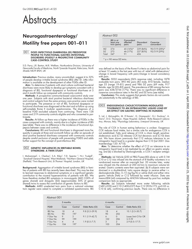
Abstract 001
FGID
3 months
Odds Ratio (CI)
6 months
Odds Ratio (CI)Case Control Case Control
IBS Yes 18 4 9.8 (3.2–29.9) 18 4 10.1 (3.3–30.7)IBS No 90 197 90 202F Diar Yes 9 2 9.1 (1.9–42.7) 6 0 Lower limit CI for OR
is 2.32F Diar No 99 199 102 206F Dys Yes 4 4 1.9 (0.5–7.7) 3 2 2.9 (0.5–17.7)F Dys No 104 197 105 204
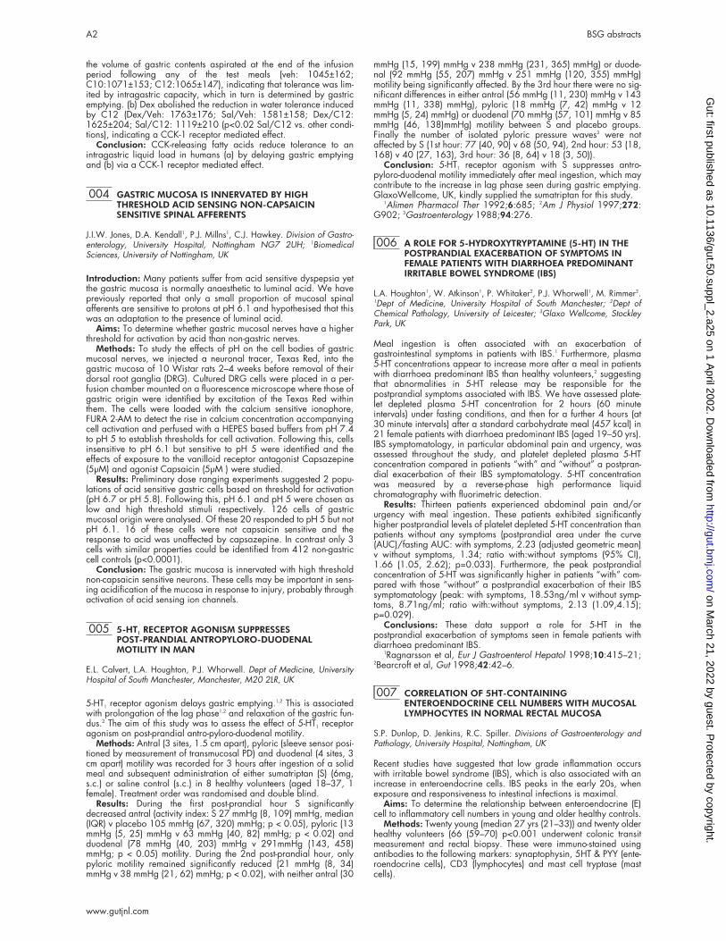
Abstract 002
IBS in atleast onetwin (pairs)
Concordantaffectedpairs
Discordantpairs
Casewiseconcordant
MZ 274 45 (16%)* 229 (84%) 90/319 (28%)*DZ 273 46 (17%) 227 (83%) 92/319 (29%)Total 547 91 456
* p = NS versus DZ twins.
Gut 2002;50(Suppl II):A1–A125 A1
www.gutjnl.com
on March 21, 2022 by guest. P
rotected by copyright.http://gut.bm
j.com/
Gut: first published as 10.1136/gut.50.suppl_2.a25 on 1 A
pril 2002. Dow
nloaded from
the volume of gastric contents aspirated at the end of the infusionperiod following any of the test meals (veh: 1045±162;C10:1071±153; C12:1065±147), indicating that tolerance was lim-ited by intragastric capacity, which in turn is determined by gastricemptying. (b) Dex abolished the reduction in water tolerance inducedby C12 (Dex/Veh: 1763±176; Sal/Veh: 1581±158; Dex/C12:1625±204; Sal/C12: 1119±210 (p<0.02 Sal/C12 vs. other condi-tions), indicating a CCK-1 receptor mediated effect.
Conclusion: CCK-releasing fatty acids reduce tolerance to anintragastric liquid load in humans (a) by delaying gastric emptyingand (b) via a CCK-1 receptor mediated effect.
004 GASTRIC MUCOSA IS INNERVATED BY HIGHTHRESHOLD ACID SENSING NON-CAPSAICINSENSITIVE SPINAL AFFERENTS
J.I.W. Jones, D.A. Kendall1, P.J. Millns1, C.J. Hawkey. Division of Gastro-enterology, University Hospital, Nottingham NG7 2UH; 1BiomedicalSciences, University of Nottingham, UK
Introduction: Many patients suffer from acid sensitive dyspepsia yetthe gastric mucosa is normally anaesthetic to luminal acid. We havepreviously reported that only a small proportion of mucosal spinalafferents are sensitive to protons at pH 6.1 and hypothesised that thiswas an adaptation to the presence of luminal acid.
Aims: To determine whether gastric mucosal nerves have a higherthreshold for activation by acid than non-gastric nerves.
Methods: To study the effects of pH on the cell bodies of gastricmucosal nerves, we injected a neuronal tracer, Texas Red, into thegastric mucosa of 10 Wistar rats 2–4 weeks before removal of theirdorsal root ganglia (DRG). Cultured DRG cells were placed in a per-fusion chamber mounted on a fluorescence microscope where those ofgastric origin were identified by excitation of the Texas Red withinthem. The cells were loaded with the calcium sensitive ionophore,FURA 2-AM to detect the rise in calcium concentration accompanyingcell activation and perfused with a HEPES based buffers from pH 7.4to pH 5 to establish thresholds for cell activation. Following this, cellsinsensitive to pH 6.1 but sensitive to pH 5 were identified and theeffects of exposure to the vanilloid receptor antagonist Capsazepine(5µM) and agonist Capsaicin (5µM ) were studied.
Results: Preliminary dose ranging experiments suggested 2 popu-lations of acid sensitive gastric cells based on threshold for activation(pH 6.7 or pH 5.8). Following this, pH 6.1 and pH 5 were chosen aslow and high threshold stimuli respectively. 126 cells of gastricmucosal origin were analysed. Of these 20 responded to pH 5 but notpH 6.1. 16 of these cells were not capsaicin sensitive and theresponse to acid was unaffected by capsazepine. In contrast only 3cells with similar properties could be identified from 412 non-gastriccell controls (p<0.0001).
Conclusion: The gastric mucosa is innervated with high thresholdnon-capsaicin sensitive neurons. These cells may be important in sens-ing acidification of the mucosa in response to injury, probably throughactivation of acid sensing ion channels.
005 5-HT1 RECEPTOR AGONISM SUPPRESSESPOST-PRANDIAL ANTROPYLORO-DUODENALMOTILITY IN MAN
E.L. Calvert, L.A. Houghton, P.J. Whorwell. Dept of Medicine, UniversityHospital of South Manchester, Manchester, M20 2LR, UK
5-HT1 receptor agonism delays gastric emptying.1,2 This is associatedwith prolongation of the lag phase1,2 and relaxation of the gastric fun-dus.2 The aim of this study was to assess the effect of 5-HT1 receptoragonism on post-prandial antro-pyloro-duodenal motility.
Methods: Antral (3 sites, 1.5 cm apart), pyloric (sleeve sensor posi-tioned by measurement of transmucosal PD) and duodenal (4 sites, 3cm apart) motility was recorded for 3 hours after ingestion of a solidmeal and subsequent administration of either sumatriptan (S) (6mg,s.c.) or saline control (s.c.) in 8 healthy volunteers (aged 18–37, 1female). Treatment order was randomised and double blind.
Results: During the first post-prandial hour S significantlydecreased antral (activity index: S 27 mmHg (8, 109) mmHg, median(IQR) v placebo 105 mmHg (67, 320) mmHg; p < 0.05), pyloric (13mmHg (5, 25) mmHg v 63 mmHg (40, 82) mmHg; p < 0.02) andduodenal (78 mmHg (40, 203) mmHg v 291mmHg (143, 458)mmHg; p < 0.05) motility. During the 2nd post-prandial hour, onlypyloric motility remained significantly reduced (21 mmHg (8, 34)mmHg v 38 mmHg (21, 62) mmHg; p < 0.02), with neither antral (30
mmHg (15, 199) mmHg v 238 mmHg (231, 365) mmHg) or duode-nal (92 mmHg (55, 207) mmHg v 251 mmHg (120, 355) mmHg)motility being significantly affected. By the 3rd hour there were no sig-nificant differences in either antral (56 mmHg (11, 230) mmHg v 143mmHg (11, 338) mmHg), pyloric (18 mmHg (7, 42) mmHg v 12mmHg (5, 24) mmHg) or duodenal (70 mmHg (57, 101) mmHg v 85mmHg (46, 138)mmHg) motility between S and placebo groups.Finally the number of isolated pyloric pressure waves3 were notaffected by S (1st hour: 77 (40, 90) v 68 (50, 94), 2nd hour: 53 (18,168) v 40 (27, 163), 3rd hour: 36 (8, 64) v 18 (3, 50)).
Conclusion: 5-HT1 receptor agonism with S suppresses antro-pyloro-duodenal motility immediately after meal ingestion, which maycontribute to the increase in lag phase seen during gastric emptying.GlaxoWellcome, UK, kindly supplied the sumatriptan for this study.
1Alimen Pharmacol Ther 1992;6:685; 2Am J Physiol 1997;272:G902; 3Gastroenterology 1988;94:276.
006 A ROLE FOR 5-HYDROXYTRYPTAMINE (5-HT) IN THEPOSTPRANDIAL EXACERBATION OF SYMPTOMS INFEMALE PATIENTS WITH DIARRHOEA PREDOMINANTIRRITABLE BOWEL SYNDROME (IBS)
L.A. Houghton1, W. Atkinson1, P. Whitaker2, P.J. Whorwell1, M. Rimmer3.1Dept of Medicine, University Hospital of South Manchester; 2Dept ofChemical Pathology, University of Leicester; 3Glaxo Wellcome, StockleyPark, UK
Meal ingestion is often associated with an exacerbation ofgastrointestinal symptoms in patients with IBS.1 Furthermore, plasma5-HT concentrations appear to increase more after a meal in patientswith diarrhoea predominant IBS than healthy volunteers,2 suggestingthat abnormalities in 5-HT release may be responsible for thepostprandial symptoms associated with IBS. We have assessed plate-let depleted plasma 5-HT concentration for 2 hours (60 minuteintervals) under fasting conditions, and then for a further 4 hours (at30 minute intervals) after a standard carbohydrate meal (457 kcal) in21 female patients with diarrhoea predominant IBS (aged 19–50 yrs).IBS symptomatology, in particular abdominal pain and urgency, wasassessed throughout the study, and platelet depleted plasma 5-HTconcentration compared in patients “with” and “without” a postpran-dial exacerbation of their IBS symptomatology. 5-HT concentrationwas measured by a reverse-phase high performance liquidchromatography with fluorimetric detection.
Results: Thirteen patients experienced abdominal pain and/orurgency with meal ingestion. These patients exhibited significantlyhigher postprandial levels of platelet depleted 5-HT concentration thanpatients without any symptoms (postprandial area under the curve(AUC)/fasting AUC: with symptoms, 2.23 (adjusted geometric mean)v without symptoms, 1.34; ratio with:without symptoms (95% CI),1.66 (1.05, 2.62); p=0.033). Furthermore, the peak postprandialconcentration of 5-HT was significantly higher in patients “with” com-pared with those “without” a postprandial exacerbation of their IBSsymptomatology (peak: with symptoms, 18.53ng/ml v without symp-toms, 8.71ng/ml; ratio with:without symptoms, 2.13 (1.09,4.15);p=0.029).
Conclusions: These data support a role for 5-HT in thepostprandial exacerbation of symptoms seen in female patients withdiarrhoea predominant IBS.
1Ragnarsson et al, Eur J Gastroenterol Hepatol 1998;10:415–21;2Bearcroft et al, Gut 1998;42:42–6.
007 CORRELATION OF 5HT-CONTAININGENTEROENDOCRINE CELL NUMBERS WITH MUCOSALLYMPHOCYTES IN NORMAL RECTAL MUCOSA
S.P. Dunlop, D. Jenkins, R.C. Spiller. Divisions of Gastroenterology andPathology, University Hospital, Nottingham, UK
Recent studies have suggested that low grade inflammation occurswith irritable bowel syndrome (IBS), which is also associated with anincrease in enteroendocrine cells. IBS peaks in the early 20s, whenexposure and responsiveness to intestinal infections is maximal.
Aims: To determine the relationship between enteroendocrine (E)cell to inflammatory cell numbers in young and older healthy controls.
Methods: Twenty young (median 27 yrs (21–33)) and twenty olderhealthy volunteers (66 (59–70) p<0.001 underwent colonic transitmeasurement and rectal biopsy. These were immuno-stained usingantibodies to the following markers: synaptophysin, 5HT & PYY (ente-roendocrine cells), CD3 (lymphocytes) and mast cell tryptase (mastcells).
A2 BSG abstracts
www.gutjnl.com
on March 21, 2022 by guest. P
rotected by copyright.http://gut.bm
j.com/
Gut: first published as 10.1136/gut.50.suppl_2.a25 on 1 A
pril 2002. Dow
nloaded from
Results: 5HT-containing E cell numbers correlated with CD3+lamina propria lymphocyte counts, R2=0.8861, p=0.009. There wasa reduction in synaptophysin+ E cells (p=0.007), CD3 lamina proprialymphocytes (p<0.02), crypt intraepithelial lymphocytes (p<0.02) andmast cells (p=0.02) in the older group. Median colonic transit was28.8 (13.8–43.5) and 45.6 (23.4–57) for the old and young respec-tively (p=NS) with no correlation to E or lymphocyte count.
Conclusions: In health lymphocyte numbers correlate with E cellnumbers. Age-related decline in inflammatory cells may account forreduced E cell numbers in the older age group.
008 THE ROLE OF ANTICIPATION IN THE BRAINPROCESSING OF HUMAN VISCERAL PAIN
L.J. Gregory1,2, L. Yaguez2, S.J. Coen1, E. Amaro2, S. Smale3,A.R. Hobson1, S.C.R. Williams2, D.G. Thompson1, Q. Aziz1. 1GI Sciences,University of Manchester; 2Institute of Psychiatry, London; 3King’s CollegeHospital, London, UK
Introduction: Psychophysiologists have demonstrated that learnedautonomic responses can be produced in the gastrointestinal (GI) tractto external stimuli. By employing classical conditioning techniques, theformation of a learned association between a previously unrelatedstimulus (conditioned stimulus, CS) and a biologically relevant stimulus(unconditioned stimulus, UCS) can be maximised in order tointerrogate the effect that anticipation has on the cortical processing ofoesophageal pain.
Methods: Six healthy volunteers (5 male) with a mean age of 22years (age range 20–26 years) participated in the study.
Oesophageal stimulation: a standard manometry catheter with asilicone balloon attached was passed trans-nasally into the distaloesophagus.
Protocol: Comprised of three contiguous phases. (1) Learningphase: Presentation of 20 trials of a blue coloured circle (CS) pairedwith a phasic, painful oesophageal distension (UCS). (2) Anticipation:Randomised presentation 10 trials of CS alone, and 10 trials of CS +UCS. (3) Extinction: Presentation of 20 trials of CS alone. Behaviouraldata measuring subjective perception of stimulus was acquired preand post acquisition using visual analogue scales.
Magnetic Resonance Imaging: Non-contiguous axial slices wereacquired using a 1.5 Tesla system and an event related design.
Results: During the learning phase anterior cingulate cortex, bilat-eral insula, thalamus, left cerebellum, inferior frontal cortex, periaque-ductal grey and secondary sensory cortex were activated. Theseregions were also activated in the anticipation and the extinctionphases with the exception of the periaqueductal grey matter and withadditional activation in the right dorsolatereral prefrontal cortex(DLPFC).
Conclusions: Anticipation of painful visceral stimuli results in acti-vation of cerebral regions normally associated with processing painfulsensory information.We therefore demonstrate that the cognitive-evaluative component of the pain matrix significantly contributes to thecentral processing of visceral pain.
009 VALUE OF A DIETITIAN-LED CLINIC IN THEMANAGEMENT OF YOUNG PATIENTS WITH IRRITABLEBOWEL SYNDROME (IBS)
V.A. Chudleigh, E. Brennan, S.A. Tarry, S. Myers, S.J. Lewis, J.O. Hunter.Department of Gastroenterology, Addenbrooke’s Hospital, CambridgeCB2 2QQ, UK
Introduction: Irritable bowel syndrome (IBS) entails a heavy clinicalload for gastroenterologists. It may often successfully be treated bydiet. In order to reduce medical outpatient attendances we haveestablished a dietitian-led IBS clinic (DLC).
Methods: Patients aged 16–45 were selected by review of GPreferral letters by consultants, and randomised to DLC or standardmedical appointments (MOPD). Those fulfilling the Rome criteria, withno history of rectal bleeding, chronic medication, or psychiatric illness,were eligible for DLC if screening before their clinic visit revealed therewas no evidence of an anxiety state using a validated questionnaire,stool culture was negative and haematological and biochemical mark-ers including C reactive protein and gliadin antibodies normal. Physi-cians who saw the patients randomised to MOPD were allowed toinvestigate them as appeared clinically indicated.
Results: Of 58 patients randomised to DLC, 15 were excluded (11because of an anxiety state), but 43 fulfilled admission criteria. 7failed to keep the first appointment, so that 36 followed a
standardised dietary protocol. In 22, (61%) symptoms weresuccessfully relieved. 47 patients were randomised to MOPD. Only 1received a full IBS screen, and 23 unnecessary investigations wereperformed, including colonoscopies and barium x-rays. 17 werereferred for dietary treatment and 12 accepted, of which 42%obtained symptomatic relief.
Conclusion: DLC provides an effective way of screening and treat-ing young patients with IBS whose results compare favourably withthose obtained when these patients are referred to MOPD.
010 THE IMPACT OF GUT DIRECTED HYPNOTHERAPY UPONHEALTH RELATED QUALITY OF LIFE IN PATIENTSSUFFERING FROM IRRITABLE BOWEL SYNDROME
G.D. Smith, K.R. Palmer. Gastrointestinal Unit, Western General Hospital,Edinburgh, UK
Introduction: Health related quality of life (HRQoL) is impaired inpatients suffering from irritable bowel syndrome (IBS), but measure-ment of this remains poorly quantified. The treatment of severe IBS isoften unsuccessful, although gut directed hypnotherapy has beenshown to improve IBS symptoms but its effect upon HRQoL status hasnot been defined.
Aim: In this study we have defined the impact of gut directed hyp-notherapy upon HRQoL status in IBS patients.
Methods: Seventy five patients (55 females; median age 37.1)with a diagnosis of IBS (consistent with Rome II diagnostic criteria)underwent gut directed hypnotherapy. The predominant symptomswere abdominal pain in 46 patients (61%), altered bowel habit in 24(32.5%) and abdominal bloating in 5 (6.5%). Physical symptomswere prospectively recorded using seven day diary cards. Outcomemeasures were Hospital Anxiety and Depression Scales (HAD-A &HAD-D) and a IBS disease specific quality of life tool (IBSQoL). Meas-urements were taken at baseline (pre-treatment ) and at three months(post-treatment). Pre and post treatment scores were coded and com-pared using Wilcoxon signed ranks test.
Results: There were statistical improvements (p<0.001) in alldomains of the IBSQoL (emotional health, mental health, physicalhealth, sleep, energy, diet, social role and physical role) aftertreatment. Improvements were most marked in female patients,particularly those with predominant abdominal pain. Significantimprovements were seen for both males and females for anxiety and(HAD-A p<0.001; HAD-D p<0.05).
Summary/Conclusion: Gut directed hypnotherapy has a verypositive impact upon psychological well being and HRQoL in IBS. Thisappears most effective in patients with a predominant symptom ofabdominal pain and bloating. A randomised controlled study of hyp-notherapy is recommended in IBS.
011 ATTITUDES OF GENERAL PRACTITIONERS ANDHOSPITAL SPECIALISTS TO FUNCTIONALGASTROINTESTINAL DISORDERS
L.M. Gladman, D.A. Gorard. Wycombe Hospital, High Wycombe, BucksHP11 2TT, UK
Patients with functional gastrointestinal (GI) disorders in primary carediffer from those seen in hospital clinics. General practitioners (GPs)and hospital specialists may have different views of functionaldisorders.
A questionnaire asking about understanding of functional GI disor-ders was sent to a random sample of 200 UK GPs, and a randomsample of 200 clinician members of the British Society of Gastroenter-ology (consultants). Non-responders were sent reminders after 1month.
137 (69%) GPs and 167 (84%) consultants replied. Not allanswered all questions. 62 GPs believed functional GI symptoms torepresent a “real” currently unexplained GI disorder; 67 believed thesymptoms to have a psychosomatic basis, probably somatisation of apsychological illness. One GP believed such symptoms wereimaginary. In contrast most, 120, consultants believed functional GIsymptoms represent a real GI disorder with 36 perceiving them tohave a psychological basis, χ2 = 26.7, p<0.001. However GPs andconsultants had similar perceptions about the prevalence ofpsychological illness in their functional GI patients. A fifth of eachgroup believed psychological disturbance to be present in <15%patients, a third believed it to be present in 15–30%, and the restbelieved it to occur in >30% patients. More consultants believedunderstanding of functional GI disorders has improved in the last 20
BSG abstracts A3
www.gutjnl.com
on March 21, 2022 by guest. P
rotected by copyright.http://gut.bm
j.com/
Gut: first published as 10.1136/gut.50.suppl_2.a25 on 1 A
pril 2002. Dow
nloaded from
years (101 responding positively, 65 negatively) than GPs (66positive, 69 negative), χ2 = 4.31, p<0.05. Most consultants, 115, andmost GPs, 81, thought that treatment for functional GI disorders hadnot improved in the past 20 years.
Only 29 of 137 GP respondents had heard of the Manning criteriafor diagnosing IBS, compared to 134 of 166 consultants, χ2 = 107,p<0.0001. Only 16 of 137 GPs had heard of the Rome criteria fordiagnosing functional GI disorders, compared to 139 of 167 consult-ants, χ2 = 154, p<0.0001. Despite greater awareness among consult-ants, 60 did and 104 did not use Manning criteria, 67 did and 100did not use Rome criteria. Of 123 responding GPs, only 14 use Man-ning and 4 use Rome criteria.
GPs and consultants have some differing views on functional GI dis-orders, but in both primary and secondary care most doctors do notuse Manning or Rome criteria to diagnose functional GI disorders.
Oesophageal free papers012–025
012 OESOPHAGEAL CANCER AND CACHEXIA: THE EFFECTSOF THALIDOMIDE ON WEIGHT LOSS AND LEAN BODYMASS IN A SEQUENTIAL (METABOLIC) STUDY
Z.H. Khan1, E. Simpson2, A.T. Cole1, I. Macdonald2, D. Pye2, A. Austin1,J.G. Freeman1. 1Department of Gastroenterology, Derby City Hospital,Derby, UK; 2University Department of Physiology and Medical Physics,Queens Medical Centre, Nottingham, UK
Aim: To investigate the potential for using thalidomide as ananti-cachectic agent in patients with advanced oesophageal cancerby studying its effect on body composition and weight.
Methods: 11 patients with non-obstructing and in-operableoesophageal cancer were included in the study.
Study protocol: Patients were established on an isocaloric diet overa 10-day period. Body weight, body composition studies with DEXAscanning, REE (resting energy expenditure) and serum levels of insulin,thyroxine, catecholamines and cortisol were measured at the entryand then after two weeks on diet alone. Patients were then started onthalidomide for 2 weeks and the measurements were repeated. Qual-ity of life (QOL) was similarly measured as a secondary end point.
Results: Ten patients completed the study protocol. The averagecaloric intake remained the same throughout the study period in allthese patients. 9/10 (95% CI 0.60, 0.98) lost weight on diet alone.The mean gain on thalidomide in the following two weeks was 1.29kg (median 1.25kg). A similar trend was shown in lean body mass.There were missing data for one patient, so nine were analysed. 8/9(95% CI 0.57, 0.98) initially lost mass on diet alone. The mean gainon thalidomide in the following two weeks was 1.75 kg (median 1.33kg). The mean change in REE was 1.75 (95% CI –0.42, 3.91) on tha-lidomide. Amongst hormonal assay, changes in catecholaminesapproached statistical significance.The mean change in catecho-lamines on thalidomide was −0.71 (95% CI −1.60, 0.02) .
Conclusions: In this sequential study of patients with progressiveinoperable cancer, thalidomide treatment appeared to reverse loss ofweight and lean body mass over the two week trial period. Howeverto establish its role as an anti-cachectic treatment a full placebo-controlled trial is warranted.
013 A 5-YEAR, DOUBLE-BLIND, RANDOMISED COMPARISONOF RABEPRAZOLE AND OMEPRAZOLE IN GORDMAINTENANCE TREATMENT: EFFICACY RESULTS
B. Thjodleifsson1, A. Morocutti2, K.D. Bardhan3. 1University Hospital, Rey-kjavik, Iceland; 2Eisai Ltd, London, UK; 3Rotherham General Hospital,Rotherham, UK
Background: Many studies have found proton-pump inhibitors to beeffective and safe in preventing relapse of gastro-oesophageal refluxdisease (GORD) over a period of several months to a year. There is,however, little evidence from randomised trials about their long-termsafety and efficacy.
Objectives: To compare the efficacy and safety of rabeprazoleand omeprazole in the prevention of relapse in patients with healedgastro-oesophageal reflux disease during 5 years of treatment.
Methods: Patients were eligible for the study if they had previouslybeen diagnosed with GORD, which had healed as shown by endos-copy. Patients received randomised, double-blind treatment with rab-eprazole (10 mg or 20 mg) or omeprazole (20 mg) once daily for upto 5 years. The main outcome measure was endoscopically confirmedGORD relapse (Hetzel–Dent score = 2). Endoscopy was done after13, 26, and 52 weeks, and yearly thereafter, or if symptomssuggested GORD relapse.
Results: 243 patients entered the study, of whom 123 completedall 5 years of treatment. Relapse rates were 9/78 (11.5%) in the 20mg rabeprazole group, 8/82 (9.9%) in the 10 mg rabeprazolegroup, and 11/83 (13.3%) in the 20mg omeprazole group. The dif-ferences in relapse rates were not statistically significant. All threetreatments were safe and well tolerated.
Conclusions: Rabeprazole at a daily dose of 10 mg is as effectiveas rabeprazole 20 mg or omeprazole 20 mg in preventing relapse ofGORD over 5 years of treatment.
014 OESOPHAGEAL MANOMETRY AND PH STUDIESCHANGE THE MANAGEMENT AND OUTCOME OFPATIENTS WITH NON-CARDIAC CHEST PAIN
Y.H. Oo, D. Foy, H. Allen, P.J. Winwood. Department of Gastroenterol-ogy, Royal Bournemouth Hospital, Bournemouth, UK
Background: Oesophageal disease is a well-recognized cause ofnon-cardiac chest pain (NCCP). The role of Oesophageal Manometry(OM) and pH studies remain unclear, particularly in changingoutcome.
Aim: To assess whether Oesophageal Manometry and pH studiesaffect the management and outcome of NCCP patients in a districthospital.
Methods: Retrospective study of patients with NCCP with repeatedadmissions to hospital (Negative ETT, normal Coronary Angiogram ornormal Thallium scan) who were further investigated with OM and pHstudies between November 1998 and May 2001 (2.5 years/60patients). Diffuse Oesophageal Spasm (DOS), Nutcracker oesoph-agus and Achalasia, as defined by Spechler and Castell (Gut2001;49:145–51), were the only motility disorders recognized ascauses of NCCP in this study.
Results: All patients had normal endoscopy or barium swallows.17 (28%) patients had significant reflux disease, 14 (23%) had DOSand 6 (10%) had nutcracker oesophagus (of whom 50% also hadreflux). Normal studies were found in 25%. 5 patients hadnon-specific oesophageal dysmotility and 2 patients had hypomotility.All patients with significant reflux disease were treated with PPI and 3patients had anti-reflux surgery. 90% of patients with nutcrackerOesophagus and DOS were treated with Nitrates or calcium blockerswith/without PPI. 37% of patients had reflux symptoms and predictivevalues for significant reflux were 64% (positive), and 92% (negative).22% of patients had dysphagia. Predictive values for significant dys-motility were 69% (positive) and 72% (negative). Management waschanged in 67% (40 patients) who had OM and pH studies. Thenature of the diagnosis was carefully explained in all patients withpositive studies. Only one (1.6%) has been readmitted and one(1.6%) had further cardiac investigations (mean follow-up 1.5years).
Conclusions: A positive diagnosis of oesophageal dysmotility orreflux changed the management, reduced readmission rates and theneed for further cardiac investigations. The presence or absence of GIsymptoms has a high predictive value for OM and pH abnormalitiesin NCCP.
015 OESOPHAGEAL MOTOR FUNCTION ANDGASTRO-OESOPHAGEAL REFLUX IN VENTILATEDNEONATES
D. Kufeji, A. Anggiansah1, W.J. Owen1, E.H. Dykes. Department of Paedi-atric Surgery University Hospital Lewisham, London; 1OesophagealLaboratory, St Thomas’ Hospital, London, UK
Introduction: Sick neonates often require ventilation for prolongedperiods of time. Gastro-oesophageal reflux (GOR) is very common innewborn infants, particularly those who are preterm. This can lead tosignificant morbidity and in extreme cases the neonate can only besuccessfully weaned off the ventilator after anti-reflux surgery.
Aim: To evaluate oesophageal motor function and acid clearancemechanisms in ventilated neonates.
Methods: Combined pressure and pH monitoring was undertakenin 10 neonates requiring assisted ventilation using Dentsleeve
A4 BSG abstracts
www.gutjnl.com
on March 21, 2022 by guest. P
rotected by copyright.http://gut.bm
j.com/
Gut: first published as 10.1136/gut.50.suppl_2.a25 on 1 A
pril 2002. Dow
nloaded from
micromanometric assembly and a paediatric (1.5mm diameter)antimony pH sensor. Study repeated when baby was off the ventilator.
Results: Mean gestational age = 33 weeks and mean birth weight1510 grams (range 28–36 weeks). Mean duration of recording = 58minutes. LOS pressure = 20 mmHg off ventilation and 40.6 mmHgduring positive pressure ventilation. A total of 683 pressure wavesequences were recorded. There were 4 major patterns normalperistalsis (69.8%, of which 16.5% low amplitude), reverse peristalsis3.6%, synchronous activity 3.2%, non transmitted activity 21.7%.Eleven waveforms (1.6%) could not be adequately categorised. Refluxepisodes (pH drop > 0.5 for 10seconds) = 50 with a mean refluxduration of 22 seconds. An average of 2 normal swallows wererequired to return pH to pre reflux levels.
Conclusion: Ventilated neonates seem to have high oesophagealand LOS pressures that may protect them against reflux. However theyexhibit a large proportion of ineffective oesophageal motor activity.During periods of reflux the oesophagus was cleared efficiently byperistaltic oesophageal contractions.
Reference: Omari TI et al. J Pediatr Surg 1999;34:1795–8.
016 INTRAGASTRIC PH IN AMBULANT SUBJECTS AND ITSRELATIONS TO PHYSIOLOGICAL AND PATHOLOGICALREFLUX
R.P. Arasaradnam, L.F. Smith, S.A. Riley. Dept of Gastroenterology &Oesophageal Studies, Central Sheffield Teaching Hospitals, NorthernGeneral Hospital, Sheffield, UK
Background and Aims: Episodes of gastro-oesophageal reflux(GOR) are usually associated with a loss of lower oesophagealsphincter (LOS) pressure. However, on many occasions barrierpressure is lost yet reflux does not occur. This suggests that other fac-tors also influence the occurrence of reflux. The aim of this study wasto measure pH at the gastric cardia in ambulant subjects anddetermine its relations to physiological and pathological reflux.
Methods: 17 asymptomatic volunteers (9 males, aged 21–33years) and 17 patients (11 males, aged 33–53 years) with non-erosivereflux disease were studied. Standard station pull-through manometrywas performed to locate the LOS. Under ambulant conditions, pH wasmeasured at 5cm above and at 2 and 10cm below the LOS.
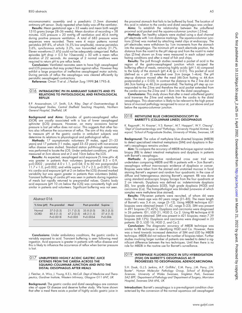
Results: As expected, oesophageal acid exposure (% time pH< 4)was greater in patients than volunteers (pre-prandial 8.5 v 0.9,p<0.0002 ; prandial 4.0 v 1.1, p<0.04; 0 to 60 min post-prandial11.7 v 1.0, p<0.002; and while supine 13.7 v 2.3, p<0.001). Gas-tric cardia acid exposure (pH at 2 cm below the LOS) showed markedvariability but was again greater in patients than volunteers (table).Transient buffering of cardia pH was seen in patients during ingestionof meals but rapidly returned to pre-prandial values. Gastric bodyacid exposure (pH 10 cm below the LOS) was consistently high andsimilar in patients and volunteers. Significant buffering was not seen.
Conclusions: Under ambulatory conditions, the gastric cardia isvariably exposed to acid. Transient buffering is seen following mealingestion. Acid exposure is greater in patients with reflux disease andthis is likely to influence the occurrence of reflux when barrier pressureis lost.
017 UNBUFFERED HIGHLY ACIDIC GASTRIC JUICEEXTENDS FROM THE CARDIA ACROSS THESQUAMO-COLUMNAR JUNCTION AND INTO THEDISTAL OESOPHAGUS AFTER MEALS
J. Fletcher, A. Wirz, J. Young, K.E.L. McColl. Dept of Medicine and Thera-peutics, Gardiner Institute, Western Infirmary, Glasgow G11 6NT, UK
Background: The gastric cardia and distal oesophagus are commonsites of upper GI disease and deserve further study. We have shownthat after a meal there exists a pocket of highly acidic gastric juice in
the proximal stomach that fails to be buffered by food. The location ofthis acid in relation to the cardia and distal oesophagus was unclear.
Aims: To establish the relationship between the unbufferedproximal acid pocket and the squamo-columnar junction ( Z-line).
Methods: Ten healthy subjects were studied using a dual channelpH electrode with 1cm distance markings. The squamo-columnar junc-tions (Z-line) was marked by attaching metal clips at endoscopy. ThepH electrodes were withdrawn by 1cm increments from the stomachinto the oesophagus. The minimum pH at each electrode position, thedistance from the nostril to the pH step-up and from the nostril to metalclips (Z-line) shown on X-ray were measured in each subject underfasting conditions and after a meal of fish and chips.
Results: The pull through studies revealed a pocket of acid in theregion of the gastro-oesophageal junction which escaped thebuffering effect of meals, remaining highly acidic (pH 1.6) comparedto the body of the stomach (pH 4.4) (p < 0.01). This pocket of acid(defined as < pH 2) extended over 2cm (range 1–4cm). The pHstep-up distance moved after the meal (46.0cm fasting vs 44.4cmpostprandial p < 0.05). In contrast the distance to the Z line did not(46.3cm fasting vs 46.2cm postprandial). The fasting pH step up cor-responded to the Z-line and therefore the acid pocket extended fromthe cardia across the Z-line and 1.8cm into the distal oesophagus.
Conclusions: This study shows that after a meal unbuffered gastricacid traverses the Z-line and extends from the cardia to the distaloesophagus. This observation is likely to be relevant to the high preva-lence of mucosal pathology recognised to occur at, just above and justbelow the squamo-columnar junction.
018 METHYLENE BLUE CHROMOENDOSCOPY INBARRETT’S (COLUMNAR LINED) OESOPHAGUS
K. Ragunath1, N. Krasner1, V.S. Raman1, M.T. Haqqani2, W.Y. Cheung3.1Dept of Gastroenterology and 2Pathology, University Hospital Aintree, Liv-erpool; 3School of Postgraduate Studies, University of Wales, Swansea, UK
Background: The value of methylene blue directed biopsies (MBDB)to detect specialised intestinal metaplasia (SIM) and dysplasia in Bar-rett’s oesophagus remains unclear.
Aim: To compare the accuracy of MBDB technique against randombiopsy (RB) to detect intestinal metaplasia and dysplasia in patientswith Barrett’s oesophagus.
Methods: A prospective randomised cross over trial wasundertaken comparing MBDB and RB in patients with = 3cm Barrett’soesophagus without macroscopic evidence of dysplasia or cancer.Biopsies were taken from the stained and unstained mucosa in focalstaining Barrett’s segment and random four quadrantic in the case ofdiffuse and heterogeneous staining Barrett’s segment. RB was doneusing standard endoscopic biopsy forceps from the four quadrants at2 cm intervals. Dysplasia was defined as: indeterminate dysplasia(ID), low grade dysplasia (LGD), high grade dysplasia (HGD) andcarcinoma (Ca). The histopathologist was blinded (unaware of whichsamples were methylene blue stained).
Results: Fifty-seven patients were recruited, of whom 44 weremale. The mean age was 60 years range (31–85). The mean lengthof Barrett’s was 5.4 cm, range (3–12). Using MBDB technique 651biopsies were obtained (mean 11.42, range 5–23). SIM was presentin 491 biopsies (75.42%). Dysplasia and carcinoma were diagnosedin 26 patients: ID1, LGD 21, HGD 2, Ca 2. Using RB technique 618biopsies were obtained. SIM was present in 421 biopsies; mean 7.39biopsies (68.12%). Dysplasia and carcinoma were diagnosed in 23patients: ID 3, LGD 16, HGD 2, and Ca 2.
Conclusion: The diagnostic accuracy of MBDB technique wassimilar to RB technique in identifying HGD and Ca. However, therewas a trend towards increased detection of SIM and LGD by MBDBtechnique. MBDB did not reduce the number of biopsies taken. Furtherstudies involving larger number of patients are needed to detect a sig-nificant difference between the two techniques. Until then there is norole for MBDB in the routine use for Barrett’s surveillance.
019 INTERPHASE FLUORESCENCE IN SITU HYBRIDISATION(FISH) ON BARRETT’S OESOPHAGUS AS ITPROGRESSES TO OESOPHAGEAL ADENOCARCINOMA
S.H. Doak, G.J.S. Jenkins, A.P. Griffiths1, E.M. Parry, J.M. Parry, J.N.Baxter2. Human Molecular Pathology Group, School of BiologicalSciences, University of Wales Swansea, Singleton Park, SwanseaSA2 8PP; 1Department of Pathology and 2Department of Surgery, MorristonHospital, Swansea SA6 6NL, UK
Introduction: Barrett’s oesophagus is a pre-malignant condition char-acterised by the conversion of the normal squamous cell oesophageal
Abstract 016
% time (pH) Pre-prandial Meal Post-prandial Supine
Normal 37.0 (5.5) 21.7 (5.5) 35.8 (5.0) 38.3 (5.5)GORD 80.5 (1.5) 47.2 (3.5) 68.2 (1.5) 57.4 (1.5)
P<0.0018 P<0.005 P<0.0034 P<0.006
BSG abstracts A5
www.gutjnl.com
on March 21, 2022 by guest. P
rotected by copyright.http://gut.bm
j.com/
Gut: first published as 10.1136/gut.50.suppl_2.a25 on 1 A
pril 2002. Dow
nloaded from
epithelium to a mucosa comprised of columnar cells as a result ofchronic gastro-oesophageal reflux. This lesion progresses in astep-wise fashion through histologically identifiable stages andultimately develops into oesophageal adenocarcinoma in approxi-mately 10% of patients. To determine when specific genetic alterationsarise during this neoplastic progression FISH was employed.
Methods: Gastroscope cytology brushes were used to exfoliateepithelial cells from patients at each stage of progression (Barrett’smetaplasia to oesophageal adenocarcinoma). Interphase cell prepa-rations were generated and subsequently analysed by application offluorescently labelled centromeric probes for chromosomes 4, 8, 9, 20& Y and locus specific probes for the p53, p16 & Rb genes.
Results: Increased copy numbers of chromosomes 4 & 8 occurredin 13/15 & 10/15 Barrett’s metaplastic samples respectively, thusrepresenting the most prominent and earliest alteration arising duringneoplastic progression. Loss of the p16 tumour suppressor gene alsoarises during metaplasia (4/15) and was found to precedechromosome 9 amplifications, but in contrast, p53 loss is a laterchange first appearing in HGD. Increasing loss of chromosome Yoccurs with progression.
Discussion: Aneuploidy is an early occurrence during the progres-sion of Barrett’s oesophagus with copy number increases ofchromosomes 4 & 8 present in the majority of metaplastic samples.HGD appears to be the stage at which most aberrations accumulate,thus this genetic instability may possibly account for the highproportion of these patients that progress to cancer.
020 CYTOKINES INDUCE PREFERENTIAL SQUAMOUSEPITHELIAL CELL REPAIR FOLLOWINGPHOTODYNAMIC THERAPY FOR PATIENTS WITHBARRETT’S OESOPHAGUS: AN IN VITRO MODEL
T.K.L. Wong1, L.B. Lovat1, P. Sirieix2, R.C. Fitzgerald2. 1National MedicalLaser Centre, Department of Surgery, Royal Free and University CollegeSchool of Medicine, London; 2Cancer Cell Unit, Hutchison/MRC ResearchCentre, Cambridge CB2 2XZ, UK
Background: Photodynamic therapy (PDT) is an emerging endo-scopic treatment for patients with dysplasia in Barrett’s oesophagus.Application of PDT to Barrett’s oesophagus ideally leads to regenera-tion of non-dysplastic, stable squamous mucosa. A limitation of thistechnique is the persistence of Barrett’s epithelium, including buriedglands, which may still have dysplastic potential. Since the cellularmicroenvironment is crucial to epithelial repair it might be possible tomanipulate this to promote squamous epithelial re-growth.
Aims: To investigate (a) differences in early repair (restitution) of anoesophageal cell monolayer following mechanical or PDT injury; (b)whether restitution can be altered by adding growth factors/cytokines.
Methods: Cell lines; Squamous (OE21), Barrett’s (OE33) andco-cultures were injured mechanically or with PDT (5-aminolevulinicacid and blue light) using a novel applicator. Wounds were measuredover 24 hours and immunofluorescence for cytokeratins identifiedsquamous versus columnar cells. Transforming Growth Factor beta(TGF-β1), Hepatocyte Growth Factor (HGF), Interleukin 8 (IL-8) andKeratinocyte Growth Factor (KGF) were added individually to assesstheir effect on restitution compared with serum free media.
Results: In co-culture, squamous cells (OE-21) underwent greaterrestitution than columnar cells (OE-33). In both mechanical wound andPDT assays of co-cultures, TGF-β1 increased cell repair by restitutioncompared with controls (p<0.05). This effect was not seen in individu-ally cultured cell lines. KGF and HGF stimulated restitution ofsquamous and co-culture cells after mechanical injury and also inhib-ited columnar cells significantly (p<0.05). IL-8 had no effect on cellrestitution.
Conclusions: Restitution, in the first 24 hours after PDT andmechanical injury in vitro, can be influenced by growth factors. It maybe possible to manipulate the microenvironment to favour squamousre-epithelialisation after PDT.
021 BARRETT’S SURVEILLANCE IS WORTHWHILE ANDDETECTS CURABLE CANCERS
D.M. Aldulaimi, M. Cox, C.U. Nwokolo, D.E. Loft. University HospitalsCoventry and Warwickshire NHS Trust, UK
Aim: To establish whether Barrett’s surveillance is worthwhile in termsof incident cancers and whether outcomes are favourable.
Method: A prospective non-randomised single centre Barrett’s sur-veillance program commencing 1/1/1992 through 1/4/2001 (100months). Oesophagectomy recommended for high grade dysplasia orcarcinoma.
Results: Of 23,725 endoscopies, 506 patients were diagnosed asBarrett’s oesophagus and 24 (5%) had carcinoma at diagnosis(prevalence cancers). 126 patients had at least one surveillanceendoscopy. 248 surveillance endoscopies were performed spanning338 patient years. 13 surveillance (incidence) cancers were detected.The surveillance cancers were all detected after at least one year ofsurveillance and no patient had dysphagia at diagnosis. In the preva-lence cancer group 12 of the 24 patients underwent oesophagec-tomy. Lymph nodes showed evidence of metastases in 10 of the 12resections. In the surveillance group 10 patients underwentoesophagectomy. All had carcinoma in the resection specimen.Lymph nodes showed evidence of metastases in 1of the 10 resections.3 patients in the surveillance cancer group did not have anoesophagectomy. 1 of these patients died. 1 patient in the prevalencecancer group (4% of group; 8% of those operated) and 7 patients inthe surveillance cancer group (54% of group; 70% of those operated)remain disease-free more than 2 years post-oesophagectomy. Assum-ing the 7 patients in the surveillance cancer group are cured and thatthe cost of endoscopy is £120, the cost per cancer cured is £4250.One curable cancer was detected per 48 patient years of surveillance(338/7).
Conclusion: 5% of Barrett’s patients undergoing endoscopy haveprevalent cancers. If surveillance is performed, 4% per year (13/338%) develop cancer and 2 % per year are cured of their cancers. Mostsurveillance cancers are operable and of those undergoing surgery70% are cured. Barrett’s surveillance is cost-effective compared withother cancer screening or surveillance initiatives.
022 SURVEILLANCE FOR BARRETT’S OESOPHAGUS:EXPERIENCE FROM A DISTRICT GENERAL HOSPITAL
J.A. Fallowfield, P.J. Winwood, N.A. Davies, M. Lesna. RoyalBournemouth Hospital, UK
Background: The incidence of adenocarcinoma of the oesophago-gastric junction is rising. Barrett’s oesophagus (BO) is considered apremalignant condition for this cancer. The effectiveness ofendoscopic cancer surveillance programmes is unproven and contro-versial.
Aims: To measure the incidence and outcome of adenocarcinomain a BO surveillance population over a 3 year period and to evaluatethe effectiveness of endoscopic screening in a DGH.
Methods: All patients with BO attending Royal Bournemouth Hos-pital Endoscopy Unit between 1998–2001 were included. Caseswere identified from the pathology computer database.
Results: We identified 299 patients with known BO in a biannualsurveillance programme, with a mean age of 65 years. In the 3 yearstudy period there were 34 BO-associated adenocarcinomasdetected. 7 (19%) were identified as a result of a surveillance endos-copy. There were no interval cancers in the surveillance group. 27(81%) were diagnosed de novo at index endoscopy. The mean age ofpatients with BO-adenocarcinomas was 69 years; 29/34 (85%) weremale. Cancer incidence per patient year of follow-up was 1 : 79. Allof the 7 BO-adenocarcinomas detected during endoscopy were earlystage (<T2, N0) and had oesophagectomy (5/7) or endoscopicmucosal resection (2/7). All have survived to date (range 9–28months). De novo BO-adenocarcinomas were generally moreadvanced at presentation. 17/27 were suitable only for palliativetherapy; 16/17 have died.
Conclusions: New oesophageal cancers were found duringsurveillance endoscopy at a higher rate compared with mostpublished studies. The reason for the high detection rate in this studymay be due to the advanced age of this surveillance population.Nevertheless, most adenocarcinomas occurred in patients without aprevious diagnosis of BO. There was a bias towards early stage can-cers in patients with BO under surveillance. The outcome in thesepatients has been favourable compared with BO-related cancersdiagnosed de novo at index endoscopy. Our experiences supportendoscopic surveillance in selected patients with BO.
023 PHOTODYNAMIC THERAPY TO ERADICATE DYSPLASIAAND EARLY CARCINOMA IN BARRETT’S OESOPHAGUS
N.F. Jamieson, A. Mosse, S.G. Bown, L.B. Lovat. National Medical LaserCentre, Department of Surgery, Royal Free & University College School ofMedicine, London, UK
Background: Photodynamic therapy (PDT) is a minimally-invasivealternative to oesophagectomy for high-grade dysplasia (HGD) orintramucosal adenocarcinoma (T1m AdCa) arising in Barrett’s colum-nar lined oesophagus. Initial reports using 5-amino laevulinic acid
A6 BSG abstracts
www.gutjnl.com
on March 21, 2022 by guest. P
rotected by copyright.http://gut.bm
j.com/
Gut: first published as 10.1136/gut.50.suppl_2.a25 on 1 A
pril 2002. Dow
nloaded from
(ALA) suggest that HGD can be eradicated in 80% of patients. Ouraim is to identify parameters associated with successful eradication ofdisease.
Methods: 15 previously untreated patients (13 M, 2 F) with HGD(11) or T1m AdCa (4) were treated over a 3-year period. Ethicalapproval was obtained. Patients were photosensitised with ALA60mg/kg (light activation 635 nm) and received 30 PDT sessions:median of 2 per patient (range 1–3). ALA light doses were500–1000J/cm diffuser fibre and treatments took approximately 40minutes per 4cm length of columnar mucosa. Two delivery deviceswere tested (16 and 25mm diameter). 3 patients were treatedadditionally with the photosensitiser meso-tetrahydroxyphenylchlorin(mTHPC, 0.15mg/kg).
Results: Disease (HGD 5, AdCa 3) was eradicated in 8 patients(53%): 6 patients (40%) with ALA alone, 2 with additional mTHPC.Median follow-up is 10 months (range 2–29) with no deaths and nooesophageal strictures. One patient developed new HGD 2 yearsafter successful treatment and is being re-treated. Benign glands “bur-ied” under neo-squamous epithelium were seen in 7/15 cases. Failureof treatment was associated with the length of Barrett’s segment(median in responders 6cm (4–8cm), non-responders 8cm (6–13cm),p=0.02) and with the presence of multi-focal disease (success in only2/8 versus 6/7 for unifocal disease, p=0.04). Age, sex, presence ofhiatus hernia, and size of delivery device did not appear to influenceoutcome.
Discussion: Using current treatment parameters, PDT with ALA fordysplasia and T1m carcinoma in Barrett’s oesophagus is effective inless than one half of patients. A long Barrett’s segment and multi-focaldisease are associated with a poor outcome. Techniques to achieve adeeper effect (such as adding an iron chelator to ALA or using a dif-ferent photosensitiser) may give better results.
024 USE OF ENDOCINCH© FOR THE MANAGEMENT OFGASTRO-OESOPHAGEAL REFLUX DISEASE
Q. Arfin1, Z. Mahmood1, B.P. McMahon3, P. Byrne1, J.V. Reynolds1,E. Murphy1, V. Trimble1, D.G. Weir2. 1St James’s Hospital; 2Trinity College;3AMNCH, Dublin, Ireland
A method has been developed whereby sutures can be placed via anendoscope just below the oesophago-gastric junction (OGJ) whosepurpose is to improve the function of the OGJ and thereby preventoesophageal reflux. The aim of this work is to assess the safety andefficiency of the BARD Endocinch© for the treatment of GORD. 20patients with symptoms of GORD were recruited; all were followed for6 months and 14 for 1 year. The inclusion criteria included a depend-ence on proton pump inhibitor (PPI) drugs to control their reflux symp-toms, and a documented oesophageal acid reflux. Exclusion criteriawere age less then 18 years, pregnancy, dysphagia, obesity (BMI) >40, previous upper intestinal surgery and an hiatus hernia > 2 cms.Pre-procedure assessment included symptom scoring, oesophagealendoscopy, manometry and 24 hour oesophageal pH, and completedquality of life (QOL) questionnaire. Post procedure symptomatology,QOL and adverse events were assessed at 1, 3, 6 and 12 months.Repeat endoscopy, manometry and 24 hour pH were performed at 3months. Mean age was 37 (22 – 58 yrs.). All received conscioussedation (Midazolam and Pethidine). The median duration of the pro-cedure was 50 minutes. The mean heartburn symptom score(heartburn frequency x severity) was 19 pre-procedure and 3 at sixmonths (p = 0.0004). Moderate to severe regurgitation symptomsimproved from 76% to 12% at 6/12 (p<0.004). Overall the mean pHDemeester score reduced from 39.7 to 30.8 (p = 0.03), upright events13 ± 6.2 to 9.4 ± 6.0 (p = 0.008), numbered reflux episodes 168 ±68 to 117.4 ± 67.7 (p = 0.015). 12/20 patients had a normal pHprofile at 3 months follow up. Use of PPI reduced from 100% to 38%.No significant adverse events occurred. QOL assessment showed sig-nificant improvement in all modalities (p = 0.001). Endocinch is a safe
and effective method of managing GORD. Three months postprocedure demonstrated improved symptoms, reduced acid reflux andreduced requirement for PPI drugs. The degree of improvement insymptoms and PPI requirement remained constant for up to 1 year postprocedure.
025 COMPARISON OF SURGICAL PERFORMANCE IN UPPERGASTROINTESTINAL SURGERY USING HIERARCHICALLOGISTIC REGRESSION
P.P. Tekkis1, P. McCulloch1, I.S. Benjamin1, J. Poloniecki2. RISK & ASCOTGroup of Hospitals; Guy’s Kings & St Thomas’ School of Medicine; PublicHealth Sciences, St George’s Hospital, London, UK
Introduction: Predictive models are increasingly being applied toevaluate surgical performance in upper GI surgery. We describe theuse and limitations of hierarchical models in making quantitative com-parisons between teaching and non-teaching institutions.
Methods: A longitudinal study of 981 patients undergoing majoroesophagogastric resections from 31 UK hospitals from 1995 to2000. Primary outcome was in-hospital mortality and risk-adjustedmortality. A two-level random effect logistic regression model wasdeveloped using age, pre-operative POSSUM and staging as “level1” (patient) risk factors and teaching unit status as “level 2” riskfactors.
Results: Mortality in the study was 11.3%. On univariate analysiscrude operative mortality was significantly different between units(range 0% to 26.8%, p=0.001) and between teaching (8.8%, n=374)and non-teaching (13.3%, n=607) hospitals (p=0.032). Followingrisk-adjustment for patient related covariates, the teaching hospital sta-tus was not an independent predictor of outcome in the hierarchicalmodel (Odds ratio 0.87, CI=0.63–1.21) despite a significantvariation in inter-hospital operative mortality.
Conclusions: Although the divergence in performance may relateto bias in data collection, the study suggests that the ‘institution or sur-geon effect’ plays a determining role in the quality of healthcare pro-vision in Upper GI surgery.
Funding: The Royal College of Surgeons of England.
Gastroduodenal free papers026–032
026 A COMPARISON OF SYSTEMATIC REVIEWS OFHELICOBACTER PYLORI ERADICATION FORNON-ULCER DYSPEPSIA
J. Deeks, B. Delaney2, D. Forman1, P. Moayyedi2. ICRF/NHS Centre forStatistics in Medicine, Oxford, UGPD Cochrane Group; 1University ofLeeds; 2Primary Care Sciences Building University of Birmingham, UK
Objectives: We have published a Cochrane systematic review on theefficacy of H. pylori eradication therapy in non-ulcer dyspepsia(NUD). We reported that this intervention had a statistically significanteffect in curing dyspepsia symptoms. A US systematic reviewsuggested there was no significant effect of H. pylori eradicationtherapy on NUD symptoms. We explored reasons for these discrepantresults.
Results: We identified six differences in methodology. The USreview included all dual, triple and quadruple H. pylori eradicationtherapies, searched until December 1999, did not contact authors,included abstracts, assumed drops outs were treatment failures and
Abstract 026
Trials RR of remaining dyspeptic OR of cure
All abstracts 10 0.90 (0.86, 0.94), P<0.001 1.41 (1.13, 1.76), P=0.002All H pylori regimens 11 0.90 (0.86, 0.94), P<0.001 1.40 (1.14, 1.73), P=0.002Remove all 2000 trials 5 0.92 (0.86, 0.99), P=0.03 1.38 (0.90, 2.11), P=0.14Code drop outs as failures 9 0.90 (0.86, 0.95), P<0.001 1.39 (1.11, 1.74), P=0.003Only published data used 9 0.90 (0.85, 0.95), P<0.001 1.34 (1.05, 1.72), P=0.02
BSG abstracts A7
www.gutjnl.com
on March 21, 2022 by guest. P
rotected by copyright.http://gut.bm
j.com/
Gut: first published as 10.1136/gut.50.suppl_2.a25 on 1 A
pril 2002. Dow
nloaded from
analysed results as odds ratio for cure. The Cochrane review includedall therapies proven to successfully eradicate H. pylori, searched untilMay 2000, contacted authors, only included abstracts if further infor-mation was available, excluded drop-outs from the analysis and ana-lysed results as relative risk of remaining dyspeptic. The influencesthese factors had on the conclusion of the review are outlined in table.Excluding trials published in 2000 had a major impact on the results,reducing the number of trials in the review and widening 95% confi-dence intervals. Use of the odds ratio increased heterogeneity and arandom effects model yielded a non-significant overall effect. Otherdifferences in methodology did not make a difference in this instance.
Conclusions: The results of this review in a fast developing fielddepend on inclusion of all relevant articles. The ability to continuallyupdate Cochrane reviews ensures that they are the more appropriateformat for publishing reviews in research areas that are fast evolving.
027 EFFECTS OF HELICOBACTER PYLORI EXTRACTS ONENDOTHELIAL CELL PROLIFERATION AND MIGRATIONIN VITRO
H.R. Pearce1, N.J. Brown1, K.D. Bardhan2, J.C. Atherton3, N. Kalia1. 1Sur-gical & Anaesthetic Sciences, Royal Hallamshire Hospital, University ofSheffield; 2District General Hospital, Rotherham; 3University of Nottingham,Nottingham, UK
Background/Aims: Helicobacter pylori (H. pylori) infection isassociated with delayed healing of peptic ulcers which in turn isdependent upon angiogenesis or new blood vessel formation.Proliferation and migration of endothelial cells (ECs) are crucial stagesof angiogenesis. This study aimed to determine whether H.pyloriinhibited these two processes in vitro.
Methods: Extracts of three H. pylori strains were tested on humanumbilical vein ECs: a cagA+ vacA s1/m1 (toxigenic) strain, its VacA-isogenic mutant (non-toxigenic) and a cagA- vacA s2/m2 (non-toxigenic) strain. Campylobacter jejuni and Eschericia coli were alsotested. To determine proliferation, ECs were exposed to extracts for24, 48, 72 and 96 hrs. An MTT proliferation assay quantified ECviability and Hoescht/Propidium Iodide staining identified apoptotic,necrotic and viable ECs. Migration in response to vascular endothelialgrowth factor, (VEGF) was assayed over 4.5 hrs using a microchemo-taxis chamber following a 24 hr pre-incubation period. Relevant con-trols were performed in all cases.
Results: Control ECs significantly proliferated at, 72, and 96 hrs(P<0.01). No proliferation was observed with the 3 H.pylori strains orC.jejuni. ECs treated with E.coli showed similar proliferation tocontrols. No significant increase in apoptotic or necrotic cell numberwas observed. VEGF significantly increased control migration(P<0.01) which was not inhibited by any of the bacterial extracts.
Conclusion: H.pylori extracts inhibit EC proliferation in vitro by acytostatic mechanism but do not inhibit EC migration. Thisanti-proliferative effect is also demonstrated by C.jejuni. Inhibition ofEC proliferation may decrease angiogenesis, despite no effect onmigration, at the ulcer site which may in turn explain the delay in ulcerhealing associated with H.pylori infection.
028 STUDIES OF THE EFFECT OF H. PYLORI CAGA +VEVERSUS CAGA −VE H. PYLORI INFECTION ON ACIDSECRETION IN HEALTHY VOLUNTEERS
D. Gillen, A. Wirz, J.E.S. Ardill, J. Crabtree, K.E.L. McColl. University ofGlasgow, Scotland, UK
Introduction: The presence of a Cag+ve strain of H. pylori is protec-tive against oesophagitis and GO junction cancer. Some havesuggested that this effect may be due to acid hyposecretion inCag+ves. However, we have previously reported that Cag+ve
subjects have a higher degree of hypergastrinaemia than Cag-ves, yeta similar level of acid secretion basally and in response to gastrinstimulation. It remained unclear why the higher plasma gastrin was notleading to an increased acid secretion in Cag+ve infection.
Aims: To determine the effect of Cag status on gastric physiology.Methods: 15 Cag+ve and 11 Cag-ve H. pylori positive healthy
subjects and 27 H. pylori negative healthy subjects had their acid out-put and serum gastrin measured basally (BAO) and in response toinfusion of Gastrin 17 at 7,20,60.180 and 800pmol/Kg/h. Thisallowed one to calculate their sensitivity to gastrin ie gastrinconcentration achieving 50% maximal acid output (MAO).
Results: The Cag+ves had a reduced sensitivity to gastrincompared with both Cag-ves and H. pylori negatives. However, theCag+ves also have a higher gastrin level resulting in a similar acidoutput to both Cag+ve and H. pylori −ves (see table).
Discussion: The higher gastrin and lower sensitivity to gastrin inCag+ves are likely to be explained respectively by more severe antralgastritis and more severe body gastritis.
Conclusion: Any protective effect of Cag+ve infection in reflux dis-ease cannot be explained by effects on acid secretion but might beexplained by effects of hypergastrinaemia.
029 LAPAROSCOPY SIGNIFICANTLY IMPROVES THEPERCEIVED PREOPERATIVE COMPUTEDTOMOGRAPHIC STAGE OF GASTRIC CANCER
G. Blackshaw, J. Barry, P. Edwards, G.V. Thomas, M.C. Allison, W. Lewis.Royal Gwent Hospital, Newport NP20 2UB, UK
Background: The recent audit of oesophagogastric cancer in Walesdemonstrated that many surgeons continue to undertake smallcaseloads and revealed an open and close laparotomy rate of 23%.Wider use of laparoscopy was advocated strongly.1
Aims: The aim of this study was to examine the benefit of universalstaging laparoscopy in the preoperative staging of gastric cancer andto determine the strength of agreement with the true histopathologicalstage.
Methods: One hundred consecutive patients [median age 71years (35–86), 59 male] were studied prospectively. All patientsunderwent staging computed tomography (Siemens somatom +4)prior to laparoscopy. The strength of agreement between theperceived preoperative radiological stage, the laparoscopic stageand the histopathological stage was determined by means of theweighted Kappa statistic (Kw).
Results: See table.
Conclusion: Laparoscopy improved the perceived preoperativestage from fair to moderate for T stages and there was a significanttwofold improvement from fair to good for M stages. This resulted inan open and close laparotomy rate of 12% rather than the 33% (Chi212.65, P<0.0001) that would have resulted without laparoscopy.
Abstract 028
Cag+ve HVs Cag−ve HVs H. pylori −ve
BAO 3.2 (2.6–9.6) 2.4 (0.3–3.8) 2.3 (0.9–5.4)7pmol/Kg/h 7.8 (3.3–14.3) 7.7 (2.7–14.2) 8.6 (5.6–15.2)20pmol/Kg/h 15.7 (11.5–23.5) 15.0 (9.7–20.2) 17.2 (11.2–25.2)
Sensitivity (ngL−1) 165* (79–242) 107 (78.4–198) 92.9 (59.6–120)MAO (mmol h−1) 36.2 (26.5–48.1) 34.7 (23.5–45.2) 34.9 (29.4–47.8)
Values are medians (interquartile range); *p<0.02 vs H. pylori −ves.
Abstract 029
Stage
Computed tomography Laparoscopy
T M T M
Sensitivity (%) 67 36 56 70Specificity (%) 67 92 98 98Kw 0.35* 0.30** 0.49* 0.70*Kw 95% C.I. 0.18–0.53 0.11–0.49 0.35–0.64 0.57–0.85
* p<0.0001. ** p<0.001.
A8 BSG abstracts
www.gutjnl.com
on March 21, 2022 by guest. P
rotected by copyright.http://gut.bm
j.com/
Gut: first published as 10.1136/gut.50.suppl_2.a25 on 1 A
pril 2002. Dow
nloaded from
1Pye JK, Crumplin MKH, Foster ME, Biffin A, Charles J. One-yearsurvey of carcinoma of the oesophagus and stomach in Wales. Br JSurg 2001;88:278–85.
030 THE EFFECT OF REDUCED QUALITY OF LIFE ON THESUBSEQUENT DEVELOPMENT OF DYSPEPSIA ANDIRRITABLE BOWEL SYNDROME: A PROSPECTIVECOHORT STUDY
P. Moayyedi1, S. Duffett2, S. Mason3, J. Brown3, D. Forman4, A.T.R. Axon2.1Gastroenterology Unit, City Hospital, Birmingham; 2Centre for DigestiveDiseases, The General Infirmary at Leeds; 3Northern and Yorkshire CTRU;4Centre for Cancer Research, University of Leeds, UK
Introduction: Dyspepsia and irritable bowel syndrome (IBS) areassociated with reduced quality of life (QoL). The temporalrelationship between these events is unclear. We evaluated this in acohort study.
Methods: This cohort study was nested in a randomised controlledtrial that evaluated the clinical benefit of H pylori screening and treat-ment in the community. Subjects between the ages of 40–49 yearswere randomly selected to attend their local general practice. H pyloristatus was assessed by 13C-urea breath test and infected individualswere randomised to eradication therapy or placebo and followed upfor two years. QoL was assessed by the Psychological General WellBeing Index (PGWBI), dyspepsia by the Leeds Dyspepsia Question-naire and IBS by the presence of 3 or more Manning’s criteria.Assessments were made at baseline and at two years. Reduced QoLwas defined as a PGWBI of < 106 (the mean score at baseline).
Results: 32,929 subjects were invited, 8,407 attended and wereeligible, 1,769 were H pylori positive and had complete follow-up.Subjects that had dyspepsia or IBS at baseline were excluded.71/576 (12%) of subjects with a PGWBI > 106 that did not havedyspepsia at baseline had dyspepsia at two years compared with76/388 (20%) of subjects with PGWBI < 106 (relative risk [RR] =0.62; 95% confidence interval [CI] = 0.47 to 0.85; p=0.003).30/772 (4%) of subjects with a PGWBI > 106 that did not have IBSat baseline had IBS at two years compared with 67/622 (11%) ofsubjects with PGWBI < 106 (RR = 0.36; 95% CI = 0.24 to 0.55;p<0.0001). The associations between reduced PGWBI andsubsequent development of dyspepsia and IBS remained in logisticregression models controlling for age, gender, H pylori eradication,NSAID use, social class, smoking coffee and alcohol intake.
Conclusion: Reduced QoL is an important risk factor for the subse-quent development of dyspepsia and IBS. Drugs that improve thesedisorders may not improve QoL as much as cross-sectional surveyssuggest.
031 PROTON PUMP INHIBITOR THERAPY REDUCESBIOAVAILABILITY OF DIETARY VITAMIN C
E. Henry, A. Wirz, C. Mowat, V. Fyfe, K.E.L. McColl. Dept of Medicineand Therapeutics, Gardiner Institute, Western Infirmary, Glasgow G116NT, UK
Background and Aims: Vitamin C is denatured in gastric juice ofhigh pH being converted irreversibly to diketogulonic acid. We haveexamined the effect of the elevation of intragastric pH which occursduring proton pump inhibitor therapy on the bioavailability of dietaryvitamin C.
Methods: 29 healthy volunteers (13 female, 15 H. pylori positive)had their fasting plasma vitamin C measured on 4 occasions beforeand again on four occasions during the last week of a one monthcourse of omeprazole 40mg/day. Vitamin C was measured by HPLCand a mean value calculated for each patient for before treatment andduring the fourth week of treatment. 24h intragastric pH was alsomonitored in each patient before and during the last week oftreatment. Dietary intake of vitamin C was measured over the weekpre-treatment and last week of treatment using daily food diaries andthe Diet 5 dietary analysis programme.
Results: Prior to commencing omeprazole, the mean plasmavitamin C concentration (µg/ml) in the H. pylori −ve subject was 25.1(range 16.1–33) and substantially lower at 17.4 (6.7–29) in the H.pylori +ve subject (p<0.001). Mean daily dietary intake of vitamin C(mg/day) was also markedly lower in the H. pylori +ves (44, 10–130)versus −ves (141, 23–282) (p<0.001) and in the former below therecommended minimum value of 60mg/day. The 4 week course ofomeprazole lowered the mean plasma vitamin C concentration by15% (p=0.005) and the fall was similar in the H. pylori +ve and −ve
subjects. Dietary intake of Vitamin C (mg/day) was the same before(94.7) and during omeprazole treatment (95.3).
Conclusion: Proton pump inhibitor therapy lowers the bioavailabil-ity of dietary Vitamin C. This is likely to be of clinical significance inH. pylori +ve subjects who have a deficient dietary intake and lowplasma vitamin C concentration pre-treatment. The further reduction insystemic vitamin C in H. pylori +ves during proton pump inhibitortherapy may contribute to their propensity to develop atrophic gastritisduring such therapy.
032 GASTROINTESTINAL HAEMORRHAGE AND OVER THECOUNTER IBUPROFEN USE
C.L. Sheen1, J. Wang1, J.F. Dillon2, D.N. Bateman3, K. Simpson4, T.M.MacDonald1. 1Medicines Monitoring Unit and 2Dept of Gastroenterology,Ninewells Hospital & Medical School, Dundee; 3Scottish PoisonsInformation Bureau and 4Scottish Liver Transplant Unit, Royal Infirmary,Edinburgh, UK
Introduction: Ibuprofen, a frequently used analgesic, is availablewithout prescription (over the counter, OTC). Upper gastrointestinalcomplications (UGIC) ranging from minor dyspeptic symptoms to lifethreatening events such as haemorrhage and perforation may occur.Risks of UGIC depend on factors such as age, previous history of GIand other comorbid diseases, and the dose of ibuprofen used. Wehave calculated the excess number of UGIC requiring hospitalisationthat may be expected from the amount of ibuprofen sold for OTC usein 2000 in the United Kingdom (UK) for a low risk population.
Methods: The risk for UGIC was calculated from the population inTayside, Scotland who had redeemed a prescription for ibuprofen (=1200mg/day, equivalent to the maximum daily dose (MDD) availableOTC) between Jan 1989 and Dec 1995, and were low risk for GIevents. We linked exposure to hospitalisation for UGIC in thesepatients exposed and not exposed to ibuprofen. IMS Health (UK) sup-plied data on the total weight of ibuprofen sold in the UK in 2000.Assuming the UGIC risk in Tayside was the same as the UK, the excessnumber of UGIC for the estimated OTC use in 2000 was calculated.
Results: The risk of UGIC whilst exposed to OTC MDD ibuprofenwas 1.62 events/thousand patient years (TPY) and unexposed was0.85 events/TPY. Thus, the excess risk was 0.75 events/TPY. 46,000kg of ibuprofen was sold OTC in 2000. Assuming all usage at theMDD, 81 UGIC would be attributable to OTC ibuprofen exposure. Anequivalent of 1.3 events per million population.
Conclusion: There is a small estimated excess risk of serious GIevents associated with ibuprofen at doses available OTC. Ibuprofenwhen used at recommended OTC dosages in a low risk populationmust be considered very safe.
Endoscopy free papers033–048
033 WORKLOAD AND TRAINING: AN AUDIT COMPARINGTHE WORK PATTERNS OF GASTROENTEROLOGYTRAINEES WITH OTHER MEDICAL SPECIALTIES
I.G. Beveridge, I.R. Gooding, M.J. Lockett, J.E.D. Mawdsley. Departmentof Gastroenterology, St Mary’s Hospital, London W2, UK
Background: Most consultant gastroenterologists provide a service inboth acute medicine and gastroenterology and most trainees seekdual accreditation. Reports confirm a growing workload forconsultants and the need for workforce expansion. One of the aims ofthe “Calman” specialist registrar programme was to provide higherquality training in a shorter time period. A recent survey in our regionsuggested that changes arising from the “New Deal” would lead toreductions in clinic and endoscopy experience amounting to a loss ofabout 30 weeks of training. The aim of this study was to compare theworkload and training patterns of gastroenterology trainees with thoseof other medical specialties.
Methods: All trainees in our region were asked to complete a sur-vey documenting their typical weekly timetable and an identicalsurvey was completed by a sample of trainees from other medical spe-cialties.
Results: 25 gastroenterology trainees from 14 hospitals (5teaching, 9 DGH) completed questionnaires. 23 trainees from other
BSG abstracts A9
www.gutjnl.com
on March 21, 2022 by guest. P
rotected by copyright.http://gut.bm
j.com/
Gut: first published as 10.1136/gut.50.suppl_2.a25 on 1 A
pril 2002. Dow
nloaded from
specialties in 4 hospitals (1 teaching, 3 DGH) participated. Specialtiesrepresented included; cardiology, endocrinology, respiratory medi-cine, rheumatology and elderly care. Gastroenterology traineesattend a median of 2 clinics (range 2–3) and 3 endoscopy sessions(range 2–5) per week. By contrast other trainees attend a median of 2clinics (range 1–5) but have only 1 session (range 0–2) for other spe-cialty activities (e.g. bronchoscopy, angiography etc.). Gastroenterol-ogy trainees had less available time for general ward-work and themajority (55%) had no allocated session for adminstration, teachingor research. 85% of other trainees had at least 1 session allocated forthese activities.
Conclusions: Gastroenterology trainees, like their consultant train-ers, have work patterns which reflect the increasing pressures on bothgeneral medical and gastroenterology services. The “New Deal” andfuture “working time directives” are likely to affect the provision ofservices and training in gastroenterology and specialty bodies mustconsider these pressures in order to continue to provide high qualitytraining in gastroenterology.
034 WHAT DOES OPEN ACCESS ENDOSCOPY ACHIEVE?
N. Sharma, K. Kane, R. Boulton. Department of Gastroenterology, Univer-sity Hospital Birmingham NHS Trust, Birmingham
Background: Open access endoscopy is widely practiced in the UKand recent government emphasis on rapid access and assessment ofsuspected cancer has increased demand.
Aims: (1) To determine whether open access endoscopy identifiessignificant numbers of patients with malignant upper GI disease. (2)To determine whether we could identify low risk groups that could bemanaged without endoscopy.
Methods: Data on all open access endoscopies was collected overa 2 year period. A retrospective analysis was undertaken to identifypatients with a diagnosis of gastric and oesophageal cancer. Allpatients with cancer had their notes reviewed for referral symptoms.
Results: See table. All patients with cancer under the age of 55years had at least one alarm symptom of weight loss or dysphagia.
Conclusion: 36% of endoscopies were performed in under 55s.Upper GI malignancy was rare in this group. All patients in theyounger group with cancer had symptoms that would have beenappropriate referrals under urgent investigation of cancer guidelines.Recent meta-analyses suggest a “test and treat” policy in youngpatients is cost effective. Our study suggests that adoption of thispolicy should reduce the number of endoscopies in young patients.
035 DOES A NORMAL OPEN ACCESS UPPER GIENDOSCOPY (OAE) RESULT IN BENEFIT TO THEPATIENT AND THE GP?
G. Mulholland, J.A.H. Forrest. Stobhill Hospital, Glasgow, UK
Introduction and aim: Stobhill Hospital offers GPs an unrestrictedOAE Service. GPs receive a standard endoscopy report and no man-agement advice is given. The aims of the study were to ascertain if anormal endoscopy changed the number of GP consultations,investigations, hospital outpatient referrals, admissions and drug pre-scribing costs in the year following the procedure compared with theprevious year.
Study design: GP case records of every patient with a normal OAEover a 12 month period were scrutinised. Data collected includedpatient characteristics and past medical history. In the year precedingand following OAE, consultation rates, prescribing costs, the numberof investigations, hospital referrals and admissions were obtained.Total drug costs were calculated for each patient.
Results: 247 patients were identified (150 females and 97 males).Age range 18–84 (mean 48.3 yrs). 13% had a past history of previ-ous GI disease and 20% had a history of psychiatric illness. Following
OAE there were significant falls in both the average number of consul-tations (6.5 vs 5.7, p<0.05) and those for upper GI symptoms (2.2 vs0.7, p<0.01). There were no changes for other GI symptoms (0.31 vs0.30, p>0.05) and non GI symptoms (4.1 vs 4.7, p>0.05).The aver-age drug cost per patient (£83.3 vs £82.9), the cost of drugs for dys-pepsia (£64.5 vs £62.50) and for psychotropic drugs (£16.6 vs£16.0) did not alter significantly (p>0.05). The number ofinvestigations (38 vs 69, p<0.05), hospital referrals (132 vs 206,p<0.01) and admissions (21 vs 58, p<0.01) rose in the post endos-copy year.
Conclusion: The overall number of consultations and those forupper GI symptoms fell significantly in the year following endoscopycompared with the previous year, whilst those for other GI symptomsand non GI symptoms did not change. Overall prescribing costs didnot alter whilst the number of investigations, hospital referrals andadmissions rose significantly. A normal OAE confers little benefit to thepatient and the GP.
036 WHY DO PATIENTS FAIL TO ATTEND FOR OPENACCESS UPPER GI ENDOSCOPY?
G. Mulholland, M. McConnachie, J.A.H. Forrest. Stobhill Hospital,Glasgow, UK
Introduction and aims: The DNA rate to our GP Open AccessEndoscopy Service (OAE) is approximately 15%. We carried out astudy to ascertain if those failing to attend had specific characteristicswhich would allow us to advise GPs which patients might beunsuitable for OAE.
Methods: Characteristics of 50 consecutive patients who failed toattend were compared with 50 who attended over the same timeperiod. Both groups completed three questionnaires, namely: (1)Demographic and lifestyle details, including alcohol and cigaretteconsumption, employment and home circumstances. (2) The modifiedGlasgow Dyspepsia Scoring Questionnaire, as an index of their dys-pepsia. (3) Fear of the endoscopy (where 0 = no fear and 10 = terri-fied). The DNA group was asked to select an appropriate reason forfailure to attend from a list of prepared options.
Results: There was no significant difference between the meanages of the DNA group (48.0 yrs) and those attending (54.8 yrs)(p>0.05). The DNA group were more likely to be male (p<0.05),smokers (p<0.05), to live alone (p<0.05) and had a significantlyhigher fear score (6.9 vs 3.9; p< 0.05) but a lower dyspepsia score(7.4 vs 8.9; p<0.05) than those attending. The most frequently statedreason for non-attendance was fear of the test (44%), and 46% ofthose stated they would have attended had the test been betterexplained to them.
Conclusion: Those who fail to attend for OAE tend to be male, aresmokers, live alone and have a high fear score for the test. The DNArate could be reduced if GPs were advised of the characteristics ofthose likely to fail to attend and a better explanation of the procedurewas given to patients, especially those with a high fear score.
037 THE IMPACT OF PATIENT CHOICE ONGASTROENTEROLOGY (GI) CLINIC AND ENDOSCOPYSERVICE EFFICIENCY
A. Douglass, P.A. Cann. Endoscopy Centre, James Cook University Hospi-tal, Middlesbrough, UK
Introduction: Direct Booking, a part of the Government’s NHS plan,empowers patients to choose the timing of their forthcoming hospitalappointment. This is intended to promote patient ownership of care. Itis anticipated, as a consequence of this, that there will follow a reduc-tion in hospital cancellations/rearrangements and an increase inattendance rates.
Aims: To re-design GI clinic and endoscopy booking systems toallow patients a choice of hospital appointments. To assess the effecton cancellation, rearrangement and attendance rates.
Methods: Initially, all patients being referred from GPs to our openaccess colonoscopy service and to one of our GI clinics were involved.Upon receipt of the referral, patients were contacted immediately byreturn of post, inviting them to telephone the unit to negotiate aconvenient date. Two call centres were established—for clinic andopen access colonoscopy referrals.
Results: Clinic: In the preceding 6 months, 259 were seen, the“Did Not Attend” (DNA) rate was 9 % and the cancellation &rearrangement rate was 15 %. In the first 2 months of the new system,119 patients were seen. The DNA rate and patient cancellation &rearrangement rate had fallen to 2.5 % (p < 0.05) and 2 % (p < 0.05)respectively.
Abstract 034
Age (years) Endoscopies No. of cancers per group (%)
55 and over 2079 40 (1.92)Under 55 1174 6 (0.51)Total 3253 46 (1.41)
A10 BSG abstracts
www.gutjnl.com
on March 21, 2022 by guest. P
rotected by copyright.http://gut.bm
j.com/
Gut: first published as 10.1136/gut.50.suppl_2.a25 on 1 A
pril 2002. Dow
nloaded from
Open Access Colonoscopy: In the preceding 6 months, 619 wereseen. The DNA rate was 5 % and the cancellation & rearrangementrate was 32%. In the first 2 months of the new system, 189 patientswere seen. The DNA rate and patient cancellation & rearrangementrate had fallen to 2% (p < 0.05) and 11% (p < 0.05) respectively.
Conclusions: This new approach to the organisation of hospitalappointments demonstrates that allowing patients to choose a conven-ient date, does result in a significant reduction in DNA and cancella-tion & rearrangement rates. This not only reduces time wasted inclinics and endoscopy sessions but also by administrative staff, secre-taries, GPs and patients. The system is now being implemented acrossall of our GI subservices and will most likely act as a template for otherdepartments.
038 AUDIT OF FAST TRACK 2-WEEK GI CANCER WAIT
E. Stoker, A. Elsender, D. Bradbury, M. Cullen, V. Chirnside, N.Thompson. The Newcastle upon Tyne Hospitals NHS Trust, UK
Aims: To audit the first 6 months of the 2-week cancer wait for Upperand Lower GI malignancy over 3-hospital site and 17 Consultantteams.
Methods: Prospective audit of a centralised fast tracked GI 2-weekcancer referral system, GPs are offered either outpatient appointmentor outpatient endoscopy. Main outcomes were to measure the numberof patient’s referred and subsequent number of cancers diagnosed.The data was also analysed to assess (1) time from referral to assess-ment, (2) age and symptoms of those with cancers, (3) differentialuptake by GP practices.
Results: 87 GP surgeries were offered the service. A total of 263patients were referred in the 6-month period. 112 upper from 38 GPpractices (mean 2.8 patients per practice), 151 lower from 42 GPpractices (mean 3.2 patients per practice). 25 cancers were detected,7 upper (6.3%), 18 lower (11.3%). 50% of upper + 59% of lower GIreferral were male. Age range was 25–89 years with 55% being60–79yrs.Time from referral to initial assessment: upper < 1 week 56patients (50%), < 2 weeks 54 patients (48.2%), >2 weeks 2 (1.8%) ptpreference: lower < 1 week 57 patients (37.7%), < 2 weeks 91patients (60.3%), > 2 weeks 3 patients (2%) pt preference. Age atdiagnosis of carcinoma upper 1- 50–59, 3- 70–79, 3- 80>, lower 4-60–69, 6- 70–79, 6–80>. Symptomatic presentation of carcinoma,upper GI: dyspepsia 2, dyspepsia + weight loss 2, dyspepsia + vom-iting 4, dyspepsia + anorexia 2, dysphagia 4. Lower GI: abdominalmass 7, rectal mass 1, rectal bleeding + change in bowel habit +6weeks 6, rectal bleeding – anal symptoms >60yrs 5, change in bowelhabit >60yrs 8, Iron deficiency anaemia 3. Outpatient appointmentrequests accounted for 34.8% of upper and 71.6% of lower GI refer-rals. Conclusion The proportion of diagnosed carcinomas is in excessof with the expected targets. The 2-week referral system has been suc-cessfully implemented despite logistical difficulties, but use of the sys-tem by GPs is patchy.
039 DOES PRECEEDING TREATMENT WITH A PROTONPUMP INHIBITOR (PPI) DELAY OESOPHAGO-GASTRICCANCER DIAGNOSIS?
V. Kaushik, J. Limdi1, A.D. Higham. Departments of Medicine, Royal Lan-caster Infirmary, Ashton Road, Lancaster; 1North Manchester General Hos-pital, Delaumay’s Road, Crumpsall, Manchester
Background: Previous presentations to the society have highlightedconcerns over the use of PPI’s and their potential to delay the diagno-sis of oesophago-gastric cancer.
Aim: To determine whether PPI treatment prior to endoscopy resultsin delay in diagnosis of oesophago-gastric cancer in a district generalhospital setting.
Method: A retrospective case notes audit of patients withoesophago-gastric cancer diagnosed within Morecambe Bay NHSTrust within the last year. All patient records identified with appropri-ate documentation were studied.
Results: A total of 62 case note records were identified. 21patients (33.9%) were taking a PPI and 41 patients (66.1%) had notreceived a PPI prior to their first endoscopy. No significant differencewas seen in the rate of missed cancers in either group. In thosepatients receiving PPI treatment one case of cancer was missed at firstendoscopy (4.8%) compared to two patients in the untreated group(4.9%). Interestingly, whilst the majority of endoscopies wereperformed by either consultant physicians or surgeons, all cases ofmissed cancer had their first endsocopy performed by a junior hospi-tal doctor.
Conclusions: Preceeding treatment with a PPI does not seem todelay the diagnosis of oesophago-gastric cancer. In contrast, cancersare more likely to be missed by relatively inexperienced endoscopists.
040 A 10 YEAR RETROSPECTIVE STUDY OF UPPER GIADENOCARCINOMA. HOW CAN WE IMPROVE EARLYDIAGNOSIS?
S.J. Panter1,3, H. O’Flanagan2, M.G. Bramble1,3, A.P.S. Hungin2,3. 1JamesCook University Hospital, Middlesbrough; 2NoReN, Eaglescliffe HealthCentre, Stockton; 3CIHR, University of Durham
Background: The detection of Early Gastric Cancer (EGC) in the UKremains low despite the widespread availability of gastroscopy. It hasbeen suggested that acid-suppressing therapy (AST) given prior togastroscopy can delay diagnosis.
Aims: This large retrospective study aimed to examine the historyprior to the diagnosis of oesophago-gastric adenocarcinoma andidentify the potential for making the diagnosis earlier.
Methods: All upper GI adenocarcinomas diagnosed in South TeesHealth District (population ∼ 350,000) were identified from the patho-logy and NYCRIS databases. The GP records were reviewed and thedata correlated with the pathology and hospital records.
Results: 747 patients were identified (April 1991 to April 2001).92% had primary adenocarcinomas (29% oesophageal; 71%gastric). 6% were excluded (mis-coded on database) and in 2% infor-mation was missing. The M: F was 65.8:34.2; mean age at diagnosiswas 70 years (range 28–94). The symptoms at time of diagnosisincluded of anaemia/weight loss/dysphagia in 78.4%, epigastricpain (without alarm symptoms) in 10.1%, dyspepsia/ reflux/heartburn in 6% and haematemesis/malaena in 5.5%. Of the 685patients with adenocarcinoma 48% had AST between 1st GP consul-tation and diagnosis and in 67% this was initiated immediately. Over-all a cancer diagnosis was suspected by the GP in 38.3% of cases.For those treated prior to gastroscopy the mean time from 1st GP visitto diagnosis was 26.7 weeks compared with 8.8 in those not treated(p<0.001). Of the 357 patients not treated with AST prior to diagno-sis only 12.6% had had a previous gastroscopy within 3 years ofdiagnosis, whereas of the 328 prescribed AST prior to diagnosis thisfigure was 42.4% (p<0.001). Overall, 35.5% of patients had a previ-ous upper GI investigation within 3 years of diagnosis (75.7% OGD,14.8% barium studies, 9.5% US.) 28% had investigations more than3 years previously.
Conclusion: 1 in 2 patients receive AST prior to diagnosis increas-ing the mean time from 1st GP visit to diagnosis by 17.9 weeks. 35%of cancers were not diagnosed at the time of their first investigation.As 22% had only benign symptoms at diagnosis decisions regardingtreatment or investigation should not be based solely upon symptomsbut on an overall assessment of risk.
041 EMPIRICAL ACID-SUPPRESSING DRUG THERAPY (AST)IS ASSOCIATED WITH DELAYED DIAGNOSIS OFOESOPHAGO-GASTRIC CANCER
S.J. Panter1,2, H. O’Flanagan3, M.G. Bramble1,2, A.P.S. Hungin 2,3. 1JamesCook University Hospital, Middlesbrough; 2CIHR, University of Durham;3NoReN, Eaglescliffe Health Centre, Stockton on Tees
Aims: This retrospective study aimed to identify whether there are dif-ferences in the patterns of presentation of oesophago-gastricadenocarcinoma and so identify any potential to improve detection.
Methods: All upper GI adenocarcinomas diagnosed in South TeesHealth District (population ∼ 350,000) were identified from the hospi-tal pathology and NYCRIS databases (April 1991 to April 2001). TheGP records were reviewed and the data collected correlated with thepathology and hospital records.
Results: 747 patients were identified. 92% had primary adenocar-cinomas (29% oesophageal; 71% gastric). 6% were excluded(mis-coded on database) and 2% of information was missing. The M:F was 65.8:34.2; mean age at diagnosis 70 years (range 28–94).The naked eye appearance at diagnostic OGD was ulcer 43.2%,mass 29.2%, stricture 14.5%, a mucosal abnormality 9.9%, other3.2%. Of the cancers presenting as a mucosal abnormality (n=68)61.8% were treated with AST compared with 51.7% of the ulcer can-cers (n=296), 43.4% of the strictures (n=99) and 39% of the tumourmasses (n=200) (p=0.003). This treatment resulted in the diagnosis ofmucosal abnormalities being delayed by 23.3 weeks (overall meantime from 1st GP visit to diagnosis 24.6 weeks, with AST 33.5 weeks,without AST 10.2 weeks), ulcer cancers being delayed by 23 weeks
BSG abstracts A11
www.gutjnl.com
on March 21, 2022 by guest. P
rotected by copyright.http://gut.bm
j.com/
Gut: first published as 10.1136/gut.50.suppl_2.a25 on 1 A
pril 2002. Dow
nloaded from
(overall 20.6, with AST 31.6, without AST 8.6 weeks), strictures beingdelayed by 11.1 weeks (overall 13.6, with AST 19.9, without AST8.8 weeks) and the cancers presenting as tumour masses beingdelayed by 8.8 weeks (overall 12.1, with AST 17.5, without AST 8.7weeks). Of the ulcer cancers 24.2% had had a previous gastroscopywithin 3 years of the diagnosis compared with 27.9% of the mucosalabnormalities, 24.2% of the strictures and 19.5% of the cancers pre-senting as a tumour mass (p=0.008).
Conclusion: The results show that AST therapy is associated with adelay in the diagnosis of all morphological types of oesophago-gastricadenocarcinoma through all modes of referral including OP clinicsand open access. Ulcer-cancers and mucosal abnormalities are morelikely to have received AST and are more likely to have had a previ-ous gastroscopy (within 3 years of diagnosis). This suggests morepotential to diagnose these types of oesophago-gastric cancer earlierby avoiding AST treatment and performing a gastroscopy earlier.
042 PREDICTIVE VALUE OF ALARM SYMPTOMS INDYSPEPSIA FOR SIGNIFICANT UPPERGASTROINTESTINAL PATHOLOGY
C.J. Larkin, C. Patterson, P. Murphy, N. McDougall, J.S.A. Collins, S.D.Johnston, T.C.K. Tham. Ulster Hospital, Belfast, Craigavon Hospital, AntrimHospital, Royal Victoria Hospital, Belfast, N Ireland, UK
Background and Aim: Alarm symptoms in dyspepsia are thought topredict significant pathology at endoscopy (OGD). However few stud-ies have assessed their predictive value. Our aim was to determine thepredictive value of alarm symptoms for significant pathology.
Methods: Consecutive outpatients with dyspepsia undergoing OGDwere studied prospectively. Inpatients with acute upper GI haemor-rhage were excluded. Patient demographics, OGD indications, allsymptoms including alarm symptoms, ie. vomiting, weight loss,dysphagia, haematemesis, melaena, anaemia were prospectivelyrecorded. Logistic regression analysis with backward elimination wasused to determine which symptoms were significantly associated withparticular pathologies and the odds ratios were determined.
Results: 449 patients were recruited. Dysphagia was significantlyassociated with oesophagitis (odds ratio [OR] 2.5), oesophagealulcer (OR 9.1), oesophageal cancer (OR 24.7). Haematemesis wassignificantly associated with oesophagitis (OR 5.6). Melaena was sig-nificantly associated with gastric ulcer (OR 12.5), duodenal ulcer (OR5.8). Anaemia was significantly associated with Barretts (OR 5.2) andoesophageal ulcer (OR 9.1). Abdominal pain was significantly asso-ciated with duodenal ulcer (OR 4.8). Chest pain was significantlyassociated with oesophagitis (OR 4.5). Atypical dyspepsia wassignificantly associated with oesophagitis (OR 2.9). Vomiting, weightloss were not associated with significant pathology.
Conclusions: Alarm symptoms of dysphagia, haematemesis,melaena, anaemia were predictive of significant pathology at endos-copy. In particular dysphagia was strongly predictive of oesophagealcancer. Vomiting and weight loss were not predictive of significantpathology. Non alarm symptoms such as chest pain and atypical dys-pepsia were predictive of oesophagitis.
043 CLO™ TESTING: OPTIMAL NUMBER OF BIOPSIESREQUIRED TO DIAGNOSE HELICOBACTER PYLORIINFECTION BASED ON ITS TOPOGRAPHICALDISTRIBUTION
K.P. Basavaraju, P. Cleary, N.K. Ahluwalia. Department of Gastroenterol-ogy, Stepping Hill Hospital, Stockport, UK
Background: Due to increasing demand, there is immense pressureto perform more numbers of gastroscopies (OGD) per list in busyDGHs. Accurate diagnosis of H.pylori is critical to optimalmanagement of patients undergoing OGD. It is customary to put 1antral biopsy in a CLO™ well, though it is known that increasing thenumbers & sites of biopsies increases the diagnostic accuracy. Wehave recently shown1 that taking 3 biopsies (2 antral + 1 body=CLO3)is superior to histology (77.5% sensitivity) as well as 2 antral biopsies(82.5% sensitivity).
Aims: To assess whether the increased accuracy of CLO3 is due toits topographical diversity rather than merely increased number ofbiopsies per CLO well, we compared CLO3 with 1 antral + 1 bodybiopsy (CLO2).
Methods: We recruited consecutive patients over the age of 18years undergoing OGD in a busy DGH who had evidence of gastritis,duodenitis, gastric or duodenal ulcers.
Results: 100 patients underwent CLO testing. 30 patients wereCLO positive. Of these 28 were positive in CLO2 & all 30 in CLO3.
Assuming a gold standard of 100% sensitivity for CLO3 (based on ourprevious publication), the sensitivity of CLO2 is 93.3% (95%CI=78.7% to 98.2%) (see table).
Conclusion: While it may be preferable to take 2 antral and 1body biopsies for a CLO™, if due to time constraints or patientrestlessness, less number of biopsies are taken, we propose at least 2topographically different biopsies for CLO™ testing.
1Vassallo J, Hale R, Ahluwalia NK. Eur J Gastroenterol Hepatol2001;13:387–90.
044 ECONOMIC ANALYSIS OF PROSPECTIVERANDOMISED TRIAL OF ENDOSCOPY VERSUSNON-INVASIVE H. PYLORI TESTING IN DYSPEPSIA
K.E.L. McColl, L.S. Murray, D. Gillen, A. Walker, A. Wirz, J. Fletcher,C. Mowat, E. Henry, A. Kelman, A. Dickson. University Dept of Medicine& Therapeutics, Western Infirmary, Glasgow G11 6NT, UK
We have previously reported that non-invasive H. pylori testing is aseffective and safe as endoscopy in uncomplicated dyspepsia and pre-ferred by the patients.
Aim: To compare the two investigative strategies with respect to uti-lization and costs of medical care over the subsequent 12 months.
Methods: The study randomised 708 patients <55 years of agereferred for endoscopic investigation of uncomplicated dyspepsia.356 underwent endoscopy plus urea breath test and 352 had only theurea breath test. All H. pylori positive patients received eradicationtherapy. Details on utilization of health resources were obtained frompatients, GP and hospital records. The costs of the health resourcesutilized were obtained from NICE, BNF and NHS Reference Costs.
Results: Endoscopy usage was 8.2% in the group randomised tothe breath test compared to 101.7% in the endoscopy group. Therewas no increased utilization of other health resources in thoserandomised to non-invasive H. pylori testing (see table).
Conclusion: Non-invasive H. pylori testing is considerably morecost effective than endoscopy for the management of uncomplicateddyspepsia.
045 FIBRIN GLUE INJECTION FOR THE TREATMENT OFACUTE UPPER GASTROINTESTINAL BLEEDING
K. Wallis, P. Vlavianos, H.J.N. Andreyev, D. Westaby. Dept of Gastroen-terology & ICMS, Chelsea & Westminster Hospital, London, UK
Introduction: The rebleeding rate from bleeding peptic ulcers is stillvery high despite a range of available treatments. It has beensuggested that Fibrin Glue (FG) injection improves the final outcome.
Abstract 043
CL02 +ve CLO2 −ve
CLO3 +ve 28 2CLO3 −ve 0 70
Abstract 044
Costs per patient over year postrandomisation
Breath test only Endoscopy
Endoscopies £16.83 £252.55Breath tests £20.96 £21.25Other GI investigations £0.98 £0.80GP visits £19.66 £15.16Hospital visits £14.40 £21.80HP eradication therapy £16.58 £17.35PPI therapy £46.68 £49.36H2A therapy £24.45 £16.04Antacids/Alginates £5.17 £6.11Total £165.71 £400.43
A12 BSG abstracts
www.gutjnl.com
on March 21, 2022 by guest. P
rotected by copyright.http://gut.bm
j.com/
Gut: first published as 10.1136/gut.50.suppl_2.a25 on 1 A
pril 2002. Dow
nloaded from
Aim: To review our experience with Fibrin Glue (FG) injection forthe treatment of acute upper gastrointestinal bleeding, from high riskupper GI lesions.
Methods: Retrospective case note audit of clinical outcome, of thepatients treated with FG injection for spurting or oozing bleeding ordue to the presence of a visible vessel following an acute upper GIbleeding, were reviewed. The complication rate, the rebleeding rate,the need for radiological or surgical interventions and the mortalityrate were recorded.
Results: From August 1999 to October 2001, 54 patients, 35 menand 19 women were treated with FG injections in 57 admission epi-sodes. Mean age was 62 years (range 17–90). The source of bleed-ing was oesophageal in 20%, gastric in 27% , duodenal in 49% andmultiple in 4% of the cases. A visible present was clearly seen in 81%of the cases, was highly suspected in 13% and in 6% spurting or ooz-ing bleeding was present. In addition to FG, in 63% and 5% of thecases, adrenaline 1:10,000 and ethanolamine oleate respectivelywere injected. Twenty patients were re-injected in total, ten during asecond look endoscopy and 10 due to rebleeding. There were nocomplication related to the treatment. There were 16 (28%)rebleeding episodes, 3 (5%) patients underwent embolisation afterrebleeding, 4 (7%) patients had a surgical intervention (2 primary fail-ures and 2 after further rebleeding) and 6 (11%) patients died, one ofthem having not rebled. From the patients who died, 2 had advancedmalignancies and 2 died from post operative complications. Overallcontrol of the bleeding episode with endoscopic treatment was 82%.
Conclusion: In our experience, FG injection is an effective way oftreating upper gastrointestinal bleeding from high risk lesions. A pro-spective study is needed to investigate whether it offers better resultsthan other endoscopic treatments.
046 ENDOSCOPIC CLOSURE WITH METALLIC CLIPS FORMUCOSAL DEFECT AFTER ENDOSCOPIC MUCOSALRESECTION IN PATIENTS WITH INTRAMURALTUMOURS OF THE STOMACH
M. Inomata, M. Endo, H. Obara, Y. Hashimoto, Y. Miyamoto, S. Oana, T.Terui, T. Chiba, S. Orii, K. Suzuki. First Dept of Internal Medicine, IwateMedical University School of Medicine, Morioka, Japan
Background and Aims: Endoscopic mucosal resection (EMR) iswidely used in Japan for treating intramural gastric tumours. The suc-cess of this local treatment requires negative margins. To achieve thisrequirement, the area of the mucosa resected by EMR has beenincreasingly lager in recent years. The larger the mucosal defect, how-ever, the greater is the risk of complications such as bleeding or per-foration after the procedure. The purpose of this study was to clarify ifendoscopic closure with metallic clips for mucosal defect by EMR wasable to decrease the rate of bleeding
Methods: The population of the study consisted of 150 patientswho underwent EMR for intramural tumours of the stomach. Patientswere divided into two groups. The first group was the patients withoutendoscopic mucosal closure, and the second was the patients treatedwith endoscopic mucosal closure using metallic clips after EMR.
Results: The number of patients of the first group was 94. In 11(12%) patients of this group, bleeding following EMR was observed.In patients with mucosal defect less than 20 mm in diameter in thisgroup, bleeding was not detected. The number of patients of the sec-ond group was 56. The complete closure rate was 96% (54/56). Inthis group, bleeding following EMR was encountered in only two(3.6%) patients. Those two patients had been unsuccessful in completeclosure of the mucosal defect, because the size of the defect was over40 mm in diameter.
Conclusions: Endoscopic closure with metallic clips for mucosaldefect after EMR was useful for decreasing the bleeding followingEMR, especially in large defects over 20 mm in diameter.
047 INTENSIVE CARE ENDOSONOGRAPHY AND GUIDEDFINE-NEEDLE ASPIRATION FOR DIAGNOSIS ANDMANAGEMENT OF POSTERIOR MEDIASTINITIS
A. Fritscher-Ravens, L. Schirrow, W. Pothmann, W.T. Knöfel, N.Soehendra, P. Swain.
Background: Acute mediastinitis is a serious complication, occurringafter esophageal perforation, thoracic surgery and rarely spontane-ously due to infections. Clinical and CT scan signs may benonspecific, especially in postoperative patients.
Methods: We prospectively evaluated the value of endosonography(EUS) with guided fine-needle aspiration (FNA) in the diagnosis and
identification of etiologic agents in critically ill patients with suspectedposterior mediastinitis. EUS-FNA was performed at the bedside in theintensive care unit using a Pentax 34UX echoendoscope and a Hitachiconsole. 18 patients with clinically suspected medias-tinitis wereexamined with intensive care team support. CT was carried out priorto EUS in all 18 patients and was inconclusive in 9.
Results: EUS detected mediastinal lesions in 16 out of 18 patients(89%). Thirteen had recently undergone surgery (10 esophagectomy,1 other esophageal surgery, 1 head/neck cancer surgery, 1 compli-cation after dilatative tracheotomy, 3 with suspected “spontaneous”medias-tinitis). In all 16 patients infectious organisms were detected(bacterial: n=14, fungal: n=1, tuberculosis: n=1). Culture and sensitiv-ity of EUS-FNA specimens lead to appropriate drug-therapy. In twopatients MRSA were detected, leading to isolation care. Elevenpatients improved, 6 patients died. In two patients, in whom EUS didnot detect a mediastinitis, one was a false-negative on autopsy. Therewere no complications.
Conclusion: Bedside EUS-FNA of posterior mediastinal lesions incritically ill patients was an effective and relatively non-invasive way todetect mediastinitis and provide material to identify the etiologicagent.
048 ENDOOSONOGRAPHY GUIDED FINE-NEEDLEASPIRATION (EUS-FNA) IN THE DIAGNOSIS OF SMALLSPLENIC LESIONS
A. Fritscher-Ravens1, N. Soehendra2, P. Swain1. 1Department of Gastroen-terology, Royal London Hospital, London; 2Dept of Interdisciplinary Endos-copy, University Hospital, Hamburg, Germany
Background: Tissue diagnosis of splenic lesions is usually obtainedusing CT or US guidance, but is limited to a size of approximatelyminimum 1.5 cm. It may be dangerous if the lesion is adjacent to thesplenic hilum or not surrounded with sufficient residual splenic tissue toprotect the puncture site. However, tissue diagnosis is essential in avariety of diseases to direct therapy. We used EUS-FNA, performed inreal time conditions in unknown splenic foci to reveal tissue diagnosis.
Methods: EUS-FNA was performed in 12 patients, when US- orCT-guided biopsy failed to achieve the diagnosis (n=5), was notattempted due to the small size of the lesions (n=5, size of foci: 0.9–1.4 cm), or was supposed to be too dangerous due to the fear of itbeing a hemangioma, or covered by insufficient residual splenictissue. EUS and EUS-FNA was carried out using a linearecho-endoscope and 22 gauge needles for cytology. In each of thepatients a separate pucture for bacteriology was carried out in addi-tion to that for cytology.
Results: The age of the patients was 19–68 years (median: 32; 7males). The size of the lesions was 0.8–4.2 cm; median: 1.4 cm.Cytology was inadequate in 1 patient, in whom only blood was aspi-rated. Bacteriology was positive for staphylococcus aureus in onepatient and cultures were positive for mycobacterium tuberculosis intwo. Final diagnosis was tuberculosis in 2, Hodgkin’s disease in 2,sarcoidosis in 2 and metastasis of colon cancer, abscess, infarctionand exclusion of a recurrent non Hodgkin’s lymphoma in one each.All patients with or exclusion of a suspected malignancy werefollowed up for at least 6 months. Those with benign diseases werefollowed up to check that other techniques including bacteriologicalculture, confirmed diagnosis. There were no complications despite thefact that 2 patients had severe septicemia and one agranulocytosis.
Conclusion: EUS-FNA cytodiagnosis in patients with unknownsplenic lesions seems feasible even in very small foci, when CT- or USguided biopsy fail. Additional material for bacteriology may showbenign diseases such as abscesses or tuberculosis.
Inflammatory bowel diseasefree papers 049–062
049 ALTERED COLONIC GLYCOPROTEIN EXPRESSION INUNAFFECTED MONOZYGOTIC TWINS OFINFLAMMATORY BOWEL DISEASE PATIENTS
K. Bodger1, A.R. Dodson2, C. Tysk3, F. Campbell2, E. Lindberg3, G.Järnerot3, J.M. Rhodes1. Depts of 1Medicine and 2Pathology, University ofLiverpool, UK; 3Orebro Medical Centre Hospital, Orebro, Sweden
Introduction: Alterations in epithelial glycoprotein expression ininflammatory bowel disease (IBD) include increased expression of the
BSG abstracts A13
www.gutjnl.com
on March 21, 2022 by guest. P
rotected by copyright.http://gut.bm
j.com/
Gut: first published as 10.1136/gut.50.suppl_2.a25 on 1 A
pril 2002. Dow
nloaded from
oncofetal carbohydrate antigens, TF (galactose β1,3Nacetylgalactos-amine) and sialyl-Tn (sTn, sialyl 2,6Nacetylgalactosamine). Previouschromatographic colonic mucin analysis in monozygotic twins withIBD suggested a possible genetic basis for these changes (Tysk et al,Gastroenterology 1991;100:419). The present study aimed toexplore this further by assessing mucosal expression of specific glyco-protein epitopes amongst IBD twins.
Methods: Formalin-fixed rectal biopsies from 22 monozygotic twinpairs with IBD were studied (6 pairs concordant for UC or CD; 16healthy twins). Affected twins were in clinical and endoscopicremission. Expression of sTn and TF was assessed by indirect immuno-histochemistry using a specific monoclonal antibody and peanut lectinrespectively, and compared with expression in rectal biopsies fromnormal mucosa (irritable bowel syndrome) controls (n=14 sTn; n=17TF) with investigators blinded for diagnosis. TF positivity was scoredfrom 0 to 3.
Results: Compared to histologically normal controls, unaffectedtwins showed significantly greater TF positivity and a trend towardsgreater sTn positivity (table).
Of the 5 unaffected twins who were sTn positive, 4 had twins withUC and 1 with CD. One of the sTn-positive/TF positive healthy twinshas subsequently developed UC.
Conclusions: The positive findings in unaffected twins support pre-vious evidence of a biochemical mucin defect. This could be the resulteither of a direct genetically determined alteration in glycosylation orof a secondary, eg cytokine-mediated, alteration in glycosylation. Thealtered glycosylation could be relevant in determining changes in themucosa-associated bacterial flora.
050 IL-10 GENE THERAPY AMELIORATES TNBS INDUCEDCOLITIS
J.O. Lindsay1, C. van Montfrans2, A.A. te Velde2, F.M. Brennan1, H.J.F.Hodgson1, M.S. Rodriguez Pena2. 1Imperial College School of Medicine,London, UK; 2Academic Medical Centre, Amsterdam, Holland
Introduction: Recent therapeutic strategies for Crohn’s disease (CD)have focused on modulating the immune response by targetingcytokines and their receptors. For example, daily injections of recom-binant IL-10, a pivotal immunoregulatory cytokine, are safe and welltolerated, but have shown limited benefit in clinical trials. An alterna-tive therapeutic approach using adenoviral vectors to deliver IL-10 hasproven superiority over daily IL-10 injections in IL-10 -/- mice with coli-tis.
Aims: The aetiology of CD is more complex than the deletion of asingle immunoregulatory gene. Thus, we investigated the therapeuticefficacy of adenoviral vectors encoding IL-10 (AdvmuIL-10) in micewith trinitrobenzene sulfonic acid (TNBS) induced colitis; a Th1 celldependent disease that is also unresponsive to daily IL-10 injections.
Methods: Balb/C mice received a single i.v. injection of 1x10⁸
PFU AdvmuIL-10, empty cassette virus (Adv0) or PBS. After 24 hourscolitis was induced by two intra-rectal doses of 0.5 mg TNBS or vehi-cle separated by seven days. Mice were sacrificed 48 hours after thesecond dose of TNBS.
Results: Mice treated with AdvmuIL-10 suffered significantly lessweight loss over time than Adv0 or PBS controls after TNBS adminis-tration (p<0.0001 by 2 way ANOVA). In addition, AdvmuIL-10therapy led to a significant reduction in stool pro-inflammatorycytokine levels and acute phase response. Finally, the histologicalscore of mice with TNBS colitis treated with AdvmuIL-10 wassignificantly lower than Adv0 or PBS treated controls (7.1±0.9compared to 10.4±1.0 and 10.8±1.3 respectively; p<0.05). Thetherapeutic efficacy of AdvmuIL-10 was associated with a decrease inthe TNF-α, IFN-γ and IL-6 levels detected in colonic homogenates frommice with TNBS colitis. AdvmuIL-10 had no effect on cytokine releasefrom stimulated systemic lymphocytes.
Conclusion: AdvmuIL-10 offers superior therapeutic efficacy todaily IL-10 injections in the TNBS model of colitis. Gene therapystrategies using adenoviral vectors encoding IL-10 may prove to be apotent therapy for chronic inflammatory conditions such as Crohn’sdisease.
051 RELEVANCE OF THIOPURINE METHYLTRANSFERASEACTIVITY IN INFLAMMATORY BOWEL DISEASEPATIENTS MAINTAINED ON LOW DOSEAZATHIOPRINE
S. Campbell, K. Kingstone, S. Ghosh. Gastrointestinal Unit, Department ofMedical Sciences, University of Edinburgh, Western General Hospital,Crewe Road, Edinburgh EH4 2XU, UK
Background: It is well recognized that patients with low TPMT activ-ity are more susceptible in developing bone marrow suppression sideeffects. In the UK, it is not uncommon to prescribe AZA at doses lowerthan 2 mg/kg body weight, but the relationship of the various dosingregimens on effectiveness of maintenance with reference to TPMTactivity has not been investigated. We aimed to find the impact ofTPMT activity on the clinical course of IBD patients treated with lowdose azathioprine (AZA, <2mg/kg).
Methods: We measured TPMT activity from blood samples from113 IBD patients who were taking AZA, discontinued AZA because ofside effects or had never taken AZA. TPMT activity was compared with17 healthy controls. Relapse rates and time to first relapse were com-pared in IBD patients and stratified according to their TPMT activity.
Results: Patients who became neutropenic had a significantlylower mean TPMT activity than the mean TPMT activity of patientswhom developed other side effects (ANOVA, p<0.05). Patients whobecame neutropenic within the first 4 months maintained this degreeof neutropenia throughout AZA therapy. Survival curves wereconstructed (time to first relapse) for low-dose AZA treated patients forTPMT activity of <20 nmol/hour/ml and >20 nmol/hour/ml. Therewere a lower number of relapses in IBD patients with lower TPMT lev-els (p<0.05).
Conclusion: Mean TPMT activity was significantly lower in patientson a low dose of AZA in remission compared with those that relapsed.TPMT activity was significantly lower in patients who discontinuedAZA due to neutropenia than those who discontinued due to otherside-effects. Though speculative, it is possible that the higher dose ofAZA is only necessary in patients with higher TPMT activity. Our studyresults also provide an explanation for the commonly observedphenomenon of prolonged remissions on a low dose of AZA in a pro-portion of UK IBD patients.
052 BEHÇET’S DISEASE AND TNF: A MISSING GENETICLINK?
T. Ahmad1, G. Wallace2, T. James3, M. Bunce4, K. Mulcahy-Hawes1, A.Armuzzi1, F. Fortune5, M. Stanford2, K. Welsh6, S. Marshall4, D. Jewell1.1Departments of Gastroenterology, 4Transplant Immunology, University ofOxford; 2Dept of Ophthalmology, St Thomas Hospital, London; 3Dept ofOphthalmology, Calderdale Royal Hospital, Halifax; 5Department of OralMedicine, St James Hospital, Leeds; 6National Heart and Lung Institute,Imperial College, London, UK
Background: BD is a chronic multisystem inflammatory disorder asso-ciated with gastrointestinal tract inflammation in up to 50% of patients.Experimental and clinical evidence, most notably the efficacy of theanti-TNF agents infliximab, thalidomide and pentoxifylline, implicatesTNF in disease pathogenesis. Association with HLA-B*51 has beenreported worldwide but the relative risk varies widely suggesting thismay be due to linkage disequilibrium (LD) with polymorphisms innearby genes including TNFA.
Aims: To determine whether functional TNFA promoter polymor-phisms are associated with susceptibility to BD.
Methods: LD mapping of 140 polymorphisms across 12 geneswas carried out using PCR-SSP. Disease associations were evaluatedat each biallelic SNP and by haplotype. 149 Caucasian BD patientsand 350 healthy control subjects were studied.
Results: TNF-1031C was associated with disease (Pc=0.00003;RR 2.4; This allele was found on three TNF promoter haplotypes (T3,T6, T7) associated with disease (T3: P=0.03, RR 1.5, CI 1.1–2.3; T6:P=0.04, RR 3.5, CI 1.1–12.7; T7: P=0.001, RR 2.5, CI 1.4–4.3).Extended HLA haplotypes based on T3 and T7 were constructed. PeakRR on these was found at B*51 (RR 4.6 CI 2.8–7.7) and B*57 (RR 3.1CI 1.7–5.6) respectively. The association with B*51 was found to be
Abstract 049
IBD affectedtwin Healthy twin
Normalcontrols
TF-positive 22/22 15/16 5/20TF score (mean[sd]) 1.91 [0.56] 1.50 [0.63] 0.30 [0.57]Sialyl-Tn positive 12/22 5/16 1/14
Healthy twin versus normal controls: TF p=<0.0001; sTn p=0.18.
A14 BSG abstracts
www.gutjnl.com
on March 21, 2022 by guest. P
rotected by copyright.http://gut.bm
j.com/
Gut: first published as 10.1136/gut.50.suppl_2.a25 on 1 A
pril 2002. Dow
nloaded from
independent of TNF-1031C. Subgroup analysis of HLA-B*51, B*57negative patients however revealed that the association withTNF-1031C remained (P=0.024; RR=1.9 CI 1.1–3.2).
Conclusions: (1) TNF-1031C is associated with susceptibility toCaucasian BD independently of the recognised association with B*51and the novel association with B*57. (2) Transracial and functionalstudies are now required to dissect out the complex linkage disequilib-rium demonstrated by this study.
053 THE MOLECULAR CLASSIFICATION OF THE CLINICALMANIFESTATIONS OF CROHN’S DISEASE
T. Ahmad1, A. Armuzzi1, M. Bunce2, K. Mulcahy-Hawes1, S. Marshall2, T.Orchard1, J. Crawshaw1, O. Large1, A. de Silva1, J. Cook2, M. Barnardo2,S. Cullen1, K. Welsh3, D. Jewell1. 1Departments of Gastroenterology,2Transplant Immunology, University of Oxford, Oxford; 3National Heartand Lung Institute, Imperial College, London, UK
Background: Crohn’s disease (CD) is characterised by extensiveheterogeneity in terms of disease location, behaviour and response totreatment. The major focus of recent genetic research in CD has beenthe identification of susceptibility rather than phenotype determininggenes. NOD2 on chromosome 16 and the HLA region onchromosome 6 have been associated with disease overall but thereare no data regarding contribution to specific disease subtypes.
Methods: We studied 240 accurately characterised CaucasianCD patients who had been followed up at a single centre for a mediantime of 16 years and 354 healthy controls. Three NOD2 variants(Arg702Trp, Gly908Arg, Leu1007fsinsC) were studied and linkagedisequilibrium mapping applied across 340 HLA polymorphisms, bro-ken down into 24 discrete gene haplotypic blocks (alleles). Geneticcomparisons were made between CD patients and healthy controls.
Results: The NOD2 variants were associated with ileal diseaseonly (P<0.0001; RR=4.1). All 42 patients who possessedLeu1007fsinsC had ileal disease (P=0.0001). The risk of ileal diseasewas greatest in compound heterozygotes and homozygotes(P<0.0001; RR=37.4). Early age of onset was associated withcarriage of Leu1007fsinsC (p=0.006) and compound heterozygosity(p=0.03). In contrast alleles on specific extended HLA haplotypesdetermine overall susceptibility (Cw*0802, P=0.0004, RR=3.05;DRB1*0701, P=0.03, RR=1.61), location (Perianal: MICA*010,P=0.01, RR=2.1; Colonic: BAT1A, P=0.0003, RR=3.6) andbehaviour (Fistulating disease: DRB1*0103, P=0.02, RR=3.4).
Conclusions: (1) The clinical heterogeneity of CD may be definedby specific genotypes. (2) Patient stratification by such a molecularclassification may lead to a better understanding of the differentmechanisms that underlie this clinical heterogenetity.
054 BEHÇET’S OR CROHN’S DISEASE? COMBINATIONHLA-B*51 AND NOD2 MOLECULAR TYPING MAY HELPDECIDE
T. Ahmad1, G. Wallace2, T. James3, M. Bunce4, K. Mulcahy-Hawes1, A.Armuzzi1, F. Fortune5, M. Stanford2, K. Welsh6, S. Marshall4, D. Jewell1.1Departments of Gastroenterology, 4Transplant Immunology, University ofOxford; 2Dept of Ophthalmology, St Thomas Hospital, London; 3Dept ofOphthalmology, Calderdale Royal Hospital, Halifax; 5Department of OralMedicine, St James Hospital, Leeds; 6National Heart and Lung Institute,Imperial College, London, UK
Background: BD is a chronic multisystem inflammatory disorder thatshares many clinical features with Crohn’s disease (CD), includinggastrointestinal inflammation, oral ulceration, uveitis and erythemanodosum. There are however important differences in prognosis andtreatment. Association with HLA-B*51 and susceptibility to BD hasbeen reported worldwide although in Caucasian BD this allele ispresent in only one third of patients. Three groups have reported asso-ciations between variants in the NOD2 gene and susceptibility to CD.
Aims: To determine whether NOD2 LRR variants are associatedwith susceptibility to Caucasian BD and to determine whetherHLA-B*51 and NOD2 typing may be used to distinguish BD from CD.
Methods: PCR-SSP was used to determine HLA-B types and NOD2variants (non-synonymous SNPs - Arg702Trp and Gly908Arg;frameshift mutation - Leu1007fsinsC). 129 Caucasian BD patients,244 CD patients and 350 healthy Oxfordshire control subjects werestudied.
Results: HLA-B*51 was significantly associated with BD only;32.4% vs. 9.9%; p=2x10⁻¹⁰. No association was found between theNOD2 variants and susceptibility to BD despite stratification by B*51
status. When BD and CD patients were compared possession of aNOD2 variant without HLA-B*51 carried a likelihood ratio (LR) of CDrather than BD of 6.8. Conversely possession of HLA-B*51 without aNOD2 variant carried a LR of 4.7.
Conclusion: (1) Polymorphisms in the LRR of the NOD2 gene,associated with CD, do not confer susceptibility to Caucasian BD. (2)NOD2 and HLA-B*51 typing may be used to molecularly distinguishBD from CD. (3) Polymorphisms in other regions of the NOD2 geneneed to be studied in BD before NOD2 can be excluded as a suscep-tibility gene for BD.
055 IMPORTANCE OF DIFFERENT NOD2 MUTATIONS INUK CAUCASIAN CROHN’S DISEASE.
D.A. van Heel, D.P. McGovern, N.J. Lench, D.P. Jewell. Wellcome TrustCentre for Human Genetics, University of Oxford, UK
Introduction: NOD2 has recently been identified as the susceptibilitygene for Crohn’s disease on chromosome 16. Three distinct NOD2mutations (Arg702Trp, Gly908Arg and Leu1007fsinsC) have beenreported to be independently associated with Crohn’s disease in Euro-pean Continental and North American populations. NOD2 is amonocyte protein, which recognises intracellular bacterial compo-nents and through NF-kB activation stimulates cytokine production.
Aims: To assess the relative importance of the three NOD2mutations, previously associated with Crohn’s disease (CD), in the UKpopulation.
Methods: We genotyped 587 IBD families (containing 252 UC,294 CD trios, 321 unrelated CD cases) and 239 healthy controls (HC)(all UK Caucasian). Family based (ASPEX transmission disequilibriumtest, TDT) and case-control association analyses were performed(unrelated cases were selected one per family, at random, and anaverage taken of 1000 such selections). Population attributable risk(PAR) was calculated.
Results: Significant associations with Crohn’s disease were seenfor the Arg702Trp (TDT Transmitted/Untransmitted 65/37, P=0.01,allele frequency in unrelated CD cases 10.6% vs. HC 2.8%) andLeu1007fsinsC (47/9, P<0.0001, 7.1% vs. 2.2%) mutations. Noassociation was seen with Gly908Arg (11/12, P=0.1, 1.9% vs.0.7%). Estimates of PAR were Arg702Trp 14%, Leu1007fsinsC 10%,all tested mutations 23%. Genotype relative risks of Crohn’s diseasewere: Arg702Trp heterozygotes 3.5, homozygotes 58.9;Leu1007fsinsC heterozygotes 2.7, homozygotes 53.7; possession ofany 2 mutant alleles 38.2.
Conclusion: The Arg702Trp variant is the most common NOD2mutation in UK Crohn’s disease, and carries the greatest relative riskand population attributable risk. The biological function of theArg702Trp mutation is unknown. The Leu1007fsinsC mutation is alsocommon in UK Crohn’s disease, whereas the Gly908Arg mutation isextremely rare. The PAR calculation suggests there would be 23% lessCrohn’s disease in the UK if these NOD2 mutations had not arisen.
056 HAPLOTYPIC ANALYSIS OF HEAT SHOCK PROTEIN 70(HSP70) SINGLE NUCLEOTIDE POLYMORPHISMS(SNPS) IN SUSCEPTIBILITY AND PHENOTYPE OFINFLAMMATORY BOWEL DISEASE (IBD)
A. Armuzzi1, T. Ahmad1, S. Marshall 2, K. Mulcahy-Hawes1, O. Large1, J.Crawshaw1, K.L. Ling1, A.P. de Silva1, M. Bunce2, K. Welsh3, D. Jewell1.1Departments of Gastroenterology, 2Transplant Immunology, University ofOxford; 3National Heart and Lung Institute, Imperial College, London, UK
Background: The HSP-70 gene family comprises three genes(HSP70-1, HSP70-2 and HSP70-Hom) located in the HLA class IIIregion, an area implicated in determining disease susceptibility andphenotype in both Crohn’s disease (CD) and ulcerative colitis (UC).The encoded proteins, expressed in response to cellular stress, areinvolved in intracellular protein folding and the chaperoning ofpeptides through the endoplasmic reticulum.
Aims: To determine whether haplotypes constructed from SNPs intwo HSP70 genes are associated with disease susceptibility, locationand behaviour of IBD.
Methods: We studied 580 accurately characterised CaucasianIBD patients (263 CD, 317 UC) and 341 healthy controls. Three syn-onymous HSP70-1 SNPs (A-110C, G190C, C438T) and twonon-synonymous HSP70-Hom SNPs (T2437C, G2763A) were studiedusing PCR-SSP.
Results: Six HSP70 gene haplotypes were constructed from the fivebiallelic SNPs. H1 (AGTCG) conferred protection to CD overall
BSG abstracts A15
www.gutjnl.com
on March 21, 2022 by guest. P
rotected by copyright.http://gut.bm
j.com/
Gut: first published as 10.1136/gut.50.suppl_2.a25 on 1 A
pril 2002. Dow
nloaded from
(p=0.04, RR 0.7, CI 0.5-0.9). H5 (CCCTG) was associated withcolonic disease (p=0.01, RR 1.62, CI 1.1–2.3) and H2 (AGCCG)with ileal disease (p=0.03, RR 1.8, CI 1.05–3.2). A negative associ-ation with H1 and perianal disease (p=0.008, RR 0.53, CI 0.3-0.8)was also found. When disease behaviour was analysed, H4(AGCTG) (p=0.04, RR 1.51, CI 1.02–2.1) was associated and H1(p=0.04, RR 0.65, CI 0.4–1.0) protective for stenotic ileal disease.No association was found with HSP70 gene haplotypes and UC sus-ceptibility or phenotype.
Conclusion: Specific HSP70 gene haplotypes are associated withclinical CD phenotypes. Functional studies are required to clarifywhether these associations are due to linkage disequilibrium withSNPs in other HLA genes.
057 CHINESE PATIENTS IN SINGAPORE WITH LATE ONSETULCERATIVE COLITIS HAVE MORE SEVERE DISEASE
K.L. Ling, C.J. Ooi, W. Luman, W.K. Cheong, H.S. Ng (introduced by D.P.Jewell). Department of Gastroenterology, Singapore General Hospital,Outram Road, Singapore
Introduction: The clinical features and natural course of ulcerativecolitis (UC) in older patients is controversial. Reports in Western litera-ture before 1980 suggested that UC in older patients is more aggres-sive, but recent studies suggest the opposite. There have been noreports on Orientals.
Aim: Describe and compare the features and disease course ofChinese UC patients in Singapore diagnosed before and after 50years of age.
Methods: The notes of all Chinese UC patients followed up inthe Singapore General Hospital for the last 30 years were reviewed.Late onset colitis was defined as UC diagnosed at or after 50 years ofage. Extensive colitis was defined as UC extending proximal to thesplenic flexure. Severity was determined using Truelove and Wittscriteria.
Results: One hundred and thirty eight patients were diagnosedbefore 50 years of age (92 men 46 women, mean age 32), and 37diagnosed at or after 50 (19 men 18 women, mean age 60). Theywere followed up for at least 1 year. Mean time to diagnosis fromsymptom onset was 8 months for younger patients, and 14 months forolder patients. Significantly more older patients had extensive diseaseat diagnosis, 18(50%) versus 36(26%) in younger patients(p=0.019). Thirteen (9%) younger patients had severe disease atdiagnosis, compared with 9(24%) older patients (p=0.023). Nine(25%) patients with late onset colitis only had the one initial attack ofcolitis versus 51(37%) of younger patients. A greater proportion ofolder patients (22%) were steroid dependant or needed a steroidsparing agent compared with younger patients (12%). These did notreach statistical significance. Nine (24.1%) patients with late-onsetcolitis required proctocolectomy for fulminant colitis or poorly control-led disease versus 15 (10.9%) patients with early-onset disease(p=0.034).
Conclusions: Chinese patients with late onset UC havesignificantly more severe and extensive disease at onset. They aremore likely to require surgery for fulminant colitis or poorly controlledcolitis compared with younger patients.
058 IN SCOTLAND, MONTH OF BIRTH IS NOT LINKED TORISK OF CROHN’S DISEASE IN CHILDHOOD
D. Joy, S. Ghosh. GI Unit, Western General Hospital, University ofEdinburgh, UK
Introduction: The incidence of juvenile onset Crohn’s disease (CD)continues to rise in Scotland over the past 3 decades. A recent Danishstudy has reported that month of birth may be linked to risk of CD laterin childhood.1 This would appear to incriminate potential infectiousagents operating in utero or early in childhood.
Objective: To investigate the influence of birth date on thedevelopment of CD later in life.
Methods: From the Scottish Hospitals Discharges LinkedDatabase we identified 438 patients with juvenile onset CD(age of symptom onset before or at 16 years of age) between 1981and 1995. This covered the entire Scottish population. All casenotes were retrieved and the diagnosis verified. The month of birthdistribution of the CD cases was compared to that expected frompopulation statistics obtained from the General Register Office forScotland. Seasonal trends were analysed using the von Mises distribu-tion.
Results: The table shows the seasonal pattern by month of birth.The value within brackets gives the expected number of CD births.52% were born in the first half of the year; there were 226 observedCD births between January and June against expected number of 220(obs/exp = 1.03). 48% were born in the second half of the year; there212 observed CD births between July and December againstexpected number of 219 (obs/exp 0.96). 26% were born in winterDec-Feb), 23% in spring (Mar-May), 27% in summer (Jun-Aug) and24% in autumn (Sept-Nov). Seasonal analysis using the von Mises dis-tribution gave a χ2 = 3.67, p = ns.
Conclusion: Our data, inclusive of the entire Scottish populationaged 0–16 years, do not support significant influence of in utero orperinatal exposure to seasonal environmental/infectious agents in thelater onset of juvenile CD.
1BMJ 2001;323:907.
059 OUTCOME OF IN-PATIENT MANAGEMENT OF SEVERECOLITIS: A BSG IBD CLINICAL NETWORK SURVEY
A.B. Hawthorne1, S.P.L. Travis2, and BSG IBD Clinical Network. 1UniversityHospital of Wales, Cardiff; 2John Radcliffe Hospital, Oxford, UK
Data on outcome of in-patient management of severe colitis is mainlyavailable from single-centre studies in specialist hospitals. In prepara-tion for a national clinical trial, prospective centres were asked torecord clinical details of all patients admitted for intensive treatment ofulcerative colitis (UC) between May 1 and July 31 2001.
Methods: Data forms were sent to 45 centres intending to partici-pate in the trial, and data collected on UC extent, duration, severity,outcome of treatment, and for patients undergoing colectomy –reasons for surgery.
Results: 116 patients, 53% male (median age 42, range 17–100)from 29 centres were reported. All received steroids. In 32 (28%) itwas the first attack, and 78 (68%) fulfilled Truelove and Witts’ criteriafor a severe attack at admission. 47 (41%) responded completely(stool freq. <3), 36 (31%) had a partial response to treatment (stools>3, or visible blood), and 33 (28%) had a colectomy during thatadmission. On admission, median stool freq. (10/day), pulse(90bpm), temp. (37.0oC) and haemoglobin (12.3g/dl) did not differsignificantly between the three groups. Admission CRP (n=78, median63mg/l) did not predict response, but admission ESR (n=44) was sig-nificantly higher in the colectomy group (median 65mm/hr) vscomplete response (34mm/hr) or partial response (20mm/hr) groups(Kruskall-Wallis, p=0.005). Outcome was not influenced by diseaseextent, duration or other factors. 15 received ciclosporin, with 9avoiding colectomy. Follow-up questionnaires were received for 26colectomy patients, who had surgery after a median 10 days in hos-pital. Four (15% had toxic dilatation, and 1 (4%) had perforation. Thecommonest reason for surgery was failure to remit after 7 days (77%).Three had procto-colectomy, 23 had subtotal colectomy. Conclu-sions: A colectomy rate of 28% for severe colitis across the UK is simi-lar to that reported from single centres. At admission, ESR was morepredictive of outcome than CRP. Duration, previous attacks, extent orseverity of symptoms on admission did not predict outcome.Recruitment of 116 patients in 3 months makes large trials on severecolitis potentially viable.
Abstract 058
Jan Feb Mar Apr May Jun Jul Aug Sep Oct Nov Dec
40 32 33 36 33 52 31 36 27 41 35 42(38) (33) (30) (35) (32) (52) (30) (37) (29) (42) (40) (41)
A16 BSG abstracts
www.gutjnl.com
on March 21, 2022 by guest. P
rotected by copyright.http://gut.bm
j.com/
Gut: first published as 10.1136/gut.50.suppl_2.a25 on 1 A
pril 2002. Dow
nloaded from
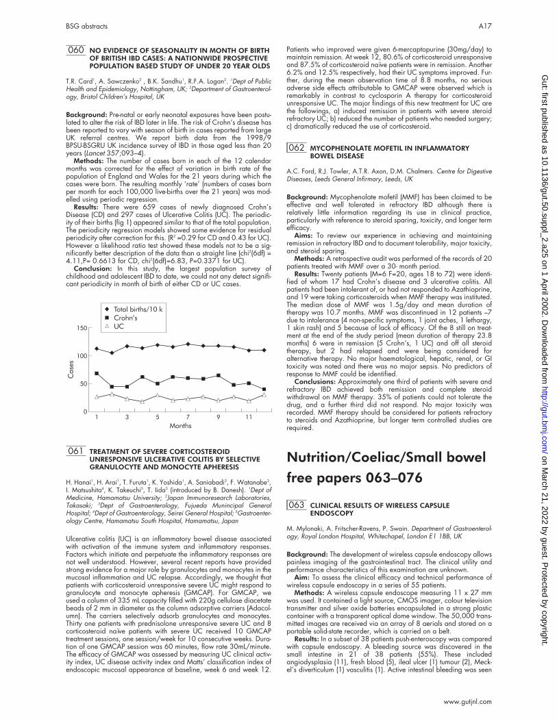
060 NO EVIDENCE OF SEASONALITY IN MONTH OF BIRTHOF BRITISH IBD CASES: A NATIONWIDE PROSPECTIVEPOPULATION BASED STUDY OF UNDER 20 YEAR OLDS
T.R. Card1, A. Sawczenko2 , B.K. Sandhu1, R.F.A. Logan2. 1Dept of PublicHealth and Epidemiology, Nottingham, UK; 2Department of Gastroenterol-ogy, Bristol Children’s Hospital, UK
Background: Pre-natal or early neonatal exposures have been postu-lated to alter the risk of IBD later in life. The risk of Crohn’s disease hasbeen reported to vary with season of birth in cases reported from largeUK referral centres. We report birth data from the 1998/9BPSU-BSGRU UK incidence survey of IBD in those aged less than 20years (Lancet 357;093–4).
Methods: The number of cases born in each of the 12 calendarmonths was corrected for the effect of variation in birth rate of thepopulation of England and Wales for the 21 years during which thecases were born. The resulting monthly ‘rate’ (numbers of cases bornper month for each 100,000 live-births over the 21 years) was mod-elled using periodic regression.
Results: There were 659 cases of newly diagnosed Crohn’sDisease (CD) and 297 cases of Ulcerative Colitis (UC). The periodic-ity of their births (fig 1) appeared similar to that of the total population.The periodicity regression models showed some evidence for residualperiodicity after correction for this. (R2 =0.29 for CD and 0.43 for UC).However a likelihood ratio test showed these models not to be a sig-nificantly better description of the data than a straight line (chi2(6df) =4.11,P= 0.6613 for CD, chi2(6df)=6.83, P=0.3371 for UC).
Conclusion: In this study, the largest population survey ofchildhood and adolescent IBD to date, we could not any detect signifi-cant periodicity in month of birth of either CD or UC cases.
061 TREATMENT OF SEVERE CORTICOSTEROIDUNRESPONSIVE ULCERATIVE COLITIS BY SELECTIVEGRANULOCYTE AND MONOCYTE APHERESIS
H. Hanai1, H. Arai1, T. Furuta1, K. Yoshida1, A. Saniabadi2, F. Watanabe3,I. Matsushita4, K. Takeuchi5, T. Iida5 (introduced by B. Danesh). 1Dept ofMedicine, Hamamatsu University; 2Japan Immunoresearch Laboratories,Takasaki; 3Dept of Gastroenterology, Fujueda Munincipal GeneralHospital; 4Dept of Gastroenterology, Seirei General Hospital; 5Gastroenter-ology Centre, Hamamatsu South Hospital, Hamamatsu, Japan
Ulcerative colitis (UC) is an inflammatory bowel disease associatedwith activation of the immune system and inflammatory responses.Factors which initiate and perpetuate the inflammatory responses arenot well understood. However, several recent reports have providedstrong evidence for a major role by granulocytes and monocytes in themucosal inflammation and UC relapse. Accordingly, we thought thatpatients with corticosteroid unresponsive severe UC might respond togranulocyte and monocyte apheresis (GMCAP). For GMCAP, weused a column of 335 mL capacity filled with 220g cellulose diacetatebeads of 2 mm in diameter as the column adsorptive carriers (Adacol-umn). The carriers selectively adsorb granulocytes and monocytes.Thirty one patients with prednisolone unresponsive severe UC and 8corticosteroid naïve patients with severe UC received 10 GMCAPtreatment sessions, one session/week for 10 consecutive weeks. Dura-tion of one GMCAP session was 60 minutes, flow rate 30mL/minute.The efficacy of GMCAP was assessed by measuring UC clinical activ-ity index, UC disease activity index and Matts’ classification index ofendoscopic mucosal appearance at baseline, week 6 and week 12.
Patients who improved were given 6-mercaptopurine (30mg/day) tomaintain remission. At week 12, 80.6% of corticosteroid unresponsiveand 87.5% of corticosteroid naïve patients were in remission. Another6.2% and 12.5% respectively, had their UC symptoms improved. Fur-ther, during the mean observation time of 8.8 months, no seriousadverse side effects attributable to GMCAP were observed which isremarkably in contrast to cyclosporin A therapy for corticosteroidunresponsive UC. The major findings of this new treatment for UC arethe followings, a) induced remission in patients with severe steroidrefractory UC; b) reduced the number of patients who needed surgery;c) dramatically reduced the use of corticosteroid.
062 MYCOPHENOLATE MOFETIL IN INFLAMMATORYBOWEL DISEASE
A.C. Ford, R.J. Towler, A.T.R. Axon, D.M. Chalmers. Centre for DigestiveDiseases, Leeds General Infirmary, Leeds, UK
Background: Mycophenolate mofetil (MMF) has been claimed to beeffective and well tolerated in refractory IBD although there isrelatively little information regarding its use in clinical practice,particularly with reference to steroid sparing, toxicity, and longer termefficacy.
Aims: To review our experience in achieving and maintainingremission in refractory IBD and to document tolerability, major toxicity,and steroid sparing.
Methods: A retrospective audit was performed of the records of 20patients treated with MMF over a 30- month period.
Results: Twenty patients (M=6 F=20, ages 18 to 72) were identi-fied of whom 17 had Crohn’s disease and 3 ulcerative colitis. Allpatients had been intolerant of, or had not responded to Azathioprine,and 19 were taking corticosteroids when MMF therapy was instituted.The median dose of MMF was 1.5g/day and mean duration oftherapy was 10.7 months. MMF was discontinued in 12 patients –7due to intolerance (4 non-specific symptoms, 1 joint aches, 1 lethargy,1 skin rash) and 5 because of lack of efficacy. Of the 8 still on treat-ment at the end of the study period (mean duration of therapy 23.8months) 6 were in remission (5 Crohn’s, 1 UC) and off all steroidtherapy, but 2 had relapsed and were being considered foralternative therapy. No major haematological, hepatic, renal, or GItoxicity was noted and there was no major sepsis. No predictors ofresponse to MMF could be identified.
Conclusions: Approximately one third of patients with severe andrefractory IBD achieved both remission and complete steroidwithdrawal on MMF therapy. 35% of patients could not tolerate thedrug, and a further third did not respond. No major toxicity wasrecorded. MMF therapy should be considered for patients refractoryto steroids and Azathioprine, but longer term controlled studies arerequired.
Nutrition/Coeliac/Small bowelfree papers 063–076
063 CLINICAL RESULTS OF WIRELESS CAPSULEENDOSCOPY
M. Mylonaki, A. Fritscher-Ravens, P. Swain. Department of Gastroenterol-ogy, Royal London Hospital, Whitechapel, London E1 1BB, UK
Background: The development of wireless capsule endoscopy allowspainless imaging of the gastrointestinal tract. The clinical utility andperformance characteristics of this examination are unknown.
Aim: To assess the clinical efficacy and technical performance ofwireless capsule endoscopy in a series of 55 patients.
Methods: A wireless capsule endoscope measuring 11 x 27 mmwas used. It contained a light source, CMOS imager, colour televisiontransmitter and silver oxide batteries encapsulated in a strong plasticcontainer with a transparent optical dome window. The 50,000 trans-mitted images are received via an array of 8 aerials and stored on aportable solid-state recorder, which is carried on a belt.
Results: In a subset of 38 patients push-enteroscopy was comparedwith capsule endoscopy. A bleeding source was discovered in thesmall intestine in 21 of 38 patients (55%). These includedangiodysplasia (11), fresh blood (5), ileal ulcer (1) tumour (2), Meck-el’s diverticulum (1) vasculitis (1). Active intestinal bleeding was seen
150
100
50
01
Cases
3 5 7
Months
9 11
Total births/10 k
Crohn�s
UC
BSG abstracts A17
www.gutjnl.com
on March 21, 2022 by guest. P
rotected by copyright.http://gut.bm
j.com/
Gut: first published as 10.1136/gut.50.suppl_2.a25 on 1 A
pril 2002. Dow
nloaded from
in 3. No additional intestinal diagnoses were made by enteroscopy.The yield of push-enteroscopy in evaluating obscure bleeding was30% (12/38). The capsule found significantly more intestinalbleeding abnormalities than push enteroscopy (p<0.05). A source ofbleeding was identified beyond the reach of the push enteroscope in9/21 (42%). Therapy was altered in 50% and in 3 patients, who hadrequired more than 100 units of blood, directed surgery cured (2) ormarkedly reduced (1) the bleeding. Patients always preferred capsuleendoscopy to push-enteroscopy (p<0.001). There were no complica-tions. Preparation with picolax improved images in patients on iron orwith blood in the intestine. 7 patients had no push-enteroscopy. Stud-ies in volunteers (7) and patients with chronic abdominal pain (3)were mostly normal – erosions in 1, lymphangectatic cysts in 3.
Conclusions: This study shows that capsule endoscopy providessmall intestinal imaging comparable to push-enteroscopy and candiagnose intestinal bleeding at sites beyond the reach ofpush-enteroscopes. It was safe and well tolerated.
064 THE RELATIONSHIP OF ADULT COELIAC DISEASEWITH IRRITABLE BOWEL SYNDROME, IRONDEFICIENCY ANAEMIA AND FATIGUE: A PRIMARYCARE CROSS-SECTIONAL STUDY
D.S. Sanders, D. Patel, T.J. Stephenson, A. Milford Ward, E.V. McCloskey,M. Hadjivassiliou, A.J. Lobo. Supra-Regional Protein Reference Unit,Northern General Hospital & Gastroenterology and Liver Unit, Royal Hal-lamshire Hospital, Sheffield, UK
Background and Aims: Our aim was to determine the prevalence ofundiagnosed adult coeliac disease in the general population of main-land United Kingdom. We also sought to establish the relationship inprimary care between coeliac disease, irritable bowel syndrome, irondeficiency anaemia, fatigue and other coeliac related conditions.
Methods: A cross-sectional study using immunoglobulins, IgA/IgGantigliadin antibodies and endomysial antibodies to initially recognisecoeliac disease. 1200 volunteers were recruited (January 1999 toJune 2001) from Five General Practices in South Yorkshire. Any par-ticipant with a positive IgA antigliadin antibody, positive endomysialantibody or only IgG antigliadin antibody in the presence of IgA defi-ciency was offered a small bowel biopsy to confirm the diagnosis ofcoeliac disease.
Results: 12 new cases of coeliac disease were diagnosed from1200 samples. The prevalence of coeliac disease in this generalpopulation sample is 1% (95% CI 0.4–1.3%). The prevalence of coe-liac disease in participants with irritable bowel syndrome was 3.3%(4/123), for iron deficiency anaemia 4.7% (3/64) and for fatigue3.3% (3/92).
Conclusions: This is the first study to establish the prevalence ofundiagnosed adult coeliac disease in a general population from Eng-land. Underdiagnosis of coeliac disease is common in primary care.A case finding approach would avoid delays in diagnosis and theassociated morbidity or potential complications of coeliac disease. Alow threshold for serological screening of patients with coeliac associ-ated symptoms or conditions would be an optimal strategy in primarycare.
Acknowledgements: We wish to acknowledge the support ofAction Research. This study was entirely funded by Action research. DrSanders is an Action Research Training Fellow.
065 SEROPREVALENCE, CORRELATES ANDCHARACTERISTICS OF UNDETECTED COELIACDISEASE IN ENGLAND
J. West1, R.F.A. Logan1, P.G. Hill2, C.A. Lloyd2, S. Lewis3, R. Reader4,G.K.T. Holmes5, K.T. Khaw4. 1Divn of Public Health and Epidemiology,University Hospital, Nottingham; Depts of 2Chemical Pathology and 5Medi-cine, Derbyshire Royal Infirmary, Derby; 3Medical & Surgical Sciences,Nottingham City Hospital; 4Clinical Gerontology Unit, Addenbrooke’sHospital, Cambridge, UK
Background: Recent studies using various antibody tests to screen forundetected coeliac disease have shown that the prevalence of coeliacdisease (CD) in several countries is between 0.5–1.0% of the popula-tion. So far the numbers detected have been small and information asto the characteristics and consequences of undetected CD is limited.We have examined the seroprevalence of undetected CD in a largepopulation sample from the Cambridge area.
Methods: The Cambridge General Practice Health Study identifiedindividuals aged 45–74 from the age-sex registers of 11 general
practices and invited them for a health survey and a bone density scanbetween 1990–1995. We tested 7550 of the serum samplescollected for antiendomysial antibody (EMA) and used multivariateanalyses to compare EMA positive and negative subjects.
Results: The seroprevalence of undetected CD in this generalpopulation sample was 1.2% (95% CI 0.9–1.4) and did not vary sig-nificantly with age or sex. EMA +ve subjects (n=87) were 2.2kgslighter (p = 0.07) and 0.1cms shorter (p = 0.09), were more likely tohave reported their general health as being “good to excellent” (OddsRatio (OR) 1.8, 95% CI 0.9–3.5), and were less likely to report beinga current or ex-smoker (OR for current versus never 0.36, 95% CI0.14-0.90). Undetected CD was associated with a 8% reduction inmean serum cholesterol (0.5mmol/1, p<0.01) and small reductions inmean haemoglobin (0.3g/dl, p<0.01), total protein (1.0g/l, p<0.05)and corrected serum calcium (0.02mmol/1,p<.05). There was anincreased prevalence of osteoporosis in the EMA +ves (OR 3.1, 95%CI 1.3–7.3) and of mild anaemia (OR 4.6, 95% CI 2.5–8.2). FiveEMA +ves (6%) had died, a proportion similar to that in EMA–ves(8%).
Conclusions: Undetected coeliac disease is likely to affect about1% of the population of England, a figure similar to several othercountries. Although affected subjects report no increase in “poor ormoderate” health they have an increased prevalence of osteoporosisand mild anaemia. In contrast they have a favourable cardiovascularprofile which is likely to afford substantial protection from ischaemicheart disease and stroke.
066 WHAT FACTORS INFLUENCE COMPLIANCE WITH AGLUTEN-FREE DIET? A COMPARISON OF WHITECAUCASIAN AND SOUTH ASIAN COELIAC PATIENTS
J.R. Butterworth1, L. Banfield2, T.H. Iqbal1, B.T. Cooper1. 1Department ofGastroenterology and 2Department of Dietetics, City Hospital NHS Trust,Dudley Road, Birmingham B18 7QH, UK
Introduction: Lifelong and strict adherence to a gluten-free diet ismandatory in patients with coeliac disease (CD) to improve nutritionand prevent long-term complications. Little is known about whatfactors influence compliance with a gluten-free diet.
Aims: To identify factors that may influence compliance to a gluten-free diet amongst Caucasians and South Asian patients with coeliacdisease.
Methods: A questionnaire survey was sent to 130 coeliac patientsfollowed-up at our unit, (90 Caucasians and 43 South Asian).
Results: Eighty seven (66.9%) of the 130 questionnaires werereturned- 66 from the Caucasians, and 21 from the South Asianpatients (p=0.003). Patients own assessment of their strictness to agluten-free (GF) diet was significantly correlated with both small bowelhistological recovery and negative endomysial antibody status amongthe Caucasian patients (p=0.005 and <0.0001 respectively), but notamongst the South Asians. In the Caucasian patients, eight factorsappeared to correlate with compliance with a GF diet: 1.Membershipof the Coeliac Society (p<0.0001). 2.Understanding food labelling(p=0.014). 3.Obtaining GF products on prescription (p=0.047).4.Affordability of GF products (p=0.01). 5.Getting sufficient prescrip-tion GF products (p=0.017) 6.A detailed explanation of CD by a phy-sician (p=0.006). 7.Having a follow-up small bowel biopsy (p=0.03).8.Regular dietetic follow up (p=0.01). No factors were identifiedamongst the South Asians, who were less likely to attend dietetic clin-ics (p=0.005), or be members of the Coeliac Society (p=0.02) andwere more dissatisfied with information provided by doctors(p=0.026) and dieticians (p=0.011) compared with the Caucasiancoeliac patients.
Conclusions: In contrast to the South Asians, a number of factorsseem to influence compliance with a GF diet amongst Caucasianpatients with CD. Compliance seems poor amongst South Asianpatients, and a different approach is required in terms of educationand dietetic supervision compared to the Caucasian patients with CD.
067 DETECTION OF GLUTEN IN ALCOHOLIC BEVERAGESIS FEASIBLE USING DRY STRIP IMMUNOCHEMISTRY INTHE “GLUTEN HOME TEST” KIT
M.M. Skelly, C.J. Hawkey. Division of Gastroenterology, University Hospi-tal, Nottingham, UK
Introduction: It is unclear which cereal-based alcoholic drinks aresuitable for coeliac patients. A gluten “home test” (GHT) detection kithas been developed utilising dry-strip immunochemistry format with a
A18 BSG abstracts
www.gutjnl.com
on March 21, 2022 by guest. P
rotected by copyright.http://gut.bm
j.com/
Gut: first published as 10.1136/gut.50.suppl_2.a25 on 1 A
pril 2002. Dow
nloaded from
range of detection of wheat gluten from 50 to 1200 ppm.Recommended acceptable levels are <20ppm for food naturallygluten free and <200ppm for food rendered gluten free.
Aim: To demonstrate that the gluten content of a number ofalcoholic beverages can be determined by Gluten Home Test kit.
Methods: Alcoholic beverages were tested with the GHT andELISA. Control samples contained known quantities of gluten (starch)or were not cereal based (Coca-Cola). Test samples are as reported.The GHT strip contains antibodies specific for omega gliadin, a stablegluten protein, which bind to gliadin in the test beverage. When thebeverage is loaded on the strip, a gliadin-antibody- blue latex particlecombination migrates along the strip until it is trapped by immobilisedgliadin antibody in one of three positions. The positions indicate glu-ten level less than 50ppm (“Negative test”), 50–200ppm or greater(“Positive”) and more than 10% gluten (“Strong positive”). Gliadincontent of the test and control samples was determined using a directsandwich ELISA that utilised a monoclonal antibody to omega gliadin.See table.
Summary: Gluten can be readily detected in alcoholic beveragesusing dry strip immunochemistry. Results from GHT and ELISA broadlycorrelate. Kits may be useful for coeliacs to guide choice of alcoholicbeverage.
068 ASSOCIATION OF COMMON HFE GENE MUTATIONSWITH COELIAC DISEASE RESULTS IN PROTECTIONAGAINST IRON DEFICIENCY ANAEMIA
J.R. Butterworth1, W.M.C. Rosenberg2, S. Jobson3, M. Hathaway3, D.Briggs3, W.M. Howell4, D.H. Adams5, B.T. Cooper1, T.H. Iqbal1. 1Gastro-enterology Unit, City Hospital, Birmingham, UK; 2Liver Group, Division ofInfection, Inflammation and Repair, University of Southampton, UK;4Division of Human Genetics, University of Southampton, UK; 3NationalBlood Service, Birmingham, UK; 5Liver Research Laboratories, MRC Centrefor Immune Regulation, University of Birmingham, UK
Introduction: Coeliac disease (CD) and haemochromatosis are com-mon conditions in populations with Celtic origins, but are associatedwith different defined human leukocyte antigen (HLA) haplotypes.
Aims: To determine if a genetic relationship exists between the twodiseases, and if HFE mutations protect against iron deficiencyanaemia.
Methods: Polymerase chain reaction amplification using sequencespecific primers capable of identifying the 2 HFE gene mutations(H63D and C282Y), and the HLA class I and II alleles was used totype 77 Caucasoid patients with CD, and 187 matched controls.Haemoglobin and serum iron were measured at diagnosis.
Results: The two HFE gene mutations were identified in 36 patientswith CD (46.7%), and 61 (32.6%) of the controls (P=0.035). Amongstthe control population, the C282Y mutation was strongly associatedwith the HLA-A*03 and B*07 alleles and H63D with the HLA-A*25allele but these associations were not observed in the CD group. Bycontrast, the C282Y mutation in the CD patients was associated withthe HLA-A*01 and B*08 alleles. CD patients with a C282Y mutationhad significantly higher mean haemoglobin and serum iron comparedto the HFE wildtype (P=0.04 and 0.0015 respectively). No such rela-tionship was found for the H63D mutation.
Conclusion: HFE gene mutations are common in patients with CD.Different linkage disequilibrium between HFE mutations and HLA alle-les in the CD and control groups allows for selection of HFEabnormalities in the disease, and a disease-specific haplotype whichcarries C282Y and DQB1*02 is suggested. In the context of CD wepropose that HFE mutations provide a survival advantage by amelio-rating the iron deficiency seen in this condition.
069 IN-VIVO TOXICITY OF AMINO ACIDS 57–75 OFALPHA-GLIADIN IN COELIAC DISEASE
J.S. Fraser1, W. Engel2, E.L. Pollock1, S.J. Moodie1, H.J. Ellis1, H. Wieser2,P.J. Ciclitira1. 1Gastroenterology (GKT), St Thomas’ Hospital, LondonSE1 7EH, UK; 2Deutsche Forschungs Anstalt für Lebensmittelchemie,Garching, Germany
Background: Peptides from α-gliadin have been used to characterisethe immunodominant coeliac toxic cereal epitope in vitro. Followingincubation with a peptide corresponding to amino acids 57–73 ofα-gliadin, peripheral blood mononuclear cells from coeliac patientssecrete IFN-?; gluten-specific small intestinal T cell clones proliferate inresponse to peptides corresponding to residues 57–68 and 62–75 ofα-gliadin.
Aim: We wished to investigate whether a peptide corresponding toresidues 57–75 of A-gliadin exacerbates coeliac disease (CD) in vivo.
Methods: We studied four unrelated Caucasian patients withknown CD, all of whom were on a gluten-free diet. The patients under-went three separate challenges. One g peptic-tryptic gliadin (PTG)served as a positive control. Twenty to 100mg of the test peptide wasstudied on a separate occasion, and on the third a negative controlpeptide from casein, which comprised 20 amino acids incorporatingthe same residues as the test peptide, but which were in a differentorder, was assessed. Following sedation, a Quinton hydraulic multiplebiopsy capsule was positioned in the duodenum. The peptides wereinstilled into the duodenum over 2 hours. Biopsies were taken beforethe infusion, 2, 4 and 6 hours after commencing the infusions. Thebiopsy specimens were assessed blindly for villus height to crypt depthratio and enterocyte surface cell height. We used the Mann-WhitneyU test, with 95% confidence intervals, for statistical analysis.
Results: The negative control peptide caused no significantchanges to villus morphology nor small intestinal villus enterocytes inany of the patients. The villus height to crypt depth ratio and the ente-rocyte surface cell height fell significantly 4 to 6 hours after commenc-ing the infusions with both PTG and the test peptide, compared to theinitial biopsy, in all subjects (n=4).
Conclusion: A peptide corresponding to residues 57–75 ofA-gliadin, exacerbates coeliac disease in vivo.
070 BONE MINERAL DENSITY IN ANOREXIA NERVOSA: NOCHANGE OVER TIME
C. Preston1, C. Flynn1, B. Oldroyd2, J.G. Truscott2, E. Mann3, P.D. Howdle1.1Dept of Medicine, St James’s University Hospital; 2Centre for Bone andBody Composition, Leeds General Infirmary; 3Yorkshire Centre for EatingDisorders, Leeds, UK
Introduction: Patients with anorexia nervosa (AN) are at high risk ofosteoporosis. Body composition is also abnormal. The mechanismsbehind these changes are unclear although oestrogen status and nutri-tional factors are believed to play a part. There is little informationabout the natural history of osteoporosis in patients with ongoing dis-ease.
Methods: To investigate the natural course of bone mineral density(BMD) and body composition in AN, measurements were made usingdual energy x-ray absorptiometry. We studied 16 adolescents(15–19.9yrs) and 31 adults (>20yrs) all females, none had primaryamenorrhoea. Local age and sex-matched controls were used for com-parison with the adult group. We monitored the change in BMD andbody composition of 13 patients over a mean follow up period of 21months.
Results: In keeping with earlier studies, our adult anorexic patientshad significantly lower BMI (16.2vs22.2 kg/m2), BMD (PA Spine Tscore –1.92vs 0.2, total body T score -1.2 vs. 0.4), lean body mass(34.8 vs. 38.3kg) and fat mass (6.5 Vs 19.2kg) than the controls (allp<0.0001). The adolescent group did not differ significantly in eitherBMI, BMD or body composition from the adult group. Disease profileswere compared between adolescent and adult patients. Despite ashorter duration of illness (3.1 vs. 8.4yrs p=0.01) and ofamenorrhoea (2.6 vs. 5.8yrs p=0.06) the adolescents’ BMD did notdiffer significantly from the adults. This may be due to the earlier onsetof disease in adolescents (14.2 vs. 17.6 yrs) i.e. before peak bonemass is achieved. Using WHO definitions 37% of our patients hadosteoporosis and 44% had osteopenia. In the 13 follow up patientsthere was no significant change in weight over time, despite on-goingpsychiatric support. The BMD of these patients did not altersignificantly over the follow up period.
Conclusions: Patients with AN have a significant reduction inBMD. Despite psychiatric support they remain underweight but
Abstract 067
Sample (n=2–5) [Gliadin] µg/ml Gluten Home Test
Bass 0.116 Strong positiveGuinness 0.49 Strong positiveWhisky (blend) 0 NegativeWhisky (malt) 0 NegativeBudweiser 0.029 PositiveNewcastle Brown Ale 0.4715 Strong positiveStella Artois 0.1578 PositiveCoca-Cola 0 Negative
BSG abstracts A19
www.gutjnl.com
on March 21, 2022 by guest. P
rotected by copyright.http://gut.bm
j.com/
Gut: first published as 10.1136/gut.50.suppl_2.a25 on 1 A
pril 2002. Dow
nloaded from
fortunately the BMD does not deteriorate. Trials of specific treatmentsshould be undertaken and will need to include patients of all ages.
071 OSTEOPOROSIS AND COELIAC DISEASE: IS THE 1500MG/DAY GUIDELINE FOR CALCIUM ACHIEVABLE BYDIET?
A. Holdoway1, W.E. Fickling1, A.K. Bhalla2, D.A.F. Robertson1. 1RoyalUnited Hospital; 2Royal National Hospital for Rheumatic Diseases, Bath,UK
Background: Guidelines for treating and preventing osteoporosis incoeliac disease (CD) were published in 2000 in the international jour-nal Gut. These guidelines recommend that patients with CD shouldachieve a calcium intake of 1500 mg/day and adhere to a strictgluten-free (GF) diet. UK dietary surveys have shown that >50% offemale adults in the general population fail to achieve the ReferenceNutrient Intake (RNI) of 700 mg/day (DoH, 1998). In addition, GFdiets have been shown to reduce intake of foods that contributedietary calcium. Consequently the 1500 mg target may be difficult toachieve by diet alone. Our aim was to establish the intake of dietarycalcium in patients with CD.
Methods: 26 patients with CD; age range 33 – 71yrs, 8 males, 18females, were recruited via gastroenterology out patient clinics andthe local coeliac society. Median duration on a GF diet was 10 yrs,range 3–33yrs. Dietary calcium and compliance to a GF diet weredetermined using a 10-day weighed method, Dietetic interview and adietary analysis programme using data from MAFF (Dietplan) andfood manufacturers.
Results: 25/26 adhered to a strict GF diet (96% compliance).Median calcium intake was 1209 mg/day (range: 539 – 1898mg/day). 31% (8/26) achieved the 1500 mg/day. 11% failed toachieve the RNI of 700mg/day, all were female and following arestricted energy intake for the purpose of intentional weight loss.Dairy products provided a median of 57% of dietary calcium, whichis similar to the non-coeliac population. 2/26 adhered to a milk-freediet but achieved a calcium intake >1400 mg/day usingcalcium-enriched bread/soya products. 42% used the new generationcalcium-enriched GF breads. Median contribution of calcium from GFbread was 27% in those using the calcium-enriched GF breads, com-pared to 8% in those using standard GF breads.
Conclusions: Whilst it is possible to achieve a calcium intake of1500 mg/day on a strict GF diet, careful dietetic assessment isrequired in the majority of patients to identify those with sub-optimalintakes (up to 2/3 of patients) and improve calcium intake throughdiet and/or supplements. The new generation calcium-enriched GFbreads may help achieve dietary intakes of 1500 mg of calcium/day.
072 MORTALITY RISK FACTORS AND TRANSPLANTATIONINDICATIONS FOR PATIENTS ON HOME PARENTERALNUTRITION (HPN)
R.S. Hodgson1, N. Latif1, E.A.B. Cameron2, J.A.H. Binnie2, C. Murray1, S.J.Middleton1, A. Forbes1, S.M. Gabe1. 1St Mark’s Hospital, Harrow HA1;2Addenbrooke’s Hospital, Cambridge CB2, UK
The overall mortality for HPN in the UK is 8% per year and reportssuggest that 80–90% of these deaths are attributable to the underlyingdisease. We performed a retrospective analysis of all patients whodied on HPN over the last 5 years at St Mark’s and all patients whohad undergone intestinal transplantation (ITx) at Cambridge.
Results: At St Mark’s, 37 patients died on HPN since 1996 and 33complete sets of notes were traced (89%). These patients had amedian age of 58 years (30–77) and a median time on HPN of 543days (7–6777) [20F:13M]. 25 patients (76%) died from their originaldisease (HPN-disease), 4 (12%) died from complications of HPN(HPN-comp) and 4 (12%) died from other causes (HPN-other).HPN-comp deaths were sepsis (1) and liver failure (3). 2 of the 3patients who developed liver failure had chronic cholestasis andreceived >1g lipid/kg/day. In Cambridge, 9 patients had ITx since1991. 1 set of notes could not be traced and 1 patient did not haveHPN prior to transplantation. The remaining 7 had a median age of25.8 years (21–42 years) and a median of 730 days on HPN (14–2920 days) prior to transplantation [2F:5M]. Indications for ITx wererecurrent thromboses (4), recurrent central venous catheter (CVC) sep-sis (3) and PN related liver disease (2). See table.
Conclusions: HPN patients die predominantly of their underlyingdisease. ITx patients had a higher frequency of infectious and throm-botic complications prior to transplantation and perhaps HPN patientswith similar complications should be considered earlier for transplan-tation.
073 EFFECTS OF LOW FIBRE DIETS AND VITAMINCONTENT ON NEOPLASIA AND CELL PROLIFERTIONIN THE MIN MOUSE
O. Bashir, A.J. FitzGerald, N. Mandir1, R.A. Goodlad1. HistopathologyDepartment, Imperial College School of Medicine, Hammersmith Hospital,London; 1Imperial Cancer Research Fund, 44 Lincoln’s Inn Fields, London,UK
Background: Loss of APC and the production of a truncated APCprotein leads to familial adenomatous polyposis (FAP) in man andmultiple intestinal neoplasia (Min) in the mouse. Both phenotypes arecharacterised by multiple intestinal polyps. The Min mouse is thus auseful model for the study of exogenous factors on gut biology andtumour progression.
Methods: We have used the Min mouse (C57BK/6J-ApcMin) toinvestigate the actions of chow diets and fibre-free semisythetic dietson polyp progression, cell proliferation and crypt fission. The semisy-thetic diet was then used to investigate the actions of altered vitamincontent (lowered to a third of the RDA and a SS diet where the vitamincontent was increased fivefold (except for retinol and folate whichwere doubled). 60 Min mice and wild type littermates, 4 weeks old,were divided into 4 groups and fed the four diets for 8 weeks. Thenumber and size of polyps in the small and large intestines werescored later (number*volume = burden), as was the number of nativemitoses and the percentage of branching crypts.
Results: The guts of the chow fed mice were heavier, and all Mingroups had heavier guts. The intestines of the low and high vitamingroups were heavier than the SS control. There were fewer polyps andthe tumour burden was lower in the SS group. Both low and high vita-min levels lead to increased polyp number, especially in the proximalsmall intestine. There was more proliferation and crypt fission in the SSgroup, and this was reduced in the low and high vitamin groups. Theeffect of vitamins was most pronounced in the proximal intestine.
Conclusion: Low fibre semisythetic diets may reduce polyp forma-tion, suggesting that the lack of ‘fibre’ may be beneficial. Alteration ofvitamin content can enhance polyp number and tumour burden. Bothlow or high vitamin content may be a risk factor.
074 IS THE CURRENT SERVICE FOR THE INSERTION OFENDOSCOPIC FEEDING TUBES APPROPRIATE ANDACCEPTABLE? A UK SURVEY
A.J. Hughes, T.E. Bowling. City General Hospital, Newcastle Road, Stokeon Trent ST4 6QG, UK
We were concerned that gastroenterologists may be puttingthemselves in a vulnerable position, in terms of clinical risk, by insert-ing PEGs purely as a service for other clinicians. A questionnaire wastherefore sent out to hospitals in the UK to clarify current practice.Responses were received from 196 of 242 units (>80%).
The number of PEGs placed ranged from 1 to 100/100,000 popu-lation covered/year with a median of 22 with most placed by medi-cal gastro- enterologists. 59% of responding hospitals have a nutritionteam but in 22% of these the patients are still assessed for suitability ofPEG only by their own team. The endoscopist does not know whatassessment has been made of patients attending for PEG placement in25% of hospitals. In the hospitals where patients are assessed by agastro/nutrition team the endoscopist is unaware of the details of theassessment in <3%.
Consent is clearly a difficult issue as many patients are mentallyincapacitated but despite this <50% of hospitals have an appropriate,known consent policy for this group. Time from referral to insertion ofPEG is >1 week in 33% of patients and this delay may well impact onphysical wellbeing and also on bed occupancy.
Abstract 072
Group nTotal HPNdays
CVC complication
Infection/HPNday x100
Thrombosis/HPNday x100
HPN-disease 25 18345 0.13 0.05HPN-comp 4 13435 0.10 0.01HPN-other 4 11765 0.08 0.03ITx patients 7 8379 0.43 0.10
There were no differences between the groups in intestinal anatomy.
A20 BSG abstracts
www.gutjnl.com
on March 21, 2022 by guest. P
rotected by copyright.http://gut.bm
j.com/
Gut: first published as 10.1136/gut.50.suppl_2.a25 on 1 A
pril 2002. Dow
nloaded from
Gastroenterology follow up is rare. >70% of hospitals do not pro-vide any routine review or a review mechanism should complicationsarise post PEG insertion. <10% of patients are followed up beyond 1week. 80% of patients are followed up in the community, often only bydistrict nurses.
Current practice for PEG insertion is highly variable and in manyhospit- als doesn’t seem to be either appropriate or acceptable. Webelieve many endoscopists are exposing themselves to potential riskby acting as technicians and feel that national minimum standardsshould be considered.
075 COLONIC MUCOSAL INJURY FROM NATURAL ANDSYNTHETIC SULPHATED POLYSACCHARIDES
E.T. Donnelly, M. Hoper, W.G. McCluggage1, F.C. Campbell.Departments of Surgery and 1Pathology, Queen’s University Belfast,Institute of Clinical Science, Grosvenor Road, Belfast BT12 6BJ, NorthernIreland
Undegraded lambda λ Carrageenan (λCgN) is widely used as ahuman food additive but is chemically related to the pro-inflammatoryagent dextran sodium sulphate (DSS). This study tests the hypothesisthat λCgN has pro-inflammatory effects in colon.
Methods: Acute and subchronic oral administration of λCgN vsDSS vs drinking water only control against colonic disease activityand mucosal inflammation in 2 species viz mice and rats. λCgN, DSS(1–4%) or drinking water control were administered for 2–72 days toBalb/c mice (n=225) and Sprague-Dawley rats (n=45) which weremaintained on AIN76A antioxidant and mutagen-free diet. Diseaseactivity was assessed by stool consistency, blood loss, weight loss andlethargy. Mucosal injury was assessed histologically by semiquantita-tive scores of crypt loss, shortening, distortion, hyperplasia, andinflammatory infiltration.
Results: Both λCgN and DSS induced colonic inflammation, cryptinjury and crypt hyperplasia in mice and rats. Effects on colonic func-tion and mucosal injury were dose dependent and increased withduration of exposure. DSS had greater adverse effects on diseaseactivity than CgN in doses of 3–4% in mice and 1–3% in rats.
Conclusion: Although less toxic than DSS, λCgN produced cumu-lative dose dependent colonic mucosal injury and adverse effects oncolonic function of 2 species.
076 OUTCOMES IN HOME PARENTERAL NUTRITION IN ANATIONAL UNIT
F.C. Leslie, M.A. Bradley, J.L. Shaffer. Intestinal Failure Unit, Hope Hospi-tal, Stott Lane, Salford, Manchester M6 8HD, UK
Hope Hospital is one of the UKs two largest units covering patients onHome Parenteral Nutrition (HPN). In 1998 it was decided to review allnew patients commencing on HPN, to study morbidity and mortality.A mean of 27 patients started HPN each year and no patients werelost to follow-up.
85 patients commenced HPN between 15th February 1998 and15th May 2001, aged 19 years 11months – 76 years 11months,(mean 50.2 years ±16.8 months) (50 females). 19 patients were <40years, 43 were 40–60 years and 23 were >60 years. 76 patientswere entirely self-caring for their line, 9 requiring input from family oroutside care e.g. district nurse. The main diagnoses in these patientswere Crohns (29), mesenteric vascular disease (20) and surgical com-plications e.g. short bowel syndrome after surgery for e.g. ulcerativecolitis or malignancy (18). Conditions e.g. radiation enteritis,scleroderma and volvulus made up smaller numbers. 2 patients weretreated for a diagnosis of active malignancy.
Of these 76 are alive, 57 on HPN, 19 have discontinued HPN and9 have died. Mortality rates were 0% at 1 year and 10.6% at 3 years,compared to 1-year and 3-year mortality rates of 7–15% and30–32% in published series. 4 patients died of malignancy (3 previ-ously recognised and 1 new case), and 5 of unrelated conditions i.e.3 patients died of their underlying diagnosis and 6 of unrelatedcauses. No deaths were related to HPN. 19 patients (22.3%)developed a confirmed line complication - 10 (11.8%) with catheter-related sepsis, (7 patients developed >1 complication). No patientsdeveloped significant liver disease.
19 (22.3%) patients came off HPN during this period with an aver-age duration of 9.15±1.05months (range 4–21 months). No cleardiagnosis was associated with an increased chance of discontinuingHPN, but 10 patients had surgery (9 reanastomosis) prior todiscontinuing. Patients who discontinued HPN were younger
(47years±37.5 months) than those who died (55.3 years±64.4months) and those that continued on HPN (50.8years±19.4), but thiswas not significant.
Over this time we treated increasing numbers of patients andincluded patients previously thought not appropriate for HPN e.g.those not self-caring and older age groups. Despite more patientsbeing considered suitable for HPN and new methods e.g training car-ers, mortality and complication rates are low and there were no HPN-related deaths. We appear to be widening the net without worseningoutcome.
Colorectal free papers077–090
077 THE EFFECT OF ZAPRINAST, A PHOSPHODIESTERASETYPE 5 INHIBITOR, ON THE SHEEP ISOLATEDINTERNAL ANAL SPHINCTER
N. Griffin1, A.G. Acheson1, J.H. Scholefield1, V.G. Wilson2. 1Departmentof Surgery and 2School of Biomedical Sciences, Queen’s Medical Centre,Nottingham NG7 2UH, UK
Introduction: Neurogenic relaxation of the internal anal sphincter ismediated by elevation of cyclic GMP, following activation of solubleguanylyl cyclase by nitric oxide. Since the activity of the cyclic nucle-otide is also regulated by phosphodiesterase 5, we have examinedthe effect of zaprinast, an inhibitor of the enzyme, on the sheepisolated internal anal sphincter.
Method: Strips of isolated sheep internal anal sphincter were sus-pended in 5ml organ baths containing warmed and oxygenatedKrebs and isometric tension recordings made. Preparations wereexposed to a cumulative concentration of zaprinast (3x10⁻⁸M to3x10–5M), firstly paired with time controls and then in the presenceand absence of NG-nitro-L-arginine methyl ester (L-NAME, 100µM), anitric oxide synthase inhibitor. In a separate series of experiments theeffect of sodium nitroprusside, a direct activator of soluble guanylylcyclase, was examined in the absence and presence of asub-maximally effective concentration of zaprinast (3x10⁻⁶M).
Results: Zaprinast caused a concentration-related relaxation of thesheep anal sphincter with the highest concentration giving a meaneffect of 92.3%±2.6 (n=7) compared to 11% ±3.2 loss in tone withtime controls over the same period. In the presence of 100 µML-NAME, the response to higher concentrations of zaprinast was sig-nificantly reduced (p<0.05, Student’s t-test) but not abolished: 30 µMzaprinast (L-NAME) 72.4%±4.9 (n=7). SNP caused a concentration-related relaxation of the sphincter (EC50 3x10⁻⁷M) that was enhancedin the presence of zaprinast ((EC50 1x10⁻⁷M) (n=6)).
Conclusion: Zaprinast acts as a PDE 5 inhibitor to relax the sheepinternal anal sphincter, however its actions are only partly dependenton the basal release of nitric oxide from the tissue. Further experimentswith more selective PDE 5 inhibitors are warranted in order to assesstheir possible role clinically in conditions related to sphincter hyperto-nia.
078 SURVEY OF ANO-RECTAL SYMPTOMS AMONGSTPATIENTS OBTAINING TOPICAL HAEMORRHOIDALPREPARATIONS FROM COMMUNITY PHARMACIES
N. Beattie, D. Lamprell, G. Bryson, C. Wilson. Ayr, Prestwick and TroonLocal Health Care Co-operative (LHCC) and Department of Surgery, AyrHospital, UK
Rectal bleeding is a common symptom amongst the adult population.Whilst most often due to benign ano-rectal disorders, it may be theonly symptom of colorectal cancer. The incidence of rectal bleedingamongst people purchasing topical haemorrhoidal preparations (BNFSections 1.7.1, 1.7.2) over the counter (OTC) from community phar-macies is unknown.
A survey of ano-rectal symptoms was conducted in 20 participatingcommunity pharmacies within Ayr, Prestwick and Troon LHCC duringFebruary 2001. Sixty-five patients returned completed questionnairesof whom 36 (55%) had filled a prescription (FP10) and 29 (45%) hadpurchased OTC. Only 8 of the 29 (28%) purchasing OTC had soughtthe pharmacist’s advice. Twenty-eight patients (43%) were over 60years of age.
BSG abstracts A21
www.gutjnl.com
on March 21, 2022 by guest. P
rotected by copyright.http://gut.bm
j.com/
Gut: first published as 10.1136/gut.50.suppl_2.a25 on 1 A
pril 2002. Dow
nloaded from
Thirty patients (46%) reported rectal bleeding in association withanal symptoms (itch, pain or a lump), 29 (45%) reported anal symp-toms with no bleeding and 5 (8%) had rectal bleeding without othersymptoms. Four of the five patients with rectal bleeding alone had pur-chased OTC medications and only one had consulted their GP abouttheir symptoms. Three of these 5 patients might be considered to have“suspicious symptoms” (age >60 years - 2, passing dark blood - 1).
A high incidence of rectal bleeding (54%) was found amongstpatients obtaining topical haemorrhoidal preparations (61% of thosefilling prescriptions vs 45% of those purchasing OTC). The majority ofpatients (86%) reporting bleeding had associated anal symptoms andwere considered low risk for cancer. The majority (69%) reportingbleeding had consulted their GP about their symptoms. A smallnumber reporting bleeding and purchasing OTC topical haemorrhoi-dal preparations should be consulting their GPs. Community pharma-cists need to be aware of the possibility of patients treating themselvesinappropriately.
079 ANALYSIS OF SYMPTOMATOLOGY OF PATIENTSPRESENTING TO THE RECTAL BLEEDING CLINIC: NEEDFOR BETTER REFERRAL GUIDELINES
A.B. Harikrishnan, M. Bassuini, A.J. Watkins1, A.R. Morgan, N.D. Carr, J.Beynon. Department of Coloproctology, Singleton Hospital, SwanseaSA2 8QA, UK; 1University of Wales, Swansea SA2 8PP, UK
Introduction: Rectal bleeding clinics (RBC) act as the pick-up point forcolorectal cancers ideally in their early stages. Referral guidelines forRBC vary and are yet to be formalised on a national scale. Appropri-ate referral guidelines will facilitate patient selection for RBCs.
Methods and Results: 2597 patients were seen in the RBC over asix-year period between October 1994 and September 2000.Patients were referred by their general practitioners. All patients hada detailed history, clinical exam and flexible sigmoidoscopy (FS).123(5%) patients were diagnosed as having colorectal cancer (meanage 70, range 45 – 90). 390(15%) patients had polyps (mean age62, range 19 – 94) and were referred for formal colonoscopy. A totalof 15 symptoms were recorded in these patients and analysed inseparate groups (cancer, polyp, sex and age). The incidence ofchange in bowel habit (CIBH), loose stool (LS), mucous discharge(MD), blood mixed with stool (BS), weight loss, palpable abdominalmass and mass palpable per rectum were significantly (p < 0.05)increased in the cancer group. Further, in the cancer group, symptomsof CIBH, LS, MD and BS were significantly (p < 0.05) present inpatients over 70 years. Interestingly, abdominal pain, constipationand fresh bleeding per rectum were not significantly associated withthe diagnosis of malignancy or polyps.
Discussion and Conclusion: This study demonstrates and is inagreement with other studies that the symptomatology of colorectalcancer is quite distinct and should be incorporated into the RBC refer-ral guidelines. In this series only 5% of the referred patients had colo-rectal cancer and 15% had polyps. More stringent RBC referralguidelines based on these symptoms and related to age would aid inselection of patients for rectal bleeding clinics.
080 A DOUBLE-BLIND, PLACEBO CONTROLLED PILOTSTUDY TO ASSESS THE EFFECTS OF A PROBIOTIC ONTHE RESPONSE OF THE INTESTINAL MICROFLORA TOHELICOBACTER PYLORI ERADICATION THERAPY
J.A.J. Madden1, S. Plummer2, N.T. Plummer2, M. Herbison1, J.O. Hunter1.1Department of Gastroenterology, Addenbrooke’s NHS Trust, CambridgeCB2 2QQ; 2Cultech Ltd, York Chambers, York St, Swansea SA1 3NJ, UK
Introduction: Antibiotic therapy can detrimentally alter the intestinalmicroflora. Probiotics are preparations of non-pathogenic intestinalbacteria which may be beneficial to human health. The effects of sup-plementation with Bifidobacterium longum and Lactobacillus acido-philus (HLC: Cultech Ltd, Port Talbot, UK) has been studied in patientsundergoing eradication of Helicobacter pylori by antibiotics.
Methods: 30 patients positive for H. pylori serology were recruitedinto the trial (8 were excluded for non-compliance). Patients were ran-domised into three treatment groups: antibiotics days 1–7 [metronida-zole 400mg tds, amoxycillin 500mg qds, lansoprazole 30mg bd],with placebo days 1–15 (n=9), or the same antibiotics days 1–7 withprobiotics days 1–15 (n=7), or the antibiotics with placebo days 1–7and with probiotics days 8–15 (n=6). Patients provided stool sampleson days 1, 7, 12, 17 & 27. Specimens were analysed using standardmicrobiological techniques.
Results: Administration of antibiotics alone resulted in a significantincrease in numbers of aerobes between days 1 and 7 (p=0.001).When probiotics were given after antibiotics, numbers of aerobes fellsignificantly between days 7 and 27 (p=0.021), a change notobserved with antibiotics alone. When antibiotics were supplementedwith probiotics, there was a decrease in the numbers of enterobacte-riaceae between days 1 and 7 (p=0.068); these organisms werebelow limits of detection at day 27. There were no differencesbetween levels of lactic acid bacteria between the three groups.
Conclusions: Probiotic supplementation may be able to preventsome of the alterations to the intestinal microflora resulting frombroad-spectrum antibiotic therapy. This may be of importance in theprevention of antibiotic-related diseases.
081 A COMPARATIVE STUDY ON THE QUALITY OF LIFE INPATIENTS WITH ILEOSTOMY VERSUS COLOSTOMY
M.A. Silva, G. Ratnayake, K.I. Deen. University Surgical Unit, TeachingHospital of North Colombo, Ragama, Sri Lanka
Background: The choice of an ileostomy as the preferred option forproximal diversion over colostomy, has been a recent topic of interest.This study evaluated the quality of life of patients with an ileostomyand compared it with that of patients with a colostomy.
Methods: Life quality of 25 patients with an ileostomy (median age42 years, range 22–76 years) was compared with 25 patients with acolostomy (median age 44 years, range 18–70 years). A self admin-istered structured questionnaire was used with responses obtained forten life quality questions on visual analogue rating scale (0–100mm)and graded good (71–100), satisfactory (31–70) or poor (0–30).
Results: Purchase of the stoma appliance was without muchdifficulty in 22 (88%) patients with an ileostomy compared with 16(64%) patients with a colostomy (P=0.09:x2:NS). Effluent wastolerable in 18 (72%) patients with an ileostomy as compared withonly 7 (28%) patients with a colostomy (P=0.002:x2). Appetite wasnot significantly affected in all patients with an ileostomy (100%),compared with 64% of patients with a colostomy (P=0.002:x2), travelby public transport was not affected in 32% of patients with ileostomycf 28% with colostomy (NS), dress in 20% of patients with anileostomy cf 24% with colostomy (NS) and daily activities 28% ofpatients with an ileostomy cf 24% with colostomy (NS). Furthermore,68% with an ileostomy did not have a problem with personal hygiene,compared with 40% with a colostomy (NS), while 95% with an ileos-tomy abstained from sexual activity compared with 81% with a colos-tomy (NS).
Conclusion: Both ileostomy and colostomy resulted in significantimpairment of quality of life in patients. However, with an ileostomy,the effluent was more tolerable, had less of an impact on personalhygiene and preserved appetite compared with patients with a colos-tomy. There were no differences in appetite, travel, dress, daily choresand sexual activity between the two groups.
082 ADENOMA PREVALENCE AND ILEAL MUCOSA INPOUCH VS NEOTERMINAL ILEUM
C.J. Groves, I.G. Beveridge, D.J. Swain, I.C. Talbot, A.B. Price, R.K.Phillips.
Introduction: The recent findings of adenomas of ileal origin in up to42% of FAP pouches has caused some concern as to the subsequentlong-term risk of ileal pouch cancers. The development of ileal adeno-mas in FAP may be a phenomenon restricted to the pouch, occurringas a result of environmental changes. We aimed to describe polypburden and determine the characteristics of FAP pouch/ neo-terminalileal mucosa.
Methods: A video-flexible sigmoidoscope was used to comparethe ileal mucosa within the pouch to 20cm of neo-terminal ileum proxi-mal to the pouch in 32 consecutive FAP patients (18M, 14F, medianage 37, IQR 31–44). The number and size of polyps were countedand 4 polyp biopsies/ 4 normal biopsies were taken from each area.Biopsies were examined for presence of adenoma and were alsoscored for acute and chronic pouchitis by an experienced pathologistusing the Moskovitz criteria.
Results: Adenomatous polyps were seen and confirmed by biopsyin 17/32 (53%) pouches compared to just 1/32 (3%) adenomatouspolyps in the neo-terminal ileum of the same patients. The mediannumber of pouch adenomas was 4 (range 1–50) and median sizewas 3mm (range 1–40mm). 12 patients (75%) had tubular adenomaswith mild dysplasia and 5 patients (25%) had adenomas with either
A22 BSG abstracts
www.gutjnl.com
on March 21, 2022 by guest. P
rotected by copyright.http://gut.bm
j.com/
Gut: first published as 10.1136/gut.50.suppl_2.a25 on 1 A
pril 2002. Dow
nloaded from
villous or tubulovillous architecture or moderate dysplasia. There wasno severe dysplasia and there were no cancers. Acute pouchitis(median score 1, range 1–5) was found in 5/32 (16%) patients andchronic pouchitis (median score 2, range 1–4) in 18/32 (56%).Chronic pouchitis, score 4, was found in the neo-terminal ileum of1/32 (3%) patients but in all other cases the neo-terminal ileal mucosawas normal. The presence of pouchitis did not predict presence ofadenomas which occurred in 9/17(53%) patients with, compared to8/15(53%) without pouchitis.
Conclusion: The development of ileal adenomas in patients withFAP is almost exclusively restricted to the pouch, with the spectrum ofseverity being similar to that seen in the duodenum of FAP patients.Mild pouchitis occurs, but is not predictive of adenoma formation.
083 RESULTS OF NON STIMULATED GRACILIS MUSCLETRANSPOSITION FOR RESTORATION OF ANALCONTINENCE
L.S.S. Ranatunga, G. Ratnayaka, K.I. Deen. Professorial Surgical Unit, Uni-versity of Kelaniya, Sri Lanka
Background: Stimulated gracilis muscle transposition is now widelypractised. However, it is expensive and is also associated with a highpostoperative complication rate. Our aim was to assess the efficacy ofnon-stimulated gracilis transposition and to compare the outcome witha published series of stimulated graciloplasty.
Method: Between November 1997 and May 2000 13 patients(11 male; median age 22 years; range 11 to 40) underwent 15 gra-cilis transpositions (2 bilateral) without vascular delay. The ClevelandContinence Score (0–20), Maximum Resting Pressure (MRP) andMaximum Squeeze Pressure (MSP) were assessed before and afteroperation. Follow up was for a median (range) of 12 months (6 to 36months).
Results: There were 3 (20%) complications [wound infection- 2;fistula-1]. There were no complications associated with stoma closure.There was a significant improvement in continence score [Preop;median (range) – 20 (19–20) vs Postop: 11(7–12) – P< 0.001Wilcoxon Rank Sum Test] Significant improvement was also seen inMRP and MSP after operation. [Median MRP; Preop vs Postop – 10mm Hg vs 29 mm Hg – P< 0.007 and Median MSP; Preop vs Postop– 18.1 mm Hg vs 62 mm Hg – P<0.005 – Wilcoxon Rank Sum test].Compared with a collected series of stimulated graciloplasty,non-stimulated gracilis transposition showed a comparable improve-ment in continence scores and anal manometry but revealed a loweroverall complication rate. (20% vs 57%; Non-stimulated vsStimulated).
Conclusion: Gracilis transposition without stimulation is cost effec-tive, associated with low postoperative morbidity and results in signifi-cant improvement in continence.
084 FUNCTIONAL OUTCOME OF RECTOPEXY FOROBSTRUCTIVE DEFAECATION: WHAT DO THEPATIENTS THINK?
T.V. Chandra Sekaran, A.B. Harikrishnan, A.R. Morgan, J. Beynon, N.D.Carr. Department of Coloproctology, Singleton Hospital, Swansea, UK
Introduction: Obstructive defaecation (OD) is characterised byprolonged straining at stools and a sense of incomplete emptying. Thismay be secondary to intra-rectal intussusception and rectopexy hasbeen used to correct this abnormality in attempt to improve OD. Theaim of this study is to assess patient’s perspective of the functional out-come of rectopexy.
Methods: Thirty-six patients (male 2; female 34, median age54years, range 20–71) who underwent rectopexy for OD were iden-tified. Their symptoms before and after operation were analysed. Asimple functional assessment questionnaire was sent to 35 patients bypost. Twenty-seven patients (77%) responded.
Results: Symptoms of prolonged straining and incomplete evacua-tion improved in 40% of patients while 25% had no changenoticeable and another 25% had worsening of these symptoms afteroperation. Vaginal and rectal digitation resolved in only 10% ofpatients whereas 30% reported some improvement, 40% no changeand 20 % were worse after rectopexy. Ten patients (37%) were satis-fied with the outcome of surgery. Five patients (19%) wouldrecommend a rectopexy to someone with similar symptoms, while 10(37%) would not and the rest (44%) were unsure.
Conclusion: Persisting or worsening of symptoms was observed ina quarter of patients after rectopexy for OD. The overall patient satis-faction following rectopexy for OD is less than ideal. Hence carefulpatient selection and counselling prior to surgery is essential.
085 MAGNETIC RESONANCE IMAGING IN THE ROUTINEFOLLOW-UP OF COLORECTAL CANCER PATIENTS
L. Titu, D.J. Breen1, J. Hartley, A.A. Nicholson1, P.J. Drew, J.R.T. Monson.Academic Surgical Unit, University of Hull; 1Centre for MagneticResonance Investigations, Hull Royal Infirmary, Hull, UK
Background: Traditional policies for follow-up of colorectal cancerpatients after curative surgery often detect recurrent disease at a stagein which palliation is the only option. We proposed to determine if aprotocol of routine scanning by Magnetic Resonance Imaging (MRI)improves the detection of resectable recurrent colorectal cancer.
Methods: A cohort of colorectal cancer patients, who underwentcurative surgery between 1995 and 1999, were included in afollow-up programme employing routine MRI scans at 3–6 monthsintervals, in addition to established clinical, biochemical andendoscopic tests. For patients with rectal and left-sided colonictumours both the liver and pelvis were imaged, whereas patients withright-sided colon cancers underwent liver surveillance only. Caseswere analysed for the impact of MRI on the detection of resectablerecurrent disease and survival rates.
Results: Of the 278 patients who underwent MRI liver surveillance,34 (12%) developed liver metastases over a mean follow-up period of20 months (inter-quartile range 12–26). MRI detected liver metastaseswith 88% sensitivity and 90% specificity. Hepatic metastases werediagnosed solely by MRI in 14 cases (48%). Only 5 patients (15%)were eligible for hepatic resection and are alive 32–45 months aftersurgery. All cases unsuitable for surgery died with a median survivaltime of 10.5 months after diagnosis (IQ range 5–17.5). Pelvic recur-rence was observed in 29 (13%) of the 217 patients who underwentregular pelvis MRI examination over a median period of 21 months(inter-quartile range 12–27) follow-up. Recurrent pelvic disease waspicked up by MRI with 83% sensitivity and 84% specificity. In 8patients (27%) with pelvic disease MRI was the sole positive test. Sur-gery with curative intent was possible in only 5 cases (21%). None ofthe patients with local recurrence was alive after a median period of 9months (IQ range 6–22) follow-up.
Conclusions: Routine follow-up by MRI can contribute to the detec-tion of resectable liver metastases but has little impact on the diagno-sis of recurrent pelvic disease at a stage when curative therapy can besuccessfully undertaken.
086 CONTRAST RADIOLOGY, COMPUTED TOMOGRAPHY(CT) AND ULTRASONOGRAPHY (US) IN DETECTINGSEPTIC ABDOMINAL COMPLICATIONS OF CROHN’SDISEASE (CD)
G. Maconi, F. Parente, G. Sampietro, A. Russo, S. Ardizzone, M. Cristaldi,G. Pompili, M. Molteni, A.M. Taschieri, G. Bianchi Porro. Department ofGastroenterology, Radiology and Surgery; L. Sacco University Hospital,Milan, Italy; Epidemiology Unit, Local Health Authority, Milan, Italy
Background: The accuracy of radiographic and ultrasonographicevaluation of septic complications of CD is still debated.
Aim: To determine the accuracy of US and contrast radiography indetecting intestinal fistulae and abscesses complicating CD in patientsundergoing surgery.
Methods: In this prospective study the results of US, barium radiol-ogy and CT in were compared with intraoperative findings in 128consecutive patients operated upon immediately after complete evalu-ation of the intestinal tract by colonoscopy and double contrast bariumenema, small bowel enteroclysis and abdominal US, as well as CT inpatients with clinically suspected septic complications. Results wereanalysed on a per-patient basis.
Results: Internal fistulae and abscesses were identified intraopera-tively in 56 (43.7%) and 26 (20.3%) pts. Diagnostic accuracy of USand x-ray studies in detecting internal fistulae was comparable(85.2% vs 84.8%) with sensitivity of 71.4% and 69.6%, and specifi-city of 95.8% for both. The combination of US and radiographyincreased the sensitivity in diagnosing this complication to 90%. Sen-sitivity in detecting entero-enteric fistulae was significantly lower thanfor entero-mesenteric fistulae for both x-ray and US studies but no dif-ference was found between US and x-ray. The accuracy of US, bariumstudies and CT was 88.5%, 80.3%, and 77%, respectively. Presenceof abscesses was correctly detected in 90.9% of cases by US and in86.4% by CT, although accuracy was slightly higher for CT (91.8 %)than for US (86.9%).
Conclusion: Although barium radiology is widely considered themethod of choice in detection of internal fistulae, its accuracy is notentirely satisfactory. US and CT showed comparable accuracy. OnlyUS, in more severe CD, or the combination of US and barium studiescan reliably detect most internal fistulae.
BSG abstracts A23
www.gutjnl.com
on March 21, 2022 by guest. P
rotected by copyright.http://gut.bm
j.com/
Gut: first published as 10.1136/gut.50.suppl_2.a25 on 1 A
pril 2002. Dow
nloaded from
087 CYCLOOXYGENASE-2 EXPRESSION AND DYSPLASIA INHUMAN COLORECTAL ADENOMATOUS POLYPS
K.M. Sheehan1, F. O’Connell1, A. O’Grady1, M.F. Byrne2, M.B. Leader1,E.W. Kay1, F.E. Murray2. 1Department of Pathology, Beaumont Hospitaland Royal College of Surgeons in Ireland; 2Department of Gastroenterol-ogy and Clinical Pharmacology, Beaumont Hospital, Dublin, Ireland
Introduction: Cyclooxygenase-2 (COX-2) is a target of aspirin andother non-steroidal anti-inflammatory drugs and is implicated in thepathogenesis of colorectal cancer. The objective of this study was toevaluate the extent of COX-2 in pre-malignant colorectal polyps and toassess the relationship between COX-2 and the level of dysplasia inthese lesions.
Methods: Whole polypectomy specimens were retrieved by endo-scopic or surgical resection. Following formalin fixation and paraffinembedding, the polyps were histologically evaluated for size, typeand grade of dysplasia. The extent of COX-2 expression wasmeasured by avidin biotin immunohistochemical technique using amonoclonal COX-2 antibody. The extent of COX-2 expression wasgraded according to percentage epithelial COX-2 expression.
Results: Polyps were retrieved from 109 patients (72 males). Themean age was 65years (range 33–85). The polyps were of thefollowing histological type: 10 hyperplastic, 35 tubular adenomas, 55tubulovillous adenomas and 9 villous adenomas. Nineteen showedmild dysplasia, 63 moderate dysplasia and 17 focal or severedysplasia (including 2 with focal invasion). The average polyp sizewas 1.24cms (range 0.2–6.0cms). Nine hyperplastic polyps wereCOX-2 negative and 1 was positive (This patient had a co-existingadenocarcinoma elsewhere in the colon). In 8% of the adenomas,adjacent normal colon weakly expressed COX-2. COX-2 expressionwas more extensive in larger polyps (p=0.01) and in those with ahigher villous component. Polyps with mild dysplasia expressedCOX-2 in 35% of the epithelial cells whereas severely dysplastic pol-yps expressed COX-2 in 60% of the cells (p=0.03). Within a polyp,there was a corresponding increase in COX-2 expression within epi-thelium showing higher grade of dysplasia.
Conclusion: COX-2 is directly related to polyp size and grade ofdysplasia in colorectal polyps. This suggests that the role of COX-2 incolorectal cancer may be at an early stage in the adenoma-carcinomasequence and non-steroidal anti-inflammatory drugs may be usefulchemopreventative agents for this disease.
088 FLAT AND DEPRESSED NEOPLASMS: MATCHED PAIRSTUDY BETWEEN A UK AND A JAPANESEPOPULATION
N. Suzuki1,2, B.P. Saunders1, M.D. Rutter1, S. Thomas-Gibson1, C.B.Williams1, K. Ichikawa2, T. Matsumoto2, T. Arakawa2. 1Wolfson Unit forEndoscopy, St Mark’s Hospital, London, UK; 2Dept Endoscopy, Osaka CityUniversity Hospital, Japan
Background: Flat and depressed colorectal neoplasms may havemore malignant potential than polypoid lesions, and may occur morefrequently in Japan than in Western countries. As differing definitionsare used, the incidence of these lesions is ill-defined. We examinedthe proportion of such lesions detected in UK & Japan in patientsundergoing colonoscopy performed by a single colonoscopist.
Methods: 164 patients found to have polyps on colonoscopy at St.Mark’s Hospital, UK (SMH) were age and sex-matched with 164patients who had previously had polyps detected by the same colono-scopist (NS) at Osaka City University Hospital, Japan (OCU). FAP,HNPCC and IBD patients were excluded. Polyp characteristics (shape,location, size and histology) were documented.
Results: 255 polyps were confirmed histologically in the 164 SMHpatients, compared with 260 in the 164 OCU patients (see table). Ofthe 43 flat & 3 depressed lesions in SMH, no severely dysplastic orcancerous lesions were seen; whereas of the 35 flat & 6 depressedlesions in OCU, 10 (24%) showed severe dysplasia (9) or invasivecancer (1).
Conclusion: (1) There was no significant difference in theproportion of flat & depressed neoplasms detected in UK & Japanwhen the colonoscopy was performed by the same endoscopist usingthe same definition. (2) The difference in rate of malignancy on theselesions needs to be further investigated.
089 THE INFLUENCE OF ANTIBIOTICS ON IRRITABLEBOWEL SYNDROME: A RANDOMISED CONTROLLEDTRIAL
P. Moayyedi1, S. Duffett, S. Mason2, J. Brown2, A.T.R. Axon. 1City Hospi-tal, Dudley Road, Birmingham. Centre for Digestive Diseases, Leeds Gen-eral Infirmary, Great George Street, Leeds; 2 NYCTRU, University of Leeds,UK
Introduction: Observational studies have suggested that irritablebowel syndrome (IBS) occurs after infective gastroenteritis andantibiotic prescription. This suggests alteration of the intestinal bacte-rial flora may be involved in the pathogenesis of IBS. Observationalstudies, however, are difficult to interpret as results could be due toconfounding factors or bias. We therefore evaluated this hypothesis ina randomised double blind placebo controlled trial.
Methods: This trial represents a secondary outcome from a trial ofH. pylori screening and treatment in the community. Subjects betweenthe ages of 40–49 years were invited to attend their local generalpractice. H. pylori infection was assessed by 13C-urea breath test andpositive individuals were randomised to omeprazole 20 mg bd, clari-thromycin 250 mg bd and tinidazole 500 mg bd for one week oridentical placebos. Randomisation was performed at a central clinicaltrials unit using computer generated random numbers. A researchnurse interviewed subjects with an IBS questionnaire at baseline, 6months and two years. Participants were defined as having IBS if threeor more Manning’s criteria were present.
Results: 1,713/2329 (74%) participants attended at two yearswith complete questionnaires. 1,439 subjects did not have IBS atbaseline and at two years IBS was present in 63/721 (9%) receivingplacebo and 41/718 (6%) allocated antibiotics (odds ratio for thosetaking antibiotics = 0.63; 95% CI = 0.42 to 0.95; p=0.03). Evaluat-ing individual IBS symptoms suggested frequent stool (OR = 0.55;95% CI = 0.3 to 1.0; p = 0.03) and loose stool (OR = 0.43; 95% CI= 0.2 to 0.7; p = 0.002) were significantly less frequent in subjectsgiven antibiotics. 274 participants had IBS at baseline. At two years54/129 (42%) of the placebo group and 61/145 (42%) of the anti-biotic group still had IBS (OR = 1.0; 95% CI = 0.6 to 1.6; p=1.00).
Conclusion: This randomised controlled trial does not support thehypothesis that clarithromycin and tinidazole exacerbates IBS. Indeed,in those subjects without IBS at baseline, antibiotic prescriptionappears to be protective.
090 IS THE P53-APOPTOTIC PATHWAY SIGNIFICANT INTHE SHORT COURSE RADIOTHERAPY?
H. Ishikawa1,2, I.C. Talbot2. 1First Department. of Surgery, Nara MedicalUniversity, Kashihara, Japan; 2Academic Department of Pathology & ICRF,St Mark’s Hospital, Harrow, UK
Aim: Selection of patients who will benefit from preoperative adjuvantradiotherapy for operable rectal cancer remains problem. p53 isbelieved to play a significant role in apoptosis after radiation.Concentrating on short course radiotherapy, we have examined rela-tionship between apoptotic cell death, proliferative activity, and theexpression of apoptosis-regulating proteins.
Methods: 26 patients underwent operation from June 1982 toOctober 1984 after short course radiotherapy (15Gy). Patients’ agedwas between 27 and 77 years (median 57), 11 were male. Tumoursstages were (Dukes) A (2), B (11), C (13). Sections of paired biopsiesand post-irradiated surgical specimens of each tumours were immunohis-tochemically stained for p53, Bcl-2, BCL-XL, Bax, Ki67, & P21/WAF1.The proliferative index (PI) was the percentage of cells positive forKi67. The TUNEL method was used to identify apoptotic cells, theapoptotic index (AI) being the percentage of positive tumour cells.
Abstract 088
Polypoid Flat* Depressed* Adv. Cancer Total
SMH—UK—(%) 203 (79.6) 43 (16.9) 3 (1.2) 6 (2.4) 255OCU—Japan—(%) 216 (83.0) 35 (13.5) 6 (2.3) 3 (1.6) 260
*P=0.7151 Fisher’s Exact test
A24 BSG abstracts
www.gutjnl.com
on March 21, 2022 by guest. P
rotected by copyright.http://gut.bm
j.com/
Gut: first published as 10.1136/gut.50.suppl_2.a25 on 1 A
pril 2002. Dow
nloaded from
Results: After radiotherapy, the average AI increased significantlyincreased (2.2 v.s 7.5 ; p<0.01). In contrast, the proliferative activity(PI) decreased slightly (48.3 v.s 42.7; p>0.05). Bax immuno-stainingwas in 3/26(11.5%) of biopsies and in 15/26 (57.7% ) of surgicalspecimens. Regarding other proteins, there were no significant differ-ences between paired specimens. In surgical specimens, tumours withlow expression for p53 (p53-L) exhibited a high AI and a low PI, incontrast with those with p53-H. Combination of p53 and p21/ WAF1revealed a subgroup p53(L) / p21/WAF1(+) with the highest AI(10.3) and the lowest PI (30.4) of all other subgroups. ConsideringBcl-2 /Bax balance, tumours with Bcl-2 (-) / Bax (H) also showed ahigh AI (9.7) and the lowest PI (28.5). In contrary, Bcl-Xl(L) in the p53(L) was associated with the highest PI (59.0).
Conclusion: Apoptotic cell death and up-regulation of Bax proteinwere induced by radiation in vivo. These results confirm a previouslysuspected important local effect of the p53-apoptotic pathway aftershort course preoperative radiotherapy.
Immunology/Infection/Inflammation free papers091–104
091 HELICOBACTER PYLORI UPREGULATES MMP-7 INEPITHELIAL CELLS IN A CAG-DEPENDENT MANNER
J.R. Bebb1, D.P. Letley1, R.J. Thomas1, H.M. Collins2, S.A. Watson2, J.C.Atherton1. Division of Gastroenterology and Institute of Infections andImmunity, Academic Unit of Cancer Studies, University Hospital,Nottingham, UK
Background: MMP-7 (matrilysin) is a member of the metalloproteinasefamily of enzymes, which are important in normal and pathologicalremodelling of epithelial-matrix interactions. Several studies haveshown increased expression of MMP-7 in gastric cancer tissuecompared to non-cancer tissue. More virulent strains of H.pylori possessthe cagPAI (encoding a Type IV secretion system) and an active form ofVacA, a toxin that induces vacuolation in vitro. This study examines theeffect of H.pylori on MMP-7 expression in HT29 cells.
Methods: H.pylori strains 60190 (cagPAI+, vacA s1/m1), Tx30a(cagPAI-, vacA s2/m2) and the VacA and CagE isogenic mutants of60190 were co-cultured with HT29 cells for 24 hours. Cell pelletswere used for RNA extraction and reverse transcription, and cDNAlevels assessed for MMP-7 levels (and GAPDH) by Real Time PCR.HT29 supernatants were assessed for MMP-7 expression by westernblot and casein zymography and for other metalloproteinase activityby gelatin zymography. Experiments were performed at least threetimes.
Results: H.pylori pathogenic strain 60190 increased MMP-7 RNAlevels (13-fold vs untreated, p=0.06, unpaired t-test) more thannon-pathogenic strain Tx30a. Use of isogenic mutants showed thiseffect to be VacA-independent but CagE-dependent. Western blotgave a 29kDa band for 60190 and its VacA mutant. Casein zymog-raphy revealed a 29kDa band of activity for all samples but reducedintensity for strain Tx30a and the 60190 cagE mutant. This band cor-responds to the predicted size for pro-MMP-7. Gelatin zymographyshowed no differences between treatments.
Conclusion: In HT29 cells H.pylori co-culture leads to upregulationof MMP-7 at both RNA and protein level. This upregulation is partlydependent on an intact Cag Type IV secretion apparatus but is notVacA-dependent. This is a further example of subversion of hostsystems through Cag-dependent signalling and we speculate that itmay be important in the pathogenesis of H.pylori.
092 SERUM IgG4 ANTIBODIES TO COMMON FOODANTIGENS ARE ELEVATED IN IRRITABLE BOWELSYNDROME (IBS)
S. Zar, M.J. Benson, D. Kumar. St George’s Hospital Medical School,London, UK
Introduction: Food hypersensitivity is a common perception amongstIBS patients. Data from the dietary elimination and food challengestudies support the role of diet in the pathogenesis of IBS, but there areno well-established tests to identify food hypersensitivity.
Aim: To compare the serum IgG4 antibody titres to common foodantigens in IBS patients and healthy controls.
Method: 52 IBS patients [33 diarrhoea-predominant (D-IBS); 13constipation-predominant (C-IBS); 6 mixed symptoms] and 18 healthycontrols were studied. All patients underwent thorough clinical exam-ination, routine blood tests and either a colonoscopy or sigmoidos-copy + Ba enema. Serum samples were tested for IgG4 antibodies to10 common food articles including milk, eggs, wheat, rice, potatoes,chicken, beef, pork, fish and peanuts. A quantitative assay, carriedout in a central laboratory, measured antibody titres in the range of1.5–30,000MgA/L. Mann Whitney-U test was used to assessdifference in the antibody titres to individual food antigens betweenIBS patients and healthy controls.
Results: IBS patients had significantly higher IgG4 antibody titresto milk (p=0.037), peanuts (p=0.04), beef (p=0.013), pork(p=0.001) and chicken (p=0.009) compared to healthy controls. TheD-IBS group had significantly higher titres against peanuts (p=0.014),beef (p=0.017), pork (p=0.002), chicken (p=0.017) and wheat(p=0.038) compared to controls. The difference in milk antibodieswas of borderline significance (p=0.058). In the C-IBS group, IgG4titres were significantly higher for pork (p=0.018) and chicken(p=0.038) compared to controls. D-IBS group had significantlygreater IgG4 titres to wheat (p=0.023) compared to C-IBS group. Theantibody titres to potatoes, rice, and eggs were not significantly differ-ent between the three groups.
Conclusion: Serum IgG4 antibodies to common food antigens likebeef, pork, chicken, peanuts, wheat and milk are elevated in IBSpatients. In keeping with the observation in other atopic conditions,this finding suggests the possibility of a similar pathophysiological rolefor IgG4 antibodies in IBS patients. The observation that the differencein antibody titres was predominantly observed in the D-IBS group fur-ther strengthens this theory.
093 IS THERE A ROLE FOR MMP19 IN GUT INFLAMMATION?
J.S.R. Jennings, P. Komolmit, P.J. Hamlin, N. Scott, A.F. Markham, P.A.Robinson, P.D. Howdle. Academic Unit of Medicine, University of Leeds,Clinical Sciences Building, St James’s Hospital, Leeds LS9 7TF, UK
Introduction: The matrix metalloproteinases (MMPs) comprise a fam-ily of 26 enzymes, which remodel components of the extracellularmatrix (ECM). There is evidence that MMPs 1,2,3 & 9 are overexpressed in the gut in inflammatory bowel disease (IBD). The geneencoding MMP19 lies on chromosome 12q14 close to the IBD 2 sus-ceptibility locus. We therefore investigated the expression of MMP19protein in normal and diseased gut by immunohistochemistry.
Methods: Biopsy specimens were obtained at endoscopy frompatients with Ulcerative Colitis (n=6), Crohns Disease (n=11), ColonicCarcinoma (n=2), Pseudomembranous Colitis (n=2), Coeliac Disease(n=3) and normal controls (n=15). Immunohistochemistry wasperformed on paraffin embedded sections using a polyclonal rabbitanti-human MMP19 hinge region antibody (Sigma) in accordancewith standard techniques.
Results: In normal tissue, staining for MMP19 was observed in thecytoplasm of epithelial cells in surface and crypt mucosa. Within thecrypt, we also identified cytoplasmic staining of cells of neuroendo-crine origin. Some pigment was seen in the stroma and laminapropria, including the cytoplasm of mononuclear cells. This patternwas consistent in the rectum, colon, ileum, duodenum and stomach. Inthe colon, the cytoplasm of pericryptal fibroblasts was prominentlystained. In the duodenum, the cytoplasm of Brunners gland cells wasstained. The specimens from IBD and coeliac patients demonstratedincreased staining of surface and crypt mucosal epithelial cells andstromal tissue. This was not observed in colonic carcinoma orpseudomembranous colitis. No differences were seen between crohnsdisease and ulcerative colitis.
Discussion: This is the first demonstration of MMP19 protein in thegut. We have identified expression in epithelial cells of the mucosa,stromal tissue including mononuclear cells and pericryptal fibroblasts.Increased expression of MMP19 was identified in IBD and coeliac dis-ease.
094 ATTENUATION OF THE HEPATIC INFLAMMATORYRESPONSE TO PORTAL ENDOTOXAEMIA INOBSTRUCTIVE JAUNDICE USING A NOVELANTI-ENDOTOXIN PEPTIDE.
M.A. Taylor1, J.M. Black1, A. Wallace2, M. Hoper1, W.D.B. Clements1,N.V. McFerran2, T. Diamond1, M.C. Regan1. 1Department of Surgery and2Centre for Peptide and Protein Engineering, The Queen’s University of Bel-fast, Northern Ireland
Background: In obstructive jaundice (OJ), hepatic proinflammatorycytokines such as TNFα and IL6 are released in response to portal
BSG abstracts A25
www.gutjnl.com
on March 21, 2022 by guest. P
rotected by copyright.http://gut.bm
j.com/
Gut: first published as 10.1136/gut.50.suppl_2.a25 on 1 A
pril 2002. Dow
nloaded from
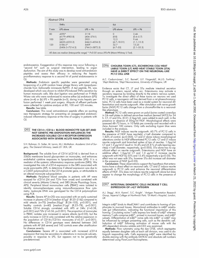
endotoxaemia. Exaggeration of this response may occur following a“second hit” such as surgical intervention, leading to organdysfunction. The aim of this study was to develop novel anti-endotoxinpeptides and assess their efficacy in reducing the hepaticproinflammatory response to a second hit of portal endotoxaemia inOJ.
Methods: Endotoxin specific peptides were generated usingbiopanning of a pVIII random linear phage library with Lipopolysac-charide from Salmonella minnesota Re995. A test peptide, P6, wasdeveloped which was shown to inhibit LPS-induced TNFa secretion byhuman monocytic cells. Bile duct ligation was performed on 9 MaleWistar rats who were randomised to receive either (a) endotoxin (LPS)alone (n=4) or (b) LPS + P6 peptide (n=5), during in-situ hepatic per-fusion performed 1 week post surgery. Aliquots of effluent perfusatewere collected for cytokine analysis at 80, 100 and 120 minutes.
Results: See table.Conclusion: This novel anti-endotoxin peptide offers an exciting
new therapeutic strategy for preventing an exaggerated endotoxin-induced inflammatory response at the time of surgery in patients withOJ.
095 THE CD14+/CD16+ BLOOD MONOCYTE SUB-SET ANDNOT GENETIC PRE-DISPOSITION INFLUENCES THEINCREASED SOLUBLE CD14 RECEPTOR EXPRESSIONASSOCIATED WITH SEVERE ACUTE PANCREATITIS
S.H. Rahman, G. Salter, M. Larvin, M.J. McMahon. Academic Unit of Sur-gery, The General Infirmary, Leeds LS1 3EA, UK
Background: The soluble form of CD14 (sCD14) is derived from a55kDa membrane bound glycoprotein on monocytes, and enhancesendothelial cytokine responses to lipopolysaccharides (LPS). It is amediator of the systemic inflammatory response syndrome (SIRS). Weinvestigated the role of sCD14 expression in the SIRS associated withacute pancreatitis (AP), to determine if altered expression was due toa C260T polymorphism in the CD14 promoter gene, or attributable toan altered monocyte sub-population.
Methods: Peripheral blood samples in patients with AP wereassayed for sCD14 (24 and 72-hr from onset) and correlated withclinical severity (Atlanta Criteria), and SIRS (Acute Physiology Score,APS). Peripheral blood mononuclear cells (PBMC) were isolated toidentify immunophenotypes using immunofluorescence flow cyto-metry. Leukocyte DNA was genotyped for the CD14 polymorphismusing PCR.
Results: Severe AP (n=20) was associated with an early sustainedincrease in plasma sCD14 [median 67µgl-1 (R:25–216)] compared tomild attacks (n=70) [median:50µgl-1 (R:24–103), p<0.001), andhealthy controls (n=40) [median:51µgl-1 (R:23–78), p<0.001].Soluble CD14 strongly correlated with APS at 24hr (r2=0.38,p<0.001) and 72hr (r2=0.56, p<0.001). The proportion of monocytesin PBMC isolates was increased in severe attacks (p<0.05), but theearly increase in CD14 only correlated with the relative expansion inthe population of CD14+/CD16+ monocytes (r2=0.57, p<0.001).No differences in CD14 genotype were detected between 245patients with AP (68 severe) and 143 controls even after stratificationfor disease severity.
Conclusions: Severe AP is associated with increased sCD14expression that may be secondary to alterations in monocyte sub-sets,possibly in response to LPS, but appears not to be geneticallypre-determined.
096 CHOLERA TOXIN (CT), ESCHERICHIA COLI HEATLABILE TOXIN (LT) AND HEAT STABLE TOXIN (STA)HAVE A DIRECT EFFECT ON THE NEURONAL-LIKEPC12 CELL LINE
A.C. Casburn-Jones1, S.C. Barnett2, U.F. Fitzgerald2, M.J.G. Farthing1.1Dept Medicine, 2Dept Neuroscience, University of Glasgow, UK
Evidence exists that CT, LT and STa mediate intestinal secretionthrough an enteric neural reflex arc. Enterotoxins may activate asecretory response by binding directly to the enteric nervous system.To investigate the direct affect of these toxins on neurons we usedPC12 cells, a neurogenic cell line derived from a rat phaeochromocy-toma. PC12 cells have been used as a model system for neuronal dif-ferentiation and neurite outgrowth. After stimulation with nerve growthfactor (NGF) PC12 cells change from a chromaffin-like to a neuronal-like phenotype.
Method: PC12 cells were grown on poly-L-lysine coated coverslipsin 24–well plates in defined serum-free medium (termed SATO) for 24h. CT, LT and STa, (0.01–2.0µg/ml), were added to each well, in thepresence or absence of 50ng/ml NGF. Morphological effects wereassessed 48–72 hours, in 10 fields per coverslip and recorded with aZeiss Axiovert 100 camera. Only cells excluding trypan blue wereincluded in the analysis.
Results: NGF induces neurite outgrowth: 60.7% of PC12 cells inthe presence of NGF have neurite(s) =1cell diameter compared to1.84% of control, (p<0.005). CT and LT alone induce morphologicalalterations similar to NGF-differentiated PC12 cells: CT and LT induceneurite outgrowth and branched spikes of tips of neurites. 1.0µg/mlCT and 1.0 µg/ml LT result in 16.4% and 25.8 % of cells bearing neu-rite(s) =1cell diameter, respectively, (p<0.005). STa alone has no sig-nificant effect on neurite outgrowth. Enterotoxins and NGF have anadditive effect: 1.0µg/ml CT and 1.0 µg/ml LT increase neuriteoutgrowth with NGF by 55% and 15.4% respectively a (p<0.05). Thiseffect was not seen with STa, however STa did increase cell diameterin the presence of NGF (p<0.05).
Conclusion: These observations support the hypothesis that entero-toxins have a direct effect on neuronal cells. CT and LT induce neuriteoutgrowth in PC12 cells and enhance the neuronal differentiationeffects of NGF. STa does not induce neurite outgrowth alone but doesappear to change the morphology of PC12 cells in the presence ofNGF.
097 INTESTINAL DENDRITIC CELLS INCREASE T CELLEXPRESSION OF α4β7 INTEGRIN
A.J. Stagg1, M.A. Kamm2, S.C. Knight1. 1Antigen Presentation ResearchGroup, Imperial College at Northwick Park; 2St Mark’s Hospital, London,UK
Integrin α4β7 binds to MadCAM-1 and contributes to homing of lym-phocytes to mucosal tissues. Monoclonal antibodies to α4β7 amelio-rate gut inflammation, indicating the functional importance of thishoming. Circulating naïve T cells express low levels of α4β7 whereasmemory T cells comprise α4β7+, primed in mucosal tissues, and α4β7-
subsets. Differentiation of α4β7lo naïve cells into α4β7+ or α4β7- maybe influenced by antigen presenting cells, such as the dendritic cell.Induction of α4β7 following activation of mouse cells with theAPC-dependent stimulus soluble anti-CD3 was examined.
Methods: Flow cytometry using the dye CFSE, which segregatesequally between daughter cells at each cell division, was used to dis-tinguish responding cells. Cells expressing α4β7 were identified bydouble staining with the antibody DATK32 and absolute cell numbersdetermined using FlowCount fluorospheres.
Abstract 094
T min
TNFα IL6
LPS alone LPS + P6 LPS alone LPS + P6
80 4390* 3118 0 3.46(4179–4983.8) (406–3761) (0-0) (0.6–5.7)
100 5451* 3951 12.3 9.44(4635–6233.2) (644.5–4334.5) (4.7–18.01) (1.1–15.3)
120 6186.5* 4409 32.7 13.25(5406.3–7214.3) (744.5–5872.5) (6.7–55) (2.1–35.3)
All data are median (Interquartile range) * P<0.05 versus LPS+P6 (Mann-Whitney U Test).
A26 BSG abstracts
www.gutjnl.com
on March 21, 2022 by guest. P
rotected by copyright.http://gut.bm
j.com/
Gut: first published as 10.1136/gut.50.suppl_2.a25 on 1 A
pril 2002. Dow
nloaded from
Results: Almost all mouse T cells freshly isolated from mesenteric(MLN) and peripheral (PLN-axillary, brachial and inguinal) nodesstained weakly for α4β7 but a subpopulation became α4β7hi uponactivation with anti-CD3 in a cell cycle- and accessory cell-dependentmanner. Precursor frequency analysis revealed that a small proportion(1.6±0.5%) of the starting cells gave rise to α4β7hi cells after culture.Both the proportion and number of dividing cells expressing α4β7was consistently greater for MLN than PLN (five experiments).Typically 2–3 fold fewer PLN than MLN were α4β7hi . Peyer’s patchcultures displayed intermediate values. In crossover experiments usinghighly purified T cells, MLN DC induced significantly (p<0.01) moreα4β7hi cells than PLN DC irrespective of the source of responding Tcells.
Conclusions: In addition to their other immunoregulatory roles,dendritic cells can shape immune responses by influencing the homingof the lymphocytes they activate. Modulating lymphocyte migrationvia the activating DC may be useful in therapy of intestinal inflamma-tion and development of mucosal vaccines.
098 STEROID ENHANCING EFFECT OF THE INTERLEUKIN-2ANTAGONIST BASILIXIMAB ON LYMPHOCYTESTEROID SENSITIVITY
S.D. Hearing, C.J. Crowley, T.J. Creed, M.N. Norman, C.M. Dayan. Uni-versity Division Medicine, Bristol Royal Infirmary, Bristol, UK
Up to 30% of patients with severe ulcerative colitis (UC) fail to respondto steroid therapy. We have previously shown that lymphocytes fromthese patients (and approximately 20% of normal subjects) havereduced sensitivity to steroids when measured in vitro1. Interleukin-2(IL-2) is released by activated T lymphocytes and acts as a natural ster-oid antagonist, promoting proliferation and reducing apoptosis. Wetherefore hypothesised that basiliximab (BAS), a clinically well-tolerated chimeric monoclonal IL-2 receptor blocking antibody mightenhance lymphocyte steroid sensitivity in resistant subjects.
Methods: Peripheral blood lymphocytes were isolated from 32subjects (25 healthy volunteers and 7 patients with quiescent UC), on41 occasions, using buoyant density centrifugation. Lymphocyte ster-oid sensitivity was assessed by measuring the antiproliferative effect ofdexamethasone (DEX) on phytohaemagglutinin stimulated lym-phocytes. Maximum steroid induced suppression was expressed as a% of control lymphocyte proliferation (Imax). Imax was measured in theabsence and presence of BAS (1mg/L).
Results: The addition of BAS significantly enhanced the anti- prolif-erative effect of DEX (Wilcoxon signed-rank test, p < 0.0001) (seetable). All 8 steroid-resistant subjects (Imax <60%) were modulated (invitro) to steroid-sensitive by the addition of BAS.
Conclusion: BAS has a marked in vitro steroid enhancing effect.As we have previously demonstrated an association betweenlymphocyte steroid resistance and poor response to steroids in severeUC1, this raises the possibility that BAS may be an effective adjuvanttherapy in steroid-resistant subjects. A clinical trial of basiliximab insteroid-resistant UC is planned.
1Hearing et al (1999) Gut 45:382–8.
099 EXPRESSION OF TUMOUR NECROSIS FACTOR α(TNFα) AND LYSOZYME IN NECROTIZINGENTEROCOLITIS (NEC)
S.G. Lala, D. Turton, S. Keshav. Department of Medicine, Royal Free andUniversity College Medical School, Rowland Hill Street, London NW3 2PF,UK
Background: NEC is an acute inflammatory disease of prematureneonates. Risk factors for NEC include prematurity, bacterial colonisa-tion, formula feeding and hypoxia, but its aetiology remains unknown.
TNFα is a potent proinflammatory cytokine associated with the devel-opment of NEC in animal models. In neonates with NEC, plasmaTNFα levels are not consistently elevated and do not correlate withdisease severity. However plasma TNFα levels do not accuratelyreflect cellular activity. We therefore analysed TNFα gene expressionin situ in intestinal tissue from neonates with severe NEC (Bell stageIIIB), and compared this with the expression of lysozyme, an antibac-terial enzyme produced by Paneth cells in the small intestine.
Methods: We performed in situ hybridisation, using digoxigeninlabelled riboprobes, on paraffin embedded intestinal tissue from eightneonates with NEC and four control subjects. Sense and anti-senseprobes were used, and macrophages were identified immunohisto-chemically using an antibody to CD 68.
Results: The intestinal architecture was disrupted in all cases ofNEC and necrosis, ulceration and haemorrhage were the dominanthistopathological features. Increased TNFα expression was present inall cases of NEC (8/8) whilst normal intestinal tissue and control NECsections showed no TNFα expression. TNFα expression was mostintense in infiltrating macrophages in the lamina propria and aroundareas of pneumatosis intestinalis. TNFα expression was also noted inPaneth cells, epithelial cells and circulating macrophages andcorrelated with the histological severity of NEC. Lysozyme geneexpression was noted in sections containing Paneth cells.
Conclusions: Increased TNFα expression characterises acute NECand this expression is sustained in established disease. These findingssupport data implicating TNFα in the pathogenesis of NEC in animalmodels. Treatment of NEC with anti-TNFα monoclonal antibodies maytherefore be justified in the setting of a controlled clinical trial.
100 A PRO-INFLAMMATORY CYTOKINE COCKTAILINCREASES CCK RELEASE FROM STC-1 CELLS VIA ANEFFECT ON INTRACELLULAR CALCIUM
F.C. Leslie, J.T. McLaughlin, S. Kazmi, A. Varro2, G. Warhurst1, G.J. Dock-ray2, D.G. Thompson. GI Science and 1Gut Barrier Group, Hope Hospital,Manchester; 2Physiological Laboratory, University of Liverpool, UK
Background: How mucosal inflammation produces anorexia ispoorly understood, modulation of enteroendocrine cell function is aplausible mechanism. Cholecystokinin (CCK) produces symptoms ofnausea and satiety when infused parenterally and is involved in IL-1induced anorexia.
Methods: The CCK secreting cell line, STC-1 was incubated withthe pro-inflammatory cytokines IFN-γ, IL-1β and TNF-α alone or incombination. CCK secretion was measured by radioimmunoassay. AsCCK secretion is mediated by increases in intracellular calcium ([Ca2+]i), changes in [Ca 2+]i in response to 70 mM K+ (a receptor-independent stimulus of secretion) were assessed using a dual wave-length ratio imaging technique (Fura-2). To look at effects on geneexpression we studied the effects of combined cytokines on activity ofCCK promoter-reporter constructs using a luciferase assay system andluminometry.
Results: Pre-incubation with the single cytokines IFN-γ, IL-1β andTNF-α for 2 hours produced small increases in basal CCK secretion.IFN-γ (6.25U/ml) produced a 38.8±4.6% increase in CCK secretioncompared to control (p=0.02), IL-1β (0.125ng/ml) produced a16.9±1.5% increase (p=0.005) and TNF-α (5ng/ml) produced a13.7±1.83% increase (p=ns). However when a cocktail of IFN-γ, IL-1βand TNF-α was pre-incubated together this produced an increase of109.4±24.0% over basal secretion after 2 hours incubation(p=0.001). This increase was maintained after 4 hours with a70.7±24.7% increase (p=0.02) but diminished at 8 hours to a43.6±8.0% increase (p=0.055). The cytokine cocktail had no effectson basal [Ca 2+]i, but pre-incubation for 2 hours produced a23.9±2.9% (p=0.03) increase in the [Ca 2+]i response to K+ comparedto control. However the cytokine cocktail did not stimulate CCKreporter-promoter activity during the same period of pre-incubation.
Conclusions: Short-term incubation with a cocktail of thepro-inflammatory cytokines IFN-γ, IL-1β and TNF-α has a synergisticeffect increasing basal CCK secretion in the STC-1 cell line. Thisoccurs via a mechanism involving changes in intracellular calcium.This may have implications for symptom genesis, particularly anorexiain proximal GI inflammation.
Dr Leslie is sponsored by the Wellcome Trust & Dr McLaughlin bythe DDF.
Abstract 098
Median Imax (%) Interquartile range
Dexamethasone 78.0 66.1–86.4Dexamethasone +Basiliximab
92.3 89.3–95.6
BSG abstracts A27
www.gutjnl.com
on March 21, 2022 by guest. P
rotected by copyright.http://gut.bm
j.com/
Gut: first published as 10.1136/gut.50.suppl_2.a25 on 1 A
pril 2002. Dow
nloaded from
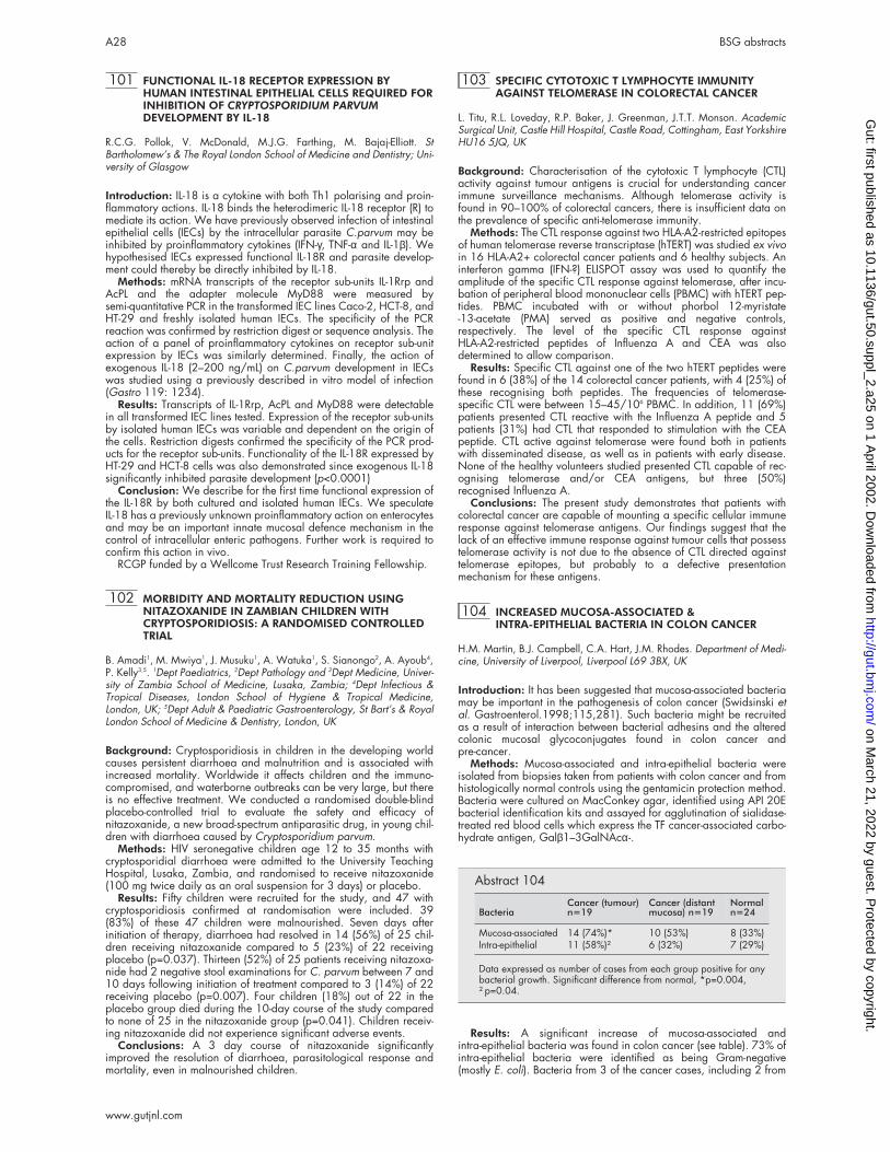
101 FUNCTIONAL IL-18 RECEPTOR EXPRESSION BYHUMAN INTESTINAL EPITHELIAL CELLS REQUIRED FORINHIBITION OF CRYPTOSPORIDIUM PARVUMDEVELOPMENT BY IL-18
R.C.G. Pollok, V. McDonald, M.J.G. Farthing, M. Bajaj-Elliott. StBartholomew’s & The Royal London School of Medicine and Dentistry; Uni-versity of Glasgow
Introduction: IL-18 is a cytokine with both Th1 polarising and proin-flammatory actions. IL-18 binds the heterodimeric IL-18 receptor (R) tomediate its action. We have previously observed infection of intestinalepithelial cells (IECs) by the intracellular parasite C.parvum may beinhibited by proinflammatory cytokines (IFN-γ, TNF-α and IL-1β). Wehypothesised IECs expressed functional IL-18R and parasite develop-ment could thereby be directly inhibited by IL-18.
Methods: mRNA transcripts of the receptor sub-units IL-1Rrp andAcPL and the adapter molecule MyD88 were measured bysemi-quantitative PCR in the transformed IEC lines Caco-2, HCT-8, andHT-29 and freshly isolated human IECs. The specificity of the PCRreaction was confirmed by restriction digest or sequence analysis. Theaction of a panel of proinflammatory cytokines on receptor sub-unitexpression by IECs was similarly determined. Finally, the action ofexogenous IL-18 (2–200 ng/mL) on C.parvum development in IECswas studied using a previously described in vitro model of infection(Gastro 119: 1234).
Results: Transcripts of IL-1Rrp, AcPL and MyD88 were detectablein all transformed IEC lines tested. Expression of the receptor sub-unitsby isolated human IECs was variable and dependent on the origin ofthe cells. Restriction digests confirmed the specificity of the PCR prod-ucts for the receptor sub-units. Functionality of the IL-18R expressed byHT-29 and HCT-8 cells was also demonstrated since exogenous IL-18significantly inhibited parasite development (p<0.0001)
Conclusion: We describe for the first time functional expression ofthe IL-18R by both cultured and isolated human IECs. We speculateIL-18 has a previously unknown proinflammatory action on enterocytesand may be an important innate mucosal defence mechanism in thecontrol of intracellular enteric pathogens. Further work is required toconfirm this action in vivo.
RCGP funded by a Wellcome Trust Research Training Fellowship.
102 MORBIDITY AND MORTALITY REDUCTION USINGNITAZOXANIDE IN ZAMBIAN CHILDREN WITHCRYPTOSPORIDIOSIS: A RANDOMISED CONTROLLEDTRIAL
B. Amadi1, M. Mwiya1, J. Musuku1, A. Watuka1, S. Sianongo2, A. Ayoub4,P. Kelly3,5. 1Dept Paediatrics, 2Dept Pathology and 3Dept Medicine, Univer-sity of Zambia School of Medicine, Lusaka, Zambia; 4Dept Infectious &Tropical Diseases, London School of Hygiene & Tropical Medicine,London, UK; 5Dept Adult & Paediatric Gastroenterology, St Bart’s & RoyalLondon School of Medicine & Dentistry, London, UK
Background: Cryptosporidiosis in children in the developing worldcauses persistent diarrhoea and malnutrition and is associated withincreased mortality. Worldwide it affects children and the immuno-compromised, and waterborne outbreaks can be very large, but thereis no effective treatment. We conducted a randomised double-blindplacebo-controlled trial to evaluate the safety and efficacy ofnitazoxanide, a new broad-spectrum antiparasitic drug, in young chil-dren with diarrhoea caused by Cryptosporidium parvum.
Methods: HIV seronegative children age 12 to 35 months withcryptosporidial diarrhoea were admitted to the University TeachingHospital, Lusaka, Zambia, and randomised to receive nitazoxanide(100 mg twice daily as an oral suspension for 3 days) or placebo.
Results: Fifty children were recruited for the study, and 47 withcryptosporidiosis confirmed at randomisation were included. 39(83%) of these 47 children were malnourished. Seven days afterinitiation of therapy, diarrhoea had resolved in 14 (56%) of 25 chil-dren receiving nitazoxanide compared to 5 (23%) of 22 receivingplacebo (p=0.037). Thirteen (52%) of 25 patients receiving nitazoxa-nide had 2 negative stool examinations for C. parvum between 7 and10 days following initiation of treatment compared to 3 (14%) of 22receiving placebo (p=0.007). Four children (18%) out of 22 in theplacebo group died during the 10-day course of the study comparedto none of 25 in the nitazoxanide group (p=0.041). Children receiv-ing nitazoxanide did not experience significant adverse events.
Conclusions: A 3 day course of nitazoxanide significantlyimproved the resolution of diarrhoea, parasitological response andmortality, even in malnourished children.
103 SPECIFIC CYTOTOXIC T LYMPHOCYTE IMMUNITYAGAINST TELOMERASE IN COLORECTAL CANCER
L. Titu, R.L. Loveday, R.P. Baker, J. Greenman, J.T.T. Monson. AcademicSurgical Unit, Castle Hill Hospital, Castle Road, Cottingham, East YorkshireHU16 5JQ, UK
Background: Characterisation of the cytotoxic T lymphocyte (CTL)activity against tumour antigens is crucial for understanding cancerimmune surveillance mechanisms. Although telomerase activity isfound in 90–100% of colorectal cancers, there is insufficient data onthe prevalence of specific anti-telomerase immunity.
Methods: The CTL response against two HLA-A2-restricted epitopesof human telomerase reverse transcriptase (hTERT) was studied ex vivoin 16 HLA-A2+ colorectal cancer patients and 6 healthy subjects. Aninterferon gamma (IFN-?) ELISPOT assay was used to quantify theamplitude of the specific CTL response against telomerase, after incu-bation of peripheral blood mononuclear cells (PBMC) with hTERT pep-tides. PBMC incubated with or without phorbol 12-myristate-13-acetate (PMA) served as positive and negative controls,respectively. The level of the specific CTL response againstHLA-A2-restricted peptides of Influenza A and CEA was alsodetermined to allow comparison.
Results: Specific CTL against one of the two hTERT peptides werefound in 6 (38%) of the 14 colorectal cancer patients, with 4 (25%) ofthese recognising both peptides. The frequencies of telomerase-specific CTL were between 15–45/106 PBMC. In addition, 11 (69%)patients presented CTL reactive with the Influenza A peptide and 5patients (31%) had CTL that responded to stimulation with the CEApeptide. CTL active against telomerase were found both in patientswith disseminated disease, as well as in patients with early disease.None of the healthy volunteers studied presented CTL capable of rec-ognising telomerase and/or CEA antigens, but three (50%)recognised Influenza A.
Conclusions: The present study demonstrates that patients withcolorectal cancer are capable of mounting a specific cellular immuneresponse against telomerase antigens. Our findings suggest that thelack of an effective immune response against tumour cells that possesstelomerase activity is not due to the absence of CTL directed againsttelomerase epitopes, but probably to a defective presentationmechanism for these antigens.
104 INCREASED MUCOSA-ASSOCIATED &INTRA-EPITHELIAL BACTERIA IN COLON CANCER
H.M. Martin, B.J. Campbell, C.A. Hart, J.M. Rhodes. Department of Medi-cine, University of Liverpool, Liverpool L69 3BX, UK
Introduction: It has been suggested that mucosa-associated bacteriamay be important in the pathogenesis of colon cancer (Swidsinski etal. Gastroenterol.1998;115,281). Such bacteria might be recruitedas a result of interaction between bacterial adhesins and the alteredcolonic mucosal glycoconjugates found in colon cancer andpre-cancer.
Methods: Mucosa-associated and intra-epithelial bacteria wereisolated from biopsies taken from patients with colon cancer and fromhistologically normal controls using the gentamicin protection method.Bacteria were cultured on MacConkey agar, identified using API 20Ebacterial identification kits and assayed for agglutination of sialidase-treated red blood cells which express the TF cancer-associated carbo-hydrate antigen, Galβ1–3GalNAcα-.
Results: A significant increase of mucosa-associated andintra-epithelial bacteria was found in colon cancer (see table). 73% ofintra-epithelial bacteria were identified as being Gram-negative(mostly E. coli). Bacteria from 3 of the cancer cases, including 2 from
Abstract 104
BacteriaCancer (tumour)n=19
Cancer (distantmucosa) n=19
Normaln=24
Mucosa-associated 14 (74%)* 10 (53%) 8 (33%)Intra-epithelial 11 (58%)† 6 (32%) 7 (29%)
Data expressed as number of cases from each group positive for anybacterial growth. Significant difference from normal, *p=0.004,†p=0.04.
A28 BSG abstracts
www.gutjnl.com
on March 21, 2022 by guest. P
rotected by copyright.http://gut.bm
j.com/
Gut: first published as 10.1136/gut.50.suppl_2.a25 on 1 A
pril 2002. Dow
nloaded from
the distant mucosa, but none of the normal controls, tested positive forTF adhesin activity. This is the first report of TF-binding intestinal bac-teria in humans. Further studies on an isolate of TF-adhesin-positivenon-pathogenic E. coli (HM44), which we have shown to lack conven-tional pathogenicity islands, from a colon cancer case show that it isable to invade HT29 human colon cancer cells.
Conclusions: These results support the hypothesis that alteredmucosal carbohydrate expression (such as TF antigen) in colon cancerand distant “unaffected” mucosa may lead to recruitment of E. coli.Bacteria which lack conventional markers of pathogenicity but whichcan invade intestinal epithelial cells could be relevant to carcinogen-esis.
Symposium: Gut flora in IBD—practical importance 105–107
105 DIAGNOSIS OF CROHN’S DISEASE IS ASSOCIATEDWITH INCREASED LEVELS OF ANTIBIOTIC USE OVERTHE PRECEEDING FIVE YEARS
T.R. Card1, R.F.A. Logan1, L.C. Rodrigues2, J.G. Wheeler2. 1Division ofPublic Health Sciences, University of Nottingham, Queen’s MedicalCentre; 2London School of Hygiene and Tropical Medicine, UK
Background: Although it is generally accepted that Crohn’s diseasehas both genetic and environmental determinants, few environmentaldeterminants are well established. In view of the widely supportednotion that dysfunction of the gut is inter-linked with its flora, weexplored the potential importance of the use of antibiotics as a riskfactor.
Methods: We selected records of incident cases of Crohn’sdisease from the General Practice Research Database. All cases werediagnosed after 1992, had no history of Ulcerative Colitis and at leastfive years of GPRD data prior to diagnosis. Controls with five yearscomplete data were randomly selected. Data were extracted on smok-ing, presentation to GP with a diagnosis of infection, oral contracep-tive use (OCP) and antibiotic use 3–5 years prior to the date ofdiagnosis of Crohn’s disease (and for a comparable period forcontrols). Data on antibiotic use was restricted in time to exclude thosethat could have been prescribed as treatment of premonitorysymptoms. Logistic regression was used to investigate the relationshipbetween antibiotic use and Crohn’s disease.
Results: 601 Crohn’s disease and 1460 controls were availablefor analysis. We found statistically significant associations betweenCrohn’s disease and use of OCP in the year before diagnosis (Oddsratio 1.7, P=0.003) and smoking (Odds ratio 1.5, P=0.001) despiteincomplete data for the latter (these are consistent with the literature).Antibiotic use 3–5 years pre-diagnosis was significantly greater incases than controls. Only 29% of cases compared to 42% of controlsreceived no antibiotics (P<0.001), and the mean number of courseswas 2.6 in cases and 1.9 in controls (P<0.0001). Adjusting for age,sex, smoking and use of OCP, antibiotic use had an odds ratio of 1.6(1.3–2.0) (P<0.001). There was a highly significant trend withincreasing numbers of courses (P=0.001). The population attributablefraction for antibiotic use was 28% (18%-36%).
Conclusions: There is a highly significant association betweenCrohn’s disease and prior antibiotic use in this data. This is unlikely tobe explained by reporting bias in view of the prospective recording ofall prescriptions in GPRD. If this association is causal then it mayexplain about a quarter of all Crohn’s disease.
106 MICROBIAL CELL WALL OLIGOMANNAN INHIBITS THENEUTROPHIL RESPIRATORY BURST: A POSSIBLEMECHANISM FOR GRANULOMA FORMATION INCROHN’S DISEASE?
C.M. Mpofu, J.M. Rhodes, S.W. Edwards. Department of Medicine andSchool of Biological Sciences, University of Liverpool, Liverpool L69 3GA,UK
Background: Crohn’s like intestinal lesions occur in Chronic Granulo-matous Disease, a condition caused by a defect in phagocytefunction. Crohns disease patients commonly have serum antibodies tobaker’s yeast (Saccharomyces cerevisiae). The epitope for thisantibody is oligomannan which is present in bacterial and yeast cell
walls. Our hypothesis is that oligomannan, shed by intramucosal bac-teria, may inhibit neutrophil and macrophage function within themucosa, leading to the granulomatous lesions seen in CD.
Methods: The effect of S. cerevesiae oligomannan on phorbol ester(PMA) induced-respiratory burst of neutrophils from healthy volunteerswas measured by luminol-amplified and isoluminol-amplified chemilu-minescence and by the cytochrome c reduction method. Neutrophilswere isolated using a one-step centrifugation method and suspendedin HBSS buffer.
Results: See fig 1. At the highest concentration used (1 mg/mL),oligomannan inhibited luminol dependent chemiluminescence by 68% (± 5 %, n = 6). Luminol chemiluminescence measures the combinedactivities of the neutrophil NADPH oxidase and myeloperoxidase, andso the oligomannan could be acting on either or both of these activi-ties. However, oligomannan (at 1 mg/mL) also inhibited PMA-stimulated isoluminol-amplified chemiluminescence and cytochrome creduction. As isoluminol and cytochrome c reduction largely measureextracellular superoxide secretion, the oligomannan may have aneffect on NADPH oxidase activity.
Conclusion: S cerevesiae oligomannan causes dose-related inhibi-tion of the PMA-induced respiratory burst. It has extracellular andprobably intracellular effects. This supports the hypothesis that micro-bial oligomannans impair mucosal phagocyte function, thus generat-ing the granulomatous phenotype of Crohn’s disease.
107 REDUCED BIFIDOBACTERIA AND INCREASED E. COLIIN RECTAL MUCOSA-ASSOCIATED FLORA IN ACTIVEINFLAMMATORY BOWEL DISEASE
N. Rayment, M. Mylonaki, B. Hudspith, J. Brostoff, D.S. Rampton. Infectionand Immunity Group, Division of Life Science, Kings College London; Aca-demic Dept of Adult & Paediatric Gastroenterology, Barts & The LondonSchool of Medicine, London, UK
Background: Colorectal bacteria probably contribute to thepathogenesis of IBD. To test the hypothesis that potentially protectivebacteria are reduced and pathogenic flora increased, we have com-pared the mucosa-associated flora in IBD and controls.
Methods: Snap-frozen rectal biopsies were taken at routinecolonoscopy from patients with ulcerative colitis (UC), Crohn’s andcontrols with normal colorectal mucosa. Fluorescent in situ hybridisa-tion was used separately to count numbers of mucosa-associated bifi-dobacteria, lactobacillus, E.coli, clostridia and sulphate-reducingbacteria.
Results: Bacteria were sited on, and superficially within rectalmucosa. Mucosa-associated bifidobacteria counts (median 14/hpf(range 4–65), n=13) in active UC were lower than in controls (79(0–146), n=24, P=0.008); the difference from quiescent UC (38(0–118), n=19) did not reach statistical significance (P=0.07).Conversely, E.coli counts were higher in active UC (80/hpf (30–186))than in controls (0 (0–16), P=0.0002) and inactive disease (2(0–144), P=0.003). Similar results were found in patients with activeCrohn’s colitis (n=4). In 6 patients with UC who had biopsies from 2sites, inflamed mucosa always showed fewer bifidobacteria(P=0.008) and more E.coli (P=0.008) than normal-looking moreproximal colon. There were no differences, between IBD and controls,of quantitative counts of other bacteria.
Conclusions: The reduction in mucosa-associated bifidobacteriaand increase in E.coli in active IBD supports the hypothesis that a defi-ciency of potentially beneficial bifidobacteria and excess of E.colicould play a role in the causation of IBD.
80
60
40
20
0%Decrease
in
chemilumine
scene
0.125 0.25 0.5
Oligomannan dose (mg/ml)
Oligomannan decreases the neutrophil respiratory
burst (n = 4-6)
1
BSG abstracts A29
www.gutjnl.com
on March 21, 2022 by guest. P
rotected by copyright.http://gut.bm
j.com/
Gut: first published as 10.1136/gut.50.suppl_2.a25 on 1 A
pril 2002. Dow
nloaded from
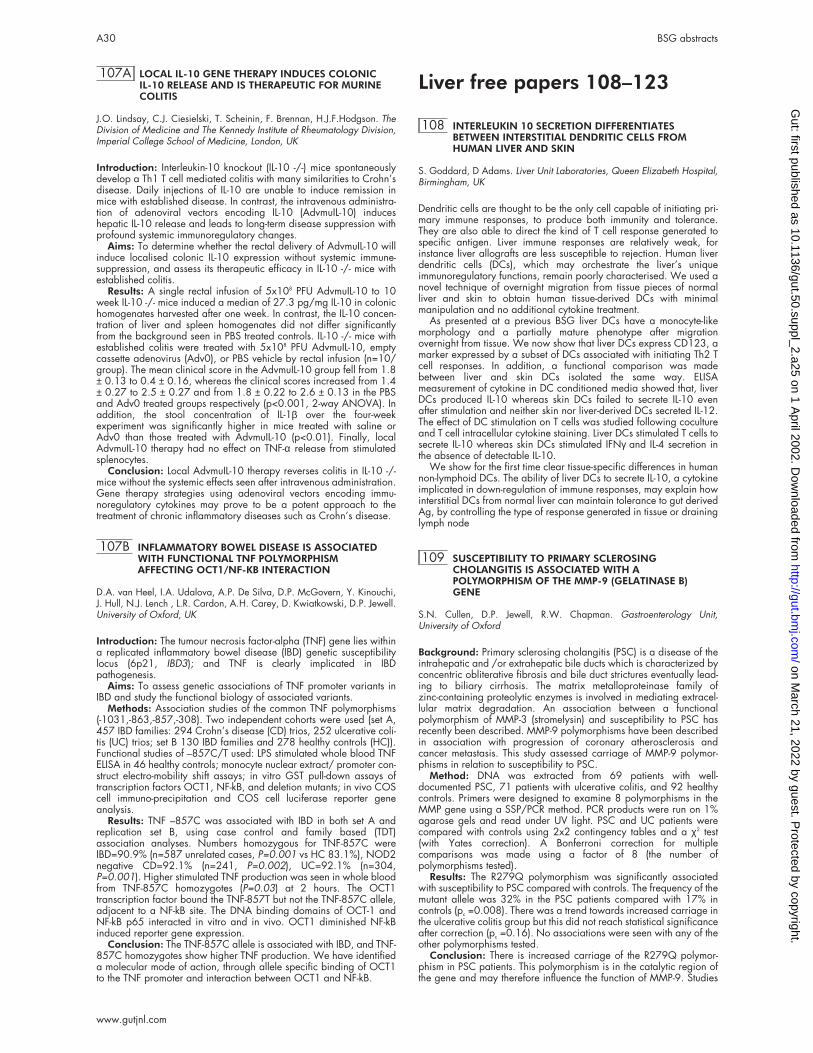
107A LOCAL IL-10 GENE THERAPY INDUCES COLONICIL-10 RELEASE AND IS THERAPEUTIC FOR MURINECOLITIS
J.O. Lindsay, C.J. Ciesielski, T. Scheinin, F. Brennan, H.J.F.Hodgson. TheDivision of Medicine and The Kennedy Institute of Rheumatology Division,Imperial College School of Medicine, London, UK
Introduction: Interleukin-10 knockout (IL-10 -/-) mice spontaneouslydevelop a Th1 T cell mediated colitis with many similarities to Crohn’sdisease. Daily injections of IL-10 are unable to induce remission inmice with established disease. In contrast, the intravenous administra-tion of adenoviral vectors encoding IL-10 (AdvmuIL-10) induceshepatic IL-10 release and leads to long-term disease suppression withprofound systemic immunoregulatory changes.
Aims: To determine whether the rectal delivery of AdvmuIL-10 willinduce localised colonic IL-10 expression without systemic immune-suppression, and assess its therapeutic efficacy in IL-10 -/- mice withestablished colitis.
Results: A single rectal infusion of 5x10⁸ PFU AdvmuIL-10 to 10week IL-10 -/- mice induced a median of 27.3 pg/mg IL-10 in colonichomogenates harvested after one week. In contrast, the IL-10 concen-tration of liver and spleen homogenates did not differ significantlyfrom the background seen in PBS treated controls. IL-10 -/- mice withestablished colitis were treated with 5x10⁸ PFU AdvmuIL-10, emptycassette adenovirus (Adv0), or PBS vehicle by rectal infusion (n=10/group). The mean clinical score in the AdvmuIL-10 group fell from 1.8± 0.13 to 0.4 ± 0.16, whereas the clinical scores increased from 1.4± 0.27 to 2.5 ± 0.27 and from 1.8 ± 0.22 to 2.6 ± 0.13 in the PBSand Adv0 treated groups respectively (p<0.001, 2-way ANOVA). Inaddition, the stool concentration of IL-1β over the four-weekexperiment was significantly higher in mice treated with saline orAdv0 than those treated with AdvmuIL-10 (p<0.01). Finally, localAdvmuIL-10 therapy had no effect on TNF-α release from stimulatedsplenocytes.
Conclusion: Local AdvmuIL-10 therapy reverses colitis in IL-10 -/-mice without the systemic effects seen after intravenous administration.Gene therapy strategies using adenoviral vectors encoding immu-noregulatory cytokines may prove to be a potent approach to thetreatment of chronic inflammatory diseases such as Crohn’s disease.
107B INFLAMMATORY BOWEL DISEASE IS ASSOCIATEDWITH FUNCTIONAL TNF POLYMORPHISMAFFECTING OCT1/NF-KB INTERACTION
D.A. van Heel, I.A. Udalova, A.P. De Silva, D.P. McGovern, Y. Kinouchi,J. Hull, N.J. Lench , L.R. Cardon, A.H. Carey, D. Kwiatkowski, D.P. Jewell.University of Oxford, UK
Introduction: The tumour necrosis factor-alpha (TNF) gene lies withina replicated inflammatory bowel disease (IBD) genetic susceptibilitylocus (6p21, IBD3); and TNF is clearly implicated in IBDpathogenesis.
Aims: To assess genetic associations of TNF promoter variants inIBD and study the functional biology of associated variants.
Methods: Association studies of the common TNF polymorphisms(-1031,-863,-857,-308). Two independent cohorts were used (set A,457 IBD families: 294 Crohn’s disease (CD) trios, 252 ulcerative coli-tis (UC) trios; set B 130 IBD families and 278 healthy controls (HC)).Functional studies of –857C/T used: LPS stimulated whole blood TNFELISA in 46 healthy controls; monocyte nuclear extract/ promoter con-struct electro-mobility shift assays; in vitro GST pull-down assays oftranscription factors OCT1, NF-kB, and deletion mutants; in vivo COScell immuno-precipitation and COS cell luciferase reporter geneanalysis.
Results: TNF –857C was associated with IBD in both set A andreplication set B, using case control and family based (TDT)association analyses. Numbers homozygous for TNF-857C wereIBD=90.9% (n=587 unrelated cases, P=0.001 vs HC 83.1%), NOD2negative CD=92.1% (n=241, P=0.002), UC=92.1% (n=304,P=0.001). Higher stimulated TNF production was seen in whole bloodfrom TNF-857C homozygotes (P=0.03) at 2 hours. The OCT1transcription factor bound the TNF-857T but not the TNF-857C allele,adjacent to a NF-kB site. The DNA binding domains of OCT-1 andNF-kB p65 interacted in vitro and in vivo. OCT1 diminished NF-kBinduced reporter gene expression.
Conclusion: The TNF-857C allele is associated with IBD, and TNF-857C homozygotes show higher TNF production. We have identifieda molecular mode of action, through allele specific binding of OCT1to the TNF promoter and interaction between OCT1 and NF-kB.
Liver free papers 108–123
108 INTERLEUKIN 10 SECRETION DIFFERENTIATESBETWEEN INTERSTITIAL DENDRITIC CELLS FROMHUMAN LIVER AND SKIN
S. Goddard, D Adams. Liver Unit Laboratories, Queen Elizabeth Hospital,Birmingham, UK
Dendritic cells are thought to be the only cell capable of initiating pri-mary immune responses, to produce both immunity and tolerance.They are also able to direct the kind of T cell response generated tospecific antigen. Liver immune responses are relatively weak, forinstance liver allografts are less susceptible to rejection. Human liverdendritic cells (DCs), which may orchestrate the liver’s uniqueimmunoregulatory functions, remain poorly characterised. We used anovel technique of overnight migration from tissue pieces of normalliver and skin to obtain human tissue-derived DCs with minimalmanipulation and no additional cytokine treatment.
As presented at a previous BSG liver DCs have a monocyte-likemorphology and a partially mature phenotype after migrationovernight from tissue. We now show that liver DCs express CD123, amarker expressed by a subset of DCs associated with initiating Th2 Tcell responses. In addition, a functional comparison was madebetween liver and skin DCs isolated the same way. ELISAmeasurement of cytokine in DC conditioned media showed that, liverDCs produced IL-10 whereas skin DCs failed to secrete IL-10 evenafter stimulation and neither skin nor liver-derived DCs secreted IL-12.The effect of DC stimulation on T cells was studied following cocultureand T cell intracellular cytokine staining. Liver DCs stimulated T cells tosecrete IL-10 whereas skin DCs stimulated IFNγ and IL-4 secretion inthe absence of detectable IL-10.
We show for the first time clear tissue-specific differences in humannon-lymphoid DCs. The ability of liver DCs to secrete IL-10, a cytokineimplicated in down-regulation of immune responses, may explain howinterstitial DCs from normal liver can maintain tolerance to gut derivedAg, by controlling the type of response generated in tissue or draininglymph node
109 SUSCEPTIBILITY TO PRIMARY SCLEROSINGCHOLANGITIS IS ASSOCIATED WITH APOLYMORPHISM OF THE MMP-9 (GELATINASE B)GENE
S.N. Cullen, D.P. Jewell, R.W. Chapman. Gastroenterology Unit,University of Oxford
Background: Primary sclerosing cholangitis (PSC) is a disease of theintrahepatic and /or extrahepatic bile ducts which is characterized byconcentric obliterative fibrosis and bile duct strictures eventually lead-ing to biliary cirrhosis. The matrix metalloproteinase family ofzinc-containing proteolytic enzymes is involved in mediating extracel-lular matrix degradation. An association between a functionalpolymorphism of MMP-3 (stromelysin) and susceptibility to PSC hasrecently been described. MMP-9 polymorphisms have been describedin association with progression of coronary atherosclerosis andcancer metastasis. This study assessed carriage of MMP-9 polymor-phisms in relation to susceptibility to PSC.
Method: DNA was extracted from 69 patients with well-documented PSC, 71 patients with ulcerative colitis, and 92 healthycontrols. Primers were designed to examine 8 polymorphisms in theMMP gene using a SSP/PCR method. PCR products were run on 1%agarose gels and read under UV light. PSC and UC patients werecompared with controls using 2x2 contingency tables and a χ2 test(with Yates correction). A Bonferroni correction for multiplecomparisons was made using a factor of 8 (the number ofpolymorphisms tested).
Results: The R279Q polymorphism was significantly associatedwith susceptibility to PSC compared with controls. The frequency of themutant allele was 32% in the PSC patients compared with 17% incontrols (pc =0.008). There was a trend towards increased carriage inthe ulcerative colitis group but this did not reach statistical significanceafter correction (pc =0.16). No associations were seen with any of theother polymorphisms tested.
Conclusion: There is increased carriage of the R279Q polymor-phism in PSC patients. This polymorphism is in the catalytic region ofthe gene and may therefore influence the function of MMP-9. Studies
A30 BSG abstracts
www.gutjnl.com
on March 21, 2022 by guest. P
rotected by copyright.http://gut.bm
j.com/
Gut: first published as 10.1136/gut.50.suppl_2.a25 on 1 A
pril 2002. Dow
nloaded from
are currently being undertaken to address the possible functionaleffects of this polymorphism.
110 PRIMARY BILIARY CIRRHOSIS (PBC): NO SPECIFICASSOCIATION WITH MICROCHIMERISM
R. Buckland, K.L.E. Dear1, A.C. Goodeve, J.C.E. Underwood2, D.Gleeson1. Division of Genomic Medicine and 2Dept of Pathology, Univer-sity of Sheffield; 1Liver Unit, Sheffield Teaching Hospitals, Sheffield, UK
Background: Some diseases may result from immunocompetent fetalcells acquired during previous pregnancies (allo-immunity) andpersisting in the mother for decades (microchimerism). In women withscleroderma who have had male children, male DNA is found in skinand peripheral blood more frequently than in control women. Primarybiliary cirrhosis (PBC) mainly affects older women and has similaritiesto graft versus host disease, consistent with a role for alloimmunity.Studies on the association of PBC with microchimerism have beensmall (<20 PBC liver specimens) and results have been conflicting.
Aim: To address the association of PBC with microchimerism in alarger cohort.
Methods: We studied (a): blood (2 extractions x 2 PCR= 4 PCR)from 55 women with PBC and 49 normal control women (irritiblebowel or G-O reflux; normal liver enzymes) and (b): archived needleliver biopsies (1 extraction x 2 PCR = 2 PCR) from 42 women withPBC, 21 women with normal liver histology or mild steatosis (normalcontrols), and 32 women with autoimmune hepatitis (AIH) (diseasecontrols). All had had >1 male child and none met ARA criteria forscleroderma or had had a blood transfusion or a liver transplant whentissue was obtained. Male DNA was assayed by PCR using specific Ychromosome primers; lower detection limit = 1/3 of DNA in 1 cell(2pg).
Results: The table shows subject numbers (%) with 0, 1 and.>2PCRs positive for male DNA.
Conclusion: PBC showed an unexpected negative association withmicrochimerism in blood and no significant association (compared toeither control group) in liver. This, the largest study to date, does notsupport a role for microchimerism in the pathogenesis of PBC.
111 FASTING INSULIN, 31,32 SPLIT PRO-INSULIN ANDINSULIN RESISTANCE IN PATIENTS WITHNON-ALCOHOLIC FATTY LIVER
G.Constable, D.Cumming, P.J.Wood, S.Wootton, M.A.Stroud. Institute ofHuman Nutrition, Southampton University Hospital, UK
Introduction: Nonalcoholic fatty liver (NAFL) is associated with insu-lin resistance and an increased risk of type II diabetes (T2DM). Devel-opment of T2DM results from either insulin resistance or defectiveinsulin processing/secretion or both. Insulin resistance leads toincreased lipolysis and flux of fatty acids (FFA) to the liver, which inturn may increase hepatic glucose output. Insulin processing involvesenzymatic conversion of proinsulin to insulin through a series ofsite-specific cleavages. Recent development of specific assays allowsdifferent molecules of the proinsulin processing pathway to bemeasured separately. 32,33 split proinsulin is the predominant formof proinsulin, known to be elevated in T2DM and impaired glucosetolerance. Fasting levels have also been shown to predict developmentof T2DM.
Methods: We measured fasting insulin, intact proinsulin and32,33 split proinsulin levels in patients with NAFL (n=24) compared toa healthy reference group (n=14). Insulin resistance index (IRI) wascalculated from fasting plasma glucose and insulin levels using the
well recognised mathematical model of glucose:insulin interactions -the ‘homeostatic model assessment’ (HOMA). Body composition wasassessed by BMI, waist:hip ratio and bioelectrical impedance analysis(BIA).
Results: Patients with NAFL were obese according to BMI: 31.19+/- 0.81 vs 26.55 +/- 0.81, p<0.004. The NAFL group had higherinsulin levels than reference: 21.44 +/- 2.64 mU/l vs 9.07 +/- 1.16mU/l, p<0.0014 and higher 32,33 split proinsulin levels: 18.50mU/l +/- 1.85 mU/l vs 8.55 +/- 0.73 mU/l, p<0.0003. The NAFLgroup were also significantly insulin resistant using HOMA,p<0.0015. These results were independent of BMI and body fatassessed by BIA.
Conclusions: NAFL is associated with hyperinsulinaemia, elevated32,33 split proinsulin levels and insulin resistance. Patients with NAFLare at increased risk of developing T2DM. Measurement of 32,33split proinsulin may help to select individuals with the highest risk fortargeted intervention. Insulin resistance causes an increased flux ofFFA to the liver leading to fat deposition if metabolic competence fordisposal is exceeded. Increased FFA flux may also result in hepaticoverproduction of glucose through mechanisms that are incompletelyunderstood.
112 INHIBITION OF APOPTOSIS OF ACTIVATED HEPATICSTELLATE CELLS BY TIMP-1 IS MEDIATED VIA EFFECTSON MMP INHIBITION: IMPLICATIONS FORREVERSIBILITY OF LIVER FIBROSIS
F. Murphy, R. Issa, X. Zhou, H. Hussain, S. Ratnarajah, P. Soloway1, H.Nagase2, M.J.P. Arthur, R.C. Benyon, J.P. Iredale. Liver Group, Division ofInfection, Inflammation & Repair, Southampton University, SouthamptonSO16 6YD, UK; 1Rosewell Park Cancer Institute, New York, USA; 2TheKennedy Institute, Imperial College, London, UK
Introduction: The activated hepatic stellate cell (HSC) is central toliver fibrosis as the major source of collagens I and III and the tissueinhibitors of metalloproteinase-1 and -2 (TIMPs). During spontaneousrecovery from liver fibrosis there is a decrease of TIMP expression, anincrease in collagenase activity and apoptosis of HSC, highlightingthe potential role of TIMP-1 and -2 in HSC survival.
Aims: To determine if TIMP-1 and TIMP-2 directly inhibit HSCapoptosis in tissue culture and in models of liver fibrosis in vivo.
Methods: Effects of recombinant TIMPs and mutated TIMP-1 on cul-tured activated HSC were examined after induction of apoptosis bycycloheximide in vitro. Rat and murine models of experimental liverfibrosis induced by CCl4 were examined during spontaneousrecovery. HSC number, TUNEL staining and TIMP-1 mRNA wereassessed.
Results: TIMP-1 and 2 demonstrated a consistent, significant anddose dependent anti apoptotic effect on HSC activated in tissueculture. A non-functional mutated TIMP-1 (T2G mutant) did not inhibitapoptosis indicating that inhibition of apoptosis was mediatedthrough MMP inhibition. Studies of experimental liver fibrosis in the ratdemonstrated that loss of activated HSC correlated with a reduction inTIMP-1 mRNA expression determined by PCR. Persistence of HSCs inmore advanced fibrosis correlated with persistent TIMP-1 mRNAexpression. After induction of fibrosis in vivo, TIMP-1 knockout micedemonstrated significantly more HSC apoptosis relative to wild typesat 3 and 7 days of spontaneous recovery.
Conclusion: TIMP-1 and -2 inhibit apoptosis of activated HSC. Theanti apoptotic effect of TIMP-1 is mediated via MMP inhibition.
113 DISTRIBUTION OF THE CONSTITUTIVE (COX-1) ANDTHE INDUCIBEL (COX-2) CYCLOOXYGENASE INHUMAN LIVER CIRRHOSIS: A POSSIBLE ROLE OFCOX-2 IN PATHOGENESIS OF LIVER CIRRHOSIS
N. Mohammed, S. Abd El-Aleem, H. Abdel Hafiz, M. Maklouf, R.F.T.McMahon. Department of Histopathology, Clinical Sciences Building,Manchester Royal Infirmary, Oxford Road, Manchester M13 9WL, UK
Several mediators of systemic vasodilatation and inflammation in livercirrhosis have been reported. Among these are prostaglandins (PGs),which have been proposed as one of the main mediators duringinflammation. In this study, liver biopsies from fifteen patients withclinically and pathologically diagnosed liver cirrhosis secondary tohepatitis B and C, were taken. In addition 3 liver biopsies from healthycontrols were used. The protein expression of the constitutive (COX-1)and the inducible (COX-2) cyclooxygenase was investigated usingimmunocytochemistry.
Abstract 110
0 1 >2 3x2 chi2
Blood PBC 27 (49) 19(35) 9 (16) 9.86Normal 13 (27) 15 (31) 21 (43) (<0.02)
Liver PBC 1(2) 10 (24) 31 (74)Normal 3 (14) 7 (33) 11 (52) 4.56 ns*AIH 5 (16) 6 (19) 21 (65) 4.32 ns*
*: vs PBC livers.
BSG abstracts A31
www.gutjnl.com
on March 21, 2022 by guest. P
rotected by copyright.http://gut.bm
j.com/
Gut: first published as 10.1136/gut.50.suppl_2.a25 on 1 A
pril 2002. Dow
nloaded from
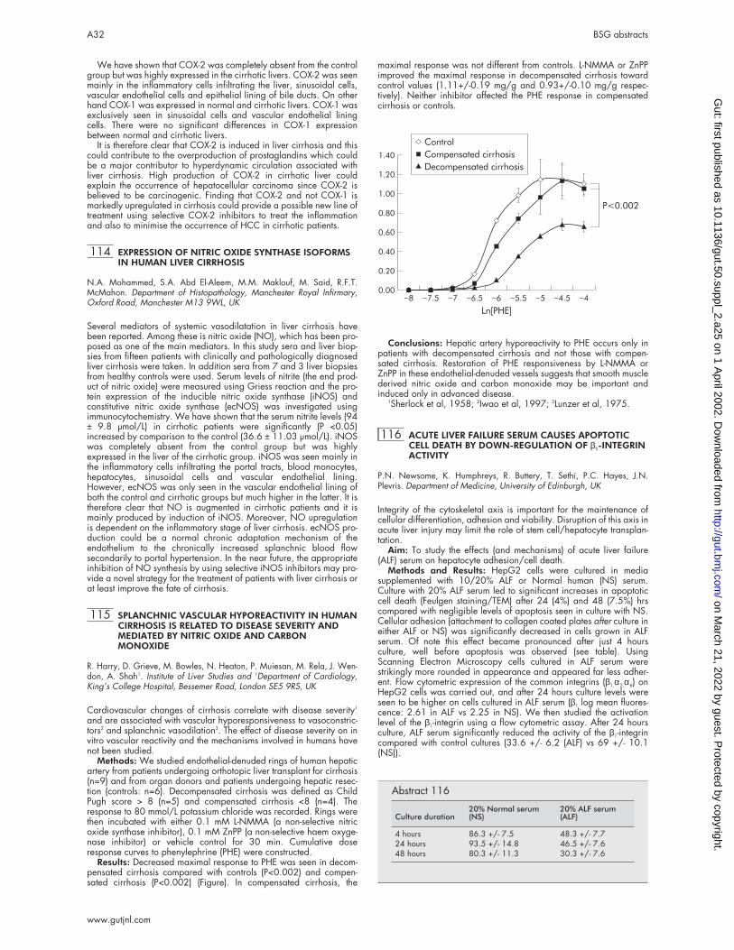
We have shown that COX-2 was completely absent from the controlgroup but was highly expressed in the cirrhotic livers. COX-2 was seenmainly in the inflammatory cells infiltrating the liver, sinusoidal cells,vascular endothelial cells and epithelial lining of bile ducts. On otherhand COX-1 was expressed in normal and cirrhotic livers. COX-1 wasexclusively seen in sinusoidal cells and vascular endothelial liningcells. There were no significant differences in COX-1 expressionbetween normal and cirrhotic livers.
It is therefore clear that COX-2 is induced in liver cirrhosis and thiscould contribute to the overproduction of prostaglandins which couldbe a major contributor to hyperdynamic circulation associated withliver cirrhosis. High production of COX-2 in cirrhotic liver couldexplain the occurrence of hepatocellular carcinoma since COX-2 isbelieved to be carcinogenic. Finding that COX-2 and not COX-1 ismarkedly upregulated in cirrhosis could provide a possible new line oftreatment using selective COX-2 inhibitors to treat the inflammationand also to minimise the occurrence of HCC in cirrhotic patients.
114 EXPRESSION OF NITRIC OXIDE SYNTHASE ISOFORMSIN HUMAN LIVER CIRRHOSIS
N.A. Mohammed, S.A. Abd El-Aleem, M.M. Maklouf, M. Said, R.F.T.McMahon. Department of Histopathology, Manchester Royal Infirmary,Oxford Road, Manchester M13 9WL, UK
Several mediators of systemic vasodilatation in liver cirrhosis havebeen reported. Among these is nitric oxide (NO), which has been pro-posed as one of the main mediators. In this study sera and liver biop-sies from fifteen patients with clinically and pathologically diagnosedliver cirrhosis were taken. In addition sera from 7 and 3 liver biopsiesfrom healthy controls were used. Serum levels of nitrite (the end prod-uct of nitric oxide) were measured using Griess reaction and the pro-tein expression of the inducible nitric oxide synthase (iNOS) andconstitutive nitric oxide synthase (ecNOS) was investigated usingimmunocytochemistry. We have shown that the serum nitrite levels (94± 9.8 µmol/L) in cirrhotic patients were significantly (P <0.05)increased by comparison to the control (36.6 ± 11.03 µmol/L). iNOSwas completely absent from the control group but was highlyexpressed in the liver of the cirrhotic group. iNOS was seen mainly inthe inflammatory cells infiltrating the portal tracts, blood monocytes,hepatocytes, sinusoidal cells and vascular endothelial lining.However, ecNOS was only seen in the vascular endothelial lining ofboth the control and cirrhotic groups but much higher in the latter. It istherefore clear that NO is augmented in cirrhotic patients and it ismainly produced by induction of iNOS. Moreover, NO upregulationis dependent on the inflammatory stage of liver cirrhosis. ecNOS pro-duction could be a normal chronic adaptation mechanism of theendothelium to the chronically increased splanchnic blood flowsecondarily to portal hypertension. In the near future, the appropriateinhibition of NO synthesis by using selective iNOS inhibitors may pro-vide a novel strategy for the treatment of patients with liver cirrhosis orat least improve the fate of cirrhosis.
115 SPLANCHNIC VASCULAR HYPOREACTIVITY IN HUMANCIRRHOSIS IS RELATED TO DISEASE SEVERITY ANDMEDIATED BY NITRIC OXIDE AND CARBONMONOXIDE
R. Harry, D. Grieve, M. Bowles, N. Heaton, P. Muiesan, M. Rela, J. Wen-don, A. Shah1. Institute of Liver Studies and 1Department of Cardiology,King’s College Hospital, Bessemer Road, London SE5 9RS, UK
Cardiovascular changes of cirrhosis correlate with disease severity1
and are associated with vascular hyporesponsiveness to vasoconstric-tors2 and splanchnic vasodilation3. The effect of disease severity on invitro vascular reactivity and the mechanisms involved in humans havenot been studied.
Methods: We studied endothelial-denuded rings of human hepaticartery from patients undergoing orthotopic liver transplant for cirrhosis(n=9) and from organ donors and patients undergoing hepatic resec-tion (controls: n=6). Decompensated cirrhosis was defined as ChildPugh score > 8 (n=5) and compensated cirrhosis <8 (n=4). Theresponse to 80 mmol/L potassium chloride was recorded. Rings werethen incubated with either 0.1 mM L-NMMA (a non-selective nitricoxide synthase inhibitor), 0.1 mM ZnPP (a non-selective haem oxyge-nase inhibitor) or vehicle control for 30 min. Cumulative doseresponse curves to phenylephrine (PHE) were constructed.
Results: Decreased maximal response to PHE was seen in decom-pensated cirrhosis compared with controls (P<0.002) and compen-sated cirrhosis (P<0.002) (Figure). In compensated cirrhosis, the
maximal response was not different from controls. L-NMMA or ZnPPimproved the maximal response in decompensated cirrhosis towardcontrol values (1.11+/-0.19 mg/g and 0.93+/-0.10 mg/g respec-tively). Neither inhibitor affected the PHE response in compensatedcirrhosis or controls.
Conclusions: Hepatic artery hyporeactivity to PHE occurs only inpatients with decompensated cirrhosis and not those with compen-sated cirrhosis. Restoration of PHE responsiveness by L-NMMA orZnPP in these endothelial-denuded vessels suggests that smooth musclederived nitric oxide and carbon monoxide may be important andinduced only in advanced disease.
1Sherlock et al, 1958; 2Iwao et al, 1997; 3Lunzer et al, 1975.
116 ACUTE LIVER FAILURE SERUM CAUSES APOPTOTICCELL DEATH BY DOWN-REGULATION OF β1-INTEGRINACTIVITY
P.N. Newsome, K. Humphreys, R. Buttery, T. Sethi, P.C. Hayes, J.N.Plevris. Department of Medicine, University of Edinburgh, UK
Integrity of the cytoskeletal axis is important for the maintenance ofcellular differentiation, adhesion and viability. Disruption of this axis inacute liver injury may limit the role of stem cell/hepatocyte transplan-tation.
Aim: To study the effects (and mechanisms) of acute liver failure(ALF) serum on hepatocyte adhesion/cell death.
Methods and Results: HepG2 cells were cultured in mediasupplemented with 10/20% ALF or Normal human (NS) serum.Culture with 20% ALF serum led to significant increases in apoptoticcell death (Feulgen staining/TEM) after 24 (4%) and 48 (7.5%) hrscompared with negligible levels of apoptosis seen in culture with NS.Cellular adhesion (attachment to collagen coated plates after culture ineither ALF or NS) was significantly decreased in cells grown in ALFserum. Of note this effect became pronounced after just 4 hoursculture, well before apoptosis was observed (see table). UsingScanning Electron Microscopy cells cultured in ALF serum werestrikingly more rounded in appearance and appeared far less adher-ent. Flow cytometric expression of the common integrins (β1,α2,α6) onHepG2 cells was carried out, and after 24 hours culture levels wereseen to be higher on cells cultured in ALF serum (β1 log mean fluores-cence: 2.61 in ALF vs 2.25 in NS). We then studied the activationlevel of the β1-integrin using a flow cytometric assay. After 24 hoursculture, ALF serum significantly reduced the activity of the β1-integrincompared with control cultures (33.6 +/- 6.2 (ALF) vs 69 +/- 10.1(NS)).
_8
_7.5
_7
_6.5
_6
_5.5
1.40
1.20
1.00
0.80
0.60
0.40
0.20
0.00
Ln[PHE]
_5
_4.5
_4
P<0.002
Control
Compensated cirrhosis
Decompensated cirrhosis
Abstract 116
Culture duration20% Normal serum(NS)
20% ALF serum(ALF)
4 hours 86.3 +/- 7.5 48.3 +/- 7.724 hours 93.5 +/- 14.8 46.5 +/- 7.648 hours 80.3 +/- 11.3 30.3 +/- 7.6
A32 BSG abstracts
www.gutjnl.com
on March 21, 2022 by guest. P
rotected by copyright.http://gut.bm
j.com/
Gut: first published as 10.1136/gut.50.suppl_2.a25 on 1 A
pril 2002. Dow
nloaded from
Conclusion: Down-regulation of β1-integrin activity appears to bean early event in cells exposed to ALF serum, which precedesimpaired cellular adhesion and apoptotic cell death, thus negating thepossible therapeutic benefits of cell transplantation in liver injury.Modulation of integrin activity may be important in optimising celltransplantation.
117 IS THE PROTHROMBIN TIME STILL USEFUL FORASSESSING FULMINANT HEPATIC FAILURE?
P.N. Newsome, N.C. Henderson, L. Germain, J.N. Plevris, P.C. Hayes,C.A. Ludlam, K.J. Simpson. Scottish Liver Transplant Unit, Royal Infirmaryof Edinburgh & Department of Haematology, University of Edinburgh, UK
Prognostic markers, such as prothrombin time, are important in guid-ing management in patients with Fulminant Hepatic Failure (FHF). Themeasurement of PT has recently changed from the Manchester methodto the Innovin method, which utilises recombinant reagents and assuch should provide less variability.
Aim: To study whether the two methods provide similar resultswhen tested on samples taken from patients with FHF admitted to theScottish Liver Transplantation Unit (SLTU).
Methods and Results: We prospectively studied 96 samples from42 patients (35 paracetamol, 3 Non-A Non-B, 2 autoimmune, 1Budd-Chiari Syndrome & 1 drug reaction) admitted to the SLTU fromAugust 2000 to March 2001 with acute liver injury. Of these 17developed encephalopathy (and thus FHF) and 9 met the King’s Col-lege Poor Prognosis criteria. The equation derived by linear regressionto relate the two values is Manchester prothrombin time = 0.61 +(1.72 x Innovin prothrombin time) (R2=0.766, p<0.001). Whilst thereis a strong association between these methods it is clear that there isnot equivalence. Significantly the Innovin measured PT consistentlyrecords shorter times than the Manchester measured PT, and thususage of the Innovin PT could potentially lead to patients not beingconsidered for liver transplantation as they would not meet KCPP crite-ria. Indeed in our cohort no patients had an Innovin PT of greater than100 seconds, which is one of the requirements for consideration ofliver transplantation for paracetamol poisoning. A time of 28.7seconds and 57.8 seconds as measured by the Innovin corresponds to50 and 100 seconds as measured by the Manchester method.
Conclusions: Our study demonstrates that Innovin measured PT isdifferent from that measured with the Manchester reagent, and there-fore its usage in FHF, as per the KCPP criteria, cannot berecommended. Widespread usage of the Innovin PT in FHF willrequire validation in larger studies before it can be recommended.This finding has important implications in the management of FHF.
118 PENTOXIFYLLINE IMPROVES SHORT TERM SURVIVALIN SEVERE ACUTE ALCOHOLIC HEPATITIS
J. Diamond, R. Krisciunaite, H.H. Mohamed, A. Bathgate, P.C. Hayes.Liver Unit, Department of Medicine, Royal Infirmary of Edinburgh,EH3 9YW, UK
Background and Aims: Pentoxifylline(PTX), an inhibitor of TumorNecrosis Factor has been reported to improve the outcome of acutealcoholic hepatitis1.The aim of the current study was to review ourexperience with this drug and to compare the results with a large con-trol population.
Methods:The treatment group comprised of 8 consecutive patientswith severe acute alcoholic hepatitis with Maddrey discrimination fac-tor (MDF) >32 who were consequently treated with PTX (400 mgorally 3 times/day) for 4 weeks. A group of 35 patients who wereadmitted before PTX was used in our unit with similar MDF scoreserved as the control group. There were no statistical significant differ-ences between the two groups as regards the demographic and clini-cal criteria or laboratory values, with the exception of an elevatedserum creatinine, which was significantly more common in the controlgroup (p < 0.05).
Results:The four week mortality in the treatment group was 0% ascompared with the control group with a 4 week mortality of 77.1% (p< 0.0001). Hepato-renal syndrome developed in 50% of patients inthe treatment group compared with 80% of the control group. Serumcreatinine on admission, development of hepato-renal syndrome andserum alkaline phosphatase were three independent factors associ-ated with mortality. Serum bilirubin and MDF showed significantimprovement over 4 weeks in the PTX treated group (p < 0.03 and0.02 respectively). The drug was well tolerated in all the patients andall received 4 weeks therapy. 2 of the patients in the treated groupdied subsequently on follow up 2 and 3 months later.
Conclusions: Treatment with pentoxifylline was well tolerated andappears promising in improving short-term survival in patients withsevere acute alcoholic hepatitis. Such improvement in survival rateappeared to be related to the significant decrease in the risk of devel-opment of hepato-renal syndrome.
1Akriviadis E, Botla R, Briggs W, et al. Pentoxifylline improvesshort-term survival in severe acute alcoholic hepatitis: a double-blind,placebo-controlled trial. Gastroenterology 2000;119:1637–48.
119 ACUTE SICKLE CELL HEPATOPATHY: A NEWCONTRAINDICATION TO PERCUTANEOUS LIVERBIOPSY
N. Zakaria, A. Knisely, B. Portmann, J. Wendon, R. Arya, J. Devlin. Insti-tute of Liver Studies, King’s College Hospital, London SE5 9RS, UK
Background: Percutaneous biopsy of the liver is an invaluable diag-nostic tool in the investigation of liver pathology and is associated witha low incidence of complications. The mortality rate post-liver biopsyis approximately 0.1%. Following recent experience of complicationsincluding mortality following liver biopsy in patients with sickle celldisease, we undertook a systematic review of our clinical experiencein this population.
Methods: Sixteen patients with sickle cell disease who underwenta percutaneous liver biopsy were identified. Clinical records,post-mortem reports, and the Coroner’s death register were reviewed.Demographics, duration of disease, frequency of sickling crises,sicklecell genotype, haematological and biochemical indices, andhistopathological findings were correlated with course after biopsyand clinical value of data yielded by biopsy.
Results: Five of 16 patients (31%) suffered serious haemorrhage,and four died (80%/25%). None of the eleven patients without biopsycomplications was in acute sickling crisis at the time of biopsy; four ofthe five patients with complications were. Four of these five patientsunderwent biopsy for an emergency indication. Chronic venousoutflow obstruction, marked hepatic sequestration of erythrocytes, andsinusoidal dilatation were strongly associated with complications.Data obtained by biopsy in patients who suffered complications wereassessed as not of substantial value in clinical management, nor likelyto have been of value had patients survived, as most of these patients(80%) had hepatic sequestration crisis. In most of those (8/11; 73%)who did not suffer complications, however, the biopsy data were use-ful.
Conclusions: Percutaneous liver biopsy in patients with acutesickle cell hepatopathy complicating sickle cell anaemia carries a highrisk. To recognise acute sickle cell hepatopathy is important; such acondition may represent a newly identified contraindication to percu-taneous liver biopsy.
120 COST EFFECTIVENESS OF HISTOACRYL GLUE VS. TIPSIN THE MANAGEMENT OF ACUTE GASTRIC VARICEALBLEEDING
S. Mahadeva, M.C. Bellamy, D. Kessel, M.H. Davies, C.E. Millson. LiverUnit, St James’s University Hospital, Leeds, UK
Introduction: The management of bleeding gastric varices (GV) hasnot been standardised. Although TIPS is used in most centres,endoscopic treatment with histoacryl glue has been shown to be effec-tive recently. Cost-effective analyses of these methods are lacking.
Methods: Review of results of patients who were treated for bleed-ing gastric varices in this institution-initially by TIPS and later with his-toacryl glue injection. Cost analysis, based on hospital charges, for afixed financial year and comparison between the two groups weremade for up to a period of 6 months, liver transplantation or death foreach patient.
Results: 20 patients with bleeding GV had TIPS from January1995 to December 1999 whilst 23 patients had histoacryl glue injec-tion from January 2000 to October 2001. There were no significantdifferences in age, sex, diagnosis, Child-Pugh classification and trans-fusion requirement between the two groups. The TIPS group had 13cases of GOV 1 and 7 with GOV 2, with 15 cases of GOV 1 and 8with GOV 2 in the glue group (p=NS). In the TIPS group, 15/20patients had the procedure within 24 hours of haemorrhage and90% of stent insertions were successful. Complications consistedof three cases of pulmonary oedema, two cases of severeencephalopathy and a 15% stenosis rate at 6 months. In the gluegroup, there were 3±1.5 endoscopies and 2±1 injections per patientwith a 96% haemostasis. There was one case of fatal (glue)
BSG abstracts A33
www.gutjnl.com
on March 21, 2022 by guest. P
rotected by copyright.http://gut.bm
j.com/
Gut: first published as 10.1136/gut.50.suppl_2.a25 on 1 A
pril 2002. Dow
nloaded from
pulmonary embolism and one blocked front endoscope lens whichrequired repair. The re-bleed rate was 35%(TIPS) vs 40%(glue)(p=0.53). The inpatient stay was shorter in the glue group (13±1 vs18±2, p=0.05), but there was no difference in the early mortality rate(17% vs 15%). The median cost within six months of initial GV bleed-ing was £2685 (1924–5717) for glue vs. £8211 (5517–12,001) forTIPS (p<0.0001).
Conclusion: In this comparable group of patients, histoacryl glueinjection is more cost effective than TIPS in managing acute GV bleed-ing. A prospective, randomised trial is required to subtantiate ouranalysis.
121 GENETIC HAEMOCHROMATOSIS (GH): WHERE AREALL THE PATIENTS?
D. Thorburn1, A. Cook2, R. Spooner3, G. Reid1, A.J. Stanley1. 1Gastroenter-ology Unit, Glasgow Royal Infirmary; 2Department of Genetics, YorkhillHospital; 3Department of Biochemistry, Gartnavel General Hospital, Glas-gow, UK
Background: Genetic haemochromatosis (GH) is the most commongenetic disorder of Caucasian populations. A single autosomal reces-sive gene mutation (C282Y) accounts for over 90% of cases of GH inthe UK. The highest carrier frequency reported for this mutation(approximately 1 in 10) is observed in North European or Celticpopulations. Therefore there is likely to be a high prevalence ofpatients with GH in Scotland, but data are scarce.
Aims: To (1) estimate the gene frequency of the C282Y mutation inhealthy control populations in Glasgow; (2) establish the prevalenceof known cases of GH in Glasgow; (3) estimate the number of patientswith undiagnosed GH in Glasgow.
Methods: C282Y mutation frequency was established anony-mously in two healthy control populations from Glasgow: umbilicalcord blood samples from consecutive newborn infants and randomlyselected healthy elderly controls. All patients in the GreaterGlasgow Health Board (GGHB) area homozygous for theC282Y mutation up to 1st August 2001 were identified. Theprevalence of GH was estimated from the frequency of GH in controlsand the known GGHB population. The number of patients with undi-agnosed GH was estimated from this figure and the number of knowncases of GH.
Results: 340 controls (163 infants, 177 elderly controls)underwent C282Y testing. The C282Y mutation gene frequency was7.4% (6.4% and 8.2% respectively) with a carrier rate of 1 in 7. Anestimated 1 in 183 (5.5 per 1000) were homozygous, which equatesto 4942 cases of GH within the 904,400 population of the GGHBarea. Only 240 (0.26 per 1000) C282Y homozygotes arerecognised. This represents 4.9% (240 / 4942) of the estimatednumber of C282Y homozygotes in the area.
Conclusions: As expected the C282Y mutation gene frequency ishigh in the control groups studied. Only a small minority (4.9%) of theestimated 4942 individuals with GH in Glasgow are recognised.Whether this reflects lack of biochemical or clinical penetrance of theC282Y mutation, or a failure of diagnosis requires futher study.
122 HEPATITIS C: WHY HAVE SO FEW PATIENTS BEENTREATED?
J. Smart, T. Jones, R.G. Batey (introduced by A.E. Duggan). GEDepartment, John Hunter Hospital, Newcastle, NSW, Australia
Hepatitis C (HCV) is a chronic illness for which a relatively effectiveanti-viral therapy exists. Many sufferers are referred to liver clinics withlong waiting lists but a significant number do not receive activetherapy.
A retrospective review of all HCV positive patients attending an out-patient liver clinic was conducted to determine the number who com-menced anti-viral therapy and the reasons why patients did not. FromOct 1994-Dec 2000 all those attending clinics were documented.Those seen from Oct 1994 - Oct 1997 were reviewed in detail todetermine the reasons why patients did not receive treatment (seetable).
Of 490 HCV patients (298M/192F) seen from Oct 1994–Dec1997, 174 received anti-viral therapy (interferon alone or with ribavi-rin) and 316 did not. 27% of females were treated compared to 41%males. Predominant risk factors for HCV in either group were 310IVDU (63%) and 85 blood products (17%). Primary reasons for notreceiving therapy: 105 (33%) did not meet Government criteria, 67(21%) lost to follow up after visit 1 or 2, 54 (17%) health, 45 (14%)
herbal, 22 (7%) social and 23 (7%) chose no treatment. Of the 1104patients seen over the 7 years, only 405 (37%) received anti-viraltherapy.
Only 37% of HCV patients attending our clinic received anti-viraltherapy. The major reasons for non-treatment were related toinadequate evaluation and education pre-referral. This did not changewith time. We have now provided referral checklists to generalpractitioners, established a new patient review clinic run by our clini-cal nurse consultant and we will evaluate the effect of these changeson treatment uptake rates. As HCV will continue to place majordemands on busy liver clinics there remains a need to optimise use ofclinic time.
123 USING THE JOINT BRITISH SOCIETIES CORONARYRISK PREDICTION CHARTS TO CALCULATECORONARY HEART DISEASE RISK AFTER LIVERTRANSPLANTATION
D.A.J. Neal1, P. Flynn1, A.E.S. Gimson1, P. Gibbs2, G.J.M. Alexander1. 1
Department of Medicine and 2University Department of Surgery,Addenbrooke’s NHS Trust, Cambridge, UK
Background: Hypertension, hypercholesterolaemia and weight gainare common after liver transplantation. It is not known whether devel-opment of these complications alters the cardiovascular risk profileafter liver transplant.
Methods: The case notes of 110 consecutive adult liver transplantrecipients surviving beyond one year were reviewed.
Results: Median follow-up was 52 months (range 6–90 months).74 % of patients developed hypertension compared with 3 % beinghypertensive before transplant (P<0.001). Hypercholesterolaemiawas present in 16 % before and 60 % after transplant (P<0.001). 29% were overweight at the time of transplant compared with 58 % aftertransplant (P<0.001). Diabetes mellitus was present in 8 % before and12 % of patients after transplant. There were 3 non-fatalcardiovascular events: 1 myocardial infarct, 1 heart failure and 1cerebellar infarct. The Joint British Societies Coronary RiskPrediction Charts categorise 10-year coronary risk (on the basis oftotal cholesterol:high density lipoprotein cholesterol ratio, systolicblood pressure, smoking, diabetes mellitus, age and gender) as < 15%, 15–30 % and > 30 %. Using these charts we categorised coron-ary heart disease risk before and after transplant (see table). If weassume patients require therapeutic intervention aimed at reducingrisk when risk is 15 – 30 % or greater, 20 patients (18 %) wouldrequire treatment before transplant compared with 52 (47 %) aftertransplant.
Conclusions: Coronary heart disease risk increases after livertransplant. The number of observed cardiovascular events is low. Wewould expect more events than we have seen.
Abstract 122
YearPatients(n) Ifn
TreatedIfn/Rib (n) Total
Oct 1994/95 154 69 - 69 (45%)1996 195 64 9 73 (37%)1997 202 47 18 65 (32%)1998 177 67 - 67 (38%)1999 196 5 70 75 (38%)2000 180 - 56 56 (31%)Total 1104 252 (23%) 153 (14%) 405 (37%)
Abstract 123
10-year risk Before transplant After transplant
>30% 0 5%15–30% 18% 42%<15% 82% 53%
A34 BSG abstracts
www.gutjnl.com
on March 21, 2022 by guest. P
rotected by copyright.http://gut.bm
j.com/
Gut: first published as 10.1136/gut.50.suppl_2.a25 on 1 A
pril 2002. Dow
nloaded from
Neoplasia and cell/molecularbiology free papers 124–139
124 BONE MARROW DERIVATION OF PERICRYPTALMYOFIBROBLASTS IN THE MOUSE AND HUMANSMALL INTESTINE AND COLON
S.J. Forbes1,2, T. Hunt1, M. Brittan1, R. Jeffery1, R. Poulsom1, K.Hodivala-Dilke1, M.R. Alison1,3 , N.A. Wright1,4. 1Histopathology Unit,Imperial Cancer Research Fund, London; 2Department of Medicine, and3Department of Histopathology, Imperial College School of Medicine;4Department of Histopathology, Barts and the London, Queen Mary’sSchool of Medicine and Dentistry, London, UK
Background: The intestinal sub-epithelial myofibroblasts (ISEMF) arefound in the lamina propria of the intestine under the epithelial cells,and are of critical importance in epithelial-mesenchymal interactions.It has been suggested that the origin of ISEMF might be from the neu-ral crest, or locally from mesenchymal stem cells sited in the muscula-ris mucosae.
Aims/Methods: In order to establish whether extra-intestinal cellscontribute to the turnover and repair of gastrointestinal tissues we stud-ied: (i) the colons and small intestines of female mice that had receivedwhole body irradiation followed by a male bone marrow transplant,(ii) gastrointestinal biopsies from female patients that had received amale bone marrow transplant and then developed graft versus hostdisease. In situ hybridisation for the Y-chromosomes was combinedwith immunohistochemistry to define the phenotype of these cells ofdonor (bone marrow) origin.
Results: In female mouse recipients of male bone marrow grafts weobserved clusters of Y-chromosomes positive/ alpha-smooth muscleactin positive myofibroblasts. While few of these were present at 7days after bone marrow transplantation, they were numerous at 14days and by 6 weeks, whole columns of pericryptal myofibroblastscould be seen surrounding crypts in both the small intestine and colon.These columns appeared to extend into the villi in the small intestine.In the human intestine we confirmed that the bone marrow-derivedcells within the intestine exhibited a myofibroblast phenotype.
Conclusion: Our data suggests that the bone marrow contributesto the regeneration of intestinal myofibroblasts after damage. This axisof gut regeneration may have therapeutic potential.
125 PITUITARY ADENYLATE CYCLASE-ACTIVATINGPOLYPEPTIDE REGULATES THE HISTIDINEDECARBOXYLASE PROMOTER VIA DUAL SIGNALLINGMECHANISMS AND A DISTINCT RESPONSE ELEMENT
J.T. McLaughlin1, N.F. Sinclair, R. Collucci, R. Raychowdhury, T.C. Wang,T.J. Koh (introduced by Q Aziz1). 1GI Sciences, Hope Hospital, Salford;Division of Digestive Diseases and Nutrition, University of MassachusettsMedical School, Worcester MA, USA
Background: The gastric enterochromaffin-like cell has recently beenshown to be under the influence of neural pituitary adenylate cyclase-activating polypeptide (PACAP). Its histidine decarboxylase gene pro-moter is known to be regulated by gastrin, and we have explored theeffects of PACAP on this regulatory sequence and the signalling path-ways responsible.
Methods: 1.8kB of the histidine decarboxylase promoter wasligated into a luciferase promoter-reporter construct and transientlytransfected into PC12 cells, either wild type or stable transfectantsexpressing the gastrin-CCKB receptor. The effects of PACAP and gas-trin on promoter activity were assessed via a series of 5’ deletions andother appropriately derived constructs. Classical signalling pathwayinhibitors were also used (H7 to block PKC, H89 to block PKA).
Results: Promoter activitation by PACAP was found to bedose-dependent and temporally biphasic, with an initial peakoccurring at 4–6 hours (5-fold over basal, p<0.01), falling to a troughat 16 hours, and then rising to a secondary peak at 24 hours (4-foldover basal, p<0.01). The initial peak was shown by inhibitor studiesto depend upon activation of both adenyl cyclase and phospholipaseC- dependent pathways, while the second peak was entirelyPKA-dependent. Deletional analysis, block mutation and electro-phoretic mobility shift assays demonstrated a PACAP-responseelement (PRE) at –177 to –170 (CCTGGAGG), wholly necessary forthe effects of PACAP but not gastrin. Gastrin also exhibited key differ-ences in the timing (small peak at 6 hours, major peak at 16 hours)and magnitude (maximally 3.5-fold) of its effects.
Conclusions: The data show that discrete neural and endocrinepathways impinging on histaminergic cells can exhibit differentpatterns of post-receptor signalling and promoter element activation.As previously described, the PACAP receptor is able to activate bothPKA- and PKC-dependent pathways. A newly characterised PRE in thepromoter is pivotal in PACAP’s effect on the HDC promoter, and quitedistinct from the gastrin response-elements which have been localisedto the +1-+48 region of the first nontranslated exon.
JMc supported by MRC Travelling Fellowship and DDF Senior Fel-lowship.
126 IDENTIFICATION BY GENE ARRAY OF PLASMINOGENACTIVATOR INHIBITOR-2 (PAI-2) AS A NOVEL TARGETOF GASTRIN IN HYPERGASTRINAEMIA
A. Varro, E. Hemers, D. Archer, A. Pagliocca, C. Haigh, S. Ahmed, R.Dimaline, G.J. Dockray. Physiological Laboratory, University of Liverpool,Liverpool L69 3BX, UK
Background and Aim: Gastrin controls acid secretion and theorganisation of the gastric mucosa. Some gastrin-regulated eventsinvolve changes in gene expression. We sought to identify major,new, gastrin-regulated genes using a gene array.
Methods: A cancer gene array was probed with samples fromAGS-cells expressing the gastrin-CCKB receptor stimulated withgastrin. The expression of gastrin-regulated genes was characterisedby Western blot and ELISA in tissue and blood of hypergastrinaemicpatients. Gene expression was studied using promoter-luciferasereporter constructs.
Results: The gene array revealed PAI-2 as a major, previouslyunknown, gastrin-regulated gene in AGS cells. The relevance wasconfirmed by showing significantly (p<0.05, t test) elevated PAI-2 inplasma of hypergastrinemic patients (pernicious anaemia (PA):plasma gastrin, 1.20±0.16 nM, plasma PAI-2, 1.4±0.3ng/ml, n = 9;multiple endocrine neoplasia type 1 (MEN-1), gastrin, 0.53±0.09nM, PAI-2, 2.0± 0.2 ng/ml, n=4; controls: gastrin, 0.03±0.01nM;PAI-2, 0.6±0.2ng/ml, n=9). Moreover, PAI-2 was detected as astrong band in Western blots of gastric biopsies of 8 of 9 PA patients,5 of 7 MEN-1 patients, but was at or below the limit of detection in 9of 10 controls. To examine cellular control mechanisms we transientlytransfected AGS cells with 2.34kb of the PAI-2 promoter in aluciferase reporter construct; gastrin (1nM) increased expression14.0±1.3 fold over control (p<0.05). Pharmacological agents anddominant negative vectors indicated that responses were mediatedpartly via protein kinase C, RhoA, and the transcription factors CREBand AP1. Over-expression of the tumour suppressor menin (which ismutated in MEN-1) significantly inhibited gastrin-stimulated PAI-2expression (p<0.05).
Conclusions: PAI-2 is a novel, gastrin-regulated gene. PAI-2 isthought to inhibit apoptosis and extracellular proteolysis, so the datasuggest a novel potential mediator of gastrin-stimulated changges inepithelial organisation.
127 PARACRINE COX-2-MEDIATED SIGNALLING BYMACROPHAGES PROMOTES TUMORIGENICPROGRESSION OF INTESTINAL EPITHELIAL CELLS.
C.W.S. Ko, K.S. Chapple, G. Hawcroft, P.L. Coletta, A.F. Markham, M.A.Hull. Molecular Medicine Unit, University of Leeds, St James’s UniversityHospital, Leeds, UK
Cyclooxygenase-2 (Cox-2) plays an important role during the earlystages of intestinal tumorigenesis. Cox-2 is expressed predominantlyby stromal macrophages in murine and human intestinal adenomas.This implies the existence of paracrine COX-2-mediated signallingbetween stromal macrophages and intestinal epithelial cells.
In vitro models were developed incorporating a murine macro-phage cell line (RAW264.7) and a non-transformed rat intestinal epi-thelial cell line (IEC-6). Functional Cox-2 expression was up-regulatedin RAW264.7 cells activated by γ-interferon and lipopolysaccharide.The effect of activated RAW264.7 cells on IEC-6 cell phenotype wastested in macrophage-conditioned medium (MCM) and co-cultureexperiments.
In the presence of activated MCM (AMCM), IEC-6 cellsreproducibly (n=5) developed ‘fibroblastoid’ morphology withdecreased contact inhibition and cell overgrowth. Similar findingswere demonstrated using the co-culture model. AMCM-cultured IEC-6cells developed a stable, homogeneous ‘transformed’ phenotype byapproximately 60 days. Incubation with non-activated MCM (NMCM)
BSG abstracts A35
www.gutjnl.com
on March 21, 2022 by guest. P
rotected by copyright.http://gut.bm
j.com/
Gut: first published as 10.1136/gut.50.suppl_2.a25 on 1 A
pril 2002. Dow
nloaded from
was not associated with significant changes compared with controlIEC-6 cells. AMCM-cultured IEC-6 cells maintained cytokeratin expres-sion but expressed decreased membranous E-cadherin, decreasedTGFβRII (associated with resistance to TGFβ1) and increased Cox-2 aswell as PAI-1 compared with control and NMCM-cultured IEC-6 cells.AMCM-cultured IEC-6 cells exhibited anchorage-independent growthin soft agar and basement membrane matrix but were non-tumorigenicin nude mice. The presence of the selective Cox-2 inhibitor SC236(Pharmacia) during (but not after) RAW264.7 cell activation inhibitedAMCM-induced IEC-6 cell changes.
Activated macrophages promote phenotypic change of IEC-6 intes-tinal epithelial cells (compatible with tumorigenic progression) via aparacrine Cox-2-dependent mechanism. These models provide directin vitro evidence for Cox-2-mediated macrophage-intestinal epithelialcell signalling during the early stages of intestinal tumorigenesis.
128 AN N-TERMINALLY TRUNCATED CYTOPLASMIC FORMOF OXYGEN-REGULATED PROTEIN 150 (ORP150), THEMAJOR INTRACELLULAR LIGAND FOR THEANTI-PROLIFERATIVE MUSHROOM LECTIN, ISESSENTIAL FOR NUCLEAR LOCALISATION SEQUENCE(NLS)-DEPENDENT NUCLEAR PROTEIN IMPORT INHUMAN INTESTINAL CANCER CELLS
L.G. Yu, J.M. Rhodes. Department of Medicine, University of Liverpool,Liverpool L69 3GA, UK
The classical NLS-dependent nuclear import system, which mediatesimport of large nuclear proteins, is fundamentally important for main-taining nuclear function. Our previous studies have shown that theinhibition of cell proliferation of mushroom Agaricus bisporus lectin(ABL) (Cancer Res 1993;53:4627) is linked to its internalisation andselective blockade of NLS-dependent nuclear protein import (J BiolChem 1999;274:4890). One of the major intracellular ABL-bindingligands is an N-terminally truncated cytoplasmic form of Orp150[Gastroenterology 2001;120(suppl1):3579]. Orp150 is a stress-related protein and is up regulated in tumours and highly expressed incancer cell lines. In this study we investigated the role of Orp150 innuclear protein import.
Nuclear protein import was performed in digitonin semi-permeabilized human colorectal cancer HT29 and gastric cancerAGS cells using a fluorescein-conjugated synthetic NLS peptide/bovine albumin complex (NLS-BSA-FITC) as a transport marker. It wasfound that introduction of an anti-Orp150 antibody, but not other irrel-evant antibodies, into the transport system resulted in 57% and 48%reduction of nuclear accumulation of NLS in HT29 and AGS cellsrespectively. Removal of cytosolic Orp150 from the transport systemby immunoabsorption caused over 40% reduction of NLS nuclearaccumulation. The ras-related nuclear transport factor Ran was identi-fied in the Orp150 immunoprecipitate. Orp150 was also identifiedby immunoblotting in the immunoprecipitates of Ran but not in theimmunoprecipitates of other Ran-associated proteins (Ran-BP1, RCC1,Ran-GAP1 and NTF2).
This result suggests that the truncated cytoplasmic Orp150 has acrucial role in NLS-dependent nuclear protein import probably bydirect interaction with Ran.
129 CONTIGUOUS HYPERPLASTIC POLYP (HP), SERRATEDADENOMA (SA) AND COLORECTAL CANCER (CRC):PATHWAYS AND TIMING
M. Lockett1, V. Johnson1, W. Smale1, S. Burke1, E. Jaeger2, J. Lloyd1,I. Talbot1, I. Tomlinson2, W. Atkin1. 1ICRF CRC Unit, St. Mark’s Hospital,Harrow, Middlesex, UK; 2Molecular & Population Genetics, Lincoln’s InnField, London, UK
Background: HPs may progress to CRC via the SA (serrated CRCpathway). In hyperplastic polyposis (HPP) this occurs by microsatelliteinstability (MSI) and chromosomal instability (CI). In sporadic CRC, theserrated CRC pathway has been suggested to lead to proximal CRCwith high levels of microsatellite instability (MSI-H).
Aims: To investigate the heterogeneity of MSI and K-ras and p53mutations in contiguous SA/CRC and contiguous HP/SA to helpunderstand the timings and genetic mechanisms driving the serratedpathway.
Methods: Paraffin-embedded tissue blocks with contiguousSA/CRC or contiguous HP/SA were selected. Using a haematoxylin-eosin stained section as a reference, tissue components wereidentified, micro-dissected and DNA was extracted. MSI analysis was
done by PCR at Bat 25 and Bat 26 and by immunohistochemistry forthe mismatch repair proteins hMLH1 and hMSH2. DNA samples wereanalysed for point mutations in codons 12 and 13 of the K-ras geneusing minisequencing. Abnormal p53 accumulation was identifiedusing immunohistochemistry.
Results: 11 contiguous SA/CRC and 2 contiguous HP/SA wereanalysed. 10/11 SA/CRC samples were MS stable. MSI was seen inboth SA & CRC components in 1/11. The CRC but not the SA showedloss of hMLH1. K-ras mutations were detected in 4/11 CRCs. 3/4contiguous SAs had the same K-ras mutation (1/4 PCR failure). Theremaining 7 SA/CRCs were wild type. P53 results were available in10. All 10 CRCs and 5/10 contiguous SAs showed accumulation ofmutant p53. Neither of the two HP/SAs showed MSI or K-rasmutations.
Conclusions: All contiguous SA/CRC or HP/SA were concordantin MS and K-ras status. This supports the serrated CRC pathway. K-rasmutation appears to be an early event whereas p53 over-expression(and presumably mutation) a late event. MSI was rare in this predomi-nantly distal tumour series.
130 TELOMERASE EXPRESSION IN BARRETT’S,OESOPHAGEAL AND GASTRIC ADENOCARCINOMA
J. Barclay, B. Usselmann, D. Aldulaimi, A. Morris, M. Karteris, E. Hillhouse,C.U. Nwokolo. University Hospitals Coventry and Warwickshire and Deptof Biological Sciences, University of Warwick, Coventry, UK
Ribonucleoprotein enzyme telomerase is increased in most cancersand is present in small quantities in gastrointestinal epithelia. Telomer-ase is involved in carcinogenesis but the contribution of the geneencoding its catalytic subunit (hTERT) to the regulation of telomeraseactivity is unclear. We assessed hTERT expression and telomeraseactivity in Barrett’s, oesophageal and gastric adenocarcinoma.
Methods: hTERT mRNA was quantitated using real-time RT-PCRand telomerase activity measured by the TRAP assay in the following:paired samples from Barrett’s (n=16) and adjacent cardia, pairedsamples from gastric (n=15) and oesophageal (n=21) adenocarci-noma and adjacent macroscopically normal tissue.
Results: (median expressed as arbitrary units) In Barrett’s andadjacent cardia, telomerase activity was 20 and 11 respectively, p =0.28 and hTERT mRNA was 3.6 and 2.3 respectively, p =0.12. Therewas no significant difference. In gastric adenocarcinoma, comparedto adjacent normal tissue, telomerase activity was increasedsignificantly from 0 to 16, p = 0.01 and hTERT mRNA was increasedalso from 2.2 to 7.1, p =0.008. In oesophageal adenocarcinoma,compared to adjacent normal tissue, telomerase activity wasincreased significantly from 5 to 229, p < 0.0001 but hTERT mRNAwas not significantly different, 1.7 and 2.5, p =0.48. Comparingoesophageal cancer and Barrett’s, telomerase activity was 229 and20 respectively, p = 0.001 but hTERT mRNA was not significantly dif-ferent 2.5 vs. 3.6.
Conclusions: Telomerase enzyme activity was increased incancers but not Barrett’s suggesting that increased enzyme activityoccurs late in oesophageal carcinogenesis. In gastric adenocarci-noma, increased hTERT expression was associated with increasedenzyme activity suggesting a modest regulatory role of hTERT in thiscancer. However, in general, hTERT expression correlated poorly withtelomerase activity confirming the complexity of telomerase regulationin malignant and benign tissue of the human foregut.
131 TNF-α IN BARRETT’S OESOPHAGUS
C. Tselepis1, I. Perry1, D. Wakelin1, R. Hardy1, S.J. Darnton2, R. Harrison3,J.A.Z. Jankowski1,3. 1Epithelial Laboratory, Department of Medicine,University of Birmingham, B15 2TH, UK; 2Oesophageal Research Labora-tory, Birmingham Heartlands Hospital B9 SS; 3Department of Pathology,University of Birmingham, B15 2TT, UK
Barrett’s metaplasia of the oesophagus (BM) is an early lesion in theprogression from oesophageal inflammation, through dysplasia to thedevelopment of Barrett’s adenocarcinoma (BA). Previous workindicates that BM and BA are associated with reduced E-cadherinexpression and increased cytoplasmic/nuclear pools of its associatedprotein β-catenin. β-catenin participates in Wnt signalling andactivates oncogene transcription by complexing with T-cell factors(TCF).
Since we have previously shown that TNF-α can down-regulateE-cadherin expression we have assessed TNF-α expression in Barrett’soesophagus and examine if TNF-α can promote β-catenin mediatedtranscription of oncogenes in a gastrointestinal model system.
A36 BSG abstracts
www.gutjnl.com
on March 21, 2022 by guest. P
rotected by copyright.http://gut.bm
j.com/
Gut: first published as 10.1136/gut.50.suppl_2.a25 on 1 A
pril 2002. Dow
nloaded from
Epithelial expression of TNF-α was determined by immunohisto-chemistry and Western blot analysis of oesophageal tissue. β-cateninmediated transcription was assessed in TNF-α stimulated cell linesusing the TOPFLASH reporter system. C-myc expression was assessedby real time PCR.
Epithelial expression of TNF-α ?increases with the metaplasia-dysplasia-carcinoma sequence. In an intestinal cell model TNF-αinduces c-myc expression, which is mediated through β-catenin regu-lated transcription, independent of NF-κB activation.
In summary TNF-α is up regulated in the progression of Barrett’sOesophagus. β-catenin mediated transcription of c-myc is a pathwaywhereby elevated levels of TNF-α may lead to oncogene transcriptionin gastrointestinal epithelia.
132 INCREASED EXPRESSION OF THE SRC, MET ANDERB-B2 KINASES IN THE PROGRESSION OF BARRETTSMETAPLASIA TO ADENOCARCINOMA
M.R. Anderson, J.A. Jankowski, D.C. Rowlands, C. Tselepis. The EpithelialLaboratory, University of Birmingham, Edgbaston, UK
Background: The development of oesophageal adenocarcinoma ischaracterised by progression along the Barretts metaplasia – dyspla-sia – carcinoma sequence. It is known that the growth factor TGFα andits receptor, EGFR show increased expression along this sequence.Receptor activation leads to phosphorylation of kinase substratesinvolved in signal transduction pathways. This may promote cell pro-liferation and invasion. Further tyrosine kinase receptors may also beinvolved in dysplastic progression and could provide therapeutic tar-gets. ErbB2, src and met are three tyrosine kinases that may be influ-enced by such growth factors.
Aims: To investigate the expression of erbB2, src and met along themetaplasia – dysplasia – carcinoma sequence.
Methods: Routine immunohistochemistry staining was carried outon paraffin sections from specimens of normal squamous oesophagus,Barretts metaplasia, dysplastic Barretts and oesophageal adenocarci-noma. Staining was scored semi-quantitatively. Western blotting wascarried out on biopsy samples of normal oesophagus, Barrettsmetaplasia and oesophageal adenocarcinoma.
Results: Up-regulation of met was seen along the sequence withstrong membranous staining seen in 0/10 normal oesophagus, 4/10Barretts, 5/10 dysplasias and 7/10 carcinomas. Src is ubiquitouslyexpressed but strong membranous staining was seen in only 3/10Barretts and 6/10 carcinomas. ErbB2 showed reduced expression inBarretts compared with normal oesophagus, but then showed strongmembranous staining in 3/9 dysplasias and 4/10 carcinomas. West-ern blotting confirmed these patterns of altered expression.
Conclusions: The increased expression of met is an early step inthe metaplasia – dysplasia – carcinoma sequence. The increasedexpression of src and erbB2 appear to be later steps in the sequence,akin to EGFR. The altered expression of met, erbB2 and src suggeststhat whilst there may be some redundancy in tyrosine kinasesignaling, one of these could provide a mechanism that promotes theprogression from metaplasia to carcinoma, and may be a potentialtherapeutic target.
133 VARIATIONS IN CYTOKINE EXPRESSION IN THEMALIGNANT PROGRESSION OF BARRETT’SOESOPHAGUS AND FOLLOWING PHOTODYNAMICTHERAPY
P. Sirieix2, T.K.L. Wong1, L.B. Lovat1, R.C. Fitzgerald2. 1National MedicalLaser Centre, Department of Surgery, Royal Free and University CollegeSchool of Medicine, London; 2Cancer Cell Unit, Hutchison/MRC ResearchCentre, Cambridge CB2 2XZ, UK
Background: Oesophagitis has a Th1 cytokine profile in contrast tothe Th2 profile (IL-4, IL-10 with low levels of TGF-β) of Barrett’s oesoph-agus (BO). Cytokines influence neoplastic progression via alterationsin immune surveillance however this is poorly studied in BO. Patientswith high-grade dysplasia in BO can be treated endoscopically byphotodynamic therapy (PDT). Post-PDT, the regenerating epithelium isideally squamous, but BO may persist.
Aims: 1. To compare the cytokine levels in the malignant progres-sion of BO 2. To determine the effect of PDT on cytokine expression inBO.
Methods: Competitive RT-PCR was used to assess the cytokine pro-file of OE33 (adenocarcinoma cell line) and OE21 (squamous cellcarcinoma line) cells and of biopsies from normal squamous oesoph-agus (NO, n=10), non-dysplastic BO (n=50), and Barrett’s adenocar-cinoma (AC, n=5). For PDT patients with high grade dysplasia (n=5),
cytokines were determined by semi-quantitative PCR up to 2 monthsafter PDT compared with the pre-PDT biopsies.
Results: TGFβ, IL-1β and IL-8 expression is increased in OE33 cellscompared to OE21 (p<0.05, p<0.05 and p<0.005 respectively). InAC biopsies, IL-4 levels are increased (16 fold increase cf. NO,p<0.05) as well as IL-1β levels (>50 fold increase cf. NO and BO,p<0.05). TGFβ expression is decreased in BO (cf. NO p<0.05), butincreases again in AC to squamous mucosal levels (8 fold difference,p<0.05). Following PDT IL-10, IL-8, IL-1β and KGF were increased inBO at least 3-fold 24 hours after therapy. 2 months later these cytokinelevels reverted to baseline. TGF-β levels in BO were unaffected byPDT. Neo-squamous epithelium had a cytokine profile that was inter-mediate between NO and BO. The cytokines levels in NO were unaf-fected by PDT
Conclusions: Cytokine expression is altered in BO neoplasia andpost-PDT. Whether, the cytokine profile has a causal role in the deter-mination of the cell phenotype post-PDT merits further study.
134 LUMINAL NITROSATION POTENTIAL FOLLOWINGNITRATE INGESTION IS MAXIMAL AT THE GOJUNCTION
H. Suzuki, K. Iijima, A. Moriya, V. Fyfe, K.E.L. McColl. Dept of Medicine& Therapeutics, Western Infirmary, Glasgow, UK
Background: Acidification of nitrite in the presence of nitrosatablechemicals produces potentially carcinogenic N-nitrosocompounds.The reaction is catalysed by thiocyanate (SCN−) and inhibited byascorbic acid (AA). Saliva contains a high concentration of nitrite(NO2
−), derived from dietary nitrate (NO3−), and swallowed saliva is
the main source of NO2− entering the acid stomach.
Aim: To determine if there are regional variations in the nitrosatingpotential in the upper GI tract.
Methods: We used a validated technique involving microdialysisprobes to simultaneously measure the chemicals relevant to nitrosationin the oesophagus, cardia, proximal stomach and distal stomach of15 healthy volunteers before and following ingestion of 2mmol nitrate(equivalent to a salad portion).
Results: The NO3− ingestion increased median saliva NO2
− from37µM to 215µM and distal oesophageal NO2
− from 29µM to182µM(p<0.01 each). Within the acid stomach, the NO2
- concentration pro-gressively decreased and AA concentration progressively increasedwith distance distal to the GO junction producing the highest acidicNO2
−/AA ratio right at the GO junction. Results following NO3− inges-
tion are shown in table (values are medians).
Conclusions: 1) Nitrosation within the acid secreting stomach willbe maximal at the GO Junction. 2) Dietary nitrate may be involved inthe aetiology of mutagenesis and carcinogenesis at this site.
135 SUPPRESSION OF PROLIFERATION AND INDUCTIONOF APOPTOSIS IN HUMAN OESOPHAGEALADENOCARCINOMA CELLS BY NATURAL ANDSYNTHETIC COX-2 INHIBITORS
E. Cheong1,2, K. Ivory1, M. Parker1, M. Rhodes2, I.T. Johnson1. 1Institute ofFood Research, Norwich Research Park, Colney, Norwich NR4 7UA;2Norfolk and Norwich University Hospital, Colney, Norwich NR4 7UA, UK
Background: Adenocarcinoma arising from Barrett’s oesophagus isthe most rapidly increasing cancer in the west. Epidemiological stud-ies suggest that use of NSAIDs is associated with up to 90%decreased risk of developing oesophageal cancer. The mainbiochemical target for NSAIDs is cyclooxygenase and the isoenzyme
Abstract 134
SalivaDistalOeso
Gastriccardia
Proximalstomach
Distalstomach
pH - 7.0 2.6 1.9 1.7SCN-(µM) 1378 629 469 651 650NO2
-(µM) 215 166 20.5* 0 0AA (µM) - 7.4 7.8* 19.9 40.4NO2
-/AA ratio - 18.5 2.7* 0.01 <0.01
*p<0.01 compared to proximal and distal stomach.
BSG abstracts A37
www.gutjnl.com
on March 21, 2022 by guest. P
rotected by copyright.http://gut.bm
j.com/
Gut: first published as 10.1136/gut.50.suppl_2.a25 on 1 A
pril 2002. Dow
nloaded from
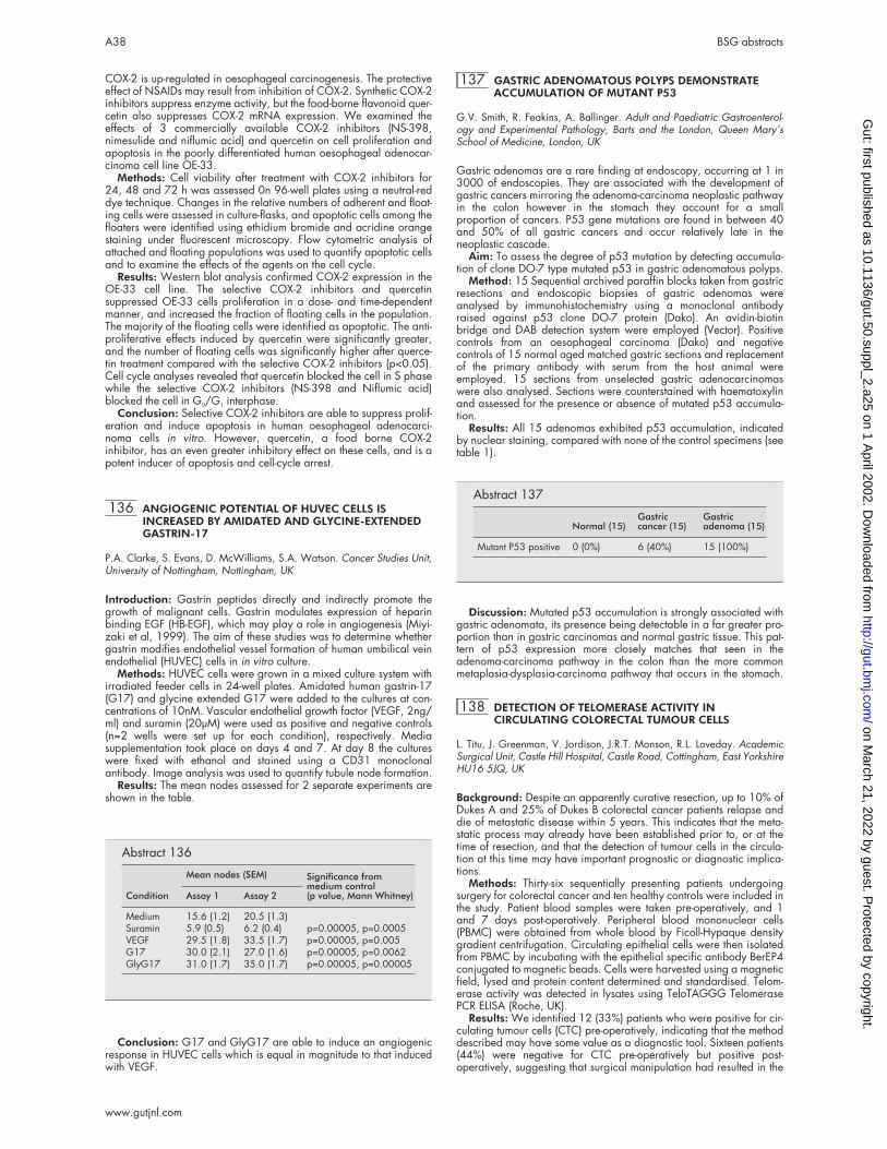
COX-2 is up-regulated in oesophageal carcinogenesis. The protectiveeffect of NSAIDs may result from inhibition of COX-2. Synthetic COX-2inhibitors suppress enzyme activity, but the food-borne flavonoid quer-cetin also suppresses COX-2 mRNA expression. We examined theeffects of 3 commercially available COX-2 inhibitors (NS-398,nimesulide and niflumic acid) and quercetin on cell proliferation andapoptosis in the poorly differentiated human oesophageal adenocar-cinoma cell line OE-33.
Methods: Cell viability after treatment with COX-2 inhibitors for24, 48 and 72 h was assessed 0n 96-well plates using a neutral-reddye technique. Changes in the relative numbers of adherent and float-ing cells were assessed in culture-flasks, and apoptotic cells among thefloaters were identified using ethidium bromide and acridine orangestaining under fluorescent microscopy. Flow cytometric analysis ofattached and floating populations was used to quantify apoptotic cellsand to examine the effects of the agents on the cell cycle.
Results: Western blot analysis confirmed COX-2 expression in theOE-33 cell line. The selective COX-2 inhibitors and quercetinsuppressed OE-33 cells proliferation in a dose- and time-dependentmanner, and increased the fraction of floating cells in the population.The majority of the floating cells were identified as apoptotic. The anti-proliferative effects induced by quercetin were significantly greater,and the number of floating cells was significantly higher after querce-tin treatment compared with the selective COX-2 inhibitors (p<0.05).Cell cycle analyses revealed that quercetin blocked the cell in S phasewhile the selective COX-2 inhibitors (NS-398 and Niflumic acid)blocked the cell in G0/G1 interphase.
Conclusion: Selective COX-2 inhibitors are able to suppress prolif-eration and induce apoptosis in human oesophageal adenocarci-noma cells in vitro. However, quercetin, a food borne COX-2inhibitor, has an even greater inhibitory effect on these cells, and is apotent inducer of apoptosis and cell-cycle arrest.
136 ANGIOGENIC POTENTIAL OF HUVEC CELLS ISINCREASED BY AMIDATED AND GLYCINE-EXTENDEDGASTRIN-17
P.A. Clarke, S. Evans, D. McWilliams, S.A. Watson. Cancer Studies Unit,University of Nottingham, Nottingham, UK
Introduction: Gastrin peptides directly and indirectly promote thegrowth of malignant cells. Gastrin modulates expression of heparinbinding EGF (HB-EGF), which may play a role in angiogenesis (Miyi-zaki et al, 1999). The aim of these studies was to determine whethergastrin modifies endothelial vessel formation of human umbilical veinendothelial (HUVEC) cells in in vitro culture.
Methods: HUVEC cells were grown in a mixed culture system withirradiated feeder cells in 24-well plates. Amidated human gastrin-17(G17) and glycine extended G17 were added to the cultures at con-centrations of 10nM. Vascular endothelial growth factor (VEGF, 2ng/ml) and suramin (20µM) were used as positive and negative controls(n=2 wells were set up for each condition), respectively. Mediasupplementation took place on days 4 and 7. At day 8 the cultureswere fixed with ethanol and stained using a CD31 monoclonalantibody. Image analysis was used to quantify tubule node formation.
Results: The mean nodes assessed for 2 separate experiments areshown in the table.
Conclusion: G17 and GlyG17 are able to induce an angiogenicresponse in HUVEC cells which is equal in magnitude to that inducedwith VEGF.
137 GASTRIC ADENOMATOUS POLYPS DEMONSTRATEACCUMULATION OF MUTANT P53
G.V. Smith, R. Feakins, A. Ballinger. Adult and Paediatric Gastroenterol-ogy and Experimental Pathology, Barts and the London, Queen Mary’sSchool of Medicine, London, UK
Gastric adenomas are a rare finding at endoscopy, occurring at 1 in3000 of endoscopies. They are associated with the development ofgastric cancers mirroring the adenoma-carcinoma neoplastic pathwayin the colon however in the stomach they account for a smallproportion of cancers. P53 gene mutations are found in between 40and 50% of all gastric cancers and occur relatively late in theneoplastic cascade.
Aim: To assess the degree of p53 mutation by detecting accumula-tion of clone DO-7 type mutated p53 in gastric adenomatous polyps.
Method: 15 Sequential archived paraffin blocks taken from gastricresections and endoscopic biopsies of gastric adenomas wereanalysed by immunohistochemistry using a monoclonal antibodyraised against p53 clone DO-7 protein (Dako). An avidin-biotinbridge and DAB detection system were employed (Vector). Positivecontrols from an oesophageal carcinoma (Dako) and negativecontrols of 15 normal aged matched gastric sections and replacementof the primary antibody with serum from the host animal wereemployed. 15 sections from unselected gastric adenocarcinomaswere also analysed. Sections were counterstained with haematoxylinand assessed for the presence or absence of mutated p53 accumula-tion.
Results: All 15 adenomas exhibited p53 accumulation, indicatedby nuclear staining, compared with none of the control specimens (seetable 1).
Discussion: Mutated p53 accumulation is strongly associated withgastric adenomata, its presence being detectable in a far greater pro-portion than in gastric carcinomas and normal gastric tissue. This pat-tern of p53 expression more closely matches that seen in theadenoma-carcinoma pathway in the colon than the more commonmetaplasia-dysplasia-carcinoma pathway that occurs in the stomach.
138 DETECTION OF TELOMERASE ACTIVITY INCIRCULATING COLORECTAL TUMOUR CELLS
L. Titu, J. Greenman, V. Jordison, J.R.T. Monson, R.L. Loveday. AcademicSurgical Unit, Castle Hill Hospital, Castle Road, Cottingham, East YorkshireHU16 5JQ, UK
Background: Despite an apparently curative resection, up to 10% ofDukes A and 25% of Dukes B colorectal cancer patients relapse anddie of metastatic disease within 5 years. This indicates that the meta-static process may already have been established prior to, or at thetime of resection, and that the detection of tumour cells in the circula-tion at this time may have important prognostic or diagnostic implica-tions.
Methods: Thirty-six sequentially presenting patients undergoingsurgery for colorectal cancer and ten healthy controls were included inthe study. Patient blood samples were taken pre-operatively, and 1and 7 days post-operatively. Peripheral blood mononuclear cells(PBMC) were obtained from whole blood by Ficoll-Hypaque densitygradient centrifugation. Circulating epithelial cells were then isolatedfrom PBMC by incubating with the epithelial specific antibody BerEP4conjugated to magnetic beads. Cells were harvested using a magneticfield, lysed and protein content determined and standardised. Telom-erase activity was detected in lysates using TeloTAGGG TelomerasePCR ELISA (Roche, UK).
Results: We identified 12 (33%) patients who were positive for cir-culating tumour cells (CTC) pre-operatively, indicating that the methoddescribed may have some value as a diagnostic tool. Sixteen patients(44%) were negative for CTC pre-operatively but positive post-operatively, suggesting that surgical manipulation had resulted in the
Abstract 136
Condition
Mean nodes (SEM) Significance frommedium control(p value, Mann Whitney)Assay 1 Assay 2
Medium 15.6 (1.2) 20.5 (1.3)Suramin 5.9 (0.5) 6.2 (0.4) p=0.00005, p=0.0005VEGF 29.5 (1.8) 33.5 (1.7) p=0.00005, p=0.005G17 30.0 (2.1) 27.0 (1.6) p=0.00005, p=0.0062GlyG17 31.0 (1.7) 35.0 (1.7) p=0.00005, p=0.00005
Abstract 137
Normal (15)Gastriccancer (15)
Gastricadenoma (15)
Mutant P53 positive 0 (0%) 6 (40%) 15 (100%)
A38 BSG abstracts
www.gutjnl.com
on March 21, 2022 by guest. P
rotected by copyright.http://gut.bm
j.com/
Gut: first published as 10.1136/gut.50.suppl_2.a25 on 1 A
pril 2002. Dow
nloaded from
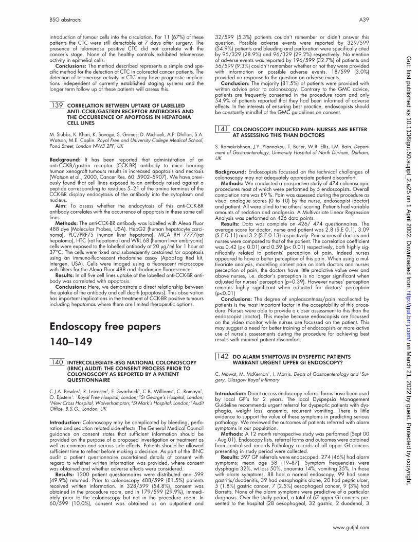
introduction of tumour cells into the circulation. For 11 (67%) of thesepatients the CTC were still detectable at 7 days after surgery. Thepresence of telomerase positive CTC did not correlate with thecancer’s stage. None of the healthy controls exhibited telomeraseactivity in epithelial cells.
Conclusions: The method described represents a simple and spe-cific method for the detection of CTC in colorectal cancer patients. Thedetection of telomerase activity in CTC may have prognostic implica-tions independent of currently established staging systems and thelonger term follow up of these patients will assess this.
139 CORRELATION BETWEEN UPTAKE OF LABELLEDANTI-CCKB/GASTRIN RECEPTOR ANTIBODIES ANDTHE OCCURRENCE OF APOPTOSIS IN HEPATOMACELL LINES
M. Stubbs, K. Khan, K. Savage, S. Grimes, D. Michaeli, A.P. Dhillon, S.A.Watson, M.E. Caplin. Royal Free and University College Medical School,Pond Street, London NW3 2PF, UK
Background: It has been reported that administration of ananti-CCKB/gastrin receptor (CCK-BR) antibody to mice bearinghuman xenograft tumours results in increased apoptosis and necrosis(Watson et al., 2000, Cancer Res. 60: 5902–5907). We have previ-ously found that cell lines exposed to an antibody raised against apeptide corresponding to residues 5–21 of the amino terminus of theCCK-BR display endocytosis of the antibody into the cytoplasm andnucleus.
Aim: To assess whether the endocytosis of this anti-CCK-BRantibody correlates with the occurrence of apoptosis in these same celllines.
Methods: The anti-CCK-BR antibody was labelled with Alexa Fluor488 dye (Molecular Probes, USA). HepG2 (human hepatocyte carci-noma), PLC/PRF/5 (human liver hepatoma), MCA RH 7777(rathepatoma), HTC (rat hepatoma) and WRL 68 (human liver embryonic)cells were exposed to the labelled antibody at 20 µg/ml for 1 hour at37°C. The cells were fixed and subsequently costained for apoptosisusing an immuno-fluorescent rhodamine assay (ApopTag Red kit,Intergen, USA). Cells were imaged using a fluorescent microscopewith filters for the Alexa Fluor 488 and rhodamine fluorescence.
Results: In all five cell lines uptake of the labelled anti-CCK-BR anti-body was correlated with apoptosis.
Conclusions: Here, we demonstrate a direct relationship betweenthe uptake of the antibody and cell death (apoptosis). This observationhas important implications in the treatment of CCK-BR positive tumoursincluding hepatomas where there are limited therapeutic options.
Endoscopy free papers140–149
140 INTERCOLLEGIATE-BSG NATIONAL COLONOSCOPY(IBNC) AUDIT: THE CONSENT PROCESS PRIOR TOCOLONOSCOPY AS REPORTED BY A PATIENTQUESTIONNAIRE
C.J.A. Bowles1, R. Leicester2, E. Swarbrick3, C.B. Williams4, C. Romaya5,O. Epstein1. 1Royal Free Hospital, London; 2St George’s Hospital, London;3New Cross Hospital, Wolverhampton; 4St Mark’s Hospital, London; 5AuditOffice, B.S.G., London, UK
Introduction: Colonoscopy may be complicated by bleeding, perfo-ration and sedation related side effects. The General Medical Councilguidance on consent states that sufficient information should beprovided on the purpose of a proposed investigation or treatment aswell as common and serious side effects. Patients should be allowedsufficient time to reflect before making a decision. As part of the IBNCaudit a patient questionnaire ascertained details of consent withregard to whether written information was provided, where consentwas obtained and whether adverse effects were considered.
Results: 1200 patient questionnaires were distributed and 599(49.9%) returned. Prior to colonoscopy 488/599 (81.5%) patientsreceived written information. In 328/599 (54.8%), consent wasobtained in the procedure room, and in 179/599 (29.9%), immedi-ately prior to the colonoscopy but not in the procedure room. In60/599 (10.0%), consent was obtained as an outpatient and
32/599 (5.3%) patients couldn’t remember or didn’t answer thisquestion. Possible adverse events were reported by 329/599(54.9%) patients and bleeding and perforation were specifically citedby 95/329 (28.9%) and 96/329 (29.2%) respectively. No mentionof adverse events was reported by 196/599 (32.7%) of patients and56/599 (9.3%) couldn’t remember whether or not they were providedwith information on possible adverse events. 18/599 (3.0%)provided no response to the question on adverse events.
Conclusion: The majority (81.5%) of patients were provided withwritten advice prior to colonoscopy. Contrary to the GMC advice,patients are frequently consented in the procedure room and only54.9% of patients reported that they had been informed of adverseeffects. In the interests of ensuring best practice, endoscopists shouldbe constantly mindful of the GMC guidelines on consent.
141 COLONOSCOPY INDUCED PAIN: NURSES ARE BETTERAT ASSESSING THIS THAN DOCTORS
S. Ramakrishnan, J.Y. Yiannakou, T. Butler, W.R. Ellis, I.M. Bain. Depart-ment of Gastroenterology, University Hospital of North Durham, Durham,UK
Background: Endoscopists focussed on the technical challenges ofcolonoscopy may not adequately appreciate patient discomfort.
Methods: We conducted a prospective study of 474 colonoscopicprocedures most of which were performed by 5 endoscopists. Overallcompletion rate was 89 %. Pain was assessed during the procedure asvisual analogue scores (0 to 10) by the nurse, endoscopist (doctor)and patient. All were blind to the others’ scoring. Patients had variableamounts of sedation and analgesia. A Multivariate Linear RegressionAnalysis was performed on 426 data points.
Results: Data was complete on 426/ 474 questionnaires. Theaverage score for doctor, nurse and patient was 2.8 (S.E 0.1), 3.09(S.E 0.11) and 3.2 (S.E 0.13) respectively. Pain scores of doctors andnurses were compared to that of the patient. The correlation coefficientwas 0.42 (p< 0.01) and 0.59 (p< 0.01) respectively, both highly sig-nificantly related to patients’ perception of pain. Indeed nursesappeared to have a better perception of this pain. When using a mul-tivariate analysis, modelling patient pain on both doctors and nursesperception of pain, the doctors have little predictive value over andabove nurses, i.e. doctor’s perception is no longer significant whenadjusted for nurses’ perception (p=0.39). However nurses’ perceptionremains highly significant when adjusted for doctors’ perception(p<0.01)
Conclusions: The degree of unpleasantness/pain recollected bypatients is the most important factor in the acceptability of this proce-dure. Nurses were able to provide a closer assessment to this than theendoscopist (doctor). This maybe because endoscopists are focussedon the video monitor while nurses are focussed on the patient. Thismay suggest a need for better training of endoscopists or more activeuse of nurse’s assessments during the procedure for achieving bestresults with minimal patient discomfort.
142 DO ALARM SYMPTOMS IN DYSPEPTIC PATIENTSWARRANT URGENT UPPER GI ENDOSCOPY?
C. Mowat, M. McKernan1, J. Morris. Depts of Gastroenterology and 1Sur-gery, Glasgow Royal Infirmary
Introduction: Direct access endoscopy referral forms have been usedby local GP’s for 2 years. The local Dyspepsia ManagementGuideline recommends urgent referral for dyspeptic patients with dys-phagia, weight loss, anaemia, recurrent vomiting. There is littleevidence to support the value of these symptoms in predicting seriouspathology. We reviewed the outcomes of patients referred with alarmsymptoms in our population.
Methods: A 12 month retrospective study was performed (Sept 00- Aug 01). Endoscopy lists, referral forms and outcomes were obtainedfrom centralised records.Pathology records of all upper GI cancerspresenting in study period were collected.
Results: 597 GP referrals were endoscoped. 274 (46%) had alarmsymptoms; mean age 58 (19–87). Symptom frequencies weredysphagia 32%, wt loss 50%, anaemia 14%, vomiting 35%. In thosewith alarm symptoms, 88 had a normal endoscopy, 99 had somegastritis/duodenitis, 39 had oesophagitis alone, 20 had peptic ulcer,5 (1.8%) gastric cancer, 7 (2.5%) oesophageal cancer, 9 (3%) hadBarretts. None of the alarm symptoms were predictive of a particulardiagnosis. Over the study period, a total of 67 upper GI cancers pre-sented to the hospital (28 oesophageal, 32 gastric, 2 duodenal, 3
BSG abstracts A39
www.gutjnl.com
on March 21, 2022 by guest. P
rotected by copyright.http://gut.bm
j.com/
Gut: first published as 10.1136/gut.50.suppl_2.a25 on 1 A
pril 2002. Dow
nloaded from
pancreatic, 2 incomplete data); mean age 73 (46–90). Only onepatient was aged less than 55. 26 of these presented via other directaccess routes. The remainder presented via A&E or clinic. 12 of 38patients referred urgently versus 2 of 22 in-hospital referrals were suit-able for attempted curative surgery. Of the 14 suitable for surgery,mean age was 68 (58–75).
Conclusions: In our population, alarm symptoms in young dyspep-tic patients rarely indicated upper GI cancer. The cancers arose inolder subjects. Those who were referred urgently had a significantlygreater chance of attempted curative surgery. Rapid assessment ofdyspeptic patients with alarm symptoms should therefore focus onolder patients. In this study, all patients with alarm symptoms aged lessthan 55 could have been managed empirically with a ‘test and treat’+/- PPI policy.
143 SEDATION FOR COLONOSCOPY; A RANDOMISED,CONTROLLED TRIAL COMPARINGPATIENT-CONTROLLED ADMINISTRATION OFPROPOFOL AND ALFENTANIL WITHPHYSICIAN-ADMINISTERED MIDAZOLAM ANDPETHIDINE
C. Roseveare1, L. Bright1, D. Dalgleish1, J. Kimble, H. Shepherd.1Southampton General Hospital; Royal Hants County Hospital, Winchester,UK
Background: Our previous studies have indicated that Patient-Controlled Sedation (PCS) using propofol and alfentanil provided aneffective alternative to a combination of diazepam and pethidinegiven as a bolus prior to the procedure, with the advantage of shorterrecovery time.
Aims: To compare efficacy of sedation and recovery time betweenPCS and a bolus combination of midazolam and pethidine (M&P); tostudy further the safety of the technique and to determine the feasibil-ity of PCS being set-up and supervised by an endoscopy nurse andendoscopist.
Methods: 67 patients undergoing colonoscopy were randomisedto receive sedation with either PCS using propofol and alfentanil or abolus of midazolam (2.5–5mg) and pethidine (25–50mg). Infusionswere connected by the endoscopy nurse with anaesthetist present inan observational capacity only. The anaesthetist only intervened ifspecific pre-defined criteria were reached. Sedation scores wererecorded during the procedure by the endoscopy nurse and painscores after the procedure by both nurse and patient, using likertscales. Recovery was confirmed using number connection tests. Impacton subsequent daily activity, amnesia and overall satisfaction wereestablished by phone at 24 hours.
Results: Sedation method had no impact on the success, difficultyor duration of the colonoscopy procedure. PCS infusions could be setup by the endoscopy nurse without causing significant delay. Patientsin the PCS group recovered significantly faster (mean 5mins vs35mins, p<0.0001) and left the endoscopy department much sooner(40mins vs 75mins, p<0.0001). No differences in respiratory orhaemodynamic observations were recorded between the two groups.Anaesthetist intervention was required for one patient sedated withM&P, but was not required for any patients sedated with PCS. Overallsedation level was lighter in the PCS group (score 3 vs 4, p=0.05) andverbal contact was lost with only 2 patients using PCS, compared to 9using M&P. Patients in the PCS group reported significantly more pain(median pain score 1 vs 0, p=0.0005), which may have reflectedgreater amnesia in the M&P group, since nurse recordings of painwere identical for the two groups. PCS had less impact on the patients’activities in the 24 hours after the procedure (medican score 0 vs 1,p=0.01). Satisfaction scores were equal in the two groups.
Conclusions: PCS using propofol and alfentanil is acceptable topatients, provides faster recovery, and appears to be at least as safeas a bolus combination of midazolam and pethidine.
144 SYNERGISTIC SEDATION WITH MIDAZOLAM ANDPROPOFOL VERSUS MIDAZOLAM AND PETHIDINE INCOLONOSCOPIES: A PROSPECTIVE, RANDOMIZEDSTUDY
G.A. Paspatis1, M. Manolaraki2, G. Xirouchakis2, N. Papanikolaou1, I.Charoniti1, A. Gritzali2. Department of 1Gastroenterology and 2Anesthesi-ology, Benizelion General Hospital, Heraklion-Crete, Greece
Background and Aims: A recent study suggests the synergistic seda-tion with a low dose of midazolam combined with propofol for
conscious sedation in colonoscopies in a selected group of patientsunder the age of 60 years. Our study sought to compare the safetyand efficacy of the synergistic sedation with a low dose of midazolamcombined with propofol versus the standard regimen of midazolamand pethidine for conscious sedation in colonoscopies in a group ofpatients that included a sufficient number of elderly patients with sev-eral comorbidities, representative of the patient profile seen in publicgeneral hospitals.
Methods: 120 consecutive patients undergoing colonoscopy wererandomly assigned to 1 of 2 medication regimens. Patients in group A(n=64, 33 over 65 years) received intravenously (IV) 2mg (<70 kgb.w.) or 3mg (>70 kg b.w.) of midazolam and a median dose of80mg propofol. Patients in group B (n=56, 22 over 65 years)received IV a median dose of 5mg midazolam and 75mg pethidine.The patient’s comfort level was assessed by a four point scale 24hours after the procedure. The time to recover from sedation wasassessed at 5, 10 and 30 min after the procedure by using the Aldretescore.
Results: Multivariate stepwise logistic regression analysis revealedthat among sex, age, duration of the test, ASA grade and the sort ofsedation, the synergistic sedation with midazolam and propofol wasthe only factor associated with a higher level of patient comfort(χ2=5.5, p<0.05) and a quicker patient recovery time (χ2=24.5-p<0.01, χ2=51.7-p<0.01 and χ2=148.4-p<0.01 for aldrete in 10, 20and 30 min respectively). Endoscopist’s evaluation of patient sedationand cardiorespiratory parameters were similar in both groups acrossall age groups.
Conclusions: Our data suggest that the sedation with midazolamcombined with propofol was superior to combination of midazolamand pethidine for colonoscopies as far as the patient comfort andrecovery times are concerned.
145 THE VARIABLE STIFFNESS COLONOSCOPE:ASSESSMENT OF EFFICACY BY MAGNETICENDOSCOPE IMAGING
S.G. Shah, J.C. Brooker, C.B. Williams, C. Thapar, N. Suzuki, B.P.Saun-ders.
Background: Variable-stiffness colonoscopes (VS scope) combinepaediatric flexibility shaft characteristics for negotiation of the sigmoidcolon with the ability to stiffen to prevent looping after straightening.Previous studies have shown a wide variation in efficacy of the stiffen-ing mechanism, based on subjective assessments and differences inmethodology. Two studies were conducted to assess the potential ben-efit of the stiffening device and its optimal use.
Methods: In study 1, the effect of routinely stiffening thestraightened VS scope (Olympus CFQ240AL) in the mid-descendingcolon was determined in 82 patients. Two insertions were performedin each patient, from the mid-descending colon to caecal pole, withand without application of the stiffening device (randomised). The timetaken to negotiate the proximal colon (from the mid-descending tocaecal pole), time to pass the scope across the splenic flexure into thetransverse colon, time to pass the right colon, and ancillary manoeu-vres used were recorded for each insertion. In study 2, consecutivepatients, excluding any with previous colonic resection, wereexamined using standard adult variable-stiffness colonoscopes.Real-time views of the procedure using magnetic endoscope imaging(MEI) were recorded in all examinations, but procedures wererandomised to be done either with (n=88), or without the endoscopistviewing (n=87) the MEI display. Whenever shaft stiffening wasapplied, the anatomical location of the colonoscope tip and stiffnessefficacy was recorded.
Results: Study 1—Time taken to negotiate the proximal colon(p=0.0041) and time to negotiate the splenic flexure (p=0.006) weresignificantly shorter and ancillary manoeuvres performed were fewer(p=0.0014), when insertion was carried out with the stiffening deviceactivated. Study 2—Shaft stiffening was used with similar frequency inpatients examined with and without the MEI view, most commonly forpassing the splenic flexure (71%), but also in the transverse colon(12%), right colon (9%) and sigmoid/descending colon (8%). Shaftstiffening was significantly more effective when used in combinationwith MEI (69% with imager vs. 45% without imager; p=0.0102).
Conclusions: Overall, the variable-stiffness device was effective incontrolling looping 57% of the time. Activating maximum scopestiffness appears to be most effective once the sigmoid colon has beennegotiated and the scope straightened with the tip in the proximalcolon, reducing the number of ancillary manoeuvres and shorteningthe insertion time through the proximal colon. Routine colonoscopeimaging with MEI further enhances the efficacy of VS scopes by help-ing to identify the optimal time for scope stiffening.
A40 BSG abstracts
www.gutjnl.com
on March 21, 2022 by guest. P
rotected by copyright.http://gut.bm
j.com/
Gut: first published as 10.1136/gut.50.suppl_2.a25 on 1 A
pril 2002. Dow
nloaded from
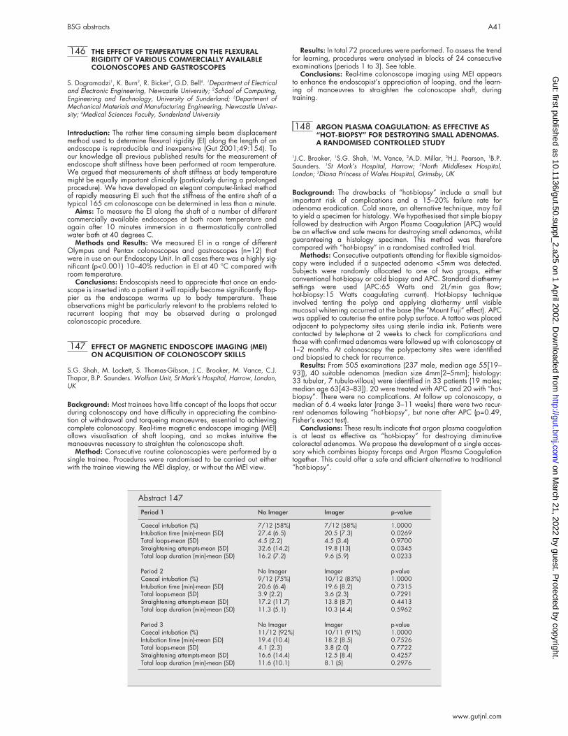
146 THE EFFECT OF TEMPERATURE ON THE FLEXURALRIGIDITY OF VARIOUS COMMERCIALLY AVAILABLECOLONOSCOPES AND GASTROSCOPES
S. Dogramadzi1, K. Burn2, R. Bicker3, G.D. Bell4. 1Department of Electricaland Electronic Engineering, Newcastle University; 2School of Computing,Engineering and Technology, University of Sunderland; 3Department ofMechanical Materials and Manufacturing Engineering, Newcastle Univer-sity; 4Medical Sciences Faculty, Sunderland University
Introduction: The rather time consuming simple beam displacementmethod used to determine flexural rigidity (EI) along the length of anendoscope is reproducible and inexpensive (Gut 2001;49:154). Toour knowledge all previous published results for the measurement ofendoscope shaft stiffness have been performed at room temperature.We argued that measurements of shaft stiffness at body temperaturemight be equally important clinically (particularly during a prolongedprocedure). We have developed an elegant computer-linked methodof rapidly measuring EI such that the stiffness of the entire shaft of atypical 165 cm colonoscope can be determined in less than a minute.
Aims: To measure the EI along the shaft of a number of differentcommercially available endoscopes at both room temperature andagain after 10 minutes immersion in a thermostatically controlledwater bath at 40 degrees C.
Methods and Results: We measured EI in a range of differentOlympus and Pentax colonoscopes and gastroscopes (n=12) thatwere in use on our Endoscopy Unit. In all cases there was a highly sig-nificant (p<0.001) 10–40% reduction in EI at 40 °C compared withroom temperature.
Conclusions: Endoscopists need to appreciate that once an endo-scope is inserted into a patient it will rapidly become significantly flop-pier as the endoscope warms up to body temperature. Theseobservations might be particularly relevant to the problems related torecurrent looping that may be observed during a prolongedcolonoscopic procedure.
147 EFFECT OF MAGNETIC ENDOSCOPE IMAGING (MEI)ON ACQUISITION OF COLONOSCOPY SKILLS
S.G. Shah, M. Lockett, S. Thomas-Gibson, J.C. Brooker, M. Vance, C.J.Thapar, B.P. Saunders. Wolfson Unit, St Mark’s Hospital, Harrow, London,UK
Background: Most trainees have little concept of the loops that occurduring colonoscopy and have difficulty in appreciating the combina-tion of withdrawal and torqueing manoeuvres, essential to achievingcomplete colonoscopy. Real-time magnetic endoscope imaging (MEI)allows visualisation of shaft looping, and so makes intuitive themanoeuvres necessary to straighten the colonoscope shaft.
Method: Consecutive routine colonoscopies were performed by asingle trainee. Procedures were randomised to be carried out eitherwith the trainee viewing the MEI display, or without the MEI view.
Results: In total 72 procedures were performed. To assess the trendfor learning, procedures were analysed in blocks of 24 consecutiveexaminations (periods 1 to 3). See table.
Conclusions: Real-time colonoscope imaging using MEI appearsto enhance the endoscopist’s appreciation of looping, and the learn-ing of manoeuvres to straighten the colonoscope shaft, duringtraining.
148 ARGON PLASMA COAGULATION: AS EFFECTIVE AS“HOT-BIOPSY” FOR DESTROYING SMALL ADENOMAS.A RANDOMISED CONTROLLED STUDY
1J.C. Brooker, 1S.G. Shah, 1M. Vance, 2A.D. Millar, 3H.J. Pearson, 1B.P.Saunders. 1St Mark’s Hospital, Harrow; 2North Middlesex Hospital,London; 3Diana Princess of Wales Hospital, Grimsby, UK
Background: The drawbacks of “hot-biopsy” include a small butimportant risk of complications and a 15–20% failure rate foradenoma eradication. Cold snare, an alternative technique, may failto yield a specimen for histology. We hypothesised that simple biopsyfollowed by destruction with Argon Plasma Coagulation (APC) wouldbe an effective and safe means for destroying small adenomas, whilstguaranteeing a histology specimen. This method was thereforecompared with “hot-biopsy” in a randomised controlled trial.
Methods: Consecutive outpatients attending for flexible sigmoidos-copy were included if a suspected adenoma <5mm was detected.Subjects were randomly allocated to one of two groups, eitherconventional hot-biopsy or cold biopsy and APC. Standard diathermysettings were used (APC:65 Watts and 2L/min gas flow;hot-biopsy:15 Watts coagulating current). Hot-biopsy techniqueinvolved tenting the polyp and applying diathermy until visiblemucosal whitening occurred at the base (the “Mount Fuji” effect). APCwas applied to cauterise the entire polyp surface. A tattoo was placedadjacent to polypectomy sites using sterile india ink. Patients werecontacted by telephone at 2 weeks to check for complications andthose with confirmed adenomas were followed up with colonoscopy at1–2 months. At colonoscopy the polypectomy sites were identifiedand biopsied to check for recurrence.
Results: From 505 examinations (237 male, median age 55[19–93]), 40 suitable adenomas (median size 4mm[2–5mm]; histology:33 tubular, 7 tubulo-villous) were identified in 33 patients (19 males;median age 63[43–83]). 20 were treated with APC and 20 with “hot-biopsy”. There were no complications. At follow up colonoscopy, amedian of 6.4 weeks later (range 3–11 weeks) there were two recur-rent adenomas following “hot-biopsy”, but none after APC (p=0.49,Fisher’s exact test).
Conclusions: These results indicate that argon plasma coagulationis at least as effective as “hot-biopsy” for destroying diminutivecolorectal adenomas. We propose the development of a single acces-sory which combines biopsy forceps and Argon Plasma Coagulationtogether. This could offer a safe and efficient alternative to traditional“hot-biopsy”.
Abstract 147
Period 1 No Imager Imager p-value
Caecal intubation (%) 7/12 (58%) 7/12 (58%) 1.0000Intubation time (min)-mean (SD) 27.4 (6.5) 20.5 (7.3) 0.0269Total loops-mean (SD) 4.5 (2.2) 4.5 (3.4) 0.9700Straightening attempts-mean (SD) 32.6 (14.2) 19.8 (13) 0.0345Total loop duration (min)-mean (SD) 16.2 (7.2) 9.6 (5.9) 0.0233
Period 2 No Imager Imager p-valueCaecal intubation (%) 9/12 (75%) 10/12 (83%) 1.0000Intubation time (min)-mean (SD) 20.6 (6.4) 19.6 (8.2) 0.7315Total loops-mean (SD) 3.9 (2.2) 3.6 (2.3) 0.7291Straightening attempts-mean (SD) 17.2 (11.7) 13.8 (8.7) 0.4413Total loop duration (min)-mean (SD) 11.3 (5.1) 10.3 (4.4) 0.5962
Period 3 No Imager Imager p-valueCaecal intubation (%) 11/12 (92%) 10/11 (91%) 1.0000Intubation time (min)-mean (SD) 19.4 (10.4) 18.2 (8.5) 0.7526Total loops-mean (SD) 4.1 (2.3) 3.8 (2.0) 0.7722Straightening attempts-mean (SD) 16.6 (14.4) 12.5 (8.4) 0.4257Total loop duration (min)-mean (SD) 11.6 (10.1) 8.1 (5) 0.2976
BSG abstracts A41
www.gutjnl.com
on March 21, 2022 by guest. P
rotected by copyright.http://gut.bm
j.com/
Gut: first published as 10.1136/gut.50.suppl_2.a25 on 1 A
pril 2002. Dow
nloaded from
149 CLINICAL STUDIES OF WIRELESS CAPSULECOLONOSCOPY
M. Mylonaki, A. Fritscher-Ravens, S. Lewkowicz, P. Swain. Royal LondonHospital, Whitechapel, London E1 1BB, UK
Background: Wireless capsule endoscopy has the potential to delivervideo images from the colon. Slow transit, intermittent rapidmovements, larger diameter lumen and capsule transmission times arechallenges requiring study.
Aim: To assess colonic images received during experimental,human volunteer and clinical studies. To improve the capacity of wire-less capsule technology to image the human colon.
Methods: Colonic views obtained during clinical studies ofcapsules with a 7.5 hr capsule were reviewed (n= 38) human volun-teers and patients referred with obscure gastrointestinal bleeding.Studies using experimental long-play with 4 silver-oxide batteries anda potential transmission time of 18 hours were used in 7. Lighter cap-sules [n=3](SG = 1.2 vs 1.7), with 2 batteries and a 2 hour rest periodmoving with the fluid phase were tested. 2 capsules were placed ret-rogradely into the transverse colon using hydraulic delivery.
Results: In clinical studies performed without colonic preparation(n=38) interpretable images of the human colon were acquired in 35,mucosal detail was seen in 34, faecal material 35, underillumination10. Technical improvements included shorter capsules (27mm,previ-ously 33mm) now transmitting for 7–8 hours using two(previously three) batteries, and experimental capsules with an 12hour life were been tested and occasionally imaged the toilet. Clinicalstudies in n=13 patients) revealed missed colon cancer/polyp (3),bleeding Meckel’s diverticulum (1), ulcerative colitis (2), angiodyspla-sia (2). Small intestinal transit times were median (4.hr), range (1.2–7.6hrs). Oro-anal transit times were 0.5–8 days. Capsules used inpatients with incomplete colonoscopies provided useful clinicalinformation. In patients selected for capsule colonoscopy managementwas altered in 50%.
Conclusion: Wireless capsule colonoscopy is feasible and hasalready delivered valuable information in clinical studies. Technicaldevelopment and better preparation and control over colonic motilityis required to extend the range and improve the images of the colonin patients. Capsule colonoscopy was a useful adjunct to conventionalcolonoscopy especially in patients with difficult recurrent bleeding orincomplete colonoscopy.
Biliary/pancreas free papers150–166
150 THE POTENTIAL ROLE OF GASTRIN IN PANCREATICCANCER PROGRESSION-RAISED SERUM AMIDATEDGASTRIN AND PROGASTRIN LEVELS IN PATIENTSWITH ADVANCED PANCREATIC CANCER
A.D. Gilliam, P.A. Clarke, B.J. Rowlands, I.J. Beckingham, S.A. Watson.Academic Unit of Cancer Studies, Academic Department of Surgery, Uni-versity Hospital, Nottingham NG7 2UH, UK
Aim: To assess the endocrine role of gastrin in pancreatic carcinomaprogression.
Method: A prospective study was established in which patientswith resectable (n=17) and advanced (n=68) pancreatic carcinomahad their serum assayed for amidated and progastrin. Hormone levelswere compared to positive controls (patients undergoing liverresection for colonic metastases, n=24). Gastrin and CCK 2 receptorexpression was measured in 17 resected pancreatic tumours and fourpancreatic tumour cell lines by RT-PCR, using the SYBR green dye, andimmunohistochemical staining or western blotting using antibodiesdirected against gastrin and CCK2.
Results: There was a significantly greater serum amidated gastrinand progastrin levels in patients with advanced pancreatic cancerwhen compared to patients with resectable disease (p=0.008* and0.046*). Patients with advanced pancreatic cancer also hadsignificantly greater amidated gastrin and progastrin levels thanpatients undergoing liver resection for colonic metastases (p=0.023*).There was, however, no significant difference in amidated gastrin lev-els in patients with resectable pancreatic carcinoma and patients withmetastatic colonic carcinoma (p=0.09*). Gastrin and CCK 2 expres-sion was confirmed at the gene level in 13 pancreatic adenocarcino-mas by RT-PCR, and immunocytochemistry in 17 pancreatic tumours.
Gastrin and CCK-2 expression was also demonstrated in all four pan-creatic tumour cell lines at the gene and protein levels by RT-PCR andwestern blotting. (*Independent samples T-test.)
Conclusion: This study confirms the autocrine, paracrine andendocrine role of gastrin in pancreatic carcinoma progression.Increased plasma levels of amidated gastrin may be a future biomar-ker for advanced pancreatic cancer. Anti-gastrin therapy mayrepresent a novel therapeutic strategy for the management of pancre-atic cancer.
151 GEMCITABINE DOES NOT INHIBIT THE BIOLOGICALACTIVITY OF A HUMAN PANCREATIC TUMOURGROWING IN THE PANCREAS OF IMMUNODEFICIENTMICE.
A.D. Gilliam, W.J. Speake, R.A. Dean, D. McWilliams, P.A. Clarke,T. Morris, S.A. Watson. Academic Unit of Cancer Studies, AcademicDepartment of Surgery, University Hospital, Nottingham NG7 2UH, UK
Introduction: Gemcitabine (Gemzar), a novel nucleoside analogue,exerts its action by inhibiting DNA synthesis. Gemcitabine is licensedas a first line treatment for patients with locally advanced or metastaticadenocarcinoma of the pancreas and as a second line treatment ofpatients with 5-FU refractory pancreatic cancer (NICE guidelines2001). The aim of this study was to assess the effect of gemcitabine onmatrix metalloproteinase (MMP), Epidermal Growth Factor Receptor(EGFR), gastrin, Cyclo-oxygenase2 (COX-2) and Cholecystokinin-2(CCK2) receptor expression of a human pancreatic tumour growing inthe pancreas of immunodeficient mice.
Method: The human pancreatic cell line, PAN1, cells were injectedinto the body of the pancreas in immunodeficient mice (1x10⁶ in a20µl volume). Mice were treated with saline or gemcitabine infusion-s.The tumours were examined for MMP 2 and 9, EGFR, COX-2, gas-trin and CCK2 receptor expression by real time PCR at the gene level,by use of the SYBR green dye, and by western blotting and zyomog-raphy at the protein level.
Results: The gemcitabine treated mice had a 40% reduction intheir tumour weights (p=0.045*) however there was no significantalteration in MMP gene or protein expression (p>0.1*), EGFR(p=0.48*), gastrin (p=0.15*), COX-2 (p=0.33*) and CCK2 receptor(p=0.20*) gene expression. (*Student’s t Test.)
Conclusion: Gemcitabine does not affect the expression of severalmolecular biological targets suggesting an important potential role fornovel biological therapies for use in conjunction with newchemotherapeutic agents in patients with pancreatic cancer.
152 MANAGEMENT OF ACUTE PANCREATITIS IN WALES:HOW GOOD ARE WE?
P.J. Arumugam, P.J. Shankar, P.N. Haray (introduced by V.I. Shah). PrinceCharles Hospital, Merthyr Tydfil, Wales, UK
Aims: To review the existing practices of Welsh Surgical Consultantsin managing acute pancreatitis and compare it with published UKguidelines.
Methods: We designed a questionnaire based on the nationalguidelines regarding the assessment, indications for intensivemanagement, timing of elective cholecystectomy, ERCP and surgicalintervention. This questionnaire was mailed to all the consultants inWales and the replies were analyzed.
Results: 50 consultants responded. 33 units assess patients with ascoring system and almost all do routine blood gas analysis and liverfunction tests on admission, but only 29(60%) perform C- Reactiveprotein to assess prognosis. 10 units managed this problem with amultidisciplinary team approach. 17 of these units did not have accessto HDU facilities. CT scan was used by a majority of these units asrequired by guidelines. Antibiotics were prescribed by most units insevere cases while surprisingly 10(20%) of these units prescribed anti-biotics routinely without specific indications even in mild cases. Onlyone in three of these units routinely performed cholecystectomy withinfour weeks of an acute attack as recommended. ERCP and peropera-tive cholangiograms were not used in accordance with the guidelines.
Conclusion: Consultants in Wales do feel a specialist, multidiscipli-nary approach is necessary but practical difficulties preventimplementation of the guidelines. However, it is a matter of some con-cern that despite national guidelines, there is a varied approachacross Wales.
A42 BSG abstracts
www.gutjnl.com
on March 21, 2022 by guest. P
rotected by copyright.http://gut.bm
j.com/
Gut: first published as 10.1136/gut.50.suppl_2.a25 on 1 A
pril 2002. Dow
nloaded from

153 PREDICTION OF SEVERITY IN ACUTE PANCREATITIS: ACOMPARATIVE STUDY OF RANSON’S SCORE AND 24AND 48 HOURS APACHE II AND III SCORING SYSTEMS
C. Chatzikostas1, G. Notas2, M. Roussomoustakaki1, I. Mouzas1,I. Koutroubakis1, P. Skordilis1, E. Matrella1, F. Dimoulios1, A. Theodoropou-lou1, D. Samonakis1, E. Vardas1, P. Antoniou1, E. Kouroumalis1,2. 1Depart-ment of Gastroenterology, University Hospital, Heraklion, Crete, Greece’2Liver Research Laboratory, University of Crete Medical School, Crete,Greece
Background/Aims: We assessed the prognostic accuracy ofRanson’s, APACHE II, and APACHE III scores in predicting acute pan-creatitis (AP) severity in non-intensive care unit (ICU) patients.APACHE III has not been previously evaluated outside ICU settings.
Methods: 126 patients with AP (56% gallstone and 9% alcoholic-related, 7% secondary, 28% idiopathic) were studied prospectively.Data conforming to scoring systems were recorded 24 (APACHE IIand III) and 48 hr (Ranson, APACHE II and III) after admission. Analy-sis was performed by using t-test, Pearson correlation, receiver operat-ing characteristic (ROC) curves and area under a ROC curve (AUC).
Results: On discharge, 117 patients (76.9%) were classified asmild and 35 (23%) as severe. There were 4 deaths (2.6%). The meanRanson’s score and the mean 24 and 48 hr APACHE II and III scoresof patients with severe AP were each significantly higher than those ofpatients with an uncomplicated outcome. All five scores correlatedstrongly with length of stay. When ROC curves were plotted, AUC forRanson’s score (0.799; cutoff 3; sensitivity, 72%; specificity, 79%;correct 73%) was found to be larger than AUC for 24 hr APACHE II(0.644; cutoff, 8; sensitivity, 53%; specificity, 62%; correct, 55%), 24hr APACHE III (0,654; cutoff, 32; sensitivity, 60%; specificity, 58%;correct, 60%), 48 hr APACHE II (0.649; cutoff, 8; sensitivity, 53%;specificity, 69%; correct, 57%), and 48 hr APACHE III (0.652; cutoff,27; sensitivity, 52%; specificity, 72%, correct, 52%). The differencebetween 24 and 48 hr APACHE II and III scores AUC did not reachstatistical significance.
Conclusion: Ranson’s score is superior to APACHE II and III in pre-dicting acute pancreatitis severity. APACHE III score is no superior toAPACHE II, and sequential 24 and 48 hr recording offers no advan-tage over 24 hour recording.
154 ACUTE AND CHRONIC PANCREATITIS: DISEASES ONTHE INCREASE
D.A.J. Lloyd1, A. Tinto2, A. Majeed3, R.C.N. Williamson4, J.D. Maxwell1,J-Y. Kang1. 1St George’s Hospital; 2Office for National Statistics; 3UniversityCollege; 4Hammersmith Hospital, London, UK
Aim: To investigate time trends for the numbers of hospital admissionsfor acute and chronic pancreatitis in England from 1989/90 to1999/00.
Methods: Data were obtained from the Hospital Episodes Statistics(HES) service from 1989/90 to 1999/00 based on ‘Finished Consult-ant Episodes’, excluding day cases, in England. Hospital admissionswere selected by primary diagnosis and admissions where surgicaloperations (excluding endoscopic procedures) were performed wereidentified. Age standardised hospital admission rates were calculatedusing the European standard population. Mortality statistics were alsoobtained.
Results: Over the 11-year study period, admission rates for acutepancreatitis rose by 43% while those for chronic pancreatitis rose by100% (see table). Admission rates for acute pancreatitis increased
progressively with age whereas those for chronic pancreatitis peakedat 35–54 years of age. For both sexes, the proportions of admissionsrequiring surgical operations increased for acute pancreatitis butdecreased for chronic pancreatitis. There was a gradual declineamong both sexes in case fatality rates for acute pancreatitis.
Conclusions: There has been a steady increase in admission ratesfor both acute and chronic pancreatitis. The rate of increase wasgreater among females for acute pancreatitis, but greater amongmales for chronic pancreatitis. These trends are likely to reflect genu-ine changes in disease epidemiology rather than alterations indiagnostic and management practice. The proportions requiringoperations have increased for acute pancreatitis but decreased forchronic pancreatitis.
155 HEREDITARY PANCREATITIS (HP) AND THE RISK OFPANCREATIC DUCTAL ADENOCARCINOMA (PDAC)
N. Howes, M.M. Lerch, R. Charnley, J. Mössner, S. Endres, J. Deviere, V.Verreman, V. Lucidi, A. Ohla, I. Ihse, C. Imrie, W. Steenbergan, A. Poll,W. Greenhalf, I. Ellis, R. Mountford, D.C. Whitcomb, J.P. Neoptolemos forthe UK and Ireland consortium of EUROPAC
Introduction: HP has an early age of symptom onset and isassociated with a high incidence of complications, of particularimportance is the reported high life time risk of PDAC. The EuropeanRegistry of Hereditary Pancreatitis and Familial Pancreatic Cancer(EUROPAC) was established in 1997 to investigate HP in Europe.
Aims: To establish the risk of PDAC in HP patients in Europe.Methods: Recruitment started in 1997,HP was diagnosed on the
basis of two family members with chronic pancreatitis of unknownaetiology. PRSS1 mutation screening was undertaken for thepublished mutations, with sequencing in negative families. The Stand-ardised Incidence Ratio (SIR) which is the ratio of observed PDAC toexpected PDAC was calculated for histologically confirmed PDACfrom families with at least three generations of HP adjusted for age,sex, nationality and surgical intervention. The cumulative lifetime inci-dence was calculated, and a multivariate analysis undertaken forpotential confounding factors.
Results: 109 families (n=342)were recruited. 47 families (n=197;99M, 98F) were suitable for PDAC analysis. 15 patients (8M, 7F)developed PDAC during 7648 person-years. Mean age of cancerdiagnosis was 56(48–69) Yrs. Expected number of cancers was 0.21yielding an overall SIR of 71(53–88). The SIR in males was72(49–94) and in females was 70(47–93). The overall lifetime risk forthe development of PDAC in our cohort of patients with HP was 40%[95%C/I];( 30–50%). The risk appeared to be minimal below the ageof 40yrs, where after it increased sharply. Multivariate analysisshowed that the risk of PDAC appeared to be independent of poten-tial confounding variables.
Conclusion: PDAC is a real and significant independent complica-tion of Hereditary Pancreatitis.
156 AUDIT OF SECHAT TESTS: WHO TO TARGET?
B.C. McKaig, N. Simpson, I. Amarkone, R.F.A. Logan. Division of Gastro-enterology, University Hospital, Nottingham NG7 2UH, UK
Introduction and Aims: 75Selenium cholic acid taurate (SeCHAT)tests are accurate in the diagnosis of bile acid malabsorption (BAM).We have audited the use of SeCHAT tests in a teaching hospital toassess if their use was appropriate, influenced patient managementand to determine the prevalence of primary bile acid malabsorption(PBAM).
Methods: Patients undergoing SeCHAT tests from 1994–2001were identified and the case notes examined for the SeCHAT result(<10% being a positive test), indication, known terminal ilealpathology, previous investigations and influence on patient manage-ment.
Results: 120 patients were identified undergoing SeCHAT tests ofwhich 51 were positive, 48 negative and 21 indeterminate (meanretention of 75SeCHAT at 7 days 3.75%, 33.8% and 12.4%, respec-tively). The indication in all cases was diarrhoea. Of the 51 positivetests, 21 had previous surgery (16 terminal ileal (TI) resections, 5cholecystectomy); 21 had known TI Crohn’s disease; one hadreceived radiotherapy involving the TI; and 2 had documented priorenteric infection. Of the negative and indeterminate tests, 4 patientshad previous enteric infection, one had coeliac disease but none hadknown TI Crohn’s disease, previous surgery or other known predispos-ing factors for BAM. Prior to a positive SeCHAT test, most (90%) had
Abstract 154
Age standardised admission rates per100,000
1989/90 1999/00 %change
M F M F M F
Acute pancreatitis 17.6 11.3 22.9 18.4 +30 +63% with operation 10 11 14 12 +42 +12Chronic pancreatitis 5.9 2.8 12.4 4.8 +113 +71% with operation 15 16 12 12 -21 -21
BSG abstracts A43
www.gutjnl.com
on March 21, 2022 by guest. P
rotected by copyright.http://gut.bm
j.com/
Gut: first published as 10.1136/gut.50.suppl_2.a25 on 1 A
pril 2002. Dow
nloaded from
imaging of the TI compared to only 40% of those with negative tests.Of those with positive tests, 47% had a short term (3 month) responseto bile acid sequestrants (BAS), but this was only sustained at 6 monthsin 20% of patients, the remainder being intolerant of BAS. 6 patientstherefore had a diagnosis of PBAM. After 3 years of follow up, onewas diagnosed with Zollinger Ellison syndrome and another with car-cinoid syndrome, leaving only 4 patients with PBAM (3 male, 1female). The incidence of PBAM was therefore 6% in our group ofpatients with diarrhoea.
Conclusions: Patients presenting with diarrhoea known to have TIdisease or dysfunction have a high probability of a positive SeCHATtest and therefore, can be assumed to have BAM as a contributor totheir symptoms and do not require formal testing. The frequency ofPBAM in our audit was 6% and BAM should be considered in thesepatients with a diagnosis of diarrhoea predominant irritable bowelsyndrome.
157 IS HORMONE REPLACEMENT THERAPY ASSOCIATEDWITH GALLSTONE FORMATION? A PROSPECTIVECOHORT STUDY
A.R. Hart1, R. Luben2, S. Oakes2, J. Camus2, A. Welch2, N. Wareham2,S.A. Bingham2, K-T Khaw2, N.E. Day2. 1 School of Medicine, University ofEast Anglia, Norwich NR4 7TJ; 2Strangeways Research Laboratories,Cambridge CB1 4RN, UK
Background: The aetiology of gallstones is unknown. High oestrogenlevels, whether endogenous or through exogenous hormonereplacement therapy (hrt), have been implicated. Oestrogens increasethe cholesterol saturation of the bile which may precipitate stone for-mation. The few epidemiological studies investigating this associationhave given conflicting results and clarification is needed. The aim ofthis study was to investigate if an association existed in a prospectivecohort investigation.
Methods: A total of 13 433 women aged 45–79 years wererecruited into EPIC-Norfolk (European Prospective Investigation IntoCancer). Participants supplied information at recruitment on use ofhormone replacement therapy and were followed up for the develop-ment of symptomatic gallstones. Each case was matched with fourcontrols for age and gender.
Results: Fifty-eight women developed symptomatic gallstones at amedian age of 64.6 years (range 43.8–79.3 yrs) after a medianfollow-up of 3.2 years (range 1.5 - 6.8 years). Use of hrt was associ-ated with a relative risk of 2.6 (95% CI=1.4–5.0) for symptomaticgallstones. The risk increased slightly after adjusting for factors associ-ated with stone formation, namely alcohol, parity and BMI (RR = 3.0,95% CI = 1.5–5.8). There was no association with duration of hrt use:women taking hrt for two or more years had a similar risk to those tak-ing it for less than 2 years (RR= 3.4, 95% CI=1.5–7.6 vs RR=3.1,95% CI=1.1–8.6).
Conclusions: Use of hormone replacement therapy is a risk factorfor gallstones. Whether this is an aetiological relationship remains tobe established, but the findings raise intriguing questions about therole of oestrogen in gallstone formation.
158 INCIDENCE OF EMERGENCY ADMISSION WITHGALLSTONE RELATED PROBLEMS IN PATIENTSAWAITING CHOLECYSTECTOMY AND ITS COSTIMPLICATIONS
K. Somasekar, P.J. Shanker, M.H. Lewis, M.E. Foster (introduced by P.S.Davies). Royal Glamorgan Hospital, Llantrisant, Mid Glamorgan, Wales,UK
Introduction: Many patients awaiting cholecystectomy are admittedas an emergency with recurrent gallstone related problems. Inaddition to the morbidity, significant costs are involved in treatingthese patients.
Aims: To study the incidence of emergency admission due to gall-stone related problems among patients awaiting cholecystectomy,and to assess the costs of treating these patients.
Methods: A retrospective analysis was performed of all thepatients who underwent elective cholecystectomy by 3 consultants in adistrict general hospital between 1999–2000. Data was collected ondemographics, the specific indication for including the patient in thewaiting list, the waiting time, details of emergency admissions duringtheir waiting period and the investigations and treatment given duringthese episodes.
Results: A total number of 156 patients underwent elective chole-cystectomy of which 122 were females and 34 were males. The mean
duration of the waiting time for cholecystectomy was 1 year. Themean age of the patients was 54 years (range 19–82 years). Of the156 patients, 37 patients (24%) were admitted as an emergency withgallstone related symptoms while awaiting surgery. Twenty eightpatients were admitted once, 8 patients were admitted twice and 1patient was admitted three times. Of the 47 episodes of admissions,32 were for biliary colic, 13 were for acute cholecystitis and 2 werefor acute pancreatitis. The average duration of each episode was 3days. The cost of each episode was £ 946 and the total cost was cal-culated to be £44,462.
Conclusions: Emergency admission with gallstone related prob-lems is common among patients awaiting cholecystectomy. By recog-nising the patients prone to recurrent gallstone related problems, it ispossible to offer them early surgery, thereby reducing patient morbid-ity and hospital costs.
159 PLACEMENT OF BILATERAL SELF-EXPANDING METALSTENTS FOR COMPLEX HILAR OBSTRUCTION DUE TOCHOLANGIOCARCINOMA
S.G. Nugent, A.F. Stone, J. Gilford, M.J. Benson. Department of Gastroen-terology, St. Helier Hospital, Carshalton, Surrey, UK
Background: In cholangiocarcinoma, complete decompression ofobstructed biliary systems is desirable to relieve symptoms, avoid sec-ondary cholangitis and facilitate palliative chemotherapy regimes.Occlusion and migration of plastic stents limit their role. Although self-expanding metal stents (SEMS) reduce these problems, their unilateralplacement for complex hilar strictures often fails to achieve adequatedrainage. We report our experience of bilateral placement of SEMSfor complex hilar strictures.
Methods: During a 32/12 period, 13 patients median age 67years (range 50 – 88 years), underwent therapeutic ERCP for obstruc-tive jaundice. All patients were found to have a cholangiocarcinoma(Bismuth stage II or higher). In these patients, following 5–10mm pap-illotomy, left and right intra-hepatic biliary systems were accessed withseparate 035 hydrophilic guidewires. Following brushing forcytology, both strictures were balloon dilated to 6mm. SEMS (Wallst-ent, Boston Scientific) were deployed into both intra-hepatic systems,the most ‘angulated’, usually the left, first. In the event of failure to stentboth sides at ERCP, the procedure was completed as a combinedERCP/PTC or PTC.
Results: In 8/13, SEMS were deployed into both left and rightducts at the time of the initial ERCP. In 6/13, after placement of thefirst SEM at ERCP, the second SEM was deployed during either a com-bined ERCP/PTC (3) or PTC (3). Double stent placement at the initialERCP failed for several reasons: hyperacute ‘angulation’ within thestricture (4); friction between second and in situ SEM within mid CBD(1); loss of wire access to the obstructed system (1). No procedurerelated complications occurred in the 11 patients double stented atERCP. In all patients good drainage was achieved (resolution of jaun-dice and symptoms, with no secondary cholangitis). Two patientsrequired further ERCP and stenting for tumour ingrowth (2 months,6months).
Conclusions: Bilateral SEM placement provides good, cost-effective palliation for many patients with complex hilar malignantstrictures. Modifications to the stent/delivery system design to facilitateplacement across strictures with hyperacute ‘angulation’ may improvesuccess rates.
160 MRCP IN A DISTRICT HOSPITAL: INDICATIONS ANDIMPACT ON AN ERCP SERVICE
N. Hussain, E. Breeze, P.M. Irving, D. Fowler, G.P. Bray. Department ofGastroenterology, Southend Hospital, Westcliff-on-Sea, Essex SS0 0RY,UK
MRCP was developed in 1991 and has been available in our hospitalfor three years. We reviewed the indications for and use of MRCP.We also assessed the effect of MRCP on ERCP usage.
116 MRCPs were done in an eight-month period in our hospital(Nov 2000 to June 2001) compared to 161 ERCPs. Annual rate ofMRCP was 183 per year for a population of 350,000 (approx 1 per2000) compared to 254 ERCPs per year (approx 1.5 per 2000). Asample of 60 MRCPs was analysed by notes and X-ray review.
A44 BSG abstracts
www.gutjnl.com
on March 21, 2022 by guest. P
rotected by copyright.http://gut.bm
j.com/
Gut: first published as 10.1136/gut.50.suppl_2.a25 on 1 A
pril 2002. Dow
nloaded from
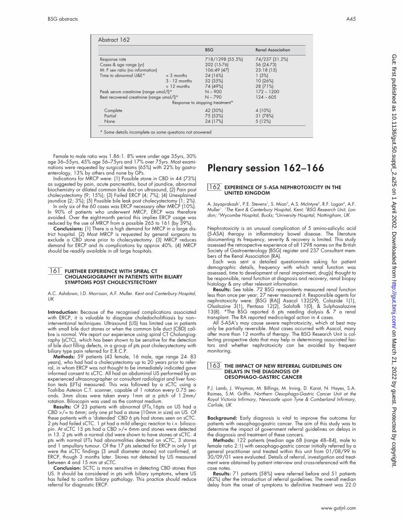
Female to male ratio was 1.86:1. 8% were under age 35yrs, 30%age 36–55yrs, 45% age 56–75yrs and 17% over 75yrs. Most exami-nations were requested by surgical teams (65%) with 22% by gastro-enterology, 13% by others and none by GPs.
Indications for MRCP were: (1) Possible stone in CBD in 44 (73%)as suggested by pain, acute pancreatitis, bout of jaundice, abnormalbiochemistry or dilated common bile duct on ultrasound, (2) Pain postcholecystectomy (9; 15%); (3) Failed ERCP (4; 7%); (4) Unexplainedjaundice (2; 3%); (5) Possible bile leak post cholecystectomy (1; 2%).
In only six of the 60 cases was ERCP necessary after MRCP (10%).In 90% of patients who underwent MRCP, ERCP was thereforeavoided. Over the eight-month period this implies ERCP usage wasreduced by the use of MRCP from a possible 265 to 161 (by 39%).
Conclusions: (1) There is a high demand for MRCP in a large dis-trict hospital. (2) Most MRCP is requested by general surgeons toexclude a CBD stone prior to cholecystectomy. (3) MRCP reducesdemand for ERCP and its complications by approx 40%. (4) MRCPshould be readily available in all large hospitals.
161 FURTHER EXPERIENCE WITH SPIRAL CTCHOLANGIOGRAPHY IN PATIENTS WITH BILIARYSYMPTOMS POST CHOLECYSTECTOMY
A.C. Ashdown, I.D. Morrison, A.F. Muller. Kent and Canterbury Hospital,UK
Introduction: Because of the recognised complications associatedwith ERCP, it is valuable to diagnose choledocholithiasis by non–interventional techniques. Ultrasound (US) has limited use in patientswith small bile duct stones or when the common bile duct (CBD) cali-bre is normal. We report our experience using spiral CT Cholangiog-raphy (sCTC), which has been shown to be sensitive for the detectionof bile duct filling defects, in a group of pts post cholecystectomy withbiliary type pain referred for E.R.C.P.
Methods: 59 patients (43 female, 16 male, age range 24- 83years), who had had a cholecystectomy up to 20 years prior to refer-ral, in whom ERCP was not thought to be immediately indicated gaveinformed consent to sCTC. All had an abdominal US performed by anexperienced ultrasonographer or consultant radiologist and liver func-tion tests (LFTs) measured. This was followed by a sCTC using aToshiba Asteion C.T. scanner, capable of 1 rotation every 0.75 sec-onds. 3mm slices were taken every 1mm at a pitch of 1.2mm/rotation. Biloscopin was used as the contrast medium.
Results: Of 23 patients with abnormal LFTs,16pts on US had aCBD >/= to 6mm; only one pt had a stone (10mm in size) on US. Ofthese patients with a ‘distended’ CBD 6 pts had stones seen on sCTC.2 pts had failed sCTC. 1 pt had a mild allergic reaction to i.v. bilosco-pin. At sCTC 15 pts had a CBD >/= 6mm and stones were detectedin 13. 2 pts with a normal cbd were shown to have stones at sCTC. 4pts with normal LFTs had abnormalities detected on sCTC, 3 stonesand 1 ampullary tumour. Of the 17 pts selected for ERCP in only 1 ptwere the sCTC findings (3 small diameter stones) not confirmed, atERCP, though 3 months later. Stones not detected by US measuredbetween 4 and 15 mm at sCTC.
Conclusion: SCTC is more sensitive in detecting CBD stones thanUS. It should be considered in pts with biliary symptoms, where UShas failed to confirm biliary pathology. This practice should reducereferral for diagnostic ERCP.
Plenary session 162–166
162 EXPERIENCE OF 5-ASA NEPHROTOXICITY IN THEUNITED KINGDOM
A. Jayaprakash1, P.E. Stevens1, S. Mian2, A.S. McIntyre3, R.F. Logan4, A.F.Muller1. 1The Kent & Canterbury Hospital, Kent; 2BSG Research Unit, Lon-don; 3Wycombe Hospital, Bucks; 4University Hospital, Nottingham, UK
Nephrotoxicity is an unusual complication of 5 amino-salicylic acid(5-ASA) therapy in inflammatory bowel disease. The literaturedocumenting its frequency, severity & recovery is limited. This studyassessed the retrospective experience of all 1298 names on the BritishSociety of Gastroenterology (BSG) register and 237 Consultant mem-bers of the Renal Association (RA).
Each was sent a detailed questionnaire asking for patientdemographic details, frequency with which renal function wasassessed, time to development of renal impairment, drug(s) thought tobe responsible, renal function at diagnosis and recovery, renal biopsyhistology & any other relevant information.
Results: See table. 72 BSG respondents measured renal functionless than once per year; 27 never measured it. Responsible agents fornephrotoxicity were: [BSG (RA)] Asacol 132(29), Colazide 1(1),Olsalazine 3(1), Pentasa 12(2), Salofalk 1(0), & Sulphasalazine13(8). *The BSG reported 6 pts needing dialysis & 7 a renaltransplant. The RA reported medico-legal action in 4 cases.
All 5-ASA’s may cause severe nephrotoxicity, which at best mayonly be partially reversible. Most cases occurred with Asacol, manyafter more than 12 months of therapy. The BSG Research Unit is col-lecting prospective data that may help in determining associated fac-tors and whether nephrotoxicity can be avoided by frequentmonitoring.
163 THE IMPACT OF NEW REFERRAL GUIDELINES ONDELAYS IN THE DIAGNOSIS OFOESOPHAGO-GASTRIC CANCER
P.J. Lamb, J. Wayman, M. Billings, M. Irving, D. Karat, N. Hayes, S.A.Raimes, S.M. Griffin. Northern Oesophago-Gastric Cancer Unit at theRoyal Victoria Infirmary, Newcastle upon Tyne & Cumberland Infirmary,Carlisle, UK
Background: Early diagnosis is vital to improve the outcome forpatients with oesophago-gastric cancer. The aim of this study was todetermine the impact of government referral guidelines on delays inthe diagnosis and treatment of these cancers.
Methods: 122 patients (median age 68 (range 48–84), male tofemale ratio 2:1) with oesophago-gastric cancer initially referred by ageneral practitioner and treated within this unit from 01/08/99 to30/09/01 were evaluated. Details of referral, investigation and treat-ment were obtained by patient interview and cross-referenced with thecase notes.
Results: 71 patients (58%) were referred before and 51 patients(42%) after the introduction of referral guidelines. The overall mediandelay from the onset of symptoms to definitive treatment was 22.0
Abstract 162
BSG Renal Association
Response rate 718/1298 (55.3%) 74/237 (31.2%)Cases & age range (yr) 202 (15-76) 56 (24-73)M: F sex ratio (no information) 106:49 (47) 23:18 (15)Time to abnormal U&E* < 3 months 24 (16%) 1 (3%)
3 - 12 months 52 (35%) 10 (26%)> 12 months 74 (49%) 28 (71%)
Peak serum creatinine (range umol/l)* N – 900 172 – 1200Best recovered creatinine (range umol/l)* N – 790 124 – 605
Response to stopping treatment*
Complete 42 (30%) 4 (10%)Partial 75 (53%) 31 (78%)None 24 (17%) 5 (12%)
* Some details incomplete as some questions not answered
BSG abstracts A45
www.gutjnl.com
on March 21, 2022 by guest. P
rotected by copyright.http://gut.bm
j.com/
Gut: first published as 10.1136/gut.50.suppl_2.a25 on 1 A
pril 2002. Dow
nloaded from
(IQR 13.0–32.2) weeks comprising patient delay in consulting a doc-tor (48%), delay in referral (24%), delay in diagnosis (14%), anddelay in commencing treatment (14%). After the introduction of guide-lines there was a significant decrease in the time from referral toendoscopy, from a median of 2.5 (IQR 2.0–5.5) to 2.0 (IQR 1.0–3.0)weeks (p=0.012). Endoscopy was carried out within 2 weeks of refer-ral in 71% (36/51) of patients compared to only 44% (31/71) beforethe guidelines (p=0.06). There was a decrease in the number ofpatients waiting over 6 weeks for endoscopy from 23% (16/71) to8% (4/51) (p=0.03). No significant reduction in total delay (median24.0 (IQR 14.0–32.0) vs. 18.0 (IQR 12.0–33.0) weeks, p=0.107) orchange in the stage of disease at diagnosis was identified followingthe introduction of the guidelines.
Conclusion: The introduction of guidelines has resulted in a signifi-cant decrease in the time from referral to endoscopy for patients withoesophago-gastric cancer. As the major delay occurs prior to referralthis has not translated into a significant reduction in overall delay. Toachieve this patient awareness must also be targeted.
164 REDUCED PALLIDAL MAGNETISATION TRANSFERRATIOS ARE ASSOCIATED WITH FATIGUE INPRE-CIRRHOTIC PATIENTS WITH PRIMARY BILIARYCIRRHOSIS
D.M. Forton1, M. Prince2, J. Allsop1, N. Patel1, J. Goldblatt2, H.C. Thomas1,M. Bassendine2, D.E.J. Jones2, S.D. Taylor-Robinson1. 1Liver Unit, ImperialCollege, London; 2Centre for Liver Research, University of Newcastle, UK
Fatigue is the commonest symptom in primary biliary cirrhosis (PBC),affecting individuals at all stages of the disease. We examine thehypothesis that a CNS abnormality related to cholestasis, rather thancirrhosis per se, underlies this symptom. Globus pallidus (PAL) hyper-intensity on T1-weighted MRI has been reported in biliary atresia, cir-rhosis, parenteral nutrition induced cholestasis and in manganeseworkers.
Methods: 18 women with PBC (14 stage I-II [mean bilirubin 11], 4stage III-IV [32]) and 8 age-matched healthy women underwentcerebral MRI and proton spectroscopy (1H MRS). Magnetisation trans-fer ratios (MTR) for white matter (WM) and 4 subcortical structureswere calculated and 1H MR spectra (TE 135ms) were obtained from8cm3 voxels in the basal ganglia (BG) and frontal WM. The patientscompleted the Fisk Fatigue Severity Score (FSSS) and a battery ofneuropsychological tests.
Results: There were no differences between the stage I-II patientsand controls in the WM or subcortical MTRs or in the 1H MRS. How-ever, the PAL/WM MTR ratio was significantly reduced in the stage I-IIPBC patients (p<0.02), with no similar changes in the thalamus, puta-men or caudate. The 4 stage III-IV patients also had reduced PAL/WMratios, although they were not statistically different from the stage I-IIpatients. When the pre-cirrhotic patients were divided into 2 equalgroups on the basis of the FSSS score, the fatigued patients had a sig-nificantly lower PAL/WM than the non-fatigued patients (p=0.05).There was no similar relationship between cognitive performance andPAL/WM.
Conclusion: We demonstrate a highly localised abnormality in thePAL in pre-cirrhotic PBC patients, which was more pronounced in thefatigued patients. It is unlikely to be due to hepatic encephalopathy asthese non-cirrhotic patients had normal MRS. We postulate that a fail-ure of biliary excretion results in the accumulation of heavy metals inthe PAL, which underlies the reported fatigue.
165 ENDOSCOPIC THERAPY FOR BLEEDING PEPTIC ULCER;A RANDOMISED, CONTROLLED TRIAL COMPARINGHEATER PROBE PLUS HUMAN THROMBIN INJECTIONWITH HEATER PROBE PLUS PLACEBO INJECTION
N.I. Church1, H.J. Dallal1, J. Masson2, A. Fraser2, N.A.G. Mowat2,D.A. Johnston3, G. Fullarton4, E. Radin5, M. Turner5, R. Prescott6, J. Plevris7,K.R. Palmer1. 1Western General Hospital, Edinburgh; 2Aberdeen RoyalInfirmary; 3Ninewells Hospital, Dundee; 4Gartnavel General Hospital,Glasgow; 5Scottish National Blood Transfusion Service; 6University of Edin-burgh Medical School; 7Royal Infirmary, Edinburgh, UK
Introduction: Ulcer haemostasis is attempted in many centres using acombination of injection and thermal application, but there is little evi-dence that combination therapy is better than use of a single agent.Furthermore previous studies of endoscopic therapy have not includeda placebo arm. In this large multicentre, double blind, randomised,placebo controlled trial we examined the hypothesis that combinedhaemostatic treatment using the heater probe plus best injection (usingthrombin) is superior to the heater probe plus placebo injection.
Method: 247 patients presenting with major peptic ulcer bleedingwere randomised to heater probe plus thrombin (group 1) or heaterprobe plus placebo (group 2). The two groups were well matched forall risk categories including age, endoscopic stigmata, shock andseverity of comorbid diseases. Endoscopic therapy was applied using150 joules on average of the heater probe followed by injection of3.5ml of thrombin or placebo.
Results: Successful primary haemostasis was achieved in 97% ofboth groups. Rebleeding developed in 19 (15%) of group 1 patientsand 17 (15%) of group 2 patients; surgery was necessary in 16 and13 of these respectively. Median blood transfusion (3; range 0–32versus 2; 0–35 units) and median duration of hospital admission (5;2–82 versus 5; 1–120 days) were similar in both groups. Adverseevents occurred in 8 group 1 patients and 4 group 2 patients.Mortality at 30 days was 8 (6%) of group 1 patients versus 14 (12%)of group 2 patients.
Conclusion: Combination of thrombin or placebo injection withthe heater probe is a safe and effective haemostatic therapy for bleed-ing peptic ulcer. The combination of thrombin with the heater probedoes not confer an additional benefit over heater probe and placebo,and thrombin injection can not be recommended in preference to anyother injection as adjunctive endoscopic therapy for bleeding pepticulcer in patients receiving heater probe therapy.
166 ERADICATION OF H. PYLORI UNLEASHES POST PPIACID HYPERSECRETION
D. Gillen, A. Wirz, K.E.L. McColl. University Dept of Medicine &Therapeutics, Western Infirmary, Glasgow G11 6NT, UK
Introduction: There is marked rebound acid hypersecretion afteromeprazole in H.pylori -ve but not +ve subjects. Oxyntic gastritisprobably prevents it in the latter.
Aim: To determine the effect of H.pylori eradication on reboundacid hypersecretion after omeprazole.
Methods: 17 healthy H.pylori +ve subjects had acid secretion studiesprior to commencing omeprazole 40mg/day for 8 weeks. During the lastweek of omeprazole, they were randomised to a 1 week course ofamoxycillin/clarithromycin or placebo. Further acid secretion studieswere performed at 1,2,4,6&8 weeks after treatment. A further breath testwas performed at 8 weeks to determine H.pylori status after treatment.
Results: Marked rebound hypersecretion to physiological levels ofgastrin was observed in the subjects who had their infectioneradicated but not in those with persisting infection (table). A similartrend was seen with respect to supraphysiological gastrin stimulation.
Abstract 166
Submaximal acid output (mmol/h)
Pre-omeprazole
Days Post-Omeprazole
Day 7 Day 14 Day 28 Day 42 Day 56
H. pylorieradicated
15.2 24.8* 28.7* 22.4 23.9* 25.8(11.8-29.0) (20.4-28.9) (22.8-30.0) (17.5-32.9) (20.8-29.1) (17.4-29.9)
H. pylori noteradicated
18.8 13.1 16.0 17.2 18.1 15.0(12.2-23.6) (9.6-20.8) (6.4-23.5) (12.7-23.1) (9.3-21.5) (10.3-22.5)
Values are medians (quartiles); *significant versus pre at p<0.03
A46 BSG abstracts
www.gutjnl.com
on March 21, 2022 by guest. P
rotected by copyright.http://gut.bm
j.com/
Gut: first published as 10.1136/gut.50.suppl_2.a25 on 1 A
pril 2002. Dow
nloaded from
Summary: Eradication of H.pylori infection at time of stopping PPItherapy unleashes marked rebound acid hypersecretion which persistsfor at least 6 weeks. Rebound acid hypersecretion following PPI is notseen in those remaining H.pylori positive.
Conclusion: This acid hypersecretion induced by H.pylori eradica-tion in patients previously receiving PPI therapy may explain the rela-tion between H.pylori eradication and development of GERD.
Plenary posters 168–197
168 TREATMENT OF CROHN’S DISEASE WITH INFLIXIMABDOES NOT REDUCE HOSPITAL ATTENDANCE ORADMISSION
I.D.R. Arnott, S. Ghosh. Gastrointestinal Unit, University Department ofMedical Sciences, Western General Hospital, Edinburgh, UK
Introduction: Infliximab is a new treatment for refractory and fistulat-ing Crohn’s disease (CD). Clinical trials and audit data have provenefficacy on disease activity and health related quality of life althoughthere is little data regarding cost effectiveness. Prior to the introductionof novel biological treatments hospitalisation and surgery were themajor costs in CD treatment but the expense of Infliximab may changethis.
Aim: We assessed whether Infliximab reduced hospitalisation orfrequency of out patient clinic review within 6 months of infusion.
Methods: We analysed 30 well-characterised CD patients whohad received a single infusion of Infliximab (5mg/kg) for refractoryactive CD. Clinical details and initial response rates have beenpublished. Patients were followed prospectively and out patient visits,number of hospital admissions and total number of inpatient dayswere collected for the 6 months prior to and following Infliximab. Datawas also compiled from the hospital electronic record of all patientepisodes. Only patients that were cared for exclusively at our institu-tion were included.
Results: Data for the 30 patients is displayed in the table. There areno significant differences in hospital attendance or admission rates.There remains no difference in the clinic visits and admission days preand post Infliximab if patients are stratified as to whether they had aresponse or not and if they are on immunosuppressive or not.
Conclusions: In the present study, Infliximab dose not reduce clinicvisits or hospitalisation within 6 months of infusion. Althoughreductions may be seen with multi-dose regimes dramatic reductions inhospital episodes are not to be expected with a single dose alone. Inf-liximab remains a highly effective drug for active CD but it is likely thatthis will have implications when drug funding is allocated.
169 THE INTERCELLULAR ADHESION MOLECULE-1POLYMORPHISMS IN IBD
J.H. Li, X.S. Yang, S. Cullen, K.L. Lin, A. Armuzzi, T. Ahmad, M.J. Neville,R. Walton, D.P. Jewell. Gastroenterology Unit, Gibson Laboratories, Rad-cliffe Infirmary, University of Oxford, Oxford OX2 6HE, UK
Ulcerative colitis (UC) and Crohn’s disease (CD), both forms of inflam-matory bowel disease (IBD) are complex traits. Intercellular adhesionmolecule-1 (ICAM1) is expressed on vascular endothelium and plays akey role in the transendothelial migration of neutrophils and T-cell acti-vation. The region harboring the ICAM1 gene on 19p13 is linked toCD in a Canadian genome wide scan, and a growing body ofevidence indicates that ICAM1 could play a role in IBD development.Our previous work has replicated the Canadian linkage of 19p13 to
CD. ICAM1 is known to contain at least two polymorphic sites, situatedin codons 241 (R/G 241) and 469 (K/E 469). A North Americanstudy has shown an association between IBD and the R241polymorphism (Yang etc 1995). We have examined potential associa-tions of ICAM1polymorphisms in 132 UC and 67 CD and ethnicallymatched 131 controls. CD patients include subgroups of 26 ileal dis-ease and 31 ileo colonic disease, 26 fistulating disease and 35 non-fistulating disease, 53 stenosing disease and 8 non-stenosing disease.UC patients include 37 patients who have undergone colectomy, 22with mild total colitis, and 37 with proctitis. Both patients and controlswere genotyped by PCR- SSP for ICAM1 polymorphisms at codonR/G241 and codon E/K469. There were no differences between thegroups in the frequency of R/G241. The control frequency of ICAM1exon 6 K469 was 38.2%. In CD overall, it was 76.9% (p<0.001). ForCD patients with ileal disease, the frequency was 70.9% and 43.3%for ileo-colonic disease. For patients with fistulating disease, the K469frequency was 69.2%, and 84.3% for non-fistulating disease. Forpatients with stenosing disease, the K469 frequency was 77.4%. ForUC overall, the frequency of ICAM1 exon 6 K469 was 69.7%(p<0.001). For patients with severe disease requiring colectomy, withmild total colitis, and with proctitis, the frequencies of thispolymorphism were 66.22 %, 75% and 81.1% respectively. Thisstudy suggests that the alteration in the amino acid sequences of E469to K469 of the ICAM1 molecule may influence IBD.
170 RECURRENT ORAL ULCERATION (ROU) ININFLAMMATORY BOWEL DISEASE (IBD): THE CLINICALHALLMARK OF A MOLECULARLY DEFINEDIBD/BEHCET’S (BD) OVERLAP GROUP
T. Ahmad1, K. Mulcahy-Hawes1, M. Bunce2, A. Armuzzi1, K. Welsh3, S.Marshall3, D. Jewell1. 1Departments of Gastroenterology, 2TransplantImmunology, University of Oxford; 3National Heart and Lung Institute,Imperial College, London, UK
Background: The clinical, endoscopic and histological features ofintestinal BD are similar to those of IBD. BD is rare in Northern Europewhere IBD is common, whilst the prevalence is high on the ‘Silk route’where IBD is rarely reported. This inverse relationship may reflect geo-graphical differences in diagnostic practice. Diagnosis of BD, requiresthe presence of ROU. We have reported that Caucasian BD is associ-ated with HLA-B*51 and B*57. We hypothesise that these markersmight also be associated with ROU in IBD and therefore molecularlydefine an overlap group.
Aims: To determine the prevalence and genetic associations ofROU in patients with IBD.
Methods: History of ROU reported in questionnaires sent to 244CD and 330 UC patients. Linkage disequilibrium mapping wascarried out across 340 polymorphisms, broken down into 24 discretegene haplotypic blocks. Genetic comparisons were made betweenIBD and healthy controls.
Results: 33.2% of UC and 38.9% of CD patients reported ROU(historical prevalence in general population 10%). In UC patients withROU, associations with alleles on two extended HLA haplotypes wereobserved. Peak relative risk (RR) was at DRB1*0103 on the first(16.5% ROU+ vs 3.8% ROU-; P=0.0003; RR=5.0) and B*57 on thesecond (12.1% ROU+ vs 3.8% ROU-; P=0.009; RR=3.5). In CDpatients with ROU a negative association was observed with B*51(3.4% ROU+ vs 13.8% ROU-; P=0.01) However 11 CD patients whocarried B*51 or B*57 reported ROU. 8 of these fulfilled the diagnos-tic criteria for BD.
Conclusions: 1. Prevalence of ROU in IBD is 3x greater than in thebackground population. 2. ROU in UC patients is associated withB*57, a BD susceptibility allele. 3. 8/11 CD patients with ROU whopossess B*51 or B*57 fulfil the criteria for diagnosis of BD. 4. In ourIBD clinic 5% of patients clinically and molecularly resemble BD.
171 LYMPHOCYTE TELOMERASE EXPRESSION ININFLAMMATORY BOWEL DISEASE
D.M. Aldulaimi, J. Barclay, K. Getliffe, A.G. Morris, E. Karteris, E.Hillhouse, C.U. Nwokolo. University Hospitals Coventry and WarwickshireNHS Trust and Dept of Biological Sciences, Warwick University, UK
Background: Telomerase knockout mice develop ulceration and atro-phy of the bowel. In humans colonic telomerase activity is decreasedin ulcerative colitis but it is unknown whether this is restricted to thecolon. The object of this study was to assess lymphocyte telomeraseenzyme activity in patients with inflammatory bowel disease and todetermine the role of the mRNA that encodes its catalytic sub-unit(hTERT) in the regulation of enzyme activity.
Abstract 168
BeforeInfliximab
AfterInfliximab P value
Number of clinic visits 3 (0-7) 2.5 (0-8) <0.7Number admitted 13/30 11/30 <0.6Total admission days 0 (0-80) 0 (0-104) <0.8
Clinic visits and admissions in the 6 months pre and post Infliximab.Data are median and ranges or frequencies.
BSG abstracts A47
www.gutjnl.com
on March 21, 2022 by guest. P
rotected by copyright.http://gut.bm
j.com/
Gut: first published as 10.1136/gut.50.suppl_2.a25 on 1 A
pril 2002. Dow
nloaded from
Methods: Blood was sampled from 47 patients with ulcerative coli-tis (UC), 37 with Crohn’s disease (CD) and 37 controls. Lymphocyteswere cultured for 72 hours with phytohaemagglutinin. Telomeraseactivity was measured in stimulated and unstimulated lymphocytesusing the Telomerase Repeat Amplification Protocol (TRAP) assay.Lymphocyte hTERT mRNA was quantified in about 40% of samples(18 UC, 14 CD and 14 controls) by real-time PCR.
Results: Expressed as median (95 CI) in arbitrary units (Stimulatedlymphocytes only). Telomerase enzyme activity in controls was 5.96(3.3 – 9.2) and was decreased significantly in UC 3.1 (3.1–4.1) p <0.05 and non-significantly in CD 2.27 (1.2–6). There was nodifference in hTERT mRNA concentration between the three groups.Telomerase activity and hTERT mRNA were generally undetectable inunstimulated lymphocytes.
Conclusion: Lymphocyte telomerase activity is decreased inulcerative colitis. This suggests that previously reported colonic telom-erase deficiency in UC extends to non-colonic tissue and could repre-sent a global defect. Factors other than hTERT mRNA expression maycontribute to the regulation of telomerase activity in stimulatedlymphocytes
172 A COMPREHENSIVE INVESTIGATION OF D-LOOPMUTATIONS IN THE MITOCHONDRIAL DNA OFCOLORECTAL TUMOURS
S. Fradley, P.D. Lewis, P. Griffiths, J.M. Parry (introduced by J. Baxter).Human Molecular Pathology Group, University of Wales Swansea, Single-ton Park, Swansea SA2 8PP, UK
The human mitochondrial genome (mtDNA) contains a shorthypervariable, non-coding control region known as the D-Loop. Anumber of recent investigations have revealed that a proportion ofcolorectal adenocarcinomas harbour mtDNA mutations, not present innormal surrounding mucosa and are classified as tumour-specific.Tumour specific mtDNA mutations have also been observed in manyother tumours including lung and bladder and it has been suggestedthat these mutations may serve as diagnostic markers for cancer. TheD-Loop, used commonly in population studies and the most mutableregion within mtDNA, may be rapidly and cost-effectively scanned formutations. Previous studies involving the search for tumour-specificmtDNA mutations in colorectal cancer have relied on small samplesizes or have failed to reveal mutations within the D-Loop. Using PCR-SSCP and DNA sequencing we have undertaken a comprehensivesurvey of the D-Loop in adenocarcinoma and normal mucosa of twentypatients to (i) demonstrate the usefulness of the D-Loop in providingtumour-specific markers for colorectal cancer, (ii) reveal the types anddistribution of mutations within the D-Loop in colorectal tumours, (iii)estimate the frequency of mutation within this region in adenocarcino-mas, (iv) establish the levels of heteroplasmy in colorectal tumours. Ofthe colorectal adenocarcinomas, 25% showed tumour specificmutations which where not present in the normal mucosa. Sequencingrevealed the mutations to be a 1-bp C:G deletion and a 1-bp C:Ginsertion at nucleotide position 309, two C:G/T:A transitions at nucle-otide positions 61 and 52 and one T:A/A:T transversion at nucleotideposition 55. This study is currently ongoing and we aim to increase thesample size by 100% and analyse adenomas and hyperplasticpolyps. Our results will allow predictions to be made concerning thecausative factors of mtDNA mutations in colorectal cancer and giveinsight into whether these mutations precede neoplasia or are a con-sequence of tumorigenesis.
173 TNM&R STAGING IN COLORECTAL CANCER: HAS THETWO WEEK WAIT FOR PATIENTS MADE ANY DIFFERENCE?
R.P. Arasaradnam, N. Bhala, A.J. Shorthouse, I.J. Adam, A.J. Lobo. Deptof Gastroenterology & Colorectal Surgery, Central Sheffield Teaching Hos-pitals, Royal Hallamshire Hospital, Sheffield, UK
Background: Colorectal Cancer (CRC) is the commonest cancer inthe United Kingdom among non-smokers. In accordance with the Gov-ernment NHS Cancer Plan, the concept of a two week wait (tww) wasintroduced to ensure that all patients with a suspicion of CRC wereseen by a specialist within two weeks of referral.
Aim: The aim of this study is to examine the effect of this implemen-tation on tumour staging for patients with colorectal cancer.
Methods: TNM staging and demographics from 140 consecutivepatients diagnosed with CRC and treated at a single regional unitwere collected for one year before the introduction of the tww and fora year afterwards, to September 2001. Of the 107 elective patients,90 were operated on. 33 patients presenting as emergencies wereexcluded from subsequent analysis.
Results: 24/40 (60%) patients were node negative in the pre twwgroup compared with 32/46 (70%) in the post tww group (χ2=0.21;ns). Similarly, before the introduction of the tww there were 9/40(23%) patients regarded as being of good prognosis (T1N0, T1N1,, orT2N0) compared with 13/46 (28%) in the post tww (χ2=0.29, ns). Thetotal number at each T stage pre tww (n=40) was T1=3; T2=8; T3=18;T4=11 compared with post tww (n=45) T1 =6, T2=7, T3=22 and T4=10(χ2=0.55, ns). Irrespective of whether they were referred pre or postthe tww, of all patients seen within 14 days (n=47) [median 9 & range0–14 days], 25/47 (53%) were node negative. This compared with38/53 (72%) of those seen greater than 14 days (n=60) [median 32& range 15–216 days] after referral (χ2=0.01, ns). Of those seenwithin 14 days, 8/47 (17%) were in the good prognostic categorycompared with 14/53 (26%) seen after (χ2=0.19, ns). Medianfollow-up (days) was 513.5 (range 38–696) & 315.5 (range12–454) for pre and post tww respectively. Overall survival (includingemergency admissions) in the pre tww was 79.31% (n=59) while inthe post tww (n=81) was 86.42% (p < 0.4749). Survival for electiveadmissions pre tww (n=47) was 91.30% compared to 91.67%(n=60) in the post tww (p=ns).
Conclusions: The two week wait has made no impact on TNMstaging for CRC at initial presentation.
174 CHANGES IN HCV SPECIFIC CD4+ RESPONSESDURING TREATMENT WITH PEGYLATEDINTERFERON-α AND RIBAVIRIN CORRELATE WITHVIRAL RESPONSE
C.L. Brooks, E.A. Sanders, S. Hadfield, S. Green, W.M. Rosenberg.
Introduction: Hepatitis C virus (HCV) establishes a chronic infectionin up to 85% of those exposed. The resultant immune mediated hepa-titis leads to progressive fibrosis and cirrhosis in a significantproportion of patients. The combination of interferon-α (IFN-α) andribavirin achieves a sustained virological response in around 40% ofthose treated. The addition of a Polyethylene Glycol (PEG) moiety tothe IFN-α molecule significantly changes the pharmacokinetics andimproves efficacy. HCV specific CD4+ T cell responses are weak orundetectable in patients with chronic hepatitis C (CHC), whilst theyare brisk and multi-specific following spontaneous resolution of acuteinfection. We aimed to characterise CD4+ HCV specific responses inCHC patients at the start of treatment with PEG-IFN-α and ribavirin,and then repeat them serially throughout the course of treatment, cor-relating with viral response.
Methods: Peripheral blood mononuclear cells were isolated bydensity gradient centrifugation from 8 patients prior to startinganti-viral treatment with PEG-IFN-α and Ribavirin. These cells were setup in culture with either recombinant HCV antigens or appropriatepositive and negative controls. Cellular proliferation was assessed byincorporation of tritiated thymidine. Cellular markers of activation andcytokine secretion were assayed by fluorescence activated cellscanning. The assays were repeated at regular intervals throughouttreatment and follow up. At each time point, viral load was measuredby a PCR based quantitation method.
Results: Throughout treatment, HCV specific lymphocyte prolifera-tion increased in magnitude in 75% of patients, apparent from amedian of 18 weeks. The greatest increase was seen in patients whoachieved sustained virological response, in which stronger responseswere maintained. There were also changes in the CD4+ cytokine pro-file induced by core and helicase recombinant proteins whichcorrelated with virological response.
Discussion: There has been one study examining serial HCV spe-cific CD4+ responses whilst on standard combination treatment[Cramp et al. 2000]. Our study has similar results, but the onset ofincreased magnitude of responses is later than seen on standard treat-ment, possibly reflecting the different pharmacokinetics. This studysupports the hypothesis that successful anti-viral treatment allows toler-ance to HCV to be broken.
175 GASTRIC EPITHELIAL CELL PROLIFERATION ANDAPOPTOSIS IN HELICOBACTER PYLORI INFECTEDMONGOLIAN GERBILS
M. Court, M.A. Aboshkiwa, M.F. Dixon, P.A. Robinson, J.E. Crabtree.Molecular Medicine Unit, St James’s Hospital, Leeds LS9 7TF, UK
Introduction: Chronic H. pylori infection in Mongolian gerbils (MG)has been demonstrated to result in gastric cancer. The aims of thisstudy were to assess the ability of the mouse adapted SS1 strain to
A48 BSG abstracts
www.gutjnl.com
on March 21, 2022 by guest. P
rotected by copyright.http://gut.bm
j.com/
Gut: first published as 10.1136/gut.50.suppl_2.a25 on 1 A
pril 2002. Dow
nloaded from
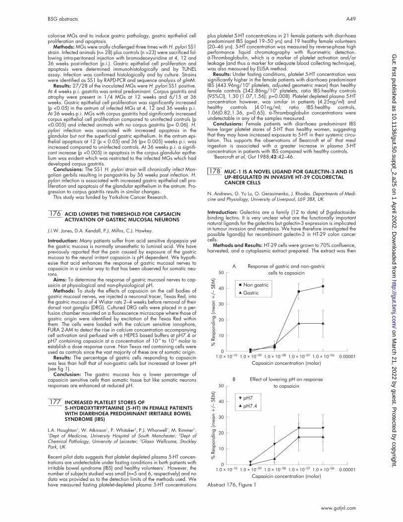
colonise MGs and to induce gastric pathology, gastric epithelial cellproliferation and apoptosis.
Methods: MGs were orally challenged three times with H. pylori SS1strain. Infected animals (n= 28) plus controls (n =23) were sacrificed fol-lowing intra-peritoneal injection with bromodeoxyuridine at 4, 12 and36 weeks post-infection (p.i.). Gastric epithelial cell proliferation andapoptosis were determined immunohistologically and by TUNELassay. Infection was confirmed histologically and by culture. Strainswere identified as SS1 by RAPD-PCR and sequence analysis of glmM.
Results: 27/28 of the inoculated MGs were H. pylori SS1 positive.At 4 weeks p.i. gastritis was antral predominant. Corpus gastritis andatrophy were present in 1/4 MGs at 12 weeks and 6/15 at 36weeks. Gastric epithelial cell proliferation was significantly increased(p <0.05) in the antrum of infected MGs at 4, 12 and 36 weeks p.i.At 36 weeks p.i. MGs with corpus gastritis had significantly increasedcorpus epithelial cell proliferation compared to uninfected controls (p<0.005) and infected animals with no corpus gastritis (p= 0.06). H.pylori infection was associated with increased apoptosis in theglandular but not the superficial gastric epithelium. In the antrum epi-thelial apoptosis at 12 (p < 0.05) and 36 (p< 0.005) weeks p.i. wasincreased compared to uninfected controls. At 36 weeks p.i. a signifi-cant increase (p <0.005) in apoptosis in the corpus glandular epithe-lium was evident which was restricted to the infected MGs which haddeveloped corpus gastritis.
Conclusions: The SS1 H. pylori strain will chronically infect Mon-golian gerbils resulting in pangastritis by 36 weeks post infection. H.pylori infection is associated with increased gastric epithelial cell pro-liferation and apoptosis of the glandular epithelium in the antrum. Pro-gression to corpus gastritis results in similar changes.
This study was funded by Yorkshire Cancer Research.
176 ACID LOWERS THE THRESHOLD FOR CAPSAICINACTIVATION OF GASTRIC MUCOSAL NEURONS
J.I.W. Jones, D.A. Kendall, P.J. Millns, C.J. Hawkey.
Introduction: Many patients suffer from acid sensitive dyspepsia yetthe gastric mucosa is normally anaesthetic to luminal acid. We havepreviously reported that the pain caused by exposure of the gastricmucosa to the neural irritant capsaicin is pH dependent. We hypoth-esise that acid enhances the response of gastric mucosal nerves tocapsaicin in a similar way to that has been observed for somatic neu-rons.
Aims: To determine the response of gastric mucosal nerves to cap-saicin at physiological and non-physiological pH.
Methods: To study the effects of capsaicin on the cell bodies ofgastric mucosal nerves, we injected a neuronal tracer, Texas Red, intothe gastric mucosa of 4 Wistar rats 2–4 weeks before removal of theirdorsal root ganglia (DRG). Cultured DRG cells were placed in a per-fusion chamber mounted on a fluorescence microscope where those ofgastric origin were identified by excitation of the Texas Red withinthem. The cells were loaded with the calcium sensitive ionophore,FURA 2-AM to detect the rise in calcium concentration accompanyingcell activation and perfused with a HEPES based buffers at pH7.4 orpH7 containing capsaicin at a concentration of 10–9 to 10–6 molar toestablish a dose response curve. Non Texas red containing cells wereused as controls since the vast majority of these are of somatic origin.
Results: The percentage of gastric cells responding to capsaicinwas less than half that of non-gastric cells but increased at lower pH(see fig 1).
Conclusion: The gastric mucosa has a lower percentage ofcapsaicin sensitive cells than somatic tissue but like somatic neuronsresponses are enhanced at reduced pH.
177 INCREASED PLATELET STORES OF5-HYDROXYTRYPTAMINE (5-HT) IN FEMALE PATIENTSWITH DIARRHOEA PREDOMINANT IRRITABLE BOWELSYNDROME (IBS)
L.A. Houghton1, W. Atkinson1, P. Whitaker2, P.J. Whorwell1, M. Rimmer3.1Dept of Medicine, University Hospital of South Manchester; 2Dept ofChemical Pathology, University of Leicester; 3Glaxo Wellcome, StockleyPark, UK
Recent pilot data suggests that platelet depleted plasma 5-HT concen-trations are undetectable under fasting conditions in both patients withirritable bowel syndrome (IBS) and healthy volunteers1. However, thenumber of subjects studied was small (n=5 and 6, respectively) and nodata was provided as to the detection limits of the methods used. Wehave measured fasting platelet-depleted plasma 5-HT concentrations
plus platelet 5-HT concentrations in 21 female patients with diarrhoeapredominant IBS (aged 19–50 yrs) and 19 healthy female volunteers(20–46 yrs). 5-HT concentration was measured by reverse-phase highperformance liquid chromatography with fluorimetric detection.α-Thromboglobulin, which is a marker of platelet activation and/orleakage (and thus a marker for adequate blood collecting technique),was also measured by ELISA method.
Results: Under fasting conditions, platelet 5-HT concentration wassignificantly higher in the female patients with diarrhoea predominantIBS (443.96ng/109 platelets, adjusted geometric mean) than healthyfemale controls (342.86ng/109 platelets; ratio IBS:healthy controls(95%CI), 1.30 (1.07,1.56); p=0.008). Platelet depleted plasma 5-HTconcentration however, was similar in patients (4.25ng/ml) andhealthy controls (4.01ng/ml; ratio IBS:healthy controls,1.06(0.82,1.36, p=0.65). α-Thromboglobulin concentrations wereundetectable in any of the samples measured.
Conclusions: Female patients with diarrhoea predominant IBShave larger platelet stores of 5-HT than healthy women, suggestingthat they may have increased exposure to 5-HT in their systemic circu-lation. This supports the observations of Bearcroft et al1 that mealingestion is associated with a greater increase in plasma 5-HTconcentration in patients with IBS compared with healthy controls.
1Bearcroft et al, Gut 1988;42:42–46.
178 MUC-1 IS A NOVEL LIGAND FOR GALECTIN-3 AND ISUP-REGULATED IN INVASIVE HT-29 COLORECTALCANCER CELLS
N. Andrews, G. Yu Lu, O. Gerasimenko, J. Rhodes. Departments of Medi-cine and Physiology, University of Liverpool, L69 3BX, UK
Introduction: Galectins are a family (12 to date) of β-galactoside-binding lectins. It is very unclear what are the functionally importantnatural ligands for the galectins but galectin-3 expression is implicatedin tumour invasion and metastasis. We have therefore investigated thepossible ligand(s) for recombinant galectin-3 in HT-29 colon cancercells.
Methods and Results: HT-29 cells were grown to 70% confluence,harvested, and a cytoplasmic extract prepared. The extract was then
0
10
20
30
40
50
%Responding(m
ean+/_
SEM)
1.0 × 10_10 1.0 × 10
_09 1.0 × 10
_08 1.0 × 10
_07 1.0 × 10
_06 0.00001
Capsaicin concentration (molar)
Response of gastric and non-gastric
cells to capsaicin
A
Non gastric
Gastric
0
10
20
30
40
50
%Responding(m
ean+/_
SEM)
1.0 × 10_10 1.0 × 10
_09 1.0 × 10
_08 1.0 × 10
_07 1.0 × 10
_06 0.00001
Capsaicin concentration (molar)
Effect of lowering pH on response
to capsaicin
B
pH7
pH7.4
Abstract 176, Figure 1
BSG abstracts A49
www.gutjnl.com
on March 21, 2022 by guest. P
rotected by copyright.http://gut.bm
j.com/
Gut: first published as 10.1136/gut.50.suppl_2.a25 on 1 A
pril 2002. Dow
nloaded from
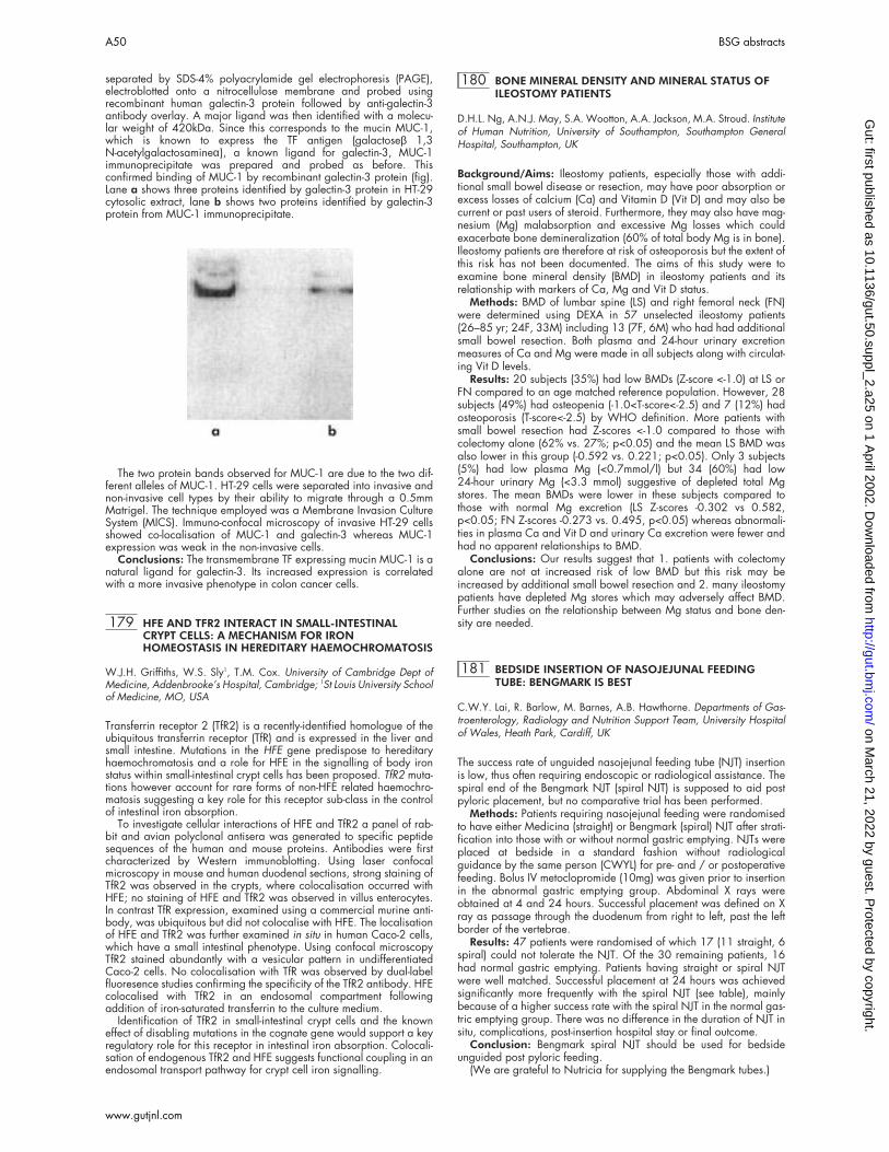
separated by SDS-4% polyacrylamide gel electrophoresis (PAGE),electroblotted onto a nitrocellulose membrane and probed usingrecombinant human galectin-3 protein followed by anti-galectin-3antibody overlay. A major ligand was then identified with a molecu-lar weight of 420kDa. Since this corresponds to the mucin MUC-1,which is known to express the TF antigen (galactoseβ 1,3N-acetylgalactosamineα), a known ligand for galectin-3, MUC-1immunoprecipitate was prepared and probed as before. Thisconfirmed binding of MUC-1 by recombinant galectin-3 protein (fig).Lane a shows three proteins identified by galectin-3 protein in HT-29cytosolic extract, lane b shows two proteins identified by galectin-3protein from MUC-1 immunoprecipitate.
The two protein bands observed for MUC-1 are due to the two dif-ferent alleles of MUC-1. HT-29 cells were separated into invasive andnon-invasive cell types by their ability to migrate through a 0.5mmMatrigel. The technique employed was a Membrane Invasion CultureSystem (MICS). Immuno-confocal microscopy of invasive HT-29 cellsshowed co-localisation of MUC-1 and galectin-3 whereas MUC-1expression was weak in the non-invasive cells.
Conclusions: The transmembrane TF expressing mucin MUC-1 is anatural ligand for galectin-3. Its increased expression is correlatedwith a more invasive phenotype in colon cancer cells.
179 HFE AND TFR2 INTERACT IN SMALL-INTESTINALCRYPT CELLS: A MECHANISM FOR IRONHOMEOSTASIS IN HEREDITARY HAEMOCHROMATOSIS
W.J.H. Griffiths, W.S. Sly1, T.M. Cox. University of Cambridge Dept ofMedicine, Addenbrooke’s Hospital, Cambridge; 1St Louis University Schoolof Medicine, MO, USA
Transferrin receptor 2 (TfR2) is a recently-identified homologue of theubiquitous transferrin receptor (TfR) and is expressed in the liver andsmall intestine. Mutations in the HFE gene predispose to hereditaryhaemochromatosis and a role for HFE in the signalling of body ironstatus within small-intestinal crypt cells has been proposed. TfR2 muta-tions however account for rare forms of non-HFE related haemochro-matosis suggesting a key role for this receptor sub-class in the controlof intestinal iron absorption.
To investigate cellular interactions of HFE and TfR2 a panel of rab-bit and avian polyclonal antisera was generated to specific peptidesequences of the human and mouse proteins. Antibodies were firstcharacterized by Western immunoblotting. Using laser confocalmicroscopy in mouse and human duodenal sections, strong staining ofTfR2 was observed in the crypts, where colocalisation occurred withHFE; no staining of HFE and TfR2 was observed in villus enterocytes.In contrast TfR expression, examined using a commercial murine anti-body, was ubiquitous but did not colocalise with HFE. The localisationof HFE and TfR2 was further examined in situ in human Caco-2 cells,which have a small intestinal phenotype. Using confocal microscopyTfR2 stained abundantly with a vesicular pattern in undifferentiatedCaco-2 cells. No colocalisation with TfR was observed by dual-labelfluoresence studies confirming the specificity of the TfR2 antibody. HFEcolocalised with TfR2 in an endosomal compartment followingaddition of iron-saturated transferrin to the culture medium.
Identification of TfR2 in small-intestinal crypt cells and the knowneffect of disabling mutations in the cognate gene would support a keyregulatory role for this receptor in intestinal iron absorption. Colocali-sation of endogenous TfR2 and HFE suggests functional coupling in anendosomal transport pathway for crypt cell iron signalling.
180 BONE MINERAL DENSITY AND MINERAL STATUS OFILEOSTOMY PATIENTS
D.H.L. Ng, A.N.J. May, S.A. Wootton, A.A. Jackson, M.A. Stroud. Instituteof Human Nutrition, University of Southampton, Southampton GeneralHospital, Southampton, UK
Background/Aims: Ileostomy patients, especially those with addi-tional small bowel disease or resection, may have poor absorption orexcess losses of calcium (Ca) and Vitamin D (Vit D) and may also becurrent or past users of steroid. Furthermore, they may also have mag-nesium (Mg) malabsorption and excessive Mg losses which couldexacerbate bone demineralization (60% of total body Mg is in bone).Ileostomy patients are therefore at risk of osteoporosis but the extent ofthis risk has not been documented. The aims of this study were toexamine bone mineral density (BMD) in ileostomy patients and itsrelationship with markers of Ca, Mg and Vit D status.
Methods: BMD of lumbar spine (LS) and right femoral neck (FN)were determined using DEXA in 57 unselected ileostomy patients(26–85 yr; 24F, 33M) including 13 (7F, 6M) who had had additionalsmall bowel resection. Both plasma and 24-hour urinary excretionmeasures of Ca and Mg were made in all subjects along with circulat-ing Vit D levels.
Results: 20 subjects (35%) had low BMDs (Z-score <-1.0) at LS orFN compared to an age matched reference population. However, 28subjects (49%) had osteopenia (-1.0<T-score<-2.5) and 7 (12%) hadosteoporosis (T-score<-2.5) by WHO definition. More patients withsmall bowel resection had Z-scores <-1.0 compared to those withcolectomy alone (62% vs. 27%; p<0.05) and the mean LS BMD wasalso lower in this group (-0.592 vs. 0.221; p<0.05). Only 3 subjects(5%) had low plasma Mg (<0.7mmol/l) but 34 (60%) had low24-hour urinary Mg (<3.3 mmol) suggestive of depleted total Mgstores. The mean BMDs were lower in these subjects compared tothose with normal Mg excretion (LS Z-scores -0.302 vs 0.582,p<0.05; FN Z-scores -0.273 vs. 0.495, p<0.05) whereas abnormali-ties in plasma Ca and Vit D and urinary Ca excretion were fewer andhad no apparent relationships to BMD.
Conclusions: Our results suggest that 1. patients with colectomyalone are not at increased risk of low BMD but this risk may beincreased by additional small bowel resection and 2. many ileostomypatients have depleted Mg stores which may adversely affect BMD.Further studies on the relationship between Mg status and bone den-sity are needed.
181 BEDSIDE INSERTION OF NASOJEJUNAL FEEDINGTUBE: BENGMARK IS BEST
C.W.Y. Lai, R. Barlow, M. Barnes, A.B. Hawthorne. Departments of Gas-troenterology, Radiology and Nutrition Support Team, University Hospitalof Wales, Heath Park, Cardiff, UK
The success rate of unguided nasojejunal feeding tube (NJT) insertionis low, thus often requiring endoscopic or radiological assistance. Thespiral end of the Bengmark NJT (spiral NJT) is supposed to aid postpyloric placement, but no comparative trial has been performed.
Methods: Patients requiring nasojejunal feeding were randomisedto have either Medicina (straight) or Bengmark (spiral) NJT after strati-fication into those with or without normal gastric emptying. NJTs wereplaced at bedside in a standard fashion without radiologicalguidance by the same person (CWYL) for pre- and / or postoperativefeeding. Bolus IV metoclopromide (10mg) was given prior to insertionin the abnormal gastric emptying group. Abdominal X rays wereobtained at 4 and 24 hours. Successful placement was defined on Xray as passage through the duodenum from right to left, past the leftborder of the vertebrae.
Results: 47 patients were randomised of which 17 (11 straight, 6spiral) could not tolerate the NJT. Of the 30 remaining patients, 16had normal gastric emptying. Patients having straight or spiral NJTwere well matched. Successful placement at 24 hours was achievedsignificantly more frequently with the spiral NJT (see table), mainlybecause of a higher success rate with the spiral NJT in the normal gas-tric emptying group. There was no difference in the duration of NJT insitu, complications, post-insertion hospital stay or final outcome.
Conclusion: Bengmark spiral NJT should be used for bedsideunguided post pyloric feeding.
(We are grateful to Nutricia for supplying the Bengmark tubes.)
A50 BSG abstracts
www.gutjnl.com
on March 21, 2022 by guest. P
rotected by copyright.http://gut.bm
j.com/
Gut: first published as 10.1136/gut.50.suppl_2.a25 on 1 A
pril 2002. Dow
nloaded from
182 LONG TERM FOLLOW UP OF PATIENTS WITH GASTRICOUTLET OBSTRUCTION RELATED TO PEPTIC ULCERDISEASE (PUD) TREATED WITH ENDOSCOPICBALLOON DILATATION AND DRUG THERAPY
S. Cherian, A. Cherukuri, P. Singh. Staffordshire General Hospital,Stafford, UK
Introduction: Previous studies suggest endoscopic balloon dilatationin gastric outlet obstruction from PUD does not achieve long-termremission and most patients eventually require surgery. However,these studies have not addressed the issue of altering the natural his-tory of the underlying PUD.
Methods: We examined medical notes of 18 patients with PUDrelated gastric outlet obstruction treated by a single consultant gastro-enterologist. In all patients, an attempt was made to establish and treatthe aetiology of PUD. Where no cause was found or its removal notpossible or where disease relapsed, long-term maintenance antisecre-tory therapy was given.
Results: Of the 18 patients, one presented with aspirationpneumonia and another with stroke and both succumbed to theirillness. Of 16 available for follow up, 6 were men and 3 were smok-ers. Their median age was 69 years (range 43–94). Fourteen patientswere treated with TTS balloon dilatation and drug therapy and 2 withdrug therapy alone. The median number of dilatations was 2 (range0–5). There were no complications from dilatation. The causes of PUDwere as detailed in table 1. Nine of the 10 HP positive patientsreceived eradication therapy. Eradication was confirmed in 5.NSAIDs were discontinued. Four patients stayed on aspirin for medi-cal reasons. Remission was achieved in all 16 patients with a medianfollow-up of 30 months (range 5–54) including 2 who died from unre-lated illness after being in remission for nearly 2 years. Three patientsbecame asymptomatic without need for maintenance therapy (2 aftersuccessful HP eradication, and 1 after withdrawal of NSAID). Theremaining 13 required long term maintenance therapy for the reasonsdetailed in table 2.
Conclusions: PUD related gastric outlet obstruction can be kept inlong-term remission by using a structured approach combining dilata-tion and removal of the cause of PUD and/or maintenance antisecre-tory therapy. TTS balloon dilatation is a simple, effective, and safeprocedure.
183 ENDOSCOPIC PALLIATIVE TREATMENT INOESOPHAGEAL AND GASTRIC CANCER: EVIDENCEFROM 948 PATIENTS IN THE POPULATION BASEDSCOTTISH AUDIT
A.M. Thompson, R. Stuart for the Scottish Audit of Gastric and Oesopha-geal Cancer.
The population based Scottish Audit of Gastric and OesophagealCancer (SAGOC) accrued data over a 2 year period on 3,293patients of whom 948, predominantly with oesophageal or junctionalcancer, received endoscopic palliative treatment (EPT).
Stent placement alone (506 patients) or LASER treatment alone(117 patients) were popular, but combination approaches supportedby radiotherapy (188 patients) or chemotherapy (134 patients) werealso administered. There was significant variation in delivery of EPT byhealthboard of residence (chi square test, P<0.001), but not by depri-vation quintile.
Complications were recorded in 221/948 patients (23%) andwere associated with multiple treatments (P<0.001). Oesophagealperforation was uncommon and occurred in 23 patients post stent and3 patients after LASER.
Stent alone was used for the relief of grade 3 or 4 dysphagia, stentand radiotherapy for grades 2,3,4 and LASER for grades 1,2,3. Themajority of patients (>65%) had normal physical activity or onlystrenuous activity restricted before undergoing EPT. Stents weredeemed by the consultant looking after the patient to have been usedappropriately for 95% of patients and LASER for 83% of patients.
Survival for all the patients receiving EPT was 40% at 6 months,17% at 12 months 10% at 18 months and 6 % at 24 months, suggest-ing benefits from EPT intervention in patients with advanced disease.
While there may be differences between the symptoms and stagingof patients receiving different types of EPT, there is evidence forregional variations in the approaches used. However, appropriate useof intervention may provide good palliation for several months.
184 IS IT NECESSARY TO BIOPSY ACUTELY BLEEDINGGASTRIC ULCERS AT THE INITIAL ENDOSCOPY?
S.J. Panter1, M.G. Bramble1. 1James Cook University Hospital, Middles-brough and CIHR, University of Durham, UK
Background: It is recommended that all gastric ulcers are biopsied toexclude malignancy with follow up endoscopies performed at 6–8week intervals until healing is seen.
Aims: This prospective study aimed to identify whether theserecommendations were being followed – especially at emergencyendoscopy.
Methods: All patients over a 20-month period diagnosed with agastric ulcer were identified and those with a definite ulcer (with amucosal breach documented as >5mm) diagnosed at endoscopywere included in the study. A record of the macroscopic judgement ofthe ulcers as being benign, suspicious or malignant as stated by theendoscopist was made. This was correlated with the histology results.
Results: 250 patients were reportedly diagnosed with gastriculcers. Of these 191 met the inclusion criteria. The male: female ratiowas106: 85 and the mean age at diagnosis was 67 years (range23–98). Of these 11 ulcers were diagnosed operatively, 79 werediagnosed as an emergency “bleeder” and 99 were diagnosed rou-tinely (29% open access; 71% routine list via OP clinic). Of the“bleeders” only 55% had biopsies taken at the initial endoscopy, withthe mean number of biopsies per ulcer being 2.7. This was despiteonly 30.4% actually requiring injection at the time of the endoscopy.Of those ulcers diagnosed routinely, biopsies were taken in 94.4%,with the mean number of biopsies being 3.5. Of the “bleeders” 6patients required laparotomy for further bleeding of whom 1 had hada single biopsy taken at endoscopy. Overall 126 ulcers were thoughtto be benign, 28 to be suspicious and 17 frankly malignant. Of thoseulcers diagnosed at an emergency endoscopy these figures were 60,11 and 3 respectively. The predictive value of the macroscopic judge-ment can be estimated as the proportion of correct macroscopic diag-noses. The overall predictive value (PVpos) of a macroscopic judgementof definite or suspicious of malignancy was 0.52 (24/46) and theoverall predictive value (PVpos) of a macroscopic judgement of benignwas 0.96 (121/126). For the “bleeders” the PVpos were 0.43 and0.97 respectively.
Conclusion: These results show that biopsies are often not taken atthe time of an emergency endoscopy. However in this study only 2%of “benign-looking” bleeding gastric ulcers were ultimately diagnosedas malignant. Therefore the priority for benign looking bleeding gas-tric ulcers remains to establish haemostasis.
Abstract 181
Success rate at 24 hours [no./total,(%)]
Straight NJT Spiral NJT p value
Normal gastric emptying 1/7 (14.3%) 7/9 (77.8%) 0.041Abnormal gastric emptying 0/7 (0%) 4/7 (57.1%) 0.070Total 1/14 (7.1%) 11/16 (68.8%) 0.001
Abstract 182, Table 1
HP Aspirin NSAIDHP +Aspirin
HP +NSAID Idiopathic
Notknown
7 2 1 2 1 2 1
Abstract 182, Table 2
Idiopathic PUDRelapse of PUD despiteremoval of cause
Old age/aspirintherapy/ oesophagitis
2 3 8
BSG abstracts A51
www.gutjnl.com
on March 21, 2022 by guest. P
rotected by copyright.http://gut.bm
j.com/
Gut: first published as 10.1136/gut.50.suppl_2.a25 on 1 A
pril 2002. Dow
nloaded from
185 CURRENT ISSUES IN THE MANAGEMENT OF COLONICPOLYPS. A RETROSPECTIVE REVIEW OF 2806CONSECUTIVE POLYPECTOMIES
J.C. Brooker, S.G. Shah, B.P. Saunders. St Mark’s Hospital, Harrow, UK
Background: Based on current knowledge, population screening andthe removal of colonic adenomas would be an effective means ofreducing the incidence of colorectal cancer. However, a sound under-standing of the distribution of adenomas and risks of polypectomy is aprerequisite to implementation, and will assist in the selection of theoptimal screening modality and the safest and most effective methodsfor polypectomy. We therefore studied polyp epidemiology andpolypectomy techniques and complications in patients from a singleendoscopy department.
Methods: Records of 938 patients (506 males; mean age 58.8years [sd 14.3]; indications: symptoms 535, neoplasia surveillance387, polyposis 55, IBD 37, not recorded 9) who had undergone1023 consecutive colonoscopies with polypectomy during a 22-monthperiod, were examined retrospectively. Complications were identifiedusing a postal questionnaire.
Results: 2806 polypectomies were performed, 37.9% byhot-biopsy, 30.6% by snare and 23.3% by Argon PlasmaCoagulation (APC). 44.7% were benign adenomas and 47.8% ofthese (excluding polyposis) were located proximal to the splenic flex-ure. However only 27% of advanced adenomas (size >1cm or villoushistology) were proximal. Polypectomy failed to yield a specimen foranalysis in 19.3% of snares and 5.2% of hot biopsies (p<0.0001).67.6% patients replied to the complications questionnaire. There wereno deaths, but 4 significant complications were identified, includingone perforation after hot-biopsy (0.16%) and three episodes of majorhaemorrhage (0.47%). Intra-procedural bleeding requiring endo-scopic therapy was significantly associated with aspirin ingestion(p=0.02), but post-procedure bleeding was not (p=0.3). Nocomplications were reported after polypectomy using APC.
Conclusions: These data support previous observations of a proxi-mal shift in the distribution of colorectal adenomas, although mostadvanced neoplasia were still found in the left colon within reach ofthe flexible sigmoidoscope. The incidence of polypectomy complica-tions was lower than previously reported, which may reflect improvedtechnique. Hot-biopsy and APC both appear to be safe for the removalof small polyps, but snare techniques, although safe, frequently fail toyield tissue for histology. No increase in secondary haemorrhage wasseen in patients taking aspirin.
186 VIDEO-ASSESSMENT OF COLONOSCOPYWITHDRAWAL TECHNIQUE
J.C. Brooker, S. Thomas-Gibson, S.G. Shah, C.J. Thapar, B.P. Saunders. StMark’s Hospital, Harrow, UK
Background: Colonoscopy withdrawal technique has been shown tobe important in the detection of adenomas. We aimed to survey cur-rent practice in a single UK endoscopy department, as well as assess-ing the effect of video recording on examination quality.
Method: With ethics committee approval and patient consent, con-secutive routine colonoscopy extubations were video recorded using aremote, closed-circuit TV system. Endoscopists were informed thatrecording would take place when a “recording-light”, conspicuouslyplaced in the endoscopy room, was illuminated. However recordingtook place continuously. Endoscopy lists were randomly assigned to“open” or “blind” recording. The video footage was reviewed by ablinded panel of 3 experienced colonoscopists. Extubations werescored by consensus for seven segments of the colon using 5 param-eters (looking behind folds, cleaning pools, adequacy of distension,time spent inspecting and quality of bowel preparation) using 50mmvisual analogue scales. Patients with IBD, previous colonic resection,current colorectal-cancer or mean preparation score <30, wereexcluded from the analysis.
Results: 93 procedures by 16 endoscopists were included, 50 forcolonic symptoms and 43 for neoplasia surveillance or a family historyof cancer. 52.7% extubations attained a mean score of less than 30(an arbitrary minimum score for acceptable quality). There was no dif-ference in extubation scores or time with blinded compared with openvideo recording (see table). There was also no difference betweengroups in performance of retroflexion in the rectum (12% overall) orthe detection of adenomas (p=0.24).
Conclusions: Over 50% of extubations were sub-optimal, andawareness of video recording made no difference to quality. We sug-gest that both better training in withdrawal technique and allottingmore time per procedure are required to improve examination quality.
187 GENETIC THROMBOPHILIA AND PERI-PARTUM LIVERFAILURE
N. Zakaria, J. Wendon, R. Arya, J. Devlin. King’s College Hospital,London, UK
Introduction: The aetiology of severe pregnancy-related disordersremains poorly characterised. One hypothesis is that the clinical disor-ders recognised - Acute Fatty Liver of Pregnancy [AFLP], HELLPsyndrome and Veno-occlusive disease - represent microangiopathicdisorders. Thrombophilic disorders have previously been implicated inserious hypertensive complications of pregnancy where microangio-pathy is evident. Accordingly, we tested the hypothesis that a higherincidence of genetic thrombophilia would be present in thispopulation.
Methods: Twenty-eight patients with a history of peri-partum liverfailure were tested (AFLP 22, HELLP syndrome 3, and Veno-occlusivedisease 3. Median age 30 years (range 21–36). Twenty-eightpatients were Caucasian and five Afro-Caribbean. All presented in thethird trimester. Only one patint had a known pre-existingpro-thrombotic disorder. All patients had thrombophilia screensperformed post-partum once fully recovered from their illness (factor Vleiden [FVL] and prothrombin G20210A gene mutations, anticardioli-pin antibody and lupus anti-coagulant, anti-thrombin III and protein Cand S levels).
Results: A pro-thrombotic disorder was present in 12/28 (43%)patients. FVL heterozygosity was present in 5/23 Caucasian patients(17.5%). PT G20210A gene heterozygosity was present in 2/28(7%). Anti-cardiolipin antibody was detected in 4/28 (14%). Lupusanticoagulant was detected in one patient with AFLP. In the patientswhere a diagnosis of AFLP was made; genetic thrombophilia waspresent in 10/23 (42%). Protein C and S deficiency was not detectedin patient.
Conclusion: The above results lend some support to the hypothesisthat microvascular thrombosis may play a pathogenic role in asubgroup of patients with peri-partum liver failure. However furtherpredisposing factors for these serious disorders require to beelucidated.
188 NEUROLOGICAL AND COGNITIVE DYSFUNCTION IN“NEVER-ENCEPHALOPATHIC” PATIENTS AWAITINGLIVER TRANSPLANTATION
M.B. Lewis, A. Stroud, M. Davies, P.D. Howdle. St James’s University Hos-pital, Beckett Street, Leeds, UK
Introduction: Patients with liver failure who are not clinicallyencephalopathic can show evidence of neuropsychological impair-ment. Inadequate treatment of previous hepatic encephalopathy isoften blamed. Repeated episodes of hepatic encephalopathy (HE) arethought to underlie the aetiology of the acquired hepatocerebraldegeneration syndrome (AHCD) which consists of cognitive impair-ment associated with, tremor, ataxia, dyskinesias and various motorabnormalities.
Method: Patients listed for liver transplantation with no past historyof hepatic encephalopathy were invited to participate in the study.Standard demographic data and patient characteristics wererecorded. A battery of neuropsychological tests was used to assesscognitive function and a structured neurological examination was per-formed. Twenty-five healthy volunteers were used as controls. The datawere compared using the unpaired t-test or the Mann-Whitney U testas appropriate.
Results: Twenty patients were included. The mean age was 53.25years (sd 9.35). The median duration of illness was 60 months (iqr6–156) and the median Childs-Pugh score was 7 (iqr 5–8). There
Abstract 186
(*50mm visualanalogue scale)
Blind recordingMean (sd)
Open recordingMean (sd)
pvalue
Looking* 23.3 (12.2) 25.6 (10.5) 0.36Cleaning pools* 27.8 (11.3) 27.6 (10.6) 0.94Distension* 32.2 (9.3) 32.4 (8.4) 0.92Qualitative time* 27.3 (13.0) 27.8 (10.5) 0.84Time (min) 6.2(4.7) 6.6(5.0) 0.67
A52 BSG abstracts
www.gutjnl.com
on March 21, 2022 by guest. P
rotected by copyright.http://gut.bm
j.com/
Gut: first published as 10.1136/gut.50.suppl_2.a25 on 1 A
pril 2002. Dow
nloaded from
were no significant differences between baseline characteristics ofpatients and controls. Highly significant global impairment ofcognitive function was detected, with memory and visuo-spatial prob-lems being prominent. No abnormalities were detected with the com-monly used trailmaking test. Twelve patients had an abnormalneurological examination displaying many of the physical signs asso-ciated with AHCD.
Discussion: Cognitive and neurological dysfunction occurs inpatients with end stage liver failure in the absence of previous HE. It isunlikely that the progressive neurological and cognitive decline thatcan occur in these patients is due to repeated episodes of HE. Theimpairment that occurs is severe, and affects many aspects ofcognitive function. This has implications for the pre-operative counsel-ling of these patients and for obtaining informed consent. Currentmethods of monitoring cognitive impairment clinically, such astrailmaking tests, may be inadequate.
189 A PROSPECTIVE ASSESSMENT OF HEPATIC VEINTRANSIT TIMES USING MICROBUBBLE-ENHANCEDULTRASOUND IN NON-INVASIVE GRADING OFHEPATITIS C (HCV) RELATED LIVER DISEASE
A.K.P. Lim1,2, N. Patel1,2, G. Hamilton1,2, M.J.K. Blomley1, D.O. Cosgrove1,G.R. Foster2, R.D. Goldin3, H.C. Thomas2, S.D. Taylor-Robinson1,2. Depart-ments of 1Imaging, 2Medicine, and 3Histopathology, Imperial CollegeSchool of Medicine, Hammersmith Hospital, Du Cane Road, London; 2,3StMary’s Hospital, Praed Street, London, UK
Purpose: Non-invasive assessment of the severity of diffuse liver dis-ease is problematic and biopsy is often needed. We evaluatedhepatic vein arrival times (AT) of an ultrasound microbubble agentand carotid-delay times (CDT) in grading diffuse liver disease inpatients with chronic hepatitis C (HCV) infection using histology as thegold standard.
Materials/Method: 51 untreated and 10 interferon-treatedpatients with biopsy-proven HCV liver disease were studied.Time-intensity curves of hepatic vein spectral Doppler signals andaudio intensity from the carotid artery were analysed after anintravenous microbubble bolus. CDT was calculated as the differencebetween carotid and hepatic vein arrival times. Grading of fibrosis (F)and inflammatory activity (NI) was carried out using the modified HAI(Ishak) scoring system. Patients were divided into mild hepatitis,(F<2/6, NI<3/18); moderate/severe hepatitis, (F>3/6, NI>4/18)and cirrhosis, (F=6).
Results: There was a monotonic decrease in the mean ATs ± 1 s.d.and CDTs ± 1 s.d. for mild, moderate/severe hepatitis and cirrhosis:50.5s ± 24.2, 33.6s ± 26.1, 14.8s ± 4.5 and 36.8s ± 29, 18.7s ±22.6 and 5.8s ± 4.9 respectively (Kruskal Wallis ANOVA, p<0.001).An AT<24s and CDT<10s was 100% sensitive for cirrhosis but only69% and 72% specific respectively, as subjects with fibrosissometimes showed early AT and CDT. 10 interferon-treated patientsshowed earlier arrival times than comparative untreated subjects.
Conclusion: AT and CDT measurements hold promise in character-ising liver disease in patients with HCV and is a highly sensitivemarker of cirrhosis. Treatment with interferon appears to prolong AT.This is important for disease monitoring and may be useful in assess-ing the efficacy of treatment regimes non-invasively and possiblyreplace repeat liver biopsy in some situations.
190 LOSS OF GLUCONEOGENETIC CAPACITY IN A FULLYANAESTHETISED PORCINE MODEL OF PARACETAMOLINDUCED ACUTE LIVER FAILURE
K. Dabos, H.R. Whalen, P.N. Newsome, J.A. Parkinson, N.C. Henderson,I.H. Sadler, P.C. Hayes, J.N. Plevris.
A porcine model of paracetamol induced acute liver failure has beenrecently developed in our laboratory. This model is suitable for patho-physiological studies in acute liver failure. As a first step weinvestigated the gluconeogenic capacity of the porcine liver in thatparticular model.
Materials and Methods: Thirty five kilogram large white pigswere maintained under general anaesthesia with isoflurane andnitrous oxide. Three pigs acting as controls received no paracetamolwhile five other pigs received paracetamol by intravenous infusion for12 hours keeping blood levels between 200 and 300 mg/L. Bloodglucose was maintained within normal limits by continuousintravenous dextrose infusion. Using 1H NMR spectroscopy wemeasured concentrations of lactate, pyruvate, threonine, glycine and
alanine at 5hourly intervals until the experiments were terminated.Experiments lasted for 28 hours and any surviving animals were theneuthanased.
Results: In control pigs there were no significant differences in theconcentrations of those substrates at any time point sampled. Animalswho received paracetamol showed significant increases in theconcentrations of lactate, pyruvate and the amino acids. Increase oflactate became significant at 15 hours and at 25 hours compared tot=0 an average increase of 405% was seen (p<0.003). Increase ofpyruvate became significant at 20 hours and at 25 hours compared tot=0 an average increase of 150% was seen (p<0.018). Increase ofthreonine became significant at 20 hours and at 25 hours comparedto t=0 an average increase of 82% was seen (p<0.048). Increase ofglycine became significant at 5 hours and at 25 hours compared tot=0 an average increase of 390% was seen (p<0.005). Finally,increase of alanine became significant at 10 hours and at 25 hourscompared to t=0 an average increase of 410% was seen (p<0.002).
Conclusion: In this model all gluconeogenic substrates studied aresignificantly increased whereas the end product of the pathway , glu-cose, is significantly decreased. This confirmed that in the model thegluconeogenic capacity of the liver is lost and further studies inhumans are required to assess the observed phenomenon.
191 GENETIC INFLUENCES IN GASTRO-OESOPHAGEALREFLUX DISEASE: A TWIN STUDY
I. Mohammed1, L. Cherkas3, S.A. Riley2, T.D. Spector3, N.J. Trudgill1.1Sandwell General Hospital, West Midlands; 2Northern General Hospital,Sheffield; 3Twin Research Unit, St Thomas’ Hospital, London
Background: A number of family pedigrees detail multiple memberswith gastro-oesophageal reflux disease (GORD). Aggregation ofGORD symptoms within families of patients with documented GORDhas also been demonstrated. This raises the possibility of a significantgenetic contribution to the aetiology of GORD. We have thereforestudied GORD symptoms in monozygotic (MZ) (100% of genesshared) and dizygotic (DZ) (approximately 50% of genes shared)twins to assess the contribution of genetic factors to GORD.
Methods: 4480 unselected twin pairs from a national volunteertwin register were asked to complete a previously validated question-naire. GORD was defined as symptoms of heartburn or acid regurgi-tation at least weekly during the past year.
Results: 5032 respondents (56% response rate), including 1940evaluable twin pairs. 922 MZ pairs (86 male, 836 female, medianage 53(range 19–81)years) and 1018 DZ pairs (71 male, 947female, age 54(20–82)years). The prevalence of GORD among thetwins was 709/3880 (18%). Both pairwise and casewise concord-ance rates were significantly higher for MZ twins (see table). Heritabil-ity estimates suggest 50% (95%CI 39–61%) of the phenotypicvariance in GORD is due to additive genetic factors (see table).
Conclusion: This study strongly suggests a substantial genetic con-tribution to the aetiology of GORD.
192 HEARTBURN IN PATIENTS WITH UNTREATEDACHALASIA
S.H.C. Anderson, A. Anggiansah, M. Aratsu, R. Anggiansah, W.J. Owen.Oesophageal Laboratory, Guy’s and St Thomas’ Hospital, London, UK
Background: Achalasia characteristically presents with dysphagiaand regurgitation. Food fermentation in the dilated oesophagus, oroesophageal distension, have been reported to cause heartburn,which may be misdiagnosed as gastroesophageal reflux disease(GORD).
Methods: We studied the medical notes of all patients diagnosedwith achalasia in our laboratory over the past 10 years, and
Abstract 191
GORD in atleast onetwin (pairs)
Concordantaffectedpairs
Discordantpairs
Casewiseconcordant
MZ 257 71 (28%)* 186 (72%) 142/328 (43%)**DZ 329 52 (16%) 277 (84%) 104/381 (27%)Total 586 123 463
* p = 0.001 versus DZ twins ** p < 0.0001 versus DZ twins
BSG abstracts A53
www.gutjnl.com
on March 21, 2022 by guest. P
rotected by copyright.http://gut.bm
j.com/
Gut: first published as 10.1136/gut.50.suppl_2.a25 on 1 A
pril 2002. Dow
nloaded from
documented the onset and pattern of heartburn and other symptoms.Where available, the lower oesophageal sphincter pressure (LOSP)and pH studies were compared.
Results: 306 patients had a manometric diagnosis of achalasia.81 were excluded having had a previous dilatation or surgery. Of theremaining 225, 10 (4%) were classified as vigorous achalasia and 5had prior diffuse oesophageal spasm. The mean duration of symptomswas 3.5 years (range 1 month to 30 years). 110 (49%) experiencedheartburn, 220 (98%) had dysphagia, 131 (58%) had regurgitation,126 (56%) had weight loss and 87 (39%) had non-heartburn chestpain. In 54 patients (24%) the heartburn preceded the dysphagia andpersisted, while in 25 patients (11%) the heartburn stopped with theonset of dysphagia. Heartburn developed after the onset of dysphagiain 46 patients (20%) and in 17 (8%) the symptoms began together.The mean LOSP was 20.2±9mmHg. There was no significantdifference in LOSP between these different symptom groups. 106patients (47%) were taking anti-dyspepsia medication, 58% of thesebeing acid suppression therapy. A 24-hour pH study was performedin 58 patients. This showed GORD in 6 (10%) and was normal in 50.Two patients showed a pH drift to around pH4 but above pH3, indi-cating food fermentation in the dilated oesophagus. There was no cor-relation between the pH study and the pattern of symptoms or theLOSP.
Conclusion: There is a long delay in reaching a diagnosis ofachalasia – up to 30 years in this study. The symptoms may not becharacteristic, and weight loss, chest pain and heartburn are frequentsymptoms. GORD is uncommon despite heartburn being present inhalf of patients. Reflux symptoms in these patients are therefore unreli-able and should be investigated with a 24-hour pH study.
193 PLASMA LEPTIN BEFORE AND AFTER CURE OFHELICOBACTER PYLORI
D.A. FreshwaterA, D. Al-Dulaimi, P. O’Hare, H. Randeva, C.U. Nwokolo.University Hospitals of Coventry & Warwickshire, Clifford Bridge Road,Coventry CV2 2DX, UK
Background: Decreasing H. pylori prevalence has been linked withincreasing incidence of oesophageal adenocarcinoma. Increasingobesity leading to gastro-oesophageal reflux, Barrett’s and oesopha-geal adenocarcinoma is one possible route of association. H. pylorigastritis is associated with increased gastric mucosal leptin andsuccessful cure leads to a fall in gastric mucosal leptin. However, forleptin produced by the stomach to influence appetite and causesignificant obesity a systemic effect should be detectable.
Methods: Ten H. pylori positive healthy subjects were studiedbefore and after cure of H. pylori. Their mean age was 36.8 yearsand mean Body Mass Index was 25.80. After an overnight fast theywere admitted to a research ward at 8 am. Blood was sampled hourlyfor six hours via an indwelling IV cannula. Plasma was separated andstored at -20 oC. Leptin was measured in duplicate by using the LincoResearch Human Leptin Radioimmunoassay. 6 hour integrated leptinwas calculated using the trapezoid rule. Paired comparison was madeby the Wilcoxon rank sum test.
Results: Before H. pylori cure, median 6 hour integrated plasmaleptin was 23.66 mcg/L.hr (7.33 – 128.22), and after cure it was26.75 mcg/L.hr (7.43–72.81) p = 0.375.
Conclusions: Plasma leptin is unchanged following cure of H.pylori in healthy subjects. Decreased gastric mucosal leptin associatedwith cure of H. pylori does not affect plasma leptin. This study weak-ens the hypothesis that a fall in gastric leptin production may beresponsible for increased appetite and obesity following H. pylorieradication.
194 PARADOXICAL DUAL EFFECT OF HELICOBACTERPYLORI ERADICATION THERAPY ON HEARTBURN ANDACID REFLUX: THE BRISTOL HELICOBACTER PROJECT
R. Harvey, A. Lane, L. Murray, J. Donovan, M. Egger, I. Harvey, S. Nair.Frenchay Hospital, Bristol; Department of Social Medicine, University ofBristol, UK
Helicobacter pylori eradication therapy has been reported in somestudies to result in increased heartburn and acid reflux. However,other studies show either no effect on these symptoms, or even a ben-eficial effect. We have investigated these contradictory findings, aspart of the community-based Bristol Helicobacter Project.
Methods: 10,537 people aged 20–59 years gave informed con-sent to take part in the Bristol Helicobacter Project, a community-based
prospective randomised controlled trial of the effects of H. pylorieradication. 1634 participants had a positive 13C-urea breath test,and were treated with either H. pylori eradication therapy (ranitidinebismuth citrate 400mg and clarithromycin 500mg twice daily for twoweeks) or placebo. The prevalence, frequency and severity ofheartburn and acid reflux were measured at baseline and two yearsafter randomisation, using a validated questionnaire.
Results: There was an overall small benefit of active treatment, with3.1% less heartburn and 2.5% less reflux at 2 years when comparedwith placebo. However, this small net benefit concealed complex dif-ferential effects. Active treatment had a more marked benefit over pla-cebo in participants with mild or no initial symptoms - at 2 years, 6.8%fewer had heartburn and 4.3% fewer had reflux. Those with initiallymoderate symptoms showed little net benefit, and those with trouble-some symptoms at randomisation were more likely to get worse afteractive treatment. Subjects with the most severe heartburn two yearsafter treatment were almost twice as likely to have been treated withactive therapy.
Conclusions: (1)There is a small net reduction in heartburn andacid reflux after H. pylori eradication therapy, but a significantsubgroup of patients get worse, particularly if they have troublesomeGORD symptoms initially. (2) The contradictory results of previousstudies may reflect differences in the selection of patients.
195 EVIDENCE OF AN ENVIRONMENTAL CAUSE FOR THERISE IN CHOLANGIOCARCINOMA
S.A. Khan1, P.L. Carmichael2, S.D. Taylor-Robinson1, N. Habib3, H.C Tho-mas1. 1Liver Unit, St Mary’s Campus; 2Department of Biological Chemistry,South Kensington Campus; 3Department of Surgery, HammersmithCampus, Faculty of Medicine, Imperial College, London, UK
Background and Hypothesis: Reported mortality from cholangiocar-cinoma (CCA) has risen steeply in the UK and other industrialisedcountries over the past 20–30 years, the cause of which has not beenadequately explained. DNA adducts are covalently modified basesresulting from carcinogen binding at the nucleotide level. Adduct for-mation is pro-mutagenic and clearly demonstrates exposure to a DNAdamaging agent. It is a key step in toxin-induced carcinogenesis. Wehypothesise that the increase in CCA mortality is caused by atemporally-associated rise in genotoxic environmental agent(s),causing cholangiocyte DNA damage.
Aims: To investigate and compare tumour and tumour-adjacentCCA tissue, and non-cancer control bile duct tissue, for the presenceof DNA adducts as a biomarker of genotoxin exposure.
Methods: DNA from 28 CCA tissues, and in 24 cases adjacentnon-tumour tissue samples from the same patients; and from bile ductsfrom 7 non-cancer patients (undergoing laparoscopic cholecystectomyfor gallstones) were investigated for the presence of DNA adductsusing the nuclease P1 method of 32P-postlabelling. Relative adductlabelling values (RAL, adducts/108 nucleotides) quantified.
Results: No difference was found in RALs between DNA from CCAtissue (mean 14, range 1–48) and tumour-adjacent tissue DNA (mean14, range 1–52). RALs were significantly higher in tissue from CCApatients than from non-cancer patients (mean 6, range 1–31, p=0.04,Mann-Whitney test). Different adduct patterns were also seen CCAcompared to non-cancer patients.
Conclusion: Quantitative and qualitative differences in adductsbetween cancer and non-cancer patients support the hypothesis thatgenotoxins play a role in the development of CCA.
196 PANCREATIC STELLATE CELLS EXPRESS LOW AFFINITYNERVE GROWTH FACTOR RECEPTOR AND UNDERGOAPOPTOSIS IN RESPONSE TO NERVE GROWTHFACTOR
F.W. Shek, A.B. Oyetade, P.A. Johnson, F.M.J. Walker, A,C. Bateman,C.D. Johnson, J.P. Iredale, D.R. Fine. Liver and Pancreatic Finrosis Group,University of Southampton, UK
Pancreatic Stellate Cells (PSCs) are central to pancreatic fibrosis. Ourgroup have previously shown that recovery from liver fibrosis canoccur and it is associated with apoptosis of Hepatic Stellate Cells.Nerve Growth Factor (NGF) stimulated apoptosis by activating theLow Affinity Nerve Growth Factor receptor (p75). We therefore stud-ied the distribtion of NGF and its receptors in sections of humanchronic pancreatitis (CP). We further studied the effects of NGF stimu-laton of apoptosis in PSCs and the expression of p75 receptor.
With immunostaining techniques we examined the distrbution ofNGF and p75 in sections of CP and normal pancreas. Passaged rat
A54 BSG abstracts
www.gutjnl.com
on March 21, 2022 by guest. P
rotected by copyright.http://gut.bm
j.com/
Gut: first published as 10.1136/gut.50.suppl_2.a25 on 1 A
pril 2002. Dow
nloaded from
PSCs were used in all the experiments. PSCs were treated with NGF(1–100ng/ml) and apoptosis was assessedby nuclear morphologyafter cell staining with acridine orange. The proliferation rate of PSCswere determined by 3H-thymidine incoporation. Expression of p75was assessed by Western blotting.
Immunostaining of the chronic pancreatitis tissues demonstratedthat p75 was positively expressed in the fibrotic bands localised in thedistribution of PSCs whereas NGF was expressed in the atrophic aci-nar parenchyma.
Exogenous NGF(10, 100ng/ml) significantly increased apoptosisunder serum-free conditions by 42±11% and 30±8% as of control.The rate of proliferation of PSCs when treated with the same concen-tration of NGF(10, 100ng/ml) was also reduced by 57±0.8% and73±2% respectively as compared to control. with western blotting wedemonstratd that p75 was espressed in PSCs.
We therefore conclude that the expression of p75 on PSCs and theresponse to NGF stimulation by apoptosis may suggest a role inrecovery of pancreatic fibrosis.
197 OUTCOME OF STENTING FOR NON-EXTRACTABLECOMMON BILE DUCT STONES IN ELDERLY PATIENTS:A DISTRICT GENERAL HOSPITAL EXPERIENCE OVERSEVEN YEARS
M.A. Karajeh, M.G. Ashton, M.J. Grundman. Gastroenterology Depart-ment, Chesterfield and North Derbyshire Hospital, Calow, ChesterfieldS44 5BL, UK
Background: Endoscopic sphincterotomy and stone extraction is anestablished treatment for symptomatic common bile duct (CBD) stones.Surgery is recommended when endoscopic extraction of stones fails.For elderly and/or debilitated patients who are at high surgical risk,long-term biliary stenting may have a role as a definitive therapy.
Methods: A retrospective analysis was conducted of all patientswho had biliary stents for retained CBD stones over a 7-year period(January 1993 to December 1999). 30 patients were identified (21women, 9 men; median age 84 years, range 49–95 years). 7/30(23%) patients had previous cholecystectomies. Follow-up data wereobtained by referral to their case notes and contacting their generalpractitioners.
Results: Successful biliary drainage was achieved in all patients.The stent was considered to be a temporary measure in 1 patient whileawaiting surgery and definitive in 29. Only 1 patient had subsequentsurgery (elective cholecystectomy and choledocholithotomy). Earlycomplications occurred in 2 patients (6.7%): both subsequently died.Late complications occurred in 5/30 (16.7%): cholangitis 4, recurrentjaundice 1. All of these had repeat endoscopic retrograde cholangio-pancreatography with successful stone extraction in 2 and re-stentingin 3. During follow-up there were 5 unrelated deaths. The remaining17/30 (57%) were well and asymptomatic at a median follow-upperiod of 20 months (range 10–63 months).
Conclusion: Endoscopic biliary stenting for irretrievable CBDstones is an effective method of establishing bile duct drainage asdefinitive treatment for patients at high surgical risk.
Biliary and pancreas posters198–208
198 PALLIATION OF RECURRENT MALIGNANT BILIARYOBSTRUCTION: ARE TWO STENTS BETTER THAN ONE?
P. Dunckley, A.S. Mee. Department of Gastroenterology, The Royal Berk-shire Hospital, London Road, Reading, Berkshire, UK
Endoscopic placement of biliary stents across malignant strictures ofthe common bile duct is effective at palliating symptoms of obstruction.Standard practice is the initial insertion of a single plastic stent.Following this a minority of patients re-present with recurrent jaundiceor cholangitis due to stent blockage. The decision as to which type ofstent to then place can be difficult: expanding metal stents have alonger survival (mean=240 days) compared to plastic stents(mean=150 days), but are more expensive (£800 and £20respectively). Without any clinically useful prognostic indicators forsurvival in these patients, a best guess approach is often used. In somepatients it is possible to place a second plastic stent alongside the first.
Between 1990 and 2001 we attempted 31 double-stents in 24patients with malignant strictures of the common bile duct. All had
re-presented with jaundice/cholangitis following the initial insertion ofa single plastic stent. Detailed records could not be raised on 6 ofthese patients. Of the remaining 18 patients (7 women, mean age81yrs, range 75–84yrs; 11 men, mean age 73.3yrs, range61–83yrs), 19 (76%) double-stents were successfully placed, 6 (24%)attempted double placements were unsuccessful. 4 patients requiredmore than 1 double-stenting procedure. 16 patients had pancreaticcarcinoma, 2 cholangiocarcinoma. The first single stent lastedbetween 7 and 189 days (mean=83.3 days) before jaundice/cholangitis recurred due to stent blockage. Subsequent double-stentslasted between 24 and 312 days (mean=127.1 days). Using eachpatient as their own control p<0.21 (NS).
Double-stents in this series lasted 6–7 weeks longer than singlestents. The double-stents are placed in more advanced malignant stric-tures introducing negative bias into these figures. We feeldouble-stenting, in patients in whom it is technically feasible, may bean alternative to repeated placements of a single stent or an expand-ing metal stent. This has potential benefits in terms of cost-effectiveness.
199 EVALUATION OF LIQUID-BASED CYTOLOGY (THINPREP) IN ENDOSCOPIC RETROGRADE BILIARY BRUSHCYTOLOGY
M.K. Shariff, P.A. Smith, M. Lombard, M. Punekar. University Departmentof Pathology and Department of Gastroenterology, Royal Liverpool Univer-sity Hospital L7 8XP, UK
Background: Thin Prep®(Cytyc Corporation) is a liquid based cytol-ogy system prepared by an automated processor. It offers betterspecimen retrieval, reduces background material such as blood andpolymorph exudates, thus improving cytomorphology and diagnosticaccuracy.
Objectives: (1) To compare the performance of endoscopic retro-grade biliary brush cytology (ERBC) prepared by Thin Prep (TP) withdirectly smeared brushings. (2) To demonstrate that variability in diag-nostic yield at ERBC can partly depend on the endoscopist.
Methods: 38 ERBC TP bile duct samples from 37 patients with bil-iary strictures, were compared with 36 ERBC samples from 35patients proceed by direct smear. In addition, for the TP group, wewere interested to see whether technique were consistent between fourdifferent endoscopist designated A, B, C, and D. Malignant and sus-picious cytology was considered positive and benign as negative. Thefinal diagnosis was based on histology at surgical resection and/orclinical follow up from medical records.
Results: 22 benign, 7 malignant, 4 suspicious, and 3unsatisfactory results were reported on direct smear with 20 benign,16 malignant, 2 suspicious and 0 unsatisfactory on Thin Prep. Theoverall sensitivity of Thin Prep for malignancy (18 of 28 positive, 64%)was significantly greater than direct smear (11 of 33 positive, 33%).The table compares the difference in sensitivity in Thin Prep group formalignant strictures with regard to different endoscopist doing theERBC.
Conclusion: (1) TP gives significantly better sensitivity and itreduces the number of unsatisfactory and suspicious results (2)variability in diagnostic yield at ERBC does exist; if this difference canbe overcome by improved technique the sensitivity could be furtherincreased.
200 LAPAROSCOPIC CHOLECYSTECTOMY RATES INIRELAND: RECENT TRENDS
A.N. Keeling, F.E. Murray. Department of Gastroenterology and ClinicalPharmacology, Beaumont Hospital and RCSI, Dublin, Ireland
Background: Studies in other countries have suggested that the over-all cholecystectomy rate increased following the introduction of thelaparoscopic technique in 1985.
Abstract 199
A B C D
Positive yield of total malignant 9/12 7/9 1/5 1/2Sensitivity 75% 77% 20% 50%
BSG abstracts A55
www.gutjnl.com
on March 21, 2022 by guest. P
rotected by copyright.http://gut.bm
j.com/
Gut: first published as 10.1136/gut.50.suppl_2.a25 on 1 A
pril 2002. Dow
nloaded from
Objective: The aim of this study was to evaluate changes in ratesof laparoscopic cholecystectomy (LC) in Ireland, over a 7 year period.We also performed a subset analysis in our 630 bed teaching hospi-tal to determine characteristics of patients, imaging modalities usedand indications for surgery.
Methods: Data for cholecystectomies performed in Ireland, in 60Irish public hospitals, were obtained from the Hospital In PatientEnquiry (HIPE) Scheme, based at the Economic and Social ResearchInstitute (ESRI). The information for the subset analysis in our hospitalwas retrieved from 139 patient chart reviews, comparing two sixmonth periods, in 1996 and 2000.
Results: Overall, the rate for total cholecystectomies in Ireland(laparoscopic and open), rose by 17% over the 7 year study period.LC rates rose by 45%, with the largest increase of 154% observed inthe 0–19 year old age group, with a 121% increase in the 20–24year old age group. Of note, there was a 25% decline in LC in the85+ age group. However, despite this overall increase in the numberof operations, the total number of deaths (0.59%), remained constant.The rate of open cholecystectomy fell by 37%, with the largest fall of55% in the 30–34 year old and 40–44 year old age group. The ratesof LC also increased in the subset group by 20%, over a 5 year period(1996–2000). A review of procedures in our hospital revealed thatrelatively ambiguous symptomology became a more frequentindication for LC, rising by 11.5% over the study period. Ultrasonog-raphy, as a mode of imaging, remained constant. However, the use ofpreoperative MRCP increased dramatically, while oral cholecystogra-phy use continued to fall. ERCP use, both diagnostic and therapeutic,also increased.
Conclusions: The overall cholecystectomy rate in Ireland has con-tinued to rise in the last 7 years. LC incidence continues to show a sus-tained rise 16 years after its introduction. Several factors may havecontributed to this, including reduced surgical threshold, increasedgallstone incidence, reduced unit cost of LC, reduced length of hospi-tal stay and increased patient expectation.
201 PRIORITISING PATIENTS FOR CHOLECYSTECTOMY
K. Somasekar, P.J. Shanker, M.H. Lewis, M.E. Foster (introduced by P.S.Davies). Royal Glamorgan Hospital, Llantrisant, Mid Glamorgan, Wales,UK
Introduction: Emergency admission with gallstone related problemsis common among patients awaiting cholecystectomy. By recognisingthe patients who are prone to recurrent gallstone related problems, itis possible to offer them early surgery.
Aims: To identify the risk factors associated with emergency admis-sions, due to recurrent gallstone related problems, in patients awaitingcholecystectomy.
Methods: A retrospective analysis was performed of all thepatients who underwent elective cholecystectomy by 3 consultants in adistrict general hospital between 1998–2000. Patients who wereadmitted as an emergency while awaiting surgery were comparedwith the remaining patients, with regard to demographics, the specificindication for inclusion in the waiting list, the waiting time, and theultrasound findings at the time of inclusion in the waiting list.
Results: Of the 211 patients in the study, (mean age 52 years,range 19–82 years), 58 patients (27.4 %) were admitted as an emer-gency with gallstone related problems while awaiting surgery (GroupI). They were compared with the remaining 153 patients(Group II).The mean duration on the waiting list before the patients in Group Iwere admitted with recurrent symptoms was 19 weeks, as against themean waiting time for surgery of 56 weeks in Group II patients. Themean duration of symptoms before being listed for surgery was 6.7months in Group I, compared to 12 months in Group II. Eighteenpatients were listed for surgery following an episode of acutecholecystitis in Group I, as against 15 patients in Group II (p<0.001).Ten patients in Group I had a stone in the Hartmann’s pouch on ultra-sound when they were listed for surgery, compared to 6 patients inGroup II (p<0.01).
Conclusions: Duration of the waiting time, by itself, may not affectthe incidence of recurrent symptoms due to gallstone disease inpatients awaiting cholecystectomy. Patients with symptoms of a shorterduration at the time of initial presentation to the surgeon may be at ahigher risk of recurrent gallstone related problems in future.Previousacute cholecystitis and ultrasound evidence of stone in the Hartmann’spouch are important risk factors that predict recurrent gallstone relatedproblems in future.
202 MUCOSAL ADDRESSIN CELL ADHESION MOLECULE-1(MADCAM-1) AND PODOPLANIN LOCALISATION INPRIMARY SCLEROSING CHOLANGITIS (PSC) &PRIMARY BILIARY CIRRHOSIS (PBC)
A. Ala1, R. Standish2, K. Khan2, M. Stubbs2, K. Hillan3, A.P. Dhillon2, H.J.F.Hodgson1. 1Centre for Hepatology, 2Dept of Histopathology, Royal Free &University College Medical School, London, UK; 3Genentech Inc, SanFrancisco, USA
Background: MAdCAM-1 is pivotal in the emigration of lymphocytesexpressing a4b7 cell surface integrin from the circulation into tissuesof the gut. MAdCAM-1 upregulation has been demonstrated in inflam-matory diseases of the liver and appears to contribute to thepathogenesis of ductopenic liver disease. We investigated the hypoth-esis that MadCAM-1 might play a role in lymphocyte trafficking withinlymphatic vasculature in these conditions. Podoplanin is a ∼38-kdmembrane glycoproteins of podocytes, reported to be a selectivemarker of lymphatic endothelium.
Aims: We report the nature and distribution of vessels on whichMAdCAM-1 is expressed in cirrhotic ductopenic liver disease.
Methods: Sections of cirrhotic liver explants from nine patients withPSC and seven with PBC were evaluated for expression ofMAdCAM-1 & CD34 using an alkaline phosphatase immunohisto-chemical technique. Sections of normal liver were used as control.Podoplanin expression was evaluated by using immunoperoxidasemethodology.
Results: MAdCAM-1 immunoreactivity was not evident in the con-trols, but present in all cirrhotic sections, localised to septal lymphoidaggregates (either around the aggregate centre or in blood vesselendothelium, thought to be high endothelial venule) as well as theperibiliary capillary plexus (PBP) endothelium (associated withpredominantly medium to large bile ducts). Similar staining patternswere seen in both PSC and PBC. Podoplanin was located in vesselswith morphological features of lymphatic channels surrounding imme-diately subjacent to and separate from lymphoid aggregates. Thesevessels were characterised by a single layer of flattened endotheliumwithout evidence of erythrocytes within their lumen, and were spatiallydistinct from vessels expressing MadCAM-1.
Conclusion: MAdCAM-1 expression demonstrated within lym-phoid aggregates and in PBP vessels identifies presumptive sites oflymphocyte emigration. We have demonstrated MAdCAM-1 immuno-reactive in and around lymphoid aggregates, and podoplaninimmunostaining shows that these vessels are not lymphatic channels.These studies suggest that the expression of MAdCAM-1 on PBPendothelium may contribute to ductopenic liver disease, providing fur-ther evidence to support an immune mediated basis to the pathogen-esis of PSC and PBC.
203 THE GENETIC SUSCEPTIBILITY TO NON-HEREDITARYCHRONIC PANCREATITIS: A CASUALTY OFXENOBIOTIC STRESS
S.H. Rahman1, D. O’Reilly2, M. Cartmell2, M. Larvin1, A. Kingsnorth2, M.J.McMahon1. 1Academic Unit of Surgery, The General Infirmary, LeedsLS1 3EA; 2The Post-graduate Medical School, Derriford Hospital, Plymouth,UK
Epidemiological studies have demonstrated a variety of potential envi-ronmental and cellular stress related factors that may alter susceptibil-ity to chronic pancreatitis (CP), however a direct causal andmechanistic role has not been established. Impaired detoxification ofenvironmental pollutants may overwhelm cellular oxidative defencesleading to lipid membrane damage, protein denaturation and DNAadduct formation.
The aim of this study was to determine the relationship betweenfunctional genetic polymorphisms in the anti-oxidant / xenobioticmetabolising enzymes, glutathione-s transferases (GSTM-1, GSTT-1and GSTP-1), and manganese superoxide dismutase (mitochondrialtargeting sequence – MTS), and susceptibility to CP. Genetic suscepti-bility may in part explain not only the potential benefits of antioxidanttherapy, but also the observed twenty-fold increased risk of pancreaticcancer in patients with CP.
In total 103 patients with chronic pancreatitis (71 alcohol induced,28 IPC and 4HP), and 206 age and sex matched controls wererecruited. The prevalence of the GSTT-1 null genotype wassignificantly under-expressed in CP (11.6 %) compared to healthycontrols (24 %, p = 0.006). After stratification for aetiology, thisassociation remained significant in non-alcohol related disease(p<0.05). In contrast to the data with GSTT-1, no significant associa-tions were observed between GSTP-1, GSTM-1, and MnSODgenotypes and susceptibility to CP.
A56 BSG abstracts
www.gutjnl.com
on March 21, 2022 by guest. P
rotected by copyright.http://gut.bm
j.com/
Gut: first published as 10.1136/gut.50.suppl_2.a25 on 1 A
pril 2002. Dow
nloaded from
The GSTT-1 functional genotype is associated with an increasedsusceptibility to CP, whether this is attributable to its phase-II conjuga-tion or anti-oxidant properties remains to be determined.
204 THE GENETIC PRE-DISPOSITION TO SEVEREPANCREATITIS IS ASSOCIATED WITH DISTURBANCESIN GLUTATHIONE REGULATION
S.H. Rahman, K. Ibrahim, M. Larvin, M.J. McMahon. Academic Unit ofSurgery, The General Infirmary, Great George Street, Leeds LS1 3EX, UK
Disturbances in glutathione regulation appear central to the pathogen-esis of severe acute pancreatitis (AP), by altering cellular integrity andimpairing anti-oxidant defences. Individual difference in the efficiencyof detoxification of the products of oxygen-derived free radicals maythus be mediated either by altered expression of anti-oxidant enzymesor depletion in cellular glutathione.
We investigated the prevalence of Ala9/Val9 biallelic functionalpolymorphism in the mitochondrial targeting sequence (MTS) regionof the manganese superoxide dismutase (MnSOD) gene in patientswith AP, and examined for interactions with the previously reportedpolymorphism of the glutathione-S transferse (GST) T-1*A gene associ-ated with severe disease (OR 4.8), and for potential influences onglutathione disturbance (glutathione and transulfuration pathway).
In total, 320 patients with AP (90 severe) and 206 matched healthycontrols were recruited. Severity of AP was assessed using the Atlantacriteria, and serial venous blood samples were taken at 24-hr intervals(from pain onset) for C-reactive protein (CRP), γ-glutamyl transpepti-dase (γ-GT), alkaline phosphatase (ALP), and alanine transferase(ALT).
A severe attack of AP was associated with an early persistentdown-regulation of hepatic function (biliary aetiology) demonstratedby lower plasma γGT (p<0.001), ALT (p<0.001) and ALP (p<0.01),that inversely correlated with CRP (r>-0.3, p<0.001). AlthoughMnSOD polymorphism was not independently associated with sever-ity of AP, among patients of the functional GSTT-1*A genotype thepolymorphic MnSOD (AA) genotype was associated with asignificantly greater peak CRP (p=0.02), but significantly lowersystemic ALT and γ-GT levels (p=0.03) compared to the wild typeMnSOD (VV) even after stratifying for biliary aetiology.
Susceptibility to severe inflammatory stress may in part be mediatedby differences in the ability to efficiently detoxify reactive oxygen spe-cies, through a profound depletion of cellular glutathione as a conse-quence of altered hepatic function.
205 DISTURBANCE OF GLUTATHIONE REGULATION INSEVERE ACUTE GALLSTONE PANCREATITIS
S.H. Rahman, M. Larvin, M.J. McMahon. Academic Unit of Surgery, TheGeneral Infirmary, Leeds LS1 3EA, UK
Impairment in hepatocellular function often accompanies the severesystemic inflammatory response and multi-organ failure observed inacute pancreatitis (AP). These disturbances may contribute to theinability of the liver to replenish glutathione levels, the depletion ofwhich has been demonstrated to increase cellular susceptibility toinflammatory stress. We therefore sought to determine if severe AP isassociated with down-regulation of glutathione metabolism by observ-ing the profile of ALT (transulfuration pathway) and γ-glutamyltranspeptidase in patients with AP of biliary origin.
In 85 patients with gallstone acute pancreatitis, blood samplestaken at 24, 48, and 72 hrs from the onset of pain were analysed forALT, γ-GT, alkaline phosphatase (ALP), and bilirubin, and correlatedwith (1) the clinical severity (Atlanta criteria), and (2) the positive acutephase protein response (CRP). In patients with a severe attack ALT,
γ-GT and ALP but not bilirubin, were significantly lower than those witha mild attack over the entire study period (see table). Plasma ALT dem-onstrated a strong correlation with γ-GT (24hr: r = 0.52, p < 0.001),and an inverse correlation with CRP (24hr: r = -0.34, p = 0.004).
Depletion of circulating ALT and γ-GT in severe disease is likely tobe secondary to a down-regulation of hepatic function, and adverselycontribute to the depletion of cellular glutathione.
206 ANALYSIS OF SPATIAL CHANGES IN THEHISTOPATHOLOGY OF PANCREATIC TUMOURS BYIMAGE ANALYSIS
A.J. Sims1, M.K. Bennett2, A. Murray1. 1Regional Medical Physics Depart-ment, 2Department of Histology, Freeman Hospital, Newcastle upon TyneNE7 7DN, UK
Objective: Pancreatic carcinoma is frequently characterised by astromal reaction. In this paper, computer image analysis techniqueswere used to determine whether the histological manifestation of sucha reaction can be detected for a group of patients, and for individualtumours.
Study design: Nineteen cases of pancreatic carcinoma treated bypancreaticduodenectomy were studied. Tissue was received fresh andfixed in formalin for 48 hours. The head of the pancreas was seriallysliced parallel to the common bile duct and blocks of the tumour weretaken which included the surgical resection margins. Blocks retrievedfor this study were stained using the sirius red light green method. Fiveimages from the centre and five from the periphery of each tumourwere captured using a 4x objective. An image segmentationtechnique based on colour cluster analysis was used to measure thefractional area of stroma, cell cytoplasm and lumen. Repeatability ofthe analysis technique was established previously by comparison withmanual point counting.
Results: Over all 19 cases, the relative area of tissue occupied bystroma at the periphery of the tumours exceeded that at the centre byan average of 8.4 percentage points (P < 0.01). Correspondingly, thearea occupied by cell cytoplasm was on average 7.8 percentagepoints (P < 0.01) greater at the centre than periphery. No significantdifference in luminal area was observed. Measurements from multipleimages from each tumour were used to detect significant differenceswithin individual tumours. In 9 cases, the fraction of stroma at theperiphery significantly exeeded that at the centre (P < 0.05) with amean increase in stromal fraction of 17.6”9.4 percentage points.None of the remaining cases had significantly more stromal tissue atthe centre that at the periphery.
Conclusions: The computerised image analysis technique used inthis study permits colour stained tissue area to be measured rapidly.Over all tumours studied, the fractional percentage of stromal tissuewas significantly greater at the periphery than at the centre. Analysisof multiple images per tumour permitted significant differences within9 of 19 tumours to be detected.
207 FROM GP REFERAL TO DEFINITIVE TREATMENT: ABREAKDOWN OF DELAYS IN THE MANAGEMENT OFPATIENTS WITH PANCREATOBILIARY TUMOURS
S.D. Mansfield, R.M. Charnley1. South Durham NHS Trust, BishopAuckland; 1Hepato-Pancreato-Biliary Surgery Unit, Freeman Hospital,Newcastle upon Tyne, UK
The aim of this study was to assess the extent of delays at differentstages in the management of patients with pancreatobiliary tumours,from GP referral to definitive treatment at a District General Hospital(DGH) or Hepato-Pancreato-Biliary (HPB) Unit.
The notes of patients with pancreatobiliary malignancy diagnosedat a DGH over a 2 year period (1/9/99 to 1/9/01) were reviewed.Mode of referral, presenting symptoms, investigations performed andtreatment were recorded. The time taken at various stages inassessment and treatment was noted, as was delay waiting for inves-tigations.
Of the 42 patients identified, 27 presented with jaundice. 24 wereseen in the clinic, 16 were acute GP admissions and 2 were A&Ereferrals. 7 were referred on to the HPB Unit (see table).
Unacceptable delays occurred in those patients referred to the HPBUnit. If assessment of the majority of patients is to be carried out atHPB Units these waiting times will need to be shortened to conform toNational Cancer Plan targets.
Abstract 205
Time Mild (60) Severe (25) P value
24 hrs ALT 123 (15 – 1515) 80 (6 – 397) < 0.001ALP 372 (86 – 1240) 182 (27 – 48) 0.007γ-GT 240 (15 – 1504) 72 (42 – 506) 0.04
72 hrs ALT 103 (7 – 859) 42 (12 – 155) < 0.001ALP 302 (108 – 184) 189 (23 – 515) 0.004γ-GT 196 (15 – 1515) 81 (6 – 397) 0.03
BSG abstracts A57
www.gutjnl.com
on March 21, 2022 by guest. P
rotected by copyright.http://gut.bm
j.com/
Gut: first published as 10.1136/gut.50.suppl_2.a25 on 1 A
pril 2002. Dow
nloaded from
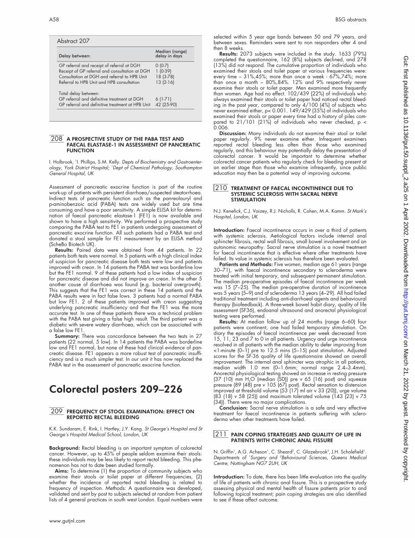
208 A PROSPECTIVE STUDY OF THE PABA TEST ANDFAECAL ELASTASE-1 IN ASSESSMENT OF PANCREATICFUNCTION
I. Holbrook, 1I. Phillips, S.M. Kelly. Depts of Biochemistry and Gastroenter-ology, York District Hospital; 1Dept of Chemical Pathology, SouthamptonGeneral Hospital, UK
Assessment of pancreatic exocrine function is part of the routinework-up of patients with persistent diarrhoea/suspected steatorrhoea.Indirect tests of pancreatic function such as the panreolauryl andp-aminobenzoic acid (PABA) tests are widely used but are timeconsuming and have a poor sensitivity. A simple ELISA kit for determi-nation of faecal pancreatic elastase-1 (FE1) is now available andshown to have a high sensitivity. We performed a prospective studycomparing the PABA test to FE1 in patients undergoing assessment ofpancreatic exocrine function. All such patients had a PABA test anddonated a stool sample for FE1 measurement by an ELISA method(ScheBo Biotech UK).
Results: Paired data were obtained from 44 patients. In 22patients both tests were normal. In 5 patients with a high clinical indexof suspicion for pancreatic disease both tests were low and patientsimproved with creon. In 14 patients the PABA test was borderline lowbut the FE1 normal. 9 of these patients had a low index of suspicionfor pancreatic disease and did not improve on creon. In the other 5another cause of diarrhoea was found (e.g. bacterial overgrowth).This suggests that the FE1 was correct in these 14 patients and thePABA results were in fact false lows. 3 patients had a normal PABAbut low FE1. 2 of these patients improved with creon suggestingunderlying pancreatic insufficiency and that the FE1 was the moreaccurate test. In one of these patients there was a technical problemwith the PABA test giving a false high result. The third patient was adiabetic with severe watery diarrhoea, which can be associated witha false low FE1.
Summary: There was concordance between the two tests in 27patients (22 normal, 5 low). In 14 patients the PABA was borderlinelow and FE1 normal, but none of these had clinical evidence of pan-creatic disease. FE1 appears a more robust test of pancreatic insuffi-ciency and is a much simpler test. In our unit it has now replaced thePABA test in the assessment of pancreatic exocrine function.
Colorectal posters 209–226
209 FREQUENCY OF STOOL EXAMINATION: EFFECT ONREPORTED RECTAL BLEEDING
K.K. Sundaram, E. Rink, I. Hartley, J.Y. Kang. St George’s Hospital and StGeorge’s Hospital Medical School, London, UK
Background: Rectal bleeding is an important symptom of colorectalcancer. However, up to 45% of people seldom examine their stools:these individuals may be less likely to report rectal bleeding. This phe-nomenon has not to date been studied formally.
Aims: To determine (1) the proportion of community subjects whoexamine their stools or toilet paper at different frequencies, (2)whether the incidence of reported rectal bleeding is related tofrequency of inspection. Methods: A questionnaire was developed,validated and sent by post to subjects selected at random from patientlists of 4 general practices in south west London. Equal numbers were
selected within 5 year age bands between 50 and 79 years, andbetween sexes. Reminders were sent to non responders after 4 andthen 8 weeks.
Results: 2073 subjects were included in the study. 1633 (79%)completed the questionnaire, 162 (8%) subjects declined, and 278(13%) did not respond. The cumulative proportion of individuals whoexamined their stools and toilet paper at various frequencies were:every time – 31%,45%; more than once a week - 67%,74%; morethan once a month – 80%,84%. 12% and 9% respectively neverexamine their stools or toilet paper. Men examined more frequentlythan women. Age had no effect. 102/439 (22%) of individuals whoalways examined their stools or toilet paper had noticed rectal bleed-ing in the past year, compared to only 4/100 (4%) of subjects whonever examined either, p< 0.001. 149/429 (35%) of individuals whoexamined their stools or paper every time had a history of piles com-pared to 21/101 (21%) of individuals who never checked, p <0.006.
Discussion: Many individuals do not examine their stool or toiletpaper regularly. 9% never examine either. Infrequent examinersreported rectal bleeding less often than those who examinedregularly, and this behaviour may potentially delay the presentation ofcolorectal cancer. It would be important to determine whethercolorectal cancer patients who regularly check for bleeding present atan earlier stage than those who examine infrequently, since publiceducation may then be a potential way of improving outcome.
210 TREATMENT OF FAECAL INCONTINENCE DUE TOSYSTEMIC SCLEROSIS WITH SACRAL NERVESTIMULATION
N.J. Kenefick, C.J. Vaizey, R.J. Nicholls, R. Cohen, M.A. Kamm. St Mark’sHospital, London, UK
Introduction: Faecal incontinence occurs in over a third of patientswith systemic sclerosis. Aetiological factors include internal analsphincter fibrosis, rectal wall fibrosis, small bowel involvement and anautonomic neuropathy. Sacral nerve stimulation is a novel treatmentfor faecal incontinence that is effective where other treatments havefailed. Its value in systemic sclerosis has therefore been evaluated.
Patients and Methods: Five women, median age 61 years (range30–71), with faecal incontinence secondary to scleroderma weretreated with initial temporary, and subsequent permanent stimulation.The median pre-operative episodes of faecal incontinence per weekwas 15 (7–25). The median pre-operative duration of incontinencewas 5 years (5–9) and of scleroderma 13 years (4–29). All had failedtraditional treatment including anti-diarrhoeal agents and behaviouraltherapy (biofeedback). A three-week bowel habit diary, quality of lifeassessment (SF36), endoanal ultrasound and anorectal physiologicaltesting were performed.
Results: At median follow up of 24 months (range 6–60) fourpatients were continent, one had failed temporary stimulation. Ondiary the episodes of faecal incontinence per week decreased from15, 11, 23 and 7 to 0 in all patients. Urgency and urge incontinenceresolved in all patients with the median ability to defer improving from<1-minute (0–1) pre to 12.5 mins (5–15) post stimulation. Adjustedscores for the SF-36 quality of life questionnaire showed an overallimprovement. The internal anal sphincter was atrophic in all patients,median width 1.0 mm (0–1.6mm; normal range 2.4–3.4mm).Anorectal physiological testing showed an increase in resting pressure(37 (10) mm H2O (median (SD)) pre v 65 (16) post) and squeezepressure (89 (48) pre v 105 (67) post). Rectal sensation to distensionimproved at threshold volume (53 (17) ml air v 33 (20)), urge volume(83 (18) v 58 (25)) and maximum tolerated volume (143 (23) v 75(34)). There were no major complications.
Conclusion: Sacral nerve stimulation is a safe and very effectivetreatment for faecal incontinence in patients suffering with sclero-derma when other treatments have failed.
211 PAIN COPING STRATEGIES AND QUALITY OF LIFE INPATIENTS WITH CHRONIC ANAL FISSURE
N. Griffin1, A.G. Acheson1, C. Sheard2, C. Glazebrook2, J.H. Scholefield1.Departments of 1Surgery and 2Behavioural Sciences, Queens MedicalCentre, Nottingham NG7 2UH, UK
Introduction: To date, there has been little evaluation into the qualityof life of patients with chronic anal fissure. This is a prospective studyassessing physical and mental health of fissure patients prior to andfollowing topical treatment; pain coping strategies are also identifiedto see if these affect outcome.
Abstract 207
Delay between:Median (range)delay in days
GP referral and receipt of referral at DGH 0 (0-7)Receipt of GP referral and consultation at DGH 1 (0-39)Consultation at DGH and referral to HPB Unit 18 (3-78)Referral to HPB Unit and HPB consultation 13 (2-16)
Total delay between:GP referral and definitive treatment at DGH 6 (1-71)GP referral and definitive treatment at HPB Unit 42 (25-90)
A58 BSG abstracts
www.gutjnl.com
on March 21, 2022 by guest. P
rotected by copyright.http://gut.bm
j.com/
Gut: first published as 10.1136/gut.50.suppl_2.a25 on 1 A
pril 2002. Dow
nloaded from
Methods: New patients attending the fissure clinic were recruitedprospectively into the study over a 2 month period. Patients wereinitially given 3 questionnaires to complete: 1) the Short-Form 36Health Survey (SF36), 2) the Pain Coping Strategies Questionnaireand 3) a general questionnaire recording patients’ demographicdetails and symptoms on a visual analogue scale (VAS). Following an8 week course of topical treatment, patients repeated the SF36 andsymptoms were again recorded on a VAS. Healing of fissure wasnoted.
Results: 23 patients entered the study; 8 male, 15 female withmean age 39 years (range 17–80). Median duration of fissure was 9months (1.5 months – 10 years). Before treatment, median VAS forpain, bleeding and irritation were 6, 1 and 5. On the SF 36, patientsscored below normal values for all scales except role emotional func-tioning and mental health. Fissure patients had more pain than ageand sex matched normal population (p=0.00, Wilcoxon). Gender didnot affect any of the SF 36 sub-scales. Duration of fissure positivelycorrelated with role physical functioning and role emotional function-ing (p<0.05). On follow-up, healing was complete in 15 patients(65%). Symptoms were significantly reduced in this group. Repeatingthe SF36 showed an improvement in role-physical functioning(p<0.05). Ignoring sensations was an adaptive pain coping strategy,being employed more often in the group responding to treatment(p=<0.05).
Conclusion: Successful treatment of chronic anal fissure leads tosymptomatic improvement and beneficially affects role physicalfunctioning. Patients’ coping strategies appear to have an influence onoutcome, with ignoring sensations being a good predictor ofresponse.
212 COLONIC DIVERTICULITIS: A DISEASE ON THE RISE?
S. Subramanian1, A. Tinto2, A. Majeed3, D.M. Melville4, A.R. Hart5, C.R.Morris5, J.D. Maxwell1, J.Y. Kang1 Departments of 1Gastroenterology and4Surgery, St George’s Hospital, London; 2Office for National Statistics,London; 3University College London; School of Medicine, University of EastAnglia5, Norwich, UK
Background: Little data exist on time trends for colonic diverticulitis.We studied hospital admission data for the whole of England from1989/1990 to 1999/2000.
Materials and Methods: Admission rates for colonic diverticulitis(ICD9: 562.1, ICD10: K57.2–57.9), excluding day cases but includ-ing diverticular abscess and perforation, operation and case fatalityrates were obtained from Department of Health Hospital Episode Sta-tistics.
Results: There has been a steady increase in age-standardisedhospital admission rates for both sexes and in all age groupsover the study period. Admission rates increased with age forboth sexes. The percentage of hospital admissions with an operationhas also risen for both males and females. There has been no signifi-cant change in case fatality rates over this time for either sex (seetable).
Conclusions: The hospital admission rates for colonic diverticulitishas increased from 1989/90 to 1999/00. As the proportion ofpatients who had surgical operations has also increased, while casefatality rates have remained much the same, the rise in admission ratesmay be due to a true increase in the incidence of colonic diverticulitis.With an aging population, colonic diverticulitis is likely be an increas-ing health problem in England.
213 THE RELATIONSHIP BETWEEN CYCLOOXYGENASE-2EXPRESSION AND MICROVESSEL DENSITY INCOLORECTAL CANCER
C. Steele1, K.M. Sheehan2, R. Cummins2, A. O’Grady2, K. Sheahan4, D.P.O’Donoghue5, D.J. Fitzgerald3, E.W. Kay2, F.E. Murray1,3. 1GastrointestinalUnit and 2Department of Pathology, Beaumont Hospital Dublin;3Department of Clinical Pharmacology, Royal College of Surgeons, Dublin;4Departments of Pathology and Gastroenterology, St Vincent’s Hospital,Dublin, Ireland
Introduction: Cyclooxygenase 2 (COX-2) is up regulated incolorectal carcinoma and is related to survival, lymph node anddistant metastases. The exact role of COX-2 in colorectal cancer, inparticular with regard to angiogenesis and tumour vascular develop-ment, is yet to be delineated. Our group have shown that VEGF medi-ates endothelial cell proliferation via COX and that adhesion ofendothelial cells to the extracellular matrix via integrins inducesCOX-2. The aim of this study was to examine the relationship betweentumour cell expression of COX-2 and vessel formation within tumourby microvessel density (MVD) in colorectal cancer.
Methods: Seventy patients for whom full clinical and pathologicaldata were available from our database were selected retrospectivelyfor analysis. Paraffin embedded tissue from archival primary tumourmaterial was analysed by immunhistochemical methods for COX-2and MVD. COX-2 polyclonal human antibody (Cayman) and anendothelial cell antibody, CD-34 (clone QBEND-10, Dako) were usedon serial sections using the avidin biotin method. COX-2 was gradedby percentage of epithelial cell staining and intensity. MVD was cal-culated by mean vessel count of five high power fields (x 200) perslide in tumour involved area. Two blinded observers performed bothanalyses.
Results: Of the 70 cases, 2 were Dukes’ stage A, 26 were stage B,29 were stage C and 13 were stage D. COX-2 was present in almost90% of cases. COX-2 staining was present in tumour epithelial cells,inflammatory cells, fibroblasts and endothelial cells. There was varia-tion in staining intensity between tumours. Correlation analysis wasperformed between intensity of COX-2 expression and MVD. No sig-nificant correlation was found between these two groups (r= -0.075).No correlation was found between Dukes stage overall or betweenindividual Dukes’ stage and MVD. (Dukes’ B=0.105, Dukes’C=0.012, Dukes’ D=-0.189).
Conclusion: This study demonstrates no association betweenmicrovessel density and either the intensity of COX-2 expression intumour cells or Dukes’ stage. These findings suggest that COX-2expression does not play a role in determining augmented neovascu-larisation associated with colorectal cancer. This is in keeping withrecent evidence that it is the host COX-2 and COX-1 that are importantin angiogenesis.
214 CYTOKERATIN IMMUNOREACTIVITY IN BENIGNPERICOLIC LYMPH NODES: ANIMMUNOHISTOCHEMICAL STUDY OF 101 LYMPHNODES
V.I. Shah1, P.J. Arumugam1, J. Beynon1. 1Singleton Hospital, Swansea,Wales, UK
Background: Several studies have demonstrated scattered singlecytokeratin immunoreactive cells in morphologically benign regionaldraining lymph nodes from cases of Dukes B colorectal cancer. In moststudies their presence correlated poorly with tumor recurrence andsurvival. It is not clear whether these cells represent native cells of thelymph node or occult micrometastasis.
Design: Formalin-fixed paraffin-embedded sections from 101histologically benign lymph nodes from 38 patients who hadundergone colorectal resections for benign conditions [diverticulardisease (18), inflammatory bowel disease (11), slow transit constipa-tion (6), volvulus (1), ischaemia (1), angiodysplasia (1)] and had nohistory of malignancy at the time of sugery or during a mean follow upperiod of atleast 30 months were immunostained with AE1/AE3(DAKO, monoclonal,1:100, 30 min, protease 1 pretreatment, 12min), Cam 5.2 (Becton-Dickinson, monoclonal,1:20, 30 min, protease1 pretreatment, 8 min) and pan-cytokeratin (DAKO, MNF116, 1:100,30 min, protease 1 pretreatment, 12 min) on a NEXES autostainerusing a Ventana detection system. The morphology of the immunore-active cells was evaluated and their number scored as 0:absent,rare:<1%, 1+:1–5%, 2+:6–10%, 3+:>10%.
Results: A single cytokeratin positive epithelioid cell was identifiedin 1 (1%) of the lymph nodes in the levels immunostained with AE1/
Abstract 212
Colonic diverticulitis
1989/90 1999-2000 % change
M F M F M F
Admission rate/ 100, 000population
20.1 28.6 23.2 31.9 16 12
% of admissions with anoperation
22.9 20 27 22 16 14
Case fatality rate % 3.1 3.2 3.4 3.4 11 5
M-Males, F-Females
BSG abstracts A59
www.gutjnl.com
on March 21, 2022 by guest. P
rotected by copyright.http://gut.bm
j.com/
Gut: first published as 10.1136/gut.50.suppl_2.a25 on 1 A
pril 2002. Dow
nloaded from
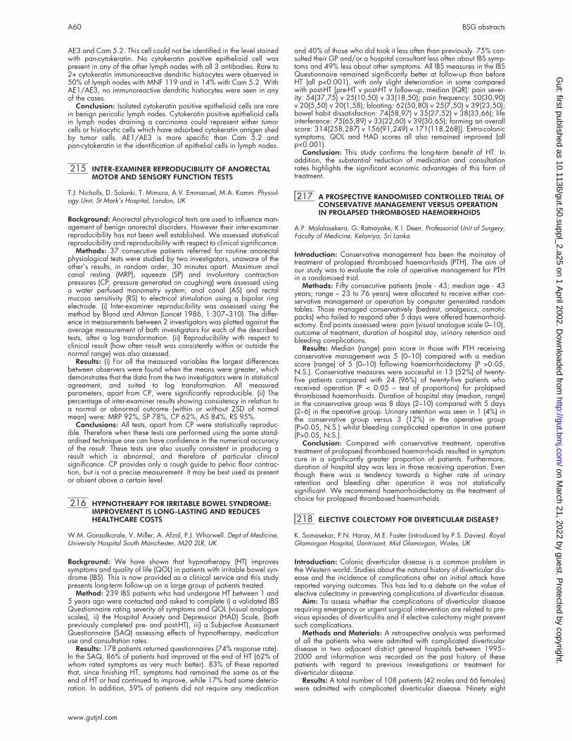
AE3 and Cam 5.2. This cell could not be identified in the level stainedwith pan-cytokeratin. No cytokeratin positive epithelioid cell waspresent in any of the other lymph nodes with all 3 antibodies. Rare to2+ cytokeratin immunoreactive dendritic histiocytes were observed in50% of lymph nodes with MNF 119 and in 14% with Cam 5.2. WithAE1/AE3, no immunoreactive dendritic histiocytes were seen in anyof the cases.
Conclusion: Isolated cytokeratin positive epithelioid cells are rarein benign pericolic lymph nodes. Cytokeratin positive epithelioid cellsin lymph nodes draining a carcinoma could represent either tumorcells or histiocytic cells which have adsorbed cytokeratin antigen shedby tumor cells. AE1/AE3 is more specific than Cam 5.2 andpan-cytokeratin in the identification of epithelial cells in lymph nodes.
215 INTER-EXAMINER REPRODUCIBILITY OF ANORECTALMOTOR AND SENSORY FUNCTION TESTS
T.J. Nicholls, D. Solanki, T. Mimura, A.V. Emmanuel, M.A. Kamm. Physiol-ogy Unit, St Mark’s Hospital, London, UK
Background: Anorectal physiological tests are used to influence man-agement of benign anorectal disorders. However their inter-examinerreproducibility has not been well established. We assessed statisticalreproducibility and reproducibility with respect to clinical significance.
Methods: 37 consecutive patients referred for routine anorectalphysiological tests were studied by two investigators, unaware of theother’s results, in random order, 30 minutes apart. Maximum analcanal resting (MRP), squeeze (SP) and involuntary contractionpressures (CP, pressure generated on coughing) were assessed usinga water perfused manometry system; anal canal (AS) and rectalmucosa sensitivity (RS) to electrical stimulation using a bipolar ringelectrode. (i) Inter-examiner reproducibility was assessed using themethod by Bland and Altman (Lancet 1986, 1:307–310). The differ-ence in measurements between 2 investigators was plotted against theaverage measurement of both investigators for each of the describedtests, after a log transformation. (ii) Reproducibility with respect toclinical result (how often result was consistently within or outside thenormal range) was also assessed.
Results: (i) For all the measured variables the largest differencesbetween observers were found when the means were greater, whichdemonstrates that the data from the two investigators were in statisticalagreement, and suited to log transformation. All measuredparameters, apart from CP, were significantly reproducible. (ii) Thepercentage of inter-examiner results showing consistency in relation toa normal or abnormal outcome (within or without 2SD of normalmean) were: MRP 92%, SP 78%, CP 62%, AS 84%, RS 95%.
Conclusions: All tests, apart from CP were statistically reproduc-ible. Therefore when these tests are performed using the same stand-ardised technique one can have confidence in the numerical accuracyof the result. These tests are also usually consistent in producing aresult which is abnormal, and therefore of particular clinicalsignificance. CP provides only a rough guide to pelvic floor contrac-tion, but is not a precise measurement. It may be best used as presentor absent above a certain level.
216 HYPNOTHERAPY FOR IRRITABLE BOWEL SYNDROME:IMPROVEMENT IS LONG-LASTING AND REDUCESHEALTHCARE COSTS
W.M. Gonsalkorale, V. Miller, A. Afzal, P.J. Whorwell. Dept of Medicine,University Hospital South Manchester, M20 2LR, UK
Background: We have shown that hypnotherapy (HT) improvessymptoms and quality of life (QOL) in patients with irritable bowel syn-drome (IBS). This is now provided as a clinical service and this studypresents long-term follow-up on a large group of patients treated.
Method: 239 IBS patients who had undergone HT between 1 and5 years ago were contacted and asked to complete i) a validated IBSQuestionnaire rating severity of symptoms and QOL (visual analoguescales), ii) the Hospital Anxiety and Depression (HAD) Scale, (bothpreviously completed pre- and post-HT), iii) a Subjective AssessmentQuestionnaire (SAQ) assessing effects of hypnotherapy, medicationuse and consultation rates.
Results: 178 patients returned questionnaires (74% response rate).In the SAQ, 86% of patients had improved at the end of HT (62% ofwhom rated symptoms as very much better). 83% of these reportedthat, since finishing HT, symptoms had remained the same as at theend of HT or had continued to improve, while 17% had some deterio-ration. In addition, 59% of patients did not require any medication
and 40% of those who did took it less often than previously. 75% con-sulted their GP and/or a hospital consultant less often about IBS symp-toms and 49% less about other symptoms. All IBS measures in the IBSQuestionnaire remained significantly better at follow-up than beforeHT (all p<0.001), with only slight deterioration in some comparedwith post-HT [pre-HT v post-HT v follow-up, median (IQR): pain sever-ity: 54(37,75) v 25(10,50) v 33(18,50); pain frequency: 50(30,90)v 20(5,50) v 20(1,58); bloating: 62(50,80) v 25(7,50) v 39(23,50);bowel habit dissatisfaction: 74(58,97) v 35(27,52) v 38(33,66); lifeinterference: 75(65,89) v 33(22,60) v 39(30,65); forming an overallscore: 314(258,287) v 156(91,249) v 171(118,268)]. Extra-colonicsymptoms, QOL and HAD scores all also remained improved (allp<0.001).
Conclusion: This study confirms the long-term benefit of HT. Inaddition, the substantial reduction of medication and consultationrates highlights the significant economic advantages of this form oftreatment.
217 A PROSPECTIVE RANDOMISED CONTROLLED TRIAL OFCONSERVATIVE MANAGEMENT VERSUS OPERATIONIN PROLAPSED THROMBOSED HAEMORRHOIDS
A.P. Malalasekera, G. Ratnayake, K.I. Deen. Professorial Unit of Surgery,Faculty of Medicine, Kelaniya, Sri Lanka
Introduction: Conservative management has been the mainstay oftreatment of prolapsed thrombosed haemorrhoids (PTH). The aim ofour study was to evaluate the role of operative management for PTHin a randomised trial.
Methods: Fifty consecutive patients (male - 43; median age - 43years; range – 23 to 76 years) were allocated to receive either con-servative management or operation by computer generated randomtables. Those managed conservatively (bedrest, analgesics, osmoticpacks) who failed to respond after 5 days were offered haemorrhoid-ectomy. End points assessed were: pain (visual analogue scale 0–10),outcome of treatment, duration of hospital stay, urinary retention andbleeding complications.
Results: Median (range) pain score in those with PTH receivingconservative management was 5 (0–10) compared with a medianscore (range) of 5 (0–10) following haemorrhoidectomy (P >0.05,N.S.). Conservative measures were successful in 13 (52%) of twenty-five patients compared with 24 (96%) of twenty-five patients whoreceived operation (P < 0.05 – test of proportions) for prolapsedthrombosed haemorrhoids. Duration of hospital stay (median, range)in the conservative group was 8 days (2–10) compared with 5 days(2–6) in the operative group. Urinary retention was seen in 1 (4%) inthe conservative group versus 3 (12%) in the operative group(P>0.05, N.S.) whilst bleeding complicated operation in one patient(P>0.05, N.S.).
Conclusion: Compared with conservative treatment, operativetreatment of prolapsed thrombosed haemorrhoids resulted in symptomcure in a significantly greater proportion of patients. Furthermore,duration of hospital stay was less in those receiving operation. Eventhough there was a tendency towards a higher rate of urinaryretention and bleeding after operation it was not statisticallysignificant. We recommend haemorrhoidectomy as the treatment ofchoice for prolapsed thrombosed haemorrhoids.
218 ELECTIVE COLECTOMY FOR DIVERTICULAR DISEASE?
K. Somasekar, P.N. Haray, M.E. Foster (introduced by P.S. Davies). RoyalGlamorgan Hospital, Llantrisant, Mid Glamorgan, Wales, UK
Introduction: Colonic diverticular disease is a common problem inthe Western world. Studies about the natural history of diverticular dis-ease and the incidence of complications after an initial attack havereported varying outcomes. This has led to a debate on the value ofelective colectomy in preventing complications of diverticular disease.
Aim: To assess whether the complications of diverticular diseaserequiring emergency or urgent surgical intervention are related to pre-vious episodes of diverticulitis and if elective colectomy might preventsuch complications.
Methods and Materials: A retrospective analysis was performedof all the patients who were admitted with complicated diverticulardisease in two adjacent district general hospitals between 1995–2000 and information was recorded on the past history of thesepatients with regard to previous investigations or treatment fordiverticular disease.
Results: A total number of 108 patients (42 males and 66 females)were admitted with complicated diverticular disease. Ninety eight
A60 BSG abstracts
www.gutjnl.com
on March 21, 2022 by guest. P
rotected by copyright.http://gut.bm
j.com/
Gut: first published as 10.1136/gut.50.suppl_2.a25 on 1 A
pril 2002. Dow
nloaded from
patients (91%) were emergency admissions and 10 patients (9%)were urgent admissions. Ninety eight patients (91%) underwent aHartmann’s procedure. Two patients had a subtotal colectomy and 4patients had a sigmoid colectomy with primary anastamosis. Fourpatients were not operated on due to their poor general condition. Outof the 108 patients, only 28 patients (26%) were previouslydiagnosed to have diverticular disease, either by barium enema orendoscopy. Eight of the twenty eight patients had required previousadmissions for acute exacerbation of their symptoms, 3 having beenadmitted twice. Only 3 patients (2.7%) had needed treatment foracute diverticulitis with intravenous fluids and antibiotics.
Conclusions: Our study has shown that elective colectomy after anattack of diverticulitis would not have a significant impact on the inci-dence of complications as most of them occur de novo in patients withno previous history of the disease. Further prospective studies areneeded in those with known diverticular disease to identify any furtherrisk factors for development of future complications. This would help toidentify a group of patients who may benefit from elective colectomy.
219 GASTROINTESTINAL SYMPTOMS AFTERRADIOTHERAPY FOR PELVIC CANCER
H.J.N. Andreyev, Z. Amin, P. Blake, D. Dearnaley, D. Tait, P. Vlavianos.Imperial College Faculty of Medicine, Chelsea & Westminster & RoyalMarsden Hospitals, London, UK
Introduction: About 13,000 patients undergo pelvic radiotherapyannually in the UK. The incidence of severe GI toxicity (fistulation,bowel obstruction, transfusion dependent bleeding or secondary can-cer) is not known but probably occurs in 4–8% at 5 years. More com-mon are symptoms such as incontinence or diarrhoea which maysignificantly impair quality of life in >30% of long term survivors.
Aim: To describe the symptoms and outcomes of patients followingpelvic radiotherapy referred to a specialist gastroenterology/ GIoncology clinic during its first year.
Methods: Oncologists were offered direct flexible sigmoidoscopyfor any patient with bright rectal bleeding without other symptoms,irrespective of proctoscopic findings. Other patients were reviewed inclinic. Data were recorded prospectively.
Results: Over 12 months, 60 patients were referred: 37 men, 23women with a median age 64 years (range 38–80). Primary tumourssites included prostate (n=33), cervix (n=12), endometrium (n=7),bladder (n=3), large bowel (n=2), and anus, vagina and ovary (n=1each). Radiotherapy was given a median 2 (range 0.5–21) years pre-viously except in 3 patients referred to exclude inflammatory boweldisease before starting treatment. Major symptoms included rectalbleeding (n=27), frequency (n=22), faecal incontinence (n=19), diar-rhoea (n=14), pain (n=8), steatorrhoea (n=5), subacute obstruction(n=4) and tenesmus (n=3). Eight patients described significantlyabnormal bowel habit before starting radiotherapy. Of patients withbleeding alone (n=19), 1 had no radiation proctitis but was bleedingfrom mucosal prolapse, 3 had unsuspected advanced adenomas and2 had squamous polyps. Sucralfate was always effective in reducingbleeding in those with radiation proctitis. All patients with tenesmus orincontinence improved or were cured with medical therapy including2 patients with marked anal sphincter changes on endoanalultrasound. Steatorrhoea was multifactorial, (2, bacterial overgrowth,2 pancreatic insufficiency, 2 fatty acid malabsorption). Pain was asso-ciated with relapse in 50%. Two patients with obstruction requiredsurgery.
Conclusions: Chronic GI symptoms after radiotherapy are oftenhighly debilitating and complex to assess, but may respond dramati-cally to simple combination therapies. Patients with new onset rectalbleeding following radiotherapy should be offered at least a flexiblesigmoidoscopy. Patients appear to benefit from being seen in aspecialist setting.
220 TIMING OF OPERATION AFTER RADIOTHERAPY FORRECTAL CANCER
H. Ishikawa1,2, I.C. Talbot2. 1First Department. of Surgery, Nara MedicalUniversity, Kashihara, Japan; 2Academic Department of Pathology & ICRF,St Mark’s Hospital, Harrow, UK
Aim: Preoperative adjuvant radiotherapy for rectal cancer has twoproblems, radiosensitivity and timing of operation. We haveexamined the effect of radiation and its timing on the relationshipbetween apoptotic cell index (AI) and proliferative activity index (PI).
Methods: Patients were given one of three alternative modalities,standard radiotherapy (SD) (40Gy, duration of 4–8weeks, n=23),
short-course radiotherapy (SC) (25Gy, 1–2weeks, n=11), orchemoradio-therapy (CR) (45Gy, 6–9weeks , n=7). AI and PI wereestimated in paired sections of biopsies and post-irradiated resectedtumours. The reduction ratio was histologically estimated andradiosensitive was judged in cases in which over 2/3 of tumour tissuewas destroyed
Results: Radiosensitive ratio and median reduction ratio were43.5% and 45% in SD, 27.3% and 25% in SC, 28.6%and 45% inCRT, respectively. In SD, the AI was significantly higher (5.9 v.s 2.7;p= 0.001) and the PI was significantly lower (33.9 v.s 50.0;p=0.028) than in the pre-therapy biopsies. In SD, the PI in the radio-sensitive subgroup was lower than in the radioresistant one (25.9 v.s46; p=0.005)). However, the AI of radiosensitive subgroup was lowerthan that of resistant one (2.4 v.s 4.2; p=0.005). Plotting each AIaccording to time course from finishing radiotherapy to operation inSD showed that the slope of in the radiosensitive subgroup wassteeper than in the radioresistant one and extrapolation back to theend of radiotherapy (day 0), suggests that in the radiosensitivesubgroup would have a high AI. The difference between two slopesmeans that in radiosensitive subgroups, the apoptotic responserapidly came and went. Plotting the AI against reduction ratiorevealed that the AI was proportional to the size of the residual tumourvolume (low in the radiosensitive).
Conclusions: Apoptosis may be a time-limited and volume-dependent phenomenon; radiosensitive tumours should be surgicallyresected earlier than the more resistant ones.
221 RANDOMISED CONTROLLED TRIAL OF BIOFEEDBACKFOR FAECAL INCONTINENCE
C. Norton, S. Chelvanayagam, M.A. Kamm. St Mark’s Hospital, WatfordRoad, Harrow, Middlesex HA1 3UJ, UK
Background: Behavioural treatment (biofeedback) has been reportedto improve symptoms in a majority of patients with faecal incotinence,but there are no trials comparing biofeedback with placebo or stand-ard medical care.
Methods: 171 consecutive patients with faecal incontinence tosolid or liquid stool were assessed by anal ultrasound and then strati-fied to structurally intact or disrupted anal sphincter muscles. Withineach of these two groups they were then randomised to one of fourgroups: (1) standard medical/nursing care (advice) (2) advice plusverbal instruction on sphincter exercises (3) hospital based computer-assisted sphincter pressure biofeedback (4) hospital biofeedback plususe of a home EMG biofeedback device. Outcome measures immedi-ately and at one year included diary, symptom questionnaire,continence score, patient’s rating of change, quality of life (SF36 anddisease specific), psychological status (HAD), and anal manometry.
Results: Improvement or cure occurred in groups 1 to 4respectively: 80%, 83%, 81%, and 76% (p=NS). Overall, 75% ofpatients had symptomatic improvement and 5% were “cured”. Majorbenefit was more likely if patients had structurally intact sphincters.Benefit was maintained for all groups at one year. Episodes of incon-tinence decreased from median 2 to 0 per week (p<0.001).Continence score (worst = 20) decreased from median 11 to 8(p<0.001). Disease specific quality of life, SF36 (vitality, social func-tioning and mental health), and HAD (anxiety and depression) all sig-nificantly improved. Patients demonstrated improved resting, squeezeand sustained squeeze pressures (all p<0.002). None of theseimprovements differed between groups.
Conclusions: Conservative therapy for faecal incontinenceimproves continence, quality of life, psychological well being, andsphincter function. Benefit is maintained in the medium term. Thepatient-therapist interaction and improved coping strategies appear tobe most important, rather than physiological feedback of sphincterfunction (biofeedback).
222 FAECAL CALPROTECTIN: NORMAL LEVELS IN A LATEMIDDLE-AGED POPULATION, EFFECTS OF LIFESTYLEFACTORS AND RELATION TO BOWEL SYMPTOMS
A. Poullis1,2, R. Foster2, A. Shetty1, T.C. Northfield2, M.K. Fagerhol3, M.A.Mendall1. 1Mayday University Hospital; 2St George’s Hospital MedicalSchool; 3University of Oslo, Norway
Background: Faecal calprotectin is more sensitive but less specificthan faecal occult blood (FOB) in the detection of colonic neoplasms(Tibble et al, Gut 2001). Little is known about levels in a late middleaged population, which would be the target for screening, and factors
BSG abstracts A61
www.gutjnl.com
on March 21, 2022 by guest. P
rotected by copyright.http://gut.bm
j.com/
Gut: first published as 10.1136/gut.50.suppl_2.a25 on 1 A
pril 2002. Dow
nloaded from
that determine levels. Furthermore it is unknown whether elevated lev-els are associated with symptoms, diarrhoea in particular.
Aims: To assess levels of faecal calprotectin and the factors thatcould effect them in a healthy late middle aged population and toassess the association with symptoms.
Methods: 230 asymptomatic subjects (155 male, 75 female) agedbetween 50 and 70 were recruited randomly from GP lists in SouthLondon. Subjects with IBD or a history of colorectal cancer (CRC) wereexcluded. A previously validated lifestyle questionnaire was com-pleted and a stool sample analysed for calprotectin by ELISA.
Results: Faecal calprotectin was bi-modally distributed, with46/230 (20%) of subjects having levels above the reference range(10mg/l). There was no association between NSAID use, units ofalcohol consumed in the previous week, being a present smoker, dailybowel frequency, and presence of abdominal pain or constipation inthe previous week and faecal calprotectin. Males had higher calpro-tectin levels than females (median 1.9 IQR 7.6 v 0.5, 2.8 p<0.0001).65% of subjects in the 3rd calprotectin tertile (C.T.) were past smokersv 45% of subjects in 1st C.T. (p=0.06). Mean cigarette pack years ofsmoking increased through each C.T. 9.8, 10.5, 17.9 (p=0.009).20% of subjects in the 3rd C.T. had suffered an episode of diarrhoeain the previous week v 12% in the 1st C.T. (p=0.05). Mean ageincreased in each C.T. 58.9,60.5,60.8 (p=0.03).
Conclusion: Smoking history, diarrhoea in the previous week,increasing age and male sex are all associated with an increasingfaecal calprotectin. Adjustment of values for the above variables mayincrease faecal calprotectins specificity as a screening marker forCRC.
223 IS CONSTIPATION A CONSEQUENCE OF GROWINGOLDER?
C. Eastwood, G.J. Davies, P.W. Dettmar1. Nutrition Research Centre,South Bank University, London, UK; 1Reckitt Benckiser Healthcare (UK)Limited, Hull, UK
Background: The number of GP visits for constipation increasesmarkedly among people over 60. Nevertheless, there is no good evi-dence that ageing per se affects colonic function. The associationbetween age and constipation may be confounded by factors such asinstitutionalisation, inactivity and chronic disease.
Aim: To assess the prevalence of functional constipation accordingto the Rome II diagnostic criteria and institutionalisation, physicalactivity and chronic disease.
Methods: Ethical approval was obtained from South Bank Univer-sity Ethics Committee. 50 FL subjects (mean age 74 years, range (65–97), 42% male) and 42 INS subjects (mean age 84 years, range(69–101) 36% male) were recruited. Bowel habit was recorded usinga 7-day bowel habit diary. Subjects were classified as constipatedaccording to Rome II diagnostic criteria, if they experienced at leasttwo of the following: less than 3 defecations per week, straining onmore than 25% of occasions, feelings of incomplete evacuation onmore than 25% of occasions. Statistical analyses were performedusing the t-test and Chi-square, as appropriate.
Results: Since the INS group was significantly older than the FLgroup, the mean ages of constipated and non-constipated subjectswere assessed in each group separately. There were no significantdifferences in age between constipated and non-constipated subjects.Constipation was associated with institutionalisation (χ2=9.9;p=0.003) and inactivity (χ2=12.2; p=0.001). Although 65% ofconstipated subjects suffered from more than one chronic disease, thisassociation was not statistically significant.
Conclusions: This study suggests that the prevalence of functionalconstipation is associated to factors related to ageing such as institu-tionalisation and inactivity. However, no association was found withchronic diseases.
224 K-RAS MUTATIONS IN COLORECTAL POLYPS: SITE,HISTOLOGY AND SIZE DO MATTER
G.L. Williams1,2, G.J. Jenkins2, J.M. Parry2, J. Beynon1. 1 Dept of ColorectalSurgery, Singleton Hospital, Swansea, UK; 2School of Biological Sciences,Swansea University, UK
Introduction: Mutation of the oncogene K-ras is thought to be impor-tant in the early progression of colorectal carcinogenesis. K-ras isinvolved in the cell signalling pathway and its mutation causes uncon-trolled cell proliferation The aim of our study was to assess therelationship between K-ras mutations and various characteristics ofcolorectal polyps such as: site, size and histology.
Methods: Polyps were collected during colonoscopy from 55 suc-cessive patients and control tissue obtained from 20 other patients.DNA was extracted from the fresh tissue and mutations were detectedfollowing PCR and restriction enzyme (mva1) digestion. Ethicalapproval was obtained.
Results: Mutations of K-ras were found in 21% of the 55 polyps;none of the controls had mutations. Of the 15 rectal polyps 33% hada mutation; whereas only 18% of the 40 colonic polyps had a mutatedK-ras. 36 polyps were <1cm (mutation rate 8%) and 19 polyps were>1cm. These larger polyps had a higher K-ras mutation rate of 47%.Histological type was also analysed, revealing that 30% oftubulovillous/villous polyps had a mutation, compared to much lowerlevels of mutations in tubular (9%) and metaplastic polyps (0). No dif-ference in mutation rate was found in varying grades of dysplasia inour study.
Discussion: This small study reveals that K-ras mutations in ourpopulation tended to be associated with polyps having a rectal loca-tion, a larger size and villous histology. Further studies are needed tounderstand the role of this important oncogene in adenoma growthand progression to carcinoma.
225 DO CYTOKINE LEVELS PREDICT PROGNOSIS INCOLORECTAL CANCER?
A.G. Prabhudesai, A.G. Heriot, J.B. Marriott, A.G. Dalgleish, D. Kumar.Departments of Colorectal Surgery and Oncology, St George’s Hospital,London, UK
Suppression of the immune system and cytokine production, as anessential function of the immune response, in patients with colorectalcancer has been extensively investigated. The aim of this study was todetermine the predictive prognostic value of cytokine levels before &after treatment in patients with colorectal cancer.
Methods: Heparinized venous blood samples were taken fromfifty-three patients (34M &19F) with primary colorectal cancer beforeand at least 10 weeks after operation. Patients who had preoperativeradiotherapy had blood samples taken before radiation therapy.Interferon (IFN) γ, Interleukin (IL) 10 and Tumour necrosis factor (TNF)α levels were measured by lipopolysaccharide stimulated bloodcultures. The patients were followed up in a Colorectal Cancer Clinicfor evidence of local recurrence, distant metastases and survival.
Results: Patients with high preoperative levels of IFN γ developeddistant metastases later than those with lower levels. (Correlationcoefft=0.812 at P=0.05) Similarly, those with high postoperative IFNγ levels developed local recurrence later than those with lower levels.(Correlation coefft =1at P=0.01;) IL 10 & TNF α levels did not show asimilar correlation. Higher levels of postoperative IL 10 levels wereassociated with development of metastatic disease. (Correlationcoefft=-0.853 at P=0.002) Higher TNF α levels before preoperativeradiotherapy were associated with a longer survival in patients withrectal cancer. (Correlation coefft=0.829 at P=0.04) Similar TNF αlevels without preoperative radiotherapy did not appear to confer thesame survival advantage.
Conclusions: Higher levels of pro-inflammatory cytokines (TNF α,IFN γ) were associated with a better outcome in terms of time to localrecurrence, distant metastases and survival. Higher levels of immuno-suppressive cytokine (IL 10) were related to the development of distantdisease. Those with lower levels of pro-inflammatory cytokines beforetreatment, and those with higher postoperative levels of immunosup-pressive cytokines need close surveillance for the development of loco-regional or systemic relapse.
226 SUCCESS OF A SIMPLE “TICK BOX” GP REFERRALFORM FOR COLORECTAL CARCINOMA AND ITSIMPACT ON ACHIEVING THE “2 WEEK WAIT” TARGET
T. Thresher, J.D. Linehan, D.C. Britton, M. Davis, J.J.T. Tate, M.E.R.Williamson. Royal United Hospital, Combe Park, Bath BA1 3NG, UK
Introduction: In June 2000 the UK government introduced the targetthat a hospital specialist should see all patients with suspected GI can-cer within 2 weeks of GP referral. With unlimited resources, allpatients with any symptoms could be seen and immediatelyinvestigated, but in the UK methods to stratify patients in terms of riskof serious pathology are needed to ensure urgent investigation ofpatients at the highest risk.
Methods: We introduced a simple 10-question tick-box GP form forreferral of all patients with rectal bleeding and change of bowel habit,which could be e-mailed or faxed directly to the endoscopy
A62 BSG abstracts
www.gutjnl.com
on March 21, 2022 by guest. P
rotected by copyright.http://gut.bm
j.com/
Gut: first published as 10.1136/gut.50.suppl_2.a25 on 1 A
pril 2002. Dow
nloaded from
department. The responses allowed stratification of patients withoutprior assessment in outpatients into 3 groups. Patients stratified as“urgent” were sent a colonoscopy appointment for <2 weeks, “soon”for <12 weeks and routine for <36 weeks. Performance of the form hasbeen audited over the 12 months following introduction. This includedmonitoring for time to colonoscopy from referral and accuracy of theform in predicting serious pathology (colorectal carcinoma, IBD).
Results: In total 691 referrals were made, of which 156 werestratified “urgent”, 447 “soon” and 88 “routine”. 73% of urgentpatients achieved a colonoscopy in <2 weeks and 94% within 3weeks including at peak holiday times e.g. Christmas etc. Those notdone in <3 weeks were sent appointments but failed to attend. 97%of soon patients were colonoscoped in <12 weeks and 100% of “rou-tines” in <36 weeks. See table.
Conclusion: This referral form has proved successful in identifyingpatients at high risk of significant pathology and has allowed appro-priate targeting of resources for rapid diagnosis of pathology withoutprior outpatient assessment. Rates of detection of serious pathology in“soon” and “routine” referrals were not significantly higher than thosethat would be anticipated in a comparable unselected population.
Gastroduodenal posters227–241
227 MANAGEMENT OF PEPTIC ULCER DISEASE IN PRIMARYCARE
A.J. Morris, C. Craig, C. Morran, H. Burns, A. Power, K. Harden, D.Walsh, R. C. Stuart. ACID 1 Study group, Digestive Disease Directorate,Glasgow Royal Infirmary, Glasgow, UK
Aim: Eradication of H. pylori infection in patients with peptic ulcerdisease patients reduces the need for long term acid suppressiontherapy and the risk of complications such as bleeding andperforation yet patients with this diagnosis continue to be treated withacid suppression therapy in primary care. We aimed to assess theextent to which these patients had been investigated for H. pyloriinfection and identify a population who might benefit from H. pyloritesting and eradication.
Methods: From 11,149 patients who had received acidsuppression in the preceding year (Total GP population 176,268) weundertook case note review and identified 3071 (27.5%) patients whohad previously diagnosed peptic ulcer disease (77.2% DU, 13.1%GU, 6.1% both, 3.5% unspecified ulcer type). 2063 patients werereceiving maintenance therapy (Defined as > 3 prescriptions/year).Of these, 1275 who had no contraindication to H. pylori eradicationwere invited to nurse led clinics for H. pylori testing: 705 attended andunderwent 13C urea breath testing to establish H. pylori status.
Results: Only 36.4% of patients identified with known peptic ulcerdisease had previously received eradication therapy. 26.9% weretaking NSAIDs concomitantly (53.6% aspirin, 37.7% other NSAIDsand 8.7% both). H. pylori prevalence was 65.6% in patients who hadnever had documented eradication therapy and, although lower ifpatients had prior eradication therapy. 23.0% of previously treatedulcer patients remained infected at the time of testing.
Conclusions: A substantial proportion of ulcer patients receivingacid suppression therapy in general practice have never received H.pylori eradication and almost a quarter of those previously treatedremain infected with the organism. There is considerable potential forimprovement in the management of this easily identifiable patientgroup but post treatment testing is important to establish eradication ofinfection.
228 SERVICE IMPLICATIONS AND SUCCESS OF THEIMPLEMENTATION OF THE TWO-WEEK REFERRALCRITERIA FOR UPPER GASTROINTESTINAL CANCERSIN A DISTRICT GENERAL HOSPITAL
D.J. Lassman, J. Elliott, A. Taylor, A.T. Green, C.E. Grimley. Gastroenterol-ogy Unit, Burnley General Hospital, Casterton Avenue, Burnley, UK
Introduction: The introduction of 2-week criteria in July 2000 hasadded a significant burden to the provision of gastroenterology serv-ices. The targets are not proven to improve outcomes for patientsfound to have cancer and it is unclear how effectively they are appliedby primary care physicians. This study addresses the appropriatenessof referrals, the success in meeting the criteria and the pick-up rate forupper GI tumours.
Methods: Data were collected prospectively by a specialist nurse.Patients referred from primary care within ‘2 week criteria’ and thosewho were thought to meet the criteria but were not referred throughthat route were included. Time to first consultation (clinic orgastroscopy) was recorded. The final diagnosis and outcomes whenavailable are also reported.
Results: 149 patients are included in the study. Their average age(range) is 67(19–92). 79 were referred by ‘two week criteria’ and theothers were reprioritised by the consultant reading the referral.Gastroscopy was performed in all cases. This was achieved in anaverage of 9 days (range 1–14) for those referred by 2-week criteriaand 15 (range 7–35) days in those thought to meet the criteria but notreferred by that route. There were 25 extra cases per month when thesystem was established. 14 malignancies were identified (9.4%). 12cases were identified correctly by GP application of 2-week criteria(15.2%). 2/70 further malignancies were identified by consultantinterpretation of routine referrals (2.9%).
Conclusion: The application of the 2-week criteria for upper GIcancers has led to an additional 25 procedures/month. Primary carephysicians achieved a cancer pickup rate of 15.2%. Additional casefinding by assessment of other referrals seems to have little additionalbenefit.
229 RABEPRAZOLE 20 MG COMPARED WITHESOMEPRAZOLE 40 MG IN THE CONTROL OFINTRAGASTRIC PH IN HEALTHY VOLUNTEERS
K. Baisley1, S. Warrington1, B. Tejura1, A. Morocutti2, N. Miller2
(introduced by Val Heatley). 1Hammersmith Medicines Research, CentralMiddlesex Hospital, London, UK; 2Eisai Ltd, London, UK
Purpose: To compare the effects of single doses of rabeprazole (RAB)20 mg and esomeprazole (ESO) 40 mg on intragastric pH in healthyH. pylori-negative volunteers.
Methods: 27 H. pylori-negative subjects underwent two 24-hourtreatment periods, separated by a 14-day washout, in a 2-way cross-over single-dose study comparing RAB 20 mg with ESO 40 mg. Intra-gastric pH was recorded over 24 h on Days 0 and 1 of each period.Percentage of time that intragastric pH >3 and >4 during each 24 hinterval, and area under the intragastric pH-time curve (AUC0–24 h),were calculated, and compared using ANOVA.
Results: There were no statistically significant differences in meanAUC0–24 h, mean percent time pH >4 and mean percent time pH >3between RAB and ESO treatments on Day 0 (pre-dose) or Day 1. OnDay 1, mean percent time pH >4 after RAB 20 mg was 43.1(SD=18.3) and after ESO 40 mg was 45.2 (SD=17.1); mean percenttime pH >3 after RAB 20 mg was 54.8 (SD = 18.3) and after ESO 40mg was 54.9 (SD = 15.1). For intragastric pH control during thenighttime hours (14–24h post dose), mean percent time pH >3 andpH >4 was significantly higher on RAB 20 mg than ESO 40 mg (pH>3: 42.1%, SD=25.3 and 25.1%, SD=18.0, respectively; pH >4:32.4%, SD=24.1 and 17.0%, SD=14.9, respectively; p=0.001). Dur-ing daytime hours, mean percent time pH >3 and pH >4 was signifi-cantly higher on ESO 40 mg than RAB 20 mg (pH >3: 76.2%,SD=15.7 and 63.8%, SD=20.7, respectively; pH >4: 65.4%,SD=21.5 and 50.7%, SD=21.3, respectively; p=0.02).
Conclusion: Over a 24 h period, there was no difference betweenRAB 20 mg and ESO 40 mg with respect to effects on intragastric pH.In the morning, the effects of ESO were greater than those of RAB,whilst during the nighttime hours, the effects of RAB were greater thanthose of ESO. These results concur with published data on the effectsof RAB and ESO on intragastric pH.
This research was supported by Eisai Ltd, London, UK.
230 “TEST AND TREAT” A PILOT STUDY OF A COMMUNITYBASED C13 UREA BREATH TEST SERVICE (C13UBT)
V. Edge, C. Macdonald, S. Raimes, A. Edgar, J. Honeyman, I. Keyes, D.Burke. Cumberland Infirmary, Carlisle and Primary Care, North Cumbria,UK
H pylori (HP) “test and treat” strategies have been shown to be effec-tive and safe in the management of uncomplicated dyspepsia in under45 year olds and may reduce endoscopy demands. Non-invasive HPtests include serology, breath tests and recently a faecal antigen test.Near patient tests have been shown to be unreliable, serology doesnot allow early follow up assessment. UBT allow non-invasive pre andpost treatment assessment of HP. C14 based tests cannot easily be usedin the community.
Aims: To assess the practicality, accessibility, appropriate use andeffectiveness of a C13UBT service in the community of North Cumbria.
BSG abstracts A63
www.gutjnl.com
on March 21, 2022 by guest. P
rotected by copyright.http://gut.bm
j.com/
Gut: first published as 10.1136/gut.50.suppl_2.a25 on 1 A
pril 2002. Dow
nloaded from
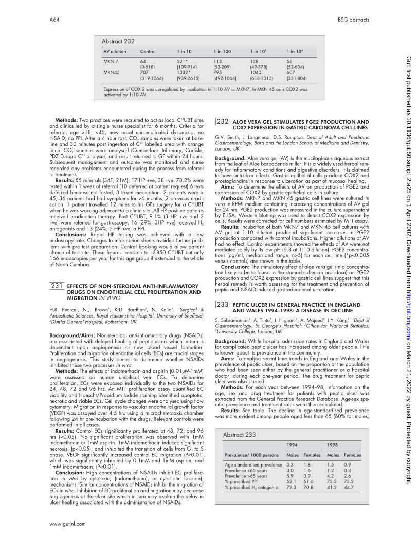
Methods: Two practices were recruited to act as local C13UBT sitesand clinics led by a single nurse specialist for 6 months. Criteria forreferral; age >18, <45, new onset uncomplicated dyspepsia, noNSAID, no PPI. After a 4 hour fast, CO2 samples were taken at base-line and 30 minutes post ingestion of C13 labelled urea with orangejuice. CO2 samples were analysed (Cumberland Infirmary, Carlisle,PDZ Europa C13 analyser) and result returned to GP within 24 hours.Subsequent management and outcome was monitored and nurserecorded any problems encountered during the process from referralto treatment.
Results: 55 referrals (34F, 21M), 17 HP +ve, 38 –ve. 78.2% weretested within 1 week of referral (10 deferred at patient request) 6 testsdeferred because not fasted, 3 taken medication. 2 patients were >45, 36 patients had had symptoms for >6 months, 2 previous eradi-cation. 1 patient travelled 12 miles to his GPs surgery for a C13UBTwhen he was working adjacent to a clinic site. All HP positive patientsreceived eradication therapy. Post C13UBT, 9.1% (3 HP +ve and 2–ve) were referred for gastroscopy, 16 (29%, 3HP +ve) received H2
antagonists and 13 (24%, 5 HP +ve) a PPI.Conclusions: Rapid HP testing was achieved with a low
endoscopy rate. Changes to information sheets avoided further prob-lems with pre test preparation. Central booking would allow patientchoice of test site. These figures translate to ∼1850 C13UBT but only166 endoscopies per year for this age group if extended to the wholeof North Cumbria.
231 EFFECTS OF NON-STEROIDAL ANTI-INFLAMMATORYDRUGS ON ENDOTHELIAL CELL PROLIFERATION ANDMIGRATION IN VITRO
H.R. Pearce1, N.J. Brown1, K.D. Bardhan2, N. Kalia1. 1Surgical &Anaesthetic Sciences, Royal Hallamshire Hospital, University of Sheffield;2District General Hospital, Rotherham, UK
Background/Aims: Non-steroidal anti-inflammatory drugs (NSAIDs)are associated with delayed healing of peptic ulcers which in turn isdependent upon angiogenesis or new blood vessel formation.Proliferation and migration of endothelial cells (ECs) are crucial stagesin angiogenesis. This study aimed to determine whether NSAIDsinhibited these two processes in vitro.
Methods: The effects of indomethacin and aspirin (0.01µM-1mM)were assessed on human umbilical vein ECs. To determineproliferation, ECs were exposed individually to the two NSAIDs for24, 48, 72 and 96 hrs. An MTT proliferation assay quantified ECviability and Hoescht/Propidium Iodide staining identified apoptotic,necrotic and viable ECs. Cell cycle changes were analysed using flowcytometry. Migration in response to vascular endothelial growth factor(VEGF) was assayed over 4.5 hrs using a microchemotaxis chamberfollowing 24 hr pre-incubation with the drugs. Relevant controls wereperformed in all cases.
Results: Control ECs significantly proliferated at 48, 72, and 96hrs (<0.05). No significant proliferation was observed with 1mMindomethacin or 1mM aspirin. 1mM indomethacin induced significantnecrosis, (p<0.05), and inhibited the transition of cells from G1 to Sphase. VEGF significantly increased control EC migration (P<0.01)which was significantly inhibited by 0.1mM and 1mM aspirin, and1mM indomethacin, (P<0.01).
Conclusion: High concentrations of NSAIDs inhibit EC prolifera-tion in vitro by cytotoxic, (indomethacin), or cytostatic (aspirin),mechanisms. Similar concentrations of NSAIDs inhibit the migration ofECs in vitro. Inhibition of EC proliferation and migration may decreaseangiogenesis at the ulcer site which in turn may explain the delay inulcer healing associated with the administration of NSAIDs.
232 ALOE VERA GEL STIMULATES PGE2 PRODUCTION ANDCOX2 EXPRESSION IN GASTRIC CARCINOMA CELL LINES
G.V. Smith, L. Langmead, D.S. Rampton. Dept of Adult and PaediatricGastroenterology, Barts and the London School of Medicine and Dentistry,London, UK
Background: Aloe vera gel (AV) is the mucilaginous aqueous extractfrom the leaf of Aloe barbadensis miller. It is a widely used herbal rem-edy for inflammatory conditions and digestive disorders. It is claimedto have anti-ulcer effects. Gastric epithelial cells produce COX2 andprostaglandins in response to ulceration as part of mucosal healing.
Aims: To determine the effects of AV on production of PGE2 andexpression of COX2 by gastric epithelial cells in culture.
Methods: MKN7 and MKN 45 gastric cell lines were cultured invitro in RPMI medium containing increasing concentrations of AV gelfor 24 hrs. PGE2 production was measured in the culture supernatantby ELISA. Western blotting was used to detect COX2 expression bycells. Results were corrected for cell numbers estimated by MTT assay.
Results: Incubation of both MKN7 and MKN 45 cell cultures withAV gel at 1:10 dilution produced significant increases in PGE2production compared with control incubations. Higher dilutions of AVhad no effect. Control experiments showed the effects of AV were notmediated solely by its low pH (6.8 at 1:10 dilution). PGE2 concentra-tions (pg/ml, median and range, n>5) for each cell line (*p<0.005versus controls) are shown in the table.
Conclusion: The stimulatory effect of aloe vera gel (in a concentra-tion likely to be to found in the stomach after an oral dose) on PGE2production and COX2 expression by gastric cell lines suggest that thisherbal remedy is worth assessing for the treatment and prevention ofpeptic and NSAID-induced gastroduodenal ulceration.
233 PEPTIC ULCER IN GENERAL PRACTICE IN ENGLANDAND WALES 1994–1998: A DISEASE IN DECLINE
S. Subramanian1, A. Tinto2, J. Higham2, A. Majeed3, J.Y. Kang1. 1Dept ofGastroenterology, St George’s Hospital; 2Office for National Statistics;3University College, London, UK
Background: While hospital admission rates in England and Walesfor complicated peptic ulcer has increased among older people, littleis known about its prevalence in the community.
Aims: To analyse recent time trends in England and Wales in theprevalence of peptic ulcer, based on the proportion of the populationwho had been seen either by the general practitioner or a hospitaldoctor, during each one-year period. The drug treatment for pepticulcer was also studied.
Methods: For each year between 1994–98, information on theage, sex and drug treatment for patients with peptic ulcer wasextracted from the General Practice Research Database. Age-sex spe-cific prevalence and treatment rates were then calculated.
Results: See table. The decline in age-standardised prevalencewas more evident among people aged less than 65 (60% for males,
Abstract 232
AV dilution Control 1 in 10 1 in 100 1 in 103 1 in 106
MKN 7 64 521* 113 138 56(0-518) (109-914) (53-209) (49-378) (52-634)
MKN45 707 1332* 795 1040 607(319-1064) (939-2615) (492-1064) (618-1315) (331-804)
Expression of COX 2 was upregulated by incubation in 1:10 AV in MKN7. In MKN 45 cells COX2 wasactivated by 1:10 AV.
Abstract 233
Prevalence/ 1000 persons
1994 1998
Males Females Males Females
Age standardised prevalence 3.3 1.8 1.5 0.9Prevalence <65 years 3.0 1.6 1.2 0.8Prevalence >65 years 5.9 3.9 4.2 2.6% prescribed PPI 52.1 51.6 73.3 73.2% prescribed H2 antagonist 72.3 70.8 41.2 44.7
A64 BSG abstracts
www.gutjnl.com
on March 21, 2022 by guest. P
rotected by copyright.http://gut.bm
j.com/
Gut: first published as 10.1136/gut.50.suppl_2.a25 on 1 A
pril 2002. Dow
nloaded from
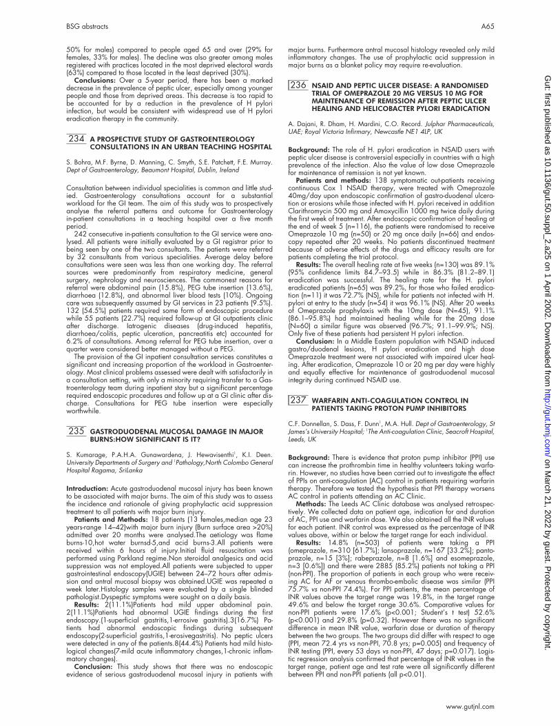
50% for males) compared to people aged 65 and over (29% forfemales, 33% for males). The decline was also greater among malesregistered with practices located in the most deprived electoral wards(63%) compared to those located in the least deprived (30%).
Conclusions: Over a 5-year period, there has been a markeddecrease in the prevalence of peptic ulcer, especially among youngerpeople and those from deprived areas. This decrease is too rapid tobe accounted for by a reduction in the prevalence of H pyloriinfection, but would be consistent with widespread use of H pylorieradication therapy in the community.
234 A PROSPECTIVE STUDY OF GASTROENTEROLOGYCONSULTATIONS IN AN URBAN TEACHING HOSPITAL
S. Bohra, M.F. Byrne, D. Manning, C. Smyth, S.E. Patchett, F.E. Murray.Dept of Gastroenterology, Beaumont Hospital, Dublin, Ireland
Consultation between individual specialities is common and little stud-ied. Gastroenterology consultations account for a substantialworkload for the GI team. The aim of this study was to prospectivelyanalyse the referral patterns and outcome for Gastroenterologyin-patient consultations in a teaching hospital over a five monthperiod.
242 consecutive in-patients consultation to the GI service were ana-lysed. All patients were initially evaluated by a GI registrar prior tobeing seen by one of the two consultants. The patients were referredby 32 consultants from various specialities. Average delay beforeconsultations were seen was less than one working day. The referralsources were predominantly from respiratory medicine, generalsurgery, nephrology and neurosciences. The commonest reasons forreferral were abdominal pain (15.8%), PEG tube insertion (13.6%),diarrhoea (12.8%), and abnormal liver blood tests (10%). Ongoingcare was subsequently assumed by GI services in 23 patients (9.5%).132 (54.5%) patients required some form of endoscopic procedurewhile 55 patients (22.7%) required follow-up at GI outpatients clinicafter discharge. Iatrogenic diseases (drug-induced hepatitis,diarrhoea/colitis, peptic ulceration, pancreatitis etc) accounted for6.2% of consultations. Among referral for PEG tube insertion, over aquarter were considered better managed without a PEG.
The provision of the GI inpatient consultation services constitutes asignificant and increasing proportion of the workload in Gastroenter-ology. Most clinical problems assessed were dealt with satisfactorily ina consultation setting, with only a minority requiring transfer to a Gas-troenterology team during inpatient stay but a significant percentagerequired endoscopic procedures and follow up at a GI clinic after dis-charge. Consultations for PEG tube insertion were especiallyworthwhile.
235 GASTRODUODENAL MUCOSAL DAMAGE IN MAJORBURNS:HOW SIGNIFICANT IS IT?
S. Kumarage, P.A.H.A. Gunawardena, J. Hewavisenthi1, K.I. Deen.University Departments of Surgery and 1Pathology,North Colombo GeneralHospital Ragama, SriLanka
Introduction: Acute gastroduodenal mucosal injury has been knownto be associated with major burns. The aim of this study was to assessthe incidence and rationale of giving prophylactic acid suppressiontreatment to all patients with major burn injury.
Patients and Methods: 18 patients (13 females,median age 23years-range 14–42)with major burn injury (Burn surface area >20%)admitted over 20 months were analysed.The aetiology was flameburns-10,hot water burnsd-5,and acid burns-3.All patients werereceived within 6 hours of injury.Initial fluid resuscitation wasperformed using Parkland regime.Non steroidal analgesics and acidsuppression was not employed.All patients were subjected to uppergastrointestinal endoscopy(UGIE) between 24–72 hours after admis-sion and antral mucosal biopsy was obtained.UGIE was repeated aweek later.Histology samples were evaluated by a single blindedpathologist.Dyspeptic symptoms were sought on a daily basis.
Results: 2(11.1%)Patients had mild upper abdominal pain.2(11.1%)Patients had abnormal UGIE findings during the firstendoscopy.(1-superficial gastritis,1-errosive gastritis).3(16.7%) Pa-tients had abnormal endoscopic findings during subsequentendoscopy(2-superficial gastritis,1-erosivegastritis). No peptic ulcerswere detected in any of the patients.8(44.4%) Patients had mild histo-logical changes(7-mild acute inflammatory changes,1-chronic inflam-matory changes).
Conclusion: This study shows that there was no endoscopicevidence of serious gastroduodenal mucosal injury in patients with
major burns. Furthermore antral mucosal histology revealed only mildinflammatory changes. The use of prophylactic acid suppression inmajor burns as a blanket policy may require re-evaluation.
236 NSAID AND PEPTIC ULCER DISEASE: A RANDOMISEDTRIAL OF OMEPRAZOLE 20 MG VERSUS 10 MG FORMAINTENANCE OF REMISSION AFTER PEPTIC ULCERHEALING AND HELICOBACTER PYLORI ERADICATION
A. Dajani, R. Dham, H. Mardini, C.O. Record. Julphar Pharmaceuticals,UAE; Royal Victoria Infirmary, Newcastle NE1 4LP, UK
Background: The role of H. pylori eradication in NSAID users withpeptic ulcer disease is controversial especially in countries with a highprevalence of the infection. Also the value of low dose Omeprazolefor maintenance of remission is not yet known.
Patients and methods: 138 symptomatic out-patients receivingcontinuous Cox 1 NSAID therapy, were treated with Omeprazole40mg/day upon endoscopic confirmation of gastro-duodenal ulcera-tion or erosions while those infected with H. pylori received in additionClarithromycin 500 mg and Amoxycillin 1000 mg twice daily duringthe first week of treatment. After endoscopic confirmation of healing atthe end of week 5 (n=116), the patients were randomised to receiveOmeprazole 10 mg (n=50) or 20 mg once daily (n=66) and endos-copy repeated after 20 weeks. No patients discontinued treatmentbecause of adverse effects of the drugs and efficacy results are forpatients completing the trial protocol.
Results: The overall healing rate at five weeks (n=130) was 89.1%(95% confidence limits 84.7–93.5) while in 86.3% (81.2–89.1)eradication was successful. The healing rate for the H. pylorieradicated patients (n=65) was 89.2%, for those who failed eradica-tion (n=11) it was 72.7% (NS), while for patients not infected with H.pylori at entry to the study (n=54) it was 96.1% (NS). After 20 weeksof Omeprazole prophylaxis with the 10mg dose (N=45), 91.1%(86.1–95.8%) had maintained healing while for the 20mg dose(N=60) a similar figure was observed (96.7%; 91.1–99.9%; NS).Only five of these patients had persistent H pylori infection.
Conclusion: In a Middle Eastern population with NSAID inducedgastro/duodenal lesions, H pylori eradication and high doseOmeprazole treatment were not associated with impaired ulcer heal-ing. After eradication, Omeprazole 10 or 20 mg per day were highlyand equally effective for maintenance of gastroduodenal mucosalintegrity during continued NSAID use.
237 WARFARIN ANTI-COAGULATION CONTROL INPATIENTS TAKING PROTON PUMP INHIBITORS
C.F. Donnellan, S. Dass, F. Dunn1, M.A. Hull. Dept of Gastroenterology, StJames’s University Hospital; 1The Anti-coagulation Clinic, Seacroft Hospital,Leeds, UK
Background: There is evidence that proton pump inhibitor (PPI) usecan increase the prothrombin time in healthy volunteers taking warfa-rin. However, no studies have been carried out to investigate the effectof PPIs on anti-coagulation (AC) control in patients requiring warfarintherapy. Therefore we tested the hypothesis that PPI therapy worsensAC control in patients attending an AC Clinic.
Methods: The Leeds AC Clinic database was analysed retrospec-tively. We collected data on patient age, indication for and durationof AC, PPI use and warfarin dose. We also obtained all the INR valuesfor each patient. INR control was expressed as the percentage of INRvalues above, within or below the target range for each individual.
Results: 14.8% (n=503) of patients were taking a PPI(omeprazole, n=310 [61.7%]; lansoprazole, n=167 [33.2%]; panto-prazole, n=15 [3%]; rabeprazole, n=8 [1.6%] and esomeprazole,n=3 [0.6%]) and there were 2885 (85.2%) patients not taking a PPI(non-PPI). The proportion of patients in each group who were receiv-ing AC for AF or venous thrombo-embolic disease was similar (PPI75.7% vs non-PPI 74.4%). For PPI patients, the mean percentage ofINR values above the target range was 19.8%, in the target range49.6% and below the target range 30.6%. Comparative values fornon-PPI patients were 17.6% (p<0.001; Student’s t test) 52.6%(p<0.001) and 29.8% (p=0.32). However there was no significantdifference in mean INR value, warfarin dose or duration of therapybetween the two groups. The two groups did differ with respect to age(PPI, mean 72.4 yrs vs non-PPI, 70.8 yrs; p=0.005) and frequency ofINR testing (PPI, every 53 days vs non-PPI, 47 days; p=0.017). Logis-tic regression analysis confirmed that percentage of INR values in thetarget range, patient age and test rate were all significantly differentbetween PPI and non-PPI patients (all p<0.01).
BSG abstracts A65
www.gutjnl.com
on March 21, 2022 by guest. P
rotected by copyright.http://gut.bm
j.com/
Gut: first published as 10.1136/gut.50.suppl_2.a25 on 1 A
pril 2002. Dow
nloaded from
Conclusion: PPI therapy was associated with a small, butsignificant decrease in AC control although the increased age andlower testing frequency in the PPI patient group may have contributedto this. A prospective study, including data on other drug use eg anti-epileptics as well as on clinically significant bleeding episodes, iswarranted.
238 ERADICATION OF HELICOBACTER PYLORI: CLINICALPRACTICE AMONG HOSPITAL CONSULTANTS IN THEUNITED KINGDOM
S. Aroori, S.G. Jacob
Aim: To assess the clinical practice of hospital doctors across UnitedKingdom in eradication of Helicobacter pylori
Methods: The study was carried between October 2000 and May2001 during which time a questionnaire was sent to 130 gastroenter-ologists and 130 general and upper G.I surgeons across United King-dom inquiring the following: (1) The pathological conditions in whichthey chose to eradicate the organism - Gastro oesophageal reflux dis-ease (GORD), gastritis, gastric ulcer, gastric erosions, duodenal ulcer,duodenitis, non-ulcer dyspepsia (NUD), a combination of some of theconditions and (2) the type of regimen used.
Results: Eighty (61.5%) gastroenterologists and sixty-two (47.5%)surgeons replied to the questionnaire. The over all response rate was55%. Almost all gastroenterologists and surgeons recommended treat-ment for eradication in duodenal and gastric ulcer disease. However,there were wide variations in recommending eradication therapy forpatients with NUD and GORD. Fifty gastroenterologists (62.5%) andthirty-two (51.6%) surgeons did not recommend eradication therapyfor patients with GORD while 47.5% of gastroenterologists and 53%of surgeons did recommend eradication in patients with NUD. 24% ofgastroenterologists and 18% of surgeons had not considered eradica-tion at all in gastritis while in patients with duodenitis, 17.5% of gas-troenterologists and 11.3% of surgeons did not favour eradication ofthe organism. Majority of surgeons and gastroenterologists favouredtriple therapy using combination of Proton Pump Inhibitor, Clarithro-mycin and Amoxicillin.
Conclusions: In this study, we have noted wide variations in thepractice of gastroenterologists and surgeons across United Kingdomin advising eradication therapy for H. pylori positive patients in vari-ous upper GI conditions. While there is a general consensus in themode of therapy offered, there seems to be varied practice in eradica-tion of the organism in conditions such as NUD and GORD.
239 A 5-YEAR, DOUBLE-BLIND, RANDOMISEDCOMPARISON OF RABEPRAZOLE AND OMEPRAZOLEIN GORD MAINTENANCE TREATMENT: GASTRICBIOPSY RESULTS
B. Thjodleifsson1, N.M. Miller2, K.D. Bardhan3. 1University Hospital,Reykjavik, Iceland; 2Eisai Ltd, London, UK; 3Rotherham General Hospital,Rotherham, UK
Background: Although long-term treatment with proton-pumpinhibitors is generally considered safe, there is still little evidence fromprospective studies about the effect of such treatment on the gastricmucosa.
Objectives: The primary objective was to assess efficacy inpreventing GORD relapse. The secondary objective reported herewas to assess the effect of 5 years’ treatment with rabeprazole oromeprazole on the gastric mucosa.
Methods: 243 patients were randomised to double-blind treatmentwith rabeprazole (10 mg or 20 mg) or omeprazole (20 mg) oncedaily for up to 5 years. Biopsy samples were taken from the corpusand antrum after 13, 26, and 52 weeks, and annually thereafter.
Results: The percentage of patients with H pylori infection fell sub-stantially during the study in all groups in the antrum, but only in the10 mg rabeprazole group in the corpus. Inflammation, activity ofinflammation, and mucosal atrophy were all more severe in H pyloripositive patients than in H pylori negative patients. Those variablesgenerally improved during the study, except inflammation in thecorpus, which improved only in the 10 mg rabeprazole group andchanged little in the other groups, and atrophy in the corpus, whichbecame more marked in all groups. Argyrophil ECL cell hyperplasiawas generally mild, with no dysplasia or neoplasia observed in anypatient. However, it became less marked during the study in the rab-eprazole groups, but tended to increase in the omeprazole group.
Conclusions: Treatment with 10 or 20 mg rabeprazole or 20 mgomeprazole once daily for 5 years is largely free of deleterious effects
on the gastric mucosa. Features of the gastric mucosa were moreaffected by H pylori status than by treatment, few differences beingobserved among the treatments.
240 THE INFLUENCE OF SMOKING AND NSAIDS ONSYMPTOM SEVERITY IN PATIENTS TAKING ANTISECRETORY THERAPY
A.J. Morris, C. Craig, C. Morran, K. Harden, H. Burns, A. Power, D.Walsh, R.C. Stuart. ACID 1 study group, Digestive Disease Directorate,Glasgow Royal Infirmary, Glasgow, UK
Aim: In a large general practice study (ACID1) of H. pylori eradica-tion in patients receiving maintenance anti-secretory therapy we inves-tigated the effects of smoking and NSAID use on dyspepsia severityscores.
Patients/Methods: 4003 patients receiving maintenance therapy(>3 scripts/year of a H2RA or PPI drug) were invited to a nurse ledcommunity dyspepsia clinics. 2353 attended and completed a modi-fied Glasgow Dyspepsia Severity Score (GDSS) and Digestive DiseaseQuality of Life Score (DDQ). Prescribing data was collected for the 12months prior to study enrolment from computer and case note records.
Results: Mean scores are presented in the table. Aspirin use alonewas associated with lower GDSS but, in combination with otherNSAIDs, it resulted in higher symptom severity and lower quality of lifescores. There was no correlation between smoking and GDSS or DDQscores. See table.
Conclusions: In dyspeptic patients receiving acid suppressingagents: (1) Aspirin use, on its own, was associated with lower symp-tom severity scores but, when combined with other NSAIDs, it resultedin higher symptom severity and poorer quality of life scores. (2) Smok-ing did not affect symptom severity or quality of life scores in patients.
241 NSAID PRESCRIBING GUIDELINES: CONTINUEDULCER BLEEDING DESPITE MANAGEMENTCONSENSUS
M.M. Skelly1, B. Pick1, R.F.A. Logan2, C.J. Hawkey1. Divisions of 1Gastro-enterology and 2Dept of Public Health and Epidemiology, University Hospi-tal, Nottingham, UK
Introduction: Use of non-steroidal anti-inflammatory drugs (NSAIDs)is associated with peptic ulcer disease (PU) complications. Local con-sensus guidelines on NSAID prescribing include a preference for useof ibuprofen < 1200mg if NSAID use was unavoidable, avoidance ofslow release preparations, use of a COX II inhibitor or co-prescriptionof a proton pump inhibitor or misoprostol for patients at high risk ofulcer complications (PU history, age > 65, use of NSAID other thanibuprofen, high NSAID doses, concomitant corticosteroids oranti-coagulation.
Aim: To study all patients admitted with upper gastrointestinalhaemorrhage (UGH) and to determine if prescribing guidelines werebeing followed in those cases on NSAIDs.
Method: All patients admitted with UGH over 4 months were iden-tified prospectively. Details were collected regarding demographics,aspirin or NSAID use, co-morbidity and features of the bleeding epi-sode. Patients found to have bled from varices were excluded fromanalysis.
Results: Ninety four patients were admitted with confirmednon-variceal upper gastrointestinal haemorrhage (53 men; mean age
Abstract 240
% patients Mean GDSS Mean DDQ
NSAID useAspirin alone 15.2 5.36* 2.57No NSAIDs 69.8 5.87 2.62Other NSAID 12.2 5.88 2.57Aspirin+Other NSAID 3.0 5.94 2.41*
Smoking StatusNever smoked 39.3 5.83 2.60Ex smoker 25.2 5.59 2.61Current smoker 35.6 5.89 2.59
p<0.001 ANOVA.
A66 BSG abstracts
www.gutjnl.com
on March 21, 2022 by guest. P
rotected by copyright.http://gut.bm
j.com/
Gut: first published as 10.1136/gut.50.suppl_2.a25 on 1 A
pril 2002. Dow
nloaded from
60+/-2.1, range 18–96). Ten patients were on non-aspirin NSAIDs(four men, mean age 52+/-8, 18–96) of whom three were on lower-risk formulations. None of these ten patients were co-prescribedgastro-protective drugs, one patient each was on aspirin and cortico-steroids in addition to the NSAID. Eight patients on NSAID had signifi-cant co-morbid disease, five of whom were aged over 65years. Nopatient on NSAID had a history of PUD. Twenty-three patients (17aged > 65, 2 with PU history) were taking aspirin. Only one wasco-prescribed a gastro-protective drug.
Summary: Patients who presented with an upper GI bleed whileon NSAIDs were not prescribed NSAIDs in accordance with locallyagreed guidelines. NSAID-associated ulcer complications could bereduced by better prescribing.
Immunology/Infection/Inflammation posters 242–257
242 IDENTIFICATION OF POSSIBLE GUT HOMINGDENDRITIC CELLS IN PERIPHERAL BLOOD BYEXPRESSION OF Β7 INTEGRIN
R.J. Rigby1, A.L. Hart1,2, E.D.A. Westcott2, M.A. Kamm2, A. Windsor2, S.C.Knight1, A.J. Stagg1. 1Antigen Presentation Research Group, Imperial Col-lege at Northwick Park; 2St Mark’s Hospital, London, UK
Dendritic cells (DC) are amongst the earliest cells to recognise entericantigens and shape T cell responses. At least in part, DC in the tissuesare derived from immature circulating DC populations. Gut DC maybe functionally different from DC at other sites and contribute to thespecial features of intestinal immune responses. Given that there aremarkers on peripheral blood DC precursors that identify skin homingcells (CD1c and CLA), this study aimed to identify peripheral bloodDC destined to home to the gut. We examined DC expression of β7,an integrin associated with mucosal homing of lymphocytepopulations.
Methods: DC were identified by multi-colour flow cytometry as anHLA-DR+lineage- (CD3-, CD14-, CD16-, CD19-, CD34-, CD56-)population in peripheral blood and in mononuclear cells extractedfrom the lamina propria of the colon or small intestine. Co-expressionof β7 with CD11c, CD1c and CLA was assessed.
Results: Gut DC from both the small and large intestine expressedβ7 integrin. In blood, most DC expressed β7 but they wereheterogeneous for the level of expression and for co-expression of themolecules associated with skin homing. Both myeloid and plasmacy-toid DC were studied. All CD11c+ ‘myeloid’ DC were β7int, with somealso expressing skin homing molecules. CD11c- ‘plasmacytoid’ DC,which are thought to migrate directly into lymphoid tissue, comprisedtwo subpopulations, β7lo and β7hi. Neither of these populationsexpressed the skin homing markers CLA or CD1c.
Conclusions: It appears that DC with markers associated withhoming to the gut mucosa can be identified in the peripheral blood. Apopulation of DC, β7hi CD11c-, that may migrate directly into lymphnodes draining the gut, has been identified. Circulating precursors ofgut DC that are functionally committed to specialised roles in the intes-tine may provide an early target for manipulating mucosal immuneresponses.
243 NEUTROPHIL RESPIRATORY BURST AND TISSUEPENETRATION ARE NORMAL IN CD DURING G-CSFADMINISTRATION
M. Harbord1, R. Day2, A. Hankin1, S. Bloom1, A. Forbes2, A.W. Segal1.1Department of Medicine, University College, London; 2St Mark’s Hospital& Academic Institute, Imperial College, London, UK
Background: Neutrophil migration into the tissues is defective inCrohn’s disease (CD). Granulocyte Colony Stimulating Factor (G-CSF)has been used to treat CD. The effects of G-CSF in CD on venous neu-trophil respiratory burst (B) and neutrophil tissue penetration (P) areunknown.
Aim: To measure B&P in CD and matched control subjects.Method: 16 outpatients were enrolled (CD: n=8, 45!8 years, 3
male; control: n=8, 42!7 years, 7 male). Subcutaneous G-CSF
(Lenograstim) 5ug/Kg was administered at 24 & 48 hours. Venousneutrophils were purified at 24 & 72 hours by Hypaque-Ficoll gradientcentrifugation after erythrocyte osmotic lysis. Oxygen consumption of1x107 neutrophils was measured in an oxygen electrode after stimula-tion with autologously opsonised human fecal flora (1x10⁹) or withPMA (1ug). Duplicate skin blisters were induced at 0 & 48 hours byapplying 0.1% cantharidin in 25µl acetone to 0.8cm2 paper discs onthe forearm, which were covered with parafilm and an adhesivedressing. At 24 and 72 hours the blister fluid was removed. The cellu-lar composition was counted microscopically. Flow cytometry usinganti-CD16 and anti-CD14 antibody labelling together with light scat-ter properties were used to quantitate neutrophils and monocytes/macrophages respectively.
Results: (mean±standard error): Increase in venous monocyte con-centration was reduced in CD subjects at 72 hours (1.2±0.11 x 10⁹/lc.f. 1.6±0.15 (p=0.03)). B & P were normal in CD. However in allsubjects, B was significantly reduced after G-CSF (flora challenge from87.1±8.5 to 44.2±6.9 (n.mol.O2/107neutrophils/min) (p=0.008);PMA challenge from 64.8±8.0 to 26.8±3.4 (p=0.002)). P was mark-edly lower than initial venous neutrophil concentration (CD (21±10%)and controls (14±5%)). P increased with G-CSF (CD from 0.72±0.68to 3.42±2.51 (106CD16+/ml) (p=0.02); controls from 0.52±0.43 to3.70±2.68 (p=0.02)) but proportionally less than the increase invenous neutrophil number (CD (12±5%) and controls (15±6%)).
Conclusion: Efficacy of G-CSF in CD will be affected by the reduc-tion in B. The increase in P during G-CSF does not parallel the increasein venous neutrophil concentration.
244 STREPTOCOCCUS FAECIUM, A POSSIBLE PROBIOTICBACTERIUM, BUT NOT LACTOBACILLUS ACIDOPHILUSOR ESCHERICHIA COLI (NISSLE), DECREASESPROINFLAMMATORY CYTOKINE PRODUCTION (IFN-Γ)BY AN IL-10 DEPENDENT MECHANISM
A.L. Hart1,2, A.J. Stagg1, R. Rigby1, A. Jones1, L. Lammers3, F. Rizzello3, P.Gionchetti3, M. Campieri3, S.C. Knight1, M.A. Kamm2. 1APRG, ImperialCollege; 2St Mark’s Hospital, London, UK; 3University of Bologna, Bologna,Italy
Introduction: Probiotics are effective in the treatment of some inflam-matory bowel diseases, but their mechanism of action is unclear.
Methods: A whole blood assay was developed to assess the effectof probiotic bacteria on gamma-interferon (IFNγ) production by poly-clonally activated T-cells. Cell wall and soluble fractions of Lactobacil-lus acidophilus, Streptococcus faecium (S. faecium) and Escherichiacoli (Nissle strain) at the equivalent of 108 colony forming units permillilitre were cultured with blood overnight. A neutralisinganti-interleukin 10 (IL-10) antibody (20µg/ml) was added to some cul-tures. Subsequently, production of IFNγ by CD8+ and CD8- T cellpopulations was determined by intracellular labelling and flowcytometry after 4-hour activation with phorbol-myristate-acetate andionomycin in the presence of monensin. The production of IL-10 inwhole blood cultures was determined by ELISA.
Results: Cell wall, but not soluble components, of S. faeciumdecreased the proportion and number of IFNγ producing CD8+ andCD8- T-cells. IFNγ production was not altered by the other probiotics.Cell activation assessed by CD69 expression was not affected. Theinhibition of IFNγ production by S. faecium was partly dependent onIL-10. However, all three bacteria stimulated IL-10 production in wholeblood suggesting that IL-10 is required but not sufficient for the inhibi-tory effect.
Conclusions: The data indicate that a cell wall component of S.faecium, a gut commensal and putative probiotic, down-regulatesT-cell IFNγ production by a mechanism that involves IL-10. This may bea direct effect of the bacteria on the T-cell or may act via additionalcell populations.
245 ASCA AND ANCA IN THE DIAGNOSIS OFINFLAMMATORY BOWEL DISEASE AND OTHERDIARRHOEAL ILLNESSES
M. Buckland, M. Mylonaki, D.S. Rampton, H.J. Longhurst. Depts of ClinicalImmunology & Gastroenterology, Barts and the London NHS Trust, LondonEC1A 7BE, UK
Background: Several studies have examined the utility of ASCA andANCA either alone or in combination for aiding the diagnosis ofinflammatory bowel disease (IBD). ASCA and ANCA in combinationare said to be more specific than either alone. ANCA-ASCA+ is the
BSG abstracts A67
www.gutjnl.com
on March 21, 2022 by guest. P
rotected by copyright.http://gut.bm
j.com/
Gut: first published as 10.1136/gut.50.suppl_2.a25 on 1 A
pril 2002. Dow
nloaded from
result expected in Crohn’s and ANCA+ASCA- in UC. We have testedthe utility of ASCA or ANCA alone or in combination in distinguishingbetween IBD and other diarrhoeal illnesses.
Methods: 150 sera from patients attending a gastroenterologyclinic with diarrhoeal disease were screened for ASCA by ELISA usingthe GENESIS IgA and IgG ASCA kits, and for ANCA by indirectimmunofluorescence on ethanol fixed neutrophils. Patients were classi-fied as Crohns, UC or other diarrhoeal illness, independently of theANCA/ASCA results, according to standard criteria.
Results: IgA ASCA alone has a sensitivity of 0.44 and specificityof 0.94 for Crohn’s disease (0.44/0/94). Increases in sensitivity areaccompanied by reduction in specificity when IgG ASCA (0.58/0.85) or IgG ASCA/ANCA (0.52/0.90) in combination are added.ANCA alone has a sensitivity of 0.54 and specificity of 0.85 for thediagnosis of UC. The addition of ASCA does not affect sensitivity orspecificity (0.54/0.86).
Conclusions: (1) The high specificity of IgA ASCA for Crohn’ssuggests that the presence of this antibody should trigger appropriatefurther investigation. However, its low sensitivity means that a negativeresult does not exclude Crohn’s. For this reason, the small loss of spe-cificity resulting from adding IgG ASCA or ANCA will result in a lossof utility, despite the increase in sensitivity. (2) The low sensitivity andspecificity of these assays for UC suggests that their utility in this settingis low. (3) While IgA ASCA screening may have value in diagnosingCrohn’s, IgG ASCA and ANCA are not useful in routine diagnosis ofpatients with diarrhoeal disease.
246 INCREASED EXPRESSION OF α-DEFENSINS WITHINCREASING SEVERITY OF TROPICAL ENTEROPATHY
W. Dhaliwal1, M. Bajaj-Elliott1, R. Poulsom2, D. Ghosh3, C. Bevins3, P.Kelly1,4. 1Dept Adult & Paediatric Gastroenterology, St Bart’s & Royal Lon-don School of Medicine, London, UK; 2Imperial Cancer Research Fund,Lincoln’s Inn, London, UK; 3Dept Immunology, Cleveland Clinic Foundation,Cleveland, Ohio, USA; 4University of Zambia School of Medicine, Lusaka,Zambia
Background: Tropical enteropathy is a term used to describe a differ-ent villous and crypt architecture to that seen in temperate climates. Itssignificance is uncertain. Paneth cells in the human small intestineexpress a powerful array of antimicrobial molecules, notably thehuman intestinal α-defensins HD5 and HD6. The objective of the cur-rent study was to determine if expression of HD5 and HD6 is relatedto enteropathy as this might indicate that tropical enteropathy is anadaptive response to a hostile environment.
Methods: Biopsies (n=84 in the first year, n=44 second year andn= 36 third year) were obtained from a longitudinal study ofenteropathy and infection in an impoverished township in Misisi,Lusaka. Defensin mRNA was quantified by competitive RT-PCR(expressed as log transcripts /µg total RNA). mRNA and peptideexpression was localised by in situ hybridisation and immunostaining.Immunoblotting was used to analyse expression of HD5 isoforms.
Results: Peptide and mRNA expression were only seen in thePaneth cell compartment. Mucosal architecture and defensin mRNAexpression showed considerable variation in the 84 biopsiesanalysed at baseline. In cross-sectional analysis, HD5 mRNA and vil-lous height (VH) were inversely correlated (Spearman’s ρ = -0.57, p =0.003). Over one year, change in HD5 and change in VH showed asimilar correlation (ρ = -0.50, p = 0.002). VH varied with time of yearand this seasonality was seen in HD5 mRNA expression levels, in areciprocal relationship to VH. A similar pattern was seen for HD6. Iso-form expression of these defensins did not vary with the severity ofenteropathy.
Conclusions: Increased severity of tropical enteropathy correlatedwith increased quantity of α-defensin mRNA in jejunal biopsies, con-sistent with the hypothesis that tropical enteropathy is an adaptiveresponse to microbial challenge.
247 THE CLINICAL SPECTRUM OF GASTROINTESTINALINVOLVEMENT IN PATIENTS WITH PRIMARYHUMORAL IMMUNODEFICIENCY; A CLINICAL SURVEYOF PATIENTS FROM IRANIAN PRIMARYIMMUNODEFICIENCY REGISTRY
Aghamohammadi Asghar, Moein Mostafa, Farhoudi Abolhasan, PourpakZahra, Rezaei Nima, Abolmaali Kamran, Movahedi Masoud, Gharagou-zlou Mohammad, Mahmoudi Maryam, Hojjati Ashrafi Taha. Departmentof Immunology, Allergy and Asthma, Children Medical Center Hospital,Tehran University of Medical Sciences
Background: Primary Humoral ImmunoDeficiencies (PHID) arecurrently increasingly recognized, thanks to novel advances in theimmunology and its laboratory techniques. Gastrointestinal involve-ment, together with respiratory infections, account for most of the com-plications and the main cause of hospitalizations in such patients.
Objectives: To determine the clinical spectrum of gastrointestinalinvolvement in patients with PHID.
Method: We have reviewed the data from the clinical files ofpatients with PHID, diagnosed according to WHO criteria, who wereenrolled in Iranian Primary Immunodeficiency Registry.
Results: We analyzed 125 patients (84 males), with the diagnosesof primary antibody deficiency including common-variable immuno-deficiency (64 pts), x-linked agammaglobulinemia (29 pts), IgA defi-ciency (20 pts), IgG-subclass deficiency (8 pts), and hyper-IgMsyndrome (4 pts). The mean age of the patients at the time of studywas 11 years. In the evolution of their disease, 78 cases (62.4%) havehad involvement. Diarrhea, being the most common type ofinvolvement was seen in 70 patients (56%). Seventeen of thesepatients (24.2%) have progressed to chronic diarrhea. Giardiosis andhepatitis were seen in 12 (9.6%) and 7 (5.6%), respectively. Also, wehad 5 cases of chronic active hepatitis and 3 cases of ulcerative coli-tis. Among nonspecific symptoms, hepatomegally was seen in 32patients and splenomegally in 28 patients. Celiac disease was seen in2 cases with the diagnosis of selective IgA deficiency.
Conclusion: Following the respiratory tract, gastrointestinal tractconstitute the second site of involvement in patients with primaryhumoral immunodeficiency. Even some patients may present withrecurrent diarrhea as the first manifestation of immunodeficiencydisorders.
248 THE RESPONSE TO PROTON PUMP INHIBITORTHERAPY IS GENETICALLY DETERMINED
E.J. Dickson, A.J. Morris1, C. Craig, J. Eskdale, G. Gallagher, R.C. Stuart.University Depts of Surgery and 1Medicine, Glasgow Royal Infirmary, UK
Aim: The metabolism of PPIs by the CYP2C19 enzyme is geneticallydetermined with considerable inter-individual variation. The aim of thisstudy was to establish the relative frequencies of fast, medium andslow metabolisers, and to examine PPI dose with respect to metaboliccapacity.
Methods: 535 patients with benign acid disorders on acidsuppression therapy, and 106 patients with gastro-oesophageal can-cer were genotyped by PCR and restriction enzyme digest todetermine their capacity to metabolise PPIs. The benign group was fur-ther subdivided according to H. pylori status and presence of pepticulcer.
Results: The frequency of each metabolic group was similar in thebenign and cancer patients. Rapid Extensive (fast) metabolisers73.5% benign, 76.4% cancer: Extensive (medium) metabolisers23.7% benign, 20.8% cancer; Poor (slow) metabolisers 2.8% benign,2.8% cancer. Thus 26.5% of patients on acid suppression therapy aremedium or slow metabolisers and have a reduced capacity tometabolise PPIs. In the H. pylori negative peptic ulcer group 87.5% ofthe fast metabolisers were on 20mg omeprazole and 12.5% were on10mg. This may be a reflection of lack of symptomatic response to alower dose. In the medium / slow group 53.8% were on 20mg ome-prazole, and 46.2% on 10mg (p=0.023, x2 test)
Conclusions: A significant proportion of patients on acid suppres-sion therapy have a reduced ability to metabolise PPIs. These patientsmay require a lower dose of PPI. The clinical implications aretreatment failure for fast metabolisers on low dose PPI, and adverseeffects and unnecessary financial cost in the medium / slow metabo-lisers.
Abbreviations: PPI = proton pump inhibitor, PCR = polymerasechain reaction.
249 HELICOBACTER PYLORI INFECTION IN CHILDHOODREDUCES THE RISK OF ATOPIC DISORDERS IN ADULTLIFE: THE BRISTOL HELICOBACTER PROJECT
C.A. McCune, A.J. Lane, R.F. Harvey, L.J. Murray, I.M. Harvey, M. Egger,J.L. Donovan, P.N. Nair. Department of Social Medicine, University ofBristol & Frenchay Hospital, Bristol, UK
Background: The prevalence of atopic disorders including asthmahas increased dramatically over the last 20 years. Exposure to earlychildhood infections, including gastrointestinal infections, mayinfluence the developing immune system in such a way as to reducethe risk of later development of asthma, eczema and similar atopicdiseases.
A68 BSG abstracts
www.gutjnl.com
on March 21, 2022 by guest. P
rotected by copyright.http://gut.bm
j.com/
Gut: first published as 10.1136/gut.50.suppl_2.a25 on 1 A
pril 2002. Dow
nloaded from
Aim: We investigated the hypothesis that Helicobacter pylori (HP)infection is associated with a decreased prevalence of atopy (asthma,allergic rhinitis and eczema).
Methods: 26,203 individuals aged 20–59 years from 7 primarycare centres in the SW of England were invited to participate in a ran-domised controlled trial of HP eradication. 10,537 agreed to partici-pate and underwent a 13C urea breath test. 3,244 individuals (2,165HP –ves, 1,079 HP +ves) supplied medication details on a validatedquestionnaire. Inhaled/oral bronchodilators, inhaled corticosteroidsor cromoglicate therapy were used as surrogate markers for asthma.Similarly oral antihistamines and topical corticosteroids were used asmarkers for allergic rhinitis and eczema respectively.
Results: Those individuals found to be HP positive were less likelyto be taking a medication for asthma, eczema or allergic rhinitis(crude OR 0.70 (0.54-0.91), p<0.007 and adjusted OR 0.75(0.57,0.99), p<0.05).
Conclusions: Childhood infection with Helicobacter pylori is asso-ciated with a reduced risk of atopic disorders in adult life.
250 PROBIOTIC BACTERIA STIMULATE NATURAL KILLER(NK) CELL ACTIVATION AND CYTOKINEPRODUCTION, AN EARLY STEP IN INNATE IMMUNITY
A.L. Hart1,2, R. Rigby1, A.J. Stagg1, K. Lammers3, F. Rizzello3, P. Gionchetti3,M. Campieri3, S.C. Knight1, M.A. Kamm2. 1APRG, Imperial College; 2StMark’s Hospital, London, UK; 3University of Bologna, Bologna, Italy
Introduction: Probiotics are effective in the treatment of inflammatorybowel diseases, but their mechanism of action is unclear. Given thatNK cells have a central regulatory role in host defence against bacte-ria, in particular in models of colitis, we studied the effect of probioticbacteria on NK cell activation and cytokine production.
Methods: Whole blood and mononuclear cells from colonic biop-sies were cultured overnight with Lactobacillus acidophilus, Strepto-coccus faecium and Escherichia coli (Nissle strain). Expression of theactivation antigen CD69 and intracellular cytokines (IFNγ, IL-10 andIL-4) were assessed by flow cytometry.
Results: At high concentrations (108 colony forming units permillilitre), cell wall fractions of all three probiotic bacteria inducedexpression of CD69 on greater than 97% of blood NK (CD3-CD8+)cells. The dose required for 50% maximal CD69 expressiondiffered between the bacteria: Escherichia coli>Lactobacillusacidophilus>Streptococcus faecium. CD69 expression was induced toa lesser extent on CD8+ (35%) and CD4+ (20%) T cells. The solublefraction of Escherichia coli (Nissle strain) but not Streptococcusfaecium also activated NK cells. IFNγ, IL-10 and IL-4 production by NKcells was detected on exposure to the different bacteria, demonstrat-ing the functional significance of this NK activation. In colonic tissue,there was a high baseline expression of CD69 by NK cells, indicatingthat these cells are activated in vivo, possibly as a result of local expo-sure to commensal bacterial antigens. This activation was furtherincreased by all of the probiotic bacteria.
Conclusions: Probiotic bacteria modulate innate immunity by acti-vating and stimulating cytokine production by NK cells. The effect var-ied with different bacterial dose, fraction and probiotic species.Modulation of innate immunity, including NK cells, may contribute tothe therapeutic action of probiotic bacteria in intestinal inflammation.
251 INTERFERON-γ BLOCKS THE INTERLEUKIN-1 ANDBACTERIALLY MEDIATED INDUCTION OF HUMANβ-DEFENSIN 2 EXPRESSION IN GASTRIC ANDINTESTINAL EPITHELIAL CELLS
M. Bajaj-Elliott1, P. Fedeli1, D. O’Neil2 . 1Department of Adult & PaediatricGastroenterology, St Bartholomew’s and the Royal London School ofMedicine & Dentistry, London, UK; 2Division of Gut Microbiology andImmunology, The Rowett Research Institute, Aberdeen, UK
Introduction: β-defensins are endogenous antibiotics secreted by theepithelia of mucosal surfaces, where their expression is augmented byinfection and inflammation. We have previously shown marked induc-tion of human-β defensin 2 (hBD-2) expression in gastric and intestinalepithelial cell lines by various pathogenic stimuli. In the present studywe have explored the role of IFN-γ; one of the major cytokinesexpressed during chronic Th1-mediated GI inflammation (e.g. Crohn’sdisease and gastritis) in the regulation of hBD-2 gene expression.
Methods: hBD-2 mRNA expression was quantified by RT-PCR.Immunohistochemistry was performed on archival paraffin-blockedsamples from patients with varying grades of gastritis (grade I-III) andfrom IBD (inflammatory bowel disease) tissue.
Results: In marked contrast to the known stimulatory effect of IL-1on hBD2 expression, IFN-γ did not induce hBD2 in a panel of gastric
and intestinal cell lines. Interestingly, pre-exposure of cells to IFN-γcompletely abolished the effects of IL-1 and pathogenic-stimuli onhBD2 gene expression. This inhibitory effect of IFN-γ was however,time-dependent. We also observed an inverse relationship betweendefensin peptide expression and the degree of inflammation in biopsysamples.
Conclusions: Our present study suggests that during Th1-drivenchronic GI inflammation, IFN-γ may act as a biological ‘off-switch’ forhBD2 expression. Downregulation of host innate defence during infec-tion and inflammation may represent one strategy employed by poten-tial pathogens in evading the host immune response.
252 DYSPEPSIA IS MORE COMMON IN ELDERLY WOMEN
K. Sundaram1,2, E. Rink2, M.A. Mendall1. 1Mayday University Hospital; 2StGeorges Hospital Medical School, UK
Background: Dyspepsia is said to be a common symptom in the gen-eral population. This has not previously been formally studied in theolder population, despite them having a higher prevalence of Helico-bacter pylori (HP) infection.
Aims: To conduct a questionnaire survey of the general populationover 60 years in the London Borough of Croydon in order to documentthe prevalence of, and risk factors for dyspeptic symptoms.
Method: A total of 1860 individuals over the age of 60 years werechosen at random from the lists of general practitioners across the Lon-don Borough of Croydon. They were sent a single sheet questionnaire.This ascertained basic demographic details and asked whethersubjects suffered from waterbrash, vomiting, bloating, nausea, upperabdominal pain or heartburn on more than one occasion or for morethan one day in the previous month. Participants were also asked howoften they took any medication for these symptoms and requested tosend a sample of saliva collected in a cotton salivette (Sarstedt) byreturn of post. The saliva samples were analysed for antibodies to HPin order to determine seropositivity as previously described (Gut2000; 46 (suppl II), A67: W141). Analyses were made using Chisquared / Fisher’s Exact test, or Mann Whitney U test.
Results: In total 1116 subjects (60%) returned both thequestionnaire and a usable sample. Of these 616 (55%) werewomen. 284 (25%) were determined as seropositive to HP infection.516 (46%) had had symptoms of dyspepsia more than once in theprevious month, and 370 (33%) specifically reported reflux relatedsymptoms (waterbrash / heartburn). HP infection was detected in 25%of both these groups. The prevalence of dyspeptic symptoms was notrelated to HP seropositivity, age, smoking, social class, BMI norNSAID use. It was more common in women (51 vs 39%, p< 0.0001).In particular women had more symptoms of bloating (25 vs 13%, p<0.0001) and nausea (11 vs 5%, p< 0.015). These differencesremained significant even after subjects on occasional or regularmedication for dyspepsia were excluded from the analysis.
Conclusion: The prevalence of self reported dyspepsia in olderindividuals is high, and is more common in women. It is not related toNSAID use, smoking or HP infection, and deserves furtherinvestigation.
253 CHOLERA TOXIN (CT), ESCHERICHIA COLI HEATLABILE (LT) AND HEAT STABLE TOXIN (STA) HAVE ANINDIRECT EFFECT ON DISTAL INTESTINAL FLUIDTRANSPORT IN THE RAT SMALL INTESTINE
M.R. Banks1, A.C. Casburn-Jones2, M.J.G. Farthing2. 1St Bartholomew’sand the Royal London Hospital; 2Glasgow University, UK
Background: CT, LT and STa induce intestinal secretion directly viacyclic AMP and cyclic GMP dependent pathways respectively.Increasing evidence exists that these enterotoxins may mediate intesti-nal secretion through a local intramural neural reflex arc. Toinvestigate this neural enterotoxin- induced intestinal secretion, wemeasured the effects of CT, LT and STa on intestinal fluid and electro-lyte transport in distal non-contiguous and transected intestinalsegments separately.
Methods: A model of distant ab-oral secretion was created inan-aesthetised 200g male Whistar rats. CT (50 µg/ml), LT(50 µg/ml),STa (2µg/ml) and saline (control) were placed independently in aproximal intestinal loop (an isolated 15cm jejunal loop) separatedfrom a distal intestinal loop (a 15cm ileal loop) by tissue glue (Inderal);the luminally placed glue maintained neurological and vascular butnot luminal continuity. The distal loop was perfused with a plasmaelectrolyte solution containing 14C-polyethylene glycol as a non-absorbable marker to measure changes in fluid and electrolyte trans-port. The experiment was repeated with the distal loop transected, foreach enterotoxin.
BSG abstracts A69
www.gutjnl.com
on March 21, 2022 by guest. P
rotected by copyright.http://gut.bm
j.com/
Gut: first published as 10.1136/gut.50.suppl_2.a25 on 1 A
pril 2002. Dow
nloaded from
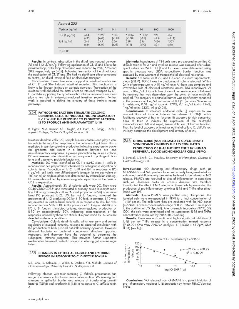
Results: In controls, absorption in the distal loop ranged between75 and 112 µl/min/g. Following application of CT, LT and STa to theproximal loop, distal loop absorption was reduced by 28%, 55% and20% respectively (p<0.05). Following transection of the distal loop,the application of CT, LT and STa had no significant effect comparedto control, on distal intestinal fluid or electrolyte transport.
Conclusions: These observations support a non-direct mechanismof CT, LT and STa- induced intestinal secretion. This mechanism islikely to be through intrinsic or extrinsic neurones. Transection of theintestinal wall abolished the distal effect on intestinal transport by CT,LT and STa supporting the hypothesis that intrinsic intramural neuronesplay a key role in enterotoxin-induced intestinal secretion. Furtherwork is required to define the circuitry of these intrinsic neuralpathways.
254 PATHOGENIC BACTERIA STIMULATE COLONICDENDRITIC CELLS TO PRODUCE PRO-INFLAMMATORYIL-12 WHILE THE RESPONSE TO PROBIOTIC BACTERIAIS TO PRODUCE ANTI-INFLAMMATORY IL-10
R. Rigby1, M.A. Kamm2, S.C. Knight1, A.L. Hart2, A.J. Stagg1. 1APRG,Imperial College; 2St Mark’s Hospital, London, UK
Intestinal dendritic cells (DC) sample luminal contents and play a cen-tral role in the regulated response to the commensal gut flora. This ismediated in part by cytokine production following exposure to bacte-rial products, and results in a balance between pro- andanti-inflammatory responses. Cytokine production by murine colonicDC was assessed after stimulation by a component of pathogenic bac-teria and a putative probiotic bacterium.
Methods: DC were identified as CD11c+MHC class II+ cells inmononuclear cell preparations obtained by collagenase digestion ofcolonic tissue. Production of IL-12, IL-10 and IL-4 in response to LPS(1µg/ml), cell walls from Bifidobacteria longum (at the equivalent of107 per ml) or medium alone was determined by intracellular staining.DC were also isolated by immunomagnetic separation on the basis ofCD11c expression.
Results: Approximately 3% of colonic cells were DC. They wereCD40+CD80+CD86+ and stimulated a primary mixed leucocyte reac-tion following overnight culture. A small proportion (<5%) of unstimu-lated DC produced IL-12. LPS, but not B. longum, increased theproportion of IL-12 producing DC by 4–10 fold. In contrast, IL-10 wasnot detected in unstimulated cultures or in response to LPS, but wasinduced in over 50% of DC in the presence of B. longum. Addition ofLPS to B. longum stimulated cultures, downregulated production ofIL-10 by approximately 50% indicating cross-regulation of theresponses induced by these two stimuli. IL-4 production by DC was notdetected under any conditions.
Conclusions: Colonic dendritic cells, which are early and centralregulators of mucosal immunity, respond to bacterial stimulation withthe production of both pro-and anti-inflammatory cytokines. Howeverdifferent bacteria or bacterial components stimulate opposingresponses, and therefore have the potential to determine thesubsequent immune response. This provides further supportiveevidence for the use of probiotic bacteria in altering gut immune regu-lation.
255 CHANGES IN EPITHELIAL BARRIER AND CYTOKINERELEASE IN RESPONSE TO C. DIFFICILE TOXIN A
S.S. Johal, K. Solomon, J. Webb, S. Dodson, Y.R. Mahida. Division ofGastroenterology, University Hospital, Nottingham, UK
Following infection with toxin-secreting C. difficile, presentation canrange from severe colitis to no colonic inflammation. We investigatedchanges in epithelial barrier and release of transforming growthfactor-β (TGF-β) and interleukin-8 (IL-8) in response to C. difficile toxinA.
Methods: Monolayers of T84 cells were pre-exposed to purified C.difficile toxin A for 3 h and cytokine release was assessed after subse-quent culture for 24 h. TGF-β and IL-8 levels were determined usingspecific bioassay and ELISA respectively. Barrier function wasassessed by measurement of transepithelial electrical resistance.
Results: See table for TGF-β and IL-8 conc. in culture supernatants,mean (±SEM). TGF-β1 was the predominant isoform released. Within24 h of pre-exposure to >10 ng/ml toxin A, there was complete andirreversible loss of electrical resistance across T84 monolayers. Atconc. <10ng/ml of toxin A, loss of monolayer resistance was followedby recovery that was dependent upon the conc. of toxin originallyapplied. This recovery of epithelial barrier was significantly enhancedin the presence of 1 ng/ml recombinant TGF-β1 (maximal % increasein resistance, 0.01 ng/ml toxin A: 179%; 0.1 ng/ml toxin: 156%;1ng/ml toxin: 232%. All p<0.04).
Conclusions: In intestinal epithelial cells, (i) exposure to lowconcentrations of toxin A induces the release of TGF-β, whichfacilitates recovery of barrier function (ii) exposure to high concentra-tions of toxin A induces the expression of the neutrophilchemoattractant IL-8 and rapid, irreversible loss of barrier function.Thus the level of exposure of intestinal epithelial cells to C. difficile tox-ins may determine the development and severity of colitis.
256 NITRIC OXIDE (NO) RELEASED BY GLYCO-SNAP-1SIGNIFICANTLY INHIBITS THE LPS STIMULATEDPRODUCTION OF IL-1β BUT NOT TNFα BY HUMANPERIPHERAL BLOOD MONONUCLEAR CELLS (PBMCS)
J. Burdsall, J. Smith, C.J. Hawkey. University of Nottingham, Division ofGastroenterology, UK
Introduction: NO donating anti-inflammatory drugs such asNO-NSAIDS and Nitroprednisolone are currently being evaluated forenhanced anti-inflammatory properties believed to be related to NOrelease. PBMC’s are recruited to sites of inflammation in conditionssuch as ulcerative colitis or Crohn’s disease. We thereforeinvestigated the effect of NO release on these cells by measuring theproduction of pro-inflammatory cytokines IL-1β and TNFα after stimu-lation with LPS.
Methods: Human PBMC’s were purified using Histopaque. Theisolated cells were re-suspended in RPMI to a final concentration of1x106 per ml. The cells were then pre-incubated with the NO donor(G-SNAP-1) over a concentration range of 0 to 1mM for 30mins priorto the addition of LPS (1µg/ml). After overnight incubation (37°C, 5%CO2), the cells were centrifuged and the supernatant IL-1β and TNFαconcentrations measured by ELISA (RnD DuoSet).
Results: There was a dramatic and highly significant inhibition ofIL-1β but not TNFα release in a concentration related response((P<0.001 One Way ANOVA analysis, IL-1β,IC50 = 61.7µM, SEM0.04) (see fig).
Conclusion: NO released from G-SNAP-1 is a potent inhibitor ofpro- inflammatory mediator IL-1β production by human PBMC’s but notTNFα.
Abstract 255
Toxin A (ng/ml) 0 0.01 0.1 1 10 100 1000
TGF-β (pg/ml) 514 *720 *830 *1316 *1122 621 552(±35) (±69) (±128) (±158) (±81) (±95) (±111)
IL-8 (pg/ml) 246 245 256 249 336 *426 *503(±34) (±24) (±28) (±11) (±10) (±21 ) (±18 )
*p<0.03.
120
100
80
60
40
20
0_5
_4.5
_4
log [G-SNP-1] M
Inhibition of IL-1b release by G-SNAP-1
_3.5
_3
%ofmaximum
stim
ulatedIL-1
release
y =_62.29x
_208.29
R2 = 0.8799
A70 BSG abstracts
www.gutjnl.com
on March 21, 2022 by guest. P
rotected by copyright.http://gut.bm
j.com/
Gut: first published as 10.1136/gut.50.suppl_2.a25 on 1 A
pril 2002. Dow
nloaded from
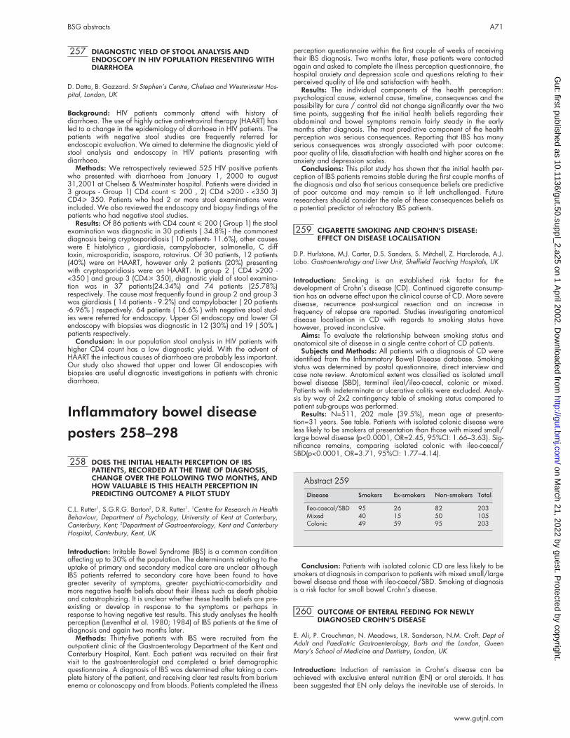
257 DIAGNOSTIC YIELD OF STOOL ANALYSIS ANDENDOSCOPY IN HIV POPULATION PRESENTING WITHDIARRHOEA
D. Datta, B. Gazzard. St Stephen’s Centre, Chelsea and Westminster Hos-pital, London, UK
Background: HIV patients commonly attend with history ofdiarrhoea. The use of highly active antiretroviral therapy (HAART) hasled to a change in the epidemiology of diarrhoea in HIV patients. Thepatients with negative stool studies are frequently referred forendoscopic evaluation. We aimed to determine the diagnostic yield ofstool analysis and endoscopy in HIV patients presenting withdiarrhoea.
Methods: We retrospectively reviewed 525 HIV positive patientswho presented with diarrhoea from January 1, 2000 to august31,2001 at Chelsea & Westminster hospital. Patients were divided in3 groups - Group 1) CD4 count < 200 , 2) CD4 >200 - <350 3)CD4> 350. Patients who had 2 or more stool examinations wereincluded. We also reviewed the endoscopy and biopsy findings of thepatients who had negative stool studies.
Results: Of 86 patients with CD4 count < 200 ( Group 1) the stoolexamination was diagnostic in 30 patients ( 34.8%) - the commonestdiagnosis being cryptosporidiosis ( 10 patients- 11.6%), other causeswere E histolytica , giardiasis, campylobacter, salmonella, C difftoxin, microsporidia, isospora, rotavirus. Of 30 patients, 12 patients(40%) were on HAART, however only 2 patients (20%) presentingwith cryptosporidiosis were on HAART. In group 2 ( CD4 >200 -<350 ) and group 3 (CD4> 350), diagnostic yield of stool examina-tion was in 37 patients(24.34%) and 74 patients (25.78%)respectively. The cause most frequently found in group 2 and group 3was giardiasis ( 14 patients - 9.2%) and campylobacter ( 20 patients-6.96% ) respectively. 64 patients ( 16.6% ) with negative stool stud-ies were referred for endoscopy. Upper GI endoscopy and lower GIendoscopy with biopsies was diagnostic in 12 (30%) and 19 ( 50% )patients respectively.
Conclusion: In our population stool analysis in HIV patients withhigher CD4 count has a low diagnostic yield. With the advent ofHAART the infectious causes of diarrhoea are probably less important.Our study also showed that upper and lower GI endoscopies withbiopsies are useful diagnostic investigations in patients with chronicdiarrhoea.
Inflammatory bowel diseaseposters 258–298
258 DOES THE INITIAL HEALTH PERCEPTION OF IBSPATIENTS, RECORDED AT THE TIME OF DIAGNOSIS,CHANGE OVER THE FOLLOWING TWO MONTHS, ANDHOW VALUABLE IS THIS HEALTH PERCEPTION INPREDICTING OUTCOME? A PILOT STUDY
C.L. Rutter1, S.G.R.G. Barton2, D.R. Rutter1. 1Centre for Research in HealthBehaviour, Department of Psychology, University of Kent at Canterbury,Canterbury, Kent; 2Department of Gastroenterology, Kent and CanterburyHospital, Canterbury, Kent, UK
Introduction: Irritable Bowel Syndrome (IBS) is a common conditionaffecting up to 30% of the population. The determinants relating to theuptake of primary and secondary medical care are unclear althoughIBS patients referred to secondary care have been found to havegreater severity of symptoms, greater psychiatric-comorbidity andmore negative health beliefs about their illness such as death phobiaand catastrophizing. It is unclear whether these health beliefs are pre-existing or develop in response to the symptoms or perhaps inresponse to having negative test results. This study analyses the healthperception (Leventhal et al. 1980; 1984) of IBS patients at the time ofdiagnosis and again two months later.
Methods: Thirty-five patients with IBS were recruited from theout-patient clinic of the Gastroenterology Department of the Kent andCanterbury Hospital, Kent. Each patient was recruited on their firstvisit to the gastroenterologist and completed a brief demographicquestionnaire. A diagnosis of IBS was determined after taking a com-plete history of the patient, and receiving clear test results from bariumenema or colonoscopy and from bloods. Patients completed the illness
perception questionnaire within the first couple of weeks of receivingtheir IBS diagnosis. Two months later, these patients were contactedagain and asked to complete the illness perception questionnaire, thehospital anxiety and depression scale and questions relating to theirperceived quality of life and satisfaction with health.
Results: The individual components of the health perception:psychological cause, external cause, timeline, consequences and thepossibility for cure / control did not change significantly over the twotime points, suggesting that the initial health beliefs regarding theirabdominal and bowel symptoms remain fairly steady in the earlymonths after diagnosis. The most predictive component of the healthperception was serious consequences. Reporting that IBS has manyserious consequences was strongly associated with poor outcome:poor quality of life, dissatisfaction with health and higher scores on theanxiety and depression scales.
Conclusions: This pilot study has shown that the initial health per-ception of IBS patients remains stable during the first couple months ofthe diagnosis and also that serious consequence beliefs are predictiveof poor outcome and may remain so if left unchallenged. Futureresearchers should consider the role of these consequences beliefs asa potential predictor of refractory IBS patients.
259 CIGARETTE SMOKING AND CROHN’S DISEASE:EFFECT ON DISEASE LOCALISATION
D.P. Hurlstone, M.J. Carter, D.S. Sanders, S. Mitchell, Z. Harclerode, A.J.Lobo. Gastroenterology and Liver Unit, Sheffield Teaching Hospitals, UK
Introduction: Smoking is an established risk factor for thedevelopment of Crohn’s disease (CD). Continued cigarette consump-tion has an adverse effect upon the clinical course of CD. More severedisease, recurrence post-surgical resection and an increase infrequency of relapse are reported. Studies investigating anatomicaldisease localisation in CD with regards to smoking status havehowever, proved inconclusive.
Aims: To evaluate the relationship between smoking status andanatomical site of disease in a single centre cohort of CD patients.
Subjects and Methods: All patients with a diagnosis of CD wereidentified from the Inflammatory Bowel Disease database. Smokingstatus was determined by postal questionnaire, direct interview andcase note review. Anatomical extent was classified as isolated smallbowel disease (SBD), terminal ileal/ileo-caecal, colonic or mixed.Patients with indeterminate or ulcerative colitis were excluded. Analy-sis by way of 2x2 contingency table of smoking status compared topatient sub-groups was performed.
Results: N=511, 202 male (39.5%), mean age at presenta-tion=31 years. See table. Patients with isolated colonic disease wereless likely to be smokers at presentation than those with mixed small/large bowel disease (p<0.0001, OR=2.45, 95%CI: 1.66–3.63). Sig-nificance remains, comparing isolated colonic with ileo-caecal/SBD(p<0.0001, OR=3.71, 95%CI: 1.77–4.14).
Conclusion: Patients with isolated colonic CD are less likely to besmokers at diagnosis in comparison to patients with mixed small/largebowel disease and those with ileo-caecal/SBD. Smoking at diagnosisis a risk factor for small bowel Crohn’s disease.
260 OUTCOME OF ENTERAL FEEDING FOR NEWLYDIAGNOSED CROHN’S DISEASE
E. Ali, P. Crouchman, N. Meadows, I.R. Sanderson, N.M. Croft. Dept ofAdult and Paediatric Gastroenterology, Barts and the London, QueenMary’s School of Medicine and Dentistry, London, UK
Introduction: Induction of remission in Crohn’s disease can beachieved with exclusive enteral nutrition (EN) or oral steroids. It hasbeen suggested that EN only delays the inevitable use of steroids. In
Abstract 259
Disease Smokers Ex-smokers Non-smokers Total
Ileo-caecal/SBD 95 26 82 203Mixed 40 15 50 105Colonic 49 59 95 203
BSG abstracts A71
www.gutjnl.com
on March 21, 2022 by guest. P
rotected by copyright.http://gut.bm
j.com/
Gut: first published as 10.1136/gut.50.suppl_2.a25 on 1 A
pril 2002. Dow
nloaded from
this study we have followed up the outcome of children newlydiagnosed in our unit with Crohn’s disease.
Methods: The case notes of patients newly diagnosed with Crohn’sdisease during 1999 and 2000 were reviewed in August 2001.
Results: Patients: 36 patients new patients were diagnosed with amedian age of 12.9 years (range 6.67–15.0). 22 had small boweland colon affected, 7 had small bowel alone and 2 had colonic dis-ease alone. 31/36 had disease severity requiring treatment with ENor steroids. The remaining 5 had localised oral/perianal disease notrequiring EN or steroids. Initial treatment: Exclusive polymeric EN(Modulin, Nestle, UK) was started in 30/31 patients, one refused andwas treated with steroids. 24/30 (80%) went into remission. 6/30required steroids to induce remission. 3 were early failures within 2days (i.e. could not drink the feed or tolerate a naso-gastric tube) and3 were late failures due to lack of response. Follow up data: Themedian time of follow up was 1.25 years. Of 24 children successfullytreated with EN, 1 has been lost to follow up, 42 % (10/24) remainin remission and 54% (13/24) have relapsed. Of the 13 children whorelapsed 54% (7/13) have been successfully treated with EN, 15%(2) failed to respond to EN and started steroids and 23% (3) weretreated with steroids. 2/7children who required steroids at presenta-tion remain in remission, 5/7 have had relapses, one of which wastreated with EN.
Conclusions: These data confirm that EN is a very effective treat-ment for newly diagnosed Crohn’s disease and can be used success-fully for treating relapses. During the period of follow up the majority(58% (18/31)) have exclusively been treated for active disease usingEN and have not required steroids. More prolonged follow up willestablish the proportion of children who can be repeatedly treatedwith EN for relapses of their Crohn’s disease.
261 LOW DOSE ORAL METHOTREXATE FOR THE MEDICALTREATMENT OF STEROID DEPENDANT/RESISTANTCROHN’S DISEASE. EXPERIENCE IN A SINGLE LARGEIBD CLINIC
A. Ansari, M. Yaneza, J. Hurst, J. Sanderson. Guy’s and St Thomas’ Hos-pital, London, UK
Introduction and Aim: Methotrexate (MTX) is a considered optionfor medical treatment of steroid dependant Crohn’s disease (CD) orthose intolerant or resistant to azathioprine (AZA). Few studies involv-ing MTX use have been published. We report the experience of MTXuse for CD in a large inflammatory bowel disease clinic.
Methods: MTX use was initiated in 42 patients with CD at a doseof 15- 25mgs/ week orally. Indication for treatment was steroiddependent CD despite AZA (n= 35) or fistula (n= 7). Efficacy wasassessed by steroid withdrawal, abbreviated Harvey Bradshaw indexor fistula closure. Adverse effects were recorded at each visit.
Results: Length of treatment- mean 34 wks, median 25.5 andrange 1- 91 wks. Total MXT dose- mean 693 mgs, median 420 mgsand range 15- 2330 mgs. Ten patients withdrew because of adverseeffects: nausea (7), transaminitis- ALT rise (1), headaches (1) and mul-tiple side effects (1). In two patients neutropenia occurred (1 severeand complicated by fungal sepsis), both patients were folate deficient.Haematinics were normal in all other cases. On an intension to treatbasis 19 of the 42 patients achieved remission (42%). 16 of the 35tolerating treatment withdrew steroids (46%), 3 out 7 achieved fistulaclosure (43%). In 16 of the 42 patients AZA was continued as concur-rent treatment (mean dose 0.75mg/kg). Whilst both cases of neutro-penia were receiving AZA, overall adverse events were notassociated with its concurrent use.
Conclusion: In practice success rates of treating CD with MTX aresimilar to those suggested by limited trial data, withdrawal is more fre-quent than with AZA therapy. Concurrent AZA treatment does notappear to be a problem, and is generally safe. However, folate defi-ciency is a risk factor for severe adverse events.
262 COMBINATION IMMUNOMODULATORY THERAPYWITH CYCLOSPORIN, AZATHIOPRINE ANDCORTICOSTEROIDS IN SEVERE ULCERATIVE COLITIS:THE EDINBURGH EXPERIENCE OF OUTCOME
S. Campbell, S. Ghosh. The Gastrointestinal Unit, University of Edinburgh,Department of Medical Sciences, Western General Hospital, Crewe Road,Edinburgh EH4 2XU, UK
Background: Cyclosporin is a fungal metabolite and is a powerfulimmunosuppressant. While response to intravenous steroids is in
excess of 60%, the remainder of patients are left with the options ofcurative panproctocolectomy or administration of intravenous rescuetherapy with cyclosporin. There have been conflicting reports withregard to efficacy of IV cyclosporin in acute UC, and there are seriousconcerns about potential toxicity and opportunistic infections such aspneumocystis carnii pneumonia. To date there has been a paucity ofdata available to help guide the gastroenterologist in the use of cyclo-sporin.
Methods: Between 1994 and 2001, there were 16 patients whohad received intravenous cyclosporin for acute exacerbation of theirknown UC (7 female:9 male, mean age 33 years). All patients weretreated with and were refractory to IV methylprednisolone (60mg/24hrs). Patients were discharged on a regimen of oral cyclosporin,oral steroids and oral azathioprine.
Results: Median disease duration was 5.4 years (range 0.9–25years). All patients were initially treated with cyclosporin at a dose of4mg/kg/day. Nine patients were simultaneously started on oral aza-thioprine (median dose 1.8mg/kg). Seven patients underwent surgery(panproctocolectomy), although none had surgery after 6 months.Comparisons were made between patients who had <7days IVsteroid versus >7 days IV steroid, and other subgroups such as stoolfrequency at 3 days and CRP at 3 days. There was no statistical sig-nificant differences between these groups.
Conclusion: Initial response rate was high (69%). Side effectswere documented in the majority of patients, but none of the patientshad to discontinue treatment because of these. At 3 years follow-up,56% of patients avoided surgery. Median bowel frequency at day 3was higher in patients who finally underwent surgery. Cyclsoporin hasa useful role as a 3rd line drug therapy for acute UC patients.
263 APHTHOUS ULCERATION IN THE COLON AFTERSODIUM PHOSPHATE BOWEL PREPARATION FORCOLONOSCOPY
R.J. Atkinson, J.O. Hunter JO. Department of Gastroenterology,Addenbrooke’s Hospital, Hills Road, Cambridge CB2 2QQ, UK
Introduction: It appears to be little known that sodium phosphate, alaxative used in bowel preparation for colonoscopy, may potentiallydamage the colonic mucosa. A 51 year old man undergoing colonos-copy for chronic diarrhoea was found to have severe aphthous ulcera-tion of the sigmoid and histology suggested Crohn’s disease. Notreatment was prescribed, but three weeks later a secondexamination, after a phosphate enema rather than sodium phosphate,was both endoscopically and histologically normal. We suspectedthat the ulcers were caused by the bowel preparation and wetherefore reviewed the presence of aphthous ulceration, in a series ofreports of colonoscopies where either sodium phosphate (Fleet) orsodium picosulphate (Picolax) had been used for bowel preparation.
Methods: 175 consecutive colonoscopy reports were retrospec-tively reviewed with respect to age, sex, indication for the exam andthe presence of ulceration.
Results: Sodium picosulphate was used in 124 colonoscopies andsodium phosphate in 51. The groups were evenly matched for ageand sex. Aphthous ulceration was documented in 4/124 (3.2%) aftersodium picosulphate but 7/51 (13.7%) after sodium phosphate{p=0.015; Fisher’s Exact Test}. The incidence of ulceration in patientsknown to have inflammatory bowel disease was only 3/27 and theincidence of ulcers was only significantly increased in the groupwhose indication for the exam was altered bowel habit. (1/24[4.2%]; 6/23 [26.1%] {p=0.048; Chi squared test}.
Conclusion: It has been shown previously that sodium picosul-phate can cause colonic ulceration. Our series confirms this findingwhich might potentially lead to an incorrect diagnosis andunnecessary treatment. The use of sodium phosphate preparation inpatients suspected of having inflammatory bowel disease may beinadvisable.
264 ROLE OF THE 5Q31 CYTOKINE CLUSTER (IBD5 LOCUS)IN GENETIC SUSCEPTIBILITY TO ULCERATIVE COLITISAND CROHN’S DISEASE IN THE UK
K. Negoro, D.A. van Heel, D.P. McGovern, N.J. Lench, D.P. Jewell. Well-come Trust Centre for Human Genetics, University of Oxford, UK
Introduction: Genetic studies in inflammatory bowel disease haveidentified several susceptibility loci. Recently genetic variation in the5q31 cytokine cluster has been linked to and strongly associated withCrohn’s disease in the Canadian population (IBD5 locus, Rioux et al.,
A72 BSG abstracts
www.gutjnl.com
on March 21, 2022 by guest. P
rotected by copyright.http://gut.bm
j.com/
Gut: first published as 10.1136/gut.50.suppl_2.a25 on 1 A
pril 2002. Dow
nloaded from
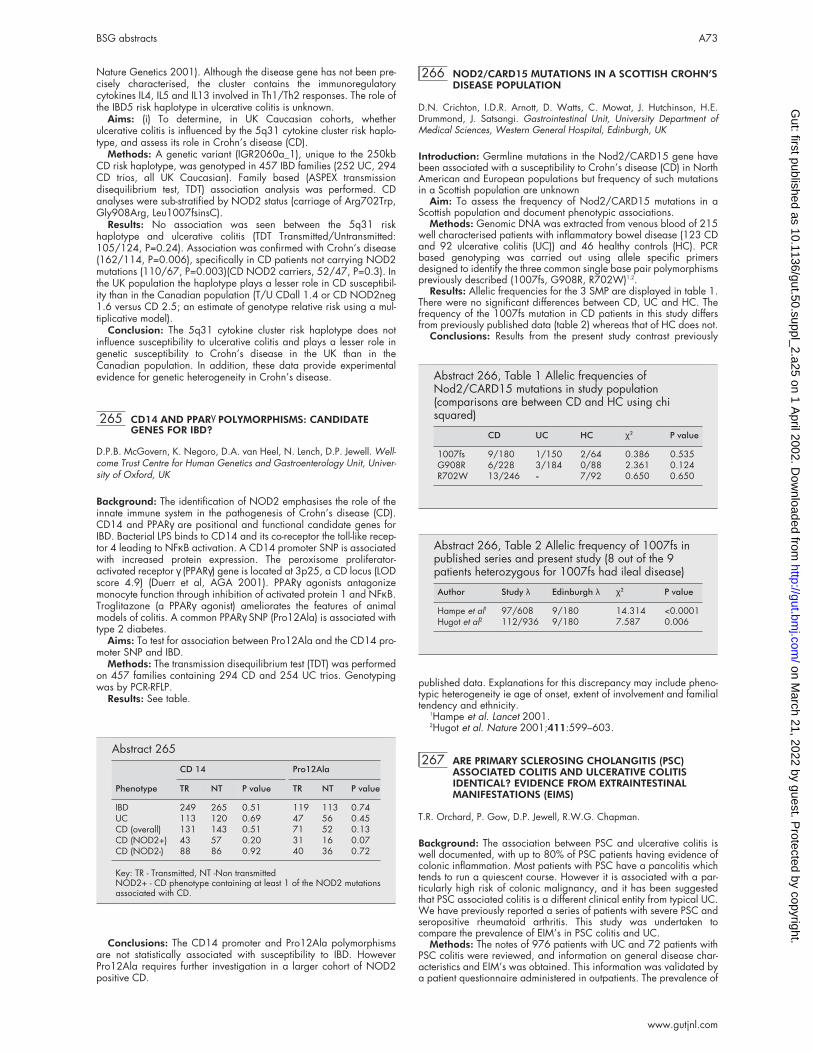
Nature Genetics 2001). Although the disease gene has not been pre-cisely characterised, the cluster contains the immunoregulatorycytokines IL4, IL5 and IL13 involved in Th1/Th2 responses. The role ofthe IBD5 risk haplotype in ulcerative colitis is unknown.
Aims: (i) To determine, in UK Caucasian cohorts, whetherulcerative colitis is influenced by the 5q31 cytokine cluster risk haplo-type, and assess its role in Crohn’s disease (CD).
Methods: A genetic variant (IGR2060a_1), unique to the 250kbCD risk haplotype, was genotyped in 457 IBD families (252 UC, 294CD trios, all UK Caucasian). Family based (ASPEX transmissiondisequilibrium test, TDT) association analysis was performed. CDanalyses were sub-stratified by NOD2 status (carriage of Arg702Trp,Gly908Arg, Leu1007fsinsC).
Results: No association was seen between the 5q31 riskhaplotype and ulcerative colitis (TDT Transmitted/Untransmitted:105/124, P=0.24). Association was confirmed with Crohn’s disease(162/114, P=0.006), specifically in CD patients not carrying NOD2mutations (110/67, P=0.003)(CD NOD2 carriers, 52/47, P=0.3). Inthe UK population the haplotype plays a lesser role in CD susceptibil-ity than in the Canadian population (T/U CDall 1.4 or CD NOD2neg1.6 versus CD 2.5; an estimate of genotype relative risk using a mul-tiplicative model).
Conclusion: The 5q31 cytokine cluster risk haplotype does notinfluence susceptibility to ulcerative colitis and plays a lesser role ingenetic susceptibility to Crohn’s disease in the UK than in theCanadian population. In addition, these data provide experimentalevidence for genetic heterogeneity in Crohn’s disease.
265 CD14 AND PPARγ POLYMORPHISMS: CANDIDATEGENES FOR IBD?
D.P.B. McGovern, K. Negoro, D.A. van Heel, N. Lench, D.P. Jewell. Well-come Trust Centre for Human Genetics and Gastroenterology Unit, Univer-sity of Oxford, UK
Background: The identification of NOD2 emphasises the role of theinnate immune system in the pathogenesis of Crohn’s disease (CD).CD14 and PPARγ are positional and functional candidate genes forIBD. Bacterial LPS binds to CD14 and its co-receptor the toll-like recep-tor 4 leading to NFκB activation. A CD14 promoter SNP is associatedwith increased protein expression. The peroxisome proliferator-activated receptor γ (PPARγ) gene is located at 3p25, a CD locus (LODscore 4.9) (Duerr et al, AGA 2001). PPARγ agonists antagonizemonocyte function through inhibition of activated protein 1 and NFκB.Troglitazone (a PPARγ agonist) ameliorates the features of animalmodels of colitis. A common PPARγ SNP (Pro12Ala) is associated withtype 2 diabetes.
Aims: To test for association between Pro12Ala and the CD14 pro-moter SNP and IBD.
Methods: The transmission disequilibrium test (TDT) was performedon 457 families containing 294 CD and 254 UC trios. Genotypingwas by PCR-RFLP.
Results: See table.
Conclusions: The CD14 promoter and Pro12Ala polymorphismsare not statistically associated with susceptibility to IBD. HoweverPro12Ala requires further investigation in a larger cohort of NOD2positive CD.
266 NOD2/CARD15 MUTATIONS IN A SCOTTISH CROHN’SDISEASE POPULATION
D.N. Crichton, I.D.R. Arnott, D. Watts, C. Mowat, J. Hutchinson, H.E.Drummond, J. Satsangi. Gastrointestinal Unit, University Department ofMedical Sciences, Western General Hospital, Edinburgh, UK
Introduction: Germline mutations in the Nod2/CARD15 gene havebeen associated with a susceptibility to Crohn’s disease (CD) in NorthAmerican and European populations but frequency of such mutationsin a Scottish population are unknown
Aim: To assess the frequency of Nod2/CARD15 mutations in aScottish population and document phenotypic associations.
Methods: Genomic DNA was extracted from venous blood of 215well characterised patients with inflammatory bowel disease (123 CDand 92 ulcerative colitis (UC)) and 46 healthy controls (HC). PCRbased genotyping was carried out using allele specific primersdesigned to identify the three common single base pair polymorphismspreviously described (1007fs, G908R, R702W)1,2.
Results: Allelic frequencies for the 3 SMP are displayed in table 1.There were no significant differences between CD, UC and HC. Thefrequency of the 1007fs mutation in CD patients in this study differsfrom previously published data (table 2) whereas that of HC does not.
Conclusions: Results from the present study contrast previously
published data. Explanations for this discrepancy may include pheno-typic heterogeneity ie age of onset, extent of involvement and familialtendency and ethnicity.
1Hampe et al. Lancet 2001.2Hugot et al. Nature 2001;411:599–603.
267 ARE PRIMARY SCLEROSING CHOLANGITIS (PSC)ASSOCIATED COLITIS AND ULCERATIVE COLITISIDENTICAL? EVIDENCE FROM EXTRAINTESTINALMANIFESTATIONS (EIMS)
T.R. Orchard, P. Gow, D.P. Jewell, R.W.G. Chapman.
Background: The association between PSC and ulcerative colitis iswell documented, with up to 80% of PSC patients having evidence ofcolonic inflammation. Most patients with PSC have a pancolitis whichtends to run a quiescent course. However it is associated with a par-ticularly high risk of colonic malignancy, and it has been suggestedthat PSC associated colitis is a different clinical entity from typical UC.We have previously reported a series of patients with severe PSC andseropositive rheumatoid arthritis. This study was undertaken tocompare the prevalence of EIM’s in PSC colitis and UC.
Methods: The notes of 976 patients with UC and 72 patients withPSC colitis were reviewed, and information on general disease char-acteristics and EIM’s was obtained. This information was validated bya patient questionnaire administered in outpatients. The prevalence of
Abstract 265
Phenotype
CD 14 Pro12Ala
TR NT P value TR NT P value
IBD 249 265 0.51 119 113 0.74UC 113 120 0.69 47 56 0.45CD (overall) 131 143 0.51 71 52 0.13CD (NOD2+) 43 57 0.20 31 16 0.07CD (NOD2-) 88 86 0.92 40 36 0.72
Key: TR - Transmitted, NT -Non transmittedNOD2+ - CD phenotype containing at least 1 of the NOD2 mutationsassociated with CD.
Abstract 266, Table 1 Allelic frequencies ofNod2/CARD15 mutations in study population(comparisons are between CD and HC using chisquared)
CD UC HC χ2 P value
1007fs 9/180 1/150 2/64 0.386 0.535G908R 6/228 3/184 0/88 2.361 0.124R702W 13/246 - 7/92 0.650 0.650
Abstract 266, Table 2 Allelic frequency of 1007fs inpublished series and present study (8 out of the 9patients heterozygous for 1007fs had ileal disease)
Author Study λ Edinburgh λ χ2 P value
Hampe et al1 97/608 9/180 14.314 <0.0001Hugot et al2 112/936 9/180 7.587 0.006
BSG abstracts A73
www.gutjnl.com
on March 21, 2022 by guest. P
rotected by copyright.http://gut.bm
j.com/
Gut: first published as 10.1136/gut.50.suppl_2.a25 on 1 A
pril 2002. Dow
nloaded from
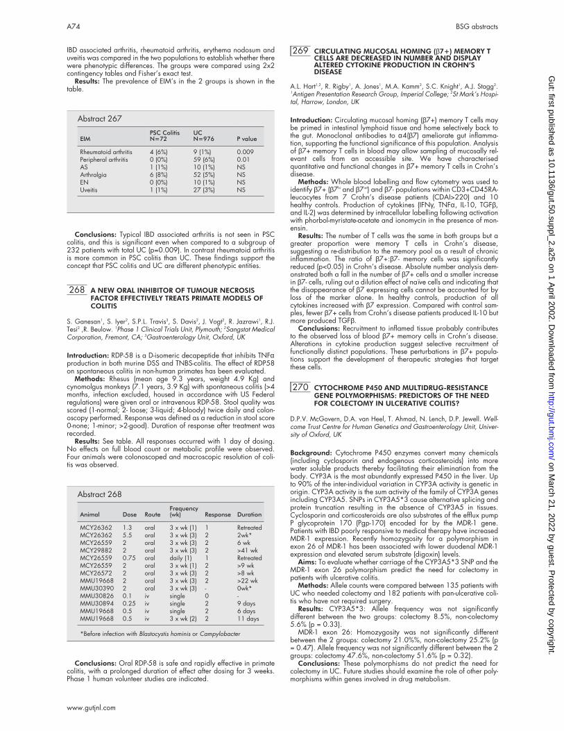
IBD associated arthritis, rheumatoid arthritis, erythema nodosum anduveitis was compared in the two populations to establish whether therewere phenotypic differences. The groups were compared using 2x2contingency tables and Fisher’s exact test.
Results: The prevalence of EIM’s in the 2 groups is shown in thetable.
Conclusions: Typical IBD associated arthritis is not seen in PSCcolitis, and this is significant even when compared to a subgroup of232 patients with total UC (p=0.009). In contrast rheumatoid arthritisis more common in PSC colitis than UC. These findings support theconcept that PSC colitis and UC are different phenotypic entities.
268 A NEW ORAL INHIBITOR OF TUMOUR NECROSISFACTOR EFFECTIVELY TREATS PRIMATE MODELS OFCOLITIS
S. Ganesan1, S. Iyer2, S.P.L. Travis3, S. Davis2, J. Vogt2, R. Jazrawi1, R.J.Tesi2 ,R. Beulow. 1Phase 1 Clinical Trials Unit, Plymouth; 2Sangstat MedicalCorporation, Fremont, CA; 3Gastroenterology Unit, Oxford, UK
Introduction: RDP-58 is a D-isomeric decapeptide that inhibits TNFαproduction in both murine DSS and TNBS-colitis. The effect of RDP58on spontaneous colitis in non-human primates has been evaluated.
Methods: Rhesus (mean age 9.3 years, weight 4.9 Kg) andcynomolgus monkeys (7.1 years, 3.9 Kg) with spontaneous colitis (>4months, infection excluded, housed in accordance with US Federalregulations) were given oral or intravenous RDP-58. Stool quality wasscored (1-normal; 2- loose; 3-liquid; 4-bloody) twice daily and colon-oscopy performed. Response was defined as a reduction in stool score0-none; 1-minor; >2-good). Duration of response after treatment wasrecorded.
Results: See table. All responses occurred with 1 day of dosing.No effects on full blood count or metabolic profile were observed.Four animals were colonoscoped and macroscopic resolution of coli-tis was observed.
Conclusions: Oral RDP-58 is safe and rapidly effective in primatecolitis, with a prolonged duration of effect after dosing for 3 weeks.Phase 1 human volunteer studies are indicated.
269 CIRCULATING MUCOSAL HOMING (β7+) MEMORY TCELLS ARE DECREASED IN NUMBER AND DISPLAYALTERED CYTOKINE PRODUCTION IN CROHN’SDISEASE
A.L. Hart1,2, R. Rigby1, A. Jones1, M.A. Kamm2, S.C. Knight1, A.J. Stagg2.1Antigen Presentation Research Group, Imperial College; 2St Mark’s Hospi-tal, Harrow, London, UK
Introduction: Circulating mucosal homing (β7+) memory T cells maybe primed in intestinal lymphoid tissue and home selectively back tothe gut. Monoclonal antibodies to α4(β7) ameliorate gut inflamma-tion, supporting the functional significance of this population. Analysisof β7+ memory T cells in blood may allow sampling of mucosally rel-evant cells from an accessible site. We have characterisedquantitative and functional changes in β7+ memory T cells in Crohn’sdisease.
Methods: Whole blood labelling and flow cytometry was used toidentify β7+ (β7hi and β7int) and β7- populations within CD3+CD45RA-leucocytes from 7 Crohn’s disease patients (CDAI>220) and 10healthy controls. Production of cytokines (IFNγ, TNFα, IL-10, TGFβ,and IL-2) was determined by intracellular labelling following activationwith phorbol-myristate-acetate and ionomycin in the presence of mon-ensin.
Results: The number of T cells was the same in both groups but agreater proportion were memory T cells in Crohn’s disease,suggesting a re-distribution to the memory pool as a result of chronicinflammation. The ratio of β7+:β7- memory cells was significantlyreduced (p<0.05) in Crohn’s disease. Absolute number analysis dem-onstrated both a fall in the number of β7+ cells and a smaller increasein β7- cells, ruling out a dilution effect of naïve cells and indicating thatthe disappearance of β7 expressing cells cannot be accounted for byloss of the marker alone. In healthy controls, production of allcytokines increased with β7 expression. Compared with control sam-ples, fewer β7+ cells from Crohn’s disease patients produced IL-10 butmore produced TGFβ.
Conclusions: Recruitment to inflamed tissue probably contributesto the observed loss of blood β7+ memory cells in Crohn’s disease.Alterations in cytokine production suggest selective recruitment offunctionally distinct populations. These perturbations in β7+ popula-tions support the development of therapeutic strategies that targetthese cells.
270 CYTOCHROME P450 AND MULTIDRUG-RESISTANCEGENE POLYMORPHISMS: PREDICTORS OF THE NEEDFOR COLECTOMY IN ULCERATIVE COLITIS?
D.P.V. McGovern, D.A. van Heel, T. Ahmad, N. Lench, D.P. Jewell. Well-come Trust Centre for Human Genetics and Gastroenterology Unit, Univer-sity of Oxford, UK
Background: Cytochrome P450 enzymes convert many chemicals(including cyclosporin and endogenous corticosteroids) into morewater soluble products thereby facilitating their elimination from thebody. CYP3A is the most abundantly expressed P450 in the liver. Upto 90% of the inter-individual variation in CYP3A activity is genetic inorigin. CYP3A activity is the sum activity of the family of CYP3A genesincluding CYP3A5. SNPs in CYP3A5*3 cause alternative splicing andprotein truncation resulting in the absence of CYP3A5 in tissues.Cyclosporin and corticosteroids are also substrates of the efflux pumpP glycoprotein 170 (Pgp-170) encoded for by the MDR-1 gene.Patients with IBD poorly responsive to medical therapy have increasedMDR-1 expression. Recently homozygosity for a polymorphism inexon 26 of MDR-1 has been associated with lower duodenal MDR-1expression and elevated serum substrate (digoxin) levels.
Aims: To evaluate whether carriage of the CYP3A5*3 SNP and theMDR-1 exon 26 polymorphism predict the need for colectomy inpatients with ulcerative colitis.
Methods: Allele counts were compared between 135 patients withUC who needed colectomy and 182 patients with pan-ulcerative coli-tis who have not required surgery.
Results: CYP3A5*3: Allele frequency was not significantlydifferent between the two groups: colectomy 8.5%, non-colectomy5.6% (p = 0.33).
MDR-1 exon 26: Homozygosity was not significantly differentbetween the 2 groups: colectomy 21.0%%, non-colectomy 25.2% (p= 0.47). Allele frequency was not significantly different between the 2groups: colectomy 47.6%, non-colectomy 51.6% (p = 0.32).
Conclusions: These polymorphisms do not predict the need forcolectomy in UC. Future studies should examine the role of other poly-morphisms within genes involved in drug metabolism.
Abstract 267
EIMPSC ColitisN=72
UCN=976 P value
Rheumatoid arthritis 4 (6%) 9 (1%) 0.009Peripheral arthritis 0 (0%) 59 (6%) 0.01AS 1 (1%) 10 (1%) NSArthralgia 6 (8%) 52 (5%) NSEN 0 (0%) 10 (1%) NSUveitis 1 (1%) 27 (3%) NS
Abstract 268
Animal Dose RouteFrequency(wk) Response Duration
MCY26362 1.3 oral 3 x wk (1) 1 RetreatedMCY26362 5.5 oral 3 x wk (3) 2 2wk*MCY26559 2 oral 3 x wk (3) 2 6 wkMCY29882 2 oral 3 x wk (3) 2 >41 wkMCY26559 0.75 oral daily (1) 1 RetreatedMCY26559 2 oral 3 x wk (1) 2 >9 wkMCY26572 2 oral 3 x wk (3) 2 >8 wkMMU19668 2 oral 3 x wk (3) 2 >22 wkMMU30390 2 oral 3 x wk (3) - 0wk*MMU30826 0.1 iv single 0 -MMU30894 0.25 iv single 2 9 daysMMU19668 0.5 iv single 2 6 daysMMU19668 0.5 iv 3 x wk (2) 2 11 days
*Before infection with Blastocystis hominis or Campylobacter
A74 BSG abstracts
www.gutjnl.com
on March 21, 2022 by guest. P
rotected by copyright.http://gut.bm
j.com/
Gut: first published as 10.1136/gut.50.suppl_2.a25 on 1 A
pril 2002. Dow
nloaded from
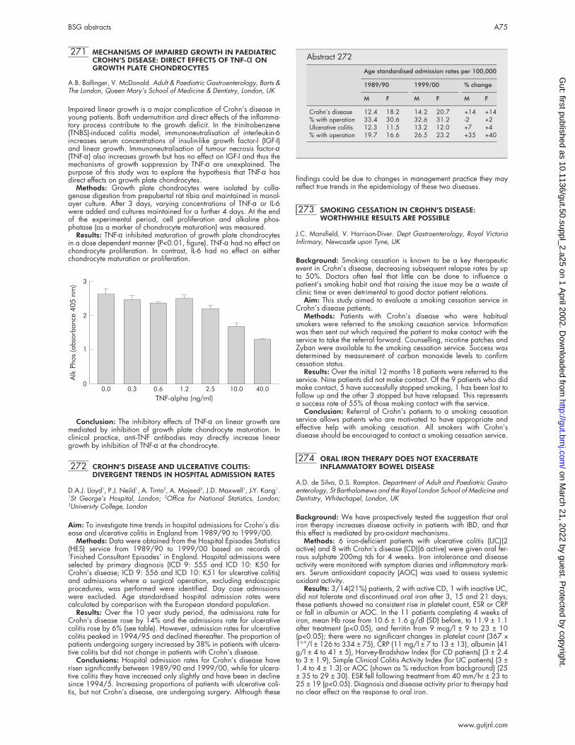
271 MECHANISMS OF IMPAIRED GROWTH IN PAEDIATRICCROHN’S DISEASE: DIRECT EFFECTS OF TNF-α ONGROWTH PLATE CHONDROCYTES
A.B. Ballinger, V. McDonald. Adult & Paediatric Gastroenterology, Barts &The London, Queen Mary’s School of Medicine & Dentistry, London, UK
Impaired linear growth is a major complication of Crohn’s disease inyoung patients. Both undernutrition and direct effects of the inflamma-tory process contribute to the growth deficit. In the trinitrobenzene(TNBS)-induced colitis model, immunoneutralisation of interleukin-6increases serum concentrations of insulin-like growth factor-I (IGF-I)and linear growth. Immunoneutralisation of tumour necrosis factor-α(TNF-α) also increases growth but has no effect on IGF-I and thus themechanisms of growth suppression by TNF-α are unexplained. Thepurpose of this study was to explore the hypothesis that TNF-α hasdirect effects on growth plate chondrocytes.
Methods: Growth plate chondrocytes were isolated by colla-genase digestion from prepubertal rat tibia and maintained in monol-ayer culture. After 3 days, varying concentrations of TNF-α or IL-6were added and cultures maintained for a further 4 days. At the endof the experimental period, cell proliferation and alkaline phos-phatase (as a marker of chondrocyte maturation) was measured.
Results: TNF-α inhibited maturation of growth plate chondrocytesin a dose dependent manner (P<0.01, figure). TNF-α had no effect onchondrocyte proliferation. In contrast, IL-6 had no effect on eitherchondrocyte maturation or proliferation.
Conclusion: The inhibitory effects of TNF-α on linear growth aremediated by inhibition of growth plate chondrocyte maturation. Inclinical practice, anti-TNF antibodies may directly increase lineargrowth by inhibition of TNF-α at the chondrocyte.
272 CROHN’S DISEASE AND ULCERATIVE COLITIS:DIVERGENT TRENDS IN HOSPITAL ADMISSION RATES
D.A.J. Lloyd1, P.J. Neild1, A. Tinto2, A. Majeed3, J.D. Maxwell1, J-Y. Kang1.1St George’s Hospital, London; 2Office for National Statistics, London;3University College, London
Aim: To investigate time trends in hospital admissions for Crohn’s dis-ease and ulcerative colitis in England from 1989/90 to 1999/00.
Methods: Data were obtained from the Hospital Episodes Statistics(HES) service from 1989/90 to 1999/00 based on records of‘Finished Consultant Episodes’ in England. Hospital admissions wereselected by primary diagnosis (ICD 9: 555 and ICD 10: K50 forCrohn’s disease; ICD 9: 556 and ICD 10: K51 for ulcerative colitis)and admissions where a surgical operation, excluding endoscopicprocedures, was performed were identified. Day case admissionswere excluded. Age standardised hospital admission rates werecalculated by comparison with the European standard population.
Results: Over the 10 year study period, the admissions rate forCrohn’s disease rose by 14% and the admissions rate for ulcerativecolitis rose by 6% (see table). However, admission rates for ulcerativecolitis peaked in 1994/95 and declined thereafter. The proportion ofpatients undergoing surgery increased by 38% in patients with ulcera-tive colitis but did not change in patients with Crohn’s disease.
Conclusions: Hospital admission rates for Crohn’s disease haverisen significantly between 1989/90 and 1999/00, while for ulcera-tive colitis they have increased only slightly and have been in declinesince 1994/5. Increasing proportions of patients with ulcerative coli-tis, but not Crohn’s disease, are undergoing surgery. Although these
findings could be due to changes in management practice they mayreflect true trends in the epidemiology of these two diseases.
273 SMOKING CESSATION IN CROHN’S DISEASE:WORTHWHILE RESULTS ARE POSSIBLE
J.C. Mansfield, V. Harrison-Diver. Dept Gastroenterology, Royal VictoriaInfirmary, Newcastle upon Tyne, UK
Background: Smoking cessation is known to be a key therapeuticevent in Crohn’s disease, decreasing subsequent relapse rates by upto 50%. Doctors often feel that little can be done to influence apatient’s smoking habit and that raising the issue may be a waste ofclinic time or even detrimental to good doctor patient relations.
Aim: This study aimed to evaluate a smoking cessation service inCrohn’s disease patients.
Methods: Patients with Crohn’s disease who were habitualsmokers were referred to the smoking cessation service. Informationwas then sent out which required the patient to make contact with theservice to take the referral forward. Counselling, nicotine patches andZyban were available to the smoking cessation service. Success wasdetermined by measurement of carbon monoxide levels to confirmcessation status.
Results: Over the initial 12 months 18 patients were referred to theservice. Nine patients did not make contact. Of the 9 patients who didmake contact, 5 have successfully stopped smoking, 1 has been lost tofollow up and the other 3 stopped but have relapsed. This representsa success rate of 55% of those making contact with the service.
Conclusion: Referral of Crohn’s patients to a smoking cessationservice allows patients who are motivated to have appropriate andeffective help with smoking cessation. All smokers with Crohn’sdisease should be encouraged to contact a smoking cessation service.
274 ORAL IRON THERAPY DOES NOT EXACERBATEINFLAMMATORY BOWEL DISEASE
A.D. de Silva, D.S. Rampton. Department of Adult and Paediatric Gastro-enterology, St Bartholomews and the Royal London School of Medicine andDentistry, Whitechapel, London, UK
Background: We have prospectively tested the suggestion that oraliron therapy increases disease activity in patients with IBD, and thatthis effect is mediated by pro-oxidant mechanisms.
Methods: 6 iron-deficient patients with ulcerative colitis (UC)(2active) and 8 with Crohn’s disease (CD)(6 active) were given oral fer-rous sulphate 200mg tds for 4 weeks. Iron intolerance and diseaseactivity were monitored with symptom diaries and inflammatory mark-ers. Serum antioxidant capacity (AOC) was used to assess systemicoxidant activity.
Results: 3/14(21%) patients, 2 with active CD, 1 with inactive UC,did not tolerate and discontinued oral iron after 3, 15 and 21 days;these patients showed no consistent rise in platelet count, ESR or CRPor fall in albumin or AOC. In the 11 patients completing 4 weeks ofiron, mean Hb rose from 10.6 ± 1.6 g/dl (SD) before, to 11.9 ± 1.1after treatment (p<0.05), and ferritin from 9 mcg/l ± 9 to 23 ± 10(p<0.05); there were no significant changes in platelet count (367 x10–9/l ± 126 to 334 ± 75), CRP (11 mg/l ± 7 to 13 ± 13), albumin (41g/l ± 4 to 41 ± 5), Harvey-Bradshaw Index (for CD patients) (3 ± 2.4to 3 ± 1.9), Simple Clinical Colitis Activity Index (for UC patients) (3 ±1.4 to 4 ± 1.3) or AOC (shown as % reduction from background) (25± 35 to 29 ± 30). ESR fell following treatment from 40 mm/hr ± 23 to25 ± 19 (p<0.05). Diagnosis and disease activity prior to therapy hadno clear effect on the response to oral iron.
3
2
1
0
AlkPhos(absorbance
405nm)
0.0 0.3 0.6 1.2
TNF-alpha (ng/ml)
2.5 10.0 40.0
Abstract 272
Age standardised admission rates per 100,000
1989/90 1999/00 % change
M F M F M F
Crohn’s disease 12.4 18.2 14.2 20.7 +14 +14% with operation 33.4 30.6 32.6 31.2 -2 +2Ulcerative colitis 12.3 11.5 13.2 12.0 +7 +4% with operation 19.7 16.6 26.5 23.2 +35 +40
BSG abstracts A75
www.gutjnl.com
on March 21, 2022 by guest. P
rotected by copyright.http://gut.bm
j.com/
Gut: first published as 10.1136/gut.50.suppl_2.a25 on 1 A
pril 2002. Dow
nloaded from
Conclusions: While a minority of patients do not tolerate it, oraliron has no detectable adverse effect on disease activity or serum anti-oxidant capacity in IBD. Most patients show a good haematologicalresponse to oral iron therapy.
275 AZATHIOPRINE METABOLITES IN INFLAMMATORYBOWEL DISEASE AND INCIDENCE OFNON-COMPLIANCE
S. Webster, D.S. Sanders1, L. Lennard, A.J. Lobo1. University of Sheffield,Section of Medicine and Pharmacology; 1Department of Gastroenterology,The Royal Hallamshire Hospital, Sheffield S10 2JF, UK
Introduction: The immunosuppressive drug azathioprine is wellestablished in the treatment of inflammatory bowel disease (IBD).Myelosuppression occurs in 2 to 5% of IBD patients Thioguaninenucleotides (TGNs) are active azathioprine metabolites, elevatedTGNs are associated with myelosuppression.
Aim: To investigate steady-state TGN metabolite concentrations ina large cohort of IBD patients.
Methods: IBD patients who had been taking azathioprine for atleast 1 year were studied. Blood samples (10ml) were taken for themonitoring of blood counts and measurement of red cell TGN metabo-lite concentrations at each clinic visit
Results: 133 patients were recruited into the study. 114 patientshad repeat metabolite assays taken over 1 year. 89 of these patientswere at TGN steady-state (metabolite assays varied by <50%). Theremaining 25 patients had wide variations in TGN concentrations (>2fold), 10 had dosage adjustments but 15 did not. 11 of these 15patients had TGN levels below the level of detection, indicating non-compliance. Steady-state TGN concentrations ranged from 59 to 566pmol/8x108 (median 172) red cells and the azathioprine dosagefrom 0.3 to 2.8 mg/kg (median 1.6) in the n=89 cohort. Within thisgroup there was no difference in TGN range or azathioprine dosagefor patients on monotherapy (n=20), azathioprine and steroids (n=15)or azathioprine and 5-amino salicylic acids (n=42). There was no sig-nificant difference in TGN metabolite concentrations between patientsin remission (n=80, median TGN 176 pmol) compared to the smallnumber with active disease (n=9, median TGN 153 pmol).
Conclusion: There was a 9 fold range in steady state TGNconcentrations between IBD patients in remission. This range was notinfluenced by concomitant steroid or aminosalicylate therapy. In addi-tion, these metabolites can be used as indicators of compliance withoral azathioprine therapy. 10% of patients were non-compliant.
276 INFLIXIMAB FOR CROHN’S DISEASE: ONE UNIT’SEXPERIENCE
P. Kennedy, J. Andreyev, B. Gazzard, J. Fell, I. Murray-Lyon, P. Vlavianos,D. Westaby, R. Zeegen. Chelsea & Westminster Hospital & Imperial Col-lege School of Medicine, London, UK
Background: The limitations of corticosteroids in the treatment ofCrohn’s Disease (CD) are well recognised. About half the patients withCD who initially respond to medical therapy relapse within one yearor become dependent on steroids with their associated risk of toxicity.One fifth of patients are unresponsive to steroids and many continueto have refractory disease despite combination therapy with immuno-modulatory drugs. Fistulating CD rarely heals with conventional medi-cal therapy. This underlines the need for an alternative agent in thetreatment of active CD. Intestinal mucosal production of TNFα isincreased in CD and has a key role in the initiation and propagationof intestinal inflammation. Infliximab (IgG1 chimeric monoclonal anti-body) binds soluble and membrane bound TNFα and so acts to neu-tralise its effect.
Methods: We undertook a retrospective analysis of patientstreated with Infliximab for refractory CD in our unit. The clinical indi-cations, treatment protocol and outcome of these patients werereviewed. Clinical records, haematological and biochemical indicesand where available endoscopic and histological findings pre andpost treatment were evaluated to assess efficacy and tolerability.
Results: Between 2000–2001, 19 patients with CD were treatedwith Infliximab after 2 consultant gastroenterologists agreed they hadfailed maximal medical therapy. Clinical indications for Infliximabtherapy were fistulating CD (n=5) and refractory luminal CD (n=14).11/19 had undergone previous GI surgery. 18/19 were unrespon-sive to and 1 intolerant of steroids. 13/19 had failed to respond to atleast 1 immuno-modulatory drug. Infliximab (5mg/kg) was adminis-tered by infusion over 2 hours with intravenous steroid and antihista-mine cover. 12/19 were treated as out-patients. A repeat course was
administered in 13 patients (median interval 8 weeks, range 2–30weeks). 7 patients had 3 or more infusions. Five patients had a com-plete response (median 8 weeks, range 2–36 weeks), 12 had a par-tial response (median 6 weeks, range 4–28 weeks) and 2 did notrespond. Two of the 5 patients with fistulating CD, closed their fistulae.Patients requiring surgery (n=4) following therapy had all undergonesurgery for their CD previously. No side effects were reported by15/17 patients. Two patients developed severe headache, preclud-ing further treatment and requiring hospital admission with CT scanand lumbar puncture in one patient.
Conclusions: Our data confirm the previously reported benefit ofInfliximab therapy in achieving rapid responses in patients failingother medical therapies. However, our findings suggest that responsesare short-lived and patients with steroid resistant CD tended to relapse,questioning the benefit of Infliximab in that group. Patients unrespon-sive to initial treatment with Infliximab failed to respond to subsequentinfusions. Perhaps Infliximab therapy should be considered primarilyas a first-line alternative to corticosteroids in the treatment of moderateto severe or fistulating CD.
278 DRB1*15 MOLECULAR SUBTYPING CONFIRMS THATCAUCASIAN ULCERATIVE COLITIS (UC) ISASSOCIATED WITH DRB1*1502 AND NOT DRB1*1501
T. Ahmad1, M. Bunce2, K. Mulcahy-Hawes1, A. Armuzzi1, J. Crawshaw1,O. Large1, K. Welsh3, D Jewell1. 1Departments of Gastroenterology, 2Trans-plant Immunology, University of Oxford; 3National Heart and Lung Institute,Imperial College, London, UK
Background: A replicated association between UC and the serotypeDR2 has been reported in the Japanese. Studies in caucasian popula-tions however have produced more conflicting results. Modernmolecular typing has shown that the Japanese association isattributable to DRB1*1502, an allele common in this ethnic group butrare in caucasians. This allele differs from DRB1*1501 by a singleamino acid (position 86 of exon 2), located within pocket 1, whichmay alter peptide binding. DRB1*15 molecular subtyping has beencarried out in 5 previous studies of Caucasian patients. Only one (Tra-chtenberg et al Hum. Immunol. 2000; 61:326) reported anassociation with DRB1*1502.
Aims: To determine whether DRB1*15 molecular subtypes orlinked polymorphisms in nearby genes confer susceptibility to cauca-sian UC in a large cohort of accurately phenotyped patients.
Methods: LD mapping of 340 polymorphisms across 24 geneswas carried out using PCR-SSP. Extended HLA haplotypes based uponDRB1*1501 and *1502 were constructed from these data. Diseaseassociations were evaluated at each allele on these extendedhaplotypes. 331 accurately phenotyped caucasian UC patients and354 healthy control subjects were studied.
Results: DRB1*1502 was associated with disease susceptibility(4.2% vs. 0.8%; P=0.004; RR 4.59; CI 1.31–16.11). A rareextended haplotype A*3201-Cw*1202-B*5201-MICA*00901-MICB*0102–1C7H3-DRB5*0101-DRB1*1502-DQB1*0601 wasconstructed. None of these alleles carried a relative risk of diseasegreater than DRB1*1502. This allele was not associated with diseasephenotype (extent or severity). DRB1*1501 was not associated withdisease susceptibility or phenotype.
Conclusion: Although rare in Caucasian UC patients, DRB1*1502appears to be associated with UC in all ethnic groups. This uniquetrans-racial concordance suggests that this allele or a closely linkedallele determines disease susceptibility.
279 TUMOUR NECROSIS FACTOR ALPHA (TNFα)PROMOTER POLYMORPHISMS AND COLITISASSOCIATED COLORECTAL NEOPLASIA (CACRN)
C. Murphy, R. Martin, P. Erwin, V. Rudralingam, R. Maxwell, C. Patterson1,J. Sloan2, D. Middleton3, F.C. Campbell. Dept Surgery, 1DeptEpidemiology, Queen’s University Belfast, Grosvenor Rd, Belfast BT12 6BJ;2Dept Pathology, Royal Victoria Hospital, Belfast BT12 6BJ; 3Northern Ire-land Regional Histocompatibility and Immunogenetics Laboratory, BelfastCity Hospital BT9 7TS, Northern Ireland
Persistent colonic inflammation in ulcerative (UC) and Crohn’s colitis(CD) confers increased colorectal cancer risk. Tumour necrosis factoralpha (TNFα) mediates inflammation in colitis. TNFα promoterpolymorphisms associated with variable TNFα production. This studytests the hypothesis that TNFα promoter polymorphisms confer cancerrisk, in colitis.
A76 BSG abstracts
www.gutjnl.com
on March 21, 2022 by guest. P
rotected by copyright.http://gut.bm
j.com/
Gut: first published as 10.1136/gut.50.suppl_2.a25 on 1 A
pril 2002. Dow
nloaded from
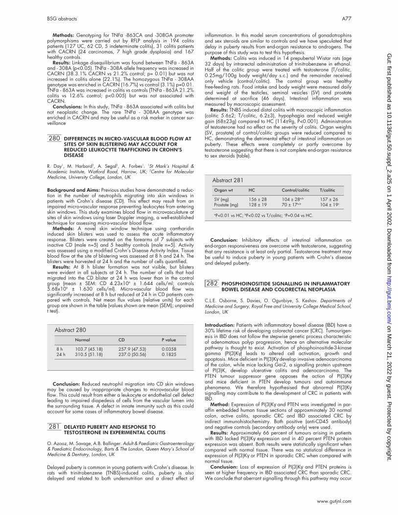
Methods: Genotyping for TNFα -863CA and -308GA promoterpolymorphisms were carried out by RFLP analysis in 194 colitispatients (127 UC, 62 CD, 5 indeterminate colitis), 31 colitis patientswith CACRN (24 carcinomas, 7 high grade dysplasia) and 167healthy controls.
Results: Linkage disequilibrium was found between TNFα - 863Aand - 308A (p<0.05). TNFα - 308A allele frequency was increased inCACRN (38.3.1% CACRN vs 21.2% control; p= 0.01) but was notincreased in colitis alone (22.1%). The homozygous TNFα - 308AAgenotype was enriched in CACRN (16.7%) vs control (3.1%) p=0.01.TNFα - 863A was increased in colitis vs controls (TNFα - 863A 21.2%colitis vs 12.6% control; p<0.005) but was not associated withCACRN.
Conclusions: In this study, TNFα - 863A associated with colitis butnot neoplastic change. The rare TNFα - 308AA genotype wasenriched in CACRN and may be useful as a risk marker in cancer sur-veillance
280 DIFFERENCES IN MICRO-VASCULAR BLOOD FLOW ATSITES OF SKIN BLISTERING MAY ACCOUNT FORREDUCED LEUKOCYTE TRAFFICKING IN CROHN’SDISEASE
R. Day1, M. Harbord2, A. Segal2, A. Forbes1. 1St Mark’s Hospital &Academic Institute, Watford Road, Harrow, UK; 2Centre for MolecularMedicine, University College, London, UK
Background and Aims: Previous studies have demonstrated a reduc-tion in the number of neutrophils migrating into skin windows inpatients with Crohn’s disease (CD). This effect may result from animpaired micro-vascular response preventing leukocytes from enteringskin windows. This study examines blood flow in microvasculature atsites of skin windows using laser Doppler imaging, a well-establishedtechnique for assessing micro-vascular blood flow.
Methods: A novel skin window technique using cantharidininduced skin blisters was used to assess the acute inflammatoryresponse. Blisters were created on the forearms of 7 subjects withinactive CD (male n=5) and 5 healthy controls (male n=5). Activitywas assessed using a modified Crohn’s Disease Activity Index. Tissueblood flow at the site of blistering was assessed at 8 h and 24 h. Theblisters were harvested at 24 h and the number of cells quantified.
Results: At 8 h blister formation was not visible, but blisterswere evident in all subjects at 24 h. The number of cells that hadmigrated into the CD blister at 24 h was lower than in the controlgroup (mean ± SEM: CD 4.23x10⁶ ± 1.644 cells/ml; controls5.68x106 ± 1.630 cells/ml). Micro-vascular blood flow wassignificantly increased at 8 h but reduced at 24 h in CD patients com-pared with controls. Net mean flux values (relative units) for eachgroup are shown in the table (values shown are mean (SEM); unpairedt test).
Conclusion: Reduced neutrophil migration into CD skin windowsmay be caused by inappropriate changes to microvascular bloodflow. This could result from either a leukocyte or endothelial cell defectleading to impaired diapedesis of cells from the vascular lumen intothe surrounding tissue. A defect in innate immunity such as this couldaccount for some cases of inflammatory bowel disease.
281 DELAYED PUBERTY AND RESPONSE TOTESTOSTERONE IN EXPERIMENTAL COLITIS
O. Azooz, M. Savage, A.B. Ballinger. Adult & Paediatric Gastroenterology& Paediatric Endocrinology, Barts & The London, Queen Mary’s School ofMedicine & Dentistry, London, UK
Delayed puberty is common in young patients with Crohn’s disease. Inrats with trinitrobenzene (TNBS)-induced colitis, puberty is alsodelayed and related to both undernutrition and a direct effect of
inflammation. In this model serum concentrations of gonadotrophinsand sex steroids are similar to controls and we have speculated thatdelay in puberty results from end-organ resistance to androgens. Thepurpose of this study was to test this hypothesis.
Methods: Colitis was induced in 14 prepubertal Wistar rats (age32 days) by intrarectal administration of trinitrobenzene in ethanol.Half of the colitic group were treated with testosterone (T/colitic,0.25mg/100g body weight/day s.c.) and the remainder receivedonly vehicle (control/colitic). The control group was healthyfree-feeding rats. Food intake and body weight were measured dailyand weight of the testicles, seminal vesicles (SV) and prostatedetermined at sacrifice (46 days). Intestinal inflammation wasmeasured by macroscopic assessment.
Results: TNBS induced distal colitis with macroscopic inflammation(colitic 5.6±2; T/colitic, 6.2±3), hypophagia and reduced weightgain (68±23g) compared to HC (114±9g, P<0.001). Administrationof testosterone had no effect on the severity of colitis. Organ weights(SV, prostate) of control/colitic groups were reduced compared toHC, demonstrating the detrimental effect of intestinal inflammation onpuberty. These effects were completely or partly overcome bytestosterone suggesting that there is not complete end-organ resistanceto sex steroids (table).
Conclusion: Inhibitory effects of intestinal inflammation onend-organ responsiveness are overcome with testosterone, suggestingthat any resistance is at least only partial. Testosterone treatment maybe useful to induce puberty in young patients with Crohn’s diseaseand delayed puberty.
282 PHOSPHOINOSITIDE SIGNALLING IN INFLAMMATORYBOWEL DISEASE AND COLORECTAL NEOPLASIA
C.L.E. Osborne, S. Davies, O. Ogunbiye, S. Keshav. Departments ofMedicine and Surgery, Royal Free and University College Medical School,London, UK
Introduction: Patients with inflammatory bowel disease (IBD) have a30% lifetime risk of developing colorectal cancer (CRC). Tumourigen-esis in IBD does not follow the stepwise genetic process characteristicof adenomatous polyp progression, hence an alternative molecularpathway is thought to exist. Activation of phosphoinositide-3-kinasegamma (PI(3)Kγ) leads to altered cell activation, growth andapoptosis. Mice deficient in PI(3)Kγ develop invasive adenocarcinomaof the colon, while mice lacking Gαi2, a signalling protein upstreamof PI(3)K, develop ulcerative colitis and adenocarcinoma. ThePTEN tumour suppressor gene opposes the action of PI(3)Kγand mice deficient in PTEN develop tumours and autoimmunephenomena. We therefore hypothesised that abnormal PI(3)Kγsignalling may contribute to the development of CRC in patients withIBD.
Method: Expression of PI(3)Kγ and PTEN was investigated in par-affin embedded human tissue sections of approxminately 30 normalcolon, active colitis, sporadic CRC and IBD associated CRC byindirect immunohistochemistry. Both positive (anti-CD45 antibody)and negative controls (secondary antibody only) were used.
Results: Approximately 66 percent of tumours arising in patientswith IBD lacked PI(3)Kγ expression and in 40 percent PTEN proteinexpression was absent. Both results were statistically significant whencompared with normal tissue. There was no statistical difference inexpression of PI(3)Kγ or PTEN in sporadic CRC when compared withnormal tissue.
Conclusion: Loss of expression of PI(3)Kγ and PTEN proteins isseen at higher frequency in IBD associated CRC than sporadic CRC.We conclude that aberrant signalling through this pathway may occur
Abstract 280
Normal CD P value
8 h 103.7 (45.18) 257.9 (47.53) 0.035824 h 310.5 (51.18) 237.0 (50.56) 0.1825
Abstract 281
Organ wt HC Control/colitic T/colitic
SV (mg) 156 ± 28 104 ± 28a,b 157 ± 26Prostate (mg) 128 ± 19 70 ± 17a,b 104 ± 19c
aP<0.01 vs HC; bP<0.02 vs T/colitic; cP=0.04 vs HC.
BSG abstracts A77
www.gutjnl.com
on March 21, 2022 by guest. P
rotected by copyright.http://gut.bm
j.com/
Gut: first published as 10.1136/gut.50.suppl_2.a25 on 1 A
pril 2002. Dow
nloaded from
together with chronic inflammation, leading to abnormalities in cellcycle progression and apoptosis. This may therefore contribute to thepostulated alternative pathway to neoplasia in IBD associated CRC.
283 ADENOMATOUS POLYPS ARE RARE IN ULCERATIVECOLITIS
T. Kitiyakara, A.S. McIntyre, D.A. Gorard. Wycombe Hospital, HighWycombe, Bucks HP11 2TT, UK
Colon cancer in ulcerative colitis (UC) does not follow the adenoma-carcinoma sequence of non-UC colorectal neoplasia. Polyps with dys-plasia do occur in UC, and there is debate whether they shouldalways be managed as dysplasia associated lesion/masses (DALMs)requiring colectomy, or whether some can be managed as sporadicadenomas by polypectomy. There are few data on the prevalence ofnon-inflammatory polyps in UC; this study aimed to see ifadenomatous polyps occur as often in UC as in patients withoutinflammatory bowel disease (IBD).
The clinical, endoscopy and histology records of 150 patients withUC undergoing colonoscopy were scrutinised for any history ofpolyps. The control group was 205 patients having colonoscopy foraltered bowel habit. Patients with rectal bleeding, anaemia, abnormalbarium enema, a personal or family history of colorectal cancer/polyps were excluded as controls, as were those in whom cancer orIBD was found at endoscopy.
The mean (SD) age of UC patients, 48.8 (13.8) years, was not dif-ferent to that of controls, 51.9 (15.5) years. Sex distribution of 79 m/ 71 f in UC, 90 m/ 115 f in controls was similar. In UC, the median(range) disease duration was 10 (0–48) years, and the mediannumber of colonoscopies was 2 (1–10). The most proximally recordedUC extent was pancolitis in 85 (57%) patients, to the hepatic flexurein 16 (11%), to the splenic flexure in 19 (13%), proctosigmoiditis in18 (12%), proctitis in 12 (8%).
Only 6 UC patients had ever had dysplastic polyps. 2 had a singleadenomatous polyp proximal to the colitis segment. 4 patients haddysplastic polyps within the colitis segment. In 2 of these the polypswere treated endoscopically as sporadic adenomatous polyps (1patient having 2 polyps). In the other 2, the lesions were consideredto be DALMs and colectomy advised. In contrast, 24 controls had atleast 1 adenomatous polyp, χ2 = 6.7, p<0.01. Metaplastic polypswere found in 4 UC patients (within colitis in 3, proximal to colitis in1) and in 24 control patients, χ2 = 9.7, p<0.01. 38 UC patients hadinflammatory pseudopolyps.
Adenomatous and metaplastic polyps occur less frequently inpatients with UC than in patients without IBD. Despite the increasedcancer risk in longstanding UC, the colitic mucosa (or possibly drugtreatment of UC) seems to protect against the formation of sporadicadenomas.
284 MAGNETIC RESONANCE IMAGING (MRI) IN PATIENTSWITH ACUTE COLITIS: A PILOT STUDY
Z.H. Khan, J. Entwisle, J. de Caestecker, S. Campbell, R.J. Robinson.Department of Gastroenterology, Department of Radiology, Glenfield Hos-pital, Leicester, UK
Aim: To evaluate the potential of MRI in patients with acute colitis.Methods: Consecutive patients admitted with acute colitis were
studied. All had AXR on admission and MRI of the colon. Axial (T1/T2) and sagittal T1 images were performed according to a predeter-mined protocol using a Siemens 1.5 Tesla Vision Scanner. Thefollowing parameters on AXR were recorded: disease extent, boweldilatation and wall thickness. At MRI, disease extent, bowel dilatation,wall thickness, changes in peri-colic fat and the presence or absenceof free fluid were noted.
Results: To date eleven patients with acute colitis have beenincluded. One male and ten females, mean age 40.7 ( range 22–71)were anlysed. Six out of eleven patients with Powell Tuck score rang-ing between 8–13 had no or minimal changes on AXR. MRI showeddisease extent and established bowel wall thickening in all six cases.Three patients required colectomy and in two of these cases MRIshowed free fluid and changes in peri-colic fat.
Conclusions: (1) MRI is more informative than a plain abdominalfilm in assessing the severity and extent of disease in patients admittedwith acute colitis. (2) Features like presence of free fluid and changesin the peri-colic fat may be important prognostic factors.
285 RELATIONSHIP BETWEEN SYMPTOMS, SITE OFDISEASE ACTIVITY AND HEIGHT/WEIGHT Z SCORE ATTHE TIME OF DIAGNOSIS OF CHILDHOOD CROHN’SDISEASE
A. Sawczenko1, B.K. Sandhu1, R.F.A. Logan2. 1Department of Gastroenter-ology, Bristol Children’s Hospital, UK; 2Dept of Public Health and Epidemi-ology, Nottingham, UK
Aim: To determine the relationship between presenting symptoms, siteof disease activity and height/weight Z scores at diagnosis of Crohn’sDisease (CD).
Methods: Data collected prospectively from the 1998/9 BPSU-BSGRU survey (Lancet : 357; 1093–4) from 364 CD cases aged < 16years were analysed.
Results: The following correlations between symptoms and site ofdisease were found ; ‘weight loss’ with jejunal and ileal activity, ‘leth-argy’ with left colonic activity, ‘abdominal pain’ with oral, transversecolonic and peri-anal activity, ‘diarrhoea’ with colonic and rectalactivity, ‘bleeding’ with jejunal, ileal, and colonic (but not rectal)activity (all p < 0.05). Anorexia was not associated with any specificsite activity. Mean height and weight Z scores were reduced at diag-nosis (-0.54, 95%CI -0.67 to -0.41, and -1.06, 95%CI -1.21 to -0.92respectively), with the correlation between height and weight being r= 0.72 (p < 0.0001). There was a negative correlation the time ofrecall of onset of symptoms to diagnosis (ie ‘delay’) and height (r =-0.22, p < 0.001), but no relationship with weight. The symptom of‘weight loss’ was associated with reduced weight (mean Z -1.3 versus- 0.7, p < 0.001), as was ‘anorexia’ (mean Z 1.9 versus –1.0, p =0.01), but there were no correlations with height. The symptom of‘abdominal pain’ was associated with reduced Z score for height(mean -0.76 versus –0.45, p = 0.03), but not for weight. Jejunal activ-ity was associated both with a lower Z score for weight (mean Z -1.6versus –1.1, p = 0.017) and height (mean Z –0.9 versus –0.5, p =0.041). Ileal activity was associated with a lower Z score for weight(mean Z –1.2 versus –0.8, p =0.02) but there was no relationship withheight. Fascinatingly there was a relationship between oesophagealdisease activity and height (mean Z –0.6 versus –0.2, p = 0.045), butnot for weight.
Conclusion: Prolonged symptoms are associated with decreasedheight at diagnosis, suggesting that earlier recognition of CD may beimportant. Ileal and especially Jejunal disease are associated withimpaired height and weight Z scores at diagnosis. Symptoms givesome indication of sites affected.
286 FOLLOW UP: TIME TO CHANGE? THE PATIENTS’ VIEW
V. Edge, C. Macdonald, D.A. Burke. Cumberland Infirmary, Carlisle, UK
Patients with long-term chronic illness (e.g. inflammatory boweldisease (IBD) and coeliac disease (CD)) require periodic monitoring oftheir condition. If stable and otherwise well, alternatives to thetraditional method of follow up may prove more practical and accept-able to patients and yet be adaptable to their changing health careneeds without loss of contact/ input from specialists. This isparticularly pertinent to services covering large geographical areas.Alternatives that have been proposed are nurse led clinics, telephoneconsultations or a personalised postal questionnaire, with relevantblood tests performed locally. Access to a consultant opinion shouldstill be available.
Aims: To assess patients views regarding follow up of their condi-tion and to determine their preferences in follow up arrangements.
Methods: 100 patients were randomly selected from our IBD andCD database and invited to complete an anonymous postal question-naire enquiring about their views on follow up.
Results: 78% responded, 1 had recently been discharged, 1suffered from dementia and 1 was illegible. Therefore 75 replies wereanalysed (38 IBD, 37 CD). 61 (28 IBD, 33 CD) patients would bewilling to be reviewed by a specialist gastroenterology nurse in aclinic, 46 (23 IBD, 23CD) would consider a telephone consultation.52 (27 IBD, 25 CD) respondents felt an update of their condition couldbe achieved by a personalised questionnaire with blood testsperformed locally for them. 60 patients selected a combination of theoptions. In 27 cases (14 IBD, 13 CD) while they were prepared toconsider alternatives, they would prefer that their follow up continuedin the clinic as before. Although CD might be considered to follow amore predictable course when compared to IBD with its likelihood forrelapse, there were no significant differences in the responses from thetwo groups. 19 people did not possess reliable weighing scales toallow home monitoring of their weight.
A78 BSG abstracts
www.gutjnl.com
on March 21, 2022 by guest. P
rotected by copyright.http://gut.bm
j.com/
Gut: first published as 10.1136/gut.50.suppl_2.a25 on 1 A
pril 2002. Dow
nloaded from
Conclusions: The majority of patients would accept alternativemethods of monitoring their condition while maintaining links withtheir supervising specialist team. The provision of personal weighingscales would permit home monitoring for all. There are potential ben-efits to both patient and our overburdened GI services by adoptingalternative methods of follow up.
287 THE ROLE OF DIET IN THE AETIOLOGY OFULCERATIVE COLITIS: A PILOT STUDY IN A EUROPEANPROSPECTIVE COHORT STUDY
A.R. Hart1, M. Bergmann2, R. Luben3, J. Camus3, S. Oakes3, A. Welch3, S.Bingham3, K-T. Khaw3, H. Boeing2, N.E. Day3. 1University of East Anglia,Norwich, UK; 2German Institute of Human Nutrition, Potsdam, Germany;3Strangeways Research Laboratories, Cambridge, UK
Background: The causes of ulcerative colitis are unknown, althoughit is plausible that dietary factors are involved. To address this hypoth-esis a prospective cohort study is needed to eliminate the biases asso-ciated with the previous case-control studies of diet and IBD.
Aims: To conduct a pilot study to determine whether it was feasibleto identify subjects who developed ulcerative colitis who areparticipating in a large European cohort study and to conduct a pro-visional analysis of the dietary data.
Methods: 25 623 men and women aged 45–74 years in Norfolk(UK) and 27 548 men and women aged 35–64 years in Potsdam(Germany) were recruited to the EPIC Study (European ProspectiveInvestigation Into Cancer & Nutrition). These subjects completed infor-mation on diet at recruitment and are being followed up for the devel-opment of ulcerative colitis. Each case was matched with four controlsand an analysis performed for food groups, adjusted for cigarettesmoking.
Results: 20 incident patients with ulcerative colitis (7 women, 13men) were identified, which is the expected number over the follow-upperiod. Analysis showed a non-significant positive association withcarbohydrate, sugar and fat consumption (for lower vs upper tertiles ofconsumption: carbohydrate OR=2.8 (95% CI=0.7–11.1), sugarOR=1.8 (95% CI=0.5–7.3, fat OR=1.6 (95% CI=0.5–5.6). A higherfish consumption appeared to protect against the development ofulcerative colitis (lower vs middle tertile OR= 0.5 (95% CI=0.1–2.1 &lower vs upper tertile OR= 0.8 (95% CI=0.2–3.2)).
Conclusions: A prospective cohort study of diet and ulcerativecolitis is feasible. Other European centres participating in EPIC nowneed to be included to increase the number of participants studied sothe role of diet in the aetiology of ulcerative colitis can be accuratelydefined.
288 COMPLIANCE WITH MAINTENANCE MESALAZINE ININFLAMMATORY BOWEL DISEASE
M.J. Shale, S.A. Riley. Northern General Hospital, Herries Road, SheffieldS5 7AU, UK
Background: Most patients with inflammatory bowel disease are pre-scribed regular medications to reduce the risk of disease relapse.Non-compliance with such treatments may increase the risk of relapseand some have suggested an increased risk of colorectal cancer. Wehave therefore conducted a survey of outpatients with IBD to determinethe prevalence of non-compliance with maintenance mesalazinetherapy, and to identify possible risk factors.
Methods: Outpatients with quiescent IBD receiving maintenancetherapy with delayed-release mesalazine (Asacol) were recruited tothe study. Patients underwent a structured interview includingassessment of drug compliance and medication use. Anxiety anddepression, quality of life, and measures of the doctor-patient relation-ship were also assessed using validated questionnaires. Poor compli-ance was defined as taking <80% of the prescribed dose.
Results: 98 patients were studied. 51 were male and ages rangedfrom 17–85 years. 63% had ulcerative colitis, 27% Crohn’s diseaseand 10% indeterminate colitis. Disease duration was 8.6 (0.2–53)years and patients had taken mesalazine 2.4 (0.4–3.6) g/day for 4.3(0.2–13) years. 64% of patients were prescribed mesalazine threetimes daily. 43% reported regular non-compliance. Non-compliancewas associated with a younger age, full-time employment, three-timesdaily dosing, education beyond the age of 16 and the patient’s per-ception of treatment efficacy. Logistic regression revealed the onlyindependent predictors of non-compliance to be three-times daily dos-ing (OR= 3.1 [95% CI 1.8–8.4]) and full time employment (OR= 2.7[95%CI 1.1–6.9]).
Conclusions: Non-compliance with maintenance mesalazine is acommon problem in patients with inflammatory bowel disease. Three-times daily dosing and full-time employment are the main predictors ofnon-compliance. These factors should be considered when selectingand advising on maintenance drug regimens.
289 RELATIONSHIP OF THIOPURINE METHYLTRANSFERASE(TPMT) ACTIVITY TO MEAN CORPUSCULAR VOLUME(MCV) IN INFLAMMATORY BOWEL DISEASE (IBD)PATIENTS MAINTAINED ON AZATHIOPRINE
D. Joy, R. MacPherson, S. Campbell, K. Kingstone, S. Ghosh.Gastrointestinal Unit, Western General Hospital, University of Edinburgh
Introduction: TPMT is a cytosolic enzyme that catalyses theS-methylation of aromatic and heterocyclic sulphydryl compoundsincluding 6-mercaptopurine and azathioprine (AZA). AZA inducesTPMT activity variably and unpredictably. We have recently reportedthat IBD patients with TPMT activity > 20 nmol/hr/ml of RBC relapsemore often if AZA is prescribed at doses < 2mg/kg/day. MCV isincreased in patients on AZA, but the implications of this increase inguiding AZA therapy is unclear. We investigated the relationshipbetween TPMT activity and MCV in IBD patients maintained on AZA.
Patients and Methods: We recruited a cohort of 84 IBD (45M,39F) patients (38 Ulcerative Colitis, 46 Crohn’s disease) who weremaintained on AZA. TPMT activity was determined from bloodsamples by a radiochemical assay on haemolysed red blood cells(RBC) as previously reported and MCV was obtained on the sameday. Relapse rates per year of follow up and time to first relapse wererelated to respective TPMT activity and MCV.
Results: The MCV did not correlate with TPMT activity (r = 0.025;p =0.8). The mean MCV was 91.45 fl (SD 7.38) in patients with TPMT< 20 nmol/hour/ml of RBC compared with MCV of 92.19 fl (SD5.77) in patients with TPMT > 20 nmol/hour/ml of RBC (p =NS). AKaplan-Meier survival curve was constructed based on time to firstrelapse for patients with MCV < 98 fl compared with MCV > 98 fland the difference was not significant by log rank analysis.
Conclusion: In IBD patients on AZA, MCV does not correlate withTPMT activity. MCV in IBD is influenced by a number of conflictingfactors such as iron or folate deficiency, and cannot be used to deter-mine AZA effect or outcome of therapy.
290 PRE-POUCH ILEITIS: A DISEASE OF THE ILEUM IN UC
A.J.G. Bell1,2, A.B. Price1, A. Forbes1, P.J. Ciclitira2, K.H. Wilkinson1, S.W.Rumbles1, R.J. Nicholls1. 1St Mark’s Academic Institute, Harrow; 2RayneInstitute, St Thomas’ Hospital, London, UK
Introduction: Inflammation of the ileum occurs in UC patients. Inaddition to “backwash ileitis”, pouchitis and pre-stomal ileitis we haveobserved inflammation proximal to the pouch in the neo-terminal ileum(NTI) and sought to ascertain the characteristics of this “pre-pouch ilei-tis” (PPI).
Methods: Retrospective notes review of those with ileal abnormali-ties amongst the 661 consecutive cases undergoing restorativeproctocolectomy for UC at a single centre to 1998. Histological slideswere reviewed. Staining for colonic metaplasia was undertaken.
Results: 19 cases were found. 3 had Crohn’s Disease (CD), 1 hada discrete NSAID stricture. These 4 are termed alternative group (AG).The remaining 15 had characteristic diffuse disease from theNTI-pouch junction proximally for varying distances (PPI group). Thedisease became milder more proximally in PPI and CD cases. In 3/15disease was limited to the pouch-NTI junction. 2 of the other 12 hadstricturing disease and 1/12 had fistulating PPI. The majoritypresented with frequency and pain. Extra-intestinal manifestationswere seen in 4/15 PPI but 0/4 AG. Smoking was unusual. In the PPIgroup half had pouchitis but few had had backwash ileitispre-operatively. A significant distal stricture was present in only 1/15PPI and 1/4 AG. The pathology was discovered by contrast studies in8/19, endoscopy in 7/19 and at surgery in 3/19. Roughly a quarterresponded to each of antibiotics, IBD therapy or resection with spon-taneous remission in the remainder. Histological appearances ofresection specimens and biopsies revealed nothing characteristicallyto distinguish PPI from pouchitis.
Conclusion: 15 cases of pre-pouch ileitis were found in patientswith confirmed UC and otherwise normal BaFT with no histologicalevidence of Crohn’s. This disease may have a distinct pathogenesisfrom Crohn’s.
BSG abstracts A79
www.gutjnl.com
on March 21, 2022 by guest. P
rotected by copyright.http://gut.bm
j.com/
Gut: first published as 10.1136/gut.50.suppl_2.a25 on 1 A
pril 2002. Dow
nloaded from
291 OSTEOPOROSIS IN CROHN’S DISEASE IS NOTDETERMINED BY NOD2 GENOTYPE
A. Sutherland-Craggs, J.C. Mansfield, M. Hudson, A. Cuthbert, S. Fisher,C. Mathew, S. Bartram, A. Daly, R. Francis, N.P. Thompson. Dept of Gas-troenterology and Hepatology, University of Newcastle upon Tyne; Dept ofGastroenterology, Freeman Hospital, Newcastle; Division of Medical andMolecular Genetics, Guy’s, King’s and St Thomas’ School of Medicine,London, UK
Background: Osteoporosis is a common and important complicationof Crohn’s disease. The risk of osteoporosis is known to be related tobody mass index, use of corticosteroid therapy and disease activity.These factors do not fully account for the variation between patients,genetic factors may also be important.
Aim: To determine whether bone density in Crohn’s disease isrelated to NOD2 gene mutations.
Methods: 80 patients had their bone density assessed by DEXAscanning at the lumbar spine, hip and femoral neck. Osteoporosiswas defined by WHO criteria as a T(30) score worse than –2.5 at anysite, osteopenia as a T(30) between –1 and –2.5. DNA wasgenotyped for the 3 NOD2 mutations previously shown to be associ-ated with Crohn’s disease (SNP 8, 12 and 13).
Results: There are no significant differences (see tablet10).Conclusion: NOD2 mutations associated with Crohn’s disease do
not appear to contribute to the incidence of osteoporosis in this condi-tion.
292 THE GENETIC PREDICTION OF THE CLINICALRESPONSE TO INFLIXIMAB IN CROHN’S DISEASE (CD):A ROLE FOR POLYMORPHISMS IN THE TNFA AND LTAGENES?
A.P. De Silva1, T. Ahmad1, S. Vermiere2, A. Armuzzi1, K. Mulcahy-Hawes1,S. Travis1, K. Welsh3, P. Rutgeerts2, D. Jewell1. 1Departments of Gastroen-terology, University of Oxford; 2University Hospital, Leuven, Belgium;3National Heart and Lung Institute, Imperial College, London, UK
Background: The monoclonal anti TNFa antibody, Infliximab offersan alternative to current medical therapy in CD with two thirds oftreated patients responding. There are however concerns regardingpotential immunogenetic and immunosuppressive effects. TNFpromoter polymorphisms at positions –1031, -863, -857 and –308have been associated with increased transcription and production ofTNFa and may therefore influence response to this agent.
Aims: To determine whether polymorphisms in the TNFA and Lym-photoxin alpha (LTA) genes can predict response to Infliximab.
Methods: 3 LTA (+A720C, +C365G and +G249A) and 7 TNFa(-T1031C, -C863A, -C857A, -G380A, -G308A, -G238A, +G488)polymorphisms were genotyped by PCR-SSP in two patient cohorts.Stage 1: 43 Caucasian patients recruited from two UK centres.Response was assessed retrospectively by two blinded physicians andby patient’s own assessment of response. Stage 2: Replication of posi-tive findings sought in 154 patients recruited from a single Belgiancentre (positive response defined as fall in CDAI >70 / remission aCDAI of <150).
Results: Stage 1: 29/43 (67%) patients responded to Infliximab.Doctor and patient assessment of response was concordant in 43/43(100%) patients. TNF-1031C was associated with a positive response(P=0.03 positive predictive value 82%). All 5 Patients homozygous forTNF-1031C responded. Stage 2: TNF-1031C and the linkedpolymorphism TNF-863A were genotyped. No association with clini-cal response to Infliximab was found.
Conclusions: The polymorphisms studied in the TNFA and LTAgenes do not predict response to Infliximab. The association with TNF-1031C observed in Stage 1 is likely to represent a Type 1 error. Alter-natively the failure to replicate this genetic association may due toclinical differences between centres in the selection of patients offeredInfliximab.
293 POLYMORPHISMS OF THE VITAMIN D RECEPTOR GENEAND INFLAMMATORY BOWEL DISEASE
P.J. Erwin, R. Martin, V. Rudralingam, R. Maxwell, J. Sloan1, F.C. Camp-bell. Department of Surgery, The Queen’s University of Belfast, GrosvenorRoad, Belfast BT12 6BJ; 1Department of Pathology, The Royal Victoria Hos-pital, Grosvenor Road, Belfast, Northern Ireland
Background: The vitamin D receptor (VDR) maps to chromosome12q, which has been identified as a region of interest in inflammatorybowel disease (IBD). VDR is the cellular receptor for 1,25 (OH)2 vita-min D3 which has antiproliferative properties. VDR polymorphisms mayconfer increased risk of Crohns Disease (CD). This study tests thehypothesis that the Taq1, Fok1 and Apa1 VDR polymorphisms associ-ate with incidence and extent of ulcerative colitis (UC) and CD.
Methods: 141 patients with UC, 71 with CD and 178 healthy con-trols were genotyped for Taq1, Fok1 and Apa single nucleotide poly-morphisms in VDR using allele specific PCR. Clinical risk factorsincluding age of onset, disease extent and duration were recorded.
Results: Homozygotes for the Taq1 polymorphism (‘tt’) wereincreased in CD patients (31%) vs control (12%), p=0.001. The Fok1‘f’ allele was was associated with extensive colitis in both UC and CD.
Conclusions: The Taq1 ‘tt’ genotype is associated with CD. TheFok1 ‘ff’ genotype is associated with extensive IBD.
294 THE PREVALENCE OF GUT TRANSLOCATION ANDSEPTIC MORBIDITY IN SURGICAL PATIENTS WITHINFLAMMATORY BOWEL DISEASE
C.E. McNaught, J. MacFie, D. Palmer, C.J. Mitchell. The Combined Gas-troenterology Unit, Scarborough Hospital, Woodlands Drive, ScarboroughYO12 6QL, UK
Introduction: Postoperative sepsis is common in patients with inflam-matory bowel disease (IBD). There is increasing evidence to suggestthat the passage of bacteria across the gut barrier to sterileextra-intestinal sites is the cause of this septic morbidity. The aim of thisstudy was to document the rate of bacterial translocation (BT) inpatients with Ulcerative Colitis (UC) and Crohn’s Disease (CD) andrelate this to the development of postoperative septic morbidity.
Methods: All patients with IBD who underwent abdominal surgerywere entered into this prospective study. Bacterial translocation wasassessed through the microbiological culture of a mesenteric lymphnode and serosal biopsy, obtained at the start of laparotomy. Septicmorbidity was defined as any positive culture in the postoperativeperiod.
Results: Sixty-four patients were recruited into the study, 28 withUC (M:F 18:10, Age 51 years) and 36 with CD (M:F 14:22, 44years). The overall prevalence of bacterial translocation was 19%(12/64 patients). The most commonly isolated organism from nodesand serosa was Escherichia Coli (42%). Twelve patients (19%) devel-oped 14 septic complications. Enteric organisms were responsible in86%. Patients with microbiological evidence of BT had a higher inci-dence of postoperative sepsis (4/12, 33% vs 8/52, 15%, P = 0.15).
Conclusions: Bacterial translocation occurs in patients with IBDand may promote the development of septic morbidity. Our data sup-ports the gut origin of sepsis hypothesis.
Abstract 291
Allele frequency %
Crohn’s disease patients
Normal Bone Density(n=30)
Osteopenia(n=34)
Osteoporosis(n=16)
SNP8 R702W 10 8.6 8.3SNP12 G908R 3.2 0 2.8SNP13 3020insC 6.5 2.9 5.9NOD2 Mutation haplotype 20.0 11.8 18.8% cases carrying NOD2 mutation 26.7 20.6 31.3
A80 BSG abstracts
www.gutjnl.com
on March 21, 2022 by guest. P
rotected by copyright.http://gut.bm
j.com/
Gut: first published as 10.1136/gut.50.suppl_2.a25 on 1 A
pril 2002. Dow
nloaded from

295 TGF-β1 EXPRESSION IS UPREGULATED IN CROHN’SDISEASE (CD) BUT NOT IN ULCERATIVE COLITIS (UC)MUCOSA AFTER INCUBATION WITH ELEMENTALDIET-WHEY ENRICHED WITH TGF-β (EWT) ANDELEMENTAL DIET-COLOSTRUM (ECO)
D. Meister, J. Bode, S. Ghosh. Gl Unit, University of Edinburgh, WesternGeneral Hospital, UK
Introduction: Chronic intestinal inflammation in IBD is caused byinappropriate immune response to luminal antigens, inadequatelydownregulated by mucosal counter-regulatory mechanisms. TGF-β1 isa potent negative regulator of mucosal inflammation. In CD, elementaldiet (ED) heals mucosal ulceration and down regulates thepro-inflammatory response. We aimed to demonstrate if enrichment ofED with TGF-β results in upregulation of mucosal expression of TGF-β1in IBD tissues compared with ED alone.
Method: Three liquid formulae were used: (1) ED (EO28, SHS Ltd,Liverpool) with addition of bovine colostrum rich in TGF-β (0.9mgTGF-β per 100gm colostrum) (ECO); (2) ED with addition of wheyenriched with TGF-β (300mg TGF-β per 1gm whey) (EWT); (3) EDalone. Colonoscopy biopsies from patients with CD (n=23), UC(n=13) and non-inflamed controls (n=19) were incubated for 24h inWaymouth medium diluted 1:20, 1:10 and 1:5 with ECO, EWT andED, using medium alone as control. Tissue viability in organ culturewas determined by BrdU uptake. Immunohistochemical staining wasperformed for TGF-β1 with a polyclonal rabbit anti TGF-β1 (SantaCruz Biotechnology, sc-146, UK) diluted 1/50 in 20 % Normal sheepserum (diluted in 0.05 molar TBS, ph 7.6). Crypts and epithelial sur-face staining was quantified with a Video-Image-Analyser Q500MC(Leica Cambridge, UK) examined under a calibrated x 10 objective.Results are expressed as % of staining expression per mm2 of tissue(mean ± SEM) and compared with one-way ANOVA.
Results: In CD, ECO incubation resulted in a significant increase inTGF-β1 expression in all 3 dilutions 1:20 (43.87±3.19, p<0.0001),1:10 (27.35±7.13,p<0.0001), 1:5 (29.4±14.0,p=0.009) com-pared with control medium alone (3.57±0.6). EWT incubationresulted in a similar increase in TGF-β1 expression,1:20(32.87±5.78,p<0.0001), 1:10(28.1±11.4,p<0.01),1:5(33.1±11.8,p<0.0001) compared with medium alone (3.57±0.6).ED incubation resulted in a modest increase in TGF-β1 expressionreaching statistical significance only in 1:10 (17.72± 5.23 Vs3.57±0.6, p=0.03). In UC and control tissues, no significant increasein TGF-β1 expression was observed after incubation with ECO andEWT.
Conclusion: Incubation of CD tissue with ED alone only modestlyupregulated TGF-β1 expression, but much more marked up-regulationwas observed after incubation with ECO and EWT. The effect of thesediets on UC and control tissue regarding TGF-β1 expression was notsignificant. Clinical trials of ECO and EWT in active CD arewarranted.
296 INCREASED SERUM LEVELS OF YKL-40 IN PATIENTSWITH INFLAMMATORY BOWEL DISEASE
I.E. Koutroubakis, E. Petinaki1, P. Dimoulios, E. Vardas, M. Roussomous-takaki, A.N. Maniatis1, E.A. Kouroumalis. Dept of Gastroenterology, Uni-versity Hospital, Heraklion; 1Laboratory of Clinical Microbiology,University of Thessaly, Greece
Background: Initiation of a fibrotic process has been suggested aspart of the intestinal response to chronic inflammation in inflammatorybowel disease. YKL-40 has been proposed as a new serum marker offibrosis. We studied therefore the serum levels of YKL-40 in patientswith ulcerative colitis (UC) and Crohn’s disease (CD) in comparison tohealthy controls.
Methods: YKL-40 serum levels were measured in 106 IBD patients(55 UC and 51 CD) and in 50 matched healthy controls using a com-mercially available enzyme-linked immunosorbent assay (MetraBiosystems, Inc., CA USA). YKL-40 levels were correlated with diseaseactivity, type and localization. UC and CD diagnosis was based onstandard criteria. Disease activity in CD was evaluated by using theCDAI score and in UC by the simple clinical colitis activity index.Standard laboratory parameters including red and white blood cellcount, haemoglobin, haematocrit, platelet count, albumin, erythrocytesedimentation rate and C-reactive protein (CRP) were routinely deter-mined in all patients.
Results: Mean serum YKL-40 levels were 91.1± 52.1 ng/ml in UCpatients and 94.9± 80.3 ng/ml in CD patients, significantly highercompared to healthy controls (66,2± 22.7 ng/ml) (P=0.015).Concerning the disease activity the mean serum YKL-40 levels were
higher in the active compared to inactive phase of both diseases.Moreover, a strong correlation between serum YKL-40 and diseaseactivity score and CRP levels was found (Spearman Rank correlationcoefficient r=0.30, P= 0.02). Patients with stenotic CD had meanYKL-40 levels (99.5 ng/ml) not significantly different compared to nonstenotic disease (93.8 ng/ml). By contrast, current smokers hadsignificantly lower levels of YKL-40 than non or ex-smokers (69.9 vs103.6 & 102.9 ng/ml respectively).
Conclusions: Serum levels of YKL-40 are increased in patients withinflammatory bowel disease and this is associated with the inflamma-tory process rather than with the degree of fibrosis.
297 DETECTION OF CHROMIUM MICROPARTICLES ININFLAMMATORY BOWEL DISEASE (IBD) TISSUES BYENERGY DISPERSIVE ANALYSIS OF X-RAYS
R.N. Mazumder1, J. Bode1, J. Lewin3, M.M. McIntyre2, S. Ghosh1. 1Gastro-intestinal Laboratory, Department of Medical Sciences and 2Department ofPathology, University of Edinburgh, Western General Hospital, Edinburgh;3 Electron microscopy unit, Royal Free Hospital and University CollegeMedical School, London, UK
Introduction: Inorganic microparticles as compounds of titanium (Ti),silicon (Si), and aluminium (Al) have been implicated in pathogenesisof chronic inflammatory bowel disease such as Crohn’s disease (CD)and benefit of reducing intake of such microparticles has been demon-strated in humans. However, the pro-inflammatory roles of Al, Si andTi are unclear. Other elements such as chromium are more stronglygranulogenic
Aim: We investigated the distribution and composition of inorganicmicroparticles in resected intestine from IBD patients and controls bylight microscopy, confocal microscopy and energy dispersive analysisof X-rays (EDAX) with special reference to chromium.
Methods: Resected human intestines were immunolabelled withCD68 and number of macrophages with inorganic microparticleswere quantitated by automated image analysis. Specimens were alsoprepared for EDAX and analysed in conjunction with a transmissionelectron microscopy. Selected intestinal tissues were from Crohn’s dis-ease, UC and controls [proximal end of cancer bowel].
Results: Immunolabelled, CD68+ ve [macrophages] were signifi-cantly greater in CD and UC compared to controls. EDAX revealedcompounds of Al, Si and Ti within the macrophages of the inflamedintestine. In addition compounds of chromium by EDAX wereidentified in the inflamed human intestinal mucosa. The percentage ofinorganic microparticle-laden macrophages were (mean±SE): 70±4vs. 50±5 vs. <1±.3 in Crohn’s, UC and controls. The area [%±SE] ofmucosa occupied by macrophages were: 6.3±0.3 vs. 4.2±0.2 vs.2.4±0.1 in CD, UC and controls.
Conclusion: We have confirmed the presence of Ti, Al and Si inintestinal tissues. For the first time we report the presence of chromiummicroparticles as well within the IBD tissues. Inorganic microparticlesparticularly chromium have been reported to be granulogenic in skin,lung and joint tissues. Whether chromium microparticles perpetuatechronic inflammation in IBD, especially CD, requires further investiga-tion.
298 PATHOGENESIS OF INCREASED BONE TURNOVER ININTESTINAL INFLAMMATION
O. Azooz, A.B. Ballinger. Adult & Paediatric Gastro, Barts & The London,Queen Mary’s School of Medicine & Dentistry, London, UK
Background: Inflammatory bowel disease is associated withosteoporosis. The aetiology is not clearly understood and may includesteroid treatment, reduced calcium intake, inflammatory cytokines andundernutrition.
Aim: To determine the relative contribution of undernutrition andinflammation to increased bone turnover in a model of colitis.
Methods: Wistar rats (n=42) were divided into 3 groups: 1)healthy controls, 2) colitis and 3) pair-fed (PF, healthy animals whosedaily food intake is matched to the colitic group, thus separating theeffects of undernutrition from inflammation). Colitis was induced byintrarectal administration of trinitrobenzenesulphonic acid (TNBS) inethanol. At day 5, trunk blood was collected for measurement ofosteocalcin (marker of bone formation) and pyridinoline cross-links(PYD, marker of bone resorption) and the colon removed forassessment of severity of inflammation by measurement of myeloper-oxidase (MPO). The right tibia was removed for measurement of bonemineral density (BMD).
BSG abstracts A81
www.gutjnl.com
on March 21, 2022 by guest. P
rotected by copyright.http://gut.bm
j.com/
Gut: first published as 10.1136/gut.50.suppl_2.a25 on 1 A
pril 2002. Dow
nloaded from
Results: Administration of TNBS produced distal colitis with a7-fold elevation in MPO, hypophagia and weight loss. At 5 days bodyweight of the colitis group was 73% of healthy controls (P<0.001).Weight was similar in colitic and PF groups. There was a 45% reduc-tion in bone formation (osteocalcin), 51% increase in bone resorption(PYD) and a 13% reduction in BMD in the colitic group. Values weresimilar to those in PF rats. See table.
Conclusions: There are early and marked changes in bone forma-tion and BMD in intestinal inflammation. Undernutrition is the majordeterminant. The inflammatory process itself, although severe, doesnot play a significant role.
Neurogastroenterology/
Motility posters 299–312
299 CO-EXISTENT FUNCTIONAL DYSPEPSIA (FD) INPATIENTS WITH IRRITABLE BOWEL SYNDROME (IBS)WORSENS EXTRA-INTESTINAL SYMPTOMATOLOGYAND QUALITY OF LIFE
E.L. Calvert, L.A. Houghton, P.J. Whorwell. Dept of Medicine, UniversityHospital of South Manchester, UK
Extra-intestinal (EI) symptomatology and poor quality of life (QOL) arefrequently reported by patients with functional gastrointestinaldisorders (FGID), such as IBS and FD1–2. Given that these twoconditions often co-exist, it was the aim of this study to assess (i) thefrequency of occurrence of FD in patients presenting with IBS and viceversa; (ii) EI symptomatology and QOL in these sub-groups; and (iii)the relationship between FGID symptom severity, EI symptomatologyand QOL. A questionnaire addressing FD, IBS (Rome II) and EI symp-tomatology, and QOL (visual analogue scales) was thereforecompleted by 80 patients presenting at the out-patients clinic with IBS(20–77 yrs; 64 female) and 77 patients presenting with FD (18–74yrs; 46 female).
Results: 52 (65%) of patients with IBS had co-existent FD (IBSFD),whilst 45 (58%) of patients with FD had co-existent IBS (FDIBS). Patientswith IBSFD reported a greater number of EI symptoms [20(19,22);mean (95% CI) out of possible total of 31] than patients with FDIBS [17(15,19); p=0.06], IBS only [13(10,16); p<0.001] or FD only[17(14,19); p=0.08]. In addition, patients with IBSFD had a poorerQOL [28.4(25.5, 31.2)] than those with FD [35.2(31.9,38.4);p=0.02] but not compared with those with FDIBS [32.4(29.3,35.6)] orIBS only [33.4(30.2,36.5)]. Interestingly, poor QOL correlated withthe number of EI symptoms reported in all patient sub-groups [IBSFD
r=-0.562, FDIBS r=-0.582, IBS r= -0.568, FD r=-0.469; p<0.01]. Fur-thermore, FGID symptom severity correlated with both the number ofEI symptoms and poor QOL in patients with IBSFD [r=0.573, p<0.001;r=-0.333, p=0.017, respectively], FDIBS [r=0.309, p=0.039; r=-0.352, p=0.019] and IBS [r=0.392, p=0.035; r=-0.335, p=0.08]but not those with FD [r=0.095, p=0.6; r=-0.018, p=0.9].
Conclusions: IBS patients with co-existent FD have moreextra-intestinal symptomatology and poorer quality of life than patientswith IBS only or FD patients with and without co-existant IBS. Theoccurrence of co-existent FGIDs and extra-intestinal symptomatologycould influence treatment choice and outcome.
1Gut 1986; 27: 37–40, 2Gut 1998; 42: 414–20.
300 USE OF OVER THE COUNTER MEDICATIONS ANDALTERNATIVE THERAPIES IN COMMUNITY BASED IBS“VOLUNTEERS”
G.D. Smith, I.D. Penman. Gastrointestinal Unit, Western General Hospital,Edinburgh, UK
Introduction: Although the majority of irritable bowel syndrome (IBS)patients are managed in primary care, relatively little is known aboutthe natural history of this condition especially treatment efficacy andthe use of non-prescription medications and alternative therapies.
Aim: To gather prospective data about the natural history of IBS ina community based group of ‘healthy volunteers’ and to examine theirutilization of over the counter (OTC) medications and alternativetherapies.
Method: Five hundred and three volunteers (419 females, median42.1 years) with a confirmed diagnosis of IBS using Rome II criteriawere recruited and assessed as previously described*. A majority ofIBS volunteers had consulted a hospital specialist at some stage (n =318, 69%). One hundred and thirty-eight (27%) volunteers hadconsulted their General Practitioner (GP) within 4 weeks and 346(69%) were taking prescribed medication. OTC preparations werebeing taken in nearly one third of IBS (see tablet12). Alternative thera-pies were employed by 15 % of patients (hypnotherapy, homeopathy,aromatherapy) and 140 volunteers employed relaxation therapies tocounter their symptoms. Dietary adjustments had been made by 80%of the study group.
Conclusion: Despite the high demand the management of IBSplaces upon health service resources in both primary and secondarycare settings many IBS patients self-medicate on a regular basis andutilise alternative therapies to treat their symptoms.
Financial support for this study was given by Glaxo Smith Kline.*Smith, G.D & Penman, I.D, Gut (2001); 48 (suppl II): A45.
301 IS LACTOSE INTOLERANCE IMPLICATED IN THEDEVELOPMENT OF POST INFECTIOUS BOWELSYMPTOMS IN PREVIOUSLY ASYMPTOMATIC PEOPLE?
S.D. Parry, J.R. Barton, M.R. Welfare. University of Newcastle Faculty ofMedicine, North Tyneside Hospital, Rake Lane, North Shields NE29 8NH,UK
Introduction: Studies looking at lactose intolerance in post-infectiousIBS subjects are scanty and the relationship between lactoseintolerance and post-infectious IBS is still debated.
Aims: We wished to see if lactose intolerance was evident in acohort of subjects with a recent diagnosis of stool culture positivegastro-enteritis.
Methods: 42 subjects with recent enteric infection underwent thecombined lactose tolerance test 3 to 6 months after theirgastro-enteritis. To reduce false positive and negative results, subjectshad fasted, refrained from smoking on the day and denied recent anti-biotic use. Using the self-complete Rome II modular questionnaire, 24were diagnosed with post-infectious IBS (16) or functional diarrhoea(8) and 18 were not (controls). Lactose intolerance was diagnosed bya test of absorption (lactose tolerance test) and malabsorption (lactosebreath hydrogen test) according to accepted protocol. An increase inplasma glucose by less than 1.1mmol/L together with a rise in thebreath hydrogen value over 20ppm from baseline together with thedevelopment of symptoms is considered diagnostic of lactose intoler-ance.
Abstract 298
Controls Colitis Pair-fed
Osteocalcin (ng/ml) 60.9±16.1 33.3±5.9* 41.0±7.0PYD (mmol/L) 1.6±0.4 2.5±0.7** 2.2±0.7BMD (mg/unit vol) 789±28 687±66† 730±48
*P=0.002; **P=0.0005; †P = 0.02 vs controls.
Abstract 300 Prescribed vs. OTCmedications in IBS “healthy volunteers”
Drug takenPrescribed(n = 346)
OTC(n=146)
Anti-spasmodics 135 24Alter gut motility 62 35Anti-diarrhoeal agents 50 26Bulk laxatives 52 12Aloe vera 0 9Herbal remedies 0 22Others 47 18
A82 BSG abstracts
www.gutjnl.com
on March 21, 2022 by guest. P
rotected by copyright.http://gut.bm
j.com/
Gut: first published as 10.1136/gut.50.suppl_2.a25 on 1 A
pril 2002. Dow
nloaded from
Results: In the subjects who had developed functional diarrhoea orIBS, none had evidence of lactose intolerance. Although there werefour tests where the plasma glucose failed to increase by more than1.1mmol/L, the breath test was not confirmatory and all the subjectswere asymptomatic post test. In the control subjects, there was onlyone positive combined test. Since this was in a subject who was cur-rently asymptomatic, lactose intolerance cannot be said to be present.In addition, there were six other subjects in the asymptomatic groupwhere the plasma glucose failed to increase by more than 1.1mmol/Lbut the breath test was not confirmatory and again, no subjectcomplained of symptoms post test.
Conclusions: Infectious diarrhoea does not cause persistentlactose intolerance. Lactose intolerance does not appear to beimplicated in the aetiology of post-infectious IBS or functionaldiarrhoea. Advice to avoid dairy products in patients presenting withpost-infectious bowel symptoms on the basis they may have lactoseintolerance is unfounded.
302 5-HYDROXYTRYPTAMINE (5-HT) CONCENTRATIONSIN THE GASTROINTESTINAL TRACT OF FED ANDSTARVED RATS
M. Bose, M.J.G. Farthing, D. Perrett. Dept of Gastroenterology, Barts & theLondon School of Medicine & Dept of Medicine, Glasgow University, UK
Introduction: There is evidence to suggest that the differing clinicalpresentation and response to therapy of men and women withfunctional gastrointestinal disorders may be related to differences inthe processing of 5-HT. There is also evidence that 5-HT levels increasein the circulation following a meal in irritable bowel syndrome patientsand that this may be related to postprandial increase in symptoms.We therefore assessed tissue 5-HT levels in the gastrointestinal tract(GIT) in fed and starved rats.
Methods: Male and female Wistar rats were fed standard rat chowor starved for 24h (200g, total n=24, six in each group). The ratswere sacrificed by cervical dislocation and the GITs dissected out andfrozen in sections immediately in liquid N2. The GITs were divided intostomach, duodenum, small bowel, proximal and distal colon. Frozensegments were weighed and fragmented using a mortar and pestleand a weighed portion placed in 1ml of 10%v/v perchloric acid(PCA). PCA samples were homogenised using an ultraturrax and spunat 13,000rpm for 15min.The resulting supernatant was analysed byHPLC with fluorimetric detection. The results were expressed asconcentration of 5-HT in each segment by pmol/mg dry weight (dw).The remaining tissue was placed in a drying oven for 18 h todetermine wet weight /dry weight ratio. Data was assessed usingMann Whitey (StatsDirect programme; significance level p<0.05).
Results: No differences were detected between male and female5-HT concentrations in the GIT but fed rats had significantly less 5-HTin the stomach than starved rats (mean 78.7 pmol/mgdw (lower 95%CL 65.4:upper 95%CL 92.1) vs mean 142.9 pmol/mgdw (lower 95%CL 96.22:upper 95% CL 189.0) p=0.02). Fed male rats had signifi-cantly less 5-HT in the stomach than starved males (mean 80.1 pmol/mgdw (CL 60.3:99.8) vs 156.2 (CL 83.1:229.0) p= 0.041).
Conclusion: Quantitative differences detected between 5-HT in thestomach of fed and starved rats may be involved in the effect of feed-ing on postprandial symptoms in functional gastrointestinal disorders.
303 SMOOTH MUSCLE CELL CHOLINERGIC DENERVATIONHYPERSENSITIVITY IN HUMAN SIGMOIDDIVERTICULAR DISEASE
M. Golder, D. Burleigh, A. Belai, N.S. Williams. Academic Department ofSurgery, 4th floor, Alex wing, The Royal London Hospital, Whitechapel,London E1 1BB, UK
Background: Abnormalities in colonic wall support tissue, in particu-lar smooth muscle, may be responsible for diverticular disease (DD).We have used a new indirect immunohistochemical (IHC) techniquefor quantifying cholinergic activity, together with IHC quantification oftype 3 smooth muscle muscarinic (M3) receptors and in-vitro pharma-cological experiments, to examine smooth muscle cholinergic activityin DD.
Methods: IHC/ image-analysis quantification of Choline AcetylTransferase (ChAT), co-localised with Protein Gene Product (PGP: amarker of total neural tissue) and smooth muscle M3 receptors, wasperformed on multiple histological sections of sigmoid colons fromeight patients (4 DD, 4 controls) following anterior resections for rectaltumours. Isotonic organ bath experiments were used to examine mus-cle sensitivities to exogenous acetyl choline.
Results: Circular muscle in DD showed a reduction in ChAT activ-ity (DD: range 10–100%, median 45. Controls: 45–100%, median95)*, an up-regulation of M3 receptors (DD: 5–27, median 16. Con-trols: 1–5, median 3)**and increased sensitivity to exogenous acetylcholine (DD: EC50 range 0.15–12µmol, median 4.0. Controls: 0.4–55, median 17)***. Longitudinal muscle showed a reduction inChAT activity (DD: range 2–90, median 45. Controls:30–100%,median 95), an up-regulation of M3 receptors (DD: 2–32, median 10.Controls: 1–5, median 2) and increased sensitivity to exogenousacetyl choline (DD: EC50 range 0.3–9.5µmol, median 4.0. Controls:0.5 –100, median 10). All p values <0.02.
Conclusions: Our results show cholinergic denervation hypersensi-tivity in DD, a recognised phenomenon in skeletal muscle, but one notpreviously reported in association with smooth muscle.
*Percentage of total PGP surface area. ** Percentage surface area.*** EC 50: Effective concentration at 50% maximum response.
304 ACUPUNCTURE FOR IRRITABLE BOWEL SYNDROME: ABLINDED PLACEBO-CONTROLLED TRIAL
A. Forbes, S. Jackson, C. Walter, S. Quaraishi, M. Jacyna, M. Pitcher. StMark’s and Northwick Park Hospitals, Harrow HA1 3UJ, UK
Irritable bowel syndrome is common and many patients currently failto find adequate relief from their symptoms. It is claimed that acupunc-ture is effective for a majority of these patients but there are few datato support this.
Sixty patients with well-established irritable bowel syndrome wererecruited to a controlled trial of traditional Chinese acupuncture. Theblinded comparator was sham acupuncture administered by thesecond of 2 therapists who alone was aware of the randomisation,and who otherwise followed the prescription of the first. The primaryend-point was a defined fall in the symptom score at 13 weeks (byintention to treat). The prior expectation was a 30% placebo response,and a response rate of about 70% from acupuncture, for which thestudy was adequately powered.
Patients in treated and sham groups improved significantly duringthe study - mean improvement in scores being equal (minus 1.9) andsignificant for both (p<0.05; 1-tailed t test). There was a small numericbut non-significant difference between the response rate in patientsreceiving acupuncture (40.7%) and sham treatment (31.2%). Some ofthe secondary end-points marginally favoured active treatment, but animproved symptom score of any degree of magnitude occurred moreoften with sham therapy (65.6% v 59.2%). For no criterion was statis-tical significance approached.
Traditional Chinese acupuncture is relatively ineffective in irritablebowel syndrome, and the magnitude of any effect appears insufficientto warrant investment in acupuncture services for this group ofpatients.
305 COMMUNITY BASED “VOLUNTEERS” WITH IRRITABLEBOWEL SYNDROME: SYMPTOM PATTERNS, HEALTHRELATED QUALITY OF LIFE AND USE OF HEALTH CARERESOURCES
G.D.Smith, I.D. Penman. Gastrointestinal Unit, Western General Hospital,Edinburgh, UK
Introduction: Irritable bowel syndrome (IBS) has a substantial impactupon patients’ quality of life and medical resources among thoseattending hospital clinics.
Aim: To examine the symptom patterns, health related quality of lifeand use of health care resources in a group of community based‘healthy volunteers’ with IBS.
Patients and Methods:Five hundred and three volunteers (419females, median 42.1 years) with a confirmed diagnosis of IBS usingRome II criteria were recruited via a national newspaper advertisingcampaign as previously described*. Abdominal pain / discomfortwas reported to be the predominant symptom in 62 % of volunteers,whereas 31 % reported altered bowel habit as most bothersome.Health related quality of life was measured with EuroQoL EQ-5D ques-tionnaire.
Results: A majority of volunteers reported a history of IBSsymptoms for over a year, with more than half reporting symptoms formore than five years. A half of all volunteers (n= 254) had receivedtheir diagnosis from their General Practitioner (GP) and 138 (27%)had consulted their GP within the previous month. 318 (63%) hadattended a hospital specialist at some stage.Most commonlyprescribed drugs for this group were anti-spasmodics (38 %), and
BSG abstracts A83
www.gutjnl.com
on March 21, 2022 by guest. P
rotected by copyright.http://gut.bm
j.com/
Gut: first published as 10.1136/gut.50.suppl_2.a25 on 1 A
pril 2002. Dow
nloaded from
drugs to alter gut motility (18 %), bulk laxatives (16 %), anti-diarrhoealagents (13 %) and analgesics (9 %) were also commonly prescribed-.Half volunteers were in full time employment (51%), of whomthree-quarters reported difficulty at work related to their IBS. BaselineEQ-5D scores indicated a poor mean state of health 68.5 for IBS suf-ferers (Mean normal population score 82.5). Volunteers also reportedhigh levels of morbidity in the pain / discomfort and anxiety / depres-sion dimensions of health.
Conclusion: Otherwise well, community based ‘volunteers’ withIBS have significantly impaired HRQoL and utilisation of healthcareresources including GP consultation, specialist referral and use ofmedications.
*Smith, G.D & Penman, I.D, Gut (2001); 48 (suppl II): A45.Financial support was provided by Glaxo Smith Kline.
306 POSTPRANDIAL SMALL BOWEL MOTILITY IN IRRITABLEBOWEL SYNDROME (IBS) PATIENTS AND HEALTHYCONTROLS IN RESPONSE TO ISOCALORIC MEALS
S. Zar, F.D. Castillo, A. Grundy, M.J. Benson, D. Kumar. St George’s Hos-pital Medical School, London, UK
Background: IBS patients frequently report postprandial worsening ofsymptoms suggesting that small bowel motility may be abnormal inthis period. The aim of this study was to compare postprandial smallbowel motor activity between IBS patients and healthy controls.
Method: Ambulatory small bowel manometry was performed in 19IBS patients {(2M/17F); 10 constipation-predominant (C-IBS); 9diarrhoea-predominant (D-IBS)} and 4 healthy controls (0M/4F). Eachsubject ingested 3 isocaloric test meals (fat, protein and carbohydraterich) in a random order. Automated data analysis was performedusing a validated software programme. Postprandial activity was ana-lysed for Motility Index (MI), contractile frequency (freq) and medianamplitude in one-hour epochs.
Results: D-IBS group had significantly higher MI (p<0.001), freq(p<0.001) and amplitude (p<0.001) compared to C-IBS group in thefirst hour. The difference in MI and amplitude were significantthroughout the postprandial period, but freq difference was not signifi-cant after the first hour. C-IBS group had significantly lower MI(p<0.001), amplitude (p<0.001) and freq (p=0.002) compared tocontrols. These differences were significant throughout the postpran-dial period except in the third hour where the difference in amplitudewas not significant. D-IBS group had significantly lower MI (p=0.038)and freq (p<0.001) compared to controls. These differences were sig-nificant throughout the postprandial period. The amplitude steadilyincreased during the postprandial period and was significantly higherin the D-IBS group as compared to controls in the third (p=0.006) andfourth (p=0.008) hour but only reached borderline significance in thefirst (p=0.162) and second (p=0.082) hours.
Conclusion: In C-IBS patients, the MI, amplitude and freq wereconsistently lower in the postprandial period as compared to thehealthy controls and D-IBS patients. This may result in the reducedbowel frequency observed in these patients. The D-IBS patients hadreduced postprandial freq and MI but higher amplitude compared tohealthy controls. This may help to explain the increased frequency andloose stools observed in these patients.
307 THE EFFECT OF PROTEIN, CARBOHYDRATE AND FATRICH ISOCALORIC MEALS ON POSTPRANDIAL SMALLBOWEL MOTILITY IN IRRITABLE BOWEL SYNDROME(IBS) PATIENTS
S. Zar, F.D. Castillo, A. Grundy, M.J. Benson, D. Kumar. St George’s Hos-pital Medical School, London, UK
Introduction: Whilst abnormalities in fasting small bowel motility inIBS patients have been well characterized, little is known about the fedresponse in these patients.
Aim: To study the motor response to isocaloric fat, protein and car-bohydrate rich meals in IBS patients and healthy controls.
Methods: 19 IBS patients {(2M/17F) 10 constipation predominant(C-IBS), 9 diarrhoea predominant (D-IBS)} and 4 healthy controls(0M/4F) were studied. Ambulant small bowel motility was measuredusing a solid-state flexible catheter positioned in the proximal jejunum.Each subject ingested the three isocaloric test meals (high fat, proteinand carbohydrate) in a random order. Automated data analysis wasperformed using a validated software programme. The postprandialmotility index (MI) and contractile frequency (freq) were calculated fortwo 60-minute epochs.
Results: In C-IBS, the MI and freq in response to a fat rich mealwere significantly greater than to either protein (p=0.006, p=0.003)or carbohydrate (p=0.001 p=0.005) rich meals in the first hour butnot in the second hour. In D-IBS, MI and freq were significantly higherfor a fat rich meal compared to a protein rich meal (p=0.009p=0.002) during the first hour and to a carbohydrate rich meal(p=0.027 p=0.048) in the second hour. In healthy controls, there wasno significant difference in the MI and freq in the first hour between thethree meals, however during the second hour, the MI and freq weresignificantly lower in response to a fat rich meal as compared to pro-tein (p=0.003 p=0.001) and carbohydrate (p=0.003 p<0.001) richmeals.
Conclusion: This study shows that postprandial motor activity inIBS patients is different from healthy individuals. In keeping with theeffect of a fat rich meal on gastric motility, healthy individuals respondto a fat rich meal with reduced MI and freq as compared to carbohy-drate and protein rich meals. The IBS patients show an increased post-prandial MI and freq in response to a fat rich meal as compared tocarbohydrate and protein rich meals. These opposing effects suggestthat food handling by small bowel is abnormal in IBS patients.
308 EFFECTS OF CALCIUM POLYCARBOPHIL ON COLONICTRANSIT IN PATIENTS WITH IRRITABLE BOWELSYNDROME
T. Chiba, M. Sato, N. Kudara, R. Chishima, M. Inomata, C. Kato, T. Sugai,S. Orii, K. Suzuki. First Department of Internal Medicine, Iwate MedicalUniversity, Morioka, Iwate 020-8505, Japan
Background: Calcium polycarbophil is useful for improving abdomi-nal symptoms in patients with irritable bowel syndrome (IBS).However, the effects of calcium polycarbophil on colonic transit havenot been characterized.
Aim: To investigate colonic transit before and after administrationof calcium polycarbophil in IBS patients.
Methods: A total of 22 IBS patients, 14 diarrhea type (9 womenand 5 men) and 8 constipation type (8 women), were selected underthe Rome II criteria: with a median age of 47 yr (range = 18 – 70 yr).Before administration of calcium polycarbophil, 3 sets of distinctivemarkers were ingested by IBS patients on 3 successive days. A singleabdominal X-ray was taken on the 5th and 7th day. Mean colonictransits were calculated by the number of markers in the colon (MetcalfAM, Phillips SF, 1987). Bowel movements and the Bristle scale scorewere also measured. After oral administration of 3,000 mg/day ofcalcium polycarbophil for 6 weeks, transit times of radiopaque mark-ers were measured again.
Results: Diarrhea type: Mean colonic transits were 3.2 ± 2.9 hr(Mean ± SD) before administration of calcium polycarbophil, 10.4 ±10.9 hr after administration (p<0.05). Bowel movements were 3.2 ±0.9 times/day before, 1.4 ± 0.5 times/day after administration(p<0.05). The Bristle scale score was 4.2 ± 0.7 before, 3.8 ± 0.4 afteradministration (p<0.05). Constipation type: Mean colonic transitswere 48.8 ± 32.8 hr (Mean ± SD) before administration of calciumpolycarbophil, 35.4 ± 37.8 hr after administration. Bowel movementswere 2.1 ± 0.4 times/week before, 4.0 ± 1.9 times/week afteradministration (p<0.05). The Bristle scale score was 1.9 ± 0.4 before,3.3 ± 0.7 after administration (p<0.05).
Conclusion: Calcium polycarbophil is useful in improving colonictransit in diarrhea and trends to accelerate colonic transit in constipa-tion. Bowel movements and stool type were improved in bothconditions.
309 ALOE VERA LIQUID MAY IMPROVE SYMPTOMS IN IBSRESISTANT TO CONVENTIONAL TREATMENTS
K. Davis, D. Kumar, 1M.A. Mendall. St George’s Hospital Medical School;1Mayday University Hospital, UK
Background: There are anecdotal reports of the efficacy of Aloe Verain IBS.
Aims: To assess the effectiveness of a liquid formulation IBSCOL.Subjects and Methods: We performed a double-blind placebo
controlled randomised trial. Subjects were aged 18–65 and met theRome 2 criteria for IBS. All had failed conventional treatments withantispasmodics or dietary manipulation. They were recruited fromgastroenterology clinics at two hospitals. Treatment was with IBSCOL50mls qds for 1 month. Subjects were interviewed at 0, 1 and 3months. The IBS questionnaire of Whorwell (APT 1997) was used todetect change in symptoms. The primary endpoint was a response to
A84 BSG abstracts
www.gutjnl.com
on March 21, 2022 by guest. P
rotected by copyright.http://gut.bm
j.com/
Gut: first published as 10.1136/gut.50.suppl_2.a25 on 1 A
pril 2002. Dow
nloaded from
treatment defined as greater than 50 point fall in the IBS score (0–500scale). Secondary endpoints were improvements in individualsymptoms (pain, distension, bowel habit and interference withlifestyle).
Results: 26 subjects were randomised into placebo and 32 intoactive treatment. 4 subjects in the placebo group and 2 in the activegroup failed to complete the study medication, the main reason beingnausea reported by 3/26(12%)of the placebo group and 1/32(3%)of the active group respectively. 22/27(82%) and 27/32(84%)respectively of subjects were followed to 1 month and16/27(60%) and 25/32(78%) for 3 months. 72% and 91% haddiarrhoea predominant or mixed IBS. 10/22(45%) in the placeboand 18/27(67%) in the active group showed a response to treatmentat 1 month (p=0.13). There was no difference by 3 months. There wassignificant improvement in pain score at 3 months (p=0.05). In thediarrhoea and mixed bowel habit sub-group, there was trend towardsan improvement in IBS score at 1 month, 44% v 67% improving(p=0.15), a significant improvement in the proportion of subjects withany pain in the past week p=0.046, a trend towards an improvementin the % of days without pain in the past week (p=0.06), and in satis-faction with bowel habit p=0.06. By 3 months these changes had dis-appeared.
Conclusion: In this study of resistant IBS, there was some evidenceof efficacy for the IBSCOL formulation of Aloe Vera that was well tol-erated. These findings require replication in a larger, and mildergroup of patients.
310 DOES ILLNESS PERCEPTION OF BACTERIALGASTRO-ENTERITIS DIFFER IN PEOPLE WITH AFUNCTIONAL GASTRO-INTESTINAL DISORDER (FGID)AND CAN IT PREDICT WHO WILL GO ON TO DEVELOPA POST INFECTIOUS FGID?
S.D. Parry, S. Corbett, P. James, J.R. Barton, M.R. Welfare. University ofNewcastle Faculty of Medicine, North Tyneside Hospital, Rake Lane, NorthShields NE29 8NH, UK
Background: IBS can develop after gastro-enteritis. Cognitive factors(such as illness representation) may be important influences on thedevelopment of post-infectious IBS but have not been extensively stud-ied.
Aims: Do differences exist in illness perception in people withrecent stool culture positive bacterial gastro-enteritis with and withouta prior FGID and also in those who go onto develop a FGIDcompared with those who do not.
Methods: 217 people with recent bacterial gastro-enteritiscompleted a postal gastro-intestinal disease questionnaire (GIDQ)incorporating the Rome II modular questions relating to IBS, functionaldyspepsia or functional diarrhoea asking about symptoms during thelast year. The cases were divided into those with (n=82) and withouta prior FGID (n=122). All cases then completed the Illness PerceptionQuestionnaire (IPQ -Weinman et al.), which taps into 5 areas of illnessperception: Causation, identity, timeline, consequences and control/cure. Those without a prior FGID were followed up and a similarGIDQ was completed at six months. 25 people subsequentlydeveloped a FGID and 82 did not.
Results: People with a prior FGID had significantly more symptoms(identity domain) and scored significantly higher on the timeline andconsequence scores than those without. People who developed aFGID had higher consequence scores than those who did not and anon-significantly higher number of symptoms. Neither comparativegroup differed in the control/cure scores or causation scores.
Conclusions: People with recent bacterial gastro-enteritis and aprior FGID have more symptoms relating to their gastro-enteritis, thinktheir illness will last longer and is more serious than those without aprior FGID. People who develop a post-infectious FGID tend to believetheir gastro-enteritis is more serious than those who do not. We con-clude therefore that having a FGID influences a person’s perception ofbacterial gastro-enteritis. However, illness perception is not a strongpredictor for the subsequent development of a FGID.
311 GENDER DIFFERENCES AND EFFECTS OF AGE ON5-HYDROXYTRYPTAMINE (5-HT) TURNOVER:IMPLICATIONS FOR IRRITABLE BOWEL SYNDROME(IBS)
P. Whitaker1, L.A. Houghton2, W. Madira1, J. Lunec1. 1Dept of ChemicalPathology, University of Leicester; 2Dept of Medicine, University Hospital ofSouth Manchester, Manchester, UK
Irritable bowel syndrome (IBS) is more common in women than men1,suggesting that they may have a predisposition to the condition. Asrecent studies have suggested that 5-HT may play a role in theaetiology of IBS2, we investigated whether fasting platelet depletedplasma 5-HT and its metobolite, 5-hydroxyindole acetic acid (5-HIAA),and platelet 5-HT concentrations are different between healthy males(n=22; aged 21–63 yrs) and females (n=22; aged 22–63 yrs). Theeffect of increasing age on fasting 5-HT and 5-HIAA concentrationswas also assessed. 5-HT and 5-HIAA concentrations were measuredby a reverse-phase high performance liquid chromatography(HPLC)with fluorimetric detection.
Results: Healthy females had similar concentrations of both fastingplatelet and plasma 5-HT (platelet: 603ng/109 platelets(343,863)ng/109 platelets, mean (95% CI); plasma: 5.60ng/ml (3.48,7.72)ng/ml)to males (platelet: 580ng/109 platelets(260,840)ng/109 platelets;plasma: 5.80ng/ml(3.40,8.40)ng/ml). However, females had signifi-cantly lower levels of plasma 5-HIAA (8.30ng/ml (4.98,11.62)ng/ml) than males (10.40ng/ml(4.90,15.90)ng/ml; p=0.008); resultingin a reduction of the ratio of 5-HIAA/5-HT, a measure of 5-HT turno-ver or metabolism (females: 1.50(1.06,1.94) v males:1.84(0.94,2.74); p=0.002). In addition, increasing age correlatedwith increased turnover in females (r=0.49, p=0.05) but not in males(r=0.301, p>0.05); but it is of interest to note that the maximum 5-HTturnover in women (upper 95% CI = 1.94) still just reached the meanvalue (1.84) for male turnover.
Conclusion: Healthy females appear to have reduced turnover of5-HT compared with males, which may play a role in their increasedpropensity to develop IBS. However, with increasing age, femalesappear to increase their ability to turnover 5-HT supporting the obser-vation that changing the ovarian steroid hormone level may affect the5-HT system3.
1Drossman et al, Gastroenterology 1977;73:811–22, 2Bearcroft etal, Gut 1998; 42: 42–46, 3Bethea et al, Molecular Neurobiology1998; 18; 87–123.
312 MODULATION OF THE HUMAN SWALLOWINGMECHANISM BY THERMAL AND CHEMICALSTIMULATION IN HEALTH AND AFTER BRAIN INJURY
S. Hamdy, S. Jilani, V. Price, N. Hall, M. Power (introduced by D.G.Thompson). Department of GI Sciences, Hope Hospital, Eccles Old Road,Salford M6 8HD, UK
Background/Aims: Swallowing is a complex neuromuscular activitydependent on sensory feedback. Following stroke, dysphagia is com-monly treated by sensory stimulation techniques including thermal andchemical stimulation. However, little data support the use of suchmethods to favourably alter swallow physiology. Our aims were toexplore the effects of thermal (cold) and chemical (citrus) alterations onthe performance of water swallowing in health and in stroke patients.
Methods: Healthy volunteers (n=65, mean age 45 yrs, 25 male,40 female) and acute (72hrs) stroke patients (n=12, mean age61.3yrs, 4 male, 8 female) admitted to a stroke rehabilitation unitwere studied. A bedside timed 50 ml water-swallowing test was per-formed in a randomised manner at a) room temperature (RT), b) cold(40C) (CD), c) 5% citrus (CT) and d) combined cold and citrus(CD+CT). The time taken to swallow (speed), and the number of swal-lows (capacity) per volume were measured with each condition on 3occasions. Comparisons between each condition were made withANOVA.
Results: In health, whilst both CD and CT alone showed a trendtowards altering the swallow, compared to RT water, only combinedCD+CT slowed the speed (10.3 vs. 11.3 ml/s, p<0.01) and thecapacity (14.6 vs. 16.4 ml/swallow, p<0.01) of swallowing. A sub-analysis, based on age showed that this effect was present both in theyoung (<60yrs) and old (>60yrs) alike. When applied to stroke, com-pared to RT, CD+CT slowed both the speed (5.0 vs. 6.1 ml/s,p=0.06) and capacity (9.7 vs.11.1 ml/swallow, p<0.05) of swallow-ing.
Conclusions: Combining thermal and chemical sensory modalitiesto water boluses can substantially alter the pattern of normal swallow-ing in health and after stroke. The ability to slow and reduce theamount of volume taken per swallow may be advantageous in patientswith dysphagia where inappropriately judged bolus intake is a factorin the development of aspiration.
BSG abstracts A85
www.gutjnl.com
on March 21, 2022 by guest. P
rotected by copyright.http://gut.bm
j.com/
Gut: first published as 10.1136/gut.50.suppl_2.a25 on 1 A
pril 2002. Dow
nloaded from
Nutrition posters 313–320
313 DOES IMPLEMENTATION OF ANTIBIOTICPROPHYLAXIS IN PEG INSERTION AFFECT INCIDENCEOF WOUND INFECTION?
V. Christopher, S.D. Hearing, J. Smithson, S. Hughes. Dept Gastroenterol-ogy, Southmead Hospital, Bristol, UK
Percutaneous gastrostomy (PEG)insertion is associated with asignificant morbidity. Recent studies suggest that antibiotics given atthe time of PEG insertion may reduce complications, in particular softtissue infection. A previous audit of all PEGs from Feb 1998 to July1999 performed in our unit revealed a high rate of PEG site infection,although most cases were not severe (14.2%). A policy of prophylac-tic antibiotics (Ampicillin 1g IV, Gentamicin 80mg IV) administered atthe time of PEG insertion was instituted and complication rate wasreaudited.
Methods: Retrospective case note review from Jan 2000 to April2001.
Results: 79 PEGs performed, 51 (65%) notes were available forreview. Mean age 70.8yrs (SD 15.3±, range 36–98), M:F =27:25.Indications for PEG: CVA/CNS disorders =27, pre-op ENTmalignancy =12, nutrition support =12. In both audits patient charac-teristics were similar and differed only in antibiotic use. Audit 1: 4/42(9%) concurrent antibiotics, 38/42 (91%) none given. Audit 2:37/51(73%) antibiotics given, 14/51 (27%) none. See table.
Conclusions: On basis of this data, there was no statisticallysignificant reduction wound sepsis, or mortality from the routine use ofantibiotics at the time of PEG insertion. However, not all case noteswere available and this reduced the number of subjects included inthis audit. This reduction in numbers may mean that non-significancedoes not exclude a clinically important difference in PEG site, woundinfection. Audit of complications following PEG insertion is continuing.
314 DO PATIENTS ON HOME GASTROSTOMY FEEDINGNEED TO ATTEND ENDOSCOPY UNITS FORMANAGEMENT OF MINOR COMPLICATIONS?
A. Hurst, A. Bassi, R. Nicholson, P. O’Toole. University Hospital Aintree(UHA), Liverpool, UK
Introduction: A growing number of patients are being dischargedinto the community with gastrostomy feeding tubes. Minor complica-tions related to the tube are not uncommon in this group but availabil-ity of expert advice in the community is limited. As a result manypatients contact the Endoscopy unit where the tube was inserted forhelp in managing minor complications.
Objectives: To identify the reasons that prompted review ofpatients with gastrostomy in the endoscopy unit. The advicegiven/action taken was also recorded to assess whether hospitalattendance could be avoided if appropriate support care wasavailable in the community.
Methods: Data was collected retrospectively over 2 years (01/99-12/00) of patients attending the endoscopy unit at UHA with PEGtube related problems.
Results: Seventy patients attended our endoscopy unit withproblems relating to their gastrostomy tube. The mean age of thesepatients was 64.5 years (range 32 – 85 years). The reasons forattendance were: red/inflamed and sore areas around the PEG stomasite (35%), broken/missing attachment (14%), blocked tubes (14%),
leaking (12%), tube displacement (12%), hypergranulation (10%) andsplit tube (2%). The management of these problems included: trimmingor attachment replacement (30%), cleaning and swabbing forinfection around stoma sites (25%), advice regarding dressing (13%),change of PEG via endoscopy (15%) or without the aid of endoscopy(7%) and miscellaneous (10%). Eighty five percent of the presentingcomplaints could have been solved in the community by an appropri-ately trained staff.
Discussion: Our experience shows that more than three quarter ofthe problems could have been dealt in the home environment andwould have required only one visit by the liaison nurse. This audit pro-vides further evidence for the recommendation by the BAPEN that cre-ating a suitable support system to continue treatment at home isdesirable and would avoid unnecessary hospital attendance.
315 IMPROVED NUTRITIONAL RECOVERY ON ANELEMENTAL DIET IN ZAMBIAN CHILDREN WITHPERSISTENT DIARRHOEA AND MALNUTRITION
B. Amadi1, M. Mwiya1, P. Kelly2, S. Sinyangwe1, B. Simpito1, M.Shisholeka1, M. Katubulushi1, M. Thomson3, J. Walker-Smith3. 1Dept Paedi-atrics & Child Health, University Teaching Hospital, Lusaka, Zambia; 2DeptAdult & Paediatric Gastroenterology, St Bart’s & Royal London School ofMedicine & Dentistry, London, UK; 3University Dept Paediatric Gastroenter-ology, Royal Free Hospital School of Medicine, London, UK
Introduction: The persistent diarrhoea-malnutrition syndrome (PDM)remains a leading cause of morbidity and mortality in many resource-poor countries, but even in hospitals treatment is unsatisfactory. Wereport a randomised controlled trial of an elemental diet compared tostandard nutritional rehabilitation for PDM in the University TeachingHospital, Lusaka.
Study design: 200 children (106 HIV seropositive, 90 HIVseronegative) were randomised to an elemental diet with Neocate(SHS International) or to a skimmed milk-based followed by soy-baseddiet. Treatment was given for 4 weeks in hospital, and intestinal andsystemic infection treated with routine therapies.
Results: 155 children completed 4 weeks of therapy, 39 died and6 were lost. They were severely malnourished with median baselineweight-for-age z scores around –4.0; 9% were underweight, 23% hadmarasmus, 47% had kwashiorkor, and 21% marasmic-kwashiorkor.Weight gain was greater in the Neocate group (median gain inweight-for-age z score 1.23, interquartile range 0.89 - 1.57)compared to Control (0.87, 0.47 - 1.25; p=0.002), despite greatercalorie intakes in the Control group. Increase in haemoglobin concen-tration was also greater in the Neocate group (0.8g/dl, 0 - 1.8) thanthe Control group (0.3, -0.6 - 1.6; p=0.04). Diarrhoea frequency andglobal recovery scores improved equally in both treatment groups.Mortality was higher in HIV seropositive children and those with crypt-osporidiosis, but did not differ between treatment groups.
Conclusions: Exclusive use of an elemental diet for 4 weeks wasassociated with significantly improved nutritional recovery in childrenwith severe PDM, irrespective of HIV infection.
316 PERCUTANEOUS ENDOSCOPIC GASTROSTOMY:PROSPECTIVE CLINICIAN REVIEW APPROPRIATELYDECREASES INSERTION
A.I. Thuraisingam, S.A. Cairns. Royal Preston Hospital, Preston PR2 9HT,UK
Aims: Percutaneous endoscopic gastrostomies (PEG) are anincreasingly frequent method for providing nutritional support. We setout to assess current practice surrounding referrals for PEG insertion ina district general hospital and their outcomes.
Methods: All patients who were referred for PEG insertion over asix-month period were assessed. Data were prospectively collected foreach referral with regards to; duration between dates of admission,referral and insertion of PEG; indications; appropriateness of referral;prior clinical assessment by dietician or speech therapists and abilityof patient to give informed consent for the procedure. Six-month followup of all patients referred was then performed by case record exam-ination.
Results: 50 patients were referred for consideration of PEG inser-tion. 24% of patients were deemed inappropriate for PEG insertion.This was either because the patient was currently unfit/had a poorprognosis or was able to swallow or because it would be technicallydifficult. There was a 44% 30-day mortality rate in the PEG insertiongroup compared to a 50% 30-day mortality rate in the PEG not
Abstract 313
Audit 1 n=42No antibioticpolicy
Audit 2 n=51Antibiotic policy
Fisher’sexact test
Wound infection 6 3 0.29C Difficile 0 3 0.2528 day mortality 5 6 1.0180 day mortality 17 19 0.83
A86 BSG abstracts
www.gutjnl.com
on March 21, 2022 by guest. P
rotected by copyright.http://gut.bm
j.com/
Gut: first published as 10.1136/gut.50.suppl_2.a25 on 1 A
pril 2002. Dow
nloaded from
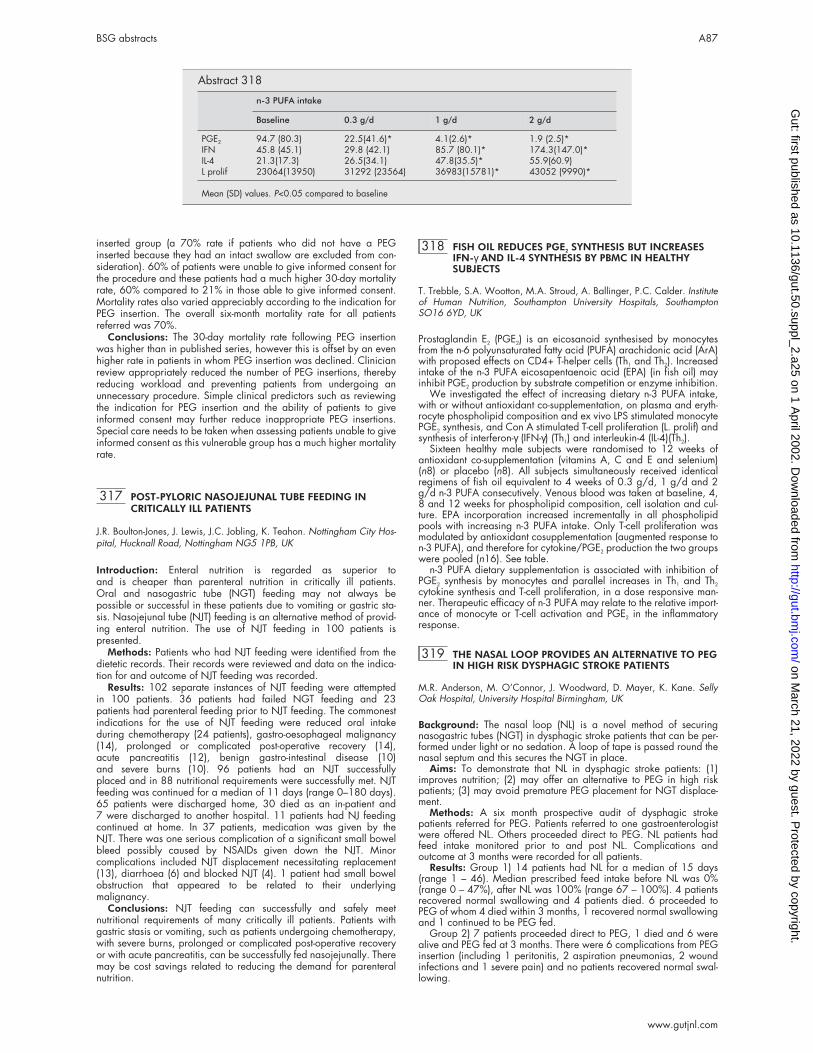
inserted group (a 70% rate if patients who did not have a PEGinserted because they had an intact swallow are excluded from con-sideration). 60% of patients were unable to give informed consent forthe procedure and these patients had a much higher 30-day mortalityrate, 60% compared to 21% in those able to give informed consent.Mortality rates also varied appreciably according to the indication forPEG insertion. The overall six-month mortality rate for all patientsreferred was 70%.
Conclusions: The 30-day mortality rate following PEG insertionwas higher than in published series, however this is offset by an evenhigher rate in patients in whom PEG insertion was declined. Clinicianreview appropriately reduced the number of PEG insertions, therebyreducing workload and preventing patients from undergoing anunnecessary procedure. Simple clinical predictors such as reviewingthe indication for PEG insertion and the ability of patients to giveinformed consent may further reduce inappropriate PEG insertions.Special care needs to be taken when assessing patients unable to giveinformed consent as this vulnerable group has a much higher mortalityrate.
317 POST-PYLORIC NASOJEJUNAL TUBE FEEDING INCRITICALLY ILL PATIENTS
J.R. Boulton-Jones, J. Lewis, J.C. Jobling, K. Teahon. Nottingham City Hos-pital, Hucknall Road, Nottingham NG5 1PB, UK
Introduction: Enteral nutrition is regarded as superior toand is cheaper than parenteral nutrition in critically ill patients.Oral and nasogastric tube (NGT) feeding may not always bepossible or successful in these patients due to vomiting or gastric sta-sis. Nasojejunal tube (NJT) feeding is an alternative method of provid-ing enteral nutrition. The use of NJT feeding in 100 patients ispresented.
Methods: Patients who had NJT feeding were identified from thedietetic records. Their records were reviewed and data on the indica-tion for and outcome of NJT feeding was recorded.
Results: 102 separate instances of NJT feeding were attemptedin 100 patients. 36 patients had failed NGT feeding and 23patients had parenteral feeding prior to NJT feeding. The commonestindications for the use of NJT feeding were reduced oral intakeduring chemotherapy (24 patients), gastro-oesophageal malignancy(14), prolonged or complicated post-operative recovery (14),acute pancreatitis (12), benign gastro-intestinal disease (10)and severe burns (10). 96 patients had an NJT successfullyplaced and in 88 nutritional requirements were successfully met. NJTfeeding was continued for a median of 11 days (range 0–180 days).65 patients were discharged home, 30 died as an in-patient and7 were discharged to another hospital. 11 patients had NJ feedingcontinued at home. In 37 patients, medication was given by theNJT. There was one serious complication of a significant small bowelbleed possibly caused by NSAIDs given down the NJT. Minorcomplications included NJT displacement necessitating replacement(13), diarrhoea (6) and blocked NJT (4). 1 patient had small bowelobstruction that appeared to be related to their underlyingmalignancy.
Conclusions: NJT feeding can successfully and safely meetnutritional requirements of many critically ill patients. Patients withgastric stasis or vomiting, such as patients undergoing chemotherapy,with severe burns, prolonged or complicated post-operative recoveryor with acute pancreatitis, can be successfully fed nasojejunally. Theremay be cost savings related to reducing the demand for parenteralnutrition.
318 FISH OIL REDUCES PGE2 SYNTHESIS BUT INCREASESIFN-γ AND IL-4 SYNTHESIS BY PBMC IN HEALTHYSUBJECTS
T. Trebble, S.A. Wootton, M.A. Stroud, A. Ballinger, P.C. Calder. Instituteof Human Nutrition, Southampton University Hospitals, SouthamptonSO16 6YD, UK
Prostaglandin E2 (PGE2) is an eicosanoid synthesised by monocytesfrom the n-6 polyunsaturated fatty acid (PUFA) arachidonic acid (ArA)with proposed effects on CD4+ T-helper cells (Th1 and Th2). Increasedintake of the n-3 PUFA eicosapentaenoic acid (EPA) (in fish oil) mayinhibit PGE2 production by substrate competition or enzyme inhibition.
We investigated the effect of increasing dietary n-3 PUFA intake,with or without antioxidant co-supplementation, on plasma and eryth-rocyte phospholipid composition and ex vivo LPS stimulated monocytePGE2 synthesis, and Con A stimulated T-cell proliferation (L. prolif) andsynthesis of interferon-γ (IFN-γ) (Th1) and interleukin-4 (IL-4)(Th2).
Sixteen healthy male subjects were randomised to 12 weeks ofantioxidant co-supplementation (vitamins A, C and E and selenium)(n8) or placebo (n8). All subjects simultaneously received identicalregimens of fish oil equivalent to 4 weeks of 0.3 g/d, 1 g/d and 2g/d n-3 PUFA consecutively. Venous blood was taken at baseline, 4,8 and 12 weeks for phospholipid composition, cell isolation and cul-ture. EPA incorporation increased incrementally in all phospholipidpools with increasing n-3 PUFA intake. Only T-cell proliferation wasmodulated by antioxidant cosupplementation (augmented response ton-3 PUFA), and therefore for cytokine/PGE2 production the two groupswere pooled (n16). See table.
n-3 PUFA dietary supplementation is associated with inhibition ofPGE2 synthesis by monocytes and parallel increases in Th1 and Th2
cytokine synthesis and T-cell proliferation, in a dose responsive man-ner. Therapeutic efficacy of n-3 PUFA may relate to the relative import-ance of monocyte or T-cell activation and PGE2 in the inflammatoryresponse.
319 THE NASAL LOOP PROVIDES AN ALTERNATIVE TO PEGIN HIGH RISK DYSPHAGIC STROKE PATIENTS
M.R. Anderson, M. O’Connor, J. Woodward, D. Mayer, K. Kane. SellyOak Hospital, University Hospital Birmingham, UK
Background: The nasal loop (NL) is a novel method of securingnasogastric tubes (NGT) in dysphagic stroke patients that can be per-formed under light or no sedation. A loop of tape is passed round thenasal septum and this secures the NGT in place.
Aims: To demonstrate that NL in dysphagic stroke patients: (1)improves nutrition; (2) may offer an alternative to PEG in high riskpatients; (3) may avoid premature PEG placement for NGT displace-ment.
Methods: A six month prospective audit of dysphagic strokepatients referred for PEG. Patients referred to one gastroenterologistwere offered NL. Others proceeded direct to PEG. NL patients hadfeed intake monitored prior to and post NL. Complications andoutcome at 3 months were recorded for all patients.
Results: Group 1) 14 patients had NL for a median of 15 days(range 1 – 46). Median prescribed feed intake before NL was 0%(range 0 – 47%), after NL was 100% (range 67 – 100%). 4 patientsrecovered normal swallowing and 4 patients died. 6 proceeded toPEG of whom 4 died within 3 months, 1 recovered normal swallowingand 1 continued to be PEG fed.
Group 2) 7 patients proceeded direct to PEG, 1 died and 6 werealive and PEG fed at 3 months. There were 6 complications from PEGinsertion (including 1 peritonitis, 2 aspiration pneumonias, 2 woundinfections and 1 severe pain) and no patients recovered normal swal-lowing.
Abstract 318
n-3 PUFA intake
Baseline 0.3 g/d 1 g/d 2 g/d
PGE2 94.7 (80.3) 22.5(41.6)* 4.1(2.6)* 1.9 (2.5)*IFN 45.8 (45.1) 29.8 (42.1) 85.7 (80.1)* 174.3(147.0)*IL-4 21.3(17.3) 26.5(34.1) 47.8(35.5)* 55.9(60.9)L prolif 23064(13950) 31292 (23564) 36983(15781)* 43052 (9990)*
Mean (SD) values. P<0.05 compared to baseline
BSG abstracts A87
www.gutjnl.com
on March 21, 2022 by guest. P
rotected by copyright.http://gut.bm
j.com/
Gut: first published as 10.1136/gut.50.suppl_2.a25 on 1 A
pril 2002. Dow
nloaded from
Conclusions: NL improves nutritional intake and allows somepatients to recover swallow without PEG. NL may avoid PEG interven-tion in patients who have a poor prognosis. Mortality was higher inNL patients which may be due to referral bias.
320 THE SHORT TERM EFFECTS OF TOTAL PARENTERALNUTRITION (TPN) ON FATIGUE: A DOUBLE BLINDPLACEBO CONTROLLED STUDY
S. Chattophadhyay1, P.M. Irving1, A. Cunliffe1, C. Archer1, D. Murphy2, F.King2, K. Durman3, J. Powell-Tuck1. 1Human Nutrition, St Bartholomews andRoyal London School of Medicine and Dentistry, 2Pharmacy, 3Dietetics,Barts and the London NHS Trust, London, UK
Aim: We wanted to know if the feeling that some patients report offeeling better soon after starting TPN was real or due to placebo.
Method: 10 patients (4F,6M), mean (range) age 51 (19–80),mean BMI (sd) 22.4(6.9), were selected. TPN was clinically indicatedin all and none had received food for at least 5 days. Patients wererandomised to receive either TPN or a dummy feed containinghypocaloric dextrose, water, sodium and potassium for the first 24hours. Central and peripheral fatigue were assessed using validatedmeasures: visual flicker fusion frequency (FFF), grip strength (GS),reaction time (RT) and a visual analogue scale (VAS). Both subjectsand observers were blind to the contents of the feed for the first 24hours. Measurements were taken before and 2,4,6 & 24 hours afterstarting the feed or placebo. Subsequently both groups received acomplete feed and further measurements were taken 24 hours later.Dummy bags had calculated Na, K and water requirements with vol-ume being made up by 5% dextrose. TPN contained calculatedrequirements of amino acids, Na, K, Ca, PO4, Mg, vitamins, trace ele-ments, energy and water.
Results: Between 2–6 hours the change in fatigue was significantlydifferent between the two groups for RT (p<0.000) and GS (p=0.02).Over the period 2–24 hours there were significant differencesbetween the two groups for RT (p=0.01) and GS (p<0.02). By 24 hthe differences between the two groups had diminished. At 48h theresults of the two groups were similar.
Conclusions: Within 6 hours of starting TPN, fatigue is improved.Water, Na, and K infusion, and placebo do not account for this effect
Radiology posters 321–325
321 ULTRASOUND ASSISTED PERCUTANEOUS LIVERBIOPSY
A. Jayaprakash1, M. Callaway2, J. Virjee2, R.E. Barry1. 1Department ofGastroenterology and 2Radiology, Bristol Royal Infirmary, Bristol, UK
Objective: To identify whether US makes a difference to the site ofliver biopsy compared to the traditional blind method.
Methods: Design: Prospective study. Setting: University teachingHospital. All patients who underwent liver biopsy between Nov 2000and Aug 2001 had the proposed biopsy site localised by traditionalpercussion by a trainee or consultant. The actual biopsy site was thendetermined using US by a single operator (trainee) in the same sitting.Then the biopsy was performed using the most appropriate site, awayfrom potential structures that can be encountered in the biopsy needlepath.
Results: All patients preferred to have US guided biopsy when theywere offered a choice between blind and US guided biopsy. 56 Liverbiopsies were performed. Of these, 50% (28) of the patients neededa change in biopsy site after US examination. The changes in thebiopsy site were, change in the angle of the needle in 4 patients, in thesame space but more anterior or posterior placement in 9 patients,one or more spaces below in 8 patients and in 7 patients biopsy sitemoved up by one space. The reasons for the change in site were,proximity to gall bladder (12 patients), lung (6), bile duct (5), kidney(1) and a vessel (1) and the better depth of the biopsy needle (5).There was no difference between consultant and trainee (p=0.77) inthe frequency with which the proposed biopsy site was changed afterUS examination.
Conclusions: US did make a difference in the liver biopsy site in50% of patients. There is no difference in the blind localisation site byconsultant and trainee. Adoption of US guided liver biopsy is prefer-able provided the resource is available.
322 OBSERVER VARIATION IN STAGING RECTAL CANCERSBY ENDORECTAL ULTRASOUND
P.J. Arumugam, B. Patel, A. Roberts, A. Watkins, P. Basnyat, R. Morgan,N.D. Carr, J. Beynon (introduced by Dr Shah). Departments of ColorectalSurgery and Radiology, Singleton Hospital, Swansea, Wales, UK
Aim: To determine the accuracy of endorectal ultrasound and the intraand inter observer variation in the preoperative staging of rectal can-cers between the specialities.
Methods: Rectal cancer patients undergoing primary surgery wereincluded. Histopathology was used as the gold standard. The observ-ers included two radiologists and two colorectal consultant surgeons.
Results: Endorectal ultrasound: Postoperatively looking at hardcopies surgeon 1 (S 1), ‘T’ staged 16 out of 31 cancers accuratelyKendall’s tau (K t 0.46) and surgeon 2 (S 2), ‘T’ staged 7 out of 31accurately (K t 0.34). Radiologist 1 (R 1), ‘T’ staged 14 out of 31 can-cers accurately (K t 0.242) and radiologist 2 (R 2), ‘T’ staged 15 outof 31 cancers (K t 0.302). (R 1) had an excellent intraobserver agree-ment in ‘T’ staging (K t 0.792) and (R 2) a perfect intraobserver agree-ment (K t 1) compared to their original preoperative staging. BetweenR1 and R 2 interobserver agreement was good (K t 0.681). Between(S1) and (S 2) interobserver agreement was moderate (K t 0.46) andbetween R1 and S 1 agreement was good (K t 0.53). The intra andinterobserver agreement for nodal ‘N’ staging were very similar to the‘T’ staging.
Conclusion: Endorectal ultrasound has been shown previously tobe the most accurate method of staging to assess local invasion in rec-tal cancer. This study does not confirm that observation and it may bedue to hard copies used rather than real time images and inflamma-tion around the tumour leading to incorrect staging. However wefound that the overall intra and inter observer agreement using hardcopies is good.
323 INITIAL EXPERIENCE WITH ENTERAL STENTS FORPALLIATION OF NON-OESOPHAGEAL PRIMARYMALIGNANT OBSTRUCTION OF THE UPPERGASTROINTESTINAL TRACT
S. Moreea, G. Naylor, C.L. Kay1, C.G. Beckett. Department of Gastroen-terology and 1Radiology, Bradford Royal Infirmary, Duckworth Lane Brad-ford BD9 6RJ, UK
Background: Expandable metal stents are increasingly used in thepalliation of malignant obstruction in the gastrointestinal (GI) tract. Itprovides a non-surgical means of relieving obstruction in patients athigh risk from surgical intervention.
Aims: To evaluate the technical success rate, complication rate andthe effectiveness of enteral stents in providing symptomatic relief ofupper GI obstruction during their initial use in our hospital.
Methods: The notes of all patients who had an enteral stent placedfor malignant obstruction of a non-oesophageal primary site in theupper GI tract were reviewed.
Results: Enteral stent placement was attempted in 14 patients (11male, 3 female, age 49–87, mean 70 years). 11/14 employed acombined endoscopic & radiological approach, 3 radiological alone,with 1 failure in each group. There were 5 cases of obstruction due totumour recurrence following surgery, 5 cases of unresectable gastriccarcinoma, 3 cases of unresectable pancreatic carcinoma and 1ampullary carcinoma. Flamingo stents (Boston Scientific Int.) wereused in 2 cases following Ivor Lewis oesophagogastrectomy. EnteralWall stents were used in the remainder of cases (Boston Scientific Int.).The technical success rate was 12/14 (86%). 1 patient required 2stents and one 3 stents. 3 patients required metal biliary stents. Therewere no immediate complications. There were no cases of stent migra-tion. 1 patient died unexpectedly within 24 hours of stent insertion.Symptom relief was obtained in the remaining 11 patients. 3 patientssurvived one week, all succumbing as inpatients to metastatic disease.8 patients were discharged from hospital. 6 patients survived between11 and 135 days (mean 35 days) and 2 are still alive at 2 and 4months. The 2 patients who could not be stented survived 11 and 21days.
Conclusions: This series demonstrates the effectiveness of enteralstenting in the palliation of malignant obstruction of the upper GI tract,enabling many patients to be supported out of hospital. Improvedpatient selection will optimise palliation. The preferred method ofplacement was a combined endoscopic/radiological approach.
A88 BSG abstracts
www.gutjnl.com
on March 21, 2022 by guest. P
rotected by copyright.http://gut.bm
j.com/
Gut: first published as 10.1136/gut.50.suppl_2.a25 on 1 A
pril 2002. Dow
nloaded from
324 THE CLINICAL USEFULNESS OF LIVER TRANSIT TIMESTUDY WITH CONTRAST ENHANCEDULTRASONOGRAPHY IN THE DIAGNOSIS OFCIRRHOSIS
C.W.Y. Lai, D. Cochlin, A.B. Hawthorne. Department of Radiology andGastroenterology, University Hospital of Wales, Heath Park, Cardiff, UK
Background: The transit time of an ultrasound contrast agent, Levovist(Schering AG, Berlin, Germany) between the antecubital and hepaticveins has been shown to be shorter in patients with cirrhosis comparedto those without cirrhosis. (Albrecht T et al. Lancet 1999; 353: 1579–83)
Aim: To assess the clinical usefulness of this technique indistinguishing between hepatitis and cirrhosis.
Methods: We studied 8 subjects with biopsy proven cirrhosis (cir-rhotic group), 8 with biopsy proven non-cirrhotic diffuse liver disease(hepatitic group) and 7 healthy controls. A standard abdominal ultra-sound study by a single radiologist (DC) was performed, looking at thehepatic echodensity, echotexture, spleen size, portal venous diameterand blood flow. A bolus injection of 2.5g Levovist at a concentrationof 300mg/ml was given in an antecubital vein. The transit time wasdefined as the time interval between the start of the injection of Levo-vist and a 10% rise in the signal intensity from baseline in apre-selected hepatic vein. The peak and gradient of the upslope of thesignal intensity curves were recorded.
Results: There was no difference in the echotexture and echoden-sity of the liver, the portal venous diameter and blood flow patternbetween the 3 groups. 4/8 cirrhotic subjects had splenomegaly andnone in the other 2 groups (p=0.011). The cirrhotic and hepatiticgroups had shorter transit time (mean=26.9 & 26.3 seconds, respec-tively) than controls (mean=36.7 seconds, p=0.012 & 0.006 respec-tively). There was no difference in transit time between the cirrhoticand hepatitic groups. The cirrhotic group had a shorter peak time(mean=34.8 seconds) than control (mean= 52 seconds, p=0.004)and hepatitic group (mean=48.4 seconds, p=0.034). However, therewas overlap in these parameters between all 3 groups.
Conclusions: The transit time of Levovist was shorter in the cirrhoticand hepatitic subjects than normal controls. The cirrhotic group hadan earlier peak time than control and hepatitic group. However, thepresence of some overlap in these parameters between the 3 groupslimited the clinical usefulness of this technique.
325 USE OF A NOVEL TECHNIQUE FOR AUTOMATEDPOLYP DETECTION IN CT COLONOGRAPHY
R.J.T. Sadleir1, A.C. Moss2, P.F. Whelan1, H.M. Fenlon3, P. MacMathuna2
(introduced by J.R. Lennon2). 1Vision Systems Laboratory, School ofElectronic Engineering, Dublin City University, Dublin 9, Ireland; 2Gastro-intestinal Unit and 3Department of Radiology, Mater Misericordiae Hospi-tal, Eccles Street, Dublin 7, Ireland
Purpose: CT Colonography (CTC), also known as “VirtualColonoscopy” is an emerging colon-imaging technique. Conventionalanalysis of raw CTC datasets by radiologists is time-consuming. Wepresent initial results of a study using novel image analysis software toenhance current techniques and “flag” potential colorectal neoplasia.
Methods and Materials: CTC datasets were obtained by abdomi-nal CT scanning according to established protocols. The raw datasetswere downloaded to a standard PC workstation and analysed usingour novel software. The first phase of the analysis process involvedextraction, or segmentation of a model of the colon lumen from thedata set and calculation the centreline for this model. The centrelinewas subsequently used for automating intraluminal navigation. Thesecond phase of the analysis process involved flagging potentialcolonic lesions, locating anomalies projecting from the colonicmucosa and flagging them as potential lesions based on their size andmorphology. The analysis results were viewed using several visualisa-tion techniques, including 2D, standard 3D and “virtual reality”stereo.
Results: We have used 5 CT datasets to date for analysis. Thistechnique takes a mean time of 57 seconds (range 48 – 72) to ana-lyse the raw datasets for review by an experienced radiologist andreduces review time by up to 50%. Although still in the early stages ofdevelopment, the automated-flagging algorithm can detect polyps assmall as 5mm and can cater for several different morphologies.
Conclusion: We have developed a technique for analysing CTCdatasets that is rapid, accessible, and comparably inexpensive. Vali-dation of this software is currently taking place. This techniqueincreases the potential uses of CTC in screening individuals forcolorectal neoplasia, by reducing viewing-time needed to detect theselesions.
Small bowel posters 326–335
326 INCIDENCE OF COELIAC DISEASE CONTINUES TOINCREASE IN SOUTH GLAMORGAN
A. Beale, R. Crimmins, G.L. Swift, P.M. Smith. Department of Gastroenter-ology, Llandough Hospital, Cardiff, UK
Introduction: The Coeliac Society is reporting increasing numbers ofnew members each year, a large proportion of whom are adults atdiagnosis. It is unsure whether this is simply due to greater exposureof the ‘coeliac iceberg’ or reflects a genuine increase in disease.
Aims: We assessed all new cases of coeliac disease (CD)diagnosed in South Glamorgan between 1996 and 2000, identifiedtheir characteristics and presenting features and compared these tofigures obtained between 1981 and 1995 in the same area.
Method: Data was obtained from clinical, pathology, dietetic andGP records in the area. Ethical approval was given Bro Taf LREC.
Results: In our population area of approximately 420 000, 125new cases of coeliac disease were diagnosed between 1996 and2000. Of these, 112 were adults (90%) and 13 were children atdiagnosis. Whilst the number of children has remained stable therehas been a significant increase in the number of adults compared tothose in the previous 3 quinquennia (see table). There was a male:fe-male ratio of 1:2. The mean age at diagnosis in adults was 53yrs,with 68 (61%) of the adult patients aged over 50 and 20 (18%) agedover 70. 30 (24%) of all patients had a known affected first degreerelative. The single most common presenting complaint was anaemia(50%) and 23 (20%) of the adults had osteoporosis at diagnosis. 14(11%) had another autoimmune disorder.
Conclusion: There has been a large increase in new cases of coe-liac disease in South Glamorgan over the last quinquennia. Thisappears to be due to the large number of new adult patients includingmany aged over 70 at diagnosis. It is vital that all clinicians haveincreased vigilance for this condition to try to reduce possible associ-ated morbidity.
327 THE HLA ASSOCIATION OF COELIAC DISEASE: ANINVESTIGATION OF UK PATIENTS WITHOUT THECOMMON DISEASE ASCOCIATED DQ2 GENOTYPE
S.J. Moodie, J.S. Fraser, A.L. King, E. Kondeatis, H.J. Ellis, P.J. Ciclitira.Gastroenterology, St Thomas Hospital, Kings College, London, UK
Coeliac disease is strongly associated with the HLA DQ2(DQA1*0501 DQB1*0201) heterodimer encoded in cis on aDRB1*03 haplotype or in trans on DRB1*07 and DRB1*11haplotypes. DQ2 negative coeliacs tend to have the diseaseassociated DRB1*04 DQA1*0301 DQB1*0302 (DQ8) haplotype.DQ8 ancestral haplotypes differ at the DRB1*04 locus and in insulindependant diabetes mellitus DRB1*04 subtypes have been shown todetermine DQ8 associated risk in different populations. We aimed toaddress this issue in UK coeliacs, and also to describe HLA types of therare DQ2 and DQ8 negative coeliacs.
Methods: 283 UK coeliacs were typed for the presence of the DQ2heterodimer in cis or trans by PCR-SSP. All those found to be DQ2negative were typed at a higher resolution for HLA class II alleles, withDRB1*04 subtyping of DQ8 haplotypes and HLA A, B, C typing of allDQ2 and DQ8 negative patients.
Results: 263/283 (93%) were DQ2 positive (252 in cis, 11 intrans), 20/283 (7%) were DQ2 negative. 15/20 of these wereDRB1*04 DQ8 positive. 40% of DQ8 haplotypes in DQ2 negativecoeliacs carried the DRB1*0404 allele, 30% the 0401 allele, 15%each 0402 and 0404 respectively. Of the 5 DQ2 and DR4 DQ8negative coeliacs, 4 carried one half of the DQA1*0501,
Abstract 326
1981–85 1986–90 1991–95 1996–2000
Adults 21 35 50 112Children 8 10 9 13
BSG abstracts A89
www.gutjnl.com
on March 21, 2022 by guest. P
rotected by copyright.http://gut.bm
j.com/
Gut: first published as 10.1136/gut.50.suppl_2.a25 on 1 A
pril 2002. Dow
nloaded from
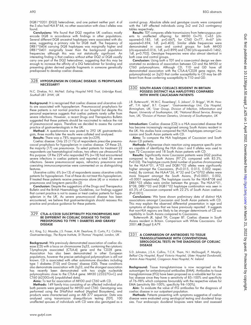
DQB1*0201 (DQ2) heterodimer, and one patient neither part. 4 ofthe 5 also had HLA B*44, no other association with class I alleles wasfound.
Conclusions: We found that DQ2 negative UK coeliacs mostlyencode DQ8 in accordance with findings in other populations.Several different DQ8 ancestral haplotypes were associated with dis-ease, suggesting a primary role for DQ8 itself. The frequency ofDRB1*0404 carrying DQ8 haplotypes was marginally higher andDRB1*0401 marginally lower than the background populationfrequencies although this was not statistically significant. Aninteresting finding is that coeliacs without either DQ2 or DQ8 usuallycarry one part of the DQ2 heterodimer, suggesting that this may beenough to increase the affinity of a DQ heterodimer for binding andpresenting gluten derived peptide fragments in someone geneticallypredisposed to develop coeliac disease.
328 HYPOSPLENISM IN COELIAC DISEASE: IS PROPHYLAXISNECESSARY?
N.C. Direkze, N.I. McNeil. Ealing Hospital NHS Trust, Uxbridge Road,Southall UB1 3HW, UK
Background: It is recognised that coeliac disease and ulcerative coli-tis are associated with hyposplenism. Pneumococcal prophylaxis forthese patients is not normal practice at our hospital but despite thispersonal experience suggests that coeliac patients rarely suffer fromsevere infections. However, a recent Drugs and Therapeutics Bulletinsuggested that these patients should be vaccinated to reduce the riskof pneumococcal sepsis. Therefore we decided to assess the currentpractice of gastroenterologists in the UK.
Method: A questionnaire was posted to 292 UK gastroenterolo-gists, three months later the results were collated and analysed.
Results: There was a 78% response rate (n=229).Coeliac disease: Only 25 (11%) of respondents prescribe pneumo-
coccal prophylaxis for hyposplenism in coeliac disease. Of these 25,the majority (17) use pneumovax. To select patients for treatment 22respondents use haematological indices, two others use ultrasound forthis purpose. Of the 229 who responded 9% (n=18) had encounteredsevere infections in coeliac patients and reported a total 36 severeinfections. Severe pneumococcal sepsis, refractory pneumonia andcoexisting immunocompromise were commented on as distinguishingfeatures.
Ulcerative colitis: 6% (n=13) of respondents assess ulcerative colitispatients for hyposplenism. Five of these do not treat this hyposplenism.If treated these patients receive pneumovax alone (4) or a mixture ofpneumovax and long-term penicillin (2)
Conclusion: Despite the suggestions of the Drugs and TherapeuticsBulletin and the British Haematology Guidelines, our findings suggestthat current practice is not to assess or treat coeliac patients for hypo-splenism in the UK. As severe pneumococcal disease has beenencountered, we believe that gastroenterologists should reassess thispractice and produce guidance for these patients.
329 CTLA-4/CD28 SUSCEPTIBILITY POLYMORPHISMS MAYBE DIFFERENT IN COELIAC DISEASE TO THOSEPREDISPOSING TO TYPE 1 DIABETES AND GRAVES’DISEASE
A.L. King, S.J. Moodie, J.S. Fraser, A.M. Dearlove, D. Curtis, P.J. Ciclitira.Gastroenterology, The Rayne Institute, St Thomas’ Hospital, London, UK
Background: We previously demonstrated association of coeliac dis-ease (CD) with a locus on chromosome 2q33, containing the cytotoxicT-lymphocyte associated (CTLA-4) gene and the CD28 gene.Association has also been demonstrated in other Europeanpopulations, however the precise aetiological polymorphism is still notknown. CD is associated with other autoimmune disorders includingtype 1 diabetes (T1D) and Graves’ disease (GD). These conditionsalso demonstrate association with 2q33, and the strongest associationhas recently been demonstrated with two single nucleotidepolymorphisms close to the CTLA-4 gene: MH30 (-23327G>C) andCT60 (6230G>A) (unpublished data).
Aims: To test for association of MH30 and CT60 with CD.Methods: 149 family trios consisting of an affected individual plus
both parents were genotyped for MH30 and CT60. Genotyping wasperformed using the SNaPshot method (Applied Biosystems), andproducts were characterised using an ABI 377 sequencer. Data wasanalysed using transmission disequilibrium testing (TDT). 100unaffected spouses of individuals with CD were also genotyped as a
control group. Absolute allele and genotype counts were comparedwith the 149 affected individuals using 2x2 and 2x3 contingencytables respectively.
Results: TDT compares allele transmissions from heterozygous par-ents to unaffected offspring: for MH30 G=70, C=65 (chi-squared=0.185, 1df, p=0.667), for CT60 G=77, A=67 (chi-squared=0.694, 1df, p=0.405). Similar allele frequencies weredemonstrated in case and control groups for both MH30(chi-squared=0.016, 1df, p=0.899) and CT60 (chi-squared=0.1462,1df, p=0.702). Genotype frequencies were also almost identical inboth case and control groups.
Conclusion: Using both a TDT and a case-control design we dem-onstrated no evidence of association between CD and the MH30 orCT60 polymorphisms. Although CD, T1D and GD have alldemonstrated association with the CTLA-4/CD28 gene region, thepolymorphism(s) on 2q33 that confer susceptibility to CD may be dif-ferent from those conferring susceptibility to T1D and GD.
330 SOUTH ASIAN COELIACS RESIDENT IN BRITAINPOSSESS DISTINCT HLA HAPLOTYPES COMPAREDWITH WHITE CAUCASIAN PATIENTS
J.R. Butterworth1, W.M.C. Rosenberg2, S. Jobson3, D. Briggs3, W.M. How-ell4, T.H. Iqbal1, B.T. Cooper1. 1Gastroenterology Unit, City Hospital,Birmingham, UK; 2Liver Group, Division of Infection, Inflammation andRepair, University of Southampton, UK; 3National Blood Service, Birming-ham, UK; 4Division of Human Genetics, University of Southampton, UK
Introduction: Coeliac disease (CD) is a HLA associated disease thathas become increasingly recognised among South Asians resident inthe UK. No studies have compared the HLA haplotypes amongst Cau-casian and South Asian patients with CD.
Aims: To compare the HLA haplotypes of Caucasian and SouthAsian CD patients.
Methods: Polymerase chain reaction using sequence specific prim-ers capable of identifying the HLA class I and II alleles was used totype 72 Caucasian and 18 South Asian patients with CD.
Results: Significantly more Caucasian patients were DQ2 positivecompared to the South Asians (97.2% compared with 83.3%,P=0.02). The haplotype counts (total number of positive chromosomes)for the HLA-A*01, A*03 and DQB1*02 alleles were significantlyhigher amongst the Caucasians, (P=0.015, 0.015, 0.002 respec-tively). By contrast, the HLA-A*26, A*32 and Cw*0702 alleles weremore frequent amongst the South Asians, (P<0.0001, 0.002,<0.0001 respectively). The haplotype counts for the HLA-B*08 andDRB1*03 alleles were similar between the groups. The HLA-A*01,B*08, DRB1*03 and DQB1*02 haplotype combination was seen in65.3% of Caucasian compared with 22.2% of South Asian coeliacs(P=0.001).
Conclusions: We have shown preliminary data of distinct HLAassociations amongst Caucasian and South Asian patients with CD.This may explain the observed differential presentation in age andsymptoms at diagnosis that we have previously reported1. It suggeststhat non-HLA regions are likely to be stronger determinants of CD sus-ceptibility in South Asians compared to Caucasians.
1Butterworth JR, Iqbal TH, Cooper BT. Coeliac disease in SouthAsians resident in Britain. Comparison with White Caucasians. Gut2001;48 (Suppl I):A79.
331 A COMPARISON OF ANTIBODIES TO TISSUETRANSGLUTAMINASE WITH CONVENTIONALSEROLOGICAL TESTS IN THE DIAGNOSIS OF COELIACDISEASE
S.D. Johnston, J.S.A. Collins, T.C.K. Tham, N.I. McDougall, P. Murphy.Belfast City Hospital, Royal Victoria Hospital, Ulster Hospital Dundonald,Antrim Area Hospital, Craigavon Area Hospital, N. Ireland
Background: Tissue transglutaminase is now recognised as theautoantigen for antiendomysial antibodies (EMA). Antibodies to tissuetransglutaminase (tTG) have been proposed as a valuable test for coe-liac disease since they have a sensitivity of 85–100% and specificityof 76–98% which compares favourably with the respective values forEMA (sensitivity 86–100%; specificity 94–100%).
Aim: To evaluate the value of tTG antibodies for the diagnosis ofcoeliac disease in our outpatient population.
Methods: Patients presenting with symptoms suggestive of coeliacdisease were evaluated using serological testing and duodenal biop-sies. Four endoscopic duodenal biopsies were taken and assessed
A90 BSG abstracts
www.gutjnl.com
on March 21, 2022 by guest. P
rotected by copyright.http://gut.bm
j.com/
Gut: first published as 10.1136/gut.50.suppl_2.a25 on 1 A
pril 2002. Dow
nloaded from
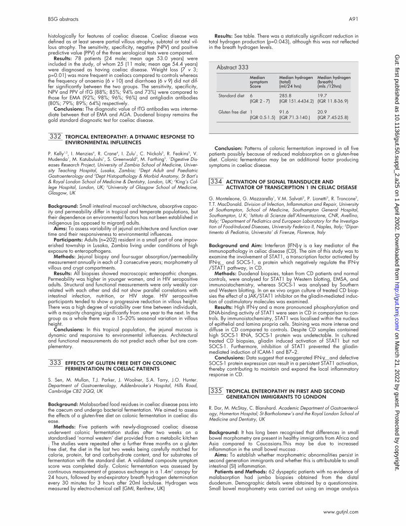
histologically for features of coeliac disease. Coeliac disease wasdefined as at least severe partial villous atrophy, subtotal or total vil-lous atrophy. The sensitivity, specificity, negative (NPV) and positivepredictive value (PPV) of the three serological tests were compared.
Results: 78 patients (24 male; mean age 53.0 years) wereincluded in the study, of whom 25 (11 male; mean age 54.4 years)were diagnosed as having coeliac disease. Weight loss (7 v 3;p=0.01) was more frequent in coeliacs compared to controls whereasthe frequency of anaemia (6 v 10) and diarrhoea (6 v 9) did not dif-fer significantly between the two groups. The sensitivity, specificity,NPV and PPV of tTG (88%; 85%; 94% and 73%) were compared tothose for EMA (92%; 98%; 96%; 96%) and antigliadin antibodies(80%; 79%; 89%; 64%) respectively.
Conclusions: The diagnostic value of tTG antibodies was interme-diate between that of EMA and AGA. Duodenal biopsy remains thegold standard diagnostic test for coeliac disease.
332 TROPICAL ENTEROPATHY: A DYNAMIC RESPONSE TOENVIRONMENTAL INFLUENCES
P. Kelly1,2, I. Menzies4, R. Crane4, I. Zulu1, C. Nickols3, R. Feakins3, V.Mudenda1, M. Katubulushi1, S. Greenwald3, M. Farthing5. 1Digestive Dis-eases Research Project, University of Zambia School of Medicine, Univer-sity Teaching Hospital, Lusaka, Zambia; 2Dept Adult and PaediatricGastroenterology and 3Dept Histopathology & Morbid Anatomy, St Bart’s& Royal London School of Medicine & Dentistry, London, UK; 4King’s Col-lege Hospital, London, UK; 5University of Glasgow School of Medicine,Glasgow, UK
Background: Small intestinal mucosal architecture, absorptive capac-ity and permeability differ in tropical and temperate populations, buttheir dependence on environmental factors has not been established inindigenous (as opposed to migrant) adults.
Aims: To assess variability of jejunal architecture and function overtime and their responsiveness to environmental influences.
Participants: Adults (n=202) resident in a small part of one impov-erished township in Lusaka, Zambia living under conditions of highexposure to enteropathogens.
Methods: Jejunal biopsy and four-sugar absorption/permeabilitymeasurement annually in each of 3 consecutive years; morphometry ofvillous and crypt compartments.
Results: All biopsies showed macroscopic enteropathic changes.Permeability was higher in younger women, and in HIV seropositiveadults. Structural and functional measurements were only weakly cor-related with each other and did not show parallel correlations withintestinal infection, nutrition, or HIV stage. HIV seropositiveparticipants tended to show a progressive reduction in villous height.There was a high degree of variability over time between individuals,with a majority changing significantly from one year to the next. In thegroup as a whole there was a 15–20% seasonal variation in villousheight.
Conclusions: In this tropical population, the jejunal mucosa isdynamic and responsive to environmental influences. Architecturaland functional measurements do not predict each other but are com-plementary.
333 EFFECTS OF GLUTEN FREE DIET ON COLONICFERMENTATION IN COELIAC PATIENTS
S. Sen, M. Mullan, T.J. Parker, J. Woolner, S.A. Tarry, J.O. Hunter.Department of Gastroenterology, Addenbrooke’s Hospital, Hills Road,Cambridge CB2 2QQ, UK
Background: Malabsorbed food residues in coeliac disease pass intothe caecum and undergo bacterial fermentation. We aimed to assessthe effects of a gluten-free diet on colonic fermentation in coeliac dis-ease.
Methods: Five patients with newly-diagnosed coeliac diseaseunderwent colonic fermentation studies after two weeks on astandardised ‘normal western’ diet provided from a metabolic kitchen. The studies were repeated after a further three months on a glutenfree diet, the diet in the last two weeks being carefully matched forcalorie, protein, fat and carbohydrate content, and for substrates offermentation with the standard diet. A validated composite symptomscore was completed daily. Colonic fermentation was assessed bycontinuous measurement of gaseous exchange in a 1.4m3 canopy for24 hours, followed by end-expiratory breath hydrogen determinationevery 30 minutes for 3 hours after 20ml lactulose. Hydrogen wasmeasured by electro-chemical cell (GMI, Renfrew, UK)
Results: See table. There was a statistically significant reduction intotal hydrogen production (p=0.043), although this was not reflectedin the breath hydrogen levels.
Conclusion: Patterns of colonic fermentation improved in all fivepatients possibly because of reduced malabsorption on a gluten-freediet. Colonic fermentation may be an additional factor producingsymptoms in coeliac disease.
334 ACTIVATION OF SIGNAL TRANSDUCER ANDACTIVATOR OF TRANSCRIPTION 1 IN CELIAC DISEASE
G. Monteleone, G. Mazzarella1, V.M. Salvati2, P. Lionetti3, R. Troncone2,T.T. MacDonald. Division of Infection, Inflammation and Repair, Universityof Southampton, School of Medicine, Southampton General Hospital,Southampton, U K; 1Istituto di Scienze dell’Alimentazione, CNR, Avellino,Italy; 2Department of Pediatrics and European Laboratory for the Investiga-tion of Food-Induced Diseases, University Federico II, Naples, Italy; 3Dipar-timento di Pediatria, Universita’ di Firenze, Florence, Italy
Background and Aim: Interferon (IFN)-γ is a key mediator of theimmunopathology in celiac disease (CD). The aim of this study was toexamine the involvement of STAT1, a transcription factor activated byIFN-γ,_ and SOCS-1, a protein which negatively regulate the IFN-γ/STAT1 pathway, in CD.
Methods: Duodenal biopsies, taken from CD patients and normalcontrols, were analysed for STAT1 by Western blotting, EMSA, andimmunoistochemistry, whereas SOCS-1 was analysed by Southernand Western blotting. In an ex vivo organ culture of treated CD biop-sies the effect of a JAK/STAT1 inhibitor on the gliadin-mediated induc-tion of costimulatory molecules was examined.
Results: High IFN-γ and a more pronounced phosphorylation andDNA-binding activity of STAT1 were seen in CD in comparison to con-trols. By immunoistochemistry, STAT1 was localised within the nucleusof epithelial and lamina propria cells. Staining was more intense anddiffuse in CD compared to controls. Despite CD samples containedhigh SOCS-1 RNA, SOCS-1 protein was undetectable. In culturedtreated CD biopsies, gliadin induced activation of STAT1 but notSOCS-1. Furthermore, inhibition of STAT1 prevented the gliadin-mediated induction of ICAM-1 and B7–2.
Conclusions: Data suggest that exaggerated IFN-γ__and defectiveSOCS-1 protein expression can result in a persistent STAT1 activation,thereby contributing to maintain and expand the local inflammatoryresponse in CD.
335 TROPICAL ENTEROPATHY IN FIRST AND SECONDGENERATION IMMIGRANTS TO LONDON
R. Dor, M. McStay, C. Blanshard. Academic Department of Gastroenterol-ogy, Homerton Hospital; St Bartholomew’s and the Royal London School ofMedicine and Dentistry, UK
Background: It has long been recognised that differences in smallbowel morphometry are present in healthy immigrants from Africa andAsia compared to Caucasians.This may be due to increasedinflammation in the small bowel mucosa .
Aims: To establish whether morphometric abnormalities persist insecond generation immigrants and whether this is attributable to smallintestinal (SI) inflammation.
Patients and Methods: 62 dyspeptic patients with no evidence ofmalabsorption had jumbo biopsies obtained from the distalduodenum. Demographic details were obtained by a questionnaire.Small bowel morphometry was carried out using an image analysis
Abstract 333
MediansymptomScore
Median hydrogen(total)(ml/24 hrs)
Median hydrogen(breath)(mls /12hrs)
Standard diet 6 285.8 19.7(IQR 2 - 7) (IQR 151.4-434.2) (IQR 11.8-36.9)
Gluten free diet 1 91.6 20.9(IQR 0.5-1.5) (IQR 71.3-140.) (IQR 7.45-25.8)
BSG abstracts A91
www.gutjnl.com
on March 21, 2022 by guest. P
rotected by copyright.http://gut.bm
j.com/
Gut: first published as 10.1136/gut.50.suppl_2.a25 on 1 A
pril 2002. Dow
nloaded from
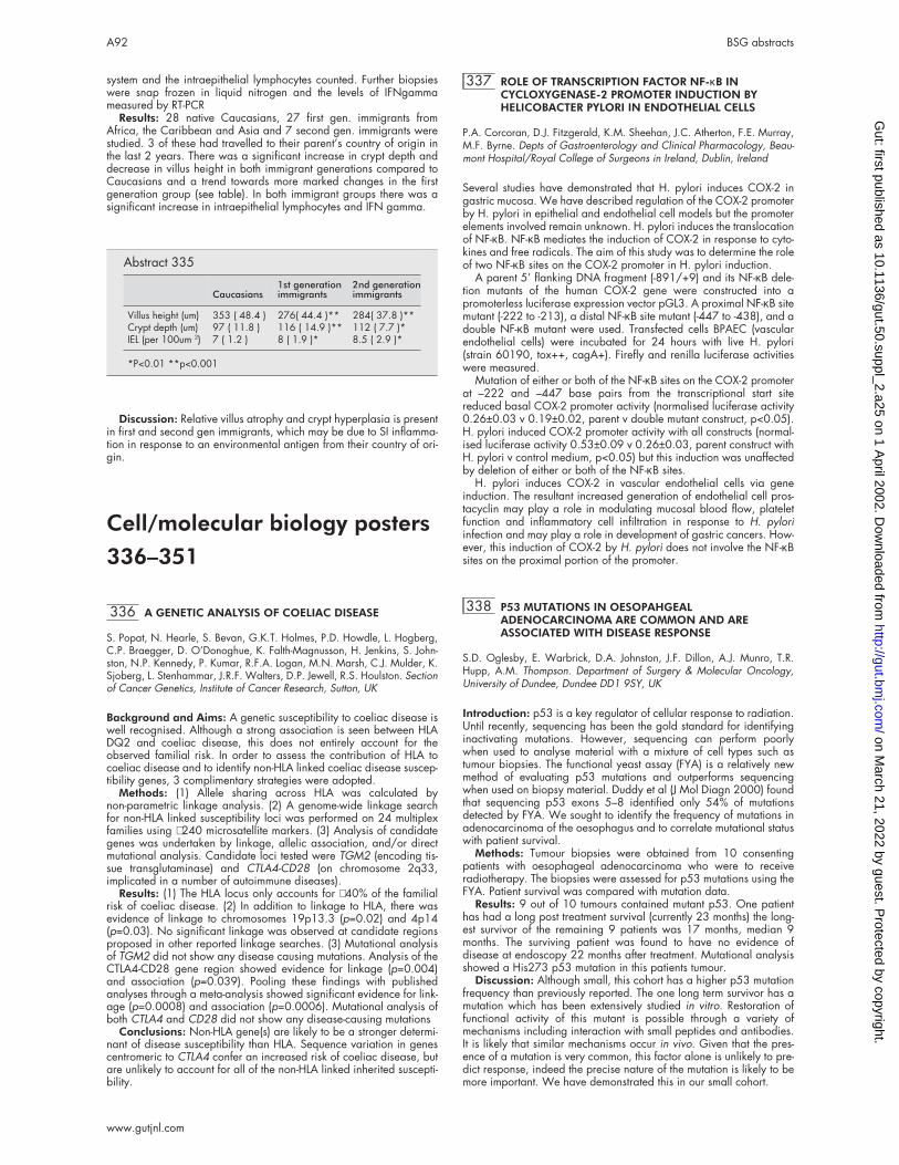
system and the intraepithelial lymphocytes counted. Further biopsieswere snap frozen in liquid nitrogen and the levels of IFNgammameasured by RT-PCR
Results: 28 native Caucasians, 27 first gen. immigrants fromAfrica, the Caribbean and Asia and 7 second gen. immigrants werestudied. 3 of these had travelled to their parent’s country of origin inthe last 2 years. There was a significant increase in crypt depth anddecrease in villus height in both immigrant generations compared toCaucasians and a trend towards more marked changes in the firstgeneration group (see table). In both immigrant groups there was asignificant increase in intraepithelial lymphocytes and IFN gamma.
Discussion: Relative villus atrophy and crypt hyperplasia is presentin first and second gen immigrants, which may be due to SI inflamma-tion in response to an environmental antigen from their country of ori-gin.
Cell/molecular biology posters
336–351
336 A GENETIC ANALYSIS OF COELIAC DISEASE
S. Popat, N. Hearle, S. Bevan, G.K.T. Holmes, P.D. Howdle, L. Hogberg,C.P. Braegger, D. O’Donoghue, K. Falth-Magnusson, H. Jenkins, S. John-ston, N.P. Kennedy, P. Kumar, R.F.A. Logan, M.N. Marsh, C.J. Mulder, K.Sjoberg, L. Stenhammar, J.R.F. Walters, D.P. Jewell, R.S. Houlston. Sectionof Cancer Genetics, Institute of Cancer Research, Sutton, UK
Background and Aims: A genetic susceptibility to coeliac disease iswell recognised. Although a strong association is seen between HLADQ2 and coeliac disease, this does not entirely account for theobserved familial risk. In order to assess the contribution of HLA tocoeliac disease and to identify non-HLA linked coeliac disease suscep-tibility genes, 3 complimentary strategies were adopted.
Methods: (1) Allele sharing across HLA was calculated bynon-parametric linkage analysis. (2) A genome-wide linkage searchfor non-HLA linked susceptibility loci was performed on 24 multiplexfamilies using ∼240 microsatellite markers. (3) Analysis of candidategenes was undertaken by linkage, allelic association, and/or directmutational analysis. Candidate loci tested were TGM2 (encoding tis-sue transglutaminase) and CTLA4-CD28 (on chromosome 2q33,implicated in a number of autoimmune diseases).
Results: (1) The HLA locus only accounts for ∼40% of the familialrisk of coeliac disease. (2) In addition to linkage to HLA, there wasevidence of linkage to chromosomes 19p13.3 (p=0.02) and 4p14(p=0.03). No significant linkage was observed at candidate regionsproposed in other reported linkage searches. (3) Mutational analysisof TGM2 did not show any disease causing mutations. Analysis of theCTLA4-CD28 gene region showed evidence for linkage (p=0.004)and association (p=0.039). Pooling these findings with publishedanalyses through a meta-analysis showed significant evidence for link-age (p=0.0008) and association (p=0.0006). Mutational analysis ofboth CTLA4 and CD28 did not show any disease-causing mutations
Conclusions: Non-HLA gene(s) are likely to be a stronger determi-nant of disease susceptibility than HLA. Sequence variation in genescentromeric to CTLA4 confer an increased risk of coeliac disease, butare unlikely to account for all of the non-HLA linked inherited suscepti-bility.
337 ROLE OF TRANSCRIPTION FACTOR NF-κB INCYCLOXYGENASE-2 PROMOTER INDUCTION BYHELICOBACTER PYLORI IN ENDOTHELIAL CELLS
P.A. Corcoran, D.J. Fitzgerald, K.M. Sheehan, J.C. Atherton, F.E. Murray,M.F. Byrne. Depts of Gastroenterology and Clinical Pharmacology, Beau-mont Hospital/Royal College of Surgeons in Ireland, Dublin, Ireland
Several studies have demonstrated that H. pylori induces COX-2 ingastric mucosa. We have described regulation of the COX-2 promoterby H. pylori in epithelial and endothelial cell models but the promoterelements involved remain unknown. H. pylori induces the translocationof NF-κB. NF-κB mediates the induction of COX-2 in response to cyto-kines and free radicals. The aim of this study was to determine the roleof two NF-κB sites on the COX-2 promoter in H. pylori induction.
A parent 5’ flanking DNA fragment (-891/+9) and its NF-κB dele-tion mutants of the human COX-2 gene were constructed into apromoterless luciferase expression vector pGL3. A proximal NF-κB sitemutant (-222 to -213), a distal NF-κB site mutant (-447 to -438), and adouble NF-κB mutant were used. Transfected cells BPAEC (vascularendothelial cells) were incubated for 24 hours with live H. pylori(strain 60190, tox++, cagA+). Firefly and renilla luciferase activitieswere measured.
Mutation of either or both of the NF-κB sites on the COX-2 promoterat –222 and –447 base pairs from the transcriptional start sitereduced basal COX-2 promoter activity (normalised luciferase activity0.26±0.03 v 0.19±0.02, parent v double mutant construct, p<0.05).H. pylori induced COX-2 promoter activity with all constructs (normal-ised luciferase activity 0.53±0.09 v 0.26±0.03, parent construct withH. pylori v control medium, p<0.05) but this induction was unaffectedby deletion of either or both of the NF-κB sites.
H. pylori induces COX-2 in vascular endothelial cells via geneinduction. The resultant increased generation of endothelial cell pros-tacyclin may play a role in modulating mucosal blood flow, plateletfunction and inflammatory cell infiltration in response to H. pyloriinfection and may play a role in development of gastric cancers. How-ever, this induction of COX-2 by H. pylori does not involve the NF-κBsites on the proximal portion of the promoter.
338 P53 MUTATIONS IN OESOPAHGEALADENOCARCINOMA ARE COMMON AND AREASSOCIATED WITH DISEASE RESPONSE
S.D. Oglesby, E. Warbrick, D.A. Johnston, J.F. Dillon, A.J. Munro, T.R.Hupp, A.M. Thompson. Department of Surgery & Molecular Oncology,University of Dundee, Dundee DD1 9SY, UK
Introduction: p53 is a key regulator of cellular response to radiation.Until recently, sequencing has been the gold standard for identifyinginactivating mutations. However, sequencing can perform poorlywhen used to analyse material with a mixture of cell types such astumour biopsies. The functional yeast assay (FYA) is a relatively newmethod of evaluating p53 mutations and outperforms sequencingwhen used on biopsy material. Duddy et al (J Mol Diagn 2000) foundthat sequencing p53 exons 5–8 identified only 54% of mutationsdetected by FYA. We sought to identify the frequency of mutations inadenocarcinoma of the oesophagus and to correlate mutational statuswith patient survival.
Methods: Tumour biopsies were obtained from 10 consentingpatients with oesophageal adenocarcinoma who were to receiveradiotherapy. The biopsies were assessed for p53 mutations using theFYA. Patient survival was compared with mutation data.
Results: 9 out of 10 tumours contained mutant p53. One patienthas had a long post treatment survival (currently 23 months) the long-est survivor of the remaining 9 patients was 17 months, median 9months. The surviving patient was found to have no evidence ofdisease at endoscopy 22 months after treatment. Mutational analysisshowed a His273 p53 mutation in this patients tumour.
Discussion: Although small, this cohort has a higher p53 mutationfrequency than previously reported. The one long term survivor has amutation which has been extensively studied in vitro. Restoration offunctional activity of this mutant is possible through a variety ofmechanisms including interaction with small peptides and antibodies.It is likely that similar mechanisms occur in vivo. Given that the pres-ence of a mutation is very common, this factor alone is unlikely to pre-dict response, indeed the precise nature of the mutation is likely to bemore important. We have demonstrated this in our small cohort.
Abstract 335
Caucasians1st generationimmigrants
2nd generationimmigrants
Villus height (um) 353 ( 48.4 ) 276( 44.4 )** 284( 37.8 )**Crypt depth (um) 97 ( 11.8 ) 116 ( 14.9 )** 112 ( 7.7 )*IEL (per 100um 2) 7 ( 1.2 ) 8 ( 1.9 )* 8.5 ( 2.9 )*
*P<0.01 **p<0.001
A92 BSG abstracts
www.gutjnl.com
on March 21, 2022 by guest. P
rotected by copyright.http://gut.bm
j.com/
Gut: first published as 10.1136/gut.50.suppl_2.a25 on 1 A
pril 2002. Dow
nloaded from
339 DOWNREGULATION OF α2β1 INTEGRIN PRECEDES THEINDUCTION OF APOPTOSIS BY BUTYRATE INCOLORECTAL CANCER CELL LINES
A. Buda, M.A. Jepson1, D. Martines2, M. Pignatelli (introduced by M. Pig-natelli). Department of Pathology and Microbiology, 1Department ofBiochemistry, University of Bristol, Bristol, UK; 2Department of Surgical andGastroenterological Sciences, University of Padua, Italy
Background: Integrins mediate cell-matrix adhesion and regulategrowth and cell survival. In colonic epithelial cells α2β1 integrincontrols glandular differentiation and proliferation. Butyrate stimulatesdifferentiation and induces apoptosis in vitro.
Aim: We investigate whether butyrate induction of apoptosis wasassociated with perturbation of integrin-mediated cell matrixadhesion.
Methods: Four colonic cancer cell lines (SW 1222, HT29,SW620, LS174T) were studied. Adhesion to extracellular matrix pro-teins was investigated by a cell-matrix adhesion assay. Expressionand cellular localization of α2β1 integrin were studied in adherent cellsafter treatment with 4 mmol/L butyrate by FACS analysis and confocalmcroscopy. Apoptosis was assessed by Annexin V binding. Aselective COX-2 inhibitor (NS-398) was also used as a control.
Results: Butyrate decreased the attachment to type I collagen inHT-29 (p=0.004) and SW620 (p=0.003) cells and type I (p=0.01)and IV (p=0.03) collagen in LS174T cells. The decreased cell attach-ment was associated with downregulation of α2β1 and increase inapoptosis in adherent cells (SW620 9.6±0.5 vs. 3.5±0.2, p=0.000;HT29 6.1±0.4 vs. 2.3±0.1, p=0.01; LS174T 9.3±0.6 vs. 4.3±0.3,p=0.003). No changes in α2β1 expression and matrix adhesion wereseen in SW1222 cells, which were also found less sensitive to thebutyrate-induction of apoptosis (2.4±0.2 vs. 3.3±0.4 p=0.123).Treatment with NS-398 increased apoptosis (5.9±0.08 vs.2.06±0.14, p=0.000) without affecting integrin expression and cellu-lar localization. Downregulation of α2β1 integrin occurred in viablecells and preceded the detection of apoptosis.
Conclusion: Cell detachment and apoptosis induced by butyrateare associated with downregulation of expression and functionalactivity of α2β1 integrin. Perturbation of cell matrix adhesion may be anovel mechanism by which butyrate induces apoptosis in colorectalcancer cells.
340 HIGH LEVELS OF MICROSATELLITE INSTABILITY(MSI-H) IN HYPERPLASTIC POLYPOSIS (HPP)ASSOCIATED COLORECTAL CANCER (CRC) PROVIDESEVIDENCE FOR THE SERRATED CRC PATHWAY
M. Lockett, W. Smale, J. Lloyd, K. Pack, S. Burke, I. Talbot, I. Tomlinson,W. Atkin. ICRF CRC Unit, St. Mark’s Hospital, Harrow, Middlesex, UK
Background: MSI-H occurs in 10–15% sporadic CRC. The precursoris unknown but may be the hyperplastic polyp (HP) (serrated CRCpathway). In HPP there are multiple, large HPs and an increased CRCrisk. If CRC in HPP showed MSI and the MSI-H phenotype (proximal,multiple, CRCs in females with mucinous, undifferentiated histologyand a prominent lymphocyte infiltrate), this would support the serratedCRC pathway.
Aim: To describe the phenotype and microsatellite (MS) status ofCRC in HPP to provide evidence for the serrated CRC pathway.
Methods: HPP patients were identified after a national call forcases and using the UK flexible sigmoidoscopy screening trialdatabase. Clinical and histological features were described. Paraffin-embedded tissue samples were microdissected and DNA wasextracted. MSI analysis was performed by PCR at Bat25, Bat 26,MycL1, D2S123, APC, D15S221 and D17S250 and compared togenomic or normal tissue DNA. MS status was defined as follows:MSI-H >3 unstable (>1 mononucleotide marker); MSI-Low (MSI-L) 1–2unstable; MS stable (MSS) 0 unstable. Tissue samples were analysedby immunohistochemistry for mismatch repair proteins hMLH1,hMSH2 and hMSH6.
Results: 42 HPP patients were identified. 32 CRCs occurred in 18patients (multiple CRCs in 6). 72% of the 18 CRC patients were male(median age 63 years male, 52 years female). None had a familyhistory of CRC. 69% of CRCs were proximal to the splenic flexure, dif-ferentiation was poor in 31%, moderate in 47% and well in 9%. 22%showed mucinous or signet ring cell histology. 19% had a Crohn’s-likeor conspicuous lymphocyte infiltrate. 6/22 (27%) CRCs were MSI-Hand were proximal and showed hMLH1 loss. 5 (23%) were MSI-L and50% MSS.
Conclusions: Both MSI and chromosomal instability pathways areimportant in HPP carcinogenesis. There is an increased prevalence of
MSI CRC in HPP. This suggests that the HP is the precursor of MSI CRCin HPP. This supports the theory that the HP may also be the precursorof sporadic MSI-H CRC.
341 PROGASTRIN STIMULATES MURINE COLONICEPITHELIAL MITOSIS AFTER DNA DAMAGE
P.D. Ottewell1, A.J.M. Watson1, T.C. Wang2, G.J. Dockray3, D.M. Pritch-ard1. Departments of 1Medicine and 3Physiology, University of Liverpool,UK; 2University of Massachusetts Medical Center, Worcester, MA, USA
Background and Aims: The non-amidated precursors of gastrinstimulate proliferation of colonic epithelial cells. Transgenic micewhich overexpress human progastrin (hGAS) are more susceptible tothe induction of colonic aberrant crypt foci and adenomas by thechemical carcinogen azoxymethane than wild type mice (FVB) andmice which overexpress amidated gastrin (INS-GAS). We have inves-tigated in murine intestinal epithelium in vivo whether alterations in theregulation of apoptosis or mitosis following DNA damage contributeto the procarcinogenic effects of progastrin.
Methods: Apoptosis and mitosis were assessed on a cell positionalbasis by light microscopic assessment of small intestinal and coloniccrypts from 10–12 week mice, following 1 or 8 Gy γ-irradiation. Miceanalysed were progastrin overexspressing (hGAS), gastrin overex-pressing (INS-GAS) and gastrin-knockout (GAS-KO) along with theirwild-type counterparts (FVB and C57BL/6). p21WAF1/CIP1 expression wasanalysed by immunohistochemistry and Western blotting using a rab-bit polyclonal primary antibody.
Results: 4.5h following 1Gy and 8Gy γ-radiation, apoptosis wasinduced to similar levels in the small intestinal and colonic crypts of allmice. Colonic mitosis was inhibited to almost undetectable levels by8Gy γ-radiation in wild-type, GAS-KO and INS-GAS mice. However,significant colonic mitosis persisted in hGAS mice 4.5h following 8Gyγ-radiation. hGAS mice also showed increased length of coloniccrypts compared to FVB and INS-GAS. γ-irradiation caused inductionof p21WAF1/CIP1 to similar levels in hGAS and FVB colonic epithelium.
Conclusions: (1) 4.5h after DNA damage by 8Gy γ-radiation,mice with elevated progastrin exhibit significantly higher levels ofcolonic mitosis than wild-type or gastrin overexpressing mice. (2) Pro-gastrin induced stimulation of mitosis following DNA damage mayaccount for its procarcinogenic properties.
342 NSAIDS INHIBIT β-OXIDATION OF PALMITATE ANDARACHIDONATE IN HCT-116 CELLS
M.J. Garle1, B. Middleton1, C.J. Hawkey 2. 1School of Biomedical Sciences,2Division of Gastroenterology, University Hospital Nottingham, UK
Introduction: Non-steroidal anti-inflammatory drugs (sulindac deriva-tives in particular) are useful for prevention of colon cancer. Earlierreports describe inhibition of palmitate β-oxidation by NSAIDS anddemonstrate that elevated cellular arachidonate levels causeapoptosis1.
Methods: We hypothesised that inhibition of β-oxidation ofarachidonate would lead to arachidonate accumulation and possiblycell death. So we determined effects of NSAIDS on palmitate and ara-chidonate β-oxidation and cell viability. HCT-116 cells were grown toconfluence on 24-well cluster plates and were incubated over 2 hoursin DPBS with 3H-palmitate or 3H-arachidonate (100µM, at 42 and 48dpm/pmole respectively) in the presence of defatted BSA (1.2mg/ml).β-Oxidation rates were measured by 3H-water production. NSAIDSincluding indomethacin, sulindac sulphoxide, sulindac sulphide andibuprofen (0–1000µM) and a palmitoyl carnitine transferase-1 inhibi-tor (etomoxir 10µM) were incubated in DPBS containing defattedalbumin for 45 min prior to 2hr incubation of chemicals with3H-palmitate or 3H-arachidonate. Effects of NSAIDS on cell death wasdetermined over 48 hr in serum free DMEM, using resazurin reductionas a viability marker. Rates of 3H-palmitate or 3H-arachidonateβ-oxidation were 152 +/- 5.0 and 21 +/- 2.0 pmoles/min/mgprotein respectively. Both activities were reduced by etomoxir (10µM)to less than 10% of control values.
Results and Conclusions: Effects of NSAIDS on β-oxidation andcell viability are shown in the table below. Ibuprofen caused an unex-pected 2-fold stimulation of 3H-arachidonate β-oxidation at concentra-tions up to 500mM. Results represent mean +/- sem of IC50 (µM),(n=4). See table.
1.Chan TA. et al (1998) PNAS (USA);95:681–686
BSG abstracts A93
www.gutjnl.com
on March 21, 2022 by guest. P
rotected by copyright.http://gut.bm
j.com/
Gut: first published as 10.1136/gut.50.suppl_2.a25 on 1 A
pril 2002. Dow
nloaded from
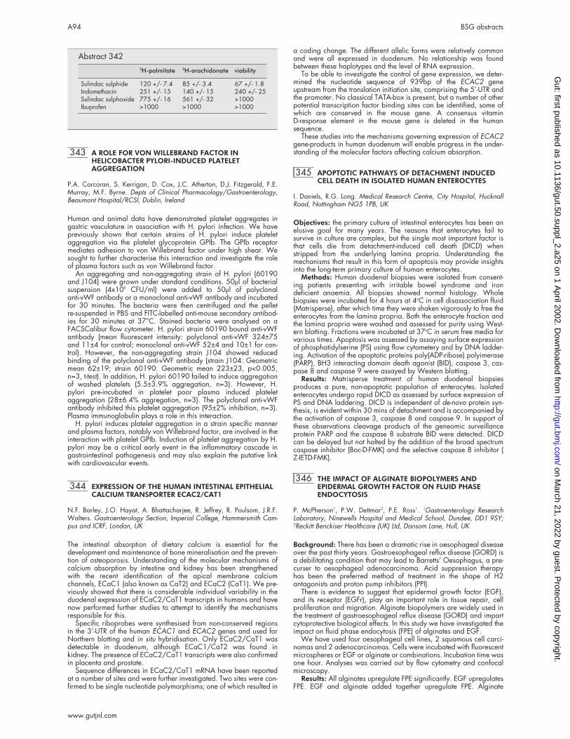
343 A ROLE FOR VON WILLEBRAND FACTOR INHELICOBACTER PYLORI-INDUCED PLATELETAGGREGATION
P.A. Corcoran, S. Kerrigan, D. Cox, J.C. Atherton, D.J. Fitzgerald, F.E.Murray, M.F. Byrne. Depts of Clinical Pharmacology/Gastroenterology,Beaumont Hospital/RCSI, Dublin, Ireland
Human and animal data have demonstrated platelet aggregates ingastric vasculature in association with H. pylori infection. We havepreviously shown that certain strains of H. pylori induce plateletaggregation via the platelet glycoprotein GPIb. The GPIb receptormediates adhesion to von Willebrand factor under high shear. Wesought to further characterise this interaction and investigate the roleof plasma factors such as von Willebrand factor.
An aggregating and non-aggregating strain of H. pylori (60190and J104) were grown under standard conditions. 50µl of bacterialsuspension (4x10⁹ CFU/ml) were added to 50µl of polyclonalanti-vWF antibody or a monoclonal anti-vWF antibody and incubatedfor 30 minutes. The bacteria were then centrifuged and the pelletre-suspended in PBS and FITC-labelled anti-mouse secondary antibod-ies for 30 minutes at 37°C. Stained bacteria were analysed on aFACSCalibur flow cytometer. H. pylori strain 60190 bound anti-vWFantibody (mean fluorescent intensity: polyclonal anti-vWF 324±75and 11±4 for control; monoclonal anti-vWF 52±4 and 10±1 for con-trol). However, the non-aggregating strain J104 showed reducedbinding of the polyclonal anti-vWF antibody (strain J104: Geometricmean 62±19; strain 60190: Geometric mean 223±23, p<0.005,n=3, t-test). In addition, H. pylori 60190 failed to induce aggregationof washed platelets (5.5±3.9% aggregation, n=3). However, H.pylori pre-incubated in platelet poor plasma induced plateletaggregation (28±6.4% aggregation, n=3). The polyclonal anti-vWFantibody inhibited this platelet aggregation (95±2% inhibition, n=3).Plasma immunoglobulin plays a role in this interaction.
H. pylori induces platelet aggregation in a strain specific mannerand plasma factors, notably von Willebrand factor, are involved in theinteraction with platelet GPIb. Induction of platelet aggregation by H.pylori may be a critical early event in the inflammatory cascade ingastrointestinal pathogenesis and may also explain the putative linkwith cardiovascular events.
344 EXPRESSION OF THE HUMAN INTESTINAL EPITHELIALCALCIUM TRANSPORTER ECAC2/CAT1
N.F. Barley, J.O. Hayat, A. Bhattacharjee, R. Jeffrey, R. Poulsom, J.R.F.Walters. Gastroenterology Section, Imperial College, Hammersmith Cam-pus and ICRF, London, UK
The intestinal absorption of dietary calcium is essential for thedevelopment and maintenance of bone mineralisation and the preven-tion of osteoporosis. Understanding of the molecular mechanisms ofcalcium absorption by intestine and kidney has been strengthenedwith the recent identification of the apical membrane calciumchannels, ECaC1 (also known as CaT2) and ECaC2 (CaT1). We pre-viously showed that there is considerable individual variability in theduodenal expression of ECaC2/CaT1 transcripts in humans and havenow performed further studies to attempt to identify the mechanismsresponsible for this.
Specific riboprobes were synthesised from non-conserved regionsin the 3’-UTR of the human ECAC1 and ECAC2 genes and used forNorthern blotting and in situ hybridisation. Only ECaC2/CaT1 wasdetectable in duodenum, although ECaC1/CaT2 was found inkidney. The presence of ECaC2/CaT1 transcripts were also confirmedin placenta and prostate.
Sequence differences in ECaC2/CaT1 mRNA have been reportedat a number of sites and were further investigated. Two sites were con-firmed to be single nucleotide polymorphisms; one of which resulted in
a coding change. The different allelic forms were relatively commonand were all expressed in duodenum. No relationship was foundbetween these haplotypes and the level of RNA expression.
To be able to investigate the control of gene expression, we deter-mined the nucleotide sequence of 939bp of the ECAC2 geneupstream from the translation initiation site, comprising the 5’-UTR andthe promoter. No classical TATA-box is present, but a number of otherpotential transcription factor binding sites can be identified, some ofwhich are conserved in the mouse gene. A consensus vitaminD-response element in the mouse gene is deleted in the humansequence.
These studies into the mechanisms governing expression of ECAC2gene-products in human duodenum will enable progress in the under-standing of the molecular factors affecting calcium absorption.
345 APOPTOTIC PATHWAYS OF DETACHMENT INDUCEDCELL DEATH IN ISOLATED HUMAN ENTEROCYTES
I. Daniels, R.G. Long. Medical Research Centre, City Hospital, HucknallRoad, Nottingham NG5 1PB, UK
Objectives: the primary culture of intestinal enterocytes has been anelusive goal for many years. The reasons that enterocytes fail tosurvive in culture are complex, but the single most important factor isthat cells die from detachment-induced cell death (DICD) whenstripped from the underlying lamina propria. Understanding themechanisms that result in this form of apoptosis may provide insightsinto the long-term primary culture of human enterocytes.
Methods: Human duodenal biopsies were isolated from consent-ing patients presenting with irritable bowel syndrome and irondeficient anaemia. All biopsies showed normal histology. Wholebiopsies were incubated for 4 hours at 4oC in cell disassociation fluid(Matrisperse), after which time they were shaken vigorously to free theenterocytes from the lamina propria. Both the enterocyte fraction andthe lamina propria were washed and assessed for purity using West-ern blotting. Fractions were incubated at 37oC in serum free media forvarious times. Apoptosis was assessed by assaying surface expressionof phosphatidylserine (PS) using flow cytometery and by DNA ladder-ing. Activation of the apoptotic proteins poly(ADP-ribose) polyimerase(PARP), BH3 interacting domain death agonist (BID), caspase 3, cas-pase 8 and caspase 9 were assayed by Western blotting.
Results: Matrisperse treatment of human duodenal biopsiesproduces a pure, non-apoptotic population of enterocytes. Isolatedenterocytes undergo rapid DICD as assessed by surface expression ofPS and DNA laddering. DICD is independent of de-novo protein syn-thesis, is evident within 30 mins of detachment and is accompanied bythe activation of caspase 3, caspase 8 and caspase 9. In support ofthese observations cleavage products of the geneomic surveillanceprotein PARP and the caspase 8 substrate BID were detected. DICDcan be delayed but not halted by the addition of the broad spectrumcaspase inhibitor (Boc-D-FMK) and the selective caspase 8 inhibitor (Z-IETD-FMK).
346 THE IMPACT OF ALGINATE BIOPOLYMERS ANDEPIDERMAL GROWTH FACTOR ON FLUID PHASEENDOCYTOSIS
P. McPherson1, P.W. Dettmar2, P.E. Ross1. 1Gastroenterology ResearchLaboratory, Ninewells Hospital and Medical School, Dundee, DD1 9SY;2Reckitt Benckiser Healthcare (UK) Ltd, Dansom Lane, Hull, UK
Background: There has been a dramatic rise in oesophageal diseaseover the past thirty years. Gastroesophageal reflux disease (GORD) isa debilitating condition that may lead to Barretts’ Oesophagus, a pre-curser to oesophageal adenocarcinoma. Acid suppression therapyhas been the preferred method of treatment in the shape of H2antagonists and proton pump inhibitors (PPI).
There is evidence to suggest that epidermal growth factor (EGF),and its receptor (EGFr), play an important role in tissue repair, cellproliferation and migration. Alginate biopolymers are widely used inthe treatment of gastroesophageal reflux disease (GORD) and impartcytoprotective biological effects. In this study we have investigated theimpact on fluid phase endocytosis (FPE) of alginates and EGF.
We have used four oesophageal cell lines, 2 squamous cell carci-nomas and 2 adenocarcinomas. Cells were incubated with fluorescentmicrospheres or EGF or alginate or combinations. Incubation time wasone hour. Analyses was carried out by flow cytometry and confocalmicroscopy.
Results: All alginates upregulate FPE significantly. EGF upregulatesFPE. EGF and alginate added together upregulate FPE. Alginate
Abstract 3423H-palmitate 3H-arachidonate viability
Sulindac sulphide 120 +/- 7.4 85 +/- 3.4 67 +/- 1.8Indomethacin 251 +/- 15 140 +/- 15 240 +/- 25Sulindac sulphoxide 775 +/- 16 561 +/- 32 >1000Ibuprofen >1000 >1000 >1000
A94 BSG abstracts
www.gutjnl.com
on March 21, 2022 by guest. P
rotected by copyright.http://gut.bm
j.com/
Gut: first published as 10.1136/gut.50.suppl_2.a25 on 1 A
pril 2002. Dow
nloaded from
H120L has the greatest impact on FPE. All batches of H120L have asimilar impact on FPE.
Conclusions: The results presented here indicate that we aregradually unravelling the properties of alginate biopolymers and howthese properties impact on FPE. This in turn may lead to improvemnetsin medications aimed at the treatment of oesophageal disease.
347 GLYCINE-EXTENDED GASTRIN CAN PROMOTE ANINCREASE IN PRO AND ACTIVE MMP-2 EXPRESSIONAT THE PROTEIN LEVEL IN CELLS
R. Asher-Dean, S.A. Evans, D.F. McWilliams, S.A. Watson. Cancer Stud-ies Unit, Dept of Surgery, QMC, Nottingham NG7 2UH, UK
Background and Aims: Over expression of various matrixmetalloproteinases (MMPs) has previously been speculated tocorrelate with tumour progression in a variety of cancers. The aim ofthis study was to investigate whether GlyG17, a hormone known to besignificantly expressed in colorectal cancers, has any effect on theexpression of MMP-2 and -9 in a mouse fibroblast cell line transfectedwith two forms of the gastrin receptor CCK-2.
Methods: Gelatin zymography and real-time PCR were used toinvestigate the protein and gene expression of MMP-2 and -9 in fourcell lines with or without the stimulation of GlyG17 (10–7M) and or theaddition of 3 different CCK-2 receptor antagonists (YM022, JB95008and JMV1155). The cell lines investigated were the humanfibrosarcoma HT1080 transfected with MT1-MMP, and the mousefibroblast NIH 3T3 transfected with classical CCK-2 or truncatedCCK-2 receptors. Matrigel invasion chambers were used to assess theinvasiveness of the cells with G17-Gly stimulation.
Results: The human fibrosarcoma cell line transfected withMT1-MMP produced a significant increase in pro and active MMP-2(p<0.05) and proMMP-9 (p<0.05) at the protein level, followingstimulation by GlyG17. Only the truncated CCK-2 receptor NIH 3T3transfectants had a significant increase (p<0.05) in proMMP-2. Noneof the cell lines tested had a significant change in gene expression forthe MMPs tested. JB95008 was the only CCK-2 receptor antagonistthat significantly reduced GlyG17 increased MMP-2 expression(p<0.05). With the stimulation of GlyG17, only the HT1080MT1-MMP transfectants had a significant (p<0.05) increase in theirmatrigel invasion.
Conclusion: The stimulation of cells by GlyG17 has been shown tobe cell line specific but not receptor specific. GlyG17 has the abilityto increase protein levels of pro and active MMP-2 and proMMP-9.GlyG17 increases the invasiveness of cells that already express activeMMP-2 but has no effect on cells that express only the pro form. Theblocking of the CCK-2 receptor reduces GlyG17 stimulation of MMP-2expression.
348 A STUDY INTO THE RELATIONSHIP BETWEENEPIDERMAL GROWTH FACTOR, ITS RECEPTOR ANDGANGLIOSIDES GM1 AND GM3
P. McPherson1, G. Scobie2, P.W. Dettmar2, P.E. Ross1. 1GastroenterologyResearch Laboratory, Ninewells Hospital and Medical School, Dundee,DD1 9SY; 2Reckitt Benckiser Healthcare (UK) Ltd, Dansom Lane, Hull, UK
Introduction: Endocytosis is a process whereby mammalian cellstake up extracellular material by a variety of different mechanisms. Inthis investigation we look specifically at receptor mediated endocyto-sis (RME). Gangliosides are necessary for the efficient functioning ofthis process. We look at the relationship between EGFr and ganglio-sides GM1 and GM3 in four oesophageal cell lines, and the impact ofgangliosides on RME.
Background: EGF is a 6 kd polypeptide that has a role in tissuerepair, cell proliferation, ulcer healing and cell migration. Many gan-gliosides have been identified on the basis of their carbohydrate struc-ture that is mainly expressed on the outer surface of the plasmamembrane. Gangliosides act like receptors for some viruses, bacteriaand bacterial toxins allowing passage into the cell.
Methods: Four oesophageal cell lines were used in this study, twosquamous cell carcinomas and two adenocarcinomas cell lines. Cellswere incubated with fluorescent labels, harvested for FACScan®analyses or slide fixed for confocal microscopy. Immunogoldtechnique was employed to analyse the plasma membrane location ofEGFr and GM1 and GM3 by electron microscopy.
Results: Confocal analyses indicates colocalisation of EGFr andganglioside on the cell membrane. FACScan® analyses indicates celllines with greater expression of EGFr also have greater amounts of
ganglioside. Immunogold electron microscopy images indicatecolocalisation of EGFr and ganglioside. Increased levels of EGFr arepresent in squamous cell carcinoma cell line than that of adenocarci-nomas. Greater amounts of ganglioside are present in squamous cellcarcinoma cell line than that of adenocarcinomas. GM3 inhibits themode of action of EGF in squamous cell carcinoma cell lines but not inadenocarcinoma cell lines.
Conclusion: These results provide us with information on the loca-tion of both EGFr and gangliosides on the cell membrane. Their loca-tion suggests that a co-operative relationship exist between them. Thisevidence indicates that gangliosides are directly involved in thesignalling mechanism that is induced when EGF binds to EGFr, andthat GM3 acts as a monitor for the binding of EGF to EGFr.
349 VARIABLE EXPRESSION OF SEP70, A NOVELSQUAMOUS EPITHELIAL STRESS PROTEIN, INBARRETT’S METAPLASIA
H.H. Dalziel, J.P. Cotton, T.R. Hupp, J.F. Dillon. GI Research Laboratories,Ninewells Hospital and Medical School, Dundee, DD1 9SY, UK
Introduction: The human oesophageal squamous epithelium isexposed to environmental extremes of heat and low extracellular pH.These stressors are thought to be predisposing factors for metaplasticchange to Barrett’s epithelium, a precursor of oesophageal adenocar-cinoma. In many human and animal epithelial cells such stressors acti-vate classical heat shock protein responses. This laboratory haspreviously demonstrated, in human oesophageal squamous epitheliumex-vivo, that HSP70 is down-regulated and that SEP70 is up-regulatedin response to thermal, ethanol, low pH and glycopaenic stress.Further study of this novel observation will help to elucidate theinvolvement of molecular chaperones in the control of mechanismssuch as p53 pathways and their role in the oesophageal metaplasia-dysplasia-adenocarcinoma sequence. The present study investigatesexpression of SEP70 and HSP70 in paired normal and Barrett’s tissue.
Methods: Pinch biopsies of normal squamous and Barrett’smetaplastic epithelium were obtained from patients undergoing upperGI endoscopy for investigation of reflux symptoms. Western blotanalysis, using mouse monoclonal antibodies to HSP70 and SEP70,was performed on tissue lysates prepared using urea or detergent lysisbuffers.
Results: Identification of tissue as squamous cells or Barrett’s wasconfirmed by detection of Barrett’s specific hAG-2 protein. HSP70was present in squamous epithelium. It was, however, variablyexpressed in Barrett’s samples. SEP70 was expressed in normalsquamous epithelium but in Barrett’s tissue urea lysates demonstrateda laddering of SEP70 immunoreactivity to different molecular weights.
Conclusion: The laddering of SEP70 immunoreactivity may be dueto detection of sub-cytoplasmic pools. This could reflect selectivetargeting for ubiquitinisation as part of the control of heat shock pro-tein responses in this tissue. Alternatively, it may be due to a general-ised dysregulation of proteosomal degradation pathways in Barrett’smetaplasia. Further study of this phenomenon will clarify the dynamicsof the novel heat shock protein response in this tissue.
350 SODIUM BUTYRATE DOWNREGULATES IGF-BINDINGPROTEIN-3 EXPRESSION IN THE ABSENCE OF DENOVO PROTEIN SYNTHESIS
N.R.J. White, P. Mulligan, I.R. Sanderson. Adult and Paediatric Gastroen-terology, St Bartholomew’s Hospital, London EC1A 7BE, UK
Butyrate, the pleitropic by-product of bacterial fermentation, hasknown effects on histone acetylation. The upregulation of genes byalteration of nucleosome-DNA interactions via the inhibition of histonedeacetylases is a well understood mechanism by which butyratedirectly upregulates gene expression. IGFBP-3 is constitutively secretedby intestinal epithelial cells and its transcription is downregulated bybutyrate. The aim of this study was to determine whether the inhibitionof IGFBP-3 by butyrate was due to an upregulation of an inhibitor ofIGFBP-3 transcription or whether it was due to a direct effect at theIGFBP-3 gene.
Methods: Caco-2 cell lines were cultured in complete medium(10%FCS) and resuspended in serum-free media 24 hours prior to theaddition of 5mM butyrate in the presence or absence ofcycloheximide (CHX) for 6, 12, 24, 36 and 48 hours. IGFBP-2 and -3mRNA transcripts were analysed by Southern blotting of semi-quantitative RT-PCR amplicons, IGFBP3 protein was assessed byWestern blotting.
BSG abstracts A95
www.gutjnl.com
on March 21, 2022 by guest. P
rotected by copyright.http://gut.bm
j.com/
Gut: first published as 10.1136/gut.50.suppl_2.a25 on 1 A
pril 2002. Dow
nloaded from
Results: Incubation of Caco-2 cells with butyrate resulted indecreased secretion and mRNA expression of IGFBP-3. To examine ifthe inhibitory effect of butyrate on IGFBP3 was dependent on de novoprotein synthesis, Caco-2 cells were stimulated with butyrate in thepresence and absence of cycloheximide (CHX). At doses of up to10µM, CHX did not affect the butyrate-induced down-regulation ofIGFBP-3. Butyrate caused an up-regulation of IGFBP-2 mRNA and thiseffect was again not altered by the protein synthesis-blocking effects ofCHX. We verified that 10µM CHX inhibited protein synthesis by inter-rupting IGFBP-3 expression.
Conclusion: Our data indicate that the modulatory effects ofbutyrate on IGFBP-2 and -3 are independent of de novo proteinsynthesis. Therefore, butyrate’s effects are not through the synthesis ofa repressor of IGFBP-3. This suggests that butyrate has direct inhibitoryeffects which may involve the modification of the acetylation status ofproteins in the nucleus.
351 ALGINATES ENHANCE THE EARLY RESPONSES OFMUCOSAL REPAIR BY STIMULATING MIGRATION INVITRO
E.M. Dunne1, R. Del Buono1, P.W. Dettmar2, I.G. Jolliffe2, M. Pignatelli1.1Department of Pathology and Microbiology, University of Bristol, School ofMedical Sciences, University Walk, Bristol BS8 1TD; 2Reckitt BenckiserHealthcare, Dansom Lane, Hull, UK
Background: Alginates are a group of naturally occurringpolysaccharides found in seaweed. Alginates are composed of blocksof two uronic acids, β- mannuronic acid (M) and 1–4 linked α-Lguluronic acid (G) the proportion and distribution of which determinesthe molecule’s chemical and physical properties. Previous in vivoexperiments have shown that one M- rich alginate H120L protects thestomach of rats against ulceration while a G- rich alginate, LFR5/60does not. It is thought that the trend in G- and M- make up of the algi-nates may be important in influencing this protective activity.
Aim: The ability of alginates to enhance early responses in mucosalrepair using an in vitro model of cellular migration.
Methods: Three alginates, H120L (low fraction G-residues),LFR5/60 (high fraction G-residues) and Poly M (95% M-residues) weretested for their ability to stimulate migration of two humanoesophageal and gastric cell lines. Epidermal Growth Factor (EGF)and Bovine Serum Albumin (BSA) were used as controls.
Results: All cell lines migrated in response to H120L and EGF. PolyM stimulated a weaker migratory response than H120L, no migrationoccurred in response to LFR5/60.
Conclusion: H120L caused stronger migration in all cell lines com-pared to LFR5/60 suggesting that M monomers are important ininducing migration in gastrointestinal cell lines. G blocks may alsoplay a role as Poly M (only M blocks) causes migration but to a muchlesser degree than H120L. Thus the composition of alginates mayinfluence the migratory response and aid in the restitution process.
ENDOSCOPY POSTERS352–391
352 AUDIT: TO DETERMINE THE QUALITY OF INFORMEDCONSENT IN PATIENTS ATTENDING FOR OGD,COLONOSCOPY AND FLEXIBLE SIGMOIDOSCOPY
M.J. Maida, J. Conibear, M.J. Dew. Gastroenterology Department, PrincePhilip Hospital, Bryngwynmawr, Llanelli SA14 8QF, UK
An audit carried out last year examining the degree of informed con-sent in competent adults found that although the majority of patientsunderstood what the procedure involved and had been interviewed bya health care professional, 34% had poor understanding of the ben-efits of the test and 75% had little or no idea of any associated risks1.This audit determines whether modifications made to information toinclude risk sent to patients in the light of these findings led to animprovement in the level of understanding.
118 patients were interviewed. As last year, 97% were awarewhich investigation was to be performed and the region to be exam-ined. 69%(63% last year) had received information prior to the proce-dure, with 80% acknowledging receipt of printed information. 99% of
patients had spoken to a health care professional, a proportion simi-lar to last year. Only 60% (88% last year) were deemed to have agood understanding of what was to occur during the test and 66%(90% last year) were aware of what was to happen after theprocedure. 70% (34% last year) had poor understanding of the ben-efits of the test. 48%(25% last year) were aware of the risks.
Conclusion: Changing the literature produced an improvement inunderstanding of associated risks, however, only 30% of patientswere aware of the benefits of the test, and less than half were clear onwhat would happen during and after the investigation. This may reflectinter-observer variation, although the question does arise whether acertain proportion of patients have the cognitive ability to grasp theinformation presented to them—it may be pertinent to explore thisissue further. Asking patients to complete a checklist is one way ofensuring that they have read material sent to them and been given theopportunity to ask questions.1. Williams L. et al: Gut 2001;48(Supp 1): A94
353 PROSPECTIVE AUDIT OF 12 MONTH REFERRALS FORGASTROSCOPY UNDER THE “THREE DAY RULE”: ANITITIATIVE TO REDUCE WAITING TIME FOR SUSPECTEDMALIGNANCY
F. Parente, S. Bargiggia, G. Bianchi Porro. Department of Gastroenterol-ogy, L.Sacco University Hospital, Milan, Italy
Background: A regional initiative, called the “Three Day Rule” (TDR),has been recently introduced in our country in order to facilitate theearlier diagnosis of malignancy. It requires patients with suspectedsevere diseases have a diagnostic procedure done within three work-ing days of referral by General Practitioners (GPs).
Aim: To assess prospectively the effectiveness and compliance withTDR for upper digestive malignancies.
Methods: We studied prospectively patients referred for gastros-copy under the TDR initiative with contemporaneous open accessreferrals over a 12 month period at a single large teaching hospital ofWestern Milan. We compared prevalence of malignancies and otherserious non-neoplastic diseases as well as the waiting times in the twogroups. The appropriateness of the indications for each referral wasalso reviewed by a gastroenterologist blinded to the outcome of thetest.
Results: 142 patients referred for gastroscopy under the TDRscheme and 767 routine referrals were studied. Significantly moreoesophageal/gastric cancers (6% v 1%) and serious benign GIlesions (grade II–III oesophagitis or peptic ulcer) were diagnosed inTDR patients in comparison with routine referrals (p<0.05, table). Therate of inappropriate referral was significantly lower in the TDR thanin the open access group (39% v 22%) (p<0.01). The estimated costof the TDR scheme (in extra list examinations alone) was 10 780Euros, which means about 1198 Euros per diagnosis of cancer butonly 229.5 Euros per “useful” diagnosis (including PUD and oesoph-agitis).
Conclusions: Significantly more upper GI cancers and seriousbenign diseases can be found within a short period to comply with theTDR scheme. However, some GPs appear to over read alarmsymptoms, and this may lead to some inappropriate referrals. Betterawareness of appropriate urgent referral criteria is needed in order toensure that the best possible use is made of the resources available forthis initiative.
Abstract 353
DiagnosisReferredby TDR
Open accessreferral P value
Peptic oesophagitis 14% 10% 0.2Peptic ulcer 12% 7% 0.06Gastric/duod. erosions 6% 8% 0.3Oes./gastric cancer 6% 1% < 0.01Other 9% 10% 0.3Normal findings 51% 64% < 0.01
A96 BSG abstracts
www.gutjnl.com
on March 21, 2022 by guest. P
rotected by copyright.http://gut.bm
j.com/
Gut: first published as 10.1136/gut.50.suppl_2.a25 on 1 A
pril 2002. Dow
nloaded from
354 THE DEVELOPMENT OF A PSYCHOMETRIC SCALE TOASSESS THE INFLUENCE OF MEDICAL ANDNON-MEDICAL FACTORS UPON GENERALPRACTITIONERS’ DECISIONS TO REFER PATIENTSWITH GASTROINTESTINAL (GI) SYMPTOMS
D. Flynn1, P. van Schaik2, A. van Wersch2, P. Cann1, A. Douglass1.1Endoscopy Centre, James Cook University Hospital, Middlesbrough, UK;2University of Teesside (Social Sciences), Middlesbrough, UK
Background: Factors that influence GPs’ referral decisions for GIcomplaints have received little attention in the literature. A greaterunderstanding of this process is essential for developing guidelinesand improving services, yet retaining aspects of the valuable primarycare screening function. This has become ever more important sincethe implementation of “Two week rule” referrals and the Government’sDirect Booking Project Initiative.
Aims & Objectives: To design a scale to measure (and rank order)the influence of medical and non-medical factors upon GP referraldecisions for patients presenting with common upper and lower GIsymptoms.
Methods: 21 GPs in a single health district were given an 80 itemquestionnaire separated into potentially potent medical (signs,symptoms, and clinical history) and non-medical factors (patient andGP characteristics—identified from the literature). GPs indicated onnine point Likert scales the relative weighting they assigned to the per-ceived importance of each factor when making referral decisions (verylikely refer, no influence on decision, very likely not refer ..... +4, 0, -4).
Results: The strongest referral drivers (in rank order) were: a posi-tive routine examination, the presence of dysphagia, symptomsconsistent with GI referral guidelines, positive faecal occult blood test,and fear of malpractice. Contrary to expectations, patient age, patientanxiety, weight loss, and GPs’ experience of GI symptoms had a neg-ligible perceived impact upon referral decisions. The strongest referralinhibitors (in rank order) were: intermittent symptoms, symptomsinconsistent with GI referral guidelines, and the presence of a plausi-ble explanation for presenting symptoms.
Conclusions: The scale was well received as a simple and quickmethod for assessing the relative importance GPs assign to factorsinvolved in referral decisions. This tool is helping us to work with pri-mary care colleagues to develop our direct access booking system.
355 VARIATION IN REPORTING OF GASTROSCOPIES BYDIFFERENT ENDOSCOPISTS AT A SINGLE CENTRE
H.L. Spencer, A.J. Lobo. Royal Hallamshire Hospital, Glossop Rd,Sheffield, S10 2JF, UK
Introduction: Gastroscopy is a fundamental investigation for the gas-troenterologist. At present, approximately 1% of the UK populationundergoes this procedure each year. The BSG and JAG have beeninvolved in drawing up training guidelines to ensure technical anddiagnostic competence. Historically however, the ethos has been “seeone, do one, teach one”. Consistent reporting of endoscopic findingsmay therefore be lacking.
Methods: An audit was carried out of gastroscopies performedunder the open access system or on the joint medical gastroscopy listfrom January 2000 to September 2001. Using the “Endoscribe” data-base, demographic details, use of sedation, indication for endoscopyand final endoscopic diagnosis were all retrieved and analysed byindividual endoscopist (two GP clinical assistants, five specialist regis-trars, and one consultant).
Results: 2127 gastroscopies were analysed (female 54%, sedated27%). The frequency of reporting the common diagnoses was veryvariable and the differences between endoscopists were highly signifi-cant when analysed using χ2 (see table).
Conclusions: There are major discrepancies in the reporting ofendoscopic examinations. We may be failing to accurately and con-sistently report our findings. This has major implications for thepatients’ perception of their illness, the GPs on-going managementand consequently the nation’s health costs.
Much more emphasis must be placed during training and on thecorrect interpretation of gastroscopies.
356 ULCER HAEMOSTASIS BY THE HEATER PROBE: BIGGERMAY NOT BE BETTER
N.I. Church1, H.J. Dallal1, J. Masson2, A. Fraser2, N.A.G. Mowat2, D.A.Johnston3, G. Fullarton4, E. Radin5, M. Turner5, R. Prescott6, J. Plevris7 andK.R. Palmer1. 1Western General Hospital, Edinburgh, UK; 2Aberdeen RoyalInfirmary, Aberdeen, UK; 3Ninewells Hospital, Dundee, UK; 4GartnavelGeneral Hospital, Glasgow, UK; 5Scottish National Blood TransfusionService, UK; 6University of Edinburgh Medical School, Edinburgh, UK;7Royal Infirmary, Edinburgh, UK
Introduction: The heater probe is a widely used thermal modality,which is used in many centres to effect ulcer haemostasis. It has beenstated that the large (3.2 mm) probe is more effective than the small(2.4 mm) probe, but this statement is not evidence based.
Method: As part of a large clinical trial, 247 patients with majorpeptic ulcer bleeding were treated with a heater probe in combinationwith endoscopic injection. Choice of heater probe was influenced bythe availability of a large probe and also by the availability of anendoscope with a 3.7mm working channel. In 216 patients the smallprobe was used (group A); in 31 patients (group B) the large probewas applied.
Results: The amount of energy used in both groups was similar(120J and 160J respectively). Injection volume was 3.5 ml in bothgroups. The groups were well matched for age, shock, comorbidity,and endoscopic stigmata.
Failure of permanent haemostasis followed treatment in 17% ofgroup A and 23% of group B patients; urgent surgery was necessaryin 11% of group A and 19% of group B. Mortality at 30 days (9% and7%) was similar in both groups. Adverse events occurred in 5% ofgroup A and 7% of group B patients.
Conclusion: The efficacy and safety of both heater probes, used inassociation with endoscopic injection is similar.
357 COLONOSCOPIC SKILL: THE LEARNING CURVEREVISITED
D. Levine, R. Teague, S.Turner, J. Wong, J. Freeman. Royal Cornwall Hos-pitals Trust, Torbay Hospital and Bristol Royal Infirmary, UK
Although there has been substantial progress in the ability to teachgastrointestinal endoscopy there are still few valid and reliable toolsfor assessment of these skills. In part this reflects the lack of agreedstandards for comparison. There is now agreement that trainingshould result in colonoscopists being able to reach the caecum in 90%of patients but demonstrating this degree of competence presentsproblems.
Assessment should, in addition to showing compliance with astandard, provide feedback to stimulate improvement and help toevaluate training programs. Cumulative sum (Cusum) analysis is anestablished method of quality control in laboratories and has recentlyproved useful both in anaesthetics and cardiac surgery. Its use inassessment of colonoscopy has been previously described but it hasnever been widely adopted.
We have re-examined the value of this technique for assessing com-pleteness of colonoscopy and describe a simple practical method formaking it available to both trainees and experienced colonoscopists.
The basis of the analysis is the assignment, to pass/fail events, ofappropriately different scores that are summated and charted to showwhen there is sustained achievement of the required standard. In thecase of 90% success for complete colonoscopy being required, eachsuccess is assigned a score of minus 0.1 and each failure a score ofplus 0.9. With sequential attempts the graph of this function will risefor as long as the success rate is below 90%, flatten out when thestandard is attained and fall if the standard is consistently exceeded.
We present such a curve for one trainee’s first 140 cases. After 20incomplete examinations intensive structured training was given. Thiswas followed by increasing success until, by about 70 cases, thestandard was being achieved consistently.
We have applied the equation to an Excel program into which thecolonoscopist enters each examination coded for “pass” or “fail” andis then able to produce a graph after suitable numbers ofexaminations.
Abstract 355
Endoscopic diagnosis Total Range (%) P value
Normal 880 (41%) 18–60 p< 0.0001Gastritis 323 (15%) 4–65 p< 0.0001Duodenitis 263 (12%) 2–33 p< 0.0001Oesophagitis 310 (15%) 2–22 p< 0.0001Hiatus Hernia 440 (21%) 8–36 p< 0.0001Miscellaneous 450 (21%) 18–35 p= 0.004
BSG abstracts A97
www.gutjnl.com
on March 21, 2022 by guest. P
rotected by copyright.http://gut.bm
j.com/
Gut: first published as 10.1136/gut.50.suppl_2.a25 on 1 A
pril 2002. Dow
nloaded from
We give each trainee the programme on a floppy disk with whichthey maintain and present the progress of training and which gives agraphic, motivating record of achievement over time. Programmes formore rigorous standards can be used by more experiencedcolonoscopists.
358 A NEW METHOD FOR RAPIDLY MEASURING THEFLEXURAL RIGIDITY OF ENDOSCOPES
K. Burn1, R. Bicker2, G.D. Bell3, S. Dogramadzi4, C. Allen4. 1School ofComputing, Engineering and Technology, University of Sunderland, UK;2Mechanical, Materials and Manufacturing Engineering, NewcastleUniversity, UK; 3Medical Sciences Faculty, Sunderland University, UK;4Department of Electrical and Electronic Engineering, Newcastle University,UK
Introduction: There is increasing interest in the use of both variablestiffness colonoscopes and thinner “floppier” flexible sigmoidoscopes.We have previously argued (Gut 2001;49:154) that some form ofsimple beam displacement methodology to determine flexural rigidity(EI) has the advantage of being relatively easy, reproducible, andinexpensive to perform. The disadvantage is that in our experience ittakes between one and two hours to accurately determine EI every 10cm along the shaft of a typical colonoscope. The aim of this study wasto develop a method of rapidly and accurately measuring EI.
Methods: A unique, computer based system has been developedthat enables EI to be continuously measured along the entire shaft ofan endoscope in <60 seconds. The user (1) places the shaft betweenthree low-friction rollers/pulleys (2) applies a known load using astandard weight and (3) pulls or pushes the endoscope through therollers. The system’s software records data from optical encodersmounted on two pulleys, one of which measures the position of theload on the shaft relative to the instruments distal end, and the otherthe load deflection itself.
Results and Discussion: The new system accurately logged EIdata in a fraction of the time (<60 seconds) that it had previously takenusing our manual system. It recorded data at 1mm (compared to100mm) intervals and had the additional advantage over the manualsystem that the EI data could be saved to computer in raw and proc-essed forms. These could then be displayed in either numeric orgraphical format using standard Windows applications such asMicrosoft Excel. A systematic comparison of the EI of a number ofcommercially available endoscopes is now in progress.
359 A CASE CONTROL STUDY OF NITROUSOXIDE/OXYGEN (N2O/O2) AND BENZODIAZEPINE/OPIATE IN LOWER GI ENDOSCOPY
C. Meaden, D.A.F. Lynch. Blackburn Royal Infirmary, Blackburn, BB2 3LR,UK
Intravenous benzodiazepine/opiate (IV) is normally used for sedationand analgesia in lower GI endoscopy. Disadvantages of thisapproach include significant cardiorespiratory complications and pro-longed recovery time. The aim was to compare N2O/O2 (Gas) with IVin patients undergoing flexible sigmoidoscopy (FS), or colonoscopy(COL).
Consecutive patients (42 FS, 72 COL) were offered sedation withgas or IV. The endoscopist determined the amount of sedation to begiven, and recorded the procedure duration and completeness.Patients recorded degree of discomfort and dissatisfaction using astandardised scoring system. An endoscopy nurse recorded recovery
time. FS-gas was compared with FS-IV, likewise for COL. Data wereanalysed on an intention to treat basis. Median values (range), 95%confidence interval for difference of the means (CI) and p values areshown.
One patient refused gas for COL. Three patients undergoing COLrequired IV as well as gas. Duration, in minutes, of FS (CI 1.2 [-0.5–2.9]) and COL (CI 2.9 [-0.5–6.3]) and completion rate (100% FS bothgroups, and 92% and 88% COL for gas and IV, respectively) was notsignificantly different between the groups. Patient discomfort using gasis mild for FS and COL, but not as good as when using IV. For both FSand COL, recovery time is significantly shorter using gas. N2O/O2 iseffective sedation and analgesia in lower GI endoscopy in the major-ity of patients.
360 NURSE COLONOSCOPY: A REVIEW OF 160 CASES
M.E.Vance, S.G. Shah, N. Suzuki, C.J. Thapar, C.B. Williams, B.P. Saun-ders. Wolfson Unit for Endoscopy, St Mark’s Hospital, London, UK
Background: It has already been shown that nurses are effective inproviding safe and accurate gastroscopy and flexible sigmoidoscopy.In this unit the nurse endoscopist has performed over 3000 diagnosticand therapeutic flexible sigmoidoscopies and assisted in 50colonoscopies. A nurse colonoscopy training programme, based onJAG 2001 guidelines, has been developed to assess whether nurseendoscopists can provide a safe and effective colonoscopy service. Areview of the first 160 procedures, performed by the nurseendoscopist is presented.
Aims: To review the first 160 colonoscopies performed by thenurse endoscopist.
Methods: A nurse endoscopist training programme was devel-oped. The first 100 cases were performed under the direct supervisionof an expert (BPS/SGS). An assessment of nurse endoscopist perform-ance was evaluated after the first 100 procedures (BPS). The following60 cases were performed without supervision. Routine polypectomywas performed by the nurse endoscopist on polyps <10mm. Details ofreferrals, examinations, and complications were recorded.
Results: 160 cases were performed (67 male, 93 female) in whichthe mean age was 56 years (16–94). Indications included pr bleeding(30%), assessment of IBD (15%), altered bowel habit (15%), pain(11%), diarrhoea (9%), anaemia (8%), cancer follow up (5%), polypfollow up (4%), FH cancer (2%) other (1%). The overall caecal intuba-tion rate was 94% (146/160) (assistance given in 8% of cases due tolooping/fixed sigmoid). Video documentation of the caecum and pro-cedure extubation were recorded in 100% of cases. Median sedationadministered was pethidine 25mg, midazolam 1.25mg, andbuscopan 20mg. Overall findings were normal 70 (48%), IBD25(17%), adenomas 18 (12%), diverticulosis 13 (9%), cancer 9 (6%),metaplastic polyps 7(5%), and others 4 (3%). Polypectomy wasperformed by the nurse endoscopist in 21 (14%) cases with no com-plications.
Conclusion: A nurse endoscopist with an experienced backgroundin flexible sigmoidoscopy can, with specialised training, safelyprogress to perform colonoscopy for diagnostic referrals.
361 MAGNETIC ENDOSCOPE IMAGING: A NEWTECHNIQUE FOR LOCALISATION OF COLONICLESIONS
S.G. Shah, S. Moss, H.J. Pearson, E. Kewka, P.K. Jalal, B.P. Saunders.Wolfson Unit, St. Mark’s Hospital, Harrow, London, Diana Princess ofWales Hospital, Grimsby, N.E. Lincolnshire, UK
Background: Precise localisation of colorectal lesions preoperativelydirects appropriate surgical management and avoids confusion atsubsequent surgery. Colonoscopy can be notoriously inaccurate andtherefore other methods must be used to localise lesions. Magneticendoscope imaging (MEI), a real-time, non x ray technique for imag-ing of the colonoscope, may assist in determining the location oflesions found at colonoscopy.
Methods: A prospective study was performed to determine theaccuracy of MEI for anatomical localisation of the colonoscope tip.The MEI system was used to identify one of four predetermined loca-tions within the colon. Once identified, two endoscopic marking clipswere attached to the colonic mucosa and 400–500ml of Urografin®radio-contrast medium injected to produce an air-contrast “enema”.The clips were subsequently localised by plain abdominal x rays,assessed by a single experienced radiologist, blinded to thecolonoscopic findings.
Abstract 359
TestDiscomfort(1–10)
Dissatisfaction(1–10) Recovery (min)
FS Gas 3 (1–10) 2 (1–10) 30 (0–75)FS IV 1.5 (1–2) 1.5 (1–2) 75 (45–120)95% CI NA NA 48 (34–63)p value* <0.001 0.009 <0.001Colon gas 4 (1–9) 2 (1–9) 40 (10–80)Colon IV 2 (1–9) 2 (1–10) 75 (20–155)95% CI NA NA 39.9 (25.6–54.1)p value 0.027 NS <0.001
*Mann-Whitney, NA, not applicable; NS, not significant (p>0.05).
A98 BSG abstracts
www.gutjnl.com
on March 21, 2022 by guest. P
rotected by copyright.http://gut.bm
j.com/
Gut: first published as 10.1136/gut.50.suppl_2.a25 on 1 A
pril 2002. Dow
nloaded from
Results: Twenty-nine consecutive patients were enrolled. The over-all accuracy of MEI when compared to the air-contrast “enema” was90% (26 of 29 cases). There were three slight errors of localisation(clips localised to proximal rather than mid-descending colon (1), or toeither side of the mid-transverse (1) and hepatic flexure (1)) but whichwere not considered to be of surgical or clinical importance.
Conclusions: MEI is a reliable and accurate method for determin-ing the anatomical position of the endoscope tip during colonoscopy.When commercially available, we believe the use of MEI will avoidthe need for unnecessary barium enema for preoperative localisationof lesions, prior to definitive surgery.
362 RELIEF OF MALIGNANT DYSPHAGIA WITH PLACEMENTOF OESOPHAGEAL METAL STENTS: DO THE BENEFITSOUTWEIGH THE RISKS?
J.A.H. Binnie, E.A.B. Cameron, W. Phillips, N. Carroll, S.J. Middleton.Department of Gastrointestinal Motility and Sciences, Addenbrooke’sHospital, Cambridge, UK
Background: Malignant dysphagia due to oesophageal cancerresults in much patient morbidity. This is in addition to the late diagno-sis of this cancer and the poor overall prognosis. This study is a reviewof our experience of metal oesophageal stent insertion foroesophageal cancer to identify factors associated with poor outcome.
Patients and Methods: Observational study of the protocol ledmanagement of 100 consecutive patients undergoing stent insertionfor oesophageal cancer from 1 May 1996 to 31 December 2000.
Results: Patients can expect a mean improvement of 4.2 (SD 2.6)points in their dysphagia score, a median hospital stay of five days(IQR 2–10), and a median life expectancy post stent insertion of 86.5days (IQR 30–168) There is an increased complication rate in olderpatients p=0.005). Improvement in dysphagia score appeared to beworse in those who experienced complications (p=0.09). Overall fiveday mortality in this series was 3% and perforation rate was 1%.
Conclusion: Palliation of malignant oesophageal strictures withexpandable metal stents in the majority of cases provides significantimprovement in dysphagia with a short hospital stay and is associatedwith a low incidence of serious complications. Complications arelargely confined to elderly patients with co-morbid conditions. Optimi-sation of patients prior to stent insertion is vital if complications are tobe minimised.
363 PUSH-ME-PULL-YOU FLEXIBLE PLASIC STENTS
A. Fritscher-Ravens, D. Muckerjee, S. Mosse, P. Swain Address required
Background: Plastic stents are commonly used to provide drainage ofobstructed biliary and pancreatic ducts for benign and malignant dis-ease. They are simple flexible tubular structures, pre-shaped by heat tofit the obstructed duct, with flaps or pig tail curves to prevent displace-ment. They are delivered over guide wire, guide catheter, or both. Apushing catheter is used to push the stent into position. The pushingcatheter can push but cannot pull the stent backwards towards theendoscope. One difficulty with stent insertion is that if the stent is inad-vertently pushed too far through the ampulla it can be difficult orimpossible to retrieve it so many endoscopists deploy longer stentsthan necessary to prevent inadvertent loss through the ampulla. Butlonger stents block more quickly and can erode into the duodenalwall.
Aim: To design, develop, and test a stent which can be pushed andpulled backwards over a guidewire or catheter.
Methods and results: An S-shaped curve was formed in the tip ofa variety of plastic stents and a perfectly fitting s shape was also cutinto the tip of the pushing catheter so that the stent and pushing cath-eter were locked together when placed over a guide wire or catheterbut which would separate as soon as the wire or catheter waswithdrawn. A variety of stents of 5, 7, 10, and 12 French size werecreated and various locking shapes were tested including dovetailsand interdigitating bars. These stents were formed from nylon,polytetrafluoroethylene, included flaps and were either straight or heatformed into curves. Some locking mechanisms were formed with ametal tips to the stent and pusher, some were made with plastic. Afriction fit lock was also tested. This mechanism was also used todeliver experimental anastomotic stents, which required the means topull back the stent once in place to exert tension against another struc-ture to form an anastomosis. They were first tested usingduodenoscopes in models of the bile duct and pancreatic duct. Subse-quently they were tested in post mortem dissections with the duodeno-scope held in a jig. Finally these stents were delivered to the
pancreatic and bile duct in survival studies in pigs. This type of stentcould be delivered significantly more quickly and with fewer misplace-ments than comparable stents without the lock. The locks alwaysreleased instantly.
Conclusion: A simple releasable lock on the tip of a plastic stentallowed better control of stent delivery. The stent can be pulled backinto an optimum position.
364 COMPARISON OF THE DIGITAL ACQUISITION SYSTEMAND SCREEN FILM SYSTEM DURING ERCP
C.J.Larkin, A. Workman, R.E. Wright, T.C.K. Tham. The Ulster Hospital,Dundonald, Belfast, N Ireland, UK
ERCPs are employed in the diagnosis and treatment of biliary andpancreatic disorders. Modern fluoroscopy systems usually allowacquisition of images for hard copy using a digital acquisition system(DAS) instead of a screen-film system (SFS). It is thought that the DASis associated with a lower radiation dose.
The aim of the study was to prospectively compare the radiationdoses to patients from digital imaging and the screen-film system.
DAP reading (Gy-cm2) which is a convenient measure of radiationexposure, screening time, and number of films were recorded.
Data was recorded on 33 patients (10 diagnostic) using the digitalimaging and 20 (8 diagnostic) using the screen-film system. AverageDAP for the DAS was 16.8 Gy-cm2 for diagnostic, and 63 Gy-cm2 fortherapeutic ERCP. Average DAP for the SFS was 13.5 Gy-cm2 fordiagnostic, and 66.8 Gy-cm2 for therapeutic ERCP. There was no sig-nificant difference in DAP between the two systems. Average screen-ing time for the DAS was 2.7 mins for diagnostic and 7.4 mins fortherapeutic ERCPs. Average screening time for the SFS was 2.3 minsfor diagnostic and 10.5 mins for therapeutic ERCPs. The averagenumber of films taken using the SFS was 2.8 for diagnostic and 3.7 fortherapeutic ERCPs.
In conclusion, there was no significant difference between theradiation dose in the two systems of image acquisition. The number ofimages taken with the DAS was however higher, a possible explana-tion of which is the ease with which images can be taken since filmsdo not need loaded each time.
365 HOW DOES THE RECEIVING CLINICIAN’SASSESSMENT OF URGENCY OF ENDOSCOPY AFFECTWAITING TIMES AND ENDOSCOPIC FINDINGS?
R. Chaudhary, D. Parham, S. Catnach, I. Barrison. Hemel HempsteadHospital, HP2 4AD, UK
Introduction: Open access endoscopy and the two week cancer ini-tiative have increased pressures on endoscopy units. In our unit allrequests for GI endoscopy are prioritised by consultants and allocateda routine, soon, or urgent slot. The purpose of this study was two fold,firstly to assess the relationship between the urgency of the endoscopy,as designated by the gastroenterologist, and the waiting time.Secondly, to determine the relationship of the assessment of theurgency of the procedure with the findings.
In a nine month period, 2584 gastroscopies and 798 flexible sig-moidoscopies were analysed using a computerised record system,enabling the urgency of the procedure to be recorded along with acoded diagnosis. Significant pathology was defined as grade 2 ormore severe oesophagitis, Barrett’s oesophagitis, oesophageal orgastric cancer, or peptic ulcer. For flexible sigmoidoscopy this wasdefined as neoplastic polyps, colorectal cancer, or inflammatorybowel disease.
Results: For upper GI endoscopy 28% were routine, 51% soon,and 22% urgent, the average waiting times were 154 days, 59 days,and 19 days respectively. The proportions of cases in each categoryand the waiting times were not significantly different for flexiblesigmoidoscopy. Significant upper GI pathology was found in 33% ofroutine cases (6 cancers), 39% of soon cases (30 cancers), and 57%of urgent cases (35 cancers). Carcinoma of the stomach oroesophagus was found in eight times as many urgent cases as in rou-tine cases (p<0.01). In flexible sigmoidoscopy, significant pathologywas found in 24% of routine cases (l cancer), 44% of soon cases (10cancers), and 54% of urgent cases (8 cancers). Nine times as manycarcinomas were found in urgent cases as in routine cases (p< 0.01).
Conclusion: This study confirms that alarm symptoms are morepredictive of the finding of serious pathology in patients with lowerbowel symptoms and although alarm symptoms often reliably predictserious upper GI disease in over half of patients considered urgent, asignificant proportion of patients with upper GI cancer will have noalarm symptoms.
BSG abstracts A99
www.gutjnl.com
on March 21, 2022 by guest. P
rotected by copyright.http://gut.bm
j.com/
Gut: first published as 10.1136/gut.50.suppl_2.a25 on 1 A
pril 2002. Dow
nloaded from
366 ACHIEVING THE “TWO WEEK STANDARD” FORSUSPECTED UPPER GI CANCERS: CONTINUING PAINWITH MINIMAL GAIN: A RETROSPECTIVE AUDIT
J.K. Loehry, T.R. Smith, S.K. Vyas. Dept of Gastroenterology, Salisbury Dis-trict Hospital, Salisbury, Wiltshire, UK
Introduction: Gastrointestinal endoscopy units UK wide continue tostrive to meet the two week standard for diagnosis of suspected upperGI cancers. In many cases this has put additional strain on alreadyoverstretched departments with negligible improvement in outcomeand potential counterproductivity.
Methods: We performed a retrospective audit of patients referredfor urgent open access gastroscopy over a six month period. Referralswere made on a standard proforma or letter marked “urgent” andmentioning the word “cancer”. These were coordinated via our Can-cer Office with fax and online facilities. We collected clinical anddemographic data and analysed both the decision to refer to actualreferral time (ideally 24 hours) and the receipt of referral to appoint-ment time.
Results: Data were collected on 79 patients (46 female and 33male), mean age 69 yrs, from April-September 2001. All but threewere faxed proforma referrals. One patient DNA’d. 33% had a nor-mal endoscopy. Only three cancers were detected (4%), all within 14days of referral. These were all unresectable OG junctional adenocar-cinomas.
Conclusion: Whilst the majority of patients were gastroscopedwithin two weeks of referral, a significant number fell short of thestandard. This was due to in part to a delay in GP referral time.Despite this, it remains to be seen whether overall achievement of thetwo week standard translates into reduced mortality from upper GIcancer.
367 PATIENT CHARACTERISTICS AND GENERALPRACTITIONERS’ REFERRAL DECISIONS FORGASTROENTEROLOGY
A. Douglass1, D. Flynn1, P. van Schaik2, A. van Wersch2, P. Cann1
1Endoscopy Centre, James Cook University Hospital, Middlesbrough, UK;2University of Teesside (Social Sciences), Middlesbrough, UK.
Introduction: A previous retrospective pilot study identified key factorsthat influenced gastrointestinal (GI) referral decisions in two localpractices.
Aim & Objectives: (1): to further assess the weighting of driversand inhibitors, identified from the pilot, for GPs’ referral decisions withpatients presenting with common GI complaints; dyspepsia (D),change in bowel habit (CHB), and rectal bleeding (RB). (2): to exam-ine the prioritisation and pathway of referrals after the initial decisionto refer.
Methods: A fractional factorial design was used to construct casestudies, of changing variables, to examine the impact of clinical fac-tors, test results, and socio-economic status upon referral decisions. 21GPs were interviewed about the factors involved in their decisions torefer patients with GI symptoms in the variable case studies.
Results: RB cases were more often referred (61%) than otherwiseidentical CBH (44%) and D (41%) cases. Males were more oftenreferred than female patients for each main symptom complex.Increased duration of symptoms resulted in increased numbers ofreferrals for RB and CBH cases, but not for D. Older patients weremore likely to be referred for each symptom complex; however, theimpact was maximal for patients with a short history of lower GI symp-toms. Abnormal blood tests prompted referral, but normal blood testsdiscouraged referrals for each symptom complex. Socioeconomic sta-tus had no impact on referral. Most of the “two week rule” referralswere for RB (27%); followed by CBH (15%) and D (10%). Almostequal numbers of RB and CBH referrals were assigned as urgent; themajority of D referrals were classified as routine. CBH and RB cases
were evenly distributed between named consultant and open access.D cases were sent mainly to an open access route (83%)
Conclusions: Key drivers and inhibitors for GP referral have beenexamined in detail and will inform our development of a direct accessbooking system. The impact of gender was unexpected. GPs are nottaking full advantage of our fast track open access endoscopyservices.
368 VOLUME AND DISTRIBUTION OF GP REFERRALS TO AGASTROENTEROLOGY (GI) UNIT: PLANNING FOR APOOLED, WEB-BASED, DIRECT BOOKING SERVICE
A. Douglass, P.A. Cann. Endoscopy Centre, James Cook University Hospi-tal, Middlesbrough, UK
Introduction: A pooled Medical/Surgical approach to GI serviceswould enable referrals to be distributed according to Consultant spe-cialist interest, allow an even spread of workload, avoid problemsarising from leave, give direction to clinic, or procedure as first contactand facilitate efficient prioritisation. Do we know how much we all getof what?
Aims & Objectives: To examine the volume, distribution, and subgroup clusters of GI referrals from GPs to a busy GI unit. To exploreoptions for redistribution/prioritisation of these referrals.
Methods: The unit serves a 300 000 population and providesopen access (OA) gastroscopy and colonoscopy services. It employsthree GI physicians, three colorectal surgeons, and one upper GI sur-geon. All GI (Medical, Surgical, and Open Access) referrals from GPsto our unit for a two month period were reviewed and analysed. Gen-eral Surgical (non GI) referrals excluded. Main classes: dysphagia,upper GI (i.e. dyspepsia, GORD, or upper abdominal pain), andlower GI (i.e. isolated rectal bleeding (RB), change in bowel habit +/-rectal bleeding (Ch.BH +/- RB).
Results: Expressed as total number for main classes per sub-serviceand (% over 45 years of age). 1303 referrals were received in the twomonths. further examples, there were 12 patients with unexplainediron deficiency anaemia without any GI symptoms.111 referrals werelabelled urgent and 26 as “Two week rule” referrals.
Discussion: This and the more detailed analysis of our total GIreferral load from primary care, is allowing us to engineer a restruc-turing of the number of “slots” we must make available on the “pooledeffort direct booking system” for each clinical sub-group category. Italso informs our designation of “appropriateness” of these slots for rel-evant clinician, nature of first contact, and speed of first contact.
369 SEVERITY AND FOLLOW UP OF COELIAC DISEASE ASASSESSED BY THE OLYMPUS “ZOOM” ENDOSCOPE
R. Badreldin, P. Barrett, L. Hodgson, W.R. Ellis, A.F. Macklon,J.Y. Yiannakou. University Hospital of North Durham, North Road,Durham, DH1 5TW, UK
Background: The Olympus GIF Q240Z endoscope has a ×100 mag-nification allowing a dissecting microscope view of the mucosa. If itcan be shown that the endoscope can accurately identify villous atro-phy, there would be a number of potential benefits, including: (i)screening and selection of routine endoscopy patients for intestinalbiopsy, and (ii) use in the assessment of patients with known Coeliacdisease (CD) who have started a gluten free diet.
Methods: An endoscopic scoring system for villous appearance (Zscore) was devised after studying patients with known CD, and com-paring endoscopic photographs with histological appearances. Z1 =normal villi; Z2 = stunted villi; Z 3 = “ridges and pits”; Z4 = flat. Fol-lowing this, 17 consecutive patients with known CD were studied overan eight month period. Z score was determined by the endoscopist.The pathology analysed by one individual (PB), blind of the
Abstract 366
GP referral time Within 24 hrs 24–48hrs > 48hrs
51/79 (65%) 12/79 (15%) 16/79 (20%)Referral-scope time Within 2 weeks 2–3 weeks > 3 weeks
59/79 (75%) 14/79 (17%) 5/79 (8%)
Abstract 368
Dysphagia Upper GI RB Ch.BH + / - RB
Med 35 124 27 179Surg 15 73 116 120OA 0 288 69 132Total 50 (92 %) 485 (67 %) 212 (60 %) 431 (62 %)
A100 BSG abstracts
www.gutjnl.com
on March 21, 2022 by guest. P
rotected by copyright.http://gut.bm
j.com/
Gut: first published as 10.1136/gut.50.suppl_2.a25 on 1 A
pril 2002. Dow
nloaded from
endoscopic findings. Cases were classified as: Group A = minimal vil-lous changes but expanded lamina propria (Marsh 1 and 2). Group B= mild to moderate villous atrophy (Marsh 3a). Group C = marked vil-lous atrophy (Marsh 3b/c).
Results: There were six patients in group A, three characterisedendoscopically as Z1, and three as Z2 (mean Z score 1.5). Four casesin group B, three were Z2, and one Z3 (mean 2.3). Seven cases werein group C, two were Z3 and five Z4 (mean 3.7). The sensitivity of“Zoom” endoscopy in identifying villous atrophy is 100%.
Conclusions: Although this is a small preliminary study, resultssuggest that the Zoom endoscope may well be effective at identifyingvillous atrophy and assessing its severity. We found that patientsresponded positively to seeing visual evidence of villous recovery.
370 PERCUTANEOUS ENDOSCOPIC JEJUNOSTOMY (PEJ):INDICATIONS, SUCCESS, AND OUTCOME
K. Matthewson, S. White, M. Atkins, S. Clark, A. Torrance, P. Bottrill, R.Farrell. Royal Victoria Infirmary, Newcastle upon Tyne NE1 4LP, UK
PEJ insertion was attempted in 17 patients (10M, 7F, median 74years) with an indication for enteral feeding. Four were unable to tol-erate standard percutaneous endoscopic gastrostomy (PEG) feedingdue to intractable vomiting or feed regurgitation and 13 were not suit-able for standard PEG insertion. Of these, three had abnormal gastricanatomy preventing PEG insertion, four had previous gastric surgery,three had diabetic gastropathy, one had oesophago-gastric cancer,one had intractable vomiting with naso-gastric feeding, and one hadmeconium ileus equivalent due to cystic fibrosis requiring N-acetylcysteine perfusion.
Patients were sedated with midazolam (mean 4.9mg) with theaddition of pethidine (mean 50mg ) in six patients and hyoscine(mean 28mg) in five patients. Prophylactic co-amoxiclav was used ineight patients and other combinations of antibiotics in eight. Avideoenteroscope was used in 11 and videogastroscope in sixpatients. The scope was advanced and withdrawn until good transillu-mination with single finger indentation of the jejunum was achieved,following which local anaesthetic was administered into the abdomi-nal wall and the jejunum punctured with the trochar from a standard16 French Gauge Merck Corflo PEG kit. The guide wire passedthrough the trochar was retrieved with biopsy forceps and theendoscope withdrawn. The feeding tube was then connected andpulled through the abdominal wall into position. If transilluminationwas not achieved within 30 minutes the procedure was abandoned.
The procedure was successful in 13 of the 17 patients (76%). Thefour failures were due to inability to transilluminate. Two of these hadstandard PEG insertion, one with a jejunal extension, and one had asurgical jejunostomy. One patient with advanced oesophagogastriccancer died three days post procedure from aspiration pneumonia.One patients developed refeeding syndrome from which theyrecovered. Seven patients died from their underlying disease amedian of two months post procedure and five are alive a median ofone year post procedure. One patient with successful PEJ insertion tol-erated bolus feeding whilst the remainder had continuous infusionsusing a feeding pump. When conventional PEG feeding is not possi-ble or poorly tolerated, PEJ insertion is a feasible option.
371 AN AUDIT OF COLONOSCOPY PRACTISE AND LONGTERM FOLLOW UP IN 505 CONSECUTIVE PATIENTS
S. Thomas-Gibson, C.J. Thapar, S.G. Shah, J.C. Brooker, C.B. Williams,B.P. Saunders. St. Mark’s Hospital and Northwick Park Hospital,Middlesex, UK
Background: Colonoscopy is the gold standard procedure for exam-ining the colon. The procedure has been under great scrutiny in recentmonths.
Aims: To prospectively audit all aspects of colonoscopy performedat this unit including long term follow up.
Methods: Details of referral, examination, endoscopist, complica-tions, and follow up were recorded prospectively. Patients completed100 point visual analogue scales for pain and satisfaction followingtheir procedure.
Results: 505 patients (246 male) underwent colonoscopy (by 27different endoscopists), median age 57 years (13–92). 468 (93%)were outpatients. 64% patients were symptomatic and 36% patientswere having surveillance or follow up colonoscopy. 19% of caseswere stated as being “very urgent” or “urgent”. Median waiting timeswere four weeks (0–13) in urgent cases and 10 weeks (0–66) for rou-tine cases. The overall caecal intubation rate was 93% (72–100%),
with surgeons, physicians, and experienced trainees performing simi-larly well (89%, 92%, 94%) and experts 98%.
Photodocumentation of the caecum was performed in 85% of casesand verified by two experienced endoscopists in 88%. In only onecase was an inexperienced trainee (<100 procedures) leftunsupervised. Pain scores estimated by the endoscopist were wellmatched with those given by the patient 29 (0–100) and 26 (0–100).Median satisfaction score was 96 (0–100). Pain perception was lessif CO2 insufflation was used median score 24.5 compared to 27.5 forair. Immediate complications were recorded in 16 (0.03%), five over-sedation, six vasovagals, three polypectomy haemorrhages, twomucosal injuries (neither requiring treatment). 69% patients returnedtheir six month follow up questionnaire. They reported a median ofone day off work or of being unwell at home. 32 patients (0.01%)reported minor bleeding, none requiring treatment. Three patientsdied within six months of follow up but none were related to theircolonoscopy.
Conclusions: Completion rates were adequate for all endoscopistsstudied and a good level of supervision is available for trainees. Therewas a high level of satisfaction with the procedure and very few imme-diate or long term complications.
372 THE IN VITRO ASSESSMENT OF A NOVEL“CONTINENT” AND DISPOSABLE CORRUGATEDSIGMOID STIFFENING OVERTUBE FORCOLONOSCOPY
G.D. Bell1, J. Butler2, S. Dogramadzi3, J. Hancock4, D. Nylander4,J. Painter4, K. Burn5, R. Bicker6. 1Medical Sciences Faculty, SunderlandUniversity, UK; 2Advanced Surgical Concepts, Bray, Eire; 3Department ofElectrical and Electronic Engineering, Newcastle University, Newcastle,UK; 4Department of Medicine, Sunderland Royal Hospital, UK; 5School ofComputing, Engineering and Technology, University of Sunderland, UK;6Department of Mechanical, Materials and Manufacturing Engineering,Newcastle University, UK
Introduction: Our group have extensive experience in the use of dif-ferent stiffening sigmoid overtubes in combination with both adult andpaediatric colonoscopes (Gut 1998;42:(suppl 1):A18 and Gut2000;46(suppl II):A33). All the overtubes were relatively expensiveand had the additional disadvantage of not having an air-tight seal toprevent leakage of inflated air and fluid faecal material.
Aims and Methods: To assess in vitro a recently patentedinexpensive disposable corrugated overtube developed by AdvancedSurgical Concepts Ltd, Bray, Eire (ASCOT). We used the combinationof a colonoscopy simulator and an Olympus variable flexion colono-scope (CF-240AL) set on its floppiest mode. Magnetic endoscopeimaging was used to assess whether the ASCOT adequately splintedthe left side of the colon
Results and Discussion: The ASCOT satisfactorily splinted the leftside of the colon when repeated simulated colonoscopies wereperformed by three experienced endoscopists. The valve at its tipremained airtight despite numerous passages of the endoscope alongthe colon simulator. The ASCOT was less stiff ( p<0.001) than the Wil-liams split overtube and its corrugated surface reduced frictionbetween the colonoscope and its inner surface. The results aresufficiently encouraging to consider formal clinical assessment inlightly sedated patients undergoing colonoscopy with either adult orpaediatric colonoscopes.
373 ST.THOMAS’S HOSPITAL BARRETT’S OESOPHAGUSAUDIT
P.G.Thatcher, R.P.H. Thompson. St Thomas’s Hospital, Lambeth Road,London, UK
Introduction: Barrett’s oesophagus was first described in 1950 andis said to have an increased cancer risk of between 20 and 125 fold.This has led to endoscopic surveillance programmes.
Aims: To assess the number of patients with Barrett’s oesophagusfound on endoscopy between November 1999 and December 2000,histological characteristics and whether follow up had found any car-cinomas during that period.
Methodology: Data were collected from the endoscribe reportingsystem using the terms “Barrett’s” and “oesophagus”. Histology wasfound via the RRS computer reporting system and analysed in relationto the OGD reports.
Results: 280 patients were found (181 female, 99 male) to havean endoscopic diagnosis of Barrett’s out of a total of approximately
BSG abstracts A101
www.gutjnl.com
on March 21, 2022 by guest. P
rotected by copyright.http://gut.bm
j.com/
Gut: first published as 10.1136/gut.50.suppl_2.a25 on 1 A
pril 2002. Dow
nloaded from
7000 procedures. Histological confirmation was made in 75% ofcases. 172 had no dysplasia, 29 mild dysplasia, one moderate, andtwo severe dysplasia. Eight had carcinomas, none were found as partof any surveillance programme. The three moderate/severe dyspla-sias had endoscopies every three months, but none progressed to car-cinoma in the one year period.
Discussion: The discrepancy in endoscopic and histological diag-nosis was probably due to the number of biopsies taken which variedbetween two and eight (not in keeping with the world congress ofGastroenterology recommendations). However, this audit did high-light that no carcinomas were found as part of any surveillanceprogram and that the dysplastic patients didn’t progress to carcinomain this period. This further supports recent papers, which havechallenged the effectiveness of endoscopic surveillance.
374 UTILITY OF TERMINAL ILEAL (TI) BIOPSIES AT ROUTINECOLONOSCOPY
D.G. Power, A.N. Keeling, M.F. Byrne, E.W. Kay, F.E. Murray.Departments of Gastroenterology, Pathology and Clinical Pharmacology,Beaumont Hospital and RCSI, Dublin 9, Ireland
Aim: The aim of this study is to compare TI biopsies (BXs) taken byendoscopists with the histology of these BXs. Thus we can see if TIviews at colonoscopy and subsequent BXs taken, actually are provenTI.
Methods: We analysed all the colonoscopy reports in onecalendar year which reported taking TI BXs. Only colonoscopies per-formed by Consultant Gastroenterologists and Specialist Gastroenter-ology Registrars were used. The Endoscribe programme, set up in theeEndoscopy suite of our 630 bed Dublin teaching Hospital, was themeans by which we accessed the reports. We then cross referencedeach individual TI BX taken at colonoscopy with its histopathologyreport.
Results: Our study period was 2 Oct 2000—2 Oct 2001. A totalof 111 TI BXs were sent for histology, with 106 being actual TI histo-logically and thus five proved to be colonic mucosa. These resultsshowed a mismatch rate of 4.5% between endoscopic TI andhistological TI. We also examined the indications for procedure, theseincluded: altered bowel habit, unexplained abdominal pain, weightloss, blood per rectum, anaemia (iron and B12), polyp/carcinoma sur-veillance, radiological abnormalities, inflammatory bowel disease,previous ileal TB, pseudomembranous colitis, suspicion of Maltoma.We concluded that the most frequent indication for colonoscopy wasinvestigation of altered bowel habit: a total of 49% of all proceduresperformed. The actual TI histological findings were varied andincluded: normal villous architecture (88%), apthous ulceration (6%),and others (6%). When TI BXs did not correlate with appropriate TIhistology, we found normal colonic mucosa +/- mild architectural aty-pia.
Conclusion: Our gastroenterology service performed a total of111 TI BXs at colonoscopy over the study period. 106 of these wereactually TI. The remaining five were colonic mucosa, giving a 4.5%mismatch. This small retrospective study was interesting in that it high-lighted the fact that an endoscopists view of TI does not always corre-late with actual TI histologically. No reason was reported for the fiveerroneous BXs. It appears that experienced endoscopists have a lowerendoscopic to histological mismatch.
375 A COMPARISON OF NURSE AND DOCTORPERFORMED COLONOSCOPY
S. Pathmakanthan, K. Smith, G. Thompson, H.G. Amer, M.T. Donnelly,S.A. Riley. Department of Gastroenterology, Northern General Hospital,Sheffield, UK
Background: Colonoscopy is the investigation of choice for examin-ing the colon but as the demand for colonoscopy increases and JAGbegin to specify quality targets, there have been shortfalls in practiseconfirmed by a recent national audit. We have developed the role ofnurse colonoscopist and now present a prospective comparison ofdoctor and nurse performed colonoscopy.
Methods: Patients attending for routine diagnostic colonoscopywere invited to participate. Examinations were performed using stand-ard videoendoscopies and were video recorded for independentreview. Endoscopists were instructed to use midazolam, pethidine,and buscopan according to their usual practice. Endoscopists andnurse assistants graded the patients’ pain and tolerance on visualanalogue scales (VAS) and a validated questionnaire and VAS weregiven to the patient following the procedure.
Results: 84 patients (38 female, aged 29 to 83 years ) wereexamined by the doctor and 83 (48 female, aged 23 to 87years) bythe nurse. Intention to treat caecal/ileal intubation rates were 98/83% and 94/72 % respectively. Failure to reach the caecum was due toobstructing cancer (1) and diverticular disease (1) in the doctor group,and patient discomfort (3), poor preparation (1), and obstructingCrohn’s stricture in the nurse group. Pain and tolerance scores weresimilar but the nurse used more midazolam and pethidine. The patho-logical spectrum was also similar but with more diverticular disease inthe doctor examined group. No significant complications occurred.
Conclusions: Appropriately trained nurses perform safe and effec-tive colonoscopy examinations with caecal and ileal intubation ratesexceeding JAG recommendations. Expansion of the nurse colono-scopist role may help to meet the increasing demand for diagnosticcolonoscopy.
376 IN INCOMPLETE BOWEL OBSTRUCTION, SELFEXPANDING METAL STENTS FOR PALLIATION OFBOWEL CANCER CAN BE DEPLOYED WITHOUTFLUOROSCOPIC IMAGING
A.A. Saeed1, M.T. Hendrickse2, P. Isaacs2, C.J. Shorrock2. 1QueenElizabeth Hospital, Gateshead, UK; 2Blackpool Victoria Hospital,Blackpool, UK
Background: Self expanding metal stents offer an attractive treatmentmodality for palliation in patients with large bowel obstruction due tocancer. We present our experience with endoscopically insertedstents, with and without the use of fluoroscopy.
Methods: 21 metal stents were placed in 18 patients, to relievesymptoms of obstruction due to malignancy. All patients had clinicalor x ray evidence of obstruction or obstructive symptoms, such as dis-tension and pain. None of the patients had complete obstruction. In10 procedures, fluoroscopic imaging was not used and the stentswere deployed under endoscopic control.
Results: Metal stents were successfully placed in 21 out of 22 pro-cedures. Relief of symptoms was achieved in 14 out of 18 patients(78%). Of these three patients were alive after a median follow up of4.3 months. 11 of them died after a median FU of 2.1 months. Onepatient died due to perforation. (Procedure related mortality 6%). Theonly other complications were incontinence and stent migration(33%). There was no difference in the success rate of the proceduresdone with or without fluoroscopic guidance.
Conclusions: When it is possible to traverse the malignant strictureendoscopically, palliative colorectal stenting can be safely performedwithout fluoroscopic imaging.
377 ENTERAL STENT PLACEMENT FOR MALIGNANTGASTRIC OUTFLOW OBSTRUCTION: SUCCESSES ANDFAILURES
S.G. Nugent, A.F. Stone, M.J. Benson. Department of Gastroenterology,St. Helier Hospital, Carshalton, Surrey, UK
Background: Gastric outflow obstruction due to locally advancedmalignant disease causes distressing symptoms. Historically, patientshave been treated with surgical bypass, with a substantial morbidity.Self expanding metal enteral stents are now available enabling endo-scopic palliation. We describe our experience with this technique overa 23 month period from December 1999 to October 2001 in a dis-trict general hospital setting.
Methods: All patients presenting with symptoms of mechanicalgastric outflow obstruction due to inoperable malignant disease, wereincluded. At endoscopy, a guide wire was manipulated across thestricture, under flouroscopic control. Placement of Wallstent enteralstent (Boston Scientific) was achieved using wire guided, “through thescope” technique and a large channel (4.2mm) operating gastro-scope (Olympus GIF 2T240). Pre-dilatation of the stricture was notperformed routinely.
Results: 15 patients, median age 78 years, (range 60–92 years)had total of 15 enteral stents. Outflow obstruction was due tocarcinoma of antrum (n=3), pyloric canal (n=3), duodenum (n=1), bileduct (n=3), and pancreas (n=5). One patient had three stents placedin series to “bridge”a long stricture involving the gastric antrum andduodenal cap. One patient required a second stent four days later,due to proximal stent migration. One patient with a pyloric canaltumour was found to have a second distal duodenal stricture after
A102 BSG abstracts
www.gutjnl.com
on March 21, 2022 by guest. P
rotected by copyright.http://gut.bm
j.com/
Gut: first published as 10.1136/gut.50.suppl_2.a25 on 1 A
pril 2002. Dow
nloaded from
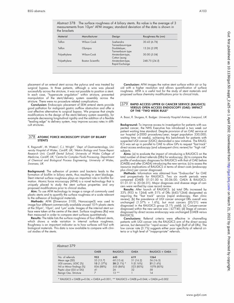
placement of an enteral stent across the pylorus and was treated bysurgical bypass. In three patients, although a wire was placedsuccessfully across the stricture, it was not possible to position a stent.In each case, “hyperacute angulation” within stricture, preventedmanipulation of the stent/delivery system assembly across thestricture. There were no procedure related complications.
Conclusion: Endoscopic placement of SEM enteral stents providegood palliation for malignant gastric outflow obstruction and offer acost effective alternative to surgical bypass. We propose that simplemodifications to the design of the stent/delivery system assembly, forexample decreasing longitudinal rigidity and the addition of a flexible“leading edge” to delivery system, may improve success rates in diffi-cult strictures.
378 ATOMIC FORCE MICROSCOPY STUDY OF BILIARYSTENTS
K. Ragunath1, M. Waters2, C.J. Wright3. 1Dept. of Gastroenterology, Uni-versity Hospital of Wales, Cardiff, UK; 2Matrix Biology and Tissue RepairResearch Unit, Cardiff Dental School, University of Wales College ofMedicine, Cardiff, UK; 3Centre for Complex Fluids Processing, Departmentof Chemical and Biological Process Engineering, University of Wales,Swansea, UK
Background: The adhesion of protein and bacteria leads to theformation of biofilm in biliary stents, thus resulting in stent blockage.Stent internal surface roughness plays an important role in biofilm for-mation. Atomic force microscopy (AFM) is a novel technology that isuniquely placed to study the stent surface properties and anyproposed modifications prior to clinical studies.
Aim: To use AFM technology to image a range of commonly usedplastic stents and to quantify the surface roughness at a scale relevantto the adhesion of biological materials.
Methods: AFM (Dimension 3100, Nanoscope3) was used toimage four different commercially available unused 10 Fr plastic stentsat the 50µm2, 10µm2, and 1µm2 scale. Images of the internal stent sur-face were taken at the centre of the stent. Surface roughness (Ra) wasthen measured in order to compare stent surfaces quantitatively.
Results: The table lists the surface roughness of four different stents,which shows a wide variation in internal surface roughness.Roughness is an important indicator as to how surfaces will foul withbiological materials. This data is now available to compare with clini-cal studies of the stents.
Conclusion: AFM images the native stent surface within air or liq-uid with a higher resolution and allows quantification of surfaceroughness. AFM is a useful tool for the study of stent materials andproposed surface chemistry modifications prior to clinical trials.
379 RAPID ACCESS UPPER GI CANCER SERVICE (RAUGICS)VERSUS OPEN ACCESS ENDOSCOPY (OAE): IMPACTOF THE “TWO WEEK RULE”
A. Bassi, R. Sturgess, K. Bodger. University Hospital Aintree, Liverpool, UK
Background: To improve access to investigation for patients with sus-pected cancer, the NHS Executive has introduced a two week outpatient waiting time standard. Despite provision of an OAE service atour hospital (>2000 procedures/year; target population 330,000;waiting time <6 weeks), achieving this benchmark for patients withsuspected UGI cancer (UGIC) demanded a new initiative. The RAUG-ICS was set up in parallel to OAE to allow GPs to request “fast track”direct access endoscopy (and subsequent clinic review) for “high risk”subjects.
Aims: (a) to evaluate the impact of introducing a RAUGICS on thetotal number of direct referrals (DRs) for endoscopy; (b) to compare theprofile of endoscopic diagnoses for RAUGICS with that of OAE before(OAEB) and after (OAEA) introducing the new service; (c) to assess theresource implications of RAUGICS in terms of direct costs (endoscopyplus clinic) per cancer diagnosed.
Methods: Information was obtained from “Endoscribe” for OAEand prospectively for RAUGICS. Two six month periods werecompared (OAEB: 01.01.00 to 30.06.00; OAEA & RAUGICS:01.01.01 to 30.06.01). Major diagnoses and disease stage of can-cers were verified by case record review.
Results: After launch of RAUGICS: (a) total DRs increased by33% (953 to 1264) with 51% of DRs (645/1264) designated asrequiring the “fast track” service (rapid endoscopy, then clinicreview); (b) the prevalence of UGI cancer amongst DRs overall wasunchanged (1.57% v 1.6%), but most cancers (20/21) werediagnosed in the RAUGICS group (3.1% yield); (c) Cost-per-cancerdiagnosed within the new service was ∼ £7740; (d) Stage of cancersdiagnosed by direct access endoscopy was unchanged (OAEB versusRAUGICS).
Conclusions: Referral criteria were effective in channellingpatients with UGI cancer into the RAUGICS arm of the direct accessservice, but demand for “rapid access” was high (half of all DRs). Thelow cancer rate (3.1%) suggests either poor specificity of referral cri-teria or a high level of “inappropriate” referrals.
Abstract 378 The surface roughness of 4 biliary stents. Ra value is the average of 3measurements from 10µm2 AFM images; standard deviation of the data is shown inthe brackets
Material Manufacturer Design Roughness Ra (nm)
Teflon Wilson Cook Soehendra 30.43 (6.76)Tannenbaum-type
Teflon Olympus Doublelayer, 13.26 (2.09)Tannenbaum type
Polyethylene Wilson-Cook Amsterdam-type, 30.00 (5.04)Cotton Leung
Polyethylene Boston Scientific Amsterdam-type, 248.75 (24.5)Rapid Exchange
Abstract 379
OAEB RAUGICS OAEA RAUGICS + OAEA
No. of referrals 953 645 619 1264Mean age (SD) 55 (15.7) 60 (15.6) 51 (16.2) 56 (16.5)UGI cancers, UGIC (%) 15 (1.57%) 20 (3.1%) * 1 (0.16%) 21 (1.6%)Nil or Minor findings (%) 836 (88%) 555 (86%) 523 (85%) 1078 (85%)Peptic ulcer (GU or DU) 41 26 32 58Benign Oes. Stricture 7 13 ** 1 14
* RAUGICS v OAEB p=0.06; v OAEA p<0.001; ** RAUGICS v OAEB p=0.044; v OAEA p=0.002
BSG abstracts A103
www.gutjnl.com
on March 21, 2022 by guest. P
rotected by copyright.http://gut.bm
j.com/
Gut: first published as 10.1136/gut.50.suppl_2.a25 on 1 A
pril 2002. Dow
nloaded from
380 ASSESSMENT OF AN OPEN ACCESS IN-PATIENTGASTROSCOPY SERVICE
E. Quinton, W.P. Goddard, R.G. Long, K. Teahon. Department of Gastro-enterology, Nottingham City Hospital, Hucknall Road, NottinghamNG5 1PB, UK
Introduction: There is a significant body of literature assessing openaccess outpatient gastroscopy (OGD). Little is written about the use oforganisation of the service OGD for inpatients except in the situationof gastrointestinal bleeding. In late 1998 we switched our inpatientgastroscopy service from a “Consultation first” service to an“Electronic Open Access Service”. This study evaluates the impact ofthee change in service organisation.
Methods: (1) The number of and reason for requests for OGD since1998 was retrieved from the electronic database. (2) “Reason forrequest” categories where >100 requests had been made in the lastyear were identified and the hospital records of the patients in thesecategories were selected randomly for review to allow review of 25%of the records. (3) On review of the records the demographic details,reason for admission, reason for referral for and outcome of OGD,eventual diagnosis, and routine laboratory parameters wererecorded. (4) The pre OGD admission record of each patient wassummarised and reviewed anonymously and independently by threeconsultant gastroenterologists to determine appropriateness of therequest for OGD.
Results: During the period from 1998 there was a 19% increase inemergency medical and surgical admissions and a 27% increase inOGD requests but this varied from a 13% increase in referrals formelaena to a 56% and 80% increase in referrals for nausea/vomitingand abdominal pain respectively. When the appropriateness of refer-rals was assessed 67% of all requests were considered appropriatebut the value varied (abdominal pain 41%, haematemesis 74%,melaena 83%, anaemia 84%, and nausea/vomiting 57%). The even-tual diagnosis was achieved at OGD in all patients in 52% (abdomi-nal pain 29%, haematemesis 79%, melaena 65%, anaemia 38%,nausea/vomiting 38%).
Conclusion: An electronic open access service is appropriate forinpatients with GI bleeding and anaemia although more selectivereferral questions may improve the appropriateness of referrals.Pre-procedure consultation is more appropriate for most other catego-ries of inpatient referrals.
381 GENERAL PRACTITIONERS’ PERCEIVED UTILITY OF APROPOSED COMPUTERISED DECISION SUPPORTSYSTEM FOR WEB BASED REFERRAL OF PATIENTSWITH GASTROINTESTINAL (GI) DISORDERS
D. Flynn1, P. van Schaik2, A. van Wersch2, P. Cann1, A. Douglass1.1 Endos-copy Centre, James Cook University Hospital, Middlesbrough, UK;2University of Teesside (Social Sciences), Middlesbrough, UK.
Introduction: A web based, direct booking system for GP referrals toa GI clinic or procedure is under development for our unit. Guidelinesfor referral of clinical sub groups will be incorporated as algorithmswithin the software of the computerised decision-support booking sys-tem. This will enable the most appropriate slot for the patient to beidentified and booked directly in real time. “Appropriateness” willrelate to relevant clinician, nature (clinic or procedure,) and speed offirst contact and provide patients with some choice of appointments.
Aims & Objectives: To assess GPs’ attitudes to the principles,design, and their eventual usage of such a system.
Methods: 21 GPs were given a description of the proposed systemand asked to rate each of eight potential benefits on a 6 point scale.GPs were also given the opportunity to discuss the potential impact ofthe system on their clinical practice and patients.
Results: The five most highly scored potential benefits were (i)faster and more reliable communication between primary andsecondary care (mean 5.22), (ii) appropriateness of the firstassessment received by patients (mean 4.53), (iii) facilitatingadherence to local clinical guidelines (mean 4.53), (iv) facilitating theexplanation of referral decisions, tests and procedures to patients(mean 4.42), and (v) provision of information on evidence-basedreports for GI symptoms (mean 4.37). 52% of GPs indicated that theywould use the system in real time only if it generated no more than twoadditional minutes on consultation times. 21% of GPs were concernedthat the system could increase administrative workload, and createunrealistic expectations of open access services for patients.
Conclusions: The majority of GPs recognised potential benefits ofthe proposed system, but the impact on consultation times is a core
design issue that will determine acceptance and usage of such a sys-tem. Other members of the primary care team may help to minimisethe need for the GP to spend “real time” online.
382 GASTRIC BIOPSY: WHERE FROM AND HOW MANYUSING THE UPDATED SYDNEY SYSTEM TO EVALUATEGASTRITIS, ATROPHY, AND INTESTINAL METAPLASIA?
M.M. Walker, R.P. Negus, A. Rice, V. Loh, J. Teare, M.R. Thursz. Facultyof Medicine, Imperial College of Science, Technology and Medicine, StMary’s Campus, London W2 1PG, UK
The topography of gastritis predicts a likely outcome in Helicobacterpylori infection. Histological markers of cancer risk include severeatrophy and intestinal metaplasia. The updated Sydney system forreporting gastritis recommends five biopsy sites, which is time consum-ing. This study aims to establish if all five gastric sites are necessary foran accurate appraisal of gastric pathology in routine practice.
Patients and Methods: 100 patients, 45 men, median age 57,range 21–88 years, attending for upper gastrointestinal endoscopyhad five gastric sites routinely biopsied according to the updated Syd-ney protocol. Biopsies were placed in order on a cellulose acetatestrip, processed, and reported by two pathologists according to theSydney System. Results were analysed to assess concordance of diag-nosis by site and to determine which of the five sites were optimal toassess gastric pathology.
Results: The majority of presenting symptoms for endoscopy weredyspepsia or abdominal pain (47 patients). At biopsy for gastritis,including grade of gastritis, there was excellent concordance betweenbody sites (100%), and good concordance between antral sites(94%). No further information was gained from separate analysis ofthe biopsy of the incisura. Intestinal metaplasia (IM) was found withgreatest prevalence in A2 (inferior antrum) -17% all biopsies and B2(greater curve, body) - 5%. In contrast, atrophy was found with great-est prevalence (21%) at A1 (superior antrum) but also as in IM, 10%of B2 biopsies showed atrophy. The overall prevalence of IM in thisstudy was 19% and atrophy, 21% from analysis of biopsies of allsites.
Discussion: This study shows that when histological assessment ofgastritis is required, in the absence of a visible lesion, two biopsies,one from the antrum and one from the body are adequate for a clini-cal diagnosis of grade and type of gastritis. However if only two sitesare biopsied, IM (a histological marker of cancer risk) will be missedin 7% of cases relative to the full Sydney protocol.
383 THE APPROPRIATENESS OF A 24 HOUR BLEEDINGSERVICE: PROSPECTIVE AUDIT OF GUIDELINES FORTHE MANAGEMENT OF GASTROINTESTINALHAEMORRHAGE
M.C. Gallagher, F.R. Vicary, V.S. Wong. Department of Gastroenterologythe Whittington Hospital London N1, UK
Background: Acute upper gastrointestinal haemorrhage (UGIH) con-stitutes a medical emergency with an incidence of 100 per 100 000and mortality rate of 14%1. The Whittington Hospital departmentalguidelines state that patients should undergo endoscopy the day afteradmission unless there are criteria for “emergency endoscopy” (out ofthe hours 9 am–5 pm): suspected varices, large bleed/continuedbleeding, or history of aortic graft. Two consultant physicians, (andtheir SPRs) and two consultant surgeons provide 24 hour endoscopycover.
Aims: To perform a six month prospective audit of the managementof suspected cases of acute UGIH at the Whittington Hospital.
Methods: All suspected cases of acute UGIH were included. Datawere collected on a standard proforma and case notes reviewed afterdischarge. Outcome measure included the timing of endoscopy, useof “out of hours” endoscopy services, endoscopic diagnosis, and mor-tality rates. Rockall scores were calculated for all patients whorequired endoscopy.
Results: 95 cases of suspected UGIH were identified. 76 patients(80%) presented via A&E and 19 (20%) were existing inpatients. 90patients underwent endoscopy. In 92% of cases, endoscopy was per-formed within 24 hours of admission/inpatient bleeding episode. Ofthese, 20 patients achieved criteria for emergency endoscopy. 17were endoscoped out of hours and three patients presented withinhours. The average Rockall score for patients endoscoped withinhours was 3.5 (range 1–10) and out of hours, 6.5 (range 1–10).Endoscopic diagnoses were as follows: peptic ulcer 37%, varices
A104 BSG abstracts
www.gutjnl.com
on March 21, 2022 by guest. P
rotected by copyright.http://gut.bm
j.com/
Gut: first published as 10.1136/gut.50.suppl_2.a25 on 1 A
pril 2002. Dow
nloaded from
15%, normal 12%, erosions 9%, oesophagitis 9%, Mallory-Weis tear2%, other 16%. Endoscopic therapy was required in 29 cases (30%).The overall Mortality Rate was 10.5%.
Conclusion: The proportion of patients in this series endoscopedwithin 24 hours (92%) compares favourably with published data (50and 56% in the BSG National Audits) 1, 2. 17/95 patients underwentout of hours endoscopy including seven with variceal bleeding. Theappropriate provision of a 24-hour bleeding service is possible in asmall unit with the collaboration of medical and surgical Endoscopists.1. Rockall et al: BMJ 1995;311:222–6 2. Rockall et al: Gut1997;41:606–11
384 SELF ADMINISTERED PHOSPHATE ENEMA IN BOWELPREPARATION FOR FLEXIBLE SIGMOIDOSCOPY: ANAUDIT OF EFFICACY AND PATIENT SATISFACTION
T.A. Raven, J. Whelan, I. Fretwell, M. Aston, P. Whitney. EndoscopyDepartment, Chesterfield Royal Hospital, Derbyshire, UK
Background: Patients attending endoscopy for a flexible sigmiodos-copy (FS) examination were being prepared using picolax except onedoctor who used phosphate enemas on arrival. Recent research byW. Atkin et al (2000)1 has shown that an phosphate enema adminis-tered on the day of the examination is as effective as picolax. To seeif this could be successful in our endoscopy unit an audit wasconducted.
Aims: To improve the way in which patients are prepared for (FS)without compromising the quality of bowel preparation.
Methods: 53 Patients were selected using two (FS) lists and had theoption of taking the enema at home instead of picolax for bowelpreparation. A questionnaire was prepared for patients to completewhile they were waiting for the (FS). A note was made of patientsunable/unwilling to administer the enema. Nursing and medical infor-mation was collected to assess the acceptability and quality of bowelpreparation.
Results: 98% of patients administered the enema successfully athome. 74% of the (FS) tests were completed to 60cm. 100% ofpatients using the enema would use it again, 12% telephoned thedepartment for advice, 38% experienced mild side effects, and 27%needed help from someone at home. The bowel preparation wasexcellent to good in 75% of cases.
Conclusion: Patients found the enema easy to use and acceptableas a method of bowel preparation. Although only a small number ofcases were audited the results are very similar to those reported in theBMJ. 1. Atkin et al: BMJ 2000;320:1504–9
385 USE OF A PAIR OF PRESSURE SENSITIVE GLOVES TODETERMINE MECHANICAL WORK DONE DURINGEITHER OGD OR COLONOSCOPY: A PILOT STUDY IN10 PATIENTS
J. Hancock1, S. Dogramadzi2, G.D. Bell1, C. Allen2, K. Burn3, I. Fletcher3,R. Bicker4. 1Medical Sciences Faculty, Sunderland University, UK;2Department of Electrical and Electronic Engineering, Newcastle University,UK; 3School of Computing, Engineering and Technology, University ofSunderland, UK; 4Department of Mechanical, Materials and ManufacturingEngineering, Newcastle University, UK
Introduction and aims: Torque steering is now the preferred methodof teaching intubation of the tortuous sigmoid colon. One of the criti-cisms of many teaching simulators is that the “feedback” forces are notrealistic. One of us (CA) had had extensive previous experience withthe use of pressure sensor gloves to estimate the mechanical forces/mechanical work done by female assembly plant workers1. Wedecided to see if (a) we could satisfactorily modify the equipment tomeasure similar forces being applied by an endoscopist’s right andleft hand during endoscopic procedures (real or simulated) and (b) ifso whether these differed depending on the diameter and stiffness ofthe endoscope employed.
Aims and methods: A single experienced endoscopist (JH)carried out a total of 10 endoscopic procedures (three OGDs, oneflexible sigmoidoscopy, and six colonoscopies) while wearing thesensor gloves. Each glove containing 20 calibrated piezo electric sen-sors which sent pressure data to a PC fifty times a second. The datawere represented both graphically and numerically using speciallywritten software.
Results and discussion: The equipment accurately and reproduc-ibly measured the, at times, quite considerable forces generated bythe right hand during torque steering. Right hand torque forces werepredictably much higher during lower GI endoscopy than duringOGD. Despite the small numbers of patients studied to date, it isbecoming clear that during colonoscopy/flexible sigmoidoscopygreater right hand torque forces are required with thin endoscopescompared with thicker ones. We are now looking at the use of NeuralNetwork/Artificial Intelligence methodology to help analyse the datain greater depth. We are confident we can apply the technique tolooking at differences between endoscopists and endoscopes duringboth real and simulated examinations. The research could have impli-cations in the design of instruments and teaching simulators as well aslive “hands on” assessment of trainees during endoscopic procedures.1. Allan CR et al: Mechatronics 1997;7:141–57
386 HAEMORRHAGIC RADIATION PROCTITIS: ANENDOSCOPIC SCORE MAY GUIDE THERAPY
R. Zinicola1, M.D. Rutter1, G. Falasco1, V. Cennamo2, S. Contini2,B.P. Saunders1. 1Wolfson Unit for Endoscopy, St. Mark’s Hospital, UK;2Endoscopia Digestiva, Ospedale Bellaria, Bologna, Italy
Background: Management of haemorrhagic radiation proctitis (HRP)remains controversial. Recently both endoscopically delivered argonplasma coagulation (APC) & local rectal application of 4% formalin(LRAF) have been reported as effective treatments. However the exactrole of these therapies is not clearly defined. We evaluated theefficacy of APC, and developed a new endoscopic score to guidetherapy.
Methods: 12 patients with significant rectal bleeding due to HRPwere retrospectively reviewed. Patients were classified using a newendoscopic grading of HRP from endoscopic videoprints, assessingconfluence and distribution of telangiestasias, percentage of surfacearea involved, and presence of fresh blood. All patients were treatedwith APC initially.
Results: Utilising the new endoscopic grading, five patients werecategorised as grade I (mild) HRP, four patients grade II (moderate)HRP, three patients grade III (severe) HRP. In 10 patients (83.3%),bleeding improved significantly following APC therapy. All patientswith grade I & II were treated successfully by APC (median twosessions, range one to four). In two grade III patients APC failed, butsubsequent formalin application was successful.
Conclusions: Our endoscopic score may help guide appropriatetreatment for HRP. APC appears safe, efficacious, and should usuallybe considered first line therapy, particularly in grade I & II HRP. How-ever with extensive (grade III) HRP, topical formalin application maybe more effective.
387 ARE MULTIPLE BIOPSIES NECESSARY IF ACOLONOSCOPY IS NORMAL?
M.C. Follows, B.J. Rembacken, D.M. Chalmers, A.T.R. Axon. Centre forDigestive Diseases, the General Infirmary at Leeds, Great George St,Leeds, LS1 3EX, UK
Aims: To correlate the histological and clinical findings in patientswith a macroscopically normal, total colonoscopy.
Method: A search was made of all colonoscopy reports performedduring 2000. Reports from patients with a macroscopically normal,total colonoscopy in which a set of serial biopsies (caecum,ascending, transverse, descending, and sigmoid colon and rectum)had been taken were identified. The histology result for each patientwas found and the indication for the examination noted in each case.Cases were excluded if any endoscopic abnormality was noted or if afull set of biopsies had not been taken.
Results: See table.Conclusions: Five cases of IBD (inflammatory bowel diease/
microscopic colitis) were identified in 282 patients. In all diarrhoeawas a presenting symptom (i.e. 4% of patients with diarrhoea hadIBD). If diarrhoea was not an indication no patients were found tohave IBD1 on serial biopsies. We recommend serial biopsies should betaken in patients with a normal colonoscopy but only if thepredominant symptom is diarrhoea.
BSG abstracts A105
www.gutjnl.com
on March 21, 2022 by guest. P
rotected by copyright.http://gut.bm
j.com/
Gut: first published as 10.1136/gut.50.suppl_2.a25 on 1 A
pril 2002. Dow
nloaded from
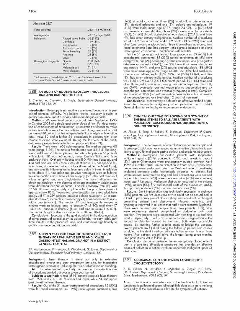
388 AN AUDIT OF ROUTINE ILEOSCOPY: PROCEDURETIME AND DIAGNOSTIC YIELD
S. Cherian, A. Cherukuri, P. Singh. Staffordshire General Hospital,Stafford ST16 3SA, UK
Introduction: Ileoscopy is not routinely attempted because of its per-ceived technical difficulty. We believe routine ileoscopy is useful inquality assurance and it provides additional diagnostic yield.
Methods: We examined colonoscopy data from September 1995to October 2001 of a single gastroenterological firm. For documenta-tion of completeness of examination, visualization of ileocaecal valveor ileal intubation were the only criteria used. A registrar endoscopistperformed 80 colonoscopies independently. For analysis of intubationrates, these 80 and a further 56 procedures in patients with priorcolonic resection were excluded. During the last year of the audit,data were prospectively collected on procedure times (PT).
Results: There were 1602 colonoscopies. The median age was 60years (range 8–95). The male to female ratio was 4.5:5.5. The diag-nostic yield from 73 ileoscopies and 67 sets of biopsies in 66 patientswith colonic IBD was: 63 normal; seven Crohn’s ileitis; and threebackwash ileitis. Of those without colonic IBD, 904 had ileoscopy and414 had biopsies. Ileal Crohn’s was identified in 11, non-specific ilei-tis in three, discrete ileal ulcers in four (three related to NSAID use),and non-specific inflammation in ileum and colon in three. In additionto the above 21, nine additional positive histologies were as follows:five non-specific ileitis, three villous atrophy (two also had duodenalvillous atrophy), and one amyloidosis). The main indications forobtaining histology in the absence of an endoscopically visible lesionwere diarrhoea and/or anaemia. Overall ileoscopy rate (IR) was67.5%. IR rose progressively to plateau for the past three years atapproximately 85%. Twenty-two colonoscopies were excluded fromanalysis of PT in 259 patients (previous colonic resection=13; impass-able stricture=7; incomplete colonoscopy=1; abandoned due to respi-ratory depression=1). The median PT and interquartile ranges inminutes were as follows: anus to caecum=7 (5–10); total time=17(14–21.5); caecum to ileum=2 (1–4); and time in ileum=1 (0.5–2).The median length of ileum examined was 10 cm (10–15).
Conclusions: Ileoscopy is the gold standard in the documentationof completeness of colonoscopy. In skilled hands, it is easy, adds onlythree minutes to the procedure time, and contributes significantly toquality assurance and diagnostic yield.
389 A SEVEN YEAR OUTCOME OF ENDOSCOPIC LASERTHERAPY FOR PALLIATIVE UPPER AND LOWERGASTROINTESTINAL MALIGNANCY IN A DISTRICTGENERAL HOSPITAL
R.P. Arasaradnam, P. Hancock, T. Woodward, G. James. Department ofGastroenterology, Doncaster Royal Infirmary, Doncaster, UK
Background: Laser therapy is useful not only in extensiveoesophageal tumour and stent overgrowth but also, for inoperablerecto-sigmoid tumours in reducing the risk of obstruction or bleeding.
Aim: To determine retrospectively outcome and complication rateof procedures carried out over a seven year period.
Subjects & Method: A total of 95 patients received laser therapyfrom 1994 until 2001. 31 of whom had lower, while 64 had uppergastrointestinal laser therapy
Results: Out of the 31 lower gastrointestinal procedures 15 (50%)were for rectal carcinoma, six (19%) recto-sigmoid carcinoma, five
(16%) sigmoid carcinoma, three (9%) tubulovillous adenoma, one(3%) sigmoid adenoma and one (3%) colonic angiodysplasia. 19(61%) were male; mean age of 78 (range 74–97). 13 (42%) hadcardiovascular co-morbidities, three (9%) cerebrovascular accidents(CVA), 5 (16%) chronic obstructive airway disease (COAD), and three(9%) had other primary malignancies. Median number of procedureswas 4 ± 1.2 over a duration of 4 ± 1.9 months. Nine (29%) remainedalive (one colonic angiodyplasia, three tubulovillous adenoma, tworectal carcinoma (later had surgery), one sigmoid adenoma and onerecto-sigmoid carcinoma). Complication rate was 0%
For the 64 upper gastrointestinal laser procedures, 39 (61%) hadoesophageal carcinoma, 15 (23%) gastric carcinoma, six (8%) stentovergrowth, one (2%) oesophago-gastric carcinoma, one (2%) gastricarteriovenous ectasia (GAVE), one (2%) Hereditary haemorrhagic tel-angiectasia (HHT), and one (2%) gastric angiodysplasia. 38 (59%)were male; mean age of 75 (range 44–88). 27 (42%) had cardiovas-cular co-morbidities, eight (13%) CVA, 14 (22%) COAD, and five(8%) had other primary malignancies. Median number of procedureswas 1.25 ± 0.9 over a 2.5 ± 0.5 month period. 12 (18%) remainedalive (three gastric carcinoma, one gastric angiodysplasia, one HHT,one GAVE: eventually required Argon plasma coagulation and sixoesophageal carcinoma: one eventually requiring a stent). Complica-tion rate was 0.05% (two with aspiration pneumonia within two weeksof the procedure and one bled after 12 hours of treatment).
Conclusions: Laser therapy is safe and an effective method of pal-liation for inoperable malignancy when performed in a DistrictGeneral Hospital setting by an experienced endoscopist.
390 CLINICAL OUTCOME FOLLOWING DEPLOYMENT OFENTERAL STENTS TO PALLIATE PATIENTS WITHMALIGNANT GASTRODUODENAL OBSTRUCTION: ADGH EXPERIENCE
M. Allison, T. Tang, P. Roberts, R. Dickinson. Department of Gastro-enterology, Hinchingbrooke Hospital, Hinchingbrooke Park, Huntingdon,PE29 6NT, UK
Background: The deployment of enteral stents under endoscopic andfluoroscopic guidance has emerged as an effective alternative to pal-liative surgery for malignant gastric outflow and duodenal obstruction.
Methods: Twenty-one consecutive patients with inoperablemalignant (gastric (28%), pancreatic (67%), and metastatic deposit(5%)) upper GI strictures were prospectively studied between April1999 to October 2001, on an “intention to treat” basis. Twenty-threeprocedures were performed using the Wallstent enteral prosthesis,implanted per-orally under fluoroscopic guidance. All patients hadsevere nausea, recurrent vomiting and their obstructions were deemedinoperable. Twelve (57%) were male and nine (43%) were female;mean age 71.6 years. Malignant obstruction occurred at the pylorus(19%), antrum (5%), first and second parts of the duodenum (66%),third part of duodenum (5%), and anastomotic sites (5%).
Results: Stent implantation was technically successful in eighteen(85%) patients. On two occasions a wire could not be passed and ina third, a previously placed plastic biliary stent could not be changedpreventing enteral stent deployment. Nausea, vomiting, anddysphagia improved in all cases that had a stent successfully placed.There were no short term complications. Two patients (11%), whowere successfully stented, complained of abdominal pain postinsertion. Two patients were readmitted with vomiting at six and ninemonths respectively. The first was due to tumour overgrowth and thesecond to distortion caused by the stent. Both were successfullytreated, by inserting further co-axial stents to bridge the stenoses.Twelve patients (67%) died during the follow up period from causesunrelated to the stent insertion, with a median survival time of threemonths. Five patients are still alive, the longest being seven months.One patient was lost to follow up.
Conclusion: In our experience, the endoscopically placed enteralstent is a safe and efficacious procedure that provides an effectivemeans of palliation to patients with an inoperable malignant upper GIobstruction.
391 ABDOMINAL PAIN FOLLOWING LAPAROSCOPICCHOLECYSTECTOMY
A. D. Gilliam, N. Davidson, K. Mylankal, D. Ziegler, E.P. Perry,J.D. Harrison. Department of Surgery, Scarborough Hospital, WoodlandsDrive, Scarborough, YO12 6QL, UK
Aim: Laparoscopic cholecystectomy is the treatment of choice forsymptomatic gallstone disease, although little data exists as to the longterm ability of the procedure to alleviate the symptoms of patients.
Abstract 387
Total patients: 282 (118 M, 164 F).
Average age: 47.13 range 16-87.Indication: Altered bowel habit. 52 (18%)
Diarrhoea 139 (49%)Constipation 10 (4%)Abdominal pain 18 (6%)Rectal bleeding 23 (8%)Anaemia 21 (8%)Other 19 (7%)
Histological diagnosis: Normal 238 (84%)IBD* 5** ( 2%)Melanosis coli 7 ( 3%)Minor changes 32 (11%)
*Inflammatory bowel disease. ** 1 case of indeterminate colitis,1 case of Crohn’s, and 3 cases of microscopic colitis.
A106 BSG abstracts
www.gutjnl.com
on March 21, 2022 by guest. P
rotected by copyright.http://gut.bm
j.com/
Gut: first published as 10.1136/gut.50.suppl_2.a25 on 1 A
pril 2002. Dow
nloaded from
Method: A prospective study of 212 patients undergoinglaparoscopic cholecystectomy was performed. A detailed postalsymptom questionnaire was designed and piloted. Patients completedthese preoperatively and at three and 12 months postoperatively.
Results: 169 patients were female. The median age of patientswas 57 years (range 21–81). Complete data was available on 68%(145 patients). 34 patients (23%) continued to be symptomatic. Onlyone patient (0.06%) reported their abdominal pain/discomfort hadbecome worse. Of the symptomatic patients with continuing abdomi-nal pain, 15 (44%) claimed their pain was identical to thatpre-operatively. 55% (26) of the symptomatic patients consulted theirGPs during the one year follow up period.
Conclusion: A significant number of patients are dissatisfied oneyear after laparoscopic cholecystectomy. All patients should bepre-operatively counselled about the risk of persistence of some painsymptoms after laparoscopic cholecystectomy. There should be a highindex of suspicion for upper GI conditions and more patients shouldundergo diagnostic endoscopy before subjecting them to surgery.
NEOPLASIA POSTERS 392–409
392 CURRENT MANAGEMENT OF IRON DEFICIENCYANAEMIA BY GENERAL PHYSICIANS DOES NOTCOMPLY WITH BRITISH SOCIETY OFGASTROENTEROLOGY GUIDELINES
H.R. Ferguson, P. Murphy. Department of Gastroenterology, CraigavonArea Hospital, Northern Ireland, UK
Introduction: Iron deficiency anaemia is a common problem encoun-tered in all medical specialties. In men and post menopausal womencommon causes are gastrointestinal blood loss or malabsorption. TheBritish Society of Gastroenterology (BSG) recently producedguidelines on the management of this condition.
Aim: To establish current practice in the investigation of irondeficiency anaemia.
Methods: The laboratory identified all patients with a haemoglobinand mean cell volume below the normal range, for the six monthperiod between January and July 2001. The charts of those patientsunder the care of consultant physicians were reviewed. Data collectedincluded details of history taking, examination, investigationsperformed, treatment and follow up.
Results: 74 patients were identified with both a low haemoglobinand mean cell volume (age range 15–90 years). 70% were female. Arectal examination was performed in 16%. 13 patients (18%) hadantiendomysial antibodies tested. 27 patients (36%) had colonoscopyor barium enema performed, planned, or were unsuitable for investi-gation. 25 (34%) had an OGD or barium meal performed, planned orwere unsuitable. One patient had duodenal biopsy. A reason foranaemia was found in 24 patients (32%). In this group five hadcolonic or gastric carcinoma, and no diagnosis of coeliac disease wasmade. 38 patients (52%) were placed on iron supplements. Excludingfour patients who were unsuitable for follow up, 49 (70%) had a hos-pital review arranged.
Conclusion: A low proportion of patients underwent full upper andlower GI tract investigation although five patients were diagnosedwith carcinoma. No cases of coeliac disease were detected, but only18% had antiendomysial antibodies tested, and one duodenal biopsywas performed. We have produced a summary of the BSG guidelinesto improve management of these patients and encourage early refer-ral for endoscopy.
393 SPEED OF DIAGNOSIS OF UPPER GASTROINTESTINALCANCER: DOES THE METHOD OF REFERRAL MATTER?
J.G. Williams, S. Gheorghiu, W.-Y. Cheung. Swansea Clinical School,University of Wales, Swansea SA2 8PP, UK
All cases of upper gastrointestinal (GI) cancer diagnosed at twodistrict general hospitals in south Wales between 1/7/93 and30/6/99 have been reviewed. Cases were identified frompathology, endoscopy and clinical information systems and dataextracted from hospital and primary care records, using a structuredproforma. Case finding was cross checked with the local cancer reg-istry and data held in central returns. The data extracted were
validated on a randomly identified 10% sample. Four hundred andthirty-nine cases were identified and data obtained on 418. Medianage was 73, range 36–96 years, male:female 258:160, cancer siteoesophagus (150), stomach (265), duodenum (2), lung primary (1).
Over the six years of the study, during which open access services(OAG) and a “one stop” Rapid Opinion Clinic (ROC) wereintroduced, there was an overall decrease in the median interval fromfirst presentation to the General Practitioner to diagnosis by histology(from 68 to 17 days, p<0.001, when first and last six monthscompared). This overall NHS delay was significantly longer for thosepatients referred through outpatients (173 cases; mean interval111.3; median 70; range 5–956 days) compared with admission(144 cases; mean 34.6; median 13, range 0–546 days), OAG (89cases; mean 56; median 27; range 3–335 days), and the ROC (12cases; mean 43.2; median 13; range 4–185 days; p<0.05 for allmethods compared with outpatients). The main reason for this delaywas the time taken to reach the diagnosis after the initial contact inhospital (mean 61.3 days for outpatients versus 26.7; 12.8; and 22.8for admission, OAG, and ROC respectively; p<0.05 for all methodscompared with outpatients). Most patients presented with a combina-tion of symptoms which included loss of appetite or weight, butisolated dysphagia occurred in 14 patients (youngest 52 years) andisolated abdominal pain or dyspepsia was noted in eight (youngest63 years).
We conclude that if upper GI cancer is suspected patients shouldbe referred for “one-stop” assessment, or admission, rather thanoutpatients. Cancer may present as isolated abdominal pain ordyspepsia.
394 THE ROLE OF FDG-PET IN THE EARLY DETECTION OFRESPONSE OF COLORECTAL LIVER METASTASES TOCHEMOTHERAPY
G.W. Couper1, F. Wallis2, A. Welch3, P.F. Sharp3, K.G.M. Park1 , J. Cas-sidy4. 1Departments of Surgery ; 2Radiology 3Biomedical Physics and4Oncology, University of Aberdeen and Aberdeen Royal Hospitals NHSTrust, UK
Aim: To determine if 2-[18F]-fluoro-2-deoxy-D-glucose (FDG)-PositronEmission Tomography (PET) could detect early response to combina-tion chemotherapy in patients with liver metastases from colorectal pri-mary cancers.
Methods: Seventeen patients were imaged immediately prior to thefirst cycle of 5-Fluorouracil (5FU)/ leucovorin using FDG-PET. Thirteenpatients had follow up scans at 14 days. Tumour / Liver Ratios (TLRs)were used to quantify the PET images. Computed Tomography (CT),serum Carcinoembryonic Antigen (CEA) levels and survival figureswere used as comparative evidence of response.
Results: Areas of enhanced FDG uptake in comparison to adjacentnormal liver tissue were seen in all patients on pre chemotherapyscans. Thirty-four liver metastases were assessed for evidence ofresponse in the thirteen patients completing both scans. A change intumour/liver ratio of less than 20% was seen in 25 of 34 lesions.Non-uniform changes in activity were seen in five patients. Twopatients with uniform reductions in FDG uptake greater than 20% hadprolonged survival.
Discussion: FDG PET is reliable in the detection of metastatic colo-rectal cancer including those patients with low CEA levels. Thechanges seen in tumour FDG uptake at two weeks were small andoften not uniform within patients. Sizable uniform reductions in activ-ity, suggestive of response, were seen in only two of 13 patients bothof who had longer than median survival. It is possible that these twopatients were the only genuine early responders and that PET has theability to identify this small group of patients
395 P53 MUTATION DOES NOT INFLUENCE COX-2IMMUNOREACTIVITY IN GASTRICADENOCARCINOMA
G.V. Smith, A.B. Ballinger. Adult and Paediatric Gastroenterology, Bartsand the London, Queen Mary’s School of Medicine, London, UK
Both COX 2 over-expression and p53 mutation are common findingsin gastric adenocarcinoma, being seen in 60% and 40% of cancersrespectively. Recently, it has been suggested that wild type p53expression suppresses COX-2 mRNA transcription by competing forthe COX-2 promoter site. If this is the case, COX-2 expression shouldbe markedly increased in tumours with mutated p53 compared withthose with wild type p53.
BSG abstracts A107
www.gutjnl.com
on March 21, 2022 by guest. P
rotected by copyright.http://gut.bm
j.com/
Gut: first published as 10.1136/gut.50.suppl_2.a25 on 1 A
pril 2002. Dow
nloaded from
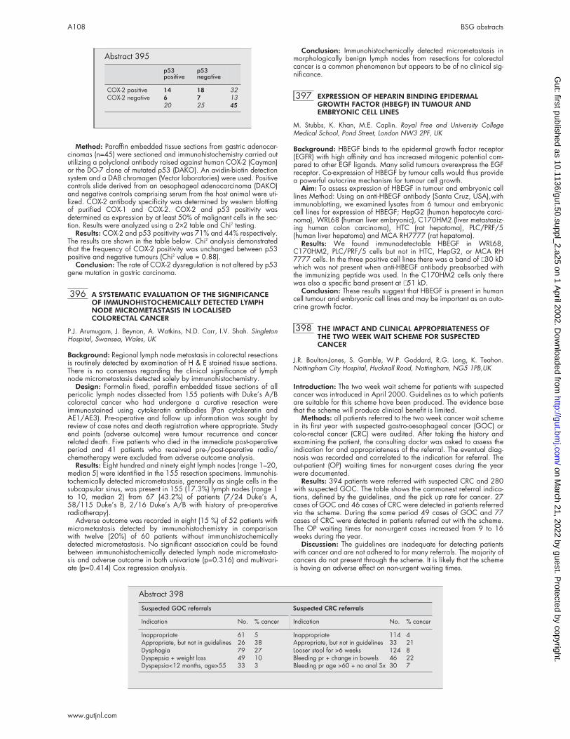
Method: Paraffin embedded tissue sections from gastric adenocar-cinomas (n=45) were sectioned and immunohistochemistry carried oututilizing a polyclonal antibody raised against human COX-2 (Cayman)or the DO-7 clone of mutated p53 (DAKO). An avidin-biotin detectionsystem and a DAB chromagen (Vector laboratories) were used. Positivecontrols slide derived from an oesophageal adenocarcinoma (DAKO)and negative controls comprising serum from the host animal were uti-lized. COX-2 antibody specificity was determined by western blottingof purified COX-1 and COX-2. COX-2 and p53 positivity wasdetermined as expression by at least 50% of malignant cells in the sec-tion. Results were analyzed using a 2×2 table and Chi2 testing.
Results: COX-2 and p53 positivity was 71% and 44% respectively.The results are shown in the table below. Chi2 analysis demonstratedthat the frequency of COX-2 positivity was unchanged between p53positive and negative tumours (Chi2 value = 0.88).
Conclusion: The rate of COX-2 dysregulation is not altered by p53gene mutation in gastric carcinoma.
396 A SYSTEMATIC EVALUATION OF THE SIGNIFICANCEOF IMMUNOHISTOCHEMICALLY DETECTED LYMPHNODE MICROMETASTASIS IN LOCALISEDCOLORECTAL CANCER
P.J. Arumugam, J. Beynon, A. Watkins, N.D. Carr, I.V. Shah. SingletonHospital, Swansea, Wales, UK
Background: Regional lymph node metastasis in colorectal resectionsis routinely detected by examination of H & E stained tissue sections.There is no consensus regarding the clinical significance of lymphnode micrometastasis detected solely by immunohistochemistry.
Design: Formalin fixed, paraffin embedded tissue sections of allpericolic lymph nodes dissected from 155 patients with Duke’s A/Bcolorectal cancer who had undergone a curative resection wereimmunostained using cytokeratin antibodies (Pan cytokeratin andAE1/AE3). Pre-operative and follow up information was sought byreview of case notes and death registration where appropriate. Studyend points (adverse outcome) were tumour recurrence and cancerrelated death. Five patients who died in the immediate post-operativeperiod and 41 patients who received pre-/post-operative radio/chemotherapy were excluded from adverse outcome analysis.
Results: Eight hundred and ninety eight lymph nodes (range 1–20,median 5) were identified in the 155 resection specimens. Immunohis-tochemically detected micrometastasis, generally as single cells in thesubcapsular sinus, was present in 155 (17.3%) lymph nodes (range 1to 10, median 2) from 67 (43.2%) of patients (7/24 Duke’s A,58/115 Duke’s B, 2/16 Duke’s A/B with history of pre-operativeradiotherapy).
Adverse outcome was recorded in eight (15 %) of 52 patients withmicrometastasis detected by immunohistochemistry in comparisonwith twelve (20%) of 60 patients without immunohistochemicallydetected micrometastasis. No significant association could be foundbetween immunohistochemically detected lymph node micrometasta-sis and adverse outcome in both univariate (p=0.316) and multivari-ate (p=0.414) Cox regression analysis.
Conclusion: Immunohistochemically detected micrometastasis inmorphologically benign lymph nodes from resections for colorectalcancer is a common phenomenon but appears to be of no clinical sig-nificance.
397 EXPRESSION OF HEPARIN BINDING EPIDERMALGROWTH FACTOR (HBEGF) IN TUMOUR ANDEMBRYONIC CELL LINES
M. Stubbs, K. Khan, M.E. Caplin. Royal Free and University CollegeMedical School, Pond Street, London NW3 2PF, UK
Background: HBEGF binds to the epidermal growth factor receptor(EGFR) with high affinity and has increased mitogenic potential com-pared to other EGF ligands. Many solid tumours overexpress the EGFreceptor. Co-expression of HBEGF by tumour cells would thus providea powerful autocrine mechanism for tumour cell growth.
Aim: To assess expression of HBEGF in tumour and embryonic celllines Method: Using an anti-HBEGF antibody (Santa Cruz, USA),withimmunoblotting, we examined lysates from 6 tumour and embryoniccell lines for expression of HBEGF; HepG2 (human hepatocyte carci-noma), WRL68 (human liver embryonic), C170HM2 (liver metastasiz-ing human colon carcinoma), HTC (rat hepatoma), PLC/PRF/5(human liver hepatoma) and MCA RH7777 (rat hepatoma).
Results: We found immunodetectable HBEGF in WRL68,C170HM2, PLC/PRF/5 cells but not in HTC, HepG2, or MCA RH7777 cells. In the three positive cell lines there was a band of ∼30 kDwhich was not present when anti-HBEGF antibody preabsorbed withthe immunizing peptide was used. In the C170HM2 cells only therewas also a specific band present at ∼51 kD.
Conclusion: These results suggest that HBEGF is present in humancell tumour and embryonic cell lines and may be important as an auto-crine growth factor.
398 THE IMPACT AND CLINICAL APPROPRIATENESS OFTHE TWO WEEK WAIT SCHEME FOR SUSPECTEDCANCER
J.R. Boulton-Jones, S. Gamble, W.P. Goddard, R.G. Long, K. Teahon.Nottingham City Hospital, Hucknall Road, Nottingham, NG5 1PB,UK
Introduction: The two week wait scheme for patients with suspectedcancer was introduced in April 2000. Guidelines as to which patientsare suitable for this scheme have been produced. The evidence basethat the scheme will produce clinical benefit is limited.
Methods: all patients referred to the two week cancer wait schemein its first year with suspected gastro-oesophageal cancer (GOC) orcolo-rectal cancer (CRC) were audited. After taking the history andexamining the patient, the consulting doctor was asked to assess theindication for and appropriateness of the referral. The eventual diag-nosis was recorded and correlated to the indication for referral. Theout-patient (OP) waiting times for non-urgent cases during the yearwere documented.
Results: 394 patients were referred with suspected CRC and 280with suspected GOC. The table shows the commonest referral indica-tions, defined by the guidelines, and the pick up rate for cancer. 27cases of GOC and 46 cases of CRC were detected in patients referredvia the scheme. During the same period 49 cases of GOC and 77cases of CRC were detected in patients referred out with the scheme.The OP waiting times for non-urgent cases increased from 9 to 16weeks during the year.
Discussion: The guidelines are inadequate for detecting patientswith cancer and are not adhered to for many referrals. The majority ofcancers do not present through the scheme. It is likely that the schemeis having an adverse effect on non-urgent waiting times.
Abstract 395
p53positive
p53negative
COX-2 positive 14 18 32COX-2 negative 6 7 13
20 25 45
Abstract 398
Suspected GOC referrals Suspected CRC referrals
Indication No. % cancer Indication No. % cancer
Inappropriate 61 5 Inappropriate 114 4Appropriate, but not in guidelines 26 38 Appropriate, but not in guidelines 33 21Dysphagia 79 27 Looser stool for >6 weeks 124 8Dyspepsia + weight loss 49 10 Bleeding pr + change in bowels 46 22Dyspepsia<12 months, age>55 33 3 Bleeding pr age >60 + no anal Sx 30 7
A108 BSG abstracts
www.gutjnl.com
on March 21, 2022 by guest. P
rotected by copyright.http://gut.bm
j.com/
Gut: first published as 10.1136/gut.50.suppl_2.a25 on 1 A
pril 2002. Dow
nloaded from
399 A SEVEN YEAR EXPERIENCE OF MANAGEMENT OFOESOPHAGEAL CANCER
M.A. Yusuf, A.H. Sadozye. Shaukat Khanum Memorial Cancer Hospital &Research Centre, Lahore, Pakistan
We conducted a retrospective review of all patients seen withoesophageal cancer at our institution over the last seven years. 225patients were seen (mean age 49.6y, range 18-85y; 121 males).
Tumour site: 72 starting in upper 1/3 (69 squamous cellcarcinoma [SCCA], three adenocarcinoma [ACA]), 67 mid 1/3 (64SCCA, 2 ACA, one collision tumour), 86 lower 1/3 or G.O. junction(45 ACA, 34 SCCA, one lymphoma, six miscellaneous).
Results: 52 received no treatment (36 of these were lost to followup during work up). 67 received purely palliative treatment, such asPEG/surgical gastrostomy, APC or other ablative techniques,radiation [DXT] or DXT/chemotherapy. Average age 51.8 yrs. 22ACA, 43 SCCA, 2 others. 15 upper 1/3, 20 mid-1/3, 32 lower 1/3.50 had surgery with curative intent, either alone or in combinationwith DXT/chemotherapy in adjuvant or neo-adjuvant setting. Averageage 48 yrs. 35 SCCA, 13 ACA, two others. Four upper 1/3, 18 mid-1/3, 28 lower 1/3. 56 had treatment with either DXT orDXT/chemotherapy. Average age 46.4 yrs, 52 SCCA, 4 ACA. 37upper 1/3, 12 mid-1/3, seven lower 1/3.
Outcome: 61 patients were lost to follow up during investigation.14 patients alive (10 had surgery, three treated with palliative intent,one with primary DXT). Median survival 20 months. 34 definitelydead (3 had surgery, 13 treated with palliative intent, 10DXT/chemotherapy, 8 had no treatment). Median survival 5.5months. 116 patients lost to follow-up after median follow up of eightmonths.
Conclusions: (1) The average age of patients seen with oesopha-geal cancer is lower in our series than reported in the literature. (2)25% of patients seen were younger than 40 yers old and may repre-sent a subgroup for further study as to the aetiology of this cancer. (3)In our country, it is difficult to follow up patients, particularly those fromrural areas and with limited means of communication, as evidencedby the large number of patients lost to follow up.
400 MATRIX METALLOPROTEINASE-2 CONCENTRATIONCORRELATES POSITIVELY WITH PATHOLOGICALTUMOUR STAGE IN PATIENTS WITHADENOCARCINOMA OF THE OESOPHAGUS ORGASTRIC CARDIA
R.E. Lawther1, G.M. Spence1, I. McAllister1, K.M. Mulholland2, A.N.J. Gra-ham1, J.A. McGuigan1, M.C. Regan1, K.R. Gardiner1. Departments of 1Sur-gery and 2Pathology, Royal Victoria Hospital, Belfast, UK
Introduction: Matrix metalloproteinase-2 (MMP-2) and -9 (MMP-9)facilitate tumour invasion and metastatic spread by degrading type IVcollagen, the main structural component of the basement membrane.The aim of this study was to determine if these endopeptidases aremarkers of disease progression in adenocarcinoma of the oesophagusor gastric cardia.
Methods: Approval for this study was obtained from the local eth-ics committee and informed consent was given by each participatingpatient. Fresh tumour samples from neoplasms of the oesophagus orgastric cardia, obtained at the time of surgery, were homogenised,centrifuged and the supernatants analysed for expression of MMP-2and -9 using matrix metalloproteinase activity assay systems (BiotrakRPN 2631 and 2634, Amersham Pharmacia Biotech, Buckingham-shire, UK). All tumours were assessed histologically by anindependent pathologist. Tumour staging was based on the UICCguidelines (5th Edition). Correlation analysis was undertaken usingSpearman’s rank testing.
Results: Tumour samples were obtained from 33 patients with con-firmed adenocarcinoma. Total and inactive MMP-2 concentrationscorrelated positively with overall pathological stage (r2 = 0.14, p <0.05; r2 = 0.15, p < 0.05 respectively). There was no correlationbetween MMP-2 and histological parameters such as depth ofinvasion, vascular involvement, or lymph node metastasis. MMP-9 didnot correlate with pathological stage or histological findings.
Conclusions: This study provides further evidence for theinvolvement of MMP-2 in tumour invasion and metastasis. MMP-2 con-centration in diagnostic biopsies from oesophago-gastric cancers maytherefore be of benefit in assisting the decision making processregarding the appropriateness of surgical resection.
401 CHANGES IN MUSCLE UNCOUPLING PROTEINEXPRESSION IN HUMANS WITH GASTROINTESTINALADENOCARCOINOMA
Peter Collins1, Chen Bing2, Peter McCulloch3, Gareth Williams2. 1RoyalFree and University College Medical School, Rowland Hill Street,Hampstead, London NW3 2PF, UK; 2Diabetes and EndocrinologyResearch Group, Department of Medicine, University of Liverpool,L69 3GA, UK; 3Division of Surgery, University Hospital Aintree, LongmoorLane, Liverpool L9 7AL, UK
The mitochondrial uncoupling proteins-2 and-3 (UCP-2 and -3) areputative mediators of thermogenesis and energy expenditure in mam-mals. We obtained skeletal muscle from 12 gastrointestinal adenocar-cinoma patients, of whom six had stable weight and six had lost2–18 kg, and from six healthy controls undergoing elective surgery.We measured mRNA levels of UCP-2 and -3 using semi-quantitativeRT-PCR.
UCP-3 mRNA levels were significantly higher in the muscle of can-cer patients with weight loss (2.2 ±0.47 arbitrary units) comparedboth with controls (0.39 ±0.20) and with cancer patients who had notlost weight (0.47±0.23; p<0.02). UCP-2 mRNA levels did not differsignificantly between groups, suggesting that UCP-2 is regulated by adifferent mechanism in human muscle and may have a differentphysiological role in this context. Elevations in muscle UCP-3 activitymay enhance energy expenditure and contribute to the tissue catabo-lism seen in cancer associated cachexia. Manipulation of UCPexpression may offer new therapeutic potential in combating the highmorbidity and mortality associated with muscle wasting both in malig-nancy and other wasting conditions such as HIV.
402 PROLIFERATION ANTIGEN MIB–1 IN METASTATICCARCINOID TUMOURS REMOVED AT LIVERTRANSPLANTATION: RELEVANCE TO PROGNOSIS
A.H.G. Davies, A.D. Amarapurkar, A.J. Stangou, D.G.D. Wright, B.C.Portmann, J.K. Ramage. Institute of Liver Studies (Carcinoid Clinic), KingsCollege Hospital, London, UK; Department of Histopathology, Adden-brooke’s Hospital, Cambridge, UK
Introduction: Metastatic carcinoid tumours are difficult to manage.Histopathology generally fails to provide prognostic information. Inspite of multidisciplinary approach, including orthotopic livertransplantation (OLT), the recurrence rate is high with a poor progno-sis.
Aim: To assess MIB–1 as a prognostic marker of early recurrenceand death in a group of patients undergoing Orthotopic LiverTransplantation for Carcinoid/neuroendocrine tumours of the liver.
Results: 14 cases were studied with an average age of 47 years.The patients were divided into two groups, those having MIB–1 indexof <2% and another group with MIB–1 of >2%. There was a trend forpatients with MIB–1 index of < 2% to exhibit a late recurrence (>24months) as compared to those with MIB–1 index >2%. The sensitivityas regard to recurrence was found to be 85.7% and specificity71.4%. Those having MIB–1 <2% showed a longer survival than withMIB–1 >2% with a sensitivity of 83.3% and specificity of 62.5%.
Conclusion: MIB–1 antibody staining was found to be a usefulmethod of predicting the prognosis of metastatic carcinoid/neuroendocrine tumours. Thus this method can provide an additionalparameter for a rational approach to therapy. However this study isvery small and a larger number of patients studied prospectively willbe needed to confirm our findings.
403 METABOLIC THERAPY INHIBITS GENOTOXIC DAMAGEIN REGENERATING INTESTINAL EPITHELIUM
K. Atherton, A.K. Daly, D.H. Phillips, F.C. Campbell. Depts of Surgery andPharmacological Sciences, Newcastle,UK; Institute of Cancer Research,Surrey, UK; Dept of Surgery, Queen’s University of Belfast, NorthernIreland, UK
Adverse effects of many dietary carcinogens are increased inconditions of tissue regeneration. This study tests the hypothesis thatmetabolic therapy may inhibit DNA damage from dietary carcinogensin regenerating human intestinal epithelium.
Methods: Metabolic therapy aimed at (i) inhibition of carcinogenbioactivating enzymes (CYP1A1/1A2) using ellipticine or furafyllineand/or (ii) promotion of Glutathione –S transferase (GST) enzymesresponsible for carcinogen detoxification, by ethoxyquin or oltipraz
BSG abstracts A109
www.gutjnl.com
on March 21, 2022 by guest. P
rotected by copyright.http://gut.bm
j.com/
Gut: first published as 10.1136/gut.50.suppl_2.a25 on 1 A
pril 2002. Dow
nloaded from
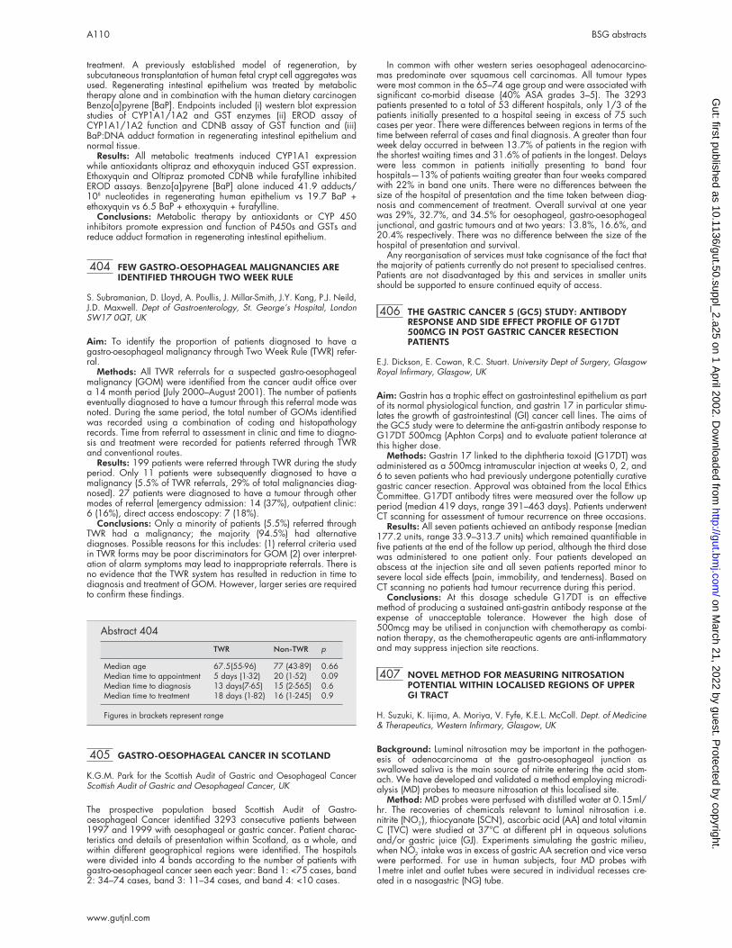
treatment. A previously established model of regeneration, bysubcutaneous transplantation of human fetal crypt cell aggregates wasused. Regenerating intestinal epithelium was treated by metabolictherapy alone and in combination with the human dietary carcinogenBenzo[a]pyrene [BaP]. Endpoints included (i) western blot expressionstudies of CYP1A1/1A2 and GST enzymes (ii) EROD assay ofCYP1A1/1A2 function and CDNB assay of GST function and (iii)BaP:DNA adduct formation in regenerating intestinal epithelium andnormal tissue.
Results: All metabolic treatments induced CYP1A1 expressionwhile antioxidants oltipraz and ethoxyquin induced GST expression.Ethoxyquin and Oltipraz promoted CDNB while furafylline inhibitedEROD assays. Benzo[a]pyrene [BaP] alone induced 41.9 adducts/108 nucleotides in regenerating human epithelium vs 19.7 BaP +ethoxyquin vs 6.5 BaP + ethoxyquin + furafylline.
Conclusions: Metabolic therapy by antioxidants or CYP 450inhibitors promote expression and function of P450s and GSTs andreduce adduct formation in regenerating intestinal epithelium.
404 FEW GASTRO-OESOPHAGEAL MALIGNANCIES AREIDENTIFIED THROUGH TWO WEEK RULE
S. Subramanian, D. Lloyd, A. Poullis, J. Millar-Smith, J.Y. Kang, P.J. Neild,J.D. Maxwell. Dept of Gastroenterology, St. George’s Hospital, LondonSW17 0QT, UK
Aim: To identify the proportion of patients diagnosed to have agastro-oesophageal malignancy through Two Week Rule (TWR) refer-ral.
Methods: All TWR referrals for a suspected gastro-oesophagealmalignancy (GOM) were identified from the cancer audit office overa 14 month period (July 2000–August 2001). The number of patientseventually diagnosed to have a tumour through this referral mode wasnoted. During the same period, the total number of GOMs identifiedwas recorded using a combination of coding and histopathologyrecords. Time from referral to assessment in clinic and time to diagno-sis and treatment were recorded for patients referred through TWRand conventional routes.
Results: 199 patients were referred through TWR during the studyperiod. Only 11 patients were subsequently diagnosed to have amalignancy (5.5% of TWR referrals, 29% of total malignancies diag-nosed). 27 patients were diagnosed to have a tumour through othermodes of referral (emergency admission: 14 (37%), outpatient clinic:6 (16%), direct access endoscopy: 7 (18%).
Conclusions: Only a minority of patients (5.5%) referred throughTWR had a malignancy; the majority (94.5%) had alternativediagnoses. Possible reasons for this includes: (1) referral criteria usedin TWR forms may be poor discriminators for GOM (2) over interpret-ation of alarm symptoms may lead to inappropriate referrals. There isno evidence that the TWR system has resulted in reduction in time todiagnosis and treatment of GOM. However, larger series are requiredto confirm these findings.
405 GASTRO-OESOPHAGEAL CANCER IN SCOTLAND
K.G.M. Park for the Scottish Audit of Gastric and Oesophageal CancerScottish Audit of Gastric and Oesophageal Cancer, UK
The prospective population based Scottish Audit of Gastro-oesophageal Cancer identified 3293 consecutive patients between1997 and 1999 with oesophageal or gastric cancer. Patient charac-teristics and details of presentation within Scotland, as a whole, andwithin different geographical regions were identified. The hospitalswere divided into 4 bands according to the number of patients withgastro-oesophageal cancer seen each year: Band 1: <75 cases, band2: 34–74 cases, band 3: 11–34 cases, and band 4: <10 cases.
In common with other western series oesophageal adenocarcino-mas predominate over squamous cell carcinomas. All tumour typeswere most common in the 65–74 age group and were associated withsignificant co-morbid disease (40% ASA grades 3–5). The 3293patients presented to a total of 53 different hospitals, only 1/3 of thepatients initially presented to a hospital seeing in excess of 75 suchcases per year. There were differences between regions in terms of thetime between referral of cases and final diagnosis. A greater than fourweek delay occurred in between 13.7% of patients in the region withthe shortest waiting times and 31.6% of patients in the longest. Delayswere less common in patients initially presenting to band fourhospitals—13% of patients waiting greater than four weeks comparedwith 22% in band one units. There were no differences between thesize of the hospital of presentation and the time taken between diag-nosis and commencement of treatment. Overall survival at one yearwas 29%, 32.7%, and 34.5% for oesophageal, gastro-oesophagealjunctional, and gastric tumours and at two years: 13.8%, 16.6%, and20.4% respectively. There was no difference between the size of thehospital of presentation and survival.
Any reorganisation of services must take cognisance of the fact thatthe majority of patients currently do not present to specialised centres.Patients are not disadvantaged by this and services in smaller unitsshould be supported to ensure continued equity of access.
406 THE GASTRIC CANCER 5 (GC5) STUDY: ANTIBODYRESPONSE AND SIDE EFFECT PROFILE OF G17DT500MCG IN POST GASTRIC CANCER RESECTIONPATIENTS
E.J. Dickson, E. Cowan, R.C. Stuart. University Dept of Surgery, GlasgowRoyal Infirmary, Glasgow, UK
Aim: Gastrin has a trophic effect on gastrointestinal epithelium as partof its normal physiological function, and gastrin 17 in particular stimu-lates the growth of gastrointestinal (GI) cancer cell lines. The aims ofthe GC5 study were to determine the anti-gastrin antibody response toG17DT 500mcg (Aphton Corps) and to evaluate patient tolerance atthis higher dose.
Methods: Gastrin 17 linked to the diphtheria toxoid (G17DT) wasadministered as a 500mcg intramuscular injection at weeks 0, 2, and6 to seven patients who had previously undergone potentially curativegastric cancer resection. Approval was obtained from the local EthicsCommittee. G17DT antibody titres were measured over the follow upperiod (median 419 days, range 391–463 days). Patients underwentCT scanning for assessment of tumour recurrence on three occasions.
Results: All seven patients achieved an antibody response (median177.2 units, range 33.9–313.7 units) which remained quantifiable infive patients at the end of the follow up period, although the third dosewas administered to one patient only. Four patients developed anabscess at the injection site and all seven patients reported minor tosevere local side effects (pain, immobility, and tenderness). Based onCT scanning no patients had tumour recurrence during this period.
Conclusions: At this dosage schedule G17DT is an effectivemethod of producing a sustained anti-gastrin antibody response at theexpense of unacceptable tolerance. However the high dose of500mcg may be utilised in conjunction with chemotherapy as combi-nation therapy, as the chemotherapeutic agents are anti-inflammatoryand may suppress injection site reactions.
407 NOVEL METHOD FOR MEASURING NITROSATIONPOTENTIAL WITHIN LOCALISED REGIONS OF UPPERGI TRACT
H. Suzuki, K. Iijima, A. Moriya, V. Fyfe, K.E.L. McColl. Dept. of Medicine& Therapeutics, Western Infirmary, Glasgow, UK
Background: Luminal nitrosation may be important in the pathogen-esis of adenocarcinoma at the gastro-oesophageal junction asswallowed saliva is the main source of nitrite entering the acid stom-ach. We have developed and validated a method employing microdi-alysis (MD) probes to measure nitrosation at this localised site.
Method: MD probes were perfused with distilled water at 0.15ml/hr. The recoveries of chemicals relevant to luminal nitrosation i.e.nitrite (NO2
-), thiocyanate (SCN-), ascorbic acid (AA) and total vitaminC (TVC) were studied at 37°C at different pH in aqueous solutionsand/or gastric juice (GJ). Experiments simulating the gastric milieu,when NO2
- intake was in excess of gastric AA secretion and vice versawere performed. For use in human subjects, four MD probes with1metre inlet and outlet tubes were secured in individual recesses cre-ated in a nasogastric (NG) tube.
Abstract 404
TWR Non-TWR p
Median age 67.5(55-96) 77 (43-89) 0.66Median time to appointment 5 days (1-32) 20 (1-52) 0.09Median time to diagnosis 13 days(7-65) 15 (2-565) 0.6Median time to treatment 18 days (1-82) 16 (1-245) 0.9
Figures in brackets represent range
A110 BSG abstracts
www.gutjnl.com
on March 21, 2022 by guest. P
rotected by copyright.http://gut.bm
j.com/
Gut: first published as 10.1136/gut.50.suppl_2.a25 on 1 A
pril 2002. Dow
nloaded from
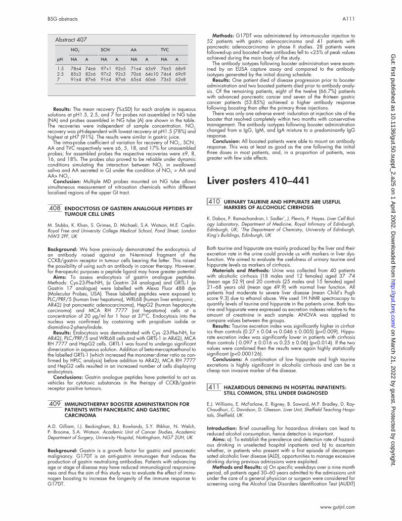
Results: The mean recovery (%±SD) for each analyte in aqueoussolutions at pH1.5, 2.5, and 7 for probes not assembled in NG tube(NA) and probes assembled in NG tube (A) are shown in the table.The recoveries were independent of sample concentration. NO2
-
recovery was pH-dependent with lowest recovery at pH1.5 (78%) andhighest at pH7 (91%). The results were similar in gastric juice.
The intra-probe coefficient of variation for recovery of NO2-, SCN-,
AA and TVC respectively were ±6, 5, 18, and 17% for unassembledprobes; for assembled probes the respective recoveries were ±9, 8,16, and 18%. The probes also proved to be reliable under dynamicconditions simulating the interaction between NO2
- in swallowedsaliva and AA secreted in GJ under the condition of NO2
- > AA andAA> NO2
-.Conclusion: Multiple MD probes mounted on NG tube allows
simultaneous measurement of nitrosation chemicals within differentlocalised regions of the upper GI tract.
408 ENDOCYTOSIS OF GASTRIN ANALOGUE PEPTIDES BYTUMOUR CELL LINES
M. Stubbs, K. Khan, S. Grimes, D. Michaeli, S.A. Watson, M.E. Caplin.Royal Free and University College Medical School, Pond Street, LondonNW3 2PF, UK
Background: We have previously demonstrated the endocytosis ofan antibody raised against an N-terminal fragment of theCCKB/gastrin receptor in tumour cells bearing the latter. This raisedthe possibility of using such an antibody in cancer therapy. However,for therapeutic purposes a peptide ligand may have greater potential
Aims: To assess endocytosis of gastrin analogue peptides.Methods: Cys-23-Phe-NH2 (a Gastrin 34 analogue) and GRTL-1 (aGastrin 17 analogue) were labelled with Alexa Fluor 488 dye(Molecular Probes, USA). These labelled peptides were exposed toPLC/PRF/5 (human liver hepatoma), WRL68 (human liver embryonic ,AR42J (rat pancreatic adenocarcinoma), HepG2 (human hepatocytecarcinoma) and MCA RH 7777 (rat hepatoma) cells at aconcentration of 20 µg/ml for 1 hour at 37°C. Endocytosis into thenucleus was confirmed by costaining with propidium iodide ordiamidino-2-phenylindole.
Results: Endocytosis was demonstrated with Cys -23-Phe-NH2 forAR42J, PLC/PRF/5 and WRL68 cells and with GRTL-1 in AR42J, MCARH 7777 and HepG2 cells. GRTL-1 was found to undergo significantdimerization in aqueous solution. Addition of beta-mercaptoethanol tothe labelled GRTL-1 (which increased the monomer:dimer ratio as con-firmed by HPLC analysis) before addition to AR42J, MCA RH 7777and HepG2 cells resulted in an increased number of cells displayingendocytosis.
Conclusions: Gastrin analogue peptides have potential to act asvehicles for cytotoxic substances in the therapy of CCKB/gastrinreceptor positive tumours.
409 IMMUNOTHERPAY BOOSTER ADMINISTRATION FORPATIENTS WITH PANCREATIC AND GASTRICCARCINOMA
A.D. Gilliam, I.J. Beckingham, B.J. Rowlands, S.Y. Iftikhar, N. Welch,P. Broome, S.A. Watson. Academic Unit of Cancer Studies, AcademicDepartment of Surgery, University Hospital, Nottingham, NG7 2UH, UK
Background: Gastrin is a growth factor for gastric and pancreaticmalignancy. G17DT is an anti-gastrin immunogen that induces theproduction of gastrin neutralising antibodies. Patients with advancingage or stage of disease may have reduced immunological responsive-ness and thus the aim of this study was to evaluate the effect of immu-nogen boosting to increase the longevity of the immune response toG17DT.
Methods: G17DT was administered by intra-muscular injection to52 patients with gastric adenocarcinoma and 41 patients withpancreatic adenocarcinoma in phase II studies. 28 patients werefollowed-up and boosted when antibodies fell to <25% of peak valuesachieved during the main body of the study.
The antibody isotypes following booster administration were exam-ined by an ELISA capture assay and compared to the antibodyisotypes generated by the initial dosing schedule.
Results: One patient died of disease progression prior to boosteradministration and two boosted patients died prior to antibody analy-sis. Of the remaining patients, eight of the twelve (66.7%) patientswith advanced pancreatic cancer and seven of the thirteen gastriccancer patients (53.85%) achieved a higher antibody responsefollowing boosting than after the primary three injections.
There was only one adverse event: induration at injection site of thebooster that resolved completely within two months with conservativemanagement. The antibody isotypes following booster administrationchanged from a IgG, IgM, and IgA mixture to a predominantly IgGresponse.
Conclusion: All boosted patients were able to mount an antibodyresponse. This was at least as good as the one following the initialthree doses in most patients, and, in a proportion of patients, wasgreater with few side effects.
Liver posters 410–441
410 URINARY TAURINE AND HIPPURATE ARE USEFULMARKERS OF ALCOHOLIC CIRRHOSIS
K. Dabos, P. Ramachandran, I. Sadler1, J. Plevris, P. Hayes. Liver Cell Biol-ogy Laboratory, Department of Medicine, Royal Infirmary of Edinburgh,Edinburgh, UK; 1The Department of Chemistry, University of Edinburgh,King’s Buildings, Edinburgh, UK
Both taurine and hippurate are mainly produced by the liver and theirexcretion rate in the urine could provide us with markers in liver dys-function. We aimed to evaluate the usefulness of urinary taurine andhippurate levels as markers of cirrhosis.
Materials and Methods: Urine was collected from 40 patientswith alcoholic cirrhosis (18 males and 12 females) aged 37 -74(mean age 52.9) and 20 controls (25 males and 15 females) aged21–68 years old (mean age 49.9) with normal liver function. Allpatients had moderate to severe liver disease (mean Child’s Pughscore 9.3) due to ethanol abuse. We used 1H NMR spectroscopy toquantify levels of taurine and hippurate in the patients urine. Both tau-rine and hippurate were expressed as excretion indexes relative to theamount of creatinine in each sample. ANOVA was applied tocompare values between the groups.
Results: Taurine excretion index was significantly higher in cirrhot-ics than controls (0.27 ± 0.04 vs 0.046 ± 0.005) (p<0.009). Hippu-rate excretion index was significantly lower in patients with cirrhosisthan controls ( 0.097 ± 0.016 vs 0.25 ± 0.06) (p<0.014). If the twovalues were combined then the results were again highly statisticallysignificant (p<0.000126).
Conclusions: A combination of low hippurate and high taurineexcretions is highly significant in alcoholic cirrhosis and can be acheap non invasive marker of the disease.
411 HAZARDOUS DRINKING IN HOSPITAL INPATIENTS:STILL COMMON, STILL UNDER DIAGNOSED
E.J. Williams, E. McFarlane, E. Rigney, B. Saward, M.P. Bradley, D. Ray-Chaudhuri, C. Davidson, D. Gleeson. Liver Unit, Sheffield Teaching Hospi-tals, Sheffield, UK
Introduction: Brief counselling for hazardous drinkers can lead toreduced alcohol consumption, hence detection is important.
Aims: a): To establish the prevalence and detection rate of hazard-ous drinking in unselected hospital inpatients and b) to ascertainwhether, in patients who present with a first episode of decompen-sated alcoholic liver disease (ALD), opportunities to manage excessivedrinking during previous admissions were exploited.
Methods and Results: a) On specific weekdays over a nine monthperiod, all patients aged 30–60 years admitted to the admissions unitunder the care of a general physician or surgeon were considered forscreening using the Alcohol Use Disorders Identification Test (AUDIT)
Abstract 407
pH
NO2- SCN- AA TVC
NA A NA A NA A NA A
1.5 78±4 74±6 97+1 92±5 71±4 63±9 76±5 68±92.5 85±3 82±6 97±2 92±3 70±6 64±10 74±4 69±97 91±4 87±6 91±4 87±6 65±4 60±6 73±5 62±8
BSG abstracts A111
www.gutjnl.com
on March 21, 2022 by guest. P
rotected by copyright.http://gut.bm
j.com/
Gut: first published as 10.1136/gut.50.suppl_2.a25 on 1 A
pril 2002. Dow
nloaded from
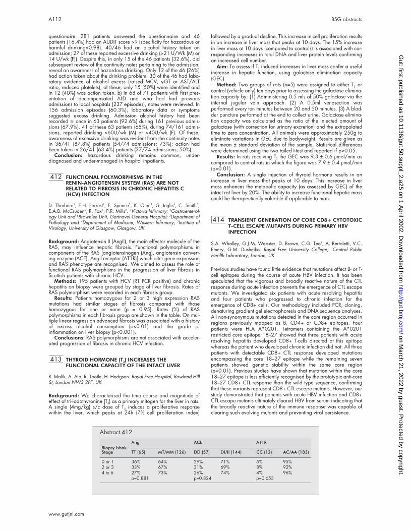
questionaire. 281 patients answered the questionnaire and 46patients (16.4%) had an AUDIT score >9 (specificity for hazardous orharmful drinking=0.98). 40/46 had an alcohol history taken onadmission; 27 of these reported excessive drinking (>21 U/Wk (M) or14 U/wk (F)). Despite this, in only 15 of the 46 patients (32.6%), didsubsequent review of the continuity notes pertaining to the admission,reveal an awareness of hazardous drinking. Only 12 of the 46 (26%)had action taken about the drinking problem. 30 of the 46 had labo-ratory evidence of alcohol excess (raised MCV, γ-GT or AST/ALTratio, reduced platelets); of these, only 15 (50%) were identified andin 12 (40%) was action taken. b) In 68 of 71 patients with first pres-entation of decompensated ALD and who had had previousadmissions to local hospitals (237 episodes), notes were reviewed. In156 admission episodes (60.3%), laboratory data or symptomssuggested excess drinking. Admission alcohol history had beenrecorded > once in 63 patients (92.6%) during 161 previous admis-sions (67.9%). 41 of these 63 patients (65%), during 74/161 admis-sions, reported drinking >60U/wk (M) or >40U/wk (F). Of these,awareness of excessive drinking was evident from the continuity notesin 36/41 (87.8%) patients (54/74 admissions; 73%); action hadbeen taken in 26/41 (63.4%) patients (37/74 admissions; 50%).
Conclusion: hazardous drinking remains common, under-diagnosed and under-managed in hospital inpatients.
412 FUNCTIONAL POLYMORPHISMS IN THERENIN-ANGIOTENSIN SYSTEM (RAS) ARE NOTRELATED TO FIBROSIS IN CHRONIC HEPATITIS C(HCV) INFECTION
D. Thorburn1, E.H. Forrest1, E. Spence2, K. Oien3, G. Inglis4, C. Smith5,E.A.B. McCruden5, R. Fox6, P.R. Mills2. 1Victoria Infirmary; 2Gastroenterol-ogy Unit and 6Brownlee Unit, Gartnavel General Hospital; 3Department ofPathology and 4Department of Medicine, Western Infirmary; 5Institute ofVirology, University of Glasgow, Glasgow, UK
Background: Angiotensin II (AngII), the main effector molecule of theRAS, may influence hepatic fibrosis. Functional polymorphisms incomponents of the RAS [angiotensinogen (Ang), angiotensin convert-ing enzyme (ACE), AngII receptor (AT1R)] which alter gene expressionand RAS phenotype are recognised. We aimed to assess the role offunctional RAS polymorphisms in the progression of liver fibrosis inScottish patients with chronic HCV.
Methods: 195 patients with HCV (RT PCR positive) and chronichepatitis on biopsy were grouped by stage of liver fibrosis. Rates ofRAS polymorphism were recorded in each fibrosis group.
Results: Patients homozygous for 2 or 3 high expression RASmutations had similar stages of fibrosis compared with thosehomozygous for one or none (p = 0.95). Rates (%) of RASpolymorphisms in each fibrosis group are shown in the table. On mul-tiple linear regression advanced fibrosis was associated with a historyof excess alcohol consumption (p<0.01) and the grade ofinflammation on liver biopsy (p<0.001).
Conclusions: RAS polymorphisms are not associated with acceler-ated progression of fibrosis in chronic HCV infection.
413 THYROID HORMONE (T3) INCREASES THEFUNCTIONAL CAPACITY OF THE INTACT LIVER
R. Malik, A. Ala, R. Tootle, H. Hodgson. Royal Free Hospital, Rowland HillSt, London NW3 2PF, UK
Background: We characterised the time course and magnitude ofeffect of tri-iodothyronine (T3) as a primary mitogen for the liver in rats.A single (4mg/kg) s/c dose of T3 induces a proliferative responsewithin the liver, which peaks at 24h (7% cell proliferation index)
followed by a gradual decline. This increase in cell proliferation resultsin an increase in liver mass that peaks at 10 days. The 15% increasein liver mass at 10 days (compared to controls) is associated with cor-responding increases in total DNA and liver protein levels confirmingan increased cell number.
Aim: To assess if T3 induced increases in liver mass confer a usefulincrease in hepatic function, using galactose elimination capacity(GEC).
Method: Two groups of rats (n=5) were assigned to either T3 orcontrol (vehicle only) ten days prior to assessing the galactose elimina-tion capacity by: (1) Administering 0.5 mls of 50% galactose via theinternal jugular vein approach. (2) A 0.5ml venesection wasperformed every ten minutes between 20 and 50 minutes. (3) A blad-der puncture performed at the end to collect urine. Galactose elimina-tion capacity was calculated as the ratio of the injected amount ofgalactose (with correction for urinary excretion) and the extrapolatedtime to zero concentration. All animals were approximately 250g toeliminate variations in GEC due to bodyweight. Results are given asthe mean ± standard deviation of the sample. Statistical differenceswere determined using the two tailed t-test and reported if p<0.05.
Results: In rats receiving T3 the GEC was 9.3 ± 0.6 µmol/min ascompared to control rats in which the figure was 7.9 ± 0.4 µmol/min(p<0.01).
Conclusion: A single injection of thyroid hormone results in anincrease in liver mass that peaks at 10 days. This increase in livermass enhances the metabolic capacity (as assessed by GEC) of theintact rat liver by 20%. The ability to increase functional hepatic masscould be therapeutically valuable if applicable to man.
414 TRANSIENT GENERATION OF CORE CD8+ CYTOTOXICT-CELL ESCAPE MUTANTS DURING PRIMARY HBVINFECTION
S.A. Whalley, G.J.M. Webster, D. Brown, C.G. Teo1, A. Bertoletti, V.C.Emery, G.M. Dusheiko. Royal Free University College; 1Central PublicHealth Laboratory, London, UK
Previous studies have found little evidence that mutations affect B- or T-cell epitopes during the course of acute HBV infection. It has beenspeculated that the vigorous and broadly reactive nature of the CTLresponse during acute infection prevents the emergence of CTL escapemutants. We investigated six patients with acute resolving hepatitisand four patients who progressed to chronic infection for theemergence of CD8+ cells. Our methodology included PCR, cloning,denaturing gradient gel electrophoresis and DNA sequence analyses.All non-synonymous mutations detected in the core region occurred inregions previously mapped as B, CD4+ or CD8+ epitopes. Fourpatients were HLA A*0201. Tetramers containing the A*0201restricted core epitope 18–27 showed that three patients with acuteresolving hepatitis developed CD8+ T-cells directed at this epitopewhereas the patient who developed chronic infection did not. All threepatients with detectable CD8+ CTL response developed mutationsencompassing the core 18–27 epitope while the remaining sevenpatients showed genetic stability within the same core region(p=0.01). Previous studies have shown that mutation within the core18–27 epitope is less efficiently recognised by the prototypic anti-core18–27 CD8+ CTL response than the wild type sequence, confirmingthat these variants represent CD8+ CTL escape mutants. However, ourstudy demonstrated that patients with acute HBV infection and CD8+CTL escape mutants ultimately cleared HBV from serum indicating thatthe broadly reactive nature of the immune response was capable ofclearing such evolving mutants and preventing viral persistence.
Abstract 412
Biopsy IshakStage
Ang ACE AT1R
TT (65) MT/MM (126) DD (57) DI/II (144) CC (12) AC/AA (183)
0 or 1 36% 64% 29% 71% 5% 95%2 or 3 33% 67% 31% 69% 8% 92%4 to 6 27% 73% 26% 74% 4% 96%
p=0.881 p=0.824 p=0.653
A112 BSG abstracts
www.gutjnl.com
on March 21, 2022 by guest. P
rotected by copyright.http://gut.bm
j.com/
Gut: first published as 10.1136/gut.50.suppl_2.a25 on 1 A
pril 2002. Dow
nloaded from
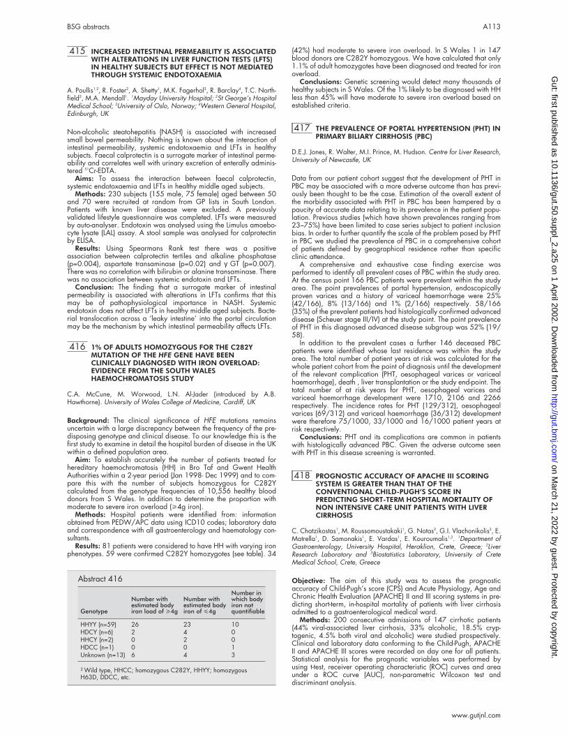
415 INCREASED INTESTINAL PERMEABILITY IS ASSOCIATEDWITH ALTERATIONS IN LIVER FUNCTION TESTS (LFTS)IN HEALTHY SUBJECTS BUT EFFECT IS NOT MEDIATEDTHROUGH SYSTEMIC ENDOTOXAEMIA
A. Poullis1,2, R. Foster2, A. Shetty1, M.K. Fagerhol3, R. Barclay4, T.C. North-field2, M.A. Mendall1. 1Mayday University Hospital; 2St George’s HospitalMedical School; 3University of Oslo, Norway; 4Western General Hospital,Edinburgh, UK
Non-alcoholic steatohepatitis (NASH) is associated with increasedsmall bowel permeability. Nothing is known about the interaction ofintestinal permeability, systemic endotoxaemia and LFTs in healthysubjects. Faecal calprotectin is a surrogate marker of intestinal perme-ability and correlates well with urinary excretion of enterally adminis-tered 51Cr-EDTA.
Aims: To assess the interaction between faecal calprotectin,systemic endotoxaemia and LFTs in healthy middle aged subjects.
Methods: 230 subjects (155 male, 75 female) aged between 50and 70 were recruited at random from GP lists in South London.Patients with known liver disease were excluded. A previouslyvalidated lifestyle questionnaire was completed. LFTs were measuredby auto-analyser. Endotoxin was analysed using the Limulus amoebo-cyte lysate (LAL) assay. A stool sample was analysed for calprotectinby ELISA.
Results: Using Spearmans Rank test there was a positiveassociation between calprotectin tertiles and alkaline phosphatase(p=0.004), aspartate transaminase (p=0.02) and γ GT (p=0.007).There was no correlation with bilirubin or alanine transaminase. Therewas no association between systemic endotoxin and LFTs.
Conclusion: The finding that a surrogate marker of intestinalpermeability is associated with alterations in LFTs confirms that thismay be of pathophysiological importance in NASH. Systemicendotoxin does not affect LFTs in healthy middle aged subjects. Bacte-rial translocation across a ‘leaky intestine’ into the portal circulationmay be the mechanism by which intestinal permeability affects LFTs.
416 1% OF ADULTS HOMOZYGOUS FOR THE C282YMUTATION OF THE HFE GENE HAVE BEENCLINICALLY DIAGNOSED WITH IRON OVERLOAD:EVIDENCE FROM THE SOUTH WALESHAEMOCHROMATOSIS STUDY
C.A. McCune, M. Worwood, L.N. Al-Jader (introduced by A.B.Hawthorne). University of Wales College of Medicine, Cardiff, UK
Background: The clinical significance of HFE mutations remainsuncertain with a large discrepancy between the frequency of the pre-disposing genotype and clinical disease. To our knowledge this is thefirst study to examine in detail the hospital burden of disease in the UKwithin a defined population area.
Aim: To establish accurately the number of patients treated forhereditary haemochromatosis (HH) in Bro Taf and Gwent HealthAuthorities within a 2-year period (Jan 1998- Dec 1999) and to com-pare this with the number of subjects homozygous for C282Ycalculated from the genotype frequencies of 10,556 healthy blooddonors from S Wales. In addition to determine the proportion withmoderate to severe iron overload (>4g iron).
Methods: Hospital patients were identified from: informationobtained from PEDW/APC data using ICD10 codes; laboratory dataand correspondence with all gastroenterology and haematology con-sultants.
Results: 81 patients were considered to have HH with varying ironphenotypes. 59 were confirmed C282Y homozygotes (see table). 34
(42%) had moderate to severe iron overload. In S Wales 1 in 147blood donors are C282Y homozygous. We have calculated that only1.1% of adult homozygotes have been diagnosed and treated for ironoverload.
Conclusions: Genetic screening would detect many thousands ofhealthy subjects in S Wales. Of the 1% likely to be diagnosed with HHless than 45% will have moderate to severe iron overload based onestablished criteria.
417 THE PREVALENCE OF PORTAL HYPERTENSION (PHT) INPRIMARY BILIARY CIRRHOSIS (PBC)
D.E.J. Jones, R. Walter, M.I. Prince, M. Hudson. Centre for Liver Research,University of Newcastle, UK
Data from our patient cohort suggest that the development of PHT inPBC may be associated with a more adverse outcome than has previ-ously been thought to be the case. Estimation of the overall extent ofthe morbidity associated with PHT in PBC has been hampered by apaucity of accurate data relating to its prevalence in the patient popu-lation. Previous studies (which have shown prevalences ranging from23–75%) have been limited to case series subject to patient inclusionbias. In order to further quantify the scale of the problem posed by PHTin PBC we studied the prevalence of PBC in a comprehensive cohortof patients defined by geographical residence rather than specificclinic attendance.
A comprehensive and exhaustive case finding exercise wasperformed to identify all prevalent cases of PBC within the study area.At the census point 166 PBC patients were prevalent within the studyarea. The point prevalences of portal hypertension, endoscopicallyproven varices and a history of variceal haemorrhage were 25%(42/166), 8% (13/166) and 1% (2/166) respectively. 58/166(35%) of the prevalent patients had histologically confirmed advanceddisease (Scheuer stage III/IV) at the study point. The point prevalenceof PHT in this diagnosed advanced disease subgroup was 52% (19/58).
In addition to the prevalent cases a further 146 deceased PBCpatients were identified whose last residence was within the studyarea. The total number of patient years at risk was calculated for thewhole patient cohort from the point of diagnosis until the developmentof the relevant complication (PHT, oesophageal varices or varicealhaemorrhage), death , liver transplantation or the study end-point. Thetotal number of at risk years for PHT, oesophageal varices andvariceal haemorrhage development were 1710, 2106 and 2266respectively. The incidence rates for PHT (129/312), oesophagealvarices (69/312) and variceal haemorrhage (36/312) developmentwere therefore 75/1000, 33/1000 and 16/1000 patient years atrisk respectively.
Conclusions: PHT and its complications are common in patientswith histologically advanced PBC. Given the adverse outcome seenwith PHT in this disease screening is warranted.
418 PROGNOSTIC ACCURACY OF APACHE III SCORINGSYSTEM IS GREATER THAN THAT OF THECONVENTIONAL CHILD-PUGH’S SCORE INPREDICTING SHORT-TERM HOSPITAL MORTALITY OFNON INTENSIVE CARE UNIT PATIENTS WITH LIVERCIRRHOSIS
C. Chatzikostas1, M. Roussomoustakaki1, G. Notas2, G.I. Vlachonikolis3, E.Matrella1, D. Samonakis1, E. Vardas1, E. Kouroumalis1,2. 1Department ofGastroenterology, University Hospital, Heraklion, Crete, Greece; 2LiverResearch Laboratory and 3Biostatistics Laboratory, University of CreteMedical School, Crete, Greece
Objective: The aim of this study was to assess the prognosticaccuracy of Child-Pugh’s score (CPS) and Acute Physiology, Age andChronic Health Evaluation (APACHE) II and III scoring systems in pre-dicting short-term, in-hospital mortality of patients with liver cirrhosisadmitted to a gastroenterological medical ward.
Methods: 200 consecutive admissions of 147 cirrhotic patients(44% viral-associated liver cirrhosis, 33% alcoholic, 18.5% cryp-togenic, 4.5% both viral and alcoholic) were studied prospectively.Clinical and laboratory data conforming to the Child-Pugh, APACHEII and APACHE III scores were recorded on day one for all patients.Statistical analysis for the prognostic variables was performed byusing t-test, receiver operating characteristic (ROC) curves and areaunder a ROC curve (AUC), non-parametric Wilcoxon test anddiscriminant analysis.
Abstract 416
Genotype†
Number withestimated bodyiron load of >4g
Number withestimated bodyiron of <4g
Number inwhich bodyiron notquantifiable
HHYY (n=59) 26 23 10HDCY (n=6) 2 4 0HHCY (n=2) 0 2 0HDCC (n=1) 0 0 1Unknown (n=13) 6 4 3
†Wild type, HHCC; homozygous C282Y, HHYY; homozygousH63D, DDCC, etc.
BSG abstracts A113
www.gutjnl.com
on March 21, 2022 by guest. P
rotected by copyright.http://gut.bm
j.com/
Gut: first published as 10.1136/gut.50.suppl_2.a25 on 1 A
pril 2002. Dow
nloaded from
Results: The in-hospital mortality was 11.5%. The mean CPS,APACHE II and III scores for survivors were found to be significantlylower than those of nonsurvivors. When ROC curves were plotted, nosignificant differences between Child-Pugh’s (AUC, 0.85), APACHE II(AUC, 0.75), and APACHE III (AUC, 0.81) overall performances werenoticed, however the overall correctness of prediction of APACHE IIIwas 9% greater than that of the CPS (Wilcoxon test: z = 2.846, p =0.004) and 11% greater than that of the APACHE II, namely, 87%,78% and 76% respectively (cutoff values, 62,10 and 15 respectively).
Conclusions: All three scores were proven to be of value in riskstratifying patients with liver cirrhosis. Although the overallperformance of APACHE III system as assessed by ROC curve analy-sis is no superior than that of the conventional Child-Pugh’s score inpredicting short term outcome of hospitalized patients with liver cirrho-sis, APACHE III correctly statifies a significantly greater number ofpatients.
419 HYPERURICAEMIA, GOUT AND CARDIOVASCULARRISK AFTER LIVER TRANSPLANTATION
D.A.J. Neal1, B.D.M. Tom2, A.E.S. Gimson1, P. Gibbs3, G.J.M. Alexander1.1Department of Medicine and 3University Department of Surgery,Addenbrooke’s NHS Trust, Cambridge, CB2 2QQ, UK; 2Centre forApplied Medical Statistics, Department of Public Health and Primary Care,Cambridge CB2 2SR, UK
Background: Hyperuricaemia and gout are recognised complica-tions of renal and cardiac transplantation. In contrast the developmentof hyperuricaemia following liver transplantation has received lessattention. Elevated serum uric acid has been cited as an independentrisk factor for cardiovascular disease in the general population. Toevaluate the prevalence of hyperuricaemia and its association withcardiovascular risk factors we reviewed the case records of 134 con-secutive liver transplant recipients with a mean follow up of 52 months(range 6 – 92 months).
Results: 47% had hyperuricaemia after liver transplant. Peak uricacid correlated significantly with corresponding serum creatinine (rs =0.694). 6% of patients developed an acute episode of gout.Hypertension, hypercholesterolaemia and a body mass index >25kg/m2 were present in 53, 46 and 48 % of hyperuricaemic patientsrespectively and in 47, 54 and 52% of patients with normal serumurate. None of these differences were significant. Cardiovascularevents comprised 1 myocardial infarct and 1 incident angina, eachpatient having hyperuricaemia, and 2 strokes, one of which hadhyperuricaemia.
Conclusions: There is an important association between livertransplantation and hyperuricaemia. Gout is a significant cause ofmorbidity but occurs less frequently than after renal or cardiac trans-plants. There was no association between hyperuricaemia and othercardiovascular risk factors. Too few cardiac events occurred to drawany conclusions about an association with uric acid.
420 OBSTETRIC CHOLESTASIS IN SOUTH WALES
C.L. Ch’ng1, M. Morgan2, J.G.C. Kingham1. Department of Gastroenterol-ogy1 and Obstetrics2, Singleton Hospital, Swansea, Wales, UK
Aims: A prospective study of the incidence, clinical and biochemicalfeatures, management and outcome of obstetric cholestasis (OC) in adefined population in South Wales, UK.
Methods: All pregnancies in an obstetric unit serving 270,000were screened for OC between March 1999 and June 2001.Diagnosis of OC was based on pruritus, abnormal liver tests andexclusion of other hepatobiliary diseases.
Results: 45 OC patients were identified among 8142 pregnancies– incidence 0.5%. Age ranged from 16 - 40 years (Median 30). Therewere seven twin and one triplet pregnancies. Sixteen were primiparaewhile 29 had had 1 to 6 previous pregnancies, ten of which werecomplicated by OC with two associated stillbirth. Three had familyhistory of OC. All were symptomatic: pruritus in 45, vomiting in four,diarrhoea in four and severe malaise in two. Two patients sufferedhyperemesis earlier in pregnancy, two pre-eclampsia and one HELLPsyndrome. Symptoms started between 8 and 39 weeks gestation(median 34). Eighteen had proven urinary tract infection either justbefore or after diagnosis of OC. 43 had elevated AST (31–519;median 141 U/l). Serum bile acids raised in 38 of 41 tested(15–179; median 34 µmol/l). γGT was modestly elevated (42–292;median 71 U/l) in only 14 episodes while bilirubin was raised in 17(16 - 34; median 20 µmol/l). Leucocytosis was seen in 28 patients.
The interval from diagnosis of OC to delivery ranged from 1 to 142days (median 8). Ursodeoxycholic acid was given to eleven patientswith improvement in symptoms in six and biochemistry in ten.Induction of labour or Caesarean section was undertaken in 33because of OC. There was no maternal or fetal death but 15 of 52babies required admission to SCBU. Symptoms and abnormal liverfunction resolved rapidly in 75% within 2 weeks of delivery.
Conclusion: OC complicates 1 in 180 pregnancies in SouthWales. It is characterised by elevated transaminases rather thancholestasis; jaundice is neither a necessary nor a common component.Combined medical and obstetric care with early delivery preventsfetal loss.
421 SURVEILLANCE FOR HEPATOCELLULAR CARCINOMAIN CIRRHOSIS: A NATIONAL AUDIT OF THE PRACTICEIN THE UK
C.W.Y. Lai, M. Wardle, V. Birkett, E. Powell, G.A.O. Thomas. Departmentof Gastroenterology, University Hospital of Wales, Cardiff, UK
Background: Hepatocellular carcinoma (HCC) is a significant causeof mortality in cirrhotic patients. Its detection at an early stageincreases the chance of curative therapy. Clear evidence of survivalbenefit with surveillance is limited. However, anecdotally, many clini-cians undertake some form of surveillance program.
Aim: To evaluate the current practice of HCC surveillance in cirrho-sis in the UK.
Methods: 1080 postal questionnaires were sent to the members ofthe British Society of Gastroenterologists (BSG), excluding radiolo-gists, pathologists and paediatricians.
Results: 525 replied (49%); of these, 120 did not look after adultcirrhotics and were excluded from analysis. Of the 405 remainingrespondents, 296 (73%) surveyed for HCC, 123/296 had protocols.Hepatologists and those with a large cirrhotic practice were morelikely to survey. 96/296 quoted an age limit (median 70) for surveil-lance. 107/296 (36%) surveyed all cirrhotics regardless of their suit-ability for curative therapy; in contrast, 127/296 surveyed only thosesuitable for liver transplantation or partial hepatectomy. 166/296(56%) chose to survey all causes of cirrhosis, whereas a smaller group(83/296, [28%]) were more selective and surveyed those with Hepa-titis B, C, haemachromatosis and alcohol liver disease. The common-est mode of surveillance was a combination of abdominal ultrasoundand alpha fetoprotein, with a wide range of test intervals (3 to 24months). In those choosing to survey, 130/296 believed it increasedsurvival, while 95/296 felt that non-surveillance might leave themlegally liable. 109/405 did not survey, 58 of who quoted the lack ofevidence for survival benefit, and 46 the lack of guidelines, as thereasons for their practice.
Conclusions: Despite the lack of guidelines or clear evidence forbenefit, a majority of clinicians who responded performed some formof surveillance. Practice was variable with a significant number beingunselective in the types of patients to survey. This has obvious resourceimplications. Guidelines are needed to rationalise and clarifypractice.
422 HYDROXYCHLOROQUINE REDUCES LIVER RELATEDMORTALITY IN HEPATITIS C ASSOCIATED (HCV)COMPENSATED CIRRHOSIS
E.A. Kouroumalis, J. Moschandreas, G. Alexandrakis, E.Matrella. Depart-ment of Gastroenterology, University Hospital and Medical School Crete,Greece
Hydroxychloroquine, a lysosomotropic and macrophage modulatingagent has been reported to improve liver biochemistry in chronic viralhepatitis.
Aim: To investigate the effect of hydroxychloroquine on survival ofHCV active cirrhotic patients
Methods: 162 patients ( 31% male, 69% female) withcompensated HCV cirrhosis were prospectively evaluated. All wereChild-Pugh A or B at entry into the study. 52 with active cirrhosis(increased aminotranferases) were treated with hydroxychloroquine200 mg tid for 6 months. A 3 month treatment was reinstituted if dur-ing follow up, aminotranferases were again high. 110 patients withinactive cirrhosis were the non treated controls. Time to decompensa-tion and death were recorded. Patients were followed up between 3and 136 months.
Results: Median time to decompensation was 81 months (95 % C,45–117 months) and was not different between the two groups
A114 BSG abstracts
www.gutjnl.com
on March 21, 2022 by guest. P
rotected by copyright.http://gut.bm
j.com/
Gut: first published as 10.1136/gut.50.suppl_2.a25 on 1 A
pril 2002. Dow
nloaded from
(p=0.7, Kaplan-Meier analysis). However, variceal bleeding, hepaticencephalopathy and spontaneous bacterial peritonitis were signs ofdecompensation only in the controls, ascites being the onlydecompensation sign in the treated group. Overall mortality, althoughlower in the treatment group just failed to reach significance (p< 0.07,356 patients should be enrolled to reach significance). However,when only liver related deaths were assessed, the survival of thetreated patients was significantly better (p< 0.05).
Conclusions: Hydroxychloroquine treatment reduces liver relatedmortality in active HCV cirrhosis, despite the fact that the control groupwere inactive cirrhotics where a more favourable prognosis is to beexpected.
423 MEASUREMENT OF AFP IN PATIENTS WITH HEPATITISC CIRRHOSIS DURING HEPATOMA SURVEILLANCE
T. Woodall, S. Sen, A.E. Gimson, G.J.M. Alexander, D.J. Lomas, S.Vowler. Department of Medicine, University of Cambridge, Hills Road,Cambridge CB2 2QQ, UK
Background: Patients with chronic hepatitis C virus ( HCV) infectionand cirrhosis have an increased risk of hepatocellular carcinoma(HCC).Surveillance to identify early tumours based on a periodic esti-mation of serum alpha-fetoprotein (AFP) is unproven.
Methods: Surveillance was undertaken in 100 consecutive patientswith chronic HCV and cirrhosis, based on a minimum of 6 monthlyserum AFP and ultrasound (US), for a median of 2 years (range 0.5 –6 ). 186 US and 662 AFP measurements were undertaken. 11patients were lost to follow up. 40 (45%) had an elevated AFP and 18had a suspicious US. 11 patients (12%) developed HCC duringsurveillance.
Results: Of 40 with an elevated AFP, 10 (25%) also had a suspi-cious US and HCC was identified in 7 of those (17.5%). The level ofAFP however, was non-discriminatory. 30 of 40 (75%) had anelevated AFP with a clear US and HCC was identified in 1. The levelof AFP was again non-discriminatory. Of 49 with a normal AFP, 8 (16%) had a suspicious US and HCC was identified in 3 (6%). 22 CTScans, 7 MR scans and 11 Angiograms were undertaken in patientswho did not have HCC. As a predictive test for HCC, elevated AFP inisolation had a positive predictive value (PPV) of only 0.18, a sensitiv-ity of 0.7 and a specificity of 0.58. In comparison, US had a PPV of0.8, sensitivity of 0.91 and specificity of 0.89.
Conclusion: The incidence of HCC in this group of Hepatitis C cir-rhotic patients is high. Surveillance is a considerable commitment butthe combination of serum AFP and US has traditionally been used forthe detection of HCC . However,an elevated AFP in isolation wascommon, had a poor predictivity for the detection of HCC and led tonumerous (expensive) investigations. Restricting surveillance to USalone would be as effective.
424 COMBINATION PROPHYLAXIS WITH LAMIVUDINE(LAM) AND HEPATITIS B IMMUNOGLOBULIN (HBIG)PREVENTS GRAFT RE-INFECTION BY HEPATITIS B (HBV)
V. Lai, E. Elias, K. O’Donnell, D. Mutimer. Liver Unit, Queen Elizabeth Hos-pital, Birmingham, Edgbaston B15 2TH, UK
Prevention of graft re-infection remains the most important issue intransplantation of HBV infected patients. Re-infection by HBV is almostinevitable if no prophylaxis is given. The course of recurrent infectionis aggressive causing sub-acute liver failure or rapid progression tocirrhosis. In the early 90’s, prophylaxis comprised of regular andrepeated administration of HBIg. Studies of LAM prophylaxis in thissetting began in 1994. Both LAM and HBIg proved ineffective forpatients with high serum HBV DNA titre.
Subsequently the combination of LAM and HBIg has been used andpreliminary reports suggest the combination is effective and safe. Theaim of this study is to review the QEH liver unit’s experience with com-bination LAM/HBIg for prevention of HBV re-infection.
This is a retrospective review of the clinical and virological outcomeof these patients. Since introduction of LAM/HBIg prophylaxis in1997, 16 HBV patients have undergone liver transplantation(15Male, median age 51). Pretreatment serum HBV DNA titres rangefrom <400 copies/ml to >4 x 10⁷. 3 patients died (12 days, 6 weeksand 8 month post transplant). Median follow up of survivors is 2 years(6months- 4 years). All survivors are serum HBsAg and HBV DNA(Roche PCR-based assay) negative.
Conclusion: LAM/HBIg prevents HBV re-infection even in high-riskpatients.
425 DISTRIBUTION OF IL-6 IN LIVER CIRRHOSIS
N.A. Mohammed, S. Abd El-Aleem, H. Abdel Hafiz, R.F.T. McMahon.Department of Histopathology, Clinical Sciences Building, ManchesterRoyal Infirmary, Oxford Road, Manchester M13 9WL, UK
Interleukin-6 (IL-6) has been proposed as one of the main inflammatorymediators. It is a major mediator of the acute phase response in infec-tious diseases and inflammatory processes inducing fever, leukocyto-sis, and increase in the hepatic synthesis of acute phase proteins. Inthis study, sera and liver biopsies from fifteen patients with clinicallyand pathologically diagnosed liver cirrhosis were taken. In additionsera from 7 and liver biopsies from 3 healthy controls were used.Serum levels of IL-6 were measured using ELISA kits and the cellulardistribution was investigated using immunohistochemistry. We haveshown that the serum IL-6 levels in cirrhotic patients (25.46 ± 11.6pg/ml) were significantly (P <0.05) increased by comparison with thecontrol group (12.14 ± 6.6 pg/ml). Immunohistochemically, in thecontrol group, IL-6 was seen only in occasional sinusoidal cells. How-ever it was widely distributed in the cirrhotic liver. In the latter, it wasmostly seen in the inflammatory cells infiltrating the liver but alsoexpressed in the sinusoidal cells, Kupffer cells, vascular endotheliallining cells and hepatocytes. Upregulation of IL-6 in cirrhotic patientscould be due to active synthesis or defective clearance bynon-functioning hepatocytes. However our findings suggest that bothmechanisms are operating since we have shown high expression ininflammatory cells which could be due to oversynthesis and highexpression in hepatocytes which could be due to accumulation in non-functioning cells. It is therefore clear that IL-6 showed systemic andlocal augmentation in cirrhotic patients and is mainly produced byinflammatory cells. Taken together these findings suggest that IL-6 pro-duction in liver cirrhosis is dependent on the inflammatory stage andthe local production of IL-6 could contribute to the inflammation, fibro-sis and immunological responses in the cirrhotic liver. Moreover thecharacteristic distribution of IL-6 in cirrhotic lobules could implicate itin the development of cirrhosis. In the near future, the appropriatemanipulation of IL-6 may provide a novel strategy for the treatment ofpatients with liver cirrhosis or at least improve the fate of cirrhosis.
426 QUANTITATIVE STUDIES OF LIVER ATROPHYFOLLOWING PORTACAVAL SHUNT IN RATS
A.M. Zaitoun1, G. Apelqvist2, H. Al-Mardini3, T. Gray1, F. Bengtsson2, C.O.Record3. 1Department of Histopathology, Queen’s Medical Centre, Univer-sity Hospital, Nottingham NG7 2UH, UK; 2Department of Clinical Pharma-cology, Lund University, S-221 85 Lund, Sweden; 3Department ofMedicine, The Royal Victoria Infirmary, Newcastle-upon-Tyne NE1 4LP, UK
Background: To evaluate the morphological changes in the liver thatoccur in portal-systemic encephalopathy after portacaval shunting(PCS).
Materials and Methods: Male rats underwent either PCS (n=35)or control sham operations (n=31). Blood samples were taken prior tosacrifice (weeks5–7) for measurement of hormone concentrations.Morphological assessments were carried out on 5µm thickhaematoxylin and eosin stained sections and digital electronmicrographs using the Prodit 5.2 image analysis system.
Results: There was a significant reduction in the liver mass of PCSrats (4.98v13.65g). Testosterone was significantly reduced(2.49v10.1ng/ml;p=0.002) and oestrogen significantly increased(77.9v51.6ng/ml;p=0.0004). Morphometric analysis showed signifi-cant reductions in the average distance between peri and postsinusoi-dal vessels (298v499Nm) in PCS rats while in zone 3 the mean areaof cytoplasm was also reduced (74.3v112.9Nm2). Within the PCSgroup there was a significant reduction in the mean area ofhepatocyte nuclei from 55.52Nm2 in zone 1 to 45.31Nm2 in zone 3and a marked difference in the mean area of cytoplasm (113.25Nm2
in zone 1 to 74.30Nm2 in zone 3). Electron microscopy revealedreduction in the cytoplasmic organelles in PCS rats in comparison withsham rats. Apoptosis was increased in zone 3 to 2.4v0.3 counts permm2 in PCS rats (P=0.00000) while mitosis was significantlyincreased in zone 1.
Conclusion: This study shows that liver atrophy after portacavalshunting has a complex aetiology. The microcirculatory disturbancesand hormonal changes after portacaval shunting induce apoptosisthat further contributes to liver atrophy in this animal model.
BSG abstracts A115
www.gutjnl.com
on March 21, 2022 by guest. P
rotected by copyright.http://gut.bm
j.com/
Gut: first published as 10.1136/gut.50.suppl_2.a25 on 1 A
pril 2002. Dow
nloaded from
427 ROLE OF TECHNICAL VARIABLES IN EARLY SHUNTINSUFFICIENCY FOLLOWING TIPSS FOR THETREATMENT OF PORTAL HYPERTENSION
S. Balata1, H.F. Lui1, A. Helmy1, D. Tripathi1, D.N. Redhead2, P.C. Hayes1.1Liver Unit, Department of Medicine, Royal Infirmary of Edinburgh, Lauris-ton Place, Edinburgh EH3 9YW, Scotland, UK; 2Department of Radiology,Royal Infirmary of Edinburgh, Lauriston Place, Edinburgh EH3 9YW, Scot-land, UK
Summary: Transjugular intrahepatic portosystemic stent shunt (TIPSS)has increasingly been used for the treatment of complications of portalhypertension. This study evaluated the technical predictors of earlyshunt insufficiency after TIPSS placement in patients with advancedliver disease.
A retrospective analysis of 399 patients undergoing TIPSS over aperiod of 9 years between July 1991-July 2000 was carried out. Theindications for TIPSS were bleeding oesophageal varices in 288patients, gastric varices in 52, refractory ascites in 42, portal hyper-tensive gastropathy in 8 and ectopic varices in 9 patients. TIPSSplacement was successful in 383 (96.0%) patients, and failed in 16(4.0%). The shunt remained patent without assistance in 40 (10.0 %)(primary patency) and 44 (11.0%) patients developed early shuntinsufficiency within 30 days from TIPSS insertion. The univariateassociation of 21 prognostic clinical and technical variables betweenpatients with early shunt insufficiency and primary patency was testedby the Chi-squared or Wilcoxon rank-sum tests. Multiple logisticregression analysis was utilized to determine the relationship of multi-ple technical and clinical variables in predicting early shunt outcome.
The two groups were comparable and representative of the wholeTIPSS cohort patients. Of the 21 technical and clinical variables, stentdiameter, distance of shunt from IVC, duration of the procedure, andportal pressure gradients post TIPSS were independent predictors ofearly shunt insufficiency.
Based on our analysis, four technical variables at index TIPSS canreliably predict early shunt insufficiency. Total protection from earlycomplications of TIPSS requires awareness of the risk factors and alow threshold maintained for early shunt revision.
428 CHARACTERISATION OF LIVER INFILTRATING T-CELLANTIGEN SPECIFICITY IN A MOUSE MODEL OFPRIMARY BILIARY CIRRHOSIS (PBC)
A.J. Robe, J.M. Palmer, J.A. Kirby, M.F. Bassendine, D.E.J. Jones. Centrefor Liver Research, University of Newcastle, UK
PBC is characterised histologically by damage to the intra-hepatic bileducts accompanied by a T-cell rich portal tract mononuclear cell(MNC) infiltrate. PBC is characterised immunologically by autoreac-tive antibody and T-cell responses to the self-antigen pyruvatedehydrogenase complex (PDC). Human studies suggest that portaltract T-cells seen in PBC liver are specific for PDC, implicating autore-active T-cell responses directed at this antigen in disease pathogen-esis. We have recently demonstrated that SJL/J mice can be inducedto break down both B-cell and splenic T-cell tolerance to self-PDC, andthat this tolerance breakdown is associated with the development ofportal tract inflammation and bile duct damage. In this study we setout to characterise the antigen specificity of the infiltrating portal tractT-cells in this model.
Female SJL/J mice of 10–12 weeks were sensitised (n=7) with amixture of murine (m) and bovine (b) PDC (study group). Control mice(n=4) received mPDC only. Mice were sacrificed at 8 weekspost-sensitisation. The spleen was removed and splenic T-cellresponses characterised by FACS and primary proliferation. Liverswere perfused in situ with collagenase via the portal vein prior toremoval. The liver infiltrating mononuclear cell population wasisolated by further collagenase digestion and density centrifugation.T-cell responses were characterised as for splenic T-cells.
As before, splenic T-cells from the study group, but not the controlsshowed T-cell reactivity with mPDC. Liver T-cells from the study group,but not controls showed a proliferative response to mPDC (studygroup: mean incorporated counts 6943±2831 v background5616±2408,p<0.01; control 6603±2290 v back6628±2035,p=ns). The liver mononuclear cell infiltrate in the studygroup was significantly expanded (6.5x10⁶ v controls2.7x106;p<0.01). and skewed towards to the CD8+ subset.
Conclusions: The anti-PDC liver T-cell auto-reactivity seen in thismodel closely resembles that seen in human PBC, further confirmingthe potential value of this model for the study of PBC pathogenesis.
429 FOCAL LIVER LESIONS: TO BIOPSY OR TO IMAGE?
B.S.F. Stacey, D.R. Fine. Southampton General Hospital, Tremona Rd,Southampton SO16 6YD, UK
Background: There is much debate about whether a focal liver lesionshould be biopsied or only imaged, given the risk of tumour seedingin the biopsy track. There is however only anecdotal evidence comingfrom case reports and small series to support this view. As imagingmethods improve many authors feel that a confident diagnosis needno longer rely on tissue. Practice varies between doctors and strongopinions are held.
Methods: A questionnaire was sent to 73 consultants and SpRsaround the region. Four hypothetical cases were given andparticipants asked to offer a diagnosis, preferred next investigationand whether or not they would biopsy the lesion. The cases includedan asymptomatic 60 year old man with a solitary liver lesion, an eld-erly lady with an apparent colonic liver metastasis, a young womanwith a likely pill related adenoma and a middle aged man with a clearhepatocellular carcinoma.
Results: 38 (52%) of those canvassed responded. Severaldiagnoses were offered for the first case (benign and malignant) butthere was more agreement on the initial investigation with CT +/- con-trast (83%). There was no agreement on whether to biopsy with 46%saying ‘no’. All respondents gave the correct diagnosis for the case ofmetastasis and 77% would use CT as the next test. However, only54% would not biopsy and this figure was significantly higher inteaching hospitals (90% v 40%). Most respondents thought the thirdcase was an adenoma or focal nodular hyperplasia and CT or MRIwere the investigations of choice (89%). About half would considerbiopsy but this figure was much higher in teaching hospitals (80% v40%). All participants identified the HCC but there was a large rangeof next investigations including CT, MRI, biopsy, cytology and angio-graphy. 57% would not biopsy but this figure was higher in teachinghospitals (90% v 44%).
Conclusions: There is a wide discrepancy in biopsy policybetween teaching hospital and DGH’s but also between specialities.Significantly, 46% would consider biopsying a metastasis and 43%would consider biopsying an HCC. Without a Grade A evidence basethis highlights the need for a large scale randomized controlled trailinto management of focal liver lesions with and without biopsy.
430 MR-GUIDED LASER THERMAL ABLATION OF PRIMARYAND SECONDARY LIVER TUMOURS
E.A. Dick, R. Joarder, M. de Jode, S. Taylor-Robinson, H. Thomas, G. Fos-ter, W.M.W. Gedroyc. St Mary’s Hospital, London W2 1NY, UK
Purpose: To test the hypothesis that MR guided hepatic tumour abla-tion is (i)safe & feasible and (ii) improves patient survival and (iii)decreases viable tumour voalume.
Methods and Materials: 125 MR guided Laser Thermal Ablations(LTA) have been performed on 40 patients (9 females, 31 males, aver-age age 59.1 years) between 1997 and 2001. The liver tumoursinclude Hepatocellular Carcinoma (HCC, n=19), metastases (n=11,mainly colorectal), carcinoid (n=5) and two benign liver tumours. 3patients were excluded from follow-up.
Results: Mean survival for all patients was 15.2 months, with anadjusted mean survival of 16 months for HCCs and 15.2 months formetastases. There were three major and five minor post-proceduralcomplications but no deaths. An average of 57% of tumour wasablated as assessed by per-procedural thermal mapping, with anaverage of 49.4% of tumour ablated assessed by pre and post abla-tion gadolinium-enhanced MRIs. Average tumour size was unchangedafter ablation. In patients with multiple liver tumours ablated tumoursgrew significantly less than untreated tumours over the same timeperiod (108% compared to 196% growth over an average follow upperiod of 5.8 months).
Conclusions: MR guided laser thermal ablation of primaryand secondary liver tumours is safe and feasible and producesa better survival in patients with HCC than would be expected inuntreated patients, as well as a mean survival in patients with metas-tases at least equal to the longest median survival in untreatedpatients. Percentage viable tumour was decreased by a mean of49.4% per LTA session.
A116 BSG abstracts
www.gutjnl.com
on March 21, 2022 by guest. P
rotected by copyright.http://gut.bm
j.com/
Gut: first published as 10.1136/gut.50.suppl_2.a25 on 1 A
pril 2002. Dow
nloaded from
431 INTRAHEPATIC CHOLESTASIS OF PREGNANCY:PATTERN OF PRESENTATION AND RESPONSE TOURSODEOXYCHOLIC ACID
E. Jwarah1, J.P. Dwyer1, D. Pring1, A.J. Turnbull, S.M. Kelly. 1Dept ofObstetrics & Gynaecology and Dept of Gastroenterology, York DistrictHospital, Wigginton Rd, York, UK
Intraheaptic cholestasis of pregnancy (IHCP) can adversely affectmaternal well-being and foetal outcomes. Early identification, monitor-ing and prompt delivery is ideal. We report our experience of IHCPwith respect to presentation, pattern of LFT abnormalities, maternaland foetal outcomes and the effect of treatment with ursodeoxycholicacid (UDCA).
Results: Over a period of 18 months 29 cases of IHCP were iden-tified, complete data was available from 18 patients. All casespresented with pruritis and were found to have abnormal LFTs. Othercauses of liver disease were excluded. Symptoms started at a mean of33.5 weeks (range 26–38). Serum bile acids were clearly elevated in14 (mean 62 umol/L, normal <14) and borderline in the other 4(mean 12umol/L). Only one patient became jaundiced (bilirubin 64umol/L). At presentation serum alkaline phosphatase ranged from114 – 456 IU/L (mean 255) and ALT 13–468 IU/L (mean 140). Ofnote in 10 patients the ALT was particularly high at over 100, mean220 IU/L (range 121–468). 14 patients were treated with UDCA witha subsequent marked improvement in LFTs, bile acids and symptoms.In 10 patients the pregnancy was actively managed with induction at38 weeks. 6 underwent a spontaneous labour (33–39 weeks) and twohad caesarean sections for obstetric reasons. 2 babies werejaundiced at delivery and one went to SCBU. Both made a full recov-ery. There was no maternal morbidity.
Conclusions: IHCP presents with pruritis and abnormal LFTs. It isreadily recognisable and responds well to UDCA with good maternaland foetal outcomes. Of particular note, the ALT is often markedlyelevated, a feature that is not widely recognised and which may rep-resent a potential source for diagnostic confusion.
432 AN AUDIT OF SPECIALITY MANAGEMENT OFALCOHOLIC LIVER DISEASE WITH JAUNDICE
E.H. Forrest (introduced by K. Cochran). Victoria Infirmary, Langside Road,Glasgow G42 9TY, UK
The development of jaundice in the alcohol abuser may indicate acutealcoholic hepatitis (AAH) or the progression to end-stage chronic alco-holic liver disease (ALD). These patients may be cared for by eitherGeneral Medicine (GM) or Gastroenterology (GI) physicians. Thisaudit aimed to identify if there were differences in the treatment andoutcome of patients managed in these different contexts.
Methods: Patients with ALD and serum Bilirubin > 80 µmol/l onadmission were identified through discharge coding over a period of27 months (June 1999 – August 2001). Only patients drinking toexcess until the two weeks prior to admission were included.Differences in management were identified in the use of cortico-steroids (CS), broad-spectrum antibiotics (AB), and nutritional support(N). GI care assumed the patient’s ongoing care for 55% or more ofthe admission episode.
Results: 79 patients were included in the study: 46 GI, 33 GM.The mean age was 51.6 ± 1.2 years with mean admission serumbilirubin of 230.4 ± 18.4 µmol/l, prothrombin time ratio of 1.57 ±0.05, and Discriminant Function of 51.4 ± 3.6. The median time to GIreview of a patient initially admitted under GM was 3 days (range 0– 12). Patients managed by GI had a longer median hospital stay (18[2–87] vs. 10 [2–89] days; p=0.001). Differences in managementand 30-day mortality are shown in the table.
Conclusion: Significant differences may exist between GI and GMphysicians in the management of ALD patients with jaundice. More
aggressive medical therapy may translate into an improved outcome.Speciality triage of these patients should be encouraged.
433 LIVER BIOPSY IN HEPATITIS C: HAVE WE FOLLOWEDNICE GUIDELINES?
D.A.J. Lloyd, S. Subramanian, A. Poullis, C.J. Tibbs, J.D. Maxwell. StGeorge’s Hospital, London SW17 0QT, UK
Aim: To audit whether patients with hepatitis C undergoing liverbiopsy were subsequently treated according to current NICEguidelines.
Methods: Patients with treatment naive RNA PCR +ve hepatitis Cundergoing liver biopsy between 1997 and 2000 inclusive wereidentified using histology and appointment records. Histology wasused to categorize the degree of liver disease into mild, moderate andcirrhotic based on a modified HAI scoring (Ishak et al. 1995) andrecent BSG management guidelines (Booth et al. 2001). Patient noteswere reviewed to assess whether patients received anti-viral treatmentby 31st October 2001.
Results: Between 1997 and 2000 113 patients with treatmentnaive RNA PCR +ve hepatitis C underwent liver biopsy; thisconstituted 41% of all liver biopsies performed. 38 patients had mildliver disease, 65 had moderate liver disease and 10 had cirrhosis. 8of the patients with mild liver disease (21%) initiated anti-viral therapy,3 as part of a UK based clinical trial. 20 of the patients with moderateliver disease (31%) initiated anti-viral treatment and 23 patients (35%)were still awaiting treatment on 31st October 2001; treatment wasmedically contraindicated in 5 patients, withheld in 7 patients,declined by 4 patients and 6 patients were lost to follow-up. 2 patientswith cirrhosis initiated anti-viral treatment and 1 patient was awaitingtreatment on 31st October 2001; treatment was contraindicated in 4patients, withheld in 1 patient and 2 patients were lost to follow-up.The median time from liver biopsy to non-trial treatment was 5 months(range 1 to 40 months). The median length of wait in those patientsstill awaiting anti-viral therapy was 21 months (range 9 to 51 months).
Conclusions: Of the 113 patients who underwent liver biopsy dur-ing the study period 66 (58%) would be considered eligible for treat-ment with combination therapy according to current NICE guidelines.Although 46 of these patients were offered treatment only 22 hadstarted by the end of the follow-up period. Lack of funding was theprincipal explanation for delay to treatment. Proposed governmentsupport to implement NICE guidelines should reduce this delay.
434 THE COMBINATION OF PEGYLATED INTERFERON(PEG-IFN) AND RIBAVIRIN (RIBA) IN THE TREATMENTOF CHRONIC HEPATITIS C (HCV) INFECTION:PRELIMINARY REPORT OF A SINGLE CENTREEXPERIENCE
G. Kanagasabai, M. Heydtmann, K. O’Donnell, R. Harrison, D. Mutimer.Queen Elizabeth Hospital Liver and Hepatobiliary Unit, and BirminghamUniversity, Edgbaston, Birmingham B15 2TH, UK
Background: The HCV prevalence in the United Kingdom may be ashigh as 1%. Patients with chronic HCV infection have been treatedwith antiviral therapy at the QE Liver Unit. Since September 2000,antiviral treatment comprised pegylated interferon (PEG-IFN, 1–1.5µg/kg weekly) and ribavirin (RIBA, 1–1.2 g/day). Treatment wasintended for 6 months (non genotype 1 and non-cirrhotic patients) or12 months.
Patients: At present, 79 patients (median age 44; 52 male; 56non-cirrhotic, 13 cirrhotic) had commenced treatment. Genotypedistribution was 34 type 1, 38 type 2 / 3, 2 type 4,1 type 5, 1 type6. Median pretreatment titre was 6.7x10⁵ copies/ml.
Results: 14 patients were withdrawn from therapy after a medianof 16.5 weeks. Indications were: intolerance of symptoms (7), neutro-penia (3), psoriasis (1), unstable angina (1), HBV co-infection(1) and
Abstract 432
CS AB NOverall 30-dayMortality
Liver-related 30-dayMortality
GI 13 29 32 11 10(28%) (63%) (70%) (24%) (22%)
GM 2 11 6 15 15(6%)* (33%)* (18%)$ (45%) (45%)*
*p<0.05, $p<0.001: GI vs. GM
BSG abstracts A117
www.gutjnl.com
on March 21, 2022 by guest. P
rotected by copyright.http://gut.bm
j.com/
Gut: first published as 10.1136/gut.50.suppl_2.a25 on 1 A
pril 2002. Dow
nloaded from
thyrotoxicosis (1). At time of analysis 53, 35 and 3 patients completed3, 6 and 12 months of treatment respectively. Biochemical response(normal ALT) was observed for 32/48 at 3 months and 21/32patients at 6 months. Of those completing 6 months treatment, HCVRNA negativity was achieved for 22/24 (92%), including 4 cirrhoticswith genotype 1 (n=1), 2 (n=1) and 3 (n=2). 25 patients requiredreduction of PEG-IFN and 16 of RIBA. Mean pre-treatment / 3 monthvalues for haemoglobin and neutrophils were 14.8 / 12.1 g/dl and3.7 / 1.9 x 10⁹/l.
Conclusions: Combination treatment with PEG-IFN and RIBAachieves biochemical response and early virological clearance for amajority of treated patients. However, side effects may be frequentand severe, and dose reduction of PEG-IFN and/or RIBA is oftenrequired.
435 EVOLUTION OF HEPATITIS B VIRUS DURING PRIMARYINFECTION
S.A. Whalley, D. Brown, G.J.M. Webster, R. Jacobs, C.G. Teo1, V.C.Emery, G.M. Dusheiko. Royal Free Hospital; 1Central Public Health Labo-ratory, London, UK
To study HBV evolution in vivo, we evaluated over 1MB-pairs ofgenetic sequences from PCR-clones obtained prospectively in sixpatients with acute resolving hepatitis and four patients whoprogressed to chronic infection. We analysed two genetic loci (3 ORF:core, surface, and polymerase). 93 mutations were observed, consist-ent with a relatively stable genetic population throughout infection.The majority of mutations were non-synonymous (average for allregions 81%). Patients with acute resolving hepatitis had a similar fre-quency of non-synonymous mutations within the core (2 x 10⁻⁴/codon)but a higher frequency of surface/polymerase mutations (1.8 x 10⁻⁴/codon) when compared to the chronic group (1.5 x 10⁻⁴/codon and0.65 x 10–4/codon, respectively; p=0.01). Genetic diversity wasquantified by the mean Hamming distances (Hd) and the number ofstrains (Ns) present within a serum sample. Overall, the core regionhad the greatest genetic diversity when compared to the surface orpolymerase regions. A rapid expansion in Hd and Ns occurred in thecore region between the earliest time during acute infection and thepeak viral load and then a subsequent contraction in genetic diversityoccurred as viral load declined. Patients with acute resolving HBVconsistently exhibited a higher genetic diversity in all three genetic lociand an increased mutation frequency in surface/polymerase regioncompared to chronic infection. We speculate that these emergingvariants contribute to the evolution of increased virulence, as indicatedby a rise in serum LFT’s, due to the increased immunological burdenthey present to the host.
436 ALBUMIN DIALYSIS (MARS DEVICE) RELIEVESCHOLESTATIC PRURITUS IN A DRAMATIC BUTNON-SUSTAINED MANNER
N. Zakaria, J. Wendon, D. Creamer, N. Heaton, J. Devlin. King’s CollegeHospital, London, UK
Background: Intractable pruritus, a debilitating symptom related tointrahepatic cholestasis, can seriously impair quality of life. Theefficacy and tolerability of presently available remedies is limited andunpredictable. Case reports have shown that plasmapheresis, andcharcoal haemoperfusion are of some benefit. Molecular AdsorbentRe-circulating System [MARS] is a novel dialysis technology which effi-ciently removes albumin bound substances (including bile salts), withpromising preliminary results in the management of decompensatedliver disease. In this present study, we hypothesized that Albumindialysis (MARS) would relieve intractable, drug-resistant cholestaticpruritus.
Patients and Methods: Five patients (2M:3F) with intractable pru-ritus were studied. The underlying conditions were PSC, chronic rejec-tion, PBC, TPN-cholestasis and drug induced cholestasis in a donorliver, respectively. All patients had received standard and second-lineanti-pruritic medication without symptomatic relief. Quality of LifeScores (QOL) scores [SF-12] and pruritus scores were calculated ineach patient before and after MARS treatment. The four patientsunderwent a median of 24 hours (range 16–48 hrs) of albumin dialy-sis over a 2 to 45 day period.
Results: Pruritus resolved completely in 4/5 patients. QOL scoresimproved in 4/5 patients at the end of MARS treatment. Pruritusrecurred in three patients at a median of 10 days (range 2–28 d). Nodirect relationship between changes in serum bilirubin and the interval
of remission was evident. Presently, one patient underwent liver trans-plantation, one was removed from the transplant list due to markedimprovement in clinical condition, one patient died after 6 weeks fromvariceal haemorrhage and two patients remain completely asympto-matic and are no longer being considered for transplantation.
Conclusion: Albumin dialysis (MARS) is a novel therapeutic toolwhich offers safe and effective, but non sustained, relief of pruritus inpatients with intrahepatic cholestasis resistant to conventional therapy.The place of this technique in the long-term management of pruritusneeds to be further defined.
437 ABNORMAL VASCULAR FUNCTION IN HEREDITARYHAEMOCHROMATOSIS: INVESTIGATING THE “IRONHYPOTHESIS”
I.S. Cadden, B.M. Mullan, M.E.Callender, D.R.McCance, I.S.Young. RoyalVictoria Hospital, Belfast, Northern Ireland
Background: Iron excess has been linked with development of coron-ary heart disease. Haemochromatosis is a common inherited disorderof iron overload. Studies have shown carriage of the mutation forhaemochromatosis is linked to cardiovascular risk. We used the non-invasive technique of Pulse Wave Analysis (PWA) to study the arterialstiffness in patients with haemochromatosis. Using applanation tonom-etery, the radial artery pressure waveform is recorded. Transfer func-tions are then applied producing the central aortic waveform andderiving the central aortic pressure. Augmentation of this pressure thusprovides a measurement of the compliance of the vascular tree, whichcan be expressed quantitatively as the augmentation index (AIx).
Methods: The AIx of patients with Haemochromatosis wasrecorded as a measure of the arterial stiffness following a 10hr fast.Age- and sex-matched healthy individuals served as controls.
Results: 10 subjects were recruited to each group (7M:3F). Therewas no significant difference between the ages of the haemochroma-tosis and control subjects, 56.6yr V 54.6yr (p=0.62). The AIx wassignificantly higher in the haemochromatosis group compared to con-trols (mean=30.1%, range=24%-38%; mean=20.5%, range=11%-31%: P=0.001).
Conclusions: These results suggest, using a non-invasive, in vivotechnique that there is abnormal vascular stiffness in haemochromato-sis The tendency to excess iron stores may increase oxidative burdenon the vascular endothelium causing injury. The resultant endothelialdysfunction may cause the increased cardiovascular risk which studieshave suggested is associated with this condition.
438 THE IMPACT OF PORTAL HYPERTENSION IN PRIMARYBILIARY CIRRHOSIS (PBC)
D.E.J. Jones, R. Walter, M.I. Prince, M. Hudson. Centre for Liver Research,University of Newcastle, UK
Inter-patient variability in the rate of progression of the chronic liverdisease PBC hampers the identification of high risk individuals suitablefor invasive therapies such as liver transplantation. In this retrospectivestudy we examined the role played by portal hypertension (PHT) andthe presence of oesophageal varices (OV) in disease outcome in acohort of 438 PBC patients followed up from diagnosis in a singlecentre. Median follow-up was 72 months with 262 subjects (60%) stillalive at the study point. Survival from diagnosis of disease to death ortransplant was significantly worse in patients developing PHT (KaplanMeier analysis (KMA) p<0.001; 5 year survival (5ys) 68% v 87% forpatients free of PHT at the study point). PHT development was not act-ing simply as a surrogate marker for development of histologicallyadvanced disease as PHT retained its adverse prognostic value whenanalysis was restricted to patients with histologically advanceddisease (Scheuer stage III/IV; KMA p <0.001; 5 ys 66% for stageIII/IV disease with PHT v 81% for stage III/IV disease without PHT).Survival was significantly worse in PHT patients developing OV thanin non-OV PHT patients (KMA p<0.001; 5ys 63% v 75%). In fact,stage III/IV disease patients with PHT but not OV had similar survivalto stage III/IV patients not developing PHT. The adverse outcomeassociated with OV development did not result simply from the conse-quences of variceal bleeding as survival following PBC diagnosis wasthe same in patients developing OV with and without bleeding onfollow-up (KMA p=ns; 5ys following PBC diagnosis 62% v 63%, 5ysfollowing diagnosis of varices 25% v25%).
A118 BSG abstracts
www.gutjnl.com
on March 21, 2022 by guest. P
rotected by copyright.http://gut.bm
j.com/
Gut: first published as 10.1136/gut.50.suppl_2.a25 on 1 A
pril 2002. Dow
nloaded from
In the 159 patients developing OV (93 of whom bled), the one yeartransplant free survival following OV diagnosis was 63%. Excludingthe 34 patients undergoing transplantation (leaving death as the studyend-point) 1ys was 68%. In the 67 patients developing OV below theage of 65 who did not undergo transplantation 1ys was 71%.
Conclusion: The development of portal hypertension is associatedwith an adverse outcome in PBC. The risk maps to patients developingvarices regardless of bleeding. The one year survival following devel-opment of varices is less than that currently being reported fortransplanted PBC.
439 NO ASSOCIATION BETWEEN NOD2 POLYMORPHISMSAND SUSCEPTIBILITY TO, OR PROGRESSION OF,PRIMARY SCLEROSING CHOLANGITIS
S.N. Cullen, T. Ahmad, R.W. Chapman, D.P. Jewell. GastroenterologyUnit, University of Oxford, UK
Background: Nod2 is a member of a protein family which regulatesapoptosis and NF-κB activation. Nod2 polymorphisms have recentlybeen found to confer susceptibility to Crohn’s disease (CD) either byaltering the recognition of components of microbial pathogens and/orvia the activation of NF-κB. NF-κB plays a crucial role in the activationof the hepatic stellate cell which is the first step in the development ofliver damage and fibrosis. Primary sclerosing cholangitis (PSC) is achronic cholestatic liver disease closely associated with inflammatorybowel disease (IBD), and is characterised by concentric obliterativefibrosis and bile duct strictures. This study examined the effects of 6Nod2 polymorphisms on susceptibility to, and progression of PSC.
Method: DNA was extracted from 83 patients with well-documented PSC and 349 control patients. 65 of the PSC patients hadulcerative colitis (UC), 6 had CD, and 12 had no associated IBD.Primers were designed to examine 6 polymorphisms in the NOD2gene using an SSP-PCR method. PSC and control patients were com-pared using 2x2 contigency tables and a χ2 test with Yates correction.
Results: None of the polymorphisms in the NOD2 gene wassignificantly associated with susceptibility to PSC. Further analyses todetermine whether NOD2 polymorphisms might contribute to thedevelopment of cirrhosis or need for transplantation, were also nega-tive. See table.
Conclusions: Polymorphisms in the Nod2 gene are not significantfactors in determining the susceptibility of individuals to developingprimary sclerosing cholangitis. In addition, despite the role of theNod2 gene in NF-κB activation, these polymorphisms do notdetermine the rate of progression or outcome of the disease.
440 PEAK PROPORTION AREA CHANGE (PPAC): A NOVELMETHOD OF THRESHOLD DETERMINATION FORDIGITAL IMAGE FIBROSIS AREA ESTIMATION ON LIVERBIOPSIES
M. Wright, R. Goldin, H. Thomas, M. Thursz. Hepatology Section, Facultyof Medicine, Imperial College, London W2 1NY, UK
Introduction: Digital image analysis allows quantitative assessmentof fibrosis on liver biopsy. Accurate determination of a threshold grey-scale level representing fibrous tissue is critical. We present anddescribe a novel and objective method of determining threshold andcompare it with the standard interactive method.
Methods: Digital images of picro sirius stained liver biopsysections were captured by microscopy and converted to greyscale.Pixel counts at 256 grey levels were measured. Differences betweeneach grey level were calculated and corrected for the number of pix-els remaining. Grey level corresponding to maximum change in pixel
count was chosen as threshold at which fibrosis area was calculated.Reproducibility was tested in comparison with interactive thresholdingby repeating the process 4 times with 30 biopsies for each method.
Results: For the PPAC method correlations of R>0.7, p<0.001were obtained between each repeated measurement, no significantdifference was observed with repeated measures ANOVA (p=0.73),intra-class correlation co-efficient 0.96. For interactive thresholdingresults were less impressive with correlations (R=0.19-0.64) not allreaching significance, repeated measures ANOVA (p=0.076), intra-class correlation co-efficient 0.77.
Conclusions: The Peak Proportion area change (PPAC) method,described here, for determining threshold in image analysis is objec-tive, reproducible and superior to standard interactive thresholding.
441 ABNORMAL LIVER FUNCTION TESTS FOLLOWINGBONE MARROW TRANSPLANTATION: WHAT IS THEAETIOLOGY AND THE ROLE OF LIVER BIOPSY?
G.T. Ho, A. Parker1, J.F. MacKenzie, A.J. Morris, A.J. Stanley. Dept ofGastroenterology, Glasgow Royal Infirmary; 1Bone Marrow TransplantUnit, Glasgow Royal Infirmary, UK
Introduction: Liver dysfunction is common in bone marrow transplant(BMT) recipients. Common causes are drugs, graft versus host disease(GVHD), infection and iron overload. We studied the prevalence ofliver abnormalities, the causes and the use of liver biopsy to aid clini-cal management in these patients.
Methods: All the allogeneic and autologous BMTs undertaken inour institution between Jan 1997 and December 1998 were studied.Subsequent liver function tests, the use and indication of liver biopsyand the final cause of liver dysfunction were determined in each case.
Results: 121 patients (63 autologous) with BMT were studied.Abnormal LFTs were found in 71% allogeneic and 33% autologousBMTs. Final diagnoses were made without resorting to liver biopsyand these are shown in the table. Liver biopsy was required in 18allogeneic and only 1 autologous BMT. 63% biopsies wereundertaken greater than 100 days post BMT for persistently abnormalLFTs to assess possible GVHD. 16 out of 18 biopsies revealed signifi-cant iron overload with only one confirming GVHD. No fibrosis/cirrhosis was found on biopsy. No adverse effects occurred as a resultof liver biopsy. In the patients with histological evidence of iron over-load, the serum ferritin was also persistently elevated.
Conclusion: Liver biopsy was required in a minority of patientswith abnormal LFTs post BMT, with most causes of liver dysfunctiondiagnosed clinically. Liver biopsy was most commonly undertaken latepost BMT in the group with persistently abnormal LFTs in which chronicGVHD was suspected. However, iron overload was the commonestfinding in this subgroup; and serum markers of iron excess coulddetermine this.
Abstract 439
Control wildtype
Controlvariant PSC wild type PSC variant p value
907 C/T 73% 27% 76% 24% 0.501482 C/T 71% 29% 75% 25% 0.481866 T/G 63% 37% 57% 43% 0.152209C/T (Arg702Trp) 95% 5% 96% 36% 0.533020 Ci (Leu1007fsinsC) 98% 2% 99% 1% 0.972722 G/C (Gly908Arg) 98% 2% 100% 0% 0.23
Abstract 441 Table showing cause of deranged LFTs
Drugs Iron XS GVHD Sepsis VOD Others
Allogeneic 10 10 13 7 3 4Autologous 10 3 0 5 0 3
Iron XS – iron overload; VOD – veno-occlusive disease
BSG abstracts A119
www.gutjnl.com
on March 21, 2022 by guest. P
rotected by copyright.http://gut.bm
j.com/
Gut: first published as 10.1136/gut.50.suppl_2.a25 on 1 A
pril 2002. Dow
nloaded from
Oesophagus posters 442–464
442 POSTER WITHDRAWN.
443 OESOPHAGEAL CANCER MANAGEMENT: GOODENOUGH GUIDELINES?
D.L. Morris, A. Ainsworth, C. Leckenby, S.M. Greenfield, P.B. McIntyre.
Background: The incidence of oesophageal cancer is increasing yetsurvival is poor and perioperative mortality remains high. Recentguidelines for referral and management issued by the department ofhealth aim at earlier detection and improved effective appropriatemanagement-but are they helpful?
Aims and Methods: A 2-year retrospective audit of oesophagealcancer referrals April 1999 to May 2001 in a district general hospitalof catchment population ∼250,000. Comparison with guidelines forurgent referral (April 2000) and management (March 2001) of upperGI cancers. Patients identified with oesophageal cancer from PAS sys-tem and endoscopy database, and notes reviewed using proforma.
Results: 44 patients with oesophageal cancer identified. Meanannual incidence of 8.8/100,000/year. Median age 75 years. His-tology found 57% adenocarcinoma, 23% squamous, 20% other type.Symptom analysis showed 70% presented with dysphagia, but signifi-cantly fewer with heartburn (20%) or reflux (9%) than referralguidelines. Some 37/44 (84%) fulfilled the symptom guidelines forurgent referral. Those who did not fulfil criteria presented with anae-mia, GI bleeding and epigastric pain, without other alarm symptoms.Endoscopy was used in diagnosis in 43/44 patients. CT scanningwas used for staging in 35/44 (79%), the remainder were deemedtoo frail for investigation. 2 patients had preoperative PET scans. EUSis unavailable. Surgical opinion was sought in 20/44 (45%) of which9 (20%) had resections at a tertiary centre. 2 received neo-adjuventchemotherapy. 1 patient was found to have inoperable disease atlaparotomy. Some 20/44 (45%) were referred for oncological opin-ion. 3 (7%) received palliative chemotherapy and 8 (18%)radiotherapy at a designated centre. Endoscopic treatment wasrequired in 26/44 (60%), 8 (18%) having metal stents. Outcome 6months after the study period found only 10/44 (22%) alive, themedian survival was 4 months from referral to death.
Conclusions: Symptom guidelines used alone would have missed16% cancers. Management guidelines suggest a specialist team toidentify those liable to benefit from treatment, but the overall outcomeis likely to remain poor as comorbidity prevented treatment in manypatients.
444 DEPRIVATION CATEGORY ANDGASTRO-OESOPHAGEAL REFLUX (GORD)
J.P. Cotton, M. Lopez, S. McLeod, J.A. Todd, D.A. Johnston, J.F. Dillon.Department of Digestive Diseases and Clinical Nutrition, Ninewells Hospi-tal, Dundee DD1 9SY, UK
Introduction: GORD and Barrett’s oesophagus have thought to beassociated with greater deprivation, although recent data suggest thatthis may be changing. We aimed to examine the association betweendeprivation, GORD and it’s complications..
Methods: A cohort of patients with GORD and Newly diagnosedBarrett’s oesophagus were constructed from a database of patientswith reflux symptoms. Deprivation index was obtained by utilising thepost code area and sector to calculate the Carstairs Deprivation score(1 least deprived, 6 most deprived). Incidences and putative risk fac-tors were examined.
Results: 658 patients were recruited with symptomatic GORD;when stratified for Carstairs Deprivation Category. In deprivation Cat-egory 1 (least deprived) 20% (14) had Barrett’s oesophagus and 79%(275) had GORD, in deprivation category 6 (most deprived) 10%(22) had Barrett’s oesophagus and 90% had GORD (Chi Squared fortrend P=0.01). Symptom score, Acid suppression therapy exposure,Body Mass Index (BMI), Smoking (pack years), and Alcohol consump-tion (units per week) were further stratified for diagnosis and depriva-tion category. There was no difference in symptom score according todeprivation category or diagnosis (P>0.05). BMI, Smoking or AlcoholConsumption was equal for diagnosis and deprivation category.Patients with Barrett’s oesophagus but not GORD in deprivationcategory one had a greater exposure and duration of therapy to pro-ton pump inhibitors (PPI) compared with deprivation category 6 (Chi
Squared for trend P=0.01) There was no difference in for Histamine 2Receptor antagonists (H2RA).
Conclusions: Barrett’s oesophagus seems to be associated withless deprivation and more proton pump inhibition, compared withGORD which is associated with more deprivation and less acidsuppression therapy. This unusual finding may represent differing aeti-ologies for GORD and Barrett’s oesophagus.
445 AMBULATORY INTRALUMINAL ELECTRICALIMPEDANCE: A RELIABLE METHOD IN THEINVESTIGATION AND DETECTION OFGASTRO-OESOPHAGEAL REFLUX
A. Chandra, A. Anggiansah, R. Moazzez, M.H. Arastu, W.J. Owen.Department of Surgery, Guy’s & St Thomas’ Hospital, London, UK
Introduction: Intraluminal Electrical Impedance (IEI) is a measure ofresistance to current flow between two electrodes. The presence of abolus in the oesophagus can be monitored with IEI. This preliminarystudy looks at ambulatory IEI (out-patient basis) in the investigation ofreflux episodes (RE), comparing this to pH monitoring and manometry.
Patients and Methods: A preliminary sample of 10 patients withsymptoms of gastro-oesophageal reflux disease prospectively under-went manometry. This was prior to an ambulatory study (for up to24-hours). The combined catheter consisted of 4 pressure transducersplaced at 0, 5, 10 and 23 cm with 2 impedance electrode pairs at 0and 5 cm proximal to the pH sensor. The pH sensor was sited 5 cmproximal to the lower oesophageal sphincter. A pH-RE occurred wherethe distal oesophageal pH was less than 4, (>2 secs). A manometricRE (Man-RE) occurred where there was a distinct simultaneousincrease in intra-oesophageal pressure (>10 mmHg) in 2/3 distalpressure sensors, with no evidence of peristalsis or upperoesophageal sphincter activity. An IEI-RE occurred where IEI valuesdropped by 50% from the baseline (>2 secs).
Results: Overall pH-RE had an associated IEI-RE on 310/538(57.6%) occasions. IEI-RE were definitively acid (associated pH-RE) in310/505 (61%) and non-acid in 105/505(21%). Gas RE weredetectable using IEI. See table.
Conclusions: Ambulant IEI has a good correlation withpH-monitored reflux events. IEI is a useful adjunct to pH monitoring instudying reflux, particularly in detecting gas and non-acid refluxevents. Manometry is insensitive in detecting pH-RE, but providesinformation regarding gas, reflux and swallow events. Thispreliminary study highlights both the potential and the need for furthervalidation and modification of IEI technique as an ambulatorytechnique.
446 SELF EXPANDING METAL STENTS: AN AUDIT OF 100CONSECUTIVE CASES
B. Smith, J. Bagshaw, S.A. Riley. Department of Gastroenterology, North-ern General Hospital, Sheffield, UK
Background: Many patients with oesophageal carcinoma presentwith advanced disease and relief of dysphagia is often the principalgoal of therapy. SEMS have become a popular method of palliationbut some have expressed concern that quality of life may be less goodfollowing SEMS insertion than following ablative methods ofpalliation. We have therefore undertaken a retrospective audit toidentify factors that might predict a less favourable outcome.
Methods: Hospital records were reviewed in 100 consecutivepatients in whom an oesophageal SEMS had been placed from June1998 onwards. Pre-placement clinical characteristics were reviewedin relation to post-placement symptoms and survival.
Abstract 445
Sensor
Total Monitored Reflux Episodes (RE)
pH-RE IEI-RE Man-RE Gas RE Non-acid RE
IEI 310 505 140 130 105PH 538 310 52 43
A120 BSG abstracts
www.gutjnl.com
on March 21, 2022 by guest. P
rotected by copyright.http://gut.bm
j.com/
Gut: first published as 10.1136/gut.50.suppl_2.a25 on 1 A
pril 2002. Dow
nloaded from
Results: 69 men and 31 women underwent SEMS placement, agesranged from 40 to 96 years and 61 patients were above 70 years.Tumours were predominantly distal; 67 in the lower third or at thegastro-oesophageal junction. 65 were adenocarcinomas. 88 Ultraflexand 12 Flamingo stents were placed, 50 straddled the gastro-oesophageal junction (GOJ). One patient died immediately followingstent placement. A further 5 died within the first week and 66 within 6months. Stent migration occurred in 2, tumour overgrowth in 4 and,despite dietary advice, food bolus obstruction in 23. 74 patients wereable to take a normal “stent” diet, 13 a blended diet, 7 liquids onlyand 4 took little because of their poor general condition. Pain wasreported in 19 before and 53 following SEMS placement. Regurgita-tion and vomiting were reported by 37 patients following SEMS. Reliefof dysphagia, post stent pain and survival appeared independent ofstent position. Vomiting, however, was seen more often when the stentstraddled the GOJ (48%) than when in the lower oesophagus but notacross the junction (36%) and least when in the mid oesophagus(16%).
Conclusions: SEMS provide reasonable relief of dysphagia inmost patients with malignant oesophageal disease. However, postSEMS pain is common and often severe and vomiting is frequent whenthe stent is placed in the lower oesophagus particularly when it strad-dles the GOJ.
447 BARRETT’S OESOPHAGUS ON THE INTERNET: AMISINFORMATION HIGHWAY?
S.J. Dwerryhouse, A.D. Hollowood, C.P Armstrong. Department ofSurgery, Frenchay Hospital, Bristol, UK
Introduction: Barrett’s oesophagus can be a difficult subject to under-stand even for those with a special interest in it. For a newlydiagnosed patient, getting to grips with the causes, risks and manage-ment of Barrett’s oesophagus can be daunting. It is, therefore, impor-tant that patients have access to all the relevant information they needto help them understand the implications of their condition.
Aims: The aim of this study was to assess the adequacy ofinformation on the controversies surrounding Barrett’s oesophagusavailable on the internet.
Methods: Using the search term “Barrett’s oesophagus”, aninternet search was carried out using 3 commonly used searchengines (HotBot, Yahoo and Altavista). The top 50 sites identified byeach search were assessed for their relevance to patients and theirdiscussion of cancer risk, surveillance and treatment.
Results: Only 98 of the 150 sites visited related specifically to Bar-rett’s oesophagus and of those designed for patients, 60% wereprimarily concerned with oesophageal cancer. 33% (33/98) of sitesmentioned surveillance of Barrett’s and 38% (37/98) discussed treat-ment options for Barrett’s oesophagus.
Conclusions: The information about Barrett’s oesophagus on theinternet provides an inaccurate perspective on the controversiesrelated to this condition. In particular, the information is potentiallymisleading with regard to cancer risk, value of surveillance and treat-ment. As specialists with an interest in Barrett’s Oesophagus, weshould take a lead in ensuring accurate, balanced information isavailable to our patients from all sources including the internet.
448 BARRETT’S OESOPHAGUS AND ASSOCIATEDADENOCARCINOMA: SIX YEAR EXPERIENCE ANDAUDIT OF A SURVEILLANCE PROGRAMME
D. Durai, E.D. Srivastava, M.C. Allison. Royal Gwent Hospital, Newport,Wales, UK
Background: Forthcoming BSG guidelines favour endoscopic surveil-lance of Barrett’s oesophagus, which could lead to a major andincreasing hospital workload and prove inconvenient for patients.
Aims: (i) to examine numbers of new patients entering our surveil-lance programme each year; (ii) to find how many Barrett’s patientsshould and should not have entered the surveillance programme; and(iii) to review all Barrett’s associated high-grade dysplasia and adeno-carcinoma seen during 1995–2000 and examine the impact ofsurveillance.
Criteria for surveillance: Patients with Barrett’s segment of 5cm ormore, aged up to 70 years and without major co-morbidities, whocould potentially withstand surgery, are offered annual endoscopicsurveillance.
Results: There were 374 patients with endoscopic diagnosis ofBarrett’s oesophagus aged between 27 and 97 years. At January
1995 there were only 7 patients under surveillance. Between 1995and 2000 99 further patients entered the programme. During the 6years 24 Barrett’s associated adenocarcinomas were diagnosed, ofwhich 22 were symptomatic and found on index endoscopy and 3had been detected in the surveillance group (one of which was aninterval cancer). One 47-year-old man had potentially curativesurgery of an asymptomatic surveillance cancer in 1999 and is well,but the other two were unfit for surgery. One 78-year-old woman withhigh-grade dysplasia was referred for photodynamic therapy. Duringthe six years 29 other patients were lost to the surveillance programmebecause they had died or were discharged due to age/comorbidity orlost to follow-up. This still left 74 patients under surveillance, but as aresult of this audit 21 further patients could be rejected from the pro-gramme: 19 had Barrett’s <5cm and 2 were aged over 70 years.
Conclusions: Endoscopic surveillance presents an increasing bur-den year by year as more incident cases are diagnosed. The yield ofsurveillance in relation to our commonly diagnosed Barrett’sassociated cancers is disappointing. Regular audits of this kind areneeded to ensure that surveillance is targeted towards those patientsmost likely to benefit.
449 EXPRESSION OF CARBONIC ANHYDRASEISO-ENZYMES IN GASTRO-OESOPHAGEAL ANDLARYNGOPHARYNGEAL REFLUX DISEASES
N. Johnston1, P.E. Ross1, P.W. Dettmar2, M. Panetti3, J.A. Koufman3.
Background: Gastro-oesophageal reflux disease (GORD) is acommon condition that has been extensively studied. It is now recog-nised that patients with laryngeal disease and voice disorders mayalso suffer from reflux of gastric contents into the upper aero-digestivetract (laryngopharyngeal reflux - LPR). Cellular defence mechanismsare important in protecting the mucosa from the damaging effects ofgastric refluxate. It has been suggested that carbonic anhydrase (CA)enzymes may be important in this regard, generating HCO3
- that couldprovide an important buffering mechanism in the oesophagealmucosa.
Aims: To investigate the pattern of expression of CA iso-enzymes inoesophageal and laryngeal mucosal biopsy specimens from patientswith reflux disease.
Methods: The localisation and expression of CA iso-enzymes weredetermined in oesophageal and laryngeal mucosal biopsy specimensusing standard immunofluorescent (IF) staining techniques combinedwith Western blot analysis.
Results: Oesophageal samples taken from patients with GORDdemonstrate an increased expression of CA I & III in inflamedsquamous epithelium, together with evidence that the enzymes weremore widely expressed throughout the epithelium. Further increases inthe level of expression of both CA iso-enzymes were detected in Bar-rett’s mucosa and adenocarcinoma although in Barrett’s mucosa IFstudies revealed that the distribution of the immunoreactive enzymewas patchy. In contrast, laryngeal squamous epithelium did not dem-onstrate any change in expression of CA I in the presence of LPR butthere was a notable decrease in CA III immunoreactivity.
Conclusion: The findings in patients with GORD suggest thatexpression of CA enzymes is modified and may be an important pro-tective mechanism as it may increase the buffering capacity within thecells. The laryngeal mucosa does not show the same pattern ofresponse, furthermore loss of expression of the CA III iso-enzyme mayincrease the sensitivity of this epithelium to damage associated withreflux disease.
450 ESOMEPRAZOLE 20 MG COMPARED WITHLANSOPRAZOLE 15 MG FOR MAINTENANCE THERAPYIN PATIENTS WITH HEALED REFLUX OESOPHAGITIS
K. Lauritsen1, on behalf of the Metropole Study Group, O. Junghard2, S.Eklund2 (introduced by P. Richardson). 1Odense Hospital, Odense,Denmark; 2AstraZeneca R&D Mölndal, Mölndal, Sweden
Aim: This study was conducted to compare the standard maintenancedose of esomeprazole 20mg once daily (od) with the maintenancedose of lansoprazole 15mg od for the prevention of recurrence ofreflux oesophagitis (RO).
Methods: 1391 patients with endoscopically verified RO (LAclassification) were enrolled in this randomised, double-blind,parallel-group, 14 country multi-centre trial. During the initial, healingphase of the study, all patients received 4–8 weeks’ open treatmentwith esomeprazole 40mg. 1236 healed (identified by endoscopy at 4
BSG abstracts A121
www.gutjnl.com
on March 21, 2022 by guest. P
rotected by copyright.http://gut.bm
j.com/
Gut: first published as 10.1136/gut.50.suppl_2.a25 on 1 A
pril 2002. Dow
nloaded from
and 8 weeks) and symptom-free (i.e. no heartburn or acid regurgita-tion during the last 7 days of treatment, investigator assessment)patients were randomised to 6 months maintenance treatment withesomeprazole 20mg od or lansoprazole 15mg od. Time to relapse(relapse of RO and/or discontinuation due to symptom recurrence)was analysed using a log-rank test.
Results: Esomeprazole maintained a significantly higher pro-portion of patients in remission than lansoprazole over the 6-monthcourse of treatment (p<0.0001, ITT analysis-log rank test). After 6months’ treatment, 83% of esomeprazole recipients were in remissioncompared with 74% of lansoprazole recipients (Life Table estimates).Higher remission rates were consistently achieved with esomeprazoleirrespective of baseline LA grade assessed at the beginning of thehealing phase. Significantly more patients were free from heartburn inthe esomeprazole group compared to the lansoprazole group at 1, 3and 6 months (p<0.05, Chi-square test). Significant differences at 6months between esomeprazole 20 mg od and lansoprazole 15 mg odwere also observed for control of epigastric pain and acid regurgita-tion (p<0.05 and p<0.001, respectively, Chi-square test).
Conclusion: Esomeprazole 20 mg od is more effective than lanso-prazole 15 mg od for maintaining healed RO and controlling accom-panying GORD symptoms.
451 PSYCHOLOGICAL FACTORS MUST BE CONSIDERED INTHE EVALUATION OF SYMPTOMS OF BENIGNOESOPHAGEAL DISEASE
S.E. Jackson, J. Weinman, A. Chandra, A. Anggiansah, W.J. Owen.Department of Surgery, Guy’s & St Thomas’ Hospital, London, UK
Introduction: In a number of patients with symptoms of gastro-oesophageal reflux disease (GORD), investigations find no organiccause and the literature suggests that psychological factors may playa role. The aim of the study was to prospectively evaluate psychologi-cal profiles of symptomatic patients attending for investigation.
Patients and Methods: Of the 79 patients approached, 71agreed to complete the questionnaire. This contained scales such asthe revised Illness Perception Questionnaire (IPQ), which included ameasure of perceived illness coherence, and the Positive andNegative Affect Schedule (PANAS). All patients underwent manom-etry and 24-hour ambulatory pH monitoring. GORD was definedwhere total percentage reflux time was greater than 5.78% (refluxoccurred where the distal oesophageal pH was less than four). Analy-sis was performed on the questionnaires results, diagnosis after inves-tigation, manometry and pH result. Spearman’s rank correlation wasused to examine correlations in the whole sample. The Mann-Whitneytest for non-parametric non-dependent data was used to examine dif-ferences between groups.
Results: Illness coherence as measured by the IPQ was inverselycorrelated with total percentage reflux time (R= -0.33, p=0.007).There was a significant difference (p=0.008) in the illness coherencemean score between GORD (n=37, mean=8.26) and non-GORD(n=34, mean=10.23) groups. Mean values of positive affect asmeasured by the PANAS differed significantly (p=0.044) betweensubjects defined manometrically as having abnormal motility (n=46,mean=36.9) and those with normal motility (n=20, mean=33.5)
Conclusions: Patients who have a low positive affect tend to havenormal motility when studied with manometry. A greater score ofillness coherence implies a reduced understanding of and mystifica-tion by the illness. Those who had a greater illness coherence scoredid not have GORD. The presence of psychological factors as deter-mined by this study seems to distinguish a subset of patients with noorganic disease.
452 TOOTH WEAR, SALIVA AND SYMPTOMS OF GORD: ISTHERE A RELATIONSHIP?
R. Moazzez1, A. Anggiansah2, D. Bartlett1, A. Chandra2, W.J. Owen2.2Department of Surgery and 1Division of Conservative Dentistry, GKT
Introduction: Dental erosion and gastro-oesophageal reflux disease(GORD) are reported to be associated. However, the role of saliva inboth conditions is unclear. This study aimed to investigate the relation-ship between saliva, dental erosion and reflux symptoms.
Patients and Methods: 104 patients attending the oesophageallaboratory complaining of symptoms of GORD were studied. Adetailed history was obtained regarding the patients’ diet, theiroesophageal (heartburn, regurgitation, dysphagia and retrosternalpain) and extra-oesophageal (hoarseness, globus and chronic cough)symptoms. Tooth wear was assessed by grading each tooth surfaceusing 4 pathological scores 2–5 with increasing severity. Score 2 rep-resents dentine exposure whilst scores 4 and 5 represent severe weareither involving the pulp or needing treatment. Stimulated salivary flowrate and buffering capacity of these patients were assessed using astandard protocol. The patients subsequently had standard manom-etry and 24-hour pH tests.
Results: See table. The severity of tooth wear varied betweenpatients. The mean (sd) tooth wear score of 3 for all sites was 4.3%(13.4%) and for scores 4 and 5 grouped together was 2.5% (12.5%).There were no associations between other symptoms of reflux or pres-ence of GORD to tooth wear and no relationship between salivaryparameters and tooth wear.
Conclusions: Regurgitation was associated with increased toothwear. Hoarseness was associated with decreased salivary flow rate.In this group of patients saliva was not associated with tooth wear orGORD.
453 DOES COOLING SENGSTAKEN-BLAKEMORE TUBESAID INSERTION?
J. Dearden, G. Hellawell, J. Pilling, K. Besherdas, N. van Someren. ChaseFarm Hospital, The Ridgeway, Enfield, Middlesex, UK
Introduction: Balloon tamponade using Sengstaken-Blakemore (SB)tubes for oesophageal varices has been in use for almost 50 years.Despite the development of endoscopic techniques, SB tubes still havean important role in the management of variceal bleeding. Standardteaching recommends the use of a cooled SB tube that increases tubestiffness and aids insertion. We surveyed current clinical practice of SBtube use in our region and also assessed whether cooling SB tubesalters the stiffness of the tubes.
Methods: A telephone questionnaire was conducted of gastroen-terology registrars and ITU departments in the North Thames region.The current clinical practice and the basis for this practice were deter-mined in each case. The stiffness of SB tubes was measured at 0°Cand 23°C by calculating the slope of the plot of load (kg) vs. strain(tube stretch/initial tube length). The time for tube warming from 0°Cto 23°C when in stationary air and when in contact with skin was alsomeasured.
Results: Fifty registrars were contacted and twenty ITU departmentswere surveyed. All ITU departments involved the gastroenterologists inthe management of acute variceal bleeds. Eight registrars had neverplaced an SB tube. The majority of the remainder (95%) used a cooledSB tube. All of the registrars based this practice upon their clinicalteaching, and 75% of these registrars thought cooling aided the inser-tion of the tube. There was no difference in the stiffness of the tubes at0°C and 23°C. The time for SB tube warming from 0°C to 23°C was120 seconds in stationary air, and 20 seconds when in contact withskin.
Abstract 452
Tooth wear [mean (sd)] and regurgitation
Regurgitation No regurgitation
Tooth wear 1.52% (5.9%) 0.65% (1.63%) P=0.048Salivary flow rate [median (range)] and hoarsenessHoarseness No hoarseness
Salivary flow rate median(range)
0.66 ml/min 1.6 ml/min P=0.011(0.4–1.1) (1.1–2.6)
A122 BSG abstracts
www.gutjnl.com
on March 21, 2022 by guest. P
rotected by copyright.http://gut.bm
j.com/
Gut: first published as 10.1136/gut.50.suppl_2.a25 on 1 A
pril 2002. Dow
nloaded from
Conclusion: The current clinical practice of trainees for SB tubeinsertion is to cool the tubes in the belief that this ‘standard’ practiceaids tube insertion. We found no change in SB tube stiffness even aftercooling to temperatures that would not be achieved during routineinsertion. Furthermore, the rapid rise in tube temperature means thattubes approach room temperature by the time they reach the bedside.In the present era of evidence-based medicine the current dogma thatSB tubes should be cooled must be discarded.
454 PHOTODYNAMIC THERAPY (PDT) FOR BARRETT’SOESOPHAGUS: ESTABLISHING THE OPTIMUM DOSEOF 5-AMINOLAEVULINIC ACID (ALA)
C.J. Kelty1, R. Ackroyd1, N.J. Brown1, S.B. Brown3, T.J. Stephenson2,M.W.R. Reed1 (introduced by A.G. Johnson). 1Academic Unit of SurgicalOncology, and 2Histopathology, Royal Hallamshire Hospital, Sheffield,UK; 3Centre for Photobiology and Photodynamic Therapy, University ofLeeds, Leeds, UK
Introduction: Adenocarcinoma of the oesophagus is increasing inincidence more rapidly than any other cancer in the Western World.The major risk factor is Barrett’s oesophagus (BO), an acquired condi-tion where the normal squamous lining is replaced by columnarepithelium showing intestinal metaplasia. This metaplastic changeconfers a lifetime risk for developing adenocarcinoma of 10–15%.ALA-PDT is effective in the treatment of BO, but an optimum dosageregimen has yet to be established. Therefore the aim of this study is todetermine the optimum dose and timing of ALA administration in PDTfor BO.
Materials and Methods: Twenty-five patients with biopsy provenBO (median length 4cm, range 2–15cm) were randomised into 5groups (n=5) and received: 30 or 60mg/kg ALA at 4 hours, 30 or60mg/kg ALA at 6 hours, or 30mg/kg at 6 hours and 4 hours beforelight activation. All patients underwent laser endoscopy undersedation using a balloon applicator and 635nm light at 68mW/cm2,with a total fluence of 85J/cm2. Endoscopy with quadrantic biopsieswas repeated 4 weeks later.
Results: All patients showed a macroscopic reduction in the lengthof BO, with biopsy proven squamous re-epithelialisation. This wasgreatest in the fractionated and 30mg/kg groups (median 60%,range 25–100%). There was no significant difference between groups(median 60%, range 20–100% across all groups). Side effects wereminimal.
Conclusions: Low dose ALA-induced PDT with red light appears tobe an effective protocol for safe and effective ablation of BO. It seemsappropriate to use a lower dose to reduce cost, improve safety andminimise potential side effects.
455 THE PREVALENCE OF BARRETT’S OESOPHAGUS (BO)IN A U.K. CENTRE OVER 15 YEARS
M. van Blankenstein1, C.P.J. Caygill2, B.J. Johnston3. 1GastroenterologyDept, Rotterdam U.H.; 2UK National Barrett’s Oesophagus Registry, RoyalFree Hospital, London NW3 2PF; 3Lady Sobell Gastrointestinal Unit, Wex-ham Park Hospital, Slough SL2 4HL, UK
Background: In the absence of population studies there are noreliable data on the prevalence of BO in the general population. How-ever, the prevalence of BO found in a large endoscoped populationshould provide information on the pattern of distribution by age andsex.
Objective: To establish the prevalence of BO by age and sex overa 15 year period in a district general hospital.
Methods: Prevalence was calculated from all histologically provencases of BO between the ages of 20–89, identified between1982–96 and the number of first endoscopies, all stratified by ageand sex.
Results: 491 cases of BO (316 in males, 175 in females) wereidentified in 21,899 endoscopies (10,939 in males, 10,960 in
females). Prevalence rose incrementally from age 20–29, from 0.16%in males and 0% in females to a maximum at age 70–79 of 4.89% inmales and 3.75% in females, prevalence declined in both sexes atage 80–89 to 3.21% and 2.44% respectively (see table). Binarylogistic regression shows that the prevalence of BO in men was dou-ble that of females, O.R. 2.01, (95% C.I. 1.67–2.43). Fitted curvesshowed a ten year shift in prevalence between males and females.
Discussion: This study shows that the prevalence of BO rosesteeply with age in both sexes. However, in females this rise was farslower between the ages of 20–59 than in males, reflected in a 10year delay in the onset of BO in females. This delay probablyaccounted for the 2:1 male:female ratio in the prevalence of BO Theseresults suggest that pre-menopausal females are to some degree pro-tected against the development of BO.
Conclusion: The prevalence of BO in a population is strongly influ-enced by its age and sex composition.
456 BARRETT’S OESOPHAGUS, AN INCREASING HAZARD?THE VIEW FROM A UK DISTRICT GENERAL HOSPITAL(DGH)
K.D. Bardhan, C. Royston, P.J. Willemse, S.J. Haggie, R.B. Jones, J.C.Cooper, L. Harvey, T. Marshall, D.N. Slater. Rotherham General HospitalsNHS Trust, Moorgate Road, Rotherham S60 2UD, UK
Introduction and Aim: We present our 20 year experience of Bar-rett’s oesophagus identified from the open access endoscopyprogramme 1977–1996 and followed up in our DGH until 31.12.00.Patients (pts), methods Barrett’s was diagnosed by histology or visuallyif >5cm length. Pts were treated with H2RA or PPI and followed byendoscopy and biopsy or by clinical means alone (includingtelephone survey) if elderly or unwell.
Results: see table. 1. Incidence 4.3% of reflux pts have Barrett’sand the prevalence of both is rising. 2. Demography Barrett’s pts area decade older than reflux pts (mean age at diagnosis 62y vs 52y)and both have a slight male preponderance (58%, 55%). 3. Compli-cations Presentation with haemorrhage and/or anaemia is more com-mon in Barrett’s (20% vs 5%) and oesophageal stricture is seen moreoften this group (11% vs 2%). 4. Mortality During a mean follow-up of7.5 years (range 0–21y) 123/368 (33%) died, mean age 78y, 24from oesophageal adenocarcinoma (OAC), 18 from other tumoursand the remainder mainly from cardio-respiratory causes. 5. Risk ofmalignancy 5 presented with, and 5 developed OAC within a year(and are excluded as incident cancers). 20 of the remaining 358(5.6%) developed OAC, mean follow-up 6.7y, range 1.1–14y i.e. 1tumour per 136 patient years, at a mean age of 72y. 12 occurredduring endoscopic follow-up (4 were cured after resection of whom 1died 8 years later of an unrelated cause) and the other 8 were patientswhose general condition precluded serial endoscopy.
Discussion and Conclusion: 1. Barrett’s affects an older refluxpopulation and is associated with more complications. 2. One intwenty develop an OAC, generally lethal, but five more die from othercauses. 3. As the prevalence of Barrett’s continues to rise, the popula-tion risk of OAC is likely to increase.
457 TRANSFECTION INTO AND EXPRESSION OF P53 INTHE HUMAN OESOPHAGUS: AN EX VIVO STUDY
E. Pohler, P.E. Ross, J.F. Dillon. Ninewells Hospital and Medical School,University of Dundee, Dundee DD1 4HN, UK
Background: Oesophageal cancers have a poor prognosis;treatment of these cancers by chemotherapy, radiotherapy andsurgery improves this in only a small minority. Mutations in the p53gene are common in oesophageal cancer and are a poor prognosticindicator. The introduction of a wild type copy of p53 may, therefore,provide a novel treatment for oesophageal cancers. In this study weaim to determine the feasibility of introduction of wild type p53 intohuman oesophageal tissue.
Abstract 455
Age bands 20–29 30–39 40–49 50–59 60–69 70–79 80–89
Males 0.16 1.15 2.05 3.93 4.05 4.89 3.21Females 0 0.22 0.75 0.97 1.96 3.75 2.44
BSG abstracts A123
www.gutjnl.com
on March 21, 2022 by guest. P
rotected by copyright.http://gut.bm
j.com/
Gut: first published as 10.1136/gut.50.suppl_2.a25 on 1 A
pril 2002. Dow
nloaded from
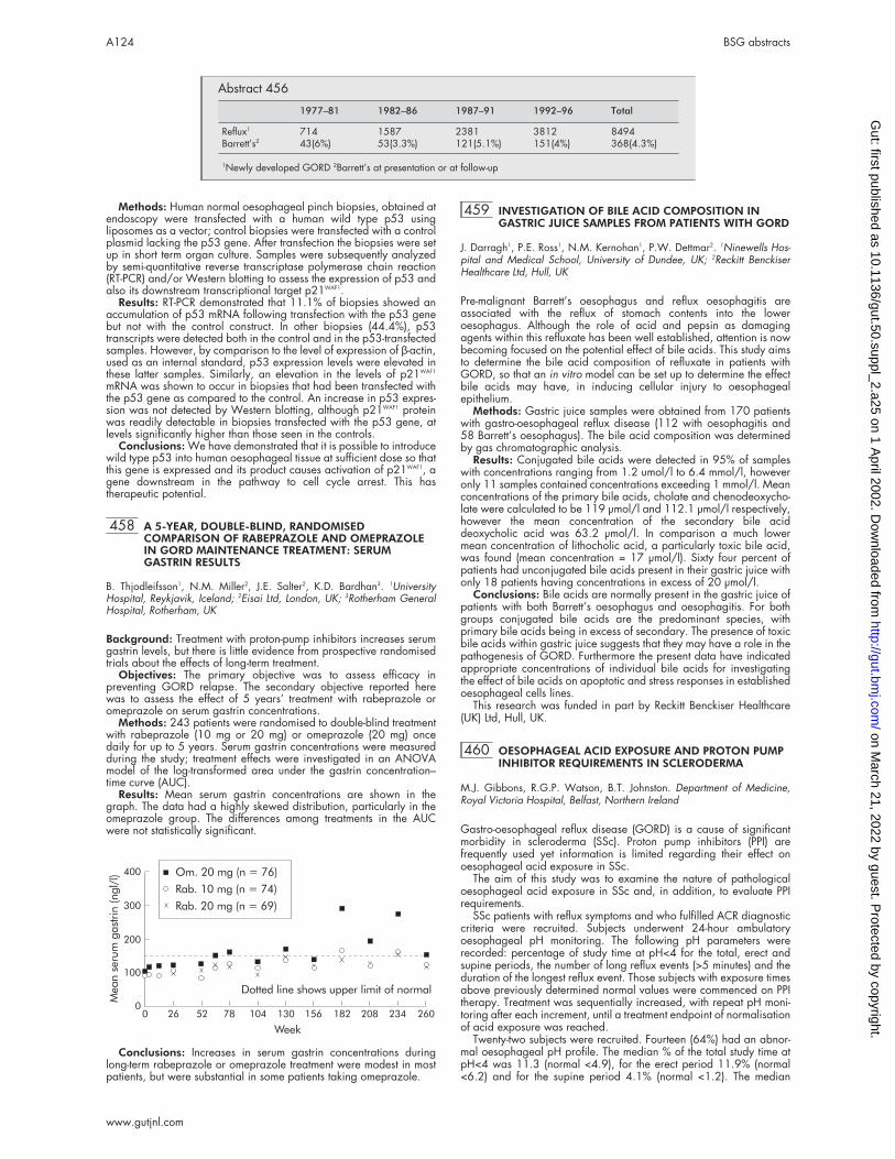
Methods: Human normal oesophageal pinch biopsies, obtained atendoscopy were transfected with a human wild type p53 usingliposomes as a vector; control biopsies were transfected with a controlplasmid lacking the p53 gene. After transfection the biopsies were setup in short term organ culture. Samples were subsequently analyzedby semi-quantitative reverse transcriptase polymerase chain reaction(RT-PCR) and/or Western blotting to assess the expression of p53 andalso its downstream transcriptional target p21WAF1.
Results: RT-PCR demonstrated that 11.1% of biopsies showed anaccumulation of p53 mRNA following transfection with the p53 genebut not with the control construct. In other biopsies (44.4%), p53transcripts were detected both in the control and in the p53-transfectedsamples. However, by comparison to the level of expression of β-actin,used as an internal standard, p53 expression levels were elevated inthese latter samples. Similarly, an elevation in the levels of p21WAF1
mRNA was shown to occur in biopsies that had been transfected withthe p53 gene as compared to the control. An increase in p53 expres-sion was not detected by Western blotting, although p21WAF1 proteinwas readily detectable in biopsies transfected with the p53 gene, atlevels significantly higher than those seen in the controls.
Conclusions: We have demonstrated that it is possible to introducewild type p53 into human oesophageal tissue at sufficient dose so thatthis gene is expressed and its product causes activation of p21WAF1, agene downstream in the pathway to cell cycle arrest. This hastherapeutic potential.
458 A 5-YEAR, DOUBLE-BLIND, RANDOMISEDCOMPARISON OF RABEPRAZOLE AND OMEPRAZOLEIN GORD MAINTENANCE TREATMENT: SERUMGASTRIN RESULTS
B. Thjodleifsson1, N.M. Miller2, J.E. Salter2, K.D. Bardhan3. 1UniversityHospital, Reykjavik, Iceland; 2Eisai Ltd, London, UK; 3Rotherham GeneralHospital, Rotherham, UK
Background: Treatment with proton-pump inhibitors increases serumgastrin levels, but there is little evidence from prospective randomisedtrials about the effects of long-term treatment.
Objectives: The primary objective was to assess efficacy inpreventing GORD relapse. The secondary objective reported herewas to assess the effect of 5 years’ treatment with rabeprazole oromeprazole on serum gastrin concentrations.
Methods: 243 patients were randomised to double-blind treatmentwith rabeprazole (10 mg or 20 mg) or omeprazole (20 mg) oncedaily for up to 5 years. Serum gastrin concentrations were measuredduring the study; treatment effects were investigated in an ANOVAmodel of the log-transformed area under the gastrin concentration–time curve (AUC).
Results: Mean serum gastrin concentrations are shown in thegraph. The data had a highly skewed distribution, particularly in theomeprazole group. The differences among treatments in the AUCwere not statistically significant.
Conclusions: Increases in serum gastrin concentrations duringlong-term rabeprazole or omeprazole treatment were modest in mostpatients, but were substantial in some patients taking omeprazole.
459 INVESTIGATION OF BILE ACID COMPOSITION INGASTRIC JUICE SAMPLES FROM PATIENTS WITH GORD
J. Darragh1, P.E. Ross1, N.M. Kernohan1, P.W. Dettmar2. 1Ninewells Hos-pital and Medical School, University of Dundee, UK; 2Reckitt BenckiserHealthcare Ltd, Hull, UK
Pre-malignant Barrett’s oesophagus and reflux oesophagitis areassociated with the reflux of stomach contents into the loweroesophagus. Although the role of acid and pepsin as damagingagents within this refluxate has been well established, attention is nowbecoming focused on the potential effect of bile acids. This study aimsto determine the bile acid composition of refluxate in patients withGORD, so that an in vitro model can be set up to determine the effectbile acids may have, in inducing cellular injury to oesophagealepithelium.
Methods: Gastric juice samples were obtained from 170 patientswith gastro-oesophageal reflux disease (112 with oesophagitis and58 Barrett’s oesophagus). The bile acid composition was determinedby gas chromatographic analysis.
Results: Conjugated bile acids were detected in 95% of sampleswith concentrations ranging from 1.2 umol/l to 6.4 mmol/l, howeveronly 11 samples contained concentrations exceeding 1 mmol/l. Meanconcentrations of the primary bile acids, cholate and chenodeoxycho-late were calculated to be 119 µmol/l and 112.1 µmol/l respectively,however the mean concentration of the secondary bile aciddeoxycholic acid was 63.2 µmol/l. In comparison a much lowermean concentration of lithocholic acid, a particularly toxic bile acid,was found (mean concentration = 17 µmol/l). Sixty four percent ofpatients had unconjugated bile acids present in their gastric juice withonly 18 patients having concentrations in excess of 20 µmol/l.
Conclusions: Bile acids are normally present in the gastric juice ofpatients with both Barrett’s oesophagus and oesophagitis. For bothgroups conjugated bile acids are the predominant species, withprimary bile acids being in excess of secondary. The presence of toxicbile acids within gastric juice suggests that they may have a role in thepathogenesis of GORD. Furthermore the present data have indicatedappropriate concentrations of individual bile acids for investigatingthe effect of bile acids on apoptotic and stress responses in establishedoesophageal cells lines.
This research was funded in part by Reckitt Benckiser Healthcare(UK) Ltd, Hull, UK.
460 OESOPHAGEAL ACID EXPOSURE AND PROTON PUMPINHIBITOR REQUIREMENTS IN SCLERODERMA
M.J. Gibbons, R.G.P. Watson, B.T. Johnston. Department of Medicine,Royal Victoria Hospital, Belfast, Northern Ireland
Gastro-oesophageal reflux disease (GORD) is a cause of significantmorbidity in scleroderma (SSc). Proton pump inhibitors (PPI) arefrequently used yet information is limited regarding their effect onoesophageal acid exposure in SSc.
The aim of this study was to examine the nature of pathologicaloesophageal acid exposure in SSc and, in addition, to evaluate PPIrequirements.
SSc patients with reflux symptoms and who fulfilled ACR diagnosticcriteria were recruited. Subjects underwent 24-hour ambulatoryoesophageal pH monitoring. The following pH parameters wererecorded: percentage of study time at pH<4 for the total, erect andsupine periods, the number of long reflux events (>5 minutes) and theduration of the longest reflux event. Those subjects with exposure timesabove previously determined normal values were commenced on PPItherapy. Treatment was sequentially increased, with repeat pH moni-toring after each increment, until a treatment endpoint of normalisationof acid exposure was reached.
Twenty-two subjects were recruited. Fourteen (64%) had an abnor-mal oesophageal pH profile. The median % of the total study time atpH<4 was 11.3 (normal <4.9), for the erect period 11.9% (normal<6.2) and for the supine period 4.1% (normal <1.2). The median
Abstract 456
1977–81 1982–86 1987–91 1992–96 Total
Reflux1 714 1587 2381 3812 8494Barrett’s2 43(6%) 53(3.3%) 121(5.1%) 151(4%) 368(4.3%)
1Newly developed GORD 2Barrett’s at presentation or at follow-up
400
300
200
100
00 26 52 78 104 130 156 182 208 234 260
Week
Meanserum
gastrin(ngl/l) Om. 20 mg (n = 76)
Rab. 10 mg (n = 74)
Rab. 20 mg (n = 69)
Dotted line shows upper limit of normal
A124 BSG abstracts
www.gutjnl.com
on March 21, 2022 by guest. P
rotected by copyright.http://gut.bm
j.com/
Gut: first published as 10.1136/gut.50.suppl_2.a25 on 1 A
pril 2002. Dow
nloaded from
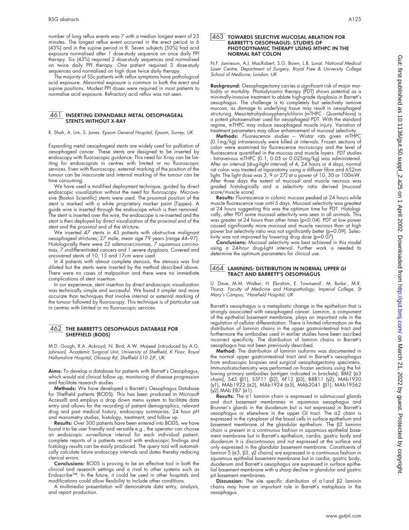
number of long reflux events was 7 with a median longest event of 23minutes. The longest reflux event occurred in the erect period in 6(43%) and in the supine period in 8. Seven subjects (50%) had acidexposure normalised after 1 dose-study sequence on once daily PPItherapy. Six (43%) required 2 dose-study sequences and normalisedon twice daily PPI therapy. One patient required 3 dose-studysequences and normalised on high dose twice daily therapy.
The majority of SSc patients with reflux symptoms have pathologicalacid exposure. Abnormal exposure is common in both the erect andsupine positions. Modest PPI doses were required in most patients tonormalise acid exposure. Refractory acid reflux was not seen.
461 INSERTING EXPANDABLE METAL OESOPHAGEALSTENTS WITHOUT X-RAY
R. Shah, A. Lim, S. Jones. Epsom General Hospital, Epsom, Surrey, UK
Expanding metal oesophageal stents are widely used for palliation ofoesophageal cancer. These stents are designed to be inserted byendoscopy with fluoroscopic guidance. This need for X-ray can be lim-iting for endoscopists in centres with limited or no fluoroscopicservices. Even with fluoroscopy, external marking of the position of thetumour can be inaccurate and internal marking of the tumour can betime consuming.
We have used a modified deployment technique, guided by directendoscopic visualization without the need for fluoroscopy. Microva-sive (Boston Scientific) stents were used. The proximal position of thestent is marked with a white proprietary marker paint (Tippex). Aguide wire is inserted through the endoscope which is then removed.The stent is inserted over the wire, the endoscope is re-inserted and thestent is then deployed by direct visualization of the proximal end of thestent and the proximal end of the stricture.
We inserted 47 stents in 43 patients with obstructive malignantoesophageal strictures; 27 male, mean age 79 years (range 44–97).Histologically there were 22 adenocarcinomas, 7 squamous carcino-mas, 7 undifferentiated cancers and 1 severe dysplasia. Covered anduncovered stents of 10, 15 and 17cm were used.
In 4 patients with almost complete stenosis, the stenosis was firstdilated but the stents were inserted by the method described above.There were no cases of malposition and there were no immediatecomplications of stent insertion.
In our experience, stent insertion by direct endoscopic visualizationwas technically simple and successful. We found it simpler and moreaccurate than techniques that involve internal or external marking ofthe tumour followed by fluoroscopy. This technique is of particular usein centres with limited or no fluoroscopic services.
462 THE BARRETT’S OESOPHAGUS DATABASE FORSHEFFIELD (BODS)
M.D. Gough, R.A. Ackroyd, N. Bird, A.W. Majeed (introduced by A.G.Johnson). Academic Surgical Unit, University of Sheffield, K Floor, RoyalHallamshire Hospital, Glossop Rd, Sheffield S10 2JF, UK
Aims: To develop a database for patients with Barrett’s Oesophagus,which would aid clinical follow up, monitoring of disease progressionand facilitate research studies.
Methods: We have developed a Barrett’s Oesophagus Databasefor Sheffield patients (BODS). This has been produced in MicrosoftAccess® and employs a drop down menu system to facilitate dataentry and allows for the recording of patient demographics, relevantdrug and past medical history, endoscopy summaries, 24 hour pHand manometry studies, histology, treatment, and follow up.
Results: Over 300 patients have been entered into BODS, we havefound it to be user friendly and versatile e.g., the operator can choosean endoscopic surveillance interval for each individual patient,complete reports of a patients record with endoscopic findings andhistology results can be easily produced. The query tool will automati-cally calculate future endoscopy intervals and dates thereby reducingclerical errors.
Conclusions: BODS is proving to be an effective tool in both theclinical and research settings and a rival to other systems such asEndoscribe™. In the future, it could be used in other hospitals andmodifications could allow flexibility to include other conditions.
A multimedia presentation will demonstrate data entry, analysis,and report production.
463 TOWARDS SELECTIVE MUCOSAL ABLATION FORBARRETT’S OESOPHAGUS: STUDIES OFPHOTODYNAMIC THERAPY USING MTHPC IN THENORMAL RAT COLON
N.F. Jamieson, A.J. MacRobert, S.G. Bown, L.B. Lovat. National MedicalLaser Centre, Department of Surgery, Royal Free & University CollegeSchool of Medicine, London, UK
Background: Oesophagectomy carries a significant risk of major mor-bidity or mortality. Photodynamic therapy (PDT) shows potential as aminimally-invasive treatment to ablate high-grade dysplasia in Barrett’soesophagus. The challenge is to completely but selectively removemucosa, as damage to underlying tissue may result in oesophagealstricturing. Meso-tetrahydroxyphenylchlorin (mTHPC - QuantaNova) isa potent photosensitiser used for oesophageal PDT. With the standardregime, mTHPC may induce oesophageal muscle injury. Variation oftreatment parameters may allow enhancement of mucosal selectivity.
Methods: Fluorescence studies – Wistar rats given mTHPC(0.1mg/kg) intravenously were killed at intervals. Frozen sections ofcolon were examined by fluorescence microscopy and the level offluorescence quantified in the mucosa and muscle layers. PDT studies- Intravenous mTHPC (0.1, 0.05 or 0.025mg/kg) was administered.After an interval (drug-light interval) of 4, 24 hours or 4 days, normalrat colon was treated at laparotomy using a diffuser fibre and 652nmlight. The light dose was 3, 9 or 27J at a power of 10, 30 or 100mW.After three days the extent of mucosal and muscle necrosis wasgraded histologically and a selectivity ratio derived (mucosalscore/muscle score).
Results: Fluorescence in colonic mucosa peaked at 24 hours whilemuscle fluorescence rose until 5 days. Mucosal selectivity was greatestat 24 hours suggesting this was the optimum time for PDT. Histologi-cally, after PDT some mucosal selectivity was seen in all animals. Thiswas greater at 24 hours than other times (p<0.04). PDT at low powercaused significantly more mucosal and muscle necrosis than at highpower but selectivity ratio was not significantly better (p=0.09). Selec-tivity was not improved by lowering drug dose (p=0.07).
Conclusions: Mucosal selectivity was best achieved in this modelusing a 24-hour drug-light interval. Further work is needed todetermine the optimum parameters for clinical use.
464 LAMININS: DISTRIBUTION IN NORMAL UPPER GITRACT AND BARRETT’S OESOPHAGUS
U. Dave, M.M. Walker, H. Ebrahim, E. Townsend1, M. Burke1, M.R.Thursz. Faculty of Medicine and Histopathology, Imperial College, StMary’s Campus; 1Harefield Hospital, UK
Barrett’s oesophagus is a metaplastic change in the epithelium that isstrongly associated with oesophageal cancer. Laminin, a componentof the epithelial basement membrane, plays an important role in theregulation of cellular differentiation. There is limited information on thedistribution of laminin chains in the upper gastrointestinal tract andfurthermore the antibodies used in earlier studies have been ascribedincorrect specificity. The distribution of laminin chains in Barrett’soesophagus has not been previously described.
Method: The distribution of laminin isoforms was documented inthe normal upper gastrointestinal tract and in Barrett’s oesophagusfrom endoscopic biopsies and surgical oesophagectomy specimens.Immunohistochemistry was performed on frozen sections using the fol-lowing primary antibodies (antigen indicated in brackets): BM2 (α3chain), 545 (β1), S5F11 (β2), 6F12 (β3), B8B11 (γ2), MAb1920(γ1), MAb1922 (α2), MAb1924 (α5), MAb2041 (β1), ΜΑb19562(γ2) MAb EB7 (α1).
Results: The α1 laminin chain is expressed in submucosal glandsand duct basement membranes in squamous oesophagus andBrunner’s glands in the duodenum but is not expressed in Barrett’soesophagus or elsewhere in the upper GI tract. The α2 chain isexpressed in the cytoplasm of the basal cells in surface epithelium andbasement membrane of the glandular epithelium. The β2 lamininchain is present in a continuous fashion in squamous epithelial base-ment membrane but in Barrett’s epithelium, cardia, gastric body andduodenum it is discontinuous and not expressed at the surface andonly expressed in the glandular basement membrane. Constituents oflaminin 5 (α3, β3, γ2 chains) are expressed in a continuous fashion insquamous epithelial basement membrane but in cardia, gastric body,duodenum and Barrett’s oesophagus are expressed in surface epithe-lial basement membrane with a sharp decline in glandular and gastricpit basement membranes.
Discussion: The site specific distribution of α1and β2 lamininchains may have an important role in Barrett’s metaplasia in theoesophagus.
BSG abstracts A125
www.gutjnl.com
on March 21, 2022 by guest. P
rotected by copyright.http://gut.bm
j.com/
Gut: first published as 10.1136/gut.50.suppl_2.a25 on 1 A
pril 2002. Dow
nloaded from