tsg-6 binds via its cub_c domain to the cell-binding domain of fibronectin and increases fibronectin...
TRANSCRIPT
TSG-6 binds via its CUB_C domain to the cell-binding domain offibronectin and increases fibronectin matrix assembly
Svetlana A. Kuznetsovaa, David J. Mahoneyb, Gema Martin-Mansoa, Tariq Alib,c, Hilke A.Nentwichb, John M. Sipesa, Bixi Zenga, Tikva Vogela, Anthony J. Dayb,c, and David D.Robertsa*
aLaboratory of Pathology, Center for Cancer Research, National Cancer Institute, National Institutes ofHealth, Bethesda, MD 20892
bMRC Immunochemistry Unit, Department of Biochemistry, University of Oxford, South Parks Road, OxfordOX1 3QU, UK
cWellcome Trust Centre for Cell-Matrix Research, Faculty of Life Sciences, University of Manchester,Manchester M13 9PT, UK
AbstractHuman plasma fibronectin binds with high affinity to the inflammation-induced secreted proteinTSG-6. Fibronectin binds to the CUB_C domain of TSG-6 but not to its Link module. TSG-6 canthus act as a bridging molecule to facilitate fibronectin association with the TSG-6 Link moduleligand thrombospondin-1. Fibronectin binding to TSG-6 is divalent cation-independent and isconserved in cellular fibronectins. Based on competition binding studies using recombinant andproteolytic fragments of fibronectin, TSG-6 binding localizes to type III repeats 9–14 of fibronectin.This region of fibronectin contains the Arg-Gly-Asp sequence recognized by α5β1 integrin, butdeletion of that sequence does not prevent TSG-6 binding, and TSG-6 does not inhibit cell adhesionon fibronectin substrates mediated by this integrin. This region of fibronectin is also involved infibronectin matrix assembly, and addition of TSG-6 enhances exogenous and endogenous fibronectinmatrix assembly by human fibroblasts. Therefore, TSG-6 is a high affinity ligand that can mediatefibronectin interactions with other matrix components and modulate some interactions of fibronectinwith cells.
Keywordsmatrix assembly; TSG-6; fibronectin; CUB domains
1. IntroductionTumor necrosis factor-stimulated gene 6 (TSG-6) is a ~35 kDa secreted protein that is producedduring inflammation and related processes. TSG-6 has anti-inflammatory activity and protects
*Correspondence should be addressed: NIH, Building 10 Room 2A33, 10 Center Dr MSC1500, Bethesda, MD 20892-1500. Phone:301-496-6264. Fax: 301-402-0043. E-mail: [email protected]'s Disclaimer: This is a PDF file of an unedited manuscript that has been accepted for publication. As a service to our customerswe are providing this early version of the manuscript. The manuscript will undergo copyediting, typesetting, and review of the resultingproof before it is published in its final citable form. Please note that during the production process errors may be discovered which couldaffect the content, and all legal disclaimers that apply to the journal pertain.Footnotes: The abbreviations used are: DOC, deoxycholate; DPBS, Dulbecco’s phosphate buffered saline; ECM, extracellular matrix;FN, fibronectin; TSG-6, tumor necrosis factor-stimulated gene-6; CUB, complement component Clr/Cls, Uegf, and bone morphogenicprotein 1
NIH Public AccessAuthor ManuscriptMatrix Biol. Author manuscript; available in PMC 2009 April 1.
Published in final edited form as:Matrix Biol. 2008 April ; 27(3): 201–210.
NIH
-PA Author Manuscript
NIH
-PA Author Manuscript
NIH
-PA Author Manuscript
joint tissues from destruction in in vivo models (Wisniewski et al., 1996; Milner and Day,2003; Szanto et al., 2004; Milner et al., 2006). TSG-6 is also expressed in the ovary duringovulation and plays an important role in female fertility by regulating cumulus cell-oocytecomplex expansion (Fulop et al., 2003; Ochsner et al., 2003). The protein consists mainly ofcontiguous Link and CUB (complement component Clr/Cls, Uegf, and bone morphogeneticprotein 1) domains. The Link module contains a hyaluronan-binding site and also interactswith heparin/heparan sulfate and thrombospondins-1 and -2 (Blundell et al., 2005; Kuznetsovaet al., 2005; Mahoney et al., 2005). Interaction with the Link module of TSG-6 is mediated bythe N-modules of thrombospondin-1, which also recognize the Link module-containingdomains of versican and aggrecan (Kuznetsova et al., 2006). TSG-6 is a catalyst and cofactorin the covalent transfer of the heavy chains (HC) of inter-α-trypsin inhibitor onto a hyaluronanacceptor (Rugg et al., 2005). Thrombospondin-1 enhances the initial covalent modification ofinter-α-trypsin inhibitor by TSG-6 (i.e., HC-TSG-6 complex formation) and transfer of HC tohyaluronan, suggesting a physiological function of thrombospondin-1 binding to TSG-6 inregulation of hyaluronan metabolism at sites of inflammation (Kuznetsova et al., 2005).
To date, no ligands have been identified that bind to the TSG-6 CUB module. Paralogous CUBmodules in other proteins have been implicated in protein-protein and protein-carbohydrateinteractions (Bork and Beckmann, 1993; Solis et al., 1998; Sieron et al., 2000).
While characterizing TSG-6 interactions with thrombospondin-1, we tested severalextracellular matrix (ECM) proteins as controls and unexpectedly found that TSG-6 interactsavidly with fibronectin (FN). FN is a prominent component of ECM and a major circulatingprotein in plasma that regulates a variety of cellular activities through direct interactions withcell surface integrin and proteoglycan receptors (Mao and Schwarzbauer, 2005). Here wedemonstrate that the CUB_C domain of TSG-6 (comprised of the CUB module and C-terminalsegment) interacts with the cell- and heparin-binding domains of FN. FN is synthesized bymany adherent cells, some of which assemble it into a fibrillar network (Mao andSchwarzbauer, 2005). The assembly process is integrin-dependent, and FN-integrininteractions initiate a step-wise process involving conformational changes that expose FN-binding sites and promote the intermolecular interactions needed for fibril formation(Schwarzbauer and Sechler, 1999). We further show that TSG-6 binding enhances FN fibrilassembly by cultured human fibroblasts. These results provide evidence for an additional levelof control of FN matrix assembly under conditions where TSG-6 is present.
2. Results2.1 characterization of recombinant CUB_C domain
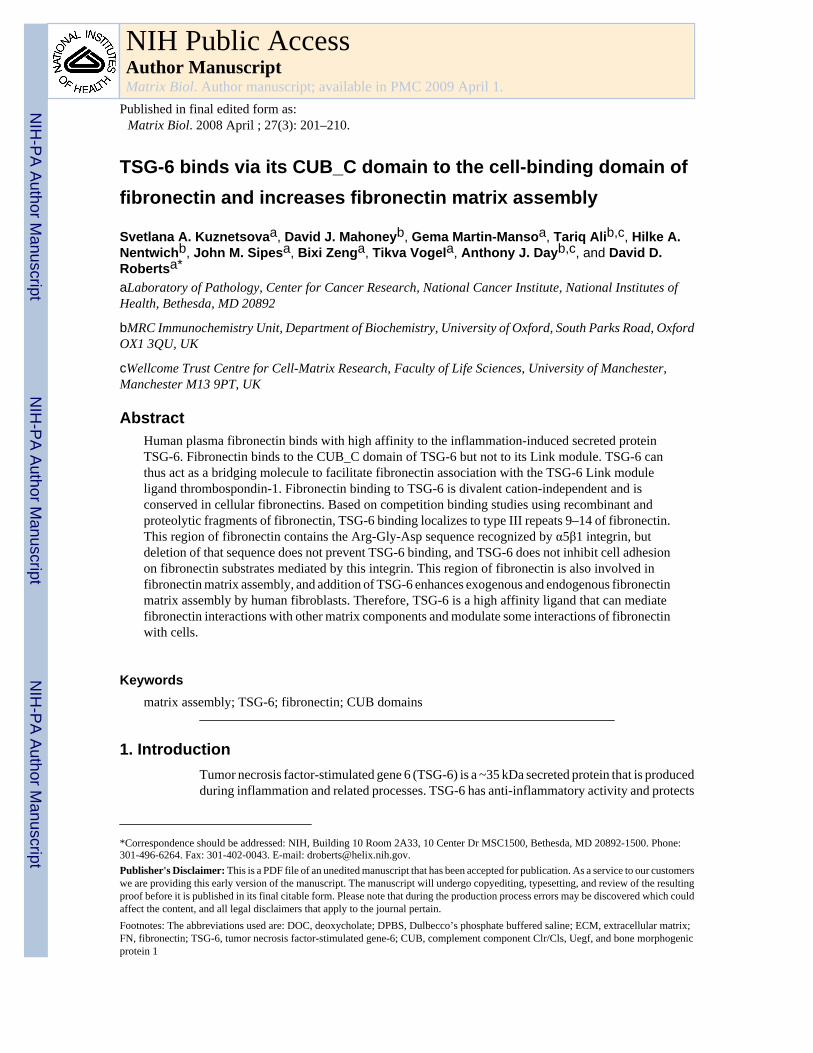
Mature human TSG-6 is comprised of a 19-amino acid N-terminal segment followed bycontiguous Link and CUB modules of 92 and 122 residues, respectively, and a C-terminalsegment of 27 amino acids (residues 18–36, 37–128, 129–250 and 251–277 in the preprotein,respectively) (Milner and Day, 2003). One-dimensional NMR spectroscopy of recombinanthuman CUB_C (residues 129–277 of the preprotein) demonstrated good dispersion ofresonances in both the methyl (−1 to 3 ppm) and amide/aromatic (6–11 ppm) regions of thespectra (Fig. 1A and B, respectively), which is consistent with that of a correctly folded protein.For example, the high-field shifted methyl resonances seen at around 0 ppm are characteristicof the presence of a stable hydrophobic core. These data reveal that the recombinant CUB_Cdomain is correctly folded in PBS (i.e., at physiological pH and salt concentration).Furthermore, the CUB_C protein used in this study has been crystallized and an X-ray structuredetermined at 2.3-Å resolution (DC Briggs, T Ali, DJ Mahoney & AJ Day, unpublished data).
Kuznetsova et al. Page 2
Matrix Biol. Author manuscript; available in PMC 2009 April 1.
NIH
-PA Author Manuscript
NIH
-PA Author Manuscript
NIH
-PA Author Manuscript
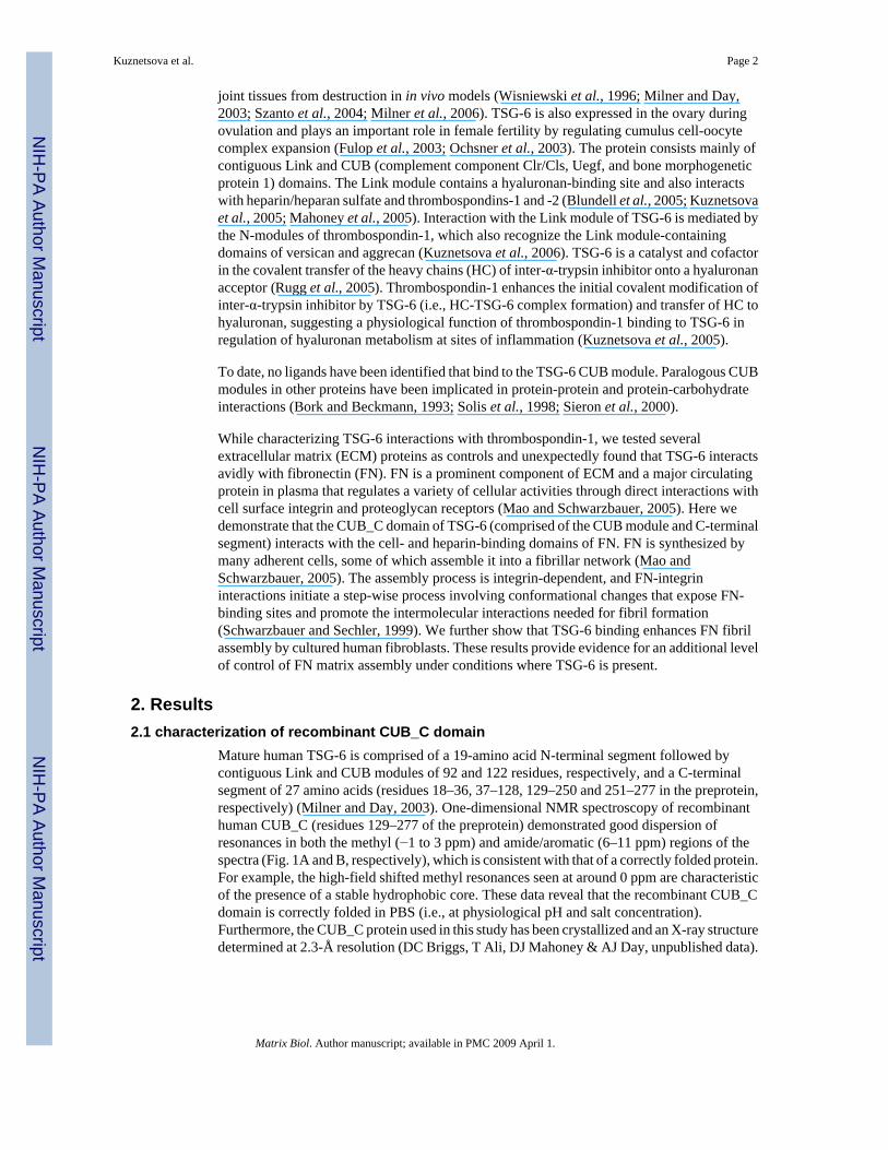
2.2 The CUB_C domain of TSG-6 is a high affinity ligand for fibronectinsFull-length recombinant human TSG-6Q, CUB_C domain, and Link module (Link_TSG6)were tested for binding to human plasma FN in a solid phase assay (Fig. 2A). 125I-FN boundto immobilized full-length TSG-6Q and its CUB_C domain in a dose-dependent manner. Weakbinding of FN to the Link module was detected only at high coating concentrations. Bindingof 125I-FN to full length TSG-6Q was not significantly influenced by divalent cations as bindingwas maintained in the presence of EDTA in buffer lacking divalent cations (Fig. 2B).
Because direct binding to immobilized proteins can be influenced both by the efficiency ofprotein adsorption on plastic and potential changes in conformation induced by this adsorption,the specificity of FN binding was further examined by inhibition assays (Fig. 2C). Bindingof 125I-FN to immobilized TSG-6Q was inhibited by soluble FN, with 50% inhibition at 10µg/ml (22 nM). In contrast, the weak binding of 125I-FN to immobilized Link_TSG6 was notsignificantly inhibited by unlabeled soluble FN within the range tested.
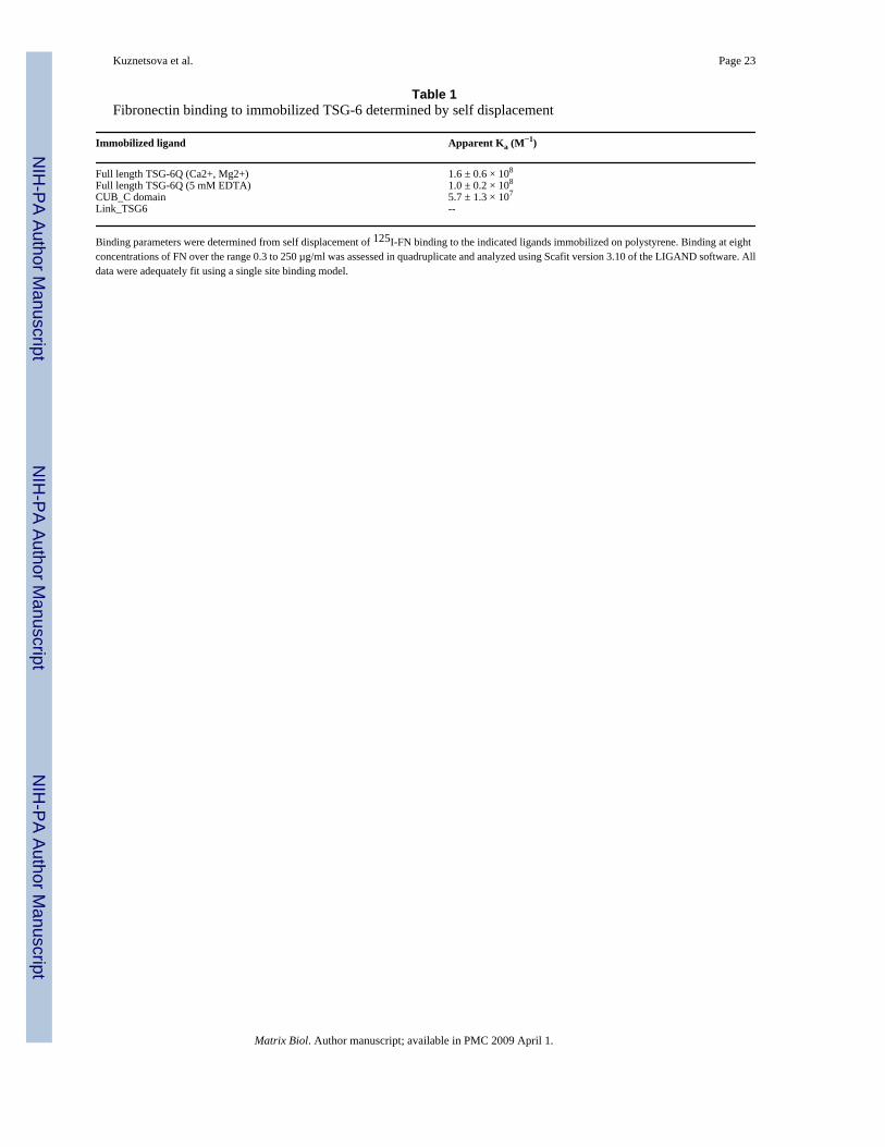
Quantitative analysis of the self displacement data yielded an apparent affinity constant of1.6×108 M−1 for FN binding to immobilized TSG-6 in the presence of divalent cations (Table1). Consistent with Fig. 2B,C, the binding affinity was only slightly decreased in the presenceof EDTA. Binding of FN to immobilized CUB_C domain occurred with an approximately 3-fold lower affinity (apparent Ka = 5.7×10⁷ M−1), whereas the weak binding to immobilizedLink_TSG6 was not displaceable by unlabeled FN, and no affinity constant could bedetermined by this method (Table 1).
Cellular FN differs from plasma FN by the inclusion of alternatively spliced exons(Schwarzbauer, 1991). Cellular FN isolated from fibroblasts inhibited binding of labeledplasma FN with a comparable dose-dependency as unlabeled plasma FN, whereas cellular FNisolated from lung was slightly less active (Fig. 2D). Therefore, both plasma and cellular FNscan interact with TSG-6.
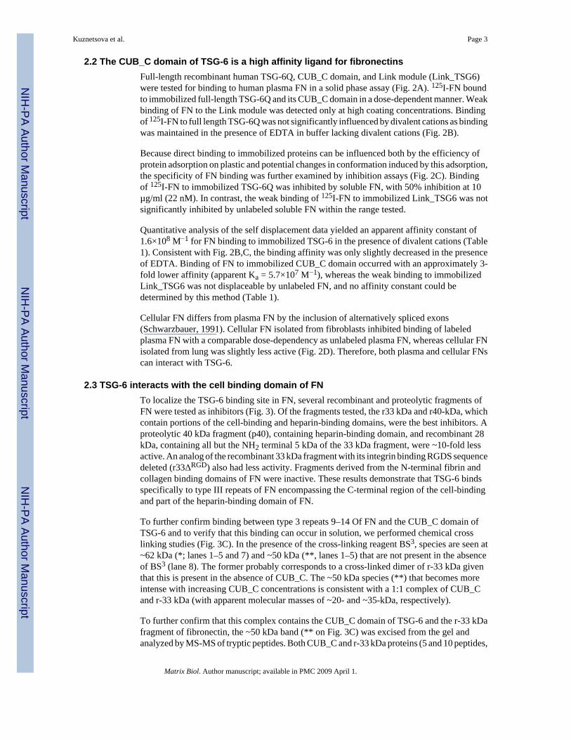
2.3 TSG-6 interacts with the cell binding domain of FNTo localize the TSG-6 binding site in FN, several recombinant and proteolytic fragments ofFN were tested as inhibitors (Fig. 3). Of the fragments tested, the r33 kDa and r40-kDa, whichcontain portions of the cell-binding and heparin-binding domains, were the best inhibitors. Aproteolytic 40 kDa fragment (p40), containing heparin-binding domain, and recombinant 28kDa, containing all but the NH2 terminal 5 kDa of the 33 kDa fragment, were ~10-fold lessactive. An analog of the recombinant 33 kDa fragment with its integrin binding RGDS sequencedeleted (r33ΔRGD) also had less activity. Fragments derived from the N-terminal fibrin andcollagen binding domains of FN were inactive. These results demonstrate that TSG-6 bindsspecifically to type III repeats of FN encompassing the C-terminal region of the cell-bindingand part of the heparin-binding domain of FN.
To further confirm binding between type 3 repeats 9–14 Of FN and the CUB_C domain ofTSG-6 and to verify that this binding can occur in solution, we performed chemical crosslinking studies (Fig. 3C). In the presence of the cross-linking reagent BS3, species are seen at~62 kDa (*; lanes 1–5 and 7) and ~50 kDa (**, lanes 1–5) that are not present in the absenceof BS3 (lane 8). The former probably corresponds to a cross-linked dimer of r-33 kDa giventhat this is present in the absence of CUB_C. The ~50 kDa species (**) that becomes moreintense with increasing CUB_C concentrations is consistent with a 1:1 complex of CUB_Cand r-33 kDa (with apparent molecular masses of ~20- and ~35-kDa, respectively).
To further confirm that this complex contains the CUB_C domain of TSG-6 and the r-33 kDafragment of fibronectin, the ~50 kDa band (** on Fig. 3C) was excised from the gel andanalyzed by MS-MS of tryptic peptides. Both CUB_C and r-33 kDa proteins (5 and 10 peptides,
Kuznetsova et al. Page 3
Matrix Biol. Author manuscript; available in PMC 2009 April 1.
NIH
-PA Author Manuscript
NIH
-PA Author Manuscript
NIH
-PA Author Manuscript
respectively) were identified by this analysis. Thus, in solution the CUB_C domain of TSG-6forms a 1:1 complex in solution with repeats 9–14 of FN.
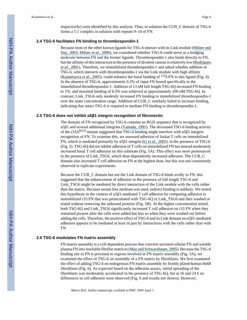
2.4 TSG-6 facilitates FN binding to thrombospondin-1Because most of the other known ligands for TSG-6 interact with its Link module (Milner andDay, 2003; Milner et al., 2006), we considered whether TSG-6 could serve as a bridgingmolecule between FN and the former ligands. Thrombospondin-1 also binds directly to FN,but the affinity of this interaction in the presence of divalent cations is relatively low (Rodrigueset al., 2001). Therefore, we immobilized thrombospondin-1 and asked whether addition ofTSG-6, which interacts with thrombospondin-1 via the Link module with high affinity(Kuznetsova et al., 2005), could enhance the basal binding of 125I-FN to this ligand (Fig. 4).In the absence of TSG-6, approximately 0.3% of input FN bound specifically to theimmobilized thrombospondin-1. Addition of 13 nM full-length TSG-6Q increased FN bindingto 1%, and maximal binding of 6.5% was achieved at approximately 200 nM TSG-6Q. Incontrast, Link_TSG6 only modestly increased FN binding to immobilized thrombospondin-1over the same concentration range. Addition of CUB_C similarly failed to increase binding,indicating that intact TSG-6 is required to mediate FN binding to thrombospondin-1.
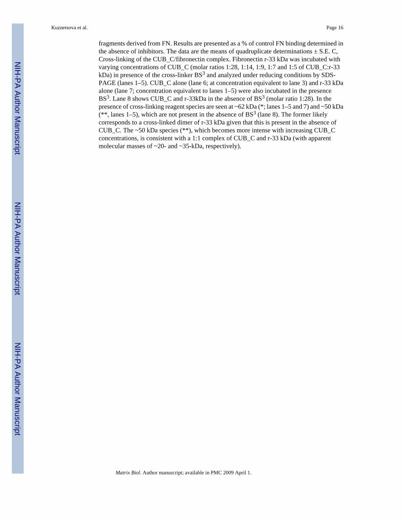
2.5 TSG-6 does not inhibit α5β1 integrin recognition of fibronectinThe domain of FN recognized by TSG-6 contains an RGD sequence that is recognized byα5β1 and several additional integrins (Yamada, 1991). The decreased TSG-6 binding activityof the r33ΔRGD mutant suggested that TSG-6 binding might interfere with α5β1 integrinrecognition of FN. To examine this, we assessed adhesion of Jurkat T cells on immobilizedFN, which is mediated primarily by α5β1 integrin (Li et al., 2002), in the presence of TSG-6(Fig. 5). TSG-6Q did not inhibit adhesion of T cells on immobilized FN but instead moderatelyincreased basal T cell adhesion on this substrate (Fig. 5A). This effect was more pronouncedin the presence of Link_TSG6, which dose-dependently increased adhesion. The CUB_Cdomain also increased T cell adhesion on FN at the highest dose, but this was not consistentlyobserved in replicate experiments.
Because the CUB_C domain but not the Link domain of TSG-6 binds avidly to FN, thissuggested that the enhancement of adhesion in the presence of full length TSG-6 andLink_TSG6 might be mediated by direct interaction of the Link module with the cells ratherthan the matrix. Because serum-free medium was used, indirect binding is unlikely. We testedthis hypothesis in the context of α5β1-mediated T cell adhesion by comparing adhesion toimmobilized r33 FN that was preincubated with TSG-6Q or Link_TSG6 and then washed ortested without removing the unbound proteins (Fig. 5B). At the higher concentration tested,both TSG-6Q and Link_TSG6 significantly increased T cell adhesion on r33 FN when theyremained present after the cells were added but less so when they were washed out beforeadding the cells. Therefore, the positive effect of TSG-6 and its Link domain on α5β1-mediatedadhesion appears to be mediated at least in part by interactions with the cells rather than withFN.
2.6 TSG-6 modulates FN matrix assemblyFN matrix assembly is a cell-dependent process that converts secreted cellular FN and solubleplasma FN into insoluble fibrillar matrices (Mao and Schwarzbauer, 2005). Because the TSG-6binding site in FN is proximal to regions involved in FN matrix assembly (Fig. 3A), weexamined the effect of TSG-6 on assembly of a FN matrix by fibroblasts. We first examinedthe effect of adding TSG-6 on endogenous FN matrix assembly by freshly plated human Hs68fibroblasts (Fig. 6). As expected based on the adhesion assays, initial spreading of thefibroblasts was moderately accelerated in the presence of TSG-6Q, but at 16 and 24 h nodifferences in cell adhesion were observed (Fig. 6 and results not shown). However,
Kuznetsova et al. Page 4
Matrix Biol. Author manuscript; available in PMC 2009 April 1.
NIH
-PA Author Manuscript
NIH
-PA Author Manuscript
NIH
-PA Author Manuscript
endogenous FN matrix deposition was markedly enhanced at both time points in the presenceof either full length TSG-6Q or CUB_C (Fig. 6 and results not shown).
To exclude potential effects of TSG-6 on secretion of endogenous FN, we also examinedincorporation of exogenous FN into fibroblast matrix by incubating Hs68 cells for 3 h withexogenous biotinylated-FN (Fig. 7). Incubation in the presence of full-length TSG-6Qmarkedly increased incorporation of exogenous FN relative to control cells (Fig. 7 lower panel).In contrast, incubation with an equimolar concentration of Link_TSG6 did not enhance FNmatrix assembly (center panel).
To confirm the qualitative results from these immunofluorescence studies, we employed asemi-quantitative assay of FN matrix assembly (Pankov and Yamada, 2004). FN that associateswith cells is incorporated into two pools that can be distinguished by their solubility indeoxycholate (DOC) detergent. The DOC-soluble pool represents FN bound by cellularreceptors and preexisting matrix fibrils, while the DOC-insoluble pool is believed to includebound FN that is incorporated into the matrix through detergent-resistant interactions such asdisulfide bonding (Pankov and Yamada, 2004). Addition of full-length TSG-6Q to Hs68 cellsincreased the amount of biotinylated-FN that became incorporated into the DOC-insolublematrix fraction (Fig. 8A). In contrast, treatment of the cells with Link_TSG6 did not affect FNmatrix assembly. This activity of TSG-6 probably involves its binding to fibrillar FN in theECM because TSG-6Q but not Link_TSG6 was incorporated into the DOC-insoluble matrixfraction in a dose-dependent manner (Fig. 8B).
3. DiscussionWe previously showed that TSG-6 specifically interacts with N-modules of thrombospondin-1and to a lesser extent with thrombospondin-2, two transient components of ECM (Kuznetsovaet al., 2005). In the present study we addressed the possibility that TSG-6 might interact withother components of ECM and thereby modulate ECM-dependent regulation of cell behavior.FN is an abundant and ubiquitous plasma and ECM component that is organized into a fibrillarnetwork by some cell types through direct interactions with cell surface receptors (Mao andSchwarzbauer, 2005; Midwood et al., 2006). This study demonstrates that TSG-6 binds humanplasma and cellular FN. FN was found to directly interact with full-length TSG-6 protein andits CUB_C domain with high affinity, whereas it did not bind avidly to the isolated Link domainof TSG-6.
Inhibition studies using recombinant and proteolytic fragments of FN localized TSG-6 bindingto the central region of FN in its type 3 repeats, which contains integrin and heparin-bindingsites. Since this domain of FN is required for the initial steps of FN matrix assembly (Midwoodet al., 2006), effects of TSG-6 on the formation of FN fibrils were investigated. We found thatTSG-6 enhances FN fibril formation mediated by human Hs68 fibroblasts. Furthermore, TSG-6is incorporated into the detergent-insoluble ECM of fibroblasts and co-localizes with FN (datanot shown). Therefore, TSG-6 may interact with FN in the ECM surrounding cultured humanfibroblasts. Such binding would target TSG-6 deposition into fibrillar FN and appears toenhance FN matrix assembly.
Interactions of FN with both α5β1 integrin and syndecan-4 are important for fibrillogenesis(Midwood et al., 2006). Because the Link module of TSG-6 binds avidly toglycosaminoglycans (e.g., heparan sulfate (Mahoney et al., 2005)), TSG-6 may facilitate FNfibrillogenesis either by directly engaging the glycosaminoglycans of syndecan-4 or byfacilitating syndecan-4 binding to FN. Alternatively, the enhanced integrin-dependentadhesion on a FN substrate that we observed in the presence of TSG-6 could enhance FNfibrillogenesis by promoting the integrin function in this process (Clark et al., 2005; Mao and
Kuznetsova et al. Page 5
Matrix Biol. Author manuscript; available in PMC 2009 April 1.
NIH
-PA Author Manuscript
NIH
-PA Author Manuscript
NIH
-PA Author Manuscript

Schwarzbauer, 2005). However, latter is less likely because Link_TSG-6 enhances adhesionon immobilized FN but does not enhance fibrillogenesis.
The Link module of TSG-6 interacts with hyaluronan, chondroitin-4-sulfate, dermatan sulfate,heparin/heparan sulfate, aggrecan, versican, pentraxin-3, thrombospondin-1 and inter-α-trypsin inhibitor (Milner and Day, 2003; Kuznetsova et al., 2005; Kuznetsova et al., 2006;Milner et al., 2006). FN is the first known ligand for TSG-6 that does not bind within the Linkmodule domain, but rather associates with the CUB_C domain, i.e. the C-terminal half of theprotein. Our data indicates that at least one of these Link module ligands, thrombospondin-1,can form a ternary complex where TSG-6 acts as bridging molecule between thrombospondin-1and FN. This resembles the bridging function of versican to mediate thrombospondin-1association with elastin-containing microfibrils on the surface of poly-I:C-stimulated vascularsmooth muscle cells (Kuznetsova et al., 2006). Thus, TSG-6 can serve to increase FNassociation with at least one ligand of the TSG-6 Link module. Further studies are needed todetermine whether this function generalizes to other TSG-6 ligands that interact with the Linkmodule. Additional work is also required to investigate whether binding of TSG-6 alters theconformation of FN or its interactions with other ECM or cell surface receptors other thanα5β1 integrin.
TSG-6 is a regulated component of ECM that is induced under specific physiological andpathological conditions (Milner and Day, 2003; Milner et al., 2006). This raises the questionof whether the FN binding activities of TSG-6 might play important roles in modulating fibrillarFN assembly or other ECM structures in inflammatory conditions or during ovulation.Considering the biological significance of ECM in regulating the compartmentalization oftissues into functional units, the possibility that TSG-6 modulates additional aspects of ECMstructure and function merits further investigation.
4. Experimental Procedures4.1 Protein Purification
Plasma FN was purified from fresh human plasma (National Institutes of Health Blood Bank)using a nondenaturing method (Akiyama and Yamada, 1985). Recombinant fragments derivedfrom individual domains of FN, expressed in E. coli, were purified and refolded as describedpreviously (Werber et al., 1991; Vogel et al., 1993). A 33 kDa cell-binding domain with theRGDS sequence deleted (r33ΔRGD) was constructed by oligonucleotide-directed mutagenesisof the expression plasmid, pFN 137-2 as described previously (Vogel et al., 1993; Negre etal., 1994). The FN fragments used in this paper and their origin from the FN sequence aresummarized in Fig. 3A. Human fibroblast and lung FNs were provided by Dr. Ralph Silverman,Fibrogenex, Inc., Chicago, IL. Other control proteins were obtained from Sigma-Aldrich. FNwas labeled with 125I using IODO-GEN (Pierce Biotechnology, Rockford, IL) as describedpreviously (Guo et al., 1992). Biotinylated-FN was prepared using sulfo-N-hydroxysuccinimidyl-biotin (Pierce Biotechnology, Rockford, IL), followed by a dialysis stepto remove unconjugated biotin (Pankov and Yamada, 2004). Recombinant full-length TSG-6Q(with a glutamine at residue 144) was expressed in Drosophila S2 cells (Nentwich et al.,2002), and Link_TSG6 was expressed in E. coli as described previously (Day et al., 1996;Kahmann et al., 1997).
4.2 Expression and Purification of TSG-6 CUB_C DomainThe CUB_C domain of human TSG-6 (residues 129–277 in the preprotein (Lee et al., 1992)with a glutamine at residue 144) was expressed in E. coli. Initial attempts to express the CUBmodule alone (i.e. residues 134–248) did not give rise to folded protein (H.A. Nentwich & A.J.Day, unpublished observations). Briefly, the CUB_C coding sequence was amplified from
Kuznetsova et al. Page 6
Matrix Biol. Author manuscript; available in PMC 2009 April 1.
NIH
-PA Author Manuscript
NIH
-PA Author Manuscript
NIH
-PA Author Manuscript

human osteoblast mRNA, as described previously for the full-length protein (Nentwich etal., 2002), cloned into pRK172 and transfected into Rosetta-gami BL21(DE3)pLysS. Cellswere harvested 4h after induction with IPTG and inclusion bodies were purified as describedin (Day et al., 1996). These were solubilized in 8M guanidine·HCl, 50mM Tris·HCl, pH 8.0,100 mM dithiothreitol, incubated for 1h at 37°C and then purified using a 100 × 5-cm S100-HR column equilibrated in 6M guanidine·HCl, 20mM Tris·HCl, pH 8.0. The partially purifiedCUB_C protein (40 ml) was refolded by dialysis overnight at 4°C into 2L 0.5M L-arginine,2mM cystine, 1 mM cysteine, 20 mM ethanolamine, 5 mM CaCl2·2H2O, pH 5.0, followed bya 6h dialysis against another 2L of refolding buffer. The protein was then dialyzed overnightat 4°C against 3 L of 0.1M Na-acetate, pH 4.0, and then purified to homogeneity using anionexchange chromatography on a 30 ml SP-Sepharose column, equilibrated in 50 mM Na-acetate,pH 4.0, and eluted with a gradient of 0 to 1M NaCl (in 50 mM Na-acetate, pH 4.0) over 13-column volumes. Finally the protein was dialyzed into PBS and stored at −20°C. Electrosprayionization mass spectrometry showed that the recombinant CUB_C domain had a molecularweight within 2 Da of the expected mass (16,776.6 Da).
4.3 One-dimensional NMR spectroscopyFollowing ion exchange chromatography, the CUB_C protein was dialysed extensively againstPBS using 3,500 MWCO snakeskin dialysis membrane, followed by concentration on aVivaspin 3,500 MWCO spin column to a concentration of ~2 mg/ml. D2O (60 µl) was addedto 540 µl of the CUB_C preparation to give a final concentration of ~ 0.11 mM. The 1-dimensional NMR spectrum was acquired at 21.7°C on a home-built NMR spectrometer witha 1H operating frequency of 599 MHz at the Department of Biochemistry, University of Oxfordand referenced against water (4.805 ppm at 21.7 °C).
4.4 Solid Phase Binding assaysImmulon® 2 HB microtiter strips with breakaway wells (ThermoLabsystems, Franklin, MA)were coated using 50 µl of the indicated concentrations of TSG-6Q, Link_TSG6 or CUB_Cdomain. For competitive binding studies, wells were coated using 10 µg/ml of full-lengthTSG-6Q, Link_TSG6 or CUB_C incubated overnight at 4°C in Dulbecco’s PBS withoutCa2+ or Mg2+. Nonspecific sites were blocked using 3% (w/v) BSA in Dulbecco’s PBS (DPBS)at room temperature for 1 h. Radioiodinated FN (0.5 µg/ml, 50 µl/well) was added alone or inthe presence of increasing concentrations of the indicated unlabeled ligands as competitors inDPBS, containing 0.5% (w/v) BSA, 0.1 mM phenylmethylsulfonyl fluoride with or withoutCa2+ and Mg2+, and incubated at 37°C for 3 h. The wells were washed with the same coldbuffer, and the bound radioactivity was quantified using a gamma counter (PerkinElmer LifeSciences).
4.5 Cross-linking and mass spectrometryCross-linking of CUB_C protein with the recombinant FN fragment r-33kDa was carried outin 150 mM NaCl, 50 mM HEPES, 7.4 with a final concentration of 125 µM of the cross-linkingagent Bis(Sulfosuccinimidyl)suberate (BS3) (Pierce) in 200 µl reactions. The cross-linkingreactions were quenched by the addition of 10 µl of 1M Tris/HCl, pH 8.0. Protein was recoveredusing 10 µl of Strataclean resin (Stratagene) and analyzed by SDS-PAGE on 10% (w/v) Tris-Tricine polyacylamide gels after boiling the samples in SDS loading buffer containing β-mercaptoethanol. A Coomassie blue-stained gel slice, corresponding to a ~50 kDa species thathad been crossed linked with BS3, was washed in 100% acetonitrile for 5 min, the acetonitrileremoved, and the gel slice vacuum dried for 15 min. The gel was hydrated with 10 mM DTTin 25mM NH4HCO3 at 56°C for 1 h, an equal volume of 55 mM iodoacetamide in 25mMNH4CO3 was added, and the gel slice then incubated for 45 min at room temperature in thedark and washed with 25 mM NH4HCO3 for 10 min. This was followed by washes with
Kuznetsova et al. Page 7
Matrix Biol. Author manuscript; available in PMC 2009 April 1.
NIH
-PA Author Manuscript
NIH
-PA Author Manuscript
NIH
-PA Author Manuscript
acetonitrile (5 min), 25 mM NH4HCO3 (5 min) and acetonitrile (5 min) and the gel slice vacuumdried. The gel slices were treated with 12.5-ng/µl trypsin (Promega) in 25 mM NH4CO3,incubated at 4°C for 45 min and then left incubating overnight at 37°C. Peptides were extractedfrom the gel slice by washing with 20 mM NH4HCO3 for 20 min at room temperature followedby two further washes for 20 min at room temperature with 5% formic acid in 50% acetonitrile.These washes were then combined, the samples concentrated, and mass-spectrometry wasperformed using MS-MS HCT ion trap mass spectrometer (Brucker).
4.6 Cell culture and treatmentNeonatal human foreskin fibroblasts (Hs68, CRL 1635, American Tissue Culture Collection,Manassas, VA) were cultured in Dulbecco’s modified Eagle’s medium (DMEM) with 4 mMglutamine and 4.5 g/l glucose, supplemented with 10% fetal bovine serum, 100 U/ml penicillinand 100 µg/ml streptomycin in an incubator at 37 °C in a humidified atmosphere containing5% CO2. Non-radioactive quantification of FN matrix assembly was performed as described(Pankov and Yamada, 2004). Briefly, Hs68 cells were cultured in wells of 6-well plates untilconfluent. Cells were washed with the same medium, 1 ml of medium containing 10 µg/mlbiotinylated FN was added to each well and incubated at 37°C in the presence or absence offull-length TSG-6Q or Link_TSG6 for 3 h, except where indicated in figure legend. Mediumwas removed and cells were washed with DPBS three times. Matrix was solubilized using 500µl DOC extraction buffer (1% sodium deoxycholate, 20 mM Tris, 2 mM N-ethylmaleimide, 2mM iodoacetic acid, 2 mM EDTA, 50 µM leupeptin, 50 µM pepstatin, 1 mMphenylmethylsulfonyl fluoride, 1 mM sodium vanadate, 50 mM NaF). After centrifugation,the DOC-insoluble pellet was solubilized in 25 µl of 2% SDS, 20 mM Tris·HCl, pH 8.8, 2 mMphenylmethylsulfonyl fluoride, 2 mM iodoacetic acid, 2 mM N-ethylmaleimide and 2 mMEDTA. Equal volumes of DOC-insoluble samples were analyzed by SDS-PAGE using 5%polyacrylamide gels (Bio-Rad Laboratories, Hercules, CA). Samples were analyzed byWestern blotting with streptavidin peroxidase (Pierce) in parallel with antibodies against β-actin (Sigma-Aldrich, St. Louis, MO) and goat anti-mouse HRP-conjugated antibody (PierceBiotechnology, Rockford, IL). TSG-6 was detected by blotting with polyclonal goat anti-TSG-6 (sc-21828, Santa Cruz Biotechnology). Immunoblots were developed with SuperSignalWest Dura Chemiluminescent substrate (Pierce Biotechnology, Rockford, IL).
To visualize endogenous FN matrix deposition, Hs68 fibroblasts were plated in 8-well glasschamber slides were incubated for the indicated times in serum free medium. To examineexogenous FN matrix deposition, Hs68 cells were plated in chamber slides in complete mediumfor 24 h and then incubated for 3 h in serum free medium in the absence or presence of 10 µg/ml of biotinylated-FN. Subsequently, the incubation medium was removed, and the cells werefixed with 4% paraformaldehyde in DPBS for 7 min at room temperature and then blockedwith DPBS containing 4% BSA for 30 min. For cells treated with biotinylated FN, the slideswere incubated with Alexa Fluor 594-streptavidin and Hoechst 33258 (Molecular Probes,Eugene, OR) at a dilution recommended by the manufacturer for 1 h at room temperature. Tovisualize endogenous FN, the slides were incubated with mouse anti-human FN (clone III, LifeTechnologies, Inc) followed by Alexa 488 anti-mouse IgG (1:500) and Hoechst 33258. Theslides were washed four times with DPBS and then rinsed in water. The cells were imagedusing an Olympus IX70 fluorescence microscope and a Spot Insight cooled digital camera(Diagnostic Instruments, Sterling Heights, MI).
4.7 Adhesion assaysJurkat T cells (provided by Dr. Kevin Gardner, National Cancer Institute) were maintained inRPMI 1640 medium supplemented with 10% FCS, 2 mM L-glutamine, and penicillin andstreptomycin (all culture medium and medium supplements were purchased from GIBCO).Purified FN and recombinants fragments of FN, diluted in Dulbecco’s PBS (without Ca2+,
Kuznetsova et al. Page 8
Matrix Biol. Author manuscript; available in PMC 2009 April 1.
NIH
-PA Author Manuscript
NIH
-PA Author Manuscript
NIH
-PA Author Manuscript
Mg2+), were coated onto 96-well flat-bottom plates (NUNC MaxiSorp) overnight at 4°C. Afteraspiration of the buffer, nonspecific adherence to plastic was blocked by incubation withDulbecco’s PBS (DPBS) containing 1% BSA (Sigma) at room temperature for 30 min. Cellswere washed in serum-free medium and resuspended at 1×10⁶ cells/ml in RPMI containing0.1%BSA. Aliquots of cells (50 µl) were added to each well containing 50 µl of the indicatedconcentrations of full-length recombinant human TSG-6Q, Link_TSG6 or CUB_C domaindiluted in RPMI/0.1% BSA. The plate was incubated at 37°C for 20 min. Nonadherent cellswere removed by washing. The number of adherent cells was quantified using the previouslydescribed colorimetric hexosaminidase assay (Wilson et al., 1999).
4.8 Data AnalysisAll experiments were reproduced at least three times. Self-displacement binding experimentswere analyzed using Scapre and Scafit version 3.10 of the LIGAND program (Munson andRodbard, 1980).
Acknowledgments
We thank Dr. Ralph Silverman and BioTechnology General for providing reagents, Victoria Higman for performingthe NMR analysis, and Iain Campbell for access to the NMR facilities. We would like to thank Emma-Jayne Keevilfrom the Biomolecular Analysis Core Facility, Faculty of Life Sciences, University of Manchester for carrying outmass spectrometry; this facility is supported by the Wellcome Trust. This research was supported by the IntramuralResearch Program of the NIH, National Cancer Institute, Center for Cancer Research (DDR) and the Arthritis ResearchCampaign (grant 16539, AJD).
ReferencesAkiyama SK, Yamada KM. The interaction of plasma fibronectin with fibroblastic cells in suspension.
J Biol Chem 1985;260:4492–4500. [PubMed: 3920218]Blundell CD, Almond A, Mahoney DJ, DeAngelis PL, Campbell ID, Day AJ. Towards a structure for a
TSG-6.hyaluronan complex by modeling and NMR spectroscopy: insights into other members of thelink module superfamily. J Biol Chem 2005;280:18189–18201. [PubMed: 15718240]
Bork P, Beckmann G. The CUB domain. A widespread module in developmentally regulated proteins.J Mol Biol 1993;231:539–545. [PubMed: 8510165]
Clark K, Pankov R, Travis MA, Askari JA, Mould AP, Craig SE, Newham P, Yamada KM, HumphriesMJ. A specific alpha5beta1-integrin conformation promotes directional integrin translocation andfibronectin matrix formation. J Cell Sci 2005;118:291–300. [PubMed: 15615773]
Day AJ, Aplin RT, Willis AC. Overexpression, purification, and refolding of link module from humanTSG-6 in Escherichia coli: effect of temperature, media, and mutagenesis on lysine misincorporationat arginine AGA codons. Protein Expr Purif 1996;8:1–16. [PubMed: 8812829]
Fulop C, Szanto S, Mukhopadhyay D, Bardos T, Kamath RV, Rugg MS, Day AJ, Salustri A, HascallVC, Glant TT, Mikecz K. Impaired cumulus mucification and female sterility in tumor necrosis factor-induced protein-6 deficient mice. Development 2003;130:2253–2261. [PubMed: 12668637]
Guo NH, Krutzsch HC, Nègre E, Zabrenetzky VS, Roberts DD. Heparin-binding peptides from the typeI repeats of thrombospondin. Structural requirements for heparin binding and promotion of melanomacell adhesion and chemotaxis. J Biol Chem 1992;267:19349–19355. [PubMed: 1527055]
Kahmann JD, Koruth R, Day AJ. Method for quantitative refolding of the link module from humanTSG-6. Protein Expr Purif 1997;9:315–318. [PubMed: 9126602]
Kuznetsova SA, Day AJ, Mahoney DJ, Rugg MS, Mosher DF, Roberts DD. The N-terminal module ofthrombospondin-1 interacts with the link domain of TSG-6 and enhances its covalent association withthe heavy chains of inter-alpha -trypsin inhibitor. J Biol Chem 2005;280:30899–30908. [PubMed:16006654]
Kuznetsova SA, Issa P, Perruccio EM, Zeng B, Sipes JM, Ward Y, Seyfried NT, Fielder HL, Day AJ,Wight TN, Roberts DD. Versican-thrombospondin-1 binding in vitro and colocalization inmicrofibrils induced by inflammation on vascular smooth muscle cells. J Cell Sci 2006;119:4499–4509. [PubMed: 17046999]
Kuznetsova et al. Page 9
Matrix Biol. Author manuscript; available in PMC 2009 April 1.
NIH
-PA Author Manuscript
NIH
-PA Author Manuscript
NIH
-PA Author Manuscript
Lee TH, Wisniewski HG, Vilcek J. A novel secretory tumor necrosis factor-inducible protein (TSG-6)is a member of the family of hyaluronate binding proteins, closely related to the adhesion receptorCD44. J Cell Biol 1992;116:545–557. [PubMed: 1730767]
Li Z, Calzada MJ, Sipes JM, Cashel JA, Krutzsch HC, Annis D, Mosher DF, Roberts DD. Interactionsof thrombospondins with α4β1 integrin and CD47 differentially modulate T cell behavior. J Cell Biol2002;157:509–519. [PubMed: 11980922]
Mahoney DJ, Mulloy B, Forster MJ, Blundell CD, Fries E, Milner CM, Day AJ. Characterization of theinteraction between tumor necrosis factor-stimulated gene-6 and heparin: Implications for theinhibition of plasmin in extracellular matrix microenvironments. J Biol Chem. 2005
Mao Y, Schwarzbauer JE. Fibronectin fibrillogenesis, a cell-mediated matrix assembly process. MatrixBiol 2005;24:389–399. [PubMed: 16061370]
Midwood KS, Mao Y, Hsia HC, Valenick LV, Schwarzbauer JE. Modulation of Cell-Fibronectin MatrixInteractions during Tissue Repair. J Invest Dermatol 2006;(126 Suppl):73–78.
Milner CM, Day AJ. TSG-6: a multifunctional protein associated with inflammation. J Cell Sci2003;116:1863–1873. [PubMed: 12692188]
Milner CM, Higman VA, Day AJ. TSG-6: a pluripotent inflammatory mediator? Biochemical Societytransactions 2006;34:446–450. [PubMed: 16709183]
Munson PJ, Rodbard D. Ligand: a versatile computerized approach for characterization of ligand-bindingsystems. Anal Biochem 1980;107:220–239. [PubMed: 6254391]
Negre E, Vogel T, Levanon A, Guy R, Walsh TJ, Roberts DD. The collagen binding domain of fibronectincontains a high affinity binding site for Candida albicans. J Biol Chem 1994;269:22039–22045.[PubMed: 8071326]
Nentwich HA, Mustafa Z, Rugg MS, Marsden BD, Cordell MR, Mahoney DJ, Jenkins SC, Dowling B,Fries E, Milner CM, Loughlin J, Day AJ. A novel allelic variant of the human TSG-6 gene encodingan amino acid difference in the CUB module. Chromosomal localization, frequency analysis,modeling, and expression. J Biol Chem 2002;277:15354–15362. [PubMed: 11854277]
Ochsner SA, Day AJ, Rugg MS, Breyer RM, Gomer RH, Richards JS. Disrupted function of tumornecrosis factor-alpha-stimulated gene 6 blocks cumulus cell-oocyte complex expansion.Endocrinology 2003;144:4376–4384. [PubMed: 12959984]
Pankov, R.; Yamada, KM. Non-Radioactive Quantification of Fibronectin Matrix Assembly. In:Bonifacino, JS.; Dasso, M.; Harford, JB.; Lippincott-Schwartz, J.; Yamada, KM., editors. CurrentProtocols in Cell Biology. Wiley InterScience; 2004. p. 10.13.11-10.13.19.
Rodrigues RG, Guo N, Zhou L, Sipes JM, Williams SB, Templeton NS, Gralnick HR, Roberts DD.Conformational regulation of the fibronectin binding and α3β1 integrin-mediated adhesive activitiesof thrombospondin-1. J Biol Chem 2001;276:27913–27922. [PubMed: 11358957]
Rugg MS, Willis AC, Mukhopadhyay D, Hascall VC, Fries E, Fulop C, Milner CM, Day AJ.Characterization of complexes formed between TSG-6 and inter-alpha -inhibitor that act asintermediates in the covalent transfer of heavy chains on to hyaluronan. J Biol Chem2005;280:25674–25686. [PubMed: 15840581]
Schwarzbauer JE. Alternative splicing of fibronectin: three variants, three functions. Bioessays1991;13:527–533. [PubMed: 1755828]
Schwarzbauer JE, Sechler JL. Fibronectin fibrillogenesis: a paradigm for extracellular matrix assembly.Curr Opin Cell Biol 1999;11:622–627. [PubMed: 10508649]
Sieron AL, Tretiakova A, Jameson BA, Segall ML, Lund-Katz S, Khan MT, Li S, Stocker W. Structureand function of procollagen C-proteinase (mTolloid) domains determined by protease digestion,circular dichroism, binding to procollagen type I, and computer modeling. Biochemistry2000;39:3231–3239. [PubMed: 10727214]
Solis D, Romero A, Jimenez M, Diaz-Maurino T, Calvete JJ. Binding of mannose-6-phosphate andheparin by boar seminal plasma PSP-II, a member of the spermadhesin protein family. FEBS Lett1998;431:273–278. [PubMed: 9708918]
Szanto S, Bardos T, Gal I, Glant TT, Mikecz K. Enhanced neutrophil extravasation and rapid progressionof proteoglycan-induced arthritis in TSG-6-knockout mice. Arthritis Rheum 2004;50:3012–3022.[PubMed: 15457471]
Kuznetsova et al. Page 10
Matrix Biol. Author manuscript; available in PMC 2009 April 1.
NIH
-PA Author Manuscript
NIH
-PA Author Manuscript
NIH
-PA Author Manuscript
Vogel T, Werber MM, Guy R, Levanon A, Nimrod A, Legrand C, Gorecki M, Eldor A, Panet A. Studieson fibronectin and its domains. I. Novel recombinant cell-binding domain of fibronectin--a modulatorof human platelet functions. Arch Biochem Biophys 1993;300:501–509. [PubMed: 8424687]
Werber, MM.; Vogel, T.; Kook, M.; Greenstein, LA.; Levanon, A.; Zelig, Y.; Havron, A.; Gorecki, M.;Panet, A. Biologicals from recombinant microorganisms and animal cells: production and recovery.New York: VCH and Balaban Publishers; 1991. p. 369-382.
Wilson KE, Li Z, Kara M, Gardner KL, Roberts DD. β1 integrin- and proteoglycan-mediated stimulationof T lymphoma cell adhesion and mitogen-activated protein kinase signaling by thrombospondin-1and thrombospondin-1 peptides. J Immunol 1999;163:3621–3628. [PubMed: 10490955]
Wisniewski HG, Naime D, Hua JC, Vilcek J, Cronstein BN. TSG-6, a glycoprotein associated witharthritis, and its ligand hyaluronan exert opposite effects in a murine model of inflammation. PflugersArch 1996;431:R225–R226. [PubMed: 8739346]
Yamada KM. Adhesive recognition sequences. J. Biol. Chem 1991;266:12809–12812. [PubMed:2071570]
Kuznetsova et al. Page 11
Matrix Biol. Author manuscript; available in PMC 2009 April 1.
NIH
-PA Author Manuscript
NIH
-PA Author Manuscript
NIH
-PA Author Manuscript
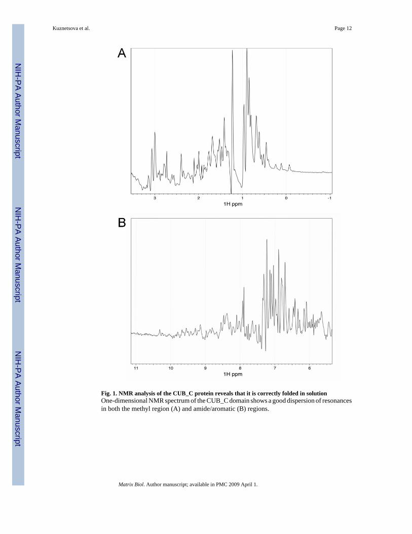
Fig. 1. NMR analysis of the CUB_C protein reveals that it is correctly folded in solutionOne-dimensional NMR spectrum of the CUB_C domain shows a good dispersion of resonancesin both the methyl region (A) and amide/aromatic (B) regions.
Kuznetsova et al. Page 12
Matrix Biol. Author manuscript; available in PMC 2009 April 1.
NIH
-PA Author Manuscript
NIH
-PA Author Manuscript
NIH
-PA Author Manuscript
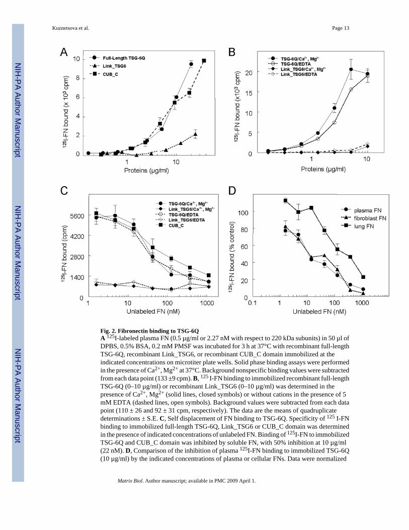
Fig. 2. Fibronectin binding to TSG-6QA 125I-labeled plasma FN (0.5 µg/ml or 2.27 nM with respect to 220 kDa subunits) in 50 µl ofDPBS, 0.5% BSA, 0.2 mM PMSF was incubated for 3 h at 37°C with recombinant full-lengthTSG-6Q, recombinant Link_TSG6, or recombinant CUB_C domain immobilized at theindicated concentrations on microtiter plate wells. Solid phase binding assays were performedin the presence of Ca2+, Mg2+ at 37°C. Background nonspecific binding values were subtractedfrom each data point (133 ±9 cpm). B, 125 I-FN binding to immobilized recombinant full-lengthTSG-6Q (0–10 µg/ml) or recombinant Link_TSG6 (0–10 µg/ml) was determined in thepresence of Ca2+, Mg2+ (solid lines, closed symbols) or without cations in the presence of 5mM EDTA (dashed lines, open symbols). Background values were subtracted from each datapoint (110 ± 26 and 92 ± 31 cpm, respectively). The data are the means of quadruplicatedeterminations ± S.E. C, Self displacement of FN binding to TSG-6Q. Specificity of 125 I-FNbinding to immobilized full-length TSG-6Q, Link_TSG6 or CUB_C domain was determinedin the presence of indicated concentrations of unlabeled FN. Binding of 125I-FN to immobilizedTSG-6Q and CUB_C domain was inhibited by soluble FN, with 50% inhibition at 10 µg/ml(22 nM). D, Comparison of the inhibition of plasma 125I-FN binding to immobilized TSG-6Q(10 µg/ml) by the indicated concentrations of plasma or cellular FNs. Data were normalized
Kuznetsova et al. Page 13
Matrix Biol. Author manuscript; available in PMC 2009 April 1.
NIH
-PA Author Manuscript
NIH
-PA Author Manuscript
NIH
-PA Author Manuscript
so that the binding of 125 I-FN to TSG-6Q in the absence of competing unlabeled FN equals100%.
Kuznetsova et al. Page 14
Matrix Biol. Author manuscript; available in PMC 2009 April 1.
NIH
-PA Author Manuscript
NIH
-PA Author Manuscript
NIH
-PA Author Manuscript
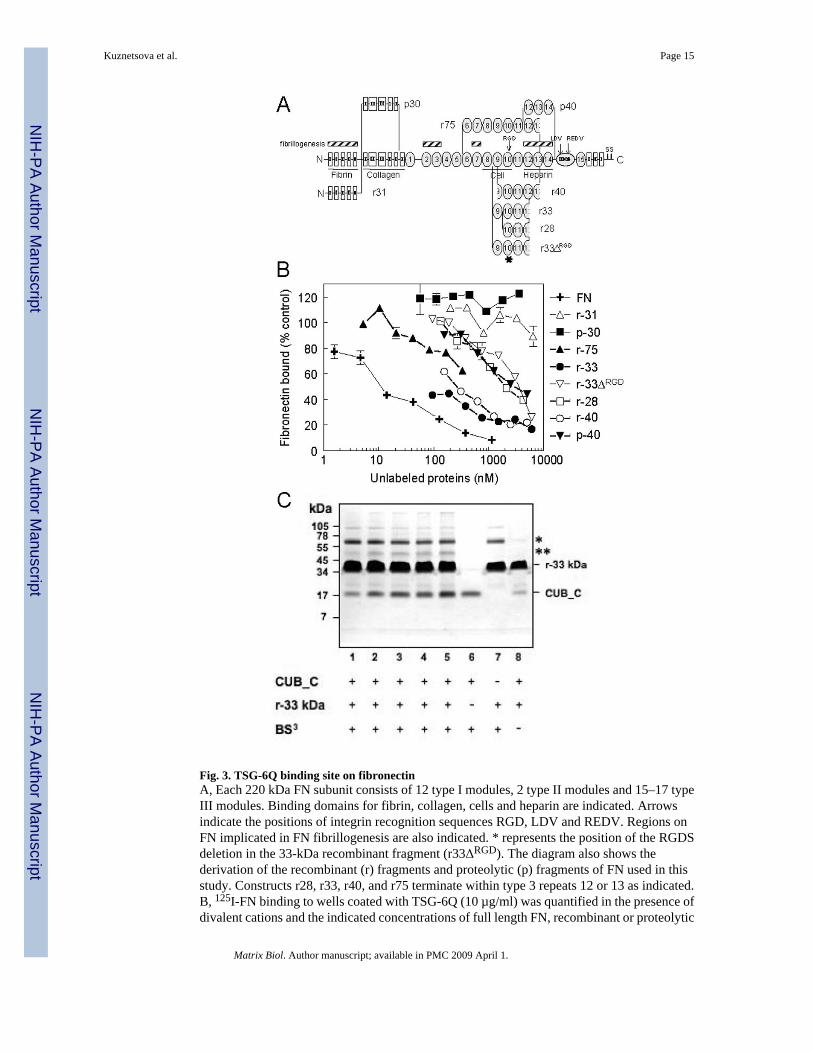
Fig. 3. TSG-6Q binding site on fibronectinA, Each 220 kDa FN subunit consists of 12 type I modules, 2 type II modules and 15–17 typeIII modules. Binding domains for fibrin, collagen, cells and heparin are indicated. Arrowsindicate the positions of integrin recognition sequences RGD, LDV and REDV. Regions onFN implicated in FN fibrillogenesis are also indicated. * represents the position of the RGDSdeletion in the 33-kDa recombinant fragment (r33ΔRGD). The diagram also shows thederivation of the recombinant (r) fragments and proteolytic (p) fragments of FN used in thisstudy. Constructs r28, r33, r40, and r75 terminate within type 3 repeats 12 or 13 as indicated.B, 125I-FN binding to wells coated with TSG-6Q (10 µg/ml) was quantified in the presence ofdivalent cations and the indicated concentrations of full length FN, recombinant or proteolytic
Kuznetsova et al. Page 15
Matrix Biol. Author manuscript; available in PMC 2009 April 1.
NIH
-PA Author Manuscript
NIH
-PA Author Manuscript
NIH
-PA Author Manuscript
fragments derived from FN. Results are presented as a % of control FN binding determined inthe absence of inhibitors. The data are the means of quadruplicate determinations ± S.E. C,Cross-linking of the CUB_C/fibronectin complex. Fibronectin r-33 kDa was incubated withvarying concentrations of CUB_C (molar ratios 1:28, 1:14, 1:9, 1:7 and 1:5 of CUB_C:r-33kDa) in presence of the cross-linker BS3 and analyzed under reducing conditions by SDS-PAGE (lanes 1–5). CUB_C alone (lane 6; at concentration equivalent to lane 3) and r-33 kDaalone (lane 7; concentration equivalent to lanes 1–5) were also incubated in the presenceBS3. Lane 8 shows CUB_C and r-33kDa in the absence of BS3 (molar ratio 1:28). In thepresence of cross-linking reagent species are seen at ~62 kDa (*; lanes 1–5 and 7) and ~50 kDa(**, lanes 1–5), which are not present in the absence of BS3 (lane 8). The former likelycorresponds to a cross-linked dimer of r-33 kDa given that this is present in the absence ofCUB_C. The ~50 kDa species (**), which becomes more intense with increasing CUB_Cconcentrations, is consistent with a 1:1 complex of CUB_C and r-33 kDa (with apparentmolecular masses of ~20- and ~35-kDa, respectively).
Kuznetsova et al. Page 16
Matrix Biol. Author manuscript; available in PMC 2009 April 1.
NIH
-PA Author Manuscript
NIH
-PA Author Manuscript
NIH
-PA Author Manuscript

Fig. 4. TSG-6 enhances fibronectin binding to thrombospondin-1Uncoated wells (Δ) or wells coated using 10 µg/ml thrombospondin-1 (TSP1) were blockedwith BSA and incubated with 0.5 µg/ml 125I-FN alone (▲) or in the presence of the indicatedconcentrations of full length TSG-6Q (●), CUB_C (▼), or Link_TSG6 (○). Results arepresented as a percent of the input FN bound for quadruplicate determinations ± SD.
Kuznetsova et al. Page 17
Matrix Biol. Author manuscript; available in PMC 2009 April 1.
NIH
-PA Author Manuscript
NIH
-PA Author Manuscript
NIH
-PA Author Manuscript
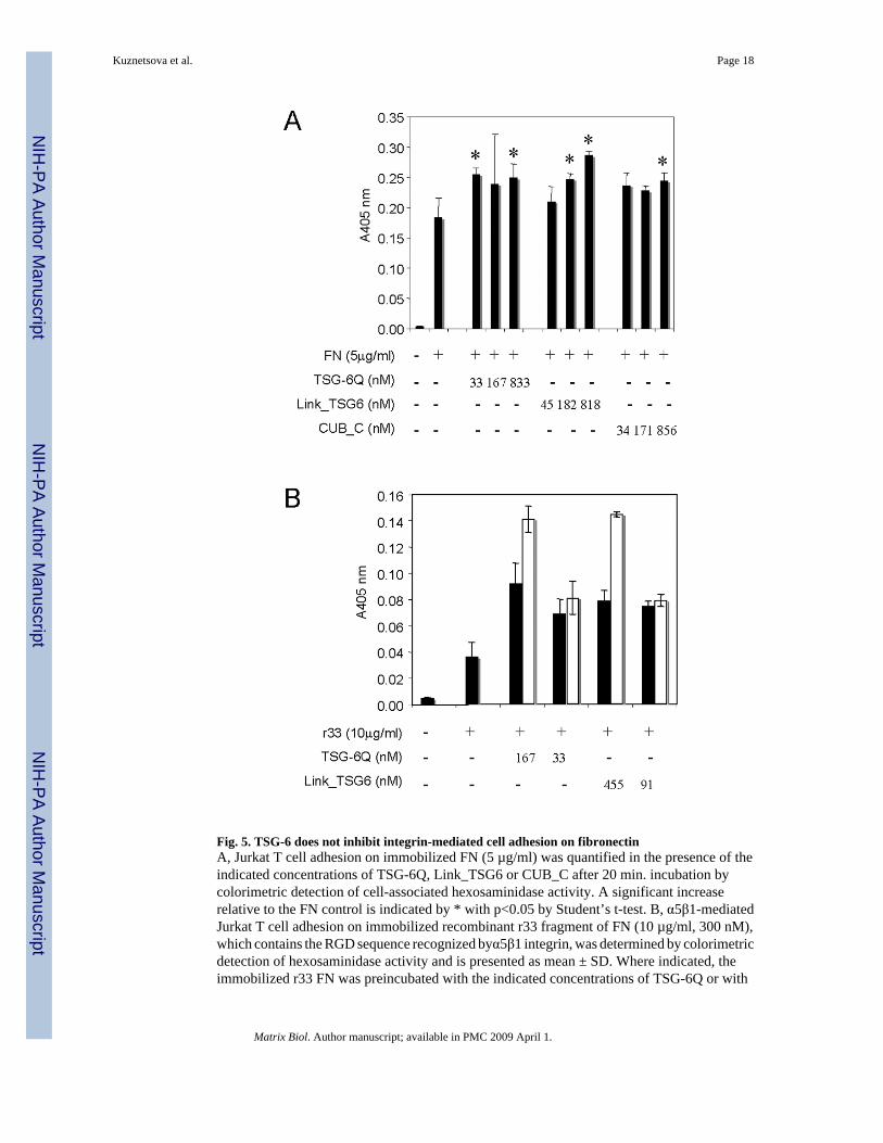
Fig. 5. TSG-6 does not inhibit integrin-mediated cell adhesion on fibronectinA, Jurkat T cell adhesion on immobilized FN (5 µg/ml) was quantified in the presence of theindicated concentrations of TSG-6Q, Link_TSG6 or CUB_C after 20 min. incubation bycolorimetric detection of cell-associated hexosaminidase activity. A significant increaserelative to the FN control is indicated by * with p<0.05 by Student’s t-test. B, α5β1-mediatedJurkat T cell adhesion on immobilized recombinant r33 fragment of FN (10 µg/ml, 300 nM),which contains the RGD sequence recognized byα5β1 integrin, was determined by colorimetricdetection of hexosaminidase activity and is presented as mean ± SD. Where indicated, theimmobilized r33 FN was preincubated with the indicated concentrations of TSG-6Q or with
Kuznetsova et al. Page 18
Matrix Biol. Author manuscript; available in PMC 2009 April 1.
NIH
-PA Author Manuscript
NIH
-PA Author Manuscript
NIH
-PA Author Manuscript

Link_TSG6 for 30 min at 37 °C and then washed to remove unbound TSG-6 prior to addingJurkat cells (solid bars) or used without washing (open bars).
Kuznetsova et al. Page 19
Matrix Biol. Author manuscript; available in PMC 2009 April 1.
NIH
-PA Author Manuscript
NIH
-PA Author Manuscript
NIH
-PA Author Manuscript
Fig. 6. TSG-6Q and CUB_C increase endogenous fibronectin matrix assembly in fibroblastsHs68 human fibroblasts were plated on glass in serum-free medium alone or in the presenceof 4 µg/ml TSG-6Q or 2 µg/ml CUB_C and incubated for 24 h. The cells were then fixed andstained with FN antibody to visualize matrix deposition (green) and Hoechst 33258 to visualizenuclei (blue). Representative fields imaged at equal exposure times are shown with the scalebar = 50 µm.
Kuznetsova et al. Page 20
Matrix Biol. Author manuscript; available in PMC 2009 April 1.
NIH
-PA Author Manuscript
NIH
-PA Author Manuscript
NIH
-PA Author Manuscript
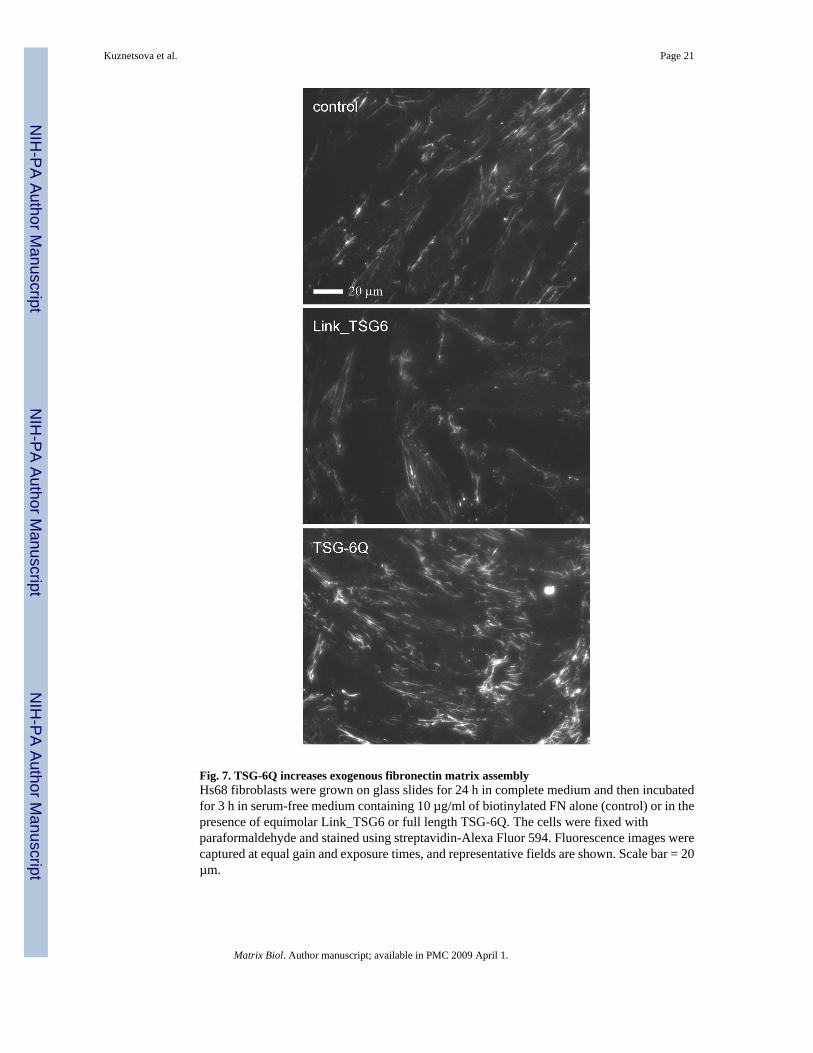
Fig. 7. TSG-6Q increases exogenous fibronectin matrix assemblyHs68 fibroblasts were grown on glass slides for 24 h in complete medium and then incubatedfor 3 h in serum-free medium containing 10 µg/ml of biotinylated FN alone (control) or in thepresence of equimolar Link_TSG6 or full length TSG-6Q. The cells were fixed withparaformaldehyde and stained using streptavidin-Alexa Fluor 594. Fluorescence images werecaptured at equal gain and exposure times, and representative fields are shown. Scale bar = 20µm.
Kuznetsova et al. Page 21
Matrix Biol. Author manuscript; available in PMC 2009 April 1.
NIH
-PA Author Manuscript
NIH
-PA Author Manuscript
NIH
-PA Author Manuscript

Fig. 8. TSG-6 is incorporated into and increases fibronectin incorporation into detergent-insolublematrixA, Hs68 fibroblasts were cultured overnight in normal medium, washed with medium withoutserum containing 1% BSA, and incubated in the same medium alone or supplemented with12.5 µg/ml (57 nM) of biotinylated FN without additional agents or in the presence of a 3-foldmolar excess of TSG-6Q or Link_TSG6. Deoxycholate (DOC)-insoluble fractions wereresolved on 7.5% polyacrylamide gels, transferred to nitrocellulose membranes, and probedwith HRP-conjugated streptavidin to determine the amount of incorporated FN. The samemembranes were re-probed with antibodies against actin to assess the efficiency of proteinextraction and gel loading. The results shown are representative of three independentexperiments and were analyzed by densitometry. The FN signals were normalized to therespective actin signals, and the mean for each duplicate is presented normalized to a value of1.0 for FN alone. B, Samples from the DOC-insoluble fraction were assayed byimmunoblotting with goat polyclonal anti-TSG-6 to assess the incorporation of TSG-6.
Kuznetsova et al. Page 22
Matrix Biol. Author manuscript; available in PMC 2009 April 1.
NIH
-PA Author Manuscript
NIH
-PA Author Manuscript
NIH
-PA Author Manuscript
NIH
-PA Author Manuscript
NIH
-PA Author Manuscript
NIH
-PA Author Manuscript
Kuznetsova et al. Page 23
Table 1Fibronectin binding to immobilized TSG-6 determined by self displacement
Immobilized ligand Apparent Ka (M−1)
Full length TSG-6Q (Ca2+, Mg2+) 1.6 ± 0.6 × 10⁸
Full length TSG-6Q (5 mM EDTA) 1.0 ± 0.2 × 10⁸
CUB_C domain 5.7 ± 1.3 × 10⁷
Link_TSG6 --
Binding parameters were determined from self displacement of 125I-FN binding to the indicated ligands immobilized on polystyrene. Binding at eightconcentrations of FN over the range 0.3 to 250 µg/ml was assessed in quadruplicate and analyzed using Scafit version 3.10 of the LIGAND software. Alldata were adequately fit using a single site binding model.
Matrix Biol. Author manuscript; available in PMC 2009 April 1.