transvascular migration of l2c leukemic cells studied in the liver of the guinea pig
TRANSCRIPT

Virchows Arch [Pathol Anat] (1985) 406:425-440 ViMiows Archiv A © Springer-Verlag 1985
Transvascular migration of L2C leukemic cells studied in the liver of the guinea pig
Biagio Azzarelli, Jans Muller, L. David Mirkin, and Michael P. Goheen Indiana University School of Medicine, Department of Pathology, Division of Neuropathology, 635 Barnhill Drive, Indianapolis, IN 46223, USA
Summary. The possible routes of transvascular migration of leukemic cells in the liver were studied in guinea pigs with an L2C lymphoblastic cell-line inoculation leukemia. Invasion of the hepatic parenchyma theo- retically can occur in three ways:
1. Through the intact sinusoidal endothelium, utilizing either pre- existent gaps (normal in the liver), or newly created pores, whether inter- endothelial or intraendothelial. We could not convincingly demonstrate this, but could not wholly exclude this either.
2. After destruction or retraction of the endothelium, either on ac- count of the remarkable sinusoidal engorgement and distension by masses of leukemic cells, or by direct assault on the endothelium by the leukemic cells. We can clearly demonstrate the former, and hold it to be the major cause of hepatic infiltration. Evidence for a direct endotheliolytic effect was not uncovered in our studies.
3. Secondary infiltration from the portal triads. Heavy leukemic infil- tration of the triads, whether from the portal or hepatic veins, or from the lymphatics, is indeed and early an consistent feature - but the infiltra- tion of the hepatic lobule shows no peripheral, or any other zonal prefer- ence.
In both portal and hepatic veins, leukemic cells transverse the endo- thelium through a cytoplasmic "pore" , adjacent to cell junctions, with- out obvious damage to the endothelium.
Key words: Leukemia - Liver - Ultrastructure - Endothelium - Guinea pig
Introduction
Considering the importance of the metastatic process, it is surprising that relatively few ultrastructural studies have been concerned with the route followed by the neoplastic cells in crossing the blood vessel wall (Ludatscher
Offprint requests to. B. Azzarelli at the above address

426 B. Azzarelli et al.
et al. 1967; Mladenov et al. 1967; Locker et al. 1970; Chen et al. 1972; Dingemans 1973; Dingemans 1974; Campbell 1975; Sindelar et al. 1975; Carr et al. 1976; DeBruyn et al. 1977; Roos et al. 1977; Dingemans et al. 1978). The usual experimental procedure consists of the injection into the vascular system of cell suspensions obtained by fragmentation of solid tu- mors. The procedures used to obtain such suspensions as weil as the method of introducing such cells in a single bolus into the circulation leaves such studies open to serious logical objections. Our ultrastructural observations in the guinea pig describe the transvascular metastatic route followed by leukemic cells, two weeks after the subcutaneous inoculation with a known (L2C) leukemic cell line.
Materials and methods
A suspension of 7.0 x 10⁵ to 2.8 x 10⁶ L2C cells in pyrogen free saline was inoculated under the skin of the flank of eleven strain II guinea pigs of 300-350 gm each. The fresh L2C cells are obtained from a terminal donor guinea pig previously inoculated with L2C frozen cells obtained from the laboratory of Dr. Ira Green, National Institutes of Health. Two further guinea pigs serve as normal controls. At intervals of one to fifteen days (5 in the first eight days, the other 6 at 12-15 days) after inoculation, the animals are anesthesized with an intraper- itoneal injection of Nembutal Sodium solution, 20-40 mg per kg body weight. Through cardiac puncture an aliquot of blood is obtained for total and differential blood cell counts. Under 1% Procaine anesthesia tracheostomy is performed, followed by thoracotomy. Ventilation is maintained manually with a rubber cuff, controlled by direct visualization of pulmonary expansion. The animals are then perfused through the ascending aorta with a chilled, double- aldehyde fixative consisting of 2.5% Glutaraldehyde with 1.5% Paraformaldehyde in 0.08 M Cacodylate buffer (pH 7.2) with 0.15% Calcium Chloride added (osmolarity 1.285 + 15 millios- mol). In some 15 20 min 1,000 ml of this solution is used. A constant flow is maintained by adjusting the fluid pressure with a sphygmomanometer cuff around a plastic bag (pressure 120-150 mm of mercury). At autopsy, tissue blocks of every major organ including the brain and the area of inoculation, are obtained. In the 5 pre-leukemic animals (1-8 days), no hepatic infiltration was seen (16 blocks, thick sections). Of the 6 leukemic animals (12-15 days), all 24 liver blocks revealed infiltration. Six blocks were further trimmed down and utilized for thin sections. The tissue is further fixed for two hours in the same donble-aldehyde solution, then post-fixed in 2% Osmium Tetroxide for two hours. Then the tissues are dehydrated in a graded series of chilled ethanol, passed through Propylene Oxide, placed in a 1 : 1 mixture of Propylene Oxide and Epon 812 for 45 min and finally embedded in Epon 812. One micron thick sections are stained with a 1% solution of Toluidine Blue. From these sections specific areas are chosen for electron microscopic examination. The blocks are trimmed and the area remaining is thin sectioned on a LKB ultramicrotome. The thin sections are collected on 200 mesh copper grids (ultrahigh transmission grids, Polysciences, Inc., Warrington, PA, USA, stained with Uranyl Acetate and Lead Citrate and viewed at 60 KV in a Philips 300 electron microscope.
Results
The L2C leukemic cells are characterized by small amounts of cytoplasm. The surface is covered by numerous, widely spaced microvilli. There are large numbers of ribosomes, many in rosettes, also a few cisternae of the rough endoplasmic reticulum and often viral particles, almost in every cell (none were found in normal white blood cells), none of which will be de- scribed (Nadel et al. 1967). Other organelles are inconspicuous. The nuclei
Leukemic migration 427
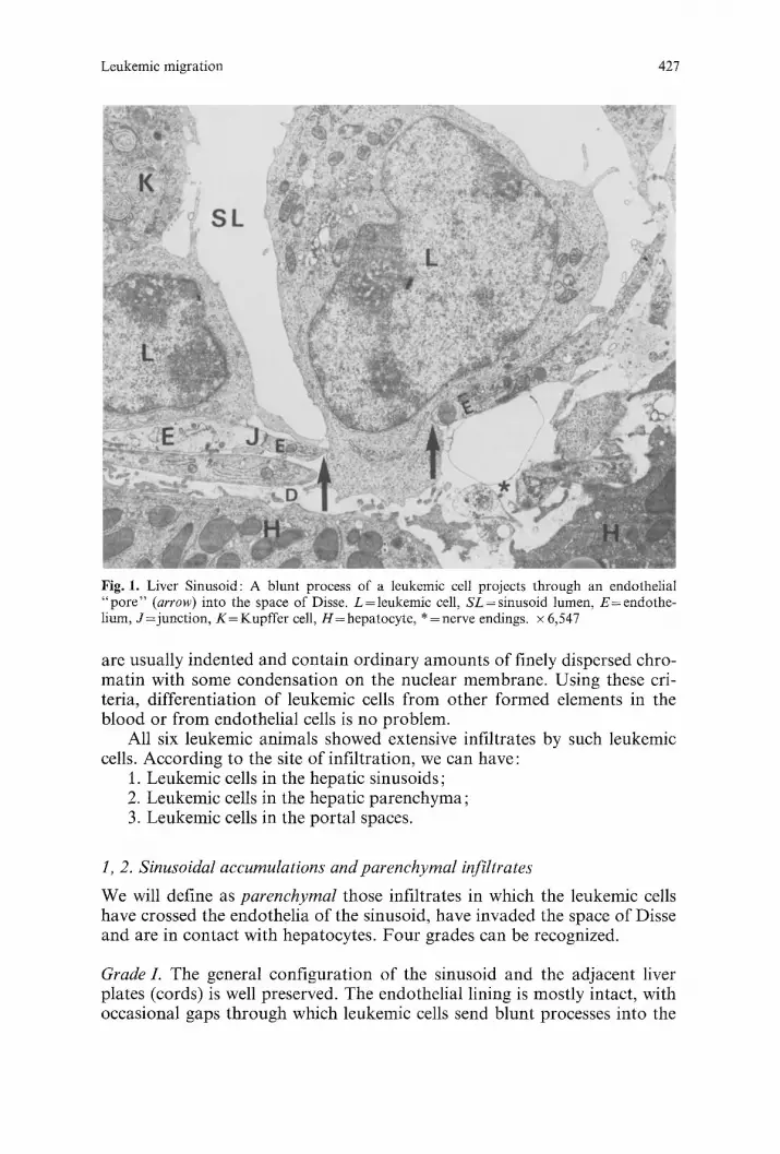
Fig. 1. Liver Sinusoid: A blunt process of a leukemic cell projects through an endothelial "po re" (arrow) into the space of Disse. L=leukemic cell, SL=sinusoid lumen, E=endothe- lium, J=junct ion, K = Kupffer cell, H = hepatocyte, * = nerve endings. × 6,547
are usually indented and contain ordinary amounts of finely dispersed chro- matin with some condensation on the nuclear membrane. Using these cri- teria, differentiation of leukemic cells from other formed elements in the blood or from endothelial cells is no problem.
All six leukemic animals showed extensive infiltrates by such leukemic cells. According to the site of infiltration, we can have:
1. Leukemic cells in the hepatic sinusoids; 2. Leukemic cells in the hepatic parenchyma; 3. Leukemic cells in the portal spaces.
1, 2. Sinusoidal accumulations and parenchymal infiltrates
We will define as parenchymal those infiltrates in which the leukemic cells have crossed the endothelia of the sinusoid, have invaded the space of Disse and are in contact with hepatocytes. Four grades can be recognized.
Grade L The general configuration of the sinusoid and the adjacent liver plates (cords) is well preserved. The endothelial lining is mostly intact, with occasional gaps through which leukemic cells send blunt processes into the
428 B. Azzarelli et al.
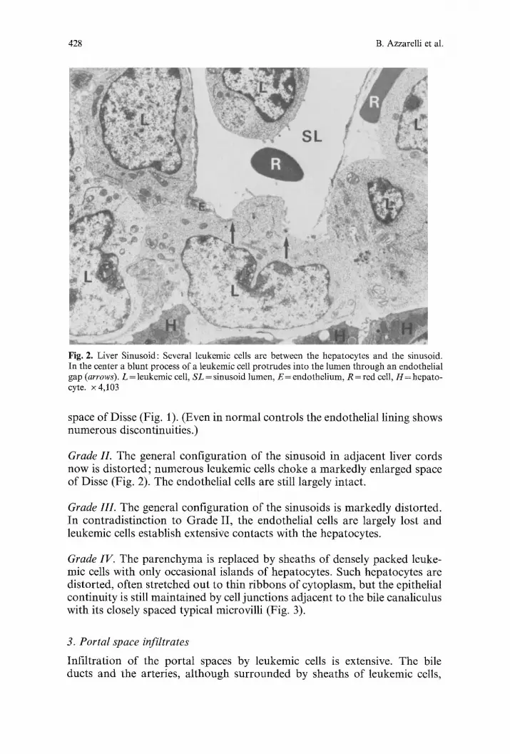
Fig. 2. Liver Sinusoid: Several leukemic cells are between the hepatocytes and the sinusoid. In the center a blunt process of a leukemic cell protrudes into the lumen through an endothelial gap (arrows). L = leukemic cell, SL = sinusoid lumen, E = endothelium, R = red cell, H= hepato- cyte. × 4,103
space of Disse (Fig. 1). (Even in normal controls the endothelial lining shows numerous discontinuities.)
Grade II. The general configuration of the sinusoid in adjacent liver cords now is distorted; numerous leukemic cells choke a markedly enlarged space of Disse (Fig. 2). The endothelial cells are still largely intact.
Grade III. The general configuration of the sinusoids is markedly distorted. In contradist inction to Grade II, the endothelial cells are largely lost and leukemic cells establish extensiVe contacts with the hepatocytes.
Grade IV. The parenchyma is replaced by sheaths of densely packed leuke- mic cells with only occasional islands of hepatocytes. Such hepatocytes are distorted, often stretched out to thin ribbons of cytoplasm, but the epithelial continuity is still maintained by cell junctions adjacent to the bile canaliculus with its closely spaced typical microvilli (Fig. 3).
3. Portal space infiltrates
Infil tration of the portal spaces by leukemic cells is extensive. The bile ducts and the arteries, a l though surrounded by sheaths of leukemic cells,

Leukemic migration 429
Fig. 3. Two Liver Sinusoids: Leukemic cells number 4 and 6 are compressing two adjacent hepatocytes. Note the bile canaliculus (center). SL-sinusoidal lumen, L= leukemic cell, H= hepatocyte, E= endothelium, D = space of Disse, Arrow =junction. x 5,088
are normal. Extensive leukemic infil tration, however, are seen in the veins, bo th por ta l and hepatic (Figs. 4 and 5). We will classify these leukemic cells according to their locat ion as follows: intravascular, in t ramural and migrating.
a) Intravascular. Only a few leukemic cells are present, r andomly distr ibuted th roughou t the b lood vessel lumen. We taust keep in mind, however, that

430 B. Azzarelli et al.
Fig. 4. Extensive leukemic infiltrates of a portal space extending to the adjacent liver parenchy- ma where only an island of hepatocytes is still present (H). Notice subendothelial leukemic cells; one of them is in mitosis (large arrow). Two leukemic cells appeared marginated (small arrow). One micron section, Epon, Toluidine Blue stained, x 812
these are perfused preparations where the central portion of the vascular lumen has been cleansed. Most of the cells are in close proximity to the endothelium, we define such cells as marginated if they establish contact with the endothelium for one-third or more of the total cell perimeter. Whereas the free cells in the lumen are round in cross section, marginated cells are flattened against the vascular outline. Moreover, in the flattened portion of the marginated cells the cytoplasmic membrane looses its custom- ary microvilli. The slit defined by the cytoplasmic membrane parallel to the endothelium will be from 15-50 nm wide.
In a few instances a leukemic cell is completely surrounded by a thin endothelial cytoplasmic layer (Fig. 6), the space between the leukemic cells
Fig. 5. Extensive leukemic infiltrates in a portal space. There is massive subendothelial accumu- lation of leukemic cells. Between arrows is an area lacking endothelial lining; the leukemic cells are directly apposed to the basal lamina, x 850
Fig. 6. Portal Vein: A leukemic cell is completely encircled by a thin endothelial cytoplasmic layer. LU=lumen, J= junc t ion , E = endothelium, L = leukemic cells, SM= smooth muscle. x 3,300

!!
Lu
432 B. Azzarelli et al.
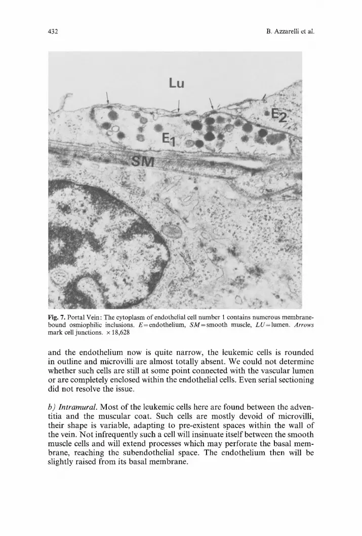
Fig. 7. Portal Vein: The cytoplasm of endothelial cell number I contains numerous membrane- bound osmiophilic inclusions. E--endothelium, SM=smooth muscle, LU=lumen. Arrows mark cell junctions, x 18,628
and the endothel ium now is quite narrow, the leukemic cells is rounded in outline and microvilli are almost totally absent. We could not determine whether such cells are still at some point connected with the vascular lumen or are completely enclosed within the endothelial cells. Even serial sectioning did not resolve the issue.
b) Intramural. Most o f the leukemic cells here are found between the adven- titia and the muscular coat. Such cells are most ly devoid o f microvilli, their shape is variable, adapt ing to pre-existent spaces within the wall of the vein. N o t infrequent ly such a cell will insinuate itself between the smooth muscle cells and will extend processes which may perfora te the basal mem- brane, reaching the subendothel ial space. The endothel ium then will be slightly raised f rom its basal membrane .

Leukemic migra t ion 433
Lu \ o
Fig. 8. Por ta l Vein: Large endothel ia l gap (large arrows). The basa l l amina is exposed to the lumen. The leukemic cells n u m b e r 1 and 2 are unde r the endo the l ium. Leukemic cell n u m b e r 3 is unde r a s m o o t h musc le cell. L = leukemic cell, E = endo the l ium, BL = basal lamina , L U = l u m e n . x 29,157
The endothelium in other areas eventually becomes markedly separated from its basal membrane by leukemic cells. The endothelium bulges into the lumen where in addition there a r e a variety of changes, as compared to the normal flat endothelium of control animals. The most constant find- ing is the presence of numerous dense, osmiophilic membrane-bound round granules about 180-214 nm in diameter (Figs. 7 and 8). There is a marked
I
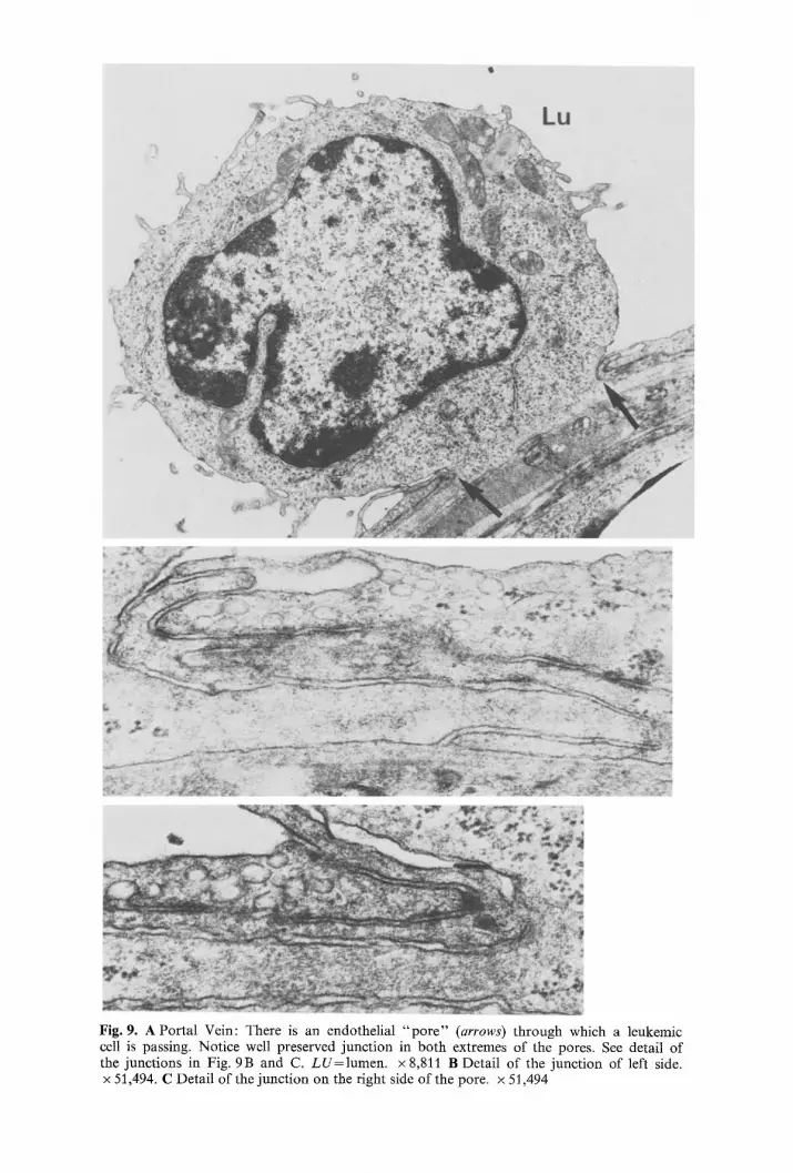
Fig. 9. A Portal Vein: There is an endothelial " p o r e " (arrows) through which a leukemic cell is passing. Notice well preserved junction in both extremes of the pores. See detail of the junctions in Fig. 9B and C. LU=lumen . × 8,811 B Detail of the junction of left side. x 51,494. C Detail of the junction on the right side of the pore. x 51,494
Leukemic migration 435
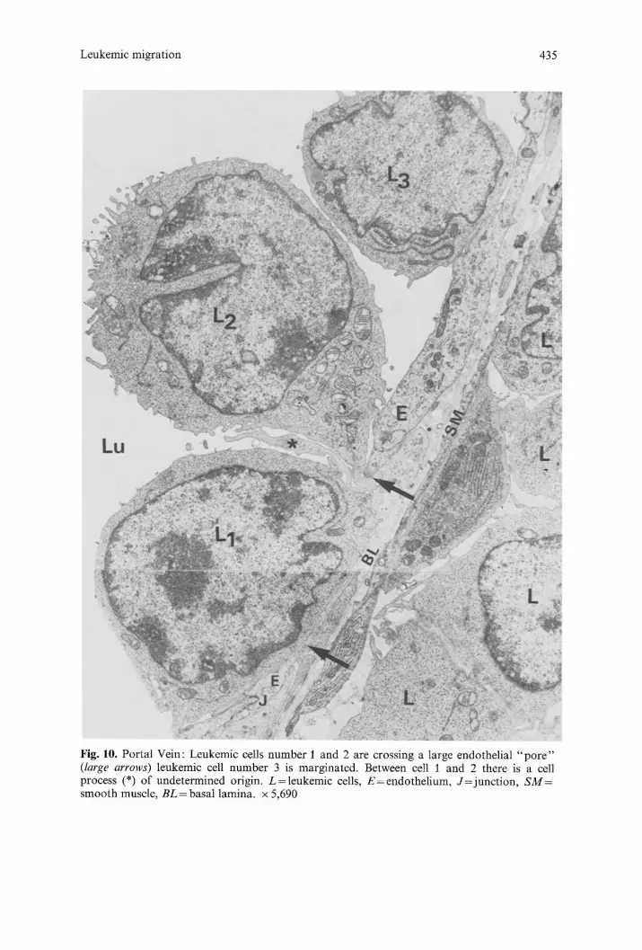
Fig. 10. Portal Vein: Leukemic cells number 1 and 2 are crossing a 1arge endothelial " p o r e " (large arrows) leukemic cell number 3 is marginated. Between cell i and 2 there is a cell process (*) of undetermined origin. L=leukemic cells, E=endothe l ium, J= junc t ion , SM= smooth muscle, BL = basal lamina, x 5,690
436 B. Azzarelli et al.
decrease of organelles in some of these endothelial cells; others show exten- sive disruptions of the cytoplasmic membranes. In some areas the endotheli- al cells have become completely lost and the basal membrane is exposed to the lumen (Fig. 8).
c) Actual migration of leukemic cells. We will concern ourselves with those images in which portions of the leukemic cell are actually crossing the endo- thelial barrier. Such passage is generally through a large "po re" of as much as 14 microns across. One or several cells may be observed migrating through a single pore, with the endothelial membrane adjacent to the pores well preserved. Endothelial junctions in the vicinity of such pores are rather elaborate with imbricating configurations (Figs. 9A, 9 B and 9 C). At least one edge of a pore is always close to such an endothelial junction (Fig. 10). However, on many occasions the pore is literally flanked by two junctions on each side (Fig. 9A), which suggests that such pores occur at the apex of an endothelial cell. Occasional fenestrations, randomly distributed can be seen in the endothelial cells of the portal veins in control animals. A, residual cytoplasmic layer is always found in such a fenestration which differentiates it from a pore; pores are never seen in the veins of control animals.
Discussion
This L2C acute B type lymphocytic leukemic arose spontaneously in a fe- male strain-II guinea pig (Congdon and Lorenz 1954). With a potent inocu- lum of 10 » cells all injected animals became leukemic. Death takes place 2-4 weeks after inoculation with extensive leukemic infiltration of all viscera and the brain, there is less intense involvement of the bone marrow. Termin- ally, white blood ceU counts in excess of 100,000 cells/mm 3 with 99% blasts in the peripheral smear are obtained.
Neoplastic dissemination in the liver is usually studied by the injection of cell suspensions, either into the general circulation (Chew et al. 1976; Campbell 1977) or into the vena porta (Dingemans 1973; Dingemans et al. 1978). Although this single-dose injection of neoplastic cells is different from the natural situation, it does provide the opportunity to observe the morphological events in a predetermined time sequence. In our natural mod- el malignant cells are continously in circulation and all stages of the meta- static process are going on concurrently.
In an attempt to reconstruct the events we will single out some of the pertinent observations. It should be realized that there are methodologic problems in studying the putative migration of neoplastic cells through the endothelial cells of the hepatic sinusoids, since normally the endothelial lining contain gaps through which a cell could invade. Therefore, in the case of the hepatic sinusoidal endothelia, the creation of new openings is not a necessary precondition to explain the presence of leukemic cells in the Disse space or among the hepatocytes. In the erlier stage of hepatic parenchymal invasion (Grade I) the leukemic cell is marginated and may
Leukemic migration 437
project a blunt process through an endothelial gap. Whether this is passage through the endothelial cell (transcytoplasmic diapedesis, Dingemans 1973; Dingemans 1974; Roos et al. 1977) or through a widened intercellular gap (intercellular extravasation, Sindelar et al. 1975) is hard to say. Typically liver sinusoids show cytoplasmic fenestrations (Motta and Porter 1974; Grisham et al. 1975; Nopanitaya and Grisham 1975; Wisse 1970; Brooks and Haggis 1973) and intercellular gaps (Wisse 1983). In addition when the sinusoids are filled with leukemic cells, such leukemic cells may well protrude somewhat through pre-existing gaps. To interpret such a protru- sion as a beginning migration is tempting but unjustified. We will only consider those leukemic cells as traveling through an endothelium if some- where near one third to one half of the cell is found on either side of the gap, and this we did not observe in the hepatic sinusoids. Neither could be develop satisfactory evidence that the numbers or types of gaps were really increased as the result of the leukemic infiltration, as compared to normal control animals. To rephrase this, are these gaps indeed the result of a specific interaction between the endothelium and the neoplastic cells?
In the second stage of infiltration leukemic cells are in the liver plates, but they are still separated from the sinusoid by an intact endothelium (Fig. 2). Occasional blunt processes do extend into the sinusoidal lumen, sometimes referred to as tails by other authors (Dingemans et al. 1978). It is tempting to link this stage of hepatic invasion to the first stage but it is quite disturbing that in no instance could we, or other investigators (Dingemans et al. 1978) document intermediate stages in which a cell crosses the endothelium, as defined by us previously as about one-third way through.
In the third grade of hepatic parenchymal invasion the sinusoids are markedly distended by leukemic cells, but the endothelium now is frag- mented and may have completely disappeared. The process of endothelial fragmentation could not be followed well: the endothelia are either fairly intact, or have entirely disappeared. The origin of these large endothelial defects has been the subject o fmuch speculation. According to some (Locker et al. 1970; Chew et al. 1976; Fonck-Cussac et al. 1969) there is attrition following the stretching of the sinusoid; according to others (Cotmore and Carter 1973) there is possible anoxic damage to the endothelial cells, still others (Dingemans et al. 1978) believe there is retraction followed perhaps by regeneration of the endothelium. Retraction, with exposure of the under- lying basal membrane, has been seen in vitro in binding of malignant cells to monolayers of vascular endothelia (Kramer and Nicolson 1979). Move- ment of neoplastic cells to the basal membrane occurs because the basement membrane is closely adherent to the endothelium and thus becomes exposed once the endothelium retracts (Kramer et al. 1980; Nicolson et al. 1981). To what extent this is important in the liver where the basal lamina is so incomplete is still in question. A different mechanism of malignant extra- vasation, similar to that of leukocytes crossing the endothelium during in- flammation (Marchesi and Florey 1960) has been proposed with the cells passing through the endothelial junctions (Ludatscher et al. 1967 ; Sindelar et al. 1975; Carr et al. 1976).
438 B. Azzarelli et al.
All the leukemic animals show extensive leukemic infiltrates in the portal triads, they are continuous with those in the periphery of the hepatic lobules. Whether the portal infiltrates develop first in the portal triads as has been suggested in human lymphocytic leukemia (Goldberg 1960) or are secondary to the parenchymal infiltration is unclear. The parenchymal infiltration shows no zonal preference and therefore cannot itself be derived from the portal infiltrates. The veins, but not the arteries or the bile ducts, do show extensive leukemic infiltration and it is clear that there is transendothelial passage of leukemic cells. The direction of the migration, whether from the lumen outwards or back into the lumen can of course not be established by static morphologic studies. The leukemic cells, either singly or in groups, cross through a pore in an otherwise intact endothelial cell. Such pores are generally close to the cell junction but the junctions themselves remain intact. The portal veins in the triads normally show fenestrations (Grisham et al. 1975), these are differentiated from pores in that the fenestrations remain covered by a thin layer of cytoplasmic membrane. In addition, fenes- trations occur at random through the endothelial cell whereas the pores are always close to the cell junctions. Passage through pores, as described here, has previously been reported for leukemic myelogenous elements in the bone marrow (DeBruyn et al. 1977), of erythroblasts in rats with routine erythroblastosis in the spleen (Cho and DeBruyn 1975), in subcutaneously implanted myelogenous tumors (DeBruyn and Cho 1979) and by us in the leptomeningeal veins of L2C leukemic guinea pigs (Azzarelli et al. 1984). This crossing of the blood vessel wall, so characteristic of malignant cells, is of course not limited to such cells but is the usual mechanism of egress into the circulation of normal, myelogenous elements in the bone marrow (DeBruyn et al. 1971; Campbell 1972). Only mature cells will cross and crossing is through transient pores, characteristically located close to the cell junction. This selectivity for mature elements is lost when leukemic cells enter the bone marrow sinusoid following the same route (DeBruyn et al. 1977).
It is interesting to observe that in no instance could we demonstrate the presence of fibrin or platelets around or near tumour cells, either in the hepatic sinusoids or in the portal vessels. This fits in with observations by other authors (Cotmore and Carter 1973; Dingemans 1973). This nega- tive finding may be significant, since it has been postulated that the attach- ment of embolized malignant cells to the vascular walls is largely dependent upon entrapment of tumor cells within a platelet thrombus and/or a fibrin mesh-work (Wood 1958; Wood et al. 1961; Chew and Wallace 1976; Sinde- lar et al. 1975; Jones et al. 1971).
In summary, we believe that the invasion of the hepatic parenchyma proper follows intrasinusoidal leukemic cell proliferation. This proliferation leads to endothelial disruption or perhaps retraction. The possibility that occasional leukemic cells migrate through gaps of a previously intact endo- thelium cannot be ruled out.
We also have documented that there is extensive passage of cells through the endothelium in all the veins in the portal triads, which then must be

Leukemic migration 439
true for both the portal and the hepatic system. But whether this represents intra or extravasation could not be determined.
References
Azzarelli B, Mirkin D, Goheen M, Muller J, Crockett C (1984) The leptomeningeal vein: A site of re-entry of leukemic cells into the systemic circulation. Cancer 54:1333-1347
Brooks SEH, Haggis GH (1973) Scanning electron microscopy of rat liver. Application of freeze-fracture and freeze-drying techniques. Lab Invest 29:60-64
Campbell F (1975) Ultrastructure of the sinus wall of murine bone marrow in myelogenous leukemia. Am J Anat 142:319-334
Campbell FR (1972) Ultrastructure studies of transmural migration of blood cells in the bone marrow of rats, mice and guinea pigs. Am J Anat 135:521-536
Campbell FR (1977) Ultrastructural study of liver sinusoids of mice during invasion by leuke- mic myelocytes. J Natl Cancer Inst 58 : 369-376
Carr I, McGinty F, Norris P (1976) The fine structure of neoplastic invasion: Invasion of liver, skeletal muscle and lymphatic vessels by the Rd/3 tumor. J Pathol 118:91 99
Chen L, Handler EE, Handler ES, Weiss L (1972) An electron microscopic study of the bone marrow of the rat in experimentat myelogenous leukemia. Blood 39:99-112
Chew EC, Josephson RL, Wallace AC (1976) Morphologic aspects of the arrest of circulating cancer cells. In : Weiss L (ed) Fundamental Aspects of Metastasis. North-Holland Publish- ing Company, pp 121-150
Chew EC, Wallace AC (1976) Demonstration of fibrin in early stages of experimental metasta- sis. Cancer Res 36:1904-1909
Cho Y, Deßruyn PP (1975) Passage of red blood cells through the sinusoidal wall of the spleen. Am J Anat 142: 91-106
Congdon CC, Lorenz E (1954) Leukemia in guinea pigs. Am J Pathol 30:332359 Cotmore SF, Carter RL (1973) Mechanism of enhanced intrahepatic metastases in surfactant-
treated hamsters: An electron microscopy study. Int J Cancer 11:725-738 Deßruyn PH, Becker RP, Michelson S (1977) The transmural migration and release of blood
cells in acute myelogenous leukemia. Am J Anat 149:147-268 Deßruyn PH, Michelson S, Thomas TB (1971) The migration of blood cells of the bone
marrow through the sinusoidal wall. J Morphol 133:417-438 Deßruyn PH, Cho Y (1979) Entry of metastatic malignant cells into the circulation from
a subcutaneously growing myelogenous tumor. J Natl Cancer Inst 62:1221-1223 Dingemans KP (1973) A morphological study of the invasion of liver tissue by tumor cells.
Arch Chirurg Neurol 25:351-362 Dingemans KP (1974) Invasion of liver tissue by blood-born mammary carcinoma. J Natl
Cancer Inst 53:1813-1824 Dingemans KP, Roos E, van den Bergh Weerman M, van de Pavert IV (1978) Invasion
of liver tissue by tumour cells and leukocytes : Comparative ultrastructure. J Natl Cancer Inst 60: 583-598
Fonck-Cussac Y, Delage J, Petit J (1969) Observations ultrastructurales sur le mode d'implanta- tion endovasculaire des metastases d'un cancer bronchique. Poumon Coeur 25:231-234
Goldberg BM (1960) A study of malignant lymphomas and lenkemias. I. The significance of liver portal space "infiltration" in lymphogenous leukemia (with reference to the involve- ment of the lymphatics). Cancer 13:513-519
Grisham JW, Nopanitaya W, Compagno J, Nagel AEH (1975) Scanning electron microscopy of normal rat liver. The surface structure of its cells and tissue components. Am J Anat 144:295-322
Jones DS, Wallace AC, Fraser EF (1971) Sequence of events in experimental metastases of Walker 256 tumor: Light, immunofluorescent and electron microscopic observations. J Natl Cancer Inst 46:493-504
Kramer RH, Nicolson GL (1979) Interactions of tumour cells with vascular endothelial cell monolayers : A model for metastatic invasion. Proc Natl Acad Sci USA 76: 5704-5708
440 B. Azzarelli et al.
Kramer RH, Gonzalez R, Nicolson GL" (1980) Metastatie tumor cells adhere preferentially to the extracellular matrix underlying vascular endothelial cells. Int J Cancer 26: 639-645
Locker J, Goldblatt P J, Leighton J (1970) Ultrastructural features of invasion in chick embryo liver metastasis of Yoshida ascites hepatoma. Cancer Res 30:1623 1644
Ludatsher RM, Luse SA, Suntzeff V (1967) An electron microscopic study of pulmonary tumor emboli from transplanted Morris hepatoma 5123. Cancer Res 27:1939-1952
Marchesi VT, Florey HW (1960) Electron microscopic observations on the emigration of leukocytes. Q J Exp Physiol 45 : 343-348
Mladenov Z, Heine U, Beard D, Beard JW (1967) Strain MC 29 avain leukosis virus, myelocy- toma, endothelioma and renal growths: Pathomorphological and ultrastructural aspects. J Natl Cancer Inst 38:251-285
Motta P, Porter KR (1974) Structure of rat liver sinusoids and associated tissue spaces as revealed by scanning electron microscopy. Cell Tiss Res 148:111-125
Nadel E, Banfield W, Burstein S et al. (1967) Virus particles associated with strain II guinea pig leukemia (L2C/M-BJ). J Natl Cancer Inst 38 : 979-982
Nicolson GL, Irimura T, Gonzalez R, Rouslahti E (1981) The role of fibronectin in adhesion of metastatic melanoma cells to endothelial cells and their basal lamina. Exp Cell Res 135:461~465
Nicolson GL (1982) Metastatic tumor cell attachment and invasion assay utilizing vascular endothelial cell monolayers. J Histochem Cytochem 30:214-220
Nopanitaya W, Grisham JW (1975) Scanning electron microscopy of mouse intrahepatic struc- tures. Exp Mol Pathol 23:441M58
Roos E, Dingemans KP, van de Pavert IV et al. (1977) Invasion of lymphosarcoma cells into the perfused mouse liver. J Natl Cancer Inst 58 : 399M07
Sindelar WF, Tralka TS, Ketcham AS (1975) Electron microscopic observations on formation of pulmonary metastases. J Surg Res 18:137-161
Wisse E (1970) An electron microscopic study of the fenestrated endothelial lining of the rat liver sinusoids. J Ultrastruct Res 31 : 125-150
Wisse E, DeZanger RB, Jacobs R, McCuskey RS (1983) Scanning electron microscope observa- tions on the structure of portal veins, sinusoids and central veins in the rat liver. Scan Elect Microsc III: 1441-1452
Wood S Jr (1958) Pathogenesis of metastasis formation observed in vivo in the rabbit ear chamber. AMA Arch Path 66:550-568
Wood S Jr, Holyoke ED, Yardley JH (1961) Mechanisms of metastasis production by blood- borne cancer cells. Proc 4th Canadian Cancer Conf. Academic Press, Inc, New York, pp 167 223
Accepted February 18, 1985