transition step during assembly of hiv tat:p-tefb transcription complexes and transfer to tar rna
TRANSCRIPT
Published Ahead of Print 24 September 2012. 10.1128/MCB.00206-12.
2012, 32(23):4780. DOI:Mol. Cell. Biol. D. FrankelTyler B. Faust, Elizabeth Quezada, David S. Booth and Alan Iván D'Orso, Gwendolyn M. Jang, Alexander W. Pastuszak, Transfer to TAR RNATat:P-TEFb Transcription Complexes and Transition Step during Assembly of HIV
http://mcb.asm.org/content/32/23/4780Updated information and services can be found at:
These include:
REFERENCEShttp://mcb.asm.org/content/32/23/4780#ref-list-1at:
This article cites 121 articles, 51 of which can be accessed free
CONTENT ALERTS more»articles cite this article),
Receive: RSS Feeds, eTOCs, free email alerts (when new
http://journals.asm.org/site/misc/reprints.xhtmlInformation about commercial reprint orders: http://journals.asm.org/site/subscriptions/To subscribe to to another ASM Journal go to:
on June 10, 2014 by guesthttp://m
cb.asm.org/
Dow
nloaded from
on June 10, 2014 by guesthttp://m
cb.asm.org/
Dow
nloaded from
Transition Step during Assembly of HIV Tat:P-TEFb TranscriptionComplexes and Transfer to TAR RNA
Iván D’Orso,a* Gwendolyn M. Jang,a Alexander W. Pastuszak,a* Tyler B. Faust,a Elizabeth Quezada,a* David S. Booth,a,b andAlan D. Frankela
Department of Biochemistry and Biophysics, University of California, San Francisco, California, USA,a and Biophysics Graduate Program, University of California at SanFrancisco, San Francisco, California, USAb
Transcription factors regulate eukaryotic RNA polymerase II (Pol II) activity by assembling and remodeling complexes at multi-ple steps in the transcription cycle. In HIV, we previously proposed a two-step model where the viral Tat protein first preas-sembles at the promoter with an inactive P-TEFb:7SK snRNP complex and later transfers P-TEFb to TAR on the nascent tran-script, displacing the inhibitory snRNP and resulting in Pol II phosphorylation and stimulation of elongation. It is unknownhow the Tat:P-TEFb complex transitions to TAR to activate the P-TEFb kinase. Here, we show that P-TEFb artificially recruitedto the nascent transcript is not competent for transcription but rather remains inactive due to its assembly with the 7SK snRNP.Tat supplied in trans is able to displace the kinase inhibitor Hexim1 from the snRNP and activate P-TEFb, thereby uncouplingTat requirements for kinase activation and TAR binding. By combining comprehensive mutagenesis of Tat with multiple cell-based reporter assays that probe the activity of Tat in different arrangements, we genetically defined a transition step in whichpreassembled Tat:P-TEFb complexes switch to TAR. We propose that a conserved network of residues in Tat has evolved to con-trol this transition and thereby switch the host elongation machinery to viral transcription.
The assembly of RNA polymerase II (Pol II) transcription com-plexes is a dynamic process, which is controlled by transcrip-
tional activators at multiple steps of the transcription cycle (7, 30,98, 117). Activators that function during initiation typically pos-sess a DNA-binding domain for promoter-specific recruitmentand an activation domain (AD) that mediates interactions withthe basal transcription machinery, coactivators/corepressors, orchromatin-remodeling factors (33, 63, 71, 98). Some activatorsfunction during elongation and may assemble into basal tran-scription complexes to generate processive complexes that elon-gate without premature pausing, such as Sp1 and CTF (7, 8); as-semble at paused transcription complexes to stimulate subsequentelongation, such as bacteriophage � Q protein and eukaryotic fac-tor SII (87, 88, 115); or assemble on newly initiated transcripts toread through subsequent pause sites, such as bacteriophage � Nprotein, c-Myc, and HIV Tat (14, 16, 60, 84, 86).
Studies of HIV Tat, which regulates elongation of the viralpromoter (14, 29, 34, 79), have provided key insights into the hostelongation machinery, largely through the discovery of one of itscofactors, positive transcription elongation factor (P-TEFb). P-TEFb, composed of a cyclin subunit (CycT1, -T2a, or -T2b) and akinase (Cdk9) (64, 111, 121), is recognized as a global regulatorthat overcomes Pol II pausing at promoter-proximal regions (13,26, 41, 72, 79, 81, 82, 94). Unlike DNA-binding transcription fac-tors, Tat utilizes an RNA-binding domain (RBD) (residues 49 to57) to contact the TAR stem-loop located at the 5= end of nascentviral pre-mRNAs and uses its AD (residues 1 to 48) to recruitP-TEFb (CycT1:Cdk9) to TAR, where Cdk9 phosphorylates thePol II carboxy-terminal domain (CTD) and elongation factors,such as negative elongation factor (NELF) and the 5,6-dichloro-1-�-D-ribofuranosylbenzimidazole (DRB) sensitivity-inducingfactor (DSIF), to stimulate processivity (79, 81, 110, 113). Thetiming appears to be governed by the interaction of the Tat:P-TEFb complex with RNA, similar to how pre-mRNA-processingfactors are timed to function at specific RNA sites during the cou-
pling of transcription elongation and RNA processing (21, 32,109).
In addition to interacting with Tat and TAR, P-TEFb exists inan inactive form bound to the 7SK snRNP, which is composed ofHexim1, Larp7, and Mepce proteins and the noncoding 7SKsnRNA (48, 53, 58, 66, 74, 116, 120). Tat competes with the kinaseinhibitor Hexim1 to release the active form of P-TEFb (2, 66, 96)and recently has been found associated with the 7SK snRNP com-plex (21, 70, 101). Interestingly, these Tat:P-TEFb:7SK snRNPcomplexes were found assembled at the HIV promoter, with theinhibitory snRNP ejected in a Tat-TAR-dependent manner (1,21). This led to a model in which P-TEFb is held inactive by the7SK snRNP in paused Pol II complexes until Tat mediates thetransfer of P-TEFb to TAR and subsequent kinase activation. Al-though the molecular details are not yet clear, there is evidencethat Tat may utilize two aspects of molecular mimicry during theactivation process: first, the Tat AD appears to compete withHexim1 for a shared binding surface on CycT1, and second, theTat RBD appears to recognize a TAR-like motif (GA-UC) withinthe 5= stem-loop of 7SK snRNA, as well as an additional feature inthe 3=-end stem-loop (2, 25, 53, 54, 70, 95). Thus, while Tat alone
Received 27 February 2012 Returned for modification 29 March 2012Accepted 17 September 2012
Published ahead of print 24 September 2012
Address correspondence to Iván D’Orso, [email protected], or AlanD. Frankel, [email protected].
* Present address: Iván D’Orso, Department of Microbiology, University of TexasSouthwestern Medical Center, Dallas, Texas, USA; Alexander W. Pastuszak, ScottDepartment of Urology, Baylor College of Medicine, Houston, Texas, USA;Elizabeth Quezada, Stowers Institute for Medical Research, Kansas City, Missouri,USA.
Copyright © 2012, American Society for Microbiology. All Rights Reserved.
doi:10.1128/MCB.00206-12
4780 mcb.asm.org Molecular and Cellular Biology p. 4780–4793 December 2012 Volume 32 Number 23
on June 10, 2014 by guesthttp://m
cb.asm.org/
Dow
nloaded from

is able to displace Hexim1 and extract P-TEFb from the 7SKsnRNP complex in vitro and in vivo (2, 21, 54), the Tat-TAR in-teraction likely also plays a significant role in the context of ac-tively transcribing complexes in vivo. Indeed, we speculated thatan intermediate step exists in which Tat does not activate the P-TEFb kinase until the Tat:P-TEFb complex is transferred to TAR,a step that has not been recapitulated in vitro (21, 54).
To uncover intermediate steps during Tat activation, we uti-lized a set of cell-based reporter assays designed to probe variousarrangements of HIV transcription complexes, in part based onthe principles of artificial recruitment (50, 83), together with sys-tematic mutagenesis of the Tat AD to identify genetically separa-ble activities. This approach identified residues in Tat required forthe TAR-dependent and -independent steps of the viral transcrip-tion cycle, which can be interpreted in the context of the recentTat:P-TEFb crystal structure (107). The results suggest that adap-tive conformational changes in Tat:P-TEFb are used to transitionHIV transcription complexes bound at the promoter into the ac-tive nascent RNA-bound form. In particular, it appears that theN-terminal region of Tat, in close proximity to the Cdk9 T loop,may stabilize the P-TEFb kinase upon CycT1 interaction, while azinc finger motif (ZnF2) containing the evolutionarily conservedTyr26 and acetylatable Lys28 residues (20, 22) is needed to assem-ble the Tat:P-TEFb complex on TAR RNA, but not at the pro-moter. These results are consistent with a model in which HIVtranscription complexes are remodeled into an elongation-com-petent form via the Tat-TAR interaction and concomitant releaseof the 7SK snRNP.
MATERIALS AND METHODSCell culture, plasmids, and transcription reporter assays. HeLa, 293,and 293T cells were cultured in Dulbecco’s modified Eagle’s medium(DMEM) with 10% fetal bovine serum (FBS) at 37°C with 5% CO2. Site-directed Tat mutants were made using 100 ng of the relevant plasmid,5=-phosphorylated oligonucleotides, and High Fidelity Turbo Pfu (Strat-agene). Primer sequences used in the mutagenesis experiments will beprovided upon request. For the reporter assays, HeLa or 293T cells weretransfected using Polyfect (Qiagen) with 25 ng of a firefly luciferase (FFL)reporter plasmid and 1 ng of a cytomegalovirus (CMV)-Renilla (RL) lu-ciferase plasmid in 48-well plates. Activator-expressing plasmids weretransfected in varying amounts, as indicated in the figures. All point mu-tants were tested at five plasmid concentrations to determine the linearrange of the assay, and only values within the linear range were used. In allcases, reporter activities are presented as fold activation relative to re-porter alone and normalized to RL and are averages of three experiments.Luciferase levels were measured 40 to 44 h posttransfection using theDual-Luciferase Reporter Assay (Promega).
RNA interference (RNAi) assays. HeLa HIV RRE:FFL reporter cells(23) were plated to a density of 1.5 � 105 cells per well in 12-well platesand transfected with Larp7 small interfering RNA (siRNA) (target se-quence, 5=-ACAAGCGAGUAAACAUAUA-3=; Dharmacon) at a finalconcentration of 100 �M, or with pBS vector, using Lipofectamine 2000(Invitrogen) for 42 h. The cells were next transfected with a Strep-taggedTat plasmid (or empty vector) using Polyjet (SignaGen) for 42 h. Celllysates were analyzed in luciferase reporter assays for CycT1N-Rev activa-tion and by Western blotting for Larp7 knockdown using P-TEFb/7SKsnRNP subunits and �-actin antibodies (21).
Reverse transcription (RT)-PCR. Total RNA from transfected HeLacells was isolated using TRIzol (Invitrogen). First-strand cDNAs weresynthesized using Superscript III (Invitrogen), and PCR was performedwith a KOD kit (Novagen). We modified a previously described strategyto monitor transcription initiation and elongation using primers thatbind to the HIV:RRE luciferase reporter plasmid (55). To monitor initi-
ation, we used primers P1 (5=-TCTCTCTGGTTAGACCAGTG-3=) andP4 (5=-AACGCACACCGGCCTTATTCC-3=), which amplified a promot-er-proximal 122-bp fragment. To monitor elongation, we used primersP2 (5=-AGTGGGCGCAGCGTCAATGAC-3=) and P4 (5=-AACGCACACCGGCCTTATTCC-3=), which amplified a 229-bp fragment. 18S rRNAwas amplified using gene-specific primers and an 18S quantum mRNAstandard for internal controls (Ambion).
Affinity purifications, coimmunoprecipitation assays, and Westernblots. We used 293 cells for protein-protein interaction experiments be-cause protein expression levels were higher than in HeLa cells. 293 cells(3.5 � 106 to 4.0 � 106) were plated into 10-cm dishes �24 h beforetransfecting 4 to 8 �g DNA using Polyjet (SignaGen). The cells werewashed with 1� PBS 38 to 40 h posttransfection, suspended in 1� hypo-tonic lysis buffer (10 mM HEPES-KOH, pH 7.9, 1.5 mM MgCl2, 10 mMKCl, 0.5 mM dithiothreitol [DTT], cOmplete Mini EDTA-free proteaseinhibitor cocktail [Roche] supplemented with 0.1% Igepal CA-630), andimmediately centrifuged (13,500 � g for 5 min at 4°C) to separate nuclearand cytoplasmic fractions. The nuclear pellet was extracted in 20 mMHEPES-KOH, pH 7.9, 1.5 mM MgCl2, 0.34 M NaCl, 0.2 mM EDTA, 25%(vol/vol) glycerol, 0.5 mM DTT, and cOmplete mini EDTA-free proteaseinhibitor cocktail (Roche) with shaking for 60 min. This crude nuclearextract was further diluted with �1.3 volumes of 20 mM HEPES-KOH,pH 7.9, 1.5 mM MgCl2, 0.2 mM EDTA, 0.18% NP-40, 0.5 mM DTT, andprotease inhibitor cocktail; mixed for 30 min at 4°C; and centrifuged at13,500 � g for 10 min to remove insoluble material. For the coimmuno-precipitations (IPs), nuclear extracts containing Flag-tagged or Strep-tagged proteins were incubated with 10 �l of EZview Red anti-flag M2(Sigma) beads or Strep-Tactin Superflow beads (IBA), respectively, at 4°Cfor 2 h with rotation and washed 3 times with 0.5 ml wash buffer (20 mMHEPES-KOH, pH 7.9, 1.5 mM MgCl2, 0.15 M NaCl, 0.2 mM EDTA, 0.1%NP-40). Elutions were performed in 50 �l wash buffer containing 0.2mg/ml 3� flag peptide (Sigma) or desthiobiotin (IBA), respectively, withshaking for 30 min at 4°C.
RNA was extracted from the immunoprecipitated material by protei-nase K (Roche) treatment at 55°C for 2 h, phenol-chloroform extraction,and ethanol precipitation. The RNA was analyzed by SDS-PAGE andethidium bromide staining. CycT1 immunoprecipitations showed onedetectable RNase-sensitive species of about 330 nucleotides (nt) (7SKsnRNA) as previously described (116). The identity of the immunopre-cipitated RNA was verified by transferring the ethidium bromide-stainedgel to zeta probe membranes (Bio-Rad) for 2 h at 500 mA and hybridizingto a 7SK probe corresponding to the first 100 nt of stem-loop I.
For Western blots, immunoprecipitated samples were electropho-resed on 4 to 15% gradient TGX gels (Bio-Rad) and transferred to poly-vinylidene difluoride (PVDF) (Bio-Rad) or 0.45 �M nitrocellulose (Bio-Rad) membranes, blocked in PBS containing 0.2% Tween 20 and 5%nonfat milk for 1 h, and incubated with primary antibodies from 1 h toovernight at 4°C. The monoclonal anti-Strep antibody was horseradishperoxidase (HRP) conjugated (Millipore). Primary antibodies were re-ported previously (21), except Larp7 (AV40847; Sigma). Secondary anti-bodies coupled to HRP, donkey anti-rabbit IgG–HRP (sc-2313), goat an-ti-mouse IgG–HRP (sc-2005), and donkey anti-goat IgG–HRP (sc-2020)(Santa Cruz Biotechnologies) were incubated at 1:10,000 dilutions for 1 h;the blots were developed using Supersignal West Pico or West Femtochemiluminescent substrates (ThermoFisher).
ChIP assays. Chromatin immunoprecipitation (ChIP) assays wereperformed as previously described (21) using a HeLa cell line containingthe HIV:RRE promoter driving FFL expression transfected with CycT1N-Rev in the absence or presence of Tat AD using calcium phosphate. Thecells were incubated for 36 h and washed in PBS before cross-linking andlysis (23).
Clustering analysis. Heat maps representing the activities of Tat mu-tants in each transcription reporter assay were generated using MATLABversion 7.4.0 (MathWorks, Natick, MA). Data were clustered using a Eu-clidean-distance metric, and centroid linkage was performed with the
A Transition Step during Tat Activation
December 2012 Volume 32 Number 23 mcb.asm.org 4781
on June 10, 2014 by guesthttp://m
cb.asm.org/
Dow
nloaded from

program Cluster 3.0 (18). Clusters were formed among nodes with corre-lation thresholds of 0.90 or greater. Residue identity was calculated usingan in-house Python script based on the multiple-sequence alignment ofTat proteins from 1,496 HIV/SIVcpz isolates (http://www.hiv.lanl.gov;2009). Pearson correlation coefficients (r) between all pairwise assays werecalculated using Excel software (Microsoft).
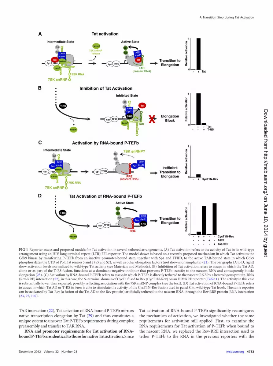
RESULTSReporter assays to detect intermediates during Tat activation.Our current model of Tat activation proposes that P-TEFb is ini-tially recruited to the HIV promoter in a kinase-inactive state dueto its association with the 7SK snRNP. Following Tat:P-TEFb pre-assembly at the promoter, the kinase is subsequently activatedupon P-TEFb transfer to TAR on the nascent RNA. During thistransition, release of inhibitory factors, Hexim1 and 7SK snRNA,triggers Cdk9 phosphorylation of the Pol II CTD and elongationfactors, facilitating escape into productive transcription elonga-tion (Fig. 1A) (21, 35, 94). Transcription is an inherently dynamicprocess requiring the coupling of various steps; therefore, it isdifficult to define intermediate states that may arise as Tat:P-TEFbcomplexes assembled at the promoter transfer to TAR to formelongation-competent complexes. To address this problem, wehave devised additional cell-based reporter assays that comple-ment the original Tat activation assay (Fig. 1A). These alternateassays examine Tat:P-TEFb complex assembly at the promoterand at the nascent RNA in TAR-independent contexts, therebydecoupling the Tat requirements for P-TEFb kinase activationfrom TAR association during transcriptional activation (Fig. 1B toD). Although the Tat activation assay constitutes all the steps re-quired for transcription activation, we propose that differences inactivities between the assays may help delineate discrete steps.
In the first assay (inhibition of Tat activation) (Fig. 1B), anRBD-deficient Tat protein (T-RS) excludes wild-type Tat from thepromoter by preferentially assembling with P-TEFb through theTat AD but cannot facilitate transfer of P-TEFb to TAR, thusblocking transition to elongation (23). Therefore, this reporterassay monitors Tat:P-TEFb preassembly at the promoter irrespec-tive of TAR and Tat RNA-binding activity. Comparison betweeninhibition of Tat activation and the standard Tat activation assay(Fig. 1A) allows the uncoupling of Tat:P-TEFb requirements forpromoter assembly versus transition to the nascent RNA and thusmay define a step prior to TAR binding (21, 23, 43, 68).
A second assay (activation by RNA-bound P-TEFb) (Fig. 1C)was designed to test if the elongation block imposed by loss of theTat RBD can be overcome by artificially tethering P-TEFb tothe nascent RNA (Tat bypass), thereby completely eliminating theneed for Tat, as well as TAR. For this assay, the N-terminal cyclinbox of CycT1 (the minimal domain required for Tat activity [40])was fused to HIV Rev (CycT1N-Rev) and used to activate an HIVpromoter derivative (HIV:RRE) in which TAR was replaced byRRE (Table 1). Relatively weak activity (�5-fold activation) (Fig.1C) suggested that Tat is indispensable for activation, althoughP-TEFb is bound to the nascent RNA.
Since RNA-bound P-TEFb alone only modestly activated tran-scription (Fig. 1C), in a third assay (Tat activation of RNA-boundP-TEFb) (Fig. 1D), we coexpressed T-RS with CycT1N-Rev orCdk9-Rev and found that T-RS no longer functioned as an inhib-itor, but rather, synergistically stimulated transcription when P-TEFb was tethered to RNA (Fig. 1D and data not shown). Tat isknown to activate transcription when tethered to the nascent tran-
script through another protein (Rev), using a heterologous pro-tein-RNA interaction (Tat-Rev–RRE) (23, 102). In this context,the Tat-Rev fusion activates transcription to a level comparable toCycT1N-Rev and T-RS, thus highlighting the importance of theTat AD, but not the RBD, during Tat activation of RNA-boundP-TEFb (Fig. 1D). Since full-length Tat or the Tat AD also stimu-lated transcription of RNA-bound P-TEFb to the same level asT-RS (data not shown), we conclude that the RS moiety does notalter Tat AD function and stimulation is not a consequence of theartificial T-RS fusion (23); therefore, we use fused and unfused Tatinterchangeably throughout the paper.
Our results indicate that P-TEFb recruitment to the nascenttranscript is insufficient to recapitulate Tat activation (5, 36, 42),as the Tat AD is required to fully activate the P-TEFb kinase. Theseresults support a model in which inactive P-TEFb complexes areloaded at the HIV promoter and inhibitory factors are releasedupon Tat-TAR binding, upon delivery of the Tat AD to the nas-cent RNA through a heterologous protein-RNA interaction, or bythe Tat AD in trans through protein-protein interactions withP-TEFb bound to the nascent RNA (Fig. 1D) (21). We presentadditional experimental evidence demonstrating that Tat-medi-ated disassembly of 7SK snRNP from P-TEFb underlies activationby RNA-bound P-TEFb (see below).
Tat activates transcription elongation in trans throughP-TEFb bound to the nascent RNA. Two previous reports sug-gested that the sole function of Tat is to recruit P-TEFb to thenascent RNA (5, 85); however, we observed only weak activitywith P-TEFb artificially recruited to the nascent RNA alone (Fig.1C) and, surprisingly, substantial stimulation with the addition ofTat AD (Fig. 1D). Since our experiments were performed in HeLacells using the N-terminal domain of CycT1 fused to Rev(CycT1N-Rev) whereas both previous studies measured activitiesin 293T cells using full-length CycT1-Rev fusions, we comparedCycT1N-Rev and CycT1-Rev activation in HeLa cells and ob-served only slightly higher activity with full-length CycT1 alone(�5-fold) and, as with the N-terminal fragment, substantial stim-ulation with Tat AD (Fig. 2A). Conversely, Tat and P-TEFb arti-ficially recruited to the nascent RNA through the Rev-RRE inter-action demonstrated much lower activation in 293T cells than inHeLa cells (Fig. 2B), likely reflecting activation from the moretightly repressed basal state in HeLa cells (�35-fold less luciferasefor the reporter alone), which have low NF-�B levels (4, 114).Since P-TEFb artificially tethered to the transcript still requiresTat for efficient activation, we explored the mechanism in greaterdetail to uncover TAR-dependent and -independent steps utilizedby the Tat:P-TEFb complex.
To test whether Tat activation of RNA-bound P-TEFb recapit-ulates the known function of Tat in elongation, we measured ini-tiating and elongating transcripts and found that both CycT1N-Rev and CycT1-Rev alone only slightly stimulated elongation (Fig.2C). Only simultaneous expression of Tat AD and CycT1N-Rev orCycT1-Rev stimulated elongation to levels comparable to those ofthe Tat-Rev control on the same HIV:RRE derivative promoter(Fig. 2C). Mutation of the Tat-TAR recognition motif (TRM) ofCycT1 at Cys261 (17, 22, 40, 107), a known region of Tat AD inter-action that affects direct protein-protein interactions (11, 17, 22, 40),decreased transcriptional stimulation by the Tat AD �4-fold andtranscription elongation rates (Fig. 2D), corresponding to inefficientdisplacement of Hexim1, the P-TEFb kinase inhibitor, from 7SKsnRNP complexes (see below). Thus, although bypassing the Tat-
D’Orso et al.
4782 mcb.asm.org Molecular and Cellular Biology
on June 10, 2014 by guesthttp://m
cb.asm.org/
Dow
nloaded from
TAR interaction (22), Tat activation of RNA-bound P-TEFb mirrorsnative transcription elongation by Tat (29) and thus constitutes aunique system to uncover Tat:P-TEFb requirements during complexpreassembly and transfer to TAR RNA.
RNA and promoter requirements for Tat activation of RNA-bound P-TEFb are identical to those for native Tat activation. Since
Tat activation of RNA-bound P-TEFb significantly reconfiguresthe mechanism of activation, we investigated whether the samerequirements for activation still applied. First, to examine theRNA requirements for Tat activation of P-TEFb when bound tothe nascent RNA, we replaced the Rev-RRE interaction used totether P-TEFb to the RNA in the previous reporters with the
FIG 1 Reporter assays and proposed models for Tat activation in several tethered arrangements. (A) Tat activation refers to the activity of Tat in its wild-typearrangement using an HIV long terminal repeat (LTR) FFL reporter. The model shown is based on a recently proposed mechanism in which Tat activates theCdk9 kinase by transferring P-TEFb from an inactive promoter-bound state, together with Sp1 and TFIID, to the active TAR-bound state in which Cdk9phosphorylates the CTD of Pol II at serines 5 and 2 (S5 and S2), as well as other elongation factors (not shown for simplicity) (21). The bar graphs (A to D, right)show activation levels normalized to wild-type Tat activity (see Materials and Methods). (B) Inhibition of Tat activation refers to assays in which the Tat AD,alone or as part of the T-RS fusion, functions as a dominant-negative inhibitor that prevents P-TEFb transfer to the nascent RNA and consequently blockselongation (23). (C) Activation by RNA-bound P-TEFb refers to assays in which P-TEFb is directly tethered to the nascent RNA by a heterologous protein-RNA(Rev-RRE) interaction (57), in this case, the N-terminal domain of CycT1 fused to Rev (CycT1N-Rev) on an HIV:RRE reporter (Table 1). The activity in this caseis substantially lower than expected, possibly reflecting association with the 7SK snRNP complex (see the text). (D) Tat activation of RNA-bound P-TEFb refersto assays in which Tat AD or T-RS in trans is able to stimulate the activity of the CycT1N-Rev fusion used in panel C to wild-type Tat levels. The same reportercan be activated by Tat-Rev (a fusion of the Tat AD to the Rev protein) artificially tethered to the nascent RNA through the Rev:RRE protein-RNA interaction(23, 97, 102).
A Transition Step during Tat Activation
December 2012 Volume 32 Number 23 mcb.asm.org 4783
on June 10, 2014 by guesthttp://m
cb.asm.org/
Dow
nloaded from

MS2cp-MS2 (MS2 coat protein-MS2 RNA) interaction and ob-served modest (3- to 10-fold) activation by CycT1-MS2cp chime-ras without Tat, none by the unfused constructs, and substantialstimulation by the Tat AD (Fig. 3A). These results are identical tothose observed using Rev-RRE tethering (Fig. 1 and 2), suggestingthat the nature of the protein-RNA interaction used to recruitP-TEFb to the nascent RNA is irrelevant for activation by the TatAD in trans.
While tethering P-TEFb to the promoter appears to be an im-portant component of Tat activation of RNA-bound P-TEFb, weasked whether the specific nature of P-TEFb recruitment was cru-cial for Tat activation. When CycT1 was directly bound to theDNA template via a Gal4 DNA-binding domain fusion (85, 103),we observed only �3.5-fold stimulation by the Tat AD even whenfour Gal4 sites were present (Fig. 3B), whereas tethering Tat to thenascent transcript via the Rev-RRE interaction on the same re-porter resulted in strong activation (�80-fold) (data not shown).Thus, Tat shows strong activation only when P-TEFb is recruitedto the nascent RNA but not to DNA, consistent with the hypoth-esis that Tat activates the P-TEFb kinase when transferred to nas-cent RNA, but not when it is still bound to the initiating and/orpreelongation Pol II complex (21). Similarly, Tat delivered asGal4-Tat to the viral promoter requires multiple Gal4-bindingsites to achieve levels of activation above basal activity, albeitmuch less than native Tat activation (reference 103 and data notshown), and thus apparently does not activate the P-TEFb kinasewell in this artificial context (see Discussion).
Given the importance of the Sp1 and TATA elements withinthe HIV promoter for Tat activation (4, 21, 103), we individuallymutated these elements and lost activation by CycT1-Rev and TatAD, as well as Tat-Rev (Fig. 3C and data not shown); in contrast,deletion of the NF-�B sites had no effect. As loss of Sp1 and TATAelements also affected basal transcription levels, this potentiallylinks the basal machinery to Tat activity during elongation (4).The requirement for TBP/TFIID at the TATA box was furtherconfirmed using a mutated TATA box (TGTA) and a compensa-tory TBP mutant (TBPm3) with altered specificity (Fig. 3D) (105,112). Thus, Tat activation of RNA-bound P-TEFb recapitulatesthe known HIV promoter requirements for Tat activation andindicates the importance of assembling the correct basal tran-scription machinery.
P-TEFb chimeras assemble into 7SK snRNP complexes, andTat activation correlates with Hexim1 displacement. Based onour model that inactive P-TEFb complexes initially assemble atthe HIV promoter and that Tat remodels the complex by ejectingHexim1 and completely displacing 7SK snRNP when Tat:P-TEFb
transfers to TAR, we hypothesized that RNA-bound P-TEFb maysimilarly be held in an inactive state by 7SK snRNP until Tat dis-places the kinase inhibitor or induces conformational changesthat eject 7SK snRNP. In support of this hypothesis, we found thatboth full-length and N-terminal CycT1 and CycT1-Rev chimerasassemble with Cdk9, as well as components of the 7SK snRNPcomplex (Hexim1, Larp7, and the 7SK RNA). Hexim1 and Larp7are displaced by wild-type Tat and the RNA-binding-deficient Tatmutants (Tat AD and T-RS), albeit at different levels, during Tat:P-TEFb complex formation (Fig. 4A and data not shown). Muta-tion of the Tat interaction surface of CycT1 (TRM; C261A) (17,22, 40) leads to less efficient Tat or Tat AD displacement ofHexim1 (Fig. 4B), while Tat mutants that cannot interact withCycT1 do not eject Hexim1 (data not shown). Notably, the degreeof Hexim1 displacement (Fig. 4B) correlates well with activationof the P-TEFb kinase and the transition to elongation (Fig. 2D).
Since our model proposes that Tat activation of RNA-boundP-TEFb relies on 7SK snRNP displacement (Fig. 1C and D), weasked whether depleting endogenous Larp7 could mechanisticallyreplace Tat, rendering activation of the HIV:RRE reporter Tatindependent. Indeed, 7SK snRNP depletion (�70% efficiency)increases both basal levels (�4-fold) and activation by RNA-bound P-TEFb (�3-fold) in the absence of Tat (Fig. 4C). Additionof Tat further activates transcription, either with or without Larp7depletion, suggesting that incomplete Larp7 knockdown mayleave residual 7SK-bound complexes for Tat disassembly or that7SK snRNP removal may not phenocopy all aspects of Tat func-tion. Further investigation is needed to more precisely define theeffects of 7SK snRNP depletion.
To examine the dynamics of complex assembly between Tatand P-TEFb bound to the nascent RNA at the HIV promoter andtranscribed regions, we monitored occupancy of CycT1N-Rev,components of the 7SK snRNP complex, as well as Sp1 and Pol IIin ChIP experiments using a HeLa cell line with an integratedHIV:RRE reporter in the absence or presence of Tat AD (Fig. 4D).As previously seen with Tat and an HIV reporter, Tat activation ofRNA-bound P-TEFb is accompanied by increased Sp1 and Pol IIoccupancy at the promoter and Pol II CTD serine 2 phosphoryla-tion (S2P-CTD) throughout the gene body (21, 85), consistentwith enhancing the transition into elongation. In agreement, theP-TEFb chimera (CycT1N-Rev and Cdk9) is found upstream ofthe transcription start site (TSS) (�1) with increased occupancywithin the gene only in the presence of Tat AD, consistent withP-TEFb traveling with elongating complexes (51, 80). Impor-tantly, Hexim1 occupancy is observed at the promoter (�75) andin the region of the nascent RNA (�103) without Tat AD but is
TABLE 1 Transcription reporter and activator combinations used in all cell-based assays
Promoter reporter DNA or RNA elementTAR-dependentTat:P-TEFb activation Activator Cell-based assay
HIV HIV TAR Yes Tat Tat activationHIV HIV TAR Yes Tat � T-RS, Tat AD Inhibition of Tat activationHIV:RRE HIV RRE No Tat-Rev TAR-independent Tat activationHIV:RRE HIV RRE No P-TEFb–Rev Activation by RNA-bound P-TEFbHIV:RRE HIV RRE No P-TEFb–Rev � Tat, Tat AD or T-RS Tat activation of RNA-bound P-TEFbHIV:MS2 MS2 phage No P-TEFb–MS2cp Activation by RNA-bound P-TEFbGal4:HIV:RRE Yeast Gal4 and HIV RRE No Gal4-Tat, Gal4–P-TEFb, Tat-RevHIV HIV TAR Yes HJ Tat P-TEFb-dependent TAR-binding Tat activationHIV:BIV BIV TAR No HJ Tat P-TEFb-dependent TAR-binding Tat activation
D’Orso et al.
4784 mcb.asm.org Molecular and Cellular Biology
on June 10, 2014 by guesthttp://m
cb.asm.org/
Dow
nloaded from

significantly decreased in both regions upon Tat AD expression.Near-complete loss of Hexim1 at the nascent RNA (�103) is con-sistent with increased Hexim1 displacement upon Tat-TAR bind-ing (21). However, in the context of the integrated reporter, weobserved that Larp7 occupancy did not change in the presence ofTat AD, a result that partially conflicts with the immunoprecipi-tation experiments in which Tat AD, but not Tat, completelyejects Hexim1 from P-TEFb (Fig. 4B). Thus, in this chimeric con-
text, it appears that Hexim1 displacement, not Larp7/7SK RNA, isrequired for kinase activation and transition to elongation (Fig. 2and 4D). In agreement with this, it was recently shown that Tatstably associates with the 7SK snRNP complex by competitivelydisplacing Hexim1 and, unexpectedly, that the Tat:7SK snRNPcomplex displays lower CTD kinase activity than P-TEFb assem-bled on the super elongation complex (21, 45, 101). These resultsindicate that Tat activates transcription through the RNA-bound
FIG 2 Tat activates RNA-bound P-TEFb at the transcription elongation step. (A) Schematic of the CycT1 and Rev fusions and their activities on an HIV:RREluciferase reporter. N in the CycT1 nomenclature refers to the fully active N-terminal cyclin box (40). Also shown is a Western blot probed against the C-terminalFlag tag of each protein to show relative steady-state expression levels (-Flag). HeLa cells were transfected with 25 ng of reporter plasmid, 1 ng of the CycT1 orRev plasmid, and 0.1 ng (�) or 1 ng (��) of T-RS. The error bars indicate standard deviations. (B) The same experiment as in panel A, conducted in 293T cells.Activation in the 293T cells is substantially lower than in HeLa cells and reflects the higher basal activity of the promoter (41,000 6,700 versus 1,200 21 relativeluciferase units, which represents �25-fold activation versus �135-fold). (C) Schematic of RT-PCR products used to assess initiation and elongation. The gelshows the products resulting from transfection of HeLa cells with the indicated constructs, and the graph plots the calculated elongation efficiencies expressed inarbitrary units (AU) (22). An 18S rRNA fragment was coamplified as an internal control. (D) Activity of a CycT1 mutant (C261Y) that weakens the interactionwith Tat (11, 17, 39) in HeLa cells, using the same activation and RT-PCR assays as for panels A and C.
A Transition Step during Tat Activation
December 2012 Volume 32 Number 23 mcb.asm.org 4785
on June 10, 2014 by guesthttp://m
cb.asm.org/
Dow
nloaded from

P-TEFb complex without completely dismantling the Tat-7SKRNA interaction. It remains to be determined if these results re-flect sequential displacement of the Hexim1 and Larp7/7SKsnRNA inhibitory components.
Genetic dissection of Tat activation steps and Tat:P-TEFbcomplex assembly. Having established assays reflecting differentaspects of Tat activation, we proceeded to define the unique mo-lecular surfaces of the Tat AD governing Tat:P-TEFb preassemblyand transfer to TAR. For this, we introduced Ala substitutions at45 positions within the Tat AD and assessed their activities in thethree previously described assays: (i) standard Tat activation, (ii)inhibition of Tat activation, and (iii) Tat activation of RNA-bound P-TEFb (Fig. 1 and 5A). For Tat activation, which moni-tors the entire activation process on an HIV luciferase reporter, 30full-length Tat mutants decreased activity by at least 4-fold (Fig.5A), including at least 15 that are zinc-coordinating residues orotherwise contribute to the structural stability of Tat within the
P-TEFb complex (20, 91, 92, 107). In inhibition of Tat activation,which monitors Tat:P-TEFb preassembly at the promoter andprecedes Tat-TAR interaction (23), 21 mutations engineered intothe TAR-binding-deficient Tat mutant decreased inhibition �4-fold (Fig. 5A). The mutations resulting in the most striking differ-ences in activity between these two assays reside in the N-terminalacidic and cysteine-rich regions. A nearly identical activity patternwas observed during Tat activation of RNA-bound P-TEFb bythese same TAR-binding-deficient Tat mutants. However, this as-say monitors P-TEFb kinase activation on the nascent RNA inde-pendent of TAR (Fig. 1D and 5A). The reduced requirement forTat AD residues in the last two TAR-independent assays mostlikely reflects the bypassed requirement for RNA binding duringTat:P-TEFb assembly and transcription activation (Fig. 1). Forexample, Lys28 acetylation by p300/CBP-associated factor(PCAF) was previously shown to enhance the affinity of Tat:TAR:P-TEFb complexes by affecting Tat interaction with CycT1 TRM
FIG 3 Tat activates P-TEFb bound to RNA but not DNA and uses basal transcription elements. (A) Activation of RNA-bound P-TEFb through the MS2protein-RNA interaction by T-RS in HeLa cells. The cells were transfected with 25 ng of an HIV:MS2 reporter plasmid, 1 ng of the CycT1 or MS2cp plasmid, and1 ng of T-RS. The Western blot shows steady-state expression levels of C-terminally Flag-tagged proteins. The error bars indicate standard deviations. (B)Activation of DNA-bound P-TEFb, using Gal4:HIV:RRE reporters containing 1, 2, or 4 Gal4-binding sites and Gal4-CycT1 and T-RS under the same conditionsas for panel A. Tat-Rev activates this reporter through the RRE by 85-fold 4.7-fold (data not shown). (C) Sp1 and TATA elements, but not NF-�B sites, arerequired for Tat activation of RNA-bound P-TEFb. The HIV:RRE reporters shown were cotransfected with 1 ng of CycT1N-Rev and 1 ng of T-RS, and activationlevels were normalized to a cotransfected CMV-RL plasmid. (D) Requirement for TBP using an altered-specificity mutant. Activation assays were performed asfor panel C using an HIV:RRE reporter containing a mutant TATA box (TGTA) complemented with a TBP mutant (TBPm3) that has enhanced bindingspecificity for the mutated TATA box (105).
D’Orso et al.
4786 mcb.asm.org Molecular and Cellular Biology
on June 10, 2014 by guesthttp://m
cb.asm.org/
Dow
nloaded from

FIG 4 RNA-bound P-TEFb assembles with 7SK snRNP complexes, and Tat displaces Hexim1 at the viral promoter. (A) Flag-tagged CycT1N or full-lengthCycT1, unfused or fused to Rev, was expressed in 293 cells with or without Strep-tagged Tat or Tat AD and immunoprecipitated using Flag beads. Mock refersto a Flag immunoprecipitation of cells transfected with an empty vector. The composition of P-TEFb/7SK snRNP components in the immunoprecipitation wasanalyzed by Western blotting using Cdk9, Hexim1, and Larp7 antibodies, while 7SK snRNA was visualized directly as a 330-nt RNA species whose identity wasconfirmed by Northern blotting. (B) Flag-tagged CycT1N-Rev (the wild type and a C261A mutant) was transfected into 293 cells with or without Strep-taggedTat or Tat AD, and the P-TEFb/7SK snRNP composition was evaluated as for panel A. (C) Larp7 depletion by RNAi. Western blots show that Larp7 is reducedby �70% but that the other 7SK snRNP subunits or �-actin used as a control are unaffected. The plot shows reporter assays in HeLa cells transfected with anHIV:RRE reporter (with [�] or without [�] Larp7 siRNA) in the presence of CycT1N-Rev (with or without Tat and Tat AD). Fold activation represents valuesnormalized to the activity of the reporter alone without siRNA. The error bars indicate standard deviations. (D) ChIP assays were performed with chromatinextracts prepared from the HIV:RRE reporter cell line 48 h after a Flag-tagged CycT1N-Rev transfection alone (gray bars) or cotransfected with Strep-tagged TatAD (black bars) using the indicated antibodies. The values represent the percentages of input DNA immunoprecipitated and are the averages of two independentPCR assays from two separate experiments.
December 2012 Volume 32 Number 23 mcb.asm.org 4787
on June 10, 2014 by guesthttp://m
cb.asm.org/
Dow
nloaded from

(21); however, since mutation of Lys28 does not affect inhibitionof Tat activation, acetylation probably does not play a role in theearly stages of Tat:P-TEFb preassembly but may be important forejection of the 7SK snRNP complex or transfer of P-TEFb to thenascent RNA. An adjacent residue, Tyr26, shows an identical phe-notype and may be functionally coupled to Lys28.
To confirm the differential requirements of Tat AD residues
during the transition of preassembled Tat:P-TEFb complexes toTAR RNA, we constructed a parallel set of mutations in the con-text of a chimeric Tat protein referred to as HJTat. This Tat variantcontains the RBD of Jembrana disease virus (JDV) Tat instead ofthe native HIV RBD (22, 99) and can therefore bind two ortholo-gous TAR elements (HIV and bovine immunodeficiency virus[BIV]) (Fig. 5B and Table 1). While the P-TEFb kinase is needed to
FIG 5 Activities of single-site Tat Ala mutants in reporter and coprecipitation assays. (A) Heat map of Tat activities (wild type or single-site Ala mutants) acrossthe entire AD (residues 1 to 48), except the initiating Met1 and preexisting Ala residues 21 and 42 in the HXB2 reference sequence. The basis for each of the fiveassays—Tat activation, inhibition of Tat activation, Tat activation of RNA-bound P-TEFb, P-TEFb-dependent TAR-binding Tat activation, and P-TEFb-independent TAR-binding Tat activation—is described in Fig. 1B. Quantitative raw activity data for all assays (data not shown) were used to derive the relativeactivity value for each mutant (0 to 1) represented in the heat map (black to yellow). In the HJTat context, Cys25, Cys27, Cys30, His33, Cys34, and Cys37 werenot mutated, since they completely disrupted protein folding in the other three assays. The black boxes with white asterisks represent the expected phenotype andnot actual data. P-TEFb-dependent TAR-binding Tat activation and P-TEFb-independent TAR-binding Tat activation are assays in which HJTat activates anHIV reporter or an engineered HIV promoter with HIV TAR replaced by BIV, respectively (Table 1). (B) Schematic of HIV Tat and the HJTat chimera composedof the HIV Tat AD and the JDV RBD, which is able to bind to both HIV and BIV TARs in P-TEFb-dependent and P-TEFb-independent modes, respectively (22,99). Conserved residues between the HIV and JDV Tat RBDs used for HIV TAR binding (P-TEFb-dependent TAR-binding Tat activation) are highlighted in red.Schematic of ternary complexes showing that HJTat binding to HIV TAR is dependent on interactions between Tat and CycT1/P-TEFb while binding to BIV TARis P-TEFb independent. (C) Pearson correlation coefficients (r) for pairwise comparisons of all assays. (D) Affinity-purified Strep-tagged HJTat (wild-type andselect Tat AD Ala mutants) was analyzed by Western blotting for interactions with endogenous CycT1, Cdk9, and Larp7. Mock refers to an empty-vector-transfected control. (E) Surface representation of Tat (gray), CycT1 (gold), and Cdk9 (blue) using the coordinates of the Tat:P-TEFb structure (107). MutatedTat residues examined in panel D map to the CycT1 TRM (green) or are positioned to interact with Cdk9 (red). The Cdk9 T loop essential for kinase activity isindicated.
D’Orso et al.
4788 mcb.asm.org Molecular and Cellular Biology
on June 10, 2014 by guesthttp://m
cb.asm.org/
Dow
nloaded from

activate both reporters, P-TEFb is needed only for Tat:P-TEFbcomplex assembly on HIV TAR (referred to as P-TEFb-dependentTAR-binding Tat activation). Hence, this comparison identifiesTat AD residues that are specifically required for P-TEFb-depen-dent and -independent RNA-binding mechanisms and can fur-ther refine models in which preassembled Tat:P-TEFb complexesare remodeled as they transition from the inactive promoter-bound state to the active TAR-bound state (Fig. 1A) (21). Indeed,residues indispensable for Tat activation through BIV TAR (P-TEFb-independent TAR-binding Tat activation) are virtuallyidentical to those for the TAR-independent assays (Fig. 5A). Con-versely, the indispensability of N-terminal residues, as well asTyr26 and Lys28, for activation through HIV TAR indicated thatthese residues are needed for interactions in the context of theTat:TAR:P-TEFb ternary complex, but not Tat:P-TEFb. A highPearson correlation coefficient (r) distinctly clusters the two TAR-dependent assays (Tat activation and P-TEFb-dependent TAR-binding Tat activation) and the three TAR-independent assays(inhibition of Tat activation, Tat activation of RNA-bound P-TEFb, and P-TEFb-independent TAR-binding Tat activation),whereas the values calculated between dissimilar assays displaymuch lower coefficients (Fig. 5C). Strong correlation between thetwo TAR-dependent assays reinforces a model in which specificresidues and Tat acetylation are needed for the transfer of preas-sembled Tat:P-TEFb complexes from the promoter to TAR.
To better define the roles of N-terminal Tat residues, as well asTyr26 and Lys28, we affinity purified Strep-tagged HJTat mutantsrepresentative of the residue classifications defined below and ex-amined their association with endogenous P-TEFb and 7SKsnRNP subunits (Fig. 5D). All of the N-terminal mutants testedshowed reduced interaction with Cdk9, but not CycT1 (Fig. 5D),consistent with the Tat:P-TEFb structure, in which residues Leu8through Trp11 form a 310 helix positioned near the T loop of Cdk9(Fig. 5E) that may stabilize the complex (107). CycT1 and Cdk9interactions were abolished by mutation of known structural res-idues, Cys22 and Lys41, but were unaffected by mutation ofAsn24, Tyr26, and Lys28. As expected, Hexim1 did not copurifywith wild-type or mutant Tat proteins (2, 21), while Larp7 (7SKsnRNP) copurified to similar levels, except with the Lys28 mutant,which, interestingly, showed increased Larp7 association. In lightof our previous studies showing that Lys28 is needed to assemblehigh-affinity Tat:P-TEFb complexes on TAR (22), this observa-tion suggests potentially coupled roles for Lys28 in both Tat-7SKsnRNP disassembly and Tat:P-TEFb:TAR complex formation.
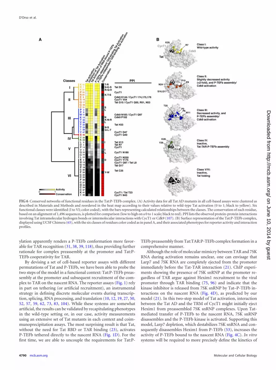
An evolutionarily conserved network of residues in the Tat:P-TEFb complex. With the large functional data sets generated forall Tat AD mutants using multiple cell-based reporter assays (Fig.5A), it becomes possible to cluster residues based on their activi-ties and to map them onto the recent Tat:P-TEFb structure (107).Moreover, this comprehensive analysis permits us to define func-tional interfaces in the context of the two-step model of activation,where Tat first preassembles with inactive P-TEFb/7SK complexesat the promoter and is then transferred to TAR (21). By hierarchi-cal clustering, we categorized six classes of residues showingstrong functional correlations, high evolutionary conservation inmost cases (Fig. 6A), and physical clustering within the structure(Fig. 6B). Class I residues (yellow) do not disrupt Tat activity inany assay when mutated and are not evolutionarily conserved;conversely, at the other extreme, class VI residues (black) showsevere defects in all assays when mutated and are strictly evolu-
tionarily conserved. This is likely the consequence of disruptingTat folding and is exemplified by the cysteine-rich domain, whichfolds into a compact structure composed of two -helices medi-ating the coordination of two Zn ions (20, 107). Class V residues(gray) show moderate defects when mutated but also are likely toaffect folding or perhaps interactions with other transcription fac-tors. Mutation of class II residues (orange) only slightly (�2-fold)decreases activity in the TAR-dependent Tat activation assays anddoes not affect activation in assays not operating through TAR(inhibition of Tat activation and Tat activation of RNA-boundP-TEFb). These residues are positioned in the structure to perturbP-TEFb assembly or Cdk9 activation. Similarly, class III residues(blue) show pronounced decreases in Tat activation when mu-tated, with 3 residues (Pro3, Pro6, and Trp11) possibly involved inCdk9 interactions (107). Single-site Ala mutagenesis has beengenerally useful in defining protein-protein interactions, protein-folding residues, and energetic parameters (15, 67, 89, 108); how-ever, in cases where the substitutions are nonconservative, such asPro3, Pro6, and Trp11, changes in structure or other long-rangeeffects may also underlie the functional disruptions observed.
Class IV residues Tyr26 and Lys28 (green) show the most se-vere defects in the TAR-dependent assays yet are not apparentlyassociated with structural defects, as Tat:P-TEFb complex forma-tion is not abolished (Fig. 5D). Indeed, unlike the class V and VIfolding residues, these mutants show no defect in activation assaysoperating in a TAR-independent manner and thus may not berequired for the early stage of Tat:P-TEFb preassembly at the pro-moter. Tyr26 and Lys28 are located within the ZnF2 motif, nearthe CycT1 TRM, which is disordered in the Tat:P-TEFb structuredue to the absence of TAR and acetylated Lys28 (Fig. 5 and 6) (20,39, 107). Therefore, it will be especially interesting to reevaluatethese mutational data once a cocrystal structure with TAR be-comes available. TAR may well be important for interacting withthe two residues, either directly or through conformationalchanges in P-TEFb or Tat upon RNA binding (22). Alternatively,because they cluster in the middle of the CycT1 TRM interface,they may be energetically important in the cooperative assemblyof Tat:P-TEFb on the RNA, perhaps coordinating communica-tion between protein and RNA conformations to increase affinity(9, 46).
DISCUSSION
Since the seminal discovery that HIV Tat enhances transcriptionelongation (29, 49, 56, 93), many contributing host factors havebeen identified, including P-TEFb (64, 79, 80, 121), 7SK snRNA(74, 116), Hexim1 (58, 66, 96), 7SK snRNP (21, 70, 101), compo-nents of the super elongation complex (44, 45, 100, 101), and thebasal transcription machinery (85, 98). However, the mechanismand timing of assembly of these factors at the viral promoter dur-ing active elongation and their requirement for TAR RNA arepoorly understood. Our recent results point to an activationmodel having at least two discrete steps, where Tat and P-TEFb areinitially assembled with the inhibitory 7SK snRNP into transcrip-tion complexes at the promoter and later transferred to TAR onthe nascent transcript, triggering release of the snRNP and activa-tion of Cdk9 (21) (Fig. 1A). The crystal structure of the Tat ADbound to P-TEFb clearly shows how CycT1 and Cdk9 act as atemplate to fold Tat, possibly explaining the need to preassemblethe complex before TAR binding (20, 107). P-TEFb enhances theaffinity and specificity of Tat for TAR, while Cdk9 autophosphor-
A Transition Step during Tat Activation
December 2012 Volume 32 Number 23 mcb.asm.org 4789
on June 10, 2014 by guesthttp://m
cb.asm.org/
Dow
nloaded from
ylation apparently renders a P-TEFb conformation more favor-able for TAR recognition (31, 38, 39, 118), thus providing furtherrationale for complex preassembly at the promoter and Tat:P-TEFb cooperativity for TAR.
By devising a set of cell-based reporter assays with differentpermutations of Tat and P-TEFb, we have been able to probe thetwo steps of the model in a functional context: Tat:P-TEFb preas-sembly at the promoter and subsequent recruitment of the com-plex to TAR on the nascent RNA. The reporter assays (Fig. 1) relyin part on tethering (or artificial recruitment), an instrumentalstrategy in defining discrete molecular events during transcrip-tion, splicing, RNA processing, and translation (10, 12, 19, 27, 50,52, 57, 59, 62, 73, 83, 104). While these systems are somewhatartificial, the results can be validated by recapitulating phenotypesin the wild-type setting or, in our case, activity measurementsusing an extensive set of Tat mutants in each context and coim-munoprecipitation assays. The most surprising result is that Tat,without the need for Tat RBD or TAR binding (23), activatesP-TEFb tethered directly to the nascent RNA (Fig. 1D). For thefirst time, we are able to uncouple the requirements for Tat:P-
TEFb preassembly from Tat:TAR:P-TEFb complex formation in acomprehensive manner.
Although the role of molecular mimicry between TAR and 7SKRNA during activation remains unclear, one can envisage thatLarp7 and 7SK RNA are completely ejected from the promoterimmediately before the Tat-TAR interaction (21). ChIP experi-ments showing the presence of 7SK snRNP at the promoter re-gardless of TAR argue against Hexim1 recruitment to the viralpromoter through TAR binding (75, 96) and indicate that thekinase inhibitor is released from 7SK snRNP by Tat–P-TEFb in-teractions on the nascent RNA (Fig. 4D), as predicted by ourmodel (21). In this two-step model of Tat activation, interactionbetween the Tat AD and the TRM of CycT1 might initially ejectHexim1 from preassembled 7SK snRNP complexes. Upon Tat-mediated transfer of P-TEFb to the nascent RNA, 7SK snRNPdisassembles and the P-TEFb kinase is activated. Supporting thismodel, Larp7 depletion, which destabilizes 7SK snRNA and con-sequently disassembles Hexim1 from P-TEFb (53), increases theactivity of P-TEFb bound to the nascent RNA (Fig. 4C). In vitrosystems will be required to more precisely define the kinetics of
FIG 6 Conserved networks of functional residues in the Tat:P-TEFb complex. (A) Activity data for all Tat AD mutants in all cell-based assays were clustered asdescribed in Materials and Methods and reordered in the heat map according to their values relative to wild-type Tat activation (0 to 1; black to yellow). Sixfunctional classes were identified (I to VI; color coded), with the bars representing calculated relationships between the classes. The conservation of each residue,based on an alignment of 1,496 sequences, is plotted for comparison (low to high on a 0 to 1 scale; black to red). PPI lists the observed protein-protein interactionsinvolving Tat intramolecular hydrogen bonds or intermolecular interactions with CycT1 or Cdk9 (107). (B) Surface representation of the Tat:P-TEFb complex,displayed using UCSF Chimera (65), with the six classes of residues color coded as in panel A, and their associated phenotypes for reporter activity and interactionprofiles.
D’Orso et al.
4790 mcb.asm.org Molecular and Cellular Biology
on June 10, 2014 by guesthttp://m
cb.asm.org/
Dow
nloaded from

assembly and displacement of these factors and the activity of theP-TEFb kinase alone, bound to Tat, and in the context of theTat:7SK snRNP complex (38, 45, 79, 101, 110, 119). In apparentdisagreement with our model, a previous observation that P-TEFbis easily extracted from chromatin with high-salt treatment (6)suggested that 7SK snRNP complexes do not stably associate withchromatin (76, 78); however, recent genome-wide experimentsexamining the assembly of noncoding RNAs with chromatindemonstrated that 7SK snRNA is enriched several thousandfold inthe chromatin fraction (69), raising the possibility that 7SK com-plexes selectively assemble at specific promoters (28).
It is particularly instructive to compare the activities of Tat andP-TEFb recruited to the HIV promoter by other means. For ex-ample, recruiting Tat through a heterologous DNA-protein inter-action (Gal4-Tat) requires multiple Gal4-binding sites and stillresults in weaker activation than TAR-mediated recruitment (ref-erence 103 and data not shown); this is also true for P-TEFb re-cruitment through DNA (Fig. 3B). Similarly, recruiting Gal4 –P-TEFb to the hsp70 promoter in Drosophila activates transcriptionin the absence of heat shock to a much lower level than by heatshock factor 1 (61), suggesting that these artificial recruitmentmechanisms only partially recapitulate function. Even Tat activa-tion in the context of heterologous protein-RNA interactions (Fig.1D) (5, 97, 102) does not fully recapitulate activity. For example,acetylation of Tat Lys28 is dispensable in heterologous systems butis important in the native HIV Tat-TAR context to modulate theaffinity for TAR and fine tune escape into productive elongation(22). Interestingly, we also find that mutation of Lys28 reduces7SK snRNP dismantling in the context of Tat activation by RNA-bound P-TEFb (Fig. 5D). Thus, these heterologous systems can beespecially informative compared with the wild-type mechanism ofTat activation. Our results provide strong evidence that Tat doesnot bind TAR directly at the promoter and that a preassembly stepis required. The generation of an in vitro system that recapitulatesthe results observed in vivo will be needed to further define the roleof Tat residues in the two-step model.
Comprehensive Tat mutagenesis coupled with analyses usingmultiple cell-based reporter assays defined networks of functionalresidues whose evolutionary constraints are not readily explainedby the Tat:P-TEFb structure (107). Evolutionarily conserved res-idues constitute molecular surfaces for essential interactions,allosteric communication, or transmission of conformationalchanges into functional behavior (3, 90, 106). Many proteins un-dergo conformational changes upon transient interactions, ofteninvolving disorder-to-order transitions (47, 77). The intrinsic dis-order and structural flexibility of Tat may be relevant for interac-tions with more than one binding partner and may allow compet-ing interactions to occur in a sequential manner (24, 37), such asswitching from unbound to RNA-bound states during activation.While further investigation is needed to uncover the moleculardetails, the observed functional and structural clustering of resi-dues suggests that Tat:P-TEFb complexes have evolved networksof residues to orchestrate the switch from transcription initiationto elongation, RNA binding, and the assembly/disassembly of 7SKsnRNP complexes.
ACKNOWLEDGMENTS
We thank Robert Nakamura and members of the Frankel laboratory forcritical reading of the manuscript and James Fraser for helpful discus-sions. We thank K.-T. Jeang for the TBPm3 construct.
This work was supported by NIH grants AI29135 and P50 GM082250(HARC Center) and funding from the California Center for AntiviralDrug Discovery to A.D.F., NIH grants K99A112185 and R00AI083087and Welch Foundation grant I-1782 to I.D., UC GREAT fellowship2004-21 to A.W.P., NIH postdoctoral fellowship GM077868 to G.M.J.,NIH Research supplement to promote diversity in Health Related Re-search to E.Q., and NIH fellowships R25GM56847 and F31GM09535 toD.S.B.
REFERENCES1. Barboric M, Lenasi T. 2010. Kick-sTARting HIV-1 transcription elon-
gation by 7SK snRNP deporTATion. Nat. Struct. Mol. Biol. 17:928 –930.2. Barboric M, et al. 2007. Tat competes with HEXIM1 to increase the
active pool of P-TEFb for HIV-1 transcription. Nucleic Acids Res. 35:2003–2012.
3. Barua B, Pamula MC, Hitchcock-DeGregori SE. 2011. Evolutionarilyconserved surface residues constitute actin binding sites of tropomyosin.Proc. Natl. Acad. Sci. U. S. A. 108:10150 –10155.
4. Berkhout B, Gatignol A, Rabson AB, Jeang KT. 1990. TAR-independent activation of the HIV-1 LTR: evidence that tat requiresspecific regions of the promoter. Cell 62:757–767.
5. Bieniasz PD, Grdina TA, Bogerd HP, Cullen BR. 1999. Recruitment ofcyclin T1/P-TEFb to an HIV type 1 long terminal repeat promoter prox-imal RNA target is both necessary and sufficient for full activation oftranscription. Proc. Natl. Acad. Sci. U. S. A. 96:7791–7796.
6. Biglione S, et al. 2007. Inhibition of HIV-1 replication by P-TEFb in-hibitors DRB, seliciclib and flavopiridol correlates with release of freeP-TEFb from the large, inactive form of the complex. Retrovirology4:47–58.
7. Blau J, et al. 1996. Three functional classes of transcriptional activationdomain. Mol. Cell. Biol. 16:2044 –2055.
8. Brown SA, Weirich CS, Newton EM, Kingston RE. 1998. Transcrip-tional activation domains stimulate initiation and elongation at differenttimes and via different residues. EMBO J. 17:3146 –3154.
9. Calabro V, Daugherty MD, Frankel AD. 2005. A single intermolecularcontact mediates intramolecular stabilization of both RNA and protein.Proc. Natl. Acad. Sci. U. S. A. 102:6849 – 6854.
10. Carey M, Lin YS, Green MR, Ptashne M. 1990. A mechanism forsynergistic activation of a mammalian gene by GAL4 derivatives. Nature345:361–364.
11. Chen D, Fong Y, Zhou Q. 1999. Specific interaction of Tat with thehuman but not rodent P-TEFb complex mediates the species-specific Tatactivation of HIV-1 transcription. Proc. Natl. Acad. Sci. U. S. A. 96:2728 –2733.
12. Clement SL, Lykke-Andersen J. 2008. A tethering approach to studyproteins that activate mRNA turnover in human cells. Methods Mol.Biol. 419:121–133.
13. Core LJ, Lis JT. 2008. Transcription regulation through promoter-proximal pausing of RNA polymerase II. Science 319:1791–1792.
14. Cullen BR. 1990. The HIV-1 Tat protein: an RNA sequence-specificprocessivity factor? Cell 63:655– 657.
15. Cunningham BC, Wells JA. 1989. High-resolution epitope mapping ofhGH-receptor interactions by alanine-scanning mutagenesis. Science244:1081–1085.
16. Das A. 1992. How the phage lambda N gene product suppresses tran-scription termination: communication of RNA polymerase with regula-tory proteins mediated by signals in nascent RNA. J. Bacteriol. 174:6711–6716.
17. Das C, Edgcomb SP, Peteranderl R, Chen L, Frankel AD. 2004.Evidence for conformational flexibility in the Tat-TAR recognition motifof cyclin T1. Virology 318:306 –317.
18. de Hoon MJ, Imoto S, Nolan J, Miyano S. 2004. Open source clusteringsoftware. Bioinformatics 20:1453–1454.
19. Dorris DR, Struhl K. 2000. Artificial recruitment of TFIID, but not RNApolymerase II holoenzyme, activates transcription in mammalian cells.Mol. Cell. Biol. 20:4350 – 4358.
20. D’Orso I, Frankel AD. 2010. HIV-1 Tat: its dependence on host factorsis crystal clear. Viruses 2:2226 –2234.
21. D’Orso I, Frankel AD. 2010. RNA-mediated displacement of an inhib-itory snRNP complex activates transcription elongation. Nat. Struct.Mol. Biol. 17:815– 821.
22. D’Orso I, Frankel AD. 2009. Tat acetylation modulates assembly of a
A Transition Step during Tat Activation
December 2012 Volume 32 Number 23 mcb.asm.org 4791
on June 10, 2014 by guesthttp://m
cb.asm.org/
Dow
nloaded from

viral-host RNA-protein transcription complex. Proc. Natl. Acad. Sci.U. S. A. 106:3101–3106.
23. D’Orso I, Grunwell JR, Nakamura RL, Das C, Frankel AD. 2008.Targeting tat inhibitors in the assembly of human immunodeficiencyvirus type 1 transcription complexes. J. Virol. 82:9492–9504.
24. Dunker AK, Silman I, Uversky VN, Sussman JL. 2008. Function andstructure of inherently disordered proteins. Curr. Opin. Struct. Biol. 18:756 –764.
25. Durney MA, D’Souza VM. 2010. Preformed protein-binding motifs in7SK snRNA: structural and thermodynamic comparisons with retroviralTAR. J. Mol. Biol. 404:555–567.
26. Espinosa JM. 2010. The meaning of pausing. Mol. Cell 40:507–508.27. Farrell S, Simkovich N, Wu Y, Barberis A, Ptashne M. 1996. Gene
activation by recruitment of the RNA polymerase II holoenzyme. GenesDev. 10:2359 –2367.
28. Faust T, Frankel AD, D’Orso I. 2012. Transcription control by longnon-coding RNAs. Transcription 3:78 – 86.
29. Feinberg MB, Baltimore D, Frankel AD. 1991. The role of Tat in thehuman immunodeficiency virus life cycle indicates a primary effect ontranscriptional elongation. Proc. Natl. Acad. Sci. U. S. A. 88:4045– 4049.
30. Fishburn J, Mohibullah N, Hahn S. 2005. Function of a eukaryotictranscription activator during the transcription cycle. Mol. Cell 18:369 –378.
31. Fong YW, Zhou Q. 2000. Relief of two built-in autoinhibitory mecha-nisms in P-TEFb is required for assembly of a multicomponent tran-scription elongation complex at the human immunodeficiency virustype 1 promoter. Mol. Cell. Biol. 20:5897–5907.
32. Fong YW, Zhou Q. 2001. Stimulatory effect of splicing factors on tran-scriptional elongation. Nature 414:929 –933.
33. Frankel AD, Kim PS. 1991. Modular structure of transcription factors:implications for gene regulation. Cell 65:717–719.
34. Frankel AD, Young JA. 1998. HIV-1: fifteen proteins and an RNA.Annu. Rev. Biochem. 67:1–25.
35. Fuda NJ, Ardehali MB, Lis JT. 2009. Defining mechanisms that regulateRNA polymerase II transcription in vivo. Nature 461:186 –192.
36. Fujinaga K, et al. 1998. The ability of positive transcription elongationfactor B to transactivate human immunodeficiency virus transcriptiondepends on a functional kinase domain, cyclin T1, and Tat. J. Virol.72:7154 –7159.
37. Ganguly D, Chen J. 2009. Atomistic details of the disordered states ofKID and pKID. Implications in coupled binding and folding. J. Am.Chem. Soc. 131:5214 –5223.
38. Garber ME, et al. 2000. CDK9 autophosphorylation regulates high-affinity binding of the human immunodeficiency virus type 1 tat-P-TEFbcomplex to TAR RNA. Mol. Cell. Biol. 20:6958 – 6969.
39. Garber ME, Wei P, Jones KA. 1998. HIV-1 Tat interacts with cyclin T1to direct the P-TEFb CTD kinase complex to TAR RNA. Cold SpringHarb. Symp. Quant. Biol. 63:371–380.
40. Garber ME, et al. 1998. The interaction between HIV-1 Tat and humancyclin T1 requires zinc and a critical cysteine residue that is not conservedin the murine CycT1 protein. Genes Dev. 12:3512–3527.
41. Gilchrist DA, et al. 2010. Pausing of RNA polymerase II disrupts DNA-specified nucleosome organization to enable precise gene regulation. Cell143:540 –551.
42. Gold MO, Yang X, Herrmann CH, Rice AP. 1998. PITALRE, thecatalytic subunit of TAK, is required for human immunodeficiency virusTat transactivation in vivo. J. Virol. 72:4448 – 4453.
43. Green M, Ishino M, Loewenstein PM. 1989. Mutational analysis ofHIV-1 Tat minimal domain peptides: identification of trans-dominantmutants that suppress HIV-LTR-driven gene expression. Cell 58:215–223.
44. He N, et al. 2011. Human polymerase-associated factor complex (PAFc)connects the super elongation complex (SEC) to RNA polymerase II onchromatin. Proc. Natl. Acad. Sci. U. S. A. 108:E636 –E645.
45. He N, et al. 2010. HIV-1 Tat and host AFF4 recruit two transcriptionelongation factors into a bifunctional complex for coordinated activa-tion of HIV-1 transcription. Mol. Cell 38:428 – 438.
46. Hobson D, Uhlenbeck OC. 2006. Alanine scanning of MS2 coat proteinreveals protein-phosphate contacts involved in thermodynamic hotspots. J. Mol. Biol. 356:613– 624.
47. Janin J, Bahadur RP, Chakrabarti P. 2008. Protein-protein interactionand quaternary structure. Q. Rev. Biophys. 41:133–180.
48. Jeronimo C, et al. 2007. Systematic analysis of the protein interaction
network for the human transcription machinery reveals the identity ofthe 7SK capping enzyme. Mol. Cell 27:262–274.
49. Kao SY, Calman AF, Luciw PA, Peterlin BM. 1987. Anti-termination oftranscription within the long terminal repeat of HIV-1 by tat gene prod-uct. Nature 330:489 – 493.
50. Keaveney M, Struhl K. 1998. Activator-mediated recruitment of theRNA polymerase II machinery is the predominant mechanism for tran-scriptional activation in yeast. Mol. Cell 1:917–924.
51. Keen NJ, Gait MJ, Karn J. 1996. Human immunodeficiency virus type-1Tat is an integral component of the activated transcription-elongationcomplex. Proc. Natl. Acad. Sci. U. S. A. 93:2505–2510.
52. Koh SS, Ansari AZ, Ptashne M, Young RA. 1998. An activator target inthe RNA polymerase II holoenzyme. Mol. Cell 1:895–904.
53. Krueger BJ, et al. 2008. LARP7 is a stable component of the 7SK snRNPwhile P-TEFb, HEXIM1 and hnRNP A1 are reversibly associated. Nu-cleic Acids Res. 36:2219 –2229.
54. Krueger BJ, Varzavand K, Cooper JJ, Price DH. 2010. The mechanismof release of P-TEFb and HEXIM1 from the 7SK snRNP by viral andcellular activators includes a conformational change in 7SK. PLoS One5:e12335. doi:10.1371/journal.pone.0012335.
55. Kurosu T, Peterlin BM. 2004. VP16 and ubiquitin; binding of P-TEFbvia its activation domain and ubiquitin facilitates elongation of tran-scription of target genes. Curr. Biol. 14:1112–1116.
56. Laspia MF, Rice AP, Mathews MB. 1989. HIV-1 Tat protein increasestranscriptional initiation and stabilizes elongation. Cell 59:283–292.
57. Li Q, Peterlin BM. 2009. Genetic analysis of P-TEFb function via het-erologous nucleic acid tethering systems. Methods 48:375–380.
58. Li Q, et al. 2005. Analysis of the large inactive P-TEFb complex indicatesthat it contains one 7SK molecule, a dimer of HEXIM1 or HEXIM2, andtwo P-TEFb molecules containing Cdk9 phosphorylated at threonine186. J. Biol. Chem. 280:28819 –28826.
59. Lim SR, Hertel KJ. 2004. Commitment to splice site pairing coincideswith A complex formation. Mol. Cell 15:477– 483.
60. Lis J, Wu C. 1993. Protein traffic on the heat shock promoter: parking,stalling, and trucking along. Cell 74:1– 4.
61. Lis JT, Mason P, Peng J, Price DH, Werner J. 2000. P-TEFb kinaserecruitment and function at heat shock loci. Genes Dev. 14:792– 803.
62. Majello B, Napolitano G, Lania L. 1998. Recruitment of the TATA-binding protein to the HIV-1 promoter is a limiting step for Tat trans-activation. AIDS 12:1957–1964.
63. Malik S, Roeder RG. 2010. The metazoan Mediator co-activator com-plex as an integrative hub for transcriptional regulation. Nat. Rev. Genet.11:761–772.
64. Mancebo HS, et al. 1997. P-TEFb kinase is required for HIV Tat tran-scriptional activation in vivo and in vitro. Genes Dev. 11:2633–2644.
65. Meng EC, Pettersen EF, Couch GS, Huang CC, Ferrin TE. 2006. Toolsfor integrated sequence-structure analysis with UCSF Chimera. BMCBioinformatics 7:339 –345.
66. Michels AA, et al. 2004. Binding of the 7SK snRNA turns the HEXIM1protein into a P-TEFb (CDK9/cyclin T) inhibitor. EMBO J. 23:2608 –2619.
67. Milla ME, Brown BM, Sauer RT. 1994. Protein stability effects of acomplete set of alanine substitutions in Arc repressor. Nat. Struct. Biol.1:518 –523.
68. Modesti N, Garcia J, Debouck C, Peterlin M, Gaynor R. 1991. Trans-dominant Tat mutants with alterations in the basic domain inhibitHIV-1 gene expression. New Biol. 3:759 –768.
69. Mondal T, Rasmussen M, Pandey GK, Isaksson A, Kanduri C. 2010.Characterization of the RNA content of chromatin. Genome Res. 20:899 –907.
70. Muniz L, Egloff S, Ughy B, Jady BE, Kiss T. 2010. Controlling cellularP-TEFb activity by the HIV-1 transcriptional transactivator Tat. PLoSPathog. 6:e1001152. doi:10.1371/journal.ppat.1001152.
71. Naar AM, Lemon BD, Tjian R. 2001. Transcriptional coactivator com-plexes. Annu. Rev. Biochem. 70:475–501.
72. Nechaev S, Adelman K. 2011. Pol II waiting in the starting gates: regu-lating the transition from transcription initiation into productive elon-gation. Biochim. Biophys. Acta 1809:34 – 45.
73. Nevado J, Gaudreau L, Adam M, Ptashne M. 1999. Transcriptionalactivation by artificial recruitment in mammalian cells. Proc. Natl. Acad.Sci. U. S. A. 96:2674 –2677.
74. Nguyen VT, Kiss T, Michels AA, Bensaude O. 2001. 7SK small nuclear
D’Orso et al.
4792 mcb.asm.org Molecular and Cellular Biology
on June 10, 2014 by guesthttp://m
cb.asm.org/
Dow
nloaded from

RNA binds to and inhibits the activity of CDK9/cyclin T complexes.Nature 414:322–325.
75. Nilson KA, Price DH. 2011. The role of RNA polymerase II elongationcontrol in HIV-1 gene expression, replication, and latency. Genet. Res.Int. 2011:726901. doi:10.4061/2011/726901.
76. Ott M, Geyer M, Zhou Q. 2011. The control of HIV transcription:keeping RNA polymerase II on track. Cell Host Microbe 10:426 – 435.
77. Perkins JR, Diboun I, Dessailly BH, Lees JG, Orengo C. 2010. Tran-sient protein-protein interactions: structural, functional, and networkproperties. Structure 18:1233–1243.
78. Peterlin BM, Brogie JE, Price DH. 2011. 7SK snRNA: a noncoding RNAthat plays a major role in regulating eukaryotic transcription. Wiley In-terdiscip. Rev. RNA 3:92–103.
79. Peterlin BM, Price DH. 2006. Controlling the elongation phase of tran-scription with P-TEFb. Mol. Cell 23:297–305.
80. Ping YH, Rana TM. 1999. Tat-associated kinase (P-TEFb): a componentof transcription preinitiation and elongation complexes. J. Biol. Chem.274:7399 –7404.
81. Price DH. 2000. P-TEFb, a cyclin-dependent kinase controlling elonga-tion by RNA polymerase II. Mol. Cell. Biol. 20:2629 –2634.
82. Price DH. 2008. Poised polymerases: on your mark.get set.go! Mol. Cell30:7–10.
83. Ptashne M, Gann A. 1997. Transcriptional activation by recruitment.Nature 386:569 –577.
84. Purnell BA, Emanuel PA, Gilmour DS. 1994. TFIID sequence recogni-tion of the initiator and sequences farther downstream in Drosophilaclass II genes. Genes Dev. 8:830 – 842.
85. Raha T, Cheng SW, Green MR. 2005. HIV-1 Tat stimulates transcrip-tion complex assembly through recruitment of TBP in the absence ofTAFs. PLoS Biol. 3:e44. doi:10.1371/journal.pbio.0030044.
86. Rahl PB, et al. 2010. c-Myc regulates transcriptional pause release. Cell141:432– 445.
87. Rappaport J, Reinberg D, Zandomeni R, Weinmann R. 1987. Purifi-cation and functional characterization of transcription factor SII fromcalf thymus. Role in RNA polymerase II elongation. J. Biol. Chem. 262:5227–5232.
88. Reines D, Chamberlin MJ, Kane CM. 1989. Transcription elongationfactor SII (TFIIS) enables RNA polymerase II to elongate through a blockto transcription in a human gene in vitro. J. Biol. Chem. 264:10799 –10809.
89. Rennell D, Bouvier SE, Hardy LW, Poteete AR. 1991. Systematicmutation of bacteriophage T4 lysozyme. J. Mol. Biol. 222:67– 88.
90. Reynolds KA, McLaughlin RN, Ranganathan R. 2011. Hot spots forallosteric regulation on protein surfaces. Cell 147:1564 –1575.
91. Rice AP, Carlotti F. 1990. Mutational analysis of the conserved cysteine-rich region of the human immunodeficiency virus type 1 Tat protein. J.Virol. 64:1864 –1868.
92. Rice AP, Carlotti F. 1990. Structural analysis of wild-type and mutanthuman immunodeficiency virus type 1 Tat proteins. J. Virol. 64:6018 –6026.
93. Rice AP, Mathews MB. 1988. Transcriptional but not translationalregulation of HIV-1 by the tat gene product. Nature 332:551–553.
94. Saunders A, Core LJ, Lis JT. 2006. Breaking barriers to transcriptionelongation. Nat. Rev. Mol. Cell Biol. 7:557–567.
95. Schulte A, et al. 2005. Identification of a cyclin T-binding domain inHexim1 and biochemical analysis of its binding competition with HIV-1Tat. J. Biol. Chem. 280:24968 –24977.
96. Sedore SC, et al. 2007. Manipulation of P-TEFb control machinery byHIV: recruitment of P-TEFb from the large form by Tat and binding ofHEXIM1 to TAR. Nucleic Acids Res. 35:4347– 4358.
97. Selby MJ, Peterlin BM. 1990. Trans-activation by HIV-1 Tat via a het-erologous RNA binding protein. Cell 62:769 –776.
98. Sikorski TW, Buratowski S. 2009. The basal initiation machinery: be-yond the general transcription factors. Curr. Opin. Cell Biol. 21:344 –351.
99. Smith CA, Calabro V, Frankel AD. 2000. An RNA-binding chameleon.Mol. Cell 6:1067–1076.
100. Smith E, Lin C, Shilatifard A. 2011. The super elongation complex(SEC) and MLL in development and disease. Genes Dev. 25:661– 672.
101. Sobhian B, et al. 2010. HIV-1 Tat assembles a multifunctional transcrip-tion elongation complex and stably associates with the 7SK snRNP. Mol.Cell 38:439 – 451.
102. Southgate C, Zapp ML, Green MR. 1990. Activation of transcription byHIV-1 Tat protein tethered to nascent RNA through another protein.Nature 345:640 – 642.
103. Southgate CD, Green MR. 1991. The HIV-1 Tat protein activates tran-scription from an upstream DNA-binding site: implications for Tat func-tion. Genes Dev. 5:2496 –2507.
104. Stargell LA, Struhl K. 1996. A new class of activation-defective TATA-binding protein mutants: evidence for two steps of transcriptional acti-vation in vivo. Mol. Cell. Biol. 16:4456 – 4464.
105. Strubin M, Struhl K. 1992. Yeast and human TFIID with altered DNA-binding specificity for TATA elements. Cell 68:721–730.
106. Suel GM, Lockless SW, Wall MA, Ranganathan R. 2003. Evolutionarilyconserved networks of residues mediate allosteric communication inproteins. Nat. Struct. Biol. 10:59 – 69.
107. Tahirov TH, et al. 2010. Crystal structure of HIV-1 Tat complexed withhuman P-TEFb. Nature 465:747–751.
108. Tsiang M, et al. 1995. Functional mapping of the surface residues ofhuman thrombin. J. Biol. Chem. 270:16854 –16863.
109. Ujvari A, Luse DS. 2006. RNA emerging from the active site of RNApolymerase II interacts with the Rpb7 subunit. Nat. Struct. Mol. Biol.13:49 –54.
110. Wada T, Takagi T, Yamaguchi Y, Watanabe D, Handa H. 1998.Evidence that P-TEFb alleviates the negative effect of DSIF on RNA poly-merase II-dependent transcription in vitro. EMBO J. 17:7395–7403.
111. Wei P, Garber ME, Fang SM, Fischer WH, Jones KA. 1998. A novelCDK9-associated C-type cyclin interacts directly with HIV-1 Tat andmediates its high-affinity, loop-specific binding to TAR RNA. Cell 92:451– 462.
112. Xiao H, Lis JT, Jeang KT. 1997. Promoter activity of Tat at stepssubsequent to TATA-binding protein recruitment. Mol. Cell. Biol. 17:6898 – 6905.
113. Yamaguchi Y, et al. 1999. NELF, a multisubunit complex containingRD, cooperates with DSIF to repress RNA polymerase II elongation. Cell97:41–51.
114. Yang L, et al. 1997. Distinct transcriptional pathways of TAR-dependentand TAR-independent human immunodeficiency virus type-1 transac-tivation by Tat. Virology 235:48 – 64.
115. Yang XJ, Roberts JW. 1989. Gene Q antiterminator proteins of Esche-richia coli phages 82 and lambda suppress pausing by RNA polymerase ata rho-dependent terminator and at other sites. Proc. Natl. Acad. Sci.U. S. A. 86:5301–5305.
116. Yang Z, Zhu Q, Luo K, Zhou Q. 2001. The 7SK small nuclear RNAinhibits the CDK9/cyclin T1 kinase to control transcription. Nature 414:317–322.
117. Yankulov K, Blau J, Purton T, Roberts S, Bentley DL. 1994. Transcrip-tional elongation by RNA polymerase II is stimulated by transactivators.Cell 77:749 –759.
118. Zhang J, et al. 2000. HIV-1 TAR RNA enhances the interaction betweenTat and cyclin T1. J. Biol. Chem. 275:34314 –34319.
119. Zhou M, et al. 2000. Tat modifies the activity of CDK9 to phosphorylateserine 5 of the RNA polymerase II carboxyl-terminal domain duringhuman immunodeficiency virus type 1 transcription. Mol. Cell. Biol.20:5077–5086.
120. Zhou Q, Yik JH. 2006. The Yin and yang of P-TEFb regulation: impli-cations for human immunodeficiency virus gene expression and globalcontrol of cell growth and differentiation. Microbiol. Mol. Biol. Rev.70:646 – 659.
121. Zhu Y, et al. 1997. Transcription elongation factor P-TEFb is requiredfor HIV-1 tat transactivation in vitro. Genes Dev. 11:2622–2632.
A Transition Step during Tat Activation
December 2012 Volume 32 Number 23 mcb.asm.org 4793
on June 10, 2014 by guesthttp://m
cb.asm.org/
Dow
nloaded from