transition from the locked in to the completely locked-in state: a physiological analysis
TRANSCRIPT

Our reference: CLINPH 2005751 P-authorquery-v8
AUTHOR QUERY FORM
Journal: CLINPH
Article Number: 2005751
Please e-mail or fax your responses and any corrections to:
E-mail: [email protected]
Fax: +31 2048 52799
Dear Author,
Please check your proof carefully and mark all corrections at the appropriate place in the proof (e.g., by using on-screen annotation in the PDFfile) or compile them in a separate list.
For correction or revision of any artwork, please consult http://www.elsevier.com/artworkinstructions.
Any queries or remarks that have arisen during the processing of your manuscript are listed below and highlighted by flags in the proof. Clickon the ‘Q’ link to go to the location in the proof.
Location inarticle
Query / Remark: click on the Q link to goPlease insert your reply or correction at the corresponding line in the proof
Q1 This section comprises references that occur in the reference list but not in the body of the text. Pleaseposition each reference in the text or, alternatively, delete it. Any reference not dealt with will be retainedin this section.
Thank you for your assistance.

1
2
3
4
5
6789
101112
1314
1 6
171819
202122232425
2 6
4748
49
50
51
52
53
54
55
56
57
Clinical Neurophysiology xxx (2010) xxx–xxx
CLINPH 2005751 No. of Pages 10, Model 5G
10 September 2010
Contents lists available at ScienceDirect
Clinical Neurophysiology
journal homepage: www.elsevier .com/locate /c l inph
Transition from the locked in to the completely locked-in state:A physiological analysis
A. Ramos Murguialday a,e,⇑, J. Hill c, M. Bensch d, S. Martens c, S. Halder a, F. Nijboer e, B. Schoelkopf c,N. Birbaumer a,f, A. Gharabaghi b
a Institute of Medical Psychology and Behavioral Neurobiology, MEG Center, University of Tübingen, Tübingen, Germanyb Functional and Restorative Neurosurgery Unit, Department of Neurosurgery, and Neuroprosthetic Research Group, Werner Reichardt Centre for Integrative Neuroscience,University of Tübingen, Tübingen, Germanyc Empirical Inference Dept., Max Planck Institute for Biological Cybernetics, Tübingen, Germanyd Computer Engineering Dept., Eberhard-Karls-University Tübingen, Germanye Fatronik-Tecnalia Germany, Tübingen, Germanyf Ospedale San Camillo, Istituto di Ricovero e Cura a Carattere Scientifico, Venezia-Lido, Italy
a r t i c l e i n f o a b s t r a c t
272829303132333435
Article history:Accepted 10 August 2010Available online xxxx
Keywords:ALSLocked-in syndromeBCIECoG
36373839404142434445
1388-2457/$36.00 � 2010 Published by Elsevier Ireladoi:10.1016/j.clinph.2010.08.019
⇑ Corresponding author at: Institute of MedicalNeurobiology, MEG Center, University of Tübingen, T70712987712; fax: +49 7071295956.
E-mail address: [email protected] (A.R. Murgu
Please cite this article in press as: MurguialdayNeurophysiol (2010), doi:10.1016/j.clinph.2010
Objective: To clarify the physiological and behavioral boundaries between locked-in (LIS) and the com-pletely locked-in state (CLIS) (no voluntary eye movements, no communication possible) through electro-physiological data and to secure brain–computer-interface (BCI) communication.Methods: Electromyography from facial muscles, external anal sphincter (EAS), electrooculography andelectrocorticographic data during different psychophysiological tests were acquired to define electro-physiological differences in an amyotrophic lateral sclerosis (ALS) patient with an intracraniallyimplanted grid of 112 electrodes for nine months while the patient passed from the LIS to the CLIS.Results: At the very end of the LIS there was no facial muscle activity, nor external anal sphincter but eyecontrol. Eye movements were slow and lasted for short periods only. During CLIS event related brainpotentials (ERP) to passive limb movements and auditory stimuli were recorded, vibrotactile stimulationof different body parts resulted in no ERP response.Conclusions: The results presented contradict the commonly accepted assumption that the EAS is the lastremaining muscle under voluntary control and demonstrate complete loss of eye movements in CLIS. Theeye muscle was shown to be the last muscle group under voluntary control. The findings suggest ALS as amultisystem disorder, even affecting afferent sensory pathways.Significance: Auditory and proprioceptive brain–computer-interface (BCI) systems are the only remain-ing communication channels in CLIS.
� 2010 Published by Elsevier Ireland Ltd. on behalf of International Federation of ClinicalNeurophysiology.
46
58
59
60
61
62
63
64
65
66
67
68
1. Introduction
Currently there is a lack of physiological measures to define thetransition from the LIS to the CLIS. Furthermore, no standardizedscale exists for the late stages of ALS. Once the zero value in theALS functional rating scale (ALS-FRS) is obtained classification ofthe disease stage in ALS is complicated and this scale does not dif-ferentiate between CLIS and LIS. However, there is a fundamentaldifference between the two: communication is still possible withinLIS but up to now impossible in the CLIS (Kübler and Birbaumer,2008; Hinterberger et al., 2005a).
69
70
71
72
73
nd Ltd. on behalf of International F
Psychology and Behavioralübingen, Germany. Tel.: +49
ialday).
AR et al. Transition from the.08.019
In 1966, Plum and Posner defined the ‘‘locked-in” syndrome asthe clinical syndrome due to bilateral lesions of the corticospinaland corticobulbar tracts in the ventral portion of the pons with pres-ervation of the tegmentum, describing a neurological condition ofquadriplegia, anarthria and a paralysis of all facial muscles exceptthe vertical eye movements. (Plum and Posner, 1966). Conscious-ness is thought to be fully preserved and can be demonstratedthrough voluntary blinking. However, LIS is not a homogenous neu-rological entity but has numerous variations (Bauer et al., 1979).Kübler and Birbaumer define LIS as a state of almost complete paral-ysis with voluntary eye movement control, eye blinks or twitches ofthe lip (Kübler and Birbaumer, 2008). The complete LIS (CLIS) is de-fined as a condition in which all motor control is lost.
Bauer and coworkers further differentiate these states: (1) theclassical LIS, which refers to total paralysis except for eye move-ments and blinking, combined with preserved consciousness, (2)
ederation of Clinical Neurophysiology.
locked in to the completely locked-in state: A physiological analysis. Clin

74
75
76
77
78
79
80
81
82
83
84
85
86
87
88
89
90
91
92
93
94
95
96
97
98
99
100
101
102
103
104
105
106
107
108
109
110
111
112
113
114
115
116
117
118
119
120
121
122
123
124
125
126
127
128
129
130
131
132
133
134
135
136
137
138
139
140
141
142
143
144
145
146
147
148
149
150
151
152
153
154
155
156
157
158
159
160
161
162
163
164
165
166
167
168
169
170
171
172
173
174
175
176
177
178
179
180
181
182
183
184
185
186
187
188
189
190
191
192
193
194
195
196
197
198
199
200
201
202
2 A.R. Murguialday et al. / Clinical Neurophysiology xxx (2010) xxx–xxx
CLINPH 2005751 No. of Pages 10, Model 5G
10 September 2010
incomplete LIS, which refers to a state in which other voluntary mo-tions are present (e.g. movement of thumb) and (3) total LIS, whichconsists of a total paralysis including paralysis of eye-musclescombined with preserved consciousness (Bauer et al., 1979).
Bauer and coworkers suggest that voluntary blinking could be abehavioral tool with which a differential diagnosis between theclassical LIS and the coma and prolonged coma-like states can bemade (Bauer et al., 1979). Furthermore, Bauer suggests that EEGcould serve as an electrophysiological tool to differentiate CLISfrom coma.
Several other progressive, systemic or traumatic neurologicaldiseases may result in a LIS and CLIS such as ALS, Guillain–Barré,end-stage Parkinson disease, multiple sclerosis, traumatic brain in-jury and others with different etiological and neuropathologicalfeatures. Thus, the enormous variation of LIS and CLIS asks for aphysiologically based scale to quantify the degree of the ‘‘locked-in” state. ALS seems to be a particularly useful model for this scalebecause of the frequent change from LIS to CLIS within one patient.
Markland differentiated LIS from Coma by the presence of EEG-reactivity and ‘‘alertness” in LIS (Markand, 1976). However, Kotc-houbey et al. have shown that EEG-reactivity (event related syn-chronization ERS, and event related desynchronization, ERD) issometimes present in Vegetative State (VS) (Kotchoubey et al.,2003).
Eye movement paralysis, sensory dysfunctions and vesicorectaldisorders (Yuki et al., 1995) are frequent complications after longrespiratory support and paralysis. Therefore, their differentiationpower is low as some groups reported patients with ventilationfor many years with some patients showing incomplete and vari-able presence of the above mentioned signs (Hayashi et al., 1991;Okamoto et al., 1993; Yoshida et al., 1992).
The only remaining possibility to retain communication in theCLIS depends on neuroprosthetic devices, particularly brain–com-puter-interfaces (BCIs). Successful application of visual or auto-nomic signals or sniffing in the CLIS was only reported once in asingle case using recordings of pH from mouth saliva (Wilhelmet al., 2006). BCIs depend on differentiable neural signals (i.e. to en-code a ‘‘yes” and ‘‘no” signal). Therefore, a precise characterizationand prediction of the CLIS seems mandatory (Kübler and Birbau-mer, 2008). Peripheral autonomic psychophysiological measurescould also be used for communication analogous to the use of skinconductance responses (SCR), heart rate (HR) and respiration in liedetection. Patients may signal ‘‘yes” or ‘‘no” by changing one ofthose response systems activities. In the paralyzed artificially ven-tilated ALS patient HR-variability might be severely reduced by thepaced artificial respiration and lack of muscle activity necessary forHR-increases. Voluntary operant regulation of HR-decrease is ex-tremely difficult to learn and uses different psychophysiologicalmechanisms (Cuthbert et al., 1981) than control of HR-increase.Therefore, HR-control, sniffing or breathing control is not possiblein advanced ALS. The situation for SCR in ALS is not clear and nodata exist. Skin alterations due to the disease and as a consequenceof extended bed rest are frequent (Masur et al., 1995) and may pre-vent voluntary control. In the patient reported here SCR measuredfrom the palm of the hand was virtually absent, probably corre-lated with the unresponsiveness of parts in the somatosensory sys-tem. Furthermore, late stage ALS and CLIS exhibit sympathetic andparasympathetic signs (Pinelli et al., 1995), decreased heart ratevariation (Pisano et al., 1995), alterations of the excretory functionof the salivary glands (Giess et al., 2000), and disturbance of thegastrointestinal tract [Toepfer et al., 1997,1999). These findingssuggest parasympathetic abnormalities. Studies using 123IMIBG-SPECT have shown deficient sympathetic cardiac innervations(Druschky et al., 1999) and electrophysiological data demonstratedalterations of the sympathetic skin responses in ALS, indicating adegeneration of sympathetic nerve fibers (Masur et al., 1995).
Please cite this article in press as: Murguialday AR et al. Transition from theNeurophysiol (2010), doi:10.1016/j.clinph.2010.08.019
Postmortem histology of ALS patient’s tissue showed neuronaldegeneration and loss of Onuf’s nucleus in the ventral horns ofthe spinal cord, explaining alterations in bowel and bladder inner-vations (Pullen and Martin, 1995; Carvalho et al., 1995).
These findings challenge the previously accepted hypothesisabout the non involvement of the striatic pelvic floor sphinctermuscles and the survival of the Onuf nucleus motor neurons inCLIS. However the same group already reported an increase in neu-romuscular jitter, fibrillation potentials and fiber density in theexternal anal sphincter (EAS) (de Carvalho et al., 1995).
Postganglionic sympathetic dysfunction affecting epidermaland dermal structures have been reported (Masur et al., 1995;Dettmers et al., 1993). Furthermore, some groups reported involve-ment of the peripheral sensory and autonomic nervous system(Bradley et al., 1983; Dyck et al., 1975; Steiner et al., 1984).
Recently, de Carvalho et al. (2008) concluded, that electrophys-iological evidence for chronic neurogenic change plays an impor-tant role in ALS.
While new technologies are helping to understand the physiol-ogy of the LIS and CLIS and to communicate with these patients, adata based physiological measurement is needed to evaluate thedegree of motor degeneration and physiological changes involvedin the above mentioned states.
To investigate the transition between LIS and CLIS, we per-formed six different physiological tests in an ALS patient who wentthrough the above mentioned transition to gain insight into theassociated physiological changes and to propose the most appro-priate approach for a communication system in CLIS, supportedby neurophysiological data.
2. Methods
2.1. Patient
The patient was a 40 year old end-stage ALS patient. He wasdiagnosed with ALS in 1997, artificially ventilated since 2000 andentered the CLIS in March 2008. The last successful communicationsession was through vertical eye movement and was recorded onthe 16th of March 2008. The last previous communication modeconsisted of a mouth-twitch, therefore we performed electromyog-raphy (EMG) experiments in November 2007 to determine if themuscle contractions could be elicited and controlled by the patientbut could not produce any response. External sphincter controlmeasurements were performed and from the 4th of February2008 onwards were negative. There was, however although rare,weak eye movement control. Vision was severely compromisedby necrosis of the cornea due to insufficient fluid availabilitycaused by paralysis and apparent lack of adequate nursing. The pa-tient underwent an epidural electrocorticographic (ECoG) 112electrode grid and two 5 and 11 electrode strips implantation overthe pre-motor, motor and somatosensory areas in December 2007in order to guarantee BCI-based communication. Furthermore hesuffered from diabetes type II, pneumonia, MRSA colonization,bedsores, and chronic constipation. There were no upper motorneuron degeneration signs (such as spasticity, hyperreflexia/in-creased tendon reflexes of the lower extremity etc.) indicating nosevere affection of the corticospinal tract. We performed threeefferent control tests at different days, recording eye, facial andsphincter muscles activity. Three selected brain activity assess-ment tests are reported. Vibrotactile, auditory and proprioceptivestimuli were used to investigate the integrity and functionality ofthe different afferent pathways. Motor imagery was used to studyself modulated brain activity during LIS and CLIS. The experimentalprotocol was approved by the ethics committee of the University ofTübingen, Medical Faculty. Informed consent was obtained fromthe patient and given before entering CLIS and was confirmed sev-
locked in to the completely locked-in state: A physiological analysis. Clin

203
204
205
206
207
208
209
210
211
212
213
214
215
216
217
218
219
220
221
222
223
224
225
226
227
228
229
230
231
232
233
234
235
236
237
238
239
240
241
242
243
244
245
246
247
248
249
250
251
252
253
254
255
256
257258
260260
261
262
263
264
265
266
267
268
269
270
271
272
273
274
275
276
277
278
279
280
281
282
283
284
A.R. Murguialday et al. / Clinical Neurophysiology xxx (2010) xxx–xxx 3
CLINPH 2005751 No. of Pages 10, Model 5G
10 September 2010
eral days before the implantation following the protocol describedin Section 3 of Haselager et al. (2009). The patient gave consent bysignaling ‘‘yes” or ‘‘no” with vertical eye movements 2 months be-fore he became completely locked in. The sessions with his consentwere videotaped and are available on request with the usual legalprerequisites; the following questions were asked repetitively toincrease reliability among other personal questions:
Do you wish to continue the experiments with brain wavecommunication?Do you want to continue brain communication and life supporteven if your eye movements stop in the future?Do you want to receive electrodes implanted in your brain?(after extensive information by the neurosurgeon).
These questions were repeated 2 days before the operation, butdue to the prominent paralysis no significant response patternemerged. According to German law, informed consent in patientswithout communication capacity has to be given by the legal rep-resentative. In this case a social worker of the responsible hospitalwas declared legal representative and gave written informed con-sent to the neurosurgical and experimental procedure.
2.2. Experiment A: facial muscle control
2.2.1. Study designQuestions with known ‘‘yes–no” answers were presented to the
patient. After each question the patient had 4 s to produce muscleactivity around the mouth region in order to answer the questions‘‘yes” and then a Stop cue was presented with an inter-trial intervalof 4 s (Fig. 1E). The patient was asked to do nothing to respond to‘‘No” question. We performed 3 sessions of 5 runs each. One runimplies 12 repetitions of each answer class.
2.2.2. Data acquisitionThree bipolar Ag/AgCl electrodes from Myotronics-Noromed
were used for EMG data acquisition and placed on the risorius mus-cle and two on the zygomaticus major muscle. The reference elec-trode was placed over the olecranon and the ground electrodewas placed on the clavicle. (See Fig. 1B).
Data were acquired using a BrainAmp 32-channel amplifierfrom Brainproducts GmbH, Munich Germany. Sampling rate was2500 Hz.
2.2.3. Signal processingData were filtered between 10 and 500 Hz, rectified and seg-
mented in preselected time windows using the end of the ‘‘yes”
285286
287
288
289
290
291
292
293
294
295
296
297
298
299
300
301
Fig. 1. (A) X-ray of patient with the epidurally implanted electrodes. (B) EMGelectrodes distribution on the face muscles. (C) EOG electrodes location. (D)External anal sphincter electrode. (E) Timing diagram.
Please cite this article in press as: Murguialday AR et al. Transition from theNeurophysiol (2010), doi:10.1016/j.clinph.2010.08.019
or ‘‘no” question as trigger for the different movement classes.All the segments corresponding to one movement class were con-catenated one after the other. Then we separated the EMG data in200 ms windows with 25 ms overlap. Four different features thathave been extensively used in muscle activity classification (Te-nore et al., 2009) were calculated (mean absolute value, varianceWillison amplitude and waveform length). The waveform length(WL) feature resulted in higher accuracy for discriminating be-tween classes. The WL of the signal provides both information ofthe signal amplitude and its frequency expressed within the WLamplitude. It is obtained by the summation of the absolute valuesgiven by the difference in amplitude from a point in time (j) with aprevious predefined time point (j � 1) within a time window.
WL ¼XN
j¼1
j Xj � Xj�1 j
Being Xj the jth point of N points time window of raw EMG.
2.3. Experiment B: external anal sphincter control
2.3.1. Data acquisitionIt has been demonstrated (Lopez et al., 1999) that there is a
strong correlation between surface electrodes applied to the peri-neal skin and concentric needle electrodes in the diagnosis of analsphincter reaction. We used one single bipolar non-invasive analsensor from Medicheck, Vossbuch, Germany for EAS activity dataacquisition (see Fig. 1D).
2.3.2. Study designFollowing the protocol used in the facial muscle control exper-
iment, but the patient was asked to contract the sphincter for YESand relax for NO. During the contraction periods the subject wasverbally instructed to maintain contraction and at certain pointtry to produce a peak in the EMG, to perform a short and intenseEAS contraction. The signal was processed as in experiment A.
2.4. Experiment C: eye movement control
2.4.1. Data acquisitionSix monopolar Ag–AgCl sintered electrodes from EASYCAP
GmbH, Herrsching-Breitbrunn, Germany were placed followingstandard physiological landmarks around the eyes for electroocu-lography (EOG) acquisition. The reference electrode was placedon the patient’s nose (see Fig. 1C). Sampling rate was 2500 Hz.
2.4.2. Study designAs in Experiment A, a ‘‘yes–no” protocol was selected, using eye
movements in a predefined direction (vertical or horizontal) forYES and doing nothing for NO with inter-trial-intervals of 30 sand trial duration of 30 s.
2.5. Experiment D, E and F: electrocorticogram (ECoG)
2.5.1. Data acquisition/instrumentsIntracranial brain activity was acquired using a BrainAmp 64-
channel amplifier from Brainproducts GmbH, Munich Germany.Sampling rate was set at 500 Hz. An epidurally implanted 112 elec-trode custom made grid from Ad-Tech Medical Instrument Corpo-ration, Wisconsin, USA was used for electrocorticogram (ECoG)data acquisition (see Fig. 1A) with electrodes S032 and G085 theground and reference respectively (see Fig. 1A).
Vibrotactile stimuli were presented using the Quaerosys stimu-lator, Schotten, Germany. The envelope of the signal was 23–29 Hzwith a carrier frequency of 200 Hz and duration of touch of2000 ms.
locked in to the completely locked-in state: A physiological analysis. Clin

302
303
304
305
306
307
308
309
310
311
312
313
314
315
316
317
318
319
320
321
322
323
324
325
326
327
328
329
330
331
332
333
334
335
336
337
338
339
340
341
342
343
344
345
346
347
348
349
350
351
352
353
354
355
356
357
358
359
360
361
362
363
364
365
366
367
368
369
370
371
372
373
374
375
376
377
378
379
380
381
382
383
384
385
386
387
388
389
390
391
392
393
394
395
396
397
398
399
400
401
402
403
404
405
406
407
408
409
410
411
412
413
414
415
416
417
418
419
420
421
422
423
424
425
426
427
428
429
4 A.R. Murguialday et al. / Clinical Neurophysiology xxx (2010) xxx–xxx
CLINPH 2005751 No. of Pages 10, Model 5G
10 September 2010
2.5.2. Study designIn the passive movement experiment D we tried to measure pa-
tient’s ability to perform motor imagery and his proprioceptiveafferent pathways integrity. An auditory stimulus was presentedrequiring a right foot (dorsoplantar flexion and extension) or righthand movement imagery (hand open and close) in three differentconditions: motor imagery task without passive movement, motorimagery task with passive movement and passive movement with-out motor imagery. For the motor imagery condition the patientwas instructed to imagine ‘‘kinesthetically” the movement as ifhe was doing it actively. In the conditions with passive movementsthe hand and foot were extended and flexed passively every 2 s.Three seconds after the instruction period indicating hand or footmovement, a GO cue was presented, followed by a 25 s task periodending with an END auditory stimulus. If passive movement wasrequested, the patient limbs were passively moved for the 25 svarying between flexion and extension at a 0.5 Hz frequency. Therewas an inter-trial randomized rest time of 6.5–7 s between theEND auditory cue and the beginning of the next instructions period(see Fig. 1E). We performed 3 sessions with 9 runs each. One runimplies 15 repetitions of each movement.
In the auditory oddball experiment E, 245 standard tones (lowpitch) and 45 deviant tones (high pitch) of 100 ms duration werepresented at 70 dB via canal phones with an ITI of 850 ms. The pa-tient was instructed to attend to rare, deviant tones.
In the vibrotactile experiment F vibrotactile stimulation was ap-plied at three different locations. Right hand index finger tip, rightfoot big toe tip and the lip were used for stimulator placement.Three different auditory cues representing the three anatomicallocations of the vibrotactile stimulators were presented to the sub-ject before the actual vibrotactile stimulation. After the cues werepresented, the vibration was applied for 12 s with an inter-trialinterval of 10 s. The patient was instructed to focus his attentiontowards the stimulated body part.
2.5.3. Signal processingIn the passive movements experiment D, and the vibrotactile
experiments F, data were band-pass filtered between 0.5 and150 Hz. The data were transformed into the frequency domaingenerating 2 Hz frequency bins using the Power Spectral Density(PSD) estimate (Welch’s method) with a time window of 500 msand no overlap. A center-surround local spatial filtering approach,in which a radial difference-of-Gaussians function was used toweight the electrodes at each spatial location, was applied to theECoG data. After discarding non-functioning bad channels, theremaining negative weights and positive weights were separatelyscaled to sum 1, so that the original reference cancelled out. Inoff-line cross-validation experiments, this procedure was foundto improve signal-to-noise ratio slightly while ensuring that foreach resulting signal, the contribution from electrodes more thanone row or column distant was very small. The 25 s trials were di-vided into five segments of 5 s windows with no overlap. We cal-culated the area under the curve (AUC) scores for the comparisonbetween the brain activity in the different conditions and a 3 swindow of the inter-trial-interval used as rest. Hand versus footbrain activity was compared in each condition.
For the auditory oddball experiment E a standard P300 time do-main analysis was done averaging over all trials (Polich et al.,1997). One of the electrodes on the grid was used as referencefor the off-line analysis (G105 shown on Fig. 1A). We assumed thatthe distance from this electrode to the auditory cortex enabled astable reference not influenced by auditory stimuli. The data weredownsampled to 100 Hz and FIR-bandpass filtered (order: 500) be-tween 0.5 and 9 Hz and cut into 1100 ms segments from 100 msbefore the stimulus presentation to 1000 ms afterwards.
Please cite this article in press as: Murguialday AR et al. Transition from theNeurophysiol (2010), doi:10.1016/j.clinph.2010.08.019
3. Results
Two months before the surgical intervention the patient couldstill communicate with a twitch of the mouth. This response disap-peared slowly but some reflexive movements of his lips could stillbe observed weeks before the surgery.
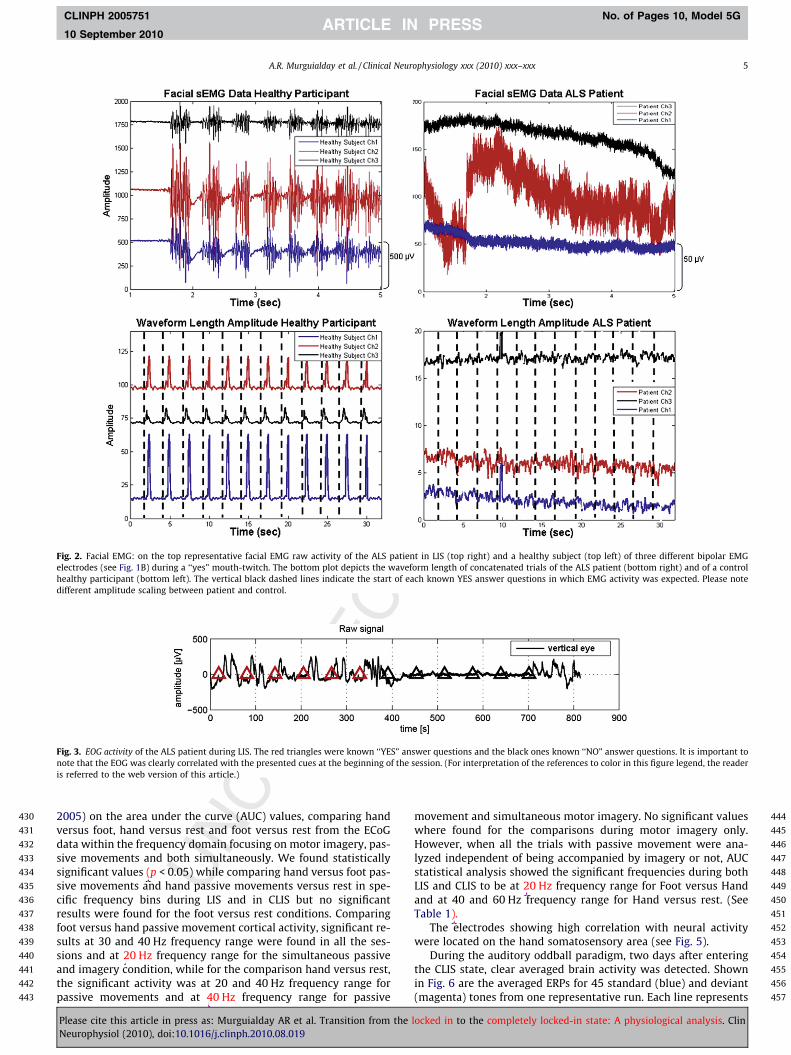
The possibility of communication through the mouth-twitchwas tested every day presenting questions with known ‘‘yes–no”answers to the patient and open questions with unknown answers,always with negative results after that date. Furthermore EMGactivity of the previously active muscles was acquired in order totest if any remaining muscle activity was present, without success.No activity was found using any of the four calculated EMG fea-tures. Fig. 2 presents a comparison between a healthy subjectand the ALS patient.
We used a feedforward multilayer perceptrons (MLPs) withvarying numbers of hidden layer neurons (empirically chosen tobe between 0.5 and 4 times the dimension of the input space) forclassifying and testing the facial EMG data of the patient. A classi-fication accuracy of 51% (chance level) was obtained (more infor-mation regarding classification process is presented in Tenoreet al., 2009).
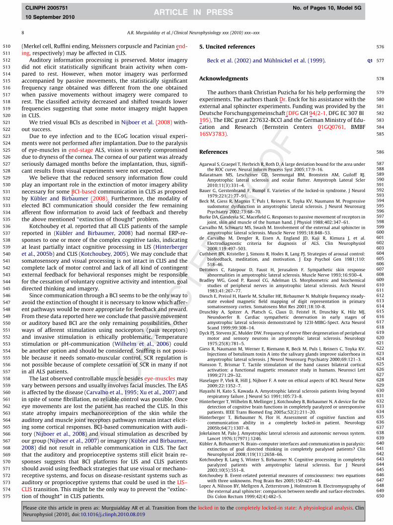
After the mouth-twitch based communication pathway ceased,the eyes appear to be the next suitable option. The patient couldcommunicate with eye movements at two occasions before thesurgery and two occasions after the surgery. This communicationpathway was tested every day several times and the EOG was re-corded. Fig. 3 depicts how the patient was able to move his eyesto answer questions with known answer for a successful session.The red triangles indicate YES answer questions and the black indi-cate NO answer questions.
Although the patient suffered fatigue, some days in which thepatient could communicate for longer time were recorded untilMarch 2008. In the beginning of February 2008 two EAS experi-ments were performed, with a week of separation between them.The possibility of remaining anal sphincter control was investi-gated with negative results. In Fig. 4A and B the raw and trans-formed EAS data of a healthy person is presented. Red and bluecrosses indicate the start and ending of a verbally triggered con-traction respectively. A clear difference between rest and EAS con-traction is shown. In Fig. 4C and D, concatenated raw andtransformed data from the ALS patient for YES (expected EAS activ-ity) and NO (no expected EAS activity) answers is presented. Incontrary to the healthy person there is no EAS activity. Corticospi-nal tract lesions were not visible in MR images from spinal cordexcluding the motor neuron degeneration as the most likely causeof the sphincter pathology.
The last communication with the patient took place on the 16thof March 2008. After this date the patient was considered to be inthe CLIS. During CLIS we performed somatosensory stimulation inorder to test the non visual afferent pathways to the patient’sbrain. In locked-in ALS patients vision is compromised because oflack of adequate eye lubrication and moisture. Many different at-tempts for BCI communication not reported here were tried duringCLIS, all without significant results. During the vibrotactile stimu-lation (Experiment C) no correlated activity was found in any ofthe implanted electrodes during any of the stimulations performedon the three different body parts (Index finger, toe and lip). This re-sult is in complete contrast with similar procedures that elicitedclear cortical responses in healthy and epilepsy samples (Dieschet al., 2001; Ray et al., 2008; Hansson and Brismar, 1999).
Nevertheless, while passive movements were performed on pa-tient’s foot and hand, activation of the somatosensory cortex wasdetected with and without simultaneous motor imagery task.The statistical analysis was performed, following (Agarwal et al.,
locked in to the completely locked-in state: A physiological analysis. Clin
430
431
432
433
434
435
436
437
438
439
440
441
442
443
444
445
446
447
448
449
450
451
452
453
454
455
456
457
Fig. 2. Facial EMG: on the top representative facial EMG raw activity of the ALS patient in LIS (top right) and a healthy subject (top left) of three different bipolar EMGelectrodes (see Fig. 1B) during a ‘‘yes” mouth-twitch. The bottom plot depicts the waveform length of concatenated trials of the ALS patient (bottom right) and of a controlhealthy participant (bottom left). The vertical black dashed lines indicate the start of each known YES answer questions in which EMG activity was expected. Please notedifferent amplitude scaling between patient and control.
Fig. 3. EOG activity of the ALS patient during LIS. The red triangles were known ‘‘YES” answer questions and the black ones known ‘‘NO” answer questions. It is important tonote that the EOG was clearly correlated with the presented cues at the beginning of the session. (For interpretation of the references to color in this figure legend, the readeris referred to the web version of this article.)
A.R. Murguialday et al. / Clinical Neurophysiology xxx (2010) xxx–xxx 5
CLINPH 2005751 No. of Pages 10, Model 5G
10 September 2010
2005) on the area under the curve (AUC) values, comparing handversus foot, hand versus rest and foot versus rest from the ECoGdata within the frequency domain focusing on motor imagery, pas-sive movements and both simultaneously. We found statisticallysignificant values (p < 0.05) while comparing hand versus foot pas-sive movements and hand passive movements versus rest in spe-cific frequency bins during LIS and in CLIS but no significantresults were found for the foot versus rest conditions. Comparingfoot versus hand passive movement cortical activity, significant re-sults at 30 and 40 Hz frequency range were found in all the ses-sions and at 20 Hz frequency range for the simultaneous passiveand imagery condition, while for the comparison hand versus rest,the significant activity was at 20 and 40 Hz frequency range forpassive movements and at 40 Hz frequency range for passive
Please cite this article in press as: Murguialday AR et al. Transition from theNeurophysiol (2010), doi:10.1016/j.clinph.2010.08.019
movement and simultaneous motor imagery. No significant valueswhere found for the comparisons during motor imagery only.However, when all the trials with passive movement were ana-lyzed independent of being accompanied by imagery or not, AUCstatistical analysis showed the significant frequencies during bothLIS and CLIS to be at 20 Hz frequency range for Foot versus Handand at 40 and 60 Hz frequency range for Hand versus rest. (SeeTable 1).
The electrodes showing high correlation with neural activitywere located on the hand somatosensory area (see Fig. 5).
During the auditory oddball paradigm, two days after enteringthe CLIS state, clear averaged brain activity was detected. Shownin Fig. 6 are the averaged ERPs for 45 standard (blue) and deviant(magenta) tones from one representative run. Each line represents
locked in to the completely locked-in state: A physiological analysis. Clin

458
459
460
461
462
463
464
465
Fig. 4. External anal sphincter: (A) Raw EAS EMG activity of a healthy person following random auditory triggers. Red ‘‘C” indicates (contraction) and blues ‘‘R” (relaxation).The high peaks of EMG activity correspond to high intensity contractions. (B) WL extracted feature of activity in A. (C) Raw EAS EMG signal of the ALS patient and (D) WLextracted feature of activity in C. (C.1) and (D.1) ‘‘YES” answer trials (contraction) concatenated and (C.2) and (D.2) ‘‘NO” answer trials (relaxation) concatenated. (Forinterpretation of the references to color in this figure legend, the reader is referred to the web version of this article.)
6 A.R. Murguialday et al. / Clinical Neurophysiology xxx (2010) xxx–xxx
CLINPH 2005751 No. of Pages 10, Model 5G
10 September 2010
one of the 48 non-artefacted recording channels of the ECoG grid,re-referenced to another common grid channel (see Fig. 1A, G-105). The figure shows the N1/P2 component for the deviants, aswell as the delayed P300 response in the range of 400–700 ms.These results demonstrate intact afferent auditory pathways and
466
467
468
469
470
471
472
473
474
475
476
477
478
479
480
481
482
483
484
485
486
487
488
489
Table 1Statistically significant frequency bins obtained from the area under the curve (AUC)analysis performed in the LIS (sessions 1 and 2) and CLIS (session 3) comparingdifferent conditions. Passive stands for passive movement alone of the patients handor foot, Pass&Imag stands for passive movement while the patient was asked toimagine the same movement and Pass&Imag + Pass stands for all the trials in whichthe patient’s limb was passively moved independently of simultaneous imagery ornot. Please note the stability of the statistically significant frequency ranges in thedifferent conditions during locked-in state and completely locked-in state.
Conditions Session 1 and 2freq. bins (Hz) LIS
Session 3 freq.bins (Hz) CLIS
Foot versus hand 16–20 Hz 20–24 HzPass&Imag 36–38 HzFoot versus hand 16–22 Hz and 30–32 Hz 20–42 HzPass&Imag + Pass 60–62 Hz and 70–72 HzFoot versus hand 32–36 Hz and 40–42 Hz 29–36 and 40–42 HzPassive 56–58 HzHand versus rest 6–8 Hz 22–26 HzPass&Imag 40–42 Hz and 60–62 Hz 38–40 HzHand versus rest 6–8 Hz 20–26 Hz and 30–34 HzPass&Imag + Pass 40–46 Hz 40–44 Hz
60–86 Hz and 92–94 Hz 60–66 HzHand versus rest 24–26 Hz 22–28 HzPassive 40–42 Hz 40–44 Hz
74–76 and 84–86 Hz 58–60 Hz
Please cite this article in press as: Murguialday AR et al. Transition from theNeurophysiol (2010), doi:10.1016/j.clinph.2010.08.019
automatic attention, even though the N1/P2 complex is not clearlyvisible for the standard tones.
4. Discussion and conclusion
LIS–CLIS transition was analyzed in one patient with ALS in sev-eral neurophysiological experiments that should provide informa-tion about the different stages of nervous system disorders.Between LIS and CLIS there are some clear physiological differ-ences. Although the data presented in this paper are from one sin-gle patient only, who underwent the LIS–CLIS transition, this dataset is unique and could be used as a first step towards a physiolog-ical data based instrument to define end stages of ALS and probablysome other neurological diseases.
In the present patient the last remaining controllable muscleswere the eye-muscles. This contradicts the hypothesis that theexternal anal sphincter is the last remaining controllable musclein ALS. Jokelainen and Palo found no reports of rectal or bladderdysfunction in their review of 300 ALS patients (Jokelainen andPalo, 1976). But it was also reported that eye and sphincter mus-cles are affected by the disease (Carvalho et al., 1995; Pullen andMartin, 1995; Xu et al., 2007; Palmowski et al., 1995; Balaratnamet al., 2010; Okamoto et al., 1993) but we have not found any pub-lication reporting the complete extinction of eye and sphinctermovement control as observed in the present patient.
The fact that there is no cortical activation during vibrotactilestimulation may indicate that skin mechanoreceptors informationis not reaching the cortical areas or that the skin mechanoreceptorsdo not function properly, at least in response to moderate to med-
locked in to the completely locked-in state: A physiological analysis. Clin

490
491
492
493
494
495
496
497
498
499
500
501
502
503
504
505
506
507
508
509
Fig. 5. ECoG activity during passive movement: on the top area under the curve (AUC) scores comparing foot versus hand movements of session 3 (CLIS); on the left sidepassive movements alone and on the right passive movements and motor imagery simultaneously AUC scores. In black the values that are statistically significant comparinghand versus foot movements. At the bottom the AUC scores in the frequency bin from 30 to 34 Hz plotted on the ALS patient X-rays (left, sessions 1 and 2 LIS and right, session3 CLIS) comparing hand versus foot passive movements. In white the statistically significant AUC values. The hole in the center of the electrode grid is due to high impedancesor lost channels.
Fig. 6. Averaged auditory evoked ERPs during CLIS for 45 standard (blue) and deviant (magenta) tones. Each line represents one ECoG grid electrode recording. The verticalblack line indicates beginning of the stimulus (time = 0 s). (For interpretation of the references to color in this figure legend, the reader is referred to the web version of thisarticle.)
A.R. Murguialday et al. / Clinical Neurophysiology xxx (2010) xxx–xxx 7
CLINPH 2005751 No. of Pages 10, Model 5G
10 September 2010
ium stimulus intensities. In contrast, proprioceptive information isprocessed in the brain as shown in the passive movement experi-ments. We hypothesize that joint receptors and muscle mechano-receptors are less affected by the consequences of long-termimmobilization than skin mechanoreceptors. Skin mechanorecep-tors are located more at the body surface and therefore being moresusceptible to damage from atrophy and skin deformation. Thesedata suggest that muscle mechanoreceptors or joint receptorsdegenerate later than the skin mechanoreceptors and that at leastsome of the group Ia, II and Ib afferent fibers, muscle spindles and
Please cite this article in press as: Murguialday AR et al. Transition from theNeurophysiol (2010), doi:10.1016/j.clinph.2010.08.019
Golgi tendon organs are preserved in the CLIS. It has been proposedthat joint receptor afferent input to the brain might be only signif-icant when muscle spindle afferents do not contribute to proprio-ception (Burke et al., 1988). Taken together, the data indicate thatif the above mentioned mechanoreceptors are not preserved thensome of the joint receptors such as Ruffini endings, Pacinian end-ings and Golgi tendons and their respective fibers (slow and fastadapting fibers type II) should be preserved. On the other hand,the slowly adapting fibers type I and II (SAI and SAII), fast adaptingtype I and II (FAI and FAII) or their corresponding receptor types
locked in to the completely locked-in state: A physiological analysis. Clin
510
511
512
513
514
515
516
517
518
519
520
521
522
523
524
525
526
527
528
529
530
531
532
533
534
535
536
537
538
539
540
541
542
543
544
545
546
547
548
549
550
551
552
553
554
555
556
557
558
559
560
561
562
563
564
565
566
567
568
569
570
571
572
573
574
575
576
577
578
579
580
581
582
583
584
585
586
587588589590591592593594595596597598599600601602603604605606607608609610611612613614615616617618619620621622623624625626627628629630631632633634635636637638639640641642643644645646647648649650
Q1
8 A.R. Murguialday et al. / Clinical Neurophysiology xxx (2010) xxx–xxx
CLINPH 2005751 No. of Pages 10, Model 5G
10 September 2010
(Merkel cell, Ruffini ending, Meissners corpuscle and Pacinian end-ing, respectively) may be affected in CLIS.
Auditory information processing is preserved. Motor imagerydid not elicit statistically significant brain activity when com-pared to rest. However, when motor imagery was performedaccompanied by passive movements, the statistically significantfrequency range obtained was different from the one obtainedwhen passive movements without imagery were compared torest. The classified activity decreased and shifted towards lowerfrequencies suggesting that some motor imagery might happenin CLIS.
We tried visual BCIs as described in Nijboer et al. (2008) with-out success.
Due to eye infection and to the ECoG location visual experi-ments were not performed after implantation. Due to the paralysisof eye-muscles in end-stage ALS, vision is severely compromiseddue to dryness of the cornea. The cornea of our patient was alreadyseriously damaged months before the implantation, thus, signifi-cant results from visual experiments were not expected.
We believe that the reduced sensory information flow couldplay an important role in the extinction of motor imagery abilitynecessary for some BCI-based communication in CLIS as proposedby Kübler and Birbaumer (2008). Furthermore, the modality ofelected BCI communication should consider the few remainingafferent flow information to avoid lack of feedback and therebythe above mentioned ‘‘extinction of thought” problem.
Kotchoubey et al. reported that all CLIS patients of the samplereported in (Kübler and Birbaumer, 2008) had normal ERP-re-sponses to one or more of the complex cognitive tasks, indicatingat least partially intact cognitive processing in LIS (Hinterbergeret al., 2005b) and CLIS (Kotchoubey, 2005). We may conclude thatsomatosensory and visual processing is not intact in CLIS and thecomplete lack of motor control and lack of all kind of contingentexternal feedback for behavioral responses might be responsiblefor the cessation of voluntary cognitive activity and intention, goaldirected thinking and imagery.
Since communication through a BCI seems to be the only way toavoid the extinction of thought it is necessary to know which affer-ent pathways would be more appropriate for feedback and reward.From these data reported here we conclude that passive movementor auditory based BCI are the only remaining possibilities. Otherways of afferent stimulation using nociceptors (pain receptors)and invasive stimulation is ethically problematic. Temperaturestimulation or pH-communication (Wilhelm et al., 2006) couldbe another option and should be considered. Sniffing is not possi-ble because it needs somato-muscular control. SCR regulation isnot possible because of complete cessation of SCR in many if notin all ALS patients.
The last observed controllable muscle besides eye-muscles mayvary between persons and usually involves facial muscles. The EASis affected by the disease (Carvalho et al., 1995; Xu et al., 2007) andin spite of some fibrillation, no reliable control was possible. Onceeye movements are lost the patient has reached the CLIS. In thisstate atrophy impairs mechanoreception of the skin while theauditory and muscle joint receptors pathways remain intact elicit-ing some cortical responses. BCI-based communication with audi-tory (Nijboer et al., 2008) and visual stimulation as described byour group (Nijboer et al., 2007) or imagery (Kübler and Birbaumer,2008) did not result in reliable communication in CLIS. The factthat the auditory and proprioceptive systems still elicit brain re-sponses suggests that BCI platforms for LIS and CLIS patientsshould avoid using feedback strategies that use visual or mechano-receptive systems, and focus on disease-resistant systems such asauditory or proprioceptive systems that could be used in the LIS–CLIS transition. This might be the only way to prevent the ‘‘extinc-tion of thought” in CLIS patients.
Please cite this article in press as: Murguialday AR et al. Transition from theNeurophysiol (2010), doi:10.1016/j.clinph.2010.08.019
5. Uncited references
Beck et al. (2002) and Mühlnickel et al. (1999).
Acknowledgments
The authors thank Christian Puzicha for his help performing theexperiments. The authors thank Dr. Enck for his assistance with theexternal anal sphincter experiments. Funding was provided by theDeutsche Forschungsgemeinschaft (DFG GH 94/2-1, DFG EC 307 BI195), The ERC grant 227632-BCCI and the German Ministry of Edu-cation and Research (Bernstein Centers 01GQ0761, BMBF16SV3783).
References
Agarwal S, Graepel T, Herbrich R, Roth D. A large deviation bound for the area underthe ROC curve. Neural Inform Process Syst 2005;17:9–16.
Balaratnam MS, Leschziner GD, Seemungal BM, Bronstein AM, Guiloff RJ.Amyotrophic lateral sclerosis and ocular flutter. Amyotroph Lateral Scler2010;11(3):331–4.
Bauer G, Gerstenbrand F, Rumpl E. Varieties of the locked-in syndrome. J Neurol1979;221(2):77–91.
Beck M, Giess R, Magnus T, Puls I, Reiners K, Toyka KV, Naumann M. Progressivesudomotor dysfunction in amyotrophic lateral sclerosis. J Neurol NeurosurgPsychiatry 2002;73:68–70.
Burke DA, Gandevia SC, Macefield G. Responses to passive movement of receptors injoint, skin and muscle of the human hand. J Physiol 1988;402:347–61.
Carvalho M, Schwartz MS, Swash M. Involvement of the external anal sphincter inamyotrophic lateral sclerosis. Muscle Nerve 1995;18:848–53.
de Carvalho M, Dengler R, Eisen A, England JD, Kaji R, Kimura J, et al.Electrodiagnostic criteria for diagnosis of ALS. Clin Neurophysiol2008;119:497–503.
Cuthbert BN, Kristeller J, Simons R, Hodes R, Lang PJ. Strategies of arousal control:biofeedback, meditation, and motivation. J Exp Psychol Gen 1981;110:518–46.
Dettmers C, Fatepour D, Faust H, Jerusalem F. Sympathetic skin responseabnormalities in amyotrophic lateral sclerosis. Muscle Nerve 1993;16:930–4.
Bradley WG, Good P, Rasool CG, Adelman LS. Morphometric and biochemicalstudies of peripheral nerves in amyotrophic lateral sclerosis. Arch Neurol1983;41:267–77.
Diesch E, Preissl H, Haerle M, Schaller HE, Birbaumer N. Multiple frequency steady-state evoked magnetic field mapping of digit representation in primarysomatosensory cortex. Somatosens Mot Res 2001;18:10–8.
Druschky A, Spitzer A, Platsch G, Claus D, Feistel H, Druschky K, Hilz MJ,Neundoerfer B. Cardiac sympathetic denervation in early stages ofamyotrophic lateral sclerosis demonstrated by 123I-MIBG-Spect. Acta NeurolScand 1999;99:308–14.
Dyck PJ, Stevens JC, Mulder DW. Frequency of nerve fiber degeneration of peripheralmotor and sensory neurons in amyotrophic lateral sclerosis. Neurology1975;25(8):781–5.
Giess R, Naumann M, Werner E, Riemann R, Beck M, Puls I, Reiners C, Toyka KV.Injections of botulinum toxin A into the salivary glands improve sialorrhoea inamyotrophic lateral sclerosis. J Neurol Neurosurg Psychiatry 2000;69:121–3.
Hansson T, Brismar T. Tactile stimulation of the hand causes bilateral corticalactivation: a functional magnetic resonance study in humans. Neurosci Lett1999;271:29–32.
Haselager P, Vlek R, Hill J, Nijboer F. A note on ethical aspects of BCI. Neural Netw2009;22:1352–7.
Hayashi H, Kato S, Kawada A. Amyotrophic lateral sclerosis patients living beyondrespiratory failure. J Neurol Sci 1991;105:73–8.
Hinterberger T, Wilhelm B, Mellinger J, Kotchoubey B, Birbaumer N. A device for thedetection of cognitive brain functions in completely paralyzed or unresponsivepatients. IEEE Trans Biomed Eng 2005a;52(2):211–20.
Hinterberger T, Birbaumer N, Flor H. Assessment of cognitive function andcommunication ability in a completely locked-in patient. Neurology2005b;64(7):1307–8.
Jokelainen M, Palo J. Amyotrophic lateral sclerosis and autonomic nervous system.Lancet 1976;1(7971):1246.
Kübler A, Birbaumer N. Brain–computer interfaces and communication in paralysis:extinction of goal directed thinking in completely paralysed patients? ClinNeurophysiol 2008;119(11):2658–66.
Kotchoubey B, Lang S, Winter S, Birbaumer N. Cognitive processing in completelyparalyzed patients with amyotrophic lateral sclerosis. Eur J Neurol2003;10(5):551–8.
Kotchoubey B. Event-related potential measures of consciousness: two equationswith three unknowns. Prog Brain Res 2005;150:427–44.
Lopez A, Nilsson BY, Mellgren A, Zetterstrom J, Holmstrom B. Electromyography ofthe external anal sphincter: comparison between needle and surface electrodes.Dis Colon Rectum 1999;42(4):482–5.
locked in to the completely locked-in state: A physiological analysis. Clin
651652653654655656657658659660661662663664665666667668669670671672673674675676677678679
680681682683684685686687688689690691692693694695696697698699700701702703704705706707708
A.R. Murguialday et al. / Clinical Neurophysiology xxx (2010) xxx–xxx 9
CLINPH 2005751 No. of Pages 10, Model 5G
10 September 2010
Masur H, Schulte-Oversohl U, Papke K, Oberwittler C, Vollmer J. Sympathetic skinresponse in patients with amyotrophic lateral sclerosis. Funct Neurol1995;10:131–5.
Mühlnickel W, Lutzenberger W, Flor H. Localization of somatosensory evokedpotentials in primary somatosensory cortex: a comparison between PCA andMUSIC. Brain Topogr 1999;11(3):185–91.
Nijboer F, Sellers EW, Mellinger J, Jordan MA, Halder S, Matuz T, et al. A brain–computer interface (BCI) for people with amyotrophic lateral sclerosis (ALS).Clin Neurophysiol 2008;119(8):1909–16.
Nijboer F, Furdea A, Gunst I, Mellinger J, MacFarland DJ, Birbaumer N, et al. Anauditory brain–computer interface. J Neurosci Methods 2007;167(1):43–50.
Okamoto K, Hirai S, Amari M, Iizuka T, Watanabe M, Murakami N, et al. Oculomotornuclear pathology in amyotrophic lateral sclerosis. Acta Neuropathol (Berl)1993;85:458–62.
Markand Omkar N. Electroencephalogram in ‘‘Locked-In” syndrome.Electroencephalogr Clin Neurophysiol 1976;40(5):529–34.
Palmowski A, Jost WH, Prudlo J, Osterhage J, Ksmann B, Schimrigk K, et al. Eyemovement in amyotrophic lateral sclerosis: a longitudinal study. German JOphthalmol 1995;4(6):355–62.
Pinelli P, Pisano F, Miscio G. The possible role of a secondary pathogenetic factor inamyotrophic lateral sclerosis. Adv Neurol 1995;68:29–40.
Pisano F, Miscio G, Mazzuero G, Lanfranchi P, Colombo R, Pinelli P. Decreased heartrate variability in amyotrophic lateral sclerosis. Muscle Nerve1995;18:1225–31.
Plum F, Posner JB. The diagnosis of stupor and coma, vol. 197. Philadelphia: FADavis; 1966. p. 93.
Polich J, Alexander JE, Baue LO, Kuperman S, Morzorati S, O’Connor SJ, et al. P300topography of amplitude/latency correlations. Brain Topogr 1997;9(4):275–82.
709
Please cite this article in press as: Murguialday AR et al. Transition from theNeurophysiol (2010), doi:10.1016/j.clinph.2010.08.019
Pullen AH, Martin JE. Ultrastructural abnormalities with inclusions in Onuf’snucleus in motor neuron disease (amyotrophic lateral sclerosis). NeuropatholAppl Neurobiol 1995;21:327–40.
Ray S, Niebur E, Hsiao SS, Sinai A, Crone NE. High-frequency gamma activity (80–150 Hz) is increased in human cortex during selective attention. ClinNeurophysiol 2008;119:116–33.
Steiner TJ, Sethi KD, Rose FC. Autonomic function in motor neurone disease. In: RoseFC, editor. Research progress in motor neurone disease. London: Pitman; 1984.p. 180–8.
Tenore F, Ramos Murguialday A, Fahmy A, Acharya S, Etienne-Cummings R, ThakorNV. Decoding of individuated finger movements using surfaceelectromyography. IEEE Trans Biomed Eng 2009;56(5):1427–34.
Toepfer M, Schroeder M, Klauser A, Lochmüller H, Hirschmann M, Riepl RL, et al.Delayed colonic transit times in amyotrophic lateral sclerosis assessed withradio-opaque markers. Eur J Med Res 1997;2(11):473–6.
Toepfer M, Riepl RL, Müller-Felber W, Endres S, Folwaczny C. Noninvasive (13)C-octanoic acid breath test shows delayed gastric emptying in patients withamyotrophic lateral sclerosis. Digestion 1999;60:567–71.
Wilhelm B, Jordan M, Birbaumer N. Communication in locked-in syndrome: effectsof imagery on salivary pH. Neurology 2006;67(3):534–5.
Xu Y, Zheng J, Zhang S, Kang D, Zhang J, Fan D. Needle electromyography of therectus abdominis in patients with amyotrophic lateral sclerosis. Muscle Nerve2007;35:383–5.
Yuki N, Yamada M, Yuasa T, Kaneko K, Inuzuka T, Arai M, et al. Atypical motorneuron disease with severe ophthalmoloplegia: a report of two cases. J Neurol1995;242:541–6.
Yoshida M, Murakami N, Hashizume Y, Itoh E, Takahashi A. A clinicopathologicalstudy of two respiratoraided long-survival cases of amyotrophic lateralsclerosis(in Japanese with English abstract). Clin Neurol (Tokyo) 1992;32:259–65.
locked in to the completely locked-in state: A physiological analysis. Clin