transfer of free polymannose-type oligosaccharides from the cytosol to lysosomes in cultured human...
TRANSCRIPT

The Rockefeller University Press, 0021-9525/97/01/45/15 $2.00The Journal of Cell Biology, Volume 136, Number 1, January 13, 1997 45–59 45
Transfer of Free Polymannose-type Oligosaccharides from the Cytosolto Lysosomes in Cultured Human Hepatocellular Carcinoma HEPG2 Cells
Agnès Saint-Pol, Chantal Bauvy, Patrice Codogno, and Stuart E.H. Moore
Unité de Neuroendocrinologie et Biologie Cellulaire Digestives, Institut National de la Santé et de la Recherche Médicale, U410, Faculté de Médecine Xavier Bichat, 75018 Paris, France
Abstract.
Large, free polymannose oligosaccharides generated during glycoprotein biosynthesis rapidly ap-pear in the cytosol of HepG2 cells where they undergo processing by a cytosolic endo H–like enzyme and a mannosidase to yield the linear isomer of Man
5
GlcNAc (Man[
a
1-2]Man[
a
1-2]Man[
a
1-3][Man
a
1-6]Man[
b
1-4]GlcNAc). Here we have examined the fate of these partially trimmed oligosaccharides in intact HepG2
cells. Subsequent to pulse–chase incubations with
d
-[2-
3
H]mannose followed by permeabilization of cells with streptolysin O free oligosaccharides were isolated from the resulting cytosolic and membrane-bound compart-ments. Control pulse–chase experiments revealed that total cellular free oligosaccharides are lost from HepG2 cells with a half-life of 3–4 h. In contrast use of the vac-
uolar H
1
/ATPase inhibitor, concanamycin A, stabilized
total cellular free oligosaccharides and enabled us to demonstrate a translocation of partially trimmed oli-gosaccharides from the cytosol into a membrane-bound compartment. This translocation process was unaf-fected by inhibitors of autophagy but inhibited if cells were treated with either 100
m
M swainsonine, which provokes a cytosolic accumulation of large free oli-gosaccharides bearing 8-9 residues of mannose, or agents known to reduce cellular ATP levels which lead to the accumulation of the linear isomer of Man
5
GlcNAc in the cytosol. Subcellular fractionation studies on Percoll density gradients revealed that the cytosol-generated linear isomer of Man
5
GlcNAc is de-graded in a membrane-bound compartment that cosed-iments with lysosomes.
T
he
glycosylation of proteins with N-linked carbohy-drate in the endoplasmic reticulum is a commonand important posttranslational modification. Sur-
prisingly, this process, accomplished by the transfer of apolymannose-type oligosaccharide from a lipid carrier(dolichol) onto polypeptide (Kornfeld and Kornfeld, 1985),is accompanied by the release of free polymannose-typeoligosaccharides into the lumen of the ER (Anumula andSpiro, 1983; Cacan et al., 1987). As large amounts of freeoligosaccharides are generated in this way an understand-ing of the fate of this material became important. It wasinitially thought that free oligosaccharides generated inthe lumen of the ER might be exported from the cell byvesicular transport as a consequence of the effect of bulkflow (Wieland et al., 1987). In fact this was found not to bethe case as free oligosaccharides were not recovered fromthe incubation media of cultured HepG2 cells (Moore andSpiro, 1990) but detected in the cytosol (Moore and Spiro,1994). More recently free polymannose-type oligosaccha-
rides bearing the terminal reducing di-
N
-acetylchitobiosemoiety have been shown to be transported out of the ERinto the cytosol in permeabilized HepG2 cells (Moore et al.,1995). In addition to the transfer of free oligosaccharidesfrom the lumen of the ER into the cytosol, there is now ev-idence to suggest that some free oligosaccharides may begenerated in the cytosol by either the release of cytosoli-cally disposed oligosaccharides from dolichol (Kmiécik et al.,1995), or by the degradation of glycoproteins, initiated bya cytosolic N-glycanase (Suzuki et al., 1994), that have beentranslocated out of the ER into the cytosol (Wiertz et al.,1996). These reports highlight the crucial role that the cy-tosol plays in the processing and perhaps generation offree oligosaccharides which are generated during the bio-synthesis and quality control of glycoproteins. What thenis the fate of these free cytosolic oligosaccharides?
It has been known for some years that the cytosol con-tains both an endo H–like enzyme (Pierce et al., 1979) andan
a
-mannosidase (Shoup and Touster, 1976; Tulsiani andTouster, 1987). In vitro experiments with preparations ofthe cytosolic
a
-mannosidase have revealed it to possess twonotable features, firstly it is inactive towards large polyman-nose-type oligosaccharides bearing the di-
N
-acetylchitobi-ose moiety at their reducing termini (Oku and Hase, 1991)
Address all correspondence to S.E.H. Moore, INSERM U410, 16 rueHenri Huchard 75018 Paris, France. Tel.: 33 1 44856134. Fax: 33 142288765.
on August 24, 2016
jcb.rupress.orgD
ownloaded from
Published January 13, 1997

The Journal of Cell Biology, Volume 136, 1997 46
and secondly its limit digest product is the linear isomer ofMan
5
GlcNAc: Man(
a
1-2)Man(
a
1-2)Man(
a
1-3)(Man[
a
1-6])Man(
b
1-4)GlcNAc; Tulsiani and Touster, 1987; Oku andHase, 1991). In intact cells cytosolic-free oligosaccharidesgenerated during the biosynthesis of glycoproteins are ap-parently subjected to the actions of these two cytosolic en-zymes to yield the linear isomer of Man
5
GlcNAc (Mooreand Spiro, 1994).
Here we report on the fate of this cytosolic free oli-gosaccharide in intact HepG2 cells. It is shown that par-tially trimmed cytosolic oligosaccharides are translocatedinto a membrane bound compartment by a nonautophagicprocess that requires energy. Subcellular fractionation ofHepG2 cell homogenates on Percoll density gradientsrevealed that cytosolic oligosaccharides are ultimatelydegraded in a compartment that cosediments with lyso-somes. Along with a previous report describing the trans-port of free oligosaccharides from the lumen of the ERinto the cytosol (Moore et al., 1995), this report describes anovel trafficking pathway for free oligosaccharides thatlinks the endoplasmic reticulum to the lysosome via thecytosol.
Materials and Methods
Culture and Radiolabeling of Cells
HepG2 cells were cultivated in RPMI 1640 (GIBCO BRL, Paisley, UK)containing 10% FCS (GIBCO) as previously described (Moore and Spiro,1990). Cells were pulse radiolabeled with 40
m
Ci
d
-[2-
3
H]mannose (20 Ci/mmol; Amersham, Slough, UK) for 20 min in 0.5 ml glucose-free Dul-becco’s modified Eagle Medium (GIBCO BRL) supplemented with 5%dialyzed FCS, 2 mM glutamine, 5 mM fucose, and 1 mM sodium pyruvate.Cells were chased in complete growth medium containing 5 mM fucoseand 5 mM mannose. When pulse–chase studies were performed in thepresence of swainsonine (Sigma Chemical Co., St. Louis, MO) and con-canamycin A (Ciba-Geigy, Switzerland), the cells were preincubated withthese inhibitors for 1 h before the onset of radiolabeling and were addedto both the pulse and chase media at the appropriate concentrations. Be-fore subcellular fractionation studies cells were pulse radiolabeled for 20min with 200
m
Ci
d
-[2-
3
H]mannose and chased as described above. Otherdrugs including 3-methyladenine, asparagine, deoxyglucose, sodium azide,and oligomycin (all from Sigma Chemical Co.) were included in the chasemedia only and were added 45 min after the onset of the chase incuba-tions, whereas leupeptin (Sigma Chemical Co.) was added at the begin-ning of each chase period.
Permeabilization of Cells
At the end of pulse–chase experiments, cells were released from tissueculture flasks with trypsin/EDTA and washed twice in 1.0 ml of permeabil-ization buffer: 250 mM mannitol, 5 mM Hepes (pH 7.3), 2 mM EGTA, 1 mMCaCl
2
, 2 mM MgCl
2
. Cells were then incubated at 4
8
C for 20 min in 0.5 mlof the permeabilization buffer containing 1 U/ml streptolysin O (Well-come Diagnostics, Dartford, UK). Cells were recovered by centrifugationand the supernatant was kept. Subsequent to washing the cells twice with0.5 ml permeabilization buffer at 4
8
C, they were incubated with 0.5 mlprewarmed permeabilization buffer (37
8
C) for 5 min and the permeabi-lized cells were then recovered by centrifugation to yield the membrane-bound compartment (MBC)
1
fraction. The final supernatant was pooledwith the SLO-containing supernatant and the two subsequent perme-abilization buffer washes to yield 2 ml of the cytosolic compartment fraction(Cytosol).
Preparation of Free Oligosaccharides from Cytosolic and MBC Fractions of HepG2 Cells
Neutral free oligosaccharides were prepared from the cytosol and MBCfractions as previously described (Moore and Spiro, 1994). Briefly, thepellet of permeabilized cells (MBC) was extracted with chloroform/meth-anol/125 mM Hepes (pH 7.2) containing, 4 mM MgCl
2
, 3:2:1, and after vig-orous shaking the upper methanolic phase was recovered, dried, and re-dissolved in water. This material and the cytosolic fractions were desaltedon combined columns of AG 1-X2 (acetate form) and AG 50-X2 (H
1
form), unbound neutral material was then loaded onto columns of char-coal, which were then washed with water before elution of oligosaccharidematerial from the charcoal with 30% ethanol. Free oligosaccharides wereanalyzed on plastic thin layer chromatography plates coated with silica(Merck, Darmstadt, Germany) which were developed in
n
-propanol/ace-tic acid/water (3:2:1) for 12 h. Resolved components were visualized byfluorography.
Structural Analysis of Man
5
GlcNAc Oligosaccharides
After resolution of free oligosaccharides by thin layer chromatographycomponents of interest were eluted from the chromatography plates withwater and passed over coupled columns of AG 50 (H
1
form) and AG 1(acetate form). Nonretained neutral components were dried and sub-jected to two mannosidase treatments. One aliquot was treated with 1 UJack bean
a
-mannosidase (Sigma Chemical Co.) overnight at 37
8
C in 40 mMsodium acetate, pH 4.5. Another aliquot was digested overnight at 37
8
Cwith 5
m
U
a
1-2 mannosidase (Oxford Glycosystems, Abingdon, UK) in100 mM sodium acetate buffer, pH 5.0. The digestion products were thendesalted as described above, concentrated, and resolved by thin layerchromatography on plastic sheets coated with cellulose (0.1 mm thickness;Merck). Chromatographs were developed in pyridine/ethyl acetate/water/acetic acid 5:5:3:1 for 10 h, and after drying resolved components were vi-sualized by fluorography. Quantitation of the resolved products wasachieved by their elution from the cellulose plates and assaying radioac-tive components by scintillation counting.
Percoll Density Gradient Fractionation
HepG2 cells were washed three times with ice-cold PBS containing 1 mMCaCl
2
, 1 mM MgCl
2
and once with ice-cold subcellular fractionationbuffer (SFB), 250 mM sucrose, 20 mM Hepes, 1 mM EDTA, pH 7.2. Thecells were scraped from tissue culture flasks in 5 ml SFB and cellular pro-tein was assayed using a bicinchoninic acid protein assay kit (SigmaChemical Co.). The cell pellet obtained after centrifugation at 600
g
for 10min was resuspended in SFB (1.5 mg/ml protein) and placed on ice for 15min. Cell homogenization was carried out using a tight-fitting Dounce ho-mogenizer (30 passages). After centrifuging the homogenate at 600
g
for10 min, the supernatant was removed and kept on ice, and the pellet wasresuspended with SFB and rehomogenized and centrifuged as above.Pooled supernatants were adjusted to 5 ml with SFB and 3 ml of an 80%Percoll solution was added (Rijnboutt et al., 1992). The gradient wasformed by centrifugation for 35 min at 92,570
g
Av. (Rijnboutt et al.,1992). 400-
m
l fractions were collected from the top of the tube with a nee-dle mounted on a syringe. To isolate free oligosaccharides from the Per-coll gradient, fractions were pooled (see Fig. 9), diluted in SFB, and aftercentrifugation for 90 min at 100,000
g
Av organelles were recovered sepa-rately from the Percoll pellet. Free oligosaccharides were prepared fromorganelle fractions as described above, except that before ion-exchangechromatography, sucrose was eliminated from the samples by Biogel P2gel filtration.
HRP Uptake
HepG2 cells were washed with MEMH (MEM containing 1 mM sodiumpyruvate, 1 mM
l
-glutamine and 20 mM Hepes/NaOH, pH 7.2) and HRPuptake was performed in MEMH containing 5 mg/ml HRP (Sigma Chem-ical Co.) for 5 or 15 min at 37
8
C (van Weert et al., 1995). ExtracellularHRP was then removed by washing the cells with ice-cold MEMH over aperiod of 10 min, followed by either a 5-min or a 2-h chase period, corre-sponding to the 5 or 15 min pulse times, respectively (van Weert et al.,1995). HRP-loaded cells were then washed and homogenized as describedabove.
1.
Abbreviations used in this paper
: CCM A, concanamycin A; DOG,deoxyglucose; GlcNAc, N-acetylglucosamine; Man, mannose; MBC, mem-brane bound compartment; 3-MA, 3-methyladenine; SLO, streptolysin O;SW, swainsonine.
on August 24, 2016
jcb.rupress.orgD
ownloaded from
Published January 13, 1997

Saint-Pol et al.
Free Oligosaccharide Trafficking
47
Enzymatic Assays
HRP activity was assayed in 50 mM phosphate buffer (pH 5.0) containing0.1% Triton X-100, using 83
m
g/ml
O
-dianisidine and 1% H
2
O
2
as sub-strates (van Weert et al., 1995). The reaction was performed for 5 min atroom temperature in the dark; absorbance was measured at 460 nm.
Lysosomal
b
-
d
-hexosaminidase activity was measured using
p
-nitro-phenyl
N
-acetylglucosamine as described previously (Opheim and Touster,1977).
b
1, 4 Galactosyltransferase was assayed by the method of Barkeret al. (1972).
NADPH cytochrome c reductase activity was measured as previouslydescribed in a 50 mM phosphate, 0.1 mM EDTA buffer, pH 7.7, using 1mg/ml NADPH and 25
m
g/ml cytochrome c as substrates. Absorbance in-creases were measured at 550 nm over a 3-min period.
High pH Anion Exchange Chromatography
High pH anion exchange chromatography (HPAEC) was carried out on aDionex apparatus as previously described (Townsend et al., 1991). Com-ponents were eluted at 1 ml/min with buffer A for 10 min followed by a lin-ear gradient of 0–10% buffer B over 25 min (buffer A: 50 mM NaOH; bufferB: 500 mM sodium acetate in buffer A). Column effluent was monitoredfor radioactive components with a Flow Scintillation Analyzer RadiomaticFlow-one/
b
(Packard, Instrument Company, Meriden, CT) using a scintil-lation fluid (UltimaFlow one AP, Packard) flow rate of 2 ml/min. Standardoligosaccharides, prepared as previously described (Michalski et al., 1990;Haeuw et al., 1991) were monitored by pulsed electrochemical detection.
Results
Free Oligosaccharides Appear Rapidly in the Cytosolof HepG2 Cells during Glycoprotein Biosynthesis and Are Then Cleared from This Compartment
In a previous study we have shown that free oligosaccha-rides generated from oligosaccharide-lipid, bearing the di-
N
-acetylchitobiose moiety (OS-GN2), are rapidly trans-ported out of the ER into the cytosol (Moore et al., 1995).This process was observed in vitro upon addition of ATPto permeabilized HepG2 cells. In the present report wehave followed the subcellular trafficking of free polyman-nose oligosaccharides in vivo by permeabilizing the plasmamembrane of [
3
H]mannose-radiolabeled HepG2 cells withSLO. After centrifugation of permeabilized cells, free oli-gosaccharides were isolated from the supernatant (cyto-solic compartment) and the residual cell pellet, containingintact intracellular organelles (membrane bound compart-ments; MBCs). Fig. 1
A
shows the results of such an exper-iment. During the pulse the MBC contains free oligosac-charides (OS-GN2; Fig. 1,
open arrowheads
) whereas thecytosol contains the same components and in additiontheir counterparts bearing a single residue of
N
-acetylglu-cosamine at their reducing termini (OS-GN1). Betweenthe pulse and 1-h chase period there is a loss of OS-GN2from the MBC and an increase in free oligosaccharides inthe cytosolic compartment. These observations representthe previously described ER-to-cytosol transport process(Moore et al., 1995). After transport out of the ER, OS-GN2 are subject to a cytosolic endo H–like activity to yieldtheir OS-GN1 counterparts. OS-GN1 are now potentialsubstrates for the cytosolic mannosidase which trims thesecomponents down to a limit digest product; the linear iso-mer of Man
5
GlcNAc (Moore and Spiro, 1994; see Fig. 2
B
for representation of this structure). However, as can beseen in Fig. 1
A
, free oligosaccharides neither accumulatein the cytosol nor reappear in an MBC. We reasoned thatif cytosolic oligosaccharides are translocated into a degra-
dative compartment they might be difficult to detect dueto their rapid hydrolysis into free mannose and
N
-acetyl-glucosamine. Because the lysosome is the most likely com-partment in which cytosolic oligosaccharides might be de-graded we chose to perform pulse–chase experiments inthe presence of a vacuolar H
1
/ATPase inhibitor.
The Clearance of Free Oligosaccharidesfrom the Cytosol Is Not Dependent upon VacuolarH
1
/ATPase Activity
CCM A (Woo et al., 1992) is a vacuolar H
1
/ATPase whichhas been shown to abolish acidification of lysosomes with-out affecting cellular ATP levels (Woo et al., 1992: Kata-oka et al., 1995), we reasoned that if free oligosaccharidesare translocated into an acidic MBC from the cytosol thisreagent may either inhibit their access to, or, failing that,block their degradation within an acidic compartment.Free oligosaccharides isolated from the cytosol and MBCsof cells from a pulse–chase experiment performed in thepresence of CCM A are shown in Fig. 1
B
, results showthat when compared to the control experiment (Fig. 1
A
),this reagent had very little effect on early events (0–1 h)during the pulse–chase experiment and we conclude fromthis that the generation of free oligosaccharides in the ERand their transport out of this compartment into the cyto-sol is unaffected by CCM A. In contrast Fig. 1
B
showsthat, after 1 h of chase, CCM A provokes a steady accumu-lation of free oligosaccharides bearing predominantly 7-4residues of mannose, in an MBC. Although the effect ofCCM A is most marked with respect to oligosaccharidesrecovered from the MBCs we noted systematically, afterlong chase periods, that this reagent also causes a small butsignificant accumulation of free oligosaccharide materialin the cytosolic compartment. We also observed, after 8 hof chase in the presence of CCM A, that 7.9% of total freeoligosaccharides produced by HepG2 cells could be recov-ered from the incubation medium, and after 20 h of chasethis figure rose to 18.0%. As the quantity of free oligosac-charides recovered from the incubation media of CCMA–treated cells represented only a small fraction of totalcellular free oligosaccharides observed during the time frameof our experiments the contribution made by these com-ponents to the quantitative aspects of our studies have notbeen taken into account.
These results show that CCM A has only small effectson the appearance and decay of radioactivity associatedwith free oligosaccharides in the cytosol of HepG2 cellsbut, in contrast, this reagent provokes a marked accumula-tion of free oligosaccharides associated with an MBC.
The Isomeric Structure of the Man
5
GlcNAcIsolated from MBCs of CCM A–treated HepG2 CellsIs Consistent with Its Cytosolic Origin
To evaluate the possibility that the CCM A–provoked ac-cumulation of free oligosaccharides associated with anMBC is related to the loss of these components from thecytosol, we next investigated the isomeric configuration ofthe Man
5
GlcNAc associated with the MBCs. The cytosol isknown to contain an endo H–like enzyme and an
a
-man-nosidase which together process the large oligosaccharidesthat are transported out of the ER into the cytosol to yield
on August 24, 2016
jcb.rupress.orgD
ownloaded from
Published January 13, 1997

The Journal of Cell Biology, Volume 136, 1997 48
the limit digest product Man
5
GlcNAc (Fig. 2,
LINEAR
)(Moore and Spiro, 1994). Although products closely re-lated to CCM A are known to inhibit the degradation ofproteins in the lysosomes of certain cell lines (Yoshimoriet al., 1991), we could not rule out the possibility that inour hands CCM A caused the accumulation of partiallydegraded polymannose- or hybrid-type oligosaccharidesderived from incomplete lysosomal glycoprotein degra-dation. If this were the case, then any resulting freeMan
5
GlcNAc should have a structure consistent with itspassage through the Golgi apparatus while N-linked to aprotein. Accordingly, the Man
5
GlcNAc isolated fromMBCs of CCM A–treated HepG2 cells would be expectedto be the branched isomer of this oligosaccharide (Fig. 2,
BRANCHED
) (Kornfeld and Kornfeld, 1985). Thus trans-port of this latter component into an MBC of CCM A–treatedHepG2 cells would lead to the accumulation of an isomerof Man
5
GlcNAc different from that expected from the limitdigest product of the cytosolic mannosidase. To distinguishbetween the two possible origins of the Man
5
GlcNAc ob-served to accumulate in the presence of CCM A, we haveisolated this component from the MBCs and subjected itto digestion with a nonspecific
a
-mannosidase (Jack bean)
and an
a
-1, 2 mannosidase (Amano and Kobata, 1986) asshown in Fig. 2. The linear isomer of Man
5
GlcNAc hastwo
a
-1, 2 linked mannose residues whereas its branchedcounterpart contains no such linkages. Results show thatthe structure isolated from the MBCs of CCM A–treatedHepG2 cells is sensitive to the
a
-1, 2 mannosidase yieldingfree mannose and the tetrasaccharide Man
3
GlcNAc. Jackbean
a
-mannosidase treatment of the Man
5
GlcNAcyielded free mannose and the disaccharide Man
b
-1, 4GlcNAc, in the ratio 4:1, indicating that this componentpossessed a single terminal reducing
N
-acetylglucosaminemoiety and that all the
a
-linked mannose residues wereaccessible to an exomannosidase. Similar treatment of theMan
4
GlcNAc isolated from the MBCs of CCM A–treatedHepG2 cells revealed it also to be sensitive to the
a
-1, 2mannosidase yielding the digest products Man
3
GlcNAcand mannose (results not shown). These results clearly dem-onstrate that the isomeric configurations of the free oli-gosaccharides associated with the MBCs of CCM A–treatedHepG2 cells are compatible with their having been gener-ated by the cytosolic mannosidase and not as a conse-quence of incomplete degradation of free oligosaccharidesderived from lysosomal glycoprotein catabolism.
Figure 1. The effect of CCMA on the generation and fateof free oligosaccharides inHepG2 cells. Control (A) orCCM A–treated (B) HepG2cells were pulse radiolabeledwith d-[2-3H]mannose for 20min, and then chased for theindicated times. Cells werethen cooled, placed in suspen-sion, and permeabilized withSLO as described in Materialsand Methods to yield fractionscorresponding to the cytosol(CYTOSOL) and membrane-bound compartments (MBCs).Free oligosaccharides wereprepared from each of thesefractions and then resolvedby thin layer chromatography,on silica-coated plates. Theabbreviations associated withthe solid arrowheads are:G1M9, Glc1Man9GlcNAc; M9,Man9 GlcNAc; M8, Man8-GlcNAc; M7, Man7GlcNAc;M6, Man6GlcNAc; M5, Man5-GlcNAc; M4, Man4GlcNAc;M3, Man3GlcNAc. Those as-sociated with the open arrow-heads are: G1M9, Glc1Man9GlcNAc2; M9, Man9GlcNAc2;M8, Man8GlcNAc2. on A
ugust 24, 2016jcb.rupress.org
Dow
nloaded from
Published January 13, 1997

Saint-Pol et al. Free Oligosaccharide Trafficking 49
The Loss of Free Oligosaccharides from theCytosol Can Be Accounted for Both Quantitatively and Kinetically by Their Recovery within an MBC of CCM A–treated HepG2 Cells
The ability of CCM A to block the degradation of MBC-associated free polymannose type oligosaccharides has en-abled us to further establish the hypothesis that there is acytosol-to-MBC translocation of free oligosaccharides. Ac-cordingly, we have verified that the loss of free oligosac-charides from the cytosol could be quantitatively and ki-netically accounted for by their reappearance in an MBC.Quantitation of the free oligosaccharides generated during
the control and CCM A pulse–chase incubations is shownin Fig. 3. In the control incubations there is a loss of totalfree oligosaccharide from cells such that after 8 h of chaseonly 25% of the cytosolic free oligosaccharides remain.However in the presence of CCM A the total quantity offree oligosaccharide remains approximately constant be-tween 1 and 8 h of chase and the loss of free oligosaccha-rides from the cytosol can be accounted for, both quantita-tively and kinetically, by their recovery from the MBCs(Fig. 3).
Next, we wanted to know whether the free oligosaccha-rides associated with the MBCs of CCM A–treated HepG2cells are located within the MBC or are merely associatedwith the cytosolic face of its delineating membrane. Thisproblem was addressed by performing a pulse–chase incu-bation in the presence of CCM A as described in Fig. 1.After 6 h of chase the cells were first permeabilized withSLO to release the remaining cytosolic oligosaccharidesand then repermeabilized with saponin. Results indicatedthat .90% of the free oligosaccharides associated with theMBCs of SLO-permeabilized cells could be released upontheir repermeabilization with saponin (results not shown).Identical results were obtained if the saponin treatmentwas substituted by freeze–thawing (not shown). These re-sults confirm the hypothesis that in CCM A–treatedHepG2 cells there is a translocation of free oligosaccha-rides from the cytosolic compartment into the lumen of anMBC.
Figure 2. The linear isomer of Man5GlcNAc accumulates in amembrane-bound compartment of CCM A–treated HepG2 cells.HepG2 cells were pulse radiolabeled and chased for 4 h in thepresence of CCM A as described in Materials and Methods. Thecells were then permeabilized with SLO and oligosaccharideswere purified from the resulting MBC as previously described.The component migrating as Man5GlcNAc was recovered and al-iquots of this oligosaccharide were then digested with either jackbean a-mannosidase (JACK BEAN) or an a-1, 2 mannosidase(a-1, 2). After desalting, the digestion products were resolved bythin layer chromatography on a cellulose-coated plate. Subse-quent to visualization of resolved components by fluorographythey were eluted from the chromatography plate and assayedby scintillation counting. The molar equivalents of the resolvedcomponents were calculated and, after summing, the percent-age molar distribution of the digestion products was calculated.The cytosolic mannosidase generates an isomer (LINEAR) ofMan5GlcNAc that contains two a-1, 2-linked mannose residueswhereas Golgi mannosidase I leads to the formation of aMan5GlcNAc isomer (BRANCHED) devoid of a-1, 2-linkedmannose residues. The abbreviations are as follows: M5GN,Man5GlcNAc; M4GN, Man4GlcNAc; M3GN, Man3GlcNAc;M2GN, Man2GlcNAc; MGN, ManGlcNAc; M, Mannose.
Figure 3. Quantitation of the transfer of free oligosaccharidesfrom the cytosol into a membrane-bound compartment of CCMA–treated HepG2 cells. Cells were pulse–radiolabeled and chasedin either the absence (CONTROL) or presence (CCM A) ofCCM A. After isolation from both the cytosolic and membrane-bound compartments free oligosaccharides were resolved by thinlayer chromatography as described in Fig. 1. Subsequent to fluo-rography each of the resolved components were quantitated bydensitometry. The molar equivalent (MEq.) of any given oligo-saccharide was then calculated by dividing its estimated quantityby the number of mannose residues the component contained.Molar equivalents of all cytosolic (open circles) oligosaccharideswere summed and the same procedure was applied to free oli-gosaccharides derived from the MBCs (closed circles). The opentriangles represent the sum of the molar equivalents of the cyto-solic and MBC components. The values reported in this figurewere calculated from data obtained from two pulse–chase experi-ments.
on August 24, 2016
jcb.rupress.orgD
ownloaded from
Published January 13, 1997

The Journal of Cell Biology, Volume 136, 1997 50
Inhibition of Cytosolic Processing of FreePolymannose-type Oligosaccharides Affects Their Subcellular Trafficking
We then wanted to know if the structure of cytosolic-freeoligosaccharides play a role in their transfer into an MBC.To achieve this the total cellular molar equivalent of eachfree oligosaccharide was calculated after 1, 2, and 4 h ofchase in the presence of CCM A as shown in the upperpart of Fig. 4. The percentage of each oligosaccharide oc-curring in the MBCs was then computed and displayed inthe lower portion of Fig. 4. Irrespective of the total cellularquantity of Man9-8GlcNAc z17% of these two oligosac-charides are recovered from the MBC after 1, 2, and 4 h ofchase. Because the permeabilization procedure used inthese studies leads to the release of z80% of cellular lac-tate dehydrogenase (results not shown), the small percent-age of Man9-8GlcNAc occurring in the MBCs during thechase incubations may represent free oligosaccharides innonpermeabilized cells. However, as the chase progressesthere is a steady increase in the proportion of free Man7-5-GlcNAc oligosaccharides occurring in the MBCs. Theseresults suggest that oligosaccharides bearing 8 or 9 resi-dues of mannose might be poorly transferred into a MBC.To gain more insight into the mechanism of the transfer offree oligosaccharides from the cytosol to a MBC, we ex-amined the effect of inhibition of the cytosolic mannosi-dase on the transfer of cytosolic oligosaccharides into theMBC. If, as suggested above, large free oligosaccharidesbearing 8 or 9 residues of mannose are poorly transferred
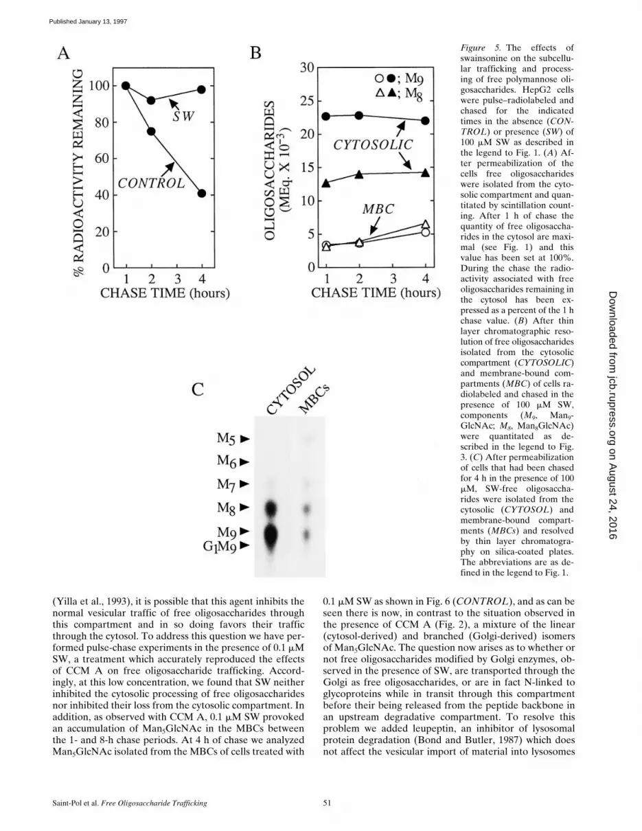
from the cytosol into an MBC, then an inhibitor of the cy-tosolic mannosidase would be expected to slow down thetransfer of the larger free polymannose-type oligosaccha-rides from the cytosol into an MBC of HepG2 cells. Athigh concentrations swainsonine (SW), a nonspecific man-nosidase inhibitor (Elbein et al., 1981), inhibits the cyto-solic a-mannosidase (Tulsiani and Touster, 1987) in addi-tion to Golgi mannosidase II and lysosomal mannosidases.Accordingly, HepG2 cells were pulse radiolabeled andchased in the presence of 100 mM SW, and, subsequent topermeabilization with SLO, free oligosaccharides wereprepared from both the MBCs and the cytosol as de-scribed for Fig. 1. Fig. 5 A demonstrates that after 4 h ofchase SW causes an inhibition of the loss of radioactivityassociated with free oligosaccharide material from the cy-tosolic compartment of HepG2 cells. Furthermore thinlayer chromatography of the oligosaccharides recoveredfrom the cytosolic compartment of HepG2 cells chased for4 h in the presence of 100 mM SW revealed thatMan9GlcNAc and Man8GlcNAc oligosaccharides are sta-bilized in the cytosol for up to 4 h and that there is littletransfer of these components into the MBC. Although Fig.5 B shows that free oligosaccharides bearing 9 and 8 resi-dues of mannose are stabilized in the cytosolic compart-ment of SW-treated cells, we observed (Fig. 5 C) a smallaccumulation of Man5GlcNAc in the MBCs derived fromthese cells. As described above in addition to inhibitingthe cytosolic mannosidase SW is also known to inhibitGolgi mannosidase II which leads to the production of gly-coproteins bearing hybrid-type oligosaccharide chains (El-bein et al., 1981). Hybrid-type oligosaccharide chains pos-sess a core structure which contains the branchedMan5GlcNAc moiety (Fig. 2), now, if such glycoproteins,or free oligosaccharides, are transported from the Golgicomplex to lysosomes, whose mannosidase activity hasbeen compromised by SW, we would then expect to see anaccumulation of a free oligosaccharide corresponding tothe branched isomer of Man5GlcNAc in the MBCs of SW-treated cells. Results (not shown) demonstrated that 100mM SW provokes the appearance of only the branchedisomer of Man5GlcNAc in the MBCs of HepG2 cells.Thus, as expected from its ability to trap large free poly-mannose type oligosaccharides in the cytosol, 100 mMSW abolished the appearance of linear Man5GlcNAc inthe MBCs. In summary, results show that incubation ofHepG2 cells with 100 mM SW arrests the cytosolic trim-ming of free polymannose type oligosaccharides and slowsdown their egress from this compartment.
The Golgi Apparatus Does Not Play a Major Role in the Trafficking of Free Oligosaccharides in HepG2 Cells
Taken together the results presented so far provide evi-dence to show that most free oligosaccharides follow atrafficking pathway from the ER into the cytosol and theninto an MBC possessing a mannosidase with a low pH op-timum. However, the appearance of the branched isomerof Man5GlcNAc in the MBCs of SW-treated cells sug-gested that there may also exist a vesicular traffickingpathway for free oligosaccharides involving the Golgi com-plex. Furthermore, as CCM A is known to affect themovement of glycoproteins through the Golgi complex
Figure 4. Different oligosaccharides appear in the membrane-bound compartment at different rates. Using the same data thatwas used for Fig. 3 the total cellular molar equivalents of the re-solved oligosaccharides were displayed as shown at the 1, 2 and 4 hchase times (top), whereas the percent of each indicated compo-nent found to be present in the MBCs is plotted directly below.The abbreviations are; M9, Man9GlcNAc; M8, Man8GlcNAc; M7,Man7GlcNAc; M6, Man6GlcNAc; M5, Man5GlcNAc.
on August 24, 2016
jcb.rupress.orgD
ownloaded from
Published January 13, 1997
Saint-Pol et al. Free Oligosaccharide Trafficking 51
(Yilla et al., 1993), it is possible that this agent inhibits thenormal vesicular traffic of free oligosaccharides throughthis compartment and in so doing favors their trafficthrough the cytosol. To address this question we have per-formed pulse-chase experiments in the presence of 0.1 mMSW, a treatment which accurately reproduced the effectsof CCM A on free oligosaccharide trafficking. Accord-ingly, at this low concentration, we found that SW neitherinhibited the cytosolic processing of free oligosaccharidesnor inhibited their loss from the cytosolic compartment. Inaddition, as observed with CCM A, 0.1 mM SW provokedan accumulation of Man5GlcNAc in the MBCs betweenthe 1- and 8-h chase periods. At 4 h of chase we analyzedMan5GlcNAc isolated from the MBCs of cells treated with
0.1 mM SW as shown in Fig. 6 (CONTROL), and as can beseen there is now, in contrast to the situation observed inthe presence of CCM A (Fig. 2), a mixture of the linear(cytosol-derived) and branched (Golgi-derived) isomersof Man5GlcNAc. The question now arises as to whether ornot free oligosaccharides modified by Golgi enzymes, ob-served in the presence of SW, are transported through theGolgi as free oligosaccharides, or are in fact N-linked toglycoproteins while in transit through this compartmentbefore their being released from the peptide backbone inan upstream degradative compartment. To resolve thisproblem we added leupeptin, an inhibitor of lysosomalprotein degradation (Bond and Butler, 1987) which doesnot affect the vesicular import of material into lysosomes
Figure 5. The effects ofswainsonine on the subcellu-lar trafficking and process-ing of free polymannose oli-gosaccharides. HepG2 cellswere pulse–radiolabeled andchased for the indicatedtimes in the absence (CON-TROL) or presence (SW) of100 mM SW as described inthe legend to Fig. 1. (A) Af-ter permeabilization of thecells free oligosaccharideswere isolated from the cyto-solic compartment and quan-titated by scintillation count-ing. After 1 h of chase thequantity of free oligosaccha-rides in the cytosol are maxi-mal (see Fig. 1) and thisvalue has been set at 100%.During the chase the radio-activity associated with freeoligosaccharides remaining inthe cytosol has been ex-pressed as a percent of the 1 hchase value. (B) After thinlayer chromatographic reso-lution of free oligosaccharidesisolated from the cytosoliccompartment (CYTOSOLIC)and membrane-bound com-partments (MBC) of cells ra-diolabeled and chased in thepresence of 100 mM SW,components (M9, Man9-GlcNAc; M8, Man8GlcNAc)were quantitated as de-scribed in the legend to Fig.3. (C) After permeabilizationof cells that had been chasedfor 4 h in the presence of 100mM, SW-free oligosaccha-rides were isolated from thecytosolic (CYTOSOL) andmembrane-bound compart-ments (MBCs) and resolvedby thin layer chromatogra-phy on silica-coated plates.The abbreviations are as de-fined in the legend to Fig. 1.
on August 24, 2016
jcb.rupress.orgD
ownloaded from
Published January 13, 1997

The Journal of Cell Biology, Volume 136, 1997 52
(Rohrer et al., 1995), to the SW-containing chase medium.As shown in Fig. 6 the quantity of the branched isomer ofMan5GlcNAc is reduced in a dose-dependent mannerwithout any affect being observed on the recovery of itslinear counterpart. Therefore by using 0.1 mM SW to pro-voke an accumulation of Man5GlcNAc in the MBCs ofHepG2 cells, we demonstrate that the two isomers of thisfree oligosaccharide have different origins. Results indi-cate that the bulk of free Man5GlcNAc produced by Golgienzyme modifications (branched isomer) observed in theMBCs of SW-treated cells arises from its passage throughthe Golgi apparatus while N-linked to glycoprotein andnot as a free oligosaccharide. Thus, although we cannotrule out the possibility that small amounts of free oligosac-charides are transported from the ER to the Golgi appara-tus our results demonstrate that if this pathway does existit represents only a minor trafficking route for free oli-gosaccharides in HepG2 cells.
Inhibitors of Macroautophagy Do Not Affect the Clearance of Free Oligosaccharides from the Cytosol of HepG2 Cells
Having demonstrated that the bulk of free oligosaccha-rides are transferred from the cytosol into a degradativeorganelle, we went on to investigate the mechanism of thistranslocation process. Theoretically macroautophagy could
be responsible for the clearance of oligosaccharides fromthe cytosol. To test this possibility we have performedchase incubations in the presence of well known inhibitorsof macroautophagy. Pulse–chase experiments were per-formed as described in Fig. 1 except that the inhibitorswere added to the chase incubations 45 min after the onsetof the chase period. 3-MA inhibits the sequestration stepof the autophagic process (Seglen and Gordon, 1982),therefore if autophagy were responsible for the clearanceof free oligosaccharides from the cytosol, we would expectthis reagent to cause an accumulation of fully trimmedfree oligosaccharides in this compartment. Fig. 7 revealsthis not to be the case and in fact after 8 h of chase, 3-MAhad no effect on the clearance of free oligosaccharidesfrom the cytosolic compartment of HepG2 cells. However,we did note that 3-MA caused an accumulation of free oli-gosaccharides in the MBCs, although this accumulationwas not as large as that observed in the presence of CCMA, it is known that 3-MA interferes with the acidificationof lysosomes (Caro et al., 1988). Asparagine has beenshown to block the fusion of autophagosomes with the ly-sosomal compartment (Hoyvik et al., 1991) and would,if autophagy were responsible for the clearance of cyto-solic oligosaccharides, lead to the accumulation of fullytrimmed free oligosaccharides within MBCs. Again Fig. 7demonstrates that this was not the case and shows that thechase incubation performed in the presence of asparagineis not significantly different from the control chase incuba-tion.
Figure 6. Leupeptin inhibits the formation of the branched iso-mer of Man5GlcNAc in swainsonine-treated HepG2 cells. HepG2cells were pulse–radiolabeled and chased for 4 h in the presenceof 0.1 mM SW (CONTROL). Where indicated the chase mediawere supplemented with leupeptin (LEU) at either 100 or 300mg/ml. After permeabilization of the cells and isolation of freeoligosaccharides from the MBCs the resulting radioactive compo-nents were resolved by HPAEC as described in Materials andMethods. The region of the chromatograph corresponding to theelution times of the linear (LINEAR) and branched (BRANCHED)isomers of Man5GlcNAc is shown.
Figure 7. Inhibitors of macroautophagy do not affect the clear-ance of free oligosaccharides from the cytosol of HepG2 cells.Pulse–chase experiments were performed exactly as described forFig. 1. Control or CCM A–treated cells were pulse radiolabeledand chased for 8 h in the presence of CCM A, 3-methyladenine(3-MA), or asparagine (Asn). After permeabilization of the cellsas described in Materials and Methods, radioactivity associatedwith free oligosaccharides isolated from the cytosolic (CYTO-SOL) and membrane-bound compartments (MBCs) was assayedby scintillation counting.
on August 24, 2016
jcb.rupress.orgD
ownloaded from
Published January 13, 1997

Saint-Pol et al. Free Oligosaccharide Trafficking 53
The Transfer of Free Oligosaccharides from the Cytosol into an MBC Requires Energy
To further investigate the nature of the cytosol-to-MBCtranslocation of free oligosaccharides in HepG2 cells, wehave investigated the energy dependence of this process.For this purpose we have performed chase incubations inthe presence of reagents known to deplete ATP levels incultured cells (Krijnse-Locker et al., 1994). Fig. 8 A dem-onstrates that when HepG2 cells are chased in the pres-ence of deoxyglucose and sodium azide the total molarequivalents of free oligosaccharides within the cytosol re-mains approximately constant when compared to that ob-served during control chase periods. Thin layer chromato-graphic examination of the cytosol- and MBC-derivedoligosaccharides generated in cells treated with inhibitorsof mitochondrial respiration for 8 h is shown in Fig. 8 Band shows that although cytosolic demannosylation of freeoligosaccharides has been completed in drug-treated cells,the transfer of the cytosolic terminal digest product into anMBC is severely impaired leading to a marked cytosolicaccumulation of Man5GlcNAc. We observed essentiallyidentical results when we performed chase incubations inthe presence of 10 mg/ml oligomycin, a specific inhibitor ofthe mitochondrial H1/ATPase (Ziegler and Penefsky, 1993)(results not shown).
Subcellular Fractionation Reveals That the Pseudolinear Isomer of Man5GlcNAc Is Degradedin Lysosomes
We have examined the identity of the MBC in which cyto-solically generated free oligosaccharides are degraded bysubcellular fractionation. As in the presence of CCM A,the linear isomer of Man5GlcNAc is stabilized in an MBCof permeabilized HepG2 cells we reasoned that in the ab-sence of this drug free oligosaccharides are degradedwithin a subcellular compartment possessing a vacuolarH1/ATPase and an a-mannosidase with an acidic pH opti-mum. Although the lysosome best fits these criteria endo-somes also possess a vacuolar H1/ATPase (Mellman et al.,1986), and in addition they are known to contain acidic hy-drolases (Authier et al., 1994). To determine the nature ofthe MBC in which cytosolic-free oligosaccharides are de-graded, we have fractionated HepG2 cell homogenates ona Percoll gradient known to be able to resolve endosomesfrom lysosomes (Rijnboutt et al., 1992). Initially we choseto fractionate HepG2 cells after treatment with CCM A,however results presented in Fig. 9 A demonstrate thatwhen compared to subcellular fractionation of untreatedcells this reagent caused a marked change in the distribu-tion of the lysosomal marker enzyme, b-hexosaminidase,along the Percoll gradient while not disturbing the dis-tributions of either the Golgi or endoplasmic reticulummarker enzymes. When CCM A–treated cells were pulseradiolabeled and then fractionated on a Percoll gradientwe observed that free oligosaccharides (OS-GN2, notshown) are mainly localized to a region of the gradientcontaining endoplasmic reticulum and Golgi marker en-zymes (Fig. 9 B). After 4 h of chase in the presence ofCCM A, free oligosaccharides are now distributed through-out the gradient in similar but not identical fashion to thatobserved for the lysosomal enzyme b-hexosaminidase.
Figure 8. The effects of cellular ATP depleting agents on theclearance of free oligosaccharides from the cytosol of HepG2cells. HepG2 cells were pulse–radiolabeled and chased for the in-dicated times as described in the legend to Fig. 1. Where indi-cated a mixture of deoxyglucose and sodium azide (DOG/AZIDE)was added to the chase medium 45 min after the onset of thechase period (to give a final concentration of 10 mM for eachdrug). (A) After permeabilization of the cells free oligosaccha-rides were prepared from the cytosolic fraction and the molarequivalents of these components were calculated as described inthe legend of Fig. 3. As the quantity of free oligosaccharidesreaches maximal levels after 1 h of chase this value was set at100% for both the control and drug-treated cells. (B) Free oli-gosaccharides isolated from the cytosolic compartment (CYTO-SOL) and membrane-bound compartments (MBCs) derivedfrom cells chased for 8 h in the presence and absence of DOG/AZIDE were separated by thin layer chromatography on silica-coated plates as described in the legend to Fig. 1. The abbrevia-tions are as defined in the legend to Fig. 1. The oligosaccharidemigrating as Man6GlcNAc is known to comprise a mixture ofGlc1Man5GlcNAc and Man6GlcNAc (Moore and Spiro, 1994); theformer is the likely degradation product of Glc1Man9GlcNAc bythe cytosolic mannosidase.
on August 24, 2016
jcb.rupress.orgD
ownloaded from
Published January 13, 1997

The Journal of Cell Biology, Volume 136, 1997 54
Due to difficulties in interpreting these results we haveused 0.1 mM SW to provoke an accumulation of free oli-gosaccharides in the MBC of HepG2 cells before subcellu-lar fractionation. As described above we have found that,at a concentration of 0.1 mM, SW mimics the effects ofCCM A on the subcellular trafficking of free oligosaccha-rides in HepG2 cells. Treatment of HepG2 cells with 0.1mM SW for 4 h did not dramatically affect the distributionsof the lysosomal, Golgi, or endoplasmic reticulum markerenzymes along the Percoll gradient (compare the toppanel of Fig. 10 with control fractionations in Fig. 9 A). Toidentify endosomal and lysosomal compartments, HepG2cells, treated for 4 h with 0.1 mM SW, were allowed to en-docytose a brief pulse of the fluid phase marker, HRP, andthen chased for various times (van Weert et al., 1995). Cel-lular homogenates were then fractionated on Percoll den-sity gradients as shown in Fig. 10 (bottom). Results showthat, after the pulse, HRP is found uniquely in a region ofthe gradient (see top, fraction II), in which are found lightmembranes, including the endoplasmic reticulum andGolgi apparatus. After 4 h of chase the HRP is now mainlyfound at the bottom of the gradient, a region shown tocontain the lysosomes (see top, fraction IV). These resultsare consistent with the transfer of the fluid phase markerfrom the endosomal compartment to the lysosomal com-partment as previously described in HepG2 cells (van Weertet al., 1995). Cells treated with 0.1 mM SW were then pulseradiolabeled, chased for 1 and 4 h, homogenized, and thensubjected to subcellular fractionation on Percoll densitygradients. Fig. 11 A shows that after the pulse free oli-gosaccharides (OS-GN2, results not shown) are localizedto a region of the gradient corresponding to the endoplas-
mic reticulum/Golgi region of the gradient, after 4 h ofchase free oligosaccharides (mainly Man7-5GlcNAc, resultsnot shown) now colocalize with the lysosomal marker en-zyme b-hexosaminidase. Although only a small amount offree oligosaccharides were recovered from subcellular or-ganelles after 1 h of chase, these components were distrib-uted equally between regions of the gradient containingthe lysosomal marker (fraction IV) and the endoplasmicreticulum/Golgi markers (fraction II). At this early chasetime the gradient fraction II contained free oligosaccha-rides bearing 9 and 8 residues of mannose whereas thoseoccurring in the gradient fraction IV comprised oligosac-charides bearing 7-5 residues of mannose (results notshown). After thin layer chromatography of free oligosac-charides recovered from the Percoll density gradients wasperformed, the Man5GlcNAc was recovered and quanti-tated as shown in the lower part of Fig. 11 A. We were un-able to detect this oligosaccharide in the subcellular or-ganelles of pulse-radiolabeled cells, but after 1 h of chase,small amounts of this component could be detected alongthe density gradient but were largely found in the lysoso-mal region of the gradient. After 4 h of chase the distribu-tion of Man5GlcNAc along the Percoll gradient was indis-tinguishable from that of total free oligosaccharides andindicated that this component is uniquely localized in thelysosomal region of the gradient. HPAEC of theMan5GlcNAc recovered from the lysosomal region of thePercoll gradients (fraction IV) revealed that after 1 h ofchase this fraction contained the linear isomer ofMan5GlcNAc, indicative of its having been generated bythe cytosolic mannosidase. After 4 h of chase both the lin-ear and branched isomers of Man5GlcNAc could be de-
Figure 9. CCM A affects thedistribution of a lysosomalmarker enzyme on Percolldensity gradients. (A) Con-trol cells (open circles) orcells treated with CCM A for4 h were homogenized andfractionated on Percoll den-sity gradients as described inMaterials and Methods. Thesedimentation position of ly-sosomes, the Golgi appara-tus, and ER were identifiedby performing assays forb-d-hexosaminidase (b-HEX), b-1, 4-galactosyl-transferase (GAL T’ASE),and NADPH cytochromeC reductase (NADPHRED’ASE), respectively. (B)CCM A–treated cells werepulse radiolabeled (PULSE)and chased (CHASE) in thepresence of CCM A for 4 hbefore subcellular fraction-ation as described above. Af-ter collecting the gradient infour fractions (I-IV) asshown in the upper panel of
A, organelles were separated from Percoll and cytosol by centrifugation as described in Materials and Methods. Free oligosaccharideswere isolated from the resulting organelles and assayed by scintillation counting.
on August 24, 2016
jcb.rupress.orgD
ownloaded from
Published January 13, 1997

Saint-Pol et al. Free Oligosaccharide Trafficking 55
tected in the lysosomal region of the Percoll gradient.Therefore we show that after 4 h of chase in either thepresence of CCM A or 0.1 mM SW free oligosaccharidesare closely associated with a lysosomal marker enzyme af-ter subcellular fractionation on Percoll density gradients.
DiscussionPermeabilization of the plasma membrane of HepG2 cellssubsequent to pulse-chase incubations has enabled us toevaluate the hypothesis that cytosolic-free oligosaccha-rides are sequestered into and degraded by lysosomes.Short chase incubations revealed that free oligosaccha-rides rapidly appear in the cytosol at a time during whichthere is a loss of these components from the MBCs (Fig.1, A and B). This observation can be accounted for by thepreviously observed rapid translocation of large, free poly-mannose-type oligosaccharides out of the ER into the cy-tosol (Moore et al., 1995). At present it is unclear whetherthis ER-to-cytosol transport of free oligosaccharides is thesole mechanism responsible for the appearance of free oli-gosaccharides in the cytosol. Recently, it has been pro-posed that newly synthesized glycoproteins may be trans-located out of the ER and degraded in this compartment
(Wiertz et al., 1996) by the actions of a cytosolic N-glyca-nase (Kitajima et al., 1995; Suzuki et al., 1994) and the pro-teasome (Ciechanover, 1994; Driscoll and Goldberg, 1990).Clearly such a phenomenon would also give rise to large,free polymannose-type oligosaccharides in the cytosol. What-ever the origin of cytosolic oligosaccharides in HepG2cells radioactivity associated with these components reachmaximal levels at z1 h of chase and declines thereafter(Fig. 1, A and B). We have tested the hypothesis that cyto-solic-free oligosaccharides are translocated into lysosomesto be degraded. To do this we have performed pulse–chaseexperiments in the presence of a vacuolar H1/ATPase in-hibitor. Concanamycins (Woo et al., 1992) are antibioticswhich are closely related to bafilomycin A (Bowman et al.,1988), an agent which has been extensively used to investi-gate the acidification of intracellular organelles (Yoshi-mori et al., 1991; Yilla et al., 1993; Clague et al., 1994). InHepG2 cells the use of bafilomycin has demonstrated thatvacuolar acidification is required for the transfer of fluidphase markers from endosomes to lysosomes (van Weertet al., 1995) and concanamycin B has been shown to per-turb the trafficking and processing of glycoproteins in lateGolgi compartments. Presumably then, vacuolar ATPaseinhibitors block lysosomal degradation by inhibiting vesic-ular transport of certain substrates to the lysosome, andby increasing intralysosomal pH, thereby reducing acidichydrolase activity. Accordingly, we reasoned that thisreagent should allow us to examine the fate of cytosolicoligosaccharides without interference by those oligosac-charides generated during glycoprotein catabolism inthe lysosome. We have shown that CCM A is able to sub-stantially inhibit the SW-provoked appearance of thebranched isomer of Man5GlcNAc in the MBC of HepG2cells suggesting that glycoconjugates that have traversedthe Golgi apparatus and which are destined for the lyso-some are stabilized in the presence of this agent (data notshown). Here we show that despite its ability to block lyso-somal glycoprotein degradation CCM A provoked an ac-cumulation of free oligosaccharide in an MBC of HepG2cells. This accumulation coincided precisely with the lossof free oligosaccharide from the cytosolic compartment.In addition we were able to show that the structures ofthe oligosaccharides that accumulated in an MBC of CCMA–treated cells were characteristic of their having beengenerated by the cytosolic mannosidase. These resultsclearly demonstrate the transfer of free oligosaccharidesfrom the cytosol into an MBC. We went on to show thattreating HepG2 cells with 100 mM SW blocked the trim-ming of cytosolic oligosaccharides by the cytosolic a-man-nosidase and inhibited their loss from this compartment.As in the presence of 100 mM, SW oligosaccharides bear-ing 9 and 8 residues of mannose are stabilized with only aminor quantity of Man7GlcNAc being detected in the cy-tosolic compartment we conclude that most, if not all, cy-tosolic-free oligosaccharides must be derived from a pre-Golgi compartment (SW does not inhibit either the Golgimannosidase I (Elbein et al., 1981), Fig. 5 C, or ER man-nosidase I (Weng and Spiro, 1996). Our results show thatMan7-5GlcNAc oligosaccharides can be cleared from thecytosol although our results strongly suggest that thesmaller the oligosaccharide the more efficient its clearancefrom the cytosol. The small amounts of Man7GlcNAc that
Figure 10. Subcellular fractionation of SW-treated HepG2 cellson Percoll density gradients. (Top) Cells treated for 4 h with 0.1 mMSW were homogenized and fractionated on Percoll density gradi-ents as described in the legend to Fig. 9. b-d-hexosaminidase(closed circles), b-1, 4-galactosyltransferase (triangles) and NADPHcytochrome C reductase (open circles) were assayed to identifythe sedimentation positions of lysosomes, the Golgi apparatusand the ER, respectively. (Bottom) Cells were treated for 1 hwith 0.1 mM SW, pulse labeled with HRP (open symbols), andthen chased for 4 h (solid symbols) as described in Materials andMethods. HRP-labeled cells were homogenized and a postnu-clear supernatant was fractionated on Percoll gradients. Materialfrom the gradient was recovered in 20 fractions each of which wasassayed for HRP activity.
on August 24, 2016
jcb.rupress.orgD
ownloaded from
Published January 13, 1997

The Journal of Cell Biology, Volume 136, 1997 56
are transferred into the MBC are slowly trimmed duringthe chase suggesting that CCM A induced neutralizationof degradative organelles is not complete (Fig. 1 B). Al-though lysosomal enzymes are known not to generate lin-ear isomers of polymannose-type oligosaccharides (Michal-ski et al., 1990; Al Daher et al., 1991), it is not clearwhat isomer of Man5GlcNAc would be generated fromMan7GlcNAc in the CCM A–sensitive MBC, but it isinteresting to note that analysis of the MBC-derivedMan5GlcNAc generated during CCM A chases always ledto the detection of small amounts of an oligosaccharidethat lost one mannose residue upon digestion with the a-1,2 mannosidase (Fig. 2). In conclusion evidence demon-strates that cytosolic oligosaccharides are partially trimmedin the cytosol and transferred into a membrane boundcompartment. Concerning the relationship between cyto-solic trimming and clearance of free oligosaccharides fromthe cytosol two observations have to be discussed. First,Figs. 1 and 4 indicate that there is an apparent “hold up”of the trimming of cytosolic oligosaccharides at theMan7GlcNAc stage. Second, inspection of the rate of freeoligosaccharide clearance from the cytosol indicates thatthis process is somewhat slower than the rate of appear-ance of these components in the cytosol (which is appar-ently complete after 1 h of chase; Fig. 3), a fact that wouldlead to a steady accumulation of oligosaccharide materialin this compartment. These two observations may be re-lated. We have preliminary data showing that the 5 mMmannose required to be added to the chase incubation me-
dia may both slow down the trimming of cytosolic-free oli-gosaccharides and slow down their clearance from thiscompartment (data not shown). Thus under physiologicalconditions it may be that cytosolic trimming is more efficientallowing a more rapid generation of the Man5GlcNAc,which may be cleared from the cytosol more efficientlythan its more highly mannosylated counterparts.
After Trimming in the Cytosol-freeOligosaccharides Are Translocated into Lysosomes Where They Are Degraded
Subcellular fractionation of HepG2 cells chased in thepresence of 0.1 mM SW for 1 and 4 h indicated that the dis-tribution of free oligosaccharides along Percoll gradientswas the same as that observed for the lysosomal markerenzyme b-hexosaminidase. Because we were interested inthe final destination of cytosolic-free oligosaccharides, wehave not directly addressed the question of whether freeoligosaccharides are in fact transported from the cytosoldirectly into the lysosome. It is possible that the cytosol-to-MBC oligosaccharide translocation machinery is locatedon a prelysosomal compartment which ultimately fuseswith lysosomes. In this respect our results (Figs. 1–4) withthe vacuolar ATPase inhibitor, CCM A, do not necessarilyimply that the MBC into which free oligosaccharides aretransported contains a pH-sensitive a-mannosidase. It ispossible that free oligosaccharides may be stabilized inCCM A–treated HepG2 cells because the MBC into which
Figure 11. Characterizationof free oligosaccharides iso-lated from organelles cosedi-menting with lysosomalmarker enzymes on Percollgradients. (A) HepG2 cellspretreated with 0.1 mM SWfor 1 h, were pulse radiola-beled with d-[2-3H]mannoseand chased for 1 and 4 h inthe presence of SW. Afterhomogenization of the cellsand fractionation of the post-nuclear supernatants on Per-coll gradients organelleswere recovered from frac-tions I-IV (see Fig. 10). Freeoligosaccharides (top) wererecovered from the or-ganelles of each fraction andassayed by scintillationcounting. The results havebeen expressed as a percent-age of the total free oligosac-charides recovered from theorganelles. The quantities ofradioactivity (cpm 3 1023)
associated with free oligosaccharides isolated from the organelles were 74.7, 27.9, and 197.3, for the pulse, 1 and 4 h chase times, respec-tively. (Bottom) After thin layer chromatography of the oligosaccharides from each subcellular fraction the oligosaccharide migratingas Man5GlcNAc was quantitated by scintillation counting and recovery of this component as a percentage of the total Man5GlcNAc re-covered from the subcellular fractions I–IV is represented for the pulse and the two chase times. The asterisk indicates that this com-ponent was not detected in the pulse incubation. (B) The Man5GlcNAc isolated from fraction IV of the Percoll density gradients as de-scribed above was analysed by HPAEC as described in Materials and Methods. The region of the chromatograph corresponding to theelution times of the linear (LINEAR) and branched (BRANCHED) isomers of Man5GlcNAc is shown.
on August 24, 2016
jcb.rupress.orgD
ownloaded from
Published January 13, 1997

Saint-Pol et al. Free Oligosaccharide Trafficking 57
they have been translocated cannot fuse with lysosomes whosemembrane pH gradient has been perturbed. Indeed sub-cellular fractionation studies performed on CCM A–treatedcells indicate that substantial amounts of free oligosaccha-ride are found in the endosomal region of the Percoll gra-dient suggesting that free oligosaccharides may not betransported directly into lysosomes. However, some of ourobservations suggest that free oligosaccharides may betransported directly into lysosomes. First, in the presenceof CCM A, we were able to detect substantial amounts offree oligosaccharides bearing 3 and 4 residues of mannosein the vesicular compartment, suggesting that once trans-located into this compartment, these components comeinto contact with and are slowly acted upon by an a-man-nosidase with a low pH optimum. However, as endosomesmay contain an acidic a-mannosidase (Authier et al., 1994),this result still leaves the possibility that free oligosaccha-rides are transported into endosomes and are then rapidlytransferred to lysosomes by vesicular fusion. Although wecannot rule out this possibility, we noted that even whencells were chased in the presence of 0.1 mM SW for only 1 h,the majority of Man5GlcNAc recovered from the densitygradient occurred in the lysosomal fraction and not in theendosomal fraction. The fact that substantial quantities ofthe endosomal marker HRP remain associated with endo-somes 4 h after an HRP pulse suggests that fluid transferbetween endosomes and lysosomes would not be rapidenough to account for the absence of free oligosaccharidesin the endosomal compartment, if indeed these componentshad been translocated from the cytosol into endosomes,and then onto lysosomes by vesicular transport.
How Are Free Oligosaccharides Cleared fromthe Cytosol?
Examining the kinetics of loss of free oligosaccharidesfrom cytosol yields useful information concerning themechanism of this translocation process. First, as the cyto-plasm of cells is continually sequestered by vesicles anddelivered to lysosomes by macroautophagy, we wonderedwhether or not this bulk sequestration of the cytosol couldaccount for the delivery of free cytosolic oligosaccharidesto the lysosome. It has been shown that by starving hepa-tocytes of serum, autophagic sequestration can be stimu-lated 10-fold and under these conditions only 4% of thecytoplasm can be sequestered per hour (Kopitz et al.,1990). Here, despite the fact that HepG2 cells are chasedin complete growth medium, a condition known to inhibitautophagy, we noted that oligosaccharides are clearedfrom the cytosol with a half-life of z3–4 h. Thus, it is ap-parent that autophagic sequestration cannot account forthe transfer of free oligosaccharides into a vesicular com-partment of HepG2 cells. In accordance with this wefound that 3-MA, a well known inhibitor of autophagic se-questration (Seglen and Gordon, 1982), was without effecton the loss of oligosaccharide material from the cytoplasm.Furthermore as autophagic sequestration is a nonselectiveprocess, it should theoretically transfer all cytosolic oli-gosaccharides irrespective of structure into lysosomes. Ourresults with CCM A (Fig. 4) and 100 mM SW (Fig. 5) sug-gest that trimming of polymannose oligosaccharides to atleast Man7GlcNAc is required before they are efficiently
transferred into lysosomes, indicating that free oligosac-charides are not being sequestered into the lysosomal deg-radative compartment by bulk uptake of the cytosol. Wedemonstrate that the cytosol-to-lysosome translocation offree oligosaccharides is strongly impaired if the cells arechased under conditions known to deplete cellular ATPlevels. This result is not surprising as in the presence ofCCM A or 0.1 mM SW the cytosol-to-lysosome transfer offree oligosaccharides must occur against a substantial con-centration gradient. It remains to be determined whetherthis transport process is accomplished by a transportermolecule or by an as yet unidentified mechanism such as atype of receptor mediated microautophagy. In conclusionour results suggest that free oligosaccharides are seques-tred into lysosomes by an energy requiring process thatdisplays oligosaccharide specificity.
What Are the Consequences of the Sequestration of Cytosolic-free Oligosaccharides in Lysosomes?
The results we have obtained with HepG2 cells throw lighton a problem that has perplexed researchers investigatingthe genetic disorder, a-mannosidosis (see Discussion inTulsiani and Touster, 1987; and conclusion in Daniel et al.,1992). Mannosidosis patients have a genetic deficiency inlysosomal a-mannosidase (Carroll et al., 1972) which leadsto severe clinical symptoms. At a biochemical level thelesion is characterized by the presence of large quantitiesof free oligosaccharides in the tissues and urine from af-fected individuals. Surprisingly, however, a substantialproportion of the oligosaccharides isolated from the urineof mannosidosis patients possess structures not compat-able with their having been formed by the incomplete deg-radation of complex oligosaccharides derived from lysoso-mal glycoprotein degradation. In fact the structures ofthese oligosaccharides were found to be linear in nature(Nordén et al., 1974; Strecker et al., 1976; Daniel et al.,1992), the largest of which being identical to the linearMan5GlcNAc shown in Fig. 2. The trafficking of free poly-mannose oligosaccharides from ER to cytosol and, afterprocessing, into the lysosome may now explain the accu-mulation of the linear oligosaccharides observed in sub-jects deficient in lysosomal a-mannosidase, however, howthese components gain access to the extracellular fluidsstill remains to be elucidated. In fact we have calculatedthat in CCM A–treated HepG2 cells 15% of all oligosac-charide structures (including N-linked polymannose-,complex-, and hybrid-type structures, and free oligosac-charides) occur as free polymannose species indicatingthat this recently outlined free oligosaccharide traffickingpathway must process large quantities of material in cellsactively engaged in glycoprotein synthesis (data not shown).This observation is in line with the fact that the urine ofmannosidosis patients may contain up to 250 mg/liter ofsmall linear oligosaccharides (Strecker et al., 1976).
An intriguing question can now to be addressed: Whyhas the cell developed such an elaborate trafficking path-way for the degradation of free oligosaccharides generatedin the lumen of the ER when two vesicular pathways al-ready exist between the ER and lysosome? One explana-tion for this is that free oligosaccharides must be rapidlysegregated from their N-linked counterparts in the ER in
on August 24, 2016
jcb.rupress.orgD
ownloaded from
Published January 13, 1997

The Journal of Cell Biology, Volume 136, 1997 58
order to minimize their interference with the folding andtrafficking of glycoproteins, a process now thought to in-volve lectins situated along the secretory pathway (Fiedlerand Simmons, 1995). Alternatively free oligosaccharidesmaybe delivered to the cytosol for a purpose other than tobe trimmed in order for their ultimate degradation in thelysosome. Interestingly, the cytosol contains an actin-bind-ing protein, comitin (Weiner et al., 1993), which has re-cently been shown to be also a mannose-binding lectin(Jung et al., 1996). It was proposed that while the lectinmoiety of this protein could bind to cytosolically disposedmannose-containing oligosaccharide lipids of the Golgi/ER its actin-binding domain could tether it to microfila-ments (Jung et al., 1996), thereby forming a bridge be-tween the cytoskeleton and organelles of the secretorypathway. Could cytosolic-free oligosaccharides competefor binding sites on comitin thereby modulating this pro-cess?
In conclusion our results show that after their rapidappearance in the cytosol during glycoprotein biosynthesisfree oligosaccharides are trimmed by the cytosolic man-nosidase and transferred into lysosomes by an energy-dependent mechanism. Our results show that the lysosomeis the site for the final degradation of these oligosaccha-rides and suggest that the lysosomal membrane is itself in-volved in the uptake of free polymannose-type oligosac-charides from the cytosol.
We thank Dr. J.R. Green (Ciba-Geigy Ltd.) for the gift of concanamycinA, Dr. J.-C. Michalski for supplying the two standard Man5GlcNAc oli-gosaccharides, and Dr. C. Rabouille for critical reading of the manuscript.HPAEC was performed by Dr. Thierry Fontaine (Laboratoire des As-pergillus, Institut Pasteur, Paris, Dir. Dr. J.-P. Latgé).
This work was supported by institutional funding from the Institut Na-tional de la Santé et de la Recherche Médicale (INSERM) and by grantsfrom the Association Vaincre les Maladies Lysosomales and a EuropeanCommunity Human Mobility and Research Training Fellowship (to S.E.H.Moore).
Received for publication 24 May 1996 and in revised form 16 October1996.
References
Al Daher, S., R. De Gasperi, P. Daniel, N. Hall, C.D. Warren, and B. Winches-ter. 1991. Substrate specificity of human lysosomal a-d-mannosidase in rela-tion to genetic a-mannosidosis. Biochem. J. 277:743–751.
Amano, J., and A. Kobata. 1986. Purification and characterization of a novela-mannosidase from Aspergillus saitoi. J. Biochem. 99:1645–1654.
Anumula, K.R., and R.G. Spiro. 1983. Release of glucose-containing polyman-nose oligosaccharides during glycoprotein biosynthesis. J. Biol. Chem. 258:15274–15282.
Authier, F., B.I. Posner, and J.J.M. Bergeron. 1994. Hepatic endosomes are amajor physiological locus of insulin and glucagon degradation in vivo. InCellular Proteolytic Systems. A.J. Ciechanover and A.L. Schwartz, editors.89–113.
Barker, R., K.W. Olsen, J.H. Shaper, and R.L. Hill. 1972. Agarose derivativesof uridine diphosphate and N-acetyglucosamine for the purification of a ga-lactosyltransferase J. Biol. Chem. 253:7135–7147.
Bischoff, J., L. Liscum, and R. Kornfeld. 1986. The use of 1-deoxymannojirimy-cin to evaluate the role of various a-mannosidases in oligosaccharide pro-cessing in intact cells. J. Biol. Chem. 261:4766–4774.
Bond, J.S., and P.E. Butler. 1987. Intracellular proteases. Annu. Rev. Biochem.56:333–364.
Bowman, E.J., A. Siebers, and K. Altendorf. 1988. Bafilomycins: a class of in-hibitors of membrane ATPases from microorganisms, animal cells, and plantcells. Proc. Natl. Acad. Sci. USA. 85:7972–7976.
Cacan, R., R. Cecchelli, and A. Verbert. 1987. Catabolic pathway of oligosac-charide-diphospho-dolichol. Eur. J. Biochem. 166:469–474.
Caro, L.H.P., P.J.A.M. Plomp, E.J. Wolvetang, C. Kerkhof, and A.J. Meijer.1988. 3-Methyladenine, an inhibitor of autophagy, has multiple effects onmetabolism. Eur. J. Biochem. 175:325–329.
Carroll, M., N. Dance, P.K. Masson, D. Robinson, and B.J. Winchester. 1972.Human mannosidosis—the enzyme defect. Biochem. Biophys. Res. Com-mun. 49:579–583.
Ciechanover, A. 1994. The ubiquitin-proteasome proteolytic pathway. Cell. 79:13–21.
Clague, M.J., S. Urbé, F. Aniento, and J. Gruenberg. 1994. Vacuolar ATPaseactivity is required for endosomal carrier vesicle formation. J. Biol. Chem.269:21–24.
Daniel, P.F., J.E. Evans, R. De Gaspari, B. Winchester, and C.D. Warren. 1992.A human lysosomal a(1-6)-mannosidase active on the branched trimannosylcore of complex glycans. Glycobiology. 2:327–336.
Driscoll, J., and A.L. Goldberg. 1990. The proteasome (multicatalytic protease)is a component of the 1500-kDa proteolytic complex which degrades ubiq-uitin-conjugated proteins. J. Biol. Chem. 265:4789–4792.
Elbein, A.D., R. Solf, P.R. Dorling, and K. Vosbeck. 1981. Swainsonine: an in-hibitor of glycoprotein processing. Proc. Natl. Acad. Sci. USA. 78:7393–7397.
Fiedler, K., and K. Simmons. 1995. The role of N-glycans in the secretory path-way. Cell. 81:309–312.
Haeuw, J.-F., G. Strecker, J.-M. Wieruszeski, J. Montreuil, and J.-C. Michalski.1991. Substrate specificity of rat liver cytosolic a-D-mannosidase. Eur. J.Biochem. 202:1257–1268.
Hoyvik, H., P.B. Gorden, T.O. Berg, P.E. Stromhaug, and P.O. Seglen. 1991.Inhibition of autophagic-lysosomal delivery and autophagic lactolysis by as-paragine. J. Cell Biol. 113:1305–1312.
Jung, E., P. Fucini, M. Stewart, A.A. Noegel, and M. Schleicher. 1996. Linkingmicrofilaments to intracellular membranes: the actin-binding and vesicle-associated protein comitin exhibits a mannose-specific lectin activity. EMBO(Eur. Mol. Biol. Organ.) J. 15:1238–1246.
Kataoka, T., M. Muroi, S. Ohkuma, T. Waritani, J. Magae, A. Takatsuki, S.Kondo, M. Yamasaki, and K. Nagai. 1995. Prodigiosin 25-C uncouples vacu-olar type H1-ATPase, inhibits vacuolar acidification and affects glycopro-tein processing. FEBS Lett. 359:53–59.
Kitajima, K., T. Suzuki, Z. Kouchi, S. Inoue, and Y. Inoue. 1995. Identificationand distribution of peptide: N-glycanase (PNGase) in mouse organs. Arch.Biochem. Biophys. 319:393–401.
Kmiécik, D., V. Herman, C.J.M. Stroop, J.-C. Michalski, A.M. Mir, O. Labiau,A. Verbert, and R. Cacan. 1995. Catabolism of glycan moieties of lipid inter-mediates leads to a single Man5GlcNAc oligosaccharide isomer: a study withpermeabilized CHO cells. Glycobiology. 5:483–494.
Kopitz, J., G.O. Kisen, P.B. Gorden, P.O. Bohley, and P.O. Seglen. 1990. Non-selective autophagy of cytosolic enzymes by isolated rat hepatocytes. J. CellBiol. 111:941–953.
Kornfeld, R., and S. Kornfeld. 1985. Assembly of asparagine-linked oligosac-charides. Annu. Rev. Biochem. 54:631–664.
Krijnse-Locker, J., M. Ericsson, P.J.M. Rottier, and G. Griffiths. 1994. Charac-terisation of the budding compartment of mouse hepatitis virus: evidencethat the transport from the RER to the Golgi complex requires only one ve-sicular transport step. J. Cell Biol. 124:55–70.
Michalski, J.-C., J.-F. Haeuw, J.-M. Wieruszeski, J. Montreuil, and G. Strecker.1990. In vitro hydrolysis of oligomannosyl oligosaccharides by the lysosomala-D-mannosidases. Eur. J. Biochem. 189:369–379.
Mellman, I., R. Fuchs, and A. Helenius. 1986. Acidification of the endocyticand exocytic pathways. Ann. Rev. Biochem. 55:663–700.
Moore, S.E.H., C. Bauvy, and P. Codogno. 1995. Endoplasmic reticulum-to-cyto-sol transport of free polymannose oligosaccharides in permeabilised HepG2cells. EMBO (Eur. Mol. Biol. Organ.) J. 14:6034–6042.
Moore, S.E.H., and R.G. Spiro. 1990. Demonstration that Golgi endo-a-D-mannosidase provides a glucosidase-independent pathway for the formationof complex N-linked oligosaccharides of glycoproteins. J. Biol. Chem. 265:13104–13112.
Moore, S.E.H., and R.G. Spiro. 1994. Intracellular compartmentalisation anddegradation of free polymannose oligosaccharides released during glycopro-tein biosynthesis. J. Biol. Chem. 269:12715–12721.
Nordén, N.E., A. Lundblad, S. Svensson, and S. Autio. 1974. Characterisationof two mannose-containing oligosaccharides isolated from the urine of pa-tients with mannosidosis. Biochemistry. 13:871–874.
Oku, H., and S. Hase. 1991. Studies on the substrate specificity of neutral a-man-nosidase purified from japanese quail oviduct by using sugar chains from gly-coproteins. J. Biochem. 110:982–989.
Opheim, D.J., and O. Touster. 1977. The purification and characterization ofrat liver lysosomal a-L-fucosidase. J. Biol. Chem. 252:739–743.
Pierce, R.J., G. Spik, and J. Montreuil. 1979. Demonstration and cytosolic local-isation of an endo-N-acetyl-b-D-glucosaminidase activity towards an asialo-N-acetyl-lactosaminic-type substrate in rat liver. Biochem. J. 180:673–676.
Rijnboutt, S., W. Stoorvogel, H.J. Geuze, and G.J. Strous. 1992. Identificationof subcellular compartments involved in biosynthetic processing of cathep-sin-D. J. Biol. Chem. 267:15665–15672.
Rohrer, J., A. Schweizer, K.F. Johnson, and S. Kornfeld. 1995. A determinantin the cytoplasmic tail of the cation-dependent mannose 6-phosphate recep-tor prevents trafficking to lysosomes. J. Cell Biol. 130:1297–1306.
Seglen, P.O., and P.B. Gordon. 1982. 3-Methyladenine: specific inhibitor of au-tophagic/lysosomal protein degradation in isolated rat hepatocytes. Proc.Natl. Acad. Sci. USA. 79:1889–1892.
Shoup, V., and O. Touster. 1976. Purification and characterisation of the a-d-mannosidase of rat liver cytosol. J. Biol. Chem. 251:3845–3852.
on August 24, 2016
jcb.rupress.orgD
ownloaded from
Published January 13, 1997
Saint-Pol et al. Free Oligosaccharide Trafficking 59
Strecker, G., B. Fournet, S. Bouquelet, J. Montreuil, J.L. Dhondt, and J.-P. Far-riaux. 1976. Etude chimique des mannosides urinaires excrétées au cours dela mannosidose. Biochimie. 58:579–586.
Suzuki, T., K. Kitajima, S. Inoue, and Y. Inoue. 1994. Does an animal peptide:N-glycanase have the dual role as an enzyme and a carbohydrate-bindingprotein? Glycoconjugate J. 11:469–476.
Townsend, R.R., P.H. Atkinson, and R.B. Trimble. 1991. Separation of high-mannose isomers from yeast and mammalian sources using high-pH anion-exchange chromatography. Carbohydrate Res. 215:211–217.
Tulsiani, D.R.P., and O. Touster. 1987. Substrate specificities of rat kidney lyso-somal and cytosolic a-D-mannosidases and effects of swainsonine suggest arole of the cytosolic enzyme in glycoprotein catabolism. J. Biol. Chem. 262:6506–6514.
van Weert, A.W.M., K.W. Dunn, H.J. Geuze, and F.R. Maxfield. 1995. Trans-port from late endosomes to lysosomes, but not sorting of integral mem-brane proteins in endosomes, depends on the vacuolar proton pump. J. CellBiol. 130:821–834.
Weiner, O.H., J. Murphy, G. Griffiths, M. Schleicher, and A.A. Noegel. 1993.The actin-binding protein comitin (p24) is a component of the Golgi appara-tus. J. Cell Biol. 123:23–34.
Weng, S., and R.G. Spiro. 1996. Endoplasmic reticulum kifunensine-resistant
a-mannosidase is enzymatically and immunologically related to the cytosolica-mannosidase. Arch. Biochem. Biophys. 325:113–123.
Wieland, F.T., M.L. Gleason, T.A. Serafini, and J.E. Rothman. 1987. The rateof bulk flow from the endoplasmic reticulum to the cell surface. Cell. 50:289–300.
Wiertz, E.J.H.J., T.R. Joner, L. Sun, M. Bogyo, H.J. Geuze, and H.L. Ploegh.1996. The human cytomegalovirus US11 gene product dislocates MHC class Iheavy chains from the endoplasmic reticulum to the cytosol. Cell. 84:769–779.
Woo, J.-T., C. Shinohara, K. Sakai, K. Hasumi, and A. Endo. 1992. Isolation,characterization and biological activities of concanamycins as inhibitors oflysosomal acidification. J. Antibiotics. 45:1108–1116.
Yilla, M., A. Tan, K. Ito, K. Miwa, and H.L. Ploegh. 1993. Involvement of thevacuolar H1-ATPases in the secretory pathway of HepG2 cells. J. Biol.Chem. 268:19092–19100.
Yoshimori, T., A. Yamamoto, Y. Moriyama, M. Futai, and Y. Tashiro. 1991.Bafilomycin A1: a specific inhibitor of vacuolar-type H1-ATPase, inhibitsacidification and protein degradation in lysosomes of cultured cells. J. Biol.Chem. 266:17707–17712.
Ziegler, M., and H.S. Penefsky. 1993. The adenine nucleotide translocase mod-ulates oligomycin-induced quenching of pyranine fluorescence in submito-chondrial particles. J. Biol. Chem. 268:25320–25328.
on August 24, 2016
jcb.rupress.orgD
ownloaded from
Published January 13, 1997