mitochondrial iron trafficking and the integration of iron metabolism between the mitochondrion and...
TRANSCRIPT

Mitochondrial iron trafficking and the integration of ironmetabolism between the mitochondrion and cytosolDes R. Richardsona,1, Darius J. R. Lanea, Erika M. Beckera, Michael L.-H. Huanga, Megan Whitnalla,Yohan Suryo Rahmantoa, Alex D. Sheftelb, and Prem Ponkac,d,1
aIron Metabolism and Chelation Program, Discipline of Pathology, University of Sydney, NSW 2006, Australia; bInstitut für Zytobiologie,Philipps-Universität Marburg, Marburg 35037, Germany; cLady Davis Institute for Medical Research, Jewish General Hospital, Montreal, QC,Canada H3T 1E2; and dDepartments of Physiology and Medicine, McGill University, Montreal, QC, Canada H3A 2T5
Edited by Solomon H. Snyder, Johns Hopkins University School of Medicine, Baltimore, MD, and approved April 30, 2010 (received for review March 15, 2010)
Themitochondrion iswell known for its key role in energy transduction. However, it is lesswell appreciated that it is also a focal point of ironmetabolism. Iron is needed not only for heme and iron sulfur cluster (ISC)-containing proteins involved in electron transport and oxidativephosphorylation, but also for a wide variety of cytoplasmic and nuclear functions, including DNA synthesis. The mitochondrial path-ways involved in the generation of both heme and ISCs have been characterized to some extent. However, little is known concerning theregulation of iron uptake by the mitochondrion and how this is coordinated with iron metabolism in the cytosol and other organelles (e.g.,lysosomes). In this article, we discuss the burgeoning field of mitochondrial iron metabolism and trafficking that has recently beenstimulated by the discovery of proteins involved inmitochondrial iron storage (mitochondrial ferritin) and transport (mitoferrin-1 and -2). Inaddition, recent work examining mitochondrial diseases (e.g., Friedreich’s ataxia) has established that communication exists between ironmetabolism in the mitochondrion and the cytosol. This finding has revealed the ability of the mitochondrion to modulate whole-cell iron-processing to satisfy its own requirements for the crucial processes of heme and ISC synthesis. Knowledge of mitochondrial iron-processing pathways and the interaction between organelles and the cytosol could revolutionize the investigation of iron metabolism.
iron sulfur cluster | heme | Friedreich’s ataxia | frataxin | sideroblastic anemia
The mitochondrion is mostly ap-preciated for its role in energytransduction. However, it is lesswell known that this organelle
can be considered a focal point when itcomes to the metabolism of the mostcommon transition metal in cells, namelyiron (1). It is the reversible oxidation statesof iron that enable the mitochondrion tocatalyze electron transport via heme- andiron sulfur cluster (ISC)-containing pro-teins and use this process in energy trans-duction. Considering this alone, it is nowonder that the mitochondrion plays sucha critical role in iron metabolism. In fact,the mitochondrion is the sole site of hemesynthesis and a major generator of ISCs,both of which are present in mitochondriaand cytosol (2). Although the molecularpathways involved in the generation ofheme and ISCs are well known, only morerecently have some of the molecularplayers responsible for mitochondrial irontransport been identified. Clearly, thesemolecular circuits are vital for the supplyof the metal ion that is needed for gen-erating the final biologically importantend-products, namely heme and ISCs. Incontrast to the mitochondrion, at thewhole-cell level the molecular pathwaysand regulation of iron uptake and storagehave been well characterized. Hence, thesewill be first briefly described to providebasic background on the field of iron me-tabolism (3, 4) before examining what isknown regarding the mitochondrion.
Cellular Iron Metabolism and TransportIron is an essential metal for the organismbecause of its unparalleled versatility as
a biological catalyst. Consequently, iron isa crucial element required for growth.However, the very chemical properties ofiron that allow this versatility also createa paradoxical situation, making acquisitionby the organism very difficult. Indeed, atpH 7.4 and physiological oxygen tension,the relatively soluble iron(II) is readilyoxidized to iron(III), which upon hydrolysisforms insoluble ferric hydroxides. As aresult of this virtual insolubility and po-tential toxicity because of redox activity,iron must be constantly chaperoned. Infact, specialized molecules for the acqui-sition, transport, and storage of iron ina soluble, nontoxic form have evolved tomeet the organism’s iron requirements.Over the last 15 years there has been awide variety of unique molecules discov-ered that play a role in iron metabolism,and the most relevant of these to this re-view are listed in Table S1.Because of its redox properties, iron can
catalyze the production of reactive oxygenspecies (ROS) that can be highly toxic(3). Therefore, under normal physiologicalconditions, iron is specifically transportedin the blood by diferric transferrin (Tf)(4, 5). All tissues acquire iron by thebinding of Tf to the transferrin receptor 1(TfR1), with this complex then being in-ternalized by receptor-mediated endocy-tosis (4, 5) (Fig. 1A). Recent studies havedemonstrated that the internalization ofthe Tf-containing endosome via the cyto-skeleton is under control of intracellulariron levels (6, 7). In part, this uptakemechanism is mediated by myotonic dys-trophy kinase-related Cdc42-binding ki-nase alpha (MRCKα) (6). This molecule
plays a role in organizing the actin cyto-skeleton and is up-regulated by iron-depletion through the iron regulatoryprotein (IRP)–iron-responsive element(IRE) interaction (see below) (6). MRCKαcolocalizes with Tf-TfR1 complexes fol-lowing their internalization and it has beenshown that attenuation of MRCKα ex-pression causes a significant decrease inTf-mediated iron uptake (7). Additionally,it is known that Sec15l1, which is involvedin the mammalian exocyst complex (8),plays a role in iron uptake from Tf via itsrole in exocytosis (9, 10). In fact, Sec15l1 islinked to the Tf cycle through its inter-action with Rab11 (a GTPase involved invesicular trafficking) and a mutation inSec15l1 leads to the anemia found in he-moglobin-deficit mice (9, 10).Within the endosome, the affinity of Tf
for iron is decreased by the low pH gen-erated through the activity of a protonpump (11, 12). Importantly, the TfR1 fa-cilitates liberation of iron from Tf in thepH range attained by the endosome (pH5–5.5) (13). In vitro, iron release fromTf requires a “trap,” such as pyrophos-phate (13), but a physiological chelatorserving this role has not been identified. Inerythroid cells, once iron(III) is released
Author contributions: D.R.R., D.J.R.L., E.M.B.,M.L.-H.H.,M.W.,Y.S.R., A.D.S., and P.P. wrote the paper.
The authors declare no conflict of interest.
This article is a PNAS Direct Submission.1To whom correspondence may be addressed. E-mail: [email protected] or [email protected].
This article contains supporting information online atwww.pnas.org/lookup/suppl/doi:10.1073/pnas.0912925107/-/DCSupplemental.
www.pnas.org/cgi/doi/10.1073/pnas.0912925107 PNAS | June 15, 2010 | vol. 107 | no. 24 | 10775–10782
PERSPECTIV
E

from Tf in the endosome, it is thought tobe reduced to iron(II) by a ferrireductasein the endosomal membrane known as thesix-transmembrane epithelial antigen ofthe prostate 3 (14, 15) (Fig. 1A). After thisstep, iron(II) is then transported acrossthe endosomal membrane by the divalentmetal transporter-1 (DMT1) (16) andforms, as generally believed, the cytosoliclow Mr labile or chelatable iron pool (17).This pool of iron is thought to supply themetal for storage in the cytosolic proteinferritin and for metabolic needs, includingiron uptake by the mitochondrion forheme and ISC synthesis.Iron can also be released from the cell
by the transporter, ferroportin1 (18) (Fig.1A). Ferroportin1 expression can be reg-ulated by the hormone of iron metabo-lism, hepcidin. Hepcidin is a key regulatorof systemic iron metabolism (18) and istransported in the blood bound to α2-macroglobulin (18). Hepcidin secretion bythe liver is stimulated by high iron levelsand also inflammatory cytokines, such asinterleukin-6 (19).
Regulation of Cellular Iron HomeostasisThe chelatable iron pool is thought tocontrol the activity of IRPs-1 and -2(Fig. 1A). The IRPs are RNA-bindingproteins that bind to IREs in the 3′- and5′-untranslated regions in mRNAs ofmolecules playing crucial roles in the up-take, utilization, export, and storage ofiron (e.g., TfR1, ferritin, etc.) (20).
IRP-1 performs two functions: (i) regu-lating iron homeostasis via binding IREsand (ii) having cytosolic aconitase activitywhen containing an [4Fe-4S] cluster (21).IRP-2 is thought to be the principal RNA-binding protein in vivo and is regulated byiron-dependent proteasomal degradation(22–24). Although the IRP-IRE mecha-nism plays crucial roles in the regulation ofiron metabolism, complete understandingof the homeostatic mechanisms involvedin iron metabolism in relation to commu-nication between the cytosol and mito-chondrion have yet to be deciphered, andare considered in the following sections.
Intracellular Iron Transport and Communicationwith the Mitochondrion.Once iron is trans-ported out of the endosome via DMT1, itenters the chelatable or labile iron pool (Fig.1A) that traditionally was thought to consistof low Mr complexes (e.g., iron-citrate) (17,25, 26). The only strong evidence that sucha pool exists comes from studies with che-lators that mobilize iron from cells (27, 28).However, it is just as likely that these com-pounds remove iron from organelles andproteins as it is that they chelate iron fromgenuine cytosolic low Mr complexes (29).Studies using reticulocytes, which are
highly active in terms of iron uptake,demonstrate that these cells contain verylittle iron as lowMr complexes (30). In fact,the only low Mr iron present had kineticsof iron uptake consistent with an end-product rather than an intermediate (30).
Furthermore, the inhibitor of heme syn-thesis, succinylacetone, led to a reductionin this low Mr iron, suggesting it was hemeor a heme-containing molecule (30). Thesestudies, coupled with the findings in pre-vious investigations by others, led to a hy-pothesis that iron is always transported, atleast in erythroid cells, bound within hy-drophobic pockets of proteins that act asintermediates (or chaperones) and preventcytotoxic redox chemistry (30).As yet, such iron chaperone molecules
remain elusive, although Vyoral et al. (31)identified a high Mr intermediate that ap-peared to donate its iron to ferritin afterincubation of K562 cells with Tf. Morerecently, a protein known as poly (rC)-binding protein 1 has been identified thatdonates iron to ferritin and may play animportant role in this process (32). Al-though work identifying chaperones thattransport iron remain preliminary, it isnotable that for copper, which is the sec-ond most-abundant transition metal ion inmammalian tissues, much evidence ofchaperone-mediated transport has beendescribed (33, 34). Like iron, copper isalso cytotoxic because of its redox activity,and is never found free at significant con-centrations, but is always bound to trans-porters, chaperone molecules, and targetproteins (33–36). In fact, copper uptakeand efflux involves chaperones as well asorganelle interactions (33–36).Considering the arguments above, it has
been shown that highly efficient iron tar-geting to the mitochondrion is evident inerythroid cells where ferrochelatase insertsiron(II) into protoporphyrin IX [PPIX(11)]. Because Tf-bound iron is efficientlyused for heme synthesis (11, 30) and nolow Mr cytoplasmic iron-transport inter-mediate has been found in reticulocytes,an intimate direct transfer of iron from Tfto the mitochondrion was proposed tooccur (37, 38). This idea has developed inmore recent years and has led to the “kissand run” hypothesis (11) (Fig. 1B). Thismodel suggests that a direct transfer ofiron from the Tf-containing endosome tothe mitochondrion occurs, by-passing thecytosol (11, 28, 30, 39). The precise mo-lecular details involved in a possible con-tact between the endosome and mito-chondrion remain unknown. However,molecular motors, docking complexes,myosin Vb (40), and molecules involvedin regulating the cytoskeleton, namelyMRCKα (5), and also vesicular docking[e.g., Sec15l1 (9, 10)], point to mechanismsthat may facilitate kiss and run (29).
Communication Between the Cytosol andMitochondrion: Regulation of Iron Uptake. Ifa direct system of iron transfer exists be-tween the endosome and mitochondrion,regulation of iron metabolism by the cellmust be coordinated with iron uptake and
Fig. 1. Schematics of cellular iron uptake. (A) The process or iron uptake and utilization. (B) The “kissand run” hypothesis (see text).
10776 | www.pnas.org/cgi/doi/10.1073/pnas.0912925107 Richardson et al.

transport to the mitochondrion. Such ho-meostatic mechanisms are not fully un-derstood, but may work in concert with theIRP-IRE mechanism to regulate iron ho-meostasis. Evidence that regulatory pro-cesses exist that couple cytosolic andmitochondrial iron metabolism can be de-duced from studies in vitro and in vivo. Forexample, when heme synthesis is inhibited inthe mitochondrion using succinylacetone,iron continues to enter the organelle (30, 41,42). This finding could be interpreted in twoways. First, it may indicate little communi-cation and coupling between the cytosolicand mitochondrial iron-metabolism ma-chineries, as iron continues to be trans-ported into the organelle irrespective of thelack of heme synthesis. Second, it couldsuggest that iron continues to enter the mi-tochondrion because of a failure to generateheme, which leads to a signal-to-import ironin an effort to rescue heme synthesis.The latter concept suggests a coupling
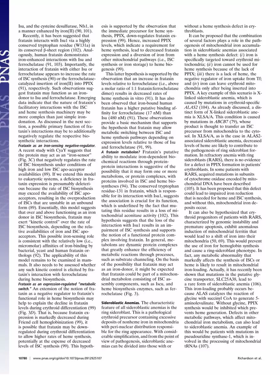
between the cytosolic- and mitochondrialiron-processing pathways and is supportedby several lines of evidence. For example,Tf and Tf-dependent iron uptake are in-creased when heme synthesis is inhibitedusing reticulocytes in vitro (41, 42), andthis is accomplished by an increase inTfR1 cycling rate (42). This response oc-curs despite on-going mitochondrial iron-loading because of the inhibition of hemesynthesis. Hence, the lack of heme gener-ation appears to result in a compensatoryincrease in Tf-bound iron uptake. Otherevidence comes from studies in vivo usingthe muscle creatine kinase conditionalfrataxin knockout mouse (43), where con-ditional frataxin-deletion in cardiomyocytesleads to depressed ISC and heme synthesis,mitochondrial iron-loading, TfR1 up-regu-lation, and thus increased iron uptake fromTf (44, 45) (Fig. 2).Under these latter conditions, in the
absence of frataxin, the defect in ISC andheme synthesis appears to lead to a “res-cue response,” where iron uptake is in-creased and targeted away from thecytoplasm and ferritin toward the mito-chondrion. This response leads to a rela-tive cytosolic iron-deficiency and mito-chondrial iron-loading (44, 45). In thelatter case, it was hypothesized that a sig-nal (potentially caused by decreased ISCor heme levels) was sent from the mito-chondrion to the cytoplasm to up-regulateiron uptake to compensate for the de-pressed ISC and heme synthesis resultingfrom frataxin-deficiency (45). Althoughincreased directional targeting of iron tothe mitochondrion occurred, leading toiron-loading in this organelle, it does notlead to an effective reversal of the mito-chondrial defect (44, 45), because frataxin,which plays a crucial role in ISC and hemesynthesis, is deleted in cardiomyocytes(43, 46–48). Similarly, Li et al., using cul-
tured cells in vitro, have also demon-strated that there is a cytosolic iron-deficitin fibroblasts and lymphoblasts fromFriedreich’s ataxia (FA) patients, as shownby increased IRP1/2-RNA-binding activ-ity (49). Finally, overexpression of mito-chondrial ferritin (Ftmt) in the mitochon-drion leads to mitochondrial iron-loadingand cytosolic iron-deprivation (50). Hence,there is evidence that alterations in mito-chondrial iron homeostasis lead to changesin cellular iron metabolism, suggestingcommunication between compartmentsand a modulating influence of the mi-tochondrion.The discussion above demonstrates that
cytosolic iron metabolism is regulated, atleast in part, by the mitochondrial demandfor iron that is critical for heme and ISCsynthesis. This finding appears logical,because the mitochondrion is a focal pointof iron processing. Such compensatoryalterations in iron trafficking, demon-strating communication between the cy-tosol and mitochondrion, are also foundin other conditions where mitochondrialiron-processing is disturbed (51). For ex-ample, similar mitochondrial iron-loadingoccurs in patients with a splice mutationin the ISC scaffold protein, Iscu (52), orwith a deficiency in glutaredoxin-5 that isinvolved in ISC/heme synthesis (53, 54).Moreover, in a patient with a glutaredoxin-5 deficiency, this problem led to decreasedferritin and increased TfR1 expression(54), which was a similar metabolic phe-notype to that found in frataxin knockoutmice (44, 45). Further studies are necessaryto define at the molecular level how thiscommunication occurs between the cytosoland the mitochondrion, and may be facili-
tated by a better understanding of themechanisms of mitochondrial iron importand release.Considering the regulation of whole-cell
iron metabolism via molecular defectsin the mitochondrion described above, it isintriguing to consider the possibility thatsimilar mechanisms of communicationexist for other organelles that play key rolesin iron metabolism. For example, the ly-sosome is involved in ferritin degradation,recycling of stored iron, and autophagy ofother organelles, including the iron-richmitochondrion (55). The rupture of asmall number of lysosomes is an earlyupstream event in many cases of apopto-sis, particularly oxidative stress-inducedapoptosis; necrosis results from a majorlysosomal rupture. Consequently, it hasbeen suggested that regulation of the ly-sosomal content of redox-active iron ap-pears essential for cell survival (56).
Mitochondrial Iron ImportAlthough DMT1 is responsible for theexport of iron(II) from the endosome, theitinerary of the metal from the cytosol tothe inner mitochondrial membrane isnot well understood. However, it is knownthat the inner membrane of the mito-chondrion contains proteins capable oftransporting iron into the mitochondrialmatrix. Foury and Roganti suggested a rolefor the eukaryotic, mitochondrial solutecarriers, Mrs3 and Mrs4, in mediatingmitochondrial iron metabolism in yeast(57). A further study demonstrated thatthese proteins are essential for yeastgrowth under iron-limiting conditions,suggesting that, at least in this organism,an additional iron transport mechanism ispresent (58). A recent investigation of thefunction of Mrs3 and -4 in small mito-chondrial particles showed these proteinstransport iron(II) along a concentrationgradient (59).Studies with the zebrafish mutant with
profound anemia, frascati, led to the dis-covery of the Mrs3 and -4 homolog,termed mitoferrin-1 (SLC25A37) andmitoferrin-2 (SLC25A28) (60). These ho-mologous proteins are important for mi-tochondrial iron uptake in erythroid andnonerythroid cells, respectively (60, 61)(Fig. 1A). Mitoferrin-1 is localized on theinner mitochondrial membrane and func-tions as an essential importer of iron formitochondrial heme and ISC in erythro-blasts and is necessary for erythropoiesis(60). Mitoferrin-1 is highly expressed inerythroid cells and in low levels in othertissues, whereas mitoferrin-2 is ubiqui-tously expressed (61). The half-life ofmitoferrin-1 (but not mitoferrin-2) in-creases in developing erythroid cells andthis may be part of a regulatory mecha-nism aiding mitochondrial iron uptake(61). Recently, Abcb10, a mitochondrial
Fig. 2. Model of alteration in iron uptake inFriedreich’s ataxia. Frataxin-deficiency results inincreased mitochondrial-targeted iron uptake andcytosolic iron-deficiency (see text).
Richardson et al. PNAS | June 15, 2010 | vol. 107 | no. 24 | 10777

inner-membrane ATP-binding cassettetransporter, was found to physically inter-act with mouse Mfrn1, and thereby en-hance the stability of the protein andincrease mitochondrial iron-import (62).Interestingly, Abcb10 has been suggestedto play some role in heme synthesis andcan be rapidly induced by the transcriptionfactor, GATA-1, which plays a role inerythroid differentiation (63).It is still unknown how iron is trans-
ported across the outer mitochondrialmembrane and clearly other transportersmay yet be identified. Considering this,a large-scale computational screen identi-fied three potential transporters that maybe involved in mitochondrial iron metab-olism, namely SLC25A39, SLC22A4, andTMEM14C (64). In fact, targeted knock-down of these genes in zebrafish resultedin profound anemia. Furthermore, silenc-ing of Slc25a39 in murine erythroleukemiacells impaired iron incorporation intoPPIX to form heme (64).It is also notable that a mutation in the
transmembrane mitochondrial protein,sideroflexin1, is responsible for the skeletalabnormalities and hematological phenotypein the flexed-tail mouse model, namelyhypochromic, microcytic anemia, and side-rotic granules in erythrocytes (65). Becauseof its predicted five-transmembrane do-mains, this molecule has been suggestedto be a transporter essential for mitochon-drial iron metabolism. Indeed, it has beenspeculated to transportmolecules intooroutof the mitochondrion for iron utilization orheme synthesis (65). Sideroflexin may notnecessarily transport iron and couldmediatethe uptake of other metabolites essential forheme synthesis (e.g., pyridoxine) that isnecessary for the formation of pyridoxalphosphate, a coenzyme required for the firststep in heme production (65).
Mitochondrial Iron MetabolismThree Major Pathways: Heme Synthesis, ISCSynthesis, and Storage. Once iron is trans-ported into the mitochondrion it can thenbe used for heme synthesis, ISC synthesis,or stored in Ftmt. It is essential that mi-tochondrial iron is maintained in a safeform to prevent oxidative damage, as mi-tochondria are a major source of cytotoxicROS (3). Hence, it is likely that as in thecytosol, iron is carefully transported withinthe mitochondrion in a form distal to theaqueous environment, deep in the hydro-phobic pockets of communicating proteinsthat form iron transport pathways. Al-though the molecular nature of these cir-cuits remains unclear, the pathways thatuse iron are well known and are discussedbelow.Mitochondrial iron storage: mitochondrial ferritin.Like a typical ferritin, Ftmt shows ferrox-idase activity and binds iron (66). In contrastto cytoplasmic ferritin, Ftmt is encoded
by an atypical intronless gene (66). How-ever, its role is unclear, particularly con-sidering its tissue distribution pattern.Indeed, Ftmt was found at the highest lev-els in the testes and the erythroblasts ofsideroblastic anemia patients (67, 68). Mi-tochondrial ferritin has also been detectedin the heart, brain, spinal cord, kidney, andpancreas (67). Unlike cytosolic ferritin,Ftmt is not highly expressed in the liver andspleen, suggesting that it plays a distinctrole. These findings led to the hypothesisthat Ftmt plays a role in protection fromiron-dependent oxidative damage (67).The role of Ftmt in iron metabolism was
examined by employing a stably transfectedcell line that hyper-expresses Ftmt (50).These studies showed that overexpressionof Ftmt resulted in increased IRP-1/2-RNA-binding activity, decreased cytosolicand mitochondrial aconitase activity (sug-gesting decreased ISC synthesis), de-creased cytoplasmic ferritin, increasedTfR1 expression, decreased heme synthe-sis, decreased frataxin expression, and in-creased iron-loading of Ftmt (50). Hence,Ftmt overexpression leads to partitioningof iron away from heme and ISC synthesisin the mitochondrion. This effect not onlyalters mitochondrial iron metabolism, butalso whole-cell iron metabolism (50), lead-ing to a cytosolic iron-deficiency that re-duces the proliferation of neoplastic cellshyper-expressing Ftmt in vivo (69). As al-ready discussed, very similar alterations ofgene expression also occur after ablation offrataxin expression (44, 45) and indicatecommunication between the cytosol andmitochondrion.Iron-sulfur cluster biosynthesis. Being a majorsite of ISC assembly, the mitochondrionplays a pivotal role in the biosynthesis ofISC proteins (1, 2). ISCs consist of ironand sulfide anions (S2-), which assemble toform [2Fe-2S] or [4Fe-4S] clusters (2).These clusters form cofactors in proteinsthat perform vital functions, such as elec-tron transport, redox reactions, metaboliccatalysis, and other such functions (70).In eukaryotic cells, more than 20 mole-cules facilitate maturation of ISC proteinsin the mitochondria, cytosol, and nucleus(2). Functional defects in ISC proteinsor components involved in their biogenesislead to human diseases (71, 72). Bio-synthesis of ISCs and their insertion intoapo-proteins requires both the mitochon-drial and cytosolic machinery. The firstmolecule identified in the ISC machinerywas the enzyme NifS from Azobacter vin-landii, which is a cysteine desulfurase thatparticipates in ISC assembly as a sulfur do-nor (73). This enzyme is highly conservedand in humans is known as Nfs1 (74).ISCs are assembled on scaffold proteins
and then transferred to apo-proteins. InEscherichia coli, this scaffold protein isknown as ISC assembly protein U (IscU)
and leads to sequential assembly leadingto [2Fe-2S] units that then form a [4Fe-4S]cluster (75). In yeast cells, these reactionsare accomplished by Isu1 and -2 (76),but in humans, the function of IscU isperformed via the splicing of IscU mRNAto lead to two transcripts which generatea cytosolic (Iscu1) or mitochondrial (Is-cu2) isoform. The maturation of extra-mitochondrial ISC proteins requires themitochondrial ISC assembly system (77).The mitochondrion contributes a yet-to-bediscovered compound necessary for thebiogenesis of ISCs outside of this com-partment (i.e., in the cytosol or other or-ganelles) (70).The mechanism involved in iron delivery
to Iscu1 is not clear, but it has been sug-gested to involve frataxin as an iron donor(2). Moreover, frataxin has been shown toplay a critical role in the early stages ofISC genesis (78), and this is discussed indetail below. In yeast, Isu1 only providesa cluster for de novo cluster productionfrom which an HSP70-type chaperonesystem transfers the new clusters to apo-proteins (2). Yet another molecule,ABCB7, appears to mediate cytosolic ISCbiogenesis (79). The maturation of cyto-solic ISCs is inhibited by mutations inABCB7 and this causes the disease, X-linked sideroblastic anemia with cerebellarataxia (XLSA/A). This condition is char-acterized by loss of cytosolic ISC proteins,defects in heme metabolism, and in-creased mitochondrial iron levels (79).Heme biosynthesis. The third major mito-chondrial metabolic pathway that utilizesiron is that of heme synthesis that is ex-clusive to this organelle (1, 11). Heme issynthesized by a pathway composed ofeight sequential reactions in the mito-chondrion and cytoplasm (1, 11). The firstand last three steps in the heme biosyn-thesis pathway take place in the mito-chondrion. The first enzyme in the path-way, 5-aminoleuvulinate synthase (ALAS),catalyzes the condensation of glycine andsuccinyl CoA to form 5-aminolevulinate(1, 11). There are two genes for ALAS, theubiquitously expressed ALAS1 and theerythroid-specific ALAS2, which is underthe regulation of iron via the IRP system(1). In nonerythroid cells, the rate ofheme synthesis is dependent on the rateof 5-aminolevulinate synthesis via ALAS1(11). In contrast, in erythroid cells, the rate-limiting step may be the delivery of Tf-iron (11).The subsequent four steps of heme
biosynthesis take place in the cytosol, fol-lowing which coproporphyrinogen IIIis decarboxylated and then oxidized inthe mitochondrial intermembranespace to produce PPIX (1, 11). Cop-roporphyrinogen may be transported intomitochondria by either peripheral-typebenzodiazepine receptors (80) or
10778 | www.pnas.org/cgi/doi/10.1073/pnas.0912925107 Richardson et al.
potentially ABCB6 (81). The seventh step,which is catalyzed by protoporphyrinogenIX oxidase generates PPIX. In the finalreaction of the pathway, iron(II) is insertedinto PPIX by the ISC protein, ferrochela-tase, in the mitochondrial matrix (11).
Mitochondrial Iron Export. Apart fromimporting iron, the mitochondrion synthe-sizes heme and ISCs that subsequently aretransported out to the cytosol. These exportprocesses are important to understand, asdecreased release of iron as heme or ISCs—or their precursors—can contribute tomitochondrial iron-loading, as found in thefrataxin knockout mouse (45). A candidateiron exporter has been identified, namelymammalian mitochondrial ABC protein 3(MTABC3; also known as ABCB6) (82).This protein can rescue mitochondrial iron-loading, respiratory dysfunction, and mito-chondrial DNA damage in atm1-deficientyeast cells (82). Of relevance, the humanortholog of atm1 is ABCB7, which cancomplement atm1 deficiencies in yeastcells, enabling the maturation of ISC-con-taining proteins in the cytosol (79). It hasbeen suggested that ABCB7 transportsISCs to the cytoplasm (79), although thishas not been directly shown.It is unknown how heme is exported
from the mitochondrion. However, its lowsolubility and highly toxic nature suggest anefficient heme-carrier must be involved.Considering this, heme-binding protein 1has been identified as a candidate for thiscarrier (83). The expression of this mole-cule is ubiquitous, but it is also increasedduring erythroid differentiation and highlevels are found in the liver (83). Heme-binding protein 1 binds one heme permole of protein (83) and, although it couldbe a candidate for heme transport, directevidence for this is lacking.
Diseases of Mitochondrial IronMetabolismMechanisms of iron transport across mi-tochondrial membranes have evolved tosupply the necessary iron to mitochondriaand also maintain the balance of cytosoliciron (1). Mitochondrial iron levels mustbe tightly controlled as iron induces ROS,which can damage the organelle (84).The importance of these tight controls ishighlighted by the fact that alterations inmitochondrial iron homeostasis havepathological consequences (1, 70). Inrecent years, cellular and animal models ofmitochondrial iron dysfunction have pro-vided valuable information in identifyingnew proteins to elucidate the pathwaysinvolved in mitochondrial iron homeosta-sis. Interesting examples of mitochondrialdiseases that have provided importantinsight into the processes of mitochondrialiron metabolism are Friedreich’s ataxiaand sideroblastic anemia. These condi-
tions are discussed in detail in the follow-ing sections.
Friedreich’s Ataxia and the Metabolic Role ofFrataxin. FA is a rare disease leading tosevere neuro- and cardio-degeneration andis caused by a deficiency of frataxin (85).The decrease in frataxin expression iscaused by an intronic GAA-repeat ex-pansion in intron-1 of FRDA. Frataxin isa vital protein that is highly expressed intissues rich in mitochondria (e.g., heartand neurons) (86), with deletion leading tolethality (87).The suggested functions for frataxin all
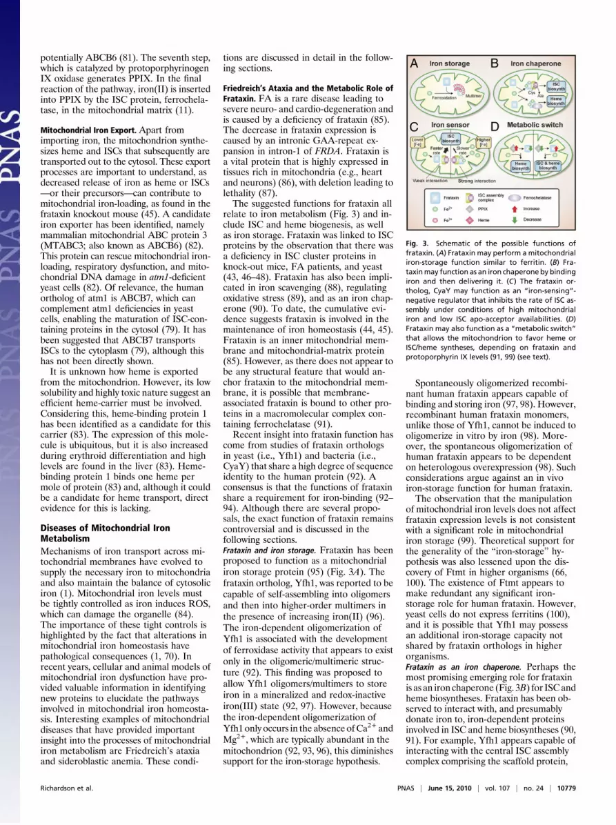
relate to iron metabolism (Fig. 3) and in-clude ISC and heme biogenesis, as wellas iron storage. Frataxin was linked to ISCproteins by the observation that there wasa deficiency in ISC cluster proteins inknock-out mice, FA patients, and yeast(43, 46–48). Frataxin has also been impli-cated in iron scavenging (88), regulatingoxidative stress (89), and as an iron chap-erone (90). To date, the cumulative evi-dence suggests frataxin is involved in themaintenance of iron homeostasis (44, 45).Frataxin is an inner mitochondrial mem-brane and mitochondrial-matrix protein(85). However, as there does not appear tobe any structural feature that would an-chor frataxin to the mitochondrial mem-brane, it is possible that membrane-associated frataxin is bound to other pro-teins in a macromolecular complex con-taining ferrochelatase (91).Recent insight into frataxin function has
come from studies of frataxin orthologsin yeast (i.e., Yfh1) and bacteria (i.e.,CyaY) that share a high degree of sequenceidentity to the human protein (92). Aconsensus is that the functions of frataxinshare a requirement for iron-binding (92–94). Although there are several propo-sals, the exact function of frataxin remainscontroversial and is discussed in thefollowing sections.Frataxin and iron storage. Frataxin has beenproposed to function as a mitochondrialiron storage protein (95) (Fig. 3A). Thefrataxin ortholog, Yfh1, was reported to becapable of self-assembling into oligomersand then into higher-order multimers inthe presence of increasing iron(II) (96).The iron-dependent oligomerization ofYfh1 is associated with the developmentof ferroxidase activity that appears to existonly in the oligomeric/multimeric struc-ture (92). This finding was proposed toallow Yfh1 oligomers/multimers to storeiron in a mineralized and redox-inactiveiron(III) state (92, 97). However, becausethe iron-dependent oligomerization ofYfh1 only occurs in the absence ofCa2+ andMg2+, which are typically abundant in themitochondrion (92, 93, 96), this diminishessupport for the iron-storage hypothesis.
Spontaneously oligomerized recombi-nant human frataxin appears capable ofbinding and storing iron (97, 98). However,recombinant human frataxin monomers,unlike those of Yfh1, cannot be induced tooligomerize in vitro by iron (98). More-over, the spontaneous oligomerization ofhuman frataxin appears to be dependenton heterologous overexpression (98). Suchconsiderations argue against an in vivoiron-storage function for human frataxin.The observation that the manipulation
of mitochondrial iron levels does not affectfrataxin expression levels is not consistentwith a significant role in mitochondrialiron storage (99). Theoretical support forthe generality of the “iron-storage” hy-pothesis was also lessened upon the dis-covery of Ftmt in higher organisms (66,100). The existence of Ftmt appears tomake redundant any significant iron-storage role for human frataxin. However,yeast cells do not express ferritins (100),and it is possible that Yfh1 may possessan additional iron-storage capacity notshared by frataxin orthologs in higherorganisms.Frataxin as an iron chaperone. Perhaps themost promising emerging role for frataxinis as an iron chaperone (Fig. 3B) for ISCandheme biosyntheses. Frataxin has been ob-served to interact with, and presumablydonate iron to, iron-dependent proteinsinvolved in ISC and heme biosyntheses (90,91). For example, Yfh1 appears capable ofinteracting with the central ISC assemblycomplex comprising the scaffold protein,
Fig. 3. Schematic of the possible functions offrataxin. (A) Frataxin may perform amitochondrialiron-storage function similar to ferritin. (B) Fra-taxinmay function as an iron chaperone by bindingiron and then delivering it. (C) The frataxin or-tholog, CyaY may function as an “iron-sensing”-negative regulator that inhibits the rate of ISC as-sembly under conditions of high mitochondrialiron and low ISC apo-acceptor availabilities. (D)Frataxin may also function as a “metabolic switch”that allows the mitochondrion to favor heme orISC/heme syntheses, depending on frataxin andprotoporphyrin IX levels (91, 99) (see text).
Richardson et al. PNAS | June 15, 2010 | vol. 107 | no. 24 | 10779
Isu, and the cysteine desulfurase, Nfs1, ina manner enhanced by iron(II) (90, 101).Recently, it has been suggested that
frataxin interacts with Isu1 via a highlyconserved tryptophan residue (W131a) inits conserved β-sheet region (102). Anal-ogously, human frataxin demonstratesiron-enhanced interactions with Isu andferrochelatase (91, 103). Importantly, theinteraction of frataxin with either Isu orferrochelatase appears to increase the rateof ISC synthesis (90) or the ferrochelatase-catalyzed insertion of iron(II) into PPIX(91), respectively. Such observations sug-gest frataxin may function as an iron-donor to Isu and ferrochelatase. Emergingdata indicate that the nature of frataxin’sfacilitatory interactions with the ISCand heme synthesis machinery may bemore complex than just simple iron-donation. As discussed in the next sec-tion, a possible primary function of fra-taxin’s interactions may be to additionallynegatively regulate the respective bio-synthetic interactions.Frataxin as an iron-sensing negative-regulator.A recent study with CyaY suggests thatthe protein may act as an “iron-sensor”(Fig. 3C) that negatively regulates the rateof ISC biosynthesis under conditions ofhigh iron and low ISC apo-acceptoravailabilities (89). If we extend this modelto eukaryotic systems, a deficiency in fra-taxin expression is presumably deleteri-ous because the rate of ISC biosynthesismay exceed the availability of ISC apo-acceptors, resulting in the overproductionof ISCs that are unstable in an unboundform (89). Essentially, this model suggeststhat over and above functioning as an irondonor in ISC biosynthesis, frataxin mayexert “kinetic control” over the rate ofISC biosynthesis, depending on the rela-tive availabilities of iron and ISC apo-acceptors. This possible iron-sensing roleis consistent with the relatively low (i.e.,micromolar) affinities of iron-binding bybacterial, yeast and human frataxin or-thologs (92). The applicability of thismodel remains to be examined in mam-mals. It also needs to be assessed whetherany such kinetic control is elicited by fra-taxin’s interaction with ferrochelataseduring heme biosynthesis.Frataxin as an expression-regulated “metabolicswitch.” An extension of the notion of fra-taxin as a negative regulator to frataxin’sfunctional role in heme biosynthesis mayhelp to explain the decline in frataxinlevels during erythroid differentiation (99)(Fig. 3D). That is, because frataxin ex-pression is markedly decreased duringFriend cell hemoglobinization (99), itis possible that frataxin may be down-regulated during erythroid differentiationto allow higher rates of heme synthesis,potentially at the expense of decreasedlevels of ISC synthesis (99). This hypoth-
esis is supported by the observation thatthe immediate precursor for heme syn-thesis, PPIX, down-regulates frataxin ex-pression (99). Hence, increased PPIXlevels, which indicate a requirement forheme synthesis, lead to decreased frataxinexpression and a diversion of iron fromother mitochondrial pathways (i.e., ISCsynthesis or iron storage) to heme bio-genesis (99).This latter hypothesis is supported by the
observation that an increase in frataxinlevels relative to ferrochelatase (i.e., abovea molar ratio of 1:1 frataxin:ferrochelatasedimer) results in decreased rates ofheme synthesis in vitro (91). It has alsobeen observed that iron-bound humanfrataxin has a higher putative binding af-finity for ferrochelatase (17 nM) thanIsu (480 nM) (91). These observationsprovide a basic mechanism that supportsthe hypothesis that frataxin may allowmetabolic switching between ISC andheme synthesis pathways depending onexpression levels relative to those of Isuand ferrochelatase (91, 99).A frataxin metabolon? Frataxin’s putativeability to modulate iron-dependent bio-chemical reactions through protein-protein interactions is suggestive of thepossibility that it may form one or moremetabolons, or protein complexes, withproteins involved in ISC and heme bio-syntheses (94). The conserved tryptophanresidue-131 in frataxin, which is respon-sible for its interaction with Isu1, suggeststhe association is crucial for its function,which is underlined by the fact that mu-tating this residue results in a loss of mi-tochondrial aconitase activity (102). Thishypothesis suggests that the loss of theinteraction with Isu1 results in an im-pairment of ISC synthesis and supportsthe notion of a functional protein com-plex involving frataxin. In general, me-tabolons are dynamic protein complexesthat greatly enhance the efficiency ofmetabolic reactions through processes,such as substrate channeling. On the basisof the possibility that frataxin may actas an iron-donor, it might be expectedthat frataxin could be part of a mitochon-drial metabolon consisting of ISC as-sembly components, such as Iscu, andheme biosynthesis enzymes, such as fer-rochelatase (Fig. 3).
Sideroblastic Anemias. The characteristicfeature of all sideroblastic anemias is thering sideroblast. This is a pathologicalerythroid precursor containing excessivedeposits of nonheme iron in mitochondriawith peri-nuclear distribution responsi-ble for the ring appearance. With consid-erable simplification, and from the point ofview of pathogenesis, sideroblastic ane-mias can be divided into those with or
without a heme synthesis defect in ery-throblasts.It can be proposed that the combination
of several factors plays a role in the path-ogenesis of mitochondrial iron accumula-tion in sideroblastic anemias associatedwith a heme synthesis defect: (i) iron isspecifically targeted toward erythroid mi-tochondria; (ii) iron cannot be used forheme synthesis because of the lack ofPPIX; (iii) there is a lack of heme, thenegative regulator of iron uptake from Tf;and (iv) iron can leave erythroid mito-chondria only after being inserted intoPPIX. A key example of this scenario is X-linked sideroblastic anemia, which iscaused by mutations in erythroid-specificALAS2 (104). As already discussed, a dis-tinct form of X-linked sideroblastic ane-mia is XLSA/A. This condition is causedby mutations in ABCB7 (79), whoseproduct is thought to transfer an ISCprecursor from mitochondria to the cyto-sol. In XLSA/A, as is the case in ALAS2-associated sideroblastic anemia, decreasedlevels of heme are likely to contribute tothe pathogenesis of ring sideroblast for-mation. In refractory anemia with ringsideroblasts (RARS), there is no evidencefor a defect in PPIX formation in patients’erythroblasts. In some patients withRARS, acquired mutations in subunitsof cytochrome oxidase encoded by mito-chondrial DNA have been described(105). It has been proposed that this defectcould lead to impaired iron reductionthat is needed for heme and ISC synthesis,and without this, mitochondrial iron de-posits occur.It can also be hypothesized that ery-
throid progenitors of patients with RARS,characterized by genomic instability andpremature apoptosis, exhibit anomalousinduction of mitochondrial ferritin thatwould lead to a shift of iron into theirmitochondria (50, 69). This would preventthe use of iron for hemoglobin synthesisand cause a ring-sideroblast phenotype. Infact, any metabolic abnormality thatmarkedly affects the synthesis of ISCs orheme is likely to result in mitochondrialiron-loading. Actually, it has recently beenshown that mutations in the putative gly-cine transporter, SLC25A38, lead toa rare form of sideroblastic anemia (106).This iron-loading probably occurs be-cause ALAS catalyzes the reaction ofglycine with succinyl CoA to generate 5-aminolevulinate. Without glycine, PPIXsynthesis would be inhibited which pre-vents heme generation. Defects in othermetabolic pathways, which affect mito-chondrial iron metabolism, can also leadto sideroblastic anemia. An example ofthis would be patients with mutations inpseudouridine synthase-1, which is in-volved in the processing of mitochondrialtRNAs (107).
10780 | www.pnas.org/cgi/doi/10.1073/pnas.0912925107 Richardson et al.

SummaryAlthough the mitochondrion is a focalpoint for iron utilization in heme and ISCsynthesis, there has been little realizationthat the mitochondrion can play an im-portant role in orchestrating whole-celliron metabolism. Indeed, analysis of dis-ease states has enabled understanding ofthe role of the mitochondrion in regu-lating cellular iron metabolism. There is
strong evidence to suggest that the mito-chondrion can modulate the cellular ironuptake machinery to satisfy its demand.Considering this evidence, it is likely thatsignaling pathways exist that allow com-munication between the mitochondrionand cytoplasm, enabling the mitochon-drial iron processing machinery to besupplied with this metal to allow heme andISC synthesis.
ACKNOWLEDGMENTS. D.R.R.’s laboratorywas supported by grants from the National Healthand Medical Research Council of Australia, theUS Muscular Dystrophy Association (MDA),MDA New South Wales, Friedreich’s Ataxia Re-search Alliance (FARA) Australia, and FARA USA.P.P and A.D.S. were supported by grants fromthe Canadian Institutes of Health Research.Y.S.R. was a grateful recipient of a Cancer InstituteNew South Wales Early Career DevelopmentFellowship.
1. Napier I, Ponka P, Richardson DR (2005) Iron traffickingin the mitochondrion: Novel pathways revealed bydisease. Blood 105:1867–1874.
2. Lill R, Mühlenhoff U (2008) Maturation of iron-sulfurproteins in eukaryotes: Mechanisms, connected pro-cesses, and diseases. Annu Rev Biochem 77:669–700.
3. Eaton JW, Qian M (2002) Molecular bases of cellulariron toxicity. Free Radic Biol Med 32:833–840.
4. Klausner RD, Ashwell G, van Renswoude J, Harford JB,Bridges KR (1983) Binding of apotransferrin to K562cells: Explanation of the transferrin cycle. Proc NatlAcad Sci USA 80:2263–2266.
5. Iacopetta BJ, Morgan EH (1983) The kinetics of trans-ferrin endocytosis and iron uptake from transferrinin rabbit reticulocytes. J Biol Chem 258:9108–9115.
6. Cmejla R, Petrak J, Cmejlova J (2006) A novel ironresponsive element in the 3'UTR of human MRCKα.Biochem Biophys Res Commun 341:158–166.
7. Cmejla R, et al. (2010) Human MRCKα is regulated bycellular iron levels and interferes with transferrin ironuptake. Biochem Biophys Res Commun 395:163–167.
8. Zhang XM, Ellis S, Sriratana A, Mitchell CA, Rowe T(2004) Sec15 is an effector for the Rab11 GTPase inmammalian cells. J Biol Chem 279:43027–43034.
9. Lim JE, et al. (2005) A mutation in Sec15l1 causesanemia in hemoglobin deficit (hbd) mice. Nat Genet37:1270–1273.
10. Zhang AS, Sheftel AD, Ponka P (2006) The anemia of“haemoglobin-deficit” (hbd/hbd) mice is caused bya defect in transferrin cycling. Exp Hematol 34:593–598.
11. Ponka P (1997) Tissue-specific regulation of iron me-tabolism and heme synthesis: Distinct control mech-anisms in erythroid cells. Blood 89:1–25.
12. Dunn LL, Rahmanto YS, Richardson DR (2007) Ironuptake and metabolism in the new millennium. TrendsCell Biol 17:93–100.
13. Bali PK,ZakO,AisenP (1991)Anewrole for thetransferrinreceptor in the release of iron from transferrin. Bio-chemistry 30:324–328.
14. Ohgami RS, et al. (2005) Identification of a ferrireduc-tase required for efficient transferrin-dependent ironuptake in erythroid cells. Nat Genet 37:1264–1269.
15. Sendamarai AK, Ohgami RS, Fleming MD, LawrenceCM (2008) Structure of the membrane proximal ox-idoreductase domain of human Steap3, the dominantferrireductase of the erythroid transferrin cycle. ProcNatl Acad Sci USA 105:7410–7415.
16. Fleming MD, et al. (1998) Nramp2 is mutated inthe anemic Belgrade (b) rat: Evidence of a role forNramp2 in endosomal iron transport. Proc Natl AcadSci USA 95:1148–1153.
17. Jacobs A (1977) Low molecular weight intracellulariron transport compounds. Blood 50:433–439.
18. Ganz T (2007) Molecular control of iron transport.J Am Soc Nephrol 18:394–400.
19. Ganz T, Nemeth E (2009) Iron sequestration andanemia of inflammation. Semin Hematol 46:387–393.
20. Hentze MW, Muckenthaler MU, Andrews NC (2004)Balancing acts: Molecular control of mammalian ironmetabolism. Cell 117:285–297.
21. Hentze MW, Kühn LC (1996) Molecular control ofvertebrate iron metabolism: mRNA-based regulatorycircuits operated by iron, nitric oxide, and oxidativestress. Proc Natl Acad Sci USA 93:8175–8182.
22. Cairo G, Recalcati S (2007) Iron-regulatory proteins:Molecular biology and pathophysiological implica-tions. Expert Rev Mol Med 9:1–13.
23. Meyron-Holtz EG, et al. (2004) Genetic ablations ofiron regulatory proteins 1 and 2 reveal why iron
regulatory protein 2 dominates iron homeostasis.EMBO J 23:386–395.
24. Meyron-Holtz EG, GhoshMC, Rouault TA (2004) Mam-malian tissue oxygen levels modulate iron-regulatoryprotein activities in vivo. Science 306:2087–2090.
25. Greenberg GR, Wintrobe MM (1964) A labile ironpool. J Biol Chem 165:397–398.
26. Ross JF (1964) The metabolism of inorganic andhemoglobin iron. J Clin Invest 25:933.
27. Ponka P, Borová J, Neuwirt J, Fuchs O (1979)Mobilization of iron from reticulocytes. Identifica-tion of pyridoxal isonicotinoyl hydrazone as a newiron chelating agent. FEBS Lett 97:317–321.
28. Richardson DR, Milnes K (1997) The potential of ironchelators of the pyridoxal isonicotinoyl hydrazone classas effective antiproliferative agents II: The mechanismof action of ligands derived from salicylaldehydebenzoyl hydrazone and 2-hydroxy-1-naphthylaldehydebenzoyl hydrazone. Blood 89:3025–3038.
29. Sheftel AD, Zhang AS, Brown C, Shirihai OS, Ponka P(2007) Direct interorganellar transfer of iron fromendosome to mitochondrion. Blood 110:125–132.
30. Richardson DR, Ponka P, Vyoral D (1996) Distributionof iron in reticulocytes after inhibition of hemesynthesis with succinylacetone: Examination of theintermediates involved in iron metabolism. Blood87:3477–3488.
31. Vyoral D, Hradilek A, Neuwirt J (1992) Transferrin andiron distribution in subcellular fractions of K562 cellsin the early stages of transferrin endocytosis. BiochimBiophys Acta 1137:148–154.
32. Shi H, Bencze KZ, Stemmler TL, Philpott CC (2008) Acytosolic iron chaperone that delivers iron to ferritin.Science 320:1207–1210.
33. Kim BE, Nevitt T, Thiele DJ (2008) Mechanisms forcopper acquisition, distribution and regulation. NatChem Biol 4:176–185.
34. Field LS, Luk E, Culotta VC (2002) Copper chaperones:Personal escorts for metal ions. J Bioenerg Biomembr34:373–379.
35. La Fontaine S, Mercer JF (2007) Trafficking of thecopper-ATPases, ATP7A and ATP7B: Role in copperhomeostasis. Arch Biochem Biophys 463:149–167.
36. Hardman B, et al. (2007) Hormonal regulation ofthe Menkes and Wilson copper-transporting ATPasesin human placental Jeg-3 cells. Biochem J 402:241–250.
37. Isobe K, Isobe Y, Sakurami T (1981) Cytochemicaldemonstration of transferrin in the mitochondria ofimmature human erythroid cells. Acta Haematol 65:2–9.
38. Ponka P, Neuwirt J, Borova J, Fuchs O (1976) Controlof iron delivery to haemoglobin in erythroid cells.Ciba Foundation Symposium 51 - Iron Metabolism,eds Porter R, Fitzsimons DW (Elsevier/ExcerptaMedica/North-Holland, Amsterdam), pp 167–200.
39. Zhang AS, Sheftel AD, Ponka P (2005) Intracellularkinetics of iron in reticulocytes: Evidence for endosomeinvolvement in iron targeting to mitochondria. Blood105:368–375.
40. Provance DW, Jr, et al. (2004) Chemical-genetic in-hibition of a sensitized mutant myosin Vb demon-strates a role in peripheral-pericentriolar membranetraffic. Proc Natl Acad Sci USA 101:1868–1873.
41. Ponka P, Wilczynska A, Schulman HM (1982) Ironutilization in rabbit reticulocytes. A study usingsuccinylacetone as an inhibitor or heme synthesis.Biochim Biophys Acta 720:96–105.
42. Adams ML, Ostapiuk I, Grasso JA (1989) The effects ofinhibition of heme synthesis on the intracellular
localization of iron in rat reticulocytes. BiochimBiophys Acta 1012:243–253.
43. Puccio H, et al. (2001) Mouse models for Friedreichataxia exhibit cardiomyopathy, sensory nerve defectand Fe-S enzyme deficiency followed by intramito-chondrial iron deposits. Nat Genet 27:181–186.
44. Whitnall M, et al. (2008) The MCK mouse heart modelof Friedreich’s ataxia: Alterations in iron-regulatedproteins and cardiac hypertrophy are limited by ironchelation. Proc Natl Acad Sci USA 105:9757–9762.
45. HuangML-H, et al. (2009) Elucidation of the mechanismof mitochondrial iron loading in Friedreich’s ataxia byanalysis of a mouse mutant. Proc Natl Acad Sci USA106:16381–16386.
46. Babcock M, et al. (1997) Regulation of mitochondrialiron accumulation by Yfh1p, a putative homolog offrataxin. Science 276:1709–1712.
47. Rötig A, et al. (1997) Aconitase and mitochondrialiron-sulphur protein deficiency in Friedreich ataxia.Nat Genet 17:215–217.
48. Foury F (1999) Low iron concentration and aconitasedeficiency in a yeast frataxin homologue deficientstrain. FEBS Lett 456:281–284.
49. Li K, Besse EK, Ha D, Kovtunovych G, Rouault TA(2008) Iron-dependent regulation of frataxinexpression: Implications for treatment of Friedreichataxia. Hum Mol Genet 17:2265–2273.
50. Nie G, Sheftel AD, Kim SF, Ponka P (2005) Overexpressionof mitochondrial ferritin causes cytosolic iron depletionand changes cellular iron homeostasis. Blood 105:2161–2167.
51. Richardson DR, et al. (2010) The ins and outs ofmitochondrial iron-loading: The metabolic defect inFriedreich’s ataxia. J Mol Med 88:323–329.
52. Mochel F, et al. (2008) Splice mutation in the iron-sulfur cluster scaffold protein ISCU causes myopathywith exercise intolerance. Am J Hum Genet 82:652–660.
53. Wingert RA, et al.; Tübingen 2000 Screen Consortium(2005) Deficiency of glutaredoxin 5 reveals Fe-S clus-ters are required for vertebrate haem synthesis. Na-ture 436:1035–1039.
54. Camaschella C, et al. (2007) The human counterpartof zebrafish shiraz shows sideroblastic-like microcyticanemia and iron overload. Blood 110:1353–1358.
55. Kurz T, Terman A, Gustafsson B, Brunk UT (2008)Lysosomes in iron metabolism, ageing and apop-tosis. Histochem Cell Biol 129:389–406.
56. Kurz T, Terman A, Brunk UT (2007) Autophagy,ageing and apoptosis: The role of oxidative stressand lysosomal iron. Arch Biochem Biophys 462:220–230.
57. Foury F, Roganti T (2002) Deletion of the mitochondrialcarrier genes MRS3 and MRS4 suppresses mitochon-drial iron accumulation in a yeast frataxin-deficientstrain. J Biol Chem 277:24475–24483.
58. Mühlenhoff U, et al. (2003) A specific role of the yeastmitochondrial carriers MRS3/4p in mitochondrial ironacquisition under iron-limiting conditions. J BiolChem 278:40612–40620.
59. Froschauer EM, Schweyen RJ, Wiesenberger G (2009)The yeast mitochondrial carrier proteins Mrs3p/Mrs4pmediate iron transport across the inner mitochondrialmembrane. Biochim Biophys Acta 1788:1044–1050.
60. Shaw GC, et al. (2006) Mitoferrin is essential forerythroid iron assimilation. Nature 440:96–100.
61. Paradkar PN, Zumbrennen KB, Paw BH, Ward DM,Kaplan J (2009) Regulation of mitochondrial ironimport through differential turnover of mitoferrin 1and mitoferrin 2. Mol Cell Biol 29:1007–1016.
Richardson et al. PNAS | June 15, 2010 | vol. 107 | no. 24 | 10781

62. Chen W, et al. (2009) Abcb10 physically interacts withmitoferrin-1 (Slc25a37) to enhance its stability andfunction in the erythroid mitochondria. Proc NatlAcad Sci USA 106:16263–16268.
63. Shirihai OS, Gregory T, Yu C, Orkin SH, Weiss MJ(2000) ABC-me: A novel mitochondrial transporterinduced by GATA-1 during erythroid differentiation.EMBO J 19:2492–2502.
64. Nilsson R, et al. (2009) Discovery of genes essentialfor heme biosynthesis through large-scale gene ex-pression analysis. Cell Metab 10:119–130.
65. Fleming MD, Campagna DR, Haslett JN, Trenor CC, 3rd,Andrews NC (2001) A mutation in a mitochondrialtransmembrane protein is responsible for the pleiotro-pic hematological and skeletal phenotype of flexed-tail(f/f) mice. Genes Dev 15:652–657.
66. Levi S, et al. (2001) A human mitochondrial ferritinencoded by an intronless gene. J Biol Chem 276:24437–24440.
67. Santambrogio P, et al. (2007) Mitochondrial ferritinexpression in adult mouse tissues. J Histochem Cytochem55:1129–1137.
68. Cazzola M, et al. (2003) Mitochondrial ferritinexpression in erythroid cells from patients withsideroblastic anemia. Blood 101:1996–2000.
69. Nie G, Chen G, Sheftel AD, Pantopoulos K, Ponka P(2006) In vivo tumor growth is inhibited by cytosoliciron deprivation caused by the expression ofmitochondrial ferritin. Blood 108:2428–2434.
70. Lill R, et al. (2006) Mechanisms of iron-sulfur proteinmaturation in mitochondria, cytosol and nucleus ofeukaryotes. Biochim Biophys Acta 1763:652–667.
71. Sheftel A, Stehling O, Lill R (2010) Iron-sulfur proteinsin health and disease. Trends Endocrinol Metab 21:302–314.
72. Rouault TA, Tong WH (2008) Iron-sulfur clusterbiogenesis and human disease. Trends Genet 24:398–407.
73. Zheng L, White RH, Cash VL, Jack RF, Dean DR (1993)Cysteine desulfurase activity indicates a role for NIFSin metallocluster biosynthesis. Proc Natl Acad Sci USA90:2754–2758.
74. Land T, Rouault TA (1998) Targeting of a human iron-sulfur cluster assembly enzyme, nifs, to differentsubcellular compartments is regulated throughalternative AUG utilization. Mol Cell 2:807–815.
75. Agar JN, et al. (2000) IscU as a scaffold for iron-sulfurcluster biosynthesis: Sequential assembly of [2Fe-2S]and [4Fe-4S] clusters in IscU. Biochemistry 39:7856–7862.
76. Garland SA, Hoff K, Vickery LE, Culotta VC (1999)Saccharomyces cerevisiae ISU1 and ISU2: Members ofa well-conserved gene family for iron-sulfur clusterassembly. J Mol Biol 294:897–907.
77. Tong WH, Jameson GN, Huynh BH, Rouault TA (2003)Subcellular compartmentalization of human Nfu, an
iron-sulfur cluster scaffold protein, and its ability toassemble a [4Fe-4S] cluster. Proc Natl Acad Sci USA100:9762–9767.
78. Stehling O, Elsässer HP, Brückel B, Mühlenhoff U, Lill R(2004) Iron-sulfur protein maturation in human cells:evidence for a function of frataxin. Hum Mol Genet13:3007–3015.
79. Bekri S, et al. (2000) Human ABC7 transporter: Genestructure and mutation causing X-linked sideroblasticanemia with ataxia with disruption of cytosolic iron-sulfur protein maturation. Blood 96:3256–3264.
80. Verma A, Nye JS, Snyder SH (1987) Porphyrins areendogenous ligands for the mitochondrial (peripheral-type) benzodiazepine receptor. Proc Natl Acad Sci USA84:2256–2260.
81. Krishnamurthy PC, et al. (2006) Identification ofa mammalian mitochondrial porphyrin transporter.Nature 443:586–589.
82. Mitsuhashi N, et al. (2000) MTABC3, a novelmitochondrial ATP-binding cassette protein involvedin iron homeostasis. J Biol Chem 275:17536–17540.
83. Taketani S, et al. (1998) Molecular characterization ofa newly identified heme-binding protein inducedduring differentiation of urine erythroleukemiacells. J Biol Chem 273:31388–31394.
84. Levi S, Rovida E (2009) The role of iron inmitochondrial function. Biochim Biophys Acta 1790:629–636.
85. Campuzano V, et al. (1997) Frataxin is reduced inFriedreich ataxia patients and is associated withmitochondrial membranes. Hum Mol Genet 6:1771–1780.
86. Koutnikova H, et al. (1997) Studies of human, mouseand yeast homologues indicate a mitochondrialfunction for frataxin. Nat Genet 16:345–351.
87. Cossée M, et al. (2000) Inactivation of the Friedreichataxia mouse gene leads to early embryonic lethalitywithout iron accumulation. Hum Mol Genet 9:1219–1226.
88. Cavadini P, Adamec J, Taroni F, Gakh O, Isaya G (2000)Two-step processing of human frataxin by mitochon-drial processing peptidase. Precursor and intermediateforms are cleaved at different rates. J Biol Chem 275:41469–41475.
89. Adinolfi S, et al. (2009) Bacterial frataxin CyaY is thegatekeeper of iron-sulfur cluster formation catalyzedby IscS. Nat Struct Mol Biol 16:390–396.
90. Yoon T, Cowan JA (2003) Iron-sulfur cluster biosyn-thesis. Characterization of frataxin as an iron donorfor assembly of [2Fe-2S] clusters in ISU-type proteins.J Am Chem Soc 125:6078–6084.
91. Yoon T, Cowan JA (2004) Frataxin-mediated irondelivery to ferrochelatase in the final step of hemebiosynthesis. J Biol Chem 279:25943–25946.
92. Pandolfo M, Pastore A (2009) The pathogenesis ofFriedreich ataxia and the structure and function offrataxin. J Neurol 256 (Suppl 1):9–17.
93. Bencze KZ, et al. (2006) The structure and function offrataxin. Crit Rev Biochem Mol Biol 41:269–291.
94. Lane DJ, Richardson DR (2010) Frataxin, a molecule ofmystery: Trading stability for function in its iron-binding site. Biochem J 426:e1–e3.
95. Adamec J, et al. (2000) Iron-dependent self-assemblyof recombinant yeast frataxin: Implications for Friedreichataxia. Am J Hum Genet 67:549–562.
96. Adinolfi S, Trifuoggi M, Politou AS, Martin S, Pastore A(2002) A structural approach to understanding the iron-binding properties of phylogenetically differentfrataxins. Hum Mol Genet 11:1865–1877.
97. O’Neill HA, et al. (2005) Assembly of human frataxin isa mechanism for detoxifying redox-active iron. Bio-chemistry 44:537–545.
98. Cavadini P, O’Neill HA, Benada O, Isaya G (2002)Assembly and iron-binding properties of humanfrataxin, the protein deficient in Friedreich ataxia.Hum Mol Genet 11:217–227.
99. Becker EM, Greer JM, Ponka P, Richardson DR (2002)Erythroid differentiation and protoporphyrin IXdown-regulate frataxin expression in Friend cells:characterization of frataxin expression compared tomolecules involved in iron metabolism and hemo-globinization. Blood 99:3813–3822.
100. Arosio P, Ingrassia R, Cavadini P (2009) Ferritins: Afamily of molecules for iron storage, antioxidationand more. Biochim Biophys Acta 1790:589–599.
101. Wang T, Craig EA (2008) Binding of yeast frataxin tothe scaffold for Fe-S cluster biogenesis, Isu. J BiolChem 283:12674–12679.
102. Leidgens S, De Smet S, Foury F (2010) Frataxininteracts with Isu1 through a conserved tryptophanin its beta-sheet. Hum Mol Genet 19:276–286.
103. Bencze KZ, et al. (2007) Human frataxin: Iron andferrochelatase binding surface. Chem Commun (Camb)(18):1798–1800.
104. Bottomley SS (2006) Congenital sideroblastic anemias.Curr Hematol Rep 5:41–49.
105. Gattermann N (2000) From sideroblastic anemia to therole of mitochondrial DNA mutations in myelodysplas-tic syndromes. Leuk Res 24:141–151.
106. Guernsey DL, et al. (2009) Mutations in mitochondrialcarrier family gene SLC25A38 cause nonsyndromicautosomal recessive congenital sideroblastic anemia.Nat Genet 41:651–653.
107. Bykhovskaya Y, Casas K, Mengesha E, Inbal A, Fischel-Ghodsian N (2004) Missense mutation in pseudouridinesynthase 1 (PUS1) causes mitochondrial myopathy andsideroblastic anemia (MLASA). Am J Hum Genet 74:1303–1308.
10782 | www.pnas.org/cgi/doi/10.1073/pnas.0912925107 Richardson et al.
All in-text references underlined in blue are linked to publications on ResearchGate, letting you access and read them immediately.