transcriptional correlates of disease outcome in anticoagulant-treated non-human primates infected...
TRANSCRIPT
Transcriptional Correlates of Disease Outcome inAnticoagulant-Treated Non-Human Primates Infectedwith EbolavirusSara Garamszegi1, Judy Y. Yen2,3, Anna N. Honko4, Joan B. Geisbert5, Kathleen H. Rubins6,
Thomas W. Geisbert5, Yu Xia1,7, Lisa E. Hensley8, John H. Connor1,2,3*
1 Bioinformatics Program, Boston University, Boston, Massachusetts, University of America, 2 Department of Microbiology, School of Medicine, Boston University, Boston,
Massachusetts, University of America, 3 National Emerging Infectious Diseases Laboratories, Boston University, Boston, Massachusetts, University of America, 4 U.S. Army
Medical Research Institute of Infectious Diseases, Fort Detrick, Frederick, Maryland, United States of America, 5 Department of Microbiology and Immunology, University
of Texas Medical Branch, Galveston, Texas, United States of America, 6 National Aeronautics and Space Administration, Houston, Texas, United States of America,
7 Department of Bioengineering, McGill University, Montreal, Canada, 8 Integrated Research Facility at Fort Detrick, National Institute of Allergy and Infectious Diseases,
National Institutes of Health, Fort Detrick, Frederick, Maryland, United States of America
Abstract
Ebola virus (EBOV) infection in humans and non-human primates (NHPs) is highly lethal, and there is limited understandingof the mechanisms associated with pathogenesis and survival. Here, we describe a transcriptomic analysis of NHPs thatsurvived lethal EBOV infection, compared to NHPs that did not survive. It has been previously demonstrated thatanticoagulant therapeutics increase the survival rate in EBOV-infected NHPs, and that the characteristic transcriptionalprofile of immune response changes in anticoagulant-treated NHPs. In order to identify transcriptional signatures thatcorrelate with survival following EBOV infection, we compared the mRNA expression profile in peripheral bloodmononuclear cells from EBOV-infected NHPs that received anticoagulant treatment, to those that did not receive treatment.We identified a small set of 20 genes that are highly confident predictors and can accurately distinguish between survivingand non-surviving animals. In addition, we identified a larger predictive signature of 238 genes that correlated with diseaseoutcome and treatment; this latter signature was associated with a variety of host responses, such as the inflammatoryresponse, T cell death, and inhibition of viral replication. Notably, among survival-associated genes were subsets of genesthat are transcriptionally regulated by (1) CCAAT/enhancer-binding protein alpha, (2) tumor protein 53, and (3)megakaryoblastic leukemia 1 and myocardin-like protein 2. These pathways merit further investigation as potentialtranscriptional signatures of host immune response to EBOV infection.
Citation: Garamszegi S, Yen JY, Honko AN, Geisbert JB, Rubins KH, et al. (2014) Transcriptional Correlates of Disease Outcome in Anticoagulant-Treated Non-Human Primates Infected with Ebolavirus. PLoS Negl Trop Dis 8(7): e3061. doi:10.1371/journal.pntd.0003061
Editor: Anita K. McElroy, Centers for Disease Control and Prevention, United States of America
Received March 20, 2014; Accepted June 18, 2014; Published July 31, 2014
This is an open-access article, free of all copyright, and may be freely reproduced, distributed, transmitted, modified, built upon, or otherwise used by anyone forany lawful purpose. The work is made available under the Creative Commons CC0 public domain dedication.
Data Availability: The authors confirm that all data underlying the findings are fully available without restriction. The raw microarray dataset was deposited inNCBI’s Gene Expression Omnibus database (Accession number: GSE24943).
Funding: SG was supported by a fellowship from the National Science Foundation (NSF) Integrative Graduate Education and Research Traineeship (DGE-0654108) and an NSF Graduate Research Fellowship (DGE-0741448). YX was supported by a Research Starter Grant in Informatics from the PharmaceuticalResearch and Manufacturers of America Foundation. Funding for experimental animal studies was provided by the Joint Science and Technology Office forChemical and Biological Defense and the Defense Threat Reduction Agency (JSTO-CBD 4.0021.08.RD.B). This work was conducted at USAMRIID under projectnumbers 19595 and 188261 supported by JSTO-CBD. The funders had no role in study design, data collection and analysis, decision to publish, or preparation ofthe manuscript.
Competing Interests: The authors have declared that no competing interests exist.
* Email: [email protected]
Introduction
Ebola virus (EBOV; Filoviridae [1]) infection of humans and
non-human primates (NHPs) can cause viral hemorrhagic fever,
an acute systemic illness characterized by fever, bleeding diathesis,
fulminant shock, and death [2]. Although several studies have
identified candidate therapeutics that may mitigate the effects of
EBOV infection [3–12], there are currently no FDA-approved
post-exposure treatments for human use. Additionally, despite
extensive research on EBOV pathogenesis [13–15], lifecycle [16],
and interactions with the host [17–19], there are no standard
biomarkers to predict host immune response to EBOV infection,
nor are there biomarkers of drug efficacy or survival following
treatment. Here, we investigated the hypothesis that there are
signatures of gene expression associated with survival in EBOV-
infected, anticoagulant-treated NHPs.
Immune response pathways play a key role in EBOV
pathogenesis. Infection is characterized by up-regulation of
inflammatory mediators such as cytokines and chemokines,
interleukins, interferon-inducible proteins, and tumor necrosis
factor alpha (TNFa) [20–25]. In addition, EBOV infection is
associated with an early loss of lymphocytes [26,27] and the
dysregulation of coagulopathy. This dysregulation of coagulation
and subsequent hemorrhage are characteristic of EBOV infection
[14,15], and may be due to the fact that immune mediators are
over-expressed by monocytes and macrophages, which, along with
PLOS Neglected Tropical Diseases | www.plosntds.org 1 July 2014 | Volume 8 | Issue 7 | e3061

dendritic cells, are primary targets for infection [13,28,29]. High-
throughput microarray studies of the immune response of NHPs to
EBOV infection have identified dramatic and early changes in
host transcription of genes related to interferon response, cytokine
signaling, and apoptosis [22]. Similarly, studies of endothelial cells
suggest that accumulation of cytokines and other pro-inflamma-
tory factors can contribute to the observed pathologies of EBOV
infection [14]. Previous studies of clinical samples from humans
infected with Sudan virus during the 2000–2001 outbreak, suggest
that hemorrhagic symptoms and death may be associated with
acute phase proteins and coagulation factors [25].
It has been demonstrated that anticoagulant therapeutics have a
positive effect on the outcome of EBOV disease [7,9]. Notably,
approximately 33% of EBOV-infected NHPs treated with
anticoagulants, such as recombinant nematode anticoagulant
protein c2 (rNAPc2) [7], and recombinant human activated
protein C (rhAPC) [9] survived a 100% fatal EBOV infection
model (Table 1). Animals responding to anticoagulant treatment
had lower plasma viremia levels and attenuation of the pro-
inflammatory and pro-coagulant responses in both studies [7,9],
suggesting that these indicators could be markers of increased
survival. However, these late-stage markers could not identify
whether there were early transcriptional changes that were
associated with survival. In addition, these results are limited in
scope to individual gene and protein assays, which cannot assess
the host immune response to infection from a global, transcrip-
tional viewpoint [7,9]. To identify early-stage transcriptional
changes, we analyzed an existing microarray dataset that
examined the host gene expression in anticoagulant-treated NHPs.
We used this dataset to identify critical transcriptional changes that
differentiate between surviving and non-surviving NHPs following
EBOV infection. These results provide critically distinct assess-
ments of the host immune response to EBOV infection by
identifying not only global changes in transcription, but also
transcription factor activities associated with survival.
We analyzed the transcriptional profiles of peripheral blood
mononuclear cell samples taken from EBOV-infected NHPs
treated with either rNAPc2 or rhAPC, as described previously
[7,9,24]. We investigated the hypothesis that gene expression
patterns are associated with survival of EBOV challenge following
anticoagulant treatment. A previous study assessed the global
transcriptional response of NHPs to EBOV infection, but was
unable to identify survival-associated profiles, due to a lack of
anticoagulant treatment [22]. Previously, we assessed the global
transcriptional response in the context of anticoagulant treatment,
but did not seek to identify upstream transcriptional regulators
associated with survival [24]. Here, we used a new method of
analysis to identify gene sets which are associated with, and
predictive of, survival following post-infection treatment. This
approach identified two sets of statistically significant genes that
distinguish between surviving and non-surviving NHPs. One, a
minimal set of 20 genes, showed good discrimination between
survivors and non-survivors, but provided little insight into
signaling pathways that might be correlated with survival. The
second, a larger set of 238 genes, identified a number of genes that
were functionally controlled by common transcription factors that
have not been previously associated with EBOV infection in
NHPs.
Materials and Methods
Animal experiments and ethics statementThe datasets for this study were collected from previously
published results studying the affect of anticoagulant therapeutics
(rhAPC and rNAPc2) on the immune response of non-human
primates (NHPs) to lethal Ebola virus (H.sapiens-tc/COD/1995/
Kikwit-9510621; EBOV). Virus was passaged twice through
VeroE6, and once through Vero cells, prior to use. Additional
experimental methods and results of the rhAPC and rNAPc2
studies have previously been described in Geisbert et al. [7] and
Hensley et al. [9], respectively (Table 1). The cumulative dataset
used in this study includes 23 rhesus macaques: 4 untreated
controls, 8 rNAPc2-treated NHPs, and 11 rhAPC-treated NHPs
(Table 2). A total of 4 NHPs survived lethal challenge with EBOV
virus (2 rNAPc2-treated and 2 rhAPC-treated).
Animal research for these previously published studies was
conducted at the United States Army Medical Research Institute
for Infectious Diseases, in compliance with the Animal Welfare
Act and other federal statutes and regulations relating to animals
and experiments involving animals, and adheres to the principles
stated in the Guide for the Care and Use of Laboratory Animals,National Research Council, 1996. The facility is fully accredited
by the Association for Assessment and Accreditation of Laboratory
Animal Care, International. These experiments and procedures
were approved by the USAMRIID Institutional Animal Care and
Use Committee (IACUC).
Sample preparation and microarray processingExperimental methods for RNA processing and DNA micro-
array preparation have been previously described in Yen et al.[24]. Microarrays were analyzed in R, using the LIMMA package
in Bioconductor [30–32] and processed as follows: (i) background
correction was done using the subtract method [33]; (ii) within-
array normalization was done using the loess method [34]; (iii) log
ratio and log intensity values were calculated; (iv) array control
probes were removed from the dataset; and, (v) the data were zero-
transformed within each animal using baseline (pre-infection)
sample; in the case of multiple pre-infection samples, Day 0 was
used (Figure S1). The raw microarray dataset was deposited in
NCBI’s Gene Expression Omnibus (GEO; [35]) database (Acces-
sion: GSE24943; [24]).
We organized the microarrays into three groups based on NHP
response to anticoagulant drug treatment: (i) EBOV-infected
NHPs that did not receive anticoagulant treatment (‘‘EBOV
Only’’; EO); (ii) anticoagulant-treated NHPs that survived EBOV
infection (‘‘EBOV infected, Treated Survivors’’; ETS); and, (iii)
anticoagulant-treated NHPs that did not respond to treatment and
did not survive EBOV infection (‘‘EBOV infected, Treated Non-
Author Summary
Infection of humans and non-human primates (NHPs) withEbola virus (EBOV) can cause viral hemorrhagic fever, anacute systemic illness which can lead to death. The highcase fatality rates (25%–90%) make EBOV a virus ofsignificant concern from a biodefense perspective. Todate, there are no FDA-approved post-exposure treat-ments for human use, and there are no standard assays topredict how infected individuals will fare after becominginfected. We have analyzed how circulating immune cellsrespond to EBOV infection under conditions where NHPseither survive viral infection, or succumb to it. This analysisidentified genes that are correlated with, and predictive of,survival following lethal EBOV infection in NHPs. Ourresults demonstrate that small gene sets and transcrip-tional regulatory networks can be used to identifyindividual markers associated with survival following EBOVinfection.
Transcriptional Correlates of Survival Following Ebolavirus Infection
PLOS Neglected Tropical Diseases | www.plosntds.org 2 July 2014 | Volume 8 | Issue 7 | e3061
Survivors’’; ETNS), which were characterized by a mean time to
death indistinguishable from untreated NHPs, i.e. animals died
prior to Day 10 post-infection. A fourth group, characterized by
treated, non-surviving NHPs with a mean time to death greater
than the untreated controls, was excluded because any results
would have been uninformative with regard to survival or
treatment-specific transcriptional signatures. We limited our
microarrays to Days 3 and 6 post-infection, because these
timepoints were available for all treatment groups. A total of 23
arrays were included in the comparison: 4 arrays for each
treatment group on both Day 3 and Day 6, except for the EO
group on Day 3, which only had 3 samples (Table 2).
Identification of a minimal survival-associated gene setTo identify the minimal number of genes which distinguish
survivors from non-survivors, we grouped the ‘‘EBOV Only’’ and
‘‘EBOV infected, Treated Non-Survivors’’ groups together into one
cumulative ‘‘Non-Survivor’’ (NS) group. We compared survivors
against non-survivors on Day 3, Day 6, and Days 3 and 6 together
(Figure S1). Gene expression was averaged within each treatment
group for individual probes, and the difference in mean expression
(DM ) for individual probes was calculated as follows:
DM~ ETS{NS�� ��
where ETS is the mean expression in survivors (ETS), and NS is the
mean expression in non-survivors (NS). The probe (and its
corresponding gene) is considered biologically relevant if, in at least
one of the three comparisons, it meets the following criteria: (i)
statistical significance (Student’s t-test, unequal variance; P#0.05);
(ii) large magnitude (DM$2); and (iii) the differences in individual
values across a treatment group are in agreement with the difference
in means (within-group agreement).
Identification of general survival-associatedtranscriptional profile
To determine whether there is a general transcriptional profile
associated with survival, we analyzed Day 3 and 6 arrays in
EBOV-infected survivors and compared them against NHPs that
did not survive infection using six comparisons: the ETS group to
the ETNS group, and the ETS group to the EO group, at three
time points: Day 3, Day 6, and Days 3 and 6 together (Figure S1).
Gene expression was averaged and the difference in mean
expression (DM ) for individual probes is calculated as follows:
DM~ ETS{ �XX�� ��
where ETS is the mean expression in survivors (ETS), and �XX is the
mean expression in either of the two non-survivor groups (ETNS
or EO). The probe (and its corresponding gene) was considered
biologically relevant if, in at least one of the six comparisons, it met
the previously described criteria.
Statistical analysis and validationThe correlation between identified genes and survival was
evaluated for statistical accuracy using a permutation test of
hierarchical clustering. Expression values for each gene were
randomly permutated among arrays (1000 trials); for each trial,
complete linkage hierarchical clustering was used to evaluate
whether the gene list separated NHPs which survived infection
from NHPs that did not survive infection (Pearson correlation
coefficient). Clustering was completed using PyCluster [36] and
Cluster 3.0 [37]. Heatmaps and dendrograms were generated
using Java TreeView [38], and networks were rendered in
Cytoscape [39]. The classification ability of each gene set was
evaluated using leave-one out cross-validation and receiver
operating characteristic (ROC) curves. Classification was evaluat-
ed by comparing the sum of normalized distances of the left-out
sample to the mean of the survivor and non-survivor groups, for
each gene in the gene set. The area under the curve (AUC) was
used to evaluate performance of each gene set.
Biologically and statistically relevant genes were probed for
functional annotation, pathways, and upstream transcriptional
regulators using the Ingenuity Pathway Analysis suite of software
(IPA; Ingenuity Systems). We confirmed the expression profile of a
subset of our gene set using two comparisons: (1) an independently
derived microarray dataset which examined changes in gene
expression in EBOV-infected NHPs without anticoagulant treat-
ment; and, (2) reverse transcription-PCR. We examined expres-
sion over the course of infection using the following equation:
DMMA,VDS~ MAX
� �{ VDSX
� ��� ��
Where MAX is the mean expression in our dataset on Day 3 or
Day 6, and VDSX is the mean expression in the ‘‘Validation
Dataset’’, either the second microarray dataset or the RT-PCR, on
Day 3 or Days 5/6. We consider ‘‘complete agreement’’ to be a
case where the direction of expression in our dataset and the
Table 1. Summary of methods and results from previous publications.
Dataset 1 Dataset 2
Study EBOV infected NHPs, treated with rNAPc2 [7] EBOV infected NHPs, treated with rhAPC (Xigris) [9]
Survival 33% (3/9 NHPs receiving rNAPc2). Mean time todeath: 8.3 days (EBOV only); 11.7 days (EBOV+rNAPc2).
18% (2/11 NHPs receiving rhAPC). Mean time todeath: 8.3 days (EBOV only); 12.6 days (EBOV+rhAPC).
Number of Samples Used in This Study*
Survivor2rNAPc2 – Survivor2rhAPC –
Survivor+rNAPc2 2 Survivor+rhAPC 2
Non-Survivor2rNAPc2 2 Non-Survivor2rhAPC 2
Non-Survivor+rNAPc2 6 Non-Survivor+rhAPC 9
This table briefly describes the infectious agent, treatment, methods, and results previously been described in Geisbert et al. [7] and Hensley et al. [9].EBOV: Zaire Ebolavirus. NHP: non-human primate. rNAPc2: recombinant nematode anticoagulant protein c2. rhAPC: recombinant human activated protein C.* Remaining animals did not have samples available for cDNA microarray analysis.doi:10.1371/journal.pntd.0003061.t001
Transcriptional Correlates of Survival Following Ebolavirus Infection
PLOS Neglected Tropical Diseases | www.plosntds.org 3 July 2014 | Volume 8 | Issue 7 | e3061
validation dataset are the same (e.g. both up-regulated); ‘‘minor
disagreement’’ is a case where the direction of expression is
opposing in the microarray and validation datasets, but is within 1
log2 fold change of difference, and therefore not significantly
different; and, ‘‘major disagreement’’ is a case where the direction
of expression in the microarray and validation datasets are
opposing and significant in magnitude (.1). We evaluated
whether the RT-PCR results reflect similar trends in expression,
compared to the microarray data, by calculating the percentage of
‘‘complete agreement’’ or ‘‘minor disagreement’’ cases.
We compared the 245 probes from our gene set to the second
microarray, for EBOV-infected NHPs that did not receive
anticoagulant treatment. Of the 245 probes in our gene set, only
182 were available on the second microarray for confirmation
(74.3%). When comparing the two microarray datasets, an
average of 132.5 probes (72.8%) were in complete agreement or
minor disagreement regarding the changes in expression from
baseline to Day 3 or Days 5/6. Of the remaining probes, the
majority were cases in which one microarray dataset was not
differentially expressed but the other was, or vice versa; there were
only 8 cases of significant and opposing disagreement between the
two microarray datasets (4.4%).
We also confirmed a subset of our gene set using RT-PCR. Of
the 245 probes of interest, we tested 56 genes that were associated
with the upstream transcriptional regulators we identified previ-
ously; of these 56 genes, 45 passed the quality testing. For EBOV-
infected NHPs that did not receive anticoagulant treatment
(‘‘EBOV Only’’), an average of 34.5 probes (76.7%) were either
in complete agreement or minor disagreement with respect to
expression on Days 3 and 6. For EBOV-infected, anticoagulant-
treated NHPs that did not survive (‘‘EBOV infected, Treated Non-
Survivors’’), an average of 34 (75.6%) probes were either in
complete agreement or minor disagreement. Finally, for EBOV-
infected, anticoagulant-treated NHPs that survived infection
(‘‘EBOV infected, Treated Survivors’’), an average of 37.8
(83.9%) probes were either in complete agreement or minor
disagreement between the microarray dataset and RT-PCR.
Results
Identification of a minimal survival-associated gene set inEBOV-infected, anticoagulant-treated NHPs
Previous studies have reported that NHPs exhibit strong
transcriptional changes in response to EBOV infection [22,24].
We were interested in determining if these transcriptional changes
were altered by anticoagulant treatment; specifically, we were
interested to determine if there were any transcriptional correlates
associated with survival of EBOV infection.
To determine the minimal number of genes which were
associated with survival, we analyzed changes in mRNA expres-
sion in EBOV-infected NHPs with and without anticoagulant
treatment. Previously published data was used to build a
cumulative group of 23 EBOV-infected NHPs ([7,9]; Table 1): 4
EBOV-infected controls that did not receive anticoagulant
treatment, 8 EBOV-infected NHPs that were treated with
rNAPC2, and 11 EBOV-infected NHPs that were treated with
rhAPC (Table 2). We organized the samples into three groups: (i)
EBOV-infected, untreated non-survivors (‘‘EBOV Only’’; EO); (ii)
EBOV-infected, anticoagulant-treated survivors (‘‘EBOV-infected,
Treated Survivors’’; ETS); and, (iii) EBOV-infected, anticoagu-
lant-treated, non-survivors (‘‘EBOV-infected, Treated Non-Survi-
vors’’; ETNS), which were characterized by a mean time to death
indistinguishable from untreated NHPs. In order to identify a set
of probes which were associated with survival and not just
Ta
ble
2.
Sum
mar
yo
fsa
mp
les
use
din
this
stu
dy.
Gro
up
EB
OV
On
lyE
BO
V-i
nfe
cte
d,
Tre
ate
dN
on
-Su
rviv
or
EB
OV
-in
fect
ed
,T
rea
ted
Su
rviv
or
Da
y/S
am
ple
12
34
12
34
51
23
4
28
NN
NN
NN
N
0N
NN
NN
NN
NN
3N
NN
NN
NN
NN
NN
6N
NN
NN
NN
NN
NN
N
rNA
Pc2
22
22
+2
22
2+
+2
2
rhA
PC
22
22
2+
++
+2
2+
+
De
sig
na
tio
nC
H7
8C
H5
2D
B7
UG
TX
AX
XL2
01
-2C
H7
3C
L4R
94
E13
6A
A0
16
CH
04
DB
1D
DB
1L
Da
yo
fD
ea
th:
98
88
89
77
82
22
2
Th
ista
ble
de
pic
tsth
esa
mp
les
(do
ts)
use
din
this
stu
dy.
Sam
ple
sar
eg
rou
pe
dve
rtic
ally
bas
ed
on
tre
atm
en
t,th
en
by
ind
ivid
ual
anim
al.S
amp
les
are
also
gro
up
ed
ho
rizo
nta
llyb
yd
ayp
re-
and
po
st-i
nfe
ctio
n.S
um
mar
yin
form
atio
nin
clu
de
sap
plie
dtr
eat
me
nt
(if
any)
,an
dd
ate
of
de
ath
for
eac
han
imal
.D
esi
gn
atio
ns
are
fro
mth
eo
rig
inal
pu
blic
atio
ns,
Ge
isb
ert
eta
l.[7
]an
dH
en
sle
yet
al.
[9].
EBO
V:
Zai
reEb
ola
viru
s.rN
AP
c2:
reco
mb
inan
tn
em
ato
de
anti
coag
ula
nt
pro
tein
c2.
rhA
PC
:re
com
bin
ant
hu
man
acti
vate
dp
rote
inC
.d
oi:1
0.1
37
1/j
ou
rnal
.pn
td.0
00
30
61
.t0
02
Transcriptional Correlates of Survival Following Ebolavirus Infection
PLOS Neglected Tropical Diseases | www.plosntds.org 4 July 2014 | Volume 8 | Issue 7 | e3061
treatment responses, we compared the gene expression of NHPs
that survived EBOV infection (ETS) to those that did not survive
(‘‘Non-Survivors’’, NS; a combination of the EO and ETNS
groups) on Days 3 and 6 post-infection. Probes were considered of
interest if the difference in expression between groups was
statistically significant (Student’s t-test, unequal variance; P#
0.05), with a large change in transcriptional magnitude (DM$2),
and had within-group coherence (see Materials and Methods).We identified a total of 20 unique probes which differentiated
between the NHPs that survived EBOV infection (ETS) and those
that did not (NS). These probes corresponded to 16 annotated
genes, 3 genetic loci, and 1 microRNA (Figure 1). There are two
obvious patterns of differentially expressed genes which exhibit
significant and opposing regulation in the two groups (Figure 1A):
(i) 6 genes have higher expression values in survivors compared to
non-survivors (CLDN3, ILF2, ILF3, NDUFA12, RUVBL2, and
SLC38A5); and, (ii) 10 genes have lower expression values in
survivors compared to non-survivors (ACCN1, CEBPE, CRHR2,
FAM63A, HMP19, IL2RA, LTF, PSMA1, RCHY1, and
SLC9A7). Finally, the genetic loci (AC009283, LOC100289371,
and LOC440871) and microRNA (miR-122) also appear to be
down-regulated in survivors compared to non-survivors.
Due to the minimal size of the gene set, it is difficult to perform
functional enrichment or pathway analysis; however, identification
of individual gene traits and annotation is useful for elucidating
how individual genes may be associated with viral infection. For
example, over half of these genes have been previously associated
with viral infection or replication, or have been found to physically
interact with viral proteins (e.g. CLDN3 [40]; CRHR2 [41]; ILF2
[42,43]; ILF3 [44,45]; LTF [46–50]; miR-122 [51]; RCHY1 [52];
and, RUVBL2 [53]). Importantly, none of these genes have been
linked to EBOV infection, with the exception of ILF3, of which an
isoform called DRBP76 is known to bind EBOV protein VP35
[54]. Although miR-122 has not been associated with EBOV
infection, it is a known positive regulator of the replication of
hepatitis C virus, another RNA virus [55,56]. Additionally, several
of the genes associated with survival encode for transport and
membrane proteins (e.g. ACCN1, CLDN3, HMP19, NDUFA12,
SLC9A7, and SLC38A5), suggesting that small-molecule transport
proteins may play a role in survival of EBOV challenge. Several
gene products are also associated with immune response or
inflammatory response (e.g. CRHR2, IL2RA, LTF, and PSMA1),
which is consistent with previously published studies investigating
the effects of EBOV on gene expression [21,22,24].
Having identified a minimal survival-associated gene set, we
were interested in determining whether it could accurately
distinguish between survivors and non-survivors. Hierarchical
clustering is a common way to use the characteristics of a gene set
to determine if datasets are similar or dissimilar. We used
hierarchical clustering to demonstrate that we could separate
survivors (green) from non-survivors, using only this small gene set,
with a high degree of significance (permutation test, one-tailed P,
0.0001; Figure 1B). In addition, we evaluated the cumulative
ability of the gene set to distinguish survivors from non-survivors,
based on individual gene traits, using leave-one-out cross-
validation. This approach tests the robustness of a classifier, by
using one sample as a testing set and the remaining samples as a
training set, then classifying the left-out sample based on the
training set. This procedure is repeated for all samples, and the
robustness of a classifier is evaluated by a receiving operator
characteristic (ROC) curve [57–59]. Leave-one-out cross-valida-
tion showed that our classifier correctly classified samples with
100% accuracy, i.e. no survivors were mistakenly classified as non-
survivors, or vice versa (AUC = 1.00; Figure 1C).
Identification of general survival-associatedtranscriptional profile
Although a minimal gene set can be useful for identifying
individual genes associated with survival, we were also interested
to know whether there was a general transcriptional profile
Figure 1. Identification of a small gene set is sufficient forclassification of survival in EBOV-infected, anticoagulant-treated NHPs. (A) A heatmap depicting the gene expression valuesof 20 genes which were found to distinguish between ‘‘EBOV-infected,Treated Survivors’’ (ETS) and Non-Survivors (‘‘EBOV Only’’ and ‘‘EBOV-infect, Treated Non-Survivors’’; see Materials and Methods). The days post-infection are labeled at the top of each column. Blue indicates down-regulation, and red indicates up-regulation, of genes compared to pre-infection baseline, white indicates no change in expression (scaleindicated). Gene names are listed to the right. (B) A dendrogramdepicting hierarchical clustering of individual samples, using the set of 20genes (permutation test, one-tailed P,0.0001). ETS samples are indicatedin green; samples are labeled ‘‘groupX_Y’’, where X is the sample numberand Y is the days post-infection. (C) A ROC curve depicting howaccurately this gene set can classify survivors and non-survivors, based onleave-one-out cross-validation (AUC = 1.00). This gene set is indicated inblack, and a random ROC curve is indicated in grey.doi:10.1371/journal.pntd.0003061.g001
Transcriptional Correlates of Survival Following Ebolavirus Infection
PLOS Neglected Tropical Diseases | www.plosntds.org 5 July 2014 | Volume 8 | Issue 7 | e3061
associated with survival. In particular, we were interested to know
if NHPs that survived EBOV infection displayed coordinated
enrichment of specific signaling pathways or transcriptional
responses. To identify a broad, survival-associated transcriptional
profile that was also associated with anticoagulant treatment, we
compared the expression profiles of anticoagulant-treated NHPs
that survived EBOV infection (ETS) to NHPs that did not receive
anticoagulant treatment (EO) and to NHPs that received
anticoagulant treatment but did not survive EBOV infection
(ETNS). We compared survivors to the two non-survivor groups
(EO or ETNS) on Day 3, Day 6, and Days 3 and 6 together;
probes were chosen according to the previously described criteria
(see Materials and Methods).Using this approach, we identified a total of 245 unique probes,
corresponding to 238 annotated genes, which accurately differen-
tiated the three groups (Figure 2). Hierarchical clustering of the
245 probes showed that the probes clustered into four gene
expression patterns: (1) down-regulation in the EO group on both
days, compared with up-regulation in survivors on both days; (2)
down-regulation in the ETNS group on Day 6, compared with up-
regulation in survivors on both days; (3) up-regulation in the non-
survivor groups on both days, compared with down-regulation in
survivors on both days; and, (4) up-regulation in the non-survivor
groups on Day 6, compared with down-regulation in survivors on
both days (Figure 2A). In general, the overall pattern of these
separate clusters is one of significant and opposing regulation of
expression when comparing NHPs that survived EBOV infection
to NHPs that did not survive infection, regardless of treatment.
Analysis of the overall gene set using Ingenuity Pathway
Analysis (IPA; Ingenuity Systems) identified several statistically
significant cellular and molecular functions, including cell death
and survival, cellular growth and proliferation, infectious disease,
cell cycle, and immune cell trafficking. In addition, we probed the
4 gene clusters to determine if there were any cluster-specific
pathways or networks that were functionally enriched. We found
that the four main clusters were associated with: (1) molecular
transport, cell-mediated immune response, and cell development;
(2) cellular growth and proliferation, drug metabolism, cell death
and survival, cell-to-cell signaling and interaction, and cell cycle;
(3) cell-to-cell signaling and interaction, hematological system
development and function, and cellular growth and proliferation;
and, (4) cellular compromise, and cell morphology (Figure 2A). In
particular, we observed that many of the genes in Cluster 2 were
associated with cell metabolism, suggesting that this process is
down-regulated in non-survivors when compared to survivors.
Figure 2. Survival following anticoagulant treatment is asso-ciated with a broad transcriptional profile. (A) A heatmapdepicting the gene expression values of 245 probes (238 genes) which
were found to distinguish between ‘‘EBOV-infected, Treated Survivors’’(ETS), ‘‘EBOV-infect, Treated Non-Survivors’’, and NHPs that did notreceive anticoagulant treatment (‘‘EBOV Only’’; see Materials andMethods). The days post-infection are labeled at the top of eachcolumn. Blue indicates down-regulation, and red indicates up-regula-tion, of genes compared to pre-infection baseline; white indicates nochange in expression (scale indicated). Probes are grouped into 4clusters, denoted by numbers to the left of the heatmap. Functionalannotation of each cluster is listed in bold to the right of the heatmap,along with a list of representative genes associated with the annotation.(B) A dendrogram depicting hierarchical clustering of individualsamples, using the gene set of 245 probes (permutation test, one-tailed P,0.0001). ETS samples are indicated in green; samples arelabeled ‘‘groupX_Y’’, where X is the sample number and Y is the dayspost-infection. (C) A ROC curve depicting how accurately this gene setcan classify survivors and non-survivors, based on leave-one-out cross-validation (AUC = 1.00). This gene set is indicated in black, and a randomROC curve is indicated in grey.doi:10.1371/journal.pntd.0003061.g002
Transcriptional Correlates of Survival Following Ebolavirus Infection
PLOS Neglected Tropical Diseases | www.plosntds.org 6 July 2014 | Volume 8 | Issue 7 | e3061
Cluster 3 is also heavily associated with immune and inflammatory
responses, and several genes are expressed predominantly by
immune cells. These functional annotations are consistent with
previous studies assessing host immune response to EBOV
infection using microarrays [22,24]. For example, we observed
up-regulation of genes associated with innate immune response,
regulation of cytokine and chemokine production, regulation of
apoptosis, and interferon response, as reported in previous studies
[22,24]. The observed regulation of these genes is consistent with
the hypothesis that non-survivors experience severe dysregulation
of the immune response, as opposed to survivors, which maintain a
normal level of expression [22,24].
To evaluate the classification of our gene set, we hierarchically
clustered individual arrays ([37]; complete linkage method; Fig-
ure 2B). The resulting dendrogram has two major branches, in
which NHPs that survive EBOV infection (green) cluster separately
from those that did not (black; Figure 2B), indicating that the
expression profiles of survivors is distinguishable from the expression
profile of non-survivors. Correspondingly, this gene set allowed us to
accurately distinguish between the survivors and the combined non-
survivor groups (permutation test, one-tailed P,0.0001); however,
we found no significant difference in the expression profiles when
comparing the two non-survivor groups. Additionally, this gene set
is capable of perfectly classifying survivor and non-survivor groups
(Figure 2C). As in Figure 1, we used leave-one-out cross-validation
to evaluate whether survivors could be distinguished from non-
survivors. We found that cross-validation was able to correctly
classify samples with 100% accuracy, i.e. no survivors were
mistakenly classified as non-survivors, or vice versa (AUC = 1.00;
Figure 2C). These two tests confirm that the expression profiles of
survivors are distinguishable from the expression profile of non-
survivors; however, the expression profile of untreated non-
survivors (EO) is indistinguishable from that of treated non-
survivors (ETNS). Finally, we compared this list of 238 genes
against the previously identified list of 20 probes which were highly
predictive for distinguishing between survivors and non-survivors.
Of the 20 probes, we found that 17 (85%) also appeared in the set of
238 genes. These results confirm that the 238 genes are predictive
for distinguishing between treatment groups and survival outcomes,
and that the predictive power is comparable to the list of 20 highly
predictive genes.
Survival-associated genes are transcriptionallyinterconnected
To further investigate how these survival-associated genes are
functionally associated, we determined if the genes were
transcriptionally related, e.g. by being participants in a major
pathway or signaling network, or by having a common upstream
transcriptional regulator. We used IPA to probe the set of 238
genes in order to identify transcriptional regulators that were
common to a large number of genes in our dataset.
This analysis identified four transcription factors whose
downstream targets were statistically over-represented in our
network: CCAAT/enhancer-binding protein alpha (CEBPA;
Fisher’s exact test, P,0.0001), tumor protein 53 (p53; P,
0.001), megakaryoblastic leukemia 1 (MKL1; P,0.0001) and
myocardin-like protein 2 (MKL2; P,0.0001). The probability of
finding at least as many up-stream transcriptional regulators in a
gene set of 245 probes is extremely unlikely, suggesting that this
result is not due to random chance (re-sampling, P<0.01). The
transcription factors and their targets are shown in Figure 3, which
shows genes colored according to the difference in mean
expression between NHPs that survived EBOV infection and
those that did not. This network of transcription factors and their
downstream targets is highly interconnected, with several
transcription factors sharing targets; in particular, MKL1 and
MKL2 appear to co-regulate all their targets (Figure 3). As would
be expected for transcription factors that are post-translationally
activated, the four transcription factors were not themselves
significantly differentially regulated in our dataset. For each
transcription factor, we compared the expression of the down-
stream targets in NHPs which survived infection to NHPs that did
not survive infection, to determine if downstream targets
correlated with survival.
CCAAT/enhancer binding protein alpha (CEBPA)-regulated genes are down-regulated in NHPs that surviveEBOV infection when compared to non-survivors
Of the 238 genes associated with a transcriptional pattern
correlated with survival, 13 genes are transcriptionally regulated
by CEBPA (Figure 4). Of these, 2 probes (CEBPE, and IFI6)
distinguished NHPs that survived infection (‘‘EBOV-infected,
Figure 3. Survival-associated genes are transcriptionally inter-connected. A network illustrating transcriptional connections between37 genes which distinguish between ‘‘EBOV-infected, Treated Survivors’’(ETS), ‘‘EBOV-infect, Treated Non-Survivors’’ (ETNS), and NHPs that didnot receive anticoagulant treatment (‘‘EBOV Only’’; see Materials andMethods). Nodes are labeled with the gene name, edges indicateregulation of expression from transcription factor to target gene; arrowsindicate up-regulation, bars indicate down-regulation, circles indicateup- and down-regulation, and unmarked, light grey edges indicateunknown regulation. Nodes are colored according to the difference inmean expression between the ETS and ‘‘Non-Survivor’’ (NS; ETNS andEO) groups; blue indicates lower expression values in the ETS groupcompared to the NS group, red indicates higher expression values inthe ETS group compared to the NS group (scale indicated). Nodes thatare colored grey (e.g. p53) are master transcriptional regulators whichwere not identified as being differentially expressed by our protocol.doi:10.1371/journal.pntd.0003061.g003
Transcriptional Correlates of Survival Following Ebolavirus Infection
PLOS Neglected Tropical Diseases | www.plosntds.org 7 July 2014 | Volume 8 | Issue 7 | e3061
Treated Survivors’’; ETS) from those that did not (‘‘Non-
Survivors’’; NS). Both probes had lower expression values in the
ETS group. Five probes distinguished between survivors and
‘‘EBOV-infected, Treated Non-Survivors’’ (ETNS); 3 of these
probes had higher expression values in survivors (ARL4C, CDC37,
and PCNA), and 2 had lower expression values in survivors (ISG15
and S100A9). Six probes distinguished between survivors and the
‘‘EBOV Only’’ (EO) group, of which 4 had lower expression values
in survivors (CFD, FOXO3, HPR, and PTGS1). One probe,
SPINT2, distinguished between survivors and the EO group only
on Day 6. One gene, LTF, was identified by two non-identical
probes; one probe distinguished survivors from both non-survivor
groups, whereas the other probe only distinguished between
survivors and the ‘‘EBOV Only’’ group. In both cases, the probe
had lower expression values in survivors (Figure 4A).
To confirm this expression pattern, we used RT-PCR to
examine a subset of the CEBPA-regulated genes (data not shown).
We examined 11 of the 13 genes by comparing the changes in
expression on Days 3 and 6 in the microarray dataset to the RT-
PCR dataset (see Materials and Methods). The RT-PCR dataset
reflected the trends observed in the microarray dataset with above
60% accuracy in all three treatment groups (EO: 68.2%; ETNS:
77.3%; ETS: 88.6%). For CEBPA-regulated genes, the RT-PCR
confirms the expression trends observed in the microarray dataset.
The observed expression patterns of the downstream targets are
consistent with decreased transcriptional activity of CEBPA
(Figure 4B), when compared to a previous study which identified
transcriptional targets of CEBPA (GEO accession GSE2188, [60]).
Although there is a clear pattern of expression that suggests an
underlying biological mechanism (i.e. down-regulation of
CEBPA), this set of probes alone is insufficient to statistically
distinguish between NHPs that survived EBOV infection and
those that did not. Hierarchical clustering of CEBPA target
expression patterns in individual arrays reveals a dendrogram in
which the survivor and non-survivor groups are not discrete
(Figure 4C; ETS indicated in green). There is some similarity
between the expression profiles of survivors (green) and non-
survivors, especially treated non-survivors (ETNS), which appears
to be driven by within-animal responses, and not by treatment or
survival (Figure 4C). However, we found that leave-one-out cross-
validation was able to correctly classify samples with high
accuracy, i.e. very few survivors were mistakenly classified as
non-survivors (AUC = 0.92; Figure 4D). This result demonstrates
that, when considering the CEBPA signature as a classifier,
survivors are distinguishable from non-survivors, but with less
accuracy than the full gene sets identified in Figures 1 and 2.
Tumor protein 53 (p53) regulates transcription of somesurvival-associated genes
We identified 26 probes, corresponding to 26 genes, which are
transcriptionally regulated by p53 (Figure 5A). Of these, 2 probes
Figure 4. CCAAT/enhancer binding protein alpha (CEBPA)-regulated genes are down-regulated in NHPs that surviveEBOV infection when compared to non-survivors. (A) A heatmapdepicting the gene expression values of 14 probes (13 genes) which aretranscriptionally regulated by CEBPA. The days post-infection arelabeled at the top of each column. Probes are grouped according towhich comparison identified them, listed to the left. Gene names arelisted to the right. Blue indicates down-regulation, and red indicates up-regulation, of genes compared to pre-infection baseline, white indicatesno change in expression (scale indicated). (B) A network illustratingtranscriptional connections between the 14 genes and CEBPA. Blue
indicates lower expression values in ‘‘EBOV-infected, Treated Survivors’’(ETS), compared to ‘‘Non-Survivors’’ (NS), red indicates higher expres-sion values in the ETS group compared to the NS group (scaleindicated). Grey nodes (e.g. CEBPA) were not identified as beingdifferentially expressed by our protocol. (C) A dendrogram depictinghierarchical clustering of individual samples, using the gene set of 14probes. ETS samples are indicated in green; samples are labeled‘‘groupX_Y’’, where X is the sample number and Y is the days post-infection. (D) A ROC curve depicting how accurately this gene set canclassify survivors and non-survivors, based on leave-one-out cross-validation (AUC = 0.92). This gene set is indicated in black, and a randomROC curve is indicated in grey.doi:10.1371/journal.pntd.0003061.g004
Transcriptional Correlates of Survival Following Ebolavirus Infection
PLOS Neglected Tropical Diseases | www.plosntds.org 8 July 2014 | Volume 8 | Issue 7 | e3061
(CDC42 and ISG15) distinguished NHPs that survived EBOV
infection (‘‘EBOV-infected, Treated Survivors’’; ETS) from those
that did not (‘‘Non-Survivors’’; NS); in addition, both probes had
lower values in survivors. Nine probes distinguished between
survivors and ‘‘EBOV-infected, Treated Non-Survivors’’ (ETNS),
of which 7 had higher expression in survivors (BMP1, KRT8,
MDH2, PCNA, PLTP, POLD2, and TGFBI), and 2 had lower
expression values in survivors (CDK2 and TTK). Fifteen probes
distinguished between survivors and the ‘‘EBOV Only’’ (EO)
group, of which 7 had higher expression values in survivors
(ADH5, CSTF1, FAM3C, FXYD3, H2AFX, PRDX2, and
RPSA), and 8 had lower expression values in survivors (BCL2L1,
FERMT2, FOXO3, GSTM5, MVK, PSMA1, PTGS1, and
SERPINB9; Figure 5B). However, the observed expression
patterns of these targets were not consistent with any clearly
defined activation or repression of p53, suggesting that the
transcription factor may be differently regulated in different cell
types within the PBMC population.
To confirm this expression pattern, we used RT-PCR to
examine a subset of the P53-regulated genes (data not shown). We
examined 20 of the 26 genes (76.9%) by comparing the changes in
expression on Days 3 and 6 in the microarray dataset to the RT-
PCR dataset (see Materials and Methods). The three treatment
groups had comparable levels of agreement between the RT-PCR
dataset and the microarray dataset (EO: 70%; ETNS: 72.5%;
ETS: 80%). For P53-regulated genes, the RT-PCR confirms the
expression trends observed in the microarray dataset.
In a comparison to an independently derived dataset examining
the effects of p53 dosage, we found that approximately 50% of our
p53 targets had an expression pattern concordant with p53
activation, whereas the other 50% had discordant expression
patterns which suggested inhibition or down-regulation of p53
(GEO accession GSE11547, Hosako et al.). Because a consensus
pattern of expression could not be established, we cannot draw
conclusions about the activity of p53 in NHPs that survived EBOV
infection when compared to non-survivors. Despite this lack of
consensus regarding the activity of p53 in different treatment
groups, hierarchical clustering revealed that this set of probes is
able to distinguish survivors and non-survivors with some accuracy
(Figure 5C; ETS indicated in green; permutation test, one-tailed
P,0.0001). The observed dendrogram has three major branches:
(i) an ETS cluster, which is clearly separated from the other
branches of the tree; (ii) an ETNS cluster with two misclassified
arrays, which are EO and ETS samples; and, (iii) an EO cluster.
This suggests that the gene set is sufficient to distinguish survivors
Figure 5. Tumor protein 53 (p53) regulates transcription ofsome survival-associated genes. (A) A heatmap depicting the geneexpression values of 26 probes (26 genes) which are transcriptionally
regulated by p53. The days post-infection are labeled at the top of eachcolumn. Probes are grouped according to which comparison identifiedthem, listed to the left. Gene names are listed to the right. Blueindicates down-regulation, and red indicates up-regulation, of genescompared to pre-infection baseline, white indicates no change inexpression (scale indicated). (B) A network illustrating transcriptionalconnections between the 26 genes and p53. Blue indicates lowerexpression values in ‘‘EBOV-infected, Treated Survivors’’ (ETS), com-pared to ‘‘Non-Survivors’’ (NS), red indicates higher expression values inthe ETS group compared to the NS group (scale indicated). Grey nodes(e.g. p53) were not identified as being differentially expressed by ourprotocol. (C) A dendrogram depicting hierarchical clustering ofindividual samples, using the gene set of 26 probes (permutation test,one-tailed P,0.0001). ETS samples are indicated in green; samples arelabeled ‘‘groupX_Y’’, where X is the sample number and Y is the dayspost-infection. (D) A ROC curve depicting how accurately this gene setcan classify survivors and non-survivors, based on leave-one-out cross-validation (AUC = 0.88). This gene set is indicated in black, and a randomROC curve is indicated in grey.doi:10.1371/journal.pntd.0003061.g005
Transcriptional Correlates of Survival Following Ebolavirus Infection
PLOS Neglected Tropical Diseases | www.plosntds.org 9 July 2014 | Volume 8 | Issue 7 | e3061
from non-survivors, and secondarily can also distinguish the two
non-survivor groups from one another. The case of a survivor
array being misclassified as a non-survivor array is understandable,
given that the array sample is from Day 3, when gene expression
differences are relatively small between all different treatment
groups. We found that leave-one-out cross-validation was able to
correctly classify samples with some accuracy (AUC = 0.88;
Figure 5D), although the classification was not as strong as the
full dataset.
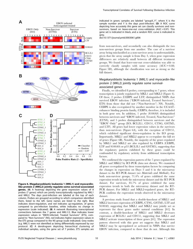
Megakaryoblastic leukemia 1 (MKL1) and myocardin-likeprotein 2 (MKL2) jointly regulate some survival-associated genes
Finally, we identified 8 probes, corresponding to 7 genes, whose
transcription is jointly regulated by MKL1 and MKL2 (Figure 6).
Of these, 2 probes (CEBPE and LTF) distinguished NHPs that
survived EBOV infection (‘‘EBOV-infected, Treated Survivors’’;
ETS) from those that did not (‘‘Non-Survivors’’; NS). Notably,
CEBPE is also co-regulated by another member in the CCAAT-
enhancer binding protein family, CEBPA; therefore, it is included
in both gene sets. In addition, 1 probe (S100A9) distinguished
between survivors and ‘‘EBOV-infected, Treated, Non-Survivors’’
(ETNS), and 5 probes distinguished between survivors and the
‘‘EBOV Only’’ group (EO; BCL2L1, CD151, CTSG, GSTM5,
and LTF). All genes had lower expression values in the survivors
than non-survivors (Figure 6A), with the exception of CD151,
which exhibited significant down-regulation in the EO group.
Importantly, MKL1 and MKL2 appear to co-regulate the full set
of genes (Figure 6B). Notably, the majority of the genes regulated
by MKL1 and MKL2 are also regulated by CEBPA (CEBPE,
LTF and S100A9) or p53 (BCL2L1 and GSTM5), suggesting that
the regulatory pattern exhibited by these genes could be
confounded by regulatory activity from additional transcription
factors.
We confirmed the expression pattern of the 7 genes regulated by
MKL1 and MKL2 by RT-PCR (data not shown). We examined
all genes co-regulated by these transcription factors by comparing
the changes in expression on Days 3 and 6 in the microarray
dataset to the RT-PCR dataset (see Materials and Methods). For
both non-survivor groups, 71.4% of genes exhibited the same
expression trends in both the microarray dataset and the RT-PCR
dataset. For survivors, 85.7% of genes exhibited the same
expression trends in both the microarray dataset and the RT-
PCR dataset. For MKL1 and MKL2-regulated genes, the RT-
PCR confirms the expression trends observed in the microarray
dataset.
A previous study found that a double-knockout of MKL1 and
MKL2 increases expression of CEBPE, CTSG, GSTM5, LTF and
S100A9, suggesting that MKL1 and MKL2 may jointly act as
repressors for these genes under natural conditions [61]. In
contrast, a double-knockout of MKL1 and MKL2 decreases
expression of BCL2L1 and CD151, suggesting that MKL1 and
MKL2 activate transcription of these genes [61]. The expression
pattern observed with this gene set suggests that MKL1 and
MKL2 may be up-regulated or activated in NHPs that survive
EBOV infection, compared to those that do not. Although this
Figure 6. Megakaryoblastic leukemia 1 (MKL1) and myocardin-like protein 2 (MKL2) jointly regulate some survival-associatedgenes. (A) A heatmap depicting the gene expression values of 8probes (7 genes) which are jointly transcriptionally regulated by MKL1and MKL2. The days post-infection are labeled at the top of eachcolumn. Probes are grouped according to which comparison identifiedthem, listed to the left. Gene names are listed to the right. Blueindicates down-regulation, and red indicates up-regulation, of genescompared to pre-infection baseline, white indicates no change inexpression (scale indicated). (B) A network illustrating transcriptionalconnections between the 7 genes and MKL1/2. Blue indicates lowerexpression values in ‘‘EBOV-infected, Treated Survivors’’ (ETS), com-pared to ‘‘Non-Survivors’’ (NS), red indicates higher expression values inthe ETS group compared to the NS group (scale indicated). Grey nodes(e.g. MKL1) were not identified as being differentially expressed by ourprotocol. (C) A dendrogram depicting hierarchical clustering ofindividual samples, using the gene set of 7 probes. ETS samples are
indicated in green; samples are labeled ‘‘groupX_Y’’, where X is thesample number and Y is the days post-infection. (D) A ROC curvedepicting how accurately this gene set can classify survivors and non-survivors, based on leave-one-out cross-validation (AUC = 0.97). Thisgene set is indicated in black, and a random ROC curve is indicated ingrey.doi:10.1371/journal.pntd.0003061.g006
Transcriptional Correlates of Survival Following Ebolavirus Infection
PLOS Neglected Tropical Diseases | www.plosntds.org 10 July 2014 | Volume 8 | Issue 7 | e3061
observed expression pattern is consistent with what would be
observed if MKL1 and MKL2 were activated, this set of probes is
too small to distinguish between survivors and non-survivors
groups (Figure 6C; ETS indicated in green). A hierarchically-
clustered dendrogram has two major branches, which are each
interspersed with EO, ETNS and ETS samples; this suggests that
the overall expression profile of survivors and non-survivors is not
statistically distinguishable. Although the dendrogram suggests
that the overall expression profile in each array is incapable of
distinguishing survivors and non-survivors, evaluation of individ-
ual gene contributions using leave-one-out cross-validation shows
that this gene set is capable of classifying samples with high
accuracy (AUC = 0.97; Figure 6D).
Discussion
These results show the potential of high-throughput transcrip-
tional studies for identifying putative markers of survival following
EBOV infection. In particular, we identified a minimal survival-
associated gene set that accurately distinguished survival outcome
following post-infection anticoagulant treatment of non-human
primates (NHPs) infected with EBOV. We identified 20 genes that
were characterized by significant, coherent and opposing expres-
sion patterns when comparing survivors and non-survivors.
Several of these genes exhibit differential regulation as early as 3
days post-infection, prior to the appearance of clinical symptoms
of EBOV infection; this early differential regulation is especially
important for the identification of early-stage biomarkers to
distinguish disease outcomes.
Importantly, several of these genes are associated with different
viral infections [46–50]. Proteins such as ILF3 and RUVBL2 are
known to suppress viral replication in other viruses [44,45,53], and
we observe that their expression is higher in survivors than non-
survivors. Notably, an isoform of ILF3 is known to bind EBOV
protein VP35, suppressing the function of the viral polymerase
[54]. This suggests a mechanism of action in which survivors may
up-regulate the transcription of certain genes, e.g. ILF3, in order
to suppress viral replication.
We also observed that microRNA 122 (miR-122) is down-
regulated in survivors compared to non-survivors, suggesting that
inhibition of miR-122 activity increases survival following EBOV
infection. To date, there have been no studies investigating
whether miR-122 interacts with the EBOV genome, but it is well-
documented that miR-122 binds the Hepatitis C virus genome to
support the replication of this virus [55,56]. Comparison of the
putative binding motifs of miR-122 [55] to the consensus sequence
of EBOV (Mayinga, Zaire, 1976) [62] reveals multiple potential
binding sites in the viral genome (data not shown). This suggests
that miR-122 is worth further investigation as a regulator of
EBOV infection.
Though a minimal set of 20 genes could separate survivors from
non-survivors, we were interested in also studying the general host
response to EBOV infection, and to determine if there was a
survival-associated transcriptional profile. We identified 238 genes
that accurately distinguished treatment groups and survival
outcomes following EBOV infection. Functional annotation of
these 238 genes confirmed that this gene set was comparable with
previously published studies of EBOV infection [21,22,24,27]. In
particular, the expression pattern that we observe for IL6, in which
non-survivors are significantly more up-regulated in early stages of
infection than survivors, is supported by similar changes in protein
concentration reported in previous studies [7,9,25]. There is also a
pattern of significant up-regulation of genes associated with
immune response in non-survivors, but not in survivors, consistent
with the hypothesis that non-survivors exhibit severe dysregulation
of the inflammatory response [21,22,24,27]. Importantly, our
work highlights the utility of using a minimal survival-associated
gene set to identify individual genes correlated with survival
following EBOV infection, which is not possible when assessing
global transcriptional responses in the host, as in previous work
[22,24].
When we compare our data to the results of a study of survival-
associated biomarkers in human samples from the Sudan virus
(SUDV) outbreak [25], we find notable similarities. Similar to this
study, we observed significant up-regulation chemokines and
cytokines, such as CCL3, CXCL10, IL1RN, IL6 and TNF,
throughout infection. This study reported that ferritin was a good
correlate of hemorrhage and death in response to SUDV infection
[25]. We observed down-regulation of ferritin throughout
infection, although this pattern was not correlated with survival
on a transcriptional level. However, we find that another iron-
binding protein, lactotransferrin (LTF) is highly correlated with
survival outcome in EBOV-infected NHPs. This similarity suggests
that iron modulation may play an important role in regulating
filovirus infection, especially in relation to coagulopathies and
hemorrhage, and that our biomarkers merit further study in a
human system.
We identified 3 transcriptional modules which were significantly
enriched in the gene set: (i) CCAAT/enhancer-binding protein
alpha (CEBPA); (ii) tumor protein 53 (p53); and (iii) megakaryo-
blastic leukemia 1 (MKL1) and myocardin-like protein 2 (MKL2).
Previous studies have shown that p53 plays a crucial role during
viral infection, which invariably disrupts normal cell cycle
processes, in a variety of DNA and RNA viruses [63–66]. In
particular, p53 is known to be associated with the Type I
interferon response and has been previously reported to enhance
viral-induced apoptosis in other infections [65,67]. In our
examination of p53-regulated genes, we found that several are
associated with regulation of apoptosis (e.g. BCL2L1 [68], CDC42
[69], CDK2 [70], FOXO3 [71], and PCNA [72]). This may
suggest a role for p53 as a mediator of apoptosis following EBOV
infection. However, due to a lack of a consensus pattern of
expression, we are unable to determine the underlying regulation
of p53 in this dataset, and therefore cannot draw conclusions
about the activity of p53 in NHPs that survived EBOV infection
when compared to non-survivors.
Interestingly, there is no consensus as to how CCAAT/
enhancer binding proteins (such as CEBPA) function in general
during viral infection, but they have been previously implicated in
promoting the replication of some viruses. For example, CEBP
binding sites exist in the Human immunodeficiency virus (HIV)
genome, and CEBPs are required for the replication and
regulation of HIV [73–75] and Simian immunodeficiency virus
[76]. Similarly, physical binding and interactions have been
observed between CEBPs and the proteins of Hepatitis B virus
[77,78], Epstein-Barr virus [79], and HIV [80]. In contrast,
CEBPs have been known to down-regulate or inhibit replication of
T-cell leukemia virus [81,82] and some human papillomaviruses
[83]. Our results suggest that strong CEBP responses are
correlated with poorer prognosis following EBOV infection. We
hypothesize that CEBP-regulated genes may contribute to the
inflammatory response to infection, or to the dysregulation of
coagulation.
Our studies are the first to suggest a role for MKL1 and MKL2
in viral infection, although roles for both proteins were recently
identified in megakaryocyte differentiation and platelet formation
[61]. Because dysregulation of coagulation is a common charac-
teristic of EBOV infection, it is possible that MKL1 and MKL2
Transcriptional Correlates of Survival Following Ebolavirus Infection
PLOS Neglected Tropical Diseases | www.plosntds.org 11 July 2014 | Volume 8 | Issue 7 | e3061
regulate coagulation in response to EBOV challenge. Indeed, we
observe that the downstream targets of MKL1 and MKL2 exhibit
an expression profile consistent with up-regulation of MKL1 and
MKL2 in survivors, compared to non-survivors. This implies that
survivors increase the regulation of coagulation processes,
potentially avoiding the typical coagulopathies associated with
late-stage EBOV infection. However, the majority of genes that
are regulated by MKL1 and MKL2 are also regulated by CEBPA
or p53, suggesting that the regulation observed is not due to the
transcriptional activity of MKL1 and MKL2 alone. Despite this,
these genes display a strong expression profile that is consistent
with up-regulation of MKL1 and MKL2 when compared to a
previous study [61], suggesting that survivors are able to recover in
part due to normal MKL1 and MKL2 function.
It is important to note that we did not find a single unique gene
that distinguished between survival outcomes of EBOV-infected
NHPs, suggesting that survival following anticoagulant treatment
is driven by a complex set of transcriptional responses. In addition,
gene sets and pathways we have identified are associated with
survival following anticoagulant treatment, and are therefore
specific to this condition. We also stress that the observed results
are in EBOV-infected NHPs, and our findings and conclusions
may not be applicable to additional viral infections, although
infection-specific signatures may exist. Under these conditions, we
identify several complex transcriptional responses that clearly
differentiate between survivors and non-survivors following EBOV
infection. In particular, we observe several survival-associated
profiles that are driven by specific upstream transcriptional
regulators (e.g. CEBPA, p53, and MKL1/MKL2). Notably, these
transcription factors have not been previously associated with
EBOV infection, and would not have been identified without
pathway analysis, due to lack of differential regulation. In
particular, the ability of a small set of 20 genes to distinguish
between survival outcomes suggests that they could potentially
serve as biomarkers of disease outcome. Our results demonstrate
that classification of treatment groups or disease outcome can be
accomplished with a small gene set, which can be useful for
identifying individual transcriptional markers associated with
survival following anticoagulant treatment of EBOV infection.
Supporting Information
Figure S1 This figure illustrates the overall process of micro-
array analysis described in this paper, including sample collection,
microarray processing and normalization, and analysis of the data
(as described in Materials and Methods).(PNG)
Table S1 This table lists the 20 genes from the minimal survival-
associated gene set (as described in Materials and Methods). The
table lists the Gene Symbol, Gene Entrez ID, a brief description of
the gene name, and a short list of gene functions as described by
the Gene Ontology ‘‘function’’ category.
(XLS)
Table S2 This table lists 241 genes from the general survival-
associated transcriptional profile (as described in Materials andMethods). The table lists the Gene Symbol, Gene Entrez ID, a
brief description of the gene name, and a short list of gene
functions as described by the Gene Ontology ‘‘function’’ category.
(XLS)
Acknowledgments
We would like to thank John Coller and the staff of the Stanford Functional
Genomics Facility for supplying us with the human cDNA microarrays
used for this study.
Footnotes: Opinions, interpretations, conclusions, and recommenda-
tions are those of the author and are not necessarily endorsed by the U.S.
Army.
Author Contributions
Conceived and designed the experiments: JYY SG JHC YX ANH LEH.
Performed the experiments: JYY ANH JBG. Analyzed the data: SG JYY.
Contributed reagents/materials/analysis tools: JBG KHR TWG LEH.
Contributed to the writing of the manuscript: SG JHC.
References
1. (2011) Virus taxonomy: classification and nomenclature of viruses: Ninth Report of
the International Committee on Taxonomy of Viruses; King AMQ, Adams, M.J.,
Carstens, E.B., and Lefkowitz, E.J., editor. San Diego: Elsevier Academic Press.
2. Feldmann H, Geisbert TW (2011) Ebola haemorrhagic fever. Lancet 377: 849–
862.
3. Feldmann H, Jones S, Klenk HD, Schnittler HJ (2003) Ebola virus: fromdiscovery to vaccine. Nat Rev Immunol 3: 677–685.
4. Feldmann H, Jones SM, Daddario-DiCaprio KM, Geisbert JB, Stroher U, et al.(2007) Effective post-exposure treatment of Ebola infection. PLoS Pathog 3: e2.
5. Geisbert TW, Daddario-Dicaprio KM, Geisbert JB, Reed DS, Feldmann F, etal. (2008) Vesicular stomatitis virus-based vaccines protect nonhuman primates
against aerosol challenge with Ebola and Marburg viruses. Vaccine 26: 6894–6900.
6. Geisbert TW, Geisbert JB, Leung A, Daddario-DiCaprio KM, Hensley LE, etal. (2009) Single-injection vaccine protects nonhuman primates against infection
with marburg virus and three species of ebola virus. J Virol 83: 7296–7304.
7. Geisbert TW, Hensley LE, Jahrling PB, Larsen T, Geisbert JB, et al. (2003)
Treatment of Ebola virus infection with a recombinant inhibitor of factor VIIa/tissue factor: a study in rhesus monkeys. Lancet 362: 1953–1958.
8. Geisbert TW, Lee AC, Robbins M, Geisbert JB, Honko AN, et al. (2010)
Postexposure protection of non-human primates against a lethal Ebola virus
challenge with RNA interference: a proof-of-concept study. Lancet 375: 1896–1905.
9. Hensley LE, Stevens EL, Yan SB, Geisbert JB, Macias WL, et al. (2007)
Recombinant human activated protein C for the postexposure treatment of
Ebola hemorrhagic fever. J Infect Dis 196 Suppl 2: S390–399.
10. Jones SM, Feldmann H, Stroher U, Geisbert JB, Fernando L, et al. (2005) Liveattenuated recombinant vaccine protects nonhuman primates against Ebola and
Marburg viruses. Nat Med 11: 786–790.
11. Richardson JS, Yao MK, Tran KN, Croyle MA, Strong JE, et al. (2009)
Enhanced protection against Ebola virus mediated by an improved adenovirus-
based vaccine. PLoS One 4: e5308.
12. Sullivan NJ, Sanchez A, Rollin PE, Yang ZY, Nabel GJ (2000) Development of apreventive vaccine for Ebola virus infection in primates. Nature 408: 605–609.
13. Geisbert TW, Hensley LE, Larsen T, Young HA, Reed DS, et al. (2003)
Pathogenesis of Ebola hemorrhagic fever in cynomolgus macaques: evidencethat dendritic cells are early and sustained targets of infection. Am J Pathol 163:
2347–2370.
14. Geisbert TW, Young HA, Jahrling PB, Davis KJ, Larsen T, et al. (2003)
Pathogenesis of Ebola hemorrhagic fever in primate models: evidence thathemorrhage is not a direct effect of virus-induced cytolysis of endothelial cells.
Am J Pathol 163: 2371–2382.
15. Hensley LE, Jones SM, Feldmann H, Jahrling PB, Geisbert TW (2005) Ebolaand Marburg viruses: pathogenesis and development of countermeasures. Curr
Mol Med 5: 761–772.
16. Flint SJ, Enquist LW, Racaniello VR, Skalka AM (2009) Principles of Virology.
SciELO Espana.
17. Alvarez CP, Lasala F, Carrillo J, Muniz O, Corbi AL, et al. (2002) C-type lectins
DC-SIGN and L-SIGN mediate cellular entry by Ebola virus in cis and in trans.
J Virol 76: 6841–6844.
18. Harty RN, Brown ME, Wang G, Huibregtse J, Hayes FP (2000) A PPxY motif
within the VP40 protein of Ebola virus interacts physically and functionally with
a ubiquitin ligase: implications for filovirus budding. Proc Natl Acad Sci U S A
97: 13871–13876.
19. Timmins J, Schoehn G, Ricard-Blum S, Scianimanico S, Vernet T, et al. (2003)
Ebola virus matrix protein VP40 interaction with human cellular factors Tsg101
and Nedd4. J Mol Biol 326: 493–502.
20. Gupta M, Mahanty S, Ahmed R, Rollin PE (2001) Monocyte-derived human
macrophages and peripheral blood mononuclear cells infected with ebola virus
secrete MIP-1alpha and TNF-alpha and inhibit poly-IC-induced IFN-alpha invitro. Virology 284: 20–25.
21. Hensley LE, Young HA, Jahrling PB, Geisbert TW (2002) Proinflammatory
response during Ebola virus infection of primate models: possible involvement of
the tumor necrosis factor receptor superfamily. Immunol Lett 80: 169–179.
Transcriptional Correlates of Survival Following Ebolavirus Infection
PLOS Neglected Tropical Diseases | www.plosntds.org 12 July 2014 | Volume 8 | Issue 7 | e3061
22. Rubins KH, Hensley LE, Wahl-Jensen V, Daddario DiCaprio KM, Young HA,et al. (2007) The temporal program of peripheral blood gene expression in the
response of nonhuman primates to Ebola hemorrhagic fever. Genome Biol 8:R174.
23. Villinger F, Rollin PE, Brar SS, Chikkala NF, Winter J, et al. (1999) Markedly
elevated levels of interferon (IFN)-gamma, IFN-alpha, interleukin (IL)-2, IL-10,and tumor necrosis factor-alpha associated with fatal Ebola virus infection.
J Infect Dis 179 Suppl 1: S188–191.
24. Yen JY, Garamszegi S, Geisbert JB, Rubins KH, Geisbert TW, et al. (2011)
Therapeutics of Ebola hemorrhagic fever: whole-genome transcriptional analysis
of successful disease mitigation. J Infect Dis 204 Suppl 3: S1043–1052.
25. McElroy AK, Erickson BR, Flietstra TD, Rollin PE, Nichol ST, et al. (2014)
Ebola hemorrhagic fever: novel biomarker correlates of clinical outcome.J Infect Dis: doi: 10.1093/infdis/jiu088.
26. Sanchez A (2001) Ebola Viruses: Wiley Online Library.
27. Reed DS, Hensley LE, Geisbert JB, Jahrling PB, Geisbert TW (2004) Depletion
of peripheral blood T lymphocytes and NK cells during the course of ebola
hemorrhagic Fever in cynomolgus macaques. Viral Immunol 17: 390–400.
28. Bosio CM, Aman MJ, Grogan C, Hogan R, Ruthel G, et al. (2003) Ebola and
Marburg viruses replicate in monocyte-derived dendritic cells without inducingthe production of cytokines and full maturation. J Infect Dis 188: 1630–1638.
29. Bray M, Geisbert TW (2005) Ebola virus: the role of macrophages and dendriticcells in the pathogenesis of Ebola hemorrhagic fever. Int J Biochem Cell Biol 37:
1560–1566.
30. Gentleman RC, Carey VJ, Bates DM, Bolstad B, Dettling M, et al. (2004)Bioconductor: open software development for computational biology and
bioinformatics. Genome Biol 5: R80.
31. Smyth GK (2005) Limma: linear models for microarray data; Gentleman R CV,
Dudoit S, Irizarry R, and Huber W., editor. New York: Springer.
32. Team RDC (2010) R: A language and environment for statistical computing. In:
Computing RFfS, editor. Vienna, Austria.
33. Ritchie ME, Silver J, Oshlack A, Holmes M, Diyagama D, et al. (2007) Acomparison of background correction methods for two-colour microarrays.
Bioinformatics 23: 2700–2707.
34. Smyth GK, Speed T (2003) Normalization of cDNA microarray data. Methods
31: 265–273.
35. Edgar R, Domrachev M, Lash AE (2002) Gene Expression Omnibus: NCBI
gene expression and hybridization array data repository. Nucleic Acids Res 30:
207–210.
36. de Hoon MJ, Imoto S, Nolan J, Miyano S (2004) Open source clustering
software. Bioinformatics 20: 1453–1454.
37. Eisen MB, Spellman PT, Brown PO, Botstein D (1998) Cluster analysis and
display of genome-wide expression patterns. Proc Natl Acad Sci U S A 95:14863–14868.
38. Saldanha AJ (2004) Java Treeview–extensible visualization of microarray data.
Bioinformatics 20: 3246–3248.
39. Shannon P, Markiel A, Ozier O, Baliga NS, Wang JT, et al. (2003) Cytoscape: a
software environment for integrated models of biomolecular interactionnetworks. Genome Res 13: 2498–2504.
40. Medigeshi GR, Hirsch AJ, Brien JD, Uhrlaub JL, Mason PW, et al. (2009) Westnile virus capsid degradation of claudin proteins disrupts epithelial barrier
function. J Virol 83: 6125–6134.
41. Kato N, Ji G, Wang Y, Baba M, Hoshida Y, et al. (2005) Large-scale search ofsingle nucleotide polymorphisms for hepatocellular carcinoma susceptibility
genes in patients with hepatitis C. Hepatology 42: 846–853.
42. Lee JW, Liao PC, Young KC, Chang CL, Chen SS, et al. (2011) Identification of
hnRNPH1, NF45, and C14orf166 as novel host interacting partners of themature hepatitis C virus core protein. J Proteome Res 10: 4522–4534.
43. Shin HJ, Kim SS, Cho YH, Lee SG, Rho HM (2002) Host cell proteins binding
to the encapsidation signal epsilon in hepatitis B virus RNA. Arch Virol 147:471–491.
44. Gomila RC, Martin GW, Gehrke L (2011) NF90 binds the dengue virus RNA 39
terminus and is a positive regulator of dengue virus replication. PLoS One 6:
e16687.
45. Wang P, Song W, Mok BW, Zhao P, Qin K, et al. (2009) Nuclear factor 90
negatively regulates influenza virus replication by interacting with viral
nucleoprotein. J Virol 83: 7850–7861.
46. Adams WC, Bond E, Havenga MJ, Holterman L, Goudsmit J, et al. (2009)
Adenovirus serotype 5 infects human dendritic cells via a coxsackievirus-adenovirus receptor-independent receptor pathway mediated by lactoferrin and
DC-SIGN. J Gen Virol 90: 1600–1610.
47. Berlutti F, Pantanella F, Natalizi T, Frioni A, Paesano R, et al. (2011) Antiviralproperties of lactoferrin–a natural immunity molecule. Molecules 16: 6992–
7018.
48. Valenti P, Antonini G (2005) Lactoferrin: an important host defence against
microbial and viral attack. Cell Mol Life Sci 62: 2576–2587.
49. Valimaa H, Tenovuo J, Waris M, Hukkanen V (2009) Human lactoferrin but
not lysozyme neutralizes HSV-1 and inhibits HSV-1 replication and cell-to-cell
spread. Virol J 6: 53.
50. Zheng Y, Zhang W, Ye Q, Zhou Y, Xiong W, et al. (2012) Inhibition of Epstein-
Barr virus infection by lactoferrin. J Innate Immun 4: 387–398.
51. Li Y, Masaki T, Yamane D, McGivern DR, Lemon SM (2013) Competing and
noncompeting activities of miR-122 and the 59 exonuclease Xrn1 in regulationof hepatitis C virus replication. Proc Natl Acad Sci U S A 110: 1881–1886.
52. Sion-Vardy N, Lasarov I, Delgado B, Gopas J, Benharroch D, et al. (2009)
Measles virus: evidence for association with lung cancer. Exp Lung Res 35: 701–
712.
53. Kakugawa S, Shimojima M, Neumann G, Goto H, Kawaoka Y (2009) RuvB-
like protein 2 is a suppressor of influenza A virus polymerases. J Virol 83: 6429–
6434.
54. Shabman RS, Leung DW, Johnson J, Glennon N, Gulcicek EE, et al. (2011)
DRBP76 associates with Ebola virus VP35 and suppresses viral polymerase
function. J Infect Dis 204 Suppl 3: S911–918.
55. Jopling CL, Schutz S, Sarnow P (2008) Position-dependent function for a
tandem microRNA miR-122-binding site located in the hepatitis C virus RNA
genome. Cell Host Microbe 4: 77–85.
56. Jopling CL, Yi M, Lancaster AM, Lemon SM, Sarnow P (2005) Modulation of
hepatitis C virus RNA abundance by a liver-specific MicroRNA. Science 309:
1577–1581.
57. Ambroise C, McLachlan GJ (2002) Selection bias in gene extraction on the basis
of microarray gene-expression data. Proc Natl Acad Sci U S A 99: 6562–6566.
58. Golub TR, Slonim DK, Tamayo P, Huard C, Gaasenbeek M, et al. (1999)
Molecular classification of cancer: class discovery and class prediction by gene
expression monitoring. Science 286: 531–537.
59. Simon R, Radmacher MD, Dobbin K, McShane LM (2003) Pitfalls in the use of
DNA microarray data for diagnostic and prognostic classification. J Natl Cancer
Inst 95: 14–18.
60. Gery S, Gombart AF, Yi WS, Koeffler C, Hofmann WK, et al. (2005)
Transcription profiling of C/EBP targets identifies Per2 as a gene implicated in
myeloid leukemia. Blood 106: 2827–2836.
61. Smith EC, Thon JN, Devine MT, Lin S, Schulz VP, et al. (2012) MKL1 and
MKL2 play redundant and crucial roles in megakaryocyte maturation and
platelet formation. Blood 120: 2317–2329.
62. Volchkov VE, Volchkova VA, Chepurnov AA, Blinov VM, Dolnik O, et al.
(1999) Characterization of the L gene and 59 trailer region of Ebola virus. J Gen
Virol 80 (Pt 2): 355–362.
63. Lazo PA, Santos CR (2011) Interference with p53 functions in human viral
infections, a target for novel antiviral strategies? Rev Med Virol 50: doi:
10.1002/rmv.696.
64. Collot-Teixeira S, Bass J, Denis F, Ranger-Rogez S (2004) Human tumor
suppressor p53 and DNA viruses. Rev Med Virol 14: 301–319.
65. Turpin E, Luke K, Jones J, Tumpey T, Konan K, et al. (2005) Influenza virus
infection increases p53 activity: role of p53 in cell death and viral replication.
J Virol 79: 8802–8811.
66. Marques JT, Rebouillat D, Ramana CV, Murakami J, Hill JE, et al. (2005)
Down-regulation of p53 by double-stranded RNA modulates the antiviral
response. J Virol 79: 11105–11114.
67. Munoz-Fontela C, Macip S, Martinez-Sobrido L, Brown L, Ashour J, et al.
(2008) Transcriptional role of p53 in interferon-mediated antiviral immunity.
J Exp Med 205: 1929–1938.
68. Hagenbuchner J, Ausserlechner MJ, Porto V, David R, Meister B, et al. (2010)
The anti-apoptotic protein BCL2L1/Bcl-xL is neutralized by pro-apoptotic
PMAIP1/Noxa in neuroblastoma, thereby determining bortezomib sensitivity
independent of prosurvival MCL1 expression. J Biol Chem 285: 6904–6912.
69. Thomas A, Giesler T, White E (2000) p53 mediates bcl-2 phosphorylation and
apoptosis via activation of the Cdc42/JNK1 pathway. Oncogene 19: 5259–
5269.
70. Huang H, Regan KM, Lou Z, Chen J, Tindall DJ (2006) CDK2-dependent
phosphorylation of FOXO1 as an apoptotic response to DNA damage. Science
314: 294–297.
71. Zhang X, Tang N, Hadden TJ, Rishi AK (2011) Akt, FoxO and regulation of
apoptosis. Biochim Biophys 1813: 1978–1986.
72. Witko-Sarsat V, Mocek J, Bouayad D, Tamassia N, Ribeil JA, et al. (2010)
Proliferating cell nuclear antigen acts as a cytoplasmic platform controlling
human neutrophil survival. J Exp Med 207: 2631–2645.
73. Henderson AJ, Calame KL (1997) CCAAT/enhancer binding protein (C/EBP)
sites are required for HIV-1 replication in primary macrophages but not CD4(+)
T cells. Proc Natl Acad Sci U S A 94: 8714–8719.
74. Henderson AJ, Connor RI, Calame KL (1996) C/EBP activators are required
for HIV-1 replication and proviral induction in monocytic cell lines. Immunity
5: 91–101.
75. Schwartz C, Catez P, Rohr O, Lecestre D, Aunis D, et al. (2000) Functional
interactions between C/EBP, Sp1, and COUP-TF regulate human immuno-
deficiency virus type 1 gene transcription in human brain cells. J Virol 74: 65–
73.
76. Mohan M, Aye PP, Borda JT, Alvarez X, Lackner AA (2008) CCAAT/
enhancer binding protein beta is a major mediator of inflammation and viral
replication in the gastrointestinal tract of simian immunodeficiency virus-
infected rhesus macaques. Am J Pathol 173: 106–118.
77. Choi BH, Park GT, Rho HM (1999) Interaction of hepatitis B viral X protein
and CCAAT/enhancer-binding protein alpha synergistically activates the
hepatitis B viral enhancer II/pregenomic promoter. J Biol Chem 274: 2858–
2865.
78. Mahe Y, Mukaida N, Kuno K, Akiyama M, Ikeda N, et al. (1991) Hepatitis B
virus X protein transactivates human interleukin-8 gene through acting on
nuclear factor kB and CCAAT/enhancer-binding protein-like cis-elements.
J Biol Chem 266: 13759–13763.
Transcriptional Correlates of Survival Following Ebolavirus Infection
PLOS Neglected Tropical Diseases | www.plosntds.org 13 July 2014 | Volume 8 | Issue 7 | e3061
79. Wu FY, Wang SE, Chen H, Wang L, Hayward SD, et al. (2004) CCAAT/
enhancer binding protein alpha binds to the Epstein-Barr virus (EBV) ZTAprotein through oligomeric interactions and contributes to cooperative transcrip-
tional activation of the ZTA promoter through direct binding to the ZII and ZIIIB
motifs during induction of the EBV lytic cycle. J Virol 78: 4847–4865.80. Ambrosino C, Ruocco MR, Chen X, Mallardo M, Baudi F, et al. (1997) HIV-1
Tat induces the expression of the interleukin-6 (IL6) gene by binding to the IL6leader RNA and by interacting with CAAT enhancer-binding protein beta (NF-
IL6) transcription factors. J Biol Chem 272: 14883–14892.
81. Hivin P, Gaudray G, Devaux C, Mesnard JM (2004) Interaction between C/
EBPbeta and Tax down-regulates human T-cell leukemia virus type Itranscription. Virology 318: 556–565.
82. Grant C, Nonnemacher M, Jain P, Pandya D, Irish B, et al. (2006) CCAAT/
enhancer-binding proteins modulate human T cell leukemia virus type 1 longterminal repeat activation. Virology 348: 354–369.
83. Bauknecht T, Shi Y (1998) Overexpression of C/EBPbeta represses humanpapillomavirus type 18 upstream regulatory region activity in HeLa cells by
interfering with the binding of TATA-binding protein. J Virol 72: 2113–2124.
Transcriptional Correlates of Survival Following Ebolavirus Infection
PLOS Neglected Tropical Diseases | www.plosntds.org 14 July 2014 | Volume 8 | Issue 7 | e3061