towards lignin-protein crosslinking: amino acid adducts of a lignin model quinone methide
TRANSCRIPT

ORIGINAL PAPER
Towards lignin-protein crosslinking: amino acid adductsof a lignin model quinone methide
Brett G. Diehl • Heath D. Watts •
James D. Kubicki • Matthew R. Regner •
John Ralph • Nicole R. Brown
Received: 12 December 2013 / Accepted: 22 January 2014 / Published online: 29 January 2014
� Springer Science+Business Media Dordrecht 2014
Abstract The polyaromatic structure of lignin has
long been recognized as a key contributor to the
rigidity of plant vascular tissues. Although lignin
structure was once conceptualized as a highly net-
worked, heterogeneous, high molecular weight poly-
mer, recent studies have suggested a very different
configuration may exist in planta. These findings,
coupled with the increasing attention and interest in
efficiently utilizing lignocellulosic materials for green
materials and energy applications, have renewed
interest in lignin chemistry. Here we focus on quinone
methides (QMs)—key intermediates in lignin poly-
merization—that are quenched via reaction with cell-
wall-available nucleophiles. Reactions with alcohol
and uronic acid groups of hemicelluloses, for example,
can lead to lignin-carbohydrate crosslinks. Our work is
a first step toward exploring potential QM reactions
with nucleophilic groups in cell wall proteins. We
conducted a model compound study wherein the lignin
model compound guaiacylglycerol-b-guaiacyl ether 1,
was converted to its QM 2, then reacted with amino
acids bearing nucleophilic side-groups. Yields for the
QM-amino acid adducts ranged from quantitative in
the case of QM-lysine 3, to zero (no reaction) in the
cases of QM-threonine (Thr) 10 and QM-hydroxypro-
line (Hyp) 11. The structures of the QM-amino acidElectronic supplementary material The online version ofthis article (doi:10.1007/s10570-014-0181-y) contains supple-mentary material, which is available to authorized users.
B. G. Diehl (&)
Department of Agricultural and Biological Engineering,
The Pennsylvania State University, 226 Forest Resources
Building, University Park, PA 16802, USA
e-mail: [email protected]
H. D. Watts
Department of Geosciences, The Pennsylvania State
University, 305 Deike Building, University Park,
PA 16802, USA
e-mail: [email protected]
J. D. Kubicki
Department of Geosciences and the Earth and
Environmental Systems Institute, The Pennsylvania State
University, 335 Deike Building, University Park,
PA 16802, USA
e-mail: [email protected]
M. R. Regner � J. Ralph
Department of Biochemistry and DOE Great Lakes
Bioenergy Research Center, Wisconsin Energy Institute,
Madison, WI 53726, USA
e-mail: [email protected]
J. Ralph
e-mail: [email protected]
N. R. Brown
Department of Agricultural and Biological Engineering,
The Pennsylvania State University, 209 Agricultural
Engineering Building, University Park, PA 16802, USA
e-mail: [email protected]
123
Cellulose (2014) 21:1395–1407
DOI 10.1007/s10570-014-0181-y
adducts were confirmed via 1D and 2D nuclear
magnetic resonance (NMR) spectroscopy and density
functional theory (DFT) calculations, thereby extend-
ing the lignin NMR database to include amino acid
crosslinks. Some of the QM-amino acid adducts
formed both syn- and anti-isomers, whereas others
favored only one isomer. Because the QM-Thr 10 and
QM-Hyp 11 compounds could not be experimentally
prepared under conditions described here but could
potentially form in vivo, we used DFT to calculate
their NMR shifts. Characterization of these model
adducts extends the lignin NMR database to aid in the
identification of lignin-protein linkages in more com-
plex in vitro and in vivo systems, and may allow for
the identification of such linkages in planta.
Keywords Nuclear magnetic resonance
spectroscopy � Lignin � Protein � Quinone
methide � Amino acid � Crosslinking � Density
functional theory
Introduction
Plant cell walls are composed of a network of
interacting polymers, namely cellulose, hemicellu-
loses, pectins, lignin, and structural proteins (Cos-
grove 2005; McQueen-Mason and Cosgrove 1994). Of
these, lignin is the major aromatic component, derived
from monolignols—phenylpropanoid units whose
biosynthesis exhibits incredible plasticity (Boerjan
et al. 2003; Ralph et al. 2004a, b; Vanholme et al.
2010). Lignin’s mode of polymerization is unique
among the cell wall polymers. Resonance stabilized
radicals are enzymatically generated from the mono-
lignols, and as the radical-bearing structures couple
combinatorially, a heterogeneous polymer containing
many types of inter-unit linkages forms. The variety of
the inter-unit linkages contributes notable recalci-
trance to the plant cell wall, stymying not only natural
degradation, but also affecting the economics of many
industrial sectors, including the pulp and paper
industry, the developing biofuels industry, agricultural
industries, and chemical industries, which all seek
higher value products from lignin (Chapple et al.
2007; Chen and Dixon 2008; Jung 1989; Jung and
Allen 1995; Li et al. 2008; Stewart et al. 2006).
Inter-unit linkages are not, however, the sole factor
influencing lignin’s recalcitrance in planta. Lignin may
be crosslinked with other polymers in the plant wall.
Hydroxyl and uronic acid groups of polysaccharides
bear mildly nucleophilic groups that can react with a key
lignin intermediate—the a-carbon of quinone methides
(QMs) (Balakshin et al. 2011; Leary 1980; Miyagawa
et al. 2012; Ralph et al. 2009; Toikka et al. 1998; Yuan
et al. 2011). These QMs form each time a monolignol
radical couples at its b-position and, because b-coupling
is prevalent, the importance of QMs in lignin structure
cannot be understated. In certain cases, particularly b-5-
and b-b-coupling, QM intermediates are quickly
trapped intramolecularly, producing phenylcoumaran
and resinol units (Leary 1980; Ralph et al. 2009).
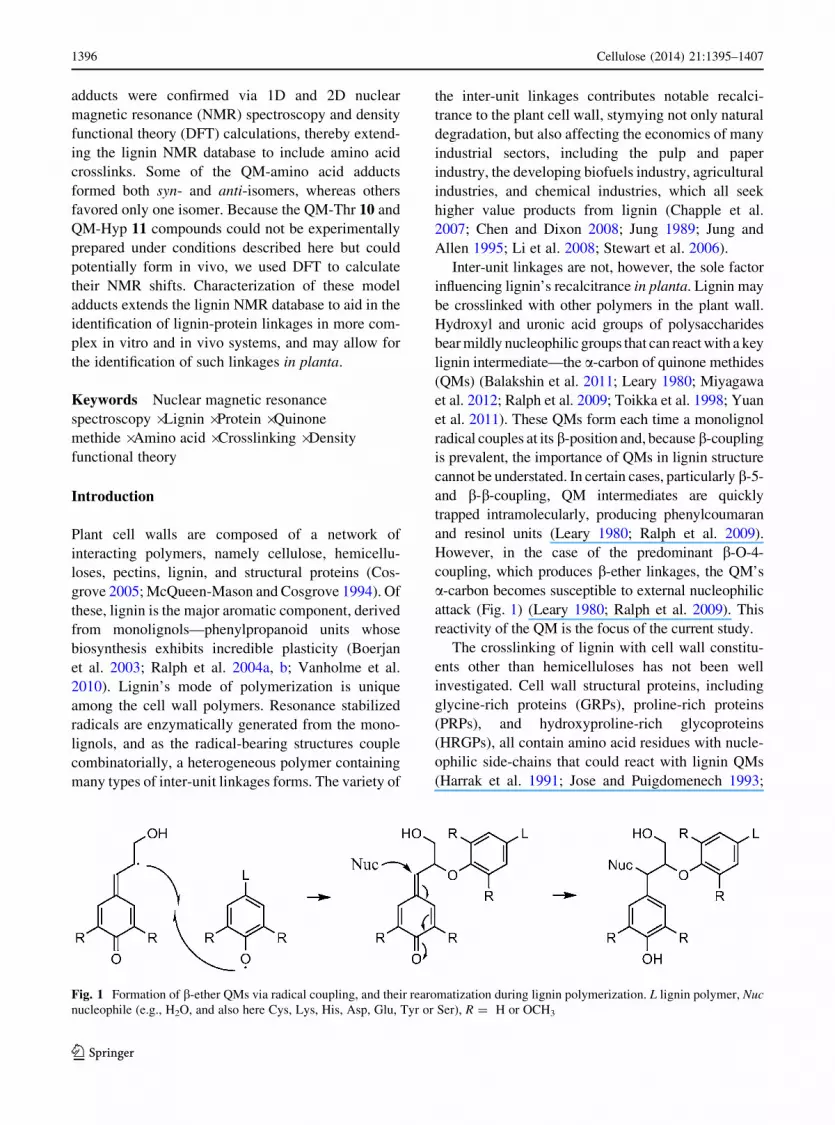
However, in the case of the predominant b-O-4-
coupling, which produces b-ether linkages, the QM’s
a-carbon becomes susceptible to external nucleophilic
attack (Fig. 1) (Leary 1980; Ralph et al. 2009). This
reactivity of the QM is the focus of the current study.
The crosslinking of lignin with cell wall constitu-
ents other than hemicelluloses has not been well
investigated. Cell wall structural proteins, including
glycine-rich proteins (GRPs), proline-rich proteins
(PRPs), and hydroxyproline-rich glycoproteins
(HRGPs), all contain amino acid residues with nucle-
ophilic side-chains that could react with lignin QMs
(Harrak et al. 1991; Jose and Puigdomenech 1993;
Fig. 1 Formation of b-ether QMs via radical coupling, and their rearomatization during lignin polymerization. L lignin polymer, Nuc
nucleophile (e.g., H2O, and also here Cys, Lys, His, Asp, Glu, Tyr or Ser), R = H or OCH3
1396 Cellulose (2014) 21:1395–1407
123

Kieliszewski et al. 2011; Ryser et al. 1997). Cell wall
proteins vary in quantity among species and cell types,
ranging from as low as 1–2 to 20 % on a dry weight
basis in wild type plants (Albersheim et al. 2010;
Cassab and Varner 1988). In Whitmore 1978a, b, 1982
showed evidence for the formation of lignin-protein
linkages in isolated cell walls of slash pine. Further
literature sources suggest that structural proteins may
crosslink with lignin, or possibly even nucleate, or
provide a template for, lignin structure, but these ideas
have not been adequately tested (Albersheim et al.
2010; Beat et al. 1989; Boerjan et al. 2003; Cassab and
Varner 1988; Harrak et al. 1991). If true, this
mechanism could provide spatial and temporal control
over lignin deposition and architecture (Beat et al.
1989). Furthermore, it has recently been suggested
that over-expression of cell wall proteins could result
in increased lignin-protein linkage formation, which
may affect cell wall physical and chemical properties,
for example increased sugar extractability (Liang et al.
2008; Xu et al. 2013). However, identifying such
linkages in planta would be difficult without first
determining diagnostic lignin-protein spectroscopic
signatures under simpler, more controlled conditions.
As a first step toward investigating potential lignin-
protein linkages in planta, we conducted a model
compound study to characterize products formed when
the lignin model compound guaiacylglycerol-b-guaia-
cyl ether 1 was converted to its QM 2, then reacted with
amino acids bearing nucleophilic side-groups (Fig. 2).
Thiols, amines, acids and alcohols have been shown to
quench QMs in a diverse array of systems. The thiol
group of glutathione reacts with an o-QM generated
from the flavonoid, quercetin (Awad et al. 2000); the
thiol group of cysteine (Cys) reacts with the relatively
unreactive p-QM, 2,6-di-tert-butyl-4-methylene-2,5-
cyclohexadienone (Bolton et al. 1997); and thiols and
thiolates react with QMs derived from anthracyclines
(Ramakrishnan and Fisher 1983). Similarly, amines
have been shown to trap lignin QMs (Ralph and Young
1983). A wide array of acid- and hydroxyl-containing
compounds react with p-QMs (Leary et al. 1977), and
primary (and to a much lesser extent, secondary)
hydroxyl groups of carbohydrates may react with QM
2 (Toikka et al. 1998). However, similar nucleophile-
QM adducts have not been characterized in lignin-
protein systems.
The nucleophilic amino acids investigated here—
Cys, lysine (Lys), histidine (His), aspartic acid (Asp),
glutamic acid (Glu), tyrosine (Tyr), serine (Ser), thre-
onine (Thr) and hydroxyproline (Hyp)—occur in plant
cell wall structural proteins and may react to form
lignin-protein crosslinks in vivo (Jose and Puigdomen-
ech 1993; Kieliszewski et al. 2011). Because cell wall
proteins are thought to exist in the wall prior to
lignification, the a-amine and a-acid groups of the
amino acids were protected to mimic their inclusion
within a peptide. This allowed reactions of the nucle-
ophilic side-chains to be determined without the com-
plication of competing reactions from the terminal a-
amine and a-acid groups. The QM-amino acid adducts
(Fig. 3) were characterized by nuclear magnetic reso-
nance (NMR) spectroscopy, density functional theory
(DFT), mass spectrometry, and UV/Visible (UV/Vis)
spectrophotometry. The characterization of these model
adducts extends the lignin NMR database to aid in the
identification of lignin-protein linkages in more com-
plex in vitro and in vivo systems (Ralph et al. 2004a, b).
Experimental
Materials
All chemicals used in the preparation of compounds 1
and 2, and lignin dehydrogenation polymer (DHP),
Fig. 2 Guaiacylglycerol-b-
guaiacyl ether 1 and its
derived quinone methide
(QM) 2
Cellulose (2014) 21:1395–1407 1397
123

Fig. 3 Lignin-cysteine
(QM-Cys) 3, lignin-lysine
(QM-Lys) 4, lignin-histidine
(QM-His) 5, lignin-aspartic
acid (QM-Asp) 6, lignin-
glutamic acid (QM-Glu) 7,
lignin-serine (QM-Ser) 8,
lignin-Tyrosine (QM-Tyr)
9, lignin-threonine
(QM-Thr) 10, and lignin-
hydroxyproline (QM-Hyp)
11 adducts derived from
QM 2
1398 Cellulose (2014) 21:1395–1407
123

were purchased from Sigma. All amino acids used in
the preparation of compounds 3-9 were purchased
from Sigma with the exception of Boc-L-His methyl
ester, which was purchased from Indofine Chemical
Company.
Model compound preparations
Compound 1 was prepared according to previous
methods, as was its QM analog (2) (Kawai et al.
1999; Landucci et al. 1981; Ralph and Young 1983).
Protected amino acids (1.05 eq) were added directly to
the anhydrous solution of 2 in dichloromethane at room
temperature. In the case of Lys, which was obtained as
Na-acetyl-L-Lys methyl ester hydrochloride, triethyl-
amine (*5 eq) was added in order to deprotonate the
terminal amine and facilitate dissolution. NMR was
used to show that triethylamine was not reactive
towards the QM. A stir bar was added, the flask was
stoppered, and the atmosphere was rendered inert by
alternating between vacuum and dry nitrogen several
times. The reaction was monitored visually; dissipation
of the yellow hue indicated consumption of the QM.
Intermittently, the reaction was also monitored by TLC
(1:1 ethyl acetate/hexanes). Lys and His reacted with
the QM within minutes, while other amino acids reacted
more slowly with the QM and were allowed to stir
overnight (Cys, Asp, Glu, Tyr) or for several days (Ser,
Thr, Hyp), again, with intermittent monitoring by TLC.
When TLC revealed that the reaction had reached
equilibrium the mixture was evaporated to dryness. In
the case of Lys (compound 4), the reaction went to
completion (complete consumption of the QM) and the
crude products were evaporated to dryness and charac-
terized without further purification. Compounds 3 and
6-9 were purified via flash chromatography using silica
gel and 1:1 ethyl acetate/hexanes as eluent. In the case
of QM-His (5), the product could not be chromato-
graphically separated (a range of eluent solvent systems
were attempted) from a-O-aryl products formed pre-
sumably due to self-dimerization of the QM (2);
however, mass spec and 2D NMR techniques were still
able to confirm the identity of the QM-His product. In
the case of QM-Ser (8), the product could not be fully
separated from unreacted Ser. The neat Ser shifts as
well as the shifts of compound 8 are labeled in the NMR
spectra (see online resource).
Lignin guaiacyl-based DHP was prepared accord-
ing to a previously published method (Terashima et al.
1995). The DHP was characterized via HSQC NMR as
described below and was found to contain shifts
typical of native lignin and DHP (Capanema et al.
2004; Kim and Ralph 2010). The DHP NMR spectrum
was then used as a reference against which diagnostic
NMR shifts of the lignin-protein model compounds
could be compared.
Nuclear magnetic resonance spectroscopy
NMR spectra (see online resource) were collected in
both acetone-d6 and DMSO-d6/pyridine-d5 (4:1 v/v,
500 ul). DMSO-d6/pyridine-d5 was chosen because it
is a preferred solvent for NMR of lignin DHP, milled
wood lignin (MWL), and whole cell walls; using the
same solvent system allows for accurate shift com-
parisons (Kim and Ralph 2010). In general, negligible
shift migration was observed between the two solvent
systems. NMR spectra were acquired on Bruker DPX-
300 (300 MHz 1H resonance freq.), DRX-400
(400 MHz 1H resonance freq.), AV-III-500
(500 MHz 1H resonance freq.) with a cryogenically-
cooled probe and inverse probe geometry (i.e., proton
coils closest to sample), AV-III-600 (500 MHz 1H
resonance freq.) with a cryogenically cooled probe,
and AV-III-850 (850 MHz 1H resonance freq.) with a
cryogenically-cooled probe. Spectral processing was
performed in Bruker’s Topspin 3.1 software. Standard
Bruker pulse programs were employed: 1H (8-16
scans), 13C (5 k-10 k scans), HMQC (Bruker pulse
program ‘inv4gptp’, 64 scans), and HMBC (Bruker
pulse program ‘inv4gslplrnd’, 64 scans). Spectra were
calibrated to the central solvent peaks (acetone: 2.05/
29.8 ppm; dimethyl sulfoxide: 2.50/39.5 ppm). In the
case of lignin DHP, NMR spectra were acquired on a
Bruker Biospin (Billerica, MA, USA) AVANCE 500
(500 MHz 1H resonance freq.) spectrometer fitted
with a cryogenically-cooled gradient probe having
inverse geometry, i.e., with the proton coils closest to
the sample. Spectra were processed with Bruker’s
Topspin 3.1 software, using the central solvent peak as
internal reference (dH/dC: dimethyl sulfoxide
(DMSO), 2.50/39.5 ppm). The synthetic lignin DHP
(*50 mg) was placed in an NMR tube (ID: 4.1 mm),
swollen homogeneously in DMSO-d6/pyridine-d5 (4:1
v/v, 500 ll) with the aid of ultrasonication (*3 h), and
then subjected to adiabatic 2D-HSQC (‘hsqcetgp-
sisp2.2’) experiments using the parameters described
by Mansfield et al. (2012). Processing used typical
Cellulose (2014) 21:1395–1407 1399
123

matched Gaussian apodization in F2 (LB = -0.3,
GB = 0.001), and squared cosine-bell and one level of
linear prediction (32 coefficients) in F1 (Mansfield
et al. 2012).
Mass spectrometry
Exact masses for compounds 3-9 (see online resource)
were calculated using ChemBioDraw Ultra 13.0. Mass
spectrometric analysis was performed on a Waters
LCT Premier time-of-flight (TOF) mass spectrometer
(Waters Corporation (Micromass Ltd.), Manchester,
UK), using MassLynxTM software Version 4.0. Sam-
ples were introduced using a Waters 2695 high
performance liquid chromatograph. Sample analysis
utilized flow injection analysis (FIA). The mobile
phase used was 90 % acetonitrile (LC–MS grade) and
10 % aqueous ammonium acetate (10 mM). The flow
rate was 0.25 mL/min. The nitrogen drying gas
temperature was set to 300 �C at a flow of 7 L/min.
The capillary voltage was 2,200 V. The mass spec-
trometer was set to scan from 100 to 1,000 m/z in
positive ion mode, using electrospray ionization (ESI).
Computational methods
Eight conformational isomers of QM-Cys, QM-Thr,
and QM-Hyp, and sixteen conformational isomers of
QM-His were built using Materials Studio 6.0 (Ac-
celrys Inc., San Diego, CA). Although many addi-
tional conformers were generated during the
conformational search, most of the low-energy con-
formers attained redundant structures and energies
upon minimization with DFT calculations (Hohenberg
and Kohn 1964; Kohn and Sham 1965); therefore, the
high-energy conformers and those that produced
redundant structures after DFT minimization were
eliminated from the test set. The low-energy conform-
ers each exhibited two distinct dihedral angles; these
dihedral angles were C4A-Ca-S-C1Cys and Cb-Cc-OB-
C4B for model 3 (Fig. 3), and C4A-Ca-N1(or N3)-
C2His and Cb-Cc-OB-C4B for model 5a or 5b (Fig. 3).
The conformational isomers that exhibited low rela-
tive energies were used for the subsequent DFT energy
minimizations and their structures (bond lengths, bond
angles, and torsion angles) were allowed to relax
during the DFT calculations.
Eight of the QM-His models exhibited a CaQM–
N1His bond and eight models exhibited CaQM–N3His
bond; these models allowed us to determine which
CaQM–NHis bond was occurring and to determine if an
observed chemical shift (a13C) at 78.8 ppm was due to
a C–N bond. Each set of eight models (i.e., compound
3 (QM-Cys), compound 5a (QM-His(N3)), compound
5b (QM-His(N1)), compound 10 (QM-Thr), or com-
pound 11 (QM-Hyp)) contained two of each of the
stereoisomers (R,R), (S,S), (R,S), and (S,R), where the
former two stereoisomers are syn and the latter two
stereoisomers are anti. These models were built to
determine if the calculated NMR chemical shifts could
differentiate the observed shifts for the syn and anti
stereoisomers of QM-His and QM-Cys. Experimental
NMR shifts for QM-Thr and QM-Hyp were not
obtained because Thr and Hyp did not react with the
QM; however, we reported the calculated shifts for
these compounds (below) as potential references for
other researchers to use.
Each model was energy-minimized without sym-
metry or atomic constraints using the DFT (Hohenberg
and Kohn 1964; Kohn and Sham 1965) method M05-
2X (Zhao et al. 2006), coupled with the
6-311??G(2df,2p) basis set (Clark et al. 1983;
Krishnan et al. 1980; Papajak et al. 2011) using the
program Gaussian 09 (Frisch et al. 2009). Note that the
dihedral angles were not constrained during the energy
minimization calculations. Following the geometry
optimization calculations, frequency calculations
assured that each model attained a potential energy
surface (PES) minimum, where no imaginary fre-
quencies were present (Frisch et al. 2009).
Subsequent gauge-independent atomic orbital
(GIAO) (Buhl et al. 1999; Cheeseman et al. 1996;
Karadakov 2008; Lodewyk et al. 2012; Schrecken-
bach and Ziegler 1995; Wolinski et al. 1990) calcu-
lations using Gaussian 09 at the mPW1PW91/6-
31G(d) theory level provided the NMR magnetic
shielding tensors (a13C and a1H) for the energy-
minimized structures (Adamo and Barone 1998; Buhl
et al. 1999; Cheeseman et al. 1996; Karadakov 2008;
Lodewyk et al. 2012; Schreckenbach and Ziegler
1995; Wolinski et al. 1990). Because our experiments
were conducted in dimethylsulfoxide (DMSO), the
GIAO calculations were also performed in a dielectric
continuum of DMSO using a self-consistent reaction
field (SCRF) (Gogonea 1998) and the integral equa-
tion formalism variant of the polarized continuum
model (IEFPCM) (Cances et al. 1997). Note that the
structures were not energy minimized within the
1400 Cellulose (2014) 21:1395–1407
123

polarized continuum because prior work showed that
doing so did not improve the precision of the
calculations (Watts et al. 2011). A multi-standard
NMR method using benzene for sp2-hybridized C- and
H-atoms, and methanol for sp3-hybridized C- and
H-atoms led to the a13C and a1H results (Sarotti and
Pellegrinet 2009, 2012; Watts et al. 2011). Benzene
and methanol were energy minimized using M05-2X/
6-311??G(2df,2p) and underwent subsequent GIAO
calculations using mPW1PW91/6-31G(d).
The precision of the multi-standard method
versus the single-standard method (e.g., tetrameth-
ylsilane as the standard) is illustrated when com-
paring single-standard results recently reported by
Mostaghni et al. (2013) with the multi-standard
results of Watts et al. (2011). Both groups reported
the d13C for b-O-4-linkages in lignin model com-
pounds; however, the mean unsigned errors, root-
mean-squared errors, and maximum errors reported
by Mostaghni et al. (2013) were approximately 10,
12 and 23 ppm, whereas those reported by Watts
et al. (2011) were approximately 2, 3 and 8 ppm.
Therefore, the multi-standard method produced
results that were more precise than those produced
by the single-standard method for lignin model
compounds with b-O-4-linkages.
For each C- or H-nucleus, we used dcalcx = rref -
rcalc ? dref to calculate the chemical shift of each H-
and C-nucleus of interest (dcalcx ) in the GG-amino acid
models (Sarotti and Pellegrinet 2009, 2012). Here, rref
is the calculated tensor of the C- or H- nucleus of the
standard (i.e., methanol or benzene), rcalc is the
calculated tensor of the nucleus of interest from the
GG-amino acid model, and dref is the experimental
chemical shift of the C- and H-nuclei in benzene or
methanol dissolved in DMSO (Gottlieb et al. 1997;
Gottlieb et al. 1997). The chemical shifts for each C-
and H-nucleus was thermodynamically weighted
using the relative, calculated Gibbs free energy of
each model to account for the thermodynamic abun-
dance of each model (Barone et al. 2002). The
calculated d13C and d1H results were then correlated
with their respective NMR data.
To compare the precision of the calculated results
with the data, the mean unsigned errors (MUE), root-
mean squared errors (RMSE), and maximum errors
were calculated. There were 18 data and result points
for model 3, 5a, and 5b that were used to calculate
these statistics.
Results and discussion
Preparation of quinone methide-amino acid
adducts
A lignin b-ether QM 2 was prepared cleanly from
guaiacylglycerol-b-guaiacyl ether 1, as previously
described (Kawai et al. 1999; Landucci et al. 1981;
Ralph and Young 1983). One of nine amino acids
bearing a nucleophilic side-group was then added to
the QM, with each reaction monitored by thin layer
chromatography. It was observed that amino acids
with amine-containing side-chains (Lys and His)
reacted with the QM quickly (within minutes),
whereas thiol-, acid-, and hydroxyl-containing amino
acids reacted slowly (over hours or days). In the case
of the secondary hydroxyl-containing amino acids
(Thr and Hyp) no cross-coupling was observed (i.e.,
compounds 10 and 11 did not form), despite attempts
to catalyze the cross-coupling reaction. Products were
purified via column chromatography and yields ranged
from quantitative in the case of compound 3 (QM-Lys)
to zero (no reaction) in the cases of compounds 10 and
11 (QM-Thr and QM-Hyp); product yield data is
reported in the electronic supplement. Cross-coupling
reactions were carried out in dichloromethane to
produce the desired lignin-protein adducts.
Solution-state NMR of compounds 3-9 and density
functional theory calculations for compounds 10
and 11
Reaction products were characterized using solution-
state 1D 1H and 13C NMR, as well as 2D heteronuclear
multiple quantum coherence (HMQC) and heteronu-
clear multiple-bond correlation (HMBC) experiments.
Full spectral assignments for compounds 3-9 can be
accessed in the online resource. Interpretation of these
results is consistent with structures 3-9 (Fig. 3),
indicating that Cys, Lys, His, Asp, Glu, Ser and Tyr
all add to QM 2 in vitro. DFT was used to predict NMR
shifts for compounds 10 (QM-Thr) and 11 (QM-Hyp),
which did not form under the synthetic conditions
employed here.
Table 1 shows the lignin a and b 1H and 13C shifts
for compounds 3-11. The c-shifts of these compounds
are almost entirely degenerate and are therefore
considered non-diagnostic. Because Thr and Hyp are
abundant in cell wall structural proteins (especially
Cellulose (2014) 21:1395–1407 1401
123

hyp, which can account for up to 33 % of the amino
acid profiles of some structural proteins), the authors
perceived that estimations of the QM-Thr and QM-
Hyp NMR chemical shifts could still be useful. Thus,
NMR shifts for compounds 10 and 11 were calculated
using DFT. As a control, DFT was also used to
calculate NMR shifts for compounds 3 and 5 (see
Fig. 1 of the online resource), showing comparison to
experimental results. Calculated 13C shifts were
generally in agreement with experimentally observed
shifts. For example, calculated 13C a-shifts overesti-
mated the observed shifts by only 0.8–3.1 ppm.
Calculated 13C b-shifts overestimated the observed
shifts by 5.4–9.8 ppm. Similar discrepancies in DFT
calculated b-shifts of b-ether compounds have been
previously reported, and further work is necessary to
refine these calculations (Watts et al. 2011). Calcu-
lated 1H shifts consistently underestimated the exper-
imentally observed shifts by about 0.5–1 ppm (online
resource Fig. 1). Thus, the calculated 1H shifts for
compounds 10 and 11 are not reproducing the
observed 1H shifts; however it could be possible with
future work to develop a method to correlate the
calculated and experimental 1H shifts, because of the
consistent underestimation of the experimental 1H
shifts by the calculated shifts. Lodewyk et al. (2012)
described a method for using empirical scaling factors
to obtain improved correlation between experimental
and calculated 1H and 13C shifts; however, doing so is
beyond the scope of the present work. In addition to
the use of scaling factors, further research to develop
multi-standard methods that are based on DFT results
is necessary. This work could require the development
and assessment of DFT methods, as well as basis sets
to obtain methods to calculate 1H shifts more
precisely.
Figure 4 highlights the location of diagnostic
HMQC NMR peak contours of the lignin-amino acid
adducts overlaid on the spectrum of a synthetic lignin
(a so-called dehydrogenation polymer, or DHP).
Differences in chemical shifts among the lignin-amino
acid adducts are most salient for the a-positions and,
as expected, less for those from the b-positions. Most
of the lignin-amino acid shifts are readily distinguish-
able from correlations of native structures in lignin;
however, the a-shifts of compound 4 (QM-Lys) are
degenerate with phenylcoumaran c-shifts. In this case,
identifying a lignin-Lys crosslink may be possible by
observing the lignin-Lys b-shifts. The a- and b-shifts
of compound 9 (QM-Tyr) are degenerate with benzyl
Table 1 1H and 13C NMR chemical shifts for lignin-amino acid adducts
Compound a-shifts b-shifts
Experimental Calculated Experimental Calculated1H/13C 1H/13C 1H/13C 1H/13C
3 (QM-Cys)a 4.3/50.3 3.8/53.4 4.7/81.8 3.7/89.6
4.4/50.6 3.5/52.9 4.6/81.7 4.0/87.1
4 (QM-Lys) 3.9/63.0 4.2/85.9
5 (QM-His)b 5.7/60.2 5.0/61.0 5.0/80.2 3.9/90.0
6 (QM-Asp)a 6.0/74.8 4.7/81.4
6.1/75.1 4.6/82.7
7 (QM-Glu)a 6.0/74.2 4.7/81.6
6.1/74.7 4.6/82.6
8 (QM-Ser)a 4.6/80.9 4.4/82.5
4.6/80.7 4.4/82.9
9 (QM-Tyr) 5.5/78.3 4.7/82.7
10 (QM-Thr)c n/a 4.4/70.7 4.3/73.7 n/a 3.2/88.5
3.4/90.4
11 (QM-Hyp)c n/a 4.7/80.5 4.6/78.7 n/a 3.2/87.7
3.6/90.9
a Products exhibited two stereoisomers, shifts for the major isomer are shown first; b only the calculated shifts of anti-5b are shown,
see the electronic supplement for calculated shifts of additional isomers of 5; c syn-isomer shifts are shown first
1402 Cellulose (2014) 21:1395–1407
123

aryl ether linkages (so called a-O-aryl linkages)
sometimes observed in synthetic and native lignin
polymers. These lignin–lignin linkages form when
QMs are quenched by phenolic moieties, and degen-
eracy is not surprising given the structural similarities
among Tyr and the lignin monomers, p-coumaryl,
coniferyl, and sinapyl alcohols. This may make it
difficult to distinguish lignin-Tyr crosslinking from
lignin–lignin a-O-aryl linkages in native lignins.
Though not depicted graphically, the lignin-peptide
linkages described herein are largely free from overlap
with previously described polysaccharide shifts in
both angiosperms and gymnosperms. However, a few
of the lignin-amino acid shifts may overlap with
signatures attributed to lignin-carbohydrate linkages.
For example, the a-shifts of compounds 6 and 7
exhibit degeneracy with lignin-carbohydrate benzyl
esters (a-shifts at 6.1/75.0 ppm) due to structural
similarity (Balakshin et al. 2011; Toikka et al. 1998).
Likewise, the a-shifts of 8 and 11 exhibit degeneracy
with lignin-carbohydrate benzyl ethers (a-shifts
located at 4.6/80.5 ppm) (Balakshin et al. 2011;
Toikka et al. 1998). Thus, caution should be exercised
when attempting to discern certain lignin-protein and
lignin-carbohydrate linkages using 1D and 2D NMR
techniques. The results of the current study indicate
that NMR identification of lignin-protein linkages,
especially linkages of the benzyl thioether and benzyl
amine types, should be possible in whole cell walls or
lignin extracts provided the linkages are adequately
abundant (Kim and Ralph 2010; Mansfield et al.
2012).
Adduct isomer determination
Of purely fundamental interest, we attempted to
resolve the stereochemistry of the products by the
use of DFT, but these efforts were largely unsuccess-
ful. For example, in the case of QM-Cys, 3, the root
mean-squared error (RMSE) between experimental
Fig. 4 HSQC NMR
spectrum of a lignin DHP
with overlaid a- and b-
correlation data from
compounds 3–11represented by red (a) and
blue squares (b). (Color
figure online)
Cellulose (2014) 21:1395–1407 1403
123

and calculated shifts was too large to reliably assign
the isomers (Table 2). Although it may have been
possible to improve the DFT results through the
addition of conformational isomers, the added com-
putational cost may not have reduced the calculated
RMSE to experimental uncertainty levels. Hence
additional attempts to resolve stereoisomers (com-
pounds 6, 7, 8) were abandoned; likewise, DFT was
not used to identify which stereoisomer was produced
in 4 and 9 (only one product was observed in each
case). Previously, addition of primary amines were
shown (via diagnostic NMR of tetrahydro-1,3-oxazine
derivatives) to strongly ([90 %) favor formation of
the syn-isomer (Ralph and Young 1983), so product 4
is likely syn.
Two a-13C NMR chemical shifts were observed for
compound 3, likely resulting from the formation of
both syn and anti stereoisomers. DFT calculated a-13C
NMR shifts are shown for both of these potential
stereoisomers. RMSEs between observed and calcu-
lated shifts were too large to reliably assign the
experimentally observed isomers.
In the case of 5 (QM-His), one a-shift was
observed, occurring at 5.7/60.2 ppm. The His system
is an interesting one to consider given the tautomer-
ization in the His imidazole group and the potential for
various regio-isomeric products (Nagy et al. 2005). In
the HMQC and HMBC spectra (see online resource)
the a-1H shows correlations to positions 2 and 5 of the
imidazole ring, though a-1H correlations to position 5
are weak and partially degenerate with correlations to
carbon A6. The NMR results suggest the formation of
both compounds 5a and 5b, resulting from either N1 or
N3 addition, but quantification of these compounds via
NMR was rendered impossible due to the aforemen-
tioned shift degeneracy. The calculated Gibbs free
energy-based Boltzmann factors in the gas-phase
suggested that compound 5b is thermodynamically
prevalent relative to compound 5a (93.5–6.5 %,
respectively), and prior work by Watts et al. (2011)
suggested that models with greater thermodynamic
abundance generally provided a-13C results that were
better correlated with experimental NMR data.
Conclusions
This study is the first to report on the synthesis of
lignin-protein model compounds and contributes to
the growing lignin NMR database. QM-amino acid
adducts were synthesized and characterized. Namely,
Cys, Lys, His, Asp, Glu, Ser, Tyr, Thr, and Hyp were
reacted with a lignin model QM—an important
intermediate in lignification. The selected QM 2
represents the structure and reactivity of QMs native
to lignin. The amino acids were selected because of
their nucleophilic side-groups; furthermore, these
amino acids are common in plant cell wall structural
proteins and represent functional groups (amines,
thiols, acids, and alcohols) that are known to react with
QMs (Awad et al. 2000; Bolton et al. 1997; Rama-
krishnan and Fisher 1983). The selected amino acids
quenched the QM with varying efficiencies (in gen-
eral, amine [ thiol [ acid [ hydroxyl) under neutral
organic solvent conditions. The secondary alcohols
(Thr, Hyp) did not react under the selected conditions,
but DFT calculations allowed for the prediction of
diagnostic NMR shifts for these lignin-protein
adducts. Although DFT was used to predict NMR
chemical shifts of lignin-protein crosslinks, the cal-
culated chemical shifts did not display the level of
accuracy required to distinguish stereoisomers. Future
studies are needed to improve the correlation between
these DFT calculations and experimentally observed
shifts.
Using the results from the lignin-protein model
compounds to identify any lignin-protein crosslinks in
planta is our goal. Based on the results herein, lignin-
protein NMR shifts should be well dispersed and, in
most cases, distinct even within the complex NMR
spectra of polymerized lignin (Fig. 4). This suggests
that the linkages may be detectable in planta if they
exist in significant quantities. This may well be
unlikely in most wild type plants due to the relatively
low abundance of cell wall proteins. However, in cases
of cell wall protein up-regulation, for example as
described by Liang et al. (2008) and Xu et al. (2013),
lignin-protein linkages are proposed to be prevalent
and perhaps important towards altering cell wall
Table 2 Observed and DFT calculated a-13C NMR chemical
shifts for compound 3
a-13C chemical shifts (ppm)
Observed Calculated RMSE
50.27 52.90–(3, syn) 2.7
50.60 53.40–(3, anti) 2.8
1404 Cellulose (2014) 21:1395–1407
123

physical and chemical properties. Thus, the ability to
conclusively identify and characterize these linkages
is crucial, and we provide a first step towards
achieving this goal.
Acknowledgments This research was supported as part of The
Center for Lignocellulose Structure and Formation, an Energy
Frontier Research Center funded by the US Department of
Energy, Office of Science, Office of Basic Energy Sciences under
Award Number DE-SC0001090, and the DOE Great Lakes
Bioenergy Research Center (DOE Office of Science BER DE-
FC02-07ER64494). The authors would like to thank and
acknowledge the Center for Lignocellulose Structure and
Formation (CLSF) and the members thereof. Student
fellowships were provided by the USDA National Needs
Program and the National Science Foundation. The authors
would like to thank Dr. Alan Benesi and Dr. Wenbin Luo for
assistance in acquiring NMR spectra of the lignin model
compounds, Dr. James Miller for acquiring mass spec data, and
Dr. Josh Stapleton for providing assistance with UV/Vis. The
primary author would also like to acknowledge Paul Munson and
Curtis Frantz for valuable discussion, and valuable interactions
with Dan Gall and other members of the Wisconsin lab.
References
Adamo C, Barone V (1998) Exchange functionals with
improved long-range behavior and adiabatic connection
methods without adjustable parameters: the mPW and
mPW1PW models. J Chem Phys 108:664–675
Albersheim P, Darvill A, Roberts K, Sederoff R, Staehelin A
(2010) Principles of cell wall architecture and assembly.
In: Plant cell walls. Garland Science, New York, New
York, pp 227–272
Awad HM, Boersma MG, Vervoort J, Rietjens IMCM (2000)
Peroxidase-catalyzed formation of quercetin quinone
methide-glutathione adducts. Arch Biochem Biophys
378:224–233
Balakshin M, Capanema E, Gracz H, Chang H, Jameel H (2011)
Quantification of lignin-carbohydrate linkages with high-
resolution NMR spectroscopy. Planta 233:1097–1110
Barone G, Duca D, Silvestri A, Gomez-Paloma L, Riccio R,
Bifulco G (2002) Determination of the relative stereo-
chemistry of flexible organic compounds by ab initio
methods: conformational analysis and Boltzmann-aver-
aged GIAO 13C NMR chemical shifts. Chem Eur J
8(14):3240–3245
Beat K, Templeton MD, Lamb CJ (1989) Specific localization
of a plant cell wall glycine-rich protein in protoxylem cells
of the vascular system. Proc Natl Acad Sci USA
86:1529–1533
Boerjan W, Ralph J, Baucher M (2003) Lignin biosynthesis.
Ann Rev Plant Biol 54:519–546
Bolton JL, Turnipseed SB, Thompson JA (1997) Influence of
quinone methide reactivity on the alkylation of thiol and
amino groups in proteins: studies utilizing amino acid and
peptide models. Chem Biol Interact 107:185–200
Buhl M, Kaupp M, Malkina OL, Malkin VG (1999) The DFT
route to NMR chemical shifts. J Comput Chem 20:91–105
Cances E, Mennucci B, Tomasi J (1997) A new integral equa-
tion formalism for the polarizable continuum model: the-
oretical background and applications to isotropic and
anisotropic dielectrics. J Chem Phys 107(8):3032–3041
Capanema EA, Balakshin MY, Kadla JF (2004) A compre-
hensive approach for quantitative lignin characterization
by NMR spectroscopy. J Agric Food Chem 52:1850–1860
Cassab IG, Varner JE (1988) Cell wall proteins. Ann Rev Plant
Physiol Plant Mol Biol 39:321–353
Chapple C, Ladisch M, Meilan R (2007) Loosening lignin’s grip
on biofuel production. Nat Biotechnol 25:746–748
Cheeseman JR, Trucks GW, Keith TA, Frisch MJ (1996) A
comparison of models for calculating nuclear magnetic
resonance shielding tensors. J Chem Phys 104:5497–5509
Chen F, Dixon RA (2008) Genetic manipulation of lignin
biosynthesis to improve biomass characteristics for agro-
industrial processes. In Vitro Cell Dev Biol Anim 44:
S28–S29
Clark T, Chandrasekhar J, Spitznagel GW, Schleyer PVR
(1983) Efficient diffuse function-augmented basis sets for
anion calculations. III. The 3-21 ? G basis set for first-row
elements, Li–F. J Comput Chem 4(3):294–301
Cosgrove D (2005) Growth of the plant cell wall. J Nat Rev Mol
Cell Biol 6:850–861
Frisch MJ, Trucks GW, Schlegel HB, Scuseria GE, Robb MA,
Cheeseman JR, Montgomery JA (2009) Gaussian 09,
revision B01. Gaussian, Inc, Wallingford
Gogonea V (1998) Self-consistent reaction field methods: cav-
ities. In: Schleyer PVR, Schreiner PR, Allinger NL, Clark
T, Gasteiger J, Kollman P, Schaefer HF III (eds) Ency-
clopedia of computational chemistry. Wiley, New York,
pp 2560–2574
Gottlieb HE, Kotlyar V, Nudelman A (1997) NMR chemical
shifts of common laboratory solvents as trace impurities.
J Org Chem 62(21):7512–7515
Harrak H, Chamberland H, Plante M, Bellemare G, Lafontaine
JG, Tabaeizadeh Z (1991) A proline-, threonine-, and
glycine-rich protein down-regulated by drought is local-
ized in the cell wall of xylem elements. Plant Phys
121:557–564
Hohenberg P, Kohn W (1964) Inhomgeneous electron gas. Phys
Rev 136(3b):B864–B871
Jose M, Puigdomenech P (1993) Structure and expression of
genes encoding for structural proteins of the plant cell wall.
New Phytol 125:259–282
Jung HG (1989) Forage lignins and their effects on fiber
digestibility. Agron J 81:33–38
Jung HG, Allen MS (1995) Characteristics of plant cell walls
affecting intake and digestibility of forages by ruminants.
J Anim Sci 73:2774–2790
Karadakov PB (2008) Ab initio calculation of NMR shielding
constants. In: Webb GA (ed) Modern magnetic resonance.
Springer, New York, pp 63–70
Kawai S, Okita K, Sugishita K, Tanaka A, Ohashi H (1999)
Simple method for synthesizing phenolic b-O-4 dilignols.
J Wood Sci 45:440–443
Kieliszewski M, Lamport DTA, Tan L, Cannon MC (2011)
Hydroxyproline-rich glycoproteins: form and function.
Ann Plant Rev 41:321–342
Cellulose (2014) 21:1395–1407 1405
123

Kim H, Ralph J (2010) Solution-state 2D NMR of ball-milled
plant cell wall gels in DMSO-d6/pyridine-d5. Org Biomol
Chem 8:576–591
Kohn W, Sham LJ (1965) Self-consistent equations including
exchange and correlation effects. Phys Rev 140(4A):
A1133–A1138
Krishnan RBJS, Binkley JS, Seeger R, Pople JA (1980) Self-
consistent molecular orbital methods. XX. A basis set for
correlated wave functions. J Chem Phys 72:650
Landucci LL, Geddes SA, Kirk TK (1981) Synthesis of 14C
labeled 3-methoxy-4-hydroxy-a-(2-methoxy-phenoxy)-b-
hydroxypropiophenone, a lignin model compound. Holzf-
orschung 35:66–69
Leary GJ (1980) Quinone methides and the structure of lignin.
Wood Sci Technol 14:21–34
Leary G, Miller IJ, Thomas W, Woolhouse AD (1977) The
chemistry of reactive lignin intermediates. Part 5. Rates of
reactions of quinone methides with water, alcohols, phe-
nols, and carboxylic acids. J Chem Soc, Perkin Trans 2
13:1737–1739
Li X, Weng JK, Chapple C (2008) Improvement of biomass
through lignin modification. Plant J 54:569–581
Liang H, Frost CJ, Wei X, Brown NR, Carlson JE, Tien M
(2008) Improved sugar release from lignocellulosic mate-
rial by introducing a tyrosine-rich cell wall peptide gene in
poplar. Clean 36(8):662–668
Lodewyk MW, Siebert MR, Tantillo DJ (2012) Computational
prediction of 1H and 13C chemical shifts: a useful tool for
natural product, mechanistic, and synthetic organic
chemistry. Chem Rev 112(3):1839–1862
Mansfield SD, Kim H, Lu F, Ralph J (2012) Whole plant cell
wall characterization using solution-state 2D NMR. Nat
Protoc 7(9):1579–1589
McQueen-Mason S, Cosgrove D (1994) Disruption of hydrogen
bonding between plant cell wall polymers by proteins that
induce wall extension. J Proc Natl Acad Sci USA
91:6574–6578
Miyagawa Y, Takemoto O, Takano T, Kamitakahara H, Na-
katsubo F (2012) Fractionation and characterization of
lignin carbohydrate complexes (LCCs) of Eucalyptus
globulus in residues left after MWL isolation. Part I:
analyses of hemicellulose-lignin fractionation (HC-L).
Holzforschung 66:459–465
Mostaghni F, Abbas T, Seyed AM (2013) Synthesis, spectro-
scopic characterization and DFT calculations of b-O-4 type
lignin model compounds. Spectrochimica Acta Part A Mol
Biomol Spec 110:430–436
Nagy PI, Tejada FR, Messer WS (2005) Theoretical studies of
the tautomeric equilibria for five-member N-heterocycles
in the gas phase and in solution. J Phys Chem 109:
22588–22602
Papajak E, Zheng J, Xu X, Leverentz HR, Truhlar DG (2011)
Perspectives on basis sets beautiful: seasonal plantings of
diffuse basis functions. J Chem Theory Comput 7(10):
3027–3034
Ralph J, Young RA (1983) Stereochemical aspects of addition
reactions involving lignin model quinone methides.
J Wood Chem Technol 3(2):161–181
Ralph J, Lundquist K, Brunow G, Lu F, Kim H, Schatz PF,
Marita JM, Hatfield RD, Ralph SA, Christensen JH (2004a)
Lignins: natural polymers from oxidative coupling of
4-hydroxyphenylpropanoids. Phytochem Rev 3:29–60
Ralph SA, Ralph J, Landucci LL (2004) NMR database of lignin
and cell wall model compounds. http://ars.usda.gov/
Services/docs.htm?docid=10491 Accessed 27 Sept 2013
Ralph J, Schatz PF, Lu F, Kim H, Akiyama T, Nelsen SF (2009)
Quinone methides in lignification. In: Rokita SE (ed)
Quinone methides. Wiley, New Jersey, pp 385–420
Ramakrishnan K, Fisher J (1983) Nucleophilic trapping of 7,11-
dideoxyanthracyclinone quinone methides. J Am Chem
Soc 105:7187–7188
Ryser U, Schorderet M, Zhao G, Studer D, Ruel K, Hauf G,
Keller B (1997) Structural cell-wall proteins in protoxylem
development: evidence for a repair process mediated by a
glycine-rich protein. Plant J 12(1):97–111
Sarotti AM, Pellegrinet SC (2009) A multi-standard approach
for GIAO 13C NMR calculations. J Org Chem 74(19):
7254–7260
Sarotti AM, Pellegrinet SC (2012) Application of the multi-
standard methodology for calculating (1)H NMR chemical
shifts. J Org Chem 77(14):6059–6065
Schreckenbach G, Ziegler T (1995) Calculation of NMR
shielding tensors using gauge-including atomic orbitals
and modern density functional theory. J Chem Phys
99(2):606–611
Stewart JJ, Kadla JF, Mansfield SD (2006) The influence of
lignin chemistry and ultrastructure on the pulping effi-
ciency of clonal aspen (Populus termuloides Michx).
Holzforschung 60:111–122
Terashima N, Atalla RH, Ralph SA, Landucci LL, Lapierre C,
Monties B (1995) New preparations of lignin polymer
models under conditions that approximate cell wall ligni-
fication. Holzforschung 49:521–527
Toikka M, Jussi S, Teleman A, Brunow G (1998) Lignin-car-
bohydrate model compounds. Formation of lignin-methyl
arabinoside and lignin-methyl galactoside benzyl ethers
via quinone methide intermediates. J Chem Soc, Perkin
Trans 1 1:3813–3818
Vanholme R, Morreel K, Ralph J, Boerjan W (2010) Lignin
biosynthesis and structure. Plant Phys 153:895–905
Watts HD, Mohamed MNA, Kubicki JD (2011) Comparison of
multistandard and TMS-standard calculated NMR shifts
for coniferyl alcohol and application of the multistandard
method to lignin dimers. J Phys Chem B 115(9):1958–1970
Whitmore FW (1978a) Lignin-carbohydrate complex formed in
isolated cell walls of callus. Phytochem 17:421–425
Whitmore FW (1978b) Lignin-protein complex catalyzed by
peroxidase. Plant Sci Lett 13:241–245
Whitmore FW (1982) Lignin-protein complex in cell walls of
Pinus elliottii: amino acid constituents. Phytochem 21(2):
315–318
Wolinski K, Hinton JF, Pulay P (1990) Efficient implementation
of the gauge-independent atomic orbital method for NMR
chemical shift calculations. J Am Chem Soc 112(23):
8251–8260
Xu Y, Chen C, Thomas TP, Azadi P, Diehl B, Tsai C, Brown N,
Carlson JE, Tien M, Liang H (2013) Wood chemistry
analysis and expression profiling of a poplar clone
expressing a tyrosine-rich peptide. Plant Cell Rep
32:1827–1841
1406 Cellulose (2014) 21:1395–1407
123

Yuan T, Sun S, Xu F, Sun R (2011) Characterization of lignin
structures and lignin-carbohydrate complex (LCC) link-
ages by quantitative 13C and 2D HSQC NMR spectros-
copy. J Agric Food Chem 59:10604–10614
Zhao Y, Shultz NE, Truhlar DG (2006) Design of density
functionals by combining the method of constraint
satisfaction with parametrization for thermochemistry,
thermochemical kinetics, and noncovalent interactions.
J Chem Theory Comput 2(2):364–382
Cellulose (2014) 21:1395–1407 1407
123