tlr2 promotes th2/th17 responses via tlr4 and tlr7/8 by abrogating the type i ifn amplification loop
TRANSCRIPT
TLR2 Promotes Th2/Th17 Responses via TLR4 and TLR7/8by Abrogating the Type I IFN Amplification Loop1
Mark H. Wenink,2* Kim C. M. Santegoets,* Jacobus C. A. Broen,* Lenny van Bon,*Shahla Abdollahi-Roodsaz,* Calin Popa,* Richard Huijbens,* Thijs Remijn,* Erik Lubberts,†
Piet L. C. M. van Riel,* Wim B. van den Berg,* and Timothy R. D. J. Radstake*
TLR2 plays an important role in the removal of Gram-positive bacteria; contrastingly, it also appears to have important protectiveeffects against unrestrained inflammation and subsequent organ injury during infection and autoimmunity. We hypothesized thatTLR2 tunes the phenotype of dendritic cells (DCs) activated through other TLRs, thereby fulfilling a crucial role in the modulationof the immune response. TLR2 potently inhibited TLR4- and TLR7/8-induced cytokine production by human DCs. The inhibitoryeffect of TLR2 on the release of TNF-� but not of IL-12p70 was mediated by PI3K. TLR2 inhibits the production of IL-12p70 bydampening the type 1 IFN amplification loop. When DCs were triggered with the potent synergistic combination of LPS (TLR4)and R848 (TLR7/8) in conjunction with a TLR2 ligand, a clear shift to more Th2- and Th17-prone responses in the naive andmemory T cell subpopulations was observed. This shift in T cell responses was inherent to the inability of TLR2-stimulated DCsto produce IL-12p70 and was dependent on the production of IL-1 and IL-6. The Journal of Immunology, 2009, 183: 6960–6970.
D endritic cells (DCs)3 are professional APCs that contin-uously probe their environment for invading microbesand are crucial for upholding tolerance or inducing im-
mune responses (1). Upon activation, DCs elicit T cell responsescrucial for the protection against invading microbes. The adaptiveimmune response evoked is specifically aimed at the pathogensensed by the DCs. To accurately restrain this threat, a balancedappearance of Th1, Th2, and Th17 cells at the compromised site isof paramount importance. However, next to their profitable role inthe immunity against pathogens, Th1 as well as Th17 cells havebeen described as mediators of autoimmune pathology. It was re-cently suggested that the conditions during the initial exposure toAgs primes APCs, thereby determining whether the resulting ef-fector phase is either a Th1 or a Th17 response, placing DCs in thecenter of (auto)immunity (2–4).
TLRs are essential pattern recognition receptors of the immunesystem widely expressed on a large variety of cells (5). An im-portant role for TLRs involves their expression on DCs, whichenables them to sense invading pathogens and mount an appropri-ate immune response. As most pathogens express several micro-bial TLR ligands, and numerous host-derived TLR ligands are gen-
erated in inflamed or degenerated tissue, simultaneous activationof multiple TLRs in vivo during chronic inflammatory diseases isconceivable. In this context, the study of the interaction and cross-talk between TLRs is of high relevance in chronic inflammatorydiseases, including inflammatory bowel disease, atherosclerosisand rheumatoid arthritis (RA). Recently, the presence of TLR li-gands was addressed in RA patients (6–8), and enhanced expres-sion of various TLRs in RA synovium further substantiates theirrole in RA (9, 10).
The ligation of TLRs results in a massive release of cytokines,creating an environment crucial for the appropriate adaptiveimmune response. Activation of TLRs such as TLR4 andTLR7/8 results in DCs releasing IL-12p70, consisting of IL-12p35 and IL-12p40 subunits, which favors the proliferation ofTh1 cells, while activation of TLR2 does not result in the re-lease of IL-12p70 and is generally thought to induce a Th2response (11). The production of IL-12p70 depends on the type1 IFN amplification loop in which an initial small production ofIFN leads to the additional transcription of IFN genes and IL-12p35. IFN regulatory factors (IRF) 1, 7, and 8 have been dem-onstrated to be intricately involved in this process leading toIL-12p70 production and Th1 responses (12–14). In addition toTh1 and Th2 cells, DCs have also been implicated in the pro-motion of Th17 cells, although the bacterial components and thetype of pattern recognition receptors involved are not evidentlyrecognized (15, 16). Recently, the activation of the intracellularbacterial sensor nucleotide oligomerization domain 2 (NOD2)was found to be implicated in the differentiation of Th17 cellsby the enhanced production of TLR-induced IL-1 and IL-23(17). Additionally, whereas type 1 IFN signaling was found tobe pivotal for the induction of Th1 responses, it also constrainsTh17 development (18, 19). Interestingly, it was recently dem-onstrated that adoptive transfer of both IL-12p70 (Th1) as wellas IL-23-polarized (Th17) T cells resulted in a clinicallyindistinguishable experimental autoimmune encephalitis. Thehistopathological features, however, were very distinct withmyeloid cell-rich infiltrates in IL-12p70-driven disease,
*Department of Rheumatology, Radboud University Nijmegen Medical Center, Ni-jmegen, The Netherlands; and †Department of Rheumatology, Erasmus Medical Cen-ter, Rotterdam, The Netherlands
Received for publication March 24, 2009. Accepted for publication September 27,2009.
The costs of publication of this article were defrayed in part by the payment of pagecharges. This article must therefore be hereby marked advertisement in accordancewith 18 U.S.C. Section 1734 solely to indicate this fact.1 M.W. was funded by an AGIKO grant from the Radboud University NijmegenMedical Center. T.R. was sponsored by the VENI and VIDI Laureate from the DutchAssociation of Research (NWO).2 Address correspondence and reprint requests to Dr. M. H. Wenink, Department ofRheumatology, Radboud University Nijmegen Medical Center, Geert Grooteplein 8,6500 HB Nijmegen, The Netherlands. E-mail address: [email protected] Abbreviations used in this paper: DC, dendritic cell; IL-ra, IL-1 receptor antagonist;IRF, IFN regulatory factor; pLPS, purified LPS; RA, rheumatoid arthritis; SOCS,suppressor of cytokine signaling.
Copyright © 2009 by The American Association of Immunologists, Inc. 0022-1767/09/$2.00
The Journal of Immunology
www.jimmunol.org/cgi/doi/10.4049/jimmunol.0900713
whereas IL-23-driven lesions were prominently populated byneutrophils (20).
While the function of TLR4 and TLR7/8 thus seems unambig-uously aimed at the induction of a Th1 response, the precise func-tion of TLR2 in immunity is less clear. In experimental autoim-mune arthritis, TLR2 deficiency results in more severe arthritis,whereas TLR4 deficiency leads to protection against severe de-structive disease (21). To date, accumulating evidence points to-ward an inhibitory role of TLR2 in inflammation. For instance,TLR2 mediates the intracellular survival of certain bacteria, suchas Porphyromonas gingivalis, in macrophages (22), and the ab-sence of TLR2 during experimental Mycobacterium tuberculosisinfection led to increased mortality due to uncontrolled inflamma-tion and injury of the lungs (23). These studies demonstrate thatTLR2 signals might have important protective effects against un-restrained inflammation and subsequent organ injury. Similarly,Gerosa et al. pointed out that there is interaction between TLR2and other pattern recognition receptors, demonstrating that TLR2coactivation inhibits the release of IL-12p70 induced by R848 insynergistic combination with LPS, �-glucan, or zymosan (24). Fu-elled by these observations, we sought to further explore the roleof TLR2 costimulation in controlling TLR-mediated responses andto delineate the underlying pathways. Here, we report that TLR2was able to specifically restrain the TLR4- and/or TLR7/8-inducedproduction of a wide array of cytokines while leaving the cytokineproduction induced by TLR3 or TLR5 untouched. Importantly, wedisclose the mechanisms by which TLR2 exerts its suppressivefunction and demonstrate that coactivation of TLR2 leads to anincreased DC-mediated T cell differentiation into Th2 and Th17cells. This shift was inherent to the inability of TLR2-stimulatedDCs to produce IL-12p70 via the abrogation of the type 1 IFNamplification loop and was dependent on IL-1 and IL-6. Collec-tively, this points toward a prominent role for TLR2-mediated sig-naling in (auto)immunity by coordinating the effector phase of animmune response toward a predominant Th1 or more Th17 medi-ated process.
Materials and MethodsIsolation and culture of monocyte-derived DCs and BDCA-1(CD1c)� myeloid DCs
PBMCs were isolated from heparinized venous blood of healthy volunteersby using density-gradient centrifugation over Ficoll-Paque (AmershamBiosciences). Monocytes were obtained using CD14 microbeads and MScolumns (Miltenyi Biotec). The positive selection kit for BDCA-1(CD1c)� from Miltenyi Biotec was used to isolate myeloid DCs. Mono-cyte-derived DCs were generated by culturing isolated monocytes in thepresence of IL-4 (500 U/ml; Schering-Plough) and GM-CSF (800 U/ml;Schering-Plough) for 6 days. Fresh culture medium was added at day 3.The local Medical Ethics Committee approved the study protocol.
Phenotypical analysis of monocyte-derived DCs
Using standardized flow cytometry protocols as described previously (25),phenotypical analysis of monocyte-derived DCs was performed. DCs werecharacterized by staining with mAbs against human CD80 (BD Bio-sciences), CD83 (Beckman Coulter), CD86 (BD Pharmingen), and MHC-IIDR/DP (clone Q1514). Cells were analyzed for the proportion of positivecells and the mean fluorescence intensity relative to cells stained with theappropriate IgG isotypes.
Stimulation of monocyte-derived DCs and myeloid DCs
Freshly isolated myeloid DCs and day 6 monocyte-derived DCs wereplated in a concentration of 0.5 � 106 DCs/ml and transferred to 24-well(1 ml) or 96-well (100 �l) culture plates. Cells were then stimulated withTLR agonists for 16–24 h. For experiments in which mRNA levels weredetermined, monocyte-derived DCs were stimulated for 4 h before theywere put in TRIzol. The concentration in which the TLR agonists wereused is as follows unless otherwise described: LPS (100 ng/ml, Escherichiacoli 0111:B4; Sigma-Aldrich), R848 (2 �g/ml; InvivoGen), Pam3CSK4 (5
�g/ml; EMC Microcollections), poly(I:C) (25 �g/ml; InvivoGen), Salmo-nella typhimurium flagellin (1 �g/ml; InvivoGen), FSL-1 (Pam2Cys, 1�g/ml; EMC Microcollections). The used E. coli LPS was double-purifiedat our laboratory according to the two-step phenol-water extraction methodto remove any contaminating proteins resulting in purified LPS (pLPS)(26). Blocking of TLR2 was accomplished by using Abs specific for TLR2(clone TL2.1, isotype IgG2a, 30 �g/ml; eBioscience). As a control Ab,clone eBM2a was used (isotype IgG2a, 30 �g/ml; eBioscience). In certainexperiments DCs were pretreated with the following for 1 h at 37°C beforeadding TLR ligands: wortmannin (PI3K inhibitor, 100 nM; Calbiochem),PD98059 (Erk inhibitor, 20 �M; Calbiochem), Ab against IL-10 (3 �g/mlR&D Systems), IFN-� and IFN-�1 (both 100 IU/ml; R&D Systems),blocking Abs specific for IFN-�R2 (www.interferonsource.com; 30 �g/ml). Supernatants were collected after 24 h for cytokine measurements.
Mixed leukocyte reaction
At day 7, DCs were harvested from their 24-well plates, washed in PBS,and resuspended in a concentration of 100 � 103 DCs/ml in culture me-dium. DCs (5 � 103) were replated in 96 round-bottom well plates. CD4�
T cells from PBMCs from healthy controls were obtained by negativeselection using microbeads and MS columns (Miltenyi Biotec). Next,CD4�CD45RA� naive T cells were separated from the CD4�CD45RO� Tcells by the use of microbeads aimed at CD45RO. CD4�CD45RA� T cells(50 � 103) or CD4�CD45RO� T cells (50 � 103) were added to the DCsin 96-well round-bottom plates. T cell proliferation was monitored at day3 during the MLR by tritiated thymidine incorporation (0.5 �Ci). Thetritiated thymidine incorporation is expressed as mean count per 5 min andSD of at least quadruplicate measurements. At day 6 of the MLR cells wereincubated with PMA (50 ng/ml; Sigma-Aldrich) and ionomycin (1 �g/ml;Sigma-Aldrich) 12 h before the collection of supernatants. In some MLRexperiments, Abs or recombinant human IL-12p70 (5 ng/ml) was added tothe wells at day 1 of the MLR. Abs used were aimed against IL-12p35 (10�g/ml), IL-12p40 (10 �g/ml), IL-6R (10 �g/ml), OX40L (10 �g/ml), andTGF� (100 ng/ml; gifted by Dr. R. Lafyatis, Boston University, Boston,MA); additional experiments were performed with IL-1 receptor antagonist(IL-1ra; 1 �g/ml).
Measurement of cytokines in culture supernatants
Levels of IL-10, TNF-�, IL-12p70, IL-6, MCP-1, IFN-�, IL-4, IL-13, andIL-17A were measured in the supernatants using commercially availablekits (Bio-Rad) according to the manufacturer’s instructions. Cytokine lev-els were measured and analyzed with the Bio-Plex system (Bio-Rad). Thesensitivity of the cytokine assay was �5 pg/ml for all cytokines measured.The levels of IL-23 (R&D Systems), IL-1�, and IL-1� (BioSource Inter-national) were measured by specific ELISA. The minimum detection limitsfor IL-23 was 32 pg/ml, and for IL-1� and IL-1� both were 3.2 pg/ml.
Intracellular cytokine staining
At day 6 of MLR with CD4�CD45RA� or CD4�CD45RO�, T cells andDCs stimulated with pLPS and R848 with or without Pam3CSK4 the cellswere stimulated for 4 h with PMA (50 ng/ml; Sigma-Aldrich) and iono-mycin (1 �g/ml; Sigma-Aldrich) in the presence of GolgiPlug (BS Bio-sciences) according to the manufacturer’s protocol. Subsequently, the cellswere fixed and permeabilized with Cytofix/Cytoperm solution (BD Bio-sciences) and then intracellularly stained with anti-IFN-�-PE (eBio-science), anti-IL-17-FITC (eBioscience), and anti-IL-13-allophycocyanin(eBioscience) or with the corresponding isotype controls. Samples weremeasured on a FACSCalibur, and data were analyzed using the CellQuestPro software (BD Biosciences) and WinMDI 2.8.
RNA isolation and real-time PCR
Total RNA was extracted in 1 ml of TRIzol reagent. Quantitative real-timePCR was performed using the ABI/Prism 7000 sequence detection system(Applied Biosystems). All PCRs were performed with SYBR Green Mastermix (Applied Biosystems), 10 ng of cDNA, and a primer concentration of300 nmol/L in a total volume of 20 �l. Quantification of the PCR signalswas performed by comparing the cycle threshold value (Ct) of the gene ofinterest of each sample with the Ct values of the reference genes GAPDHor GUS (�Ct) and were deployed as relative expression (2��Ct). Softwarepackage Primer Express Version 2.0 (Applied Biosystems) was used toidentify appropriate primer sets for IRF1, IRF7, IRF8, IFN�, IFN�, IFN�1,SOCS1, SOCS3, and A20.
6961The Journal of Immunology

Human embryonic kidney 293(HEK-293)-TLR4/MD2-CD14 cellactivation assay
HEK-293 cells stably expressing TLR4, MD2, and CD14 were obtainedfrom InvivoGen and cultured according to the manufacturer’s guidelines.After the cells reached confluency in flat-bottom 96-well plates, stimula-tions were performed. HEK-293-TLR4/MD2-CD14 cells were stimulatedwith medium or pLPS (100 ng/ml or 1 �g/ml) together with medium orPam3CSK4 (5 or 10 �g/ml). After 24 h, supernatants were collected andthe levels of IL-8 were measured using commercially available kits (Bio-Rad) according to the manufacturer’s instructions with the Bio-Plexsystem.
Western blot analysis
DCs were harvested at fixed time points after stimulation, washed in PBS,and lysed in buffer. To ascertain equal loading the protein content of all celllysates was measured. Fifty micrograms of protein was resolved on an 8%polyacrylamide gel and transferred to a nitrocellulose membrane. Mem-branes were then blocked with 5% nonfat dried milk in TBST (15 mMTris-HCL (pH 7.4), 150 mM NaCl, 0.1% Tween 20). Blots were probedovernight with mAb specific for STAT-1 and phosphorylated STAT-1(Cell Signaling Technologies) according to the manufacturer’s protocol.The membranes were subsequently treated with the appropriate secondaryAbs. Immunoreactive bands were visualized by the ECL Western blottingdetection kit (Pierce).
Statistical analysis
Differences between groups were analyzed using paired Student’s t tests orthe Mann-Whitney U test. Values of p �0.05 were considered significant.
ResultsTLR2 inhibits TLR4- and TLR7/8- but not TLR3- and TLR5-mediated production of proinflammatory mediators by DCs
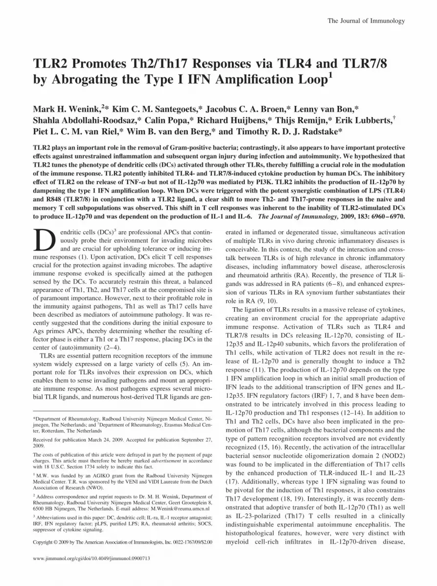
Previous observations indicated cross-talk between TLR2 andTLR4 on DCs (27). To evaluate the concept that TLR2 inhibitsthe production of proinflammatory mediators upon TLR4-me-diated activation of DCs, we costimulated DCs with increasingconcentrations of Pam3CSK4 (TLR2/1 ligand) and purified LPS(TLR4 ligand). The release of both TNF-� and IL-12p70 afterthe stimulation of DCs with pLPS was dose-dependently inhib-ited by the presence of Pam3CSK4 (Fig. 1A). Additional ex-periments using monoclonal anti-TLR2 Abs, HEK-293 cells ex-pressing TLR4, MD2, and CD14, a ligand for TLR2/6 (FSL-1or Pam2Cys), alternating order of ligand addition, and increas-ing LPS dosages clearly demonstrated that the inhibitory effectwas TLR2 specific (Fig. 1, B and E), not due to physical inter-action before engagement of TLR4 by pLPS (Fig. 1C) and
FIGURE 1. The TLR4-mediated production ofTNF-� and IL-12p70 is dose-dependently inhibited bythe costimulation of TLR2 on human monocyte-derivedDCs. A, Monocyte-derived DCs were stimulated with100 ng/ml pLPS and increasing amounts of Pam3CSK4.After 24 h, supernatants were collected and measuredfor their TNF-� and IL-12p70 content by Luminex.Bars represent means and SEM of three independentexperiments. B, DCs were activated with the combina-tion of Pam3CSK4 (5 �g/ml) and pLPS (100 ng/ml) inthe presence of blocking Abs for TLR2 or of isotypecontrol Abs. Bars represent means and SD of triplicatewells. Data are representative of three independent ex-periments. C, HEK-293-TLR4/MD-2/CD14 cells werestimulated with pLPS (100 ng/ml and 1 �g/ml) and/orPam3CSK4 (5 and 10 �g/ml) for 24 h, after which thelevels of released IL-8 were measured. Bars representmeans and SD of triplicate wells. D, Surrounding thepLPS activation of DCs Pam3CSK4 was added at var-ious time points ranging from 2 h before to 2 h after thestimulation with pLPS. Supernatants were collected af-ter 24 h, and the levels of TNF-� and IL-12p70 weredetermined. Bars represent means and SEM of three in-dependent experiments. E, DCs from three healthy vol-unteers were stimulated with pLPS (100 ng/ml) with orwithout FSL-1 (Pam2Cys, 1 �g/ml). In line with theresults obtained by the activation of TLR2/1 byPam3CSK4, the triggering of TLR2/6 by FSL-1 inhib-ited TLR4-mediated TNF-� and IL-12p70 secretion.Bars represent means and SEM of five independentexperiments.
6962 TLR2 PROMOTES Th2/Th17 RESPONSES
independent of the order of addition of pLPS/Pam3CSK4, aslong as Pam3CSK4 was added within 2 h of pLPS (Fig. 1D) andwas present over all LPS dosages (1,10, 100 ng/ml and 1 and 2�g/ml) (data not shown).
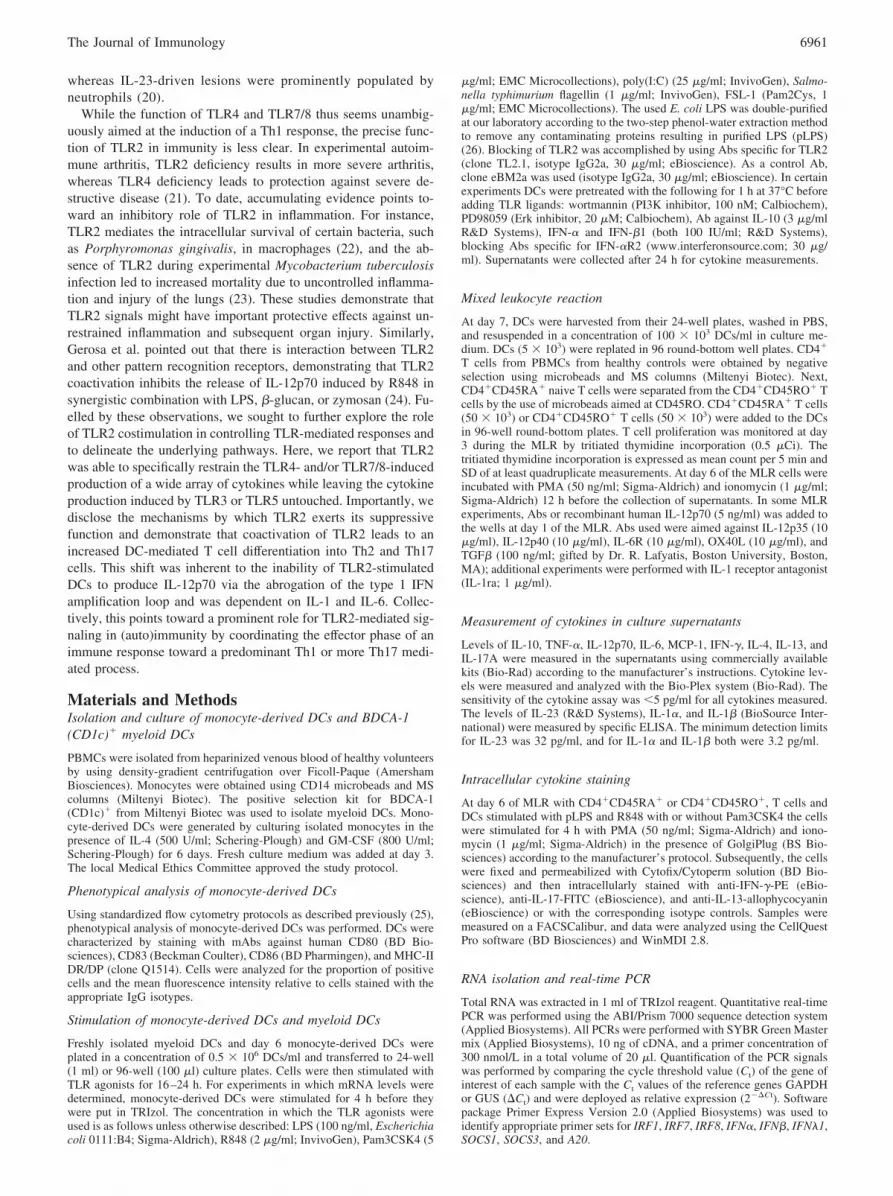
In line with these observations, the addition of Pam3CSK4also markedly inhibited the secretion of IL-10, IL-6, and IL-23,whereas MCP-1 showed the opposite effect (Fig. 2A). Based onapparent dissimilarities in the signaling cascades of the differentTLR subtypes, we subsequently evaluated the effect of TLR2 onTLR3, TLR5, and TLR7/8. Stimulation of DCs with R848(TLR7/8 ligand) resulted in the production of IL-10, TNF-�,IL-12p70, IL-6, IL-1�, IL-1�, IL-23, and MCP-1, which wasmarkedly inhibited by the addition of Pam3CSK4 for all medi-ators except IL-6 and MCP-1, underscoring the potency of theinhibitory effect of TLR2 (Fig. 2A). Strikingly, concomitantstimulation of DCs with Pam3CSK4 and poly(I:C) (TLR3 li-gand) and flagellin (TLR5 ligand) did not result in the inhibitionof cytokine production (Fig. 2B). In fact, the addition ofPam3CSK4 to either poly(I:C) or flagellin augmented the cy-tokine response compared with that of the single ligands. To
assess the in vivo relevance of our observations and extend thefindings by Gerosa et al. (24), we assessed whether the inhib-itory effect of TLR2 was present in freshly isolated myeloidDCs (CD11c�/BDCA-1�). Although the level of IL-12p70 se-cretion by myeloid DCs induced by the single ligands pLPS orR848 was very low compared with monocyte-derived DCs, theinhibitory effect of Pam3CSK4 was clearly present on the pLPSand R848 induced IL-12p70 and TNF-� production (data notshown and Fig. 2C).
The inhibitory effect of TLR2 on the release of TNF-� but not ofIL-12p70 is mediated by PI3K
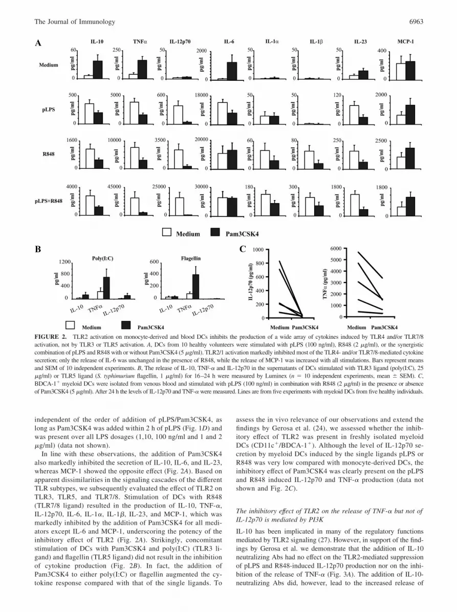
IL-10 has been implicated in many of the regulatory functionsmediated by TLR2 signaling (27). However, in support of the find-ings by Gerosa et al. we demonstrate that the addition of IL-10neutralizing Abs had no effect on the TLR2-mediated suppressionof pLPS and R848-induced IL-12p70 production nor on the inhi-bition of the release of TNF-� (Fig. 3A). The addition of IL-10-neutralizing Abs did, however, lead to the increased release of
FIGURE 2. TLR2 activation on monocyte-derived and blood DCs inhibits the production of a wide array of cytokines induced by TLR4 and/or TLR7/8activation, not by TLR3 or TLR5 activation. A, DCs from 10 healthy volunteers were stimulated with pLPS (100 ng/ml), R848 (2 �g/ml), or the synergisticcombination of pLPS and R848 with or without Pam3CSK4 (5 �g/ml). TLR2/1 activation markedly inhibited most of the TLR4- and/or TLR7/8-mediated cytokinesecretion; only the release of IL-6 was unchanged in the presence of R848, while the release of MCP-1 was increased with all stimulations. Bars represent meansand SEM of 10 independent experiments. B, The release of IL-10, TNF-� and IL-12p70 in the supernatants of DCs stimulated with TLR3 ligand (poly(I:C), 25�g/ml) or TLR5 ligand (S. typhimurium flagellin, 1 �g/ml) for 16–24 h were measured by Luminex (n � 10 independent experiments, mean � SEM). C,BDCA-1� myeloid DCs were isolated from venous blood and stimulated with pLPS (100 ng/ml) in combination with R848 (2 �g/ml) in the presence or absenceof Pam3CSK4 (5 �g/ml). After 24 h the levels of IL-12p70 and TNF-� were measured. Lines are from five experiments with myeloid DCs from five healthy individuals.
6963The Journal of Immunology
TNF-� and IL-12p70, demonstrating its regulatory role indepen-dent of TLR2.
The stimulation of DCs with TLR2 ligands was previouslyshown to lead to a potent phosphorylation of Erk1/2, leading to areduced ability of the DCs to produce IL-12p70 upon the activa-tion of TLR2 (28, 29). To evaluate whether the phosphorylation ofErk1/2 was implicated in the dampening of TLR4 and TLR7/8responses by TLR2, DCs were exposed to an inhibitor of Erk1/2before the stimulation with TLR ligands. No effect on the inhibi-tion by TLR2 was observed, demonstrating that the phosphoryla-tion of Erk1/2 by TLR2 does not play a role in the inhibition ofTLR4 and TLR7/8 responses (Fig. 3A). TLR2 has been shown tosignal through PI3K, which is a known inhibitor of the TLR4pathway. Taking this into account, we used the specific inhibitorwortmannin for the inhibition of the PI3K pathway. A clear de-pendence on the presence of functional PI3K was observed forTLR2 to exert its full inhibitory effect on the release of TNF-� andIL-10 (Fig. 3B and data not shown). However, suppression of IL-12p70 secretion by TLR2 was not influenced by the blocking ofthe function of PI3K (Fig. 3B), implicating that TLR2 inhibits viamultiple pathways.
The TLR2-mediated inhibition of IL-12p70 secretion is IRF1/8-,STAT1-, and SOCS1-dependent
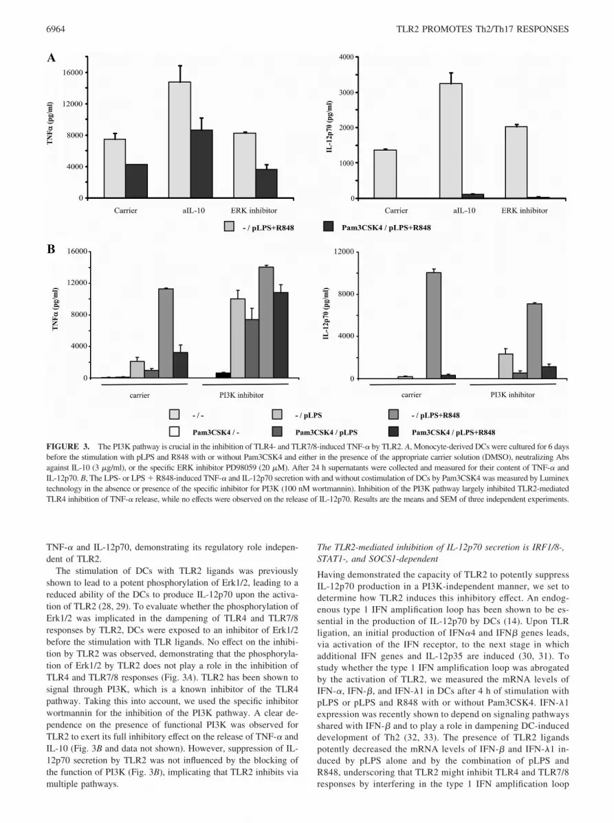
Having demonstrated the capacity of TLR2 to potently suppressIL-12p70 production in a PI3K-independent manner, we set todetermine how TLR2 induces this inhibitory effect. An endog-enous type 1 IFN amplification loop has been shown to be es-sential in the production of IL-12p70 by DCs (14). Upon TLRligation, an initial production of IFN�4 and IFN� genes leads,via activation of the IFN receptor, to the next stage in whichadditional IFN genes and IL-12p35 are induced (30, 31). Tostudy whether the type 1 IFN amplification loop was abrogatedby the activation of TLR2, we measured the mRNA levels ofIFN-�, IFN-�, and IFN-�1 in DCs after 4 h of stimulation withpLPS or pLPS and R848 with or without Pam3CSK4. IFN-�1expression was recently shown to depend on signaling pathwaysshared with IFN-� and to play a role in dampening DC-induceddevelopment of Th2 (32, 33). The presence of TLR2 ligandspotently decreased the mRNA levels of IFN-� and IFN-�1 in-duced by pLPS alone and by the combination of pLPS andR848, underscoring that TLR2 might inhibit TLR4 and TLR7/8responses by interfering in the type 1 IFN amplification loop
FIGURE 3. The PI3K pathway is crucial in the inhibition of TLR4- and TLR7/8-induced TNF-� by TLR2. A, Monocyte-derived DCs were cultured for 6 daysbefore the stimulation with pLPS and R848 with or without Pam3CSK4 and either in the presence of the appropriate carrier solution (DMSO), neutralizing Absagainst IL-10 (3 �g/ml), or the specific ERK inhibitor PD98059 (20 �M). After 24 h supernatants were collected and measured for their content of TNF-� andIL-12p70. B, The LPS- or LPS � R848-induced TNF-� and IL-12p70 secretion with and without costimulation of DCs by Pam3CSK4 was measured by Luminextechnology in the absence or presence of the specific inhibitor for PI3K (100 nM wortmannin). Inhibition of the PI3K pathway largely inhibited TLR2-mediatedTLR4 inhibition of TNF-� release, while no effects were observed on the release of IL-12p70. Results are the means and SEM of three independent experiments.
6964 TLR2 PROMOTES Th2/Th17 RESPONSES
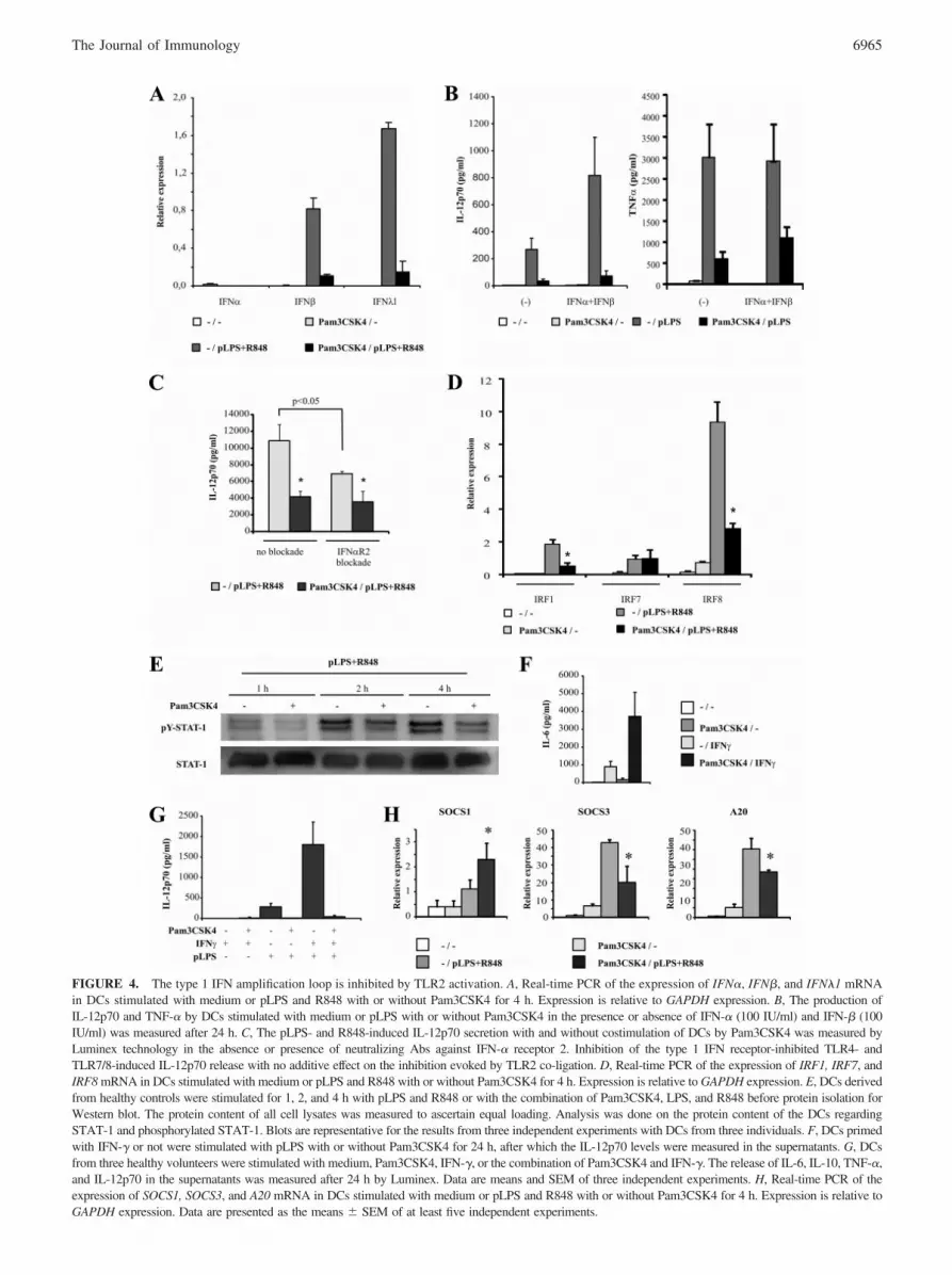
FIGURE 4. The type 1 IFN amplification loop is inhibited by TLR2 activation. A, Real-time PCR of the expression of IFN�, IFN�, and IFN�1 mRNAin DCs stimulated with medium or pLPS and R848 with or without Pam3CSK4 for 4 h. Expression is relative to GAPDH expression. B, The production ofIL-12p70 and TNF-� by DCs stimulated with medium or pLPS with or without Pam3CSK4 in the presence or absence of IFN-� (100 IU/ml) and IFN-� (100IU/ml) was measured after 24 h. C, The pLPS- and R848-induced IL-12p70 secretion with and without costimulation of DCs by Pam3CSK4 was measured byLuminex technology in the absence or presence of neutralizing Abs against IFN-� receptor 2. Inhibition of the type 1 IFN receptor-inhibited TLR4- andTLR7/8-induced IL-12p70 release with no additive effect on the inhibition evoked by TLR2 co-ligation. D, Real-time PCR of the expression of IRF1, IRF7, andIRF8 mRNA in DCs stimulated with medium or pLPS and R848 with or without Pam3CSK4 for 4 h. Expression is relative to GAPDH expression. E, DCs derivedfrom healthy controls were stimulated for 1, 2, and 4 h with pLPS and R848 or with the combination of Pam3CSK4, LPS, and R848 before protein isolation forWestern blot. The protein content of all cell lysates was measured to ascertain equal loading. Analysis was done on the protein content of the DCs regardingSTAT-1 and phosphorylated STAT-1. Blots are representative for the results from three independent experiments with DCs from three individuals. F, DCs primedwith IFN-� or not were stimulated with pLPS with or without Pam3CSK4 for 24 h, after which the IL-12p70 levels were measured in the supernatants. G, DCsfrom three healthy volunteers were stimulated with medium, Pam3CSK4, IFN-�, or the combination of Pam3CSK4 and IFN-�. The release of IL-6, IL-10, TNF-�,and IL-12p70 in the supernatants was measured after 24 h by Luminex. Data are means and SEM of three independent experiments. H, Real-time PCR of theexpression of SOCS1, SOCS3, and A20 mRNA in DCs stimulated with medium or pLPS and R848 with or without Pam3CSK4 for 4 h. Expression is relative toGAPDH expression. Data are presented as the means � SEM of at least five independent experiments.
6965The Journal of Immunology

(data not shown and Fig. 4A). IFN-� mRNA was hardly detect-able. Next, exogenous IFN-� and IFN-� were added to DCs inconjunction with pLPS, which resulted in a synergistically in-creased secretion of IL-12p70. This, however, had no effect onTLR2-mediated inhibition, suggesting that abrogation of theinitial type I IFN release is not essential (Fig. 4B). The commonIFN-� receptor 2 (IFN-�R2) was blocked to determine whetherthe type 1 IFN amplification loop does play a role in our system.The blockade of IFN-�R2 led to a clearly decreased LPS-andR848-induced production of IL-12p70, but again it did not af-fect the inhibitory capacity of TLR2 ligation (Fig. 4C).
To deepen our understanding of the TLR2-mediated inhibi-tion of the type 1 IFN amplification loop, we studied the down-stream mediators of the type 1 IFN pathway IRF1, IRF7, andIRF8. Upon TLR activation, IRF7 is essential for the initial type1 IFN production, while IRF8 is indispensable for the inductionof IL-12p70 production during the second phase of the type 1IFN amplification loop. IRF1 has an important role in the am-plification of IRF8-mediated IL-12p70 release. In our experi-mental setup, IRF1, IRF7, and IRF8 mRNA was readily presentin the DCs after the activation of TLR4 alone and by the acti-vation of both TLR4 and TLR7/8. Interestingly, the addition ofTLR2 ligands had no effect on the mRNA level of IRF7 butsignificantly reduced IRF1 and IRF8 mRNA levels induced bypLPS alone and by the combination of pLPS and R848 (data notshown and Fig. 4D). Next to IRFs, STAT-1 signaling is essen-tial for the induction of IL-12p70 production by DCs via theIFN amplification loop. To study the role for STAT-1 inhibitionby Pam3CSK4 in more detail, we evaluated the effect ofPam3CSK4 on the level of TLR4- and TLR7/8-induced phos-phorylation of STAT-1. At 60, 120, and 240 min the tyrosinephosphorylation status of STAT-1 was determined upon the ac-tivation of DCs with pLPS and R848 in the presence and ab-sence of Pam3CSK4. There was a clear inhibition of the pLPS-and R848-induced phosphorylation of STAT-1 by Pam3CSK4(Fig. 4E).
To determine whether TLR2 activation would also inhibitIFN-�-induced STAT-1-dependent cytokine production byDCs, we costimulated DCs with IFN-� and Pam3CSK4 andmonitored the secretion of inflammatory mediators such asIL-6, TNF-�, IL-10, and IL-12p70. IFN-�-dependent activationof DCs resulted in a low production of IL-6, while IL-10, TNF,and IL-12p70 were undetectable. Stimulation with bothPam3CSK4 and IFN-�, however, resulted in a synergistic re-lease of IL-6, suggesting that TLR2 stimulation does not act viadirect inhibition of STAT-1 phosphorylation (Fig. 4F). The pro-duction of IL-10, TNF-�, and IL-12p70 was comparable to thatinduced by TLR2 activation alone (data not shown). Since thecross-talk between TLR4 and IFN-� requires IRF1 and IRF8,we sought to investigate whether this synergy would be damp-ened by the presence of TLR2 ligands. A clear synergy in therelease of IL-12p70 was observed when DCs were activatedwith both IFN-� and pLPS, a response fully abrogated by theaddition of Pam3CSK4 (Fig. 4G).
The SOCS1 protein has been implicated in the control of variouscytokine and TLR responses by the inhibition of JAK/STAT path-ways (34). Recently, a role for this protein was described in thedampening of TLR responses by the TAM receptors dependent onthe type 1 IFN amplification loop (35). Indeed, a potent facilitatingeffect of TLR2 on the expression of SOCS1 induced by pLPS andR848 was observed, whereas the expression of SOCS3 and A20mRNA was decreased (Fig. 4H).
Costimulation with TLR2 ligands does not influence maturationof DC or T cell proliferation but skews T cell priming to aTh2/Th17 response
To investigate the inhibitory capacity of Pam3CSK4 on phenotyp-ical DC maturation, DCs were stimulated with pLPS or R848 andthe expression of multiple surface markers was studied. Both stim-ulation with pLPS and R848 led to a rapid up-regulation of MHCII, CD80, CD83, and CD86 that was not influenced by the additionof Pam3CSK4 (Fig. 5A). In line with this observation, the rate ofT cell proliferation induced by DCs stimulated with TLR4 andTLR7/8 was unaltered by the addition of TLR2 ligands (Fig. 5B).Based on the strong inhibition of IL-12p70 release, we postulatedthat costimulation of DCs with Pam3CSK4 alters DC-mediated Tcell priming, leading to a more Th2-prone immune response evenin combination with potent inducers of Th1 responses such aspLPS and R848. To test this, DCs were stimulated with pLPS andR848 in the presence or absence of Pam3CSK4 and subsequentlycoincubated with naive CD4�CD45RA� T cells or memoryCD4�CD45RO� T cells. The stimulation of DCs with Pam3CSK4in combination with pLPS and R848 suppressed the IFN-� releasefrom the naive T cell population and resulted in an increased re-lease of IL-4, IL-13, and IL-17 compared with DCs stimulatedwith pLPS and R848 alone (Fig. 6A). Additionally, a significant
FIGURE 5. Costimulation of TLR2 with TLR4 or TLR7/8 does notaffect DC phenotypic maturation or T cell proliferation. A, The up-regu-lation of CD80, CD83, CD86, and MHC-II expression initiated by stimu-lation of TLR4 (pLPS (100 ng/ml)) or TLR7/8 (R848 (2 �g/ml)) is notmodified by coincubation of DCs with a TLR2 ligand (Pam3CSK4 (5 �g/ml)). DCs were cultured for 24 h with medium, LPS (100 ng/ml), or R848(2 �g/ml) alone or in combination with Pam3CSK4 (5 �g/ml). Expressionof cell surface markers was performed using flow cytometry. B, Coincu-bation of DCs with the combination of LPS (100 ng/ml) or R848 (2 �g/ml)and Pam3CSK4 (5 �g/ml) had no marked effect on T cell proliferationmeasured by [3H]thymidine incorporation at day 3 of the MLR comparedwith those stimulated with LPS or R848 alone. Data are means and SEMof five independent experiments. �, p � 0.05 and ��, p � 0.01 comparedwith coincubation with medium alone.
6966 TLR2 PROMOTES Th2/Th17 RESPONSES
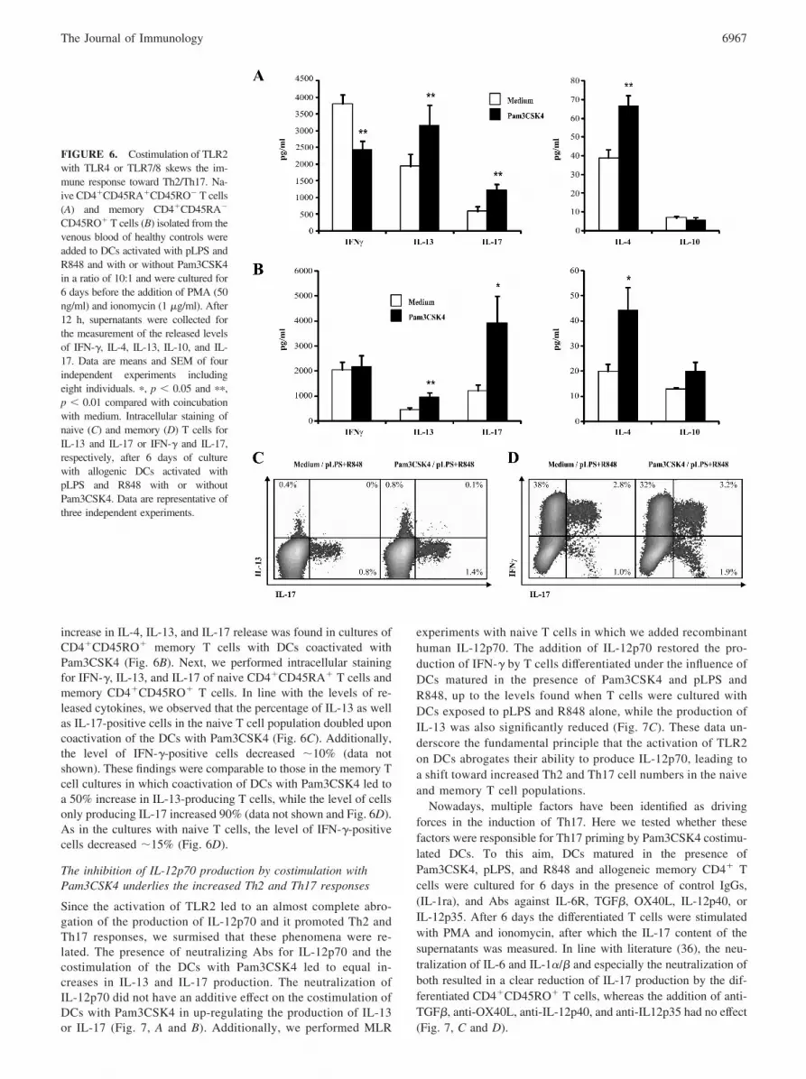
increase in IL-4, IL-13, and IL-17 release was found in cultures ofCD4�CD45RO� memory T cells with DCs coactivated withPam3CSK4 (Fig. 6B). Next, we performed intracellular stainingfor IFN-�, IL-13, and IL-17 of naive CD4�CD45RA� T cells andmemory CD4�CD45RO� T cells. In line with the levels of re-leased cytokines, we observed that the percentage of IL-13 as wellas IL-17-positive cells in the naive T cell population doubled uponcoactivation of the DCs with Pam3CSK4 (Fig. 6C). Additionally,the level of IFN-�-positive cells decreased �10% (data notshown). These findings were comparable to those in the memory Tcell cultures in which coactivation of DCs with Pam3CSK4 led toa 50% increase in IL-13-producing T cells, while the level of cellsonly producing IL-17 increased 90% (data not shown and Fig. 6D).As in the cultures with naive T cells, the level of IFN-�-positivecells decreased �15% (Fig. 6D).
The inhibition of IL-12p70 production by costimulation withPam3CSK4 underlies the increased Th2 and Th17 responses
Since the activation of TLR2 led to an almost complete abro-gation of the production of IL-12p70 and it promoted Th2 andTh17 responses, we surmised that these phenomena were re-lated. The presence of neutralizing Abs for IL-12p70 and thecostimulation of the DCs with Pam3CSK4 led to equal in-creases in IL-13 and IL-17 production. The neutralization ofIL-12p70 did not have an additive effect on the costimulation ofDCs with Pam3CSK4 in up-regulating the production of IL-13or IL-17 (Fig. 7, A and B). Additionally, we performed MLR
experiments with naive T cells in which we added recombinanthuman IL-12p70. The addition of IL-12p70 restored the pro-duction of IFN-� by T cells differentiated under the influence ofDCs matured in the presence of Pam3CSK4 and pLPS andR848, up to the levels found when T cells were cultured withDCs exposed to pLPS and R848 alone, while the production ofIL-13 was also significantly reduced (Fig. 7C). These data un-derscore the fundamental principle that the activation of TLR2on DCs abrogates their ability to produce IL-12p70, leading toa shift toward increased Th2 and Th17 cell numbers in the naiveand memory T cell populations.
Nowadays, multiple factors have been identified as drivingforces in the induction of Th17. Here we tested whether thesefactors were responsible for Th17 priming by Pam3CSK4 costimu-lated DCs. To this aim, DCs matured in the presence ofPam3CSK4, pLPS, and R848 and allogeneic memory CD4� Tcells were cultured for 6 days in the presence of control IgGs,(IL-1ra), and Abs against IL-6R, TGF�, OX40L, IL-12p40, orIL-12p35. After 6 days the differentiated T cells were stimulatedwith PMA and ionomycin, after which the IL-17 content of thesupernatants was measured. In line with literature (36), the neu-tralization of IL-6 and IL-1�/� and especially the neutralization ofboth resulted in a clear reduction of IL-17 production by the dif-ferentiated CD4�CD45RO� T cells, whereas the addition of anti-TGF�, anti-OX40L, anti-IL-12p40, and anti-IL12p35 had no effect(Fig. 7, C and D).
FIGURE 6. Costimulation of TLR2with TLR4 or TLR7/8 skews the im-mune response toward Th2/Th17. Na-ive CD4�CD45RA�CD45RO� T cells(A) and memory CD4�CD45RA�
CD45RO� T cells (B) isolated from thevenous blood of healthy controls wereadded to DCs activated with pLPS andR848 and with or without Pam3CSK4in a ratio of 10:1 and were cultured for6 days before the addition of PMA (50ng/ml) and ionomycin (1 �g/ml). After12 h, supernatants were collected forthe measurement of the released levelsof IFN-�, IL-4, IL-13, IL-10, and IL-17. Data are means and SEM of fourindependent experiments includingeight individuals. �, p � 0.05 and ��,p � 0.01 compared with coincubationwith medium. Intracellular staining ofnaive (C) and memory (D) T cells forIL-13 and IL-17 or IFN-� and IL-17,respectively, after 6 days of culturewith allogenic DCs activated withpLPS and R848 with or withoutPam3CSK4. Data are representative ofthree independent experiments.
6967The Journal of Immunology

DiscussionIn the present study, we identify TLR2 as a potent modulator ofTLR4- and TLR7/8-mediated immune responses, confirming therecent observations by Gerosa et al. (24). TLR2, on the one hand,suppresses the release of TLR4- and TLR7/8-induced proinflam-matory cytokine release, but, on the other hand, orchestrates theskewing of the immune response toward enhanced Th2 and Th17responses. Additionally, we unravel two novel pathways by whichTLR2 mediates its suppressive function, bearing potential for ther-apeutic intervention in immune diseases. By increasing the tran-scription of SOCS1 and decreasing the phosphorylation ofSTAT-1, transcription of type 1 IFNs, and the transcription factors
IRF1 and IRF8, TLR2 potently inhibits the type 1 IFN amplifica-tion loop, thereby abrogating the production of IL-12p70. More-over, the production of TNF-� is suppressed by the activation ofthe intracellular inhibitor PI3K by TLR2. We further demonstratethat the inhibition of IL-12p70 release by TLR2 underlies the pro-moted Th2/Th17-prone response.
The combination of archetypical constituents of a certain patho-gen recognized by different pattern recognition receptors is thoughtto give specificity to our innate immune response. In this light, itcan be envisaged that the myriad of host-derived TLR ligandsgenerated in inflamed or degenerated tissue are able to modifythese responses or in itself create specific immune responses. The
FIGURE 7. TLR2 stimulation mediates its Th17-inducing effect via the inhibition of IL-12p70 secretion. Naive CD4�CD45RA�CD45RO� T cells (A)and memory CD4�CD45RA�CD45RO� T cells (B) isolated from the venous blood of healthy controls were added to pLPS and R848 stimulated DCs withor without costimulation with Pam3CSK4 in a ratio of 10:1 and were cultured for 6 days in the absence or presence of Abs against IL-12p35 before theaddition of PMA (50 ng/ml) and ionomycin (1 �g/ml). After 12 h supernatants were collected for the measurement of the released levels of IL-13 and IL-17for the naive and memory T cell population, respectively. Levels of cytokine release were compared with those produced by T cells cultured with pLPS-and R848-stimulated DCs. C, Naive CD4�CD45RA�CD45RO� T cells isolated from the venous blood of healthy controls were added to DCs stimulatedwith pLPS and R848 with or without Pam3CSK4 in a ratio of 10:1 and were cultured for 6 days in the absence or presence of human IL-12p70 (5 ng/ml)before the addition of PMA (50 ng/ml) and ionomycin (1 �g/ml). After 12 h supernatants were collected for the measurement of the released levels of IFN-�and IL-13. D, Memory CD4�CD45RA�CD45RO� T cells were added to DCs activated with the combination of Pam3CSK4, pLPS, and R848 in a ratioof 10:1 and were cultured for 6 days in the presence of Abs against IL-12p35, IL-12p40, OX40 ligand, TGF�, IL-6R, or IL-1ra or the combination of anAb against IL-6R and IL-1ra before the addition of PMA (50 ng/ml) and ionomycin (1 �g/ml). After 12 h supernatants were collected for the measurementof the released levels of IL-17. Levels of IL-17 were compared with those produced by T cells cultured with DCs activated with the combination ofPam3CSK4, pLPS, and R848 in the absence of neutralizing Abs. Data are means and SEM of three independent experiments. �, p � 0.05 and ��, p � 0.01compared with coincubation with medium alone.
6968 TLR2 PROMOTES Th2/Th17 RESPONSES

role played by the different TLRs in immune activation was scru-tinized extensively during recent years. The combined activationof certain TLR pathways has demonstrated some striking syner-gistic responses in the release of proinflammatory cytokines. Theactivation of TLR4 and TLR7/8 on DCs, for example, leads tothe release of IL-12p70 and IL-23 that far surpasses the release bythe activation of either TLR alone, resulting in a prominent Th1response (10, 11). Since the ubiquitous nature of endogenous pro-teins able to trigger TLRs is such that if unchecked the host wouldbe overwhelmed by immune activation, several immune check-points must be in place. A potential inhibitory or immune tuningrole of TLRs has not been tested to a large extent, as shown in theliterature.
An inhibitory function for TLR2 has been proposed in varioussettings. In contrast to TLR4 knockout mice, TLR2�/� mice areless susceptible to lethal infections with Yersinia enterocolitica orCandida albicans (37, 38). Next to the interference in the removalof pathogens, the inhibitory effects mediated by TLR2 seem tohave a bright side. In the absence of TLR2, uncontrolled inflam-mation and excessive injury of tissues in response to infections orduring autoimmune processes have been observed (21, 23).Thereby, these studies demonstrate that TLR2 signals might haveimportant protective effects against destructive chronic immuneresponses, although the underlying pathways explaining these phe-nomena remain unidentified thus far. The present study establishesa role for TLR2 in providing an inhibitory signaling unit able tovigorously dampen TLR4- and TLR7/8-mediated activation ofDCs and provides rationale for the downstream pathways in-volved. For instance, PI3K, which functions at the early phase ofTLR signaling and modulates the magnitude of the primary acti-vation, was found to mediate the TLR2-induced inhibition of therelease of TNF-� while it did not affect IL-12p70 secretion. IL-12p70 secretion, in turn, was inhibited by the dampening of thetype 1 IFN amplification loop, seemingly by the increased tran-scription of SOCS1. PI3K as well as SOCS1 have been implicatedas crucial negative regulators of Th1 responses by suppressing theproduction of IL-12p70 (34, 39). The absence of SOCS1 in DCsenables them to invoke a potentiated Th1 response, resulting in anenhanced Ag-specific antitumor immunity (40, 41). Likewise, inan experimental arthritis model the severity of synovial inflamma-tion and joint destruction was increased in the absence of SOCS1(42). In Chlamydia pneumoniae infection it was shown thatSOCS1 inhibited the expression of type 1 IFNs and that IFN-� andsuppressed C. pneumoniae-induced lethal inflammation but im-paired effective bacterial clearance (43). These observations cor-roborate with our results and demonstrate that TLR2 is able toup-regulate the expression of SOCS1 in DCs, thereby reducingTh1 inflammatory responses.
The ability of the immune system to mount an apt response tothe wide array of different microbes is seminal to the protectionof the host against these invading pathogens as well as the limi-tation of damage evoked by this response. In this light, the para-digm that activation of multiple TLRs by pathogens acts as a com-binatorial code to induce a desired immune response is tempting.Additionally, this conceptual framework can be translated to thesituation of autoimmunity in that the environment is thought toorchestrate the type of immune response. Although the inhibitoryrole of TLR2 seems clear, in the removal of Gram-positive bac-teria, such as S. pneumoniae and Staphylococcus aureus, TLR2signaling is of vital importance (44–46). We demonstrated that theactivation of TLR2 in the presence of ligands for TLR4 andTLR7/8 leads to markedly enhanced Th17 responses. This impliesthat pathogens harboring archetypical constituents that triggermultiple TLR systems are recognized and processed, resulting in
an apt T cell response, notably a marked Th17 response whenTLR2 is added to the equation. In accordance with this notionTh17 cells seem to be essential in the clearance of both S. pneu-moniae and S. aureus (47, 48). Many experimental autoimmunemodels are induced in the presence of CFA and are dependent onthe presence of Th17 cells, while the severity of the disease pro-cesses is aggravated by the removal of Th1 cells or the neutral-ization of IFN-�. The active component of CFA consists of son-icated Mycobacterium tuberculosis. M. tuberculosis is recognizedby multiple pattern recognition receptors, among which TLR2,TLR4, TLR9, and the NOD proteins seem to be the most domi-nant. Additionally, it was recently demonstrated that IL-17-pro-ducing CD4� T cells are intricately involved in the first phase ofthe immune response against M. tuberculosis (49). Accordingly, inour system M. tuberculosis-activated DCs did not produce IL-12p70 and were able to induce elevated levels of IL-17-producingT cells (our unpublished results). In line with these findings inexperimental autoimmune encephalitis, evoked in the presence ofCFA, type 1 IFN receptor-mediated signaling pathways, importantfor the establishment of Th1 responses, constrained Th17-medi-ated inflammation (19). These recent findings put previous obser-vations by our laboratory, in which it was demonstrated thatSTAT-1�/� mice show an exacerbation of zymosan-induced ar-thritis (50), in a new light. Zymosan was demonstrated to inducestrong Th17 responses (24, 51); by removing the STAT-1-medi-ated IL-12p70/Th1 brake, a fulminant inflammatory response me-diated by Th17 cells can be expected. This underscores the delicatebalance the immune system has to cope with, keeping the differentTh cell subsets in check and thereby regulating immunity andtolerance.
In summary, we demonstrate that TLR2 is a potent modulator ofthe immune system by abrogating the type 1 IFN amplificationloop, thereby inducing markedly increased Th2 and Th17 re-sponses. This puts TLR2 in the middle of the immune network thatdecides whether the effector response against microorganisms or inautoimmunity is mainly Th1 or more Th17 mediated. Modulationof TLR2-mediated signaling might thus provide novel avenues forthe intervention within a broad spectrum of immune diseases.
DisclosuresThe authors have no financial conflicts of interest.
References1. Steinman, R. M., and M. C. Nussenzweig. 2002. Avoiding horror autotoxicus: the
importance of dendritic cells in peripheral T cell tolerance. Proc. Natl. Acad. Sci.USA 99: 351–358.
2. Langrish, C. L., Y. Chen, W. M. Blumenschein, J. Mattson, B. Basham,J. D. Sedgwick, T. McClanahan, R. A. Kastelein, and D. J. Cua. 2005. IL-23drives a pathogenic T cell population that induces autoimmune inflammation.J. Exp. Med. 201: 233–240.
3. Luger, D., P. B. Silver, J. Tang, D. Cua, Z. Chen, Y. Iwakura, E. P. Bowman,N. M. Sgambellone, C. C. Chan, and R. R. Caspi. 2008. Either a Th17 or a Th1effector response can drive autoimmunity: conditions of disease induction affectdominant effector category. J. Exp. Med. 205: 799–810.
4. Murphy, K. M., and S. L. Reiner. 2002. The lineage decisions of helper T cells.Nat. Rev. Immunol. 2: 933–944.
5. Akira, S., and K. Takeda. 2004. Toll-like receptor signalling. Nat. Rev. Immunol.4: 499–511.
6. Roelofs, M. F., W. C. Boelens, L. A. Joosten, S. Bdollahi-Roodsaz, J. Geurts,L. U. Wunderink, B. W. Schreurs, W. B. van Den Berg, and T. R. Radstake. 2006.Identification of small heat shock protein B8 (HSP22) as a novel TLR4 ligand andpotential involvement in the pathogenesis of rheumatoid arthritis. J. Immunol.176: 7021–7027.
7. Schrijver, I. A., M. J. Melief, P. P. Tak, M. P. Hazenberg, and J. D. Laman. 2000.Antigen-presenting cells containing bacterial peptidoglycan in synovial tissues ofrheumatoid arthritis patients coexpress costimulatory molecules and cytokines.Arthritis Rheum. 43: 2160–2168.
8. van, d. H., I. B. Wilbrink, I. Tchetverikov, I. A. Schrijver, L. M. Schouls,M. P. Hazenberg, F. C. Breedveld, and P. P. Tak. 2000. Presence of bacterialDNA and bacterial peptidoglycans in joints of patients with rheumatoid arthritisand other arthritides. Arthritis Rheum. 43: 593–598.
6969The Journal of Immunology
9. Radstake, T. R., M. F. Roelofs, Y. M. Jenniskens, B. Oppers-Walgreen,P. L. van Riel, P. Barrera, L. A. Joosten, and W. B. van Den Berg. 2004. Ex-pression of Toll-like receptors 2 and 4 in rheumatoid synovial tissue and regu-lation by proinflammatory cytokines interleukin-12 and interleukin-18 via inter-feron-�. Arthritis Rheum. 50: 3856–3865.
10. Roelofs, M. F., L. A. Joosten, S. Abdollahi-Roodsaz, A. W. van Lieshout,T. Sprong, F. H. van den Hoogen, W. B. van Den Berg, and T. R. Radstake. 2005.The expression of Toll-like receptors 3 and 7 in rheumatoid arthritis synovium isincreased and costimulation of Toll-like receptors 3, 4, and 7/8 results in syner-gistic cytokine production by dendritic cells. Arthritis Rheum. 52: 2313–2322.
11. Napolitani, G., A. Rinaldi, F. Bertoni, F. Sallusto, and A. Lanzavecchia. 2005.Selected Toll-like receptor agonist combinations synergistically trigger a T helpertype 1-polarizing program in dendritic cells. Nat. Immunol. 6: 769–776.
12. Liu, J., S. Cao, L. M. Herman, and X. Ma. 2003. Differential regulation of in-terleukin (IL)-12 p35 and p40 gene expression and interferon (IFN)-�-primedIL-12 production by IFN regulatory factor 1. J. Exp. Med. 198: 1265–1276.
13. Liu, J., X. Guan, T. Tamura, K. Ozato, and X. Ma. 2004. Synergistic activationof interleukin-12 p35 gene transcription by interferon regulatory factor-1 andinterferon consensus sequence-binding protein. J. Biol. Chem. 279:55609–55617.
14. Gautier, G., M. Humbert, F. Deauvieau, M. Scuiller, J. Hiscott, E. E. Bates,G. Trinchieri, C. Caux, and P. Garrone. 2005. A type I interferon autocrine-paracrine loop is involved in Toll-like receptor-induced interleukin-12p70 secre-tion by dendritic cells. J. Exp. Med. 201: 1435–1446.
15. Happel, K. I., M. Zheng, E. Young, L. J. Quinton, E. Lockhart, A. J. Ramsay,J. E. Shellito, J. R. Schurr, G. J. Bagby, S. Nelson, and J. K. Kolls. 2003. Cuttingedge: roles of Toll-like receptor 4 and IL-23 in IL-17 expression in response toKlebsiella pneumoniae infection. J. Immunol. 170: 4432–4436.
16. Schnurr, M., T. Toy, A. Shin, M. Wagner, J. Cebon, and E. Maraskovsky. 2005.Extracellular nucleotide signaling by P2 receptors inhibits IL-12 and enhancesIL-23 expression in human dendritic cells: a novel role for the cAMP pathway.Blood 105: 1582–1589.
17. van Beelen, A. J., Z. Zelinkova, E. W. Taanman-Kueter, F. J. Muller,D. W. Hommes, S. A. Zaat, M. L. Kapsenberg, and E. C. De Jong. 2007. Stim-ulation of the intracellular bacterial sensor NOD2 programs dendritic cells topromote interleukin-17 production in human memory T cells. Immunity 27:660–669.
18. Biondo, C., A. Midiri, M. Gambuzza, E. Gerace, M. Falduto, R. Galbo,A. Bellantoni, C. Beninati, G. Teti, T. Leanderson, and G. Mancuso. 2008. IFN-�/� signaling is required for polarization of cytokine responses toward a protec-tive type 1 pattern during experimental cryptococcosis. J. Immunol. 181:566–573.
19. Guo, B., E. Y. Chang, and G. Cheng. 2008. The type I IFN induction pathwayconstrains Th17-mediated autoimmune inflammation in mice. J. Clin. Invest. 118:1680–1690.
20. Kroenke, M. A., T. J. Carlson, A. V. Andjelkovic, and B. M. Segal. 2008. IL-12-and IL-23-modulated T cells induce distinct types of EAE based on histology,CNS chemokine profile, and response to cytokine inhibition. J. Exp. Med. 205:1535–1541.
21. Abdollahi-Roodsaz, S., L. A. Joosten, M. I. Koenders, I. Devesa, M. F. Roelofs,T. R. Radstake, M. Heuvelmans-Jacobs, S. Akira, M. J. Nicklin, F. Ribeiro-Dias,and W. B. van Den Berg. 2008. Stimulation of TLR2 and TLR4 differentiallyskews the balance of T cells in a mouse model of arthritis. J. Clin. Invest. 118:205–216.
22. Wang, M., M. A. Shakhatreh, D. James, S. Liang, S. Nishiyama, F. Yoshimura,D. R. Demuth, and G. Hajishengallis. 2007. Fimbrial proteins of Porphyromonasgingivalis mediate in vivo virulence and exploit TLR2 and complement receptor3 to persist in macrophages. J. Immunol. 179: 2349–2358.
23. Quesniaux, V., C. Fremond, M. Jacobs, S. Parida, D. Nicolle, V. Yeremeev,F. Bihl, F. Erard, T. Botha, M. Drennan, et al. 2004. Toll-like receptor pathwaysin the immune responses to mycobacteria. Microbes Infect. 6: 946–959.
24. Gerosa, F., B. Baldani-Guerra, L. A. Lyakh, G. Batoni, S. Esin,R. T. Winkler-Pickett, M. R. Consolaro, M. M. De, D. Giachino, A. Robbiano,et al. 2008. Differential regulation of interleukin 12 and interleukin 23 productionin human dendritic cells. J. Exp. Med. 205: 1447–1461.
25. Radstake, T. R., A. B. Blom, A. W. Sloetjes, E. O. van Gorselen, G. J. Pesman,L. Engelen, R. Torensma, W. B. van Den Berg, C. G. Figdor, P. L. van Lent,et al. 2004. Increased Fc�RII expression and aberrant tumour necrosis factor �production by mature dendritic cells from patients with active rheumatoid arthri-tis. Ann. Rheum. Dis. 63: 1556–1563.
26. Abdollahi-Roodsaz, S., L. A. Joosten, M. F. Roelofs, T. R. Radstake, G. Matera,C. Popa, J. W. van der Meer, M. G. Netea, and W. B. van Den Berg. 2007.Inhibition of Toll-like receptor 4 breaks the inflammatory loop in autoimmunedestructive arthritis. Arthritis Rheum. 56: 2957–2967.
27. Re, F., and J. L. Strominger. 2004. IL-10 released by concomitant TLR2 stim-ulation blocks the induction of a subset of Th1 cytokines that are specificallyinduced by TLR4 or TLR3 in human dendritic cells. J. Immunol. 173:7548–7555.
28. Agrawal, S., A. Agrawal, B. Doughty, A. Gerwitz, J. Blenis, D. T. van, andB. Pulendran. 2003. Cutting edge: different Toll-like receptor agonists instructdendritic cells to induce distinct Th responses via differential modulation of ex-tracellular signal-regulated kinase-mitogen-activated protein kinase and c-Fos.J. Immunol. 171: 4984–4989.
29. Dillon, S., S. Agrawal, K. Banerjee, J. Letterio, T. L. Denning, K. Oswald-Richter,D. J. Kasprowicz, K. Kellar, J. Pare, T. van Dyke, et al. 2006. Yeast zymosan, astimulus for TLR2 and dectin-1, induces regulatory antigen-presenting cells and im-munological tolerance. J. Clin. Invest. 116: 916–928.
30. Marie, I., J. E. Durbin, and D. E. Levy. 1998. Differential viral induction ofdistinct interferon-� genes by positive feedback through interferon regulatoryfactor-7. EMBO J. 17: 6660–6669.
31. Tailor, P., T. Tamura, H. J. Kong, T. Kubota, M. Kubota, P. Borghi, L. Gabriele,and K. Ozato. 2007. The feedback phase of type I interferon induction in den-dritic cells requires interferon regulatory factor 8. Immunity 27: 228–239.
32. Jordan, W. J., J. Eskdale, S. Srinivas, V. Pekarek, D. Kelner, M. Rodia, andG. Gallagher. 2007. Human interferon �-1 (IFN-�1/IL-29) modulates the Th1/Th2 response. Genes Immun. 8: 254–261.
33. Osterlund, P. I., T. E. Pietila, V. Veckman, S. V. Kotenko, and I. Julkunen. 2007.IFN regulatory factor family members differentially regulate the expression oftype III IFN (IFN-�) genes. J. Immunol. 179: 3434–3442.
34. Cottet, S., P. Dupraz, F. Hamburger, W. Dolci, M. Jaquet, and B. Thorens. 2001.SOCS-1 protein prevents janus kinase/STAT-dependent inhibition of � cell in-sulin gene transcription and secretion in response to interferon-�. J. Biol. Chem.276: 25862–25870.
35. Rothlin, C. V., S. Ghosh, E. I. Zuniga, M. B. Oldstone, and G. Lemke. 2007.TAM receptors are pleiotropic inhibitors of the innate immune response. Cell131: 1124–1136.
36. McGeachy, M. J., and D. J. Cua. 2008. Th17 cell differentiation: the long andwinding road. Immunity 28: 445–453.
37. Netea, M. G., R. Sutmuller, C. Hermann, C. A. Van der Graaf,J. W. van der Meer, J. H. van Krieken, T. Hartung, G. Adema, and B. J. Kullberg.2004. Toll-like receptor 2 suppresses immunity against Candida albicans throughinduction of IL-10 and regulatory T cells. J. Immunol. 172: 3712–3718.
38. Sing, A., D. Rost, N. Tvardovskaia, A. Roggenkamp, A. Wiedemann,C. J. Kirschning, M. Aepfelbacher, and J. Heesemann. 2002. Yersinia V-antigenexploits Toll-like receptor 2 and CD14 for interleukin 10-mediated immunosup-pression. J. Exp. Med. 196: 1017–1024.
39. Fukao, T., M. Tanabe, Y. Terauchi, T. Ota, S. Matsuda, T. Asano, T. Kadowaki,T. Takeuchi, and S. Koyasu. 2002. PI3K-mediated negative feedback regulationof IL-12 production in DCs. Nat. Immunol. 3: 875–881.
40. Hanada, T., K. Tanaka, Y. Matsumura, M. Yamauchi, H. Nishinakamura,H. Aburatani, R. Mashima, M. Kubo, T. Kobayashi, and A. Yoshimura. 2005.Induction of hyper Th1 cell-type immune responses by dendritic cells lacking thesuppressor of cytokine signaling-1 gene. J. Immunol. 174: 4325–4332.
41. Shen, L., K. Evel-Kabler, R. Strube, and S. Y. Chen. 2004. Silencing of SOCS1enhances antigen presentation by dendritic cells and antigen-specific anti-tumorimmunity. Nat. Biotechnol. 22: 1546–1553.
42. Egan, P. J., K. E. Lawlor, W. S. Alexander, and I. P. Wicks. 2003. Suppressor ofcytokine signaling-1 regulates acute inflammatory arthritis and T cell activation.J. Clin. Invest. 111: 915–924.
43. Yang, T., P. Stark, K. Janik, H. Wigzell, and M. E. Rottenberg. 2008. SOCS-1protects against Chlamydia pneumoniae-induced lethal inflammation but hamperseffective bacterial clearance. J. Immunol. 180: 4040–4049.
44. Koedel, U., B. Angele, T. Rupprecht, H. Wagner, A. Roggenkamp, H. W. Pfister,and C. J. Kirschning. 2003. Toll-like receptor 2 participates in mediation ofimmune response in experimental pneumococcal meningitis. J. Immunol. 170:438–444.
45. Kristian, S. A., X. Lauth, V. Nizet, F. Goetz, B. Neumeister, A. Peschel, andR. Landmann. 2003. Alanylation of teichoic acids protects Staphylococcus au-reus against Toll-like receptor 2-dependent host defense in a mouse tissue cageinfection model. J. Infect. Dis. 188: 414–423.
46. Lee, K. S., C. A. Scanga, E. M. Bachelder, Q. Chen, and C. M. Snapper. 2007.TLR2 synergizes with both TLR4 and TLR9 for induction of the MyD88-depen-dent splenic cytokine and chemokine response to Streptococcus pneumoniae.Cell. Immunol. 245: 103–110.
47. Ma, C. S., G. Y. Chew, N. Simpson, A. Priyadarshi, M. Wong, B. Grimbacher,D. A. Fulcher, S. G. Tangye, and M. C. Cook. 2008. Deficiency of Th17 cells inhyper IgE syndrome due to mutations in STAT3. J. Exp. Med. 205: 1551–1557.
48. Malley, R., A. Srivastava, M. Lipsitch, C. M. Thompson, C. Watkins,A. Tzianabos, and P. W. Anderson. 2006. Antibody-independent, interleukin-17A-mediated, cross-serotype immunity to pneumococci in mice immunized in-tranasally with the cell wall polysaccharide. Infect. Immun. 74: 2187–2195.
49. Khader, S. A., G. K. Bell, J. E. Pearl, J. J. Fountain, J. Rangel-Moreno,G. E. Cilley, F. Shen, S. M. Eaton, S. L. Gaffen, S. L. Swain, et al. 2007. IL-23and IL-17 in the establishment of protective pulmonary CD4� T cell responsesafter vaccination and during Mycobacterium tuberculosis challenge. Nat. Immu-nol. 8: 369–377.
50. de Hooge, A. S., F. A. van de Loo, M. I. Koenders, M. B. Bennink, O. J. Arntz,T. Kolbe, and W. B. van Den Berg. 2004. Local activation of STAT-1 andSTAT-3 in the inflamed synovium during zymosan-induced arthritis: exacerba-tion of joint inflammation in STAT-1 gene-knockout mice. Arthritis Rheum. 50:2014–2023.
51. Veldhoen, M., R. J. Hocking, R. A. Flavell, and B. Stockinger. 2006. Signalsmediated by transforming growth factor-� initiate autoimmune encephalomyeli-tis, but chronic inflammation is needed to sustain disease. Nat. Immunol. 7:1151–1156.
6970 TLR2 PROMOTES Th2/Th17 RESPONSES