title page title effects of resistance exercise with and without creatine supplementation on gene...
TRANSCRIPT
1
Title page
Title
Effects of resistance exercise with and without creatine supplementation on gene
expression and cell signalling in human skeletal muscle
Authors
Louise Deldicque1, Philip Atherton2, Rekha Patel2, Daniel Theisen1, Henri Nielens1,
Michael Rennie2, Marc Francaux1 �
Affiliation
1Department of Physical Education and Rehabilitation, Université catholique de
Louvain, Louvain-la-Neuve, Belgium
2School of Biomedical Sciences, University of Nottingham Graduate Entry Medical
School, City Hospital, Derby DE22 3DT, United Kingdom
Running title
Changes in gene expression with creatine and exercise
Contact information
� Corresponding author: Marc Francaux
Place Pierre de Coubertin 1, B-1348 Louvain-la-Neuve, Belgium
Tel: +32-10-47 44 57; Fax: +32-10-47 20 93
e-mail: [email protected]
Page 1 of 34Articles in PresS. J Appl Physiol (November 29, 2007). doi:10.1152/japplphysiol.00873.2007
Copyright © 2007 by the American Physiological Society.
2
ABSTRACT
To test the hypothesis that creatine supplementation would enhance the anabolic
responses of muscle cell signalling and gene expression to exercise, we studied nine
subjects who received either creatine or a placebo (maltodextrin) for 5d in a double-
blind fashion before undergoing muscle biopsies: at rest; immediately after exercise
(10×10 repetitions of one leg-extension at 80% 1-RM); and 24h and 72h later (all in the
morning after fasting overnight). Creatine supplementation decreased the
phosphorylation state of protein kinase B (PKB) on Thr308 at rest by 60% (P<0.05) and
that of eukaryotic initiation factor 4E-binding protein on Thr37/46 (4E-BP1) by 30%
24h post-exercise (P<0.05). Creatine increased mRNA for collagen 1(α1), glucose
transporter-4 (GLUT-4) and myosin heavy chain I at rest by 250%, 45% and 80%
respectively, and myosin heavy chain IIA (MHCIIA) mRNA immediately after exercise
by 70% (all P<0.05). Immediately after exercise, and independent of creatine, mRNA
for muscle atrophy F-box (MAFbx), MHCIIA, peroxisome proliferator-activated
receptor γ coactivator-1α and interleukin-6 were up-regulated (60-350%, P<0.05); the
phosphorylation state of p38 both in the sarcoplasm and nucleus were increased (12 and
25 fold respectively, both P<0.05). Concurrently, the phosphorylation states of PKB
(Thr308) and 4E-BP1 (Thr37/46) were decreased by 50% and 75% respectively
(P<0.05). Twenty-four hours post-exercise, MAFbx, myostatin and GLUT-4 mRNA
expression decreased below pre-exercise values (-35 to -50%, P<0.05); calpain 1
mRNA increased 70% 72h post-exercise (P<0.05) and at no other time. In conclusion,
5d of creatine supplementation does not enhance anabolic signalling but increases the
expression of certain targeted genes.
Page 2 of 34

3
Keywords (3-5 words)
MAPK, PKB, protein synthesis
Page 3 of 34
4
INTRODUCTION
Creatine supplementation has been used in various contexts, not only among athletes as
an ergogenic aid for improving muscle power output during high-intensity exercise
(16), but also as a potential therapeutic agent for patients suffering from general muscle
wasting and myopathies (24, 37, 40). Creatine supplementation leads to an increase in
muscle fiber area when associated with resistance exercise training (39) or in the case of
muscle regeneration after atrophy (18). More recently, creatine supplementation has
been shown to amplify the increase in satellite cell number and myonuclei concentration
in human skeletal muscle fibers during 4 to 16 weeks of resistance training. These
creatine-induced adaptations were associated with an enhanced muscle fiber growth in
response to strength training (32).
Evidence accumulated over the past 15 years suggests that the primary mechanism by
which creatine exerts its anabolic effect in healthy subjects who are weight training is
by allowing them to work at a higher proportion of their maximal voluntary contraction
force and thus increase the training stimulus (16). However this mechanism may not
explain the higher force production observed in patients suffering from myopathy (24,
37). Therefore the search for alternative mechanisms for the beneficial effects of
creatine has been under scrutiny. Several studies suggest that creatine could be more
effective in muscle submitted to degeneration/regeneration process induced by exercise
(32), immobilization (18) or disease (24, 37). Therefore we also hypothesised that
creatine would slow down the degeneration phase and accelerate the regeneration phase
Page 4 of 34
5
after high-intensity exercise, which usually occur 1 and 3 days post-exercise
respectively (15).
In two previous studies, we were unable to detect any difference in the myofibrillar or
sarcoplasmic protein synthetic rates or the breakdown rate of human muscle after
creatine supplementation, whether at rest (26) or post-exercise (25). This lack of effect
of creatine after exercise might have been explained by the timing of the measurements
made. For example, we measured the protein turnover rates immediately after exercise
following 5 days of supplementation, and it might have been that the creatine-plus-
exercise effects were present at a later time point. Indeed, exercise-induced increases in
protein synthesis can last for up to 72 h (29). Furthermore, there may be intricate
changes in translational signalling attributable to creatine, which are undetectable by
direct measures of muscle protein synthesis. Indeed, many recent studies in human
muscle have implicated changes in such candidate pathways with resistance exercise,
including the PKB (protein kinase B)-mTOR and the MAPK (mitogen-activated protein
kinase) pathways, key cascades in the regulation of skeletal muscle protein synthesis
and remodelling by resistance exercise (10, 13, 14, 20, 42).
Furthermore, in the past studies we did not examine the possibility of a ‘priming’ of
muscle gene expression that could be important for forthcoming synthetic responses to
resistance exercise or indeed other adaptive processes. In support of the notion for the
modulation of gene expression by creatine, there has been evidence of increased MHC
(myosin heavy chain) and IGF-I and II (insulin-like growth factor I and II) in human
subjects after creatine ingestion (11, 43). Furthermore, such gene expression changes,
Page 5 of 34
6
either in the satellite cells or in the myofiber after each exercise session, could be one
mechanism by which the induction of satellite cell nuclei into the myofiber occurs after
several weeks of resistance training, when coupled with creatine supplementation (32).
The aims of the present study were to test if 5 days of creatine supplementation induces
short-term changes in gene expression and cellular signalling at rest and after a single
bout of exercise. We hypothesised that short-term creatine supplementation would
increase expression of genes associated with the control of muscle mass, phenotype and
metabolism and augment anabolic signal transduction activity through the MAPK and
the PKB pathways.
METHODS
Subjects
Nine healthy young men (21.7 ± 0.55 y, BMI 24 ± 0.9 kg/m2) who did not partake in
any formal resistance exercise regime, were recruited for this double-blind cross-over
study. All subjects were given an oral and written account of the study before signing a
consent form. This study was approved by the Ethic Committee of the Université
catholique de Louvain and the investigation was performed according to the principles
outlined in the Declaration of Helsinki.
Experimental protocol
Page 6 of 34
7
Before the experiment, subjects participated in a pre-test to determine one repetition
maximum (1-RM) for each leg on a leg extension apparatus. The exercise consisted of a
one-leg knee extension movement from an angle of 90° to 160°. After a warm-up
comprising 3 sets of 10 repetitions at 5 kg, the load was progressively increased until
the subject could not perform more than one single repetition. Subjects were allowed 2
min rest between each set and reached 1-RM within 5-6 trials.
Subjects were instructed to refrain from vigorous physical activity 2 days prior to and
during the experimental phase. Food intake on the evening preceding each muscle
biopsy was controlled by administering a standardised dinner (22 % protein, 48 %
carbohydrate and 30 % fat). Subjects were randomly divided into two groups: one group
(n=5) received 21 g (3×7 g) oral creatine monohydrate per day for 5 days before the
beginning of the experiment and during the 3 days of the experiment while the second
group (n=4) received a placebo (maltodextrin 3×7 g per day) during the same period
(the protocol is summarized in Fig. 1). During the experimental days, the subjects were
asked to ingest creatine during the breakfast after biopsy sampling. After a wash-out
period of 6 weeks, the treatments were crossed-over for the second trial.
On the first morning of the study, participants reported to the laboratory after a 10 h
overnight fast and a first biopsy was taken at rest in the control leg chosen at random.
The procedure involved the administration of local anaesthesia (1 % lidocaine) and
sample extraction from the mid portion of the vastus lateralis muscle with a 4-mm
Bergström biopsy needle. Blood, macroscopically-visible fat and connective tissue were
Page 7 of 34
8
quickly removed, and the sample was immediately frozen in liquid nitrogen and stored
at -80 °C.
The exercise was then performed with the other leg after a warm-up of 3 sets of 10
repetitions at 5 kg. The main exercise session consisted of 10 sets of 10 repetitions at 80
% of the 1-RM of the exercising leg which corresponds to a mean value of positive
work of 19471 ± 1403.5 J for the placebo trial and 19902 ± 1504.1 J for the creatine
one. The positive work of each repetition was calculated by multiplying the moved
mass by “g” (9.81 m.s-2) and by the distance (height to which the mass was raised). In
this calculation, we neglected the friction due to the pulleys. The 1-RM was not exactly
the same for both legs in each subject resulting in different mean work between placebo
and creatine trials. These values were not statistically different. All subjects performed
the same number of repetitions during both trials. A second biopsy was taken from the
exercising leg within 30 s following the completion of the last repetition. A standardised
breakfast was given after the exercise session (475kcal; 7 % protein, 74 % carbohydrate
and 19 % fat). Each participant received a standardised dinner in the evening.
Additional biopsies were taken 24 h and 72 h later from the exercising leg, each after a
10 h overnight fast.
Protein extraction and cell fractionation
About 20-30 mg of frozen muscle were ground in a mortar and homogenized in ice-cold
hypotonic buffer (20 mM Hepes, 5 mM sodium fluoride, 1 mM sodium molybdate, 0.1
mM EDTA, 0.5 % NP-40, protease inhibitor cocktail (Roche Applied Science) for 5
min on ice. The homogenates were then centrifuged for 30 s at 10,000 g. The
Page 8 of 34
9
supernatant, containing the sarcoplasmic proteins, was stored at –80 °C. The pellet was
re-suspended in a buffer containing 20 mM Hepes, 5 mM sodium fluoride, 1 mM
sodium molybdate, 0.1 mM EDTA, 20 % glycerol, a protease inhibitor cocktail and the
same volume of a saline buffer containing 20 mM Hepes, 5 mM sodium fluoride, 1 mM
sodium molybdate, 0.1 mM EDTA, 20 % glycerol, 0.8 M NaCl and a protease inhibitor
cocktail. The solution was then homogenized on a rotary mixer for 30 min at 4 °C and
centrifuged for 10 min at 10,000 g. The supernatant, containing the nuclear proteins,
was stored at -80 °C. Sarcoplasmic and nuclear protein concentrations were determined
using a protein assay kit (Bio-Rad Laboratories) with BSA as a standard. Fraction purity
was verified and confirmed by immunoblotting for nuclear histone 1 (anti-histone 1,
1:1000, Santa Cruz).
SDS/PAGE and immunoblotting
Cell lysates (70 µg for sarcoplasmic proteins and 30 µg for nuclear proteins) were
combined with Laemmli sample buffer and separated by SDS/PAGE. After
electrophoretic separation at 40 mA, the proteins were transferred to a PVDF membrane
at 80 V for 4 h for a western blot analysis. Membranes were then incubated in a 5 %
Blotto solution. Subsequently, membranes were incubated with the following antibodies
(1:500) overnight at 4 °C: phospho-PKB Ser 473 (Cell Signaling), phospho-PKB Thr
308 (Cell Signaling), total PKB (Cell Signaling), phospho-p70s6k Thr 389 (Santa Cruz),
total p70s6k (Santa Cruz), phospho-p38 Thr 180/Tyr 182 (Cell Signaling), total p38 (Cell
Signaling), phospho-ERK1/2 Thr 202/Tyr 204 (Cell Signaling), total ERK (Cell
Signaling), phospho-4E-BP1 Thr 37/46 (Cell Signaling), total 4E-BP1 (Cell Signaling)
and MEF-2 (Santa Cruz). Antibodies from Cell Signaling were diluted in TBST
Page 9 of 34
10
containing 1 % BSA and antibodies from Santa Cruz were diluted in a 5 % Blotto
solution. PKB phosphorylated on Thr 308 and Ser 473 was analyzed because the
phosphorylation of both sites is required to achieve a high level of kinase activity (3).
Membranes were washed in TBST and incubated for 1 h at room temperature in a
secondary antibody conjugated to horseradish peroxidase (1:10,000, Cell Signaling).
After an additional 3 washes, chemiluminescence detection was carried out using an
Enhanced Chemiluminescent Western blotting kit (ECL Plus, Amersham Biosciences)
and hyperfilms (Hyperfilm ECL, Amersham Biosciences). Then, the membranes were
stripped and re-probed with a total antibody to verify the relative amount of the
analyzed proteins through the whole experiment. The films were scanned with an
ImageScanner using the Labscan software and quantified with the Image Master 1D
Image Analysis Software (Amersham Biosciences). The results represent the
phosphorylated form of the protein. A value of 1 was arbitrarily assigned to the control
conditions (placebo pre-exercise) to which the post-exercise and the creatine values
were reported. The choice of this baseline enables to depict the effects of creatine
supplementation in addition to the effects of acute exercise.
RNA extraction and quantitative Real-Time PCR
Frozen tissue samples (~30 mg) were homogenized in TRIZOL® using a Polytron. Total
RNA was extracted according to the instructions provided by the manufacturer. RNA
was quantified by spectrophotometry (260 nm) and its concentration adjusted to 1 µg/µl
using RNase-free water. A RNA agarose gel was run to verify the integrity of the RNA.
Reverse transcription (RT) was performed using the iScript synthesis kit (Bio-Rad) on a
Page 10 of 34
11
iQ5 Real-Time PCR Detection System (Bio-Rad) with 1 µg of total RNA in a reaction
volume of 20 µl (4 µl iScript reaction mix 5X, 1 µl iScript reverse transcriptase, 1 µl
RNA template, 14 µl RNase-free water). The final RT product was adjusted to 140 µl
using RNase free water. Real time RT-PCR (reverse transcription-polymerase chain
reaction) primers were designed (Table 1) for human calpain 1, collagen 1(α1), C2
subunit of proteasome, GLUT-4 (glucose transporter 4), IL-6 (interleukin 6), MAFbx
(muscle atrophy F-box), MHC I (myosin heavy chain type I), MHC IIA (myosin heavy
chain type IIA), MyoD, myostatin, PCNA (proliferating cell nuclear antigen), PGC-1α
(peroxisome proliferator-activated receptor gamma coactivator-1 α) and β-2-
microglobulin. The latter was used as the “house keeping” gene, because preliminary
experiments as well as a previous study (30) had revealed that it was not affected by
either exercise or creatine supplementation. Sybr Green® real time RT-PCR analyses
were carried out on the iQ5 Real-Time PCR Detection System (Bio-Rad) using the
following cycle conditions: 10 min at 95 °C, followed by 40 cycles of 1 min at 60 °C
and 15 s at 95 °C. For each gene, real time RT-PCR was conducted in duplicate with 25
µl reaction volume containing 12.5 µl Platinium Sybr Green qPCR SuperMix UDG
(Invitrogen), 0.75 µl of each primer (10 pmol/µl), 9 µl RNAse-free water and 2 µl of
1:5 diluted cDNA. A melt analysis was run to verify the amplified DNA product. A
value of 1 was arbitrarily assigned to the control conditions (placebo pre-exercise) to
which the post-exercise and the creatine values were reported.
Muscle creatine concentration
Muscle creatine concentration was measured on the pre-exercise biopsy as previously
described (11). Briefly, a fraction of the muscle biopsy (~15 mg) was used for
Page 11 of 34
12
spectrophotometric determination of total creatine (i.e. creatine phosphate + free
creatine). Ground muscle was extracted in 0.25 M HClO4 and neutralized with 1 M
KOH. A fraction of the supernatant was used to determine free creatine, the remaining
serving to determine total creatine content. To hydrolyze creatine phosphate, 2 M HCl
was added to the supernatant which was then heated for 15 min at 60 °C. The reaction
was stopped on ice and the supernatant neutralized with 2 M NaOH. Total creatine was
immediately determined enzymatically using a spectrophotometric method described by
Guder et al. (17) (Creatinine PAP, Boerhinger Mannheim, Germany from which the
creatininase was omitted). Phosphorylcreatine concentration was calculated by
subtracting free creatine from total creatine concentrations.
Statistical analysis
The difference in muscle creatine content between placebo and creatine conditions was
tested for significance using a paired t-test. Treatment by time interactions were
evaluated using a two-way analysis of variance for repeated measures (ANOVA). When
appropriate, Student-Newman-Keuls post hoc tests were applied. The significance
threshold was set to P < 0.05. The results are presented as the means ± SEM.
RESULTS
Muscle creatine concentration (Table 2)
After 5 days of supplementation muscle total creatine concentration increased by ~20 %
(P<0.01).
Page 12 of 34
13
Transcriptional regulation by exercise and creatine (Figures 2A-L)
Immediately after exercise, increases in PCNA mRNA (+150 %, P<0.05), MAFbx (+70
%, P<0.05), MHC IIA (+60 %, P<0.05), PGC-1α (+80 %, P<0.05) and IL-6 (+350 %,
P<0.05) were observed. The expression of MHC IIA mRNA, PGC-1α and IL-6 all
returned to pre-exercise values by 24 h or 72 h post-exercise (P<0.05). At 24h post-
exercise, PCNA mRNA nearly returned to pre-exercise values before increasing again at
72h post-exercise (+130 %, P<0.05). MAFbx mRNA decreased below pre-exercise
values at 24 h post-exercise (-50 %, P<0.05) but had returned to basal values two days
later. As for MAFbx, mRNA for myostatin (-35 %, P<0.01) and GLUT-4 (-45 %,
P<0.01) was also decreased at 24 h post-exercise, in comparison with pre-exercise
values, but had returned to baseline by 72 h post-exercise. Calpain 1 mRNA was
increased 72 h post-exercise in comparison with pre-exercise (+70 %), post-exercise
(+20 %) and 24 h post-exercise (+40 %) (P<0.05).
Creatine increased the expression of collagen 1(α1) (+250 %, P<0.05), GLUT-4 (+45
%, P<0.05) and MHC I mRNA (+80 %, P<0.05) at rest and the expression of MHC IIA
mRNA immediately after exercise (+70 %, P<0.05).
Activation of the MAPK pathway by exercise (Figures 3A-E and Figure 5)
Immediately after exercise, the phosphorylation state of p38 was increased more than
10-fold in the sarcoplasm (P<0.05) and more than 20-fold in the nucleus (P<0.01). It
returned to pre-exercise values 24 h and 72 h post-exercise in both fractions. The
phosphorylation state of ERK1/2 in the nucleus tended to be increased by exercise but
did not reach the statistical threshold (P=0.065). Immediately post-exercise, the
Page 13 of 34
14
expression of MEF-2 was doubled in the nucleus (P<0.05). Creatine had no effect on
the phosphorylation state of p38 and ERK1/2 or on the expression of MEF-2 in the
nucleus.
Alteration of the PKB pathway by exercise (Figure 4 A-D and Figure 5)
Immediately after exercise, the phosphorylation state of PKB on Thr 308 (P<0.05) and
4E-BP1 on Thr 37/46 (P<0.01) decreased by 50 % and 75 % respectively and returned
to pre-exercise values at 24 h and 72 h post-exercise. The same trend to inhibition was
observed immediately post-exercise on both PKB on Ser 473 and on p70s6k on Thr 389
but the statistical significance was not reached. Twenty-four hours post-exercise, two of
the nine subjects showed a markedly elevated phosphorylation state of p70s6k on Thr
389 whereas it remained unchanged in the other subjects. Creatine decreased the
phosphorylation state of PKB on Thr 308 at rest by 60 % (P<0.05) and the
phosphorylation state of 4E-BP1 on Thr 37/46 24 h post-exercise by 30 % (P<0.05).
DISCUSSION
We designed the studies described above to determine principally whether or not the
combination of acute exercise with 5 days of creatine supplementation would cause a
greater than normal increase in the expression of genes and in signal transduction likely
to be involved in adaptation of muscle size or biochemical properties. Indeed, exercise
is known to activate multiple signal transduction pathways and to modulate
transcriptional and translational processes.
Page 14 of 34
15
The only significant gene alterations we observed in response to creatine
supplementation were increases in the expression of GLUT-4, collagen 1(α1) and MHC
I mRNA at rest and increased expression of MHC IIA mRNA immediately post
exercise. Most of the changes in mRNA levels observed in the present study and
elsewhere (11) occurred at rest before the resistance exercise session. This suggests that
contrary to our hypotheses creatine per se is able to modify the expression of several
genes and that exercise training and/or a process of muscle degeneration/regeneration
are not essential prerequisite.
In addition to the modulation of gene expression, creatine supplementation has been
shown to augment the increase in satellite cells and myonuclei number induced by
several weeks of strength training (32). Therefore the activation, proliferation and
differentiation of satellite cells are other mechanisms by which creatine might increase
muscle mass after several weeks of supplementation combined with training. In the
current study, we found that although exercise did acutely increase a marker of satellite
cell proliferation (PCNA, a protein involved in DNA replication maximally expressed
in S phase) there was no prolonged effect over the next 24 h, but a rebound at 72 h and
no additional effect of creatine. MyoD is another key regulator of muscle remodelling
associated with cell proliferation. We did not observe any change in MyoD mRNA by
creatine nor by exercise whereas others found a doubling of MyoD expression
immediately post-exercise (36) or a peak expression at 8 h to 12 h post-exercise (45).
Although there is good evidence that the MAPK and the PKB pathways are involved in
the regulation of skeletal muscle protein synthesis and remodelling by resistance
Page 15 of 34
16
exercise (4, 10, 31, 41), short-term creatine supplementation does not seem to act via
these two cascades to increase muscle mass, contrary to our hypothesis. Creatine
supplementation had no additional effect on the phosphorylation of p38 and ERK1/2 or
on MEF-2 protein expression nor did creatine increase signalling through the PKB
pathway. The results on the MAPK and the PKB pathways are in agreement with our
previous studies showing no effect upon protein synthesis (25, 26).
Taken together, our results indicate that, although the most remarkable effects of
creatine are seen after several weeks of supplementation, some of them could be
initiated at the transcription level as soon as after 5 days of supplementation.
Nevertheless the extent of these increases is rather small and we have no evidence to
relate them to activation of satellite cells and to muscle mass accumulation observed
when creatine is combined with resistance training (32).
This study also produced novel results in the context of resistance exercise. These can
be summarized as follows: (i) an acute rise in the expression of MAFbx mRNA seen
immediately at the end of exercise followed by a subsequent fall at 24 h before the
return to basal values by 72 h; (ii) a fall in myostatin mRNA at 24 h; (iii) PCNA, a
marker of cell proliferation, was activated in a biphasic way, increasing immediately
post-exercise and after 3 days recovery, (iv) the increases in both the sarcoplasmic and
nuclear complements of p38 and ERK1/2; (v) the immediate rise after exercise of MEF-
2 protein. Therefore, acute resistance exercise induced very rapid, pronounced
alterations in gene transcription and cellular signalling, indicative by the early
modifications of some specific mRNAs seen at the end of exercise. Additional effects of
Page 16 of 34
17
creatine were much less pronounced than the effects of exercise alone. However, since
no control group undertook muscle biopsies without resistance exercise and considering
the potential influence of biopsies sampling on gene expression (38), the effects of
exercise should be analysed with care.
A large number of studies have shown modulation of multiple gene clusters in response
to resistance exercise stimulus (5, 36, 45). Indeed a recent microarray study highlights
the degree of such regulation (22). Furthermore, some gene changes may be associated
with the known effects of resistance exercise upon protein metabolism. It has been
suggested that during contractile activity, protein synthesis is depressed and protein
breakdown is stimulated. The observed changes in the component of the
ubiquitin/proteasome pathway, MAFbx, fits in well with this scenario although we did
not assess MAFbx protein level. The current results are the first to show an acute
upregulation of MAFbx mRNA in human muscle immediately after exercise, likely to
reflect increased protein breakdown at this time. At 24 h post exercise, MAFbx mRNA
expression was reversed and depressed, suggesting a reduction in protein degradation.
Myostatin is a negative regulator of muscle mass and reportedly regulates the
expression of MAFbx to modulate ubiquitin-dependent proteolysis (27). Similar to
MAFbx, 24 h post-exercise, mRNA for myostatin was depressed. The decrease in
MAFbx and myostatin mRNA observed 24 h after exercise suggests that the
ubiquitin/proteasome pathway is suppressed at this time after exercise.
Page 17 of 34

18
Strength training in humans has been shown to result in MHC type IIB to type IIA
transitions without affecting MHC type I percentage (1). Since the changes in the
amounts of the different MHC mRNA isoforms precede the corresponding changes at
the protein level (19), our data suggest that one bout of exercise already stimulates the
expression of MHC IIA observed after resistance training (1).
We observed changes in metabolic genes after resistance exercise. Immediately after
exercise, IL-6 mRNA more than tripled confirming that skeletal muscle is a major site
of IL-6 production (33). GLUT-4 mRNA abundance decreased by 24 h after exercise.
GLUT-4 mRNA seems to be regulated by the type of exercise. Endurance exercise
increased GLUT-4 mRNA (23) while the opposite effect was observed after resistance
exercise (6). PGC-1α mRNA has been reported to increase after resistance exercise and
the maximal expression level is reached about 3 h after exercise (7, 35, 38). We
observed a significant increase immediately post-exercise suggesting that this
coactivator takes part in the early response to exercise. This suggestion is in agreement
with the observation that p38 is activated and MEF-2 is more abundant in the nucleus
immediately after exercise since the transcriptional regulation of PGC-1α partially
depends on the activation of both p38 and MEF-2 (2, 44). An increase in the expression
of MEF-2 has already been reported after endurance exercise (28) but, to the best of our
knowledge, this is the first study to show an increase in MEF-2 after a bout of resistance
exercise.
ERK1/2 is another member of the MAPK pathways. Like p38, ERK1/2 is known to be
regulated by exercise (41). ERK1/2 phosphorylation increased by more than 50-fold in
Page 18 of 34
19
the sarcoplasm and by about 8-fold in the nucleus, but the statistical significance was
not reached due to the large inter-subject variability.
Some studies have reported that resistance exercise activates the PKB cascade (9, 13);
others have observed no changes (8, 9, 12, 14). It was thus surprising to observe a
decrease in the phosphorylation state of PKB on Thr 308 and the downstream target 4E-
BP1 on Thr 37/46. The same trend was found on PKB on Ser 473 and on p70s6k on Thr
389 but without reaching the statistical threshold. Recently, there have been few reports
of an inhibition of 4E-BP1 immediately after exercise in human (13, 21). The decrease
in the PKB phosphorylation state fits well with the findings that exercise in the fasted
state decreases protein synthesis and increases protein breakdown (34). Moreover, the
PKB pathway is very sensitive to nutrients, but since all biopsies were taken in the
fasted state this could have blunted the activation of the PKB pathway generally
observed during the recovery period (10). It is likely that the results would have been
different if the subjects were in a fed state.
In summary, it is generally accepted that the effects of exercise training are the results
of incremental addition of repeated bouts of exercise. We found evidence to suggest that
creatine induces changes in the expression of certain targeted genes as early as after 5
days of supplementation and in association with a single session of resistance exercise.
The increase in collagen 1(α1) and MHC I-IIA mRNA by creatine might improve
muscle framework providing a favourable environment for muscle mass accretion after
a few weeks of training. However, we have no evidence of a modulation of translational
signalling when resistance exercise is coupled to creatine supplementation.
Page 19 of 34

20
ACKNOWLEDGMENTS
This work was supported by grants to MJR from UK BBSRC (BB/X510697/1 and
BB/C516779/1), US NIH AR 49869, and the EC EXEGENESIS program and to MF
from FRSM (3.4574.03).
Page 20 of 34
21
REFERENCES
1. Adams GR, Hather BM, Baldwin KM, and Dudley GA. Skeletal muscle myosin heavy chain composition and resistance training. J Appl Physiol 74: 911-915, 1993.2. Akimoto T, Pohnert SC, Li P, Zhang M, Gumbs C, Rosenberg PB, Williams RS, and Yan Z. Exercise stimulates Pgc-1alpha transcription in skeletal muscle through activation of the p38 MAPK pathway. J Biol Chem 280: 19587-19593, 2005.3. Alessi DR, Andjelkovic M, Caudwell B, Cron P, Morrice N, Cohen P, and Hemmings BA. Mechanism of activation of protein kinase B by insulin and IGF-1. Embo J 15: 6541-6551, 1996.4. Baar K, and Esser K. Phosphorylation of p70(S6k) correlates with increased skeletal muscle mass following resistance exercise. Am J Physiol 276: C120-127, 1999.5. Bickel CS, Slade J, Mahoney E, Haddad F, Dudley GA, and Adams GR. Time course of molecular responses of human skeletal muscle to acute bouts of resistance exercise. J Appl Physiol 98: 482-488, 2005.6. Churchley EG, Coffey VG, Pedersen DJ, Shield A, Carey KA, Cameron-Smith D, and Hawley JA. Influence of Pre-exercise Muscle Glycogen Content on Transcriptional Activity of Metabolic and Myogenic Genes in Well-Trained Humans. J Appl Physiol 2007.7. Coffey VG, Shield A, Canny BJ, Carey KA, Cameron-Smith D, and Hawley JA. Interaction of contractile activity and training history on mRNA abundance in skeletal muscle from trained athletes. Am J Physiol Endocrinol Metab 290: E849-855, 2006.8. Coffey VG, Zhong Z, Shield A, Canny BJ, Chibalin AV, Zierath JR, and Hawley JA. Early signaling responses to divergent exercise stimuli in skeletal muscle from well-trained humans. Faseb J 20: 190-192, 2006.9. Creer A, Gallagher P, Slivka D, Jemiolo B, Fink W, and Trappe S. Influence of muscle glycogen availability on ERK1/2 and Akt signaling after resistance exercise in human skeletal muscle. J Appl Physiol 99: 950-956, 2005.10. Cuthbertson DJ, Babraj J, Smith K, Wilkes E, Fedele MJ, Esser K, and Rennie M. Anabolic signaling and protein synthesis in human skeletal muscle after dynamic shortening or lengthening exercise. Am J Physiol Endocrinol Metab 290: E731-738, 2006.11. Deldicque L, Louis M, Theisen D, Nielens H, Dehoux M, Thissen JP, Rennie MJ, and Francaux M. Increased IGF mRNA in human skeletal muscle after creatine supplementation. Med Sci Sports Exerc 37: 731-736, 2005.12. Deshmukh A, Coffey VG, Zhong Z, Chibalin AV, Hawley JA, and Zierath JR. Exercise-induced phosphorylation of the novel Akt substrates AS160 and filamin A in human skeletal muscle. Diabetes 55: 1776-1782, 2006.13. Dreyer HC, Fujita S, Cadenas JG, Chinkes DL, Volpi E, and Rasmussen BB. Resistance exercise increases AMPK activity and reduces 4E-BP1 phosphorylation and protein synthesis in human skeletal muscle. J Physiol 576: 613-624, 2006.14. Eliasson J, Elfegoun T, Nilsson J, Kohnke R, Ekblom B, and Blomstrand E. Maximal lengthening contractions increase p70 S6 kinase phosphorylation in human
Page 21 of 34
22
skeletal muscle in the absence of nutritional supply. Am J Physiol Endocrinol Metab 291: E1197-1205, 2006.15. Faulkner JA, Brooks SV, and Opiteck JA. Injury to skeletal muscle fibers during contractions: conditions of occurrence and prevention. Phys Ther 73: 911-921, 1993.16. Greenhaff PL, Casey A, Short AH, Harris R, Soderlund K, and Hultman E. Influence of oral creatine supplementation of muscle torque during repeated bouts of maximal voluntary exercise in man. Clin Sci (Lond) 84: 565-571, 1993.17. Guder WG, Hoffmann GE, Hubbuch A, Poppe WA, Siedel J, and Price CP. Multicentre evaluation of an enzymatic method for creatinine determination using a sensitive colour reagent. J Clin Chem Clin Biochem 24: 889-902, 1986.18. Hespel P, Op't Eijnde B, Van Leemputte M, Urso B, Greenhaff PL, Labarque V, Dymarkowski S, Van Hecke P, and Richter EA. Oral creatine supplementation facilitates the rehabilitation of disuse atrophy and alters the expression of muscle myogenic factors in humans. J Physiol 536: 625-633, 2001.19. Jaschinski F, Schuler M, Peuker H, and Pette D. Changes in myosin heavy chain mRNA and protein isoforms of rat muscle during forced contractile activity. Am J Physiol 274: C365-370, 1998.20. Karlsson HK, Nilsson PA, Nilsson J, Chibalin AV, Zierath JR, and Blomstrand E. Branched-chain amino acids increase p70S6k phosphorylation in human skeletal muscle after resistance exercise. Am J Physiol Endocrinol Metab 287: E1-7, 2004.21. Koopman R, Zorenc AH, Gransier RJ, Cameron-Smith D, and van Loon LJ. Increase in S6K1 phosphorylation in human skeletal muscle following resistance exercise occurs mainly in type II muscle fibers. Am J Physiol Endocrinol Metab 290: E1245-1252, 2006.22. Kostek MC, Chen YW, Cuthbertson DJ, Shi R, Fedele MJ, Esser KA, and Rennie MJ. Gene expression responses over 24h to lengthening and shortening contractions in human muscle: major changes in CSRP3, MUSTN1, SIX1 and FBXO32. Physiol Genomics 31: 42-52, 2007.23. Kraniou Y, Cameron-Smith D, Misso M, Collier G, and Hargreaves M. Effects of exercise on GLUT-4 and glycogenin gene expression in human skeletal muscle. J Appl Physiol 88: 794-796, 2000.24. Louis M, Lebacq J, Poortmans JR, Belpaire-Dethiou MC, Devogelaer JP, Van Hecke P, Goubel F, and Francaux M. Beneficial effects of creatine supplementation in dystrophic patients. Muscle Nerve 27: 604-610, 2003.25. Louis M, Poortmans JR, Francaux M, Berre J, Boisseau N, Brassine E, Cuthbertson DJ, Smith K, Babraj JA, Waddell T, and Rennie MJ. No effect of creatine supplementation on human myofibrillar and sarcoplasmic protein synthesis after resistance exercise. Am J Physiol Endocrinol Metab 285: E1089-1094, 2003.26. Louis M, Poortmans JR, Francaux M, Hultman E, Berre J, Boisseau N, Young VR, Smith K, Meier-Augenstein W, Babraj JA, Waddell T, and Rennie MJ. Creatine supplementation has no effect on human muscle protein turnover at rest in the postabsorptive or fed states. Am J Physiol Endocrinol Metab 284: E764-770, 2003.27. McFarlane C, Plummer E, Thomas M, Hennebry A, Ashby M, Ling N, Smith H, Sharma M, and Kambadur R. Myostatin induces cachexia by activating the ubiquitin proteolytic system through an NF-kappaB-independent, FoxO1-dependent mechanism. J Cell Physiol 209: 501-514, 2006.
Page 22 of 34
23
28. McGee SL, Sparling D, Olson AL, and Hargreaves M. Exercise increases MEF2- and GEF DNA-binding activity in human skeletal muscle. Faseb J 20: 348-349, 2006.29. Miller BF, Olesen JL, Hansen M, Dossing S, Crameri RM, Welling RJ, Langberg H, Flyvbjerg A, Kjaer M, Babraj JA, Smith K, and Rennie MJ. Coordinated collagen and muscle protein synthesis in human patella tendon and quadriceps muscle after exercise. J Physiol 567: 1021-1033, 2005.30. Murphy RM, Watt KK, Cameron-Smith D, Gibbons CJ, and Snow RJ. Effects of creatine supplementation on housekeeping genes in human skeletal muscle using real-time RT-PCR. Physiol Genomics 12: 163-174, 2003.31. Nader GA, and Esser KA. Intracellular signaling specificity in skeletal muscle in response to different modes of exercise. J Appl Physiol 90: 1936-1942, 2001.32. Olsen S, Aagaard P, Kadi F, Tufekovic G, Verney J, Olesen JL, Suetta C, and Kjaer M. Creatine supplementation augments the increase in satellite cell and myonuclei number in human skeletal muscle induced by strength training. J Physiol 573: 525-534, 2006.33. Pedersen BK, Ostrowski K, Rohde T, and Bruunsgaard H. The cytokine response to strenuous exercise. Can J Physiol Pharmacol 76: 505-511, 1998.34. Phillips SM, Tipton KD, Aarsland A, Wolf SE, and Wolfe RR. Mixed muscle protein synthesis and breakdown after resistance exercise in humans. Am J Physiol 273: E99-107, 1997.35. Pilegaard H, Saltin B, and Neufer PD. Exercise induces transient transcriptional activation of the PGC-1alpha gene in human skeletal muscle. J Physiol 546: 851-858, 2003.36. Psilander N, Damsgaard R, and Pilegaard H. Resistance exercise alters MRF and IGF-I mRNA content in human skeletal muscle. J Appl Physiol 95: 1038-1044, 2003.37. Tarnopolsky M, and Martin J. Creatine monohydrate increases strength in patients with neuromuscular disease. Neurology 52: 854-857, 1999.38. Vissing K, Andersen JL, and Schjerling P. Are exercise-induced genes induced by exercise? Faseb J 19: 94-96, 2005.39. Volek JS, Duncan ND, Mazzetti SA, Staron RS, Putukian M, Gomez AL, Pearson DR, Fink WJ, and Kraemer WJ. Performance and muscle fiber adaptations to creatine supplementation and heavy resistance training. Med Sci Sports Exerc 31: 1147-1156, 1999.40. Vorgerd M, Grehl T, Jager M, Muller K, Freitag G, Patzold T, Bruns N, Fabian K, Tegenthoff M, Mortier W, Luttmann A, Zange J, and Malin JP. Creatine therapy in myophosphorylase deficiency (McArdle disease): a placebo-controlled crossover trial. Arch Neurol 57: 956-963, 2000.41. Widegren U, Ryder JW, and Zierath JR. Mitogen-activated protein kinase signal transduction in skeletal muscle: effects of exercise and muscle contraction. Acta Physiol Scand 172: 227-238, 2001.42. Williamson D, Gallagher P, Harber M, Hollon C, and Trappe S. Mitogen-activated protein kinase (MAPK) pathway activation: effects of age and acute exercise on human skeletal muscle. J Physiol 547: 977-987, 2003.43. Willoughby DS, and Rosene J. Effects of oral creatine and resistance training on myosin heavy chain expression. Med Sci Sports Exerc 33: 1674-1681, 2001.
Page 23 of 34

24
44. Wright DC, Han DH, Garcia-Roves PM, Geiger PC, Jones TE, and Holloszy JO. Exercise-induced mitochondrial biogenesis begins before the increase in muscle PGC-1alpha expression. J Biol Chem 282: 194-199, 2007.45. Yang Y, Creer A, Jemiolo B, and Trappe S. Time course of myogenic and metabolic gene expression in response to acute exercise in human skeletal muscle. J Appl Physiol 98: 1745-1752, 2005.
Page 24 of 34
25
Table legends
Table 1
Sequences of primers used for mRNA quantification by real-time RT-PCR. C2, C2
subunit of proteasome; GLUT-4, glucose transporter 4; IL-6, interleukin-6; MAFbx,
muscle atrophy F-box; MHC I, myosin heavy chain type I; MHC IIA, myosin heavy
chain type II A; PCNA, proliferating cell nuclear antigen; PGC-1α, peroxisome
proliferator-activated receptor gamma coactivator-1α.
Table 2
Free creatine (Cr), phosphorylcreatine (PCr), and total creatine (total Cr) levels after
creatine supplementation for 5 days (3 × 7 g/day). Results are presented for the nine
subjects, for the subgroup of subjects (n=5) receiving placebo during the first trial and
creatine during the second trial and for the subgroup of subjects (n=4) receiving creatine
during the first trial and placebo during the second trial. Results are expressed as the
means ± SEM (mmol/kg wet weight). *P<0.05, **P<0.01.
Page 25 of 34

26
Figure legends
Figure 1
Experimental protocol.
Figure 2
Effect of creatine supplementation on the mRNA for myostatin (A), MyoD (B), PCNA
(C), MAFbx (D), C2 subunit of proteasome (E), calpain 1 (F), MHC I (G), MHC IIA
(H), collagen 1(α1) (I), GLUT-4 (J), PGC-1α (K) and IL-6 (L). Results are expressed as
the means ± SEM (n=9) and relative to the placebo condition before exercise. *
indicates a significant difference (P<0.05) of creatine vs. placebo same time. Two
different letters above the histograms indicate a significant difference (P<0.05) between
the different time points.
Figure 3
Effect of creatine supplementation on the phosphorylation state of sarcoplasmic p38 on
Thr 180/Tyr 182 (A), nuclear p38 on Thr 180/Tyr 182 (B), sarcoplasmic ERK1/2 on
Thr 202/Tyr 204 (C), nuclear ERK1/2 on Thr 202/Tyr 204 (D) and on the expression of
MEF-2 in the nucleus (E). Results are expressed as the means ± SEM (n=9) and relative
to the placebo condition before exercise. Two different letters above the histograms
indicate a significant difference (P<0.05) between the different time points.
Figure 4
Effect of creatine supplementation on the phosphorylation state of PKB on Thr 308 (A),
PKB on Ser 473 (B), 4E-BP1 on Thr 37/46 (C) and p70s6k on Thr 389 (D). Results are
Page 26 of 34
27
expressed as the means ± SEM (n=9) and relative to the placebo condition before
exercise. * indicates a significant difference (P<0.05) of creatine vs. placebo same time.
Two different letters above the histograms indicate a significant difference (P<0.05)
between the different time points.
Figure 5
Typical Western blot bands. All bands in one row have been obtained from one Western
blot. PKB, protein kinase B; p70s6k, p70 ribosomal protein S6 kinase; 4E-BP1,
eukaryotic initiation factor 4E-binding protein; ERK1/2, extracellular signal-regulated
kinase 1 and 2; MEF-2, myocyte enhancer factor 2; sarc, sarcoplasmic; nucl, nuclear;
pre, pre-exercise; post, post-exercise; 24 h, 24 h post-exercise; 72 h, 72 h post-exercise.
Page 27 of 34
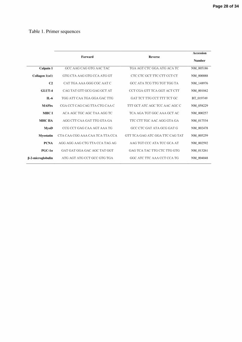
Table 1. Primer sequences
Forward ReverseAccession
Number
Calpain 1 GCC AAG CAG GTG AAC TAC TGA AGT CTC GGA ATG ACA TC NM_005186
Collagen 1(α1) GTG CTA AAG GTG CCA ATG GT CTC CTC GCT TTC CTT CCT CT NM_000088
C2 CAT TGA AAA GGG CGC AAT C GCC ATA TCG TTG TGT TGG TA NM_148976
GLUT-4 CAG TAT GTT GCG GAG GCT AT CCT CGA GTT TCA GGT ACT CTT NM_001042
IL-6 TGG ATT CAA TGA GGA GAC TTG GAT TCT TTG CCT TTT TCT GC BT_019749
MAFbx CGA CCT CAG CAG TTA CTG CAA C TTT GCT ATC AGC TCC AAC AGC C NM_058229
MHC I ACA AGC TGC AGC TAA AGG TC TCA AGA TGT GGC AAA GCT AC NM_000257
MHC IIA AGG CTT CAA GAT TTG GTA GA TTC CTT TGC AAC AGG GTA GA NM_017534
MyoD CCG CCT GAG CAA AGT AAA TG GCC CTC GAT ATA GCG GAT G NM_002478
Myostatin CTA CAA CGG AAA CAA TCA TTA CCA GTT TCA GAG ATC GGA TTC CAG TAT NM_005259
PCNA AGG AGG AAG CTG TTA CCA TAG AG AAG TGT CCC ATA TCC GCA AT NM_002592
PGC-1α GAT GAT GGA GAC AGC TAT GGT GAG TCA TAC TTG CTC TTG GTG NM_013261
β-2-microglobulin ATG AGT ATG CCT GCC GTG TGA GGC ATC TTC AAA CCT CCA TG NM_004048
Page 28 of 34
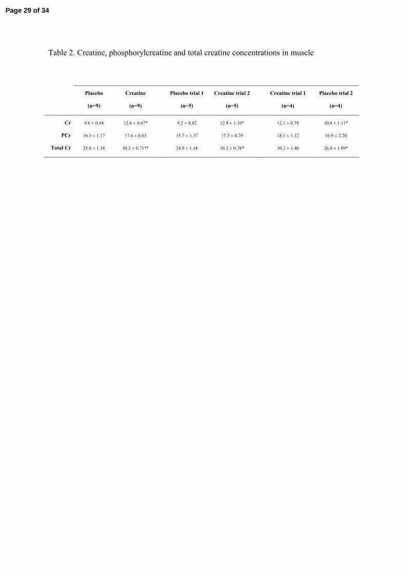
Table 2. Creatine, phosphorylcreatine and total creatine concentrations in muscle
Placebo
(n=9)
Creatine
(n=9)
Placebo trial 1
(n=5)
Creatine trial 2
(n=5)
Creatine trial 1
(n=4)
Placebo trial 2
(n=4)
Cr 9.6 ± 0.64 12.6 ± 0.67* 9.2 ± 0.82 12.9 ± 1.10* 12.1 ± 0.78 10.0 ± 1.11*
PCr 16.3 ± 1.17 17.6 ± 0.63 15.7 ± 1.37 17.3 ± 0.79 18.1 ± 1.12 16.9 ± 2.20
Total Cr 25.8 ± 1.18 30.2 ± 0.71** 24.9 ± 1.18 30.2 ± 0.78* 30.2 ± 1.40 26.9 ± 1.89*
Page 29 of 34
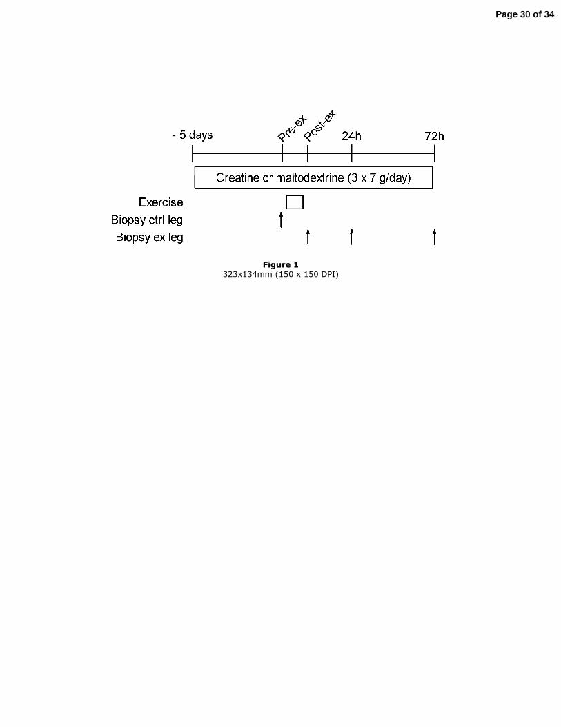
Figure 1 323x134mm (150 x 150 DPI)
Page 30 of 34
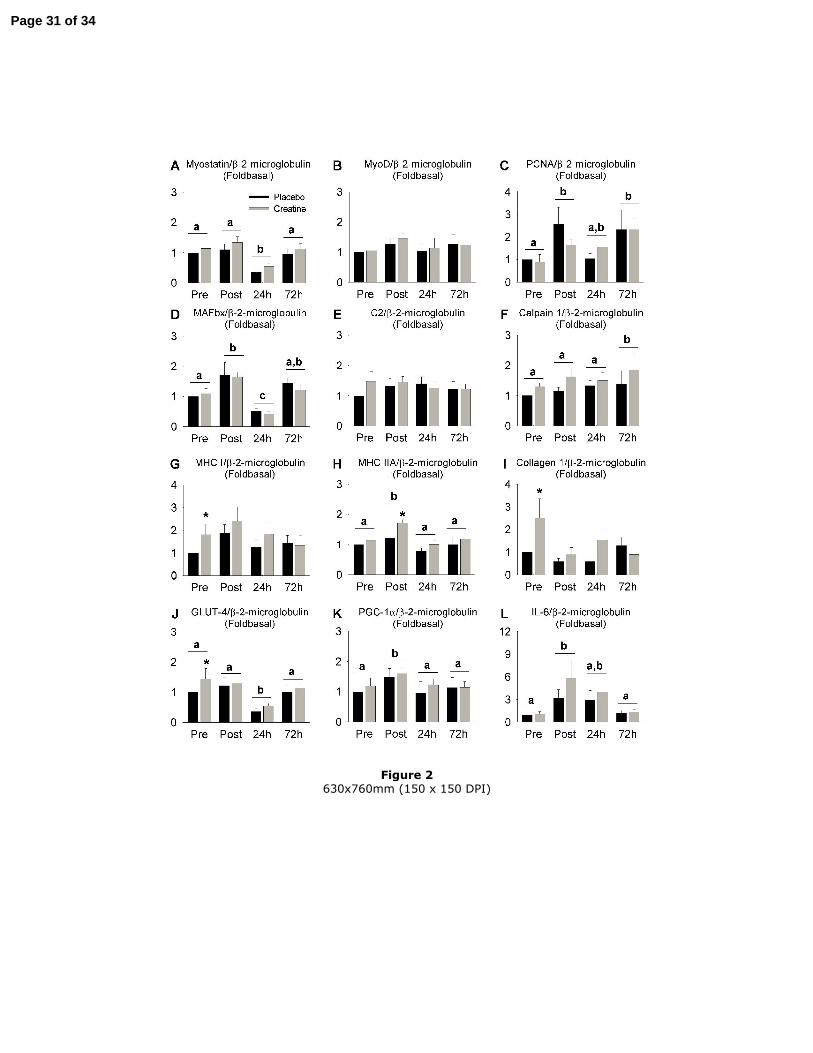
Figure 2 630x760mm (150 x 150 DPI)
Page 31 of 34
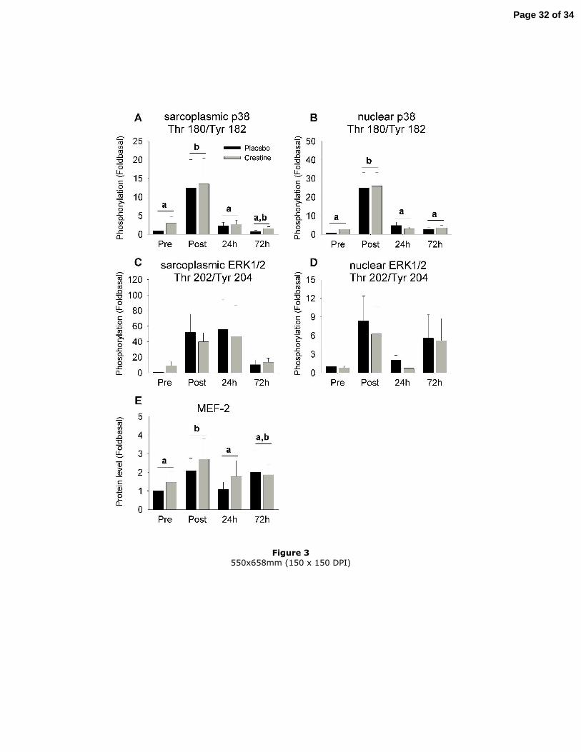
Figure 3 550x658mm (150 x 150 DPI)
Page 32 of 34
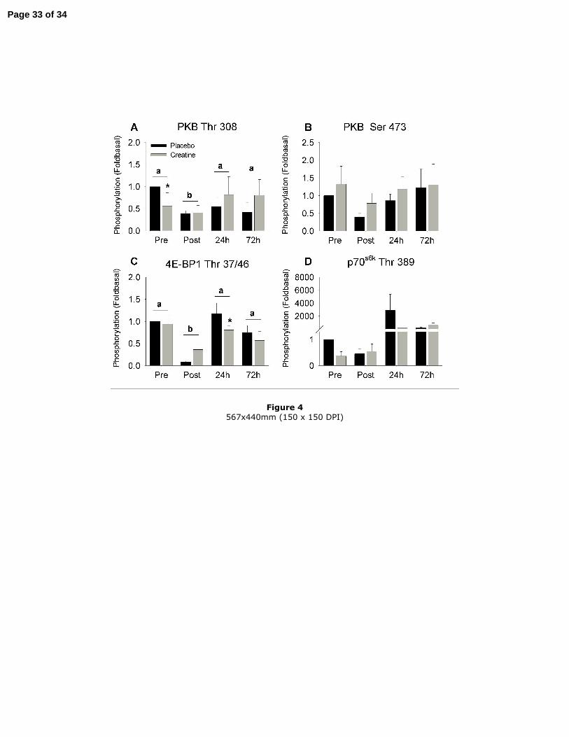
Figure 4 567x440mm (150 x 150 DPI)
Page 33 of 34
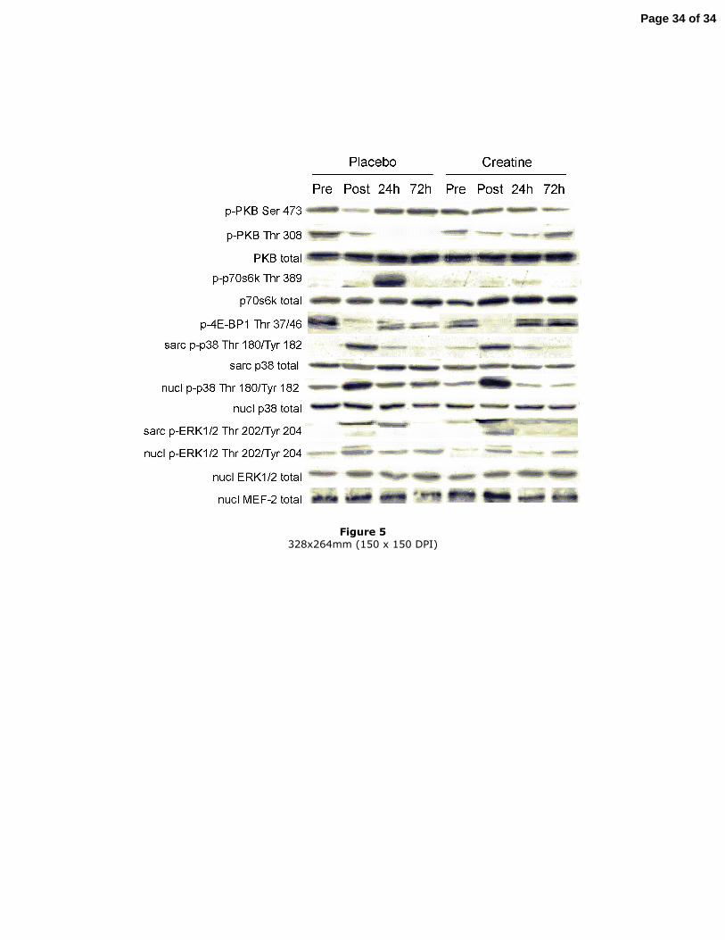
Figure 5 328x264mm (150 x 150 DPI)
Page 34 of 34