differential effect of creatine on oxidatively-injured mitochondrial and nuclear dna
TRANSCRIPT
Available online at www.sciencedirect.com
ta 1780 (2008) 16–26www.elsevier.com/locate/bbagen
Biochimica et Biophysica Ac
Differential effect of creatine on oxidatively-injuredmitochondrial and nuclear DNA
Chiara Guidi a,1, Lucia Potenza b,1, Piero Sestili a,c,⁎,1, Chiara Martinelli a, Michele Guescini a,Laura Stocchi a, Sabrina Zeppa a, Emanuela Polidori a, Giosuè Annibalini a, Vilberto Stocchi b
a Istituto di Ricerca sull’Attività Motoria, Università degli Studi di Urbino “Carlo Bo”, Via I Maggetti 26, 61029 Urbino (PU), Italyb Istituto di Chimica Biologica “Giorgio Fornaini”, Università degli Studi di Urbino “Carlo Bo”, Urbino, Italy
c Istituto di Farmacologia e Farmacognosia, Università degli Studi di Urbino “Carlo Bo”, Urbino, Italy
Received 14 March 2007; received in revised form 10 September 2007; accepted 18 September 2007Available online 5 October 2007
Abstract
Creatine is a naturally occurring compound obtained in humans from endogenous production and consumption through the diet. It is used as anergogenic aid to improve exercise performance and increase fat-free mass. Lately, creatine’s positive therapeutic benefits in various oxidativestress-associated diseases have been reported in literature and, more recently, creatine has also been shown to exert direct antioxidant effects.Oxidatively-challenged DNA was analysed to show possible protective effects of creatine. Acellular and cellular studies were carried out.Acellular assays, performed using molecular approaches, showed that creatine protects circular and linear DNA from oxidative attacks. Nuclearand mitochondrial DNAs from oxidatively-injured human umbilical vein endothelial cells were analyzed. Creatine supplementation showedsignificant genoprotective activity on mitochondrial DNA. This evidence suggests that creatine may play an important role in mitochondrialgenome stability in that it could normalize mitochondrial mutagenesis and its functional consequences. Thus, creatine supplementation could beused to prevent or ameliorate diseases related to mitochondrial DNA mutations, and possibly to delay aging.© 2007 Elsevier B.V. All rights reserved.
Keywords: Creatine; Oxidative DNA damage; Mitochondrial DNA; Nuclear DNA; Antioxidant
1. Introduction
Creatine (Cr) is the most popular supplement proposed as anergogenic aid. It is distributed throughout the body with 95%found in skeletal muscle and the remaining 5% in the brain, liver,kidney, and testes [1]. Cr is obtained through diet (∼1 g/day foran omnivorous diet) and synthesized in the liver, kidney andpancreas (∼1 g/day). The dietary intake and endogenous
Abbreviations: CCC, Covalently closed circular; Cr, Creatine; DTT, DL-dithio-threitol; MFO, Mixed-function oxidase; mtDNA, Mitochondrial DNA;nDNA, Nuclear DNA; 8-OHdG, 8-oxo-7,8-dihydro-2′-deoxyguanosine; PBS,Phosphate-buffered saline; ROS, Reactive oxygen species; PCR, Polymerasechain reaction; QPCR, Quantitative polymerase chain reaction⁎ Corresponding author. Istituto di Farmacologia e Farmacognosia, Università
degli Studi di Urbino “Carlo Bo”, Urbino, Italy. Tel.: +39 0722 303414; fax: +390722 303401.
E-mail address: [email protected] (P. Sestili).1 These authors (C.G., L.P., P.S.) contributed equally to the work described in
this report.
0304-4165/$ - see front matter © 2007 Elsevier B.V. All rights reserved.doi:10.1016/j.bbagen.2007.09.018
production of Cr match the spontaneous degradation ofphosphoCr and Cr to creatinine at a rate of 2.6% and 1.1% perday, respectively. Once creatinine is formed, it enters thecirculatory system by diffusion and is eliminated from the bodythrough glomerular filtration. Intramuscular and cerebral storesof Cr, as well as its phosphorylated form, phosphoCr, increasewith oral Cr-supplementation. The increase of these stores canoffer therapeutic benefits by preventing ATP depletion,stimulating protein synthesis or reducing protein degradation,and stabilizing biological membrane [2].
Evidence from exercise literature has shown that athletesbenefit from supplementation by increasing muscular force andpower, reducing fatigue in repeated bout activities, andincreasing muscle mass [3–7]. In the 1990s Cr supplementationbecame a popular ergogenic aid for many athletes to maintain arapid rate of adenosine triphosphate (ATP) turnover during abrief period of high intensity activity [8–15].
In a different direction, Lawler et al. reported that Cr iscapable of directly quenching aqueous radical and reactive
17C. Guidi et al. / Biochimica et Biophysica Acta 1780 (2008) 16–26
species ions in vitro [16]. A more recent study from ourlaboratory showed that Cr exerts direct antioxidant activity via ascavenging mechanism in oxidatively-injured cultured mamma-lian cells [17]. Moreover, other authors have demonstrated thatCr supplementation not only improves exercise performance andincreases fat-free mass, but is also beneficial in the prevention ortreatment of some oxidative stress associated diseases [18–21].In these diseases mitochondrial DNA (mtDNA) represents animportant target for oxidative damage. Indeed, mtDNA muta-tions have recently been reported as being an etiological factor inoxidative stress-related disorders [22] including cardiovasculardiseases and inherited [23] or acquired [24–26] neurodegener-ative disorders, several types of tumors affecting the colon,bladder, lung, breast, kidney, head and neck [27–34] mitochon-drial myopathies [35] and the normal aging process [36].
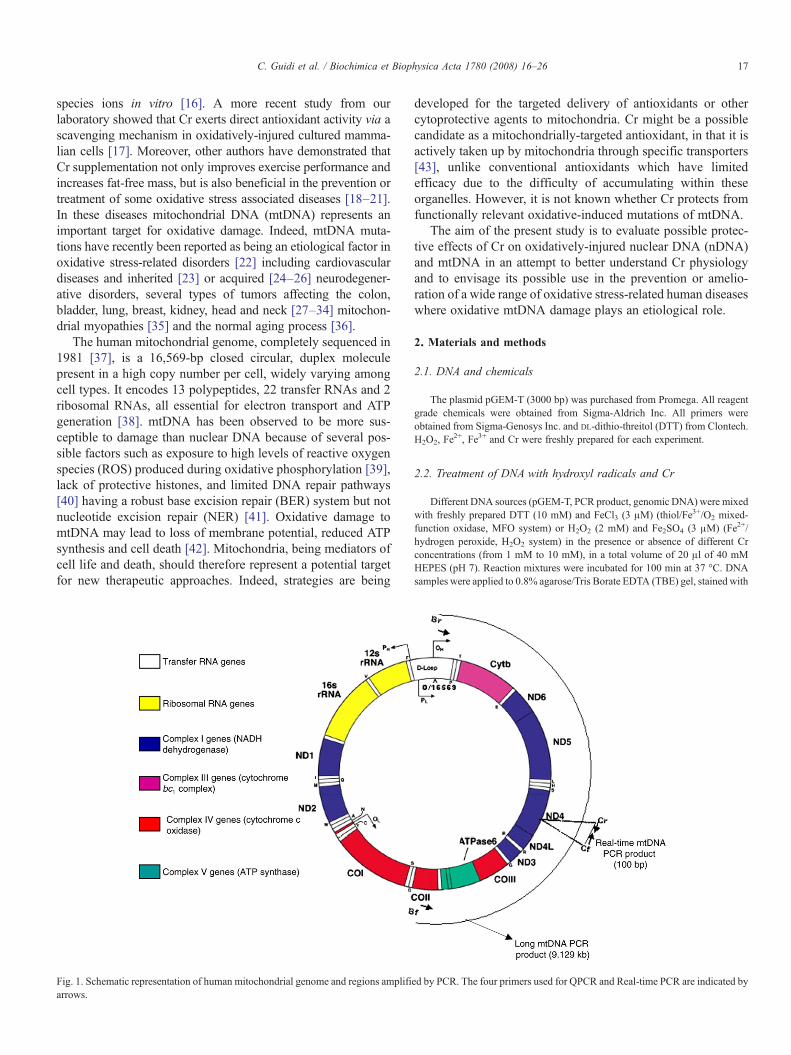
The human mitochondrial genome, completely sequenced in1981 [37], is a 16,569-bp closed circular, duplex moleculepresent in a high copy number per cell, widely varying amongcell types. It encodes 13 polypeptides, 22 transfer RNAs and 2ribosomal RNAs, all essential for electron transport and ATPgeneration [38]. mtDNA has been observed to be more sus-ceptible to damage than nuclear DNA because of several pos-sible factors such as exposure to high levels of reactive oxygenspecies (ROS) produced during oxidative phosphorylation [39],lack of protective histones, and limited DNA repair pathways[40] having a robust base excision repair (BER) system but notnucleotide excision repair (NER) [41]. Oxidative damage tomtDNA may lead to loss of membrane potential, reduced ATPsynthesis and cell death [42]. Mitochondria, being mediators ofcell life and death, should therefore represent a potential targetfor new therapeutic approaches. Indeed, strategies are being
Fig. 1. Schematic representation of human mitochondrial genome and regions amplifiarrows.
developed for the targeted delivery of antioxidants or othercytoprotective agents to mitochondria. Cr might be a possiblecandidate as a mitochondrially-targeted antioxidant, in that it isactively taken up by mitochondria through specific transporters[43], unlike conventional antioxidants which have limitedefficacy due to the difficulty of accumulating within theseorganelles. However, it is not known whether Cr protects fromfunctionally relevant oxidative-induced mutations of mtDNA.
The aim of the present study is to evaluate possible protec-tive effects of Cr on oxidatively-injured nuclear DNA (nDNA)and mtDNA in an attempt to better understand Cr physiologyand to envisage its possible use in the prevention or amelio-ration of a wide range of oxidative stress-related human diseaseswhere oxidative mtDNA damage plays an etiological role.
2. Materials and methods
2.1. DNA and chemicals
The plasmid pGEM-T (3000 bp) was purchased from Promega. All reagentgrade chemicals were obtained from Sigma-Aldrich Inc. All primers wereobtained from Sigma-Genosys Inc. and DL-dithio-threitol (DTT) from Clontech.H2O2, Fe
2+, Fe3+ and Cr were freshly prepared for each experiment.
2.2. Treatment of DNA with hydroxyl radicals and Cr
Different DNA sources (pGEM-T, PCR product, genomic DNA) were mixedwith freshly prepared DTT (10 mM) and FeCl3 (3 μM) (thiol/Fe3+/O2 mixed-function oxidase, MFO system) or H2O2 (2 mM) and Fe2SO4 (3 μM) (Fe2+/hydrogen peroxide, H2O2 system) in the presence or absence of different Crconcentrations (from 1 mM to 10 mM), in a total volume of 20 μl of 40 mMHEPES (pH 7). Reaction mixtures were incubated for 100 min at 37 °C. DNAsamples were applied to 0.8% agarose/Tris Borate EDTA (TBE) gel, stained with
ed by PCR. The four primers used for QPCR and Real-time PCR are indicated by
Table 2Thermal cycling parameters
Cycles Temperature (°C) Time
PCR 1 1× 95 5 min95 30 s
30× 62 30 s72 45 s
1× 72 7 min1× 4 hold
PCR 2 1× 95 2 min30× 95 30 s
68 9 min1× 68 12 min1× 4 hold
PCR 3 1× 93 2 min10× 93 10 s
60 30 s68 4 min 40 s
20× 93 10 s60 30 s68 4 min 40 s +
20 s per cycle1× 68 7 min1× 4 hold
PCR 4 1× 95 10 min40× 95 1 min
60 9 s1× 4 hold
18 C. Guidi et al. / Biochimica et Biophysica Acta 1780 (2008) 16–26
ethidium bromide (0.3 μg/ml) and visualized under UV light. Quantification wasmade by densitometric analysis using Quantity One Software 4.01 (Bio-Rad).
2.3. Isolation of total DNA
High molecular weight DNA was isolated with the QIAamp DNA mini kit(Qiagen) according to the manufacturer’s instructions. Total cellular DNAconcentration was determined at 260 nm using spectrophotometer (BeckmanDU- 640) and nanodrop spectrophotometer (ND-1000 Nanodrop Technologies).
2.4. Cell culture and treatment conditions
HUVEC (human umbilical vein endothelial cells) were cultured at 37 °C inan atmosphere of 95% air and 5% CO2 in M199 medium containing antibiotics,1.4 mM glutamine, 10% fetal bovine serum and 50 μg/ml endothelial cellgrowth factor. HUVEC were seeded at an appropriate density 30–36 h beforetreatments. At the oxidative challenge stage, the cell number was between 3.5and 4.5×10⁵ cells/well. Cr (10 mM), Trolox (100 μM) or o-phenanthroline(10 μM) were added to complete culture medium and were given to cells 24 h,10′ and 1 h prior to the challenge with H2O2, respectively. Trolox and o-phenanthroline were also added at the same concentrations to Saline A duringtreatment with H2O2. Oxidative challenge consisted in 30 min incubation ofantioxidant-free or antioxidant-supplemented cells with 200 μM H2O2 at 37 °Cin 2 ml of Saline A (0.145 M NaCl, 5 mM KCl, 10 mM NaHCO3, 5 mMglucose). After treatments, cells were washed with phosphate-buffered saline(PBS, 8 g/l NaCl, 1.15 g/l Na2HPO4, 0.2 g/l KH2PO4, 0.2 g/l KCl), harvested bytrypsination and processed for DNA damage or recultured in the originalmedium at different times for DNA repair and cytotoxicity studies.
2.5. Trypan blue exclusion assay
Monolayers were detached by trypsinization, an aliquot of cell suspensionwas diluted 1:1 with 0.4% trypan blue and cells were counted with a hemo-cytometer. Results are expressed as the number of viable (unstained) cells intreated and control samples.
2.6. Long PCR
Long PCRwas performed in a final volume of 25 μl using aMycycler machine(Bio-Rad). Specific primers were used to amplify a 9.129-kb fragment of themitochondrial DNA (primers Bf–Br, long mtDNA PCR, Fig. 1) and a 7.3-kbfragment of the nuclear gene GBA (glucosidase, beta acid; primers Df–Dr, longnDNA PCR).
The reaction mixture contained 60 ng template total DNA, 2.5 μl buffer 1,200 μM dNTPs, 0.5 μM of each primer and 1.5 U of Expand Long TemplatePCR system (Roche). The primer nucleotidic sequences and PCR parameters arereported in Tables 1 and 2.
Table 1Primers employed
604 bp from D-Loop mitochondrial DNA (J01415):Af 35 5′ GGAGCTCTCCATGCATTTGG 3′Ar 620 5′ GGGTGATGTGAGCCCGTCTA 3′
9.129 kb mitochondrial fragment (J01415):Bf 8080 5′ CCCCACATTAGGCTTAAAAACAGAT 3′Br 620 5′ GGGTGATGTGAGCCCGTCTA 3′
100 bp mitochondrial fragment (J01415):Cf 5′CCATTCTCCTCCTATCCCTCAAC 3′Cr 5′ CACAATCTGATGTTTTGGTTAAACTATATTT 3′
7.3 kb nuclear DNA of the GBA gene (NM_001005741):Df 5′ TTCTCCATGCAAATCTGTGT 3′Dr 5′ GAACCAGATCCTATCTGTGC 3′
100 bp nuclear DNA of the GBA gene (NM_001005741):Ef 5′ AGCATCAGGGCGGAAGC 3′Er 5′ TTTCTCCTTTAAGAGCTGCCATTT 3′
2.7. Quantitative PCR (QPCR)
The amplification products obtained by long PCR were electrophoresed on0.8% agarose/TBE gel, stained with ethidium bromide (0.3 μg/ml) andquantified by densitometric analyses of the intensity of bands using QuantityOne Software 4.01 (Bio-Rad). Treated samples were then compared with con-trols and the relative amplification was calculated according to Santos et al. [44].
Results presented herein are the mean of two sets of PCR for each target of atleast three different biological experiments.
2.8. PCR product purification
The Af–Ar mtDNA amplification products (Tables 1 and 2) were purifiedusing the GenElute PCR Clean-up kit (Sigma-Aldrich Inc) according to themanufacturer’s instructions.
2.9. Quantitative Real-Time PCR
Long mtDNA and nDNA PCR products, obtained from HUVECDNA, werealso quantified by Sybr Green Real-Time PCR, using primers Cf-Cr and Ef-Er,respectively, localized in the middle of the long PCR fragments (Table 1). Afterlong amplification, samples were diluted 10−4, while the correspondinggenomic DNA samples were diluted 10−3.
Quantitative Real-Time PCR was performed in a Bio-Rad iCycler iQ Multi-Color Real-Time PCR Detection System using 2× Quantitect SYBR Green PCRkit (Qiagen). The quantitative PCR reaction was performed at 95 °C for 10 minto activate HotStart DNA polymerase followed by 50 cycles of the two-step at95 °C for 30 s and at 60 °C for 30 s. The specificity of the amplification productsobtained was confirmed by examining thermal denaturation plots and by sampleseparation in a 3% DNA agarose gel.
Results were normalized by quantitating each sample for the amount ofinitial genomic DNAwithout previous long PCR amplification, in the same real-time PCR conditions.
Each sample was tested in triplicate, and the experimental groups (controlDNA, H2O2-treated DNA with and without Cr time 0, after 2, 4, 24 and 48 hrecovery) consisted of at least three independent experiments. The significance
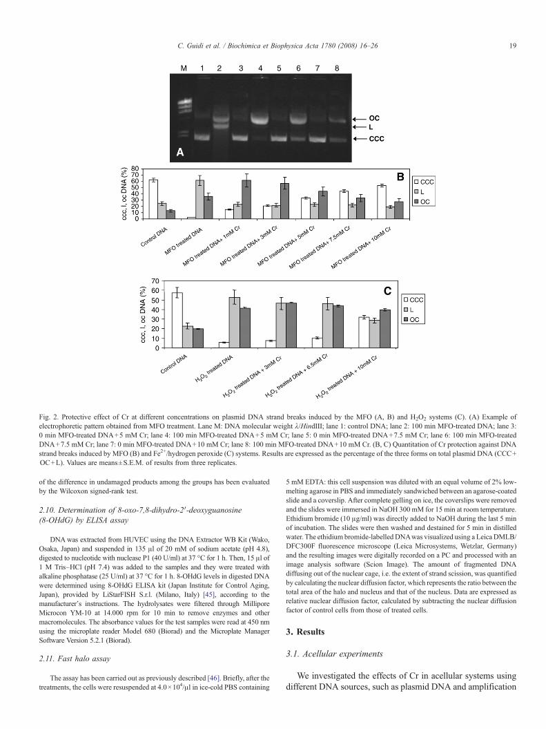
Fig. 2. Protective effect of Cr at different concentrations on plasmid DNA strand breaks induced by the MFO (A, B) and H2O2 systems (C). (A) Example ofelectrophoretic pattern obtained from MFO treatment. Lane M: DNA molecular weight λ/HindIII; lane 1: control DNA; lane 2: 100 min MFO-treated DNA; lane 3:0 min MFO-treated DNA+5 mM Cr; lane 4: 100 min MFO-treated DNA+5 mM Cr; lane 5: 0 min MFO-treated DNA+7.5 mM Cr; lane 6: 100 min MFO-treatedDNA+7.5 mM Cr; lane 7: 0 min MFO-treated DNA+10 mM Cr; lane 8: 100 min MFO-treated DNA+10 mM Cr. (B, C) Quantitation of Cr protection against DNAstrand breaks induced by MFO (B) and Fe2+/hydrogen peroxide (C) systems. Results are expressed as the percentage of the three forms on total plasmid DNA (CCC+OC+L). Values are means±S.E.M. of results from three replicates.
19C. Guidi et al. / Biochimica et Biophysica Acta 1780 (2008) 16–26
of the difference in undamaged products among the groups has been evaluatedby the Wilcoxon signed-rank test.
2.10. Determination of 8-oxo-7,8-dihydro-2′-deoxyguanosine(8-OHdG) by ELISA assay
DNAwas extracted from HUVEC using the DNA Extractor WB Kit (Wako,Osaka, Japan) and suspended in 135 μl of 20 mM of sodium acetate (pH 4.8),digested to nucleotide with nuclease P1 (40 U/ml) at 37 °C for 1 h. Then, 15 μl of1 M Tris–HCl (pH 7.4) was added to the samples and they were treated withalkaline phosphatase (25 U/ml) at 37 °C for 1 h. 8-OHdG levels in digested DNAwere determined using 8-OHdG ELISA kit (Japan Institute for Control Aging,Japan), provided by LiStarFISH S.r.l. (Milano, Italy) [45], according to themanufacturer’s instructions. The hydrolysates were filtered through MilliporeMicrocon YM-10 at 14.000 rpm for 10 min to remove enzymes and othermacromolecules. The absorbance values for the test samples were read at 450 nmusing the microplate reader Model 680 (Biorad) and the Microplate ManagerSoftware Version 5.2.1 (Biorad).
2.11. Fast halo assay
The assay has been carried out as previously described [46]. Briefly, after thetreatments, the cells were resuspended at 4.0×10⁴/μl in ice-cold PBS containing
5 mM EDTA: this cell suspension was diluted with an equal volume of 2% low-melting agarose in PBS and immediately sandwiched between an agarose-coatedslide and a coverslip. After complete gelling on ice, the coverslips were removedand the slides were immersed in NaOH 300 mM for 15 min at room temperature.Ethidium bromide (10 μg/ml) was directly added to NaOH during the last 5 minof incubation. The slides were then washed and destained for 5 min in distilledwater. The ethidium bromide-labelled DNAwas visualized using a Leica DMLB/DFC300F fluorescence microscope (Leica Microsystems, Wetzlar, Germany)and the resulting images were digitally recorded on a PC and processed with animage analysis software (Scion Image). The amount of fragmented DNAdiffusing out of the nuclear cage, i.e. the extent of strand scission, was quantifiedby calculating the nuclear diffusion factor, which represents the ratio between thetotal area of the halo and nucleus and that of the nucleus. Data are expressed asrelative nuclear diffusion factor, calculated by subtracting the nuclear diffusionfactor of control cells from those of treated cells.
3. Results
3.1. Acellular experiments
We investigated the effects of Cr in acellular systems usingdifferent DNA sources, such as plasmid DNA and amplification
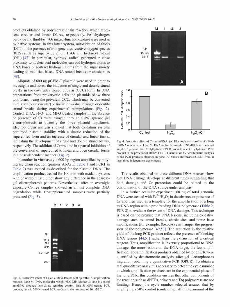
Fig. 4. Protective effect of Cr on mtDNA. (A) Electrophoretic profile of a 9-kbmtDNA region PCR. Lane M: DNA molecular weight λ/HindIII; lane 1: controlamplified product; lane 2: H2O2-treated PCR product; lane 3: H2O2-treated PCRproduct in the presence of 10 mM Cr. (B) Quantitation by densitometric analysisof the PCR products obtained in panel A. Values are means±S.E.M. from atleast three independent experiments.
20 C. Guidi et al. / Biochimica et Biophysica Acta 1780 (2008) 16–26
products obtained by polymerase chain reaction, which repre-sent circular and linear DNAs, respectively. Fe2+/hydrogenperoxide and thiol/Fe3+/O2mixed-function oxidase were used asoxidative systems. In this latter system, autoxidation of thiols(DTT) in the presence of iron generates reactive oxygen species(ROS) such as superoxide anion, H2O2 and hydroxyl radical(OH.) [47]. In particular, hydroxyl radical generated in closeproximity to nucleic acid molecules can add hydrogen atoms toDNA bases or abstract hydrogen atoms from the sugar moietyleading to modified bases, DNA strand breaks or abasic sites[48].
Aliquots of 600 ng pGEM-T plasmid were used in order toinvestigate and assess the induction of single and double strandbreaks in the covalently closed circular (CCC) form. In DNApreparations from prokaryotic cells the plasmids show threetopoforms, being the prevalent CCC, which may be convertedin relaxed (open circular) or linear forms due to single or doublestrand breaks during experimental manipulations (Fig. 2).Control DNA, H2O2 and MFO treated samples in the absenceor presence of Cr were assayed through 0.8% agarose gelelectrophoresis to quantify the three plasmid topoforms.Electrophoresis analysis showed that both oxidation systemsperturbed plasmid stability with a drastic reduction of thesupercoiled form and an increase of circular and linear forms,indicating the development of single and double strand breaks,respectively. The addition of Cr resulted in a partial inhibition ofthe conversion of supercoiled to linear and open circular formsin a dose-dependent manner (Fig. 2).
In another in vitro assay a 600-bp region amplified by poly-merase chain reaction (primers Af-Ar in Table 1 and PCR1 inTable 2) was treated as described for the plasmid DNA. Theamplification product treated for 100 min with oxidant systemswith or without Cr did not show any difference in the agarose-gel electrophoresis patterns. Nevertheless, after an overnightexposure Cr-free samples showed an almost complete DNAdegradation while Cr-supplemented samples were partiallyprotected (Fig. 3).
Fig. 3. Protective effect of Cr on a MFO-treated 600 bp mtDNA amplificationproduct. Lane M: DNA molecular weight pUC Mix Marker 8; lane 1: controlamplified product; lane 2: no template control; lane 3: MFO-treated PCRproduct; lane 4: MFO-treated PCR product in the presence of 10 mM Cr.
The results obtained on these different DNA sources showthat DNA damage develops at different times suggesting thatboth damage and Cr protection could be related to theconformation of the DNA source under analysis.
In a further acellular experiment, 60 ng of total genomicDNAwere treated with Fe2+/H2O2 in the absence or presence ofCr and then used as a template for the amplification of a longmtDNA region with a proofreading DNA polymerase (Table 2,PCR 2) to evaluate the extent of DNA damage. This techniqueis based on the premise that DNA lesions, including oxidativedamage such as strand breaks, abasic sites and some basemodifications (for example, 8oxodA) can hamper the progres-sion of the polymerase [49,50]. The reduction in the relativeyield of the long PCR product reflects the presence of blockingDNA lesions [44,51] rather than the exhaustion of a criticalreagent. Thus, amplification is inversely proportional to DNAdamage: the more lesions on the DNA target, the less ampli-fication. The amplification products obtained by long PCR werequantified by densitometric analysis, after gel electrophoresismigration, obtaining a quantitative PCR (QPCR). To obtain areal quantitative assay it is necessary to detect the cycle numberat which amplification products are in the exponential phase ofthe long PCR: this condition ensures that other components ofthe reaction such as dNTPs, primers and Taq polymerase are notlimiting. Hence, the cycle number selected assures that byamplifying a 50% control (containing half of the amount of the
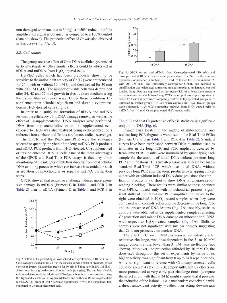
Fig. 6. QPCR on mt and nDNAs from Cr-supplemented (10 mM) andunsupplemented HUVEC. Cells were pre-incubated for 24 h in the absence(open bars) or presence (solid bars) of 10 mM Cr, treated for 30 min in Saline Awith 200 μM H2O2 and immediately assayed for QPCR. The decrease inamplification was calculated comparing treated samples to undamaged control(dashed line). Data are expressed as the mean±S.E. of at least three separatedeterminations in which two Long PCRs were performed per experiment.Student's t test was performed comparing controls to H2O2-treated groups or Cruntreated to treated groups. Pb0.05 when controls and H2O2-treated groupswere compared. ⁎, Pb0.05 comparing mtDNA from H2O2-treated cells tomtDNA from 10 mM Cr supplemented H2O2-treated cells.
21C. Guidi et al. / Biochimica et Biophysica Acta 1780 (2008) 16–26
non-damaged template, that is 30 ng), a ∼50% reduction of theamplification signal is obtained, as compared to a 100% control(data not shown). The protective effect of Cr was also observedin this assay (Fig. 4A, B).
3.2. Cell studies
The genoprotective effect of Cr in DNA acellular systems ledus to investigate whether similar effects could be observed innDNA and mtDNA from H2O2-injured cells.
HUVEC cells, which had been previously shown to besensitive to the antioxidant activity of Cr [17] were preincubatedfor 24 h with or without 10 mM Cr and then treated for 30 minwith 200 μM H2O2. The number of viable cells was determinedafter 24, 48 and 72 h of growth in fresh culture medium usingthe trypan blue exclusion assay. Under these conditions Cr-supplementation afforded significant and durable cytoprotec-tion in H2O2-treated cells (Fig. 5).
In order to quantify the formation of nDNA and mtDNAlesions, the efficiency of mtDNA damage removal as well as theeffect of Cr-supplementation, DNA analyses were performed.DNA from o-phenanthroline or trolox supplemented cellsexposed to H2O2 was also analyzed being o-phenanthroline areference iron chelator and Trolox a reference radical scavenger.
The QPCR and the Real-Time PCR were the strategiesselected to quantify the yield of the long mtDNA PCR productsand nDNA PCR products from H2O2-treated, Cr-supplementedor unsupplemented HUVEC cells. One of the main advantagesof the QPCR and Real-Time PCR assays is that they allowmonitoring of the integrity of mtDNA directly from total cellularDNA avoiding processes which can increase base oxidation suchas isolation of mitochondria or separate mtDNA purificationsteps.
QPCR showed that oxidative challenge induces more exten-sive damage in mtDNA (Primers B in Table 1 and PCR 2 inTable 2) than in nDNA (Primers D in Table 1 and PCR 3 in
Fig. 5. Effect of Cr preloading on oxidant-induced cytotoxicity in HUVEC cells.Cells were pre-incubated for 24 h in the absence (open circles) or presence (closedcircles) of 10 mM Cr, and then treated for 30 min in Saline Awith 200 μMH2O2.Also shown is the growth curve of control cells (triangles). The number of viablecells was determined after 24, 48 and 72 h of growth in fresh culture medium usingthe Trypan blue exclusion assay (seeMaterials and methods). Results represent themeans±S.E.M. from at least 5 separate experiments. ⁎ Pb0.005 (unpaired t test)compared to Cr unsupplemented cells.
Table 2) and that Cr protective effect is statistically significantonly on mtDNA (Fig. 6).
Primer pairs located in the middle of mitochondrial andnuclear long PCR fragments were used in the Real-Time PCRs(Primers C and E in Table 1 and PCR 4 in Table 2). Standardcurves have been established between DNA quantities used astemplates in the long PCR and PCR amplicons detected byReal-Time PCR. Results were normalized by quantifying eachsample for the amount of initial DNA without previous longPCR amplifications. This two-step assay was selected because astandard Real-Time PCR which uses total DNA, withoutprevious long PCR amplification, produces overlapping curveseither with or without induced DNA damages, since the ampli-fication product is too short to show DNA polymerase proof-reading blocking. These results were similar to those obtainedwith QPCR. Indeed, only with mitochondrial primers, signif-icant shifts of the Real-Time PCR amplification curves to theright were obtained in H2O2-treated samples when they werecompared with controls, reflecting the decrease in the long PCRand the presence of DNA lesions (Fig. 7A); notably, shifts tocontrols were obtained in Cr supplemented samples reflectingCr protection and minor DNA damage on mitochondrial DNAwith respect to H2O2-treated samples (Fig. 7A). Shifts tocontrols were not significant with nuclear primers suggestingthat Cr is not protective on nuclear DNA.
The effect of Cr on mtDNA, as assayed immediately afteroxidative challenge, was dose-dependent in the 3- to 10-mMrange; concentrations lower than 3 mM were ineffective (notshown). Moreover, the protection afforded by 10 mM Cr, thedose used throughout this set of experiments by virtue of itshigher activity, was significant from 0 up to 24 h repair periods,while no significant difference with Cr unsupplemented cellscould be seen at 48 h (Fig. 7B). Importantly, that Cr effects aremore pronounced at very early post-challenge times (comparethe effect at 0 h with that at 24 h) might suggest that it preventsthe induction of the lesions – i.e. a mechanism conceivable witha direct antioxidant activity – rather than acting downstream
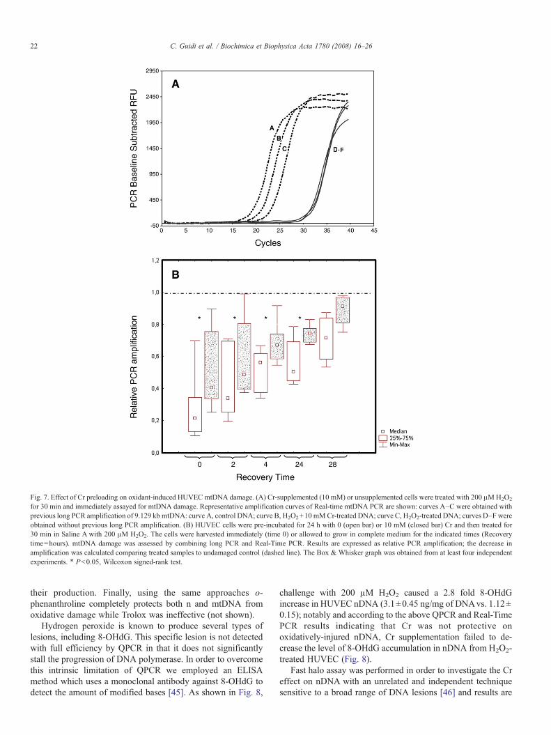
Fig. 7. Effect of Cr preloading on oxidant-induced HUVEC mtDNA damage. (A) Cr-supplemented (10 mM) or unsupplemented cells were treated with 200 μMH2O2
for 30 min and immediately assayed for mtDNA damage. Representative amplification curves of Real-time mtDNA PCR are shown: curves A–C were obtained withprevious long PCR amplification of 9.129 kb mtDNA: curve A, control DNA; curve B, H2O2+10 mMCr-treated DNA; curve C, H2O2-treated DNA; curves D–F wereobtained without previous long PCR amplification. (B) HUVEC cells were pre-incubated for 24 h with 0 (open bar) or 10 mM (closed bar) Cr and then treated for30 min in Saline A with 200 μM H2O2. The cells were harvested immediately (time 0) or allowed to grow in complete medium for the indicated times (Recoverytime=hours). mtDNA damage was assessed by combining long PCR and Real-Time PCR. Results are expressed as relative PCR amplification; the decrease inamplification was calculated comparing treated samples to undamaged control (dashed line). The Box & Whisker graph was obtained from at least four independentexperiments. ⁎ Pb0.05, Wilcoxon signed-rank test.
22 C. Guidi et al. / Biochimica et Biophysica Acta 1780 (2008) 16–26
their production. Finally, using the same approaches o-phenanthroline completely protects both n and mtDNA fromoxidative damage while Trolox was ineffective (not shown).
Hydrogen peroxide is known to produce several types oflesions, including 8-OHdG. This specific lesion is not detectedwith full efficiency by QPCR in that it does not significantlystall the progression of DNA polymerase. In order to overcomethis intrinsic limitation of QPCR we employed an ELISAmethod which uses a monoclonal antibody against 8-OHdG todetect the amount of modified bases [45]. As shown in Fig. 8,
challenge with 200 μM H2O2 caused a 2.8 fold 8-OHdGincrease in HUVEC nDNA (3.1±0.45 ng/mg of DNAvs. 1.12±0.15); notably and according to the above QPCR and Real-TimePCR results indicating that Cr was not protective onoxidatively-injured nDNA, Cr supplementation failed to de-crease the level of 8-OHdG accumulation in nDNA from H2O2-treated HUVEC (Fig. 8).
Fast halo assay was performed in order to investigate the Creffect on nDNA with an unrelated and independent techniquesensitive to a broad range of DNA lesions [46] and results are
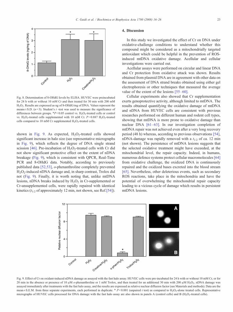
Fig. 8. Determination of 8-OHdG levels by ELISA. HUVEC were preincubatedfor 24 h with or without 10 mM Cr and then treated for 30 min with 200 mMH2O2. Results are expressed as ng of 8-OHdG/mg of DNA. Values represent themeans±S.D. (n=3). Student's t -test was used to measure the significance ofdifferences between groups. ⁎Pb0.05 control vs. H2O2-treated cells or controlvs. H2O2-treated cells supplemented with 10 mM Cr. P=0.887 H2O2-treatedcells compared to 10 mM Cr supplemented H2O2-treated cells.
23C. Guidi et al. / Biochimica et Biophysica Acta 1780 (2008) 16–26
shown in Fig. 9. As expected, H2O2-treated cells showedsignificant increase in halo size (see representative micrographsin Fig. 9), which reflects the degree of DNA single strandscission [46]. Pre-incubation of H2O2-treated cells with Cr didnot show significant protective effect on the extent of nDNAbreakage (Fig. 9), which is consistent with QPCR, Real-TimePCR and 8-OHdG data. Notably, according to previouslypublished data [52,53], o-phenanthroline completely preventedH2O2-induced nDNA damage and, in sharp contrast, Trolox didnot (Fig. 9). Finally, it is worth noting that, unlike mtDNAlesions, nDNA breaks induced by H2O2 in Cr-supplemented orCr-unsupplemented cells, were rapidly repaired with identicalkinetics (t1/2 of approximately 12 min, not shown, see Ref.[54]).
Fig. 9. Effect of Cr on oxidant-induced nDNA damage as assayed with the fast halo as20 min in the absence or presence of 10 μM o-phenanthroline or 1 mM Trolox, andassayed immediately after treatments with the fast halo assay, and the results are expremean±S.E.M. from three separate experiments, each performed in duplicate. ⁎ Pbmicrographs of HUVEC cells processed for DNA damage with the fast halo assay a
4. Discussion
In this study we investigated the effect of Cr on DNA underoxidative-challenge conditions to understand whether thiscompound might be considered as a mitochondrially targetedantioxidant which could be helpful in the prevention of ROS-induced mtDNA oxidative damage. Acellular and cellularinvestigations were carried out.
Acellular assays were performed on circular and linear DNAand Cr protection from oxidative attack was shown. Resultsobtained from plasmid DNA are in agreement with other data onthe assessment of DNA strand breaks obtained using either gelelectrophoresis or other techniques that measured the averagevalue of the extent of the lesions [55–60].
Cellular experiments also showed that Cr supplementationexerts genoprotective activity, although limited to mtDNA. Theresults obtained quantifying the oxidative damage of mtDNAand nDNA from HUVEC cells are consistent with previousresearches performed on different human and rodent cell types,showing that mtDNA is more prone to oxidative damage thannuclear DNA [61–63]. In our investigation completion ofmtDNA repair was not achieved even after a very long recoveryperiod (48 h) whereas, according to previous observations [54],nDNA-damage was rapidly removed with a t1/2 of ca. 12 min(not shown). The persistence of mtDNA lesions suggests thatthe selected oxidative treatment might have exceeded, at themitochondrial level, the repair capacity. Indeed, in humans,numerous defence systems protect cellular macromolecules [64]from oxidative challenge, the oxidized DNA is continuouslyrepaired and the oxidized bases excreted into the blood stream[65]. Nevertheless, other deleterious events, such as secondaryROS reactions, take place in the mitochondria and have thepotential of overwhelming the mitochondrial repair capacityleading to a vicious cycle of damage which results in persistentmtDNA lesions.
say. HUVEC cells were pre-incubated for 24 h with or without 10 mM Cr, or forthen treated for an additional 30 min with 200 μM H2O2. nDNA damage wasssed as relative nuclear diffusion factor (see Materials and methods). Data are the0.001 (unpaired t test) as compared to H2O2-alone treated cells. Representativere also shown in panels A (control cells) and B (H2O2-treated cells).
24 C. Guidi et al. / Biochimica et Biophysica Acta 1780 (2008) 16–26
Several studies have documented the powerful protectiveeffects of Cr in oxidative stress associated diseases [18–21] andin neuropathologies, such as Huntington disease, Parkinsondisease and amyotrophic lateral sclerosis [66–68]. The noveltyof the present work is the finding that Cr protects oxidatively-injured DNA, as shown by both acellular experiments and cellbased assays, respectively. In particular, our results show that Crsupplementation significantly protects only mtDNA. ThatnDNA is not sensitive to Cr-protection (Figs. 6–8) is notsurprising. nDNA damage following oxidative stress is usuallythought to be a function of site-specific DNA-associated Fe-based Fenton chemistry [69]. It is commonly assumed that ironis normally associated with nDNA and many reports indicatethat only iron-chelators, unlike scavenging antioxidants such asTrolox [52,53] (Fig. 9), are capable of protecting cells againstoxidative nuclear damage [53,70–72]. Importantly, Cr has beenshown to exert its antioxidant, cytoprotective activity via ascavenging mechanism rather than through iron-chelation [17]:hence, the Cr lack of protective effects on oxidatively-injurednDNA, as assayed with QPCR, Real-Time PCR, 8-OHdGELISA and fast halo assay, is not surprising. Conversely ourdata, as well as others [73–76], imply that mitochondria-accumulating scavenging compounds such as Cr are capable ofpreventing mtDNA oxidative damage. This differential activity,i.e. mtDNA protection in the absence of nDNA protection,might be due to the different conformation of the two types ofDNA. Some indications of this can be observed in acellularexperiments on circular and linear DNA (Figs. 2 and 3), whichcould resemble mitochondrial and nuclear DNA, respectively:plasmidic DNA showed oxidative damages just after 100 mintreatment, while a PCR amplification product was damagedafter a longer treatment time. Thus it is likely that circular DNA,because of its negative supercoiled structure, is more prone todamage as well as more accessible to Cr and sensitive to itsprotective effect, than linear DNA. Furthermore, in the specificcase of Cr, it should be noted that the Cr-protective effect onmtDNA might be related to its mitochondrial localization.Indeed Cr is actively taken up by specific 55 and 70 kDa mt-Crtransporters [43] into mitochondria, where it is utilized for theenergy transport between the site of ATP production and con-sumption by ATPases. Isolated, respiring mitochondria incu-bated in 15 mM Cr have been shown to accumulate 20 mM Cr[43]. Notably, under our supplementing conditions, HUVECintracellular free-Cr level has been shown to be 48.5 nmol/mgof proteins [17], i.e. 8.82 mM (HUVEC volume=5.5 μl/mg ofproteins, [77]). As a corollary, the importance of the capacity ofan antioxidant to accumulate within mitochondria in order toprotect mtDNA from oxidative damage is indirectly emphasizedby the observation that Trolox, which is not known toaccumulate within these organelles, lacks any protective effect.
Finally, it is worth noting that our results implicitly raise thequestion of whether oxidative mtDNA and nDNA lesionsrepresent lethal events. Indeed our data confirm the notion thatH2O2-induced nDNA single strand breaks do not represent acytotoxically relevant damage [69,78] since in Cr-supplementedH2O2-treated cells we observed reduced cytotoxicity in theabsence of reduced nDNA breakage. In sharp contrast, we also
showed that reduced mtDNA damage is paralleled by adecreased cytotoxic response in H2O2-injured Cr-supplementedcells. Thus, it could be inferred that, at least under mild stressingconditions, oxidative mtDNA damage, unlike nDNA damage,represents a cytotoxically relevant type of lesion. However, toanswer this important question further studies will be necessaryto understand whether this phenomenon is simply incidental orit reflects a causal relationship.
Our study is one of the few reporting the protective effect ofCr on mtDNA. Berneburg et al. [79] demonstrated that 1 mM Crabolished the induction of mtDNA mutations generated innormal human fibroblast by repetitive UV-A irradiation. In ourinvestigation we used a different cell line, different molecularapproaches and a different cellular insult (acute oxidative stress)and we found that mtDNA is protected by Cr.
Data reported by Sestili et al. [17] indicate that Cr affordscytoprotection via direct antioxidant capacity with a radicalscavenging mechanism: the results presented herein also pointto the role of free-Cr as an antioxidant and suggest that the effectof Cr on oxidatively-injured mtDNA might represent animportant mechanism contributing to its cytoprotective activity[17] in cells subjected to oxidative stress.
Other antioxidants including vitamins or cofactors such asCoenzyme Q10, ascorbic acid, vitamin E, riboflavin, thiamine,niacin, vitamin K (phylloquinone and menadione), and carnitinehave already been used in the treatment of oxidativephosphorylation disorders to increase mitochondrial ATPproduction and slow or arrest the progression of clinicalsymptoms [68,80].
Results obtained from cellular experiments suggest that Crsupplementation may play an important role in mitochondrialgenome stability in that it could normalize mitochondrialmutagenesis as well as functional consequences such as thedecrease of oxygen consumption, mitochondrial membranepotential and ATP content and finally cell survival. Controllingcell life and death, the mitochondria have become the “newcellular brain” and hence represent a new and attractivetherapeutic target for the wide range of pathologies wheremitochondrial oxidative damage is known to play an etiologicalrole.
On the basis of the results presented herein and because ofits biochemical and nutritional features, Cr could be apromising antigenotoxic agent for the treatment of the above-mentioned diseases as well as for the delaying of aging. At thisregard, it is worth noting that a very recent and independentstudy by Bendler et al. [81] has shown that long term Crsupplementation increases health and survival of mice: ourresults might contribute to the understanding of these importanteffects.
Further experiments on animal models, clinical and basicresearch will however be required in order to determine whetherCr antioxidant activity and mtDNA protection against oxidativedamage contribute to the reported amelioration [66,67,81] of thesymptoms of pathologies related to mtDNA mutations, andfuture research should also investigate whether long-term Crsupplementation is safe and may be used as a long-termsupplement in the prevention of mtDNA damage.
25C. Guidi et al. / Biochimica et Biophysica Acta 1780 (2008) 16–26
References
[1] J.B. Walker, Creatine: biosynthesis, regulation, and function, Adv.Enzymol. Relat. Areas Mol. Biol. 50 (1979) 177–242.
[2] A.M. Persky, G.A. Brazeau, Clinical pharmacology of dietary supplementCr monohydrate, Pharmacol. Rev. 53 (2001) 161–176.
[3] C.P. Earnest, P.G. Snell, R. Rodriguez, A.L. Almada, T.L. Mitchell, Theeffect of creatine monohydrate ingestion on anaerobic power indices,muscular strength and body composition, Acta Physiol. Scand. 153 (1995)207–209.
[4] A. Casey, D. Constantin-Teodosiu, S. Howell, E. Hultman, P.L. Greenhaff,Creatine ingestion favorably affects performance and muscle metabolismduring maximal exercise in humans, Am. J. Physiol. 271 (1996) E31–E37.
[5] K. Vandenberghe,M.Goris, P. VanHecke,M. VanLeemputte, L. Vangerven,P. Hespel, Long-term creatine intake is beneficial to muscle performanceduring resistance training, J. Appl. Physiol. 83 (1997) 2055–2063.
[6] S.A. Smith, S.J. Montain, R.P. Matott, G.P. Zientara, F.A. Jolesz, R.A.Fielding, Creatine supplementation and age influence muscle metabolismduring exercise, J. Appl. Physiol. 85 (1998) 1349–1356.
[7] J.S. Volek, N.D. Duncan, S.A. Mazzetti, R.S. Staron, M. Putukian, A.L.Gomez, D.R. Pearson, W.J. Fink, W.J. Kraemer, Performance and musclefiber adaptations to creatine supplementation and heavy resistance training,Med. Sci. Sports Exerc. 31 (1999) 1147–1156.
[8] P.D. Balsom, K. Soderlund, B. Ekblom, Creatine in humans with specialreference to creatine supplementation, Sports Med. 18 (1994) 268–280.
[9] I. Mujika, S. Padilla, Creatine supplementation as an ergogenic aid forsports performance in highly trained athletes: a critical review, Int. J. SportsMed. 18 (1997) 491–496.
[10] W.K.J. Volek, Cr supplementation:its effect on human muscular perfor-mance and body composition, J. Strength Cond. Res. 10 (1997) 200–210.
[11] M.S. Juhn, M. Tarnopolsky, Oral creatine supplementation and athleticperformance: a critical review, Clin. J. Sport Med. 8 (1998) 286–297.
[12] T.W. Demant, E.C. Rhodes, Effects of creatine supplementation onexercise performance, Sports Med. 28 (1999) 49–60.
[13] A.S. Graham, R.C. Hatton, Creatine: a review of efficacy and safety,J. Am. Pharm. Assoc. (Wash.) 39 (1999) 803–810 (quiz 875–807).
[14] W.J. Kraemer, J.S. Volek, Creatine supplementation. Its role in humanperformance, Clin. Sports Med. 18 (1999) 651–666 (ix).
[15] G. Benzi, Is there a rationale for the use of creatine either as nutritionalsupplementation or drug administration in humans participating in a sport?Pharmacol. Res. 41 (2000) 255–264.
[16] J.M. Lawler, W.S. Barnes, G. Wu, W. Song, S. Demaree, Direct anti-oxidant properties of creatine, Biochem. Biophys. Res. Commun. 290 (2002)47–52.
[17] P. Sestili, C. Martinelli, G. Bravi, G. Piccoli, R. Curci, M. Battistelli, E.Falcieri, D. Agostini, A.M. Gioacchini, V. Stocchi, Creatine supplemen-tation affords cytoprotection in oxidatively injured cultured mammaliancells via direct antioxidant activity, Free Radic. Biol. Med. 40 (2006)837–849.
[18] S. Kasparova, V. Brezova, M. Valko, J. Horecky, V. Mlynarik, T. Liptaj, O.Vancova, O. Ulicna, D. Dobrota, Study of the oxidative stress in a rat modelof chronic brain hypoperfusion, Neurochem. Int. 46 (2005) 601–611.
[19] S. Kasparova, D. Dobrota, V. Mlynarik, T.N. Pham, T. Liptaj, J. Horecky,Z. Braunova, A. Gvozdjakova, A study of creatine kinase reaction in ratbrain under chronic pathological conditions — chronic ischemia andethanol intoxication, Brain Res. Bull. 53 (2000) 431–435.
[20] M. Wyss, A. Schulze, Health implications of creatine: can oral creatinesupplementation protect against neurological and atherosclerotic disease?Neuroscience 112 (2002) 243–260.
[21] J.P. Pearlman, R.A. Fielding, Creatine monohydrate as a therapeutic aid inmuscular dystrophy, Nutr. Rev. 64 (2006) 80–88.
[22] D.C. Wallace, Mitochondrial diseases in man and mouse, Science 283(1999) 1482–1488.
[23] S. DiMauro, E.A. Schon, Mitochondrial respiratory-chain diseases,N. Engl. J. Med. 348 (2003) 2656–2668.
[24] I.J. Holt, D.H. Miller, A.E. Harding, Restriction endonuclease analysis ofleukocyte mitochondrial DNA in Leber’s optic atrophy, J. Neurol.Neurosurg. Psychiatry 51 (1988) 1075–1077.
[25] Z. Cao, J. Wanagat, S.H. McKiernan, J.M. Aiken, Mitochondrial DNAdeletion mutations are concomitant with ragged red regions of individual,aged muscle fibers: analysis by laser-capture microdissection, NucleicAcids Res. 29 (2001) 4502–4508.
[26] S. Wanrooij, P. Luoma, G. van Goethem, C. van Broeckhoven, A.Suomalainen, J.N. Spelbrink, Twinkle and POLG defects enhance age-dependent accumulation of mutations in the control region of mtDNA,Nucleic Acids Res. 32 (2004) 3053–3064.
[27] B.G. Heerdt, J. Chen, L.R. Stewart, L.H. Augenlicht, Polymorphisms, butlack of mutations or instability, in the promotor region of the mitochondrialgenome in human colonic tumors, Cancer Res. 54 (1994) 3912–3915.
[28] L.J. Burgart, J. Zheng, Q. Shu, J.G. Strickler, D. Shibata, Somaticmitochondrial mutation in gastric cancer, Am. J. Pathol. 147 (1995)1105–1111.
[29] W. Habano, S. Nakamura, T. Sugai, Microsatellite instability in themitochondrial DNA of colorectal carcinomas: evidence for mismatchrepair systems in mitochondrial genome, Oncogene 17 (1998) 1931–1937.
[30] W. Habano, T. Sugai, T. Yoshida, S. Nakamura, Mitochondrial genemutation, but not large-scale deletion, is a feature of colorectal carcinomaswith mitochondrial microsatellite instability, Int. J. Cancer 83 (1999)625–629.
[31] W. Habano, T. Sugai, S.I. Nakamura, N. Uesugi, T. Yoshida, S. Sasou,Microsatellite instability and mutation of mitochondrial and nuclear DNAin gastric carcinoma, Gastroenterology 118 (2000) 835–841.
[32] K. Polyak, Y. Li, H. Zhu, C. Lengauer, J.K. Willson, S.D. Markowitz, M.A.Trush, K.W. Kinzler, B. Vogelstein, Somatic mutations of the mitochondrialgenome in human colorectal tumours, Nat. Genet. 20 (1998) 291–293.
[33] M.S. Fliss, H. Usadel, O.L. Caballero, L. Wu, M.R. Buta, S.M. Eleff, J.Jen, D. Sidransky, Facile detection of mitochondrial DNA mutations intumors and bodily fluids, Science 287 (2000) 2017–2019.
[34] P. Parrella, Y. Xiao, M. Fliss, M. Sanchez-Cespedes, P. Mazzarelli, M.Rinaldi, T. Nicol, E. Gabrielson, C. Cuomo, D. Cohen, S. Pandit, M.Spencer, C. Rabitti, V.M. Fazio, D. Sidransky, Detection of mitochondrialDNA mutations in primary breast cancer and fine-needle aspirates, CancerRes. 61 (2001) 7623–7626.
[35] D.C. Wallace, A mitochondrial paradigm of metabolic and degenerativediseases, aging, and cancer: a dawn for evolutionary medicine, Annu. Rev.Genet. 39 (2005) 359–407.
[36] G.A. Cortopassi, A neutral theory predicts multigenic aging and increasedconcentrations of deleterious mutations on the mitochondrial and Ychromosomes, Free Radic. Biol. Med. 33 (2002) 605–610.
[37] S. Anderson, A.T. Bankier, B.G. Barrell, M.H. de Bruijn, A.R. Coulson, J.Drouin, I.C. Eperon, D.P. Nierlich, B.A. Roe, F. Sanger, P.H. Schreier, A.J.Smith, R. Staden, I.G. Young, Sequence and organization of the humanmitochondrial genome, Nature 290 (1981) 457–465.
[38] S. DiMauro, E.A. Schon, Mitochondrial DNAmutations in human disease,Am. J. Med. Genet. 106 (2001) 18–26.
[39] F.M. Yakes, B. Van Houten, Mitochondrial DNA damage is more extensiveand persists longer than nuclear DNA damage in human cells followingoxidative stress, Proc. Natl. Acad. Sci. U. S. A. 94 (1997) 514–519.
[40] V.A. Bohr, Repair of oxidative DNA damage in nuclear and mitochondrialDNA, and some changes with aging in mammalian cells, Free Radic. Biol.Med. 32 (2002) 804–812.
[41] D.E. Sawyer, B. Van Houten, Repair of DNA damage in mitochondria,Mutat. Res. 434 (1999) 161–176.
[42] V.W.B. Van Houten, J.H. Santos, Role of mitochondrial DNA in toxicresponse to oxidative stress, DNA Rep. 5 (2006) 145–152.
[43] B. Walzel, O. Speer, E. Zanolla, O. Eriksson, P. Bernardi, T. Wallimann,Novel mitochondrial creatine transport activity. Implications for intracel-lular creatine compartments and bioenergetics, J. Biol. Chem. 277 (2002)37503–37511.
[44] J.H. Santos, B.S. Mandavilli, B. Van Houten, Measuring oxidative mtDNAdamage and repair using quantitative PCR, Methods Mol. Biol. 197 (2002)159–176.
[45] Y. Ibuki, T. Warashina, T. Noro, R. Goto, Coexposure to benzo[α]pyreneplus ultraviolet A induces 8-oxo-7,8-dihydro-2′-deoxyguanosine forma-tion in human skin fibroblasts: preventive effects of anti-oxidant agents,Environ. Toxicol. Pharmacol. 12 (2002) 37–42.
26 C. Guidi et al. / Biochimica et Biophysica Acta 1780 (2008) 16–26
[46] P. Sestili, C. Martinelli, V. Stocchi, The fast halo assay: an improvedmethod to quantify genomic DNA strand breakage at the single-cell level,Mutat. Res. 607 (2006) 205–214.
[47] K. Kim, I.H. Kim, K.Y. Lee, S.G. Rhee, E.R. Stadtman, The isolation andpurification of a specific “protector” protein which inhibits enzymeinactivation by a thiol/Fe(III)/O2 mixed-function oxidation system, J. Biol.Chem. 263 (1988) 4704–4711.
[48] M.S. Cooke,M.D. Evans,M.Dizdaroglu, J. Lunec, OxidativeDNAdamage:mechanisms, mutation, and disease, FASEB J. 17 (2003) 1195–1214.
[49] J.H. Santos, L. Hunakova, Y. Chen, C. Bortner, B. Van Houten, Cellsorting experiments link persistent mitochondrial DNA damage with lossof mitochondrial membrane potential and apoptotic cell death, J. Biol.Chem. 278 (2003) 1728–1734.
[50] J.A. Sikorsky, D.A. Primerano, T.W. Fenger, J. Denvir, Effect of DNAdamage on PCR amplification efficiency with the relative threshold cyclemethod, Biochem. Biophys. Res. Commun. 323 (2004) 823–830.
[51] J. Cao, L. Jia, H.M. Zhou, Y. Liu, L.F. Zhong, Mitochondrial and nuclearDNA damage induced by curcumin in human hepatoma G2 cells, Toxicol.Sci. 91 (2006) 476–483.
[52] A. Guidarelli, P. Sestili, A. Cossarizza, C. Franceschi, F. Cattabeni, O.Cantoni, Evidence for dissimilar mechanisms of enhancement of inorganicand organic hydroperoxide cytotoxicity by L-histidine, J. Pharmacol. Exp.Ther. 275 (1995) 1575–1582.
[53] P. Sestili, A. Guidarelli, M. Dacha, O. Cantoni, Quercetin prevents DNAsingle strand breakage and cytotoxicity caused by tert-butylhydroperoxide:free radical scavenging versus iron chelating mechanism, Free Radic. Biol.Med. 25 (1998) 196–200.
[54] P. Sestili, R. Alfieri, D. Carnicelli, C. Martinelli, L. Barbieri, F. Stirpe, M.Bonelli, P.G. Petronini, M. Brigotti, Shiga toxin 1 and ricin inhibit therepair of H2O2-induced DNA single strand breaks in cultured mammaliancells, DNA Rep. 4 (2005) 271–277.
[55] M. Meriyani Odyuo, R.N. Sharan, Differential DNA strand breakingabilities of ⁎OH and ROS generating radiomimetic chemicals and gamma-rays: study of plasmid DNA, pMTa4, in vitro, Free Radic. Res. 39 (2005)499–505.
[56] W. Adam, J. Hartung, H. Okamoto, C.R. Saha-Moller, K. Spehar, N-hydroxy-4-(4-chlorophenyl)thiazole-2(3H)-thione as a photochemicalhydroxyl-radical source: photochemistry and oxidative damage of DNA(strand breaks) and 2′-deoxyguanosine (8-oxodG formation), Photochem.Photobiol. 72 (2000) 619–624.
[57] B.F. Godley, F.A. Shamsi, F.Q. Liang, S.G. Jarrett, S. Davies, M. Boulton,Blue light induces mitochondrial DNA damage and free radical productionin epithelial cells, J. Biol. Chem. 280 (2005) 21061–21066.
[58] G.M. Makrigiorgos, E. Bump, C. Huang, J. Baranowska-Kortylewicz, A.I.Kassis, A fluorimetric method for the detection of copper-mediatedhydroxyl free radicals in the immediate proximity of DNA, Free Radic.Biol. Med. 18 (1995) 669–678.
[59] I. Banmeyer, C. Marchand, A. Clippe, B. Knoops, Human mitochondrialperoxiredoxin 5 protects from mitochondrial DNA damages induced byhydrogen peroxide, FEBS Lett. 579 (2005) 2327–2333.
[60] Y.Y.M. Su, G. Yang, Quantitative measurement of hydroxyl radicalinduced DNA double-strand breaks and the effect of N-acetyl-L-cysteine,FEBS Lett. 580 (2006) 4136–4142.
[61] S.W. Ballinger, B. Van Houten, G.F. Jin, C.A. Conklin, B.F. Godley,Hydrogen peroxide causes significant mitochondrial DNA damage inhuman RPE cells, Exp. Eye Res. 68 (1999) 765–772.
[62] G. Deng, J.H. Su, K.J. Ivins, B. Van Houten, C.W. Cotman, Bcl-2facilitates recovery from DNA damage after oxidative stress, Exp. Neurol.159 (1999) 309–318.
[63] B.S. Mandavilli, S.F. Ali, B. Van Houten, DNA damage in brain mito-chondria caused by aging and MPTP treatment, Brain Res. 885 (2000)45–52.
[64] C. Richter, J.W. Park, B.N. Ames, Normal oxidative damage tomitochondrial and nuclear DNA is extensive, Proc. Natl. Acad. Sci.U. S. A. 85 (1988) 6465–6467.
[65] K.B. Beckman, B.N. Ames, Oxidative decay of DNA, J. Biol. Chem. 272(1997) 19633–19636.
[66] A. Bender, W. Koch, M. Elstner, Y. Schombacher, J. Bender, M. Moeschl,F. Gekeler, B. Muller-Myhsok, T. Gasser, K. Tatsch, T. Klopstock,Creatine supplementation in Parkinson disease: a placebo-controlledrandomized pilot trial, Neurology 67 (2006) 1262–1264.
[67] A.C. Ellis, J. Rosenfeld, The role of creatine in the management ofamyotrophic lateral sclerosis and other neurodegenerative disorders, CNSDrugs 18 (2004) 967–980.
[68] S.M. Hersch, S. Gevorkian, K. Marder, C. Moskowitz, A. Feigin, M. Cox,P. Como, C. Zimmerman, M. Lin, L. Zhang, A.M. Ulug, M.F. Beal, W.Matson, M. Bogdanov, E. Ebbel, A. Zaleta, Y. Kaneko, B. Jenkins, N.Hevelone, H. Zhang, H. Yu, D. Schoenfeld, R. Ferrante, H.D. Rosas,Creatine in Huntington disease is safe, tolerable, bioavailable in brain andreduces serum 8OH2′dG, Neurology 66 (2006) 250–252.
[69] O. Cantoni, P. Sestili, A. Guidarelli, L. Palomba, L. Brambilla, F.Cattabeni, Cytotoxic impact of DNA single vs. double strand breaks inoxidatively injured cells, Arch. Toxicol., Suppl. 18 (1996) 223–235.
[70] I. Latour, J.B. Demoulin, P. Buc-Calderon, Oxidative DNA damage byt-butyl hydroperoxide causes DNA single strand breaks which is not linkedto cell lysis. A mechanistic study in freshly isolated rat hepatocytes, FEBSLett. 373 (1995) 299–302.
[71] M. Melidou, K. Riganakos, D. Galaris, Protection against nuclear DNAdamage offered by flavonoids in cells exposed to hydrogen peroxide: therole of iron chelation, Free Radic. Biol. Med. 39 (2005) 1591–1600.
[72] P. Sestili, G. Diamantini, A. Bedini, L. Cerioni, I. Tommasini, G. Tarzia, O.Cantoni, Plant-derived phenolic compounds prevent the DNA single-strand breakage and cytotoxicity induced by tert-butylhydroperoxide viaan iron-chelating mechanism, Biochem. J. 364 (2002) 121–128.
[73] D.L. Hollins, H.B. Suliman, C.A. Piantadosi, M.S. Carraway, Glutathioneregulates susceptibility to oxidant-induced mitochondrial DNA damage inhuman lymphocytes, Free Radic. Biol. Med. 40 (2006) 1220–1226.
[74] S.G. Jarrett, J. Cuenco, M. Boulton, Dietary antioxidants provide dif-ferential subcellular protection in epithelial cells, Redox Rep. 11 (2006)144–152.
[75] J. Milano, B.J. Day, A catalytic antioxidant metalloporphyrin blockshydrogen peroxide-induced mitochondrial DNA damage, Nucleic AcidsRes. 28 (2000) 968–973.
[76] S.R. Pieczenik, J. Neustadt, Mitochondrial dysfunction and molecularpathways of disease, Exp. Mol. Pathol. (2007).
[77] U. Hillebrand, M. Hausberg, C. Stock, V. Shahin, D. Nikova, C.Riethmuller, K. Kliche, T. Ludwig, H. Schillers, S.W. Schneider, H.Oberleithner, 17beta-estradiol increases volume, apical surface andelasticity of human endothelium mediated by Na+/H+ exchange,Cardiovasc. Res. 69 (2006) 916–924.
[78] G.E. Iliakis, G.E. Pantelias, R. Okayasu, W.F. Blakely, Induction by H2O2of DNA and interphase chromosome damage in plateau-phase Chinesehamster ovary cells, Radiat. Res. 131 (1992) 192–203.
[79] M.Berneburg, T. Gremmel,V.Kurten, P. Schroeder, I. Hertel, A. vonMikecz,S.Wild,M. Chen, L. Declercq,M.Matsui, T. Ruzicka, J. Krutmann, Creatinesupplementation normalizes mutagenesis of mitochondrial DNA as well asfunctional consequences, J. Invest. Dermatol. 125 (2005) 213–220.
[80] S. DiMauro, M. Hirano, E.A. Schon, Approaches to the treatment ofmitochondrial diseases, Muscle Nerve 34 (2006) 265–283.
[81] A. Bender, J. Beckers, I. Schneider, S.M. Holter, T. Haack, T. Ruthsatz,D.M. Vogt-Weisenhorn, L. Becker, J. Genius, D. Rujescu, M. Irmler, T.Mijalski, M. Mader, L. Quintanilla-Martinez, H. Fuchs, V. Gailus-Durner,M.H. de Angelis, W. Wurst, J. Schmidt, T. Klopstock, Creatine improveshealth and survival of mice, Neurobiol Aging (2007).