thrombus volume is associated with cardiovascular events and aneurysm growth in patients who have...
TRANSCRIPT

Thrombus volume is associated with cardiovascular events andaneurysm growth in patients who have abdominal aorticaneurysms
Adam Parr, MBBS (Hons), Moira McCann, PhD, Barbara Bradshaw, RGN, Anwar Shahzad,MBBS, MRCP UK, Petra Buttner, PhD, and Jonathan Golledge, MChirThe Vascular Biology Unit, James Cook University, Townsville, Queensland. 4811. Australia
AbstractBackground—Patients with abdominal aortic aneurysms (AAA) are predisposed tocardiovascular events and often experience continual expansion of their aneurysm. Cardiovascularevents and expansion rates are positively correlated with aneurysm size. AAA is usuallyassociated with intraluminal thrombus, which has previously been implicated in AAApathogenesis.
Objectives—The aims of this study were to prospectively assess the association of infra-renalabdominal aortic thrombus volume with cardiovascular events and AAA growth.
Methods—98 patients with AAAs underwent computed tomography angiography (CTA). Thevolume of infra-renal aorta thrombus was measured by a previously validated technique. Patientswere followed prospectively for a median of 3 (inter-quartile range 2.0–3.6) years andcardiovascular events (non-fatal stroke, non-fatal myocardial infarction, coronaryrevascularization, amputation and cardiovascular death) recorded. 39 of the original patientsunderwent repeat CTA a median of 1.5 (inter-quartile range, 1.1–3.3) years after entry to thestudy. Kaplan-Meier and Cox-proportional analysis were used to examine the association of aorticthrombus with cardiovascular events and average weighted AAA growth.
Results—A total of 28 cardiovascular event occurred during follow-up. The incidence ofcardiovascular events was 23.4 and 49.2% for patients with small (<median) and large (≥median)volumes of aortic thrombus, respectively, at 4 years (p=0.040). AAA thrombus volume ≥medianwas associated with increased cardiovascular events (RR 2.8, 95% CI 1.01– 5.24) independent ofother risk factors including initial AAA diameter, but only of borderline significance whenpatients were censored at the time of AAA repair (RR 2.35, 95% CI 0.98–5.63). In the sub-set ofpatients with CTA follow-up median annual increase in AAA volume was 5.1cm3 (inter-quartilerange 0.8–10.3). Annual AAA volume increase was positively correlated with initial AAAdiameter (r=0.44, p=0.006) and thrombus volume (r=0.50, p=0.001). Aortic thrombus volume≥median was associated with rapid AAA volume increase (≥5cm/year), independent of initialaortic diameter (OR 15.0, 95% CI 1.9–115.7, p=0.009).
Correspondence to: Jonathan Golledge, The Vascular Biology Unit, James Cook University, Townsville. Queensland. 4811. Australia.Fax +61 7 4796 1401 Telephone +61 7 4796 1417 [email protected] disclosure and conflict of interestFunding from the National Health and Medical Research Council (540404) Australia supported this work. JG holds a PractitionerFellowships from the National Health and Medical Research Council, Australia (431503).Publisher's Disclaimer: This is a PDF file of an unedited manuscript that has been accepted for publication. As a service to ourcustomers we are providing this early version of the manuscript. The manuscript will undergo copyediting, typesetting, and review ofthe resulting proof before it is published in its final citable form. Please note that during the production process errors may bediscovered which could affect the content, and all legal disclaimers that apply to the journal pertain.
NIH Public AccessAuthor ManuscriptJ Vasc Surg. Author manuscript; available in PMC 2012 January 1.
Published in final edited form as:J Vasc Surg. 2011 January ; 53(1): 28–35. doi:10.1016/j.jvs.2010.08.013.
NIH
-PA Author Manuscript
NIH
-PA Author Manuscript
NIH
-PA Author Manuscript

Conclusion—In this small cohort infra-renal aortic thrombus volume was associated with theincidence of cardiovascular events and AAA progression. These results need to be confirmed andmechanisms underlying the associations clarified in large further studies.
IntroductionAbdominal aortic aneurysms (AAA) are a common pathology affecting approximately 7%of males and 1% of females aged ≥65 years (1). Patients who have AAAs have twoprincipal concerns. Firstly AAAs are associated with an excess risk of mortality andcardiovascular complications, such as myocardial infarction and ischemic stroke (2,3),distinct from the risk of AAA rupture. Secondly AAA tend to expand over time to a sizeoften requiring surgery and where AAA rupture is more prevalent (4). Previous studiesindicate that ~60% of small AAAs measuring ≥40mm enlarge to a diameter requiringsurgery within 5 years (5,6). The determinants of AAA progression and cardiovascularevents in these patients are currently poorly defined. The most consistent prognostic factorfor cardiovascular events and AAA progression is initial AAA diameter (2,3–14). AAA isusually associated with intra-luminal thrombus which has been shown to contain leukocytes,pro-inflammatory cytokines and proteolytic enzymes, and implicated in AAA development,progression and rupture (15–23). AAA thrombus products are also released into thecirculation where they have potential to stimulate leukocytes and other changes which mightpromote atherosclerotic plaque activation and acute coronary and cerebrovascular events(24–28). No previous study has examined the association of AAA thrombus with subsequentcardiovascular events. The first aim of this study was to examine the association of AAAthrombus volume with future cardiovascular events. The second aim was to assess theassociation of AAA thrombus with AAA growth.
MethodsPatients and clinical definitions
Patients were prospectively recruited from the vascular surgery clinic at The TownsvilleHospital, Queensland, Australia between May 2003 and July 2008. Inclusion criteriaincluded: 1) verbal and written informed consent; 2) the treating physician required acomputed tomographic angiogram (CTA) to further assess the patients. The indications forCTA included aneurysm morphology assessment prior to surgery; inadequate ultrasoundassessment; requirement for more detailed AAA diameter assessment; and analysis ofconcurrent athero-thrombosis; 3) initial maximal axial infra-renal aortic diameter of ≥30mmmeasured on CTA. Exclusion criteria included: 1) refusal to participate; 2) previous surgicalrepair of the abdominal aorta; 3) contra-indication to CTA, such as abnormal serumcreatinine and contrast allergy. Numbers and demographics of patients excluded were notrecorded.
Intermittent claudication was diagnosed by a consultant vascular physician based on anappropriate history along with clinical signs of lower limb ischemia and CTA evidence ofocclusive or stenotic peripheral artery disease. Hypertension and diabetes were defined byprevious history or treatment for these conditions. Cigarette smoking classification wasbased on smoking history and defined ultimately as ever or never smoked. Coronary heartdisease (CHD) was defined by a history of myocardial infarction, angina or coronaryrevascularisation. Body mass index (BMI) was calculated by weight (kg)/height (m)2. Ethicsapproval for this study was provided by Human Research Ethics Committees of theTownsville Health Service District and James Cook University.
Parr et al. Page 2
J Vasc Surg. Author manuscript; available in PMC 2012 January 1.
NIH
-PA Author Manuscript
NIH
-PA Author Manuscript
NIH
-PA Author Manuscript

CT AngiographyContrast enhanced CT images were obtained using a 64-slice multiscanner (Philips, NorthRyde, NSW), under a set acquisition protocol. The images were recorded at 3mm intervals,with a slice thickness of 3mm, in order to construct 3mm contiguous image slices foranalysis. 100ml of contrast agent (Ultravist 300), delivered by an automatic CT injectiondriver system (MEDRAD) was given intra-venously. A low dose preliminary CT locaterwas set above the renal arteries, which triggered the CTA when the Hounsfield Unit (HU) atthe center of the aorta reached 130 after the delivery of the contrast agent.
Workstation protocolsAAA thrombus volume was measured using a previously validated protocols with an inter-observer coefficient of variation of ~5% (29). Images from the origin of the lowest renalartery (excluding accessory arteries) to the bifurcation of the aorta were transferred toPhilips MxView Visualization Workstation software for analysis. Thrombus wasthresholded by utilizing previously defined Hounsfield units (HU) (Center level 0 HU andWindow width 140 HU). Using the volume of interest tool an encircling line was drawnaround the aorta to form a region of interest (ROI). The ROI was individually drawn foreach slice to ensure that only the aorta was included. The selected images were then savedonto the workstation and re-loaded into the 3D mode. The software program computed thevolume of thrombus in cm3. Maximal axial infra-renal aortic diameter was assessed usingthe “CTA viewer function” on the Philips Workstation. The region was scouted to find thearea of maximal diameter, taking many measurements with electronic calipers. Maximaldiameter was recorded in millimeters (to the nearest 0.1mm).
Clinical dataThe following information was collected by a vascular physician at entry into the study: sex,age, height, weight, smoking status, diabetes mellitus, hypertension, CHD and medicationhistory. Following the initial consultation a CTA was arranged and the patients weresubsequently followed up according to the AAA diameter. Patients with small AAAsmeasuring 30–39 mm were followed up yearly and those with initial AAA diameter ≥40mm every 6 months. The primary outcome recorded during follow-up was admission forcardiovascular events (non-fatal stroke, non-fatal myocardial infarction, coronaryrevascularization, amputations and cardiovascular death) in order to assess aim 1. Outcomedata was recorded during follow-up visits and subsequently checked via review of patientscharts by two independent researchers (for all patients) and by additional phone calls toselected patients (no chart entry in previous 2 years; primary residence outside TheTownsville Hospital catchment area). While the majority of patients had their AAAmonitored using ultrasound some patients underwent repeat CTA based on concern that theAAA had expanded to a size requiring surgery, difficulty in imaging by ultrasound orconcern over the accuracy of the ultrasound. Only patients undergoing repeat CTA wereincluded in order to assess aim 2 due to the lack of comparability between ultrasound andCT diameters (30).
Growth measurementsInitial and follow-up CTAs were assessed for maximal axial infra-renal aortic diameter andtotal infra-renal aortic volume, using previously validated techniques (29). Using thesemeasurements weighted annual change in AAA volume was calculated. We utilized thisassessment method since we have previously found volume to be more sensitive to changethan diameter (31).
Parr et al. Page 3
J Vasc Surg. Author manuscript; available in PMC 2012 January 1.
NIH
-PA Author Manuscript
NIH
-PA Author Manuscript
NIH
-PA Author Manuscript

Statistical analysisData was prospectively entered into a spreadsheet (Microsoft Excel) and later transferred toSPSS (Version 17.0) for Windows for further analysis. To assess aim 1 the total cohort ofpatients was included. Median initial aortic thrombus in this group was 29.6cm3 (inter-quartile range, 13.6–54.5). Aortic thrombus was defined as small (thrombus volume<25.0cm3); and large (thrombus volume ≥25.0cm3) based on rounding down thrombusmedian to the nearest 5cm3. To assess aim 2 the 39 patients with repeat CTA assessmentwere included. Median initial aortic thrombus in this group was 16.4cm3 (inter-quartilerange, 10.6–32.0). Aortic thrombus was defined as small (thrombus volume <15.0cm3); andlarge (thrombus volume ≥15.0cm3) based on rounding down thrombus median to the nearest5cm3. Initially continuous variables were assessed using Kolmogorov-Smirnov test,histograms and normal quantile-quantile plots, which demonstrated that they were notnormally distributed. Continuous variables were compared with Mann Whitney U test andnominal variables with Fischer s exact test. Kaplan-Meier analysis was used to determinefreedom from cardiovascular events. In the primary analysis patients were censored at lossto follow-up or death from causes other than cardiovascular events. In a secondary analysispatients were also censored at the time of AAA repair in order to exclude confoundingeffects of surgical intervention. Cox-proportional analysis was employed to assess theeffects of known risk factors on cardiovascular events. Categorical variables were dummy-coded. Known risk factors, including age, gender, smoking status, diabetes mellitus,hypertension, dyslipidaemia, CHD, maximal axial infra-renal aortic diameter, intermittentclaudication and use of calcium channel blockers, statins, warfarin, aspirin, angiotensinconverting enzyme inhibitor and beta-blockers were examined for inclusion in the finalmodel. Association of risk factors with cardiovascular events was examined using a stepwiseselection procedure (backward and forward likelihood ratios). Variables not in the stablemodel were then included individually in the model. Risk factors which led to a ≥10%change in the coefficient of the model were retained in the final model as a potentialconfounder. Variables not included in the final model did not significantly impact on theresults. Determinants of rapid AAA growth were assessed using logistic regression.
ResultsCharacteristics of patients at recruitment
The risk factors of the 98 patients in relation to the volume of infra-renal AAA thrombus areshown in Table 1. Median infra-renal axial AAA diameter was 47.2mm. 19 patients had anAAA with axial diameter >55mm at entry to the study. Thrombus volume was greater inlarge AAAs, males and patients who did not have intermittent claudication (Table 1).Thrombus volume was not associated with warfarin or aspirin use.
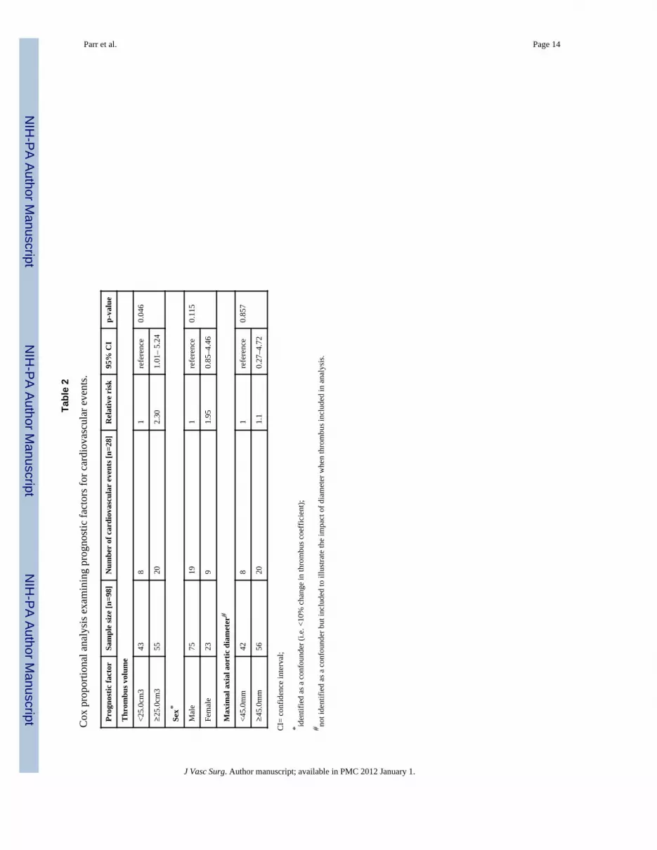
Cardiovascular eventsPatients were followed for a median of 3.0 (inter-quartile range, 2.0–3.6) years. Thefollowing cardiovascular events occurred during follow-up: Non-fatal myocardial infarction(n=13), coronary revascularization (n=4), stroke (n=1), below knee amputation (n=1), andcardiovascular death (n=9). Patients with a large volume of AAA thrombus had a higherincidence of cardiovascular events (Fig. 1). The incidence of cardiovascular events was23.4% [95% CI, 16.0–30.8] and 49.2% [95% CI, 39.8–58.6] for patients with small andlarge AAA thrombus volumes, respectively, at 4 years (p=0.040). The incidence ofcardiovascular events was 24.5% [95% CI, 16.7–32.3] and 46.6% [95% CI, 37.9–55.3] forpatients with small and large maximal axial AAA diameters, respectively, at 4 years(p=0.074) (Fig. 2). AAA thrombus volume ≥25.0cm3 was associated with increasedincidence of new cardiovascular independent of other risk factors by Cox analysis (OR 2.3,
Parr et al. Page 4
J Vasc Surg. Author manuscript; available in PMC 2012 January 1.
NIH
-PA Author Manuscript
NIH
-PA Author Manuscript
NIH
-PA Author Manuscript

95% CI 1.01–5.24, Table 2). Sex was identified as a confounding factor. All other variables,including initial AAA diameter, did not significantly impact on the results.
AAA surgeryDuring follow-up 37 patients underwent AAA repair; 12 by open and 25 by endovascularrepair. No patients died within 30 days of surgery. In a secondary analysis of cardiovascularevents the association of AAA thrombus was examined censoring these patients at AAArepair. The findings of this analysis were similar to the primary analysis. The incidence ofcardiovascular events was 26.9% [95% CI, 18.3–35.5] and 48.9% [95% CI, 40.6–59.5] forpatients with small and large AAA thrombus volumes, respectively, at 4 years (p=0.049).The incidence of cardiovascular events was 26.1% [95% CI, 17.5–34.7] and 48.6% [95% CI,38.1–59.1] for patients with small and large maximal axial AAA diameters, respectively, at4 years (p=0.065). Patients with AAA thrombus volume ≥25.0cm3 had an increasedincidence of cardiovascular events (OR 2.35, 95% CI 0.98–5.63) after adjusting for otherrisk factors although the association was no longer significant, p=0.056.
AAA growthA total of 39 patients had repeat CT scans of their AAAs a median of 1.5 years (inter-quartile range, 1.1–3.3) after their initial imaging and prior to any surgery. These 39 include15 patients selected for AAA repair and another 24 patients in who the physician treating thepatients decided to obtain a further CT. The risk factors of the 39 patients who underwentrepeat CTA in relation to slow (<5cm3) and rapid (≥5cm3) annual abdominal aortic volumeexpansion are shown in Table 3. Rapid expansion was associated with larger initial AAAdiameter and thrombus volume (p≤0.001). Annual median (inter-quartile range) AAAvolume and diameter increase were 5.1cm3 (0.8–10.3) and 1.3mm (−0.2–2.7), respectively.Annual abdominal aortic volume change was positively correlated with maximal axial initialAAA diameter (r=0.44, p=0.006) and thrombus volume (r=0.50, p=0.001) (Fig. 3). InitialAAA thrombus volume ≥15.0cm3 was associated with rapid volumetric growth (≥5cm2/year) (OR 15.0, 95% CI 1.9–115.7, p=0.009). Initial AAA diameter, smoking and diabetesmellitus were not significantly associated with rapid growth after adjusting for thrombusvolume.
DiscussionThe main finding of this study was that the volume of AAA thrombus was associated withsubsequent cardiovascular events and rapid AAA growth. This association was independentof initial AAA diameter at least in our primary analysis. There are few previous studiesexamining the association of thrombus quantity with AAA growth (18,19) and we areunaware of any previous investigation examining the relationship of thrombus volume andcardiovascular outcomes.
Large AAA diameter has been independently associated with increased risk of perioperativecardiovascular complications (32) and 3-year cardiovascular death (2,33) post-endoluminalaneurysm repair. A similar association is found in patients would have small AAAs treatedconservatively (2,3,7–9). Five large studies have previously reported that initial abdominalaortic diameter predicts future cardiovascular events (7–9) all-cause mortality (3,7,8) andcardiovascular mortality (2,3,7) even when not in the aneurysmal range (3). Thus there isconvincing data of the association between aortic diameter and cardiovascular events. Thereason for this association is however currently unknown.
A recent study by our group found thrombus volume was closely correlated with maximumaxial aortic diameter (r=0.74, p<0.0001) in patients with AAA or aortic ectasia (29). This
Parr et al. Page 5
J Vasc Surg. Author manuscript; available in PMC 2012 January 1.
NIH
-PA Author Manuscript
NIH
-PA Author Manuscript
NIH
-PA Author Manuscript

association between AAA diameter and thrombus are in keeping with the findings ofprevious smaller studies (34,35). Currently the factors determining thrombus deposition arenot know and are not examined by this study. AAA thrombus products have beendemonstrated within the systemic circulation and have potential to influence pathwaysimplicated in athero-thrombosis (24–28). We therefore postulated that AAA thrombusproducts could be one of a number of causal links between aortic diameter andcardiovascular events. Our findings support this hypothesis and suggest the associationbetween AAA thrombus and outcome warrants further assessment in larger studies.
Initial AAA diameter is recognized as the most powerful and consistent predictor of AAAgrowth (4,5,11,13,36–41). Two previous studies have examined the association betweenAAA thrombus and growth (18,19). Wolf et al. reviewed CT scans of 80 patients whounderwent repeat CT scans greater than 6 months apart (19). On the section with the largestcross sectional diameter the researchers measured maximal diameter, aneurysm area, lumenarea, thrombus thickness and arc of aortic wall covered by thrombus using hardcopy images.From these measurements they calculated AAA volume, thrombus volume and thrombuspercentage. The authors reported that large thrombus arc, thrombus percent and thrombusarea were highly significantly associated with increased AAA expansion (p<0.001 in allvariables). There was no report of the reproducibility of the measurements techniques usedand the association was not adjusted for initial AAA diameter. A recent longitudinal,prospective study (18) examined 195 AAA patients (40–49mm) with 2 CT scans greaterthan 6 months apart. The authors assessed continuous (increased growth rates) versusdiscontinuous growth. Aneurysms with no thrombus or concentric thrombus were associatedexclusively with discontinuous and slower growth rates (p=0.05). These two previousstudies along with the current investigation provide support for a role of thrombus in AAAprogression.
AAA thrombus has been demonstrated to contain large numbers of polymorphonuclearleukocytes and high concentrations of matrix metalloproteinases, elastase and plasmin(16,17,42). Elastase has been suggested to inhibit mesenchymal cells binding to fibrin andthereby impair repair of aortic injury (43). Plasmin activation of matrix metalloproteinases atthe thrombus and aortic wall interface has been postulated to enhance proteolysis (16).These mechanisms provide a potential link between AAA thrombus and progression. Inkeeping with these findings a recent study reported an association between reduced smallAAA progression and aspirin prescription and two pre-clinical studies have suggested thatanti-platelet interventions inhibit AAA progression (44–46).
In our primary assessment of risk factors for cardiovascular events we did not censorpatients at the time of AAA repair since these events are still common after surgery. In asecondary analysis we censored patients who required AAA repair at the time of surgery.This analysis showed similar findings to our primary assessment except that the associationof thrombus volume with cardiovascular events adjusted for other risk factors was only ofborderline significance.
The current study has a number of limitations. Firstly, small sample size made analysisdifficult. Interaction terms were not able to be employed to assess the relationship betweenthrombus and AAA diameter in relation to cardiovascular events. The results may beconfounded by the relationship between thrombus and AAA diameter, in addition to othervariables. A larger study is needed to adequately assess this aspect before any conclusionscan be reached. Secondly, follow up periods were short. Larger studies with longer termfollow-up are required to confirm the associations we report. Thirdly, a large portion ofpatients in the high thrombus group had an AAA repair, potentially confounding results.Fourthly, volumetric analysis of thrombus may have certain limitations. As the length of the
Parr et al. Page 6
J Vasc Surg. Author manuscript; available in PMC 2012 January 1.
NIH
-PA Author Manuscript
NIH
-PA Author Manuscript
NIH
-PA Author Manuscript

infra-renal aorta varies between individuals this method may overestimate the thrombusburden in patients with greater length. However, as volumetric analysis assesses thrombusquantity this should provide an estimation of the thrombus available for potential thrombus-derived product production and release into the circulation. Finally the findings of this studyare based on patients with AAA who underwent CT assessment only. Whether the findingscan be related to all small AAAs remains to be established.
In conclusion this is the first study to demonstrate an association between AAA thrombusvolume and subsequent cardiovascular events. This association could reflect cardiovascularrisk factors we have not been able to adjust for in this study or may result from the systemiceffects of circulating thrombus products. Further studies are required to assess the clinicalimportance of the association.
References1. Norman PE, Jamrozik K, Lawrence-Brown MM, et al. Population based randomised controlled trial
on impact of screening on mortality from abdominal aortic aneurysm. BMJ 2004 November27;329(7477):1259. [PubMed: 15545293]
2. Brady AR, Fowkes FGR, Thompson SG, et al. Aortic Aneurysm Diameter and Risk ofCardiovascular Mortality. Arterioscler Thromb Vasc Biol 2001 July 1;21(7):1203–7. [PubMed:11451752]
3. Norman P, Le M, Pearce C, et al. Infrarenal Aortic Diameter Predicts All-Cause Mortality.Arterioscler Thromb Vasc Biol 2004 July 1;24(7):1278–82. [PubMed: 15130915]
4. Brady AR, Thompson SG, Fowkes FGR, et al. Abdominal Aortic Aneurysm Expansion: RiskFactors and Time Intervals for Surveillance. Circulation 2004 July 6;110(1):16–21. [PubMed:15210603]
5. Vega de Ceniga M, Gomez R, Estallo L, et al. Growth Rate and Associated Factors in SmallAbdominal Aortic Aneurysms. European Journal of Vascular and Endovascular Surgery2006;31(3):231–6. [PubMed: 16293428]
6. Lederle FA, Wilson SE, Johnson GR, et al. Immediate Repair Compared with Surveillance of SmallAbdominal Aortic Aneurysms. N Engl J Med 2002 May 9;346(19):1437–44. [PubMed: 12000813]
7. Newman AB, Arnold AM, Burke GL, et al. Cardiovascular disease and mortality in older adultswith small abdominal aortic aneurysms detected by ultrasonography: the cardiovascular healthstudy. Annals of Internal Medicine 2001;134(3):182–90. [PubMed: 11177330]
8. Freiberg MS, Arnold AM, Newman AB, et al. Abdominal aortic aneurysms, increasing infrarenalaortic diameter, and risk of total mortality and incident cardiovascular disease events: 10-yearfollow-up data from the Cardiovascular Health Study. Circulation 2008;117(8):1010–7. [PubMed:18268154]
9. Galland R, Whiteley M, Magee T. The fate of patients undergoing surveillance of small abdominalaortic aneurysms. European Journal of Vascular and Endovascular Surgery 1998;16(2):104–9.[PubMed: 9728428]
10. Brown PM, Pattenden R, Gutelius JR. The selective management of small abdominal aorticaneurysms: the Kingston study. Journal of Vascular Surgery 1992;15(1):21–5. [PubMed: 1728677]
11. Santilli SM, Littooy FN, Cambria RA, et al. Expansion rates and outcomes for the 3.0-cm to the3.9-cm infrarenal abdominal aortic aneurysm. Journal of Vascular Surgery 2002;35(4):666–71.[PubMed: 11932660]
12. Norman P, Spencer CA, Lawrence-Brown MM, et al. C-reactive protein levels and the expansionof screen-detected abdominal aortic aneurysms in men. Circulation 2004;110(7):862–6. [PubMed:15302791]
13. Stonebridge P, Draper T, Kelman J. Growth rate of infrarenal aortic aneurysms. European Journalof Vascular and Endovascular Surgery 1996;11(1):70–3. [PubMed: 8564490]
14. Schouten O, van Laanen JH, Boersma E, et al. Statins are associated with a reduced infrarenalabdominal aortic aneurysm growth. European Journal of Vascular & Endovascular Surgery2006;32(1):21–6. [PubMed: 16520071]
Parr et al. Page 7
J Vasc Surg. Author manuscript; available in PMC 2012 January 1.
NIH
-PA Author Manuscript
NIH
-PA Author Manuscript
NIH
-PA Author Manuscript

15. Ouriel K, Donayre C, Shortell CK, et al. The hemodynamics of thrombus formation in arteries.Journal of Vascular Surgery 1991;14(6):757–62. [PubMed: 1960805]
16. Carrell TW, Burnand KG, Booth NA, et al. Intraluminal thrombus enhances proteolysis inabdominal aortic aneurysms. Vascular 2006;14(1):9–16. [PubMed: 16849017]
17. Panek B, Gacko M, Palka J. Metalloproteinases, insulin-like growth factor-I and its bindingproteins in aortic aneurysm. International Journal of Experimental Pathology 2004;85(3):159–64.[PubMed: 15255969]
18. Vega de Ceniga M, Gomez R, Estallo L, et al. Analysis of Expansion Patterns in 4-4.9 cmAbdominal Aortic Aneurysms. Annals of Vascular Surgery 2008;22(1):37–44. [PubMed:18083334]
19. Wolf YG, Thomas WS, Brennan FJ, et al. Computed tomography scanning findings associatedwith rapid expansion of abdominal aortic aneurysms. Journal of Vascular Surgery 1994;20(4):529–35. [PubMed: 7933254]
20. Satta J, Laara E, Juvonen T. Intraluminal thrombus predicts rupture of an abdominal aorticaneurysm. Journal of Vascular Surgery 1996;23(4):737–9. [PubMed: 8627917]
21. Hans SS, Jareunpoon O, Balasubramaniam M, et al. Size and location of thrombus in intact andruptured abdominal aortic aneurysms. Journal of Vascular Surgery 2005;41(4):584–8. [PubMed:15874920]
22. Houard X, Rouzet F, Touat Z, et al. Topology of the fibrinolytic system within the mural thrombusof human abdominal aortic aneurysms. The Journal of Pathology 2007;212(1):20–8. [PubMed:17352452]
23. Houard X, Ollivier V, Louedec L, et al. Differential inflammatory activity across humanabdominal aortic aneurysms reveals neutrophil-derived leukotriene B4 as a major chemotacticfactor released from the intraluminal thrombus. FASEB J 2009 May;23(5):1376–83. [PubMed:19136615]
24. Takagi H, Manabe H, Kawai N, et al. Plasma Fibrinogen and D-dimer Concentrations areAssociated with the Presence of Abdominal Aortic Aneurysm: A Systematic Review and Meta-analysis. European Journal of Vascular and Endovascular Surgery 2009;38(3):273–7. [PubMed:19560946]
25. Parry D, Al-Barjas H, Chappell L, et al. Haemostatic and fibrinolytic factors in men with a smallabdominal aortic aneurysm. British Journal of Surgery 2009;96(8):870–7. [PubMed: 19591171]
26. Danesh J, Whincup P, Walker M, et al. Fibrin D-Dimer and Coronary Heart Disease: ProspectiveStudy and Meta-Analysis. Circulation 2001 May 15;103(19):2323–7. [PubMed: 11352877]
27. Morange PE, Bickel C, Nicaud V, et al. Haemostatic Factors and the Risk of Cardiovascular Deathin Patients With Coronary Artery Disease: The AtheroGene Study. Arterioscler Thromb Vasc Biol2006 December 1;26(12):2793–9. [PubMed: 17023678]
28. Smith A, Patterson C, Yarnell J, et al. Which Hemostatic Markers Add to the Predictive Value ofConventional Risk Factors for Coronary Heart Disease and Ischemic Stroke? The Caerphilly StudyCirculation 2005 November 15;112(20):3080–7.
29. Golledge J, Wolanski P, Parr A, et al. Measurement and determinants of infrarenal aortic thrombusvolume. European Radiology 2008;18(9):1987–94. [PubMed: 18414871]
30. Singh K, Jacobsen B, Solberg S, Kumar S, Arnesen E. The difference between ultrasound andcomputed tomography measurements of aortic diameter increases with aortic diameter: analysis ofaxial images of abdomial aortic and common iliac artery diameter in normal and aneurysmalaortas. The Tromso Study, 1994–1995. Eur J Vasc Endovasc Surg 2004 Aug;28(2):158–67.[PubMed: 15234697]
31. Parr A, Jayaratne C, Buttner P, Golledge J. Comparison of volume and diameter measurement inassessing small abdominal aortic aneurysm expansion examined using computed tomographicangiography. Euro J Radiol. 2010 Mar; corrected proof.
32. Schouten O, Kok N, Hoedt M, et al. The influence of aneurysm size on preioperative cardiacoutcome in elective open infrarenal aortic aneurysm repair. Journal of Vascular Surgery2006;44(3):435–41. [PubMed: 16950412]
Parr et al. Page 8
J Vasc Surg. Author manuscript; available in PMC 2012 January 1.
NIH
-PA Author Manuscript
NIH
-PA Author Manuscript
NIH
-PA Author Manuscript

33. Koskas F, Kieffer E. Long-term survival after elective repair of infrarenal abdominal aorticaneurysm: results of a prospective multicentric study. Association for Academic Research inVascular Surgery (AURC). Annals of Vascular Surgery 1997;11(5):473–81. [PubMed: 9302059]
34. Roberts WC, Ko JM, Pearl GJ. Relation of Weights of Intraaneurysmal Thrombi to MaximalRight-to-Left Diameters of Abdominal Aortic Aneurysms. The American Journal of Cardiology2006;98(11):1519–24. [PubMed: 17126663]
35. Hans SS, Jareunpoon O, Huang R, et al. Relationship of residual intraluminal to intrathromboticpressure in a closed aneurysmal sac. Journal of Vascular Surgery 2003;37(5):949–53. [PubMed:12756338]
36. Solberg S, Singh K, Wilsgaard T, et al. Increased Growth Rate of Abdominal Aortic Aneurysms inWomen. The Tromso Study European Journal of Vascular and Endovascular Surgery 2005;29(2):145–9.
37. Watson C, Walton J, Shaw E, et al. What is the long-term outcome for patients with very smallabdominal aortic aneurysms? European Journal of Vascular and Endovascular Surgery 1997;14(4):299–304. [PubMed: 9366794]
38. Englund R, Hudson P, Hanel K, et al. Expansion rates of small abdominal aortic aneurysms.Australian and New Zealand Journal of Surgery 1998;68(1):21–4. [PubMed: 9440450]
39. Nevitt M, Ballard D, Hallett J. Prognosis of abdominal aortic aneurysms. A population-basedstudy. New England Journal of Medicne 1989;321(15):1009–14.
40. Vardulaki K, Prevost T, Walker N, et al. Growth rates and risk of rupture of abdominal aorticaneurysms. British Journal of Surgery 1998;85(12):1674–80. [PubMed: 9876073]
41. Guirguis E, Barber G. The natural history of abdominal aortic aneurysms. American journal ofsurgery 1991;162(5):481–3. [PubMed: 1951914]
42. Fontaine V, Jacob MP, Houard X, et al. Involvement of the mural thrombus as a site of proteaserelease and activat ion in human aortic aneurysms. American Journal of Pathology 2002;161(5):1701–10. [PubMed: 12414517]
43. Fontaine V, Touat Z, Mtairag EM, et al. Role of Leukocyte Elastase in Preventing CellularReColonization of the Mural Thrombus. Am J Pathol 2004 June 1;164(6):2077–87. [PubMed:15161642]
44. Dai J, Louedec L, Philippe M, et al. Effect of blocking platelet activation with AZD6140 ondevelopment of abdominal aortic aneurysm in a rat aneurysmal model. Journal of VascularSurgery 2009;49(3):719–27. [PubMed: 19028049]
45. Lindholt JS, Sorensen HT, Michel JB, Thomsen HF, Henneberg EW. Low-dose aspirin mayprevent growth and later surgical repair of medium-sized abdominal aortic aneurysms. VascEndovascular Surg 2008 Aug–Sep;42(4):329–34. [PubMed: 18728038]
46. Touat Z, Ollivier V, Dai J, Huisse MG, Bezeaud A, Sebbag U, Palombi T, Rossignol P, Meilhac O,Guillin MC, Michel JB. Renewal of mural thrombus releases plasma markers and is involved inaortic abdominal aneurysm evolution. Am J Pathol 2006 Mar;168(3):1022–30. [PubMed:16507915]
Parr et al. Page 9
J Vasc Surg. Author manuscript; available in PMC 2012 January 1.
NIH
-PA Author Manuscript
NIH
-PA Author Manuscript
NIH
-PA Author Manuscript

Figure 1.Kaplan Meier analysis showing freedom from cardiovascular events in patients with small(<26.0cm3) and large (>26.0cm3) AAA thrombus volumes. * = cardiovascular event rate at4 years.
Parr et al. Page 10
J Vasc Surg. Author manuscript; available in PMC 2012 January 1.
NIH
-PA Author Manuscript
NIH
-PA Author Manuscript
NIH
-PA Author Manuscript

Figure 2.Kaplan Meier analysis showing freedom from cardiovascular events in patients with small(<45.0mm) and large (≥45.0mm) diameter AAAs. * = cardiovascular event rate at 4 years.
Parr et al. Page 11
J Vasc Surg. Author manuscript; available in PMC 2012 January 1.
NIH
-PA Author Manuscript
NIH
-PA Author Manuscript
NIH
-PA Author Manuscript

Figure 3.Scatterplots displaying the association of annual abdominal aortic volume change withthrombus volume (a.) and maximal axial abdominal aortic diameter (b.).
Parr et al. Page 12
J Vasc Surg. Author manuscript; available in PMC 2012 January 1.
NIH
-PA Author Manuscript
NIH
-PA Author Manuscript
NIH
-PA Author Manuscript

NIH
-PA Author Manuscript
NIH
-PA Author Manuscript
NIH
-PA Author Manuscript
Parr et al. Page 13
Table 1
Risk factors of patients at recruitment in relation to AAA thrombus volumes.
Characteristic Total Thrombus <25.0cm3 Thrombus ≥25.0cm3 p-value
Patients 98 43 55
Age, years 73 (67–77) 72 (67–75) 74 (68–78) 0.245
Male 75 (76.5) 28 (65.1) 47 (85.5) 0.029
Follow-up, years 3.0 (2.0–3.6) 3.2 (2.0–3.7) 2.8 (1.9–3.4) 0.358
Maximum AAA axial diameter, mm 47.2 (34.9–54.6) 34.4 (32.2–42.6) 52.8 (49.5–58.1) <0.001
Body Mass Index, weight(kg)/height(m)2 28.2 (25.0–30.8) 28.7 (25.6–32.0) 27.8 (24.5–30.3) 0.150
Intermittent claudication 29 (29.6) 18 (41.9) 11 (20.0) 0.026
Diabetes mellitus 20 (20.4) 8 (18.6) 12 (21.8) 0.803
Ever smoked 86 (87.8) 36 (83.7) 50 (90.9) 0.357
Hypertension 75 (76.5) 33 (76.7) 42 (76.4) 1.000
Coronary heart disease 59 (60.2) 25 (58.1) 34 (61.8) 0.836
Warfarin 13 (13.3) 7 (16.3) 6 (10.9) 0.552
Aspirin 62 (63.3) 27 (62.8) 35 (63.6) 1.0
Calcium channel blocker 24 (24.5) 13 (30.2) 11 (20.0) 0.344
ACE inhibitor 42 (42.9) 22 (51.2) 20 (36.4) 0.156
Statin 65 (66.3) 24 (55.8) 41 (74.5) 0.057
Beta blocker 35 (35.7) 18 (41.9) 17 (30.9) 0.293
Continuous results are presented as medians (inter-quartile range) and compared by Mann Whitney U; nominal results are presented as number (%)and are compared by Fisher’s exact test (2-tailed significance).
J Vasc Surg. Author manuscript; available in PMC 2012 January 1.
NIH
-PA Author Manuscript
NIH
-PA Author Manuscript
NIH
-PA Author Manuscript
Parr et al. Page 14
Tabl
e 2
Cox
pro
porti
onal
ana
lysi
s exa
min
ing
prog
nost
ic fa
ctor
s for
car
diov
ascu
lar e
vent
s.
Prog
nost
ic fa
ctor
Sam
ple
size
[n=9
8]N
umbe
r of
car
diov
ascu
lar
even
ts [n
=28]
Rel
ativ
e ri
sk95
% C
Ip-
valu
e
Thr
ombu
s vol
ume
<25.
0cm
343
81
refe
renc
e0.
046
≥25
.0cm
355
202.
301.
01–
5.24
Sex*
Mal
e75
191
refe
renc
e0.
115
Fem
ale
239
1.95
0.85
–4.4
6
Max
imal
axi
al a
ortic
dia
met
er#
<45.
0mm
428
1re
fere
nce
0.85
7
≥45
.0m
m56
201.
10.
27–4
.72
CI=
con
fiden
ce in
terv
al;
* iden
tifie
d as
a c
onfo
unde
r (i.e
. <10
% c
hang
e in
thro
mbu
s coe
ffic
ient
);
# not i
dent
ified
as a
con
foun
der b
ut in
clud
ed to
illu
stra
te th
e im
pact
of d
iam
eter
whe
n th
rom
bus i
nclu
ded
in a
naly
sis.
J Vasc Surg. Author manuscript; available in PMC 2012 January 1.

NIH
-PA Author Manuscript
NIH
-PA Author Manuscript
NIH
-PA Author Manuscript
Parr et al. Page 15
Table 3
Risk factors of patients at recruitment in relation to AAA expansion ≥ or < median.
Characteristic TotalAnnual AAA volume increase<5.0cm3/year
Annual AAA volume increase≥5.0cm3/year p-value
Patients 39 19 20
Age, years 73 (69–77) 74 (69–77) 71 (68–75) 0.518
Male 27 (69.2) 13 (68.4) 14 (70.0) 0.981
Follow-up, years 1.5 (1.1–3.3) 1.4 (0.9–3.3) 1.6 (1.1–3.3) 0.771
Maximum AAA diameter, mm 42.1 (33.0–47.0) 22.0 (30.3–43.3) 44.8 (41.0–49.3) 0.001
Total AAA volume, cm3 73.6 (46.6–95.2) 49.6 (40.4–60.0) 86.0 (75.2–107.3) <0.001
Thrombus volume 16.4 (10.6–32.0) 11.1 (9.2–13.7) 27.1 (21.4–44.3) <0.001
Warfarin 2 (5.1) 1 (5.3) 1 (5.0) 1.0
Intermittent claudication 15 (38.5) 10 (52.6) 5 (25.0) 0.105
Diabetes Mellitus 9 (23.1) 6 (31.6) 3 (15.0) 0.237
Ever smoked 32 (82.1) 15 (78.9) 17 (85.0) 0.695
Hypertension 29 (74.4) 16 (84.2) 13 (65.0) 0.273
Coronary heart disease 22 (56.4) 14 (73.7) 8 (40.0) 0.054
Continuous results are presented as medians (inter-quartile range) and compared by Mann Whitney U; nominal results are presented as number (%)and are compared by Fisher’s exact test (2-tailed significance).
J Vasc Surg. Author manuscript; available in PMC 2012 January 1.