compressive mechanical properties of the intraluminal thrombus in abdominal aortic aneurysms and...
TRANSCRIPT

Compressive mechanical properties of the intraluminal thrombusin abdominal aortic aneurysms and fibrin-based thrombus mimics
John H. Ashton1, Jonathan P. Vande Geest1,2,3, Bruce R. Simon1,3, and Darren G.Haskett41Biomedical Engineering Interdisciplinary Program, University of Arizona, Tucson, AZ2BIO5 Institute, University of Arizona, Tucson, AZ3Department of Aerospace and Mechanical Engineering, University of Arizona, Tucson, AZ4Department of Agriculture and Bio Systems Engineering, University of Arizona, Tucson, AZ
AbstractAn intraluminal thrombus (ILT) forms in the majority of abdominal aortic aneurysms (AAAs). Whilethe ILT has traditionally been perceived as a byproduct of aneurysmal disease, the mechanicalenvironment within the ILT may contribute to the degeneration of the aortic wall by affectingbiological events of cells embedded within the ILT. In this study, the drained secant modulus (E5 ∼modulus at 5% strain) of ILT specimens (luminal, medial, and abluminal) procured from electiveopen repair was measured and compared using unconfined compression. Five groups of fibrin-basedthrombus mimics were also synthesized by mixing various combinations of fibrinogen, thrombin,and calcium. Drained secant moduli were compared to determine the effect of the components'concentrations on mimic stiffness. The stiffness of the mimics was also compared to the native ILT.Preliminary data on the water content of the ILT layers and mimics was measured. It was found thatthe abluminal layer (E5 = 19.3 kPa) is stiffer than the medial (2.49 kPa) and luminal (1.54 kPa) layers,both of which are statistically similar. E5 of the mimics (0.63, 0.22, 0.23, 0.87, and 2.54 kPa) isdependent on the concentration of all three components: E5 decreases with a decrease in fibrinogen(60 to 20 and 20 to 15 mg/ml) and a decrease in thrombin (3 to 0.3 units/ml), and E5 increases witha decrease in calcium (0.1 to 0.01 M). E5 from two of the mimics were not statistically different thanthe medial and luminal layers of ILT. A thrombus mimic with similar biochemical components,structure, and mechanical properties as native ILT would provide an appropriate test medium forAAA mechanobiology studies.
Keywordsaneurysm; intraluminal thrombus; AAA; compressive modulus; fibrinogen
Address for Correspondence: Jonathan P. Vande Geest, Ph.D., Assistant Professor, Department of Aerospace and MechanicalEngineering, Soft Tissue Biomechanics Laboratory, 1130 N Mountain Ave., PO Box 210119, Tucson, AZ 85721-0119, Tel: (520)621-2514, Fax: (520) 621-8191, [email protected] of Interest Statement: The authors of this paper have no conflict of interest that would inappropriately influence this work.Publisher's Disclaimer: This is a PDF file of an unedited manuscript that has been accepted for publication. As a service to our customerswe are providing this early version of the manuscript. The manuscript will undergo copyediting, typesetting, and review of the resultingproof before it is published in its final citable form. Please note that during the production process errors may be discovered which couldaffect the content, and all legal disclaimers that apply to the journal pertain.
NIH Public AccessAuthor ManuscriptJ Biomech. Author manuscript; available in PMC 2010 May 11.
Published in final edited form as:J Biomech. 2009 February 9; 42(3): 197–201. doi:10.1016/j.jbiomech.2008.10.024.
NIH
-PA Author Manuscript
NIH
-PA Author Manuscript
NIH
-PA Author Manuscript

IntroductionAbdominal aortic aneurysms (AAAs) affect a significant portion of the population in developedcountries, and the incidence is believed to be increasing as life expectancy increases. AAAsare present in 4% to 8% of men over 60 and 1% to 3% of women over 60 [1]. The long timecourse of the disease results in the formation of an intraluminal thrombus (ILT) in most AAAs[2]. Some investigators have discussed the mechanical role of the ILT, with this tissue primarilyacting as a mechanical shield, thus decreasing the stresses acting on the AAA wall [3,4].
The ILT has traditionally been perceived as a byproduct of aneurysmal disease. However, someinvestigators have hypothesized that the ILT may play a more active role in the pathophysiologyof AAA development [2]. The mechanical environment within the ILT and the AAA wall (e.g.,matrix strain) changes as the aortic wall dilates. We hypothesize that this mechanicalenvironment may affect the biological activity of cells embedded within the ILT and thereforecontribute to the degeneration of the aortic wall. The mechanical properties of the ILT havebeen investigated by a number of investigators. Many of these experimental investigationshave been based on the tensile behavior of the ILT [5-7]. These tests have primarily been onthe luminal and medial layers of the ILT, as gripping the abluminal layer is often timesimpossible. The hydraulic permeability of the ILT was investigated by Adolph et al. [8],however no distinction was made between how these values vary through the thickness of thethrombus. To our knowledge, little information exists in the literature regarding thecompressive mechanical behavior of the ILT, especially for the abluminal layer. Suchinformation may be important not only in identifying how stress distributions are distributedwithin the ILT and AAA wall, but also may be helpful in future studies on ILT mechanobiology.
Development of a thrombus mimic with similar components, structure, and mechanicalproperties as native ILT would facilitate a more controlled environment for AAAmechanobiology studies. Such a mimic would be ideal for in vitro testing as the number ofnative ILT specimens available for testing will likely continue to decrease with the increasingpopularity of endovascular repair. Fibrin-based constructs present an attractive option for themimic because they can be constructed from blood proteins and components present in theclotting cascade, and thus have a similar chemical structure as native ILT. The mechanicalproperties of these fibrin-based constructs (e.g., elastic modulus, permeability, and strength)can also be manipulated by varying the concentrations of fibrinogen [9-12], thrombin [12],factor XIII [9,13], fibronectin [14,15], platelets [11], and calcium [9,11-13,16,17].
The purpose of this study was threefold: first, to measure the drained secant modulus at 5%strain (E5) of the different layers of ILT taken from open repair; second, to measure how theconcentrations of fibrinogen, thrombin, and calcium affect E5 in fibrin based mimics; and third,to determine the apparent differences in properties between the developed mimic and nativeILT. In addition, preliminary information on the water content and microstructure of the ILTand mimics was assessed. Such information will serve as a baseline dataset for future studiesinvestigating the pressure distribution within the ILT and how the ILT may play a role inaneurysmal mechano-pathophysiology.
MethodsILT Specimens
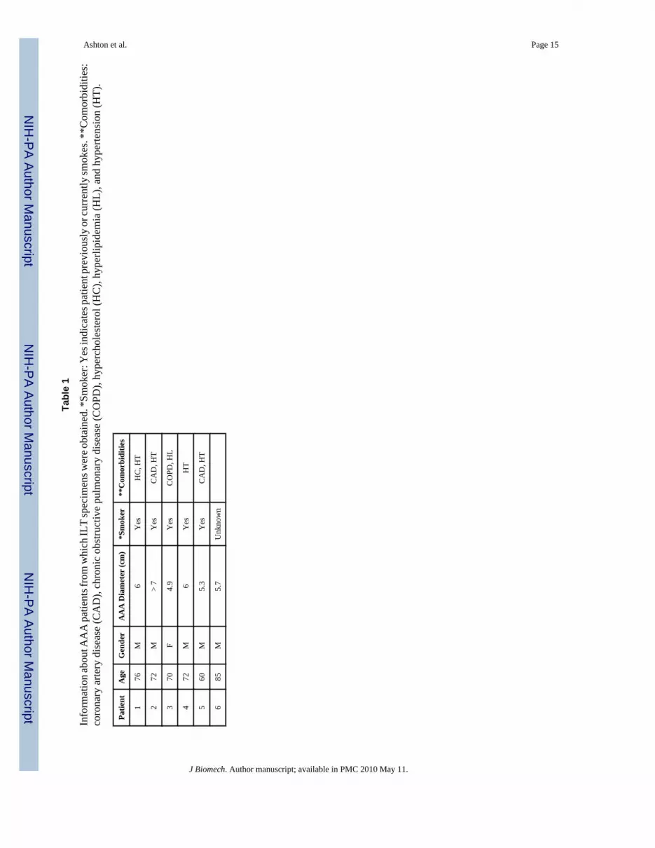
ILT specimens were procured from the elective open AAA repairs of six patients at theUniversity Medical Center, Tucson, AZ in accordance with the Human Subjects ProtectionProgram at the University of Arizona. General information about the patients is shown in Table1. Specimens were placed in an isotonic phosphate buffered solution (PBS) immediately afterextraction from the aorta and stored at 4°C until testing. All tests were performed within 48
Ashton et al. Page 2
J Biomech. Author manuscript; available in PMC 2010 May 11.
NIH
-PA Author Manuscript
NIH
-PA Author Manuscript
NIH
-PA Author Manuscript

hours of the surgery. Gentle physical manipulation was used to separate the ILT specimensinto three layers based on relative position and color: abluminal, medial, and luminal (Figure1).
Thrombus MimicsBovine fibrinogen (Sigma, F8630) was dissolved in Hank's Balanced Salt Solution (HBSS,Mediatech, 21-020-CV) to concentrations of 40 and 80 mg/ml, bovine thrombin (Sigma,T6200) was dissolved in water to a concentration of 100 units/ml, and calcium chloride wasdissolved in HBSS to yield a calcium rich HBSS solution (1 M calcium). Five groups of mimicswere constructed by mixing the solutions, additional HBSS, and water with various ratios inwell plates to yield the final concentrations listed in Table 2. Mixed solutions were set in anincubator (37°C, 10% CO2) overnight to gelate. The concentrations in M1 were chosen as abaseline, and in groups M2, M3, and M4, the concentration of one component (fibrinogen,thrombin, or calcium) was varied from the baseline to determine the effect of that component'sconcentration on E5. In M5, the concentrations of thrombin and calcium that yielded the stiffestmimics were used and the fibrinogen concentration was increased to 60 mg/ml in attempt tocreate an even stiffer mimic.
Drained Secant ModulusA circular biopunch (8 mm diameter) was used to cut cylindrically shaped samples from eachlayer of the ILT and the mimic well plates. The thickness of each sample was measured bytaking the average of five measurements using a caliper. There were 14, 15, and 18 ILT samplesisolated from the abluminal, medial and luminal layers, respectively (thickness = 3.0±0.8, 4.5±0.9, and 4.3±1.2 mm, respectively) and 32 mimic samples (thickness = 3.9±0.6 mm). Mimicswere set in a bath of PBS for at least one hour to allow fluid saturation.
Unconfined-compression stress-relaxation tests were performed on the samples in a bath ofPBS at 37°C using a dynamic mechanical analyzer (Perkin Elmer, Pyris Diamond DMA) whichhad a spatial resolution of 1 μm and a load resolution of 0.5 μN (a schematic of unconfined-compression testing is shown in Figure 2). Stress relaxation tests were performed by applyingand holding a 5% compressive strain for 20 minutes while measuring the correspondingdecrease in compressive load over time. Strains of 10% and 15% were also subsequentlyapplied for 20 minutes each. Since the samples did not equilibrate after 20 minutes, theequilibrium load was determined by fitting the data to a 3-term sum of exponentials equation:
(1)
where L is instantaneous load, t is time, A is equilibrium load, and Bi and Ci are constants. A,Bi, and Ci were fit to the data using Marquardt-Levenberg nonlinear regression technique withinthe commercially available software package SigmaStat. Drained equilibrium stress (T∞) wascalculated as the equilibrium load divided by the initial cross sectional area. E5 was calculatedas T∞ at a 5% compressive strain divided by -0.05.
Water Content and SEMAs a step towards our long term goal, preliminary water content data and SEM images wererecorded for the ILT and mimics. The initial water content of all layers of the ILT from onepatient and from each of the mimics was measured. The volume of samples saturated with PBSwas obtained by measuring the volume of fluid they displaced in a graduated cylinder. ThePBS-saturated samples were then weighed, set in a biosafety cabinet to dry, and weighed 16hours later. Initial water volume was calculated as the change in mass multiplied by the density
Ashton et al. Page 3
J Biomech. Author manuscript; available in PMC 2010 May 11.
NIH
-PA Author Manuscript
NIH
-PA Author Manuscript
NIH
-PA Author Manuscript

of water. The reported value of water content is given as the water volume divided by the PBS-saturated sample volume. Water content of M5 was not measured.
M1 was prepared for SEM imaging as follows. The sample was fixed in gluteraldehyde,formaldehyde, and 0.015% ruthenium red. It was then fixed in osmium tetraoxide. The samplewas dehydrated in ethanol and quenched in liquid nitrogen, which broke the sample into piecesto allow us to image an inside area instead of the surface. It was dried in CO2 to its criticalpoint, gold coated, and then SEM imaged (Hitachi S-3400N).
Statistical AnalysisPair-wise comparisons of the drained secant moduli of the ILT layers and the mimics wereperformed with a Mann-Whitney rank sum test using the software program SigmaStat. A non-parametric test was used due to non-normal distributions of the sample groups. Significantdifference was determined by a p-value < 0.05.
ResultsA representative ILT sample is shown in Figure 1. Figure 3 shows the load response over timefor a typical unconfined-compression stress-relaxation test. T∞ at each strain of the three ILTlayers is shown in Figure 4. The luminal and medial layers appear to have a similar stiffness,while the abluminal layer had a much stiffer response. Figure 5 shows the T∞ at each strain ofthe mimics. M5, which has the highest fibrinogen concentration and the lowest calciumconcentration, appears to be the stiffest, while M2, which has the lowest fibrinogenconcentration, was the most compliant.
Mean and standard error values of E5 for the ILT layers and mimics are reported in Table 3along with statistical comparisons between each mimic and ILT group. The abluminal layerwas found to have a significantly greater E5 than all other samples. There was no significantdifference between the E5 for the medial and luminal layers. The E5 of M4 was not significantlydifferent than the luminal layer, and the E5 of M5 was not significantly different than the mediallayer. All other mimics were significantly less stiff than the ILT layers. The differences betweenthe baseline mimic (M1) and the other mimic groups (M2, M3, M4, and M5) indicate that thestiffness of the mimic can be manipulated by adjusting the concentration of fibrinogen,thrombin, or calcium. For example, a decrease in fibrinogen (20 to 15 mg/ml) or thrombin (3.0to 0.3 units/ml) decreases E5, while a decrease in calcium (0.10 to 0.01 M) increases E5.
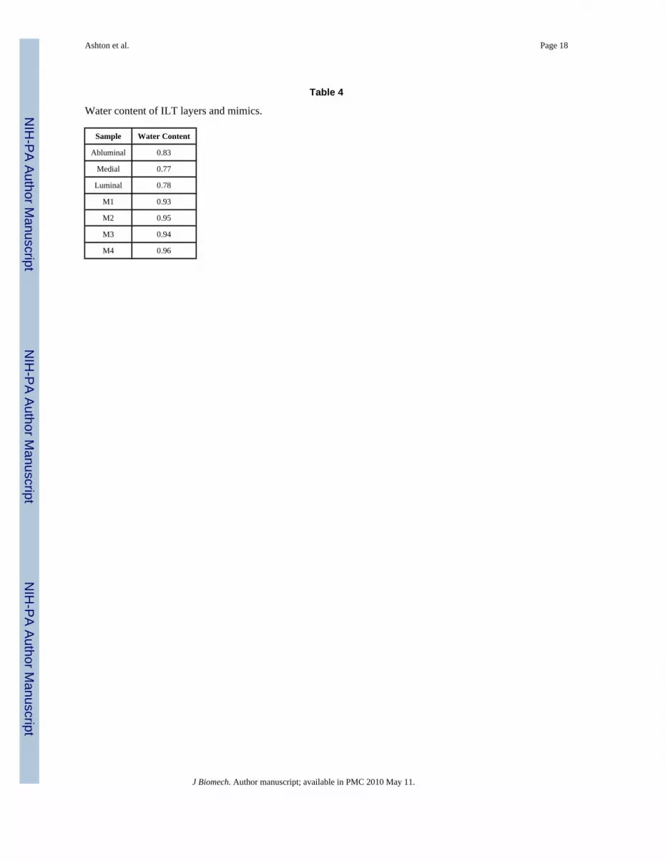
Water content for the ILT layers and mimics is reported in Table 4. Water content of the ILTranged from 0.77 to 0.83 and that of the mimics ranged from 0.93 to 0.96. Figure 6 is an SEMimage taken of M1. While there appears to be significant degree of nonhomogeneity in mimicstructure, the fibrinous network of the clot is qualitatively similar to results reported by Wanget al. for native ILT [5].
DiscussionIn this study, the compressive behavior of the luminal, medial and abluminal layers of the ILTand five thrombus mimics were investigated using an unconfined-compression setup. Wereport a higher compressive stiffness for the abluminal layer compared to the medial andluminal layers, suggesting a nonhomogenous mechanical behavior of the ILT in compression.Our results also indicate that a mimic can be constructed whose mechanical properties can bemodified controllably using variations in calcium, fibrinogen, and thrombin concentrations. Inparticular, we present here mimics which behave mechanically similar to the luminal andmedial layers of native ILT.
Ashton et al. Page 4
J Biomech. Author manuscript; available in PMC 2010 May 11.
NIH
-PA Author Manuscript
NIH
-PA Author Manuscript
NIH
-PA Author Manuscript

Several other groups have investigated the mechanical properties of the ILT. Boschetti et al.reported the compressive elastic modulus of the ILT luminal, medial, and abluminal layers at45% compressive strain to be approximately 11, 20, and 22 kPa, respectively [18], which ishigher than what we report (1.5, 2.5, and 19 kPa) at a 5% compressive strain. This increase islikely due to the difference in applied compressive strains. However, in both cases the datashow a trend of increase in modulus from the luminal to the abluminal layers. Hinnen et al.reported an ILT elastic modulus of 35 kPa based on a cyclic shear strain on the order of 0.01.However, in this study the ILT was not divided into layers [19]. This is higher than what wereport, which is likely due to the difference in methods—Hinnen et al. measured the shearmodulus and converted it to elastic modulus using the Poisson's ratio. They also report thedevelopment of a mimic with a similar elastic modulus as the ILT, but no mention is made ofaddressing the biological composition and structure of the ILT. Similarities in these propertieswill also likely be important in developing an in vitro test setup for future mechanobiologicalstudies.
Our results demonstrate that the E5 of the mimics can be adjusted by changing the concentrationof fibrinogen, thrombin, or calcium. Carr and Carr showed that the elastic modulus of athrombus increased with an increase in fibrinogen (1 to 4 mg/ml) and decreased with an increasein calcium (5 to 20 mM) [11], which is consistent with our results. Ryan et al. modeled fibrin-based constructs as a viscoelastic material and found similar relationships between storagemodulus and the concentrations of fibrinogen and calcium [12]. However, they also found thatthe storage modulus decreases with an increase in thrombin concentration (0.1 to 5 units/ml),which is opposite to the relationship we found. This difference may be due to their differencein methodology—they used cyclic shear tests while we used stress-relaxation compressiontests.
In addition, Ryan et al. examined the effects of fibrinogen, thrombin, and calciumconcentrations on the structure of the fiber network in fibrin-based constructs by analyzingcomputerized three-dimensional models based on SEM images from the constructs. Theyfound that increasing fibrinogen, increasing thrombin, and decreasing calcium decreases fiberlength and diameter and increases fiber density and branch points in the fibrin network. Theyconcluded that maximal stiffness is established in fibrin networks that consist of a balancebetween large fibers and high branching. These results suggest that there is an optimalconcentration of fibrinogen, thrombin, and calcium for making a mimic of maximum stiffness.A fibrinogen or thrombin concentration that is too high or a calcium concentration that is toolow may decrease the stiffness of the resulting mimic because, although the branching may begreat, fiber size would be too small.
Two of our mimics (M4 and M5) were not statistically different than the ILT luminal andmedial layers, respectively. The long-term goal of our group is to develop a mimic whichmechanically, structurally, and biochemically behaves similar to the ILT. Our results suggestthat our current mimics may be adequate for mimicking the luminal and medial layercompliance, while the abluminal layer will require further modification. Increasing the E5 ofthe ‘abluminal’ mimic may be accomplished by further adjusting the concentrations ofcomponents already stated or through the addition of additional components. For example, thestiffness of thrombus has also been shown to increase with the presence of factor XIII [13] andfibronectin [14]. Furthermore, platelets may be added to the mimic in future studies, as theyare a major component of thrombus and have also been shown to increase stiffness of fibrin-based constructs [11]. Addition of such biologically active components may also provide amechanism by which the aging of the ILT can be modeled.
While not the focus of the current work, determination of the hydraulic permeability of the ILTand our mimic is ongoing within our laboratory. Specifically of interest will be how these
Ashton et al. Page 5
J Biomech. Author manuscript; available in PMC 2010 May 11.
NIH
-PA Author Manuscript
NIH
-PA Author Manuscript
NIH
-PA Author Manuscript
values may change as a function of location in the ILT as well as progression of the disease(thick vs. thin ILT). This information can also be used in future studies to model the convectivetransport in this tissue, which will be essential in assessing the effectiveness of drug deliveryin. Development of a pharmacological treatment has been suggested as an alternative treatmentof AAA which have been detected but not progressed to a stage where invasive procedure (i.e.open surgery or endovascular repair) is warranted [20]. In other words, when an AAA isdetected, treatment to inhibit (or slow) the progression of AAA can be employed without delay,instead of employing a period of watchful monitoring. Doxycycline, an MMP inhibitor, is onesuch drug currently under investigation [21,22].
One of the limitations of this study is the number of ILT samples tested. For example, thereported water content data only had one sample for each group. Current research in ourlaboratory is aimed at further identifying differences in water content and structure for the ILTlayers and our mimics. For example, while we did find a qualitative similarity in our SEMmimic structure compared to ILT structure reported elsewhere, it is not immediately clearwhether the non-uniformity in structure shown in Figure 6 is real, or simply an artifact of theSEM specimen preparation methods. Another possible limitation of our study was the lengthof time in which samples were stored prior to testing; specifically as such storage may resultin degradation and changes in mechanical behavior. However, our data suggest that there wasno significant difference in E5 for samples tested within 24 hours of the AAA repair versus 24to 48 hours after the repair. It is also important to note that the E5 reported here is based oncompression at 5% strain, whereas it may not be accurate at larger strains. In fact, our resultssuggest that the drained properties above 5% may be mildly nonlinear (Figure 4). Otherresearchers have similarly reported increasing stiffness at higher strains of thrombus and fibrin-based constructs [23-25].
As previously stated, our goal is to develop a mimic which has similar properties to the ILT,and the immediate future goal of our lab is to measure and compare permeability between thegroups by analyzing the time dependent change in load of our unconfined-compression tests.Finite element analysis can also then be used to model the ILT layers and mimics as poroelasticmaterials. Further investigations will also include computationally modeling the fluidmovement through the ILT during the heart cycle and investigating the potential of differentdrug delivery strategies. Additionally, cells typically found in the ILT (e.g. macrophages andneutrophils) can be embedded into the mimics to study how the ILT may be involved in themechanobiology and progression of AAA.
AcknowledgmentsThe authors would like to thank Dr.'s Joseph Mills, Son Duong, John Hughes, and Kay Goshima of the VascularSurgery Department for their help in procuring ILT samples. Funding for this work was provided by the NSF (CAREER0644570 – JPVG) and the NIH Biomedical Cardiovascular Training Grant (HL007955 – BME GIDP).
References1. Al-Omran M, Verma S, Lindsay TF, Weisel RD, Sternbach Y. Clinical decision making for
endovascular repair of abdominal aortic aneurysm. Circulation 2004;110(23):e517–23. [PubMed:15583084]
2. Swedenborg J, Eriksson P. The intraluminal thrombus as a source of proteolytic activity. Ann N YAcad Sci 2006;1085:133–8. [PubMed: 17182929]
3. Wang DH, Makaroun MS, Webster MW, Vorp DA. Effect of intraluminal thrombus on wall stress inpatient-specific models of abdominal aortic aneurysm. J Vasc Surg 2002;36(3):598–604. [PubMed:12218961]
Ashton et al. Page 6
J Biomech. Author manuscript; available in PMC 2010 May 11.
NIH
-PA Author Manuscript
NIH
-PA Author Manuscript
NIH
-PA Author Manuscript

4. Hinnen JW, Koning OH, Visser MJ, Van Bockel HJ. Effect of intraluminal thrombus on pressuretransmission in the abdominal aortic aneurysm. J Vasc Surg 2005;42(6):1176–82. [PubMed:16376211]
5. Wang DH, Makaroun M, Webster MW, Vorp DA. Mechanical properties and microstructure ofintraluminal thrombus from abdominal aortic aneurysm. J Biomech Eng 2001;123(6):536–9.[PubMed: 11783723]
6. Di Martino E, Mantero S, Inzoli F, Melissano G, Astore D, Chiesa R, Fumero R. Biomechanics ofabdominal aortic aneurysm in the presence of endoluminal thrombus: experimental characterisationand structural static computational analysis. European Journal of Vascular & Endovascular Surgery1998;15(4):290–9. [PubMed: 9610340]
7. Vande Geest JP, Sacks MS, Vorp DA. A planar biaxial constitutive relation for the luminal layer ofintra-luminal thrombus in abdominal aortic aneurysms. J Biomech 2006;39(13):2347–54. [PubMed:16872617]
8. Adolph R, Vorp DA, Steed DL, Webster MW, Kameneva MV, Watkins SC. Cellular content andpermeability of intraluminal thrombus in abdominal aortic aneurysm. J Vasc Surg 1997;25(5):916–26. [PubMed: 9152321]
9. Alston SM, Solen KA, Broderick AH, Sukavaneshvar S, Mohammad SF. New method to prepareautologous fibrin glue on demand. Transl Res 2007;149(4):187–95. [PubMed: 17383592]
10. Blomback B, Okada M. Fibrin gel structure and clotting time. Thromb Res 1982;25(1-2):51–70.[PubMed: 6121390]
11. Carr ME Jr, Carr SL. Fibrin structure and concentration alter clot elastic modulus but do not alterplatelet mediated force development. Blood Coagul Fibrinolysis 1995;6(1):79–86. [PubMed:7795157]
12. Ryan EA, Mockros LF, Weisel JW, Lorand L. Structural origins of fibrin clot rheology. Biophys J1999;77(5):2813–26. [PubMed: 10545379]
13. Gladner JA, Nossal R. Effects of crosslinking on the rigidity and proteolytic susceptibility of humanfibrin clots. Thromb Res 1983;30(3):273–88. [PubMed: 6223406]
14. Kamykowski GW, Mosher DF, Lorand L, Ferry JD. Modification of shear modulus and creepcompliance of fibrin clots by fibronectin. Biophys Chem 1981;13(1):25–8. [PubMed: 7260326]
15. Okada M, Blomback B, Chang MD, Horowitz B. Fibronectin and fibrin gel structure. J Biol Chem1985;260(3):1811–20. [PubMed: 2857179]
16. Okada M, Blomback B. Calcium and fibrin gel structure. Thromb Res 1983;29(3):269–80. [PubMed:6845281]
17. Shen LL, Hermans J, McDonagh J, McDonagh RP, Carr M. Effects of calcium ion and covalentcrosslinking on formation and elasticity of fibrin cells. Thromb Res 1975;6(3):255–65. [PubMed:1114492]
18. Inzoli F, Boschetti F, Zappa M, Longo T, Fumero R. Biomechanical factors in abdominal aorticaneurysm rupture. Eur J Vasc Surg 1993;7:667–74. [PubMed: 8270069]
19. Hinnen JW, Rixen DJ, Koning OH, van Bockel JH, Hamming JF. Development of fibrinous thrombusanalogue for in-vitro abdominal aortic aneurysm studies. J Biomech 2007;40(2):289–95. [PubMed:16516895]
20. Rentschler M, Baxter BT. Pharmacological approaches to prevent abdominal aortic aneurysmenlargement and rupture. Ann N Y Acad Sci 2006;1085:39–46. [PubMed: 17182921]
21. Petrinec D, Liao S, Holmes DR, Reilly JM, Parks WC, Thompson RW. Doxycycline inhibition ofaneurysmal degeneration in an elastase-induced rat model of abdominal aortic aneurysm:preservation of aortic elastin associated with suppressed production of 92 kD gelatinase. J Vasc Surg1996;23(2):336–46. [PubMed: 8637112]
22. Bartoli MA, Parodi FE, Chu J, Pagano MB, Mao D, Baxter BT, Buckley C, Ennis TL, ThompsonRW. Localized administration of doxycycline suppresses aortic dilatation in an experimental mousemodel of abdominal aortic aneurysm. Ann Vasc Surg 2006;20(2):228–36. [PubMed: 16572291]
23. Roberts WW, Lorand L, Mockros LF. Viscoelastic properties of fibrin clots. Biorheology 1973;10(1):29–42. [PubMed: 4724175]
24. Storm C, Pastore JJ, MacKintosh FC, Lubensky TC, Janmey PA. Nonlinear elasticity in biologicalgels. Nature 2005;435(7039):191–4. [PubMed: 15889088]
Ashton et al. Page 7
J Biomech. Author manuscript; available in PMC 2010 May 11.
NIH
-PA Author Manuscript
NIH
-PA Author Manuscript
NIH
-PA Author Manuscript

25. Weisel JW. Structure of fibrin: impact on clot stability. J Thromb Haemost 2007;5:116–24. [PubMed:17635717]
Ashton et al. Page 8
J Biomech. Author manuscript; available in PMC 2010 May 11.
NIH
-PA Author Manuscript
NIH
-PA Author Manuscript
NIH
-PA Author Manuscript

Figure 1.ILT specimen with abluminal (A), medial (M), and luminal (L) layers.
Ashton et al. Page 9
J Biomech. Author manuscript; available in PMC 2010 May 11.
NIH
-PA Author Manuscript
NIH
-PA Author Manuscript
NIH
-PA Author Manuscript

Figure 2.Schematic of an unconfined-compression test. The undeformed sample is on the left and thedeformed sample is on the right. Step compressive strains of 5, 10, and 15% were applied toeach sample, and the time dependent load relaxation was recorded.
Ashton et al. Page 10
J Biomech. Author manuscript; available in PMC 2010 May 11.
NIH
-PA Author Manuscript
NIH
-PA Author Manuscript
NIH
-PA Author Manuscript

Figure 3.Load (L) output over time (t) for an unconfined-compression stress-relaxation test. Arrowsindicate where strains of -0.05, -0.10, and -0.15 were applied.
Ashton et al. Page 11
J Biomech. Author manuscript; available in PMC 2010 May 11.
NIH
-PA Author Manuscript
NIH
-PA Author Manuscript
NIH
-PA Author Manuscript
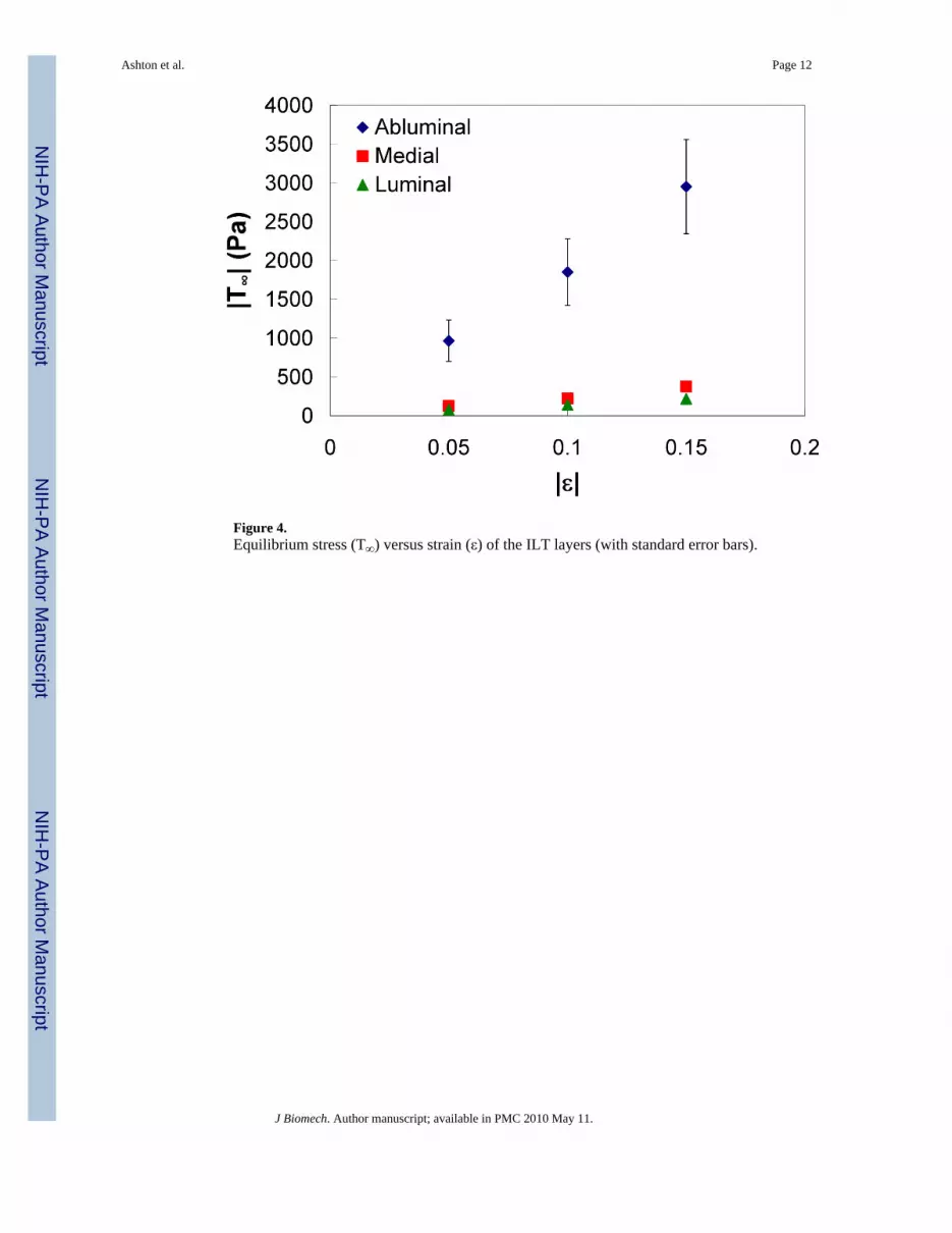
Figure 4.Equilibrium stress (T∞) versus strain (ε) of the ILT layers (with standard error bars).
Ashton et al. Page 12
J Biomech. Author manuscript; available in PMC 2010 May 11.
NIH
-PA Author Manuscript
NIH
-PA Author Manuscript
NIH
-PA Author Manuscript

Figure 5.Equilibrium stress (T∞) versus strain (ε) of the thrombus mimics (with standard error bars).
Ashton et al. Page 13
J Biomech. Author manuscript; available in PMC 2010 May 11.
NIH
-PA Author Manuscript
NIH
-PA Author Manuscript
NIH
-PA Author Manuscript

Figure 6.SEM image of M1. The scale bar is 20 microns.
Ashton et al. Page 14
J Biomech. Author manuscript; available in PMC 2010 May 11.
NIH
-PA Author Manuscript
NIH
-PA Author Manuscript
NIH
-PA Author Manuscript
NIH
-PA Author Manuscript
NIH
-PA Author Manuscript
NIH
-PA Author Manuscript
Ashton et al. Page 15
Tabl
e 1
Info
rmat
ion
abou
t AA
A p
atie
nts f
rom
whi
ch IL
T sp
ecim
ens w
ere o
btai
ned.
*Sm
oker
: Yes
indi
cate
s pat
ient
pre
viou
sly
or cu
rren
tly sm
okes
. **C
omor
bidi
ties:
coro
nary
arte
ry d
isea
se (C
AD
), ch
roni
c ob
stru
ctiv
e pu
lmon
ary
dise
ase
(CO
PD),
hype
rcho
lest
erol
(HC
), hy
perli
pide
mia
(HL)
, and
hyp
erte
nsio
n (H
T).
Patie
ntA
geG
ende
rA
AA
Dia
met
er (c
m)
*Sm
oker
**C
omor
bidi
ties
176
M6
Yes
HC
, HT
272
M>
7Y
esC
AD
, HT
370
F4.
9Y
esC
OPD
, HL
472
M6
Yes
HT
560
M5.
3Y
esC
AD
, HT
685
M5.
7U
nkno
wn
J Biomech. Author manuscript; available in PMC 2010 May 11.

NIH
-PA Author Manuscript
NIH
-PA Author Manuscript
NIH
-PA Author Manuscript
Ashton et al. Page 16
Table 2
Final component concentrations of thrombus mimics with number of samples tested (n).
Group Fibrinogen (mg/ml) Thrombin (NIH units/ml) Calcium (M)
M1 (n=9) 20 3.0 0.10
M2 (n=5) 15 3.0 0.10
M3 (n=5) 20 0.3 0.10
M4 (n=8) 20 3.0 0.01
M5 (n=5) 60 3.0 0.01
J Biomech. Author manuscript; available in PMC 2010 May 11.

NIH
-PA Author Manuscript
NIH
-PA Author Manuscript
NIH
-PA Author Manuscript
Ashton et al. Page 17
Tabl
e 3
Com
pres
sive
dra
ined
seca
nt m
odul
i (E 5
) with
stan
dard
erro
r (SE
) and
stat
istic
al d
iffer
ence
of E
5 bet
wee
n ab
lum
inal
(Abl
), m
edia
l (M
ed),
and
lum
inal
(Lum
)la
yers
and
mim
ic g
roup
s (M
1, M
2, M
3, M
4, M
5). Y
es si
gnifi
es a
stat
istic
al d
iffer
ence
bet
wee
n th
e tw
o sa
mpl
es (p
< 0
.05)
.
E5 (
±SE
)A
blM
edL
umM
1M
2M
3M
4M
5
Pap<
0.05
p<0.
05p<
0.05
p<0.
05p<
0.05
p<0.
05p<
0.05
p<0.
05
Abl
1930
0 (5
300)
--Y
esY
esY
esY
esY
esY
esY
es
Med
2490
(540
)--
No
Yes
Yes
Yes
Yes
No
Lum
1540
(220
)--
Yes
Yes
Yes
No
Yes
M1
635
(81)
--Y
esY
esY
esY
es
M2
216
(50)
--N
oY
esY
es
M3
232
(47)
--Y
esY
es
M4
868
(74)
--Y
es
M5
2540
(270
)--
J Biomech. Author manuscript; available in PMC 2010 May 11.
NIH
-PA Author Manuscript
NIH
-PA Author Manuscript
NIH
-PA Author Manuscript
Ashton et al. Page 18
Table 4
Water content of ILT layers and mimics.
Sample Water Content
Abluminal 0.83
Medial 0.77
Luminal 0.78
M1 0.93
M2 0.95
M3 0.94
M4 0.96
J Biomech. Author manuscript; available in PMC 2010 May 11.